
B C R R e a r r a n... L e u k e m i a R e...
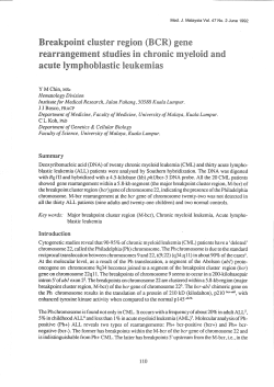
BCR Rearrangement–Negative Chronic Myelogenous Leukemia Revisited By Razelle Kurzrock, Carlos E. Bueso-Ramos, Hagop Kantarjian, Emil Freireich, Susan L. Tucker, Michael Siciliano, Susan Pilat, and Moshe Talpaz Purpose: To document the characteristics of patients with major breakpoint cluster region (M-bcr) rearrangement–negative chronic myelogenous leukemia (CML). Patients and Methods: The hematopathologist, who was blinded to patients’ molecular status, reviewed the referral bone marrows and peripheral-blood smears from 26 patients with Philadelphia (Ph) translocation– negative CML who lacked Bcr rearrangement (and other evidence of a Bcr-Abl anomaly) and 14 patients (controls) with chronic-phase Ph-positive CML. Clinical data was ascertained by chart review. Results: Among the 26 M-bcr rearrangement–negative CML patients, three pathologic subtypes emerged: (1) patients indistinguishable from classic CML (n ⴝ 9), (2) patients with atypical CML (n ⴝ 8), and (3) patients with chronic neutrophilic leukemia (n ⴝ 9). Among the 14 patients with Ph-positive CML who were included in the blinded review, 13 were classified as classic CML, and one was classified as atypical CML. The only statis- tically significant difference between M-bcr rearrangement–negative subgroups was in the proportion of patients having karyotypic abnormalities, an observation common only in patients with atypical CML (P ⴝ 0.008). However, the small number of patients in each subgroup limited our ability to differentiate between them. Interferon alfa induced complete hematologic remission in five of 14 patients; four of these remissions lasted more than 5 years. Only one of 26 patients developed blast crisis. The median survival of the 26 patients was 37 months. Conclusion: Patients with M-bcr rearrangement– negative CML fall into three morphologic subgroups. Disease evolution does not generally involve blastic transformation. Instead, patients show progressive organomegaly, leukocytosis, anemia, and thrombocytosis. Some patients in each subgroup can respond to interferon alfa. J Clin Oncol 19:2915-2926. © 2001 by American Society of Clinical Oncology. HE HALLMARK OF chronic myelogenous leukemia (CML) is the Philadelphia (Ph) translocation t(9; 22)(q34;q11), which, at the molecular level, results in the transfer of the 3' end of the ABL gene next to the 5' end of the disrupted BCR gene.1-5 The product is a chimeric BCR-ABL gene. In the majority of cases of CML, the BCR breakpoint occurs in the central, major breakpoint cluster region (M-bcr). Exon b2 or b3 of BCR is joined to exon a2 of ABL, resulting in a b2-a2 or b3-a2 junction. This transcript encodes a 210-kd protein (p210Bcr-Abl).4,5 The creation of this anomalous protein is believed to play a critical role in the pathogenesis and phenotypic manifestations of CML. Patients with Ph-positive CML have typical clinical and morphologic features: splenomegaly, neutrophilia, basophilia, frequent thrombocytosis, a low leukocyte alkaline phosphatase (LAP) score, and a hypercellular bone marrow with an increased myeloid:erythroid ratio and granulocytic hyperplasia. The disease course inevitably evolves from a chronic phase to a terminal blast transformation phase. A minority of patients (5% to 10%) do not have cytogenetic evidence of the Ph chromosome. Over the last few years, M-bcr rearrangement has been well documented in a subset of Ph-negative patients.6-11 It is now established that M-bcr rearrangement–positive CML patients have a molecular fingerprint (ie, the p210Bcr-Abl protein) and a clinical phe- notype and outcome that is indistinguishable from that of their Ph-positive counterparts.6,7 The existence of a Phnegative, M-bcr rearrangement–negative CML has, however, remained a matter of debate. Some investigators have suggested that these individuals represent part of the spectrum of chronic myelomonocytic leukemia (CMMoL).12 Others have reported that their features differ subtly, but recognizably, from classic CML, and that they should be designated atypical CML.13 A subset of these patients seems to have a disease highly reminiscent of CML.14,15 An entity called chronic neutrophilic leukemia has also been described and is characterized by a marked predominance of mature neutrophils (shift to the right, in contrast to the immature forms and shift to the left, which appear in the other subsets).16-22 T From the Departments of Leukemia, Bioimmunotherapy, Pathology, Biomathematics, and Molecular Genetics, University of Texas M.D. Anderson Cancer Center, Houston, TX. Submitted August 11, 2000; accepted March 6, 2001. Address reprint requests to Razelle Kurzrock, MD, Department of Bioimmunotherapy, Box 302, 1515 Holcombe Blvd, Houston, TX 77030. © 2001 by American Society of Clinical Oncology. 0732-183X/01/1911-2915 Journal of Clinical Oncology, Vol 19, No 11 (June 1), 2001: pp 2915-2926 Downloaded from jco.ascopubs.org on June 9, 2014. For personal use only. No other uses without permission. Copyright © 2001 American Society of Clinical Oncology. All rights reserved. 2915 2916 KURZROCK ET AL In this article, we review the characteristics of 26 patients with CML who lack the Ph chromosome and M-bcr rearrangement. Our results suggest that these patients can be further subgrouped into three pathologic categories: (1) those with disease indistinguishable from CML, (2) those with disease classifiable as atypical CML,13 and (3) those with disease suggestive of chronic neutrophilic leukemia. The phenotypic characteristics, response to therapy, and outcome of these patients are reported. PATIENTS AND METHODS Patient Population All patients referred to the University of Texas M.D. Anderson Cancer Center with a tentative diagnosis of CML had a full work-up— including blood, bone marrow aspirate, and biopsy—and molecular studies, at our center. Data on all patients were recorded in the Leukemia Department computer database. Informed consent was obtained according to institutional guidelines. Criteria for diagnosis of M-bcr rearrangement–negative chronic myelogenous leukemia were (1) a hypercellular bone marrow with granulocytic hyperplasia, (2) persistent, unexplained, peripheral granulocytic leukocytosis of ⱖ 20 ⫻ 109 cells/L, (3) absence of the Ph chromosome, and (4) no M-bcr rearrangement. Additional molecular tests for BCR-ABL, as outlined below, were also performed on selected patients and proved negative in all cases. To rule out the presence of CMMoL, other myeloproliferative disorders (essential thrombocythemia, myelofibrosis, polycythemia vera), or leukemoid reaction, patients who had any of the following features were excluded: (1) monocytosis (bone marrow monocytes ⬎ 3%, peripheral blood monocytes ⬎ 6%), (2) polycythemia, (3) persistent thrombocytosis ( ⬎ 1,000 ⫻ 109/L) in the presence of only moderate leukocytosis (⬍ 30 ⫻ 109/L), (4) significant bone marrow myelofibrosis or dysplasia, (5) bone marrow blasts ⬎ 5%, (6) coexisting significant thrombocytopenia and anemia (hemoglobin ⱕ 11 g/dL coexisting with platelets ⱕ 100 ⫻ 109/L), (7) significant leukoerythroblastosis, (8) significant numbers of tear-shaped RBCs (⬎ three per high-power field), or (9) concurrent infection or other neoplasm. Pathology Review All initial bone marrow aspirate and biopsy slides and peripheralblood smears were re-examined by our hematopathologist (C.E.B.R.) for this study. Forty patients were reviewed. These 40 patients included 14 individuals with classic, chronic-phase, Ph-positive CML and 26 patients who presented with characteristics of CML but lacked the Ph chromosome and M-bcr rearrangement. The hematopathologist was blinded to the karyotypic or molecular status of the patients being reviewed. DNA Analysis by Southern Blot Fifteen micrograms of DNA was digested with restriction endonucleases under conditions recommended by the supplier (International Biotechnologies, Inc, New Haven, CT), electrophoresed on 0.8% agarose gel, blotted, and hybridized according to the Southern blot method. The probes were labeled by oligo primer extension to a specific activity of 1 to 3 ⫻ 109 cpm/g of DNA. After hybridization, filters were washed at 60°C for 1 hour with a solution of 0.1 ⫻ silver sulfadiazine and chlorhexidine (SSC; 1 ⫻ SSC ⫽ 0.15 mol/L sodium chloride ⫹ 0.015 mol/L sodium citrate) and 0.1% sodium dodecyl sulfate. Filters were then dried and autoradiographed. Probes used to determine M-bcr rearrangement status were a 3' bcr (1.2-Kb HindIII/BG/II) genomic probe (bcr[PR-1]) and a larger universal bcr probe encompassing most of the 5.8-kb M-Bcr (Phl/bcr-3) (Oncogene Science, Inc, Manhasset, Long Island, NY). RNA Analysis by Northern Blot Total cellular RNA was isolated by a modified guanidinium isothiocyanate– cesium chloride method.23 Polyadenylated RNA was selected by chromatography on oligo (dT)-cellulose columns. The polyadenylated RNA (5 g per lane) was size fractionated by electrophoresis in 1.1% agarose gels containing 2.2 mol/L of formaldehyde and blotted with 20 ⫻ SSC. The filters were hybridized to P-radiolabeled DNA probes, washed, and radiographed under the same conditions as DNA filters. The probes used to detect BCR and ABL transcripts were a 5' EcoRI/Pst I fragment (Oncogene Science, Inc) of the BCR gene and a EcoRI/BamHI fragment derived from the human ABL gene. Polymerase Chain Reaction The amplification method, primers, and precautions to prevent false-positive and false-negative results have been previously described in detail.24-27 Amplification was performed to look for the BCR exon b3/ABL exon a2 (b3-a2) junction, the BCR exon b2/ABL exon a2 (b2-a2) junction, and the BCR exon c3 (exon 19) and ABL exon a2 junction (e19-a2).24,25 The probes used were a b2-a2 probe (28 mer with 22 bases in BCR exon b3), a b3-a2 probe (25 mer with 11 bases in BCR exon b2),26 and a c3-a2 junctional probe as previously described.25 Amplification was performed for 40 cycles. Southern blot analysis and hybridization of all amplified samples were performed. Ten microliters of the amplified product was run on 3% Nusieve/1% Seakem (FMC, Rockland, ME) composite gels, transferred overnight to Genescreen Plus membrane (New England Nuclear, Boston, MA), and baked at 80°C for 2 hours. Oligonucleotide probes complementary to the junctional BCR-ABL sequences24 were 5' end labeled with phosphorus-32, and hybridization was performed overnight. The membranes were washed as recommended by the manufacturer and exposed to Kodak XAR film (Eastman Kodak Co, Rochester, NY) for 3 to 48 hours. To confirm the integrity of the cDNA and to confirm that the amplification procedure worked, all samples were also amplified for the normal ABL product using primers encompassing ABL exon 2 and ABL exon 1b and a probe that spans this region as previously described.27 Western Blot Western blot analysis to detect Bcr-Abl proteins was performed per methods previously reported.28 Briefly, extracted proteins were electrophoresed through 6.5% polyacrylamide gels and transferred to Immobilon P filters (Millipore, Bedford, MA). Enhanced chemiluminescence detection system and the 8E9 anti-Abl mouse monoclonal antibody were used for detection (Amersham, Arlington Heights, IL). Cytogenetic Analysis Chromosome studies were performed on bone marrow cells from all patients at the time of referral and every 3 to 6 months during follow-up. A minimum of 20 metaphases were analyzed from each sample, and the karyotype was reported according to the International System for Human Cytogenetic Nomenclature. The bone marrow samples were cultured overnight without mitogenic stimulating in Downloaded from jco.ascopubs.org on June 9, 2014. For personal use only. No other uses without permission. Copyright © 2001 American Society of Clinical Oncology. All rights reserved. 2917 BCR REARRANGEMENT–NEGATIVE CML Ham’s F10 medium supplemented with 10% fetal calf serum. Standard cytogenetic procedures were used on the samples, and the slide preparations were stained with Giemsa after a trypsin pretreatment, which yield G-banded chromosomes. Interphase Fluorescent In Situ Hybridization Details of this procedure have been previously described.29 Briefly, buffy coat was removed from heparinized blood sample, and the interphase cells were used to detect BCR-ABL using the Vysis LS1 BCR-ABL translocation probe directly labeled with SpectrumOrange fluorophore (Downers Grove, IL). This was a mixture of a BCR probe directly labeled with SpectrumGreen fluorophore and an ABL probe directly labeled with SpectrumOrange fluorophore. When viewed through a multiple-pass filter (Omega, Brattleboro, VT), the BCR probe showed green fluorescence whereas the ABL probe showed red fluorescence. In regions where both probes overlapped (the Ph translocation), an intermediate yellow color was detected. Hypermetaphase Fluorescent In Situ Hybridization 30 Details of this procedure are contained in a prior publication. This technique allows in situ analysis of large numbers of metaphases (approximately 500). Briefly, after 24-hour culture of bone marrow aspirates, 1 ug/mL of Colcemid was added for another 24 hours of culture. Cells were fixed in acid alcohol and dropped on slides. The E6B probe from 5 MB of human DNA spanning the breakpoint on chromosome 9q34 involved in the Ph translocation was labeled with biotin for visualization with fluorescein for fluorescent in situ hybridization (FISH) detection of the Ph chromosome. Criteria for evaluating Ph-positive and Ph-negative cells were as previously published30: two chromosome regions of equal green fluorescence indicated normal cells, and three chromosome regions (one clearly less robust than the other two) indicated Ph-positive cells. Response Definition Complete hematologic remission (CHR) required that all of the following be present: bone marrow blast count ⱕ 5%, no circulating blood blasts, no splenomegaly or evidence of extramedullary involvement, absolute neutrophil count ⱖ 1.5 ⫻ 109/L, platelet count ⱖ 100 ⫻ 109/L, WBC count at or below the upper limits of the normal range. Partial hematologic remission (PHR) required the WBC count to be ⱕ 20 ⫻ 109/L and at least a 50% decrease in spleen size in the presence of ⱕ 5% marrow blasts. Responses had to last at least 4 weeks before being designated CHR or PHR. Statistical Analysis The statistical estimation of distribution of survival times was performed using the Kaplan-Meier method.31 Survival was calculated from the time of initial diagnosis. For survival analysis, all patients still alive were censored at the date of last follow-up. Comparisons between survivals were made using the log-rank method. Categoric data were compared using the 2 test or Fisher’s exact test as indicated, and continuous data were compared using the Mann-Whitney test or Kruskal-Wallis as appropriate. RESULTS Patient Characteristics A total of 26 patients meeting our criteria for M-bcr rearrangement–negative CML were identified in the Leuke- Table 1. Patient Characteristics No. of Patients No. of patients No. of men/women Age, years Median Range Symptoms and signs at presentation Splenomegaly Fever Weight loss ⱖ 10 lb in 6 months Fatigue Night sweats Sweet’s syndrome % 26 14/12 63 23-88 16 4 4 4 3 1 62 15 15 15 12 4 mia Department computer database (Table 1). The present database includes long-term follow-up on the 11 patients initially reported by us in 199014 and 15 new individuals. All consecutive patients who met the criteria were included. Fifty-four percent of the patients were men. Although their median age at diagnosis was 63 years, ages ranged widely; the youngest patient was 23 years old and the oldest patient was 88 years old. Most patients had few symptoms on presentation. A minority of the group presented with fever, weight loss, and night sweats (Table 1). One individual presented with biopsy-confirmed acute febrile neutrophilic dermatosis (Sweet’s syndrome).32,33 Physical examination was generally normal except for splenomegaly, which was discerned in 62% of patients at diagnosis. The median WBC count at presentation was 36 ⫻ 109/L (range, 22 to 300 ⫻ 109/L); mature neutrophils constituted 45% to 90% of the total WBC count. By definition, patients had ⱕ 6% peripheral blood monocytes. The median monocyte percentage in the peripheral blood was 2%. The median platelet count was 251 ⫻ 109/L (range 50 to 1,046 ⫻ 109/L); median hemoglobin was 11.8 g/dL (range, 9.0 to 15.0 g/dL). Seven patients (27%) had blood basophil counts more than 2% (range, 3% to 5%). The median LAP score was 21 U (range, 1 to 372 U; normal range, 25 to 139 U). Twelve (55%) of the 22 patients with available data had LAP scores below the normal range; six (27%) had LAP scores above the normal range; and four (18%) had scores within the normal range. Bone Marrow Pathology All referral bone marrow aspirates and biopsies were re-examined for this study by our hematopathologist. Bone marrows were hypercellular (median cellularity ⫽ 95%), except for patient no. 8, whose marrow was 35% cellular. There was a marked elevation in myeloid:erythroid ratios (median ratio ⫽ 10:1) and no increase in blasts. Downloaded from jco.ascopubs.org on June 9, 2014. For personal use only. No other uses without permission. Copyright © 2001 American Society of Clinical Oncology. All rights reserved. 2918 KURZROCK ET AL Table 2. Summary of Diagnostic Features Proposed for BCR-Positive CML, BCR-Negative Atypical CML (aCML), and CMMoL* Peripheral blood Monocytosis Basophilia Immature granulocytes Blasts Bone marrow Granulocytic dysplasia Erythroid cells Other LAP score Abnormal karyotype other than Ph chromosome BCR-Positive CML (%) BCR-Negative aCML (%) CMMoL (%) ⬍3 ⬎2 ⬎ 20 ⱕ2 3-8 ⱕ2 10-20 ⬎2 ⬎8 ⬍2 ⬍ 10 ⬍2 ⫺ ⬍ 10 ⫹/⫺ 10-15 ⫹/⫺ ⬎ 15 Decreased Rare Decreased Frequent Normal Frequent *Summarized from references13,34-36. To ascertain the differences between the study patients and those with classic CML, bone marrows from 14 patients with chronic-phase Ph-positive CML were included in the review. The pathologist was blinded to the cytogenetic and molecular status of the patients. Thirteen of the 14 patients with Ph-positive CML were given a diagnosis of CML on pathologic review. However, one patient was given a diagnosis of atypical CML (based on criteria in Table 2).13,34-36 With regard to the 26 patients with M-bcr rearrangement– negative CML, three pathologic patterns emerged. First, there were nine patients whose bone marrow pathology was not distinguishable from that of classic Ph-positive CML (patients no. 3, 12, 14, 15, 16, 18, 21, 24, and 25)(Table 3 and Fig 1). However, two of these patients had abnormally large megakaryocytes, a feature that was believed to be unusual for classic CML (but has not been included in the criteria for diagnosis of atypical CML).13,34,35 The second category of patients could be classified as atypical CML according to previously established criteria (Table 2 and Fig 2).13,34,35 There were eight patients in this subgroup (patients no. 1, 2, 4, 6, 9, 11, 17, and 22). These patients generally showed granulocytic dysplasia in the bone marrow as well as increased erythroid cells. In the peripheral blood, there was a shift to the left but not as pronounced as that in classic CML. Basophilia was absent. However, within this group of eight patients, a spectrum existed from those clearly distinguishable from CML because of the presence of most of the above features in those in whom only very subtle distinguishing findings were present. For example, patient no. 6 was classified as atypical CML but only on the basis of lack of basophilia. The third group of patients could best be classified as chronic neutrophilic leukemia based on bone marrow and peripheral-blood pathology (n ⫽ 9; patients no. 5, 7, 8, 10, 13, 19, 20, 23, and 26).16-21 These patients had marked blood and bone marrow neutrophilia with a shift to the right (Figs 3 and 4). Karyotype Cytogenetic studies were performed on all patients. Neither the Ph translocation t(9;22) nor its variants were seen in any patient. Indeed, no patient had an abnormality of chromosome 22. At the time of presentation, 15 patients (60%) had a normal diploid karyotype. One patient had no mitotic cells. The following karyotypic abnormalities were seen in the other 10 patients: trisomy 21 (patients no. 2 and 4), trisomy 8 (patients no. 2 and 9), loss of the long arm of chromosome 20 (patients no. 7 and 22), loss of the long arm of chromosome 13 or translocation involving this region (patients no. 16 and 17), trisomy 19 (patient no. 2), t(9;14) (p13;q32) (patient no. 8), loss of chromosome 17 (patient no. 11), and trisomy 14 (patient no. 6). Most of these patients had cytogenetic anomalies in a minority of the metaphases with the majority of metaphases remaining diploid. In addition, some of the above patients had several distinct clonal anomalies that coexisted. For instance, patient no. 2 showed 22 diploid metaphases, one metaphase with trisomy 8, one metaphase with trisomy 21, and one metaphase with trisomy 19. Eighteen patients also had follow-up karyotype analysis performed one or more times after the baseline test. Three patients showed clonal evolution (patients no. 9, 10, and 11). The abnormal karyotypes that emerged with time in these individuals included a translocation between the short arm of chromosome 1 and the long arm of chromosome 6, loss of the long arm of chromosome 20, and trisomy 21 combined with loss of the long arm of chromosome 15. Molecular Studies Rearrangement in the M-bcr was not observed in any of the 26 patients (Southern blot analysis of DNA; Table 3). Northern blot analysis of mRNA was performed on four patients (patients no. 2, 4, 11, and 22) and demonstrated only normal BCR and ABL transcripts. PCR was performed on four patients (patients no. 18, 19, 20, and 24); no evidence of the b2-a2, b3-a2, or e19-a2 BCR-ABL junctions was discerned. Western blot was performed on 10 patients (patients no. 3, 9, 14, 15, 16, 18, 19, 20, 24, and 26) and showed no evidence of p210Bcr-Abl(product of b2-a2 and b3-a2 BCR-ABL mRNAs),3-5 p190Bcr-Abl(product of e1-a2 BCR-ABL mRNAs),26 or p230Bcr-Abl(product of e19-a2 BCR-ABL mRNA).37 FISH analysis was performed on three patients (patients no. 14, 15, and 24) and showed only normal chromosomes 9 and 22. Downloaded from jco.ascopubs.org on June 9, 2014. For personal use only. No other uses without permission. Copyright © 2001 American Society of Clinical Oncology. All rights reserved. 2919 BCR REARRANGEMENT–NEGATIVE CML Table 3. Patient No. Molecular Characteristics M-Bcr* mRNA PCR and FISH Western Blot 1 2 Germline Germline ND ND ND ND 3 Germline ND Only normal BCR and ABL transcripts seen ND ND 4 Germline ND 5 6 7 8 9 Germline Germline Germline Germline Germline Only normal BCR and ABL transcripts seen ND ND ND ND ND No p210Bcr-Abl, p190Bcr-Abl, or p230Bcr-Abl ND 10 11 Germline Germline 12 13 14 Germline Germline Germline ND Only normal BCR and ABL transcripts seen ND ND ND 15 Germline ND 16 Germline ND 17 18 Germline Germline ND ND 19 Germline ND 20 Germline ND 21 22 Germline Germline 23 24 Germline Germline ND Only normal BCR and ABL transcripts seen ND ND 25 26 Germline Germline ND ND ND ND ND ND ND ND ND ND ND I-FISH: normal chromosomes 9 and 22 Hypermetaphase FISH: normal chromosomes 9 and 22 ND ND PCR: No b2-a2, b3-a2, or e19-a2 junction PCR: No b2-a2, b3-a2, or e19-a2 junction PCR: No b2-a2, b3-a2, or e19-a2 junction ND ND ND PCR: No b2-a2, b3-a2, or e19-a2 junction Hypermetaphase FISH (500 cells): normal chromosomes 9 and 22. ND ND ND ND ND ND No p210Bcr-Abl, p[190Bcr-Abl, or p230Bcr-Abl ND ND ND ND No p210Bcr-Abl, p[190Bcr-Abl, or p230Bcr-Abl No p210Bcr-Abl, p[190Bcr-Abl, or p230Bcr-Abl No p210Bcr-Abl, p[190Bcr-Abl, or p230Bcr-Abl ND No p210Bcr-Abl, p[190Bcr-Abl, or p230Bcr-Abl No p210Bcr-Abl, p190Bcr-Abl, or p230Bcr-Abl No p210Bcr-Abl, p[190Bcr-Abl, or p230Bcr-Abl ND ND ND No p210Bcr-Abl, p[190Bcr-Abl, or p230Bcr-Abl ND No p210Bcr-Abl, p190Bcr-Abl, or p230Bcr-Abl Abbreviations: BM, bone marrow; I-FISH, interphase FISH; ND, not done; PB, peripheral blood; PCR, polymerase chain reaction. *Both a 3⬘ bcr probe and a probe encompassing almost all of the 5.8-kb M-bcr region were used. Treatment A wide variety of therapies were used in these patients. The most common drug administered was hydroxyurea. Of 17 patients who were given this compound, 13 achieved a PHR, three achieved a CHR, and two showed no adequate response. The median duration of response was, however, only 5 months (range, 1.5 to 80⫹ months). Fourteen patients were treated with interferon alfa (IFN-␣; dose range, 3 ⫻ 106 units/d to 9 ⫻ 106 units/d). Six of these patients (43%) responded (five CHRs and one PHR). The durations of the CHRs were 1, 63, 84, 101, and 100⫹ months. Other management strategies were used in only a very small number of patients. Three patients were treated with busulphan; they achieved short-lived PHRs (3, 7, and 12 months). Two patients had splenectomy, and neither improved. One patient received low-dose cytarabine with no response, and another was given high-dose cytarabine Downloaded from jco.ascopubs.org on June 9, 2014. For personal use only. No other uses without permission. Copyright © 2001 American Society of Clinical Oncology. All rights reserved. 2920 KURZROCK ET AL Fig 1. CML, typical (patient no. 24). Bone marrow smear shows increased granulocytic elements in all stages of maturation with predominance of progranulocytes, myelocytes, and metamyelocytes. Basophils are increased. (Wright-Giemsa stain, original magnification ⴛ600) combined with cisplatin with no response. The one individual who received fludarabine attained a PHR that lasted 4 months. One of the two patients who underwent allogeneic bone marrow transplantation responded; this patient achieved a 6-month PHR. Disease Evolution To date, 18 patients have died. The cause of death was unknown in three patients, and two died because of cardiac disease. Ten patients developed progressive anemia and thrombocytopenia. This was generally accompanied by marked leukocytosis and hepatosplenomegaly. Four of these patients succumbed to sepsis, pneumonia, or both, and Fig 2. CML, atypical (patient no. 4). Bone marrow smear shows increase in neutrophils and precursors. Rare neutrophils have pseudo-Pelger-Huet nuclei (upper left). Occasional dyserythropoiesis (above center) and dysplastic micromegakaryocytes (lower right) were also identified. Basophils were not increased. (Wright-Giemsa stain, original magnification ⴛ600) Fig 3. Chronic neutrophilic leukemia (patient no. 26). Bone marrow smears show increased granulocytic elements with predominance of segmented neutrophils and bands. (Wright-Giemsa stain, original magnification, ⴛ600) two succumbed to cerebral bleeds. One patient developed chloromas, and one had refractory Sweet’s syndrome.32,33 Basophilia appeared in two patients and reached levels of 11% and 31% in the bone marrow. Only one patient developed significant marrow fibrosis, and no patient developed significant persistent monocytosis. All but three patients maintained a normal bone marrow blast count. Two patients eventually showed 8% and 9% bone marrow blasts, respectively, and one patient developed an acute myeloid leukemic phenotype with 61% blasts. (The latter patient had undergone allogeneic bone marrow transplantation as therapy.) In one patient, progressive lymphadenopathy was observed. Fig 4. Blood smear from patient no. 26 with chronic neutrophilic leukemia shows numerous neutrophils and occasional myelocyte. (WrightGiemsa stain, original magnification ⴛ600) Downloaded from jco.ascopubs.org on June 9, 2014. For personal use only. No other uses without permission. Copyright © 2001 American Society of Clinical Oncology. All rights reserved. 2921 BCR REARRANGEMENT–NEGATIVE CML Breakdown of Phenotypic Characteristics and Course by Subcategory Comparisons between subgroups should be interpreted with caution because of the small number of patients in each subgroup. Overall, the only characteristic in which there was a statistically significant difference was karyotype (Table 4). The majority of patients with atypical CML showed chromosome aberrations, whereas those in the other subgroups were usually diploid. The distribution of sex by category reached marginal significance (P ⫽ .07). A detailed examination of the phenotype and outcome of each subgroup is discussed below and in Table 4. Patients with typical CML. As mentioned earlier, nine patients (four men and five women) had disease indistinguishable from typical CML by bone marrow and peripheral-blood pathology review. The median age was 60 years. Two patients (22%) presented with “B” symptoms. Most (78%) had splenomegaly at the time of diagnosis. The median initial WBC count was 57 ⫻ 109/L; hemoglobin, 13.2 g/dL; and platelet count, 263 ⫻ 109/L. Mature neutrophils comprised a median of 58% of WBCs (range, 45% to 90%). Only two of the eight patients with available LAP scores had low values, while three had elevated values. All of the patients but one had a diploid karyotype. The one exception had a translocation between the short arm of chromosome 1 and the long arm of chromosome 13. With regard to treatment, five of the patients received IFN-␣, and three responded. The responses included two CHRs (lasting 63 and 101 months) and a PHR (lasting 8 months). Five patients were treated with hydroxyurea. One of these individuals was lost to follow-up, whereas the others attained a PHR lasting from 3 to 20 months. Evolution to blast crisis did not occur. To date, five of the patients have died. Progressive disease was characterized by worsening anemia and thrombocytopenia (n ⫽ 4), significant basophilia (n ⫽ 1), bone marrow fibrosis (n ⫽ 1), and Sweet’s syndrome (n ⫽ 1). Two patients died of pneumonia. Patients with atypical CML. On pathologic review, eight patients (seven men and one woman) were classified as having atypical CML (Table 2).13,34 The median age was 60 years. Three patients had B symptoms on presentation. Most (75%) had splenomegaly. The median WBC count at diagnosis was 36 ⫻ 109/L; hemoglobin, 11.7 g/dL; platelet count, 270 ⫻ 109/L. Mature neutrophils comprised a median of 77% of WBCs (range, 56% to 86%). Four patients had low LAP scores at diagnosis, while two had very high LAP scores. (Scores were not available in two patients.) All except one patient had one or more karyotypic abnormalities: Table 4. Clinical Features by Subcategory*† Feature Men:women, n Age, years Median Range B symptoms (night sweats, fever, and/or weight loss) No. % Splenomegaly No. % WBC ⫻ 109/L Median Range Hemoglobin, g/dL Median Range Platelets ⫻ 109/L Median Range Patients with low LAP No. % Patients with diploid karyotype No. % IFN-␣ responders No. % Total treated Patients who evolved to blast crisis (n ⫽ 5) No. % Median survival, month CML (n ⫽ 9) Atypical CML (n ⫽ 8) Chronic Neutrophilic Leukemia (n ⫽ 9) P 4:5 7:1 3:6 .07‡ 60 39-68 74 23-88 NS§ 2 22 3 38 2 22 NS‡ 7 78 6 75 3 33 NS‡ 57 23-108 36 22-300 36 21-97 NS㛳 13.2 10.7-14.9 11.7 8.9-15.0 11.0 8.6-13.2 NS㛳 263 118-767 270 50-1046 188 135-711 NS㛳 2 25 4 57 6 75 NS‡ 8 89 1 13 6 75 .008‡ 3 60 5 1 14 7 2 100 2 NS‡ 0 0 57 1 13 29 0 0 48 NS‡ 60 51-78 NS¶ Abbreviation: NS, not significant. *Subcategory was determined by pathologic review as outlined in Patients Methods and Results. Patients labeled CML had disease indistinguishable (by blood and bone marrow examination) from classic Ph-positive CML; patients with atypical CML had features outlined in Table 3,13,34 and patients with chronic neutrophilic leukemia did not meet the criteria for the other diagnoses and showed marked blood and bone marrow neutrophilia with a shift to the right. †All features were recorded at time of diagnosis. ‡Statistical analysis performed by Fisher exact test. §NS ⫽ not statistically significant (P ⬎ .1 in all cases). 㛳Statistical analysis performed by Kruskal-Wallis test. ¶Statistical analysis performed by log-rank method. trisomy 21 (n ⫽ 2), trisomy 8 (n ⫽ 2), trisomy 14 (n ⫽ 1), monosomy 17 (n ⫽ 1), 13q⫺ (n ⫽ 1), and trisomy 19 (n ⫽ 1). Downloaded from jco.ascopubs.org on June 9, 2014. For personal use only. No other uses without permission. Copyright © 2001 American Society of Clinical Oncology. All rights reserved. 2922 KURZROCK ET AL Fig 5. Survival of 26 patients with BCR rearrangement–negative CML. Distributions were estimated using the Kaplan-Meier method. Tick marks represent the point of last follow-up for patients who are still alive. Only one patient (14%) of seven treated with IFN-␣ responded. However, this patient has remained in CHR for 100⫹ months. Four patients of five treated with hydroxyurea responded with PHRs, which were, however, short-lived (2 to 4 months). Evolution to blast crisis occurred in one patient. This patient developed blastic transformation 6 months after allogeneic bone marrow transplantation. Progressive disease in other patients was characterized by anemia and thrombocytopenia (n ⫽ 5), marked hepatosplenomegaly and leukocytosis (n ⫽ 5), minimal increase in blasts (bone marrow blasts of 8% and 9%; n ⫽ 2), severe basophilia (bone marrow basophils of 31%; n ⫽ 1), and chloromas (n ⫽ 1). Seven patients have died. Cause of death included progressive disease with resistant leukocytosis, anemia, thrombocytopenia, and hepatosplenomegaly (n ⫽ 3), cerebral bleed associated with thrombocytopenia (n ⫽ 2), and infection (n ⫽ 2). Patients with chronic neutrophilic leukemia. Nine patients (three men and six women) were characterized as having chronic neutrophilic leukemia based on blood and bone marrow pathology review. The median age was 74 years. However, the range of ages was wide; the youngest patient was 23 years old and the oldest patient was 88 years old. Two patients presented with B symptoms. Three patients (33%) presented with splenomegaly, and one had Sweet’s syndrome at the time of diagnosis. The median WBC count was 36 ⫻ 109/L; hemoglobin, 11.0 g/dL; and platelet count, 188 ⫻ 109/L. Mature peripheral-blood neutrophils comprised a median of 72% of WBCs (range, 59% to 90%). Six patients had low LAP scores. (LAP score was elevated in one patient, not per- Fig 6. Survival of patients indistinguishable from classic CML (curve A; n ⴝ 9), those with atypical CML (curve B; n ⴝ 8), and those with chronic neutrophilic leukemia (curve C; n ⴝ 9). Distributions were estimated using the Kaplan-Meier method. Tick marks represent the point of last follow-up for patients who are still alive. formed in one patient, and normal in one patient). Six of the patients had a diploid karyotype; one had insufficient metaphase for analysis, one had a deletion of the long arm of chromosome 20, and one had a t(9;14) anomaly. Both patients treated with IFN-␣ responded, with CHRs lasting 1 and 84 months. (The latter patient lost her CHR but remains in PHR after 156 months of IFN-␣ therapy.) Seven patients were treated with hydroxyurea, and all responded (CHR, n ⫽ 3; PHR, n ⫽ 4). The median duration of response was 20 months (range, 1.5 to 80⫹ months). None of the patients progressed to blast crisis. Five patients have died. The following causes of death were found: progressive disease characterized by hepatosplenomegaly, ascites, and lymphadenopathy (n ⫽ 1), myocardial infarction (n ⫽ 1), graft-versus-host disease after allogeneic transplantation (n ⫽ 1), and unknown (n ⫽ 2). Survival Our 26 patients had an overall median survival of 37 months (Fig 5). Their actuarial 2-year survival was 61% (95% confidence interval [CI], 39% to 77%); 3-year survival, 51%, (95% CI, 30% to 69%); and 5-year survival, 27% (95% CI, 11% to 47%). Although patients in the subgroup with atypical CML had shorter median survivals compared with those in the other subgroups, there was no statistically significant difference in survival among the three patient subgroups (P ⫽ .7, log-rank test) (Fig 6 and Table 4). The 3-year survival rate was 53% (95% CI, 18% to 80%) for the patients who were Downloaded from jco.ascopubs.org on June 9, 2014. For personal use only. No other uses without permission. Copyright © 2001 American Society of Clinical Oncology. All rights reserved. 2923 BCR REARRANGEMENT–NEGATIVE CML indistinguishable from classic CML, 50% (95% CI, 15% to 77%) for those with atypical CML, and 53% (95% CI, 18% to 80%) for those with chronic neutrophilic leukemia. The 5-year survival rates were 36% (95% CI, 6% to 68%), 13% (95% CI, 1% to 42%), and 40% (95% CI, 10% to 70%), respectively (Fig 6). DISCUSSION CML is a generic designation given to specific clinical features. The best-studied form of CML has the classic Ph-positive genotype and accounts for more than 90% of cases. A marked granulocytic proliferation is its phenotypic hallmark, and the BCR-ABL chimeric gene is its molecular fingerprint. Patients with a Ph-negative CML have been described for many years. However, the earliest cases predated the emergence of molecular techniques that eventually revealed that some of these patients still harbored the aberrant BCR-ABL hybrid gene. Their disease was therefore identical to Phpositive CML at the molecular (as well as the phenotypic) level.6-11 Hence, Ph-negative and Ph-positive CML have had to be redefined as M-bcr–negative CML versus M-bcr– positive CML. The percentage of patients with M-bcr rearrangement among Ph-negative CML patients has varied from less than 10% to more than 50% in different series.12,15,19-22,38,39 The classification of the remaining patients is less clear-cut.36 Some investigators claim that at least a portion of these patients are reminiscent of those with M-bcr–positive disease,14,15,39 whereas other investigators have described clear distinctions.12,13,34 To some extent, the variation in both the rates of M-bcr rearrangement positivity and the phenotypic descriptions of the patients may be due to the initial selection criteria; some studies included patients who could have been diagnosed with CMMoL, whereas others had more strict criteria. The situation is further complicated by the fact that M-bcr rearrangement–negative CML, Phpositive CML, CMMoL, M-bcr–negative CML, essential thrombocytosis, myelofibrosis, and polycythemia vera seem to be nosologically related disorders. Nevertheless, the exuberant marrow fibrosis, expanded RBC mass, florid megakaryocytic hyperplasia, or significant monocytosis that are the hallmark of the non-CML myeloproliferative disorders and CMMoL were not seen in our patients, an observation supporting the distinctness of the M-bcr rearrangement–negative subgrouping of patients with a CML phenotype. Furthermore, pathologic review of our patients’ bone marrow and peripheral-blood findings indicated that three morphologic subgroups existed: (1) those with disease indistinguishable from CML, (2) those with atypical CML (Table 2),13,34-36 and (3) those with chronic neutrophilic leukemia. With rare exceptions, nearly all cases of Ph-positive CML have a p210-encoding BCR-ABL gene in which the breakpoint occurs in the central M-bcr. The M-bcr encompasses five exons termed b1 to b5 that correspond to the 12th to 16th exon of the BCR gene.3 The resultant transcript contains either a b2a2 (BCR exon 13 [or M-bcr exon b2] joined to ABL exon 2) or a b3a2 junction (BCR exon 14 [or M-bcr exon b2] joined to ABL exon 2) or both. A small number of patients have been described who have a Ph chromosome–positive CML, albeit with a very indolent course, and lack M-bcr rearrangement (Southern blot of DNA) or the classic b2-a2 or b3-a2 junction (PCR of cDNA). These patients have been found to have yet another junction— c3-a2 (otherwise known as e19-a2)—that joins the 19th exon of BCR to ABL exon 2 and is translated into a p230Bcr-Abl.25,37,40-44 Rare anecdotal reports of Ph-positive CML with e1-a2 junctions leading to the production of p190Bcr-Abl(in the absence of p210Bcr-Abl) have also been reported3,40,45-48 and may be associated with a phenotype intermediate between CML and CMMoL.45 Other junctions occur as well, but they are extremely rare.40,49,50-54 Because alternate breakpoints exist, it could be argued that tests for M-bcr rearrangement alone are inadequate to rule out these unusual junctions. However, it should be noted that, almost without exception,40,49 alternate junctions were found in patients bearing a Ph translocation (rather than in Phnegative patients). Furthermore, 14 of our patients had one or more additional molecular tests performed (beyond Southern blot). These tests included Western blot, FISH, polymerase chain reaction, and Northern blot of mRNA, which ruled out the presence of Bcr-Abl proteins, subchromosomal translocations between chromosomes 9 and 22, alternate junctions, and aberrant BCR or ABL transcripts, respectively (Table 3). Importantly, six of the nine patients who had disease indistinguishable on a pathologic basis from classic Ph-positive CML had molecular tests (Western blot, n ⫽ 6; FISH, n ⫽ 3) in addition to Southern blot (Table 3). These tests eliminated the possibility that a BCR-ABL configuration existed and resulted in production of a BcrAbl protein but was missed by Southern blot because of a deletion within BCR or an alternative BCR-ABL junction. Furthermore, three of the patients with pathology consistent with chronic neutrophilic leukemia had Western blot that ruled out the presence of a p230Bcr-Abl as well as polymerase chain reaction (n ⫽ 2) that ruled out an e19-a2 junction. The latter abnormalities have been associated with a chronic neutrophilic leukemia phenotype by some investigators,25 but not by others.42 Downloaded from jco.ascopubs.org on June 9, 2014. For personal use only. No other uses without permission. Copyright © 2001 American Society of Clinical Oncology. All rights reserved. 2924 KURZROCK ET AL Eight M-Bcr–negative patients were given a diagnosis of atypical CML in accordance with the French-AmericanBritish (FAB) Cooperative Group guidelines.13 Interestingly, one of the 14 patients with Ph-positive CML examined by the pathologist in the blinded review also received this diagnosis. Therefore, the FAB guidelines13 (with an emphasis on peripheral-blood findings) as used by us (albeit with an emphasis on bone marrow aspirate and biopsy findings in addition to peripheral-blood morphology) confirm the uniqueness of atypical CML but are not absolute in their ability to differentiate CML from atypical CML and its variants. Furthermore, within the subgroup of atypical CML, a spectrum of changes was noted, with some individuals showing all the features delineated in Table 2, while others demonstrated only very subtle changes. One patient was put in this category on the basis of absent basophils alone. The FAB guidelines for atypical CML were published in 1994.13 Not surprisingly, therefore, some of the patients previously published by our group in 1990 as typical CML14 were now reclassified as atypical CML. M-bcr–negative atypical CML patients were distinguished clinically from the other M-bcr–negative categories by a predominance of men, frequent cytogenetic abnormalities, and relatively lower response rates to IFN-␣ (except in one patient who attained a prolonged CHR). In keeping with the findings of others,13,34 karyotypic changes varied from patient to patient and included abnormalities of chromosomes 8, 13, 14, 17, 19, and 21. This was also the only subgroup in which evolution of disease was associated with increased blast counts. Three patients had eventual bone marrow blast elevation to 61% (after allogeneic bone marrow transplantation), 8%, and 9%. Significant monocytosis was not observed with disease progression, supporting the differentiation of these patients from those with CMMoL. Chronic neutrophilic leukemia has been previously published in case reports by other investigators.16-21 Most describe this entity as characterized by neutrophilia with a shift to the right (in contrast to CML in which the granulocytic series show a shift to the left), hepatosplenomegaly, and elevated LAP scores. The diagnosis of chronic neutrophilic leukemia should be made with considerable caution and only after all possibilities of a leukemoid reaction and other myeloproliferative disorders have been eliminated. Clinical, cytogenetic, and molecular studies are critical in this differentiation. Our nine patients with this disorder all had marked, right-shifted, peripheral-blood, and bone marrow neutrophilia. However, only one third had splenomegaly at diagnosis, and only one patient had an elevated LAP score (Table 4). A previous report described successful treatment of two such patients with IFN-␣.20 Two of our patients with this entity were treated with IFN-␣, and both attained a CHR. In one patient, IFN-␣ had to be discontinued 1 month after CHR because of side effects. However, the second patient remained in CHR for 84 months. After this time, she no longer met the criteria for CHR, but her blood counts remained controlled and her disease has remained in PHR. She has received a total of 156⫹ months of treatment with IFN-␣. There were several areas of similarities and differences between the three subcategories (Table 4). However, these distinctions did not reach statistical significance with the exception of the karyotype differences (P ⫽ .008) (chromosomal abnormalities seen predominantly in atypical CML), perhaps because of the small number of patients in each category. In conclusion, we report that M-bcr rearrangement– negative patients with CML may be pathologically indistinguishable from classic Ph-positive CML or may present as atypical CML13 or as chronic neutrophilic leukemia.16-21 Most patients have a diploid karyotype except in the subgroup with atypical CML. With rare exception, M-bcr– negative CML patients do not progress to blast crisis. Rather, in most patients, progression is manifested by increasing leukocytosis and organomegaly, as well as worsening anemia and thrombocytopenia. Some patients in all three subcategories respond to IFN-␣, although the small number of individuals precludes convincing statistical comparisons. The median survival ranged from 29 months in the atypical CML patients to 57 months in those indistinguishable from classic CML. Only five patients of the cohort of 23 with long enough follow-up have lived more than 5 years, and these five included four individuals who achieved a CHR on IFN-␣. Whether or not IFN-␣ contributed to their prolonged survival remains debatable because favorable selection criteria may have influenced the choice to treat with this agent. The molecular and biologic basis of these entities remains to be explored. REFERENCES 1. Nowell PC, Hungerford DA: A minute chromosome in human chronic granulocytic leukemia. Science 132:1197, 1960 2. Rowley JD: A new consistent chromosomal abnormality in chronic myelogenous leukemia identified by quinacrine fluorescence and Giemsa staining. Nature 243:290-292, 1973 3. Kurzrock R, Gutterman JU, Talpaz M: The molecular genetics of Philadelphia chromosome-positive leukemias. N Engl J Med 319:990998, 1988 4. Kloetzer W, Kurzrock R, Smith L, et al: The human cellular ABL gene product in the chronic myelogenous leukemia cell line K562 has Downloaded from jco.ascopubs.org on June 9, 2014. For personal use only. No other uses without permission. Copyright © 2001 American Society of Clinical Oncology. All rights reserved. 2925 BCR REARRANGEMENT–NEGATIVE CML an associated tyrosine protein kinase activity. Virology 140:230-238, 1985 5. Konopka JB, Watanave SM, Witte ON: An alternation of the human c-abl protein in K562 leukemia cells unmasks associated tyrosine kinase activity. Cell 37:1035-1042, 1984 6. Kurzrock R, Blick MB, Talpaz M, et al: Rearrangement in the breakpoint cluster region and the clinical course in Philadelphianegative chronic myelogenous leukemia. Ann Intern Med 105:673-679, 1986 7. Shtalrid M, Talpaz M, Blick M, et al: Philadelphia-negative chronic myelogenous leukemia with breakpoint cluster region rearrangement: Molecular analysis, clinical characteristics, and response to therapy. J Clin Oncol 6:1569-1575, 1988 8. Bartram CR, Kleihauer E, deKlein A, et al: c-abl and bcr are rearranged in a Ph1-negative CML patient. EMBO J 4:683-686, 1985 9. Morris CM, Reeve AE, Fitzgerald PH, et al: Genomic diversity correlates with clinical variation in Ph-negative chronic myeloid leukaemia. Nature 320:281-283, 1986 10. Dreasen O, Rassool F, Sparkes RS, et al: Do oncogenes determine clinical features in chronic myeloid leukaemia? Lancet 1:1402-1405, 1987 11. Wiedemann LM, Karhi KK, Shivji MKK, et al: The correlation of breakpoint cluster region rearrangement and p210phl/ABL expression with morphological analysis of Ph-negative chronic myeloid leukemia and other myeloproliferative diseases. Blood 71:349-355, 1988 12. Martiat P, Michaux JL, Rodhain J: Philadelphia-negative (Ph-) chronic myeloid leukemia (CML): Comparison with Ph⫹ CML and chronic myelomonocytic leukemia. Blood 78:205-211, 1991 13. Bennett JM, Catovsky D, Daniel MT, et al: The chronic myeloid leukemias: Guidelines for distinguishing chronic granulocytic, atypical chronic myeloid, and chronic myelomonocytic leukemia—Proposals by the French-American-British Cooperative Leukemia Group. Br J Haematol 87:746-754, 1994 14. Kurzrock R, Kantarjian HM, Shtalrid M, et al: Philadelphia chromosome-negative chronic myelogenous leukemia without breakpoint cluster region rearrangement: A chronic myeloid leukemia with a distinct clinical course. Blood 75:445-452, 1990 15. Selleri L, Emilia G, Luppi M, et al: Chronic myelogenous leukemia with typical clinical and morphological features can be Philadelphia chromosome negative and “bcr negative.” Hematol Pathol 4:67-77, 1990 16. Kwong YL, Cheng G: Clonal nature of chronic neutrophilic leukemia. Blood 82:1035-1038, 1993 17. Orazi A, Cattoretti G, Sozzi G: A case of chronic neutrophilic leukemia with trisomy 8. Acta Haemat 81:148-151, 1989 18. You W, Weisbrot IM: Chronic neutrophilic leukemia. Am J Clin Pathol 72:233-242, 1979 19. Storek J: Chronic neutrophilic leukemia: Case report documenting the absence of bcr-abl rearrangement. Am J Hematol 41:304, 1992 (letter) 20. Meyer S, Feremans W, Cantiniaux B, et al: Successful alpha2b-interferon therapy for chronic neutrophilic leukemia. Am J Hematol 43:307-309, 1993 21. Zittoun R, Rea D, Ngoc LH, et al: Chronic neutrophilic leukemia: A study of four cases. Ann Hematol 68:55-60, 1994 22. Bartram CR, Carbonell F: bcr Rearrangement in Ph-negative CML. Cancer Genet Cytogenet 21:183-184, 1986 23. Chomcyznski P, Sacchi N: Single-step method of RNA isolation by acid guanidinium thiocyanate-phenol-chloroform extraction. Anal Biochem 162:156-159, 1987 24. Kawasaki ES, Clark SS, Coyne MY, et al: Diagnosis of chronic myeloid and acute lymphocytic leukemias by detection of leukemiaspecific mRNA sequences amplified in vitro. Proc Natl Acad Sci U S A 85:5698-5703, 1988 25. Pane F, Frigeri F, Sindona M, et al: Neutrophilic chronic myeloid leukemia: A distinct disease with a specific molecular marker (BCR-ABL with C3/A2 junction). Blood 88:2410-2414, 1996 26. Dhingra K, Talpaz M, Riggs MG, et al: Hybridization protection assay: A rapid, sensitive and specific method for detection for Philadelphia chromosome-positive leukemias. Blood 77:238-242, 1991 27. Kurzrock R, Estrov Z, Kantarjian H, et al: Conversion of interferon-induced, long-term cytogenetic remissions in chronic myelogenous leukemia to polymerase chain reaction negativity. J Clin Oncol 16:1526-1531, 1998 28. Guo JQ, Lian J, Glassman A, et al: Comparison of bcr-abl protein expression and Philadelphia chromosome analyses in chronic myelogenous leukemia patients. Am J Clin Pathol 106:442-448, 1996 29. Seong D, Thall P, Kantarjian HM, et al: Philadelphia chromosome-positive myeloid cells in the peripheral blood of chronic myelogenous leukemia patients: Comparison with the frequency detected in cycling cells of the bone marrow. Clin Cancer Res 4:861-867, 1998 30. Seong D, Giralt S, Fischer H, et al: Usefulness of detection of minimal residual disease by ‘hypermetaphase’ fluorescent in situ hybridization after allogeneic BMT for chronic myelogenous leukemia. Bone Marrow Transplant 19:565-570, 1997 31. Kaplan EL, Meier P: Non-parametric estimation from incomplete observations. J Am Stat Assoc 53:457-481, 1958 32. Cohen PR, Kurzrock R: Chronic myelogenous leukemia and Sweet syndrome. Am J Hematol 32:134-137, 1989 33. Cohen PR, Talpaz M, Kurzrock R: Malignancy-associated Sweet’s syndrome: Review of the world literature. J Clin Oncol 6:1887-1897, 1988 34. Costello R, Sainty D, Lapage-Pochitaloff M, et al: Clinical and biological aspects of Philadelphia-negative/BCR-negative chronic myeloid leukemia. Leuk Lymphoma 25:225-232, 1997 35. Shepherd PCA, Ganesan TS, Galton DAG: Haematological classification of the chronic myeloid leukaemias. Baillieres Clin Haematol 1:887-906, 1987 36. Galton DAG: Haematological differences between chronic granulocytic leukaemia, atypical chronic myeloid leukaemia, and chronic myelomonocytic leukaemia. Leuk Lymphoma 7:343-350, 1992 37. Wada H, Mizutani S, Nishimura J, et al: Establishment and molecular characterization of a novel leukemic cell line with Philadelphia chromosome expressing p230 BCR/ABL fusion protein. Cancer Res 55:3192-3196, 1995 38. Dobrovic A, Morley AA, Seshadri R, et al: Molecular diagnosis of Philadelphia-negative CML using the polymerase chain reaction and DNA analysis: Clinical features and course of M-bcr negative and M-bcr positive CML. Leukemia 5:187-190, 1991 39. van der Plas DC, Grosveld G, Hagemeijer A: Review of clinical, cytogenetic, and molecular aspects of Ph-negative CML. Cancer Genet Cytogenet 52:143-156, 1991 40. Melo JV: BCR-ABL gene variants. Baillieres Clin Haematol 10:203-222, 1997 41. Saglio G, Guerrasio A, Rosso C, et al: New type of BCR/ABL junction in Philadelphia chromosome-positive chronic myelogenous leukemia. Blood 76:1819-1824, 1990 42. Mittre H, Leymarie P, Macro M, et al: A new case of chronic myeloid leukemia with c3/a2 BCR/ABL junction: Is it really a distinct disease? Blood 11:4239-4241, 1997 Downloaded from jco.ascopubs.org on June 9, 2014. For personal use only. No other uses without permission. Copyright © 2001 American Society of Clinical Oncology. All rights reserved. 2926 KURZROCK ET AL 43. Yamagata T, Mitani K, Kanda Y, et al: Elevated platelet count features the variant type of BCR/ABL junction in chronic myelogenous leukaemia. Br J Haematol 94:370-372, 1996 44. Wilson G, Frost L, Goodeve A, et al: BCR-ABL transcript with an e19a2 (c3a2) junction in classical chronic myeloid leukemia. Blood 89:3064, 1997 (letter) 45. Melo JV, Myint H, Galton DAG, et al: P190Bcr-Abl chronic myeloid leukaemia: The missing link with chronic myelomonocytic leukaemia? Leukemia 8:208-211, 1994 46. Kurzrock R, Shtalrid M, Romero P, et al: A novel c-abl protein product in Philadelphia-positive acute lymphoblastic leukaemia. Nature 325:631-635, 1987 47. Melo JV, Myint H, Galton DA, et al: Lack of correlation between ABL-BCR expression and response to interferon-alpha in chronic myeloid leukemias. Br J Haematol 92:684-686, 1996 48. Saglio G, Pane F, Martinelli G, et al: BCR/ABL transcripts and leukemia phenotype: An unsolved puzzle. Leuk Lymphoma 26:281286, 1997 49. Hochhaus A, Reiter A, Skladny H, et al: Cross NCP: A novel BCR-ABL fusion gene (e6a2) in a patient with Philadelphia chromo- some-negative chronic myelogenous leukemia. Blood 88:2236-2240, 1996 50. Negrini M, Tallarico A, Pazzi I, et al: A new chromosomal breakpoint in Ph positive, bcr negative chronic myelogenous leukemia. Cancer Genet Cytogenet 61:11-13, 1992 51. Saglio G, Guerrasio A, Tassinari A, et al: Variability of the molecular defects corresponding to the presence of a Philadelphia chromosome in human hematologic malignancies. Blood 72:12031208, 1988 52. Melo JV: The diversity of BCR-ABL fusion proteins and their relationship to leukemia phenotype. Blood 88:2375-2384, 1996 53. van der Plas DC, Soekarman D, van Gent AM, et al: BCR-ABL mRNA lacking ABL exon a2 detected by polymerase chain reaction in a chronic myelogenous leukemia patient. Leukemia 5:457-461, 1991 54. Iwata S, Mizutani S, Nakazawa S, et al: Heterogeneity of the breakpoint in the ABL gene in cases with BCR/ABL transcript lacking ABL exon a2. Leukemia 8:1696-1702, 1994 Downloaded from jco.ascopubs.org on June 9, 2014. For personal use only. No other uses without permission. Copyright © 2001 American Society of Clinical Oncology. All rights reserved.


© Copyright 2026