
Separation of Myeloid and Erythroid Progenitors Based on
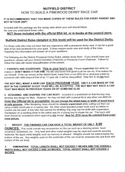
From www.bloodjournal.org by guest on January 26, 2015. For personal use only.
Separation of Myeloid and Erythroid Progenitors Based on Expression
of CD34 and c-kit
By Marg 0. De Jong, Gerard Wagemaker, and Albertus W. Wognum
In this repolz, a novel approach is described t o physically
separate erythroid progenitors from monocyte and granulocyte progenitors, basedon the expression of CD34 and Kit.
Using biotin-labeled human Kit ligand (KL) andflow cytometry, Kit was detectable on 2% to 3% of the nucleated cells
in rhesus monkey bone marrow. Combination of biotin-KL
with CD34 monoclonal antibodies (MoAb) showed that Kit
was expressedon subsets of CD34"" and CD34P"" cells. Our
data clearlydemonstratethat CD34wscells are more heterogeneous with respect t o Kit expression than observed in
studies using Kit MoAb. A small cluster, approximately 7%
of the CD34P0* cells, expressed CD34 at submaximal levels
and stained brightly with biotinylated KL. This CD34Pos/kithi
fraction contained predominantly erythroid progenitors
(burst-forming units-erythroid; BFU-E). The majority of the
granulocytic and monocytic progenitors (colony-forming
units-granulocytelmacrophage; CFU-GM) were CD34P0*/
kitmed.Some BFU-Ewere also detected in theCD34P"'/kitmod
and CD34'0w/kitPo'fractions at low frequency. In the latter
subset, most erythroid colony-forming units (CFU-E) were
recovered. Using three-color flow cytometry, we analyzed
expression ofKit in relation to thatof CD34 and the class II
major histocompatibili antigen, RhLA-DR. Themost immature bone marrow cells that can be identified in vitro, ie,
CD34Po'/RhLA-DR'ow cells,were kitmed.The CD34Po'/kith' and
CD34Po'/kit"agsubsets predominantly contained the more
mature RhLA-DRb"'" cells. Ourresults demonstratethat erythroid precursors express c-kit at much higher levels than
monomyeloid precursors and pluripotent progenitors. The
difference in expression levelsof CD34 and c-kit can be exploited to isolate BFU-E populations that are virtually devoid
of nonerythroid cells.
0 1995 by The American Societyof Hematology.
T
cell de~elopment."~"~~
In vivo administration ofKL leads
to a dose-dependent expansion of both primitive and differentiated progenitor cells in the bone marrow (BM) of mice,
nonhuman primates, or human patients. As a result, mobilized progenitors appear in the peripheral blood. At the same
time, there is an increase in the number of mature cells of
multiple lineages.*'"' The combined in vitro andinvivo
activities of KL indicate that it acts on primitive progenitors
as well as more differentiated lineage-committed cells.
Studies with monoclonal antibodies (MoAb) against Kit
have provided information about the cellular distribution of
c-kit on murine and human cells. In humans, Kit is expressed
on a large fraction of BM cells that express CD34, an antigen
specific for early hematopoietic cells, including stem cells
and multipotent and lineage-committed progenitor cells."
The CD34P""/kitP"'fraction includes cells that coexpress surface proteins characteristic for differentiating progenitors
(eg, CD33, CD38, CD71, and HLA-DR) but also developmentally immature cells that lack or express only low levels
of these antigens.'R-44In keeping with this, the CD34P""/kitP"'
subset contains committed monomyeloid
and
erythroid
progenitors (colony-forming units-granulocyte/macrophage
[CFU-GM] and burst-forming units-erythroid [BFU-E], respectively), as well as cells that produce clonogenic progeny
in long-term cultures in the presence of cytokines or stromal
feeder ~ e l l sSuch
. ~ cells
~ ~were
~ ~undetectable
~ ~
or present at
very low frequencies in the CD34Ykitnegfraction. Also, Kit
has been detected on small subsets of CD34P"' BM cells that
coexpress B (CD 10, CD19) or T (CD2, CD7) lymphoidspecific antigens, but the majority of B- and T-cell precursors
appear to be kit"eg.3"4'.44
BFU-E and CFU-E differ in their responsiveness to stimulation by KL, or inhibition byKL MOA^,^^,^' indicating a
declining role of c-kit during late stages of erythroid differentiation. In line with this, a decreasing Kit expression during
differentiation of granulocytic, monocytic, and erythroid
cells was shown by staining BM cells or cultured BFU-E
progeny with '"I-labeled KL.'"4" However, the exact pattern
of Kit expression especially during the early stages of differentiation remains unclear. This may partially be due to the
HE C-KIT GENE encodes a type I transmembrane glycoprotein and is a member of the tyrosine kinase receptor
family.' The ligand of Kit exists in both transmembrane and
soluble forms and is variously known as mast cell growth
factor, Steel factor, stem cell factor, and Kit ligand ( K L ) . 2 - 6
Both Kit (CD1 17) and its ligand are important for normal
hematopoietic cell development in vivo, as demonstrated by
the severity of hematopoietic defects in WiW mutant mice,
which do not have functional C-kit,'.' andinSteelmutant
mice, which lack active KL.'."'.'
The molecular cloning of human and rodent KL has permitted detailed studies into the functional properties of this
cytokine. The soluble form of KL, which consists of the Nterminal extracellular domain of the full-length transmembrane protein, is a poor stimulator of hematopoietic cells in
vitro on its own. However, in the presence of other hematopoietic growth factors (HGFs), such as granulocyte-macrophage colony-stimulating factor (GM-CSF), interleukin-3
(IL-3), and erythropoietin (EPO), KL enhances proliferation
and differentiation of immature hematopoietic cells as well
as monomyeloid and erythroid progenitors.'"-24In addition,
KL affects the development of early lymphoid cells in the
presence of IL-7, but is inactive at later stages of T- and B-
From the Institute of Hematology, Erasmus University, Rotterdam,
The Netherlands.
Submitted April 11, 199.5; accepted July 20, 1995.
Supported by the Netherlands Cancer Foundation Koningin Wilhelmina Fonds, the Dutch Organization,for Scientijic Research
W O , the Royal Netherlands Academy of Arts and Sciences and
Contracts of the Commission of the European Communities.
Address reprint requests to Albertus W. Wognum, PhD, Institute
of Hematology, Rm Ee1387A, Erasmus University, PO BOX 1738,
3000 DR Rotterdam, The Netherlands.
The publication costsof this article were defrayedin part by page
charge payment. This article must therefore be hereby marked
"advertisement" in accordance with 18 U.S.C. section 1734 solely to
indicate this fact.
0 1995 by The American Society of Hematology.
0006-4971/95/8611-0020$3.00/0
4076
Blood, Vol 86, No 11 (December l ) , 1995: pp 4076.4085
From www.bloodjournal.org by guest on January 26, 2015. For personal use only.
c-kit DISTRIBUTION ON HEMATOPOIETIC CELLS
4077
centrifugation for 30 minutes at 2,000 rpm at room temperature
over a discontinuous bovine serum albumin (BSA) density gradient6'
consisting of 25%, 23%, 22%, 21%, and 17% (wt/vol) BSA in 0.2
moln Tris-buffer/phosphate buffer, pH 7.2. Fractions were collected
the Kit MoAb. In addition, some Kit MoAbs inhibit outand washed in HH. Erythrocytes were lysed using 10 mmoln potasgrowth of KL-responsive ~ e l l which
s ~may~ impede
~ ~ ~sium~bicarbonate,
~ ~ ~155 mmol/L
~ ~ ammonium chloride, pH 7.4, confunctional characterization of purified kitPo' subsets. Detectaining 0.1 mmol/L EDTA.
tion methods based on binding of the ligand itself provide
Immunocytochemical staining and jlowcytometry.
Cells were
more reliable information about the capacity of cells tobind
stained overnight on ice with biotin-KL (1 nmoVL) in HHcontaining
2% (voVvol) fetal calf serum, 2% (voUvol) rhesus monkey serum,
and respond to a specific ligand than the
use of receptor
MoAbs. Biotin-labeled growth factors have been used exten- 0.05% (wt/vol) sodium azide, and DNAse (0.5 mglmL). Similar
results were obtained by incubation for 2 hours on ice. Specificity
sively to study the expression
of functional growth factor
of binding of the biotin-KL samples was determined by incubating
receptors on BM ~ e l l s ! ~ - ~In
~ the present study, we have
the cells with biotin-KL in the presence of either the blocking Kit
biotinylated recombinant humanKL and examined the distriantibody SR-I (ascites 1:200 dilution; provided by Dr V. Broudy,
bution of functional receptors forKL on rhesus monkeyBM
University of Washington, Seattle, WA) or a 100-fold molar excess
cells. With this method, we show that erythroid progenitors
of unbiotinylated KL. The cells were incubated for 30 minutes on
can be distinguished and physically separated from immature ice with streptavidin-phycoerythrin (streptavidin-PE, I: 150 vol/vol;
multipotent cells and committed monocyte and granulocyte
Molecular Probes, Eugene, OR). After each incubation the samples
progenitors on the basis of CD34 and kit expression.
were washed in HH with fetal calf serum and azide. Fluorescence
signals were amplified by incubating the cells for 30 minutes on
MATERIALS AND METHODS
ice with biotinylated PE MoAb and streptavidin-PE, as described
earlier." During the last streptavidin-PE incubation, cells were douBiotinylation. Recombinant human KL (a gift from Dr S. Gillis,
ble-stained with a CD34 MoAb (antibody 566; provided by Dr T.
Immunex, Seattle, WA)'was biotinylated using biotin-N-hydroxy
Egeland, The National Hospital, Oslo, Norway) thatwas labeled
succinimide ester (NHS-Biotin; Pierce, Rockford, IL) as described
with fluorescein isothiocyanate (FITC; Sigma) using standard procepreviously for other growth f a ~ t o r sBriefly,
. ~ ~ ~NHS-Biotin
~ ~ ~ ~ ~disdures. For three-color analysis, cells were also incubated with a
solved in dimethyl sulfoxide (DMSO) was added to IO-ng aliquots
peridinin chlorophyll protein (PerCP)-labeled antibody against the
of KL in 0.1 moVL carbonate-bicarbonate buffer, pH 8.4, containing
human class I1 histocompatibility antigen HLA-DR (Becton Dickin0.02% (voVvol) Tween-20 at molar biotin:protein (BP) ratios of
son, Mountain View, CA) that crossreacts with rhesus monkey
IO: 1, 100: 1, or 300: 1. A control sample was incubated with DMSO
RhLA-DR antigens. To study the expression of the transferrin recepwithout biotin. After 3 hours of incubation at room temperature in
tor, double-staining experiments with biotin-KL and FITC-conjuthe dark, biotin-= molecules were separated from the remaining
gated CD71 MoAb (Becton Dickinson) were performed. Sorted cells
free biotin molecules in the samples by size exclusion chromatograwith low CD34 expression were incubated with FITC-labeled CD71
phy on a I-mL Sephadex G-25 column (Pharmacia, Uppsala, SweMoAb as well.
den), equilibrated in phosphate-buffered saline containing 0.02%
Samples were analyzed using a FACScan or sorted using a FACS
(wt/vol) Tween-20. To test the efficiency of the biotinylation, biotinVantage (Becton Dickinson, San Jose, CA). Cells were illuminated
KL was adsorbed onto streptavidin-agarose beads (Sigma, St. Louis,
with the 488-nm line of an argon ion laser. Green FITC fluorescence
MO), and the amount of biologic activity that remained in the superwas measured through a 530-nnd30-nm bandpass filter. Orange PE
natant was determined. Biotin-KL was stored at -80°C in the presfluorescence was measured through a 575-nnd26-nm or a 585-nm/
ence of 0.02% (wt/vol) sodium azide.
42-nm bandpass filter. Red PerCP fluorescence was measured
Biologic activiry assay. The biologic activity of biotin-KL was
through a 650-nm longpass filter. Cells were analyzed in a light
measured in a proliferation assay using cells from the human factorscatter window as indicated in Fig 2C to include cells with intermedidependent megakaryocyte cell line M07e.60.6'Cells were grown in
intermediate
ate to high forward light scatter (FLS) andlowto
a-minimal essential medium (aMEM; GIBCO, Gaithersburg, MD)
perpendicular light scatter (PLS) properties and to exclude granulosupplemented with 10%(vol/vol) fetal calf serum (FCS), 0.05 mmoV
cytes, dead cells, and cellular debris.
L P-mercaptoethanol, 5 ng/mL human IL-3, and 10% (voVvo1) conIn vitro culrure in semisolid medium. Sorted populations were
ditioned medium of the 5637 cell line. Cultures were maintained at
assayed for their content of CFU-GM, CFU-E, and BFU-E by in
37°C in a humidified atmosphere of 10% COz in air. To determine
vitro colony formation in semisolid methylcellulose culture medium.
the biologic activity of biotin-KL, 5 X IO' cells per well of 96-welI
In 35-mm petri dishes (Becton Dickinson), unsorted cells were plated
microtiter plates (Falcon 3072; Becton Dickinson Labware, Lincoln
at a concentration of 50,000 per dish; sorted subsets were plated at
Park, NJ) were cultured in 200 p L medium containing serial dilutions
10,000 per dish (for cells from the light scatter window as shown
of growth factor. The cells were cultured for 40 to 48 hours, after
in Fig 2C) or at 500 to 1,O00 per dish (for subsets of CD34P"scells)
which0.25 pCi 3H-thymidine was added to each well. The cells
in I mL methylcellulose medium as de~cribed.'~Methylcellulose
were harvested after 16 to 18 hours of thymidine incorporation, and
cultures included the following components: 0.8% (wt/vol) methylthe radioactivity was measured in a liquid scintillation counter. At
cellulose in aMEM (GIBCO), 5% (vol/vol) FCS, 1.5% (wt/vol)
a B P ratio of l O : l , KL retained all of its biologic activity, while
BSA, 10 pglmL insulin, 0.6 mglmL human transferrin, 15 pmoVL
the biotinylation efficiency was greater than 99%. as determined by
linoleic acid, 15 pmoln cholesterol, 0.1 mmoVL P-mercaptoethanol,
adsorption onto streptavidin-agarose beads. This preparation was
used for further experiments.
0.1 pmollL sodium selenite, 1 mg/mL nucleosides, 1 0 0 U/mL penicillin, and 50 pg/mL streptomycin. Recombinant rhesus monkey
Low-densig BM cell preparation. BM aspirates from young
IL-3 was used at a final concentration of 30 ng/mL, human recombiadult rhesus monkeys (Macaca mularra) from the TNO Primate
Center, Rijswijk, The Netherlands, were collected in Hanks' HEPES
nant GM-CSF (Behringwerke AG, Marburg, Germany) at 5 ng/mL,
buffered salt solution (HH) with heparin and DNAse. The buffy coat
human KL at 200 ng/mL, G-CSF at 100 nglmL, and human recombifraction was collected after centrifuging the cells for 15 minutes at
nant EPO (Behringwerke AG) at 4 U/mL. CFU-GM cultures were
2,500 rpm at room temperature. Low-density cells were obtained by
grown in the presence of IL-3, GM-CSF, and KL, with or without
differences in techniques and reagents used to detect and
isolate Kit-expressing cells. The ability to distinguish kitneg,
k i t I O W , and kithi cells is influenced by the binding affinity of
From www.bloodjournal.org by guest on January 26, 2015. For personal use only.
4078
DE JONG, WAGEMAKER, AND WOGNUM
I
0
r h k b i o t i n - K L
A\
r 11. I I
,
o,75b KL
+ biotin-KL
0
I l’ I t l l
l
o,8%
anti-Kit + biotin-KL
0
I
l
I
0.2%
0
IO’
10’
IO‘
10’
lo’
l00
lo’
102
10‘
103
PE-fluorescence
K*
Fig l . Expression of
and specificity of biotin-KL staining on M07e and rhesus monkey BM cells. Cells wera sequentially stained with
biotin-KL and streptavidin-PE. Rhesus monkey BM cells were double-stained with FITC-labeled CD34 MoAb and analyzed inside a liaht scatter
window as indicated in Fig 2. (A) M07e cells, (B) rhesus monkey BM cells, and (C) CDWW rhesus monkey BM cells. Hlstograms rapresent
(from top to bottom) cells incubated with biotin-KL, biotin-KL in the presence of a 100-fold molar excess of unlabeled KL, biotin-KL in the
presence of the SR-1 anti-kit antibody, and control calls incubated without biotin-KL. Markers were set on tha basis of background fluorescance
of unstained cells to indicate the percentages of k
Pcells and to show the specificity of the binding of biotin-KL to the cells.
adding G-CSF. BFU-E cultures were grown in thepresence of EPO,
KL, and 0.2 mmoVL bovine hemin. CFU-E cultures were grown in
the presence of EPO and 0.14 mmol/L hemin. Cultures were maintained in a humidified atmosphere of 10%CO2 in air. Colonies were
counted at day 4 (CFU-E) and day 11 or 12 (CFU-GM and BFUE). Data of duplicate dishes were expressed as average number of
colonies per 1,OOO cells plated. Standard errors were obtained by
taking the square root of the absolute number of colonies counted,
assuming that crude colony counts are Poisson-distributed.6’.” Incubation of the cells with biotin-= did not stimulate colony formation,
because no difference in colony numbers was seen between cells
that had and cells that had not been incubated with biotin-KL.
In vitro culture in liquid medium. For liquid suspension cultures,
an automated single-cell deposit unit (Becton Dickinson) was used to
sort individual cells directly into separate wells of 96-well microtiter
plates (Falcon 3072; Becton Dickinson Labware). In different experiments, either 1 or 1,000 cells were sorted into 200 pL culture medium
per well. Cells were grown in aMEM without HGFs or supplemented
with various combinations (final concentrations) of IL-3 (30 ng/mL),
GM-CSF (5 nglmL), KL (200 nglmL), and EPO (4 U/&). In cultures started with one cell per well, half of the culture medium was
replaced every week. Cells were expanded before reaching confluency by transfer to l mL medium in 24-well culture plates (Costar,
Cambridge, MA). Phenotypic analysis was performed by staining
with antibodies against CD34, CD71, CDllb, and HLA-DR, followed by FACScan analysis. In cultures started with 1,000 cells per
well, separate cultures wereused for ‘H-thymidine incorporation
after 3, 5, or 7 days of culture and for phenotypic and morphologic
analysis of cells produced after 7 days of culture.
RESULTS
Biotin-KLstaining of MO7e cells andrhesus monkey BM
cells. Cytochemical staining properties of biotin-= on
M07e cells and on rhesus monkey BM cells were studied
usingflow cytometry. All cells of the KL-responsive cell
line M07e and a fraction of the BM cells were stained
brightly with biotin-= in combination with PE-conjugated
streptavidin (Fig 1A and B). Because the number of kitws
cells in unfractionated BM was very low (2% to 3% of the
nucleated cells, corresponding to 7% of the cells inside the
light scatter window, indicated in Fig 2), Kit expression was
also studied on cells inside a window based on high CD34
expression (Fig 1C). Within the CD34P”Ssubset, 30% of the
cells were kitw’. The fluorescence signal of M07e as well
asBM cells, incubated with biotin-KL in the presence of‘
either unlabeled KL or the blocking anti-Kit antibody SRwas almost identical to that of control cells incubated
without biotin-KL. This indicated that binding of biotin-KL
to these cells was specific and due to binding to Kit.
Distribution of c-kit on rhesus monkey BM cells. Because of the low frequency of kitP”’cells in unfractionated
BM (Fig l), the relation between CD34 and Kit expression
was studied on low-density BM cells (Fig 2). Expression
of Kitwas detectable on 15.2% ? 6.1% (three different
experiments) of the low-density BM cells inside the light
scatter window indicated in Fig 2. Combination of biotinKL with CD34 MoAb showed that 30% to 50% of these kitpositive cells were CD34-positive. The CD34p” cells were
heterogeneous with respect to Kit expression. A small subset,
containing 7% of the CD34P”scells, expressed high levels
of c-kit (region 1 inFig 2A). CD34 expression on these
CD34Vkithi cells was lower than on another subset that
expressed Kit at intermediate levels (CD34ws/kit”ed;region
2 ) . which indicated that the latter population included the
more immature cells. This kitmd fraction contained more
than 60% of the CD34P””cells. The remainder of CD34P”’
cells had no detectable Kit expression (CD34P”s/kitneg;
region
3). Finally, a subset of cells with low CD34 expression ex-
From www.bloodjournal.org by guest on January 26, 2015. For personal use only.
c-kit DISTRIBUTION
ON HEMATOPOIETIC CELLS
1A
I"
4
I
g
c
1000
D
1o4lB
a
excess KL
- I
Q
II
1
4079
103
region 3
I
4
region 4
loo
10'
IO*
103
CD34 expression
lo4
0
400
800
forward light scatter
l"
loo lo1
IO* lo3
lo4
RhLA-DR expression
Fig 2. Expression of CD34 and Kit onrhesus monkey BM cells, light scatter properties and RhLA-DR expression of different subsets. Lowdensity rhesus monkey BM cells, stained with biotin-KL and streptavidin-PE and counterstained with FITC-labeled CD34 MoAb and PerCPlabeled HLA-DR MoAb after amplification of the fluorescence signal, were analyzed by flow cytometry. (A) Dot plot
of kit versus CD34
expression. The rectangular boxes indicate the windows used t o sort cells on the basis of CD34 and Kit expression levels. (B) Nonspecific
binding of biotin-KL was evaluated in the presence of a 100-fold molar excess of unlabeled KL. The horizontal line is set on the basis of
background fluorescence of unstained cells to discriminate between brightly stained cells and cells with low or no Kit signal. (C) PLS versus
FLS of low-density cells and of cells in theregions 1 through 4as identified in A. Indicated is thescatter window that wasused in thesorting
experiments t o include cells with intermediate t o high FLS and low to intermediate PLS properties and to exclude granulocytes, dead cells,
and cellular debris. (D) RhLA-DR fluorescence histograms of cells in the same regions as in (C).
pressed c-kit in a range from low to high. Most cells in this
region, particularly those with high Kit expression, showed
a small shift in fluorescence intensity after staining with the
CD34 MoAb, as compared with cells stained with isotypecontrol
MoAb.
Therefore,
this
subset
was
designated
CD34'"" (CD34'"W/kitPo';region 4).
As shown in Fig 2C, CD34P"'/kith' and CD34P"'/kitn"dcells
(regions 1 and 2) displayedlight scatter properties characteristic of immature, blast-like cells, ie, intermediate to high
FLSandlowPLS.
CD34P"s/kit"s and CD34'""/kitP'" cells
(regions 3 and 4) were more heterogeneous with respect to
light scatterand also containedcells withrelatively high
PLS. Additionally, small cells with low FLS were found in
the CD34P"'/kit""gfraction (region 3).
Analysis of RhLA-DR expression on thedifferent subsets
(Fig 2D) indicatedthat most of the CD34P"'/kith'and CD34""'/
kitnegcells (regions 1 and 3) were RhLA-DRhrIgh',whereas
the CD34P"s/kit""dcluster (region 2) contained RhLA-DRbrLghh'
and RhLA-DRd"" cells at almost equal frequencies. The presence of RhLA-DRd"" cells in region 2, in combination with
the high CD34 expression of this subset, indicated that very
immaturecellsexpress Kit at low tointermediate levels.
RhLA-DR expression was alsolow on theCD341""/kith' cells,
but these represent relatively mature cells, as CD34 levels
on these cells were very low.
Colony-forming potential of different fructions q f rhesus
monkey BM. We assayed the functional abilities of the subsets, discussed in the previoussection, in standardcolony
assays in semisolid culture media. The results of two such
experimentsareshown
in Table 1. Most CFU-GMwere
found in the CD34P"'/kit'"edfraction, which was 20 to 30
times enriched in CFU-GM as compared with the low-density cells. A much lower proportion of CFU-GM was recovered in the CD341"'"/kit""S subset, and less than 1% of the
CFU-GM were found in the CD34P"'/kit'" fraction.
The CD34P"'/kit'" fraction contained at least 30-fold more
BFU-E than CFU-GM. As shown in Table 1 , 200 to 400 of
every 1,000 CD34P"s/kith'cells developed into a BFU-E colony, compared with one to three of every 1,000 low-density
cells, demonstratinganenrichment
of 100- to 200-fold.
From www.bloodjournal.org by guest on January 26, 2015. For personal use only.
4080
WOGNUM
DE JONG, WAGEMAKER, AND
Table 1. Colony Formation In Vitro of Different Sorted Fractions of Low-Density Rhesus Monkey B M Cells
BFU-E
CFU-GM
1
No. of Colonies/
io3cells
%
Fractlon
Sorted
Experiment
Region
A
2
3
4
B
1
2
3
4
CD34PoE/kith'
CD34P"s/kitme"
CD34PoS/kit"eQ
CD34'0w/kitP"s
density
Low
CD34pos/kith'
CD34PoS/kitmed
CD34Pos/kit"eQ
CD34'""/kitPoS
fraction
Scatter
% Recovery
0.33 419.3 14.5
0.5 i- 2.7
3.89
181.5 i- 9.5
2.06
16.0 i- 2.8
3.70
0.0 -+ 0.0
100
9.3 t 0.2
0.45
0.0 i- 0.0
3.30
101.5 t 7.1
2.65
13.5 i- 2.6
5.88
0.0 i- 0.0
100
3.6 t 0.4
75.9
3.5
0.0
100
0.0
93.0
9.9
0.0
100
No. of Colonies/
io3 cells
21.0
0.0
18.0
3.2
188.0
3.5
0.0
0.5
1.1
t 36.9
t 3.2
i- 0.0
t 3.0
i- 0.3
t 13.7
i- 1.3
t 0.0
i- 0.5
i- 0.4
CFU-E
% Recovery
43.2
25.5
0.0
20.8
100
76.9
10.5
0.0
0.0
2.7
100
No. of Colonies/
io3 cells
% Recovery
48.8
0.5 2 3.1
0.0 t 0.0
0.0 2 0.0
133.3 ? 5.2
29.9 i- 0.5
39.0 i- 11.3
0.0 i 0.0
i- 0.0
13.0 2 4.6
2.6 t 0.7
0.0
0.0
16.5
100
6.8
0.0
0.0
29.4
100
Cells inside a light scatter window as shown in Fig2C were sorted on the basis of CD34 and kit expression into regionsas shown in Fig2A.
B) are shown. Data from duplicate dishes are expressed as average number of colonies
Results of two independent sorting experiments (A and
per I O 3 cells plated t SE (the square root of the absolute number of colonies counted; see Materials and Methods). Recovery was calculated
as shown in Fig 2C
relative to the unsorted low-density fraction (experiment A), or relative to the fraction sorted inside a light scatter window
(experiment B)
SomeBFU-E were detected in the CD34pos/kit"'"dand the
CD34'""/kitP"' fractions, but at 20- to 50-fold lower frequencies than in the CD34p"'/kith' fraction. The erythroid origin
of kith' cells was confirmed by double-staining of cells with
biotin-KL and CD71 MoAb, which showed that kith' cells
expressed high levels of the transferrin receptor (Fig 3).
The number of CFU-E colonies recovered after sorting
was very low (Table I). Most of the CFU-E were present
in the CD34'""/kitp''" fraction, suggesting thatthisfraction
contained differentiating erythroid cells. In agreement with
this, erythroblasts were the predominant cell type identified
in cytocentrifuge preparations from this fraction (data not
shown). Moreover, restaining of sorted CD34'""/kitp"s cells
with CD71 MoAb showed high CD71 expression on these
cells (Fig 4). However, because of the low CFU-E recovery,
we cannot exclude the possibility that CFU-E were present
in other subsets as well.
Growth fuctor responses in liquid culture. To study the
effect of different growth factors on theshort-term proliferation and differentiationof the varioussubsets, 1,000 cells
per well were sorted into liquid medium containing different
(combinations of) cytokines. The highest proliferation was
found in the wellswith cellsfromthe CD34po'/kit'"edand
CD34P"s/kith'fractions when cultured in the presence of KL
IL-3 + GM-CSF.FACS analysis of the differentfractions after7
days of cultureshowed
mainlyerythroid
(CD7 I P"'/CDl 1b'lcg/RhLA-DKeg) cellsin the CD34""'/kith'
1 '""/CD 1 l bpYRhLA-DWeg)
fractions, and granulocytic (CD7
and monocytic (CD7l'""/CDl lbPos/RhLA-DRPO")
cells in the
CD34p'"/kit'""dfractions. Examples of CD71 and RhLA-DR
expression oncultured erythroid cells andon cultured granulocyte and monocyte precursors are shown
in Fig 5A and
B, respectively. In agreement with these findings, cytospin
preparations showed cells from the erythroid lineage in the
CD341""/kitp'" and CD34pos/kith' cell cultures, and monomyeloid cells in the CD34P"s/kit'ncdand CD34P"'/kit"eg cell cultures (data not shown).
To further characterize the long-term differentiation potential of the various fractions, individual cells were sorted
directly into separate wells of 96-well microtiter plates and
+
l o 4T
lo3
102
101
100
loo
10'
102
io3
1
transferrin receptor (CD71) expression
Fig 3. Expression of CD71
and Kit on low-density rhesus
monkey B M cells. Cells were
stained with biotin-KL, streptavidin-PE, and FITC-labeled CD71
MoAb. (A) Dot plot of kit versus
CD71 expression of cells inside
the scatter window as indicated
in Fig 2C. (B) Nonspecific binding
of biotin-KL was evaluatedin the
presence of a 100-fold molar excess of unlabeled KL.
From www.bloodjournal.org by guest on January 26, 2015. For personal use only.
c-kit DISTRIBUTION ON HEMATOPOIETIC CELLS
4081
l
Table 2. Proliferation of Different Subsets of CD34P"' Low-Density
Rhesus Monkey BMCells
No. of Wells (of a total of 192)
Phenotype
Proliferating
Expanded
Fraction
Region
1
2
3
1oo
l 0'
I o3
IO2
I o4
FITC fluorescence intensity
Fig 4.CD71
expression on sorted CD34'ow/kitPo'rhesus monkey
BM cells. Low-density BMcells, stained with biotin-KL, streptavidinPE, and FITC-labeled CD34 MoAb, were sorted in a window similar
to that of region 4 in Fig 2A. Subsequently, half of the sample was
incubated with buffer only (thin line) and the other
half, with FITClabeled CD71 MoAb (bold line), and reanalyzed.
cultured in the presence of IL-3, GM-CSF, and KL for a
period of 4 weeks. No significant difference was seen betweencultures with and without EPO (data not shown).
Ninety percent of the wells with CD34P"s/kith'cells and 8 1%
with CD34Pos/kit'"d cells contained proliferating cells,
whereas only 33% of the CD34Pos/kitneS
cells showed proliferation (Table 2). There was a large difference between the
fractions with respect to the amount of cells produced in the
wells and the nature of these clones. In general, the kith'
clones proliferated faster and were exhausted sooner than
the kitmedclones, which continued to grow up to 4 weeks
after sorting. The number of cells in the kitnegclones remained very low. Microscopic inspection of the wells
showed erythroid cells in the kithi clones and granulocytic
Fig 5. Phenotypic analysis of
cultured fractions of rhesus monkeyBM cells. Low-densityBM
cells were stained with biotin-KL,
streptavidin-PE, and FITC-labeled
CD34 MoAb. Cells (1,000 per well)
were sorted on the
basis of CD34
and kit expression, as indicated
by the regions in Fig 2A. Sorted
cells were cultured in medium in
the presence of KL + 11-3 GMCSF. After 7 days of culture, the
cells were incubated with FITClabeled CD71 MoAb and PE-labeled HLA-DR MoAb and analyzed by flow cytometry. Shown
are cultures thatwerestarted
with CD34Po'/kithi (A) or CD34P0"/
kitmd (B) cells.
4"'
CD34pos/kithi
CD34poS/kitmed
CD34P"s/kitn'a
173
155
64
130
51
0
E
G
M
46
0
1
4
0
2
Cells were incubated with biotin-KL and
CD34 MoAb and sorted
one cell per well into liquid medium containing
IL-3, GM-CSF, and
KL with or without EPO. Wells in which proliferation occurred were
counted. Before reaching confluency, clones with extensive proliferation were then expanded into l-mL cultures. Expanded clones that
contained sufficient numbers of cells were stained with antibodies
against CD71, CDllb, and HLA-DR.
Abbreviations: E, erythroid (CD71P"s/CD11b"eg/RhLA-DRP0S);
G,
granulocytic (CD71'"w/CD11bPoS/RhLA-DR"eg);
and M, monocytic
~CD7110W/CD11bPoS/RhLA-DRPoS).
and monocytic cells in the kitmedand kitnesclones. About
25% of the wells with kith' cells (Table 2) proliferated sufficiently to perform a FACS phenotyping experiment, resulting in46
erythroid (CD71P"') and one granulocytic
(RhLA-DR"es/CD1lbP"")clone. Only 6 of 192 wells with
kitmd cells contained enough cells to be analyzed in this
way, resulting in four granulocytic (RhLA-DRneg/CD1lbpos)
and two monocytic (RhLA-DRP"'/CD1lbP"') clones.
DISCUSSION
Expression of Kit on r n ~ r i n e ~and
~ - h~ * ~ m a BMn ~ ~
cells has been studied extensively using Kit-specific MoAb.
However, the exact expression pattern of Kit during the early
stages of hematopoietic cell differentiation especially is still
not clear. Differences between published results may partially be explained by the use of different MoAbs, because
the ability to distinguish kith', kitmsd,and kit'"" cells is influenced by the binding affinity of the Kit MoAb. Moreover,
as Kit MoAb can inhibit outgrowth of kitPusce11s~o~42~46~47
It
.
A
+
CD71 (transferrin receptor) expression
From www.bloodjournal.org by guest on January 26, 2015. For personal use only.
4082
is possible that the kitws cells do not develop optimally in
culture if high-affinity MoAbs are used for sorting. This
would lead to a serious underestimation of the number of
colony-forming cells in the kitPossubset. Cell staining methods based on the ligand itself cause no such inhibition. In
addition, such methods by definition provide more reliable
information about the capacity of the cells to bind and respond to a specific ligand than staining with MoAb against
the receptor. In this study, we have used biotinylated KL to
examine the expression of Kiton subsets of low-density
rhesus monkey BM cells. We were able to detect Kit on 2%
to 3%of the nucleated cells, similar to frequencies previously obtained for human BM using Kit MOA^.^'.^' Double-staining with biotin-KL and CD34 MoAb showed Kit
expression on subsets of CD34PoSas well as CD34'"" rhesus
monkey BM cells.
A small fraction of CD34P"scells with a high Kit expression was detected, consisting almost exclusively of BFL-E.
In line with this, CD34Tkith' cells produced erythroblasts
in liquid suspension cultures, and the kith' cells expressed
high levels of the transferrin receptor. Some BFU-E were
also found in the kitmedpopulation. This might reflect insufficient separation of this subset and the kith' cells, although
the kith' cells are quite a distinct cluster. It is also possible
that these kitmedcells represent a separate population, eg, one
that is more immature than the kithiBFU-E, because the kith'
cells express CD34 at a lower level than the kitmd population.
Heterogeneity within the BFU-E population has also been
observed by Simmons et al," who detected a small CD34p0s/
kitnegBFU-E fraction in sorting experiments using kit MoAb
YB5.B8. Using another MoAb, NU-c-kit, Gunji et a14*separated CD34POSBM cells into kith', kit'"", and kitnegsubsets.
In contrast with our results, these investigators found the
highest BFU-E frequencies in the kit'"" subset, whereas
BFU-E frequencies in the kithisubset were very
Unfortunately, no information was provided about the overall
BFU-E recovery in the sorted fractions, so it cannot be ruled
out that colony formation by kith' cells was underestimated.
This might be in accordance with results reported by Broudy
et al,40 who showed that outgrowth of a small subset of
human BFU-E is not inhibited by the Kit antibody SR-1.
Such inhibition apparently does not occur with cells stained
with biotinylated KL, as demonstrated by the high recovery
of BFU-E and CFU-GM after sorting (Table 1).
Most CFU-E were present in the CD34'ow/kitP"s
fraction.
This population was almost completely erythroid, as demonstrated by high CD7 1 expression. The results indicating that
BFU-E as well as CFU-E display Kit correlate well with the
insufficient erythropoiesis and the occurrence of macrocytic
anemia in WIW mutant mice, which do not have functional
c-kit. Also in accordance with these results, Papayannopoulou et aP9 and Ashman et a13' have shown that BM cells that
had been isolated via either immune adherence to the SR-1
antibody or immune rosetting using the YBS.B8 antibody
were highly enriched for erythroid cells. Binding studies
with Iz5I-KL on cultured BFU-E progeny showed that proerythroblasts labeled much more densely than erythrob l a s t ~Although
.~~
Kit is present on both BFU-E and CFUE, KL is necessary only for BFU-E outgrowth, but not for
DE JONG,WAGEMAKER, AND WOGNUM
CFU-E and later erythroid cell^.^.^^ Collectively, the data
suggest that Kitexpression reaches its maximum at the BFUE stage and gradually declines during terminal erythroid
differentiation. This pattern of Kit expression appears similar
to that of CD7I7' and the EPO receptor.55."
The CD34P"s/kit"egfraction contained virtually no colonyforming cells. Although the presence of very immature cells
in this fraction cannot be ruled out completely, most of the
cells in this subset are more mature, based on the relatively
low CD34 and high RhLA-DR expression, characteristic of
activated and differentiating cells. These CD34Ykitnegcells
appear to represent primarily monocyte and granulocyte precursors. In addition, part of the CD34pos/kit""g
fraction has
low FLS and PLS properties and probably consists ofBlymphocyte precursor cells. In previous studies, Kit has been
detected ononly small subsets of CD34P"s/CD10Pos
and
CD34Pos/CD19p'B-lymphocyte precursors.u This is consistent with an involvement of KL in early but not later stages
of B-cell d e ~ e l o p m e n t . ' ~ ~ ~ ~ ~ ~ ~ . ~ '
Part of the CD34ps/kitm"dcells showed high CD34 and
low RhLA-DR expression, a phenotype that has previously
been associated with the most immature cells that canbe
identified in human BM.45.74.75
Recently, we have shown that
the CD34PoS/DRd""
rhesus monkey BM subset contains multipotential progenitors with high proliferative capacity." Preliminary results from transplantation experiments indicate
that the cells that can reconstitute lethally irradiated rhesus
monkeys are also present in this subset. In accordance with
the expression of Kit on immature rhesus monkey BM cells,
murine BM cells expressing Kit were enriched for hematopoietic stem ~ e l l s . ~ ~These
, " , ~ ~results support the conclusion
that Kit is already expressed at low to intermediate levels at
a very early stage of hematopoiesis. Further studies focusing
on subsets of CD34ws/kitmed/RhLA-DRd""
rhesus monkey
BM cells will be useful to establish the importance of individual hematopoietic growth factors during stem cell proliferation and differentiation, and to provide candidate stem
cell fractions for transplantation studies.
In summary, our data are consistent with a model in which
immature, multipotent progenitors are CD34Ykitmd. Along
the monomyeloid lineage, these cells differentiate into
CD34pos/kitmd
CFU-GM. Expression of Kit declines after the
CFU-GM stage, and the cells lose CD34 expression. Along
the erythroid lineage, CD34p"s/kitmed
progenitors differentiate
to kith'BFU-E, which gradually lose CD34 when they differentiate into CD34'""/kith' CFU-E. This is followed by a gradual disappearance of Kit expression during terminal differentiation into mature red blood cells. The ability to distinguish
the CD34po'/kith1population provides a methodto obtain
highly enriched BFU-E populations that are devoid of nonerythroid cells.
ACKNOWLEDGMENT
We thankDrs S. Gillis (Immunex, Seattle, WA) forthe gift of
KL, T. Egeland (University Hospital, Oslo, Norway) for supplying
CD34MoAb 566, and V.C. Broudy (University of Washington,
SR-l. We are grateful to K.J.
Seattle, WA) for thekitantibody
Neelis,M. van Teeffelen, and H. Wiersema for providing bone
marrow aspirates and to H. Busking-van der Lelij, W. Dimjati, D.
From www.bloodjournal.org by guest on January 26, 2015. For personal use only.
c-kit DISTRIBUTION ON HEMATOPOIETIC CELLS
Kieboom-Pluimes, T. Visser, and Y. Westerman for excellent technical assistance. We thank Dr W.A.M. Loenen for critically reading
the manuscript.
REFERENCES
1. Chabot B, Stephenson DA, Chapman VM, Besmer P, Bernstein
A: The proto-oncogene c-kit encoding a transmembrane tyrosine
kinase receptor maps to the mouse W locus. Nature 335238, 1988
2. Anderson DM, Lyman SD, Baird A, Wignall JM, Eisenman J,
Rauch C, March CJ, Boswell HS, Gimpel SD, Cosman D, Williams
D E Molecular cloning of mast cell growth factor, a hematopoietin
that is active in both membrane boundand soluble forms. Cell
63:235, 1990
3. Huang E, Nocka K, Beier DR, Chu TY, Buck J, Lahm HW,
Wellner D, Leder P, Besmer P: The hematopoietic growth factor
KL is encoded by the S1 locus and is the ligand of the c-kit receptor,
the gene product of the W locus. Cell 63:225, 1990
4. Martin FH, Suggs SV, Langley KE, Lu HS, Ting J, Okino KH,
Morris CF, McNiece IK, Jacobsen FW, Mendiaz EA, Birkett NC,
Smith KA, Johnson MJ, Parker VP, Flores JC, Patel AC, Fisher EF,
Erjavec HO, Herrera CJ, Wypych J, Sachdev RK, Pope JA, Leslie
I, Wen D, Lin C-H, Cupples RL, Zsebo KM: Primary structure and
functional expression of rat and human stem cell factor DNAs. Cell
63:203, 1990
5. Zsebo KM, Wypych J, McNiece IK, Lu HS, Smith KA, Karkare SB, Sachdev RK, Yuschenkoff VN, Birkett NC, Williams LR,
Satyagal VN, Tung W, Bosselman RA, Mendiaz EA, Langley KE:
Identification, purification, and biological characterization of hematopoietic stem cell factor from buffalo rat liver-conditioned medium.
Cell 63:195, 1990
6. Zsebo KM, Williams DA, Geissler EN, BroudyVC, Martin
FH, Atkins HL, Hsu RY, Birkett NC, Okino KH, Murdock DC,
Jacobsen FW, Langley KE, Smith KA, Takeishi T, Cattanach BM,
Galli SJ, Suggs SV: Stem cell factor is encoded at the S1 locus of
the mouse and is the ligand for the c-kit tyrosine kinase receptor.
Cell 63:213, 1990
7. Geissler EN, Ryan MA, Housman DE: The dominant-white
spotting (W) locus of the mouse encodes the c-kit proto-oncogene.
Cell 55:185, 1988
8. Flanagan JG, Leder P The kit ligand: A cell surface molecule
altered in steel mutant fibroblasts. Cell 63:185, 1990
9. Copeland NG, Gilbert DJ, Cho BC, Donovan PJ, Jenkins NA,
Cosman D, Anderson D, Lyman SD, Williams DE: Mast cell growth
factor maps near the steel locus on mouse chromosome 10 and is
deleted in a number of steel alleles. Cell 63:175, 1990
10. Bernstein A, Forrester L, Reith AD, Dubreuil P, Rottapel R:
The murine W/c-kit and Steel loci and the control of hematopoiesis.
Semin Hematol 28: 138, 1991
11. Broxmeyer HE, Hangoc G , Cooper S, Anderson D, Cosman
D, Lyman SD, Williams DE: Influence of murine mast cell growth
factor (c-kit ligand) on colony formation by mouse marrow hematopoietic progenitor cells. Exp Hematol 19:143, 1991
12. Broxmeyer HE, Cooper S, Lu L, Hangoc G, Anderson D,
Cosman D, Lyman SD, Williams DE: Effect of murine mast cell
growth factor (c-kit proto-oncogene ligand) on colony formation by
human marrow hematopoietic progenitor cells. Blood 77:2142, 1991
13. Carow CE, Hangoc G, Cooper SH, Williams DE, Broxmeyer
HE: Mast cell growth factor (c-kit ligand) supports the growth of
human multipotential progenitor cells with a high replating potential.
Blood 78:2216, 1991
14. McNiece IK, Langley KE, Zsebo KM: Recombinant human
stem cell factor synergises with GM-CSF, G-CSF, IL-3 and epo to
stimulate human progenitor cells of the myeloid and erythroid lineages. Exp Hematol 19:226, 1991
15. Metcalf D, Nicola NA: Direct proliferative actions of stem
4083
cell factor on murine bone marrow cells in vitro: Effects of combinationwith colony-stimulating factors. h o c Natl Acad Sci USA
88:6239, 1991
16. Migliaccio G , Migliaccio AR, Druzin ML, Giardina PJ, Zsebo
KM, Adamson JW: Effects of recombinant human stem cell factor
(SCF) on the growth of human progenitor cells in vitro. J Cell
Physiol 148:503, 1991
17. Tsuji K, Zsebo KM, Ogawa M: Enhancement of murine blast
cell colony formation in culture by recombinant rat stem cell factor,
ligand for c-kit. Blood 78:1223, 1991
18. de Vries P, Brasel KA, Eisenman JR, Alpert AR, Williams
DE: The effect of recombinant mast cell growth factor on purified
murine hematopoietic stem cells. J Exp Med 173:1205, 1991
19. Brandt J, Briddell RA, Srour EF, Leemhuis TB, Hoffman R:
Role of c-kit ligand in the expansion of human hematopoietic progenitor cells. Blood 79:634, 1992
20. Heyworth CM, Whetton AD, Nicholls S, Zsebo K, Dexter
TM: Stem cell factor directly stimulates the development of enriched
granulocyte-macrophage colony-forming cells and promotes the effects of other colony-stimulating factors. Blood 80:2230, 1992
21. Migliaccio G, Migliaccio AR, Druzin ML, Giardina PJ, Zsebo
KM, Adamson JW: Long-term generation of colony-forming cells
in liquid culture of CD34+ cord blood cells in the presence of
recombinant human stem cell factor. Blood 79:2620, 1992
22. Williams N, Bertoncello I, Kavnoudias H, Zsebo K, McNiece
I: Recombinant rat stem cell factor stimulates the amplification and
differentiation of fractionated mouse stem cell populations. Blood
79:58, 1992
23. Abboud MR, Xu F, Payne A, Laver J: Effects of recombinant
human Steel factor (c-kit ligand) on early cord blood hematopoietic
precursors. Exp Hematol 22:388, 1994
24. Steen R, Morkrid L, Tjmnfjord GE, Egeland T: c-kit ligand
combined with GM-CSF and/or IL-3 can expand CD34+ hematopoietic progenitor subsets for several weeks in vitro. Stem Cells 12:214,
1994
25. McNiece IK, Langley KE, Zsebo KM: The role of recombinant stem cell factor in early B cell development. Synergistic interaction with IL-7. J Immunol 146:3785, 1991
26. Katayama N, Shih J, Nishikawa S, Kina T, Clark S, Ogawa M:
Stage-specific expression of c-kit protein by murine hematopoietic
progenitors. Blood 82:2353, 1993
27. Morrissey PJ, Mckenna H, Widmer MB, Braddy S, Voice R,
Charrier K, Williams DE,WatsonJD: Steel factor (c-kit ligand)
stimulates the in vitro growth of immature CD3-/CD4-/CD8- thymocytes: Synergy with L-7. Cell Immunol 157:118, 1994
28. Molineux G , Migdalska A, Szmitkowski M, Zsebo K, Dexter
TM: The effects on hematopoiesis of recombinant stem cell factor
(ligand for c-kit) administered in vivo to mice either alone or in
combination with granulocyte colony-stimulating factor. Blood
78:961, 1991
29. Ulich TR, del Castillo J, Yi ES, Yin S, McNiece I, Yung YP,
Zsebo KM: Hematologic effects of stem cell factor in vivo and in
vitro in rodents. Blood 78:645, 1991
30. Bodine DM, Orlic D, Birkett NC, Seidel NE, Zsebo KM:
Stem cell factor increases colony-forming unit-spleen number in
vitro in synergy with interleukin-6, and in vivo in SllSld mice as a
single factor. Blood 79:913, 1992
31. Andrews R, Bartelmez S, Knitter G, Myerson D, Bernstein
I, Appelbaum F, Zsebo K: A c-kit ligand, recombinant human stemcell factor, mediates reversible expansion of multiple CD34+ colony-forming cell types in blood and marrowof baboons. Blood
80:920, 1992
32. Bodine DM, Seidel NE, Zsebo KM, Orlic D: In vivo administration of stem cell factor to mice increases the absolute number of
pluripotent hematopoietic stem cells. Blood 82445, 1993
From www.bloodjournal.org by guest on January 26, 2015. For personal use only.
4084
33. Hoffman R, Tong J, Brandt J, Traycoff C, Bruno E, McGuire
BW, Gordon MS, McNiece 1, Srour EF: The in vitro and in vivo
effects of stem cell factor on human hematopoiesis. Stem Cells 276,
1993
34. McNiece IK, Briddell RA, Hartley CA, Smith KA, Andrews
RG: Stem cell factor enhances in vivo effects of granulocyte colony
stimulating factor for stimulating mobilization of peripheral blood
progenitor cells. Stem Cells 2:36, 1993
35. Tong J, Gordon MS, Srour EF, Cooper RJ, Orazi A, McNiece
I, Hoffman R: In vivo administration of recombinant methionyl human stem cell factor expands the number of human marrow hematopoietic stem cells. Blood 82:784, 1993
36. Andrews RC, Briddell RA, Knitter CH, Opie T, Bronsden M,
Myerson D, Appelbaum FR, McNiece IK: In vivo synergy between
recombinant human stem cell factor and recombinant human granulocyte colony-stimulating factor in baboons enhanced circulation of
progenitor cells. Blood 84800, 1994
37. Civin Cl, Strauss LC, Brovall C, Fackler MJ, Schwartz JF,
Shaper JH: Antigenic analysis of hematopoiesis. 111. A hematopoietic
progenitor cell surface antigen defined by a monoclonal antibody
raised against KG-la cells. J lmmunol 133:157, 1984
38. Ashman LK, Camhareri AC, To LB, Levinsky RJ, Juttner CA:
Expression of the YB5.BS antigen (c-kit proto-oncogene product) in
normal human hone marrow. Blood 78:30, 1991
39. Papayannopoulou T, Brice M, Broudy VC, Zseho KM: Isolation of c-kit receptor-expressing cells from hone marrow, peripheral
blood, and fetal liver: Functional properties and composite antigenic
profile. Blood 78:1403, 199I
40. Broudy VC, Lin N, Zseho KM, Birkett NC, Smith KA, Bernstein ID, Papayannopoulou T: Isolation and characterization of a
monoclonal antibody that recognizes the humanc-kit receptor. Blood
79338, 1992
4 1. Strobl H, Takimoto M, Majdic 0, Hocker P, Knapp W: Antigenic analysis of human haemopoietic progenitor cells expressing
the growth factor receptor c-kit. Br J Haematol 82:287, 1992
42. GunjiY, Nakamura M, Osawa H, Nagayoshi K, Nakauchi
H, MiuraY, Yanagisawa M, Suda T: Human primitive hematopoietic
progenitor cells are more enriched in KIT'"" cells than in
cells. Blood 82:3283, 1993
43. Yamaguchi Y, Gunji Y , Nakamura M, Hayakawa K, Maeda
M, Osawa H, Nagayoshi K, Kasahara T, Suda T: Expression of ckit mRNA and protein during the differentiation of human hematopoietic progenitor cells. Exp Hematol 21: 1233, 1993
44. Simmons PJ, Aylett GW, Niutta S, To LB, Juttner CA, Ashman LK: c-kit is expressed by primitive human hematopoietic cells
that give rise to colony-forming cells in stroma-dependent or cytokine-supplemented culture. Exp Hematol 22: 157, 1994
45. Briddell RA,BroudyVC.
Bruno E, Brandt JE, Srour EF,
Hoffman R: Further phenotypic characterization and isolation of
human hematopoietic progenitor cells using a monoclonal antibody
to the c-kit receptor. Blood 79:3159, 1992
46. Lerner NB, Nocka KH, Cole SR, Qiu FH, Strife A, Ashman
LK, Besmer P: Monoclonal antibody YB5.B8 identifies the human
c-kit protein product. Blood 77:1876, 1991
47. Liesveld JL, Broudy VC, Harhol AW, Abhoud CN: Effect of
stem cell factor on myelopoiesis potential inhuman Dexter-type
culture systems. Exp Hematol 23:202, 1995
48. Foxwell BM, Taylor D, Greiner B, Mihatsch MJ, Olivieri V,
Ryffel B: Biotinylated recombinant interleukin-2. A tool for research
on the interleukin-2 receptor. J Immunol Methods 1 13:231, 1988
49. Newman W, Beall LD, Bertolini DR, Cone JL: Modulation
of TGF-0 type 1 receptor: Flow cytometric detection with biotinylated TGF-P. J Cell Physiol 141:170, 1989
50. Taki S, Shimamura T, Abe M, Shirai T, Takahara Y: Biotiny-
DE JONG, WAGEMAKER,AND WOGNUM
lation of humaninterleukin-2 for Row cytometry analysis of interleukin-2 receptors. J Immunol Methods 12233, 1989
5 1 . Peters DK, Norback DH: Binding and internalization of biotinylated interleukin-2 in human lymphocytes. Blood 76:97, 1990
52. Wognum AW, Lansdorp PM, Humphries RK, Krystal G: Detection and isolation of the erythropoietin receptor using hiotinylated
erythropoietin. Blood 76x597, 1990
53. Pieri I, Bamtault D: Biotinylated basic fibroblast growth factor is biologically active. Anal Biochem 195:214, 1991
54. de Jong MO, Rozemuller H, Visser JWM, Bauman JGJ: A
sensitive method to detect cell surface receptors using biotinylated
growth factors. Prog Histochem Cytochern 26:119, 1992
55. Wognum AW, Krystal G, Eaves CJ, Eaves AC, Lansdorp
PM: Increased erythropoietin-receptor expression on CDW-positive
honemarrow cells from patients with chronic myeloid leukemia.
Blood 79542, 1992
56. Wognum AW, van Gils FCJM, Wagemaker G: Flow cytometric detection of receptors for interleukin-6 onbonemarrowand
peripheral blood cells of humans and rhesus monkeys. Blood
81:2036, 1993
57. Wognum AW, Westerman Y, Visser TP, Wagemaker G: Distribution of receptors for granulocyte-macrophage colony-stimulating factor on immature CD34+ hone marrow cells, differentiating
monomyeloid progenitors, and mature bloodcell subsets. Blood
84:764, I994
58. WognumAW,Visser TP, de Jong MO,Egeland T, Wagemaker G: Differential expression of receptors for interleukin-3 on
suhsets of CD34-expressing hematopoietic cells of rhesus monkeys.
Blood 86381, 1995
59. de Jong MO, Rozemuller H, Bauman JGJ, Visser JWM: Biotinylation of interleukin-2 (IL-2) for flow cytometric analysis of IL2 receptor expression: Comparison of different methods. J Immunol
Methods 184: 10 I , 1995
60. Avanzi GC, Lista P, Giovinazzo B, Miniero R, Saglio G,
BenettonG, Coda R, Cattoretti G, Pegoraro L: Selective growth
response to IL-3 of a human leukaemic cell line with megakaryoblastic features. Br J Haematol 69:359, 1988
61. Hendrie PC, Miyazawa K,YangYC,Langefeld
CD, Broxmeyer HE: Mast cell growth factor (c-kit ligand) enhances cytokine
stimulation of proliferation of the human factor-dependent cell line,
M07e. Exp Hematol 19: 1031, 1991
62. Dicke KA, Lina PH, van Bekkum DW: Adaptation of albumin
density gradient centrifugation to human bone marrow fractionation.
Rev EurEtud Clin Biol 15:305, 1970
63. Blackett NM: Statistical accuracy to he expected from cell
colony assay; with special reference to the spleen colony assay. Cell
Tissue Kinet 7:407, 1974
64. lscove NN: The role of erythropoietin in regulation of population size and cell cycling of early and late erythroid precursors in
mouse hone marrow. Cell Tissue Kinet 10:323, 1977
65. Okada S, Nakauchi H, Nagayoshi K, Nishikawa S, Nishikawa
S. Miura Y, Suda T: Enrichment and characterization ofmurine
hematopoietic stem cells that express c-kit molecule. Blood 78: 1706.
1991
66. lkuta K, Weissman IL: Evidence that hematopoietic stem cells
express mouse c-kit but do not depend on steel factor for their
generation. Proc Natl Acad Sci USA 89: 1502, I992
67. Okada S, Nakauchi H, Nagayoshi K, Nishikawa S, Miura Y,
Suda T: In vivo and in vitro stem cell function of c-kit- and ScaI -positive murine hematopoietic cells. Blood 80:3044. 1992
68. Ogawa M, Shih JP, Katayama N: Enrichment for primitive
hemopoietic progenitors of marrowcells from 5-fluorouracil-treated
mice and normal mice. Blood Cells 20:7, 1994
69. Daj CH, Krantz SB, Zseho KM: Human burst-forming units-
From www.bloodjournal.org by guest on January 26, 2015. For personal use only.
c-kit DISTRIBUTION ON HEMATOPOIETIC CELLS
erythroid need direct interaction with stem cell factor for further
development. Blood 78:2493, 1991
70. Loken MR, Shah VO, Dattilio KL, Civin CI: Flow cytometric
analysis of human bone marrow: I. Normal erythroid development.
Blood 69:255, 1987
71. Wickrema A, Krantz SB, Winkelmann JC, Bondurant MC:
Differentiation and erythropoietin receptor gene expression in human
erythroid progenitor cells. Blood 801940, 1992
72. Ogawa M, Matsuzaki Y, Nishikawa S, Hayashi S, Kunisada
T, Sudo T, Kina T, Nakauchi H, Nishikawa S: Expression and functionof c-kit in hemopoietic progenitor cells. J Exp Med 174:63,
1991
73. Nishikawa S, Kusakabe M, Yoshinaga K, Ogawa M, Hayashi
S, Kunisada T, Era T, Sakakura T, Nishikawa S: In utero manipula-
4085
tion of coat color formation by a monoclonal anti-c-kit antibody:
Two distinct waves of c-kit-dependency during melanocyte development. EMBO J 10:21ll, 1991
74. Brandt J, Baird N, Lu L, Srour E, Hoffman R: Characterization of a human hematopoietic progenitor cell capable of forming
blast cell containing colonies in vitro. J Clin Invest 82:1017, 1988
75. Sutherland H, Eaves C, Lansdorp P, Thacker J, Hogge D:
Differential regulation of primitive human hematopoietic cells in
long-term cultures maintained on genetically engineered murine stromal cells. Blood 78:666, 1991
76. Orlic D, Fischer R, Nishikawa S, Nienhuis AW, Bodine DM:
Purification and characterization of heterogeneous pluripotent hematopoietic stem cell populations expressing high levels of c-kit receptor. Blood 82:762, 1993
From www.bloodjournal.org by guest on January 26, 2015. For personal use only.
1995 86: 4076-4085
Separation of myeloid and erythroid progenitors based on expression
of CD34 and c-kit
MO De Jong, G Wagemaker and AW Wognum
Updated information and services can be found at:
http://www.bloodjournal.org/content/86/11/4076.full.html
Articles on similar topics can be found in the following Blood collections
Information about reproducing this article in parts or in its entirety may be found online at:
http://www.bloodjournal.org/site/misc/rights.xhtml#repub_requests
Information about ordering reprints may be found online at:
http://www.bloodjournal.org/site/misc/rights.xhtml#reprints
Information about subscriptions and ASH membership may be found online at:
http://www.bloodjournal.org/site/subscriptions/index.xhtml
Blood (print ISSN 0006-4971, online ISSN 1528-0020), is published weekly by the American
Society of Hematology, 2021 L St, NW, Suite 900, Washington DC 20036.
Copyright 2011 by The American Society of Hematology; all rights reserved.


© Copyright 2026