
Pathology H i g H -y i e l d ... s e c T i O n i... DO not delete, used for running headers
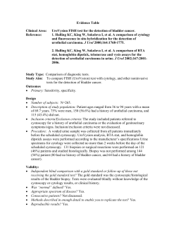
H i g h -y i e l d P r i n c i p l e s IN Pathology “Digressions, objections, delight in mockery, carefree mistrust are signs of health; everything unconditional belongs in pathology.” —Friedrich Nietzsche `` Inflammation `` Neoplasia The fundamental principles of pathology are key to understanding diseases in all organ systems. Major topics such as inflammation and neoplasia appear frequently in questions aimed at many different organ systems, and such topics are definitely high yield. For example, the concepts of cell injury and inflammation are key to understanding the inflammatory response that follows myocardial infarction, a very common subject of boards questions. Similarly, a familiarity with the early cellular changes that culminate in the development of neoplasias—for example, esophageal or colon cancer—is critical. Finally, make sure you recognize the major tumor-associated genes and are comfortable with key cancer concepts such as tumor staging and metastasis. Want updates, corrections, and more? www.usmlerx.com/firstaid 243 FAS1_2012_08-Pathol.indd 243 11/16/11 4:10 PM 244 SECTION II Pathology PATHOLOGY—INFLAMMATION `` PATHOLOGY—INFLAMMATION Apoptosis Programmed cell death; ATP required. Intrinsic or extrinsic pathway; both pathways → activation of cytosolic caspases that mediate cellular breakdown. No significant inflammation. Characterized by cell shrinkage, nuclear shrinkage and basophilia (pyknosis), membrane blebbing, nuclear fragmentation (karyorrhexis), nuclear fading (karyolysis), and formation of apoptotic bodies, which are then phagocytosed. Intrinsic apoptosis vs. extrinsic apoptosis The intrinsic pathway occurs during embryogenesis, hormone induction (e.g., menstruation), and atrophy (e.g., endometrial lining during menopause) and as a result of injurious stimuli (e.g, radiation, toxins, hypoxia). Changes in proportions of anti- and pro-apoptotic factors lead to increased mitochondria permeability and cytochrome c release. Intrinsic Extrinsic 2 extrinsic pathways: 1. Ligand receptor interactions (Fas ligand binding to Fas [CD95]). 2. Immune cell (cytotoxic T-cell release of perforin and granzyme B). FasLigand Bax (pro-apoptotic) Bcl-2 (anti-apoptotic) CD95 (Fas-R) Granzyme B Perforin Mitochondria Cytochrome c Cytosolic caspases activated Killer T cell Cellular breakdown Necrosis FAS1_2012_08-Pathol.indd 244 Enzymatic degradation and protein denaturation of a cell resulting from exogenous injury. Intracellular components extravasate. Inflammatory process (unlike apoptosis). Types of necrosis 1. Coagulative—heart, liver, kidney 2. Liquefactive—brain, bacterial abscess, pleural effusion 3. Caseous—TB, systemic fungi 4. Fatty—pancreas (saponification) 5. Fibrinoid—blood vessels 6. Gangrenous—dry (ischemic coagulative) OR wet (with bacteria); common in limbs and in GI tract 11/16/11 4:10 PM Pathology PATHOLOGY—INFLAMMATION Cell injury Hypoxia SECTION II Reversible with O2 Irreversible ↓ ATP synthesis Cellular swelling (no ATP → impaired Na+/K+ pump) Nuclear chromatin clumping ↓ glycogen Fatty change Ribosomal detachment (↓ protein synthesis) Nuclear pyknosis, karyolysis, karyorrhexis Ca2+ influx → caspase activation Plasma membrane damage Lysosomal rupture Mitochondrial permeability 245 Areas susceptible to hypoxia: Organ Location Watershed areas* Heart Kidney Splenic flexure, ACA/MCA Subendocardial tissue Proximal tubule (cortex) Thick ascending limb (medulla) Neurons Liver Area around central vein *Watershed areas receive dual blood supply from most distal branches of 2 arteries. Infarcts: red vs. pale Red (hemorrhagic) infarcts occur in loose tissues with collaterals, such as liver, lungs, or intestine, or following reperfusion. Pale infarcts occur in solid tissues with single blood supply, such as heart, kidney, and spleen. Heart REd = REperfusion. Reperfusion injury is due to damage by free radicals. Kidney Liver Red infarcts Pale infarcts Shock FAS1_2012_08-Pathol.indd 245 Lung Hypovolemic/cardiogenic Septic Low-output failure ↑ TPR Low cardiac output Cold, clammy patient High-output failure ↓ TPR Dilated arterioles, high venous return Hot patient 11/16/11 4:10 PM 246 SECTION II Pathology PATHOLOGY—INFLAMMATION Atrophy Reduction in the size or number of cells. Causes include: 1. ↓ hormones (uterus/vagina) 2. ↓ innervation (motor neuron damage) 3. ↓ blood flow 4. ↓ nutrients 5. ↑ pressure (nephrolithiasis) 6. Occlusion of secretory ducts (cystic fibrosis) Inflammation Characterized by rubor (redness), dolor (pain), calor (heat), tumor (swelling), and functio laesa (loss of function). Fluid exudation ↑ vascular permeability, vasodilation, endothelial injury. Fibrosis Fibroblast emigration and proliferation; deposition of extracellular matrix. Resolution Restoration of normal structure. Granulation tissue—highly vascularized, fibrotic. Abscess—fibrosis surrounding pus. Fistula—abnormal communication. Scarring—collagen deposition resulting in altered structure and function. Acute—Neutrophil, eosinophil, and antibody mediated. Acute inflammation is rapid onset (seconds to minutes), lasts minutes to days. Chronic—Mononuclear cell mediated: characterized by persistent destruction and repair. Associated with blood vessel proliferation, fibrosis. Granuloma: nodular collections of epithelioid macrophages and giant cells. FAS1_2012_08-Pathol.indd 246 11/16/11 4:10 PM Pathology PATHOLOGY—INFLAMMATION Leukocyte extravasation SECTION II 247 Neutrophils exit from blood vessels at sites of tissue injury and inflammation in 4 steps: Step Vasculature/stroma Leukocyte 1. Rolling E-selectin P-selectin Sialyl LewisX 2. Tight binding ICAM-1 LFA-1 (“integrin”) 3. Diapedesis—leukocyte travels between endothelial cells and exits blood vessel PECAM-1 PECAM-1 4. Migration—leukocyte travels through interstitium to site of injury or infection guided by chemotactic signals Bacterial products CILK: C5a IL-8 LTB4 Kallikrein Various 1. Rolling 2. Tight binding 3. Diapedesis 4. Migration 5. Phagocytosis x S-L Vessel lumen PMN PMN LFA-1 E-selectin PMN PMN ICAM-1 Endothelium Interstitium PMN PMN Free radical injury FAS1_2012_08-Pathol.indd 247 Free radicals damage cells via membrane lipid peroxidation, protein modification, and DNA breakage. Initiated via radiation exposure, metabolism of drugs (phase I), redox reaction, nitric oxide, transition metals, leukocyte oxidative burst. Free radicals can be eliminated by enzymes (catalase, superoxide dismutase, glutathione peroxidase), spontaneous decay, antioxidants (vitamins A, C, E). Pathologies include: 1. Retinopathy of prematurity 2. Bronchopulmonary dysplasia 3. CCI4 leading to liver necrosis (fatty change) 4. Acetaminophen 5. Iron overload 6. Reperfusion after anoxia (e.g., superoxide), especially after thrombolytic therapy 11/16/11 4:10 PM 248 SECTION II Wound healing Pathology PATHOLOGY—INFLAMMATION Phase Mediators Characteristics Inflammatory (immediate) Platelets, neutrophils, macrophages Clot formation, ↑ vessel permeability and neutrophil migration into tissue; macrophages clear debris 2 days later Proliferative (2–3 days after wound) Fibroblasts, myofibroblasts, endothelial cells, keratinocytes Deposition of granulation tissue and collagen, angiogenesis, epithelial cell proliferation, dissolution of clot, and wound contraction (mediated by myofibroblasts) Remodeling (1 week after wound) Fibroblasts Type III collagen replaced by type I collagen, ↑ tensile strength of tissue Granulomatous diseases 1. Mycobacterium tuberculosis 2. Fungal infections (e.g., histoplasmosis) 3. Treponema pallidum (syphilis) 4. M. leprae (leprosy) 5. Bartonella henselae (cat scratch disease) 6. Sarcoidosis 7. Crohn’s disease 8. Berylliosis Th1 cells secrete γ-interferon, activating macrophages. TNF-α from macrophages induce and maintain granuloma formation. Anti-TNF drugs can break down granulomas, leading to disseminated disease. Transudate vs. exudate Transudate Exudate Hypocellular Protein poor Specific gravity < 1.012 Due to: ↑ hydrostatic pressure ↓ oncotic pressure Na+ retention Cellular Protein rich Specific gravity > 1.020 Due to: Lymphatic obstruction Inflammation Erythrocyte sedimentation rate (ESR) FAS1_2012_08-Pathol.indd 248 Products of inflammation (e.g., fibrinogen) coat RBCs and cause aggregation. When aggregated, RBCs fall at a faster rate within the test tube. ↑ ESR ↓ ESR Infections Inflammation (e.g., temporal arteritis) Cancer Pregnancy SLE Sickle cell (altered shape) Polycythemia (too many) CHF (unknown) 11/16/11 4:10 PM Pathology PATHOLOGY—INFLAMMATION Iron poisoning Cell death due to peroxidation of membrane lipids. Symptoms Acute—gastric bleeding. Chronic—metabolic acidosis, scarring leading to GI obstruction. β-pleated sheet demonstrable by apple-green birefringence of Congo red stain under polarized light A ; affected tissue has waxy appearance. A Amyloidosis. Note the apple-green birefringence of the amyloid deposits in the artery wall. Types Protein Derived from Bence Jones AL Ig light chains (multiple myeloma) AL = Light chain. Secondary AA Serum amyloid-associated (SAA) protein (chronic inflammatory disease) AA = Acute-phase reactant. Senile cardiac Transthyretin AF AF = old Fogies. Diabetes mellitus type 2 Amylin AE AE = Endocrine. Medullary carcinoma of the thyroid A-CAL Calcitonin A-CAL = CALcitonin. Alzheimer’s disease β-amyloid Amyloid precursor protein (APP) Dialysis-associated β2-microglobulin MHC class I proteins FAS1_2012_08-Pathol.indd 249 249 One of the leading causes of fatality from toxicologic agents in children. Mechanism Amyloidosis SECTION II 11/16/11 4:10 PM 250 SECTION II Pathology PATHOLOGY—Neoplasia `` PATHOLOGY—Neoplasia Neoplastic progression Hallmarks of cancer—evading apoptosis, self-sufficiency in growth signals, insensitivity to anti-growth signals, sustained angiogenesis, limitless replicative potential, tissue invasion, and metastasis. Epithelial cell layer • Normal cells with basal → apical differentiation Basement membrane Normal • Cells have increased in number––hyperplasia • Abnormal proliferation of cells with loss of size, shape, and orientation––dysplasia Hyperplasia Carcinoma in situ • Neoplastic cells have not invaded basement membrane • High nuclear/cytoplasmic ratio and clumped chromatin • Neoplastic cells encompass entire thickness Carcinoma in situ/ preinvasive • Cells have invaded basement membrane using collagenases and hydrolases (metalloproteinases) • Can metastasize if they reach a blood or lymphatic vessel Invasive carcinoma Metastasis––spread to distant organ • Must survive immune attack • "Seed and soil" theory of metastasis • Seed = tumor embolus • Soil = target organ––liver, lungs, bone, brain, etc. Metastatic focus Blood or lymphatic vessel (Adapted, with permission, from McPhee S, Lingappa VR, Ganong WF, et al. Pathophysiology of Disease: An Introduction to Clinical Medicine, 3rd ed. New York: McGraw-Hill 2000: 84.) FAS1_2012_08-Pathol.indd 250 11/16/11 4:10 PM Pathology PATHOLOGY—Neoplasia SECTION II 251 -plasia definitions Reversible Hyperplasia—↑ in number of cells. Metaplasia—one adult cell type is replaced by another. Often 2° to irritation and/or environmental exposure (e.g., squamous metaplasia in trachea and bronchi of smokers). Dysplasia—abnormal growth with loss of cellular orientation, shape, and size in comparison to normal tissue maturation; commonly preneoplastic. Irreversible Anaplasia—abnormal cells lacking differentiation; resemble primitive cells of same tissue, often equated with undifferentiated malignant neoplasms. Little or no resemblance to tissue of origin. Neoplasia—a clonal proliferation of cells that is uncontrolled and excessive. Neoplasia may be benign or malignant. Desmoplasia—fibrous tissue formation in response to neoplasm. Tumor grade vs. stage Grade Degree of cellular differentiation based on histologic appearance of tumor. Usually graded I–IV based on degree of differentiation and number of mitoses per high-power field; character of tumor itself. Stage Degree of localization/spread based on site and size of 1° lesion, spread to regional lymph nodes, presence of metastases; spread of tumor in a specific patient. Stage usually has more prognostic value than grade. Stage = Spread. TNM staging system: T = size of Tumor N = Node involvement M = Metastases (most important prognostic factor) Tumor nomenclature Cell type Benign Malignanta Epithelium Adenoma, papilloma Adenocarcinoma, papillary carcinoma Mesenchyme Leukemia, lymphoma Blood cells Blood vessels Hemangioma Angiosarcoma Smooth muscle Leiomyoma Leiomyosarcoma Skeletal muscle Rhabdomyoma Rhabdomyosarcoma Connective tissue Fibroma Fibrosarcoma Bone Osteoma Osteosarcoma Fat Lipoma Liposarcoma > 1 cell type Mature teratoma (women) Immature teratoma and mature teratoma (men) aThe term carcinoma implies epithelial origin, whereas sarcoma denotes mesenchymal origin. Both terms imply malignancy. Tumor differences Benign Usually well differentiated, slow growing, well demarcated, no metastasis. Malignant May be poorly differentiated, erratic growth, locally invasive/diffuse, may metastasize. FAS1_2012_08-Pathol.indd 251 11/16/11 4:10 PM 252 SECTION II Pathology PATHOLOGY—Neoplasia Cachexia Loss of weight, muscle atrophy, and fatigue that occur in chronic disease (e.g., cancer, AIDS, heart failure, tuberculosis). Mediated by TNF-α (nicknamed cachectin), IFN-γ, and IL-6. Disease conditions associated with neoplasms Condition Neoplasm 1. Down syndrome 1. ALL (we ALL fall Down), AML 2. Xeroderma pigmentosum, albinism 2. Melanoma, basal cell carcinoma, and especially squamous cell carcinomas of skin 3. Chronic atrophic gastritis, pernicious anemia, postsurgical gastric remnants 3. Gastric adenocarcinoma 4. Tuberous sclerosis (facial angiofibroma, seizures, mental retardation) 4. Astrocytoma, angiomyolipoma, and cardiac rhabdomyoma 5. Actinic keratosis 5. Squamous cell carcinoma of skin 6. Barrett’s esophagus (chronic GI reflux) 6. Esophageal adenocarcinoma 7. Plummer-Vinson syndrome (atrophic glossitis, esophageal webs, anemia; all due to iron deficiency) 7. Squamous cell carcinoma of esophagus 8. Cirrhosis (alcoholic, hepatitis B or C) 8. Hepatocellular carcinoma 9. Ulcerative colitis 9. Colonic adenocarcinoma 10. Paget’s disease of bone 10. 2° osteosarcoma and fibrosarcoma 11. Immunodeficiency states 11. Malignant lymphomas 12. AIDS 12. Aggressive malignant lymphomas (nonHodgkin’s) and Kaposi’s sarcoma 13. Autoimmune diseases (e.g., Hashimoto’s thyroiditis, myasthenia gravis) 13. Lymphoma 14. Acanthosis nigricans (hyperpigmentation and epidermal thickening) 14. Visceral malignancy (stomach, lung, breast, uterus) 15. Dysplastic nevus 15. Malignant melanoma 16. Radiation exposure 16. Sarcoma, papillary thyroid cancer FAS1_2012_08-Pathol.indd 252 11/16/11 4:10 PM Pathology PATHOLOGY—Neoplasia Oncogenes Gene Gene product abl CML Tyrosine kinase c-myc Burkitt’s lymphoma Transcription factor bcl-2 Follicular and undifferentiated lymphomas (inhibits apoptosis) Anti-apoptotic molecule erb-B2 Breast, ovarian, and gastric carcinomas Tyrosine kinase ras Colon carcinoma GTPase L-myc Lung tumor Transcription factor N-myc Neuroblastoma Transcription factor ret Multiple endocrine neoplasia (MEN) types IIA and IIB Tyrosine kinase c-kit Gastrointestinal stromal tumor (GIST) Cytokine receptor Gene 253 Gain of function → cancer. Need damage to only 1 allele. Associated tumor Tumor suppressor genes SECTION II Loss of function → cancer; both alleles must be lost for expression of disease. Associated tumor Gene products Rb Retinoblastoma, osteosarcoma Rb gene product blocks G1 → S phase of the cell cycle p53 Most human cancers, Li-Fraumeni syndrome p53 gene product blocks G1 → S phase of the cell cycle BRCA1 Breast and ovarian cancer DNA repair protein BRCA2 Breast cancer DNA repair protein p16 Melanoma APC Colorectal cancer (associated with FAP) WT1 Wilms’ tumor NF1 Neurofibromatosis type 1 NF2 Neurofibromatosis type 2 DPC Pancreatic cancer DPC—Deleted in Pancreatic Cancer. DCC Colon cancer DCC—Deleted in Colon Cancer. FAS1_2012_08-Pathol.indd 253 11/16/11 4:10 PM 254 SECTION II Pathology PATHOLOGY—Neoplasia Tumor markers PSA Prostate-specific antigen. Used to screen for prostate carcinoma. Can also be elevated in BPH and prostatitis. Prostatic acid phosphatase Prostate carcinoma. CEA Carcinoembryonic antigen. Very nonspecific but produced by ∼ 70% of colorectal and pancreatic cancers; also produced by gastric, breast, and thyroid medullary carcinomas. α-fetoprotein Normally made by fetus. Hepatocellular carcinomas. Nonseminomatous germ cell tumors of the testis (e.g., yolk sac tumor). β-hCG Hydatidiform moles, Choriocarcinomas, and Gestational trophoblastic tumors. CA-125 Ovarian, malignant epithelial tumors. S-100 Melanoma, neural tumors, astrocytomas. Alkaline phosphatase Metastases to bone, obstructive biliary disease, Paget’s disease of bone. Bombesin Neuroblastoma, lung and gastric cancer. TRAP Tartrate-resistant acid phosphatase. Hairy cell leukemia—a B-cell neoplasm. CA-19-9 Pancreatic adenocarcinoma. Calcitonin Thyroid medullary carcinoma. Oncogenic microbes FAS1_2012_08-Pathol.indd 254 Tumor markers should not be used as the 1° tool for cancer diagnosis. They may be used to confirm diagnosis, to monitor for tumor recurrence, and to monitor response to therapy. TRAP the hairy animal. Virus Associated cancer HTLV-1 Adult T-cell leukemia/lymphoma HBV, HCV Hepatocellular carcinoma EBV Burkitt’s lymphoma, Hodgkin’s lymphoma, nasopharyngeal carcinoma HPV Cervical carcinoma (16, 18), penile/anal carcinoma HHV-8 (Kaposi’s sarcoma–associated herpesvirus) Kaposi’s sarcoma, body cavity fluid B-cell lymphoma HIV Primary CNS lymphoma H. pylori Gastric adenocarcinoma and lymphoma Schistosoma haematobium Squamous cell carcinoma of transitional epithelium, e.g., bladder 11/16/11 4:10 PM Pathology PATHOLOGY—Neoplasia SECTION II 255 Chemical carcinogens Toxin Organ Impact Aflatoxins (Aspergillus) Liver Hepatocellular carcinoma Vinyl chloride Liver Angiosarcoma CCl4 Liver Centrilobular necrosis, fatty change Nitrosamines (smoked foods) Stomach Gastric cancer Cigarette smoke Larynx Lung Squamous cell carcinoma Squamous cell and small cell carcinoma Renal cell carcinoma Transitional cell carcinoma Kidney Bladder Asbestos Lung Mesothelioma and bronchogenic carcinoma Arsenic Skin Liver Squamous cell carcinoma Angiosarcoma Naphthalene (aniline) dyes Bladder Transitional cell carcinoma Alkylating agents Blood Leukemia Hormone/agent Effect Neoplasm ACTH or ACTH-like peptide Cushing’s syndrome Small cell lung carcinoma ADH SIADH Small cell lung carcinoma and intracranial neoplasms PTH-related peptide, TGF-β, TNF, IL-1 Hypercalcemia Squamous cell lung carcinoma, renal cell carcinoma, and breast carcinoma Erythropoietin Polycythemia Renal cell carcinoma, hemangioblastoma, hepatocellular carcinoma, pheochromocytoma Antibodies against presynaptic Ca2+ channels at neuromuscular junction Lambert-Eaton syndrome (muscle weakness) Thymoma, small cell lung carcinoma Hyperuricemia due to excess nucleic acid turnover (i.e., cytotoxic therapy) Gout, urate nephropathy Leukemias and lymphomas Paraneoplastic effects of tumors Psammoma bodies FAS1_2012_08-Pathol.indd 255 Laminated, concentric, calcific spherules seen in: 1. Papillary adenocarcinoma of thyroid 2. Serous papillary cystadenocarcinoma of ovary 3. Meningioma 4. Malignant mesothelioma PSaMMoma: Papillary (thyroid) Serous (ovary) Meningioma Mesothelioma 11/16/11 4:10 PM 256 SECTION II Pathology PATHOLOGY—Neoplasia Cancer epidemiology Male Female Incidence Prostate (32%) Lung (16%) Colon/rectum (12%) Breast (32%) Lung (13%) Colon/rectum (13%) Lung cancer incidence has dropped in men, but is level for women. Mortality Lung (33%) Prostate (13%) Lung (23%) Breast (18%) Cancer is the 2nd leading cause of death in the United States (heart disease is 1st). Common metastases Metastasis 1º Tumor Brain Lung > breast > kidney > skin (melanoma). 50% of brain tumors are from metastases. Typically multiple well-circumscribed tumors at gray/white matter junction. Liver Colon > stomach > pancreas. Liver and lung are the most common sites of metastasis after the regional lymph nodes. Bone Prostate, breast > lung > thyroid, testes. Metastatic bone tumors are far more common than 1° tumors. Lung = lytic. Prostate = blastic. Breast = lytic and blastic. Want updates, corrections, and more? www.usmlerx.com/firstaid FAS1_2012_08-Pathol.indd 256 11/16/11 4:10 PM


© Copyright 2026