
Nasopharyngeal Adenoid Cystic Carcinoma Case Report and Review acta medica C
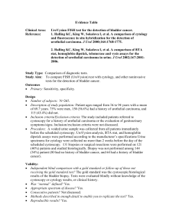
Acta Medica 2013; 2: 59–64
acta medica
CASE R EPORT
Nasopharyngeal Adenoid Cystic Carcinoma
Case Report and Review
Melahat DÖNMEZ1*, [MD]
Onur ORAL2, [MD]
Yıldız ALBAYRAK2, [MD]
Pınar NERCIŞ3, [MD]
Sema HÜCÜMENOĞLU, [MD]
A BST R AC T
Adenoid Cystic Carcinoma (ACC) is a rare slow-growing salivary gland neoplasm with malignant behavior and a high recurrence rate. It originates from
salivary glands. Nasopharyngeal located ACC (NACC) is a very rare malignancy due to its location with specific biological characteristics. There is no consensus at present regarding clinical characteristics, treatment approaches and
prognostic factors. Our aim is to review the literature related to this topic in
light of our case about this rare malignancy.
A 36-year-old female at 35 weeks pregnancy was admitted to the Gynecology
and Obstetrics service for hypertension. The patient had a history of a nasopharyngeal carcinoma for which she underwent radiotherapy 11 years ago.
A neurology and eye consultation required for ptosis of her left eye An orbito-cranial MR demonstrated expansive mass lesions that were initially thought
to be mucoceles filling the nasal cavity, left maxillary and sphenoid sinuses. A
biopsy of the lesions was reported as ‘adenoid cystic carcinoma’. The patient
died 6 months later.
1 Department of Pathology, Ministry of Health
Diskapi Yildirim Beyazit Training and Research
Hospital, Ankara, Turkey
2 Department of Pathology, Ministry of Health
Ankara Training and Research Hospital, Ankara,
Turkey
3 Department of Radiology, Ministry of Health
Ankara Training and Research Hospital, Ankara,
Turkey
* Corresponding Author: Melahat Donmez
Ministry of Health Diskapi Yildirim Beyazit
Training and Research Hospital, Department of
Pathology, Ankara-TURKEY
[email protected]
Our case is special due to the patient’s arrival with cranial nerve invasion, slow
progression, expansive growth pattern, misdiagnosis as a mucocele clinically and radiographically. Because of its high rate of cranial nerve invasion, this
neoplasm must be considered in the differential diagnosis. In sinonasal mass
lesions with nasal obstructive symptoms, the possibility of malignancy must
be eliminated. Despite the fact that papillomas spread expansively, a biopsy
must be obtained for prompt diagnosis and treatment. Additionally patients
diagnosed with nasopharyngeal carcinoma, even if they are treated and cured,
must be called for control in routine intervals because of the malignant behavior and high recurrence rate.
Key words: Primary salivary gland type nasopharyngeal carcinoma (SNPC),
perineural invasion, distant metastasis, malignancy, salivary gland neoplasm
Received 8 October 2013, Accepted 28 October 2013
Published online 31 October 2013
Introduction
Adenoid Cystic Carcinoma (ACC) is a rare,
slow-growing salivary gland neoplasm with malignant behavior and a high recurrence rate. It originates from secretory glands in the major and minor
salivary glands. It is seen in the palate, the buccal
mucosa, lips and floor of the mouth in decreasing
frequency [1-4] .
In nasopharyngeal location, the most common
malignant epithelial tumor is ‘nonkeratinized squamous cell carcinoma’. Primary salivary gland type
nasopharyngeal carcinoma (SNPC) is highly rare
and consists of 0.48% of all nasopharyngeal carcinomas [5]. Therefore, its clinical characteristics,
© 2013 Acta Medica. All rights reserved.
treatment approaches, and prognostic factors are
not completely known [6] .
Nasopharyngeal Adenoid Cystic Carcinoma
(NACC) is a primary SNPC and very rare malignancy that has specific biological characteristics. Due
to the scarcity of cases, there is no clear consensus
in terms of clinical characteristics, treatment approaches and prognostic factors. Our aim is to review the literature related to this topic by presenting a case with NACC. Different from the nasopharyngeal squamous cell carcinomas, NACC shows
perineural invasion that is a specific biological behavior. The tumor progresses through the cranial nerve; it reaches the orbital and cranial cavity,
59
Nasopharyngeal Adenoid Cystic Carcinoma Case Report and Review
Picture 1. T2-weighted axial cross-section. Mass observed here holds left compartment of Sfenid Sinus And
left posterior ethmoid sinus cells, spreading the posterior orbita, middle cranial fossa, along the sphenoid wing,
multi compartmental, expansile, hyperintence. Optic foramen and the superior orbital fissure is seen that the
mass was obliterated.
which worsen the patients’ prognosis. Nasal cavity,
skull floor, pterygopalatine fossa, and zygomatic fossa are common locations that can render surgical resection more difficult [6-9] .
Case Presentation
A 36 year-old female patient at 35 weeks pregnancy
presented with hypertension to the Gynecology and
Obstetrics Department. The patient reported smoking 10 cigarettes a day. She reported left eyelid droop
Picture 2. T1-weighted fat-suppressed axial cross-sectional. Because of the pregnancy opaque material could
not be granted. Isointense structure is observed.
two months ago. She also reported that she was diagnosed as nasopharyngeal cancer eleven years ago
and she underwent radiotherapy (RT). Additionally,
neurology and opthalmology were consulted for
ptosis of the left eyelid. On examination, ptosis of
her left eye lid, limitation in eye movement and 2nd,
3rd and 6th cranial nerve paralysis were noted.
On orbito-cranial MR there were expansile mass
lesions filling the nasal cavity, left maxillary and
sphenoid sinus; eroding the concha and hard palate; obliterating the optic canal, superior-inferior orbital fissure, entering mid-left cranial fossa and orbits at the orbital apex; surrounding the left cavernous sinus, that were at first thought to be in accordance with the diagnosis of mucocele (Picture 1 and
2). Nasopharyngeal incisional biopsy was then taken.
Figure 1 (A) and (B). Microscopic appearance of the ACC showing neoplastic proliferating cells were of uniform
basaloid character, and formed cribriform structures in wide spaces. (Hematoxylin-Eosin)
(A)
60
(B)
© 2013 Acta Medica. All rights reserved.
Acta Medica 2013; 2: 59–64
On macroscopic examination of the biopsy
material, two pieces of grey-white irregular tissue
sized 1.5x0.5x0.5 cm and 0.8x0.3x0.3 cm were observed. On serial sections in the microscopic examination, it was found that the entire specimen contained neoplastic proliferation. Proliferating cells
were of uniform basaloid character, and formed
cribriform structures in wide spaces (Figure 1 (A)
and (B), Figures 2 and 3). Hyaline or mucoid material was present in the lumens. In some focal areas, mucoid stroma was observed. On histochemical
studies with PAS and mucin, the pseudocystic areas
were positive, and negative on Pas/Diastase staining
(Figures 4 and 5). Thus the image was diagnosed as
‘adenoid cystic carcinoma’.
The patient delivered a baby girl of 2400 grams
via caesarean section. There were no early complications. The patient was referred to obstetric/gynecology, otorhinolaryngology and internal medicine.
However, the patient died 6 months later.
Figure 2. Imunhistochemical PanCK staining is positive
Donmez et al.
Discussion
ACC is a tumor with different morphologic configurations of basal epithelial and myoepithelial cells.
Despite a grossly solid appearance with an infiltrative
pattern, it can sometimes have a well-demarcated,
Figure 3. Immunhistochemical S100 staining is positive
Figure 4. Immunohistochemical GFAP staining is
positive
Figure 5 (A) and (B). Histochemical studies with PAS (A) and Mucin (B), the pseudocystic areas were positive.
(A)
© 2013 Acta Medica. All rights reserved.
(B)
61
Nasopharyngeal Adenoid Cystic Carcinoma Case Report and Review
expansive pattern. In most cases, it does not have
a capsule, and consists of pseudoglandular, “cribriform” structures with small glandular lumens [4].
Some cases with ACC present with a newly identified, rare, sclerosant’ pattern [7,8,9]. A combination
of these two patterns can be seen in the original or
recurrent tumors. Additionally, this neoplasm can
also present in a hybrid pattern in combination with
other tumors [10]. With regards to the cell type, a
combination of intercalated ductal epithelial cells,
myoepithelial cells, secretory cells and pluripotential reserve cells exists. Thus, the combination of
cells in a tumor is no different from that in a benign
mixed tumor and has a similar histogenesis. On incisional biopsies, if the periphery of the lesion is satisfactorily sampled, when perineural invasion is not
observed, the diagnosis should be questioned [1] .
NACC’s are tumors with a long, slow progressing history that are generally diagnosed late. The
time between the appearance of the lesion and the
first symptom is 2-5 years. Sometimes, computerized tomography can be helpful to diagnose bone
erosion on the skull floor. Sometimes, with MR, involvement of the infra-temporal fossa and cavernous
sinus, or perineural and perivascular infiltration can
also be noted and suggest a separate diagnosis [11].
However, in our case, this was not considered a different diagnosis radiologically. In our case, a biopsy
was obtained due to an initial diagnosis of a mucocele, clinically and radiologically.
NACC occur more often in females, approximately 49 years of age. The most common symptoms in decreasing frequency include tinnitus, nasal
obstruction, epistaxis, headache, facial numbness,
eye weakness, diplopia, vision loss and vertigo [12).
Studies of Liu et al and Cao et al [12,13] about nasopharyngeal ACC and clinical features are shown
in detail table 1. Studies show that cranial nerve
Table 1. Clinical features and treatment
NUMBER OF PATIENTS
FACTOR
Liu et al
Cao et al
GENDER
Males
Females
11
15
18
18
AGE
Older
Younger
17*
9
18**
18
SKULL BASE INVASION
No
Yes
15
11
8
28
CRANIAL NERVE INVASION
No
Yes
19
7
15
21
STAGE
Early
Advanced
14
12
8
28
*Older: ≥40 years, ** Older: ≥45 years
invasion occurs, which differs from other nasopharyngeal neoplasms. The tumor progresses through
the cranial nerve, reaches the orbital cavity and cranial cavity and this worsens the patients’ prognosis.
[12,14] .
NACC rarely spreads to lymph nodes. At the
same time, the tumor has the tendency for hematogenous spread and can demonstrate distant metastasis without cervical node metastasis (Table 2)
[12,13] .
Cao et al defined clinical features of all nasopharyngeal SNPC subtypes with ACC. They are shown
in detail in Table 3. Among cases of SNPC, the most
common subtype is NACC, and the subtype with
the highest metastasis is ACC [13]. Among all the
nasopharyngeal SNPC cases, cervical lymph node
metastasis of ACC is observed in less than 20% of
cases [18] .
Table 2. Published Clinical Studies of Nasopharyngeal Adenoid Cystic Carcinoma
CASES
LYMPH NODE
METASTASES (%)
DISTANT
METASTASES (%)
TREATMENT S
SURVIVAL
Lee at al.15
11
N
60.0
R, S+R
N
Wang et al.14
5
15.0
35.0
R,S
78.0%, 5-yr OS
Schramm and Immola16
11
N
N
S+R
100.0%, 3-yr OS
Wen et al.17
21
14.3
30.0
S+R, R
42.9%, DFS
Liu et al.12
26
7.7
26.9
S+R,R
54.8%, 5-yr OS
Cao et al.13
36
13.9
30.6
S+R, R
61.3%, 5-yr OS
AUTHOR
R = radiotherapy; S = surgery; N = no result; OS = overall survival; DFS = disease free survival
62
© 2013 Acta Medica. All rights reserved.
Acta Medica 2013; 2: 59–64
Donmez et al.
Table 3. Clinical features of primary salivary gland-type
carcinomas of the nasopharynx [13] .
NUMBER OF PATIENTS (%)
FACTOR
ACC
MEC
AC
GENDER
Males
Females
18 (50.0%) 4 (36.4%)
18 (50.0%) 7 (63.6%)
4 (57.1%)
3 (42.9%)
AGE
≥45 y
Younger
18 (50.0%) 5 (45.5%)
18 (50.0%) 6 (54.5%)
4 (57.1%)
3 (42.9%)
SKULL BASE INVASION
No
Yes
8 (22.2%) 9 (81.8%)
28 (77.8%) 2 (18.2%)
3 (42.9%)
4 (57.1%)
CRANIAL NERVE INVASION
No
Yes
15 (55.6%) 11 (100.0%) 4 (57.1%)
21 (44.4%) 0 (0.0%) 3 (42.9%)
STAGE
Early
Advanced
8 (22.2%) 8 (62,7%)
28 (77.8%) 3 (27.3%)
HISTOLOGIC GRADE
Low
High
-
5 (45.5%)
6 (54.5%)
3 (42.9%)
4 (57.1%)
5 (71,4%)
2 (28.6%)
LYMPH NODE METASTASES
No
Yes
31 (86.1%) 10 (90.9%) 3 (42.9%)
5 (13.9%) 1 (9.1%) 4 (57.1%)
DISTANT METASTASES
No
Yes
25 (69.4%) 11 (100.0%) 5 (71,4%)
11 (30.6%) 0 (0.0%) 2 (28.6%)
MEC = MUCOEPIDERMOİD CARCINOMA; AC =
ADENOCARCINOMA
In contrast to non-keratinized and keratinized
carcinoma, the sensitivity to RT is low, and therefore, surgery is accepted as the main treatment in
stages 1, 2, 3 NACCs [19]. The optimal treatment is
radical surgery followed by RT. However, the anatomy of the nasopharynx brings additional risks and
technical problems related to involvement of critical
neural and vascular structures [11]. Due to a low incidence of SNPC, not many studies on chemotherapy have been performed previously [6] .
In studies comparing nasopharyngeal keratinized and non-keratinized carcinomas, survival
rates of SNPC malignancies involving the nasopharynx have a better prognosis. However, cranial nerve
involvement and the presence of an advanced stage
can affect survival dramatically. Because the incidence of NACC is very low, and statistical studies on
the number of cases are very few, multi-centric studies should be conducted in order to evaluate future
treatment strategies and prognostic factors [20] .
In reports by Lin Yu-Chin et al [21], cases of salivary and nonsalivary ACC were investigated clinicopathologically. When major salivary gland, minor
salivary gland and nonsalivary ACC were compared,
sinonasal and tracheo-bronchial ACC have an advanced course with a more positive margin in cases
of sinonasal and lacrimal ACC (Table 4). There was
no difference between groups in terms of perineural invasion. Sinonasal, lacrimal and trachebronchial ACC have a worse prognosis than ACCs originating from the major salivary gland. Thus they concluded that a bad prognosis of sinonasal, lacrimal
Table 4. Pathological features of salivary and nonsalivary ACC [21]
Site
No. of patients
Advanced stage*
High grade**
Positive margin
Perineural invasion
Major salivary
Parotid
Submandibular
Sublingual
32 (total)
21
8
3
34.4% (11/32)
15.6% (5/32)
53.1% (17/32)
65.6% (21/32)
Minor salivary
Intraoral
Tongue
Buccal
Mauth floor
Palate
Sinonasal
51 (total)
26
5
5
2
14
25
57.2% (15/26)
76% (19/25)
11.5% (3/26)
12.0% (3/25)
53.8% (14/26)
84% (19/25)
61.5% (16/26)
76% (21/25)
50% (1/2)
44.4% (4/9)
71.4% (5/7)
100% (2/2)
0% (0/9)
14.3% (1/7)
50% (1/2)
77.8% (7/9)
42.9% (3/7)
100% (2/2)
66.7% (6/9)
100% (7/7)
Nonsalivary
Ear
Lacrimal
Tracheeobronchial
18
2
9
7
*Stage 3,4; **Histological grade 3
© 2013 Acta Medica. All rights reserved.
63
Nasopharyngeal Adenoid Cystic Carcinoma Case Report and Review
and tracheobronchial ACC can result from late di- a slow-growing tumor that spreads in an expansive
agnosis, difficult resection and tumor biology in dif- way, rather than in an invasive pattern, causing a diferent locationsThe origin of ACC plays role in de- agnosis of mucocele to be skipped clinically and raterming the prognosis. It is reported that sinonasal diologically. This led to a delay in diagnosis, leading
ACC has a worse prognosis than ACC of the major to her death.
salivary gland [22]. Minor salivary gland ACC has
In conclusion, malignancy must be considered
been divided into sinonasal and intraoral groups. in the different diagnosis of mass lesions of the siBecause the prognostic results of minor salivary nonasal area, and with nasal obstructive symptoms.
gland ACCs are heterogenous, the prognosis of in- The mass must be biopsied even though the papiltra-oral ACC with major salivary gland involvement loma spreads expansively, to eliminate malignancy.
are the same.
And besides, patients diagnosed with nasophaOur case is difficult in terms of diagnosis and ryngeal carcinoma, even if they are treated and cured,
treatment, and the clinical prognosis is poor. Just as must be called control in routine intervals because of
in our case, patients with ACC generally die [4]. It is malignant behavior and a high recurrence rate.
REFERENCES
[1] Rosai J. Major and minor salivary glands. In: Rosai J, Ackerman
L, editors. Rosai and Ackerman’s Surgical Pathology 10th ed.
Edinburg: Elsevier; 2011. p. 833-835.
[12] Liu TR, Yang AK, Guo X, Li QL, Song M, He JH, Wang YH,
Guo Z. Adenoid cystic carcinoma of the nasopharynx: 27-year
experience. Laryngoscope 2008; 118 (11): 1981-8.
[2] Ellis ER, Million RR, Mendenhall WM, Parsons JT, Cassisi
NJ. The use of radiation therapy in the management of minor salivary gland tumors. Int J Radiat Oncol Biol Phys 1988;
15:613–7.
[13] Cao CN, Zhang XM, Luo JW, Xu GZ, Gao L, Li SY, Xiao JP,
Yi JL, Huang XD, Liu SY, Xu ZG, Tang PZ. Primary salivary
gland-type carcinomas of the nasopharynx: Prognostic factors
and Outcome. Int. J. Oral Maxillofac. Surg. 2012; 41: 958–964.
[3] Garden AS, Weber RS, Ang KK, Morrison WH, Matre J,
Peters LJ. Postoperative radiation therapy for malignant tumors of minor salivary glands. Cancer 1994; 73:2563–9.
[4] El-Naggar AK, Huvos AG. Tumors of the salivary glands: adenoid cystic carcinoma. In: Barnes EL, Eveson JW, Reichart P,
Sidransky D, editors. World Health Organization classification of tumours: pathology & genetics. Head and neck tumours.
Lyon: IARCPress; 2005. p. 221–2.
[5] He JH, Zong YS, Luo RZ, Liang XM, Wu QL, Liang YJ.
Clinicopathological characteristics of primary nasopharyngeal adenocarcinoma. Ai Zheng 2003; 22:753–457.
[6] Liu TR, Chen FJ, Qian CN, Guo X, Zeng MS, Guo ZM, He
JH, Cao JY, Yang AK, Zhang GP. Primary salivary gland type
carcinoma of the nasopharynx: therapeutic outcomes and
prognostic factors. Head Neck 2010; 32:435–44.
[7] Perzin KH, Gullane P, Clairmont AC. Adenoid cystic carcinomas arising in salivary glands: a correlation of histologic
features and clinical course. Cancer 1978; 42 (1): 265-82.
[8] Spiro RH, Huvos AG, Strong EW. Adenoid cystic carcinoma
of salivary origin. A clinicopathologic study of 242 cases. Am J
Surg 1974; 128:512-20.
[9] Albores-Saavedra J, Wu J, Uribe-Uribe W. The sclerosing
variant of adenoid cystic carcinoma; a previously unrecognized neoplasm of major salivary glands. Ann Diagn Pathol
2006; 10:1-7.
[10] Snyder ML, Paulino AF. Hybrid carcinoma of the salivary
gland: salivary duct adenocarcinoma adenoid cystic carcinoma. Histopathology 1999; 35:380–383.
[11] Bradley PJ. Adenoid cystic carcinoma of the head and neck:
a review. Curr Opin Otolaryngol Head Neck Surg 2004; 12
(2):127-32.
64
[14] Wang CC, See LC, Hong JH, Tang SG. Nasopharyngeal adenoid cystic carcinoma: five new cases and a literature review. J
Otolaryngol 1996; 25:399–403.
[15] Lee DJ, Smith RR, Spaziana JT, Rostock r, Holliday M,
Mosos H. Adenoid cystic carcinoma of nasopharynx. Case reports and literature review. Ann Otol Rhinol Laryngol 1985;
94:269-72.
[16] Schramm VL Jr, Imola MJ. Management of nasopharyngeal
salivary gland malignancy. Laryngoscope 2001;111:1533-44.
[17] Wen SG, Tang PZ, Xu ZG, Qi YF, Li ZJ, Liu WS.
Therapeutic modalities of nasopharyngeal adenoid cystic carcinoma. Zhonghua Er Bi Yan Hou Tou Jing Wai Ke Za Zhi
2006;41:359-361.
[18] Gomez DR, Hoppe BS, Wolden SL, Zhung JE, Patel SG,
Kraus DH, et al. Outcomes and prognostic variables in adenoid cystic carcinoma of the head and neck: a recent experience. Int J Radiat Oncol Biol Phys 2008;70: 1365–72.
[19] Takagi D, Fukuda S, Furuta Y, et al. Clinical study of adenoid
cystic carcinoma of the head and neck. Auris Nasus Larynx
2001;28 (Suppl 1):S99–S102.
[20] Huang QH, Li YH, Liu Q. A survival analysis of 1761 nasopharyngeal carcinoma cases diagnosed during 1976–2005 in
Sihui city in Guangdong province. Zhonghua Yu Fang Yi Xue
Za Zhi 2007;41 (Suppl):98–100.
[21] Lin YC, Chen KC, Lin CH, Kuo KT, Ko JY, Hong RL.
Clinicopathological features of salivary and non-salivary adenoid cystic carcinomas. Int J Oral Maxillofac Surg. 2012
Mar;41 (3):354-60.
[22] Oplatek A Ozer E, Agrawal A, Bapna S, Schuller DE.
Patterns of recurrence and survival of head and neck adenoid cystic carcinoma after definitive resection. Laryngoscope.
2010 Jan;120 (1):65-7
© 2013 Acta Medica. All rights reserved.


© Copyright 2026