
Prevalence of Tongue Anomalies in Hamadan, Iran
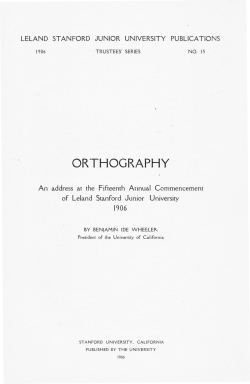
Iranian J Publ Health, Vol. 37, No.2, 2008, pp.101-105 Iranian J Publ Health, Vol. 37, No.2, 2008, pp.101-105 Original Article Prevalence of Tongue Anomalies in Hamadan, Iran *F Mojarrad 1, P Bakianian Vaziri 2 1 Downloaded from http://journals.tums.ac.ir/ on Monday, August 20, 2012 2 Dept. of Paedodontics, Faculty of Dentistry, Hamadan University of Medical Science, Iran Dept. of Oral Medicine, Faculty of Dentistry, Hamadan University of Medical Science, Iran (Received 11 Aug 2007; accepted 17 Jun 2008) Abstract Background: Since the earliest days of medicine, the tongue has been considered a good reflection of systemic disease. Hippocrates, Galen and others considered the tongue to be barometer of health. In addition, the early diagnose of tongue lesions help to recognize the some systemic diseases. The purpose of this study was to determine the frequency of different morphological variations (anomaly) of the tongue in a population of school children aged 6-12 years in Hamadan, Iran. Method: This cross sectional study was held with questionnaires and face-to-face interview among 1600 schoolchildren 612 years old (800 girls, 800 boys) with cluster randomize method were selected and examined. Each school was considered as a cluster that was selected by randomized selections in view of the total sample size. Results: Tongue lesions were found in 39.7% of the children. Overall, the most frequent condition was geographic tongue (27%) and fissured tongue (12.9%). Microglosia and median rhomboid glossitis were in 0.2% of cases. Conclusion: The present study indicates a higher frequency of tongue abnormalities specially geographic tongue than previous studies ̦ however further investigation are required to indicate if hereditary and congenital factors play a vital role or if the environmental factors in this region vary with those in their regions. On the other hand the findings from this survey should serve as a baseline for future studies. Keywords: Atrophic Tongue, Fissured Tongue, Geographic Tongue, Tongue Anomaly, Iran Introduction The tongue is an accessible organ of the oral cavity that has been used for millennia as an indictor of health in both Western and Eastern medical philosophies (1). Traditionally, tongue lesions have been considered as disorder of primary concern regarding oral and general health (2). It has been considered a good reflect of systemic diseases. Hippocrates, Galen and others considered the tongue as the barometer of health, emphasizing its diagnostic and prognostic importance (3). Functions of tongue include positioning food on the teeth, initiation of swallowing, control of breathing, growth & development of jaw, perception of taste, pain, speech, sucking, chewing, etc (4). Epidemiological studies show wide variability in prevalence of different tongue lesions in children (5). Fissured tongue has been reported to be frequently associated with diabetes mellitus (6). Allergy has been suggested as an important etiologic factor in geographic tongue (7, 8). Atro- phic tongue is a sign in pernicious anaemia, iron deficiency anaemia and riboflavin deficiency (9). Paediatric patients with unremarkable medical histories rarely complain of symptomatic tongue lesions. However, the tongue should be inspected carefully during the examination procedure. Several benign conditions may be evident that should be brought to the attention of the patients (10). The purpose of this study was to determine the frequency of different morphological variations of the tongue in a population of school children aged 6-12 yr in Hamadan, Iran. Materials and Methods In this study, 1600 schoolchildren (800 girls, 800 boys) aged 6-12 yr with cluster randomize method were selected and examined between March 2006 and October 2006 in Hamadan, western Iran. Each school was considered as clusters that were selected by random selection in view of the total sample size. *Corresponding author: Tel: +98 09126072522, E-mail:[email protected] 101 Downloaded from http://journals.tums.ac.ir/ on Monday, August 20, 2012 F Mojarrad and P Bakianian Vaziri: Prevalence of Tongue... The sample size was estimated according to prior studies (calculated as P= 0.05, confidence coefficient 99% and error coefficient 3% of the number of the sample size 1400). A total of 138 Hamadan primary schools, were randomly selected. Forty children in each school (cluster) and 8 for each level were randomly selected. It was necessary to grasp the tongue with gauze and use a disposable month mirror in order to visualize the posterior dorsal adequately. We examined all the children for the presence of the following tongue lesions: 1) ankyloglossia, 2) bifid tongue, 3) fissured tongue, 4) geographic tongue, 5) median rhomboid glossitis, 6) hairy tongue, 7) crenation tongue, 8) furred tongue, 9) macroglossia, 10) microglossia 11) sublingual varicosities. All analyses were done using the SPSS software by chi-square test. Statistical significance was assured at P≤ 0.05. Results We found tongue lesions in 39.7% of the children. Most of the children had only one type of lesion, but 3.6% of them had two or more conditions. The prevalence of tongue lesions was higher in girls (43.5%) than in boys (35.9%) (Fig. 1). Most of the tongue lesions were seen in 10 yr boys (38.3%) and 8 yr girls (27.9%). Overall, the most frequent condition was geographic tongue (27%), which was more common in girls (31.6%), than in boys (21.4%). Distribution of the different forms of tongue lesions is presented in (Fig. 2). Besides these relatively frequent lesions, the prevalence of some rarely occurring conditions was as follows: bifid tongue 0.4%, ankylosed tongue 0.8%, microglossia and MRG 0.2%. These tongue lesions showed no association with sex or age. 38.3 40 287 boy 348 girl 35 27.9 30 23.6 22.3 25 percentage 20.7 20 17.1 17.8 14.6 15 10.1 7.7 10 5 0 N=635 7 8 9 10 Fig.1: young Hamedanian patients distributed according to age and sex 102 11 age Downloaded from http://journals.tums.ac.ir/ on Monday, August 20, 2012 Iranian J Publ Health, Vol. 37, No.2, 2008, pp.101-105 Fig .2: The percent distribution of tongue anomalies by sex Table 1: summary of prevalence of tongue lesions Discussion According to the literature, the frequency of tongue lesion in children shows a wide variability (5). In the present survey, 39.7 % of all children had tongue lesions. This finding is higher than other reported results (Table.1). Geographic tongue and fissured tongue were the most frequent lesions in all the age groups. This finding almost agrees with data in the literature (Table 2). Country Year Subjects Reference 2003 Tongue lesions (%) 35.11 Hungary 1017 5 Iran(Gilan) 2003 33.3 1120 11 Turky 2003 52.2 5150 2 Turky 2005 4.95 906 12 Table 2: Summary of prevalence of geographic tongue and fissured tongue Author(s) Luigi(13) Redman(14) Aboyans(15)-Ghaemmaghami Ghose(16) -Baghdady Kullaa-(9) Mikkonen Sawyer(17) Salem(18) Crivell(19) Sedano(20) Bessa(21) Kleinman(22) Arendorf (23) Vander Ross Bezerra(24) Voros- Balog(5) Rabiei(11) Avcu , Kanli(2) Sanei(25) Sadolah(26) Garcia –Pola(27) Shulman(28) Ugar - Cankal(12) Country Italy USA Iran Iraq Finland Nigeria Saudi Arabia Mexico Denmark South Africa Hungary Iran(Gilan) Turky Iran(Tehran) Iran(Kerman) USA Turky Year 1963 1970 1973 1982 1982 1984 1987 1988 1989 1989 1994 1996 2000 2003 2003 2003 2003 2003 2004 2005 2005 Geographic T (%) 28.7 1.41 43 2.5 3 1.9 9.8 0.6 1.6 21 13.4 1.2 6.2 3 4.48 1.05 Fissured T (%) 1.08 2.56 2.6 5 0.8 0.8 2 15.7 0.6 29.2 11 20 0.9 103 Downloaded from http://journals.tums.ac.ir/ on Monday, August 20, 2012 F Mojarrad and P Bakianian Vaziri: Prevalence of Tongue... The prevalence of geographic tongue and fissured tongue together in the present study was 77.2% which agrees with previous reports, where an association between geographic and fissured tongue has been suggested. Voros- Balog et al. found geographic tongue in 44.82% of children with fissured tongue, while 8.75% of children with fissured tongue had geographic tongue (5). Chosack et al. found geographic tongue in 23.8% of children with fissured tongue and fissured tongue in 48.8% of children with geographic tongue (29). Ghose and Baghdady reported a significant association between geographic and fissured tongue only in males (16). It has been suggested that there may be a connection between the occurrence of geographic and fissured tongue (5). Allergy has been suggested as an important etiologic factor in geographic tongue, as some investigations have shown a significant increase in the frequency of allergy amongst patients with geographic tongue (7, 8). Moreover, allergy cannot easily be diagnosed from a single criterion, like anamnesis data. Allergic diathesis is widespread over the world, and early recognition could facilitate treatment to prevent the progression of symptoms, thus geographic tongue might be a useful sign to recognize an allergic predisposition. On the other hand, geographic tongue is usually asymptomatic, however; a burning sensation or sensitivity to hot or spicy foods may be noted. Only rarely, does significant pain develop and persist (30). Although it is suggested that most of the tongue fissuring seen in children should be considered as variation in normal anatomy, rather than an abnormality (31), some systemic disease might influence its occurrence. Fissured tongue is more frequent in patients with diabetes mellitus than in healthy persons (6). In conclusion, this epidemiologic survey of schoolchildren has demonstrated that geographic and fissured tongue has a significantly high prevalence in proportion with other tongue lesion. Although fissured tongue is thought to be hereditary, allergy has been suggested as an important etiologic factor in geographic tongue so clinicians should be aware of medical conditions causes 104 tongue lesions. Variations in frequency of geographic and fissured tongue in children may be because of different diagnostic criteria used by the concerned examiners، differences in the race, sex and age of samples and, methodologies and procedures by different researches. Acknowledgements The authors gratefully acknowledge the help of Deans of all schools who made it possible to examine many students and performing examinations in order to get necessary data for this article. References 1. Pemberton MN (2006). Recognizing tongue conditions. Available from: www.pub med.com. 2. Avcu N, Konli A (2003) The Prevalence of tongue. Lesions in 5150 Turkish dental out Patients. Oral Dis, 9:188-93. 3. Bouquot JE, Gundlach KKH (1986). Odd tongue: The Prevalence of Lingual Disease. Quintessence Internet; 17:719: 30. 4. Gyton AC (1985). Anatomy and Physiology. 1st ed. Saunders College Publishing, pp: 85-89. 5. Voros-Balog T, Vincze N, Banoczy J (2003). Prevalence of tongue lesions in Hungrian children. Oral Dis ̦ 9: 84-7. 6. Albrecht M, Banoczy J, Dinya E, Tamas GY (1996). Occurrence of oral mucosal diseases in diabetes mellitus. Fogorv Sz, 89:358-91. 7. Marks MB (1967).Physical signs of allergy of the respiratory tract in children. Ann Allergy, 25:310-17. 8. Marks R,Simons MJ (1979). Geographic tongue-a manifestation of atopy. Br J Dermatol, 101:159-62. 9. Kullaa-Mikkonen A, Mikkonen M, Kotilainen R (1982). Prevalence of diffirent morphologic forms of the human tongue in young Finns. Oral Surg ̦ 53: 152-56. 10. Mc Donald RE‚ Avery DR‚ Dean JA (2004). Dentistry for the child and Adolescent. 8th ed. Mosby, pp:137-40. Downloaded from http://journals.tums.ac.ir/ on Monday, August 20, 2012 Iranian J Publ Health, Vol. 37, No.2, 2008, pp.101-105 11. Rabiei M ̦ Mohtashami Z, Amiri H, Masoodi Rad M Niazi (2003). Frequency of Tongue Anomalies inPrimar School Of Lahidjan. J Med Facul Guilan Univers ̦ 12(45):36-42. 12. Ugar- Cankal D, Denizici S, Hocaoglu T (2005). Prevalence of tongue lesions among Turkish school children. Saudi Med J ̦ 26:1962-67. 13. Luigi G (1968). Indagine clinico-statistica sulla frequenze della lingua scrotale، della lingua geografica،della linguanera villosa، della glossite rombica mediana، della anchiloglossia e del torus palatinus in 3274 stoma topazienti. Rass Int Stomatol, 19:261-68. 14. Redman RS (1970). Prevalence of geographic tongue, fissured tongue, median rhomboid glossitis and hairy rongue among 3611 Minnesota school children. Oral Path ̦ 30:390-8. 15. Aboyans V, Chaemmaghami A (1973). The incidence of fissured tongue among 4009 Iranian dental outpatients. Oral Surg, 36:34-8. 16. Ghose LJ, Baghdady VS (1982). Prevalence of geographic and plicated tongue in 6090 Iraqi schoolchildren. Community Dent Oral Epidemiol, 10:214-16. 17. Sawyer DR, Taiwo EO, Mosadomi A (1984). oral anomalies in Nigerian children. Community Dent Oral Epidemiol ̦ 12:269-73. 18. Salem G, Holm AK, Fattah R, Basset S, Nasser C (1987). Developmental oral anomalies among schoolchildren in Giza region Saudi Arabia. Community Dent Oral Epidemiol. 15:150-51. 19. Crivelli MR, Augass Adler I, Quarragiono C, Balerque P (1998). Influence of socioeconomic status on oral Mucosa lesion prevalence in school children. Community Dent Oral Epidemiol, 16:58-60. 20. Sedano HO, Freyre CI, Garza ML (1989). Clinical orodental abnormalities in Mexican children. Oral Surg Oral Med Oral Path ̦ 68:300-11. 21. Bessa CFN, Santos PJB, Aguiar CF, do Carmo MAV (2004). Perevalence of oral Mucosal alterations in children from 0 to 12 years old. J Oral Pathol and Med, 33:17-22. 22. Kleinman DV, Swango PA, Pindborg JJ (1994). Epidemiology of oral mucosal lesions in United States Schoolchildren: 1986-87. Community Dent Oral Epidemiol ̦ 22:243-53. 23. Arendorf TM, Van der Ross R (1996). Oral in a black pre- school South African population. Community Dent Oral Epidemiol, 24:296-97. 24. Bezerra S (2000). Costa Isabel. Oral Conditions in childrens from birth to 3 years J clin Pediat Dent. 25:79-81. 25. Sanei AS (2000). Epidemiologic study of geographic tongue in population of Tehran. Beheshti Univ Dent J, 16(2): 21015. 26. Sadolah S, Mahboob Roshankar R (1995). Study of Buring Sensation of geographic and fissured tongue in school children. Teb Tazkeih, 17: 43-46. 27. Garcia-Pola MJ,Garcia JM, Gonzalez M (2002). Estudio epidemiologico de la patologia dela Mucosa oral en la poblacion infantile de 6 anos de Oviedo (Espana). Medicina Oral, 7:184-91. 28. Shulman JD (2005). Prevalence of oral mucosal lesions in children and youths in the USA. Int J of Paed Dent, 15:89-97. 29. Chosack A, Zadik D, Eidelman E (1974). The scrotal tongue in 70359 Israeli schoolchildren. Community Dent Oral Epidemiol, 2:253-57. 30. Jainkittirong A, Langlais RP (2005). Geographic Tongue: Clinical Characteristics of 188 Cases. Contemp Dent Pract, 1(6):123-35. 31. Farman AG (1976) .Tongue fissures. A classification and comparative prevalence study among 825 European Caucasian and 605 Xhosa Negro schoolchildren .J Biol Buccale, 4:349-64. 105


© Copyright 2026