
ADVANCES IN PEDIATRICS Pediatric Overuse Injuries in Sports *, Quynh B. Hoang, MD
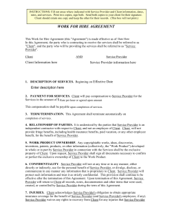
Advances in Pediatrics 59 (2012) 359–383
ADVANCES IN PEDIATRICS
Pediatric Overuse Injuries in Sports
Quynh B. Hoang, MDa,*, Mohammed Mortazavi, MDb
a
Sports Medicine Program, Department of Orthopedics, Children’s Hospital Colorado, University
of Colorado Denver Health Sciences Center, 13123 East 16th Avenue, B060, Aurora, CO 80045,
USA; bChildren’s Hospital Colorado, University of Colorado Denver Health Sciences Center,
13123 East 16th Avenue, B060, Aurora, CO 80045, USA
Keywords
Overuse injuries Apophysitis Sports Pediatric
Key Points
Overuse injuries in youth sports are increasingly common as more children and
adolescents participate in some form of athletics.
Overuse injuries are chronic injuries that occur when repetitive stress is placed on
bone, muscle or tendon without adequate time for healing and recovery.
Familiarity and basic knowledge of common sports-related overuse injuries is
important so that proper diagnosis can be made. This allows timely treatment to
minimize time loss from participation and ensures a safe return to sports.
Although management of overuse injuries are centered around relative rest and
activity modifications, identifying youths at risk of these injuries is key so that
education, prevention, and early diagnosis and treatment can occur.
INTRODUCTION
As more children participate in recreational or organized athletics, pediatricians
are evaluating more sports-related injuries in their practice. Each year an estimated 30 million children in the United States participate in some form of organized athletics [1] and this number likely will continue to grow. Moreover,
many children today are playing on multiple teams during a given season or
are playing different sports throughout the year. Some children have no
such thing as an ‘‘off season.’’ Consequently, an increase in the incidence of
acute injuries and in chronic injuries related to overuse has been seen.
When repetitive stress is placed on bone, muscle, or tendon without adequate
time for healing and recovery, microtrauma occur in these structures, resulting in
an overuse injury. Physiologically, growing cartilage in pediatric bone is vulnerable to stress. These cartilaginous growth plates and apophyses lend to unique
injury patterns that are specific to children. During times of rapid growth,
*Corresponding author. E-mail address: [email protected]
0065-3101/12/$ – see front matter
doi:10.1016/j.yapd.2012.04.005
Ó 2012 Elsevier Inc. All rights reserved.
360
HOANG & MORTAZAVI
apophysitis is a common overuse injury. An apophysis is a secondary ossification
center that gives contour to bone and is also the site of attachment of the muscle
tendon. In a growing child, the muscles attached to these apophyses are overly
tight, and when overworked, repetitive traction forces are placed on these vulnerable growth centers. The result is chronic irritation, inflammation, and microavulsions at the bone-cartilage junction. Common locations of apophysitis
include the heel (Sever disease), elbow (little league elbow), and knee (OsgoodSchlatter disease). Overuse injuries to the physis and epiphysis are also common
in children, particularly in the overhead athlete or the gymnast. Examples include
little league shoulder and gymnast’s wrist. Repetitive stress and microtrauma can
also lead to overuse tendon injury (tendinitis), which is another major subset of
overuse injuries in children. Some of the more common tendinitis in children
that are discussed in this article include patellar tendinitis and iliotibial band tendinitis (IT syndrome), although foot and ankle tendinitis and shoulder (rotator cuff)
tendinitis can also occur in young athletes. Finally, stress fractures are another
example of an overuse injury. Repetitive compressive or tensile forces weaken
the bone through a continuum of bone marrow edema without a visible fracture
line (stress reaction) and can lead to a complete fracture (stress fracture). Once
a rare phenomenon, stress fractures are being reported at a much higher incidence today with the advent of organized youth sports [2,3]. Although this article
reviews only spondylolysis (stress fracture of the pars interarticularis of the vertebral body), predominant sites of stress fractures in young athletes also include the
tibia, fibula, femur, metatarsals, and navicular bone in the foot [2,3]. Regardless,
they all have a common denominator with a multifactorial phenomenon: training
errors, biomechanics, diet, and, in girls, menstrual function.
As primary care providers, familiarity and basic knowledge of some
common sports-related overuse injuries become important as increasingly
more children participate in sports. That way, proper diagnosis can be made
and timely treatment or referral to a sports medicine specialist can commence
to minimize the time lost from participation.
UPPER EXTREMITY INJURIES
Little league shoulder (proximal humerus epiphysitis)
Little league shoulder refers to an overuse injury of the proximal humerus in overhead athletes with open growth plates. It is believed to result from repetitive
microtrauma to the proximal humeral epiphysis and physis from constant traction and rotational torque stresses during throwing and overhead activities
[4,5]. Although most commonly seen in male baseball players, little league
shoulder (or proximal humeral epiphysitis) can affect all overhead athletes,
including tennis players, swimmers, volleyball players and even gymnasts
[6,7]. It commonly occurs between 11 and 16 years of age [4,8], when the proximal humeral physis growth is at its peak, leaving it vulnerable to stress injury.
In baseball players, risk factors that contribute to the development of little
league shoulder include improper pitching mechanics and frequent pitching
without rest periods [4].
PEDIATRIC OVERUSE INJURIES IN SPORTS
361
The typical patient presents with progressive pain over the lateral aspect of
the proximal humerus that occurs with throwing and overhead activities [4,8].
Physical examination shows tenderness over the proximal and lateral aspect of
the humerus in approximately 70% of the patients [8]. Thus, a normal examination does not preclude the diagnosis, and a classic history of pain with
throwing or overhead activity is key.
Radiographs should be ordered to confirm the diagnosis and also help rule
out other causes of shoulder pain. Findings on radiographs include widening,
sclerosis, and irregularity of the humeral physis, which can best be appreciated
on anteroposterior view of the shoulder in external rotation (Fig. 1).
Obtaining comparison views of the unaffected shoulder is often helpful to
confirm the diagnosis if radiograph findings are subtle. If radiograph findings
are negative and clinical suspicion remains high, further imaging with either
MRI or bone scan may be considered.
Treatment of little league shoulder includes rest from throwing and overhead activities until pain is significantly decreased. On average this takes
approximately 3 months before symptoms resolve and the athlete is able to return to play. Evidence of radiographic healing typically lags behind the clinical
course, and therefore normal radiographs are not necessarily required to allow
an athlete to return to activities. During this period of relative rest, physical
therapy should also be initiated for strengthening exercises, and if the athlete
is a baseball player, evaluation of throwing mechanics and progression through
an interval throwing program when the patient is pain-free is also crucial. The
most important factor in the management of little league shoulder, however, is
education and prevention. One should ensure adherence to youth baseball
pitching guidelines set forth by the USA Baseball Medical & Safety Advisory
Committee and the American Academy of Pediatrics (AAP) as they pertain
to pitch count, pitch types, and adequate rest period.
Fig. 1. Radiograph of right shoulder proximal humerus epiphysitis. Notice the widened and
sclerotic proximal humerus physis compared with normal left shoulder.
362
HOANG & MORTAZAVI
Multidirectional shoulder instability
Shoulder instability in the young athlete can be caused by an acute traumatic
injury (shoulder dislocation) or can be atraumatic secondary to underlying
capsular laxity. Multidirectional instability (MDI) is a well-known cause of
chronic shoulder pain and is generally a bilateral, atraumatic condition
affecting shoulder function. MDI refers to laxity within the glenohumeral joint
in all planes of motion, leading to hypermobility and decreased stability. It is
caused by generalized capsular laxity and insufficiency of the labrum, ligaments, and muscles that stabilize the shoulder joint [9–11].
The presenting symptoms of multidirectional instability often have an insidious onset and are usually associated with increases in duration, frequency, or
intensity of overhead activities, such as swimming, baseball, racquet sports, and
volleyball [9,12]. Athletes complain of activity-related generalized shoulder
pain, usually worse in the dominant arm. Symptoms typically wax and wane
with changes in activity level and overhead motions. Patients usually have
no history of frank shoulder dislocation, but recurrent shoulder subluxations
may be noted even with activities of daily living [9,11–13]. MDI may also
present with pain from related conditions, such as shoulder impingement or
labral tears.
Examination findings for MDI should be expected in both shoulders. With
adequate relaxation, shoulder laxity in multiple planes can be evaluated. The
humerus can be passively translated anteriorly and posteriorly, and the classic
feature of MDI is demonstration of the sulcus sign. For the sulcus sign, the
arm is pulled inferiorly and the presence of a dimple below the acromion
signifies the presence of inferior instability. According to early studies by
Neer and Foster [14], the combination of posterior and inferior laxity is classic
for MDI [9,15,16]. For symptomatic anterior instability, the apprehension test
can be applied. With the patient’s arm in the abducted and externally rotated
position, passive stressing of the shoulder into further external rotation will
cause a sensation of the shoulder wanting to dislocate (Fig. 2). This reaction
results in patient guarding and apprehension and is considered a positive test.
The apprehension test can then be followed by the relocation test, which also
tests for symptomatic anterior instability. For this test, the arm is again abducted and externally rotated, but this time with the patient lying supine.
While in this position, the arm and humeral head is then pulled forward
toward the examiner. This movement will cause pain and apprehension,
which can then be relieved by applying posterior force to the shoulder to
stabilize it (Fig. 3). If this improves the pain and apprehension, it is considered
a positive test.
Workup with imaging is usually not indicated for MDI without associated
injury, because MDI is a clinical diagnosis [9,12,15]. Radiographs of the
shoulder are usually normal, although inferior and superior translation can
be noted and misinterpreted for frank dislocation without knowledge of the
patient history. If concern exists about rotator cuff or labral disease, MRI
with arthrogram of the shoulder can help identify these lesions.
PEDIATRIC OVERUSE INJURIES IN SPORTS
363
Fig. 2. Apprehension test.
Treatment of MDI without known associated labral or rotator cuff disease is
usually conservative, with relative rest from offending activities and physical
therapy. After an initial period of rest and antiinflammatories to improve
pain, physical therapy should begin with strengthening exercises for the
dynamic rotator cuff and scapular stabilizer muscles [9,15,17]. An endurance
Fig. 3. Relocation test.
364
HOANG & MORTAZAVI
program should also be added to strengthening exercises to prevent fatigueinduced injury on return to activities. When the athlete has regained painfree full strength and range of motion, gradual return to play through
increasing sport-specific activities can be initiated.
Medial epicondyle apophysitis (little league elbow)
Little league elbow is intended to refer specifically to medial epicondyle apophysitis. It is a traction injury to the medial epicondylar physis and can be
best understood by considering how throwing forces are applied to the medial
elbow. Important anatomic considerations of the medial elbow include the
medial epicondyle, medial epicondyle apophysis, and ulnar collateral ligament.
Injury to this area is caused by repetitive valgus stress to the medial elbow seen
in overhead sports [18,19]. Force mechanisms include both compression to the
lateral elbow involving the radiocapitellar joint and traction to the medial
elbow created by repetitive dynamic valgus stress involved in throwing
motions [19,20].
The incidence of medial epicondyle apophysitis is increasing as the numbers
and intensity of organized youth sport have increased [21–23]. This condition
most commonly presents between 9 and 12 years of age before closure of the
medial epicondyle apophysis [18,21,24]. Once the medial epicondylar physis
closes, valgus forces are transmitted to other medial elbow structures, and
injuries such as flexor-pronator tendonitis or ulnar collateral ligament sprains
occur instead. Medial epicondyle apophysitis is most common in baseball
pitchers, but is also seen in infielders, catchers, outfielders, and athletes participating in other overhead activities, such as football quarterbacks and tennis
players [21–23].
Typically, patients have no history of acute injury. Athletes will complain of
insidious onset of medial elbow pain with throwing, but in severe cases pain
may also occur with nonthrowing activities. Often athletes report a history
of weaker or less-accurate throws. Inquiring about what position they play,
the number of teams and seasons per year, throwing history (including pitch
count, number of practices and games), and any recent changes in throwing
techniques becomes important. Physical examination will reveal tenderness
localized over the medial epicondyle, and pain can be reproduced with valgus
stressing. Having the patient flex the wrist or pronate the forearm against resistance can also elicit pain. Range of motion may also be limited and strength
decreased because of pain.
Workup generally involves imaging that starts with plain radiographs of the
elbow. Radiographs are useful for ruling out other abnormalities such as fractures, osteochondral defects, loose bodies, growth plate irregularities, tumors,
and infections. In medial epicondyle apophysitis, radiographs are typically
normal, but widening of the apophysis and even an avulsion are not
uncommon. A comparison view of the opposite elbow may be helpful when
clinical suspicion is high, because radiographs show a large degree of variation
in normal growth centers. MRI is rarely necessary to confirm the diagnosis.
PEDIATRIC OVERUSE INJURIES IN SPORTS
365
Treatment of medial epicondyle apophysitis consists of complete throwing
restriction and symptom control with ice and antiinflammatories. Based on
severity and duration of symptoms, the timeline for recovery will be different
for every individual. An initial period of immobilization with a brace or cast
may be necessary for the most severe cases in patients experiencing persistent
pain at rest. Most will benefit greatly from initiating physical therapy early, and
this is highly recommended [18,20,21]. The initial treatment phase involves
initiation of range-of-motion exercises and joint mobilizations as necessary to
prevent joint contractures. General conditioning is encouraged and core
strengthening is usually started right away [18,21]. When tolerated, gradual
strengthening is then used. Physical therapy should also address the athlete’s
throwing mechanics to decrease load forces on the elbow. When athletes are
pain-free with full range of motion and near full strength, an interval throwing
program should be initiated, which will gradually progress them through stages
of increased throwing velocity and repetitions [18,21,22,24]. One critical
component of rehabilitation is that return to play is gradual, because potential
complications include worsening symptoms, abnormal bone development, or
an avulsion injury.
AS with little league shoulder, education and prevention are key in the
management of little league elbow. Proper throwing mechanics should be
taught and pitching guidelines should be followed. Current AAP recommendations along with those of Little League Baseball specify age-specific maximum
pitch counts, required rest days, and recommended pitch types; maximum
pitch counts are 50 to 105 pitches per game depending on age [25]. USA Baseball Medical & Safety Advisory Committee recommends no more than 75 to
125 pitches per week depending on age [26].
Gymnast’s wrist (distal radius epiphysitis)
Gymnast’s wrist is a well-known overuse phenomenon that affects 25% to 40%
of all gymnasts, and up to 80% in the most elite groups [27–29]. This injury
involves a stress reaction of the distal radius physis and epiphysis and is caused
by repetitive forces across the wrist joint, particularly with axial loading of the
wrist in hyperextension [30–33]. Other forces experienced at the wrist by
gymnasts, such as rotational and distraction forces, also have been noted to
play a significant role in causing injury to the distal radius epiphysis [30,34].
Gymnasts are often involved in activities requiring weight-bearing with their
wrist in hyperextension, such as the beam, tumbling, vault, and floor routines.
These activities create direct axial and rotational forces onto the distal radius
physis, which over time results in microtraumatic changes to the epiphysis
and the vulnerable cartilaginous growth plate.
Obtaining a sports-specific history is vital, because gymnasts commonly
present with recent increases in activity level with wrist-loading routines
[28,30,31,35]. A history should be obtained of the athlete’s level, hours per
week, and types of routines causing the most wrist pain. The most common
complaint is wrist pain that begins at the onset of the offending activity,
366
HOANG & MORTAZAVI
worsens during the activity, and improves with rest. The physical examination
shows tenderness along the radial physis and pain with wrist hyperextension
and axial loading [29,30,35], as in the table push-off test, which usually reproduces the wrist pain (Fig. 4).
Decreased range of motion, especially with extension, and swelling are also
seen in more advanced cases [30,31,35], and the athlete’s grip strength is
usually decreased on the symptomatic side.
Radiographs are obtained to evaluate for radiographic evidence of stress
injury to the distal radial physis. Comparison views of the unaffected wrist
are often very helpful. These radiographic findings include widening, sclerosis,
and calcification of the radial physis consistent with repetitive microtrauma
[27,30,36]. The origin of these changes has been postulated to occur from
blood supply compromise, leading to abnormal endochondral ossification of
the physis [30,34,37]. More-advanced disease may lead to radial physis growth
arrest and a positive ulnar variance (greater relative length of distal ulna with
respect to the distal radius) [30,34,38]. Follow-up radiographs are critical to
document healing or progression of physeal injury [30,34,39].
Gymnast’s wrist can be separated into three different stages [30,34,39]. Stage
one presents with clinical symptoms without any radiographic changes. Stage
two exhibits radiographic changes as noted earlier (Fig. 5). Stage 3 has radiographic findings of a positive ulnar variance. MRI of the wrist is usually unnecessary for this diagnosis and is only obtained when other possible abnormality is
suspected [30,39].
The mainstay of treatment of gymnast’s wrist involves rest from wrist-loading
activities. Ice, antiinflammatories, and immobilization in a wrist brace as needed
for pain control can be used. Physical therapy is usually helpful and a graded return to activity is essential. Physical therapy during the rest and rehabilitation
phase should include focused assessment of limb alignment, laxity, strengthening, and proprioception [28,30]. A wrist brace or taping to reduce wrist
Fig. 4. Table push-off test.
PEDIATRIC OVERUSE INJURIES IN SPORTS
367
Fig. 5. (A) Left wrist with subtle widening of radial physis and sclerosis through radial metaphysis. (B) Right wrist normal comparison view.
hyperextension on return to gymnastics is often helpful in reducing loads over the
radial physis [30]. Gymnasts with stage one injuries can gradually return to
activity once pain has resolved, which usually takes 3 to 6 weeks. Stage two
injuries may take much longer, sometimes even up to 3 months, to heal, and
require both clinical and radiographic confirmation of resolving injury before
the patient can return to gymnastics [28,30,37]. Stage three injuries usually will
require a period of immobilization with bracing and orthopedic consultation
and close follow-up with serial radiographs to assess for premature closure of
the distal radial physis [28,30,37]. Participation should be abruptly stopped and
the wrist reevaluated on any recurrence of pain.
LOWER EXTREMITY INJURIES
Iselin disease (fifth metatarsal apophysitis)
Iselin disease, first described in 1912 by Dr Iselin, is another chronic overuse
injury seen in young athletes that involves the apophysis of the fifth metatarsal
[40,41]. It results from repetitive traction on the apophysis at the base of the
fifth metatarsal, where the peroneal brevis tendon attaches. The apophysis
typically appears at 8 to 9 years of age and fuses between 12 and 15 years
of age, making ages 8 to 15 years most common for presentation of Iselin
disease [42–44].
Although it occurs with running and jumping activities, Iselin disease is more
commonly associated with cutting sports that create inversion stress on the
ankle, which requires repetitive activation of the peroneal muscles [44–46]. A
history of trauma is usually absent. However, acute inversion injuries have
been reported as both initiating and exacerbating events [43,45,46]. Presenting
368
HOANG & MORTAZAVI
symptoms include pain and tenderness at the base of the fifth metatarsal. It is
typically insidious in onset, aggravated with activity, and gradually worsens
over time without rest from activity.
On examination, a prominence of the fifth metatarsal base may be present,
sometimes with soft tissue swelling and mild erythema. Focal tenderness is
found at the base of the fifth metatarsal, and resisted eversion can illicit pain
in this area. Full dorsiflexion and plantar-flexion with inversion also may
produce discomfort in the same location. In some patients with longstanding
Iselin disease, gait may be altered, with patients attempting to weight-bear on
the medial side of the foot to avoid lateral foot pain. Like other apophyseal
overuse injuries, Iselin disease is a clinical diagnosis. Radiographs can be obtained to confirm the presence of an apophysis at the base of the fifth metatarsal
and to rule out other boney abnormalities. The oblique view of the foot usually
produces the best view of a small shell-shaped fleck of bone oblique to the fifth
metatarsal shaft (Fig. 6) [43–45]. The apophysis may appear widened, fragmented, and thickened.
Treatment of Iselin disease is centered around activity modification, icing,
antiinflammatories, and peroneal stretching and strengthening. For more symptomatic or recalcitrant cases, physical therapy should focus on ankle flexibility,
particularly of the evertors and plantar-flexors; ankle strengthening; and
proprioceptive exercises [41,43,45,47]. These exercises can improve dynamic
ankle stabilization, which can minimize repetitive traction forces on the fifth
metatarsal apophysis from ankle instability. With mild to moderate cases,
symptoms typically improve with conservative treatment and one can anticipate return to play in 3 to 6 weeks. More severe cases may require an
Fig. 6. Fifth metatarsal apophysis.
PEDIATRIC OVERUSE INJURIES IN SPORTS
369
immobilization period with a walking boot or cast followed by physical therapy
for 2 to 4 weeks before return to play [43–46]. In almost all cases, this condition
resolves by the time of closure of the apophysis, typically by 15 years of age.
Rarely, a nonunion of the apophysis causing symptoms into late adolescence
can occur with Iselin disease, which may warrant orthopedic consultation to
discuss surgical excision or fixation if conservative treatment fails [41,45,46].
Sever disease (calcaneal apophysitis)
Calcaneal apophysitis, or Sever disease, is the most common cause of heel pain
in young athletes and accounts for 8% of all pediatric overuse injuries [42,43].
Sever disease is a chronic traction injury that occurs at the calcaneal apophysis
with repetitive tension forces applied by the calf muscles through the Achilles
tendon. Radiographically, the calcaneal apophysis appears at 8 to 9 years of age
and typically fuses between 12 and 15 years of age (Fig. 7) [48,49].
Calcaneal apophysitis is most commonly seen in athletes participating in
sports requiring running, jumping, and plantar-flexion activation. Common
sports include soccer, basketball, track, gymnastics, and dance [42,45,48,49].
Athletes present with pain in one or both heels, although bilateral involvement is typical. The most common age group affected is 10 to 12 years of
age, with the condition being two to three times more common and occurring
1 to 2 years later in boys because of later skeletal maturity [42,45,49]. Pain is
activity-related and reported to be along the heel or at the posterior aspect of
the heel where the Achilles tendon inserts onto the apophysis. Patients will
often limp or walk on toes to prevent painful walking. Examination reveals
tenderness along the calcaneal apophysis, either medial or lateral, and can be
elicited through medial and lateral compression of the calcaneus along the
apophysis (Fig. 8).
Fig. 7. Calcaneal apophysis.
370
HOANG & MORTAZAVI
Fig. 8. Calcaneal squeeze test.
Tenderness can also be found at the Achilles insertion site on the calcaneus.
Athletes will also often have tight calf muscles and Achilles tendon, with weakness on dorsiflexion because of pain. Radiographs typically appear normal or
may reveal a fragmented hyperdense sclerotic apophysis [43,49]. Radiographs
of the calcaneus are not necessary to confirm the diagnosis if the history and
clinical findings are consistent, but can be obtained to rule out other bony
disease. Studies have found that up to 5% of radiographs in patients with
heel pain reveal pathologic conditions, making radiographic evaluation worthwhile even in light of a clear clinical picture for Sever disease [50].
Treatment consists of relative rest and activity modification, coupled with ice
and antiinflammatories as needed for pain. Rehabilitative exercises focused on
calf and Achilles stretching and eccentric strengthening of the Achilles can help
decrease traction forces applied to the apophysis. Although high-level evidence
supporting the use of orthotics is still lacking, heel cups, lifts, and pads have
been reported to reduce traction forces and axial loads on the calcaneal apophysis, helping to decrease symptoms [42,49,51]. When pain and flexibility
improve, the athlete may gradually return to sports as symptoms permit,
because it is a self-limited process that will resolve when the calcaneal apophysis closes [43,48,49]. More severe symptoms of persistent pain and limping
may benefit from a short period of immobilization with a walking boot or
casting before gradual return to sports [43,45,49]. With early diagnosis and
appropriate treatment, immobilization is rarely required and only reported in
1% to 3% of patients [48]. In fewer than 1% of cases, neglected Sever disease
can progress to calcaneal avulsion injury [52].
Osgood-Schlatter disease (tibial tubercle apophysitis)
Osgood-Schlatter disease is a tibial tubercle apophysitis that commonly causes
anterior knee pain in children and adolescents. It results from repetitive traction
of the patella tendon on the tibial tubercle apophysis, leading to microtrauma,
PEDIATRIC OVERUSE INJURIES IN SPORTS
371
inflammation, and pain. It frequently presents between the ages of 10 and 15
years in children who participate in jumping and running sports, such as
basketball, volleyball, gymnastics, and soccer [53,54]. Approximately 30% of
patients have bilateral knee involvement [55,56].
The typical clinical presentation is aching pain and swelling over the tibial
tubercle that is exacerbated by jumping and running activities, and with direct
pressure such as kneeling. On examination, patients experience point tenderness, and associated swelling or prominence over the tibial tubercle may be
present. Resisted knee extension, which activates eccentric quadriceps muscle
movement, may also reproduce the pain. Evaluation with radiographs is often
not necessary because it is a clinical diagnosis, but these should be obtained to
rule out other abnormalities, such as fractures or tumors, if the clinical presentation is atypical. Radiograph findings of Osgood-Schlatter disease include
overlying soft tissue swelling and fragmentation or irregularity of the tibial
tubercle ossification center.
Management of Osgood-Schlatter disease is conservative, aimed at symptom
control. Treatment involves relative rest and activity modification, along with
icing and analgesics with nonsteroidal antiinflammatory drugs (NSAIDs). Use
of an infrapatellar strap, which helps to decrease traction forces on the tibial
tubercle, may be used for symptomatic relief during activity. Physical therapy
focusing on quadriceps and hamstring flexibility can also help reduce symptoms.
Osgood-Schlatter disease is typically a self-limited process, with symptoms
resolving spontaneously when the child or adolescent reaches skeletal maturity.
However, some individuals develop persistent symptoms that remain into
adulthood. One retrospective study reported that 60% of patients with childhood Osgood-Schlatter disease described pain with kneeling as adults, although
76% of them did not have any pain or limitations with activity [57]. For the few
who experience complications, such as persistent painful ossicles in the distal
patellar tendon and painful kneeling, and those for whom conservative
management fails, surgical treatment to shave the tibial tubercle can be performed in skeletally mature patients [58,59].
Sinding-Larsen-Johansson disease (apophysitis of the inferior pole of the
patella)
Sinding-Larsen-Johansson disease is another common overuse injury that causes
anterior knee pain. It is similar to Osgood-Schlatter disease, except that it is a traction apophysitis that occurs at the inferior pole of the patella where the proximal
patella tendon attaches. Presentation typically occurs between 9 and 12 years,
which is slightly younger than in Osgood-Schlatter disease. Patients present
with activity-related anterior knee pain localized to the inferior pole of the patella.
Like Osgood-Schlatter disease, symptoms are aggravated by jumping and
running. On examination, the pain can be reproduced with direct palpation of
the inferior pole of the patella. Diagnosis is made clinically, although radiographs
can be helpful to rule out other abnormalities. Radiograph findings may show
fragmentation of the distal pole of the patella.
372
HOANG & MORTAZAVI
Like Osgood-Schlatter disease, it is a self-limited process that responds well
to conservative treatment involving relative rest and activity modification. Pain
control with NSAIDS and stretching exercises to improve quadriceps and
hamstring flexibility can also improve symptoms.
Patellar tendinopathy (‘‘jumper’s knee’’)
Repetitive loading of the quadriceps muscle during jumping and running
activities can lead to patellar tendinopathy, an overuse injury resulting in
partial-thickness tears in the deep layers of the patella tendon. Although the
condition is seen commonly in jumping sports, such as basketball, volleyball,
and track and field (high and long jump), athletes in other sports such as soccer
and football are also affected [60,61]. Once thought to be an inflammatory
tendinitis, histopathologic and biochemical studies have now shown that it is
a degenerative tendinosis. The pathogenesis is tendon overload from strain
that is applied to the patella tendon, resulting in tissue microtrauma [62].
Consequently, management of patellar tendinopathy should be aimed at rehabilitation rather than antiinflammatory strategies.
Patients present with insidious onset of anterior knee pain that is aggravated
with activity or even with prolonged sitting. Generally, these patients have
a history of recent increase in intensity, frequency, or duration of sport activity
participation. The pain is well localized and examination reveals tenderness
over the patellar tendon. Although primarily a clinical diagnosis, radiographs
may sometimes reveal calcification in the tendon, and they can also be helpful
to rule out other associated conditions such as Osgood-Schlatter or SindingLarsen-Johansson disease. Ultrasound has also become a popular imaging
modality for diagnosis because the patella tendon lies superficial and can be
visualized easily.
Because patellar tendinopathy is an overuse injury from mechanical overload, treatment includes relative rest and refraining from the aggravating activities while still symptomatic. The role of NSAIDs remains controversial,
because patellar tendinopathy is a noninflammatory condition. The literature
contains no conclusive evidence showing that NSAIDs have an effect on the
treatment outcome of patellar tendinopathy [63], although patients with acute
pain may benefit from the analgesic effects. Conservative management through
physical therapy remains the mainstay of treatment. Although scientific
evidence on the efficiency of nonoperative treatment is still lacking, management of patellar tendinopathy continues to focus largely on eccentric strengthening of the tendon [64]. Improving quadriceps and hamstring flexibility can
also be beneficial in prevention and treatment of patellar tendinopathy [65].
In some patients for whom conservative management fails after 6 months of
therapy, surgical options can be explored.
Patellofemoral pain syndrome
Patellofemoral pain syndrome refers to anterior knee pain originating from the
patellofemoral joint and the surrounding supporting structures. It is the most
common cause of all knee overuse injuries. Other terms that have been used
PEDIATRIC OVERUSE INJURIES IN SPORTS
373
to refer to this condition include anterior knee pain, runner’s knee, and chondromalacia
patella. The cause of patellofemoral pain is multifactorial and includes biomechanical problems, dysfunction of muscles, and overuse. Anatomically, the
patella articulates with the trochlear groove of the femoral condyles in the distal
femur. Proper tracking of the patella in this groove is dependent on multiple
forces such as strength of the quadriceps muscles, supporting soft tissue structures, patellar stabilizing mechanisms, and biomechanics of the patient. Biomechanical problems include lower extremity malalignment, patellar
hypermobility, strength imbalances, and muscle inflexibility. Examples of malalignment of the lower extremity include femoral anteversion, genu valgum,
excessive lateral insertion of the patellar tendon (increased Q angle), tibial
torsion, and foot hyperpronation from pes planus (flat feet). Increases in activities and these predisposing factors causes overload stress on the patellofemoral
joint. The result is pain.
Patients complain of a dull aching pain underneath or around the patella that
occurs with activity. Prolonged sitting or going up and down stairs typically also
aggravates the pain. Mild swelling or a sense of crepitation may also be reported.
Symptoms are commonly bilateral, although presentation in one knee is not
unusual. Physical examination requires assessment from hips to feet, because malalignment problems can contribute to patellofemoral pain. Abnormal tracking of
the patella can be evaluated by extending the patient’s knee while in the seated
position. If abrupt or excessive lateral tracking of the patella occurs as the knee
extends, this is considered positive J tracking. On palpation, tenderness may be
present under the patellar facets. Compressing down on the patient’s patella while
displacing it inferiorly can also elicit pain as the patient is asked to contract the
quadriceps; this is a positive patellar grind test (Fig. 9).
Functional deficiencies such as core and pelvic weakness, quadriceps and
hamstring tightness, and IT band tightness should also be assessed. Radiographs are not necessary for diagnosing patellofemoral pain, but they can
Fig. 9. Patellar grind test.
HOANG & MORTAZAVI
374
help confirm abnormalities in patellar alignment (ie, patella alta, femoral
trochlea hypoplasia, lateral patella tilt) or rule out other bony abnormalities.
Like many overuse injuries, relative rest and modifying activities based on
subjective pain is part of the treatment. Because the origin of patellofemoral
pain is multifactorial, management should also address the various predisposing
factors, with correction of biomechanical problems being the primary focus.
Physical therapy for core and hip strengthening has thus become a mainstay of
treatment for this condition. Part of the rehabilitation also addresses any strength
imbalances, hamstring and quadriceps inflexibility, and IT band tightness. Use of
over-the-counter or customized orthotics may be necessary to correct hyperpronation from pes planus. Patellar stabilizing braces can also be helpful to minimize
patellar hypermobility and lateral tracking. For patients who do not respond to
conservative management, consultation with an orthopedic surgeon for consideration of a lateral release or patellar realignment surgery is reasonable, although no
evidence shows that surgical intervention is effective.
HIP INJURIES
Pelvic apophysitis
Pediatric overuse injuries commonly involve the pelvis during times of rapid
growth when bone growth exceeds muscle ability to stretch sufficiently, leading
to increased inflexibility and traction forces on the pelvic apophyses. Knowledge of the bony anatomy, muscular attachments, and its actions are key for
understanding and diagnosing pelvic apophysitis.
The pelvis has multiple apophyses that may be involved in overuse injuries
depending on the type of activity and age, because these secondary ossification
centers appear and close at different ages (Table 1).
Table 1
Pelvic apophyses- muscle attachments and ages at time of their ossification
Apophysis
Originating
muscle
Greater trochanter
None
Lesser trochanter
Anterior superior
iliac spine
Anterior inferior
iliac spine
Iliac crest
Ischial tuberosity
Biceps femoris, hip
extensors
Inserting muscle
Age of
appearance (y)
Age of
closure (y)
9–10
14–16
None
Sartorius
Gluteus maximus
and medius,
external hip
rotators
Iliopsoas
None
9–10
12–14
14–16
14–16
Rectus femoris
None
12–14
14–16
Tensor fascia latae
Internal and
external
obliques,
transversus
abdominis
None
13–15
16–18
15–17
21–25
PEDIATRIC OVERUSE INJURIES IN SPORTS
375
The muscles originating or attaching at each apophysis create traction forces
with activity as the muscle-tendon group repetitively pulls against the
apophysis, producing microavulsion, inflammation, and cellular breakdown
[66–68]. Eventually the cartilaginous apophysis weakens and leads to widening
of the apophysis, putting the athlete at high risk for an acute avulsion injury
[49,69–71]. The most commonly injured apophyses of the pelvis include the
ischial tuberosity, anterior inferior iliac spine (AIIS), and anterior superior iliac
spine (ASIS), followed less commonly by the lesser trochanter, iliac crest, and
rarely the greater trochanter [67,69]. The actions of the muscles involved at the
injured apophysis are often repetitive motions common to specific types of
sports. For example, sprinters will often experience ischial tuberosity apophysitis from recurrent ballistic hamstring use during explosive sprints, and figure
skaters will experience iliac apophysitis from explosive jumping and twisting
that engages their oblique muscles. Pelvic apophysitis occurs commonly in
a variety of sports, including soccer, running, gymnastics, skating, football,
and dance, but may occur in any sport [49,66,67].
Athletes present with well-localized activity-related pain at the site of the
involved apophysis. In contrast to acute avulsion injuries, the athlete’s pain
is insidious in nature and is usually described as aching or throbbing. Without
appropriate rest, the pain will progress and athletes may develop limp with
activities and pain with walking or standing. With continued activity despite
worsening pain, athletes are at risk for avulsion fracture at the apophysis,
and may describe a sudden painful event in which a pop is felt followed by
intense pain and disability of the involved muscle [42,49,68,70,71].
Physical examination will reveal significant localized pain directly over the
involved apophysis on palpation. Some local swelling may be present, but
erythema, bruising, and deformity are usually not appreciated unless an avulsion fracture has occurred. The pain is worsened by resisted activation or
passive stretching of the involved muscle group. Muscle tightness is often
appreciated bilaterally, and range of motion is usually decreased on the affected
side. With more advanced apophysitis, mild contractures may be noted.
Workup involves radiographs of the pelvis and hip to visualize the apophysis
of concern. However, radiographs typically appear normal, making this a clinical
diagnosis. In moderate to severe cases, radiographs may show an apophysis with
widening and sclerosis. Radiographs should be obtained in the presence of
concerns for constitutional symptoms, tumors, infection, or persistent symptoms
not responding to therapy. Avulsion fractures should be ruled out with radiographs if the history is concerning for such. On MRI, edema of the apophysis
can be appreciated in its early stages before widening and sclerosis [69–71].
However, MRI is unnecessary for the diagnosis and should only be obtained
when other concerns exist, such as infections, tumors, or stress fractures.
The mainstay of treatment for pelvic apophysitis is rest and activity modification to prevent further injury from the insulting activity. Ice and ibuprofen can be
used for pain as needed. Weight-bearing is allowed as tolerated. However, protected weight-bearing on crutches may be necessary initially if the athlete has
376
HOANG & MORTAZAVI
pain with walking and standing. If needed, physical therapy can usually be initiated by 1 to 2 weeks, with a focus on flexibility and range of motion, and progressing to core and pelvic strengthening and lower extremity strengthening. When
symptom-free, it is important to engage the athlete in a graded return to sportsspecific activities before full participation. In general, most can expect to return
to full participation at 4 to 6 weeks [49,68].
IT band syndrome (IT band tendinitis)
IT band syndrome is a very common cause of lateral hip pain and lateral knee
pain, and is found especially in runners. Anatomically, the IT band is a large
connective tissue structure that runs from the iliac crest down to the lateral
tibia. With hip flexion and extension, the IT band glides anteriorly over the
greater trochanter in the femur and then glides back posterior to the trochanter,
respectively. The friction that is created by the IT band sliding back and forth
results in pain. A trochanteric bursa allows for smooth gliding of the IT band
through reducing friction, but when this bursa becomes inflamed, an associated
trochanteric bursitis can also develop, resulting in painful movement. Running
downhill causes the worst pain because symptoms typically occur when the hip
is flexed at 30 . This reduced foot strike angle on downhill running is a risk
factor for developing IT band syndrome, because track athletes who run on
level surfaces, and thus have greater foot strike angle, have been found to
have a lower incidence of this condition.
Clinically, patients present with achy pain over the lateral upper thigh and
knee with running. On examination, they may have tenderness over the
greater trochanter if a bursitis has developed, or they may have tenderness
in the knee over the lateral femoral condyle. Symptomatic patients generally
have findings of a tight IT band on examination, as indicated by a positive
Ober test. For this test, the patient is in a side-lying position, with the affected
side up. The examiner then extends the symptomatic hip while supporting the
leg. With IT band inflexibility, the knee and leg will remain suspended off the
table once the examiner no longer supports the leg up (Fig. 10).
Physical examination also will often reveal biomechanical abnormalities.
Varus alignment of the lower extremity (bowlegged), overpronation from
high arches, or lateral tilt of the pelvis can all place strain on the IT band, predisposing individuals to this condition [72].
Treatment aims at reducing stress on the IT band, both through physical
therapy and correction of any biomechanical problems. In addition to stretching the IT band and hamstrings to decrease friction, physical therapy should
also strengthen gluteal and hip abductors to minimize pelvic tilt. Ice and
NSAIDs for symptomatic control can be helpful. More importantly, addressing
training errors, such as excessive downhill running or rapid increases in
activity, can help prevent recurrences. For those who fail conservative management, consultation with a surgeon for surgical release of the IT band is an
option. Most athletes are generally able to gradually return to sport activities
within 2 to 6 weeks once they are asymptomatic [73].
PEDIATRIC OVERUSE INJURIES IN SPORTS
377
Fig. 10. Ober test.
BACK INJURIES
Spondylolysis
Spondylolysis is a stress fracture of the pars interarticularis, the bony connection between the inferior and superior articulating facets of the vertebral
complex. Estimates show that spondylolysis may contribute to nearly 50% of
cases of back pain in young athletes, and physicians should always maintain
a high index of suspicion for this diagnosis [53]. The cause of the defect in
the pars interarticularis is stress to the posterior elements of the spine from
repetitive flexion and hyperextension motion, combined with truncal rotation.
Gymnastics, football (lineman), weight lifting, dance, and volleyball are
common sports requiring repetitive hyperextension that may lead to spondylolysis [3,53]. The stress fracture occurs most commonly at the L5 vertebral level,
followed by the L4 level [74–76], and can be unilateral or bilateral. If the stress
fracture at the pars interarticularis occurs bilaterally, then spondylolisthesis can
develop, which is a forward slippage of one vertebral body on the vertebral
body below it. Diagnosis and grading of spondylolisthesis can be made on
lateral radiographs based on percentage of anterior displacement.
Patients with spondylolysis typically present with insidious onset of activityrelated low back pain that worsens with spine hyperextension. The
prototypical patient falls into one of three categories: (1) the female athlete
with a hyperlordotic spine with increased motion and flexibility, (2) the male
athlete with poor flexibility, particularly of the paraspinal musculature, and
who has recently undergone a growth spurt, and (3) the deconditioned athlete
with poor core and pelvic strength who recently started a new sport. On physical examination tenderness to palpation may be present at the affected vertebral level. Findings may also show increased lordosis of the lumbar spine,
poor hamstring flexibility, and weak core and pelvic strength. The pain is
reproducible when the patient extends the back or with single-leg hyperextension during the Stork test (Fig. 11).
378
HOANG & MORTAZAVI
Fig. 11. Stork test.
Diagnostic workup should begin with plain radiographs, including anteroposterior and lateral views of the lumbar spine. Obtaining oblique views to
look for the ‘‘Scotty dog’’ sign in the pediatric patient remains debatable
because of the increase in radiation exposure and uncertainty regarding
whether it improves diagnostic accuracy. In general, plain radiographs have
been shown to have poor sensitivity in demonstrating pars defects [77] but
can helpful to assess for other gross bony abnormalities.
Further imaging with single-photon emission CT (SPECT) scan, CT scan,
and/or MRI is generally necessary to confirm the diagnosis. Which imaging
modality is most sensitive and specific for detecting a pars stress fracture
remains controversial. Each has its own advantages and disadvantages. The
SPECT scan has increased sensitivity of up to 68% over radiographs [78,79]
and can be helpful in distinguishing symptomatic pars stress fractures from
asymptomatic lesions based on whether increased uptake is seen on the scan.
However, it has poor specificity and cannot rule out other pathologies that
also demonstrate increased uptake, such as infections or tumors. CT, on the
other hand, provides excellent visualization of bony anatomy and can identify
the exact location of the stress fracture. It can also be used on follow-up to
assess bony healing. The downfall is the amount of radiation exposure.
More recently, use of MRI for diagnosing spondylolysis has become more
popular because of the lack of radiation exposure in pediatric patients and
the ability to detect bone marrow edema (stress reaction) and soft tissues.
With improvements in magnetic field and strength and fat saturation techniques, high-resolution MRI images have increased diagnostic accuracy.
However, the efficacy is still being studied [80–83].
PEDIATRIC OVERUSE INJURIES IN SPORTS
379
No gold standard for management of spondylolysis currently exists. Its
clinical outcome has little correlation with radiographic evidence of healing
status [84]. That is, patients with healed stress fractures or with bony
nonunions can be asymptomatic. Thus, treatment consists primarily of a period
of high-impact and extension-based activity restrictions to alleviate pain and
allow for bony healing and/or progression to fibrous nonunion. No universal
guideline exists for the duration of activity restriction, although most authors
would agree that a period of relative rest of approximately 3 months is reasonable. During this time physical therapy is started once pain is improved, with
rehabilitation emphasizing core and back stabilization and hamstring flexibility.
The use of bracing in the treatment of spondylolysis remains controversial;
some still advocate for bracing until the patient is asymptomatic, which may
take up to 9 to 12 months [53,85]. Outcome studies show similar results
with return to sports and bony healing regardless of whether bracing was
used. Given the lack of evidence supporting therapeutic bracing, many will
reserve bracing for severe cases in which pain is experienced at rest and with
daily activities. Most patients with symptomatic spondylolysis respond well
to nonoperative management. Once symptom-free, the athlete may gradually
return to sport activities without restrictions. For recalcitrant cases for which
conservative measures fail after 9 to 12 months, surgical options may be considered, with posterolateral fusion having a success rate of close to 90% [84].
SUMMARY
Overuse injuries in the pediatric and adolescent population are a growing
problem in the United States as more children participate in recreational and
organized sports. It is not uncommon for children and adolescents to play
on multiple teams simultaneously or to be involved in sports year-round.
Without adequate rest, the demands of exercise can exceed the body’s ability
to repair tissues, leading to repetitive microtrauma and overuse injury. Unlike
in adults, the consequences of overuse injury in the pediatric and adolescent
athlete are far more serious because the growing bones are vulnerable to stress.
The ability to identify individuals who are at risk of overuse injuries is key so
that education, prevention, and early diagnosis and treatment can occur.
Preventive measures of modifying training factors (ie, magnitude, intensity,
and frequency of sports participation) and correcting improper biomechanics
(alignment, laxity, inflexibility, and muscle imbalance) should always be part
of the management plan.
References
[1] Adirim TA, Cheng TL. Overview of injuries in the young athlete. Sports Med 2003;33:
75–81.
[2] Bennell KL, Brukner PD. Epidemiology and site specificity of stress fractures. Clin Sports Med
1997;16:179–96.
[3] Micheli LJ. Overuse injuries in children’s sports: the growth factor. Orthop Clin North Am
1983;14(2):337–60.
380
HOANG & MORTAZAVI
[4] Kocher MS, Waters PM, Micheli LJ. Upper extremity injuries in the paediatric athlete. Sports
Med 2000;30:117–35.
[5] Tullos HS, Fain RH. Little league shoulder: rotational stress fracture of proximal epiphysis.
J Sports Med 1974;2:152–3.
[6] Kibler WB, Safran MR. Musculoskeletal injuries in the young tennis player. Clin Sports Med
2000;19(4):781–92.
[7] Patel DR, Nelson TL. Sports injuries in adolescents. Med Clin North Am 2000;84:
983–1007.
[8] Carson WG Jr, Gasser SI. Little leaguer’s shoulder. A reports of 23 cases. Am J Sports Med
1998;26:575–80.
[9] Castagna A, Nordenson U, Garofalo R, et al. Minor shoulder instability. Arthroscopy
2007;23(2):211–5.
[10] Matsen FA III, Lippitt SB, Sidles JA, et al, editors. Practical evaluation and management of the
shoulder. Philadelphia: WB Saunders; 1994. p. 60–81.
[11] Ovesen J, Nielsen S. Anterior and posterior shoulder instability. A cadaver study. Acta
Orthop Scand 1986;57(4):324–7.
[12] Brems JJ, Bergfeld J. Multidirectional shoulder instability. Orthop Trans 1991;15:84.
[13] Owens BD, Duffey ML, Nelson BJ, et al. The incidence and characteristics of shoulder
instability at the United States Military Academy. Am J Sports Med 2007;35(7):
1168–73.
[14] Neer CS, Foster CR. Inferior capsular shift for involuntary inferior and multidirectional instability of the shoulder. A preliminary report. J Bone Joint Surg Am 1980;62(6):897–908.
[15] Wnorowski DC, Keenan MA. Multidirectional Glenohumeral Instability. Available at:
http://emedicine.medscape.com/article/1262368-overview. Accessed November 21,
2011.
[16] Turkel SJ, Panio MW, Marshall JL, et al. Stabilizing mechanisms preventing anterior dislocation of the glenohumeral joint. J Bone Joint Surg Am 1981;63(8):1208–17.
[17] Mahaffey BL, Smith PA. Shoulder Instability in Young Athletes. Am Fam Physician
1999;59(10):2773–82.
[18] Cassas KJ, Cassettari AW. Childhood and adolescent sports-related overuse injuries. Am
Fam Physician 2006;73(6):1014–22.
[19] Whiteside JA, Andrews JR, Fleisig GS. Elbow injuries in young baseball players. Phys
Sportmed 1999;27(6):87–102.
[20] Andrish JT. Osteochondritis dissecans in a young pitcher: why early recognition matters.
Phys Sportmed 1997;25:85–90.
[21] Benjamin HJ, Briner WW Jr. Little league elbow. Clin J Sport Med 2005;15(1):37–40.
[22] Cain EL Jr, Dugas JR, Wolf RS, et al. Elbow injuries in throwing athletes: a current concepts
review. Am J Sports Med 2003;31:621–35.
[23] Christopher NC, Congeni J. Overuse injuries in the pediatric athlete: evaluation, initial
management, and strategies for prevention. Clin Pediatr Emerg Med 2002;3:118–28.
[24] Lyman S, Fleisig GS, Andrews JR, et al. Effect of pitch type, pitch count, and pitching
mechanics on risk of elbow and shoulder pain in youth baseball pitchers. Am J Sports
Med 2002;30:463–8.
[25] Protecting Young Pitching Arms. Little League Web site. Available at: http://www.littleleague.org/
Assets/old_assets/media/pitch_count_publication_2008.pdf. Accessed December 2, 2011.
[26] USA Baseball Medical & Safety Advisory Committee Guidelines. American Sports Medicine Institute Web site. Available at: http://www.asmi.org/asmiweb/usabaseball.htm. Accessed December 2, 2011.
[27] Gaine D, Roy S, Singer KM, et al. Stress changes of the distal radial growth plate, A radiographic survey and review of the literature. Am J Sports Med 1992;20:290–8.
[28] Mandelbaum BR, Bartolozzi AR, Davis CA, et al. Wrist pain syndrome in the gymnast:
pathogenetic, diagnostic, and therapeutic considerations. Am J Sports Med 1986;17:
305–17.
PEDIATRIC OVERUSE INJURIES IN SPORTS
381
[29] Dobyns JH, Gahcl GT. Gymnast’s wrist. Hand 1990;6:493–505.
[30] DiFiori JP, Puffer JC, Mandelbaum BR, et al. Factors associated with wrist pain in the young
gymnast. Am J Sports Med 1996;24:9–14.
[31] Webb BG, Retigg LA. Gymnastic wrist injuries. GUTT. Sports Med Rep 2008;7(5):289–95.
[32] Koh TJ, Grabiner MD, Weiker GG. Technique and ground reaction forces in the back hand
spring. Am J Sports Med 1997;20:61–6.
[33] Markoff JL, Shapiro MS, Mandelbaum BR, et al. Wrist loading patterns during pommel
horse exercises. J Biomech 1990;23:1001–11.
[34] DiFiori JP, Caine DJ, Malina RM. Wrist pain, distal radial physeal injury, and ulnar variance
in the young gymnast. Am J Sports Med 2006;34:840–9.
[35] Mitchell JA, Adams BD. Hand and wrist injuries: wrist pain in gymnasts. In: Renstrom PAFH,
editor. Clinical Practice of Sports injury Prevention Care. The Encyclopedia of Sports Medicine, vol. 5. Boston: Blackwell Scientific Publications; 1993. p. 78–85.
[36] Roy S, Caine D, Singer K. Stress changes of the distal radial epiphysis in young gymnasts:
a report of twenty-one cases and a review of the literature. Am J Sports Med 1985;13:301–8.
[37] Gabel GT. Gymnastic wrist injuries. J Sports Med 1998;17:611–21.
[38] DiFiori JP, Puffer JC, Dorey AF. Ulnar variance in young gymnasts: a three-year study. Med
Sci Sports Exerc 2001;33:S223.
[39] Rettig AC. Athletic injuries of the wrist and hand: part II: overuse injuries of the wrist and traumatic injuries of the hand. Am J Sports Med 2004;32:262–73.
[40] Iselin H. Wachstumbeschwerden zer zeit der knockern entwicklung metatarsi quinti. Deut Z
Chir 1912;117:529 [in German].
[41] Lehman RC, Gregg JR, Torg E. Iselin’s disease. Am J Sports Med 1986;14:494–6.
[42] Stricker PR, Wasilewski C. Apophysitis. In: Puffer JC, editor. 20 Common problems in sports
medicine. New York: McGraw-Hill; 2002. p. 353–66.
[43] Gillespie H. Osteochondroses and apophyseal injuries of the foot in the young athlete. Curr
Sports Med Rep 2010;9(5):265–8.
[44] Canale ST, Richardson DR. Osteochondroses and related problems of the foot and ankle. In:
DeLee JC, Drez D Jr, Miller MD, editors. DeLee and Drez’s Orthopaedic Sports Medicine.
Principles and Practice. 3rd edition. Philadelphia: Saunders Elsevier; 2010.
[45] Frush TJ, Lindenfeld TN. Peri-epiphyseal and overuse injuries in adolescent athletes. Sports
Health 2009;1(3):201–11.
[46] Canale TS, Williams KD. Iselin’s disease. J Pediatr Orthop 1992;12(1):90–3.
[47] Schwartz B, Jay RM, Schoenhaus HD. Apophysitis of the fifth metatarsal base. J Am Podiatr
Med Assoc 1991;81:128–30.
[48] Weiner DS. Calcaneal apophysitis: simple diagnosis, simpler treatment. J Fam Pract
2007;56(5):352–5.
[49] Wilson JC, Rodenburg RE. Apophysitis of the lower extremities. Contemp Pediatr
2011;28(6):38–46.
[50] Rachel JN, Williams JB, Sawyer JR, et al. Is radiographic evaluation necessary in children
with a clinical diagnosis of calcaneal apophysitis (sever disease)? J Pediatr Orthop
2011;31(5):548–50.
[51] Orava S, Virtanen K. Osteochondroses in athletes. Br J Sports Med 1982;16:161–8.
[52] Lee KT, Young KW, Park YU, et al. Neglected Sever’s disease as a cause of calcaneal apophyseal avulsion fracture: case report. Foot Ankle Int 2010;31(8):725–8.
[53] Cassas KJ, Cassettari-Wayhs A. Childhood and adolescent sports-related overuse injuries.
Am Fam Physician 2006;73(6):1014–22.
[54] Hogan KA, Gross RH. Overuse injuries in pediatric athletes. Orthop Clin North Am
2003;34:405–15.
[55] Wall EJ. Osgood-Schlatter disease: practical treatment for a self-limited condition. Phys
Sportmed 1998;26:29–34.
[56] Bloom OJ, Mackler L, Barbee J. Clinical inquiries. What is the best treatment for OsgoodSchlatter disease? J Fam Pract 2004;53:153–6.
382
HOANG & MORTAZAVI
[57] Krause BL, Williams JP, Catterall A. Natural history of Osgood-Schlatter disease. J Pediatr
Orthop 1990;10(1):65–8.
[58] Weiss JM, Jordan SS, Andersen JS, et al. Surgical treatment of unresolved Osgood-Schlatter
disease: ossicle resection with tibial tubercleplasty. J Pediatr Orthop 2007;27(7):844–7.
[59] Flowers MJ, Bhadreshwar DR. Tibial tuberosity excision for symptomatic Osgood-Schlatter
disease. J Pediatr Orthop 1995;15(3):292–7.
[60] Blazina ME, Kerlan RK, Jobe FW, et al. Jumper’s knee. Orthop Clin North Am 1973;4:
665–78.
[61] Ferreti A, Ippolito E, Mariani P, et al. Jumper’s knee. Am J Sports Med 1983;11:58–62.
[62] Leadbetter WB. Cell-matrix response in tendon injury. Clin Sports Med 1992;11:533–78.
[63] Almekinders LC, Temple JD. Etiology, diagnosis, and treatment of tendinosis: an analysis of
the literature. Med Sci Sports Exerc 1998;30:1183–90.
[64] Peers K, Lysens R. Patellar tendinopathy in athletes. Current diagnostic and therapeutic
recommendations. Sports Med 2005;35(1):71–87.
[65] Witvrouw E, Bellemans J, Lysens R, et al. Intrinsic factors for the development of patellar
tendinitis in an athletic population: a two year prospective study. Am J Sports Med
2001;29:190–5.
[66] Peck DM. Apophyseal injuries in the young athlete. Am Fam Physician 1995;51(8):
1891–8.
[67] Kocher MS, Tucker R. Pediatric athlete hip disorders. Clin Sports Med 2006;25(2):241–53.
[68] Sopano JV. Common overuse injuries in the pediatric and adolescent athlete. Clin Pediatr
Emerg Med 2007;8(1):7–14.
[69] Ferbach SK, Wilkinson RH. Avulsion injuries of the pelvis and proximal femur. AJR Am J
Roentgenol 1981;137:581–4.
[70] Metzmaker JN, Pappas AM. Avulsion fractures of the pelvis. Am J Sports Med 1985;13(5):
349–58.
[71] Pointinger H, Munk P, Poeschl GP. Avulsion fracture of the anterior superior iliac spine
following apophysitis. Br J Sports Med 2003;37(4):361–2.
[72] Glazer JL, Hosey RG. Soft-tissue injuries of the lower extremity. Prim Care 2004;31(4):
1005–24.
[73] Farber AJ, Wilckens JH, Jarvis CG. Pelvic pain in the athlete. In: Seidenberg PH, Beutler AI,
editors. The sports medicine resource manual. Philadelphia: Saunders, Elsevier; 2008.
p. 317.
[74] Cavalier R, Herman MJ, Cheung EV, et al. Spondylolysis and spondylolisthesis in children
and adolescents. I: diagnosis, natural history, and nonsurgical management. J Am Acad Orthop Surg 2006;14:417–24.
[75] Lawrence JP, Greene HS, Graeur JN. Back pain in athletes. J Am Acad Orthop Surg
2006;14:726–35.
[76] Jones GT, Macfarlane GJ. Epidemiology of low back pain in children and adolescents. Arch
Dis Child 2005;90:312–6.
[77] Ward CV, Latimer B, Alander DH, et al. Radiographic assessment of lumbar facet distance
spacing and spondylolysis. Spine 2007;32:E85–8.
[78] Collier BD, Johnson RP, Carrera GF, et al. Painful spondylolysis and spondylolisthesis studied
by radiology and single photon emission computed tomography. Radiology 1985;154:
207–11.
[79] Bellah RD, Summerville MD, Treves ST, et al. Low back pain in adolescent athletes: detection
of stress injury to the pars interarticularis with SPECT. Radiology 1991;180:509–12.
[80] Bhatia NN, Chow G, Timon SJ, et al. Diagnostic modalities for the evaluation of pediatric
back pain: a prospective study. J Pediatr Orthop 2008;28:230–3.
[81] Standaert CJ, Herring SA. Expert opinion and controversies in sports and musculoskeletal
medicine: the diagnosis and treatment of spondylolysis in adolescent athletes. Arch Phys
Med Rehabil 2007;88:537–40.
PEDIATRIC OVERUSE INJURIES IN SPORTS
383
[82] Hollenberg GM, Beattie PF, Meyers SP, et al. Stress reactions of the lumbar pars interarticularis: the development of new MRI classification criteria. Spine 2002;27:181–6.
[83] Udeshi U, Reeves D. Routine thin slice MRI effectively demonstrates the lumbar pars interarticularis. Clin Radiol 1999;54:615–9.
[84] Kim HJ, Green DW. Spondylolysis in the adolescent athlete. Curr Opin Pediatr 2011;23:
68–72.
[85] Solomon R, Brown T, Gerbino PG, et al. The young dancer. Clin Sports Med 2000;19:
717–39.


© Copyright 2026