
The Management of Encephalitis: Clinical Practice America
IDSA GUIDELINES
The Management of Encephalitis: Clinical Practice
Guidelines by the Infectious Diseases Society of
America
Allan R. Tunkel,1 Carol A. Glaser,2 Karen C. Bloch,3 James J. Sejvar,4 Christina M. Marra,5 Karen L. Roos,6
Barry J. Hartman,7 Sheldon L. Kaplan,8 W. Michael Scheld,9 and Richard J. Whitley10
1
Monmouth Medical Center, Long Branch, New Jersey; 2California Department of Health Services, Richmond; 3Vanderbilt University School of
Medicine, Nashville, Tennessee; 4Centers for Disease Control and Prevention, Atlanta, Georgia; 5University of Washington School of Medicine,
Seattle; 6Indiana University School of Medicine, Indianapolis; 7Weill Cornell Medical Center, New York, New York; 8Baylor College of Medicine,
Houston, Texas; 9University of Virginia School of Medicine, Charlottesville; and 10University of Alabama at Birmingham
EXECUTIVE SUMMARY
Encephalitis is defined by the presence of an inflammatory process of the brain in association with clinical
evidence of neurologic dysfunction. Of the pathogens
reported to cause encephalitis, the majority are viruses.
However, despite extensive testing, the etiology of encephalitis remains unknown in most patients. Another
major challenge for patients with encephalitis is to determine the relevance of an infectious agent identified
outside of the CNS; these agents may play a role in the
neurologic manifestations of illness but not necessarily
by directly invading the CNS. In addition, it is important to distinguish between infectious encephalitis and
postinfectious or postimmunization encephalitis or en-
Received 9 April 2008; accepted 23 April 2008; electronically published 26 June
2008.
It is important to realize that guidelines cannot always account for individual
variation among patients. They are not intended to supplant physician judgment
with respect to particular patients or special clinical situations. The Infectious
Diseases Society of America considers adherence to these guidelines to be
voluntary, with the ultimate determination regarding their application to be made
by the physician in the light of each patient’s individual circumstances.
Reprints or correspondence: Dr. Allan R. Tunkel, Dept. of Medicine, Monmouth
Medical Center, 300 Second Ave., Long Branch, NJ 07740 ([email protected]).
Clinical Infectious Diseases 2008; 47:303–27
2008 by the Infectious Diseases Society of America. All rights reserved.
1058-4838/2008/4703-0001$15.00
DOI: 10.1086/589747
cephalomyelitis (e.g., acute disseminated encephalomyelitis [ADEM]), which may be mediated by an immunologic response to an antecedent antigenic
stimulus from an infecting microorganism or immunization. Noninfectious CNS diseases (e.g., vasculitis,
collagen vascular disorders, and paraneoplastic syndromes) can have clinical presentations similar to those
of infectious causes of encephalitis and should also be
considered in the differential diagnosis.
In the approach to the patient with encephalitis, an
attempt should be made to establish an etiologic diagnosis. Although there are no definitive effective treatments in many cases of encephalitis, identification of
a specific agent may be important for prognosis, potential prophylaxis, counseling of patients and family
members, and public health interventions. Epidemiologic clues that may help in directing the investigation
for an etiologic diagnosis include season of the year,
geographic locale, prevalence of disease in the local
community, travel history, recreational activities, occupational exposure, insect contact, animal contact,
vaccination history, and immune status of the patient.
Various clinical clues may also be helpful to physicians
in considering specific etiologies.
The diagnostic evaluation of a patient who presents
with encephalitis needs to be individualized and should
be guided by epidemiologic and clinical clues and labIDSA Guidelines for Management of Encephalitis • CID 2008:47 (1 August) • 303
Downloaded from cid.oxfordjournals.org at IDSA on August 13, 2011
Guidelines for the diagnosis and treatment of patients with encephalitis were prepared by an Expert Panel of
the Infectious Diseases Society of America. The guidelines are intended for use by health care providers who
care for patients with encephalitis. The guideline includes data on the epidemiology, clinical features, diagnosis,
and treatment of many viral, bacterial, fungal, protozoal, and helminthic etiologies of encephalitis and provides
information on when specific etiologic agents should be considered in individual patients with encephalitis.
Etiology
1. Epidemiologic clues and assessment of risk factors to
identify potential etiologic agents should be sought in all patients with encephalitis (table 2) (A-III).
2. Clinical clues (general and specific neurologic findings)
may be helpful in suggesting certain causative agents in patients
with encephalitis (table 3) (B-III).
3. In patients with encephalitis and a history of recent
infectious illness or vaccination, the diagnosis of ADEM should
be considered (B-III).
304 • CID 2008:47 (1 August) • Tunkel et al.
Diagnosis
4. Specific diagnostic studies should be performed for the
majority of patients who present with encephalitis (table 4) (AIII).
5. Additional diagnostic studies should be performed for
patients with encephalitis on the basis of specific epidemiologic
and clinical clues (tables 2, 3, and 5) (A-III).
Diagnostic Studies outside the CNS
6. Cultures of body fluid specimens (e.g., from blood,
stool, nasopharynx, or sputum), if clinical and epidemiologic
clues are suggestive, should be performed in an attempt to
identify various viral, bacterial, and fungal etiologies of encephalitis (table 5) (B-III); positive results do not necessarily
indicate that the isolated microorganism is the etiology of encephalitis and must be interpreted in the context of the appropriate epidemiologic findings, clinical findings, and other
diagnostic study results.
7. Biopsy of specific tissues for culture, antigen detection,
nucleic acid amplification tests (such as PCR), and histopathologic examination should be performed in an attempt to establish an etiologic diagnosis of encephalitis (table 5) (A-III).
8. Certain causes of encephalitis may be diagnosed by
detection of IgM antibodies in serum (table 5) (A-III).
9. Although acute- and convalescent-phase serum samples
are generally not useful in establishing the etiology during the
acute presentation in a patient with encephalitis, they may be
useful for the retrospective diagnosis of an infectious agent
(table 5) (B-III).
10. Nucleic acid amplification tests (such as PCR) of body
fluids outside of the CNS may be helpful in establishing the
etiology in some patients with encephalitis (table 5) (B-III).
Neurodiagnostic Studies
11. MRI is the most sensitive neuroimaging test to evaluate patients with encephalitis (A-I).
12. CT, with and without contrast enhancement, should
be used to evaluate patients with encephalitis if MRI is unavailable, impractical, or cannot be performed (B-III).
13. Fluorine-18 fluorodeoxyglucose positron emission tomography (FDG-PET) scanning is not routinely recommended
for patients with encephalitis.
14. Electroencephalography (EEG) is rarely helpful in establishing an etiology in patients with encephalitis, but it has
a role in identifying patients with nonconvulsive seizure activity
who are confused, obtunded, or comatose and should be performed in all patients with encephalitis (A-III).
15. CSF analysis is essential (unless contraindicated) in
all patients with encephalitis (A-III).
Downloaded from cid.oxfordjournals.org at IDSA on August 13, 2011
oratory data; these generally include cultures and analysis (i.e.,
antigen detection and nucleic acid amplification tests, such as
PCR) of body fluid specimens, biopsy of specific tissues (with
culture, antigen detection, PCR, and histopathologic evaluation) outside the CNS, and serologic testing (for specific IgM
and acute- and convalescent-phase IgG antibody titers). MRI
of the brain should be performed in all patients, with CT used
only if MRI is unavailable, unreliable, or cannot be performed;
neuroimaging findings may also suggest disease caused by specific etiologic agents. CSF analysis is critical, unless contraindicated, and may be quite helpful in establishing an etiology.
Detection of specific viral IgM antibodies in CSF specimens
obtained from patients with encephalitis caused by numerous
viruses is considered to be diagnostic of neuroinvasive disease.
CSF cultures are generally of limited value in the determination
of the viral causes of encephalitis but are very important in the
diagnosis of bacterial and fungal infections. The utility of nucleic acid amplification testing (e.g., PCR) of CSF specimens
has greatly increased the ability to diagnose infections of the
CNS, especially viral infections caused by the herpesviruses. At
present, brain biopsy is rarely performed to establish the etiology of encephalitis, but it may play a role in some patients
with encephalitis of unknown etiology whose conditions deteriorate despite treatment with acyclovir.
Despite the wide range of viruses that have been reported
to cause encephalitis, specific antiviral therapy is generally limited to infections caused by the herpesviruses—specifically, herpes simplex virus—and HIV. Acyclovir treatment should be
initiated in all patients with suspected encephalitis, pending
results of diagnostic studies. During the appropriate season,
patients who present with clinical clues suggestive of rickettsial
or ehrlichial infection should be treated empirically with
doxycycline. Empirical therapy for acute bacterial meningitis
should also be initiated if clinically indicated. In patients with
acute disseminated encephalomyelitis, corticosteroids are recommended; plasma exchange should be considered in patients
who do not respond to this treatment.
Recommendation categories are shown in table 1.
Table 1. Infectious Diseases Society of America–US Public Health Service Grading System for
ranking recommendations in clinical guidelines.
Category, grade
Definition
Strength of recommendation
A
B
C
Quality of evidence
Good evidence to support a recommendation for use
Moderate evidence to support a recommendation for use
Poor evidence to support a recommendation
I
II
Evidence from ⭓1 properly randomized, controlled trial
Evidence from ⭓1 well-designed clinical trial, without randomization; from cohort or case-controlled analytical studies (preferably
from 11 center); from multiple time-series; or from dramatic results from uncontrolled experiments
III
Evidence from opinions of respected authorities, based on clinical
experience, descriptive studies, or reports of expert committees
NOTE. Adapted from Canadian Task Force on the Periodic Health Examination [2].
Treatment
Empirical Therapy
21. Acyclovir should be initiated in all patients with suspected encephalitis, pending results of diagnostic studies (AIII).
22. Other empirical antimicrobial agents should be initiated on the basis of specific epidemiologic or clinical factors
(tables 2 and 3), including appropriate therapy for presumed
bacterial meningitis, if clinically indicated (A-III).
23. In patients with clinical clues suggestive of rickettsial
or ehrlichial infection during the appropriate season, doxycycline should be added to empirical treatment regimens (A-III).
Specific Therapy
Viruses
24. Herpes simplex virus: acyclovir is recommended (AI).
25. Varicella-zoster virus: acyclovir is recommended (BIII); ganciclovir can be considered an alternative (C-III); adjunctive corticosteroids can be considered (C-III).
26. Cytomegalovirus: the combination of ganciclovir plus
foscarnet is recommended (B-III); cidofovir is not recommended, because its ability to penetrate the blood-brain barrier
has been poorly studied.
27. Epstein-Barr virus: acyclovir is not recommended; the
use of corticosteroids may be beneficial (C-III), but the potential risks must be weighed against the benefits.
28. Human herpesvirus 6: ganciclovir or foscarnet should
be used in immunocompromised patients (B-III); use of these
agents in immunocompetent patients can be considered (CIII), but there are not good data on their effectiveness.
29.
B virus: valacyclovir is recommended (B-III); alternative agents are ganciclovir (B-III) and acyclovir (C-III).
30. Influenza virus: oseltamivir can be considered (C-III).
31. Measles virus: ribavirin can be considered (C-III); intrathecal ribavirin can be considered in patients with subacute
sclerosing panencephalitis (C-III).
32. Nipah virus: ribavirin can be considered (C-III).
33. West Nile virus: ribavirin is not recommended.
34. Japanese encephalitis virus: IFN-a is not
recommended.
35. St. Louis encephalitis virus: IFN-2a can be considered
(C-III).
IDSA Guidelines for Management of Encephalitis • CID 2008:47 (1 August) • 305
Downloaded from cid.oxfordjournals.org at IDSA on August 13, 2011
Diagnostic Studies in the CNS
16. For certain viral agents, the presence of virus-specific
IgM in CSF specimens may be indicative of CNS disease caused
by that pathogen (table 5) (A-III).
17. Nucleic acid amplification tests (such as PCR) should
be performed on CSF specimens to identify certain etiologic
agents in patients with encephalitis (table 5) (A-III). Although
a positive test result is helpful in diagnosing infection caused
by a specific pathogen, a negative result cannot be used as
definitive evidence against the diagnosis.
18. Herpes simplex PCR should be performed on all CSF
specimens in patients with encephalitis (A-III). In patients with
encephalitis who have a negative herpes simplex PCR result,
consideration should be given to repeating the test 3–7 days
later in those with a compatible clinical syndrome or temporal
lobe localization on neuroimaging (B-III).
19. Viral cultures of CSF specimens are of limited value
in patients with encephalitis and are not routinely
recommended.
20. Brain biopsy should not be routinely used in patients
with encephalitis but should be considered in patients with
encephalitis of unknown etiology whose condition deteriorates
despite treatment with acyclovir (B-III).
Table 2. Possible etiologic agents of encephalitis based on epidemiology and risk factors.
Epidemiology or risk factor
Agammaglobulinemia
Possible infectious agent(s)
Enteroviruses, Mycoplasma pneumoniae
Age
Neonates
Herpes simplex virus type 2, cytomegalovirus, rubella virus, Listeria
monocytogenes, Treponema pallidum, Toxoplasma gondii
Infants and children
Eastern equine encephalitis virus, Japanese encephalitis virus, Murray
Valley encephalitis virus (rapid in infants), influenza virus, La Crosse
virus
Elderly persons
Eastern equine encephalitis virus, St. Louis encephalitis virus, West
Nile virus, sporadic CJD, L. monocytogenes
Animal contact
Bats
Rabies virus, Nipah virus
Birdsa
West Nile virus, Eastern equine encephalitis virus, Western equine
encephalitis virus, Venezuelan equine encephalitis virus, St. Louis
encephalitis virus, Murray Valley encephalitis virus, Japanese encephalitis virus, Cryptococcus neoformans (bird droppings)
Cats
Rabies virus, Coxiella burnetii, Bartonella henselae, T. gondii
Dogs
Rabies virus
a
Eastern equine encephalitis virus, Western equine encephalitis virus,
Venezuelan equine encephalitis virus, Hendra virusb
Old World primates
B virus
Raccoons
Rabies virus, Baylisascaris procyonis
a
Rodents
Eastern equine encephalitis virus (South America), Venezuelan equine
encephalitis virus, tickborne encephalitis virus, Powassan virus
(woodchucks), La Crosse virus (chipmunks and squirrels), Bartonella
quintanab
Sheep and goats
C. burnetii
Skunks
Rabies virus
Swinea
Japanese encephalitis virus, Nipah virusb
a
White-tailed deer
Immunocompromised persons
Borrelia burgdorferi
Varicella zoster virus, cytomegalovirus, human herpesvirus 6, West
Nile virus, HIV, JC virus, L. monocytogenes, Mycobacterium tuberculosis, C. neoformans, Coccidioides species, Histoplasma capsulatum, T. gondii
Ingestion items
Raw or partially cooked meat
T. gondii
Raw meat, fish, or reptiles
Gnanthostoma species
Unpasteurized milk
Tickborne encephalitis virus, L. monocytogenes, C. burnetii
Insect contact
Mosquitoes
Eastern equine encephalitis virus, Western equine encephalitis virus,
Venezuelan equine encephalitis virus, St. Louis encephalitis virus,
Murray Valley encephalitis virus, Japanese encephalitis virus, West
Nile virus, La Crosse virus, Plasmodium falciparum
Sandflies
Bartonella bacilliformis
Ticks
Tickborne encephalitis virus, Powassan virus, Rickettsia rickettsii, Ehrlichia chaffeensis, Anaplasma phagocytophilum, C. burnetii (rare), B.
burgdorferi
Tsetse flies
Trypanosoma brucei gambiense, Trypanosoma brucei rhodesiense
Occupation
Exposure to animals
Rabies virus, C. burnetii, Bartonella species
Exposure to horses
Hendra virus
Exposure to Old World primates
B virus
(continued)
306
Downloaded from cid.oxfordjournals.org at IDSA on August 13, 2011
Horses
Table 2. (Continued.)
Epidemiology or risk factor
Possible infectious agent(s)
Laboratory workers
West Nile virus, HIV, C. burnetii, Coccidioides species
Physicians and health care workers
Varicella zoster virus, HIV, influenza virus, measles virus, M.
tuberculosis
Veterinarians
Rabies virus, Bartonella species, C. burnetii
Person-to-person transmission
Herpes simplex virus (neonatal), varicella zoster virus, Venezuelan
equine encephalitis virus (rare), poliovirus, nonpolio enteroviruses,
measles virus, Nipah virus, mumps virus, rubella virus, Epstein-Barr
virus, human herpesvirus 6, B virus, West Nile virus (transfusion,
transplantation, breast feeding), HIV, rabies virus (transplantation),
influenza virus, M. pneumoniae, M. tuberculosis, T. pallidum
Recent vaccination
Acute disseminated encephalomyelitis
Recreational activities
Camping/hunting
All agents transmitted by mosquitoes and ticks (see above)
Sexual contact
HIV, T. pallidum
Spelunking
Rabies virus, H. capsulatum
Swimming
Enteroviruses, Naegleria fowleri
Season
Late summer/early fall
Influenza virus
Cytomegalovirus, Epstein-Barr virus, West Nile virus, HIV, tickborne
encephalitis virus, rabies virus, iatrogenic CJD, T. pallidum, A. phagocytophilum, R. rickettsii, C. neoformans, Coccidioides species, H.
capsulatum, T. gondii
Travel
Africa
Rabies virus, West Nile virus, P. falciparum, T. brucei gambiense, T.
brucei rhodesiense
Australia
Murray Valley encephalitis virus, Japanese encephalitis virus, Hendra
virus
Central America
Rabies virus, Eastern equine encephalitis virus, Western equine encephalitis virus, Venezuelan equine encephalitis virus, St. Louis encephalitis virus, R. rickettsii, P. falciparum, Taenia solium
Europe
West Nile virus, tickborne encephalitis virus, A. phagocytophilum, B.
burgdorferi
India, Nepal
Rabies virus, Japanese encephalitis virus, P. falciparum
Middle East
West Nile virus, P. falciparum
Russia
Tickborne encephalitis virus
South America
Rabies virus, Eastern equine encephalitis virus, Western equine encephalitis virus, Venezuelan equine encephalitis virus, St. Louis encephalitis virus, R. rickettsii, B. bacilliformis (Andes mountains), P.
falciparum, T. solium
Southeast Asia, China, Pacific Rim
Japanese encephalitis virus, tickborne encephalitis virus, Nipah virus,
P. falciparum, Gnanthostoma species, T. solium
Unvaccinated status
Varicella zoster virus, Japanese encephalitis virus, poliovirus, measles
virus, mumps virus, rubella virus
NOTE. CJD, Creutzfeldt-Jacob disease.
a
Unless indicated, these animals are a reservoir or incidental hosts that do not directly transmit the infectious agent to humans,
but transmission is via a vector (e.g., mosquito or tick).
b
Agent may be directly transmitted by animal contact.
307
Downloaded from cid.oxfordjournals.org at IDSA on August 13, 2011
Winter
Transfusion and transplantation
All agents transmitted by mosquitoes and ticks (see above),
enteroviruses
Table 3. Possible etiologic agents of encephalitis based on clinical findings.
Clinical presentation
Possible infectious agent
General findings
Hepatitis
Lymphadenopathy
Coxiella burnetii
HIV, Epstein-Barr virus, cytomegalovirus, measles virus, rubella virus, West
Nile virus, Treponema pallidum, Bartonella henselae and other Bartonella
species, Mycobacterium tuberculosis, Toxoplasma gondii, Trypanosoma
brucei gambiense
Parotitis
Rash
Mumps virus
Varicella zoster virus, B virus, human herpesvirus 6, West Nile virus, rubella
virus, some enteroviruses, HIV, Rickettsia rickettsii, Mycoplasma pneumoniae, Borrelia burgdorferi, T. pallidum, Ehrlichia chaffeensis, Anaplasma
phagocytophilum
Respiratory tract findings
Venezuelan equine encephalitis virus, Nipah virus, Hendra virus, influenza virus, adenovirus, M. pneumoniae, C. burnetii, M. tuberculosis, Histoplasma
capsulatum
Cytomegalovirus, West Nile virus, B. henselae, T. pallidum
Retinitis
Urinary symptoms
Neurologic findings
Cerebellar ataxia
Cranial nerve abnormalities
Varicella zoster virus (children), Epstein-Barr virus, mumps virus, St. Louis
encephalitis virus, Tropheryma whipplei, T. brucei gambiense
Herpes simplex virus, Epstein-Barr virus, Listeria monocytogenes, M. tuberculosis, T. pallidum, B. burgdorferi, T. whipplei, Cryptococcus neoformans,
Coccidioides species, H. capsulatum
HIV, human transmissible spongiform encephalopathies (sCJD and vCJD),
measles virus (SSPE), T. pallidum, T. whipplei
Myorhythmia
Parkinsonism (bradykinesia, masked facies, cogwheel
rigidity, postural instability)
T. whipplei (oculomasticatory)
Japanese encephalitis virus, St. Louis encephalitis virus, West Nile virus, Nipah virus, T. gondii, T. brucei gambiense
Poliomyelitis-like flaccid paralysis
Japanese encephalitis virus, West Nile virus, tickborne encephalitis virus; enteroviruses (enterovirus-71, coxsackieviruses), poliovirus
Herpes simplex virus, West Nile virus, enterovirus 71, L. monocytogenes
Rhombencephalitis
NOTE. These findings may or may not be present at the time that the patient presents with encephalitis. sCJD, sporadic Creutzfeldt-Jacob disease;
SSPE, subacute sclerosing panencephalitis; vCJD, variant Creutzfeldt-Jacob disease.
36. HIV: HAART is recommended (A-II).
37. JC virus: reversal of immunosuppression (A-III)—or
HAART in HIV-infected patients (A-II)—is recommended.
Bacteria
38.
Bartonella bacilliformis: chloramphenicol, ciprofloxacin, doxycycline, ampicillin, or trimethoprim-sulfamethoxazole
is recommended (B-III).
39.
Bartonella henselae: doxycycline or azithromycin, with
or without rifampin, can be considered (C-III).
40.
Listeria monocytogenes: ampicillin plus gentamicin is
recommended (A-III); trimethoprim-sulfamethoxazole is an alternative in the penicillin-allergic patient (A-III).
41.
Mycoplasma pneumoniae: antimicrobial therapy
(azithromycin, doxycycline, or a fluoroquinolone) can be considered (C-III).
42.
Tropheryma whipplei: ceftriaxone, followed by either
trimethoprim-sulfamethoxazole or cefixime, is recommended
(B-III).
308 • CID 2008:47 (1 August) • Tunkel et al.
Mycobacteria
43.
Mycobacterium tuberculosis: 4-drug antituberculous
therapy should be initiated (A-III); adjunctive dexamethasone
should be added in patients with meningitis (B-I).
Rickettsioses and ehrlichioses
44.
Anaplasma phagocytophilum: doxycycline is recommended (A-III).
45.
Ehrlichia chaffeensis: doxycycline is recommended (AII).
46.
Rickettsia rickettsii: doxycycline is recommended (AII); chloramphenicol can be considered an alternative in selected clinical scenarios, such as pregnancy (C-III).
47.
Coxiella burnetii: doxycycline plus a fluoroquinolone
plus rifampin is recommended (B-III).
Spirochetes
48.
Borrelia burgdorferi: ceftriaxone, cefotaxime, or penicillin G is recommended (B-II).
Downloaded from cid.oxfordjournals.org at IDSA on August 13, 2011
Dementia
St. Louis encephalitis virus (early)
Table 4. Diagnostic evaluation to consider in determining the microbial etiology in patients with encephalitis (A-III).
Class of microorganism
Additional diagnostic
studies for
immunocompromised patients
General diagnostic evaluation
Viruses
Culture of respiratory secretions and nasopharynx, throat, and stool specimens
CSF PCR for cytomegalovirus,
JC virus, human herpesvirus 6, and West Nile virus
a
DFA of sputum for respiratory viruses
…
a
PCR of respiratory specimens
…
Culture and/or DFA of skin lesions (if present) for herpes simplex virus and varicella zoster virus
a
…
b
Serologic testing for HIV
…
Serologic testing for Epstein-Barr virus
…
c
Serologic testing (acute and convalescent phase) for St. Louis encephalitis virus, Eastern equine encephalitis vic
c
c
c
rus, Venezuelan equine encephalitis virus, La Crosse virus, West Nile virus
c
c
CSF IgM for West Nile virus, St. Louis encephalitis virus, varicella zoster virus
…
d
Bacteria
c
Rickettsiae and ehrlichiae
CSF PCR for herpes simplex virus 1, herpes simplex virus 2, varicella zoster virus, Epstein-Barr virus, enteroviruses
…
Blood cultures
…
CSF cultures
…
Serologic testing (acute and convalescent phase) for Mycoplasma pneumoniae
…
PCR of respiratory secretions for M. pneumoniae
…
Serologic testing (acute and convalescent phase) for Rickettsia rickettsi, Ehrlichia chaffeensis, and Anaplasma
phagocytophilum
…
DFA and PCR of skin biopsy specimen (if rash present) for R. rickettsiae
…
Blood smears for morulae
…
e
PCR of whole blood and CSF specimens for Ehrlichia and Anaplasma species
Spirochetes
…
…
Serologic testing for Borrelia burgdorferi (ELISA and Western blot),
…
CSF VDRL
…
CSF B. burgdorferi serologic testing (ELISA and Western blot); calculate IgG antibody index
…
Fungi
CSF FTA-ABS
…
Chest radiograph
…
PCR and culture of respiratory secretions
…
CSF AFB smear and culture
…
CSF PCR (Gen-Probe Amplified Mycobacterium tuberculosis Direct Test)
…
Blood cultures
…
CSF cultures
…
Serum and CSF cryptococcal antigen
…
g
…
Urine and CSF Histoplama antigen
g
Serum and CSF complement fixing or immunodiffusion antibodies for Coccidioides species
Protozoa
…
…
Serum IgG for Toxoplasma
h
gondii
NOTE. These tests may not be required in all patients with encephalitis; certain tests should not be performed unless a consistent epidemiology is present.
Additional tests should be considered on the basis of epidemiology, risk factors, clinical features, general diagnostic studies, neuroimaging features, and CSF
analysis (tables 2, 3, and 5). Recommended tests should not supplant clinical judgment; not all tests are recommended in all age groups. AFB, acid-fast bacilli;
DFA, direct fluorescent antibody; FTA-ABS, fluorescent treponemal antibody, absorbed; RPR, rapid plasma reagin; VDRL, Venereal Disease Research Laboratory.
a
See table 3 for likely infecting microorganisms.
In patients who are HIV seronegative but in whom there is a high index of suspicion for HIV infection, plasma HIV RNA testing should be performed.
Depending on time of year and/or geographic locale.
d
Results should be interpreted in conjunction with Epstein-Barr virus serologic testing; quantitative PCR should be done, because a low CSF copy number
may be an incidental finding.
e
Low yield of CSF PCR.
f
CSF FTA-ABS is sensitive but not specific for the diagnosis of neurosyphilis; a nonreactive CSF test result may exclude the diagnosis, but a reactive test
result does not establish the diagnosis.
g
Depends on a history of residence in or travel to an area of endemicity.
h
Positive results may suggest the possibility of reactivation disease in an immunocompromised host.
b
c
49.
Treponema pallidum: penicillin G is recommended (AII); ceftriaxone is an alternative (B-III).
Fungi
50.
Coccidioides species: fluconazole is recommended (AII); alternatives are itraconazole (B-II), voriconazole (B-III),
and amphotericin B (intravenous and intrathecal) (C-III).
51.
Cryptococcus neoformans: initial treatment with amphotericin B deoxycholate plus flucytosine (A-I) or a lipid formulation of amphotericin B plus flucytosine (A-II) is
recommended.
52.
Histoplasma capsulatum: liposomal amphotericin B
followed by itraconazole is recommended (B-III).
IDSA Guidelines for Management of Encephalitis • CID 2008:47 (1 August) • 309
Downloaded from cid.oxfordjournals.org at IDSA on August 13, 2011
Serum RPR and FTA-ABS
f
Mycobacteria
…
Tick vector; rodent reservoir
New England states, Canada, Asia
Mosquito vector; bird reservoir
Powassan virus
St. Louis encephalitis virus
B virus
Herpesviridae
West Nile virus
Tickborne encephalitis
virus
Mosquito vector; bird reservoir
Australia, New Guinea
Mostly affects aboriginal children
Murray Valley encephalitis
virus
Transverse myelitis is a prominent finding
Human-to-human transmission reported
Clinical features include tremors, myoclonus, parkinsonism,
and poliomyelitis-like flaccid paralysis (may be irreversible)
Transmission reported via transfusion, transplantation,
breast-feeding
Vesicular eruption at site of bite or scratch, followed by
neurologic symptoms in 3–7 days
More severe in elderly persons; 1 in 150 develop neuroinvasive disease (meningitis, encephalitis, acute flaccid
paralysis)
East, and southern Europe
Increased incidence in older persons (age, 150 years) and
immunocompromised persons
Old World primates (macaques); transmitted by bite or
scratch
Abrupt onset of fever, headache, neck stiffness, and
vomiting
Acute encephalitis
Poliomyelitis-like paralysis
Case-fatality rate, 3%–30%
Urinary symptoms (dysuria, urgency, and incontinence) may
be early features
SIADH (one-third)
Most severe in elderly persons
May have tremors, seizures, headache, nausea, vomiting,
stupor, paresis
Case-fatality rate, 10%–15%
Focal findings in 150% of patients
Rapid disease progression in infants
Case-fatality rate, 15%–30%
Seizures and parkinsonian features common
Poliomyelitis-like flaccid paralysis
Case-fatality rate, 20%–30%
Most cases presumed to be subclinical
Fulminant onset, with seizures, paralysis, focal weakness
Fatality rare
Associated pneumonia
Clinical features
Mosquito vector; bird reservoir
North and Central America, Africa, parts of Asia, Middle
Eastern Russia, central Europe, Far East
Epidemic; sporadic
Tick vector; rodent reservoir
Unpasteurized milk
Adults aged 150 years
North America (endemic in western United States, with
large periodic outbreaks in eastern United States), Central
and South America
Mosquito vector; swine and bird reservoirs
Most common cause worldwide of epidemic encephalitis
Japan, China, Korea, Taiwan, Southeast Asia, India, Nepal,
northern Australia
Mainly children; epidemic, endemic
Mosquito vector; chipmunk and squirrel reservoir
Midwestern and eastern United States
School-aged children
Children and immunocompromised patients
Sporadic
Epidemiology
Japanese encephalitis
virus
Flaviviridae
La Crosse virus
Bunyaviridae
Adenovirus
Adenoviridae
Viruses
Etiology
Table 5. Epidemiology, clinical features, diagnosis, and treatment of selected causes of encephalitis.
Downloaded from cid.oxfordjournals.org at IDSA on August 13, 2011
310
b
b
b
Wound decontamination
Prophylactic antiviral therapy
after bite or scratch (valacyclovir; B-III)
Acyclovir (C-III), valacyclovir (BIII), or ganciclovir (B-III) for
established disease
testing difficult because of cross-reactivity with HSV
CSF PCR; low yield of CSF culture
Supportive
Ribavirin not recommended
Supportive
Supportive
IFN-a-2b (C-III)
Supportive
Supportive
Supportive
IFN-a not recommended
Supportive
Supportive
a
Treatment
Culture and PCR of vesicles at site of bite, conjunctivae and
pharynx (only available at specific laboratories); serologic
the spinal cord
T2-weighted and FLAIR MRI findings (∼30% of cases) may
reveal hyperintense lesions in the substantia nigra, basal
ganglia, and thalami; similar lesions may also be seen in
Serum IgM; serologic testing
CSF IgM (preferred); CSF PCR (!60% of results are
positive)
b
Virus may be cultured from blood in early viremic phase
CSF IgM
Serum IgM; serologic testing
basal ganglia, and thalami
CSF capture IgM ELISA (nearly 100% of samples yield positive results by day 7 of illness)
MRI may show hyperintense lesions in substantia nigra,
Serum IgM; serologic testing (may cross-react with other
flaviviruses)
b
Serum IgM; serologic testing
CSF IgM
b
MRI may show high signal intensities in basal ganglia
Serologic testing
b
Serum IgM; IgG capture ELISA
CSF IgM; CSF antigen
MRI shows mixed intensity or hypodense lesions in thalami, basal ganglia, and midbrain on T1 which are hyperintense on T2 and FLAIR images
CSF IgM
Serologic testing
Viral culture or PCR of respiratory site specimen
Culture or PCR of CSF or brain specimen
Diagnosis
JC virus (PML)
Papovaviridae
Influenza virus
Orthomyxoviridae
VZV
Human herpesvirus 6
5%–10% Of all cases; one of the most common causes of
HSV-1 and -2
Immunomodulating therapy (natalizumab, rituximab)
malignancies)
Cell-mediated immunodeficiencies (e.g., AIDS, hematologic
Reye’s syndrome (now uncommon)
fects children
Worldwide; sporadic; rare cause of encephalitis; often af-
immunocompromised
Can occur in patients without rash, especially if
persons (e.g., those with AIDS)
Focal neurologic findings, especially visual field cuts
Visual loss
Limb weakness, gait disturbance, coordination difficulties
Cognitive dysfunction
May be associated with bilateral thalamic necrosis (ANE)
Prior or concomitant respiratory tract symptoms
Brain biopsy, if needed
matter hyperintensity (on T2 or FLAIR)
MRI shows ⭓1 nonenhancing, confluent subcortical white
specificity, 98%–100%); quantitative PCR available
CSF PCR (for diagnosis of PML, sensitivity 50%–75%;
specimen
Viral culture, antigen detection, and PCR of respiratory tract
on T2-weighted images also observed
around the ventricles or increased periventricular signal
demyelinating lesions; homogeneous enhancement
(herpes zoster ophthalmicus)
mic, hemorrhagic infarctions, or small infarcts mixed with
tion in the ophthalmic division of the trigeminal nerve
MRI and MRA may reveal large vessel arteritis and ische-
Brain biopsy, if needed
antibody
195% in immunocompromised person); CSF VZV IgM
CSF PCR for VZV (sensitivity, 80%–95%, and specificity,
DFA of skin lesions; serum IgM for primary varicella
lobes and limbic system; limbic encephalitis described
matter of frontal and parietal lobes to edema of temporal
MRI may reveal hyperintense T2-weighted signal in white
healthy adults (positive predictive value, 30%)
CSF PCR (sensitivity, 195%); high rate of detection in
Serologic testing; culture
if needed
Viral culture and antigen detection in brain biopsy specimen,
volvement is nearly pathognomonic
FLAIR and T2-weighted images; bilateral temporal lobe in-
or inferior frontal lobe edema with high signal intensity on
MRI in patients with HSV-1 infection reveals temporal and/
week into therapy)
able; CSF antibodies (may be of benefit if obtained 11
195% and 199%, respectively); quantitative PCR avail-
CSF PCR for HSV-1 and HSV-2 (sensitivity and specificity,
white and gray matter and in spinal cord
T2-weighted MRI may reveal hyperintensities in cortical
CSF PCR for EBV (results may be false positive)
Serologic testing
Viral culture of brain biopsy specimens, if needed
weighted images
with nonspecific white matter abnormalities on T2-
MRI may reveal subependymal gadolinium enhancement
PCR available
sitivity, 82%–100%; specificity, 86%–100%); quantitative
CSF PCR for CMV (for immunocompromised persons, sen-
contralateral hemiplegia weeks to months after reactiva-
Large vessel granulomatous arteritis presents with delayed
cits and seizures
Reactivation leads to encephalitis with focal neurologic defi-
are unusual; vasculitis reported
ited or severe encephalitis; delirium is common; seizures
Recrudescent disease (herpes zoster) seen in immunocom-
petent persons but more often in immunocompromised
Primary cerebellar involvement in children, usually self-lim-
Seizures
All age groups, but incidence highest in adults; all seasons
No seasonal predilection
recipients; role in immunocompetent hosts unclear
Immunocompromised (reactivation), particularly in transplant
Recent exantham
SIADH
more common in neonates
Less commonly, a brainstem syndrome
HSV-1 infection more common in adults; HSV-2 infection
normalities, memory impairment, seizures
Fever, hemicranial headache, language and behavioral ab-
Transverse myelitis
nial nerve palsies
Seizures, coma, personality changes, cerebellar ataxia, cra-
monitis, adrenalitis, myelitis, polyradiculopathy)
Evidence of widespread CMV disease (e.g., retinitis, pneu-
All age groups; all seasons
identified sporadic encephalitis worldwide
Exposure to saliva from those with asymptomatic shedding
Congenital infection in 1% of neonates
rarely reported in immunocompetent patients
Immunocompromised persons (especially those with AIDS);
EBV
CMV
Downloaded from cid.oxfordjournals.org at IDSA on August 13, 2011
311
(A-II)
HAART in patients with AIDS
suppression (A-III)
Reversal or control of immuno-
Oseltamivir (C-III)
Corticosteroids (C-III)
Ganciclovir (C-III)
Acyclovir (B-III)
tent persons)
sons; C-III in immunocompe-
immunocompromised per-
Ganciclovir or foscarnet (B-III in
Acyclovir (A-I)
Acyclovir not recommended
text)
Corticosteroids (C-III); (see
Supportive
III)
Ganciclovir plus foscarnet (B-
HIV
Retroviridae
Vaccinia
Poxviridae
Poliovirus
Nonpolio enteroviruses
Picornaviridae
Risk factors for HIV infection
Worldwide
Rare adverse event of smallpox vaccination
documented
Most cases postinfectious, but neuroinvasive disease also
Related to administration of oral vaccine
Fecal-oral spread
Africa and Asia; unvaccinated in developed countries
Principally in infants
Worldwide in distribution
Peak incidence in late summer and early fall
ous other numbered enteroviruses
Echoviruses, coxsackieviruses, and enterovirus 71 and vari-
South Asia
ing sites; close contact exposure to infected humans
opid bat reservoir; exposure to infected bats or bat roost-
Close exposure to infected pigs (probably respiratory); pter-
Nipah virus
sensorineural hearing loss
b
b
Corticosteroids (if suggestive
HIV serologic testing; quantitative HIV RNA (viral load)
CSF PCR
MRI may reveal high signal intensity on T2 and FLAIR images in periventricular regions and centrum semiovale;
may also be seen in advanced HIV without neurologic
complications
Acute encephalopathy with seroconversion
Most commonly presents as HIV dementia (forgetfulness,
loss of concentration, cognitive dysfunction, psychomotor
retardation)
HAART (A-II)
cinia; C-III)
of postimmunization vac-
Supportive
CSF PCR; CSF IgM
Supportive
ease (C-III)
chronic and/or severe dis-
Antibody testing in serum not helpful
CSF cultures; CSF PCR
Throat and stool cultures
Serologic testing
abnormalities in midbrain, pons, and medulla
Intraventricular g-globulin for
Supportive
Ribavirin (C-III)
Supportive
Supportive
(C-III)
Intrathecal ribavirin for SSPE
life-threatening disease; C-III)
Ribavirin (investigational for
Supportive
a
Treatment
Focal neurologic deficits 2–30 days after vaccination
Abrupt onset of encephalopathy
Disturbances in consciousness, seizures; flaccid paralysis
(myoclonus, tremors, ataxia, cranial nerve defects)
Enterovirus 71 outbreak in children with rhombencephalitis
MRI in enterovirus 71 outbreak revealed increased signal
CSF RT-PCR; CSF culture (less sensitive than PCR)
meningoencephalitis in agammaglobulinemia (rare)
Stool and throat cultures (only suggestive of CNS etiology)
Severe illness in neonates; syndrome of chronic enteroviral
Contact Special Pathogens Branch at CDC
cerebral hemispheres
brain, but mainly in subcortical and deep white matter of
MRI may demonstrate discrete focal lesions throughout the
CSF culture
Serologic testing
Aseptic meningitis more common than encephalitis
Myoclonus, dystonia, areflexia, and hypotonia; pneumonitis
vomiting
CSF culture (positive results in 17%–58%); CSF PCR
Culture of saliva specimens
Headaches and vomiting, seizures, altered consciousness;
b
a 1:1 relationship with the myoclonic jerks
In SSPE, EEG reveals well-defined periodic complexes with
Detection of viral RNA in brain tissue
unknown)
CSF antibodies; CSF PCR (sensitivity and specificity
RT-PCR of nasopharynx and urine specimens for viral RNA
Serologic testing
Fever, headache, altered mental status, dizziness and
b
Culture of nasopharynx and urine specimens
Serologic testing for recent measles
Contact Special Pathogens Branch at CDC
Diagnosis
Previous parotitis (∼50%)
signs, coma, and death
acquisition of measles before 2 years of age)
Unvaccinated persons
mental deterioration, seizures, myoclonic jerks, motor
and declining intellectual performance progressing to
SSPE has variable incubation, with most cases seen 4–8
years after primary infection (most important risk factor is
SSPE has insidious onset, with subtle personality changes
zures are common
Decline of consciousness; focal neurologic signs and sei-
vere flulike illness
Fever, drowsiness, seizures, and coma accompanying se-
Clinical features
Measles inclusion body encephalitis within 1–6 months
Worldwide; sporadic
Unvaccinated children and adults
body fluids or excretions from horses
Horses likely infected from secretions of bats; humans from
Natural host reservoirs thought to be fruit bats
Australia
Epidemiology
Mumps virus
Measles virus
Hendra virus
Paramyxoviridae
Etiology
Table 5. (Continued.)
Downloaded from cid.oxfordjournals.org at IDSA on August 13, 2011
312
Mosquito vector; horse, rodent, and bird reservoir
Florida and southwestern United States, Central and South
America
Sporadic; epidemic
Mosquito vector; bird reservoir
North America (west of the Mississippi and in prairie provinces in Canada); Central and South America
Mainly affects children and adults (age, 150 years)
Epidemic, but no human cases in the United States since
1994
Venezuelan equine encephalitis virus
Western equine encephalitis virus
Sandfly vector
Middle altitudes of Andes mountains
Cat bite or scratch (especially kittens and feral cats); can be
transmitted by other animals (e.g., rodents, dogs) or fleas
Encephalopathy almost exclusively in children and young
adults
Bartonella bacilliformis
(Oroya fever)
Bartonella henselae and
other Bartonella species
(Cat scratch disease)
Bacteria
Human transmissible spongiform encephalopathy
Sporadic CJD in patients aged ⭓40 years
Variant CJD in setting of exposure to BSE
More common in adults
Unvaccinated
Rubella virus
Transmissible spongiform
encephalopathies
Mosquito vector; bird reservoir in North America, and rodents and marsupials in South America
North America (especially Atlantic and Gulf states), Central
and South America (uncommon)
Mainly affects children and elderly persons; sporadic
Transmitted by the bite of an infected animal; may be transmitted after unrecognized bites from bats
Organ donation (few recognized cases)
Rare and sporadic in the United States, but worldwide in
distribution (50,000–100,000 deaths annually)
Eastern equine encephalitis virus
Togaviridae
Rabies virus
Rhabdoviridae
Regional lymphadenopathy at site of bite or scratch
Seizures in 150%
Neuroretinitis
Acute onset of severe headache
Seizures, hallucinations, delirium, and/or reduced
consciousness
Profound hemolytic anemia
Sporadic CJD characterized by dementia and ataxia followed by myoclonic jerks or other movement disorders,
rapidly progressive dementia, and ultimately death
Variant CJD produces early psychiatric and sensory abnormalities, followed by
cerebellar ataxia and rapidly progressive dementia; survival somewhat longer than that for sporadic CJD
Most infections presumed to be subclinical
Headache, altered consciousness, seizures
Case-fatality rate, !5%
Most infections presumed to be subclinical
Prominent myalgias; headache; pharyngitis
Respiratory tract infections may occur
Rash followed within 1–6 days by headache, dizziness, behavioral abnormalities
Seizures
Most infections presumed to be subclinical
Abrupt onset with fulminant course; seizures common;
headache, altered consciousness
Case-fatality rate, 50–70%, with high frequency of sequelae
among survivors
Furious form (most common) consisting of agitation, hydrophobia, bizarre behavior, and delirium progressing to disorientation, stupor, and coma
Paralytic form consisting of ascending paralysis and later
cerebral involvement
Downloaded from cid.oxfordjournals.org at IDSA on August 13, 2011
313
Chloramphenicol, ciprofloxacin,
doxycycline, ampicillin, or trimethoprim-sulfamethoxazole
(B-III)
Doxycycline or azithromycin,
with or without rifampin (CIII)
Serologic testing
Culture of blood and/or CSF specimens (low yield)
CSF PCR
Serologic testing
Culture of blood, CSF, and/or lymph node specimens (low
yield)
PCR of lymph node tissue specimens
CSF PCR (low yield)
b
b
Supportive
Supportive
Supportive
Supportive
Supportive
Supportive
Postexposure prophylaxis with
rabies immunoglobulin and
vaccine (ineffective after onset of disease)
Normal or slightly elevated CSF protein concentration, absence of pleocytosis; 14-3-3 protein generally present
(limited specificity)
EEG in sporadic CJD reveals generalized slowing early, followed by bisynchronous periodic sharp wave discharges
that have a 1:1 relationship with the myoclonic jerks
In sporadic CJD, MRI may be normal or show increased
signal on T2 and FLAIR in basal ganglia, as well as cortical ribboning on FLAIR and diffusion-weighted studies
In variant CJD, high signal intensity in the pulvinar on T2weighted MRI images is considered to be
pathognomonic
Serologic testing with detection of IgM in serum
CSF IgM; viral antigen detection in brain
CT results reported as normal; data extremely limited
b
Serologic testing
Culture of blood and pharynx specimens (results are positive up to the eighth day of illness)
CSF IgM; CSF PCR
b
Serologic testing
CSF IgM
b
b
Serologic testing
CSF WBC count may be higher than expected for viral illness (1300 cells/mm3)
Viral culture or antigen detection in brain
MRI may show nonenhancing focal lesions in basal ganglia,
thalami, and brainstem
b
Serum antibodies in unvaccinated patients
Culture of saliva specimens; RT-PCR of saliva specimens
Immunofluorescent detection of viral antigens in nuchal biopsy specimen
CSF antibodies in unvaccinated patients; RT-PCR of CSF
specimens
Antigen detection and histopathologic examination (Negri
bodies) of brain biopsy specimen
(syphilis)
Treponema pallidum
disease)
Borrelia burgdorferi (Lyme
Spirochetes
Mountain spotted fever)
Rickettsia rickettsii (Rocky
ehrlichiosis)
monocytotrophic
Ehrlichia chaffeensis (human
Coxiella burnetii (Q fever)
ehrlichiosis)
(human granulocytotrophic
Anaplasma phagocytophilum
Rickettsioses and ehrlichioses
Mycobacterium tuberculosis
Mycobacteria
ple’s disease)
Tropheryma whipplei (Whip-
Mycoplasma pneumoniae
Listeria monocytogenes
Etiology
Table 5. (Continued.)
Sexual contact
North America, Europe, and Asia
Tick vector
to Oklahoma
area of highest endemicity extends from North Carolina
North, Central, and South America; in the United States,
Tick vector
of the United States
South central, southeastern, mid-Atlantic, and coastal states
Tick vector
Ingestion of unpasteurized milk products
Interhuman spread reported
Inhalation of small particle aerosols
sues); rarely through tick bite
Contact with cats, sheep, goats (especially placental tis-
Europe and mid-Atlantic and northern United States
focality
Rarely can mimic HSV encephalitis with temporal lobe
Psychiatric features
General paresis; rapidly progressive, dementing illness
culitis are most common neurologic manifestations
Facial nerve palsy, particularly bilateral, meningitis, and radi-
late (often with arthritis)
Can occur early (often shortly after erythema migrans) or
Altered mental status; intractable seizures
spread, including variable spread to palms and soles
beginning 3–5 days after onset of illness with rapid
Maculopapular (often petechial) rash on wrists and ankles
Eventually seizures and coma
Confusion, photophobia, stupor, hallucinations
Seizures and coma
Meningoencephalitis rare (1%)
altered mental status
Ceftriaxone (B-III)
CSF VDRL specific but not sensitive
CSF FTA-ABS sensitive but not specific
Penicillin G (A-II)
Serum RPR and FTA-ABS
signal on T2 and FLAIR
MRI may demonstrate areas of inflammation with increased
Penicillin G (B-II)
Cefotaxime (B-II)
tibody index; CSF PCR (low sensitivity)
Ceftriaxone (B-II)
Serologic testing of CSF (ELISA plus Western blot); CSF an-
pregnancy (C-III)
clinical scenarios, such as
Chloramphenicol in selected
Doxycycline (A-II)
Doxycycline (A-II)
lone plus rifampin (B-III)
Doxycycline plus a fluoroquino-
Doxycycline (A-III)
with meningitis (B-I)
Dexamethasone in patients
ide, ethambutol (A-III)
Isoniazid, rifampin, pyrazinam-
1–2 years (B-III)
methoxazole or cefixime for
lowed by trimethoprim-sulfa-
Ceftriaxone for 2–4 weeks, fol-
roquinolone) (C-III)
mycin, doxycycline, or fluo-
Antimicrobial therapy (azithro-
(if penicillin allergic), (A-III)
Trimethoprim-sulfamethoxazole
Ampicillin plus gentamicin (A-III)
a
Treatment
Blood serologic testing (ELISA plus Western blot)
men at site of rash
PCR and direct immunofluorescence of skin biopsy speci-
Serologic testing
b
PCR of CSF specimens (low yield)
Serologic testing
b
of whole blood specimens
Morulae in mononuclear cells of blood smears (!20%); PCR
Serologic testing
b
PCR of CSF specimens (low yield)
Serologic testing
b
PCR of whole-blood specimens
Morulae within PMNs in blood smears (sensitivity ∼20%);
Test (∼94%)
Tick vector
Gen-Probe Amplified Mycobacterium tuberculosis Direct
and HIV infection
CSF AFB smear and culture; CSF PCR has been reported to
Microorganism detection at sites outside CNS
gastrectomy, pregnancy, diabetes mellitus, alcoholism,
Abrupt onset of fever, headache, and myalgias followed by
PCR
PAS-positive cells in CSF; CSF PCR
have a low sensitivity, although higher sensitivity with the
lowed by lacunar infarctions and hydrocephalus
Patients more commonly present with basilar meningitis fol-
b
Small bowel biopsy with histopathologic examination and
PCR of respiratory sample
Serum IgM; IgG serologic testing
Culture of CSF specimens
Culture of blood specimens
Diagnosis
nosuppressive drug therapy, transplantation, lymphoma,
Risk factors include very young and advanced age, immu-
More common in developing areas of the world
pathognomonic
Oculomasticatory myorhythmia thought to be
lar ataxia, psychiatric signs
May have dementia, ophthalmoplegia, myoclonus, cerebel-
encephalopathy
Most commonly presents with progressive subacute
Diffuse or focal encephalitis
Uncommon etiology of meningoencephalitis
Previous upper or lower respiratory tract disease
Causality in encephalitis remains controversial
nystagmus)
Rhombencephalitis (ataxia, cranial nerve deficits,
Clinical features
More common in children; year-round
immunity)
Immunocompromise (especially defects in cell-mediated
Pregnancy, alcohol abuse
Neonates !1 month of age; adults 150 years of age
Epidemiology
Downloaded from cid.oxfordjournals.org at IDSA on August 13, 2011
314
Sporadic
Most common among immunocompromised persons (especially those with AIDS) but may occur in immunocompetent patients
Bird droppings
Endemic in Ohio and Mississippi River valleys in the United
States, and other microfoci throughout Americas, Africa,
eastern Asia, and Australia
More common in immunocompromised persons
Cryptococcus neoformans
Histoplasma capsulatum
Immunocompromise (especially deficiencies in cell-mediated immunity, such as AIDS)
Chronic alcoholism
Immunocompromised persons (especially those with deficiencies in cell-mediated immunity) and immunocompetent hosts
Swimming in lakes and brackish water
Travel to tropical and subtropical areas of endemicity
Severe disease more common in children and in adults
from areas of nonendemicity who do not receive
prophylaxis
Acanthamoeba species
Balamuthia mandrillaris
Naegleria fowleri
Plasmodium falciparum
(malaria)
Protozoa
Semiarid regions of the southwestern United States, Mexico, and South America
Dissemination associated with infancy and old age, male
sex, nonwhite race, pregnancy, and immunosuppression
Coccidioides species
Fungi
CSF neutrophilic pleocytosis, low glucose concentration
Motile trophozoites may be seen in wet mount of warm
CSF
Thin and thick blood smears revealing P. falciparum
Change in taste or smell 2–5 days after swimming; meningismus, papilledema, and nystagmus are common; occasional cranial nerve abnormalities and ataxia
Acute progression to coma and death
Impaired consciousness, seizures, focal neurologic deficits,
and/or psychosis in a patient exposed to malaria
Quinine, quinidine, artesunate,
or artemether (A-III)
Atovaquone/proguanil (B-III)
Corticosteroids not
recommended
Exchange transfusion with
110% parasitemia or cerebral malaria (B-III)
Amphotericin B (intravenous
and intrathecal) plus rifampin
plus other agents (e.g.,
azithromycin, sulfisoxazole,
miconazole) (C-III)
Macrolide (azithromycin or clarithromycin) plus pentamidine
plus flucytosine plus fluconazole plus sulfadiazine plus a
phenothiazine (thioridazine or
trifluoperazone) (C-III)
Most cases diagnosed postmortem
Serologic testing (IFA through CDC and California Encephalitis Project)
MRI with space occupying lesions that enhance and/or
hydrocephalus
Histopathologic examination and indirect immunofluorescence of brain biopsy specimen
Fever, headache, vomiting, ataxia, hemiparesis, cranial
nerve palsies
Encephalopathy
Liposomal amphotericin B for
4–6 weeks, followed by itraconazole for at least 1 year
and until resolution of CSF
abnormalities (B-III)
Amphotericin B deoxycholate
plus flucytosine for 2 weeks,
followed by fluconazole for 8
weeks (A-I)
Lipid formulation of amphotericin B plus flucytosine for 2
weeks, followed by fluconazole for 8 weeks (A-II)
Amphotericin B plus flucytosine for 6–10 weeks (A-I; B-I
in HIV-infected patients)
Reduction of increased intracranial pressure by lumbar
puncture (A-II); may need to
consider placement of lumbar drain or VP shunt
Fluconazole (A-II)
Itraconazole (B-II)
Voriconazole (B-III)
Amphotericin B (intravenous
and intrathecal) (C-III)
Trimethoprim-sulfamethoxazole
plus rifampin plus ketoconazole (C-III)
Fluconazole plus sulfadiazine
plus pyrimethamine (C-III)
Urine for Histoplasma antigen; visualization of yeast in sputum or blood by special stains
Yeast in CSF visualized by special stains; CSF Histoplasma
antigen; CSF Histoplasma antibody
Blood fungal culture; serum cryptococcal antigen
CSF fungal culture; CSF cryptococcal antigen
Serum complement fixing or immunodiffusion antibodies
CSF complement fixing or immunodiffusion antibodies; CSF
culture
Serologic testing (available in specialized laboratories)
Brain biopsy (including culture)
Subacute presentation with altered mental status and/or focal deficits
Seizures, hemiparesis, and fever
More commonly a chronic meningitis; may present as acute
encephalitis
Isolated meningoencephalitis or associated with systemic
findings (hepatosplenomegaly, pneumonia, bone marrow
suppression)
More commonly a chronic meningitis
May present acutely as meningoencephalitis
Usually a subacute or chronic meningitis
Approximately 50% of patients develop disorientation, lethargy, confusion, or memory loss
Downloaded from cid.oxfordjournals.org at IDSA on August 13, 2011
315
Tsetse fly vector
Antelope and cattle are primary reservoir
Trypanosoma brucei rhodesiense (East African
trypanosomiasis)
CSF and peripheral eosinophilia
CSF and serum antibodies (Purdue Department of Veterinary Pathobiology)
Identification of larvae in tissue
MRI reveals deep white matter lesions
CSF and peripheral eosinophilia
Identification of worms in tissues
Serologic testing; immunoblot testing against purified glycoprotein fraction of cyst fluid
CSF antibodies (negative test does not rule out the
diagnosis)
CT or MRI reveals cystic lesions, some with calcifications;
ring enhancement and edema suggest cyst degeneration
MRI reveals multifocal, usually bilateral but asymmetric and
large hyperintense lesions on T2 and FLAIR involving
mainly subcortical, brainstem, cerebellum, and periventricular white matter, but sometimes subcortical gray matter; MRI may be normal
Myeloencephalitis
Headache and radicular pain with later paralysis and bladder
incontinence
Seizures most common presentation; hydrocephalus;
chronic meningitis
Encephalitis can rarely occur when burden of cysts in parenchyma is very high or after therapy
Abrupt onset of neurologic symptoms several days after viral illness or vaccination
Generally absence of fever
Multifocal neurologic signs including optic neuritis, and brain
and spinal cord demyelinating lesions
Disturbances of consciousness range from stupor and confusion to coma
Monophasic illness
Giemsa staining of chancre (if present); serologic testing;
serial Giemsa stained thin and thick smears of peripheral
blood or bone marrow specimens
CSF smears; CSF IgM
Inoculation of patient specimens into mice or rats
Giemsa staining of chancre (if present) and lymph nodes;
serologic testing; serial Giemsa stained thin and thick
smears of peripheral blood or bone marrow specimens
Card agglutination test for trypanosomiasis used in the field
(sensitivity, 96%)
CSF smears; CSF IgM
Serum IgG may define those at risk for reactivation disease
CSF PCR has lack of sensitivity and standardization
MRI shows multiple ring-enhancing lesions in patients with
AIDS; in congenital toxoplasmosis, may reveal hydrocephalus and calcifications
Diagnosis
Severity of disease based on number of eggs ingested
Symptoms range from CNS dysfunction to severe deficits,
coma, and death
Acute, with early CNS disease
Sleep disturbances; refractory headaches; leads to death
within weeks to months
Chronic, with late CNS disease developing in months to
years
Irritabililty, personality changes, inability to concentrate,
sleep disturbances, severe headache, ataxia, extrapyramidal signs
Progressive neurologic impairment with coma and death
Extrapyramidal symptoms and signs; usually nonfocal in
transplant recipients, mixture of nonfocal and focal in
those with malignancies, and focal in those with AIDS
Seizures, hemiparesis, and cranial nerve abnormalities
common
Convulsions and chorioretinitis in congenital toxoplasmosis
Clinical features
Corticosteroids (B-III)
Plasma exchange in patients
not responding to corticosteroids (B-III)
IVIG in patients not responding
to plasma exchange (C-III)
Need for treatment should be
individualized
Albendazole (B-II)
Praziquantel (C-II)
Corticosteroids (B-II)
Occasional surgical resection,
when necessary
Albendazole (B-III)
Ivermectin (B-III)
Albendazole plus diethylcarbamazine (C-III)
Corticosteroids (B-III)
Melarsoprol (A-II)
Eflornithine (A-II)
Melarsoprol (A-II)
Pyrimethamine plus either sulfadiazine or clindamycin (A-I)
Trimethoprim-sulfamethoxazole
(B-I)
Pyrimethamine plus either atovaqone, clarithromycin, azithromycin, or dapsone (B-III)
a
Treatment
b
a
Although grade C indicates that there is poor information to support a recommendation, the treatment should be considered if no other option is available.
A 4-fold increase in convalescent-phase IgG antibody titers may be necessary to establish the diagnosis.
NOTE. AFB, acid-fast bacilli; ANE, acute necrotizing encephalopathy; BSE, bovine spongiform encephalopathy; CDC, Centers for Disease Control and Prevention; CJD, Creutzfeldt-Jacob disease; CMV, cytomegalovirus;
DFA, direct fluorescent antibody; EBV, Epstein-Barr virus; EEG, electroencephalography; FLAIR, fluid-attenuated inversion recovery; FTA-ABS, fluorescent treponemal antibody, absorbed; HSV, herpes simplex virus; IFA,
immunofluorescent antibody; IVIG, intravenous immunoglobulin; MRA, magnetic resonance angiography; PAS, periodic acid-Schiff; PML, progressive multifocal leukoencephalopathy; PMN, polymorphonuclear; RPR, rapid
plasma reagin; SIADH, syndrome of inappropriate release of antidiuretic hormone; SSPE, subacute sclerosing panencephalitis; VCA, viral capsid antibody; VDRL, Venereal Disease Research Laboratory; VP, ventriculoperitoneal; VZV, varicella zoster virus.
Acute disseminated
encephalomyelitis
More common in children
Generally a history of recent infection or vaccination
Mexico, Central America, South America, Southeast Asia
Increasingly recognized among immigrants from areas of
endemicity worldwide
Acquired by egg ingestion; larval stage causes CNS disease
Taenia solium (cysticercosis)
Postinfectious/postimmunization encephalomyelitis
Southeast Asia (particularly Thailand and Japan) and Latin
America
Ingestion of undercooked fish, frogs, snakes, and fowl
Gnathostoma species
Baylisascaris procyonis
Raccoon exposure; playing in or eating dirt contaminated
with raccoon feces
Children
Tsetse fly vector
Humans primary reservoir
Trypanosoma brucei gambiense (West African
trypanosomiasis)
Helminths
Reactivation of disease in immunocompromised patients
(especially those with AIDS)
Intrauterine infection may lead to severe necrotizing
encephalitis
Epidemiology
Toxoplasma gondii
Etiology
Table 5. (Continued.)
Downloaded from cid.oxfordjournals.org at IDSA on August 13, 2011
316
Helminths
60.
Baylisascaris procyonis: albendazole plus diethycarbamazine can be considered (C-III); adjunctive corticosteroids
should also be considered (B-III).
61.
Gnathostoma species: albendazole (B-III) or ivermectin (B-III) is recommended.
62.
Taenia solium: need for treatment should be individualized; albendazole and corticosteroids are recommended (BIII); praziquantel can be considered as an alternative (C-II).
Postinfectious/postvaccination status
63. Acute disseminated encephalomyelitis: high-dose corticosteroids are recommended (B-III); alternatives include
plasma exchange (B-III) and intravenous immunoglobulin (CIII).
INTRODUCTION
Encephalitis is defined by the presence of an inflammatory
process of the brain in association with clinical evidence of
neurologic dysfunction [1]. The syndrome of acute encephalitis
shares many clinical features with acute meningitis, such that
patients with either syndrome may present with fever, headache,
and altered level of consciousness. Although mental status
changes early in the disease course are generally more common
in patients with encephalitis, this finding does not reliably dif-
ferentiate patients with encephalitis from those with bacterial
meningitis, and it is important to consider both diagnoses at
presentation. Other findings in patients with encephalitis include acute cognitive dysfunction, behavioral changes, focal
neurologic signs, and seizures. In most cases, there is some
concomitant meningeal inflammation, in addition to the encephalitic component—a condition commonly referred to as
“meningoencephalitis.”
It is important to try to distinguish between infectious encephalitis and postinfectious or postimmunization encephalitis
or encephalomyelitis; these latter syndromes are presumed to
be mediated by an immunologic response to an antecedent
antigenic stimulus provided by the infecting microorganism or
immunization or to other antigens revealed as part of the initial
infection or vaccination. One example of this condition is
termed “ADEM” and is more commonly seen in children and
adolescents. Distinction between acute infectious encephalitis
and ADEM is important, because the management approach
is different. Encephalitis should also be distinguished from encephalopathy (e.g., secondary to metabolic disturbances, hypoxia, ischemia, drugs, intoxications, organ dysfunction, or systemic infections), which is defined by a disruption of brain
function in the absence of a direct inflammatory process in the
brain parenchyma.
The objective of this guideline is to provide clinicians with
evidence-based recommendations in the approach to patients
with encephalitis. Recommendation categories are shown in
table 1 [2]. The initial treatment approach to the patient with
suspected encephalitis includes early recognition of the clinical
syndrome, appropriate diagnostic evaluation (including neuroimaging, serologic testing, and CSF analysis, which often includes serologic and molecular studies), and emergent administration of certain antimicrobial agents (see below).
Unfortunately, despite extensive testing to identify an etiologic
agent, most cases of presumed infectious encephalitis remain
unexplained. Another major challenge in patients with encephalitis is to determine the significance of an infectious agent
found outside the CNS, usually identified by serologic testing
or culture of a non-CNS site in the clinical context of encephalitis; these agents (e.g., hepatitis C virus, rotavirus, M.
pneumoniae, Chlamydia species, and respiratory syncytial virus)
may play a role in the CNS manifestations of illness, but not
necessarily by directly invading the CNS, or they may be present
and unrelated to the encephalitis.
The Guidelines Panel addressed the following clinical questions in patients with suspected or proven encephalitis:
1. What are the epidemiologic and clinical clues that suggest a specific etiology of encephalitis?
2. What general diagnostic studies outside the CNS should
be performed in patients with suspected encephalitis?
IDSA Guidelines for Management of Encephalitis • CID 2008:47 (1 August) • 317
Downloaded from cid.oxfordjournals.org at IDSA on August 13, 2011
Protozoa
53.
Acanthamoeba: trimethoprim-sulfamethoxazole plus
rifampin plus ketoconazole (C-III) or fluconazole plus sulfadiazine plus pyrimethamine (C-III) can be considered.
54.
Balamuthia mandrillaris: pentamidine, combined with
a macrolide (azithromycin or clarithromycin), fluconazole, sulfadiazine, flucytosine, and a phenothiazine can be considered
(C-III).
55.
Naegleria fowleri: amphotericin B (intravenous and
intrathecal) and rifampin, combined with other agents, can be
considered (C-III).
56.
Plasmodium falciparum: quinine, quinidine, or artemether is recommended (A-III); atovaquone-proguanil is an
alternative (B-III); exchange transfusion is recommended for
patients with 110% parasitemia or cerebral malaria (B-III);
corticosteroids are not recommended.
57.
Toxoplasma gondii: pyrimethamine plus either sulfadiazine or clindamycin is recommended (A-I); trimethoprimsulfamethoxazole alone (B-I) and pyrimethamine plus either
atovaquone, clarithromycin, azithromycin, or dapsone (B-III)
are alternatives.
58.
Trypanosoma brucei gambiense: eflornithine is recommended (A-II); melarsoprol is an alternative (A-II).
59.
Trypanosoma brucei rhodesiense: melarsoprol is recommended (A-II).
asked to identify ties to companies developing products that
might be affected by promulgation of the guideline. Information was requested regarding employment, consultancies,
stock ownership, honoraria, research funding, expert testimony,
and membership in company advisory committees. The panel
made decisions on a case-by-case basis as to whether an individual’s role should be limited as a result of conflict. No
limiting conflicts were identified.
METHODOLOGY
ETIOLOGY
Panel composition. A panel of experts comprising infectious
diseases specialists (pediatric and adult) and neurologists from
North America who are experts in encephalitis was convened.
The panelists had both laboratory and clinical experience with
patients with encephalitis and other CNS infections.
Literature review and analysis. For the guideline, the panel
reviewed the literature on the diagnosis and management of
encephalitis through articles obtained via searches of the Medline database that were published since 1996, as well as articles
in published reviews and authoritative book chapters. Computerized searches included only review of articles published
in the English language and were limited to human-only studies. In evaluating the evidence in regard to diagnosis and management of encephalitis, the panel followed the process used
in the development of other guidelines published by the Infectious Diseases Society of America (table 1). Given the lack
of randomized, controlled data on the diagnosis and management of encephalitis, many recommendations were developed
from case reports and small series, combined with the opinion
of expert panel members.
Consensus development based on evidence. The development of the guideline was done initially via drafts prepared by
the panel Chair, followed by intensive review by panel members
and comment via e-mail on specific aspects of the guideline.
The panel met via teleconference on 2 occasions and in person
at the 2006 Annual Meeting of the Infectious Diseases Society
of America. Additional work on the guideline was performed
via e-mail and telephone between the Chair and specific panel
members. All members of the panel participated in the preparation and review of the guideline. Feedback from external
peer reviewers was obtained. The guideline was reviewed and
approved by the Standards and Practice Guidelines Committee
and Board of Directors prior to dissemination.
Guidelines and conflicts of interest. All members of the
panel complied with the Infectious Diseases Society of America
policy on conflicts of interest, which requires disclosure of any
financial or other interest that might by construed as constituting an actual, potential, or apparent conflict. Members of
the panel were provided with the Infectious Diseases Society
of America conflict of interest disclosure statement and were
A wide variety of pathogens have been reported to cause encephalitis (table 5), most of which are viruses [1, 3–10]. The
epidemiology of various causes of encephalitis has changed in
recent years in the United States, primarily as a result of the
decrease in vaccine-preventable conditions, such as measles,
mumps, rubella, and varicella. In general, the most commonly
identified etiologies in the United States are herpes simplex
virus, West Nile virus, and the enteroviruses, followed by other
herpesviruses. Although M. pneumoniae is the most common
agent identified in some studies in patients with encephalitis,
the significance is unclear; M. pneumoniae is generally diagnosed by serologic methods (which are inherently problematic),
is not neurotropic, and is rarely detected within the CNS. In
many cases of encephalitis (32%–75%), however, the etiology
remains unknown, despite extensive diagnostic evaluation. In
the California Encephalitis Project, an underlying cause of encephalitis was not identified in 208 (62%) of 334 patients during
1998–2000, despite extensive testing and evaluation [11]; of
note, ∼10% of patients initially thought to have an infectious
cause of their encephalitis ultimately received a diagnosis of a
noninfectious condition. In a follow-up report of 1570 cases
over a 7-year period [12], a confirmed or probable etiologic
agent was identified for only 16% of cases of encephalitis, and
an additional 13% of cases had a possible etiology identified.
Of the confirmed or probable cases, 69% were viral, 20% were
bacterial, 7% were prion related, 3% were parasitic, and 1%
were fungal. However, it is important to note that the failure
to identify an etiologic agent in many of these cases may be
related to referral bias towards diagnostically challenging cases,
as well as lack of access to appropriate specimens and suboptimal specimen handling. In another study from Finland, the
etiology of encephalitis remained undefined in as many as 64%
of patients, despite extensive laboratory evaluation [13].
Although many cases of encephalitis go without an identified
etiology, attempts at identification of a specific etiologic agent
are important for prognosis, potential prophylaxis, counseling
of patients and families, and public health interventions. For
the majority of patients who present with encephalitis, we recommend that specific diagnostic studies be performed (table 4).
318 • CID 2008:47 (1 August) • Tunkel et al.
Downloaded from cid.oxfordjournals.org at IDSA on August 13, 2011
3. What neurodiagnostic tests should be performed in
patients with encephalitis?
4. What tests of CSF and brain tissue specimens can help
in establishing the etiology of encephalitis?
5. What specific empirical antimicrobial agents(s) should
be used in patients with suspected encephalitis?
6. Once the etiology of encephalitis is determined, what
specific treatment regimen should be administered?
Additional diagnostic studies should be performed on the basis
of specific epidemiologic and clinical clues (tables 2 and 3).
It is beyond the scope of this guideline to review, in detail,
every specific infectious etiology of encephalitis. Instead, the
text of the guideline will concentrate on those etiologies that
are most common and those with particular public health significance that should be considered in patients who present
with a clinical syndrome consistent with encephalitis. The tables
also contain the specifics of other etiologic agents that clinicians
should consider in individual patients who present with
encephalitis.
What Are the Epidemiologic and Clinical Clues that Suggest
a Specific Etiology of Encephalitis?
DIAGNOSIS
Certain diagnostic studies should be performed or considered
in patients who present with encephalitis (table 4), in hopes of
identifying treatable infectious etiologies; additional studies are
based on specific epidemiologic and clinical findings (tables 2
and 3). The diagnostic evaluation in patients with encephalitis
should include a complete blood cell count, tests of renal and
hepatic function, coagulation studies, and chest radiography,
although results of these studies are generally nonspecific. Neuroimaging studies and CSF analyses are critical to document
CNS pathology (table 5). Although there may be no definitive
treatment for many causes of encephalitis, establishing the diagnosis may still affect management (e.g., discontinuation of
therapy that is not necessary). More details, with discussion of
available evidence based on specific etiologic agents, are shown
in table 5 and discussed in the text below [1, 3–10, 14–36, 40–
45].
What General Diagnostic Studies Outside the CNS Should Be
Performed in Patients with Suspected Encephalitis?
Evidence Summary
Cultures. Cultures of specimens of body fluids other than
CSF may be useful in establishing the etiologic diagnosis in
selected patients with encephalitis. All patients with encephalitis
should undergo blood culturing to identify potential bacterial
and fungal etiologies, although positive culture results may be
indicative of encephalopathy secondary to systemic infection
rather than encephalitis. Specific clinical findings should also
direct other sites for culture (e.g., stool, nasopharynx, and sputum). In patients with varicella or herpes zoster, the etiology
may be determined by scrapings from the base of active vesicles
and testing by direct fluorescent antibody to identify viral antigen. However, a positive result for a vesicular fluid sample
does not necessarily indicate that this is the etiology of encephalitis, because varicella zoster virus may be reactivated in
the context of CNS disease caused by other agents; furthermore,
a negative result cannot be used to exclude the diagnosis. For
most arboviral infections in humans, viremia is of such low
magnitude and brief duration that virus is generally undetectable by the time the patient seeks medical attention.
Biopsy. Biopsy of specific tissues with culture, antigen detection, nucleic acid amplification testing (e.g., PCR), and histopathologic examination of specimens may aid in the etiologic
diagnosis. Biopsy of skin lesions should be performed; for example, biopsy with direct fluorescent antibody of maculopapular or petechial lesions may identify R. rickettsii, the etiologic
agent of Rocky Mountain spotted fever. In patients with rabies,
IDSA Guidelines for Management of Encephalitis • CID 2008:47 (1 August) • 319
Downloaded from cid.oxfordjournals.org at IDSA on August 13, 2011
Evidence summary. Epidemiologic clues that may help in establishing the etiologic diagnosis of encephalitis include season
of the year, geographic locale, prevalence of disease in the local
community, travel history, recreational activities, occupational
exposures, insect or animal contacts, vaccination history, and
the immune status of the patient (table 2) [1, 3–10, 14–36].
Although the clinical features of many of these infectious agents
overlap, certain pathogens may be associated with distinct neurologic features based on tropism for specific areas in the CNS.
In addition, other systemic physical examination findings (e.g.,
rash or upper respiratory or pulmonary findings) may also
suggest certain etiologic agents (table 3). Epidemiologic and
clinical clues that may suggest a particular etiologic diagnosis
often cannot be elicited from the patient, who may be confused,
disoriented, obtunded, or comatose; therefore, relevant information may need to be provided by relatives or friends.
In patients with encephalitis and a history of recent infectious
illness or vaccination, the diagnosis of ADEM should be considered [1, 37–39]. ADEM is a monophasic illness thought to
be an autoimmune response to a preceding antigenic challenge;
a febrile illness or immunization often precedes the neurologic
syndrome and varies according to the precipitant (e.g., it typically occurs 1–14 days after vaccination or ⭐1 week after the
appearance of a rash in an exanthematous illness). A number
of different viral infections have been associated with ADEM,
including measles, mumps, rubella, varicella zoster, EpsteinBarr virus infection, cytomegalovirus infection, herpes simplex,
hepatitis A, influenza, and enterovirus infections. Immunizations temporally associated with this syndrome include vaccines
against anthrax and against Japanese encephalitis, yellow fever,
measles, influenza, smallpox, and rabies viruses; however, a
causal association in the context of these immunizations is
difficult to establish. Fever is usually absent at the onset of
neurologic illness, and patients present with multifocal neurologic signs affecting the optic nerves, brain, and spinal cord
[37, 38]. The disturbance of consciousness ranges from stupor
and confusion to coma.
320 • CID 2008:47 (1 August) • Tunkel et al.
tissue specimens is also useful in establishing the diagnosis of
B. henselae infection [51].
What Neurodiagnostic Tests Should Be Performed in Patients
with Suspected Encephalitis?
Evidence Summary
Neuroimaging. CT and MRI are most frequently used to evaluate patients with encephalitis, with MRI being more sensitive
and specific [52]. These studies may also be useful in excluding
other conditions with a clinical presentation similar to that of
encephalitis. CT (with and without intravenous contrast administration) should only be used if MRI is unavailable, impractical, or cannot be performed. Although MRI is useful for
detection of early changes in encephalitis, it does not necessarily
assist in differentiation of a specific etiology of encephalitis,
and the findings may initially be normal or remain normal
during the course of illness. Diffusion-weighted imaging is superior to conventional MRI for the detection of early signal
abnormalities in viral encephalitis caused by herpes simplex
virus, enterovirus 71, and West Nile virus [52].
Some characteristic neuroimaging patterns have been observed in patients with encephalitis caused by specific agents
(table 5). In patients with herpes simplex encephalitis, there
may be significant edema and hemorrhage in the temporal
lobes, as well as hypodense areas on T1-weighted images and
nonhomogeneous contrast enhancement; bilateral temporal
lobe involvement is nearly pathognomonic for herpes simplex
encephalitis, but this is a late development. More than 90% of
patients with herpes simplex encephalitis documented by CSF
PCR will have abnormalities seen on MRI [53]. In patients with
encephalitis caused by flaviviruses and Eastern equine encephalitis virus, MRI may display a characteristic pattern of mixed
intensity or hypodense lesions on T1-weighted images in the
thalamus, basal ganglia, and midbrain; these lesions are hyperintense on T2 and fluid-attenuated inversion recovery
(FLAIR) images. In patients with enterovirus 71 encephalitis,
MRI may demonstrate hyperintense T2 and FLAIR lesions localized to the midbrain, pons, and medulla. Follow-up MRI
may also be useful for evaluation of evolving necrosis or
demyelination.
In cases of suspected ADEM, MRI is the diagnostic neuroimaging procedure of choice, generally revealing multiple focal or confluent areas of signal abnormality in the subcortical
white matter and, sometimes, subcortical gray matter on T2
and FLAIR sequences [37–39]. Lesions are generally enhancing
and display similar stages of evolution, with most or all lesions
either enhancing or nonenhancing, a feature that helps distinguish these lesions from the subcortical white matter lesions
of multiple sclerosis. This also differs from MRI findings for
patients with progressive multifocal leukoencephalopathy, in
which the lesions are similar but rarely enhance and uncom-
Downloaded from cid.oxfordjournals.org at IDSA on August 13, 2011
full-thickness skin biopsy from the nape of the neck with staining of sensory neurons using immunofluorescent antibodies
has a sensitivity of 50%–94% and a specificity that approaches
100%; the diagnosis of rabies can also be established by identification of rabies virus in the brain (via immunofluorescence
or histopathologic examination) of the infecting animal if the
animal is available for testing.
Serologic testing. Some causes of encephalitis may be diagnosed by detection of IgM antibodies in serum (e.g., primary
varicella virus and many arboviruses). In recent years, IgM and
IgG capture ELISAs have become the most useful and widely
used tests for the diagnosis of arboviral encephalitis, although
there may be cross-reactivity, particularly among the flaviviruses
(e.g., Japanese encephalitis, St. Louis encephalitis, and West Nile
viruses). Plaque-reduction neutralization testing is recommended in areas where multiple flaviviridae cocirculate or in
patients who have received previous vaccination against a related arbovirus (e.g., prior Japanese encephalitis or yellow fever
immunization in the setting of suspected flavivirus encephalitis). Antibodies (ELISA and confirmatory Western blot) to B.
burgdorferi and serologic testing for Rickettsia, Ehrlichia, and
Anaplasma species should be performed in all patients with
encephalitis who reside in or have traveled to an area of endemicity, given that positive results would identify a treatable
etiology; however, empirical therapy directed towards these latter microorganisms should never be withheld, because acutephase serologic test results may be negative.
Because of the amount of time involved, assessment of acuteand convalescent-phase serum samples to show seroconversion
to a specific pathogen is generally not useful in the decision to
institute specific therapy, but this does remain a helpful tool
for the retrospective diagnosis of infection with a specific agent.
At the time of initial presentation, we recommend that serum
specimens be stored and tested at a later time with convalescentphase serum samples. In other diseases in which encephalitis
may be a result of reactivation of previously acquired infection
(e.g., toxoplasmic encephalitis in patients with AIDS), detection
of serum IgG antibodies may identify persons at risk for encephalitis with a specific agent.
Nucleic acid amplification tests. PCR of biologic specimens for amplification of microbial nucleic acid from outside
the CNS has been used to establish the etiology of some cases
of encephalitis and should be performed in the proper clinical
setting. For example, molecular testing of saliva samples may
establish the diagnosis of rabies [46, 47]. In patients with human monocytotrophic ehrlichiosis (caused by E. chaffeensis)
and in those with human granulocytotrophic ehrlichiosis
(caused by A. phagocytophilum), the diagnostic sensitivity of
PCR with whole blood samples ranges from 56% to 100% and
from 54% to 86%, respectively [48–50]. PCR of lymph node
or destroyed during CSF processing, and the cytologic features
of eosinophils are not easily discernible without Wright or
Giemsa staining. A decreased CSF glucose concentration is unusual in viral encephalitis and suggests disease caused by bacteria (e.g., L. monocytogenes and M. tuberculosis), fungi, or protozoae (e.g., Naegleria species). Up to 10% of patients with viral
encephalitis can have completely normal CSF findings. Despite
clues provided by conventional CSF analysis, additional CSF
studies (see below) (table 5) are needed to establish the specific
cause of encephalitis.
The CSF findings in patients with ADEM are generally similar
to those seen in patients with viral encephalitis—that is, lymphocytic pleocytosis, elevated protein concentration, and normal glucose concentration. Pleocytosis in ADEM tends to be
less marked than in acute infectious encephalitis, and it may
be absent. Markers of intrathecal immunoglobulin synthesis,
including oligoclonal bands and elevated IgG index and synthesis rate, may be present, although less frequently than in
multiple sclerosis.
What Tests of CSF and Brain Tissue Specimens Can Help
in Establishing the Etiology of Encephalitis?
Evidence Summary
Antibody. Detection of CSF antibody is a helpful diagnostic
tool in some patients with encephalitis. New diagnostic assays
have simplified the diagnosis of certain viral CNS infections.
The presence of virus-specific IgM in CSF is usually indicative
of CNS disease, because IgM antibodies do not readily diffuse
across the blood-brain barrier. For example, the detection of
IgM antibodies by an ELISA assay in CSF specimens obtained
from patients with presumed flavivirus encephalitis is considered to be diagnostic of neuroinvasive disease. CSF varicella
zoster virus IgM antibodies may also be present in patients with
a negative CSF varicella zoster virus PCR result (see “Nucleic
acid amplification tests,” below).
Nucleic acid amplification tests. The development of PCR
for amplification of microbial nucleic acids has greatly increased
the ability to diagnose infections of the CNS, especially viral
infections that are caused by herpesviruses and enteroviruses
[57–60]. The utility of PCR assays for the diagnosis of herpes
simplex encephalitis (usually caused by herpes simplex virus
type 1 in adults) has been reliably demonstrated, with reported
sensitivities and specificities of 96%–98% and 95–99%, respectively, in adults [61]; CSF PCR results are positive early in
the disease course and remain positive during the first week of
therapy, although false-negative results may occur if hemoglobin or other inhibitors are present in CSF. The sensitivity and
specificity of CSF PCR for herpes simplex encephalitis in neonates and infants is more variable, with a reported sensitivity
of 75%–100% [62]. An initially negative CSF PCR result for
herpes simplex virus may become positive if the test is repeated
IDSA Guidelines for Management of Encephalitis • CID 2008:47 (1 August) • 321
Downloaded from cid.oxfordjournals.org at IDSA on August 13, 2011
monly affect the gray matter. Because MRI findings may be
negative early in the clinical course in patients with ADEM,
scanning should be repeated if clinically indicated.
FDG-PET scanning has been studied in patients with encephalitis, with findings published in case reports and small
case series. FDG-PET scanning usually shows hypermetabolism,
although there can also be areas of hypometabolism [54], indicating that there are no clear characteristics that distinguish
encephalitis from other conditions. Therefore, FDG-PET is not
routinely recommended for patients with encephalitis, although
it could be used as an adjunct in patients with other compatible
clinical and diagnostic findings.
EEG. EEG is a sensitive indicator of cerebral dysfunction
and may demonstrate cerebral involvement during the early
stage of encephalitis [55]. The results of EEG are generally
nonspecific but can be helpful in suggesting a specific etiologic
diagnosis of encephalitis. In 180% of patients with herpes simplex encephalitis, there is a temporal focus demonstrating periodic lateralizing epileptiform discharges [56]. These stereotypical sharp and slow wave complexes occur at intervals of 2–
3 s and are typically seen on days 2-14 after symptom onset.
The EEG abnormalities in brainstem encephalitis may be disproportionately mild, compared with the clinical state of the
patient; diffuse slow wave activity and intermittent rhythmic d
activity have been described in these patients. Although EEG
is rarely useful in identifying a pathogen, it has a role in identifying patients with nonconvulsive seizure activity who are
confused, obtunded, or comatose. The severity of abnormal
EEG findings does not usually correlate with the extent of disease in the acute phase of illness, but rapidly improving EEG
findings often indicate a good prognosis.
CSF analysis. Evaluation of CSF samples is essential (unless
contraindicated) for all patients with encephalitis. In instances
in which CSF specimens are not available, the likely etiology
of the encephalitis should be based on epidemiology, clinical
features, and results of other diagnostic studies (tables 2, 3 and
5). In patients with viral encephalitis, CSF analysis typically
reveals a mild mononuclear pleocytosis, although a polymorphonuclear cell predominance may initially be seen if the sample is obtained early in the course of illness; persistent neutrophilic pleocytosis has been observed in patients with West Nile
virus encephalitis. CSF protein concentration is generally mildly
or moderately elevated. Patients may have significant numbers
of RBCs in the CSF, as a result of the development of hemorrhagic encephalitis. The presence of CSF eosinophils may
suggest certain etiologic agents (i.e., highest with the helminths,
but this may be seen with T. pallidum, M. pneumoniae, R.
rickettsii, C. immitis, and T. gondii), and accurate laboratory
identification of these cells is important. Eosinophils can be
mistaken for neutrophils if CSF cell counts are done in an
automated cell counter; eosinophils can also be easily distorted
322 • CID 2008:47 (1 August) • Tunkel et al.
deteriorate neurologically despite treatment with acyclovir. For
example, in patients with diffuse toxoplasmic encephalitis without focal lesions, definitive diagnosis can only be established
by brain biopsy. The importance of this approach was reflected
by a study from the National Institutes of Allergy and Infectious
Diseases and Collaborative Antiviral Study Group that revealed
that 43% of patients with suspected herpes simplex encephalitis
had an alternative diagnosis [69], although this study was conducted prior to routine use of MRI; this high percentage of
alternative diagnoses identified through brain biopsy is now
unlikely. Neuroimaging should be used to guide the neurosurgeon to a specific area of the brain for biopsy; a biopsy
specimen is obtained from an area of abnormality, in a noneloquent area of the brain, with a recommendation that at least
1 cm3 of tissue be removed. Once tissue is obtained, a portion
of the sample should be sent for pathogen isolation, PCR, immunofluorescence, and electron microscopy; a second portion
should be placed in formalin and sent for routine histopathologic examination, with appropriate staining for infectious
agents. If considering brain biopsy in a patient with encephalitis, the biopsy should be performed earlier—rather than
later—in the clinical course, in hopes of identifying a potentially
treatable infectious etiology.
TREATMENT
What Specific Empirical Antimicrobial Agent(s) Should Be
Used in Patients with Suspected Encephalitis?
Evidence summary. Although a wide range of viruses have
been reported to cause encephalitis, specific antiviral therapy
for viral encephalitis is generally limited to disease caused by
the herpesviruses, especially herpes simplex virus. Because the
earlier that treatment is started for herpes simplex encephalitis,
the less likely that death or serious sequelae will result, acyclovir
(10 mg/kg intravenously every 8 h in children and adults with
normal renal function; 20 mg/kg intravenously every 8 h in
neonates) should be initiated in all patients with suspected
encephalitis as soon as possible, pending results of diagnostic
studies. Other empirical antimicrobial agents should be initiated on the basis of specific epidemiologic or clinical factors
(tables 2 and 3), including appropriate therapy for presumed
bacterial meningitis if clinically indicated [70]. In patients with
clinical clues suggestive of rickettsial or ehrlichial infection during the appropriate season, doxycycline should be added to
empirical treatment regimens [71].
Once the Etiology of Encephalitis Is Determined, What Specific
Treatment Regimen Should Be Administered?
Evidence summary. Following the identification of a particular microorganism (by antibody studies, molecular methods,
or culture) in a patient with encephalitis, appropriate antimicrobial therapy or management should be initiated (table 5)
Downloaded from cid.oxfordjournals.org at IDSA on August 13, 2011
1–3 days after the initiation of treatment [63, 64], and the
presence of !10 WBCs/mm3 in CSF has been associated with
a higher likelihood of a negative CSF PCR result [65]. Therefore, in undiagnosed cases in which patients have clinical features of herpes simplex encephalitis or temporal lobe lesions
on neuroimages, consideration should be given to repeating
the PCR for herpes simplex virus 3–7 days later on a second
CSF specimen. In this instance, a negative CSF PCR result may
allow discontinuation of acyclovir therapy.
PCR can detect varicella zoster virus DNA, although a negative test result does not exclude the diagnosis of varicella encephalitis. PCR is also of value for detection of cytomegalovirus,
with a high sensitivity and specificity for CNS involvement.
Epstein-Barr virus can be detected by PCR, although a positive
test result does not necessarily denote CNS infection, because
latently infected mononuclear cells can cause a false-positive
result and should be correlated with clinical findings and a
compatible serologic test result. Results of CSF PCR for West
Nile virus are positive in !60% of serologically confirmed cases.
Measurement of JC virus DNA concentrations in CSF samples
may be a useful virologic marker of disease activity for progressive multifocal leukoencephalopathy in HIV-infected patients receiving HAART [66], because it may indicate response
to therapy. CSF PCR may detect evidence of M. pneumoniae
in children with acute encephalitis [67], but the yield of this
test was very low (2%) in one recent review [29], indicating
that evidence of suspected infection with this microorganism
should be determined by serologic testing or PCR of respiratory
specimens. Although a positive CSF PCR result is very helpful
for documentation of infection caused by a specific pathogen,
a negative PCR result cannot be used as definitive evidence
against the diagnosis.
Culture. CSF cultures are of limited value in the isolation
of viral causes of encephalitis. In a review of 22,394 viral cultures of CSF samples, viruses were recovered from only 5.7%
of specimens, the majority of which were enteroviruses (98.4%)
and herpes simplex viruses (1.3%) [68]. Because viruses can
be detected more rapidly and with greater sensitivity through
nucleic acid amplification, viral culture generally offers no benefit and is, therefore, not routinely recommended. CSF cultures
remain important in the diagnosis of nonviral causes of encephalitis, especially encephalitis caused by bacteria (e.g., L.
monocytogenes) and fungi, although a number of the bacterial
causes (e.g., Mycoplasma, Bartonella, Ehrlichia, and Rickettsiae
species and T. pallidum) cannot be isolated in culture.
Brain biopsy. Brain biopsy to establish the etiology of encephalitis is rarely used today and is not routinely recommended, given the availability of diagnostic PCR and antibody
assays. However, biopsy will continue to play a limited role in
the diagnosis of some cases of encephalitis and should be considered in patients with encephalitis of unknown etiology who
lished the efficacy of antiviral therapy for varicella zoster virus–
associated encephalitis, on the basis of case reports and small
series, acyclovir (10–15 mg/kg intravenously every 8 h for 10–
14 days) is the drug of choice [95]. Ganciclovir has shown
efficacy in some patients with varicella zoster virus meningoencephalitis [96] and can be considered as an alternative agent
for treatment.
Corticosteroids have been proposed for primary varicella
zoster virus encephalitis and in immunocompetent patients
with severe varicella zoster virus encephalitis and vasculopathy
[41, 95], but there are no reliable data to support their use.
Cytomegalovirus. The optimal approach to the antiviral
treatment of cytomegalovirus encephalitis is not clearly defined
[97]. Ganciclovir (5 mg/kg intravenously every 12 h for 2–3
weeks) has been used, although therapeutic failures are common. Response to treatment in patients who have cytomegalovirus encephalitis with ganciclovir or foscarnet alone has not
improved survival; even prophylaxis with ganciclovir or foscarnet, at doses used for maintenance in cases of cytomegalovirus retinitis, does not guarantee protection against development of cytomegalovirus encephalitis [16]. A combination
of ganciclovir (5 mg/kg intravenously every 12 h) and foscarnet
(60 mg/kg intravenously every 8 h or 90 mg/kg intravenously
every 12 h) for 3 weeks, followed by maintenance therapy, is
recommended and has led to success in HIV-infected patients,
with improvement or stabilization in 74% of 31 patients with
either cytomegalovirus encephalitis or myelitis [98]. However,
cytomegalovirus ventriculoencephalitis was reported in a severely immunocompromised bone marrow transplant recipient
receiving combination ganciclovir and foscarnet for treatment
of cytomegalovirus viremia and retinitis, as a result of development of drug resistance [17]. Effective concentrations of ganciclovir and foscarnet may be difficult to achieve in the CSF.
In HIV-infected infants with cytomegalovirus encephalitis,
combination ganciclovir and foscarnet, along with HAART, has
shown efficacy in some patients [99]. Cidofovir is not an alternative, because its ability to penetrate the blood-brain barrier
is poorly studied. Because CMV encephalitis almost always develops in the context of profound suppression of cell-mediated
immunity, the treatment approach should include attempts to
decrease immunosuppression whenever possible.
Epstein-Barr virus. Acyclovir inhibits replication of Epstein-Barr virus in vitro, but a meta-analysis of 5 clinical trials
did not show benefit in the treatment of infectious mononucleosis [100]. Although acyclovir has been used in some cases
of CNS disease [101], it probably provides little or no benefit
and is not recommended.
Corticosteroids were reported to be helpful in several anecdotal reports of neurologic complications of infection with
Epstein-Barr virus (including encephalomyelitis) and have been
used in patients with increased intracranial pressure [3]; these
IDSA Guidelines for Management of Encephalitis • CID 2008:47 (1 August) • 323
Downloaded from cid.oxfordjournals.org at IDSA on August 13, 2011
(or continued, if herpes simplex virus is identified as the etiology). In the following sections, we review some of the specific
data for our recommendations for treatment of encephalitis
caused by selected treatable viruses and ADEM. Although other
therapies may be appropriate if some of these infectious agents
cause disease outside the CNS, only therapy specific for encephalitis is included. The rationale for recommendations for
therapy of other microorganisms that are not discussed below can be found in other reviews, book chapters, and published guidelines (table 5) (http://www.idsociety.org) [3, 29–39,
70–86].
Herpes simplex virus. Acyclovir is the treatment of choice
for patients with herpes simplex encephalitis [3, 4, 61, 87], but
morbidity and mortality remain high (mortality at 18 months
after treatment, 28%). In patients with herpes simplex encephalitis, predictors of an adverse outcome include age of the
patient (130 years), level of consciousness (Glasgow coma
score, !6), and duration of symptoms prior to starting acyclovir
therapy (14 days) [88]; mortality decreased to 8% if therapy
was initiated !4 days after onset of clinical symptoms. In a
retrospective, multicenter trial of 93 adult patients, multivariate
analysis also identified a Simplified Acute Physiology Score ⭓27
at hospital admission and a delay of 12 days between hospital
admission and administration of acyclovir therapy as independent predictors of poor outcome [89]. The dosage of acyclovir
in patients with normal renal function is 10 mg/kg intravenously every 8 h for 14–21 days. Recently, the use of higherdose acyclovir (20 mg/kg intravenously every 8 h for 21 days)
in neonates with herpes simplex encephalitis has decreased
mortality to 5%, with ∼40% of survivors developing normally
[90, 91]. Relapse of herpes simplex encephalitis has been reported after completion of acyclovir therapy [92]. In studies
of neonates, ∼8% had a documented relapse if treated with
acyclovir for 10 days at a dosage of 10 mg/kg intravenously
every 8 h, although relapse has not been documented when
higher doses (20 mg/kg intravenously every 8 h) were administered for 21 days. A negative CSF PCR result at the end of
therapy was associated with a better outcome, suggesting that
another CSF specimen should be subjected to PCR for herpes
simplex virus at the end of therapy in patients who have not
had the appropriate clinical response; if the result is positive,
antiviral therapy should be continued [93]. It is not clear how
often relapse occurs in children or adults, but relapse rates as
high as 5% have been reported.
Use of adjunctive corticosteroids was assessed in one nonrandomized, retrospective study of 45 patients with herpes simplex encephalitis treated with acyclovir [94]. Although a worse
outcome was observed in patients who were not treated with
corticosteroids, these results need to be confirmed before this
adjunctive treatment can be recommended.
Varicella zoster virus. Although no clinical trial has estab-
324 • CID 2008:47 (1 August) • Tunkel et al.
of oral ribavirin in patients during an outbreak of West Nile
virus infection in Israel found no significant benefit, but there
was a potentially deleterious effect in 11% of patients who
survived and 45.4% of patients who died [110]; although multivariate analysis suggested that use of ribavirin may have been
a surrogate marker for use in sicker patients [111], its use is
not recommended. Oseltamivir has been used in children with
influenza B–associated encephalitis [26], although it is unclear
that therapy aided in the recovery of the patients; oseltamivir
and its metabolite, oseltamivir carboxylate, were not detected
in the CSF in another report of a patient with influenza B–
associated encephalitis [112].
Various adjunctive agents have been studied for their use in
patients with viral encephalitis. IFN-a has been used in the
context of nonrandomized, nonblinded assessments of patients
with West Nile virus encephalitis, but the results are inconclusive [113], and a randomized, placebo-controlled trial among
children infected with the closely related Japanese encephalitis
virus demonstrated no benefit [114]. A recently concluded
study sponsored by the National Institutes of Allergy and Infectious Diseases and Collaborative Antiviral Study Group assessed the efficacy of intravenous immunoglobulin containing
high anti–West Nile virus antibody titers in patients with West
Nile virus neuroinvasive disease in a randomized, placebocontrolled trial; results of this trial are still pending. Early initiation of therapy with IFN-a-2b was shown, in a limited series,
to reduce the severity and duration of complications of St. Louis
encephalitis virus meningoencephalitis [115]; however, a prospective, randomized trial is needed before this approach is
warranted. In children with x-linked agammaglobulinemia and
enteroviral encephalitis, intraventricular g-globulin therapy
(0.2 mL/kg) via an Ommaya reservoir could be considered,
although benefits are unproven [116].
The recommended treatment for exposure to rabies virus is
postexposure prophylaxis with rabies immunoglobulin and vaccination [117–119]. One recent case involving a 15-year-old
girl who survived without postexposure vaccination was reported after she was treated with ribavirin and drug-induced
coma [120]. Despite the recovery of this patient, the protocol
has been unsuccessful in other cases [121, 122], and no proven
therapy for clinical rabies has been established.
ADEM. Although not fully assessed in randomized, placebo-controlled trials, high-dose intravenous corticosteroids
(methylprednisolone, 1 g intravenously daily for at least 3–5
days) are generally recommended for ADEM [39]. Reports of
successful treatment with plasma exchange have also been documented, although no data from randomized trials are available. Plasma exchange should be considered in patients who
respond poorly to corticosteroids [123]; responses in ADEM
have been reported with plasma exchange, although the coadministration of corticosteroids and cyclophosphamide is fre-
Downloaded from cid.oxfordjournals.org at IDSA on August 13, 2011
data suggest that they may be beneficial in selected patients,
but their potential benefits must be weighed against potential
risk of perpetuating viral infection or of delaying the diagnosis
of or partially treating AIDS-related CNS lymphoma.
Human herpesvirus 6. Randomized, controlled clinical trials to evaluate the usefulness of antiviral agents in patients with
primary or viral reactivation caused by human herpesvirus 6
are lacking. Case reports have described the successful treatment of human herpesvirus 6–associated encephalitis in bone
marrow transplant recipients with ganciclovir or foscarnet
[102–104], but reactivation and neurologic symptoms have also
developed while patients were receiving antiviral prophylaxis
with these medications. Despite these contradictions, treatment
with one of these agents, alone or in combination, in immunocompromised patients with human herpesvirus 6 encephalitis may be reasonable, because no other therapies are currently
available. The data are less clear for benefit of treatment in
immunocompetent patients [105], but therapy with one of
these agents can be considered.
B Virus. Prophylactic antiviral therapy is recommended for
individuals who have a high risk exposure to B virus [106]; use
of valacyclovir (1 g orally every 8 h for 14 days) is recommended, because it leads to higher serum concentrations. Acyclovir (12.5–15 mg/kg intravenously every 8 h) has been suggested for acute infection; case reports in patients with B virus
encephalitis have demonstrated full recovery, although the efficacy of this approach has not been systematically examined.
Valacyclovir (1 g orally every 8 h) is recommended because
higher serum concentrations can be achieved. Some experts
recommend ganciclovir (5 mg/kg intravenously every 12 h for
a minimum of 14 days or until all CNS symptoms have resolved) if the CNS is involved; subsequent administration of
valacyclovir for suppression of latent infection may also be
considered.
Other viruses. The use of antiviral therapy is less well defined in patients with encephalitis caused by other viruses (table
5). Although no controlled trials are available for the treatment
of measles virus encephalitis, ribavirin may decrease the severity
and duration of measles in normal adults and immunocompromised children with life-threatening disease [107]. Although
ribavirin therapy is not currently recommended for treatment
of measles virus encephalitis, if administered, it should be continued for 2–3 weeks. In 5 patients with subacute sclerosing
panencephalitis, use of intraventricular ribavirin was associated
with clinical improvement in 4 patients [108], suggesting that
this mode of therapy should be further studied for its efficacy
in this condition. Ribavirin has been used in an open-label trial
in patients with Nipah virus encephalitis [109]; the results suggested that the drug was able to reduce the mortality of acute
Nipah encephalitis with no associated serious side effects, but
more data are needed. One study that examined the efficacy
quent and makes interpretation of these results difficult. There
are limited data on the use of intravenous immunoglobulin for
the treatment of ADEM, but this approach may be considered
in patients who have not responded to corticosteroids or plasma
exchange [124–126].
PERFORMANCE MEASURES
1. The diagnostic approach to patients with encephalitis
must include neuroimaging—either MRI or CT. If neuroimaging is not used, the medical record should include documentation of the specific reasons.
2. Empirical antimicrobial therapy for patients with suspected encephalitis should include rapid administration of intravenous acyclovir at appropriate dosages; if appropriate, treatment for bacterial meningitis and rickettsial or ehrlichial
infection should be included.
3.
Once an etiologic agent of encephalitis is identified,
antimicrobial therapy should be targeted to that infectious
agent, or therapy should be discontinued if treatment directed
against the etiologic agent is not available.
Financial support. The Infectious Diseases Society of America.
Potential conflicts of interest. S.L.K. received investigator-initiated
grants from Pfizer, Wyeth, and Sanofi-Pasteur and served on the advisory
board for Pfizer. All other authors: no conflicts.
References
1. Johnson RT. Acute encephalitis. Clin Infect Dis 1996; 23:219–24; quiz
225–6.
2. Canadian Task Force on the Periodic Health Examination. The periodic health examination. Can Med Assoc J 1979; 121:1193–254.
3. Scheld WM, Whitley RJ, Marra CM, eds. Infections of the central
nervous system. 3rd ed. Philadelphia: Lippincott Williams & Wilkins,
2004.
4. Whitley RJ, Gnann JW. Viral encephalitis: familiar infections and
emerging pathogens. Lancet 2002; 359:507–14.
5. Romero JR, Newland JG. Viral meningitis and encephalitis: traditional
and emerging viral agents. Semin Pediatr Infect Dis 2003; 14:72–82.
6. McCarthy M. Newer viral encephalitides. Neurologist 2003; 9:189–99.
7. Solomon T. Exotic and emerging viral encephalitides. Curr Opin Neurol 2003; 16:411–8.
8. Kennedy PGE. Viral encephalitis: causes, differential diagnosis, and
management. J Neurol Neurosurg Psychiatry 2004; 75:10–5.
9. Solomon T. Flavivirus encephalitis. N Engl J Med 2004; 351:370–8.
10. Sejvar JJ. The evolving epidemiology of viral encephalitis. Curr Opin
Neurol 2006; 19:350–7.
11. Glaser CS, Gilliam S, Schnurr D, et al. In search of encephalitis etiologies: diagnostic challenges in the California Encephalitis Project,
1998–2000. Clin Infect Dis 2003; 36:731–42.
12. Glaser CS, Honarmand S, Anderson LJ, et al. Beyond viruses: clinical
profiles and etiologies associated with encephalitis. Clin Infect Dis
2006; 43:1565–77.
13. Kupila L, Vuorinen T, Vainionpaa R, et al. Etiology of aseptic meningitis and encephalitis in an adult population. Neurology 2006; 66:
75–80.
14. Day JN, Lalloo DG. Neurological syndromes and the traveler: an
approach to differential diagnosis. J Neurol Neurosurg Psychiatry
2004; 75(Suppl 1):i2–9.
IDSA Guidelines for Management of Encephalitis • CID 2008:47 (1 August) • 325
Downloaded from cid.oxfordjournals.org at IDSA on August 13, 2011
Acknowledgments
15. Leiby DA, Gill JE. Transfusion-transmitted tick-borne infections: a
cornucopia of threats. Transfus Med Rev 2004; 18:293–306.
16. Arribas JR, Storch GA, Clifford DB, Tselis AC. Cytomegalovirus encephalitis. Ann Intern Med 1996; 125:577–87.
17. Miller GG, Boivin G, Dummer JS, et al. Cytomegalovirus ventriculoencephalitis in a peripheral blood stem cell transplant recipient.
Clin Infect Dis 2006; 42:e26–9.
18. Zerr DM, Corey L, Kim HW, et al. Clinical outcomes of human
herpesvirus 6 reactivation after hematopoietic stem cell transplantation. Clin Infect Dis 2005; 40:932–40.
19. Kleinschmidt-DeMasters BK, Marder BA, Levi ME, et al. Naturally
acquired West Nile virus encephalomyelitis in transplant recipients.
Arch Neurol 2004; 61:1210–20.
20. Ravindra KV, Freifeld AG, Kalil AC, et al. West Nile virus–associated
encephalitis in recipients of renal and pancreas transplants: case series
and literature review. Clin Infect Dis 2004; 38:1257–60.
21. Bode AV, Sejvar JJ, Pape J, et al. West Nile virus disease: a descriptive
study of 228 patients hospitalized in a 4-county region of Colorado
in 2003. Clin Infect Dis 2006; 42:1234–40.
22. Hankins DG, Rosekrans JA. Overview, prevention, and treatment of
rabies. Mayo Clin Proc 2004; 79:671–6.
23. Srinivasan A, Burton EC, Kuehnert MJ, et al. Transmission of rabies
virus from an organ donor to four transplant recipients. N Engl J
Med 2005; 352:1103–11.
24. Perry RT, Halsey NA. The clinical significance of measles: a review.
J Infect Dis 2004; 189(Suppl 1):S4–16.
25. Hsu VP, Hossain MJ, Parashar UD, et al. Nipah virus encephalitis
reemergence, Bangladesh. Emerg Infect Dis 2004; 10:2082–7.
26. Newland JG, Romero JR, Varman M, et al. Encephalitis associated
with influenza B virus infection in 2 children and a review of the
literature. Clin Infect Dis 2003; 36:e87–95.
27. Bitnun A, Ford-Jones E, Blaser S, Richardson S. Mycoplasma pneumoniae encephalitis. Semin Pediatr Infect Dis 2003; 14:96–107.
28. Daxboeck F. Mycoplasma pneumoniae central nervous system infections. Curr Opin Neurol 2006; 19:374–8.
29. Christie LJ, Honarmand S, Talkington DF, et al. Pediatric encephalitis:
what is the role of Mycoplasma pneumoniae? Pediatrics 2007; 120:
305–13.
30. Lugassy MM, Louis ED. Neurologic manifestions of Whipple’s disease.
Curr Infect Dis Rep 2006; 8:301–6.
31. Gerard A, Sarrot-Reynauld F, Liozon E, et al. Neurologic presentation
of Whipple’s disease: report of 12 cases and review of the literature.
Medicine 2002; 81:443–57.
32. Walker M, Kublin JG, Zunt JR. Parasitic central nervous system infection in immunocompromised hosts: malaria, microsporidiosis, leismaniasis, and African trypanosomiasis. Clin Infect Dis 2006; 42:
115–25.
33. Perez MT, Bush LM. Balamuthia mandrillaris amebic encephalitis.
Curr Infect Dis Rep 2007; 9:323–8.
34. Murray WJ, Kazacos KR. Raccoon roundworm encephalitis. Clin Infect Dis 2004; 39:1484–92.
35. Gavin PJ, Kazacos KR, Shulman ST. Baylisascariasis. Clin Microbiol
Rev 2005; 18:703–18.
36. Shafir SC, Wise ME, Sorvillo FJ, Ash LR. Central nervous system and
eye manifestations of infection with Baylisascaris procyonis. Curr Infect
Dis Rep 2006; 8:307–13.
37. Dale R. Acute disseminated encephalomyelitis. Semin Pediatr Infect
Dis 2003; 14:90–5.
38. Leake JA, Albani S, Kao AS, et al. Acute disseminated encephalomyelitis in childhood: epidemiologic, clinical, and laboratory features.
Pediatr Infect Dis J 2004; 23:756–65.
39. Bennetto L, Scolding N. Inflammatory/post-infectious encephalomyelitis. J Neurol Neurosurg Psychiatry 2004; 75:22–8.
40. Leite C, Barbosa A, Lucato LT. Viral diseases of the central nervous
system. Top Magn Reson Imaging 2005; 16:189–212.
41. Steiner I, Budka H, Chaudhuri A, et al. Viral encephalitis: a review
42.
43.
44.
45.
46.
47.
48.
49.
50.
52.
53.
54.
55.
56.
57.
58.
59.
60.
61.
62.
63.
64.
326 • CID 2008:47 (1 August) • Tunkel et al.
65. De Tiege X, Heron B, Lebon P, et al. Limits of early diagnosis of
herpes simplex encephalitis in children: a retrospective study of 38
cases. Clin Infect Dis 2003; 36:1335–9.
66. Bossolasco S, Calori G, Moretti F, et al. Prognostic significance of JC
virus DNA levels in cerebrospinal fluid of patients with HIV-associated
progressive multifocal leukoencephalopathy. Clin Infect Dis 2005; 40:
738–44.
67. Bitnun A, Ford-Jones EL, Petric M, et al. Acute childhood encephalitis
and Mycoplasma pneumoniae. Clin Infect Dis 2001; 32:1674–84.
68. Polage CR, Petti CA. Assessment of the utility of viral culture of
cerebrospinal fluid. Clin Infect Dis 2006; 43:1578–9.
69. Whitley RJ, Cobbs CG, Alford CA Jr, et al. Diseases that mimic herpes
simplex encephalitis: diagnosis, presentation and outcome. JAMA
1989; 262:234–9.
70. Tunkel AR, Hartman BJ, Kaplan SL, et al. Practice guidelines for the
management of bacterial meningitis. Clin Infect Dis 2004; 39:1267–84.
71. Centers for Disease Control and Prevention. Diagnosis and management of tickborne riskettsial diseases: Rocky Mountain spotted fever,
ehrlichiosis, and anaplasmosis—United States: a practical guide for
physicians and other health-care and public health professionals.
MMWR Recomm Rep 2006; 55(RR-4):1–29.
72. Halperin JJ, Shapiro ED, Logigian E, et al. Practice parameter: treatment of nervous system Lyme disease (an evidence-based review).
Report of the Quality Standards Subcommittee of the American Academy of Neurology. Neurology 2007; 69:91–102.
73. Marconi VC, Garcia HH, Katz JT. Neurocysticercosis. Curr Infect Dis
Rep 2006; 8:293–300.
74. Johnson RH, Einstein HE. Coccidioidal meningitis. Clin Infect Dis
2006; 42:103–7.
75. Williams PL. Coccidioidal meningitis. Ann NY Acad Sci 2007; 1111:
377–84.
76. Schmid C, Nkunku S, Merolle A, et al. Efficacy of 10-day melarsoprol
schedule 2 years after treatment for late-stage gambiense sleeping
sickness. Lancet 2004; 364:789–90.
77. Wang A, Kay R, Poon WS, Ng HK. Successful treatment of amoebic
meningoencephalitis in a Chinese living in Hong Kong. Clin Neurol
Neurosurg 1993; 95:249–52.
78. Jain R. Naegleria meningitis: a rare survival. Neurol India 2002; 50:
470–2.
79. Barnett ND, Kaplan AM, Hopkin RJ, et al. Primary amoebic meningoencephalitis with Naegleria fowleri: clinical review. Pediatr Neurol
1996; 15:230–4.
80. Visvesvara GS, Moura H, Schuster FL. Pathogenic and opportunistic
free-living amoebae: Acanthamoeba spp., Balamuthis mandrillaris,
Naegleria fowleri, and Sappinia diploidea. FEMS Immunol Med Microbiol 2007; 50:1–26.
81. Deetz TR, Sawyer MH, Billman G, et al. Successful treatment of
Balamuthia amoebic encephalitis: presentation of 2 cases. Clin Infect
Dis 2003; 37:1304–12.
82. Griffith KS, Lewis LS, Mali S, Parise ME. Treatment of malaria in the
United States: a systematic review. JAMA 2007; 297:2264–77.
83. Warrell DA, Looareesuwan S, Warrell MJ, et al. Dexamethasone proves
deleterious in cerebral malaria: a double-blind trial in 100 comatose
patients. N Engl J Med 1982; 306:313–9.
84. Hoffman SL, Rastama D, Punjabi NH, et al. High-dose dexamethasone in quinine-treated patients with cerebral malaria: a double-blind
placebo-controlled trial. J Infect Dis 1988; 158:325–31.
85. Riddle MS, Jackson JL, Sanders JW, Blazes DL. Exchange transfusion
as an adjunct therapy in severe Plasmodium falciparum malaria: a
meta-analysis. Clin Infect Dis 2002; 34:1192–8.
86. Pai PJ, Blackburn BG, Kazacos KR, et al. Full recovery from Baylisascaris procyonis eosinophilic meningitis. Emerg Infect Dis 2007; 13:
928–30.
87. Whitley RJ, Kimberlin DW. Herpes simplex encephalitis in children
and adolescents. Semin Pediatr Infect Dis 2005; 16:17–23.
88. Ito Y, Kimura H, Yabuta Y, et al. Exacerbation of herpes simplex
Downloaded from cid.oxfordjournals.org at IDSA on August 13, 2011
51.
of diagnostic methods and guidelines for management. Eur J Neurol
2005; 12:33–43.
Romero JR, Newland JG. Diagnosis of viral encephalitides: nonzoonotic-associated viruses. Pediatr Infect Dis J 2006; 25:739–40.
Romero JR, Newland JG. Diagnosis of viral encephalitides: zoonoticassociated viruses. Pediatr Infect Dis J 2006; 25:741–2.
Bloch KC, Glaser CA. Diagnostic approaches for patients with suspected encephalitis. Curr Infect Dis Rep 2007; 9:315–22.
Schuster FL, Honarmand S, Visvesvara GS, Glaser CA. Detection of
antibodies against free-living amoebae Balamuthia mandrillaris and
Acanthamoeba species in a population of patients with encephalitis.
Clin Infect Dis 2006; 42:1260–5.
Hemachudha T, Wacharapluesadee S. Antemortem diagnosis of human rabies. Clin Infect Dis 2004; 39:1085–6.
Nagaraj T, Vasanth JP, Desaia A, et al. Ante mortem diagnosis of
human rabies using saliva samples: comparison of real time and conventional RT-PCR techniques. J Clin Virol 2006; 36:17–23.
Everett ED, Evans KA, Henry RB, et al. Human ehrlichiosis in adults
after tick exposure: diagnosis using polymerase chain reaction. Ann
Intern Med 1994; 120:730–5.
Olano JP, Masters E, Hogrefe W, et al. Human monocytotrophic
ehrlichiosis, Missouri. Emerg Infect Dis 2003; 9:1579–86.
Horowitz HW, Aguero-Rosenfeld ME, McKenna DF, et al. The clinical
and laboratory spectrum of culture proven human granulocytic ehrlichiosis: comparison with culture negative cases. Clin Infect Dis
1998; 27:1314–7.
Hansmann Y, DeMartino S, Piemont Y, et al. Diagnosis of cat scratch
disease with detection of Bartonella henselae by PCR: a study of patients with lymph node enlargement. J Clin Microbiol 2005; 43:
3800–6.
Maschke M, Kastrup O, Forsting M, Diener HC. Update of neuroimaging in infectious central nervous system disease. Curr Opin Neurol
2004; 17:475–80.
Domingues RB, Fink MC, Tsanaclis AMC, et al. Diagnosis of herpes
simplex encephalitis by magnetic resonance imaging and polymerase
chain reaction assay of cerebrospinal fluid. J Neurol Sci 1998; 157:
148–53.
Lee BY, Newberg AB, Liebeskind DS, et al. FDG-PET findings in
patients with suspected encephalitis. Clin Nucl Med 2004; 29:620–5.
Markand ON. EEG in the diagnosis of CNS infections. In: Roos KL,
ed. Central nervous system infectious diseases and therapy. New York:
Marcel Dekker, 1997:667–90.
Whitley RJ, Tilles J, Linneman C, et al. Herpes simplex encephalitis:
clinical assessment. JAMA 1982; 247:317–20.
Cinque P, Bossolasco S, Lundkvist A. Molecular analysis of cerebrospinal fluid in viral diseases of the central nervous system. J Clin Virol
2003; 26:1–28.
DeBiasi RL, Tyler KL. Molecular methods for diagnosis of viral encephalitis. Clin Microbiol Rev 2004; 17:903–25.
Huang C, Morse D, Slater B, et al. Multiple-year experience in the
diagnosis of viral central nervous system infections with a panel of
polymerase chain reaction assays for detection of 11 viruses. Clin
Infect Dis 2004; 39:630–5.
Boriskin YS, Rice PS, Stabler RA, et al. DNA microarrays for virus
detection in cases of central nervous system infection. J Clin Microbiol
2004; 42:5811–8.
Tyler KL. Update on herpes simplex encephalitis. Rev Neurol Dis
2004; 1:169–78.
Kimberlin DEW, Lakeman FD, Arvin AM, et al. Application of the
polymerase chain reaction to the diagnosis and management of neonatal herpes simplex virus disease. J Infect Dis 1996; 174:1162–7.
Weil AA, Glaser CA, Amad Z, Forghani B. Patients with suspected
herpes simplex encephalitis: rethinking an initial negative polymerase
chain reaction result. Clin Infect Dis 2002; 34:1154–7.
Elbers JM, Bitnun A, Richardson SE, et al. A 12-year prospective
study of childhood herpes simplex encephalitis: is there a broader
spectrum of disease? Pediatrics 2007; 119:e399–407.
89.
90.
91.
92.
93.
94.
95.
96.
97.
98.
100.
101.
102.
103.
104.
105.
106.
107.
108.
109.
110.
111.
112.
113.
114.
115.
116.
117.
118.
119.
120.
121.
122.
123.
124.
125.
126.
diagnosed by polymerase chain reaction and treated with ribavirin
and review of the literature. Clin Infect Dis 1993; 16:654–60.
Hosoya M, Mori S, Tomoda A, et al. Pharmacokinetics and effects of
ribavirin following intraventricular administration for treatment of
subacute sclerosing panencephalitis. Antimicrob Agents Chemother
2004; 48:4631–5.
Chong HT, Kamarulzaman A, Tan CT, et al. Treatment of acute Nipah
encephalitis with ribavirin. Ann Neurol 2001; 49:810–3.
Chowers MY, Lang R, Nassar F, et al. Clinical characteristics of the
West Nile fever outbreak, Israel, 2000. Emerg Infect Dis 2001; 7:675–8.
Gea-Banacloche J, Johnson RT, Bagic A, et al. West Nile virus: pathogenesis and therapeutic options. Ann Intern Med 2004; 140:545–53.
Straumanis JP, Tapia MD, King JC. Influenza B infection associated
with encephalitis: treatment with oseltamivir. Pediatr Infect Dis J
2002; 21:173–5.
Kalil AC, Devetten MP, Singh S, et al. Use of interferon-a in patients
with West Nile encephalitis: report of 2 cases. Clin Infect Dis 2005;
40:764–6.
Solomon T, Nguyen MD, Wills B, et al. Interferon alpha-2a in Japanese
encephalitis: a randomized double-blind placebo-controlled trial. Lancet 2003; 361:821–6.
Rahal JJ, Anderson J, Rosenberg C, et al. Effect of interferon-a2b
therapy on St. Louis viral meningoencephalitis: clinical and laboratory
results of a pilot study. J Infect Dis 2004; 190:1084–7.
McKinney RE Jr, Katz SL, Wilfert CM. Chronic enteroviral meningoencephalitis in agammaglobulinemic patients. Rev Infect Dis
1987; 9:334–56.
Centers for Disease Control and Prevention. Human rabies prevention—United States, 2008: recommendations of the Advisory Committee on Immunization Practices. MMWR Morb Mortal Wkly Rep
2008; 57(RR-03):1–26.
Warrell MJ, Warrell DA. Rabies and other lyssavirus diseases. Lancet
2004; 363:959–69.
Hankins DG, Rosekrans JA. Overview, prevention, and treatment of
rabies. Mayo Clin Proc 2004; 79:671–6.
Centers for Disease Control and Prevention. Recovery of a patient
from clinical rabies—Wisconsin, 2004. MMWR Morb Mortal Wkly
Rep 2004; 53:1171–3.
Hemachudha T, Sunsaneewitayakul B, Desudchit T, et al. Failure of
therapeutic coma and ketamine for therapy of human rabies. J Neurovirol 2006; 12:407–9.
Centers for Disease Control and Prevention. Human rabies—Indiana
and California, 2006. MMWR Morb Mortal Wkly Rep 2007; 56:361–5.
Weinshenker BG. Plasma exchange for severe attacks of inflammatory
demyelinating diseases of the central nervous system. J Clin Apheresis
2001; 16:39–42.
Marchoni E, Marinou-Aktoipi K, Ugetti C, et al. Effectiveness of
intravenous immunoglobuliln treatment in adult patients with steroid-resistant monophasic or recurrent acute disseminated encephalomyelitis. J Neurol 2002; 249:100–4.
Nishikawa M, Ichiyama T, Hayashi T, et al. Intravenous immunoglobulin therapy in acute disseminated encephalomyelitis. Pediatr
Neurol 1999; 21:583–6.
Kleiman M, Brunquell P. Acute disseminated encephalomyelitis: response to intravenous immunoglobulin. J Child Neurol 1995; 10:
481–3.
IDSA Guidelines for Management of Encephalitis • CID 2008:47 (1 August) • 327
Downloaded from cid.oxfordjournals.org at IDSA on August 13, 2011
99.
encephalitis after successful treatment with acyclovir. Clin Infect Dis
2000; 30:185–7.
Raschilas F, Wolff M, Delatour F, et al. Outcome of and prognostic
factors for herpes simplex encephalitis in adult patients: results of a
multicenter study. Clin Infect Dis 2002; 35:254–60.
Whitley RJ. Neonatal herpes simplex virus infection. Curr Opin Infect
Dis 2004; 17:243–6.
Kimberlin D. Herpes simplex virus, meningitis and encephalitis in
neonates. Herpes 2004; 11(Suppl 2):65A–76A.
Valencia I, Miles DK, Melvin J, et al. Relapse of herpes encephalitis
after acyclovir therapy: report of two new cases and review of the
literature. Neuropediatrics 2004; 35:371–6.
Tyler KL. Herpes simplex virus infections of the central nervous system: encephalitis and meningitis, including Mollaret’s. Herpes
2004; 11(Suppl 2):57A–64A.
Kamei S, Sekizawa T, Shiota H, et al. Evaluation of combination
therapy using aciclovir and corticosteroid in adult patients with herpes
simplex virus encephalitis. J Neurol Neurosurg Psychiatry 2005; 76:
1544–9.
Gilden D. Varicella zoster virus and central nervous system syndromes.
Herpes 2004; 11(Suppl 2):89A–94A.
Poscher ME. Successful treatment of varicella zoster meningoencephalitis in patients with AIDS. AIDS 1994; 8:1115–7.
Griffiths P. Cytomegalovirus infection of the central nervous system.
Herpes 2004; 11(Suppl 2):95A–104A.
Anduze-Faris BM, Fillet AM, Gozlan J, et al. Induction and maintenance therapy of cytomegalovirus central nervous system infection
in HIV-infected patients. AIDS 2000; 14:517–24.
Rohrer T, Rinaldi D, Bubl R, et al. Combined treatment with zidovudine, lamivudine, nelfinavir and ganciclovir in an infant with human immunodeficiency virus type 1 infections and cytomegalovirus
encephalitis: case report and review of the literature. Pediatr Infect
Dis J 1999; 18:382–6.
Torre D, Tambini R. Acyclovir for treatment of infectious mononucleosis: a meta-analysis. Scand J Infect Dis 1999; 31:543–7.
van der Horst C, Joncas J, Ahronheim G, et al. Lack of effect of
peroral acyclovir for the treatment of acute infectious mononucleosis.
J Infect Dis 1991; 164:788–92.
Mookerjee BP, Vogelsang G. Human herpes virus-6 encephalitis after
bone marrow transplantation: successful treatment with ganciclovir.
Bone Marrow Transplant 1997; 20:905–6.
Cole PD, Stiles J, Boulad F, et al. Successful treatment of human
herpesvirus 6 in a bone marrow transplant recipient. Clin Infect Dis
1998; 27:653–4.
Singh N, Paterson DL. Encephalitis caused by human herpesvirus-6
in transplant recipients: relevance of a novel neurotropic virus. Transplantation 2000; 69:2474–9.
Birnbaum T, Padovan CS, Sporer B, et al. Severe meningoencephalitis
caused by human herpesvirus 6 type B in an immunocompetent
woman treated with ganciclovir. Clin Infect Dis 2005; 40:887–9.
Cohen JI, Davenport DS, Stewart JA, et al. Recommendations for
prevention of and therapy for exposure to B virus (ceercopithecine
herpesvirus 1). Clin Infect Dis 2002; 35:1191–203.
Mustafa MM, Weitman SD, Winick NJ, et al. Subacute measles encephalitis in the young immunocompromised host: report of two cases
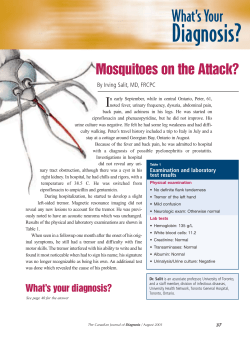


© Copyright 2026