
Use of Silicone Materials to Simulate Tissue Biomechanics as
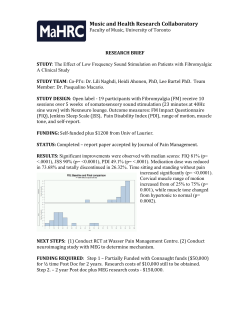
ORIGINAL INVESTIGATION
Use of Silicone Materials to Simulate Tissue
Biomechanics as Related to Deep Tissue Injury
Jessica L. Sparks, PhD; Nicholas A. Vavalle, MS; Krysten E. Kasting; Benjamin Long, MS; Martin L. Tanaka, PhD;
Phillip A. Sanger, PhD; Karen Schnell, MSN; and Teresa A. Conner-Kerr, PhD
ABSTRACT
CONCLUSION: Indentation tests and the prototype patient
simulator trial demonstrated similar trends with high pressures
closest to the bony prominence with decreasing magnitude toward
the interfacial surface. Qualitatively, silicone mimicked the
phenomenon observed in muscle of nonuniform stress under
concentrated loading. Although shear moduli were within
biological ranges, stress and stiffness values exceeded those of
porcine muscle. This research represents a first step toward
development of a preclinical model simulating the biomechanical
conditions of stress and strain in deep muscle, since local
biomechanical factors are acknowledged to play a role in DTI
initiation. Future research is needed to refine the capacity of
preclinical models to simulate biomechanical parameters in
successive tissue layers of muscle, fat, dermis, and epidermis
typically intervening between bone and support surfaces, for body
regions at risk for DTI.
KEYWORDS: deep tissue injury, soft tissue biomechanics,
pressure ulcer, patient simulator
OBJECTIVE: Deep tissue injury (DTI) is caused by prolonged
mechanical loading that disrupts blood flow and metabolic
clearance. A patient simulator that mimics the biomechanical
aspects of DTI initiation, stress and strain in deep muscle tissue,
would be potentially useful as a training tool for pressure-relief
techniques and testing platform for pressure-mitigating products.
As a step toward this goal, this study evaluates the ability of
silicone materials to mimic the distribution of stress in muscle
tissue under concentrated loading.
METHODS: To quantify the mechanical properties of candidate
silicone materials, unconfined compression experiments were
conducted on 3 silicone formulations (Ecoflex 0030, Ecoflex 0010,
and Dragon Skin; Smooth-On, Inc, Easton, Pennsylvania). Results
were fit to an Ogden hyperelastic material model, and the resulting
shear moduli (G) were compared with published values for
biological tissues. Indentation tests were then conducted on
Ecoflex 0030 and porcine muscle to investigate silicone’s ability to
mimic the nonuniform stress distribution muscle demonstrates under
concentrated loading. Finite element models were created to quantify
stresses throughout tissue depth. Finally, a preliminary patient
simulator prototype was constructed, and both deep and superficial
‘‘tissue’’ pressures were recorded to examine stress distribution.
RESULTS: Indentation tests showed similar stress distribution
trends in muscle and Ecoflex 0030, but stress magnitudes were
higher in Ecoflex 0030 than in porcine muscle. All 3 silicone
formulations demonstrated shear moduli within the range of
published values for biological tissue. For the experimental
conditions reported in this work, Ecoflex 0030 exhibited greater
stiffness than porcine muscle.
ADV SKIN WOUND CARE 2015;28:59Y68
INTRODUCTION
Pressure ulcers (PrUs) are a common condition in both persons
who use wheelchairs and those unable to sit out of bed. They cost
the United States alone more than $1.2 billion1 and affect 10% of
all hospitalized patients.2 Pressure ulcers can be broadly classified
as 1 of 2 types: superficial or deep.3 Superficial ulcers affect skin
layers near the epidermis and are formed as a result of damaging
frictional and shear forces in the presence of moisture and heat.3,4
Deep PrUs are the focus of this study. These ulcers develop in
deep muscle tissue next to bony prominences such as the sacrum,
Jessica L. Sparks, PhD, is an Associate Professor of Chemical, Paper, and Biomedical Engineering, Miami University, Oxford, Ohio. Nicholas A. Vavalle, MS, is a doctoral candidate in biomedical
engineering, Wake Forest University, Winston-Salem, North Carolina. Krysten E. Kasting is a bioengineering undergraduate student, Miami University, Oxford, Ohio. Benjamin Long, MS, is an
Instructor of Physical Therapy, Winston-Salem State University, Winston-Salem, North Carolina. Martin L. Tanaka, PhD, is an Assistant Professor of Engineering and Technology, Western Carolina
University, Cullowhee, North Carolina. Phillip A. Sanger, PhD, is a Professor of Electrical and Computer Engineering, Purdue University, West Lafayette, Indiana. Karen Schnell, MSN, owns Blue Sky
Health Concepts Consulting, Mebane, North Carolina. Teresa A. Conner-Kerr, PhD, is Dean of the College of Health Sciences, University of North Georgia, Dahlonega, Georgia. Dr Sparks
and Mr Vavalle have disclosed that Wake Forest University is a past recipient of grant funding from the US Department of Education (awarded to T.A.C.-K.). Mr Long has disclosed that
Winston-Salem State University is a past recipient of grant funding from the US Department of Education (awarded to T.A.C.-K.). Dr Sanger has disclosed that his institution is a past
recipient of grant funding from the Golden Leaf Foundation. Ms Schnell has disclosed that she has previously received an honorarium from Winston-Salem State University, and is a past
recipient of payment for writing or reviewing a manuscript from Winston-Salem State University. Dr Kerr has disclosed that Winston-Salem State University is a past recipient of a Title III US Department
of Education grant. Ms Kasting and Dr Tanaka have disclosed that they have no financial relationships related to this article. Acknowledgments: The authors acknowledge Nick Ashworth, Isaac Crisp,
Andrew York, and Erik Ellington for their assistance with data acquisition and graphical user interface development. The authors also thank Kristen Pone, Christen Isley, and Peggy Furr, for their
assistance with fiberglass casting and material acquisition. Funding for this research was provided by the US Department of Education (grant P031B085015-9 to T.A.C.-K.) and the Goldenleaf
Fund (to P.A.S.). Submitted July 5, 2013; accepted in revised form April 2, 2014.
WWW.WOUNDCAREJOURNAL.COM
59
ADVANCES IN SKIN & WOUND CARE & FEBRUARY 2015
Copyright © 2015 Wolters Kluwer Health | Lippincott Williams & Wilkins. Unauthorized reproduction of this article is prohibited.
ORIGINAL INVESTIGATION
ischial tuberosity, or greater trochanter of the femur.3,5,6 Deep PrUs
are caused by prolonged mechanical loading (compression) that
interferes with blood flow and clearance of metabolic byproducts.
As the deep muscle tissue undergoes necrosis, it becomes stiffer,
projecting the mechanical stresses to more superficial tissues, which
then bear the mechanical load.7,8 Since the injury develops under
intact skin, the damage is difficult to detect at early stages. These
potentially life-threatening injuries have been termed deep tissue
injury (DTI).9
Biomechanical research6,10 has demonstrated that concentrated
stresses in deep tissues near bony prominences cannot be readily
predicted from surface pressure maps, which are currently a key
technology for pressure-related risk assessment.3,11 Since DTIs develop deep in the subdermal tissue layer, the use of interfacial
pressure mapping to evaluate clinical strategies for DTI prevention, such as cushions, mattresses, and repositioning techniques,
can be misleading.6,12,13 If clinicians could more accurately evaluate the stresses that develop within deep muscle tissue, they could
provide better information to healthcare providers regarding the
ability of repositioning protocols to lessen the local mechanical load
at deep, high-risk sites. In addition, clinicians could generate better
test protocols for evaluating the effectiveness of pressure-relieving
products, such as mattress and cushions, for DTI prevention. These
goals can potentially be achieved in the long term by developing a
novel patient simulator with biomechanical properties similar to
actual human tissues, including compressive properties of muscle
tissue and stresses near the bone-muscle interface.
Although a variety of buttock phantoms have been developed
for wheelchair-cushion testing, few of these32,33 generate information regarding the stress or deformation of deeper material adjacent
to a simulated ‘‘bone.’’ Those that have been reported were made
of polyvinyl chloride cast around a wooden core and thus lacked
realistic mechanical properties of biological tissues.32,33
Because the composition and microstructure of biological tissue
are enormously complex, the construction of a suitably accurate
simulator for DTI prevention is a significant challenge. The longterm goal of this work is the development of a simulator that
mimics the biomechanical conditions of stress and strain in deep
muscle, since local biomechanical factors are acknowledged to
play a role in DTI initiation.6Y8,34,35 As a step toward this goal, this
study evaluates the ability of soft silicone materials to mimic specific features of the compressive mechanical behavior of biological
muscle tissue. In particular, this study will focus on (1) shear modulus, a mechanical property indicative of how stiff a material feels
to the touch, and (2) the ability of a material to mimic the nonuniform stress distribution in tissues subjected to indentationtype loading, such as that which occurs in muscle compressed by a
bony prominence. These material features are expected to be among
the relevant features necessary for reproducing, in a synthetic
ADVANCES IN SKIN & WOUND CARE & VOL. 28 NO. 2
environment, the biomechanical conditions associated with DTI
initiation. Silicone rubber was selected as a good candidate material for this initial study because of its ability to retain its shape
and its resistance to degradation and because it can be readily obtained in different degrees of stiffness to mimic mechanical properties
of biological tissue.
The objectives of the study are as follows:
Measure the mechanical properties for 3 formulations of silicone
using uniaxial unconfined compression experiments, and compare
resulting shear moduli to published values for biological tissues
(muscle, fat, and skin) tested in compression.
Using an indenter with realistic bony geometry, conduct indentation experiments in both silicone and muscle tissue specimens
and examine how pressure varies as a function of distance from
the bony prominence (indenter tip).
Demonstrate proof of principle that a prototype patient simulator can be used to obtain internal pressure measurements at
multiple tissue depths near a specified bony prominence.
METHODS
Biomechanical Testing
Specimen Preparation. Three formulations of silicone rubber were
obtained from Smooth-On, Inc (Easton, Pennsylvania): Dragon
Skin, Ecoflex 0010, and Ecoflex 0030. Cylindrical samples (average
diameter, 35.8 mm; average height, 24.5 mm) were prepared according to the manufacturer’s specifications for uniaxial compression tests by mixing the appropriate 2-part liquid forms of each
formulation together and pouring the mixture into a mold. They
were then allowed to cure for the recommended amount of time
(75 minutes for Dragon Skin, 4 hours for each Ecoflex). During the
curing phase, a level was used to verify that the top and bottom of
each sample were parallel, in order to avoid asymmetric loading
during the uniaxial compression test. For this test series, a total of
18 specimens were produced, 6 of each type of rubber. Each specimen was measured with calipers after demolding to ensure consistent
dimensions.
An additional set of 3 Ecoflex 0030 samples (average diameter,
59.3 mm; average height, 26 mm) were prepared for indentation
testing using the same preparation methods. Samples of porcine
muscle obtained from the local grocer were also prepared to undergo
similar testing. Six porcine muscle samples were prepared as cylinders (average diameter, 60.2 mm; average height, 28.2 mm). The
specimens were presliced to uniform thickness, and samples were
cut using a circular guide and surgical blade.
Uniaxial Unconfined Compression. Uniaxial unconfined compression testing is a standardized method for evaluating the mechanical properties of compliant materials, such as polymers or soft
tissues.14 Uniaxial compression tests were conducted for each silicone specimen using an Electroforce LM1 Test Bench mechanical
60
WWW.WOUNDCAREJOURNAL.COM
Copyright © 2015 Wolters Kluwer Health | Lippincott Williams & Wilkins. Unauthorized reproduction of this article is prohibited.
ORIGINAL INVESTIGATION
testing system (Bose Corporation, Eden Prairie, Minnesota) with
a 250-N capacity load cell. The uniaxial compression test consisted of loading the specimen at a constant rate (1.0% strain per
second) until the selected maximum strain value (25% compression)
was reached. The strain rate and maximum strain values were chosen
to reflect test conditions that have been previously reported for
biological tissues tested in compression.15,16 Force and displacement data were recorded in all tests. Force data were converted to
engineering stress by dividing by the initial cross-sectional area of
the specimen. Displacement data were used to calculate engineering strain as change in length divided by original length. The
experimental setup was identical for all 3 silicone formulations.
Petroleum jelly was applied to the top and bottom of each sample
before testing to reduce the effects of friction.
Indentation Testing. For the indentation experiments, a human
sacrum model (3B Scientific, Tucker, Georgia) was mounted to
the Bose Electroforce LM1 Test Bench mechanical testing system
using a custom-mounting fixture (Figure 1). The spinous tubercle
of the bone model was used to indent the specimens. Prior to
indentation, 2 Millar Mikro-Tip Pressure Catheters (SPR-524;
ADInstruments, Colorado Springs, Colorado) were inserted into
the specimens at 2 or 5 mm from the top and bottom surfaces of
the specimen, for porcine muscle and Ecoflex 0030 samples, respectively. Pressure sensor locations were termed deep (near the
bony prominence) and superficial (distant from the bony prominence)
(Figure 1). A needle was used to create guide holes for sensor insertion, and the guide holes were prefilled with petroleum jelly to
create a smooth coupling between the specimens and miniature pressure sensors. The indentation test consisted of loading the specimen
at a constant rate of 0.5 mm/s for 8 seconds. Force and pressure (at
both deep and superficial depths) data were recorded in all tests.
Table 1.
LITERATURE RANGES FOR SHEAR MODULI
OF BIOLOGICAL TISSUES: ALL DATA
REPORTED FROM COMPRESSION TESTING
G (kPa)
Biological Tissue
Muscle
Longitudinal
Transverse
Active
Relaxed
Skin
Fat
51Y10523Y25
11Y5423,25
17.1Y30.522,25
4.6Y23.86,15,16,22
2.8Y31.96,20,26
1.9Y31.96,20,26
Patient Simulator Prototype. Based on the results of the material
characterization experiments (Tables 1 and 2), Dragon Skin was
used as muscle, Ecoflex 0010 as fat, and Ecoflex 0030 as skin in the
initial simulator prototype. The prototype design was based on
approximate human anatomical structure of the pelvis and upper
thigh (Figure 2). The external geometry of the simulator was formed
using a fiberglass cast from waist to midthigh. The inside of the
fiberglass mold was coated with a thin layer of plaster to allow for
easy removal of the silicone after molding. A thin layer of the skin
simulant material was ‘‘painted’’ on the plaster and ultimately
formed the outer surface of the simulator prototype. Simulated
muscles were created by pouring muscle simulant into separate
molds that were previously constructed to mimic the approximate
shape of the major muscles of the pelvis and hip region. The muscles were affixed to the bony pelvis (Human Skeleton Model; 3B
Scientific) in the corresponding anatomical locations. The bony
pelvis with attached muscles was then suspended inside the fiberglass mold in the appropriate anatomical orientation. Simulated fat
material (in liquid form) was then poured into the mold, to fill the
spaces between the skin layer and the muscles. The fat material was
allowed to solidify.
The completed simulator prototype, shown in Figure 2, was
designed to determine whether it is feasible to produce and detect
differences in deep internal pressures (near a bony prominence)
Figure 1.
INDENTATION TEST SETUP FOR PORCINE MUSCLE WITH
CUSTOM-MOUNTED 3B SCIENTIFIC SACRUM BONE INDENTER
Table 2.
BEST-FIT HYPERELASTIC MATERIAL
CONSTANTS FOR SILICONE RUBBER
FORMULATIONS
Ogden Model Terms
Silicone Type
Shear Modulus
G (kPa)
Strain Hardening
Exponent >
Poisson Ratio M
Dragon Skin
Ecoflex 0010
Ecoflex 0030
75.449
12.605
22.081
5.836
4.32
0.825
0.4999
0.4999
0.4999
Load cell and both deep and superficial Millar Mikro-Tip Pressure Catheters are labeled.
WWW.WOUNDCAREJOURNAL.COM
61
ADVANCES IN SKIN & WOUND CARE & FEBRUARY 2015
Copyright © 2015 Wolters Kluwer Health | Lippincott Williams & Wilkins. Unauthorized reproduction of this article is prohibited.
ORIGINAL INVESTIGATION
Figure 2.
PRELIMINARY SIMULATOR PROTOTYPE
A, Simulator prototype instrumented with pressure transducers at both deep and superficial locations over the sacrum. B, Computed tomography scan of prototype showing skeletal
anatomy. C, Simulator control software and user interface.
insertion technique described above. One pressure sensor was inserted in deep tissue adjacent to the spinous tubercle of the sacrum.
The second sensor was also inserted over the spinous tubercle but
in more superficial tissue just beneath the skin. Manual pressure
was then applied with an open palm over the instrumented region,
and pressures recorded from both sensors.
and more superficial pressures (closer to the skin surface) in simulated soft tissues. Internal pressure data can be transferred from
the simulator to the computer, exhibited on screen for immediate
feedback, and stored for future analysis (Figure 2C). In a preliminary
trial, the simulator was instrumented with 2 Millar Mikro-Tip Pressure Catheters (Millar, Inc, Houston, Texas) using the needle-guided
Figure 3.
ECOFLEX 0030 FINITE ELEMENT MODEL AT MAXIMUM INDENTATION
A, Color mapping shows the normal stress distribution through the sample. B, Pressure related to distance from the indenter along line LS of the model.
ADVANCES IN SKIN & WOUND CARE & VOL. 28 NO. 2
62
WWW.WOUNDCAREJOURNAL.COM
Copyright © 2015 Wolters Kluwer Health | Lippincott Williams & Wilkins. Unauthorized reproduction of this article is prohibited.
ORIGINAL INVESTIGATION
Figure 4.
PORCINE MUSCLE FINITE ELEMENT MODEL AT MAXIMUM INDENTATION
A, Color mapping shows the normal stress distribution through the sample. B, Pressure related to distance from the indenter along the line LM of the model.
Finite Element Modeling
Statistical Analysis
Computational finite element (FE) simulations were developed
to create virtual (in silico) models of both types of mechanical
tests: uniaxial unconfined compression and indentation. Models
of uniaxial unconfined compression were used to determine the
mechanical properties (Table 2 and Appendix) for each silicone
formulation, by fitting hyperelastic Ogden model parameters to
the average experimental stress-strain results for each silicone formulation. The calculated silicone mechanical properties were then
implemented in separate FE simulations of the silicone indentation
experiments. Analogous simulations of the porcine muscle indentation experiments were also created, using previously published
and validated mechanical property values for muscle.17
The computer simulations of the indentation experiments were
used to quantify the expected stress everywhere in the specimen,
from immediately adjacent to the spinous tubercle indenter to the
most distant regions from the indenter tip. The models were also
used to examine how the stresses in the specimen varied with time,
from initial indenter contact until peak indentation was achieved.
To validate these model-predicted stress distributions, model output
was compared directly against the measured ‘‘deep’’ and ‘‘superficial’’ pressure values, which were recorded at known depths in
the sample throughout the indentation (Figures 3 and 4). Results of
these comparisons were used to verify the accuracy of the models.
Details of the FE model development are provided in the Appendix.
To assess the accuracy of the mechanical property values implemented in the FE simulations for each synthetic tissue analog,
linear regression analyses of model output versus experimental
values were conducted for each simulation.18 A slope value near
unity indicated a one-to-one relationship between experiment and
model, and an R2 value near unity indicated a high goodness of fit.
WWW.WOUNDCAREJOURNAL.COM
RESULTS
FE Model Parameters
Best-fit material constants for all silicone rubber formulations are
given in Table 2. Material constants for muscle17 are given in Table 3.
Experimental Results and FE Model Validation
Uniaxial Unconfined Compression. The measured peak stresses for
Dragon Skin, Ecoflex 0010, and Ecoflex 0030 were 73.0 T 5.2, 12.1 T 0.75,
and 24.0 T 1.7 kPa, respectively (mean T 1 SD). Stress versus strain
results are illustrated in Figure 5, showing experimental data compared with best-fit FE model results for all silicone formulations.
Linear regression (models vs experiments) of the ramp phase
of compression showed high goodness of fit (R2 = 0.999), with
slope values at or near unity (slope = 0.99Y1.00, P G .05). These
results indicate a good fit of the models to the experimental data,
giving confidence in the accuracy of the material property values
(Table 2) implemented in the models.
63
ADVANCES IN SKIN & WOUND CARE & FEBRUARY 2015
Copyright © 2015 Wolters Kluwer Health | Lippincott Williams & Wilkins. Unauthorized reproduction of this article is prohibited.
ORIGINAL INVESTIGATION
4A for Ecoflex 0030 and for porcine muscle, respectively. Model
results are presented as normal compressive stress (S22) in the
direction of loading. Stresses were then converted to units of millimeters of mercury to facilitate comparison against pressures measured experimentally at 2 locations per specimen: deep (near the
indenter) and superficial (opposite the indenter).
Similar stress distribution patterns can be seen in the Ecoflex
0030 and porcine muscle (Figures 3B and 4B) with highest stresses
located adjacent to the bony prominence and with a lessening
degree farther from the prominence. However, the Ecoflex 0030
material showed much greater stress magnitudes compared with
muscle tissue. Experimental data showed good agreement with these
trends. Ecoflex 0030 had an average measured peak deep pressure
(near the indenter) of 142.5 mm Hg and an average measured peak
superficial pressure (opposite the indenter) of 18.0 mm Hg, whereas
the porcine muscle demonstrated an average peak deep pressure of
20.0 mm Hg and average superficial peak pressure of 2.4 mm Hg.
Figures 3 and 4 provide direct comparisons of the experimental
pressures recorded at known locations in the sample at maximum
indentation, with the FE model predictions of stress at these same
locations. Results suggest that the virtual FE models of indentation
provide a good representation of the stress distributions produced
Table 3.
HYPERELASTIC MATERIAL CONSTANTS FOR
MUSCLE USED IN FE SIMULATIONS17
[-]
kPa
kPa-1
>1=0.1316402E+01
>2=0.1835933E+02
K1=1.02571
K2=0.145209 E-04
D1=0.194987E-01
D2=0.166315
Note: For this material, D1 = 0.194987E-01 kPa-1 is equivalent to Poisson’s ratio M = .495.
Comparison with Biological Tissues. Table 2 shows a summary
of the shear modulus values of the 3 silicone rubber formulations,
which can be compared against a range of shear moduli for muscle, fat, and skin tissues found in the literature (Table 1). Biological
tissue shear moduli were taken only from studies in which the tissues were tested in compression, because biomechanical property
data can vary significantly depending on the test mode used. The
shear moduli of all silicone materials evaluated in the present study
fell within the range of reported values for shear moduli of muscle,
fat, and skin, for biomechanical tests conducted in compression.
Indentation Testing. The model-predicted stress distributions in
the tissue, at maximum indentation, are shown in Figures 3A and
Figure 5.
RESULTS OF UNIAXIAL COMPRESSION TESTS ON SILICONE RUBBERS (DRAGON SKIN, ECOFLEX 0010, AND ECOFLEX
0030) COMPARED WITH FINITE ELEMENT SIMULATIONS
ADVANCES IN SKIN & WOUND CARE & VOL. 28 NO. 2
64
WWW.WOUNDCAREJOURNAL.COM
Copyright © 2015 Wolters Kluwer Health | Lippincott Williams & Wilkins. Unauthorized reproduction of this article is prohibited.
ORIGINAL INVESTIGATION
Figure 6.
PRESSURES MEASURED IN EXPERIMENTS COMPARED AGAINST FINITE ELEMENT MODEL-PREDICTED PRESSURES,
FOR SILICONE AND MUSCLE INDENTATION TESTS
A, Ecoflex 0030 indentation test: deep and superficial pressures in relationship to time for both experimental and model data. B, Porcine muscle indentation test: deep and superficial
pressures in relationship to time for both experimental and model data.
by this form of indentation loading in the experimental specimens,
for both Ecoflex 0030 and passive porcine muscle.
The results described above emphasized pressure as a function
of location within the specimen. The FE model results were also
compared against experimental data to examine how pressure varied
with time at a given location. These results are shown in Figure 6.
As before, the results suggest good agreement between FE simulations and the experimental pressure-time history produced during
the indentation tests at ‘‘deep’’ and ‘‘superficial’’ point locations in
the specimen. The results also confirm the finding that indentation
of Ecoflex 0030 generated higher pressure magnitudes than did
similar indentation of passive porcine muscle.
Linear regression (experiment vs model) of the indentation
pressure-time histories showed a high goodness of fit (R2 = 0.99,
P G .05) for both deep and superficial pressure in Ecoflex 0030. A
slightly weaker but still significant fit of the porcine muscle model
to the data (R2 = 0.849 and 0.92 for deep and superficial, respectively)
was seen. Deep pressure showed a slope near unity for both porcine
muscle (1.007) and Ecoflex 0030 (0.987). Superficial pressure data
showed slightly more spread in slope values. For Ecoflex 0030, a slope
of 0.777 was calculated. The slope value for superficial pressure in porcine
muscle was elevated (2.29), indicating that the model underpredicted
the experimental value (0.97 vs 2.42 mm Hg at maximum indentation).
Patient Simulator. The preliminary trial of the simulator prototype
revealed a stress distribution pattern similar to that observed in the
indentation testing (Figure 7). When manual pressure was applied
over the instrumented sacral region, it was observed that pressures in
the deep tissue adjacent to the bony prominence were higher than
those recorded in more superficial tissue near the skin surface. It is not
WWW.WOUNDCAREJOURNAL.COM
Figure 7.
RESULTS OF PRELIMINARY EXPERIMENTATION WITH
PATIENT SIMULATOR PROTOTYPE. PRESSURE IN
RELATIONSHIP TO TIME FOR SENSORS LOCATED AT DEEP
AND SUPERFICIAL LOCATIONS WITHIN THE PROTOTYPE
65
ADVANCES IN SKIN & WOUND CARE & FEBRUARY 2015
Copyright © 2015 Wolters Kluwer Health | Lippincott Williams & Wilkins. Unauthorized reproduction of this article is prohibited.
ORIGINAL INVESTIGATION
tissue near a selected bony prominence, the spinous tubercle of
the sacrum. The simulator was instrumented to provide real-time
feedback on local pressure conditions at different tissue depths. The
indentation experiments showed that, while the stress distribution
patterns in porcine muscle tissue paralleled those of the Ecoflex
0030 silicone, the stress magnitudes in silicone were substantially
higher than those in muscle tissue. The clinical implications of this
discrepancy remain to be examined. Limitations of the current simulator prototype also include the fact that the interfaces between
the tissue layers (skin-fat, fat-muscle, muscle-bone interfaces) require further development to reflect anatomical structure and that
the model ultimately must be validated against biologic tissue representing all tissue components covering a bony prominence.
In the long term, a fully developed and validated simulator, which
accurately mimics all the relevant biomechanical complexities of human
anatomy, could be used as a research platform for testing the effects
of pressure-mitigating products on deep-tissue stresses. It could also
be used to educate healthcare professionals on proper positioning
techniques for deep pressure relief in the lower body. According
to the current National Pressure Ulcer Advisory Panel and European
Pressure Ulcer Advisory Panel International Guidelines for the
Prevention and Treatment of Pressure Ulcers,9 repositioning should
be utilized for all at-risk individuals. The panels assigned the highest
level of evidence (A) to this intervention.
It is well known that biological tissues exhibit viscoelastic mechanical behaviors such as creep, the phenomenon of increasing
deformation under constant load.14Y16,20,21 Although the use of a
viscoelastic material as a tissue analog would better capture this
biomechanical property of human tissues, it could also incur practical problems with the fabrication of a durable and reusable simulator. For instance, sustained loading could cause viscoelastic
materials to deform over time, such that recovery periods may be
needed between users to allow the material to return to its original
dimensions. Because of these limitations, silicone-based materials
were selected as the best tissue analogs for the present study, because they could mimic tissue stiffness without the complicating
effects of time-dependent viscoelastic behavior.
All 3 silicone formulations had shear moduli values that fell
within the range of biological tissue, and Ecoflex 0030 and porcine
muscle demonstrated similar nonuniform stress distribution patterns;
however, Ecoflex 0030 showed much higher stress magnitudes than
muscle under indentation loading. One of the factors that could
contribute to this difference is the large range of values for biological soft tissue. Although the shear modulus of Ecoflex 0030 (22 kPa)
fell within the range of passive muscle (Table 1), it was on the upper
limits of this range. In contrast, the shear modulus of porcine muscle,
derived from the FE simulation, was found to be on the low end of
this range (~1 kPa). This difference in stiffness would account for the
higher stress magnitudes produced in Ecoflex 0030 under indentation
Figure 8.
FINITE ELEMENT MODEL SHOWING MESH PRIOR TO
INDENTATION TEST
possible to make direct comparisons with the pressure magnitudes
measured in the indentation experiments, because the depth of
the manually applied load was not recorded, and the geometric
configuration of the loading environment differed significantly from
that of the simplified indentation experiments. However, this preliminary trial demonstrated qualitatively that silicone-based tissue analogs were able to mimic the basic trend of a stress concentration
developing in the material in the region of a bony prominence, when
subjected to compressive loading.
DISCUSSION
To address the increasing human and financial burden of DTIs,
it is crucial to train healthcare workers with the best techniques
available to prevent DTI development. It is also necessary to provide
researchers with suitable platforms for testing pressure-mitigating
products for DTI prevention. Particular attention must be paid to
deep tissue stresses in high-risk areas, such as the skin and soft
tissue over certain bony prominences associated with the pelvic
girdle and lower extremity, because 95% of PrUs are known to occur
on the lower half of the body.19
In this study, the authors characterized mechanical properties
of soft tissue analogs capable of generating shear moduli and stress
distribution trends similar to biological soft tissue. With these analogs, the authors developed a preliminary patient simulator prototype
that could mimic the phenomenon of concentrated stress in deep
ADVANCES IN SKIN & WOUND CARE & VOL. 28 NO. 2
66
WWW.WOUNDCAREJOURNAL.COM
Copyright © 2015 Wolters Kluwer Health | Lippincott Williams & Wilkins. Unauthorized reproduction of this article is prohibited.
ORIGINAL INVESTIGATION
loading. Gefen et al8 have reported that muscle stiffness increases
in vivo with the development of deep PrUs. It would be informative
in future work to compare the stress distribution in silicone against
that of muscle tissue affected by DTI.
Biomechanical properties are largely dependent on test conditions.
For example, muscle demonstrates shear modulus temperature dependence.23 In addition, examination of the shear moduli reported
in Table 1 shows that passive muscle is much less stiff than active
muscle. It has been shown previously that, in the human biceps
brachii, there is a linear relationship between shear modulus and
muscle load.22 This means that as the muscle activation increases,
its shear modulus increases. This suggests that the Dragon Skin material, although high within the range of the reported shear modulus
of relaxed muscle tissues, may be a reasonable analog to the compressive properties of active muscle or even muscle under pathogenesis.22Y25
Although it had a high shear modulus, it was chosen as the muscle
simulant in the patient simulator prototype in order to account for
the significant stiffening that occurs under compression in in vivo
muscle tissue.8
Shear moduli of silicone rubbers and biological soft tissues
were compared (Tables 1 and 2) because shear modulus gives a
sense of the ‘‘feel’’ or stiffness of the material. However, in comparing the full stress-strain responses of the silicones with biological
materials, there is no perfect match. For example, none of the silicone materials showed the same degree of strain hardening that
was observed by Wu et al26 in their compression tests of skin and
adipose tissue. Based on other studies,27,28 it is expected that the
silicone rubbers would exhibit strain hardening at higher levels of
strain, yet this still differs from the strain hardening observed at
lower strain levels (0.3Y0.5) in biological tissues. Also significant to
this comparison is variation in species and anatomic location from
which the biological samples were taken, among the literature
studies used for benchmarking. Tissue properties vary from one
species to the next. Within a given species, material properties can
differ from one location to another or with temperature for the same
type of tissue.23 For instance, the fat that is found in the human foot
has a much different composition than fat found in other parts of
the body.29 This could explain discrepancies in strain hardening
between the fat data of Wu et al26 and those of Miller-Young et al.29
In summary, the biomechanical behavior of human tissues is complex, and results vary widely among studies. Silicones were not able
to mimic all the complex features of biological tissue biomechanics,
because of their significantly different structure and composition. In
the present study, it was found that the tested silicone formulations
were able to mimic certain features of tissue biomechanics that are
likely to be relevant for future simulator development, such as possessing shear moduli in the range of biological soft tissue and
exhibiting similar stress distribution patterns when subjected to
indentation loading. Much additional work is needed to examine
WWW.WOUNDCAREJOURNAL.COM
other biomechanical features, such as material density, appropriate
interface properties between different material layers, the influence
of fiber orientation on muscle mechanical properties, and the ability
of the simulated soft tissues to deform and return to their initial
configuration as joints are moved from one position to another.
CONCLUSIONS
This research represents a first step toward development of a preclinical model simulating the biomechanical conditions of stress
and strain in deep muscle, because local biomechanical factors are
acknowledged to play a role in DTI initiation.6Y8,34,35 Future research
is needed to refine the capacity of preclinical models to simulate
biomechanical parameters in successive tissue layers of muscle, fat,
dermis, and epidermis typically intervening between bone and support surfaces, for body regions at risk for DTI.
In this study, materials used to simulate soft tissues were evaluated to characterize their behavior and compare with biological
soft tissues. Three formulations of silicone were found to have
shear moduli within the range of values for soft tissues published
in the literature. Pressure at various locations within the tissue was
measured to show the nonuniform pressure gradient that develops
within the tissue upon force application during indentation-type
loading, similar to that which occurs in deep muscle tissue compressed against bony prominences.
Using this knowledge, a preliminary patient simulator prototype
was developed. Materials were selected to mimic the mechanical
properties of muscle, fat, and skin. The skeleton, molded muscles,
skin layer, and simulated fat were assembled, and miniature pressure transducers were used to record local pressures in deep tissue
near the spinous tubercle of the sacrum and in superficial tissue,
above the spinous tubercle just beneath the skin.
A patient simulator that can mimic the biomechanical aspects
of DTI initiation, namely, stress and strain distributions in soft tissues subjected to lifelike mechanical loading conditions, is needed
in the wound care industry. This future work will include extensive
further development and validation of the device design. Ultimately,
future iterations of this device could be used to train healthcare
professionals in best practices of lower body position for deep pressure relief, which could greatly reduce the number of patients suffering from DTI. The device would also be an innovative test
platform for evaluating the efficacy of products designed to prevent
PrUs, such as cushions, mattresses, and dressings.
&
REFERENCES
1. National Center for Injury Prevention and Control: CDC Injury Fact Book. Atlanta, GA: Centers
for Disease Control and Prevention; 2002.
2. Whittington K, Patrick M, Roberts J. A national study of pressure ulcer prevalence and
incidence in acute care hospitals. J Wound Ostomy Continence Nurs 2000;27:209-15.
3. Bouten CV, Oomens CW, Baaijens FP, Bader DL. The etiology of pressure ulcers: skin deep or
muscle bound? Arch Phys Med Rehabil 2003;84:616-9.
67
ADVANCES IN SKIN & WOUND CARE & FEBRUARY 2015
Copyright © 2015 Wolters Kluwer Health | Lippincott Williams & Wilkins. Unauthorized reproduction of this article is prohibited.
ORIGINAL INVESTIGATION
4. Vidal J, Sarrias M. An analysis of the diverse factors concerned with the development of
pressure sores in spinal cord injured patients. Paraplegia 1991;29:261-7.
5. Gefen A. Bioengineering models of deep tissue injury. Adv Skin Wound Care 2008;21:30-6.
6. Linder-Ganz E, Shabshin N, Itzchak Y, Gefen A. Assessment of mechanical conditions in
sub-dermal tissues during sitting: a combined experimental-MRI and finite element approach.
J Biomech 2007;40:1443-54.
7. Linder-Ganz E, Gefen A. Mechanical compression-induced pressure sores in rat hindlimb:
muscle stiffness, histology and computational methods. J Appl Physiol (1985) 2004;96:
2034-49.
8. Gefen A, Gefen N, Linder-Ganz E, Margulies SS. In vivo muscle stiffening under bone
compression promotes deep pressure sores. J Biomech Eng 2005;127:512-24.
9. European Pressure Ulcer Advisory Panel and National Pressure Ulcer Advisory Panel. Prevention and treatment of pressure ulcers: quick reference guide. Washington, DC: National
Pressure Ulcer Advisory Panel; 2009.
10. Lyder HC. Pressure ulcer prevention and management. JAMA 2003;289:223-6.
11. Garcia AD, Thomas DR. Assessment and management of chronic pressure ulcers in the
elderly. Med Clin North Am 2006;90:925-44.
12. Allen V, Ryan DW, Murray A. Repeatability of subject/bed interface pressure measurements.
J Biomed Eng 1993;15:329-32.
13. Sprigle S, Dunlop W, Press L. Reliability of bench tests of interface pressure. Assist Technol
2003;15:49-57.
14. Macosko CW. Rheology Principles, Measurements, and Applications. New York, NY: Wiley-VCH;
1994:5-136.
15. Bosboom E, Hessenlink MK, Oomens CW, Bouten CV, Drost MR, Baaijens FP. Passive transverse mechanical properties of skeletal muscle under in vivo compression. J Biomech 2001;34:
1365-8.
16. Van Loocke M, Lyons CG, Simms C. Viscoelastic properties of passive skeletal muscle in compression: stress-relaxation behaviour and constitutive modeling. J Biomech 2008;41:1555-66.
17. Then C, Menger J, Benderoth G, et al. A method for a mechanical characterisation of
human gluteal tissue. Technol Health Care 2007;15:385-98.
18. Anderson AE, Ellis BJ, Weiss JA. Verification, validation and sensitivity studies in computational
biomechanics. Comput Methods Biomech Biomed Engin 2007;10:171-84.
19. Dharmarajan T, Sugalino JT. Pressure ulcers: clinical features and management. Hosp
Physician 2002;38(3):64-71.
20. Zahouani H, Pailler-Mattei C, Sohm B, Vargiolu R, Cenizo V, Debret R. Characterization of
the mechanical properties of a dermal equivalent compared with human skin in vivo by
indentation and static friction tests. Skin Res Technol 2009;15:68-76.
21. Ferry JD. Viscoelastic Properties of Polymers. 3rd ed. New York, NY: Wiley and Sons; 1980.
22. Dresner M, Rose GH, Rossman PJ, Muthupillai R, Manduca A, Ehman RL. Magnetic resonance
elastography of skeletal muscle. J Magn Reson Imaging 2001;269-76.
23. Sapin-de Brosses E, Gennisson JL, Pernot M, Fink M, Tanter M. Temperature dependence
of the shear modulus of soft tissues assessed by ultrasound. Phys Med Biol 2010;55:
1701-18.
24. Lacourpaille L, Hug F, Bouillard K, Hogrel JY, Nordez A. Supersonic shear imaging provides
a reliable measurement of resting muscle shear elastic modulus. Physiol Meas 2012;33(3):
N19-28.
25. Ringleb SI, Bensamoun SF, Chen Q, Manduca A, An KN, Ehman RL. Applications of magnetic
resonance elastography to healthy and pathologic skeletal muscle. J Magn Reson Imaging
2007;25:301-9.
26. Wu JZ, Cutlip RG, Andrew ME, Dong RG. Simultaneous determination of the nonlinear-elastic
properties of skin and subcutaneous tissue in unconfined compression tests. Skin Res Technol
2007;13:34-42.
27. Shergold OA, Fleck NA, Radford D. The uniaxial stress versus strain response of pig skin
and silicone rubber at low and high strain rates. Int J Impact Eng 2006;32:1384-402.
28. Lee OS, Kim MS, Kim KJ, Hwang SW, Cho KS. Dynamic deformation behavior of rubber under
high strain rate compressive loading. Int J Mod Phys B 2003;17:1415-20.
29. Miller-Young JE, Duncan NA, Baroud G. Material properties of the human calcaneal fat
pad in compression: experiment and theory. J Biomech 2002;35:1523-31.
30. Abaqus: Abaqus User’s Manual (Version 6.10). Providence, RI: Dassault Systemes Simulia
Corp; 2010.
ADVANCES IN SKIN & WOUND CARE & VOL. 28 NO. 2
31. Boyce MC, Arruda EM. Constitutive models of rubber elasticity: a review. Rubber Chemistry
and Technology 2000;73:504-23.
32. Candadai RS, Reddy NP. Stress distribution in a physical buttock model: effect of simulated
bone geometry. J Biomech 1992;25:1403-11.
33. Reddy NP, Patel H, Cochran GV, Brunski JB. Model experiments to study the stress distributions
in a seated buttock. J Biomech 1982;15:493-504.
34. Linder-Ganz E, Engelberg S, Scheinowitz M, Gefen A. Pressure-time cell death threshold for
albino rat skeletal muscles as related to pressure sore biomechanics. J Biomech 2006;39:2725-32.
35. Bader D, Bouten C, Colin D, Oomens C, eds. Pressure Ulcer Research: Current and Future
Perspectives. Berlin, Germany: Springer-Verlag; 2005.
APPENDIX
Finite element simulations were created using Abaqus software
(v6.12-1; Simulia, Providence, Rhode Island). All experiments were
modeled as axisymmetric, and elements of type CAX4R (4-node,
bilinear, axisymmetric, quadrilateral with reduced integration) were
used. A typical FE mesh is shown in Figure 8. For uniaxial compression test simulation, the bottom surface was constrained in the
vertical direction, and the appropriate amount of strain was applied,
at the experimental strain rate, for each silicone formulation model.
For indentation testing, both the silicone and the porcine muscle
samples were constrained along the bottom in the vertical direction
and along the left edge in the horizontal direction. Each sample was
loaded at a constant rate of 0.5 mm/s for 8 seconds.
Silicone rubber formulations and muscle tissue17 were modeled
as hyperelastic materials. The strain energy potential, U, was defined
using the Ogden model30 (Equation 1):
U¼
N
X
2
i
i
2i
i
i
i
ð1 þ 2 þ 3 3Þ þ
N
X
1
ðJ 1Þ2i
D
i
i
where i, i, N, and Di are material coefficients, j = Jj1/3 j for j =
1,2,3, J is the volume ratio, and j are the principal stretches. Of
interest for this work, the shear modulus G can be calculated from
the i coefficients as (Equation 2):
G¼
N
X
i
i
where N = 1 for silicone and N = 2 for muscle.17 The shear
modulus was used as one means to compare the mechanical
behavior of the various tissue analogs to that of native biological
tissues reported in the literature. Poisson ratio, , can be calculated
from D1 and G as (Equation 3)17,30:
3 ð1 2Þ
D1 ¼
G ð1 þ Þ
For additional detail on hyperelasticity, a review is given by
Boyce and Arruda.31
68
WWW.WOUNDCAREJOURNAL.COM
Copyright © 2015 Wolters Kluwer Health | Lippincott Williams & Wilkins. Unauthorized reproduction of this article is prohibited.


© Copyright 2026