
BUGANDO MEDICAL CENTER INPATIENT WARD GUIDELINES FOR PATIENT MANAGEMENT
BUGANDO MEDICAL CENTER INPATIENT WARD GUIDELINES FOR PATIENT MANAGEMENT First Edition DEPARTMENT OF INTERNAL MEDICINE Updated 2012 Dedicated to all Professors, Consultants, Specialists and visiting doctors who have assisted in the education of the medical students, interns and residents in the Department of Medicine at Bugando Medical Center. Contributors and Editors: Professor Samuel Kalluvya, Dr. J.B. Kataraihya, Dr. Koy Mheta, Dr. Roderick Kabangila, Dr. Hyasinta Jaka, Dr. Bahati Wajanga, and Dr. Andrew Luhanga We would also like to acknowledge Dr. Charles Majinge, Director General of BMC, and Professor Jacob Mtabaji, Vice Chancellor of CUHAS, for their support. This handbook was created through the dedication of current, past and visiting Internal Medicine residents at Bugando Medical Center. Unless otherwise indicated, sources of information include the Oxford Handbook of Tropical Medicine, 2nd Edition (copyright 2005), UpToDate (www.utdol.com), and the Cornell Medicine Housestaff Manual, 5th Edition, 2009‐2010. Compiled and Formatted by Dr. Robert Peck and Dr. Crystal North 2 3 INFECTIOUS DISEASES 4 INPATIENT HIV MANAGEMENT PROVIDER‐INITIATED TESTING AND COUNSELING (PITC) IN THE HOSPITAL • WHO recommends universal systemic offer of HIV testing and counseling as an important step to achieve the goal of universal access to care and treatment for all people with HIV PITC should be performed for all admitted patients • Information and Consent • Discuss benefit of HIV testing, brief targeted health education on HIV, transmission and prevention, information about the test itself and access to treatment and care • Assure confidentiality • Obtain consent Æ if patient declines, document in chart and continue to counsel the patient. If the patient lacks capacity to make decision, obtain consent from close relative or next of kin. If patient is <18 years of age, obtain consent from parent. Every effort must be made to ensure voluntary informed consent to HIV testing and to ensure confidentiality of test results is maintained Test • Take sample for the Bioline Rapid Test • If Bioline Rapid Test is positive, confirm test with Determine • If Bioline positive, Determine test negative, confirm testing with Uni‐gold test If first two tests are discordant, use the Uni‐gold test to determine if HIV positive or negative Give result • Give HIV test result • Explain result • Refer for re‐test if: o patient has recent exposure (within 12 weeks prior to testing) o result is indeterminate HIV negative o o Discuss prevention, safe sex education Discuss partner testing/disclosure HIV positive o o o o o o Discuss prevention, safe sex education Discuss partner testing/disclosure Obtain CD4 count, FBP, Creatinine, AST/ALT, Hepatitis Serology, VDRL Start Cotrimoxazole 960mg PO OD until CD4 count known (see below for details) Consult CTC and link to care TB evaluation 5 STARTING ANTI‐RETROVIRALS (ART) IN TREATMENT‐NAÏVE HIV INPATIENTS: • • • • • Antiretroviral therapy (ART) should be initiated in all patients with WHO clinical stage 2 – 4, unknown CD4 count, or with a CD4 count <350 cells/mm3. ART should be initiated, regardless of CD4 count, in patients with hepatitis B virus (HBV) coinfection when treatment of HBV is indicated. Early initiation of ART has been shown to reduce mortality among HIV infected patients who qualify for ART. All HIV inpatients should be considered for ART initiation and the CTC should be consulted for any patient with indications for ART. ART should be initiated within 2 weeks. Patients with CNS infections must be carefully monitored when starting OI treatment and ART concomitantly due to potential complications such as acute rise in intracranial pressure. When considering ART, evaluate first for CNS infections such as cryptococcal meningitis, TB meningitis and space occupying lesions. Any inpatients started on ART should be monitored closely for signs of IRIS. In patients with severe IRIS, corticosteroid therapy should be considered (Prednisone 1‐2mg/kg x 2/52 then taper). o • • IRIS can present as fever, enlarging lymph nodes, worsening pulmonary infiltrates, or exacerbation of inflammatory changes at other sites. It generally presents within 3 months of the start of ART and is more common when CD4 cell count is low (<50). Most cases resolve without intervention and ART can be safely continued. Patients with PCP and immediate ART initiation must be monitored carefully since ART can trigger IRIS, particularly when steroids used in PCP treatment are prematurely discontinued. There is no data on the use of ART in severely ill, intubated patients in the ICU, however since ART can be delivered via NG tube, early treatment in this setting is still generally recommended. IF PATIENT HAS KNOWN HIV AND IS ADMITTED: • • • • • If not yet on ART (or was on ART in the past but now discontinued), consider ART as above. Ask patient about most recent CD4 count. If not known or measured within 4 months, obtain CD4 count. Obtain FBP, creatinine, electrolytes, AST/ALT, fasting glucose, VDRL, hepatitis serologies, and CXR. If the patient is on ART, determine when treatment was initiated, whether it was interrupted or changed. Review all medications, including anti‐TB medications, which the patient is taking. Check cross‐reactivities and side effects, paying close attention to combined toxicity of agents. Ask the patient about any history of opportunistic infections, STIs, malignancies. Start or continue Cotrimoxazole 960mg PO OD. If CD4 count Consider the following opportunistic conditions: <500 Seborrheic dermatitis, oral hairy leukoplakia, Kaposi’s sarcoma, lymphoma Oral, esophageal and recurrent vaginal candidiasis Recurrent bacterial infections Pulmonary and extra pulmonary TB HSV, VZV Pneumocystis jiroveci pneumonia, Toxoplasma, Bartonella, Cryptococcus, Histoplasma, Coccidiodes CMV, MAC Invasive aspergillosis, bacillary angiomatosis (disseminated Bartonella) CNS lymphoma, PML <200 <50‐100 6 Cotrimoxazole prophylaxis: Benefits: • To prevent PCP and toxoplasmosis • Reduces overall morbidity and mortality among HIV‐positive patients. (More recently, evidence supports the use of Cotrimoxazole prophylaxis among HIV patients with WHO clinical stages 1 and 2.) • Reduces mortality in HIV patients with newly diagnosed or previously treated pulmonary TB. (up to 45% reduction in mortality in the Cotrimoxazole‐treated group versus placebo.) • Reduces mortality and morbidity from malaria and diarrheal diseases in HIV patients (up to 45% in an observational study done in Uganda) • Also provides prophylaxis for bacterial pneumonia and Listeriosis Indications: • HIV patients with CD4 counts <350 • Individuals with WHO clinical stages 3 or 4 for HIV disease • HIV patients who also have TB or a history of TB across all CD4 counts Note: • If a patient has a history of anaphylaxis, Stevens‐Johnson Syndrome and urticaria with use of Cotrimoxazole, do not start this medication for prophylaxis. • If a patient has a history of rash or other mild reactions with use of Cotrimoxazole, patient should be tried on a trial of Cotrimoxazole prophylaxis. • In patients whom Cotrimoxazole is contraindicated, at this time there are no effective 2nd line medications available for prophylaxis EVALUATION AND TREATMENT FOR TUBERCULOSIS IN HIV PATIENTS: Among treated TB patients, death rates are higher in HIV‐positive than in HIV‐negative patients. Case fatality is higher in patients living with HIV with smear‐negative pulmonary and extra pulmonary TB, as these patients are generally more immunosuppressed than those with smear‐positive TB. The case‐fatality rate is reduced in patients who receive concurrent ART. How to evaluate for TB: • At least 2 sputum specimens must be obtained. One should be taken at the time of admission and one should be taken the following early‐morning (“spot + morning strategy”). • Smear‐negative PTB cases must be considered: When the sputum smear is negative; If the patient is severely ill, HIV positive and CXR is suggestive of active pulmonary TB; or if not severely ill or if HIV status is negative or unknown and there is no improvement in response to a 5‐7 day course of broad‐spectrum antibiotics (do not use fluoroquinolones) • For patients suspected of having EPTB, specimens should be obtained from all safely accessible suspected sites of involvement. 7 INITIATING ANTI‐TB MEDICATIONS IN HIV INPATIENTS: The first priority for HIV‐positive TB patients (who have not been started on ART) is to initiate TB treatment. Cotrimoxazole prophylaxis should also be initiated in HIV patients with active TB disease irrespective of CD4 cell count. All treatment naïve HIV patients with TB should also be considered for ART. • HIV‐positive TB patients started on anti‐TB medications with a CD4 count <200 should be started on ART within 2 weeks. • HIV‐positive TB patients started on anti‐TB medications with a CD4 count between 200‐350 should be started on ART within 2 months. • In the future, recommendation may change to initiating ART on any newly diagnosed HIV patient with TB within 2 weeks, regardless of CD4 count. The rationale for starting ART soon after TB diagnosis is that case‐fatality among HIV‐TB patients occurs mainly in the first 2 months of TB treatment. However, early initiation of ART (within a few weeks of starting TB treatment) means a large number of tablets to ingest, which may discourage treatment adherence. There may also be complications such as adverse effects, drug‐drug interactions and IRIS. Mild to moderate IRIS is relatively common in patients with TB started on ART. However, it is relatively rare in its severe forms. When TB is diagnosed in patients already receiving ART, TB treatment should be started immediately. There are two issues to consider in such cases: 1) whether ART needs to be modified because of drug‐drug interactions or to reduce the potential for overlapping toxicities (for example, NVP should be changed to EFV) and 2) whether the presentation of active TB in a patient on ART constitutes ART failure that requires a change in the ART regimen. 8 Malaria Treatment Guidelines P. falciparum, P. vivax, P. ovale and P. malariae The majority of malaria infection in Africa is caused by P. falciparum • • • • • • • • CLINICAL SYMPTOMS Fevers, chills (may be cyclical fevers) Headache, impaired consciousness, convulsions Arthralgias, myalgias Malaise, generalized body weakness Anorexia, nausea/vomiting, diarrhea Chest pain DIAGNOSIS Light microscopy of a thick and thin blood smear o Always send to laboratory for preparation, do not attempt self preparation o Requires well‐trained, skilled staff for evaluation o Speciation and quantification of parasites can be performed, preferred method in severe malaria Rapid diagnostic tests (RDTs) o Detects parasite‐specific antigens or enzymes, some can differentiate species • • • • OTHER INVESTIGATIONS Full blood picture Æ Assess for the presence and degree of hemolytic anemia Renal function tests Æ Assess for renal dysfunction Urine analysis Æ Assess for hemoglobinuria (blackwater fever) RBG Æ Assess for hypoglycemia (hepatic dysfunction vs. medication induced) SEVERE MALARIA (WHO Criteria) – 1 or more of the following: Clinical features Laboratory findings Prostration Hypoglycemia (RPG <2.2 mmol/l Abnormal spontaneous bleeding Metabolic acidosis (bicarb<15) Multiple convulsions Severe anemia (Hgb<5) Pulmonary edema Hyperparasitemia (>5% / 100,000/microliter) Respiratory distress Elevated serum lactate (>5 mmol/l) Impaired consciousness* Renal impairment (serum cr >265μmol/l) Shock Haemoglobinuria *Cerebral malaria can present as an impaired state of consciousness and/or seizures and can result in coma and death. It is universally fatal if untreated and even with treatment has a 20% mortality rate. Up to 10% of patients who survive will have persistent neurologic abnormalities. Risk factors for cerebral malaria include age (children and older patients), pregnancy, poor nutritional status, HIV infection, transmission intensity and h/o splenectomy. 9 TREATMENT *2010 WHO Guidelines recommend confirmation of diagnosis prior to initiation of therapy *Exceptions include severe malaria, or when diagnosis confirmation is not feasible *If blood smear is negative, discontinue malaria therapy Uncomplicated Malaria Artemisinin‐based combination therapies (ACT) are first line therapy for the treatment of uncomplicated P.falciparum malaria. Artemisinin should not be used as monothearpy. • 1st line: Artemether plus lumefantrine (ALu) PO BD x 3 days o 3 tabs BD for < 35kg; 4 tabs BD for > 35 kg nd • 2 line: Quinine 10mg/kg up to 600mg PO TDS x 3 days o Contraindications: optic neuritis, myasthenia gravis o Adverse effects: Chinconism, hypotension, hypoglycemia • Primaquine 30mg OD x 14 days (?? Used here? Is p.vivax/ovale a problem here? Not in tz guidelines) o Add to above ACT in confirmed P.vivax or ovale infections, treats dormant liver parasites o Contraindicated in severe G6PD deficiency (??? Or use higher dose like book says?) • Considerations during pregnancy o 1st trimester: Quinine 10mg/kg up to 600mg TDS x 3 days o 2nd and 3rd trimesters: ALu, as described above • Considerations in HIV positive patients o Treatment is the same as for HIV negative patients o There may be higher rates of treatment failure o Some studies show co‐trimoxazole may decrease morbidity/mortality from malaria, as well as disease incidence Severe Malaria Full doses of parenteral antimalarial treatment should be started without delay and given for at least 24 hours, irrespective of the patient’s ability to take oral medications • IV Quinine 10mg/kg IV, then 10mg/kg up to 600mg IV every 8 hours o Must infuse very slowly (over 4 hours), can cause hypotension o May cause hyperinsulinemic hypoglycemia, especially during pregnancy, QT prolongation, arrhythmias, and ototoxicity o Reduce dose by 1/3 after 48 hours in cases of renal or hepatic impairment o Quinine is safe to use in pregnancy, during all trimesters Increased risk of hypoglycemia during pregnancy; monitor the RBG • Once PO administration is appropriate, continue TDS dosing for 7‐10 days TREATMENT FAILURE • Definition: No resolution or recurrence of fever and parasitemia within 2 weeks of treatment initiation o Of note, parasitemia may rise during the first 24 hours of treatment, and does not necessarily indicate treatment failure o Parasitemia should decrease by at least 75% after 48 hours of treatment • True treatment failure on ACT is extremely rare • Consider other etiologies for symptoms if unresponsive to ACT 10 • May be the result of re‐infection, drug resistance, poor adherence, or under‐dosing of medications (i.e. vomiting) • • • • PREVENTION AND EDUCATION Malaria infection can be significantly reduced by sleeping under insecticide‐treated bednets o Especially important during childhood, when morbidity and mortality are significantly higher Reduce mosquito breeding grounds by draining areas of stagnant water and removing trash containing standing water Encourage patient and family member to present early for medical evaluation and blood smears when symptoms suggestive of malaria develop Currently, there is no effective malaria vaccination 11 Tetanus Patient with clinical symptoms of tetanus Admit to ICU Initial Prescriptions Consider ATS 10,000 IU stat 1st dose before antibiotics or debridement Metronidazole 500mg i.v. TDS MgS04 5g IV STAT (slowly) MgS04 2.5g IV 2 hourly Diazepam 20mg IV TDS Midazolam 5mg IV PRN for break through spasms - DVT / stress ulcer prophylaxis *use weight‐based dosing in children <15 years old - - - - If patient has severe spasms: → Phenobarbitone 120mg IV BD If RBG is > 10 mmol/l → Insulin Sliding Scale If patient has fever → Paracetamol 1g po TDS If patient has severe pain → Diclofenac 50mg po TDS → Morphine 5mg IV 6 hourly In case of autonomic dysregulation consider propranolol or clonidine Monitoring - - Patella reflexes initially and then 6 hourly (Magnesium toxicity) Initial Lab: Rapid Test, RBG, FBP, Creatinine, urine dipstick Wound care Alert surgeon for immediate and radical wound debridement Keep wounds open and clean - Airway Management Early intubation if airway is compromised Consider early tracheotomy in case of laryngospasm or heavy secretion Keep NPO for first 48 hours and do not place NG tube! Prevent renal failure - If urine is dark, reddish of inappropriate amount give 2 L NS immediately and prescribe 4L DNS or RL/24 hours Spasms controlled within 4 hours? Continue Therapy Yes No 12 p.t.o. 1. Stop the Mag bolus regime 2. Start infusion pump with 2g/h continuously IV 3. Reassess the patient 4. If still spasms, increase Diazepam dose (to maximum 40mg 4 hourly for adults) Tetanus - Further management Any spasms for 48 hours under the current treatment? Continue therapy Still spasms No spasms Reduce Mgs04 2.5g to 4 hourly Any spasms for 48 hours under the current treatment? Continue therapy Still spasms No spasms Reduce Mgs04 2.5g to 8 hourly Any spasms for 48 hours under the current treatment? Continue therapy 2,5g Mgs04 BD Still spasms Still spasms No spasms Reduce Mgs04 2.5g to BD Any spasms for 48 hours under the current treatment? No spasms Stop Mgs04 and monitor patient for another 24-48 hours. Diazepam can be titrated down on the ward or as an outpatient depending on the maximum dose. Give full immunization to patient before discharge! Ablett classification of Tetanus Mild: mild to moderate trismus, general spasticity, no respiratory embarrassment, no spasms, little or no dysphagia. Moderate: moderate trismus, well marked rigidity, mild to moderate but short spasms, mild tachypnea with an ↑ RR>30, mild dysphagia. Severe: severe trismus, generalised spasticity, reflex prolonged spasms, ↑RR>40, aponeic spells, severe dysphagia, tachycardia > 120. Very severe: grade III & violent autonomic disturbances involving the CVS. severe HTN & tachycardia alternating with relative hypotension & bradycardia, either of which may be present. 13 Meningitis Management Protocol Patient presents with 2/4 symptoms: (1) fever (T>38.0) (2) headache (3) altered mental status (GCS<14) and (4) neck stiffness/Kernig/Brudzinski sign. Clinical Suspicion of Meningitis Triage Patient ÆConsider ICU admission if: *focal neurologic deficit (hemiparesis, monoparesis, aphasia) *rapid deterioration in consciousness *RR > 40 cycles/min *Mean Arterial Pressure < 65 Assess for Signs of Increased Intracranial Pressure *Papilledema on fundoscopic exam? *Extensor posturing – unilateral or bilateral *Pinpoint pupils < 1mm, dilated pupil(s), or asymmetric pupils (>1mm difference) *Asymmetry in reactivity of pupils (sluggish) *GCS < 9 with focal neurologic deficit present *New‐Onset Seizure? Present Absent Start Empiric Antimicrobial Therapy • Ceftriaxone 2gm IV/IM BD • Obtain urgent serum cryptococcal antigen *Consider Ampicillin if elderly or HIV (+) not on co‐trimoxazole ppx *Consider Fluconazole 1.2g IV OD Obtain Urgent CT SCAN of Head w/ and / Mass Lesion Present: Consider alternative diagnosis Mass Lesion Absent: Peform Lumbar Puncture Perform Lumbar Puncture Start Empiric Antimicrobial Therapy • Ceftriaxone 2gm IV/IM BD • Consider Fluconazole *Can wait 1‐2 days for results of CSF cryptococcal Ag, won’t affect outcomes *Document CSF opening pressure and appearance *See next page for instructions *Order CSF analysis: gram stain, bacterial and mycobacterial cultures, cell count, glucose, protein, AFB, India ink, cryptococcal antigen *Order: Rapid Test for IDS, serum cryptococcal antigen, Urgent smear for malaria parasites, blood culture, UA, urine culture, chemistry (glucose, BUN, creatinine, electrolytes), CXR 14 LP suggestive of Bacterial Meningitis *Appearance (cloudy/pus) Positive serum or CSF cryptococcal antigen or positive india ink stain * Positive gram stain * Positive culture * Glucose <2.2 Tx for cryptococcal meningitis Tx for bacterial meningitis LP inconclusive and serum cryptococcal antigen negative Continue broad spectrum abx Assess Clinical Response after 48‐ 72 hours *daily GCS *improvement in symptoms (fever, HA, neck stiffness) Discontinue Ceftriaxone if bacterial culture negative after 72 hour Assess Clinical Response after one week Targeted Antimicrobial Therapy x 2 weeks Discontinue Fluconazole if HIV negative and serum/CSF Cryptococcal antigen negative Improvement Improvement Induction: Fluconazole 1200 ‐ 1600 mg IV OD x 2weeks plus intensive ICP management (see next page) Consolidation: Fluconazole 400 mg PO OD x 8 weeks Maintenance: Fluconazole 200 mg PO OD until CD 4 >200 for 6‐12 months No change or deterioration No change or deterioration *Consider empiric treatment for TB meningitis *Consider Head CT * Consider alternative diagnosis Consider: AmphotericinB 0.7‐1.0 mg/kg OD x 2/52 HIV Status HIV + and CD4<200 Follow treatment outlined for both bacterial meningitis and cryptococcal meningitis HIV+ and CD4>200 Follow treatment 15 outline for bacterial meningitis only ‐ DC Fluconazole HIV negative Follow treatment outline for bacterial meningitis only ‐ DC Fluconazole FURTHER CONSIDERATIONS IN CRYPTOCOCCAL MENINGITIS MANAGEMENT Adapted, with permission, from work in Uganda by David Boulware and David Meya • When performing the LP, it is very important to measure the opening pressure o For details on how to measure opening pressure, refer to the guidelines on ward procedures for lumbar puncture. o Normal opening pressure is < 20cm. All patients with an opening pressure of > 25cm will need a therapeutic LP the following day to reduce the CSF pressure to < 20cm. o If unable to measure opening pressure for therapeutic LP, typical volume removed is 20‐35mL of CSF. o All patients will need an LP on days 0, 3, 7 and 14 regardless of the opening pressure. If the opening pressure is elevated on any of these readings, daily therapeutic LPs need to be done until pressure is < 20cm. o Most patients will require 2‐3 therapeutic LPs • If Amphotericin is determined to be necessary, consider the following: o Dosing is 0.7 – 1.0mg/kg/day IV x 14 days, in addition to fluconazole o Reversible renal toxicity is associated with its use o If creatinine > 3mg/dL (convert to mmoL/L), change from OD to alternate day dosing o Perform LP on day 14 of therapy to determine if cryptococcal infection persists o Administer 1L NS immediately prior to each dose of amphotericin o Give KCl 2 tabs (600mg each) BD and MgSO4 2 tabs (250mg each) OD to supplement expected renal loss of K and Mg o Infusion related fevers and rigors are common; pre‐medicate with paracetamol to prevent this. 16 NEUROLOGY 17 18 19 Convulsions and Epilepsy CLINICAL FINDINGS (Dependent upon the type of seizure) • • • May be an aura prior to attack (smell, visual disturbance) Specific seizure activity depends on the type of seizure. Localized muscular contractions vs. generalized tonic‐clonic activity May or may not have loss of consciousness • • • • • • • • • • ETIOLOGY CNS infection (bacterial, viral, TB, malaria, Cryptococcus, toxoplasmosis) Space occupying lesion Increased intracranial pressure Stroke Epilepsy Trauma Alcohol intoxication or withdrawal Metabolic derangements Drug intoxication, overdose, or withdrawal Unknown CLASSIFICATION • • • • Simple vs. Complex o Simple: No impairment of consciousness o Complex: Consciousness is impaired Partial vs. Generalized o Partial: Remains localized to area of origin o Generalized: Simultaneous activation of all parts of the brain Secondary generalized o A convulsion which begins locally and subsequently spreads to involve the whole brain. Other types of seizures o Tonic‐clonic – sudden onset, loss of consciousness. Tonic refers to stiffening of the body, sometimes with opisthoclonus. Clonic refers to rhythmic contraction of involved muscle groups. o Myoclonic – brief, voluntary twitching of a muscle or group of muscles o Absence – Brief pauses in current activity, usually < 10 seconds, typically in children. Think of absence seizures in children who suddenly are thought not to pay attention in class, or stare off without interaction. May stop speaking mid‐sentence, for example. • • • • INVESTIGATIONS Vital signs: Hypoxia, hyper‐ or hypotension, fevers are all potential causes of convulsions RBG – rule out hypoglycemia CT head (if available) to evaluate for structural abnormalities, space occupying lesions, trauma Full blood picture 20 • • • • • • • • • • • • Blood smear for malaria parasites Liver and renal function tests, electrolytes (Na, K, Ca) ECG Lumbar puncture IMMEDIATE TREATMENT If active seizures at the time of evaluation, 10mg IV diazepam urgently o Repeat 10‐20mg IV every 5 minutes until convulsions stop or respiratory depression occurs o Place NG tube if seizure not controlled after first dose of diazepam If RBG not available and actively seizing, infuse 50mL of 50% dextrose empirically If history of alcohol use, infuse 250mg IV thiamine o Must be given prior to glucose to prevent precipitation of Wernicke’s encephalopathy If still convulsing after 2nd dose, order IV or PO phenytoin 20mg/kg while administering 3rd dose of diazepam. Continue Phenytoin at 100mg every 8 hours subsequently. o If not controlled by phenytoin, infuse IV Phenobarbital 10mg/kg (do not give further diazepam at this point due to concern for respiratory suppression). If convulsions still continue, give general anesthetic and intubate patient LONG‐TERM MANAGEMENT If seizure is an isolated event caused by a reversible etiology, chronic medication may not be warranted o Phenytoin, however, should be continued for 1 – 3 months If patient has had prior seizures, or it is caused by a non‐reversible etiology, long term medication needs to be considered Medication choice affected by type of seizure, patient characteristics, cost, side effect profile, drug interactions, desire to conceive • • • • • MEDICATION CHOICES Phenytoin (2.5mg/kg PO OD, up to 5mg/kg PO OD if tolerated) o Tonic‐clonic and partial seizures o If possible, monitor serum levels Phenobarbital (1.5mg/kg PO OD, increase slowly to 3mg/kg PO OD o Partial, general tonic‐clonic seizures o Do not use in cerebral malaria Carbamazepine (100mg PO BD, increase up to 600mg PO BD if tolerated) o Used in partial seizures only; tonic‐clonic Sodium valproate (300mg PO BD, increase up to 750mg PO BD if tolerated) o Typical absence, myoclonic and akinetic seizures Other drugs (when available) o Clonazepam, ethosuximide, vigabatrin, lamotrigine, gabapentin • OTHER CONSIDERATIONS If antiepileptic agent needs to be changed, slowly titrate down the current medication while slowly titrating up the new medication. Do not stop antiepileptic drugs abruptly whenever possible, as this may precipitate seizure activity 21 • • • Unclear when/if antiepileptic drugs can be stopped once seizure activity is under control. Higher proportion of patients will have repeat seizure events after cessation of medication. o Discussion of risks vs. benefits needs to be had with the patient Patients with repeated seizures should not drive or participate in activities where a sudden seizure could harm them or those around them. Women on anti‐seizure medications need to speak with a doctor before becoming pregnant, as some medications are dangerous during pregnancy 22 ALTERED MENTAL STATUS The differential diagnosis for altered mental status is complex and very broad. In any patient with altered mental status the following 6 categories of differential diagnoses (and investigations) should be considered. 1) Vital Sign Abnormalities – VS should be carefully evaluated in every patient with AMS • Hyperthermia/hypothermia • Hypertension/hypotension • Tachycardia/bradycardia • Hypoxia 2) Toxic/Metabolic Causes – take a VERY thorough history to identify possible exposures • Toxins such as alcohol or illegal drugs • Any prescription drugs • Herbal medications • Hyper‐ or Hypoglycemia – RBG should be performed on ALL patients with AMS! • Electrolyte abnormalities – either hypo or hyper i. Sodium ii. Potassium iii. Calcium • Uremia and Renal Failure (AKI or CKD) • Hepatic Encephalopathy (Liver Failure) • Severe Acidosis or Alkalosis 3) Infectious ‐ Remember, any infection can cause altered mental status if severe or if the patient is elderly • Meningitis – look for Kernig’s and Brudinski’s Sign, neck stiffness • Encephalitis – typically presents as AMS without meningismus • Cerebral Malaria • Bacteremia/sepsis • HIV 4) Structural • Traumatic (traumatic subdural/epidural/subarachnoid/intracerebral hemorrhage) • Stroke (ischemic or hemorrhagic) • Space Occupying Lesion i. Brain malignancy ii. Abscess (bacterial, tuberculous, toxoplasmosis) 5) Seizures – usually presents with either generalized or focal convulsions but may present only as coma in cases of non‐convulsive status epilepticus 6) Psychiatric – this is a diagnosis of exclusion The following investigations should be performed in EVERY patient with AMS: • Thorough H+P! • Random Blood Glucose (RBG) • Sodium/Potassium/Calcium/Creatinine 23 • • • • Rapid test for HIV Malaria Parasite Smear Lumbar puncture (unless contraindications – see meningitis guideline) Head CT scan if available 24 25 26 PULMONARY 27 Management of Community Acquired Pneumonia Suspected pneumonia: Obtain CXR to confirm Focal infiltrate (most likely bacterial) Assess Illness Severity (CURB‐65 Score): C = Confusion U = Urea > 7mmol/L R = Respiratory rate >30 breaths/minute B = Low blood pressure (SBP<90 or DBP<60) 65 = age > 65 years old Does patient score ≥2 or need ICU admission? NO Age<65 No comorbidities Investigations: HIV testing Treatment: Amoxicillin 500mg PO 12‐ hourly YES Age >65 or comorbidities (chronic heart, lung, or liver disease or known HIV) Investigations: HIV testing (if status unknown), blood and sputum cultures Treatment: Ceftriaxone 2g IV daily Diffuse infiltrate (concerning for PCP, TB, atypicals) Investigations: Blood culture, Sputum gram stain/culture, Sputum for TB, HIV testing Treatment: 1. Ceftriaxone 2g IV daily 2. Erythromycin 500mg PO 6‐hourly 2. Consider co‐trimoxazole 1920mg IV or PO 8‐hourly (20mg/kg/day maximum total dose) if suspect HIV 4. +/‐ Gentamicin 7mg/kg IV daily (if +CURB‐65 score (see below), recent hospitalization, or antibiotics in last 3 months) 5. +/‐ Prednisone 60mg PO q24h if O2 sat <90% 6. +/‐ anti‐TB if patient is HIV‐positive, high clinical suspicion, and either: (1) very ill, or (2) has already failed Investigations: Blood culture, Sputum gram stain/culture, HIV testing, urgent creatinine Treatment: 1. Ceftriaxone 2g IV daily 2. Gentamicin 7mg/kg IV daily (do not use more than 3 days unless creatinine has been checked) 3. Erythromycin 500mg PO 6‐ hourly 4. +/‐ anti‐TB if patient is HIV‐ positive and high clinical suspicion Special cases to consider: 1) Has patient been recently hospitalized or received antibiotics for this illness? If yes and patient previously received beta‐lactam or cephalosporin: Use Ciprofloxacin 500mg PO 12‐hourly instead. If yes but antibiotic unknown, use beta‐lactam or cephalosporin, but consider adding Gentamicin sooner for double‐ coverage against resistant organisms. 2) Is the patient known to be allergic to penicillin? If yes, use Ciprofloxacin 500mg PO 12‐hourly in place of Amoxicillin or Ceftriaxone. 3) Is there concern for aspiration pneumonia (stroke, alcohol or drug use, seizure)? If yes, patient needs anaerobic coverage. Add metronidazole 500mg PO or IV 8‐hourly. Important Principles: 1) All patients who are admitted with pneumonia should be tested for HIV. Pneumonia is often one of the first presenting signs of HIV, and can occur at any CD4 count. 2) Try to avoid use of Ciprofloxacin as much as possible because this antibiotic has some activity against tuberculosis. 28 Follow‐Up Is patient improving on current therapy? (assess after 24, 48, and 72 hours) YES NO Additional investigations: 1. Send sputum for TB if not yet sent. 2. Send serum cryptococcal antigen if IDS positive. 3. Repeat blood cultures. Consider reasons for failure: 1. Slow response (median response time is 72 hours): a. Unless patient is getting worse, or new information is available, do not change antibiotics until 72 hours. 2. Resistant or atypical organism: a. Add Cotrimoxazole (for PCP and resistant gram‐positives) and Erythromycin (for atypicals) if patient not yet on these at 72 hours. b. Reassess whether patient needs anaerobic coverage. c. Change from Ceftriaxone and Gentamicin to Ciprofloxacin if patient still not improving at 96 hours. 3. Not community‐acquired pneumonia (TB, Cryptococcus, disseminated strongyloidiasis) a. Consider empiric anti‐TB, particularly if IDS positive and not improving at 48 hours. 4. Other pulmonary process (empyema, CCF, PE, sarcoid, lung abscess, lung cancer) a. Consider repeat CXR to evaluate b. Patients with pleural effusions should undergo diagnostic thoracentesis Complete current treatment course: 1. If microbiologic diagnosis available: Adjust antibiotics to target pathogen and stop unnecessary antibiotics (for non‐ICU patients). 2. If no microbiologic diagnosis available: Change from Ceftriaxone to Amoxicillin when patient is able to take oral meds. 3. Duration of Treatment: a. Typical course is 7 days. Can extend to 10 days if patient was still febrile after 5 days of antibiotics. b. If strong suspicion for Legionella, extend Erythromycin for 14‐day course. c. If strong suspicion for PCP, extend Cotrimoxazole and Prednisone for 21‐day course. References: 1. Feldman C et al. Management of community‐acquired pneumonia in adults. S Afr Med J 2007; 96(12): 1296‐1306. 2. Mandell L et al. Infectious Diseases Society of America/American Thoracic Society Consensus Guidelines on the management of community‐acquired pneumonia in adults. Clin Inf Dis 2007; 44:S27‐72. 29 COPD and Asthma Exacerbation History Known COPD or asthma, prior hospitalizations for asthma or COPD, how many prior exacerbations, prior intubations, list of home medications, how often is patient using inhaler, social history for any environmental exposures/triggers (particularly indoor smoke), recent URI, fever, paroxysmal cough (dry or productive) typically worse at night, difficulty in breathing, exertional dyspnea, chest pain/tightness Physical Exam Observation: Tachypnea, respiratory distress (accessory muscles, inability to speak in full sentences), barrel chest, finger clubbing, hypoxia, cyanosis, nasal flaring, grunting, tripod position, altered mental status Auscultation: expiratory wheezing, increased expiratory phase, decreased breath sounds, hyperresonant to percussion, silent chest Investigations CXR: interstitial/lobular pattern if pneumonia, large lung volumes, diaphragmatic flattening, blebs/bullae ECG: evidence of right heart strain – S1Q3T3, RVH (large R wave in V1), poor R wave progression Triage Outpatient Æ mild/moderate: no hypoxia, no fever, mild respiratory distress, no co-morbidities, good social support Inpatient Æ severe: cannot complete whole sentences, RR>25, pulse>110, co-morbid conditions ICU Æ life threatening: RR>40, O2<90% or cyanosis, altered mental status, silent chest or feeble respiratory effort Management The following should be given immediately for all patients during interview: • Salbutamol 2 puffs (via spacer, if available) once and then q30 minutes until the patient has received at least three doses • Prednisolone 60mg PO OD (2mg/kg) x 5/7 or hydrocortisone 200mg IV 6 hourly x 1/7 if unable to take PO • If Salbutamol is not available, consider Aminophylline 250mg IV over 20 minutes then 1g over 24 hours • If febrile asthma or COPD exacerbation: start Ciprofloxacin or Azithromycin x 5/7 • Reassess the patient every hour for resolution of symptoms • Triage appropriately based on response to medications. Avoid sedatives. If pt still has symptoms of severe asthma (see triage sections): • Admit to ward • Continue treatments above • Consider Magnesium 2gm IV stat If remains in life threatening asthma (see triage section): • Admit to AICU • Consider Magnesium and Epinephrine 0.7mg SC stat • Consider intubation (If intubated, ensure long expiratory phase and watch for auto-PEEP). Discharge All patients should be discharged on the following meds with proper counseling • Smoking cessation counseling, exercise encouragement, education on identifying and avoiding triggers • Salbutamol 2 puffs (100 ug each) as needed (with teaching on how to use inhalers) 30 • • • • • • Inhaled corticosteroid to be continued for at least 1 year (increase dose until patient has symptoms < 2x/wk (“step up”)). o Start at 200mcg/day for children and 400mcg/day for adults and increase to maximum of 800mcg/day. o Most asthma can be controlled with inhaled corticosteroids alone but you must stress that medications are used daily If still having symptoms >2x/wk at maximum dose of inhaled corticosteroid add inhaled aminophylline BD or other controller If still having symptoms >2x/wk, add low dose oral steroids. Once patient’s symptoms have been well controlled for 6 months, reduce medications to minimum necessary for symptoms <2x/wk (“step down”). consider home O2 therapy in COPD if O2 saturation <90% on RA, outpatient follow up in 1-2 weeks. 31 Outpatient Asthma/COPD Management ETIOLOGY Asthma: Inflammatory disorder causing bronchospasm of small airways and reversible airflow obstruction. Due to a combination of genetic susceptibility and environmental triggers. COPD: Inflammatory response caused by cigarette smoking and other air pollutants, resulting in destruction of small airways and terminal alveoli. In resource‐limited settings, indoor smoke exposure from biofuels is a significant risk factor, tends to occur mainly in elderly women for this reason. CLINICAL FEATURES Asthma: COPD: • Recurrent bronchial infections • Chronic productive cough o In chronic bronchitis, may be present every morning • Dyspnea on exertion • Exacerbated by acute illness • Pursed‐lip breathing (auto‐PEEP) • Often, history of cigarette smoking or • Expiratory wheezing • Chest tightness • Cough, +/‐ productive • Dyspnea • Exacerbated with physical activity • Difficulty with exhalation *Symptoms worse in the night air pollutants in poorly ventilated area PHYSICAL EXAMINATION FINDINGS: Asthma: • • • • • Asthma: • • COPD: • Barrel‐shaped chest • Clubbing of digits Tachypnea with increased Expiratory to Inspiratory ratio Nasal flaring, intracostal retractions, paradoxical abdominal movement if respiratory distress “Tripod position” (sitting upright, leaning forward on both hands) Hyper‐resonance on percussion Expiratory +/‐ inspiratory wheezing o Silent lung exam if severe Classic clinical presentation (wheezing exacerbated by exercise, exposures, etc, and alleviated with short acting beta agonist) Where available, lung spirometry will show >15% change in peak expiratory flow either on exercise or during stimulus challenge. • • • • • DIAGNOSIS: COPD: • • • 32 Nasal flaring, intracostal retractions, paradoxical abdominal movement if respiratory distress “Tripod position” (sitting upright, leaning forward on both hands) Hyper‐resonance on percussion Expiratory wheezes, crackles Quiet lung exam • Pulmonary function testing (PFTs) will show obstructive pattern on flow volume testing. Decreased FEV1 with preserved forced vital capacity (FVC), causing decreased FEV1/FVC ratio. CXR may show large lung volumes, flattened diaphragms Laboratory investigation may show elevated bicarbonate if patient retains CO2. STAGING Asthma: COPD: (GOLD CRITERIA) • Mild Intermittent • Stage 0 (At Risk): Chronic symptoms, exposure to risk factors, normal spirometry o Day time symptoms < 2x/wk • Stage 1: Mild o Night time symptoms < 2x/mo o FEV > 80% o FEV1/FVC < 70% • Mild Persistent o FEV1 ≥ 80% o Day time symptoms > 2x/wk, o With or without symptoms but < 1x/day • Stage 2: Moderate o Night time symptoms ≥ 2x/mo o FEV1/FVC < 70% o FEV > 80% o 50% ≤ FEV1 < 80% • Moderate Persistent o With or without symptoms • Stage 3: Severe o Daily symptoms, daily use of beta2 agonist medication o FEV1/FVC < 70% o Night time symptoms > 1x/wk o 30% ≤ FEV1 < 50% o FEV 60‐80% o With or without symptoms • Severe Persistent • Stage 4: Very severe o Continual symptoms, limited o FEV1/FVC < 70% physical activity o FEV1 < 30%, or presence of CRF, or o Frequent night time symptoms right heart failure o FEV < 60% MANAGEMENT Asthma: COPD: • • • • • • • • • • Avoid triggers Check peak flow at every clinic evaluation Salbutamol 2 puffs (100µg each) as needed (with teaching on inhaler use) If symptoms occur > 2x/wk, add inhaled corticosteroid (fluticasone, budesonide) o 200µg/day for children; 400µg/day for adults o Increase up to 800µg/day If symptoms still >2x/wk, add aminophylline BD or other controller o Montelukast (leukotriene antagonist): 10mg PO OD If symptoms still >2x/wk, add low dose oral prednisolone (?dose) Once symptoms have been controlled for 6 months, reduce medications to minimum necessary for symptoms <2x/wk (“step down” approach) • • • • 33 • Smoking cessation, trigger avoidance Check peak flow at every evaluation Salbutamol 2 puffs (100µg each) as needed (with teaching on inhaler use – see below) If symptoms occur > 2x/wk, add inhaled corticosteroid (fluticasone, budesonide) or long acting anticholinergic (tiotropium 18 mcg inhaled daily) o Budesonide: 200µg/day for children; 400µg/day for adults, Increase up to 800µg/day total If symptoms still >2x/wk, add aminophylline BD or other controller o Montelukast (leukotriene antagonist): 10mg PO OD If symptoms still >2x/wk, add low dose oral prednisolone (?dose) Once symptoms have been controlled for 6 months, reduce medications to minimum necessary for symptoms <2x/wk (“step down” approach) Chronic home oxygen therapy when SpO2 < 90% on RA at rest o Ensure not CO2 retainer • • How to check PEAK FLOW o Patient needs to be standing upright. The yellow, green and red arrows on peak flow meter need to be placed in the starting position. o Patient inhales deeply, places mouth over mouthpiece, and exhales as forcefully and quickly as possible into mouthpiece. o The arrow indicates the peak flow o Repeat the above procedure twice more. The best value of the 3 tries is the patient’s peak flow. o The package insert for the peak flow includes a chart that indicates the expected peak flow for each patient, based on weight and height. o A decrease in peak flow of 20% should be concerning for an exacerbation of the underlying condition Proper inhaler use o Shake the inhaler prior to use. Exhale fully, place mouth entirely over the mouthpiece. Inhale deeply while pressing on inhaler, releasing dose of medication. Patient should hold his or her breath for 5 seconds following the inhalation, to allow for full penetration of medication. This is to be repeated with each puff of the inhaler. o When inhaled corticosteroids are being used, a spacer needs to be present between the inhaler and the patient’s mouth. The mouth needs to be rinsed after each use, to prevent oral thrush. 34 35 Approach to Pulmonary Hypertension Clinical Presentation Symptoms Shortness of breath Dyspnea on exertion Exertional syncope Exertional chest pain Lower extremity swelling RUQ fullness Abdominal distention • • • • • • • Signs • • • • • • RV heave Prominent P2 Murmur of TR and/or PR Right sided S4 Hypoxia Right‐sided heart failure: elevated JVP, ascites, lower extremity edema, hepatomegaly, pulsating liver edge, hepatojugular reflux Diagnostic Work‐up • • • CXR – look for enlarged RA, obliterated aorto‐pulmonary window, cardiomegaly, pulmonary edema ECG – look for signs of RVH and right heart strain Echocardiogram – estimate of pulmonary artery pressure Determine underlying etiology with secondary work‐up • • • • • • • History – drugs, toxins, rheumatologic disease, occupational exposures Rapid test FBP and ESR Echocardiogram to rule out left sided heart disease CXR to rule out parenchymal lung disease Lower extremity ultrasound if suspicion of pulmonary embolism Stool studies to rule out schistosomiasis Is there left sided heart disease (valvular or systolic dysfunction)? Yes Treatment of left sided heart disease Diuretics (furosemide) ACE inhibitor (captopril) Beta blocker (atenolol) o If no decompensation • Consider digoxin • If coronary artery disease, treat with aspirin and simvastatin as well • • • Treatment of pulmonary arterial hypertension Oxygen Diuretics (furosemide) – caution: excessive diuresis can lead to drop in cardiac output • Consider anticoagulation if suspicion of thromboembolic disease • Consider sildenafil – check 6‐minute walk test (meters), start 10 mg TDS and increase to 25 mg TDS, recheck 6‐minute walk test, if improved, continue therapy • • 36 No CARDIOLOGY 37 Assessment of Chest Pain for Inpatients Important Components of the Hx: ‐Assess initially for the 5 life threatening causes of Chest pain: 1) Acute Coronary Syndrome (ACS): assess relation to exertion and relief with rest, associated dyspnea, diaphoresis, or nausea, location and character of pain (Classically substernal chest pain, pressure‐like), and risk factors such as Age >55 for men and >65 for women, smoking, DM, HTN, prior MI, known hyperlipidemia, and family history 2) Pneumothorax: sudden onset with associated shortness of breath, Also possible with lightheadedness, dizziness, pain on inspiration 3) Pulmonary Embolism: Sudden onset with pain on inspiration, along with known risk factors such as recent immobility, known malignancy, prior DVT or PE, recent pregnancy or meds (i.e. contraceptive pills) DDX of CP: Emergent: PNA PTX ACS PE Aortic Dissection Non‐emergent: Musculoskeletal Pericarditis PUD Cholecystitis Esophageal Spasm Achalasia GERD Costochondritis Hepatitis Zoster 4) Pneumonia (PNA): fevers, rigors, cough, shortness of breath 5) Aortic Dissection: Sudden onset, “tearing” quality with radiation to the back, often known history of HTN. If initial history not suggestive of emergent condition, take more detailed history If History Suggestive of one of these, move to focused physical exam Focused Physical Exam: Vitals: assess for hemodynamic stability, also if symptoms suggestive of Aortic Dissection, check BP in both arms and equal pulses in all extremeties; check for fever, tachycardia Neck: assess for midline trachea Pulm: auscultate for presence of breath sounds, focal consolidations, crepitations (CCF?), percuss for hyper‐resonance or dullness, assess fremitus. Also assess for reproducibility with inspiration and palpation. CV: assess rate, listen for murmurs, presence of rub (pericarditis), S4 (occasionally heard in MI) Abdomen: Assess for RUQ or epigastric tenderness Ext: Assess for unilateral calf swelling or trauma, edema Initial Investigations ALL Adult Patients with sudden onset of severe chest pain should get urgent EKG and CXR to rule out potential life‐threatening etiologies. EKG and CXR should be performed on other patients but is not urgent. 38 Obtain History for More Common / Less Serious Causes of Chest Pain: ‐ Is the pain positional? (musculoskeletal, pericarditis) ‐Has the patient had a recent upper respiratory tract infection? (pericarditis, costochondritis) ‐Any recent heavy lifting or trauma? ‐Any history of recent melena or BRBPR (PUD)? ‐Is the pain exacerbated by certain foods (gastroesphageal reflux, biliary) or relieved by food (PUD)? ‐Does the pain improve with antacids? (gastroesophageal reflux, PUD) ‐Is there associated Dysphagia? (esophogeal spasm) ‐Has the patient recently had any nausea, vomiting, or wretching? (Boerhaave’s) Investigations (Continued): EKG: Assess for signs of myocardial ischemia (regional ST depression or T wave inversions), myocardial infarction (regional ST elevation), diffuse ST segment elevation or PR interval depressions (pericarditis), Right Heart Strain/S1Q3T3 (PE) or arrhythmia CXR: Assess for focal consolidation, pneumothroax, mediastinal widening or shift FBP: Leukocytosis (PNA), Anemia (for GIB, though Hgb may be normal initially Are Findings thus far concerning for acute coronary syndrome? Yes Treat for ACS, Transfer to ICU ASA (if not contraindicated), nitroglycerin, Heparin drip, Beta‐blocker, serial ECGs No Are Findings thus far concerning for PNA? Yes Treat for PNA Yes Treat for PE No Are Findings thus far concerning for PE? No Are Findings thus far concerning for PTX? No Yes Is the patient hemodynamically stable? No Yes Are Findings thus far concerning for Aortic Dissection? ‐Depending on size of pneumothorax consider O2 therapy vs needle decompression vs chest tube Yes No If Hx, Exam, ECG, and CXR not consistent with any of the above, turn to next page for non‐life‐threatening diagnoses. 39 Emergent Needle Decompression ‐Place 16G IV in 2nd intercostals space at mid‐clavicular line, connected to IV tubing with water seal ‐Transfer to ICU, consult surgery for chest tube ‐ Consider echocardiogram. ‐ Aggressively lower BP (start with betablocker and add hydralazine drip once patient sufficiently beta blocked) ‐ Emergent Cardiothoracic Surgical Consult Are findings thus far consistent with Pericarditis? (positional, PR depressions, diffuse ST segment elevations) No Consider Echo to evaluate for pericardial effusion Start treatment with NSAID Are findings thus far consistent with Costochondritis? (Worsened with inspiration, pain on palpation, recent viral syndrome) Yes Start treatment with NSAID No Has the patient had recent retching or vomiting? Is the pain related to eating? Yes No With Hematemesis or Melena? Does the pain improve with antacids or meals? Is the pain worse while supine? No Assess for Upper GI Bleeding Consider EGD Consider Cholecystitis or Pancreatitis. (Ultrasound, amylase, lipase, LFTs) Yes No Is the pain associated with dysphagia or odynophagia? Yes Yes No Consider GERD Initiate trial of PPI and follow for symptomatic relief Consider esophageal dysmotility, spasm, or infection. Consider barium swallow and/or EGD to further assess. Consider IDS status. Is there an associated vesicular rash over the area of chest pain? Yes No o If all possible medical conditions have been ruled out and diagnostic work‐up negative to date, consider psychogenic/anxiety‐related ChAspirin 300 mg PO 40 Consider Zoster. Start acyclovir and treat pain. A note on Acute Coronary Syndromes (ACS): Comprises all Unstable Angina (UA), Non‐ST elevation MI (MI with EKG changes consisting of T wave inversions and ST segment depressions), and STEMI (ST segment elevation MI). It is important to distinguish Cardiac chest pain from non‐cardiac etiologies. ECG is a useful tool for this, but much can be derived from the history and physical that can lead one in one direction or the other. Symptoms consistent with Non‐cardiac chest pain include sharp pain, right‐sided chest pain, a pleuritic component, associated cough, and fever. Signs on physical exam that would be consistent with non‐cardiac chest pain include focal findings on pulmonary exam, presence of fever, and reproducibility on exam. If you ever suspect the underlying etiology to be cardiac or are unsure, it is best to err on the side of getting an ECG to rule it out. Regional ST elevations (in > 2 contiguous leads) are diagnostic for STEMI. Regional ST depressions (in > 2 contiguous leads) with a clinical history consistent with ACS are highly suggest of NSTEMI or unstable angina. In cases where the clinical history is consistent with ACS but the ECG is not diagnostic, AST can be used as a cardiac marker to determine if cardiac injury has occurred. 41 42 Management of Atrial Fibrillation/Flutter Patient presents with an irregularly, irregular heart rate Is the patient hemodynamically Consider electrical unstable? Yes cardioversion in the ICU (consider heparin drip if doing (MAP < 60) cardioversion) No History: Exam: ‐prior symptoms? Onset? Heart rate, BP, pulses ‐rheumatic heart disease? Apical impulse ‐sx of thyroid disease? Murmurs ‐alcohol abuse? Pulmonary exam ‐hx of digoxin use? Echo shows Investigations: Hyperthyroidism severe valvular ‐TSH Work‐up: Low TSH disease ‐Creatinine ‐T3/T4 ‐Patient should be ‐ECG ‐Ultrasound referred to CT surgery ‐Echocardiogram ‐Treat afib as ‐Treat afib as outlined outlined below below Treatment of Atrial Fibrillation/Flutter Includes Both Rate Control and Anticoagulation ANTICOAGULATION if: RATE CONTROL: Use drugs to ‐abnormal echo (valvular dz)? reduce HR to 60‐90 (although ‐prior stroke? ‐age > 65 years? patient may remain in AFib) ‐any 2: age >65 years, HTN, DM 1) For most patients: or CCF? Beta‐blocker or Calcium‐channel ‐LV thrombus on echo blocker No to all of the ‐atenolol 50 mg PO od or Yes to any risk factors above or unable to ‐propranolol 10‐20 mg tds above with follow up ‐verapamil 20 mg bd follow up in clinic in clinic Titrate dose to reach target HR 2) If Valvular dz or CCF, give Aspirin Prophylaxis: Warfarin therapy: digoxin: ‐ 75mg PO OD ‐ consider PUD prophylaxis ‐ensure no contraindication ‐start 2.5 mg PO OD ‐follow INR with goal 2‐3 ‐titrate warfarin up by 2.5 mg PO OD until goal INR ‐INR checks monthly ‐consider PUD prophylaxis ‐load with 0.5 mg tds x 1d ‐0.25 mg daily with normal renal function; 0.125 mg if elderly or reduced RF 3) Amiodarone (3rd line) 43 GUIDELINES FOR MANAGEMENT OF HYPERTENSIVE URGENCY/EMERGENCY SBP > 220 or DBP > 120 IF focal neurological deficit, Repeat BP in both arms THEN do CT scan of head ⇒ Ask about chest pain, vision loss, difficulty in breathing, Èurine output IF CVA on CT scan or clinical | DO neurologic exam, urine analysis, ECG, CXR, creatinine exam, refer to STROKE PROTOCOL Ð EVIDENCE OF: 1. Cardiac ischemia (on ECG) 6. Acute Retinopathy (new blurry vision, retinal hemorrhage) 2. Pulmonary edema (on CXR) 3. Aortic dissection (widened mediastinum on CXR) 4. Hypertensive encephalopathy (nonfocal confusion, GCS <15/15) 5. Acute Renal Injury (Èurine output, Çcreatinine, protein/blood on urine analysis) Ó Ô NO YES DIAGNOSIS: HYPERTENSIVE EMERGENCY DIAGNOSIS: HYPERTENSIVE URGENCY 1. ADMIT TO ICU Monitor BP every 15 minutes 1. ADMIT TO WARD Goal to lower BP (MAP) 25% over 1‐2 hours using IV Goal to lower BP over 2‐3 days to 160/100 using medications PO medications MAP = [SBP+(DBPx2)] / 3 2. Does the patient take anti‐hypertensive medicines at home? 2. Treat specific complication first. Ð IF cardiac ischemia, THEN start Beta‐blocker (IV first, IF yes, restart them. then PO) + Aspirin + Nitroglycerin IF no, start Nifedipine 20mg PO BD IF aortic dissection, THEN start Beta‐blocker (IV first, 3. Recheck BP the following morning. then PO) Ð IF pulmonary edema, THEN start Nitroglycerin + IF still > 160/100, start Bendrofluazide 5mg PO Lasix IV OD 3. If none of the above and/or BP still not È by 25%, 4. Recheck BP daily. immediately give Hydralazine 10mg IV STAT * Add other medications as necessary to lower BP to 160/100 4. Recheck BP 30 minutes later. IF BP not È by 25%, start Hydralazine drip per ICU 5. Before discharge, optimize anti‐hypertension protocol and write goal MAP in orders regimen based on patient co‐morbities. Aim to lower BP slowly over 4 weeks to < 140/90 5. After 24 hours, start PO medications and titrate off IV medications * If BP È by 25% with Hydralazine IV STAT alone then add PO medications and continue Hydralazine 10mg IV prn to maintain goal BP 44 45 46 • OUTPATIENT MANAGEMENT OF COMPENSATED CONGESTIVE CARDIAC FAILURE (CCF) DEFINITION Either the inability of the myocardium to pump blood to meet the metabolic demands of the body, or consistently elevated filling pressures in the chambers of the heart ETIOLOGY • • • • Low output (accounts for >90% of CCF in adults) o Hypertension o Ischemic heart disease o Cardiomyopathy (HIV, peri‐partum, other viral, sarcoidosis, viral myocarditis, alcohol abuse) o Valvular heart disease (rheumatic heart disease, MVP or degenerative/senile) o Pericardial disease (constrictive, restrictive) o Congenital heart disease High output (rare cause of CCF, exam notable for warm extremities, good peripheral pulses) o Severe anemia o Beri‐beri o Thyrotoxicosis CLASSIFICATIONS Systolic dysfunction (decreased EF) vs. Diastolic dysfunction (increased filling pressure on echo) Compensated (optimized medication regimen) vs. Decompensated (change in functional status from baseline) FUNCTIONAL CLASSIFICATIONS – New York Heart Association *describes compensated heart failure Class I Symptoms only with above normal activity Class II Symptoms with normal activity Class III Symptoms with minimal activity Class IV Symptoms at rest INVESTIGATIONS • • • • • • • Echocardiogram o baseline echocardiogram to evaluate LV/RV function, valvular function, pericardial disease o echocardiogram yearly if possible to monitor cardiac function Electrocardiogram → evaluate for ischemic disease, LVH, arrhythmias Full blood picture → evaluate for anemia Blood chemistry → evaluate renal function, electrolytes, liver function Lipid panel → assess for hyperlipidemia CXR → usually in the acute inpatient setting to evaluate for pulmonary edema, cardiomegaly, pleural effusion MANAGEMENT OF COMPENSATED CCF IN OUTPATIENTS (For details of how to manage decompensated CCF in inpatients, refer to the appropriate protocol) Lifestyle modifications –> critically important to maximize cardiac function and exercise tolerance o low or no salt diet → to decrease fluid retention 47 o o o • fluid restriction → usually 1‐2 L of total fluid intake daily daily weights → ideal so patient can know “ dry weight” → body weight where patient has best exercise tolerance, minimal symptoms with no signs of end organ damage (renal impairment, hypotension) also allows patient to monitor for signs of fluid overload and allows self‐adjustment of medications for signs of fluid overload limit use of non‐steroidal anti‐inflammatory medications (NSAIDs) as they can worsen fluid retention and lead to worsening renal function Medical Management o ideally every patient with congestive cardiac failure should be on a multi‐drug regimen consisting of: ACE inhibitor or angiotensin receptor blocker (ARB) → to decrease LV remodeling and decrease overall mortality. Examples include: • Captopril 12.5 – 25mg PO BD or TDS, up to 150mg TDS • Lisinopril 10 – 40 mg PO OD • side effects: hypotension, cough, hyperkalemia, rash, decreased renal function Diuretic → for symptomatic relief and to decrease fluid retention • ex. frusemide 20 – 80mg PO OD or BD • side effects: hypokalemia, dehydration Beta blocker –> to decrease myocardial oxygen demand, decrease LV remodeling and decrease overall mortality. Examples include: • atenolol 3.125mg PO OD, up to 25mg PO OD • carvedilol 6.25mg PO BD, up to 25mg PO BD • propanolol 40mg PO BD, up to 320mg PO BD • side effects: bradycardia, hypotension, fatigue, heart block • contraindications: valvular heart disease, severe anemia Aldosterone antagonist → mortality benefit in NYHA class III‐ IV patients and those with ejection fraction less than 40%. • Spironolactone 25 – 50mg PO OD • Side effects: hyperkalemia o The following medications can be considered for reasons as indicated below: Aspirin → to decrease overall risk of MI, consider in patients with ischemic heart disease or risk factors • If indicated, give with PPI in peptic ulcer disease • Caution in history of GI bleeding Statin → to decrease overall risk of MI • Consider in patients with ischemic heart disease or risk factors • Simvastatin 5 – 40mg PO OD Digoxin → symptomatic improvement, but no mortality benefit. Decreases rate of hospitalization. • Consider in patients who have CCF with AF, or an EF <35% and symptomatic • 0.125 – 0.25mg PO OD • Adjust dosing in renal impairment o Other medical conditions (hypertension, diabetes mellitus, thyroid disease, anemia) should be as well controlled as possible o Due to resource limitations and financial constraints, all medications may not be possible. At the minimum an ACE inhibitor and diuretic should be used. Additional drugs will add further benefit. • RECOMMENDED GOALS FOR ROUTINE OFFICE VISITS Check patient blood pressure and heart rate 48 • • • • • • o Optimal BP systolic <140 mmHg and diastolic < 90 mm Hg (<125/<85 in patients with DM; can aim for as low BP as the patient can tolerate without symptoms—dizziness, orthostasis) o HR goal is 60 beats per minute; increase beta blocker until at HR goal or unacceptable side effects occur (dizzy, decreased exercise tolerance, reactive airway disease) Weigh patient and track weights in office visits o Establish “dry weight” for patient and adjust diuretic as necessary to maintain weight in this range Assess patient's symptoms and exercise tolerance o Sample questions: “How far can you walk before you have stop due to shortness of breath or chest discomfort?”, “How many steps can you climb before you have to stop due to shortness of breath?” “Can you lie flat at night to sleep (If not, why not)?” “Are you short of breath sitting down?” o Try to establish a patient's baseline functional status and use it as a goal for each visit. Review medications and assess compliance; adjust dosages as needed Reinforce lifestyle changes Routine investigations: o Electrocardiogram → check every 6 months o Blood chemistry → renal function, serum potassium check 2‐4 weeks after medication or dosage change, every 3‐6 months if stable high risk of hypokalemia with frusemide, hyperkalemia with spironolactone) o Lipid profile → every 3‐6 months o Echocardiogram → annually for monitoring of overall cardiac function Outpatient visits every 4‐6 months once stable, more frequently when newly diagnosed or medication regimen not yet optimized 49 OUTPATIENT CARDIOVASCULAR RISK MANAGEMENT • • As dietary habits and lifestyles become increasingly “Western,” the prevalence of traditional cardiovascular risk factors is increasing Risk factors for cardiovascular disease include: o HTN o Diabetes Mellitus o Hyperlipidemia o Smoking o Alcohol consumption o Obesity o Male sex o Family history of CVD o HIV During the clinic visit: • Assess for the presence of the above factors • Manage HTN as per the Outpatient HTN Management Guidelines • Check for DM with random blood glucose. If > 11.1 mmol/L, DM is diagnosed o Manage DM as per the Diabetes Guidelines • Check serum lipid panel o If elevated, counsel on dietary and lifestyle modifications (refer to outpatient HTN guidelines) o If still elevated after 3‐6 months, start simvastatin 10mg – 40mg daily o Check LFTs about 4 weeks after starting medication • Counsel on smoking cessation and decreased alcohol intake • If cardiovascular risk factors are present and there are no contraindications, patient should be started on Aspirin 75mg OD ‐‐PPI • Baseline ECG should be done yearly for future comparison 50 OUTPATIENT MANAGEMENT OF HYPERTENSION DEFINITIONS Normal Systolic < 120 mm Hg and diastolic < 80 mmHg Pre-HTN Systolic 120 – 139 mmHg and/or diastolic 80-89 mmHg Grade 1 HTN – Mild Systolic 140 – 159 mmHg and/or diastolic 90- 99 mmHg Grade 2 HTN – Moderate Systolic 160 – 179 mmHg and/or diastolic 100 -109 mmHg Grade 3 HTN – Severe Systolic > 180 mmHg and/or diastolic > 110 mmHg Very Severe HTN Systolic > 200mmHg and/or diastolic > 120 mmHg Hypertensive Emergency (aka Malignant HTN) [Very] Severe HTN + end organ damage **Goal for DM patients is <125/85 • • ETIOLOGY Essential (primary) hypertension – accounts for >90% of all hypertension Secondary – should be considered in patients < 35yo or with HTN that cannot be controlled with 3 agents o renal disease – acute vs. chronic o hyperaldosteronism o renovascular disease (ex. renal artery stenosis) o hypo‐ or hyperthyroidism o hyperparathyroidism o Cushing's disease o Pheochromocytoma o Medications (ex. OCPs, steroids, NSAIDs) • • • • EXAM BP should be checked for every patient during every outpatient session Technique: patient should be seated for 3‐5 minutes prior to measurement, and should be calm. BP should be checked with cuff placed on the bicep at the level of the heart. The proper cuff size must be used for accurate readings: the width of the cuff should be approximately the width of the upper arm. Elevated readings in one arm should prompt repeat BP in the other arm. If both readings are elevated, recheck BP later during patient encounter to confirm. Stimulants like caffeine should be avoided as this can elevate blood pressure Hypertension is diagnosed when BP is elevated during 2 or more consecutive visits • • • • • INVESTIGATIONS (Choices based on individual patient. Investigation for signs of end organ damage or secondary causes) Blood chemistry → electrolytes, BUN/creatinine, TSH and T3 Lipid profile Urine analysis for proteinuria ECG to evaluate for left ventricular hypertrophy (LVH) Renal artery doppler ultrasound if suspicious for renal artery stenosis 51 MANAGEMENT Lifestyle modifications –> trial of lifestyle modifications is appropriate for patients with pre‐hypertension or mild hypertension. Usually a 2‐3 month trial is appropriate to see noticeable changes. • diet: low salt, avoid fried foods and foods high in fat, increase fruits and vegetables intake • strenuous exercise 15‐ 30 minutes/day 2‐3 times a week • weight loss • decreased alcohol intake (< 2 drinks/day for men, < 1 drink/day for women) • stop smoking Medical therapy → appropriate for those who have failed a trial of non‐medical therapy or with elevated BP with consistent SBP > 160 mmHg and/or diastolic > 100 mmHg • Continue lifestyle modifications with addition of medications • Maximize dosage of first agent before adding second agent (unless unacceptable side effects occur); • Choice of specific medications is based on side effects as well as patient's individual medical history and medication profile (CCF, renal failure, etc.) Suggested therapeutic approach: 1. Start with thiazide diuretic 2. If BP poorly controlled add calcium channel blocker 3. If BP still poorly controlled add ACE inhibitor (or ARB if ACE inhibitor not tolerated) *Start with ACE inhibitor if DM, CKD, CCF, ischemic heart disease 4. Add beta blocker if BP still not well controlled with 2 or more agents 5. Add frusemide if BP remains poorly controlled 6. If BP still poorly controlled can add hydralazine, methyl‐dopa or clonidine − Classes (prescribe only one drug from each class) − Thiazide diuretics bendrofluazide 5mg PO OD hydrochlorothiazide 12.5 – 50 mg PO OD side effects: hypotension, hypokalemia, hyponatremia − Calcium Channel Blockers nifedipine (retard) 10‐20mg PO BD, up to 60mg BD amlodipine 5‐10mg PO OD − side effects: peripheral edema, dizziness, fatigue, − Angiotensin converting enzyme (ACE) inhibitors captopril 12.5 – 25mg PO BD or TDS, up to 150mg TDS lisinopril 10 – 40 mg PO OD side effects: hypotension, cough, hyperkalemia, rash, decreased renal function also consider if proteinuria is present − Angiotensin II receptor blockers (ARB's) – only use if ACE inhibitor not tolerated Losartan 25mg PO OD, up to 100mg PO OD − Beta blockers atenolol 25mg PO OD, up to 100mg PO OD carvedilol 6.25mg PO BD, up to 25mg PO BD propanolol 40mg PO BD, up to 320mg PO BD side effects: bradycardia, hypotension, fatigue, heart block − Loop diuretics 52 − − frusemide 20 – 80 mg, dosed once or twice daily Vasodilators hydralazine 25 – 50mg PO TDS or 6 hourly isosorbide mononitrate 30 – 60mg PO OD side effects: hypotension Other Methyldopa 250 – 500mg PO BD Clonidine 0.1 – 0.3mg PO BD • • • • • • • • RECOMMENDED GOALS FOR ROUTINE OFFICE VISITS Outpatient visits every 3‐4 months for well‐controlled HTN with minimal medication side effects Outpatient visits monthly for newly diagnosed or poorly controlled HTN, until BP is controlled If HTN unable to be controlled, consider investigations for secondary causes Check BP during office visit; ensure that proper technique is used as described above Review medications, assess for compliance and side effects, adjust dosages as necessary Check electrolytes and creatinine periodically (every 2‐4 weeks after dosage adjustment; every 3‐6 months after medications dosages are stabilized) Check urine analysis periodically (every 3‐4 months) Check ECG periodically (6 – 12 months) Good BP control is important as it is an important risk factor for ischemic heart disease, congestive cardiac failure, stroke, and one that is easily modifiable. 53 GASTROENTEROLOGY 54 Upper GI Bleed Management Clinical presentation: *Hematemesis *Coffee‐ground emesis *Bright red blood per rectum Attain abbreviated history initially: *h/o dyspepsia/PUD, GERD? *h/o prior bleeding? *h/o liver disease? *h/o NSAID use, warfarin use? *h/o EtOH, smoking? Abbreviated Physical Exam initially: *Epigastric tenderness on exam? *Exam findings of portal hypertension (splenomegaly, ascites etc)? **Check vitals/orthostatics (tachycardia signifies 10% volume loss, orthostasis 20% volume loss, shock >30% volume loss) loss) *Tachycardia = Pulse >100 bpm *Orthostasis = pulse increase by >10bpm, SBP drop by >20 mmHg, and/or DBP drop by >10 mmHg from lying to sitting/standing position *Shock= MAP <65 mmHg ‐Triage Patient: ÆIf pt stable or tachycardicÆAdmit to the wards ÆIf pt orthostaticÆConsider admission to the ICU ÆIf pt in shockÆAdmit to the ICU 55 ‐Stabilize the patient: *Place 2 large bore IV’s (16 or 18 gauge) antecubitally *Administer crystalloid to maintain circulatory volume (NS or LR) titrating to BP, pulse, and UOP ÆAdminister 1L if pt tachycardicÆreassess pt in 2 hours ÆAdminister 2L if pt orthostaticÆreassess pt in 2 hours ÆAdminister 2L if pt in shockÆreassess pt in 30 minutes (remember these are clinical guidelines; pt’s may need more or less fluid depending on their clinical status; be particularly aware of pts with CCF as they may develop respiratory compromise with rapid fluid infusion!) *Place Foley to ensure UOP > 0.5 cc/kg/hr *Attain blood grouping, cross match, and full blood pictureÆtransfuse blood depending on amount of estimated blood loss and pt’s current hemoglobin/hematocrit *Check PLT count, PT, PTT Ætransfuse whole blood if platelet count <50,000 Æadminister vit K 10 mg IV x1if pt has coagulopathy or on warfarin * Discontinue all anti‐hypertensives (nitrates, beta‐blockers) and diuretics (Lasix, Aldactone); remember if pt is on a chronic beta‐blocker, this may mask early signs of volume loss (pt may not present with tachycardia) Clinical picture consistent with variceal bleed Clinical picture consistent with PUD Or other cause ‐Administer medications: ‐Administer medications: *PPI bolus IVÆthen start PPI infusion *Octreotide 50 microgram IV at hourly rate (if available); continue bolusÆthen start octreotide infusion infusion at least 24h after bleed stops 50 microgram/hr x48h (if available) *If IV PPI not available, start *Start prophylactic Abx (cipro IV or omeprazole 40 mg PO BD ceftrixone IV x5d) to prevent SBP if cirrhosis is suspected Request endoscopy for the following day ‐Early endoscopic therapy: ‐Early endoscopic therapy: *Hemostatic therapy if upon EGD: 1) *Band ligation of large or bleeding active bleeding present, 2) visible vessel is varices or present, 3) adherent clot is present *Sclerotherapy (no intervention for clean‐based ulcers) Before discharge (when patient is stable) ‐Maintenance medications: *Start propranolol (titrate so HR decreases by 25%) *Start aldactone *Consider nitrates to further reduce PHTN *Iron supplementation ‐Maintenance medications: 56 *Start omeprazole 20 mg PO OD x 2/12 *If pt’s ulcer does not seem to be NSAID‐ induced, empirically tx for H. pylori with triple therapy (PPI BD, clarithromycin 500 mg BD, amoxicillin 1g BD/metronidazole 500 mg BD *Iron supplementation Guidelines for Diarrheal Illnesses History duration, dehydration, fever, mucus, pus or blood in BM, abdominal pain, antibiotic use, sick contacts, vomiting, abdominal pain, last UOP Exam Assess for orthostastasis by HR and BP and signs of dehydration Triage Outpatient: tolerating PO, no signs of severe dehydration, adequate intake Floor Admission: signs of dehydration, mild orthostatic hypotension ICU Admission: MAP <65, consider if pt has severe orthostatic hypotension General Management (for admissions) Rehydration with lactated ringers: give 2L LR to all patients STAT at time of admission. Reassess in 2 hrs, give another 2 liters if needed. Consider alternating with NS. Monitoring of blood pressure, heart rate, urine output ** caution with fluid resuscitation in CCF, monitor respiratory status Lab Tests RBG, stool (for leukocytes, blood, gram stain/culture, O&P), MPS, Rapid test Consider Differential Diagnosis ACUTE DIARRHEA WITH BLOOD (DYSENTERY) Differential: • • • ACUTE DIARRHEA WITHOUT BLOOD Differential: • • Bacterial (Shigellosis, E.coli, Campylobacter, Salmonella, Clostridium difficile , Yersini) Amoebic dysentery Trichuris (whipworm) • • • • Malaria Viral (Rotavirus, astrovirus, norovirus) Bacterial (E.coli, Cholera, Staph aureus, Giardiasis) Strongloidiasis Toxins Any mild dysentery infection 57 CHRONIC DIARRHEA (>3 WEEKS) Differential: • • • • • Protozoa (Giardia, Cryptosporidiosis, Cyclospora, Isospora, Microsporidiosis) Viral (HIV, CMV) Bacterial Strongyloidiasis Noninfectious (malabsorption, IBD, meds, herbs) DIARRHEA WITH BLOOD (DYSENTERY) Antibiotic Treatment: #1. Empiric treatment: Cipro/metronidazole, add albendazole if indicated by stool OP or if patient not improving #2. Amoebic dysentery: Metronidazole for 5 days, then diloxanide 500mg TDS x 10 days #3. Bacterial dysentery: Cipro #4. Balantidium: Tetracycline 500mg TDS x 10 days #5. Trichuriasis: albendazole or mebendazole #6. C. diff: metronidazole or PO vancomycin DIARRHEA WITHOUT BLOOD Antibiotic Treatment: #1. If not severe, no antibiotics #2. If severe*, empiric treatment: Cipro/metronidazole, add albendazole if indicated by stool OP or if patient not improving #3. Bacterial dysentery: Cipro #4. Giardia: metronidazole, tinidazole #5. Cyclospora, Cryptosporidiosis: ART #6. Strongyloidiasis: albendazole #7. Cholera: Azithromycin 1 g PO x1, erythromycin, cotrimoxazole *defined as profuse diarrhea lasting > 3d or with signs of severe dehydration CHRONIC DIARRHEA (>3 WEEKS) Antibiotic Treatment: Empiric treatment: Cipro/metro/albendazole Always search for and treat the underlying cause. The most common underlying causes for chronic diarrhea at BMC include HIV and noninfectious diarrhea. Consider anti‐diarrheal agents (like loperamide) but only in cases of chronic diarrhea that are associated with severe dehydration and not associated with blood or mucous in the stool. Do not give anti‐diarrheal agents in cases of acute diarrhea. Note on antibiotic resistance: Some bacteria causing diarrhea are becoming resistant to ciprofloxacin and ceftriaxone. If patients are not improving with these drugs, consider second line agents like macrolides. Common Antibiotic Regimens: • • • Ciprofloxacin 500mg PO BD for 5 days: covers Shigella, Yersinia, Campylobacter, Salmonella, E. coli o Ceftriaxone 1gm IV OD for 5 days can be substituted for Ciprofloxacin in patients who are not able to tolerate PO’s or in cases with high clinical suspicion for TB. Metronidazole 500mg TDS PO/IV for 5‐10 days: covers Amoebic dysentery, C. difficile, Balantidium Albendazole 400mg PO BD for 7 days covers Trichuriasis, Strongyloidiasis 58 Acute Abdominal Pain Adapted with permission from Cornell Medicine Housestaff Manual, 5th Edition (Matthew Fred) Clinical history: The details of the clinical presentation will give clues to its etiology • Character of the pain (dull, sharp, stabbing, etc) • Occurrence (constant, intermittent) • Location • Duration (for how many minutes, hours, days or weeks? All day, or intermittent?) • Exacerbating factors • Relieving factors • Radiation (does the pain stay in one spot, or move around? Does it occur in places other than the abdomen?) • Associated symptoms (any other symptoms that have been occurring recently: nausea, vomiting, diarrhea, constipation, melena, hematemsis, fever, dysuria, hematuria, chest pain, difficulty in breathing, etc) Clarify sequence of events surrounding symptoms Recent treatment for the same or similar symptoms? • • Physical examination: • Follow the same order for any organ system: Inspection, auscultation, percussion, palpation • Any peritoneal signs? • Any rebound tenderness (indicated by pain with percussion)? • Remember to do a rectal examination • A female with lower abdominal pain needs a pelvic examination Investigations: (specific choices depend on clinical presentation and level of suspicion) • Abdominal ultrasound (you can perform yourself with portable ultrasound machine) • Abdominal XRay (upright, so that if there is free air it will be visible) or CT if available • FBP, renal and liver function tests, UA • Amylase, lipase • Pregnancy test in ALL women of childbearing age • Stool for ova and parasites • Rapid test for HIV (for ALL patients!) • Consider the following: o serum for H.pylori antibody o stool and urine cultures 59 Epigastrium and periumbilical region with circulatory collapse, think: intraabdominal hemorrhage ruptured aortic aneurysm dissecting aortic aneurysm ruptured ectopic pregnancy acute pancreatitis mesenteric thrombosis with generalized pain/rigidity: perforated gastric ulcer perforated duodenal ulcer perforated gallbladder bowel perforation ruptured appendix ruptured ectopic pregnancy Right upper quadrant acute cholecystitis leaking duodenal ulcer appendicitis (high appendix) gallbladder rupture acute hepatitis liver abscess pyelonephritis perinephric abscess Right lower quadrant acute appendicitis (late) leaking duodenal ulcer pyelonephritis nephrolithiasis acute pancreatitis inflammatory bowel disease Yersinia enterocolitica ileocecal adenitis inflamed Meckel's diverticulum cholecystitis (low gallbladder) omental torsion biliary peritonitis in general: early appendicitis small bowel obstruction gastroenteritis pancreatitis gastric or duodenal ulcer Left upper quadrant acute pancreatitis perforated gastric ulcer subphrenic abscess formation jejunal diverticulitis splenic rupture or infarct leaking splenic artery aneurysm pyelonephritis perinephric abscess Hypogastrium perforated appendix perforated sigmoid diverticulum large bowel obstruction colitis Left lower quadrant diverticulitis inflammatory bowel disease pyelonephritis (low-lying kidney) nephrolithiasis appendicitis (pelvic appendix) Non GI or GU causes of an acute abdomen Acute coronary syndrome pneumonia, pleurisy diabetic ketoacidosis varicella zoster influenza, typhus, malaria radiculopathy osteomyelitis sickle cell crisis transfusion reaction tuberculous peritonitis heavy metal toxicity uremia adrenal insufficiency hypo-, hyperthyroidism familial Mediterranean fever Pott‟s disease acute porphyria glaucoma polyarteritis nodosa tabes dorsalis adapted from <Silen. 2003. Cope’s Early Diagnosis of the Acute Abdomen> 60 RENAL 61 Management of Acute Kidney Injury Acute Kidney Injury (AKI): Defined as rapid decrease in urine output to < 500 ml/day or increase in creatinine; develops over days to weeks Oliguria: Urine output 100‐500 ml/24 hr Anuria: Urine output <100 ml /24 hr Creatinine: > 140mmol/dl or increase >20% if baseline Cr is already elevated History: Confusion, fever, sweats, wt loss or gain, decreased PO intake, chest pain, SOB, vomiting, diarrhea, GI bleeding, decreased urine output, change in color of urine, swelling of face and/or extremities, medication history (including herbal meds) Physical Exam: 1) Assess Fluid Status 2) Catheterize Patient to quantify urine output and assess for obstruction Look for hypotension, tachycardia, fever or other signs of systemic infection, confusion, asterixis, periorbital edema, elevated JVP, crackles, CVA tenderness, lower extremity edema, uremic frost, skin tenting If Dehydrated or Euvolemic: If Fluid Overloaded: - Start with fluid resuscitation with normal saline fluid boluses (Start with 1L); repeat as necessary until urine output increases if no signs of fluid overload - Monitor Urine Output and respiratory status -Transfuse blood if bleeding or severe anemia - Avoid Lasix or other diuretics -Start with Lasix 20 mg IV and titrate up dose (40 mg --> 80mg -> 120 mg) until patient has urine output (approximately 500mLs after each dose) - Increase frequency of Lasix as necessary - Monitor urine output and total body fluid balance; aim for net negative 1-2 liters daily - Salt Restriction (no added sodium) If concerned for obstructive uropathy: ‐ Place foley ‐ Perform or request urgent ultrasound ‐ Consult Urology for decompression if necessary * Note of AKI Vs CKD: CKD is defined as elevated creatine/decreased glomerular filtration rate that has developed over months to years. AKI can occur in CKD patients. 62 Investigations * ECG Findings in Hyperkalemia Urine Dipstick on Admission Serum BUN and Creatinine Serum Electrolytes FBP Rapid Test Urine for sediment Urine for Gram Stain and Culture (+/- AFB) CXR ECG to assess for hyperkalemia* Abdominal/Renal Ultrasound (to assess for hydronephrosis/kidney size) Search for underlying etiology! Peaked T-waves PR Prolongation Diminished P waves AV conduction delays Sinusoidal Wave Form PEA arrest Treatment: lactulose, dextrose 50 mg amp followed by regular insulin 10 units IV, calcium gluconate (to stabilize myocardium) and urgent dialysis (if possible) Drugs To Avoid Most Common Etiologies Of Acute Renal Failure: 1) Prerenal: Dehydration Infection (Septic Shock) Congestive Heart Failure Urinalysis: bland sediment 2) Intrinsic Acute Tubular Necrosis Drugs (gentamicin, NSAIDS, herbal meds, penicillins) Rhabdomyolysis (myoglobinuria) Glomerular nephritis Recurrent Pyelonephritis (bacterial or TB) Urinalysis: "muddy brown casts" for ATN; WBC casts for AIN; RBC casts for glomerular nephritis; +/RBC or protein; gram stain or AFB +ve for pyelonephritis 3) Postrenal (Obstructive) BPH/Prostate Cancer Shistosomiasis +/- bladder cancer Kidney stones Urinalysis: bland sediment It is safe to restart ACE/ARBs once renal function has stabilized even if Cr still high. Indications for Hemodialysis (AEIOU): Acidosis (severe) or Anuria Electrolyte disturbances (hyperkalemia) Ingestion of toxins Overload of Fluid (that cannot be corrected with diuresis) Uremia (encephalopathy, pericarditis, bleeding) Watch for these signs closely. If any appear, inform family of need for dialysis; if possible, have patient transferred ASAP. 63 Avoid all these drugs in the setting of AKI: - NSAIDS - ACE-Inhibitors - ARBs - Spironolactone - Aminoglycosides - Metformin (risk of lactic acidosis) - Digoxin (risk of digoxin toxicity) Chronic Kidney Disease Management Definition: Long‐standing (>3 months), irreversible impairment in renal function (GFR<90ml/min) Equation to estimate GFR (glomerular filtration rate) in mL/min: For men: For women: 1.23 x (140‐age) x weight (kg) 1.04 x (140‐age) x weight (kg) serum creatinine (micromol/L) serum creatinine (micromol/L) Differentiation of Chronic Kidney Disease from Acute Kidney Injury: Differentiating acute from chronic kidney disease is often difficult. Remember that both processes can occur together. When in doubt about the chronicity of kidney disease, treat as an acute process. Acute Kidney Injury Chronic Kidney Disease Evidence of acute change in clinical Presence of long‐standing symptoms (>3 condition (concurrent volume depletion, months). Pt with other chronic medical infection, etc). conditions predisposing to CKD (diabetes mellitus, hypertension, etc). Normal renal size & echogenicity on renal ultrasound. Small (10‐11.5cm) & increased echogenicity on ultrasound indicative of fibrosis. Note: kidneys can be of increased Note: in some case of CKD assoc with echogenicity in acute glomerulonephritis DM, kidneys may be of normal size. as well. Stages of Chronic Kidney Disease: Stage 1: GFR >90 ml/min with evidence of kidney damage Stage 2: GFR 60‐89 ml/min Stage 3: GFR 30‐59 ml/min Stage 4: GFR 15‐29 ml/min Stage 5: GFR < 15 ml/min (end‐stage renal disease) Most Common Causes of CKD: *Diabetic nephropathy *Hypertensive nephrosclerosis *Chronic glomerulonephritis *HIV‐associated nephropathy 64 *Reflux nephropathy *Interstitial nephritis Management of Chronic Kidney Disease The 1st step is to treat any underlying conditions that are contributing to CKD (e.g. DM, HIV). The following conditions are consequences of CKD that must be actively managed. • Hypertension (both a cause & result of CKD) Goal BP 130/85 or lower 1st line agent: ACEI or ARB (reduces proteinuria & slows progression of CKD) 2nd line agent: thiazide diuretic or beta‐blocker • Anemia – results from ineffective erythropoietin production by kidneys Assess for evidence iron‐deficiency (microcytosis), supplement with ferrous sulfate if needed Treat with subcutaneous injections of erythropoietin (1unit/kg/week) for Hb<10 (if available) • Hyperkalemia Low potassium diet (limit high K foods such as potatoes, tomatoes, citrus fruits, etc) Oral furosemide (start with 40mg OD & titrate up based on K level, volume status) • Volume Overload – reduced GFR results in reduced ability to excrete solute & free water Low sodium diet Oral furosemide (start with 40mg OD & titrate up based on clinical volume status. Recheck BUN & creatinine after every dosage increase) Avoid spironolactone, as patients are prone to hyperkalemia • Hyperphosphatemia/Renal osteodystrophy Inability to excrete phosphate results in secondary hyperparathyroidism which manifests as bone pain, increased propensity for fractures. Calcium levels often are low but can be elevated in end‐stage renal disease (resulting in vascular & soft tissue calcification). Oral phosphate binders (calcium carbonate 500‐600mg TDS) Monitor phosphorous & calcium levels before & periodically after initiation of treatment • Uremia End‐stage renal failure (stage 5 CKD) results in a constellation of symptoms caused by the inability of the kidneys to effectively excrete toxins/metabolites and progressive metabolic acidosis. Symptoms include: anorexia, weight loss, fatigue, dyspnea, sleep & taste disturbances, and confusion. The only definitive treatment for this condition is renal replacement therapy (kidney transplant, hemodialysis, or peritoneal dialysis), if available. Note: When prescribing medications for patients with CKD, be sure to always check if each medication is renally excreted (as opposed to hepatically metabolized). If so, the dosage must be adjusted according to the creatinine clearance. 65 66 HEMATOLOGY/ONCOLOGY 67 68 69 70 KAPOSI’S SARCOMA • • • • Endothelial tumor caused by infection with human herpesvirus ‐8 (HHV‐8) IDS‐defining infection (IDS stage IV) Typically occurs in immunocompromised patients (IDS, post‐transplant immunosuppression) o Can occasionally occur in immunocompetent patients Sexually transmitted CLINICAL FEATURES • • • • • • • 2 main classifications of KS: endemic and epidemic o Endemic, or Classic KS tends to be cutaneous, usually slowly progressive o Epidemic, or IDS‐related KS tends to be disseminated, involve visceral organs, more aggressive Much higher incidence in males as compared to females Multiple nodular, pigmented lesions o Black on dark skin; purple on light skin Early lesions are macular and erythematous, difficult to distinguish Occurs on all parts of the body. Most common areas: face, soles, oral cavity. Any visceral organ may be involved. Commonly GI tract, lungs, lymph nodes. Pulmonary KS o Difficulty in breathing, fevers, weight loss, hypoxia. May be misdiagnosed as TB or PCP pneumonia, but will not respond to medications. Bat‐wing pattern of interstitial markings on CXR. Usually rapidly fatal. Lesions continue to grow slowly over time. Symptoms occur as a result of the location of the lesions. Patients rarely die of KS (with the exception of pulmonary KS), they die of other concurrent infections. • • DIAGNOSIS • Physical exam findings in the appropriate clinical setting are often enough for diagnosis • Punch biopsy can show characteristic histopathologic findings o Rarely, lesions can bleed profusely after biopsy. Caution in locations where this could be fatal. MANAGEMENT • Lesions are often indolent, and do not require active treatment in and of themselves • ART, if not already initiated, is first‐line therapy o Previously thought that ART regimens containing PIs were superior to those containing NNTIs in patients with KS. New data has shown that both regimens appear to be equally efficacious in this population. • Extensive or severely deforming cutaneous lesions can be treated with additional therapy. Options include: o Local surgical excision, radiation therapy, liquid nitrogen, or chemotherapy • Chemotherapy is typically reserved for patients with disseminated or visceral KS o Doxorubicin 20mg/m2, Vinblastine 6mg/m2, Bleomycin 10mg/m2 every 2 weeks o Some believe that chemotherapy cannot be given to patients with CD4 < 200. There are several centers in Africa which are giving chemotherapy to these patients with good results. • Pulmonary KS can be rapidly fatal, and may respond to chemotherapy. Relapse, however, is common. 71 ENDOCRINOLOGY 72 Management of Diabetic Ketoacidosis in the AICU Diagnosis: Elevated RBG > 11.1 in the setting of alteration in mental status, hyperventilation, abdominal pain, nausea/vomiting. Typically occurs in DM1, but can occur in ketosis‐prone DM2 1.) Initial Investigations a. Start IV infusion of 0.9% normal saline b. Take blood for glucose, sodium, potassium, urea, creatinine, Hb, and BS for MPs i. Look for any evidence of infection as precipitating cause of DKA c. Take urine for glucose, ketones, microscopy and culture d. ALL patients need to have Rapid Test for HIV e. If patient is altered, place foley catheter for monitoring of urine and NG tube f. Admit patient to AICU 2.) Fluid Therapy a. Isotonic Saline 0.9% First hour 1 Litre 0.9% NS nd rd 2 – 3 hour 1 Litre LR 4th – 5th hour 1 Litre LR (add 20‐ 40meq KCl to 4th Litre) th After 6 hour 1 Litre 6 hourly b. Once the blood glucose falls to 14mmol/L, change fluids to 5% dextrose or dextrose saline 500mL 4 hourly 3.) Other investigations a. Check urine ketones in 1st and 5th hours, and 6 hourly from there on b. Re‐check serum electrolytes (specifically potassium and creatinine) the following day 4.) Insulin a. Initial bolus of soluble insulin: 0.1 – 0.2IU/kg b. Start insulin infusion at 10IU/hr, following 2 hourly RBG i. Continue insulin infusion until ketones in urine are negative c. If RBG drops to 12mmoL/L, start 5% dextrose at 120mL/hr, adjusting the infusion rate based on the RBG. The goal RBG is 2.5 – 8.5mmoL/L. i. Continue with dextrose and insulin infusions until urine ketones are negative d. When urine ketones are negative, transition from infusion to subcutaneous BD dosing, using the following calculations: i. 0.5 – 1U insulin per kg in 24 hours ii. 2/3 of the above dose to be given in the morning, 1/3 to be given in the evening iii. Of the morning dose, 2/3 to be long‐acting (Lente) and 1/3 to be short acting (Soluble). 1. Soluble insulin should only be given if the patient is EATING. iv. Round each dose of SC insulin to the nearest 5U for ease of administration v. Be sure to give the initial SC injections of Lente and Soluble insulin (?? #) hours prior to discontinuing the insulin infusion. e. Follow RBG 6‐8 hourly f. After 24 hours of BD insulin, adjust the Soluble and Lente as indicated by the fasting RBG readings. i. If morning fasting RBG values are consistently elevated, consider increasing evening Lente dosing 73 g. Continue to follow urine ketones 6 hourly. If they begin to increase again, consider that the patient may need to return to insulin infusion. h. Once patient is on stable BD insulin regimen, can be transferred to Wards from AICU 5.) Other medications a. If the patient has type 2 DM (which rarely can cause DKA), and no evidence of CKD, consider starting Metformin 500mg TDS once the patient is stable b. If hypertensive during hospitalization, and not due to high volume fluid infusion, consider ACE‐ inhibitor as first‐line therapy, as long as there is no evidence of AKI. c. Consider other risk factors for cardiovascular disease, see corresponding guideline for evaluation and risk factor modification 6.) Education a. Consult diabetes nurse educator b. If a newly diagnosed diabetic, patient will need education about diabetes, dietary modification, insulin administration, and risk factor modification. They will be discharged on insulin therapy, and future consideration for transition to oral hypoglycemic agents can be done at the first outpatient follow‐up appointment c. If a known diabetic patient, the patient needs to be educated about the importance of medication compliance, dietary compliance, and warning signs of hyper‐and hypoglycemia, and the effect of illness on blood glucose levels. d. If not on Insulin previously, needs education on insulin administration, as well as the warning signs of hypoglycemia and what to do. 7.) Follow‐up a. Clinic follow‐up within 2‐4 weeks of hospital discharge b. For details of outpatient follow‐up for diabetes, please see appropriate guidelines. 74 DKA Management on the Wards (if no AICU bed is available) 75 76 77 GUIDELINES FOR HYPERGLYCEMIA Patient presents with RBG >11 Attain a history: FBG <7, follow yearly *Polyuria or polydypsia? *Blurry vision? *Weakness or fatigue? *Orthostasis? *Medications (steroids, ARVs) FBG >7 Yes No Recheck FBG Physical Exam & Immediate Investigations: ‐Vital signs ‐Ophthalmologic exam with fundoscopy ‐Neurological exam with attention to mental status & sensation ‐General exam for infection Urine dipstick for ketones Disposition Admit to ICU for DKA Protocol Admit to Ward Outpatient Management *MAP <60 mm Hg *If severe hyperglycemia & *Hemodynamically stable *Kussmal’s respirations patient appears ill but is *Normal/Stable examination *Altered mental status hemodynamically stable *Evidence of Acidosis *evidence of diabetic *Urine ketones +++ complications ***DKA vs. HHS *Signs of concurrent infection *urine ketones negative or 1+ *DKA – a triad of hyperglycemia, anion gap metabolic acidosis, and ketosis that occurs more commonly in DM1 but also in late stages of DM2. Patients are insulin deficient and develop a ketoacidosis with profound volume depletion and electrolyte abnormalities requiring intensive monitoring. *HHS/HONC – is a state of altered mental status secondary to hyperglycemia without ketoacidosis. The osmotic diuresis of hyperglycemia results in volume depletion. 78 Diabetes Management – Ward or Outpatient Determine Diagnosis: Type 1 Type 2 Urine ketones + Thin Urine ketones negative Pts may be overweight or obese, although many are not Age < 30* Age >30* Other autoimmune d/o Family history *both type 1 & type 2 may appear at any age Initial Investigations: FBG Serum Creatinine Serum Cholesterol Urine dipstick for albumin HemoglobinA1c when available Features consistent with Type 1 DM Initial treatment of DM1: **Education (dietary, glucose monitoring, complications including hypoglycemia) ‐Initiate Insulin Total Insulin 0.5‐1.0 units/kg/day ‐2/3 in the morning ‐1/3 in the evening ‐2/3 lente & 1/3 soluble *estimate to nearest 5 units Diabetes Follow‐up Follow‐up Management: *Follow morning FBG with additional RBG results as available *Adjust insulin (goal 3.5 – 6.5 mmol) *consider home blood glucose monitoring *Address co‐morbidities Address Risk Factors for Complications: BP: goal <130/80 – use captopril as 1st line, maximize the dose CV: treat hypercholesterolemia with statin for target goal of 5; all diabetics should take junior aspirin OD Renal: screen for & follow proteinuria; treat with BP control & captopril Ophthalmology Screening Initial treatment of DM2: ***For detailed management, see DM guidelines for outpatients*** *Education (diet & exercise, smoking, complications) ‐Initiate Metformin 500 mg BD with food If RBG is markedly elevated, can start with double therapy: ‐Chlorpropramide 250 mg OD (not to be used in the elderly) *sulfonylureas can cause hypoglycemia For Nonresponders, substitute oral sulfonylurea with injectable insulin Uncontrolled diabetes Diabetes Follow‐up Follow‐up Management: *Follow morning FBG with goal < 7mmol/l *Titrate metformin to max 1 g BD (Titrate sulfonylureas to max dose) *if RBG remains elevated on oral meds consider adding bedtime lente 79 Features consistent with Type 2 DM DIABETES MELLITUS TYPE 2: OUTPATIENT MANAGEMENT • • • Over 90% of DM is Type 2 Usually presents in adulthood, but can present in childhood (Maturity onset diabetes of the young) Increasing prevalence worldwide due to the increase in globalization and increasing rates of obesity • • CLINICAL SYMPTOMS Classically, gradual onset with weight loss, urinary frequency, polydypsia, dehydration, headaches Rarely can present in diabetic ketoacidosis (DKA) o Typically occurs in ketosis‐prone diabetes mellitus type 2 (“2B”, “Flatbush Diabetes”) o See corresponding guidelines for symptoms and inpatient management of DKA DIAGNOSIS • • Fasting blood glucose > 7.0mmol/L or random blood glucose > 11.1mmol/L Only one abnormal reading required if showing symptoms of hyperglycemia, two abnormal readings required if patient is asymptomatic. MANAGEMENT At time of diagnosis, appropriate to try diet and lifestyle modifications for 3 months in uncomplicated DM2 If unsuccessful, continue diet and lifestyle modifications, with the following medication regimen: • • Medications • First line therapy: o Metformin (500mg PO TDS, increase up to 1g TDS) Side effects: weight loss, metabolic acidosis, diarrhea Do not use in patients with renal disease, liver or heart failure • Second line therapy: o Sulfonylureas (Chlorpropramide (dose?) and glibenclamide (dose?)) Side effects: hypoglycemia • If blood glucose not at goal, combine metformin and sulfonylurea • If still uncontrolled, replace sulfonylurea with long‐acting insulin (see below) and continue metformin • Insulin o Total insulin required in 24 hours: 0.5‐1 mg/kg/day o 2/3 to be given in the morning, 1/3 to be given in the evening 2/3 of each dose to be given as long acting (Lente), 1/3 to be given as short‐acting (Soluble) Give each dose 20 minutes before the respective meal • If proteinuria present, consider addition of ACE inhibitor (captopril, lisinopril) • Unless patient has known CCF, avoid beta blockers, as they blunt the hypoglycemic response • Consider risks versus benefits of prophylactic ASA 75mg PO OD o DM is considered equivalent to having ischemic heart disease o If given, also prescribe PPI in those with PUD 80 Routine surveillance – Outpatient evaluation every 3‐6 months • Assess for and manage other cardiovascular risk factors (HTN, obesity, smoking, hyperlipidemia, exercise) • Check random blood glucose or hemoglobin A1c (goal < 8%), UA (for albumin and ketones), and renal function every 3 months • Question about symptoms suggestive of hypoglycemic episodes • Review home glucose records (goal 3.5 – 6.5mmol/L), if applicable, and adjust insulin regimen accordingly • Foot examination for non‐healing skin breaks and ulcerations o Refer to specialist if necessary • Ophthalmologic examination o Visual acuity, cataracts, retinopathy o Refer to specialist if necessary • Assess insulin injection sites for evidence of infection or scarring. Encourage rotation of injection sites. • Review education points, as outlined below. • Baseline and yearly EKGs Education at each outpatient evaluation • Counsel on dietary changes, weight loss o Avoid simple sugars (sweets, fizzy drinks, processed foods, etc) o Regular meals of complex carbohydrates, fiber • Teaching on how to monitor blood glucose levels and how to administer insulin, if needed • Warn of the signs of hypoglycemia, and to have simple sugars available at all times • Teach about foot hygiene (appropriate foot care, self foot exams, early medical evaluation) • Stress the importance of regular follow‐up 81 INTENSIVE CARE GUIDELINES LABORATORY GUIDELINES WARD PROCEDURE GUIDELINES ULTRASOUND GUIDELINES 82 ACLS PROTOCOLS Adapted with permission from Cornell Medicine Housestaff Manual, 5th Edition • • • All of the following protocols apply to unstable patients (altered mental status, unconscious, hypotensive) unless otherwise noted, and should not be initiated in patients who have a mental status and are not hypotensive. If the patient is unstable, attach cardiac monitor urgently to assess the cardiac rhythm. Place 2 large bore IVs (16G – 18G if possible). Start O2 per NC at 2L/min (more if necessary based on pulse oximetry). o The initiation of oxygen and IV access should never delay the placement of cardiac monitoring The following pages contain protocols for Pulseless Arrest (PEA/Asystole and Pulseless VF/VT), Tachyarrhythmias, and Bradyarrhythmias, as well as the dosage and infusion rates of important medications. 83 PULSELESS ARREST Perform ABCs Airway: is the airway open? Breathing: Is air moving adequately? Circulation: is there a pulse? PEA/ASYSTOLE *If Asystole – confirm in two leads (if fine VT, potential for defib CPR x 2 minutes ↓ Identify and Treat Potential Causes ↙ ↘ Drug Advanced Airway ↓ Epinephrine 1 mg IVP q3‐5 min Or Vasopressin 40 U IVP x 1 to replace 1st or 2nd dose of epinephrine ↓ Rhythm Check – organized rhythm ↙ ↘ Yes No ↓ ↓ Pulse? Shockable? ↓ ↘ ↙ ↘ Yes Yes (see VT/VF) ↓ No Post‐resuscitation care PULSELESS VF/VT CPR until Defib Advanced Airway ↓ (minimize Shock interruptions in CPR) Biphasic 200 J ↓ CPR Immediately x 2 minutes ↓ Rhythm Check: Shockable ↓ ↘ Epinephrine Yes No (see PEA/asystole) ↓ 1 mg IVP q3‐5 min Shock or ↓ CPR x 2 minutes ↓ Rhythm Check: Shockable? ↓Yes Shock ↓ CPR x 2 minutes Vasopressin 40 U IVP x1 st to replace 1 or nd 2 dose of epi Amiodarone or Lidocaine (see reverse for Dosing) Magnesium sulfate for hypomagnesemia and Torsades Identify and Treat Potential Causes (see below) Remember: ‐Minimize interruptions in chest compressions ‐Pulse and rhythm checks are not recommended after shock delivery ‐Rhythm checks should be brief ‐Pulse checks should only be preformed if there is an organized rhythm ‐Resume CPR immediately ‐As the patient’s rhythm changes, follow the respective algorithm Identify and Treat Potential Causes: During CPR: Hypovolemia Cardiac Tamponade ‐volume infusion ‐stat echo Hypoxia ‐pericardiocentesis ‐oxygen, ventilation Tension Pneumothorax Hyper/Hypokalemia ‐large bore angiocath in ‐see front page 2nd intercostal space in Acidosis midclavicular line ‐ventilation Thrombosis‐coronary ‐sodium bicarb ‐EKG, activate MI pager Toxins/Tablets Thrombosis Pulmonary ‐consider antidotes ‐ fibrinolytics ‐digoxin, opiods, BB, CCB ‐surgery/IR stat consult Hyper/Hypothermia Trauma Hypoglycemia ‐compressions should be hard and fast (100/min) ‐Minimize interruptions to chest compressions ‐Avoid hyperventilation ‐Secure airway and confirm placement ‐After an advanced airway, give continuous chest compressions without pause for breaths. Give 8‐10 breaths/minute ( one breath every 6 seconds) ‐Check rhythm/pulses only after full round (2 minutes of CPR) 84 Tachycardia Bradycardia Unstable (altered mental status, ongoing chest pain, hypotension) Synchronized cardioversion: Biphasic 120 and up with sedation Stable: 12 lead EKG/IV access/Exclude sinus tachycardia Narrow QRS (<0.12 sec) WIDE QRS (>0.12 sec): Regular Rhythm: ‐Vagal stimulation as therapeutic/diagnostic Maneuver ‐Adenosine Does rhythm convert? Yes PROBABLY REENTERY SVT ‐Observe ‐Adenosine if reoccurrence Or ‐Diltiazem ‐Metoprolol ‐Verapamil No POSSIBLY ATRIAL FLUTTER, ECTOPIC ATRIAL TACHYCARDIA, OR JUNCTIONAL TACHYCARDIA DO NOT CARDIOVERT ‐Diltiazem ‐Metoprolol ‐Verapamil IDENTIFY AND TREAT POTENTIAL CAUSES Irregular Rhythm: ATRIAL FIBRILLATION or ATRIAL FLUTTER Control Rate: ‐Diltiazem ‐Verapamil ‐Amiodarone Control Rhythm: If <48 hours: ‐if synchronized cardioversion in consultation with cardiology (A flutter = 50 J, AFib up to 200 J) ‐Amiodarone If >48 hours: ‐Anticoagulation ‐NO cardioversion (electrical or pharmacologic) without TEE first MULTIFOCAL ATRIAL TACHYCARDIA DO NOT CARDIOVERT ‐Amiodarone ‐Diltiazem ‐Metoprolol IDENTIFY AND TREAT POTENTIAL CAUSES Regular Rhythm VENTRICULAR OR UNCERTAIN RHYTHM ‐Consider Adenosine for diagnosis ‐Amiodarone ‐Synchronized cardioversion in consultation with cardiology SVT WITH ABERRENCY or SVT WITH BBB ‐Follow Narrow QRS Regular Rhythm Algorithm IDENTIFY AND TREAT POTENTIAL CAUSES Irregular Rhythm ATRIAL FIBRRILATION WTH ABBERENCY ‐Follow narrow QRS irregular rhythm algorithm WOLF PARINSON WHITE ‐Amiodarone ‐AVOID: Adenosine, beta blockers, digoxin, calcium channel blockers POLYMORPHIC VENTRICULAR TACHYCARDIA (Note: if pt becomes unstable: UNSYNCRONIZED shock: biphasic 200J) NORMAL Baseline QT: ‐Treat ischemia, lytes ‐Consider: Amiodarone, beta blockers, lidocaine ‐If impaired cardiac function: synchronized cardioversion INCREASED Baseline QT: (Torsades) ‐d/c offending agents ‐treat lytes ‐Consider Magnesium sulfate IDENTIFY AND TREAT POTENTIAL CAUSES ‐ABC’s Airway: is the airway open? Breathing: is there air moving adequately Circulation: is there a pulse? ‐12 lead EKG ‐IV access ‐Oxygen Unstable: (altered mental status, ongoing chest pain, hypotension) First line: ‐Atropine (0.5 mg IVP) ‐Transcutaneous pacing or chronotropic agents (dopamine drip*, epinephrine drip*). DO NOT DELAY IN MOBITZ TYPE II or 3rd degree AVB ‐Call CCU for TVP Second Line (drug induced: beta blocker or calcium channel blocker) ‐Glucagon ‐Calcium Stable: Observe and Monitor IDENTIFY AND TREAT POTENTIAL CAUSES *See next page for recommendations on continuous infusion Drug dosing: Adenosine: 6 mg rapid IVP followed immediately by rapid saline flush, repeat with 12 mg rapid IVP x2 (ALWAYS HAVE RHYTHM STRIP RECORDING WHEN GIVING ADENOSINE) Amiodarone*: 300 mg IVP, may repeat 150 mg IVP in 3‐5 min for pulseless arrest, 150 mg in 50 ml D5W IV over 10 min for all other tachyarrhythmias, repeat as needed Atropine: 0.5 mg IVP 3‐5 min up to 3 mg for bradycardia Digoxin: 0.5 mg IV x1, 0.25 mg IV x1 in 6 hours, (optional) 0.25 mg IV x 1 in 6 hours (half dose in pt’s with renal insufficiency and on amiodarone) Diltiazem* 0.25 mg/kg IVP over 2 min (max 20 mg), may repeat at 0.35 mg/kg (max 25 mg) IVP over 2 min Epinephrine 1 mg IVP q3‐5 min (or per ETT @2‐2.5 x/dose in 10 ml NS) Glucagon 3 mg IVP over 2 min, followed by 3 mg/hr if necessary Lidocaine*: 1‐1.5 mg/kg IVP (or per ETT @ 2‐2.5x/dose) (max 3mg/kg) Magnesium Sulfate*: 1‐2 g IVP over 1‐3 min Metoprolol 5mg IVP, may repeat q5min x3 if needed and tolerated (caution in pt’s with pulmonary dz or LV dysfunction) Procainamide: 30mg/min up to 17mg/kg Vasopressin*: 40 units IVP x 1 to replace the 1st or 2nd dose of epinephrine in pulseless arrest Verapamil 2.5‐5mg IVP over 2 min every 15‐30 minutes (max 20 mg) (caution in pt’s with LV dysfunction) 85 IMPORTANT MEDICATIONS AND DOSING Medication Bolus Dose Maintenance Dose Dobutamine (Dobutrex®) Not recommended 500 mg/250 ml D5W (2 mg/ml). 2.5‐20 micrograms/kg/min – titrate by 1 mcg/kg/min q 5min to blood pressure or desired effect; max: 40 microgram/kg/min 400 mg/250 ml D5W (1600 micrograms/ml). 1‐5 micrograms/kg/min (renal perfusion) or 5‐15 micrograms/kg/min (beta1) or >15 microgram/kg/min (alpha) Dopamine (Intropin®) Not recommended Epinephrine (Adrenalin®) ACLS: 1 mg IVP every 3‐5 min Anaphylaxis: 0.3‐0.5 mg (0.3‐0.5 mL) 1:1,000 SubQ x1, repeat prn 20‐40 mg IVP over 2 min Furosemide (Lasix ®) Labetalol (Normodyne®) Lidocaine Magnesium Sulfate Initial dose: 10‐20 mg IVP over 2 min; may repeat or double the dose up to 50 mg/dose q10 min 1‐1.5 mg/kg (max: 100 mg) IVP over 3‐5 min (not to exceed 50 mg/min); may repeat in 5 min with 0.5‐0.75 mg/kg; max 3 mg/kg Torsades de pointes/symptomatic hypomagnesemia: 2 g IVP over 2 min; recheck level 1 hour after Naloxone (Narcan®) 0.4 mg SC/IV/IM q2‐3 min PRN up to a max of 10 mg Nitroglycerine Not recommended Hydralazine 10‐20mg IM/IV every 2‐4 hours PRN 4 mg/250 ml D5W (16 micrograms/ml). 1‐3 microgram/min – titrate 0.5 mcg/min q 5 min up to 10 mcg/min to blood pressure/heart rate. 100 mg/100 ml D5W (1mg/ml) Start at 5 mg/hr – titrate by UOP; max 1000 mg/day. Re‐bolus when initiating infusion or prior to titration 400 mg/200 ml D5W (2mg/ml) Start 0.5‐2 mg/min titrate 1 mg/min q5 min to blood pressure/heart rate; max 2400 mg/day 2g/250 ml D5W (8 mg/ml) 1‐4 mg/min (30‐50 microgram/kg/min) – titrate 1 mg/min q 5 min 4g/100 ml infused over the next 2‐3 hr per level consider continuous infusion totaling 10 g over 24 hours. NOTE: if seizures persist, 2‐4 g over 5‐10 minutes, may repeat up to a total of 10 g over the next 6 hours 4 mg/250 mL D5W Give 2/3 initial bolus per hour; ½ initial bolus should be readministered 15 min after initiation of continuous infusion 50 mg/250 ml D5W (0.2 mg/ml) 2‐20 microgram/min – titrate 5 mcg/min q 5 min to effect and blood pressure 86 LABORATORY INVESTIGATIONS ON THE WARDS IMPORTANT NAMES AND PHONE NUMBERS • Lab extension: x211 o Someone is in the lab at all times, day or night o After 4pm, there is only one person in the lab, so calls may be answered more slowly o If there is no answer at the above extension, call x00 (the switchboard) and they can connect you • Lab Manager: Mr. Majaliwa • Quality Officer: Mr. Omary • Head of Hematology: Mr. Daffi • Head of Biochemistry: Mr. Majaliwa • Head of Serology: Mr. Daffi • Head of Microbiology: Mr. Igemba • Head of Parasitology: Mr. Chua IMPORTANT CONSIDERATIONS • The supply of tubes for investigations is dependent upon donations, and therefore the specific tubes available and the contained additives are constantly changing o It cannot be assumed that a purple top tube, for example, always contains the same additive • If you are unsure of whether you are using the correct tube, or the desired tube is not available, you MUST confirm with the laboratory which tubes are to be used in collection (especially for body fluid collection). o Call the above lab extension and ask for the appropriate person, as named above • Every requisition form MUST contain at least 1‐2 sentences of clinical information, so that the laboratory personnel can appropriately interpret the findings. o The more detailed clinical information you can give them, the better the interpretation o For microbiology specimens, also include current and past antibiotic use • You MUST write down the time of specimen collection on the requisition form o The time between collection and analysis can change the morphology of cells, and therefore the interpretation of the results • If at all possible, you MUST take the specimen you have collected to the laboratory immediately o Some investigations require immediate evaluation, and delay can invalidate results o If CSF, it is imperative that you take the specimen immediately to the laboratory yourself • Do NOT send samples in a syringe with a needle, this is very dangerous for the laboratory employees. • When you are in doubt, ALWAYS call the laboratory to speak about which tube to use HEMATOLOGY • Hemoglobin, full blood picture, peripheral smear, and reticulocyte count all are sent in the PURPLE top tube o Especially for the peripheral smear, detailed clinical information MUST be written o Approximately 4mL of blood must be in the tube for proper analysis • Cross‐matching is sent in the RED top tube • Coagulation studies require the preparation of a special tube that must be collected from the laboratory prior to sample collection 87 BIOCHEMISTRY • Usually sent in the RED top tube, but call the Quality Officer to confirm prior to collecting the specimen o Red top tubes occasionally contain additives that may not be compatible for certain tests. You must therefore speak with the lab regarding if this is the correct tube to use for the desired test, as it will depend on which test is being ordered, whether there is additive present, and which additive it is. SEROLOGY • Usually sent in the RED top tube, but refer to the Biochemistry section above for details BLOOD CULTURES • Obtain 2 blood culture bottles from the laboratory prior to sample collection • Insert the needle through the top of the bottle; do not remove the top in order to fill the bottle • Gently invert the specimen 5 times to prevent clotting • Take to lab urgently; it will be placed in the incubator right away BODY FLUIDS (pleural, peritoneal, CSF, etc.) • Collect one of each of the following: o GREY top tube (with oxalate additive, NOT boric acid additive): Glucose o RED top tube: Protein level (and cell count, if taken immediately to laboratory) o PURPLE top tube: Cell count (if sample not going to laboratory immediately) o STERILE glass bottle (obtained from laboratory): cultures If the glass bottle is unavailable, can use RED top tube as long as it has no additives See the above Blood Cultures section for details of specimen collection SWABS FOR CULTURE • Obtain a glass bottle with transport media from the laboratory prior to specimen collection • Collect the desired specimen with the swab, and place the swab in the transport media • Break off the remaining stick, replace the lid, and take to laboratory • Do not place the body fluid, discharge, or other specimen directly in the transport media. It must be collected on a swab first. MISCELLANEOUS • BS for MP is to be brought to the laboratory as a thick film on slide • Histopathology specimens are to be collected in containers that can be obtained from the theatre • Rapid test is to be collected in a red top with no additives 88 BASIC WARD PROCEDURES • Lumbar Puncture Supplies needed: Sterile gloves Betadine solution Cotton Lumbar puncture needle Purple top tube (x2 if histopathology being requested) Red top tube with no additive Grey top tube (with oxalate additive, NOT boric acid additive) Sterile glass bottle from the lab Tape Procedure: 1. Place the patient in the lateral decubitus position, with knees curled to chest and neck bent. 2. Locate posterior hip bones bilaterally. Find intravertebral space at approximately the level of the superior margin of the posterior hip bones. This should be approximately the L3‐4 disc space. 3. Clean skin with betadine solution 4. Apply sterile gloves 5. Using lumbar needle with stylet in place, enter the intervertebral space with the tip of the needle pointing in the direction of the umbilicus 6. Advance the needle slowly, periodically withdrawing the stylet to assess for CSF flow. Do not advance the needle without the stylet in place, as this can damage the nerve roots. 7. Once CSF flows, assess the opening pressure a. Can only be done when patient is in lateral decubitus position b. Attach IV giving set to lumbar needle. Elevate the IV tubing vertically from the attachment to the lumbar needle. Allow the CSF to fill the IV tubing until it stops flowing. Mark this location on the IV tubing, and measure the distance in centimeters. This is the opening pressure. Normal is < 20cm. c. Detach the IV giving set from the lumbar needle and proceed. 8. Drain approximately 1‐2cc of CSF into purple and red top tubes, as well as sterile glass bottle. If histopathology requested, send additional purple top tube. 9. Replace the stylet and remove the lumbar needle 10. Apply cotton and tape, ensure no CSF leakage, and lay the patient on their back for the next few hours to prevent post‐LP headache. 11. Fill out the appropriate forms a. Always send body fluid for cell count, differential, total protein, glucose, gram stain and culture. Consider silver stain, cryptococcal antigen, and AFB if clinically indicated. 12. Take the tubes to the laboratory yourself for analysis. Lumbar puncture has enough risk associated that it should not be repeated unnecessarily. 89 • Thoracentesis Supplies needed: Portable ultrasound machine Sterile gloves Betadine solution Cotton 16G IV needle IV tubing Urinary bag Scissors Purple top tube (x2 if histopathology being requested) Red top tube with no additive Grey top tube (with oxalate additive, NOT boric acid additive) Sterile glass bottle from the lab 1. Place the patient sitting on the edge of the bed, legs dangling over the side. 2. Locate pleural effusion in mid‐axillary line and mark the area about 2 intraverteberal spaces inferiorly with a pen. Use ultrasound guidance if clinical exam not revealing. 3. Using betadine, clean the skin thoroughly. 4. Apply sterile gloves. 5. Prepare the urinary bag by removing the blue cap and cutting the inside white cap to a level that the white plastic of the IV tubing will fit into the white cap of the urinary bag tubing. Do this in a clean fashion. 6. Using 18G IV needle, enter the intravertebral space, using the upper edge of the inferior rib as a guide. Do not insert the needle along the inferior rib margin, as this is where the neurovascular bundle is located. 7. Once there is fluid return, do not advance the needle further. Slowly and carefully thread the catheter over the needle into the pleural space. 8. Remove the needle, holding the catheter in place. Attach the IV tubing to the IV catheter and allow the pleural effusion to drain. 9. When the draining is complete, ask the patient to take a deep breath and hum for 10 seconds. While the patient is humming, quickly remove the catheter and apply immediate pressure with cotton. Secure the cotton with tape. 10. Drain some of the fluid into a purple top tube, red top tube, and sterile glass bottle from the lab. Use a second purple top tube if histopathology is being requested. 11. Fill out the appropriate forms a. Always send body fluid for cell count, differential, total protein, glucose, gram stain and culture. Consider AFB if appropriate. 12. Send the tubes with the forms to the lab for analysis 90 • Paracentesis Supplies needed: Portable ultrasound machine Sterile gloves Betadine solution Cotton 16G IV needle IV tubing Urinary bag Scissors Purple top tube (x2 if histopathology being requested) Red top tube with no additive Grey top tube (with oxalate additive, NOT boric acid additive) Sterile glass bottle from the lab 1. Place the patient supine on the bed. 2. Using ultrasound, locate the location of the largest pocket of ascites that is easily accessible. 3. Using betadine, clean the skin thoroughly at this location. 4. Apply sterile gloves 5. Prepare the urinary bag by removing the blue cap and cutting the inside white cap to a level that the white plastic of the IV tubing will fit into the white cap of the urinary bag tubing. Do this in a clean fashion. 6. Using 18G IV needle, enter the peritoneal space using the “Z” technique a. Once the location on the abdominal wall has been selected, retract the skin by pulling gently laterally on the abdominal wall. b. Insert the needle into the peritoneal space while the skin is being gently retracted. This ensures a distance between the skin puncture and peritoneal cavity puncture, decreasing the likelihood of subsequent ascites fluid leak. 7. Once there is fluid return, do not advance the needle further. Slowly and carefully thread the catheter over the needle into the pleural space. 8. Remove the needle, holding the catheter in place. Attach the IV tubing to the IV catheter and allow the ascites to drain to a point that is clinically safe. 9. When the draining is complete, remove the catheter and apply cotton and tape. 10. Drain some of the ascitic fluid into a purple top tube, red top tube, and sterile glass bottle from the lab. Use a second purple top tube if histopathology is being requested. 11. Fill out the appropriate forms a. Always send body fluid for cell count, differential, total protein, glucose, gram stain and culture. Consider AFB if appropriate. 12. Send the tubes with the forms to the lab for analysis 91 • Joint fluid sampling (Knee Joint) Supplies needed: Sterile gloves Betadine solution Cotton 18G needle 10‐20mL syringe Purple top tube (x2 if histopathology being requested) Red top tube with no additive Grey top tube (with oxalate additive, NOT boric acid additive) Sterile glass bottle from the lab 1. Place the patient supine on the bed 2. Clean the skin over the site of entry with betadine 3. Apply sterile gloves 4. On the affected knee, locate the area lateral to the inferior aspect of the patella and medial to the joint line where the inferior portion of the femur meets the tibial plateau 5. Using the 18G needle attached to the 10‐20mL syringe, enter this space at a 45 degree angle, withdrawing on the syringe until joint fluid is aspirated. 6. Collect as much joint fluid as can be aspirated (may only be a few mL) 7. Remove the needle from the joint space, apply cotton and tape. 8. Distribute the collected joint fluid amongst the above‐named tubes for laboratory analysis. 9. Be sure to include sufficient clinical information on the requisition form for proper analysis. Central line placement (Internal Jugular approach) Supplies needed: Ultrasound machine and gel Central Line Procedure Tray (available in hospital store) Sterile gloves x 2 sets Face mask, head cover, eye protection (gown?) 20cc of NS in a sterile syringe Betadine solution Cotton Clean, ideally sterile, dressing 1. Place the patient supine in the bed, with the head of the bed slightly lower in comparison to the foot of the bed if the patient is hypotensive 92 2. Using ultrasound, locate the internal jugular vein lateral to the trachea and superior to the clavicle. Using compression with the ultrasound probe, the internal jugular vein will collapse, while the carotid artery will continue to pulsate with compression. a. ? locate with physical exam landmarks, or use ultrasound guidance? 3. Using betadine, cleanse the skin thoroughly. 4. Use head cover, face mask, eye protection, and sterile gloves (gown?). 5. Apply sterile drape to working area, taking care to leave room for the patient to breathe comfortably. 6. Open Central Line Procedure Tray on area of sterile drape. Instill normal saline into each of the lumens of the central line and clamp them. 7. Place gel on ultrasound probe, followed by sterile glove secured in place. 8. Using sterile technique, clean the skin again with betadine solution. 9. Locate the internal jugular vein again, using ultrasound guidance. 10. Using the large bore needle with 10cc syringe attached and the ultrasound probe, puncture the skin at a 45 degree angle over the area of the IVC. Withdraw on the syringe while advancing the needle, until you see the return of dark red blood. a. If the blood is bright red or pulsating, you are likely in the carotid artery. Remove the needle and apply pressure for 4‐5 minutes. 11. Drop the angle of the needle to about 10 degrees, ensure continued return of blood. 12. Lay aside the ultrasound probe. Holding the needle VERY still, remove the 10cc syringe and lay it aside. Insert the guidewire into the needle and advance it until about 10cm of the wire remains outside the body. a. You should not meet any resistance while advancing the wire. If you meet resistance, you are at risk of puncturing the vein b. NEVER release the wire. Hold it between your thumb and forefinger at all times. 13. Once the guidewire is inserted, remove the needle over the wire. Using the provided scalpel, create a 3mm incision in the skin at the location where the guidewire enters. 14. Thread the dilator over the wire and insert it through the incision in order to allow room for the subsequent central line. Remove the dilator (being careful to hold onto the wire at all times) and hold gauze at the entry point, as it will bleed 15. Thread the central line over the guidewire, again maintaining a secure hold on the guidewire at all times. Insert the central line to 14.5cm if using the right IJ, 18.5cm if using the left IJ. 16. Remove the guidewire. 17. Using the applicator provided in the tray, secure the central line in place with sutures. 18. Cleanse the area of any blood, and apply a sterile occlusive dressing. 19. Confirm placement with portable CXR prior to use. Proper placement of the central line will result in the tip of the catheter being visible at some point between the junction of the subclavian vein and IVC and the right atrium. a. If the tip is in the right atrium, pull the catheter out a couple centimers using sterile technique, suture in place again, and repeat CXR for confirmation b. If the tip of the catheter is not in the IVC, or is otherwise incorrectly positioned, the catheter will need to be removed and the procedure repeated once again. 93 Bedside Ultrasound The AICU has its own portable ultrasound machine that is available for use by residents in the AICU. There is an ultrasound machine in the locked room on C7 for use on the wards, for which you will have a key when on call. Bedside ultrasound is a powerful tool for real-time assessment of patients, and can help you make informed decisions for the appropriate treatment of patients with complex medical problems. It can supplement your history and physical exam skills when differentiating between many clinical conditions such as types of shock or periportal fibrosis vs. cirrhosis. Bedside ultrasound is only as good as the person operating the machine and interpreting the images. Take advantage of every opportunity to build your skills performing and interpreting bedside ultrasound. Wards ultrasound machine can be found in the locked room on C7 and must be plugged in prior to use. The On/Off switch is on the back of the machine in the corner. If the screen goes blank during ultrasound use, move the circular ball in the center of the keypad to reactivate the screen. Ensure that the machine is returned to the locked room on C7 after use. AICU ultrasound machine can be found in the Doctor’s Room of the AICU. It may be used without a power source if properly charged previously. The On/Off switch is on the back of the machine in the upper corner. To turn the machine off, hold this button down until you hear two beeps and the screen goes black. Please return the machine to the Doctor’s Room and ensure that it is properly plugged in when not in use. For both machines, please ensure that the probes are properly cleaned after use and in between patients. Please also make sure that the bottles of ultrasound gel are refilled from either OB or the ultrasound room on the 3rd floor. * The on-call resident should have keys to both the ward ultrasound and AICU ultrasound machines * There is an ultrasound log book that is kept with both ultrasound machines. Make sure to fill out this book after every ultrasound. Important indications for use of bedside ultrasound: • Evaluation of volume status in septic patients by assessment of IVC variability • Assessment of liver parenchyma, congestion, and portal vein • Assessment of gall bladder • Evaluation and localization of pericardial effusion and tamponade • Rough assessment of cardiac function • Evaluation of pleural effusion and marking for thoracentesis • Assessment of ascites and marking for paracentesis • Measurement of splenomegaly • Assessment of kidneys, ureters and bladder in patients with renal failure • Evaluation for free fluid/blood in abdomen in trauma patients (i.e. FAST scan) • Placement of IV catheters in patients with difficult IV access In patients with one of these indications for bedside ultrasound at the time of pre-rounding, ultrasound should be performed before rounds so that the findings can be reported to the Specialist Physician at the time of rounds. It is important that it is done before rounds to maximize both patient care and learning. Based on your experience and confidence, the Specialist may chose to confirm your findings. Bedside ultrasound, of course, may also be performed at any other time of the day and night and will be expected as part of morning report presentations for patients with the above indications. Basic ultrasound skills all residents should be competent in performing and interpreting by the end of 94 their ICU rotation: • Localization of IVC and assessment of the respiratory variability of the IVC as a marker of volume status. • Localization of the heart, and assessment of pericardial effusion. Measurement of pericardial effusion. • Localization of the kidneys and bladder. Assessment for hydronephrosis. • Assessment of pleural effusion. • Assessment of ascites. • Assessment of liver parenchyma. 95 GUIDELINES FOR THE APPROACH TO HYPOTENSION AND SHOCK Step I: Identify your patients with hypotension, defined as one of the following: - Systolic Blood Pressure (SBP) < 90 - SBP reduced >30 mmHg below baseline - Mean Arterial Pressure (MAP) < 65 [MAP = (Diastolic Blood Pressure x 2 + SBP)/3)] Step II: Does your hypotensive patient have signs or symptoms of ineffective organ perfusion? These are signs of SHOCK: -Dizziness/Orthostasis -Encephalopathy, agitation -Respiratory distress, tachypnea due to metabolic acidosis -Decreased urine output/kidney function (place foley for monitoring: goal urine output >0.5 ml/kg/hour) -Chest pain, coronary ischemia Step III: In case of shock, immediately transfer your patient to ICU. What to do before sending patient to ICU? 1. Obtain IV access with large bore cannula (16-18 gauge IV x 2, if possible) 2. Insert a foley catheter 3. Write a detailed note with orders in the file/chart Step IV: Use the following questions to help you understand and guide treatment for your patient's hypotension: 1. Is your patient taking any medications that lower BP such as antihypertensives or diuretics? If yes, STOP THEM 2. Any signs or symptoms of infection such as fever, chills, cough, dysuria, costovertebral angle tenderness, abdominal pain, cellulitis, abcess, open wound, headache, neck stiffness? If yes, think SEPTIC SHOCK! -Bolus isotonic IV fluids (NS or RL) immediately while you continue assessment -Search for source of infection: Check urinalysis, urine culture, blood cultures, Chest X-Ray (CXR) -Consider lumbar puncture (see guidelines for meningitis) -START ANTIBIOTICS EARLY! Choose antibiotics based on likely source of infection If unsure of source, start broad spectrum antibiotics Urgent surgical consultation for abcess drainage and wound debridement -If chronically on corticosteroid therapy, your patient likely has adrenal insufficiency. Administer stress dose steroids: Hydrocortisone 50 mg 6 hourly x 7 days -Remember to re-assess your patient often! -Septic patients need frequent boluses of IV fluids -Check BP and urine output after each 1 L bolus -If BP and urine output remain low despite aggressive IV fluid resuscitation (6-8 L), start vasopressor drip (Dopamine or Epinephrine) 96 3. Any history or evidence of bleeding such as trauma, melena, hematemesis, menorrhagia? Remember: bleeding can also be internal (e.g. retroperitoneal). If yes, think HEMORRHAGIC SHOCK! -Place 2 large bore IV s (16-18 gauge) -Start isotonic IV fluids (NS or RL) immediately while you continue assessment -Send full blood picture, type and crossmatch for urgent blood transfusion (Goal hemoglobin >7) -If suspicious for coagulopathy, administer Vitamin K -If GI bleed diagnosed, see BMC guideline for management of GI bleed -If possible, stop the bleeding. Consider surgical evaluation. 4. Any history of volume depletion such as poor intake of food and drink, vomiting, diarrhea, polyuria, polydypsia? If yes, think HYPOVOLEMIC SHOCK! -Bolus isotonic IV fluids (NS or RL) immediately -Check creatinine, blood glucose, stool for parasites, urine dip stick for ketones -If blood glucose high and urine ketones present, follow Diabetic Ketoacidosis Protocol -Continue IV fluids until BP and urine output improve 5. Any signs or symptoms of myocardial infarction or heart failure such as chest pain, dyspnea, raised jugular venous pressure, new heart murmurs, S3 gallop, lower limb edema, lung crepitations, ischemic changes on ECG, arrhythmia? If yes, think CARDIOGENIC SHOCK! -Unfortunately, very difficult to treat -This is the one type of shock where IV fluids may be HARMFUL due to pulmonary edema, and IV Lasix may be helpful if blood pressure allows for administration -Initiation of Dopamine drip can sometimes be helpful -Obtain urgent ECG, Echo and CXR -Consider surgical evaluation if valvular lesion can be fixed 6. Any signs or symptoms of deep vein thrombosis (DVT), pulmonary embolism (PE), pneumothorax or pericardial effusion such as dyspnea, pleuritic chest pain, lower limb edema, decreased breath sounds, deviated trachea, muffled heart sounds, elevated jugular venous pressure, pulsus paradoxus? If yes, think OBSTRUCTIVE SHOCK! -Bolus isotonic IV fluids immediately -Obtain urgent CXR, echocardiogram and ECG -If CXR reveals tension pneumothorax, your patient needs an URGENT chest tube for decompression -If echo reveals cardiac tamponade, your patient needs an URGENT pericardiocentesis -If you are suspicious for PE, follow BMC guideline for management of DVT/PE. Consider thrombolysis with Streptokinase 7. Is your patient having an allergic reaction to any known triggers such as foods, medications, insect bites, plants? On physical exam, any evidence of skin rash, urticaria, flushing, wheezing, stridor? If yes, think ANAPHYLACTIC SHOCK! -Administer isotonic IV fluids until BP normalizes -Treatment consists of antihistamines (Loratidine), H2 blockers (Ranitidine), and corticosteroids -For severe cases, administer subcutaneous epinephrine 0.3 ml of 1:1000 solution 97 8. If your patient does not fall into any of the above categories, your patient’s hypotension may be secondary to: AUTONOMIC DYSFUNCTION especially if your patient has a history of diabetes. Limited treatment options. Can try fludricortisone or midodrine, if available ADRENAL INSUFFICIENCY especially if your patient had previously been treated with chronic corticosteroids or has history of tuberculosis which can cause atrophy of the adrenal glands. Replacement dose is hydrocortisone 20 mg every morning and 10 mg every afternoon. Counsel your patient to increase salt intake. 98


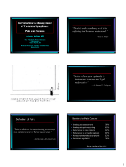


© Copyright 2026