
Living a Normal Life With the Nondominant Hemisphere: Magnetic Resonance
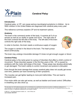
Living a Normal Life With the Nondominant Hemisphere: Magnetic Resonance
Imaging Findings and Clinical Outcome for a Patient With Left-Hemispheric
Hydranencephaly
Stephan Ulmer, Friederike Moeller, Marc A. Brockmann, Johann P.
Kuhtz-Buschbeck, Ulrich Stephani and Olav Jansen
Pediatrics 2005;116;242
DOI: 10.1542/peds.2004-0425
The online version of this article, along with updated information and services, is
located on the World Wide Web at:
http://pediatrics.aappublications.org/content/116/1/242.full.html
PEDIATRICS is the official journal of the American Academy of Pediatrics. A monthly
publication, it has been published continuously since 1948. PEDIATRICS is owned,
published, and trademarked by the American Academy of Pediatrics, 141 Northwest Point
Boulevard, Elk Grove Village, Illinois, 60007. Copyright © 2005 by the American Academy
of Pediatrics. All rights reserved. Print ISSN: 0031-4005. Online ISSN: 1098-4275.
Downloaded from pediatrics.aappublications.org by guest on June 9, 2014
Living a Normal Life With the Nondominant Hemisphere: Magnetic
Resonance Imaging Findings and Clinical Outcome for a Patient With
Left-Hemispheric Hydranencephaly
Stephan Ulmer, MD*; Friederike Moeller, MD*; Marc A. Brockmann, MD‡;
Johann P. Kuhtz-Buschbeck, MD, PhD§; Ulrich Stephani, MD, PhD㛳; and Olav Jansen, MD, PhD*
ABSTRACT. In hemihydranencephaly, the human
brain lacks 1 complete hemisphere. An occlusion of the
carotid artery, affecting all supplied territories, is
thought to be the underlying mechanism. This extremely
rare disorder, of which only 7 cases have been reported to
date, is thought to occur before the last trimester of
gestation (20th to 27th week), after neural migration but
before synaptogenesis. We report on a 36-year-old man
born at term, with no complications, from nonconsanguineous healthy parents. Cranial computed tomography
had been performed because of left-sided headaches.
Because of the imaging findings, the patient presented at
our institution for additional MRI and clinical testing
(including the Motor Activity Log, Wolf motor function
test, 2-point discrimination test, Purdue pegboard test,
gross motor function test, Physician Rating Scale, and
Aachener aphasia test, including patterns for spontaneous speech, repetition, naming, comprehension, written
language, and the token test). The patient’s disabilities
were related to deficits in fine motor control and reduced
precision. Therefore, the patient was unable to perform
the Purdue pegboard test with his affected hand. According to the Aachener aphasia test, no aphasia could be
demonstrated for this strongly left-handed patient.
Strong mirror movements were found. Cortical reorganization is possible if damage occurs in very early childhood. Motor function and speech were controlled by the
remaining, nonaffected hemisphere, with a remarkable
outcome. Because the damage is thought to occur before
synaptogenesis, existing or prepared cortical areas and
pathways have the potential to execute the lacking functions of the destroyed hemisphere. Pediatrics 2005;116:
242–245; adult outcomes, brain development, brain imaging, cerebral infarction, functional assessment, children.
H
emihydranencephaly is an extremely rare
disorder in which the human brain lacks 1
complete hemisphere. Occlusion of the ipsilateral carotid artery occurring before the last trimester of gestation (20th to 27th week of gestation) is
From the *Section of Neuroradiology, Department of Neurosurgery, and
㛳Department of Neuropediatrics, University Hospital of Schleswig-Holstein, Kiel, Germany; ‡Institute of Radiology, University Hospital of
Schleswig-Holstein, Luebeck, Germany; and §Institute of Physiology, Christian Albrechts University, Kiel, Germany.
Accepted for publication Oct 14, 2004.
doi:10.1542/peds.2004-0425
No conflict of interest declared.
Reprint requests to (S.U.) Institute of Radiology, University Hospital of
Schleswig-Holstein, Ratzeburger Allee 160, 23538 Luebeck, Germany. Email: [email protected]
PEDIATRICS (ISSN 0031 4005). Copyright © 2005 by the American Academy of Pediatrics.
242
thought to be the underlying mechanism for this
brain damage, which affects all of the supplied territories.1,2 It is still not known why this disorder
affects only 1 hemisphere. The circle of Willis does
not seem to be able to compensate for the demands in
the developing brain. In the adult brain, occlusion of
a brain-supplying vessel results in a territorial infarction; postischemic defects and gliotic changes can be
observed on MRI scans. Here, the circle of Willis and
sufficient leptomeningeal collateral vessels can compensate for vessel occlusion, so that only partial territorial infarction occurs. In principle, these mechanisms should also work in the prenatal and perinatal
periods. Therefore, fetal predisposing conditions,
leading to additional vascular aplasia with vessel
occlusion, are suspected for patients with hemihydranencephaly. The brain damage is assumed to occur after neural migration and before synaptogenesis.3 Only 7 cases have been reported to date.
With perinatal brain damage, there is always an
attempt to predict clinical outcomes according to
computed tomographic and/or MRI findings.4–11
However, even for patients with large parenchymal
defects, clinical outcomes may be excellent.2,12
CASE REPORT
We present the case of a 36-year-old man born at term, with no
complications, from nonconsanguineous healthy parents, as the
second son among 3 otherwise healthy children. The parents
noticed some clumsiness in early childhood. Right-sided hemiparesis and an equinus deformity at the ankle were diagnosed, with
surgical treatment at the age of 18 years. During childhood, the
patient received physical therapy. Additional sensorimotor and
language development during childhood was not restricted. At
the age of 28 years, the patient experienced his only general
seizure. The rest of his medical history was unremarkable. Our
patient was and is not receiving any medication. Intellect and
language were unimpaired; the patient could complete school and
is now working in a security department. He reported some
disabilities concerning fine motor control of his affected right
hand in tasks of daily living but had no other complaints. At the
time of our investigation, a computed tomographic study had
been performed at a different institution, where the patient had
presented with left-sided headaches and nuchal pain. Because of
his symptoms and the imaging findings, the patient presented at
our institution.
We performed MRI and magnetic resonance angiography with
a 1.5-T scanner (Magnetom Vision; Siemens, Erlangen, Germany).
MRI scans demonstrated a nearly complete absence of the left
hemisphere, which was replaced by cerebrospinal fluid. Only a
small residual hippocampus, a small rim of the occipital cortex,
and a small cerebral peduncle attributable to Wallerian degeneration could be identified on the left side (Fig 1). However, the
cerebellum, which is usually supplied by the posterior vascular
PEDIATRICS Vol. 116 No. 1 July 2005
Downloaded from pediatrics.aappublications.org by guest on June 9, 2014
Fig 1. Coronal, T1-weighted, MRI scans (A and B), T2-weighted MRI scans (sagittal in C and axial in D and E), and a time-of-flight
angiogram (F) for a patient with hemihydranencephaly. There was a nearly complete absence of the left hemisphere, which was replaced
by cerebrospinal fluid. Only a small residual hippocampus, a small rim of the occipital cortex (black arrows), and a small cerebral
peduncle (black arrowhead) could be identified on the left side. However, the cerebellum, which is usually supplied by the posterior
vascular territory, was completely normal. Time-of-flight angiography (F) demonstrated the absence of the left internal carotid artery
(white dot). The right internal carotid artery was without pathologic findings, separating into the middle cerebral artery and the anterior
cerebral artery. Because of moderately but chronically increased intracranial pressure, the bone of the skull was narrowed (white arrows).
territory, was completely normal. Time-of-flight angiography
demonstrated the absence of the left internal carotid artery.
Because conventional grading of hemiparesis (as proposed by
the Medical Research Council in 196413) was thought to be too
inaccurate to describe the extent of motor disability, we performed
additional motor and language tests.
Self-assessment was performed with the Motor Activity Log.14
The patient reported that he is able to drive a car with a standard
transmission. Although natural prehension movements are almost
unimpaired, the patient is unable to open a door with a key,
button or unbutton clothing, switch between television programs
with a remote control, turn the pages of a newspaper, use a spoon
or fork while eating, put on socks, or brush his hair with the
disabled hand.
For the upper extremity, the Wolf motor function test,15 2-point
discrimination test, and Purdue pegboard test16,17 were used to
examine gross and fine motor function. In the Wolf motor function
test, the patient scored 64 of 85 possible points (75%), because of
reduced speed and/or precision in almost all of the performed
tasks and because of impaired fine motor control in 3 tasks.
Functionality and quality of movement did not differ in terms of
the score. The threshold for 2-point discrimination in pinprick
testing was 10 mm for the examined dermatomes (C5–C7) on the
affected side. In the Purdue pegboard test, the patient was unable
to perform any required task with the affected hand. Strong mirror
movements were found when the patient was moving both
hands.18
For the lower extremity, we performed the gross motor function test, part E.19 Because of incomplete performance and the
inability to jump with the affected leg alone, the patient scored 93
of 100 possible points on the gross motor function test.
With the Physician Rating Scale,20 only 10 of a total of 14 points
were scored, because of impairment in the patient’s ankle.
For language testing, the Aachener aphasia test,21 a standardized, German, reference test for spontaneous speech, repetition,
naming, comprehension, and written language, was administered,
including the token test. The results confirmed our impression
that speech was not affected, according to the criteria for aphasia
of the Aachener aphasia test. The token test was completed correctly. The patient is strongly left-handed (92%, according to the
handedness test described by Milner et al22).
DISCUSSION
Hemihydranencephaly is a rare disorder. To our
knowledge, only 7 other cases have been reported in
the literature.2,23–29 Hemiparesis on the contralateral
side was observed in 5 of those cases,2,25–29 mild
mental retardation in 4,2,25,26,28,29 severe retardation
in 1,27 and seizures in 1.27 In two publications,23,24
the clinical outcome was not reported for 1 patient,
who died at the age of 4 years, as a result of bronchopneumonia.23 The immature brain may compensate for neuronal injury through cortical reorganization that is superior to such capacities in the adult
brain.12,30–33
Our patient is remarkable for several reasons.
First, according to previous reports, severe hemiparesis would have been expected, considering the subtotal hemispheric damage.4,5,7,34,35 Second, the left
hemisphere is thought to be the dominant side, harboring speech areas; however, speech is not impaired. Third, living with only 1 hemisphere could
lead to severe intellectual impairment; however, our
patient has completed school and is integrated fully
in social life. Fourth, according to the current literature, this is the oldest patient alive and healthy,
living an almost normal life.
According to the results of the clinical tests, especially the Wolf motor function test, disabilities were
related to deficits in fine motor control, as predicted
with the Motor Activity Log, and reduced speed
and/or reduced precision of the performed tasks and
inability to perform 3 of the tasks. Quality of motion
was not significantly worse with respect to functionality if the patient was able to execute a movement.
Fine motor control requires unaffected 2-point dis-
EXPERIENCE AND REASON
Downloaded from pediatrics.aappublications.org by guest on June 9, 2014
243
crimination; therefore, the patient was unable to perform the tasks of the Purdue pegboard test with his
affected right hand.
Mirror movements are thought to be caused by
loss of transcallosal inhibition.36,37 Among patients
with cerebral palsy, studies with transcranial magnetic brain stimulation revealed that branching corticospinal fibers projecting both ipsilaterally and contralaterally were associated with strong mirror
movements.31,38,39
In congenital hemiparesis, motor function can be
controlled by the nonaffected hemisphere through
ipsilateral pathways.12,30–33 In their transcranial magnetic brain stimulation study, Carr et al31 demonstrated projections innervating both left and right
motoneuron pools of homologous muscles simultaneously, with distally pronounced mirror movements.
Our findings are in accordance with those of others, demonstrating that cortical reorganization is
possible if the damage occurs in very early childhood.30,40 However, Ogden40 reported that severe
deficits of complex extrapersonal orientation ability,
spatial memory, and higher cognitive visuospatial
skills might occur as a result of right-hemispheric
speech dominance. He concluded that a shift of language representation to the right side might cause
“crowding of function” in the remaining hemisphere, which might lead to deficits in spatial memory and orientation. Our patient demonstrates that
normal development of verbal skills after left-hemispheric brain damage does not necessarily compromise memory or spatial orientation.
We cannot predict with certainty from the morphometric features whether a child will develop impaired motor and/or higher cognitive functions.
Some patients with only minor lesions are hampered
severely, and it is still unclear what determines clinical outcomes. Activation of already existing pathways, development of new connections, or axonal
migration or sprouting may be responsible for patients being able to achieve almost normal skills.
Because the lesion is thought to occur before synaptogenesis,3 it seems most likely that already existing
or prepared pathways that have the potential to execute the lacking functions of the destroyed hemisphere are used in this disease. Although there must
be a genetic predisposition to define the precentral
gyrus as the motor cortex, cortical areas may exist in
the immature brain that can adopt similar or contralateral functions and enable cortical plasticity,
with protocols executed through the uncrossed corticospinal fibers.
REFERENCES
1. Myers RE. Cerebral ischemia in the developing primate fetus. Biomed
Biochim Acta. 1989;48:S137–S142
2. van Doornik MC, Hennekam RC. Hemi-hydranencephaly with favourable outcome. Dev Med Child Neurol. 1992;34:454 – 458
3. Eyre JA, Miller S, Clowry GJ, Conway EA, Watts C. Functional corticospinal projections are established prenatally in the human foetus, permitting involvement in the development of spinal motor centres. Brain.
2000;123:51– 64
4. Wiklund LM, Uvebrant P, Flodmark O. Computed tomography as an
adjunct in etiological analysis of hemiplegic cerebral palsy, II: children
born at term. Neuropediatrics. 1991;22:121–128
244
5. Wiklund LM, Uvebrant P. Hemiplegic cerebral palsy: correlation between CT morphology and clinical findings. Dev Med Child Neurol.
1991;33:512–523
6. Molteni B, Oleari G, Fedrizzi E, Bracchi M. Relation between CT patterns, clinical findings and etiological factors in children born at term,
affected by congenital hemiparesis. Neuropediatrics. 1987;18:75– 80
7. Kotlarek F, Rodewig R, Brull D, Zeumer H. Computed tomographic
findings in congenital hemiparesis in childhood and their relation to
etiology and prognosis. Neuropediatrics. 1981;12:101–109
8. Steinlin M, Good M, Martin E, Banziger O, Largo RH, Boltshauser E.
Congenital hemiplegia: morphology of cerebral lesions and pathogenetic aspects from MRI. Neuropediatrics. 1993;24:224 –229
9. Uvebrant P. Hemiplegic cerebral palsy: aetiology and outcome. Acta
Paediatr Scand Suppl. 1988;345:1–100
10. Cioni G, Sales B, Paolicelli PB, Petacchi E, Scusa MF, Canapicchi R. MRI
and clinical characteristics of children with hemiplegic cerebral palsy.
Neuropediatrics. 1999;30:249 –255
11. Niemann G, Wakat JP, Krageloh-Mann I, Grodd W, Michaelis R. Congenital hemiparesis and periventricular leukomalacia: pathogenetic aspects on magnetic resonance imaging. Dev Med Child Neurol. 1994;36:
943–950
12. Kuhtz-Buschbeck JP, Dreesmann M, Golge M, Stephani U. Prenatal
infarction of the left middle cerebral artery: a case report of excellent
functional outcome. NeuroRehabilitation. 2000;15:167–173
13. Monrad-Krohn GHRS. The Clinical Examination of the Nervous System.
12th ed. New York, NY: Harper and Row; 1964
14. Taub E, Miller NE, Novack TA, et al. Technique to improve chronic
motor deficit after stroke. Arch Phys Med Rehabil. 1993;74:347–354
15. Wolf SL, LeCraw DE, Barton LA, Jann BB. Forced use of hemiplegic
upper extremities to reverse the effect of learned nonuse among chronic
stroke and head-injured patients. Exp Neurol. 1989;104:125–132
16. Tiffin J, Asher EJ. The Purdue pegboard: norms and studies of reliability
and validity. J Appl Psychol. 1948;32:234 –247
17. Reddon JR, Gill DM, Gauk SE, Maerz MD. Purdue pegboard: test-retest
estimates. Percept Mot Skills. 1988;66:503–506
18. Woods BT, Teuber HL. Mirror movements after childhood hemiparesis.
Neurology. 1978;28:1152–1157
19. Russell DJ, Rosenbaum PL, Cadman DT, Gowland C, Hardy S, Jarvis S.
The gross motor function measure: a means to evaluate the effects of
physical therapy. Dev Med Child Neurol. 1989;31:341–352
20. Koman LA, Mooney JF III, Smith B, Goodman A, Mulvaney T. Management of cerebral palsy with botulinum-A toxin: preliminary investigation. J Pediatr Orthop. 1993;13:489 – 495
21. Huber W, Poeck K, Weniger D, Willmes K. Aachener Aphasietest: AAT.
Goettingen, Germany: Hogrefe; 1983
22. Milner B, Branch C, Rassmussen T. Observations on cerebral dominance. In: De Rueck AVS, O’Conner M, eds. Ciba Foundation Symposium
on Disorders of Language. London, United Kingdom: Churchill; 1964
23. Muir CS. Hydranencephaly and allied disorders: a study of cerebral
defect in Chinese children. Arch Dis Child. 1959;34:231–246
24. Warkany I. Congenital Malformations: Notes and Comments. Chicago, IL:
Year Book Medical; 1971
25. Moser RP, Seljeskog EL. Unilateral hydranencephaly: case report. Neurosurgery. 1981;9:703–705
26. Suzuki M, Seki H, Yoshimoto T. Unilateral hydrocephalus combined
with occlusion of the ipsilateral internal carotid artery. Surg Neurol.
1985;24:27–30
27. Ohtsuka H. A rare patient with a false median cleft lip associated with
multiple congenital anomalies. Ann Plast Surg. 1986;17:155–160
28. Porro G, Wittebol-Post D, de Graaf M, Van Nieuwenhuizen O, SchenkRootlieb AJ, Treffers WF. Development of visual function in hemihydranencephaly. Dev Med Child Neurol. 1998;40:563–567
29. Greco F, Finocchiaro M, Pavone P, Trifiletti RR, Parano E.
Hemihydranencephaly: case report and literature review. J Child Neurol.
2001;16:218 –221
30. Cao Y, Vikingstad EM, Huttenlocher PR, Towle VL, Levin DN. Functional magnetic resonance studies of the reorganization of the human
hand sensorimotor area after unilateral brain injury in the perinatal
period. Proc Natl Acad Sci USA. 1994;91:9612–9616
31. Carr LJ, Harrison LM, Evans AL, Stephens JA. Patterns of central
motor reorganization in hemiplegic cerebral palsy. Brain. 1993;116:
1223–1247
32. Lewine JD, Astur RS, Davis LE, Knight JE, Maclin EL, Orrison WW Jr.
Cortical organization in adulthood is modified by neonatal infarct: a
case study. Radiology. 1994;190:93–96
33. Nirkko AC, Rosler KM, Ozdoba C, Heid O, Schroth G, Hess CW.
MRI FINDINGS AND OUTCOME IN HEMIHYDRANENCEPHALY
Downloaded from pediatrics.aappublications.org by guest on June 9, 2014
34.
35.
36.
37.
Human cortical plasticity: functional recovery with mirror movements.
Neurology. 1997;48:1090 –1093
Eliasson AC, Gordon AM, Forssberg H. Basic co-ordination of manipulative forces of children with cerebral palsy. Dev Med Child Neurol.
1991;33:661– 670
Forssberg H, Eliasson AC, Redon-Zouitenn C, Mercuri E, Dubowitz L.
Impaired grip-lift synergy in children with unilateral brain lesions.
Brain. 1999;122:1157–1168
Nass R. Mirror movement asymmetries in congenital hemiparesis: the
inhibition hypothesis revisited. Neurology. 1985;35:1059 –1062
Heinen F, Kirschner J, Fietzek U, Glocker FX, Mall V, Korinthenberg R.
Absence of transcallosal inhibition in adolescents with diplegic cerebral
palsy. Muscle Nerve. 1999;22:255–257
38. Carr LJ. Development and reorganization of descending motor pathways in children with hemiplegic cerebral palsy. Acta Paediatr Suppl.
1996;416:53–57
39. Farmer SF, Harrison LM, Ingram DA, Stephens JA. Plasticity of central
motor pathways in children with hemiplegic cerebral palsy. Neurology.
1991;41:1505–1510
40. Ogden JA. Visuospatial and other “right-hemispheric” functions after
long recovery periods in left-hemispherectomized subjects. Neuropsychologia. 1989;27:765–776
QUALITY OF CARE BY NEONATAL NURSE PRACTITIONERS: A REVIEW
OF THE ASHINGTON EXPERIMENT
“The Ashington experiment, an innovative neonatal service run entirely by
advanced neonatal nurse practitioners (ANNPs), has been evaluated. This is a
report of that evaluation and a review of the benefits, hazards, and implications of
nurse practitioner–led services. . . . In summary, the Ashington experience has
shown that ANNPs can provide a high standard of neonatal care without a doctor
on site; it has established the conditions needed for a successful introduction of this
model of care; it has highlighted the difficulties in measuring quality of care (a
lesson that undoubtedly applies to other aspects of health care as well as neonatology); and it has presented a challenge to the health professions to create and test
innovative ways of sustaining services that otherwise might have to be closed.”
Hall D, Wilkinson AR. Arch Dis Child Fetal Neonatal Ed. 2005;90:F195–F200
Noted by JFL, MD
EXPERIENCE AND REASON
Downloaded from pediatrics.aappublications.org by guest on June 9, 2014
245
Living a Normal Life With the Nondominant Hemisphere: Magnetic Resonance
Imaging Findings and Clinical Outcome for a Patient With Left-Hemispheric
Hydranencephaly
Stephan Ulmer, Friederike Moeller, Marc A. Brockmann, Johann P.
Kuhtz-Buschbeck, Ulrich Stephani and Olav Jansen
Pediatrics 2005;116;242
DOI: 10.1542/peds.2004-0425
Updated Information &
Services
including high resolution figures, can be found at:
http://pediatrics.aappublications.org/content/116/1/242.full.ht
ml
References
This article cites 36 articles, 10 of which can be accessed free
at:
http://pediatrics.aappublications.org/content/116/1/242.full.ht
ml#ref-list-1
Citations
This article has been cited by 5 HighWire-hosted articles:
http://pediatrics.aappublications.org/content/116/1/242.full.ht
ml#related-urls
Subspecialty Collections
This article, along with others on similar topics, appears in
the following collection(s):
Neurology
http://pediatrics.aappublications.org/cgi/collection/neurology
_sub
Psychiatry/Psychology
http://pediatrics.aappublications.org/cgi/collection/psychiatry
_psychology_sub
Permissions & Licensing
Information about reproducing this article in parts (figures,
tables) or in its entirety can be found online at:
http://pediatrics.aappublications.org/site/misc/Permissions.xht
ml
Reprints
Information about ordering reprints can be found online:
http://pediatrics.aappublications.org/site/misc/reprints.xhtml
PEDIATRICS is the official journal of the American Academy of Pediatrics. A monthly
publication, it has been published continuously since 1948. PEDIATRICS is owned, published,
and trademarked by the American Academy of Pediatrics, 141 Northwest Point Boulevard, Elk
Grove Village, Illinois, 60007. Copyright © 2005 by the American Academy of Pediatrics. All
rights reserved. Print ISSN: 0031-4005. Online ISSN: 1098-4275.
Downloaded from pediatrics.aappublications.org by guest on June 9, 2014


© Copyright 2026