
Biology For Dummies
AP Biology
FOR
DUMmIES
‰
AP Biology
FOR
DUMmIES
‰
by Peter Mikulecky, PhD
Advanced Science Teacher, Fusion Learning Center
Michelle Rose Gilman
Founder/CEO Fusion Learning Center
Brian Peterson
Science Department Chair, Fusion Learning Center
AP Biology For Dummies®
Published by
Wiley Publishing, Inc.
111 River St.
Hoboken, NJ 07030-5774
www.wiley.com
Copyright © 2008 by Wiley Publishing, Inc., Indianapolis, Indiana
Published simultaneously in Canada
No part of this publication may be reproduced, stored in a retrieval system, or transmitted in any form or by any means,
electronic, mechanical, photocopying, recording, scanning, or otherwise, except as permitted under Sections 107 or 108
of the 1976 United States Copyright Act, without either the prior written permission of the Publisher, or authorization
through payment of the appropriate per-copy fee to the Copyright Clearance Center, 222 Rosewood Drive, Danvers, MA
01923, 978-750-8400, fax 978-646-8600. Requests to the Publisher for permission should be addressed to the Legal
Department, Wiley Publishing, Inc., 10475 Crosspoint Blvd., Indianapolis, IN 46256, 317-572-3447, fax 317-572-4355, or
online at http://www.wiley.com/go/permissions.
Trademarks: Wiley, the Wiley Publishing logo, For Dummies, the Dummies Man logo, A Reference for the Rest of Us!, The
Dummies Way, Dummies Daily, The Fun and Easy Way, Dummies.com and related trade dress are trademarks or registered
trademarks of John Wiley & Sons, Inc. and/or its affiliates in the United States and other countries, and may not be used
without written permission. All other trademarks are the property of their respective owners. Wiley Publishing, Inc., is not
associated with any product or vendor mentioned in this book.
LIMIT OF LIABILITY/DISCLAIMER OF WARRANTY: THE PUBLISHER AND THE AUTHOR MAKE NO REPRESENTATIONS
OR WARRANTIES WITH RESPECT TO THE ACCURACY OR COMPLETENESS OF THE CONTENTS OF THIS WORK AND
SPECIFICALLY DISCLAIM ALL WARRANTIES, INCLUDING WITHOUT LIMITATION WARRANTIES OF FITNESS FOR A PARTICULAR PURPOSE. NO WARRANTY MAY BE CREATED OR EXTENDED BY SALES OR PROMOTIONAL MATERIALS. THE
ADVICE AND STRATEGIES CONTAINED HEREIN MAY NOT BE SUITABLE FOR EVERY SITUATION. THIS WORK IS SOLD
WITH THE UNDERSTANDING THAT THE PUBLISHER IS NOT ENGAGED IN RENDERING LEGAL, ACCOUNTING, OR
OTHER PROFESSIONAL SERVICES. IF PROFESSIONAL ASSISTANCE IS REQUIRED, THE SERVICES OF A COMPETENT
PROFESSIONAL PERSON SHOULD BE SOUGHT. NEITHER THE PUBLISHER NOR THE AUTHOR SHALL BE LIABLE FOR
DAMAGES ARISING HEREFROM. THE FACT THAT AN ORGANIZATION OR WEBSITE IS REFERRED TO IN THIS WORK AS
A CITATION AND/OR A POTENTIAL SOURCE OF FURTHER INFORMATION DOES NOT MEAN THAT THE AUTHOR OR
THE PUBLISHER ENDORSES THE INFORMATION THE ORGANIZATION OR WEBSITE MAY PROVIDE OR RECOMMENDATIONS IT MAY MAKE. FURTHER, READERS SHOULD BE AWARE THAT INTERNET WEBSITES LISTED IN THIS WORK
MAY HAVE CHANGED OR DISAPPEARED BETWEEN WHEN THIS WORK WAS WRITTEN AND WHEN IT IS READ.
For general information on our other products and services, please contact our Customer Care Department within the U.S.
at 800-762-2974, outside the U.S. at 317-572-3993, or fax 317-572-4002.
For technical support, please visit www.wiley.com/techsupport.
Wiley also publishes its books in a variety of electronic formats. Some content that appears in print may not be available in
electronic books.
Library of Congress Control Number: 2008920598
ISBN: 978-0-470-22487-8
Manufactured in the United States of America
10 9 8 7 6 5 4 3 2 1
About the Authors
Peter Mikulecky: Peter Mikulecky grew up in Milwaukee, an area of Wisconsin unique for its
high human-to-cow ratio. After a breezy four-year tour in the Army, Peter earned a BS in biochemistry and molecular biology from the University of Wisconsin – Eau Claire and a PhD in
biological chemistry from Indiana University. With science seething in his DNA, he sought to
infect others with a sense of molecular wonderment. Having taught, tutored, and mentored
in classroom and laboratory environments, Peter was happy to find a home at Fusion
Learning Center and Fusion Academy. There, he enjoys persuading students that biology
and chemistry are in fact fascinating journeys, not entirely designed to inflict pain on
hapless teenagers. His military training occasionally aids him in this effort.
Michelle Rose Gilman: Michelle Rose Gilman is most proud to be known as Noah’s mom
(Hi Munch!). A graduate of the University of South Florida, Michelle found her niche early
and at 19 was already working with emotionally disturbed and learning disabled students in
hospital settings. At 21 she made the trek to California, and there she found her passion for
helping teenage students become more successful in school and life. What started as a small
tutoring business in the garage of her California home quickly expanded and grew to the
point where traffic control was necessary on her residential street.
Today, Michelle is the Founder and CEO of Fusion Learning Center and Fusion Academy, a
private school and tutoring/test-prep facility in Solana Beach, California, serving over 2,000
students per year. She is the author of ACT For Dummies, Precalculus For Dummies, and other
books on self-esteem, writing, and motivational topics. Michelle has overseen dozens of programs over the last 20 years, focusing on helping kids become healthy adults. She currently
specializes in motivating unmotivatable adolescents, comforting their shell-shocked parents,
and assisting her staff of 35 teachers.
Michelle lives by the following motto: “There are people content with longing; I am not one
of them.”
Brian Peterson: Brian remembers a love for science going back to his own AP Biology high
school class. At the University of San Diego, Brian majored in Biology and minored in
Chemistry, with a pre-med emphasis. Before embarking to medical school, Brian took a youngadult-professional detour and found himself at Fusion Learning Center/Fusion Academy, an
independent private school, where he quickly discovered a love of teaching. Years later, he
finds himself the Science Department Head at Fusion and oversees a staff of 11 science teachers. Brian, also known as “Beeps” by his favorite students, is committed to encouraging the
love of science in his students by offering unique and innovative science curricula.
Dedication
We would like to dedicate this book to all the students who motivate us to be better teachers,
and to everyone who has waited patiently for us while we wrote this book: We are now ready
to play!
Authors’ Acknowledgments
Thanks to Bill Gladstone from Waterside Productions for being an amazing agent and for
not only giving us the opportunity to write this book but believing enough in us to continue
giving us more. You’re a brave man, Bill! Thanks to our Wiley editors, Stacy Kennedy and,
especially, Jennifer Connolly for her constant humor, compassion, and salt lick.
Publisher’s Acknowledgments
We’re proud of this book; please send us your comments through our Dummies online registration form located at
www.dummies.com/register/.
Some of the people who helped bring this book to market include the following:
Acquisitions, Editorial, and Media Development
Composition Services
Project Editor: Jennifer Connolly
Project Coordinator: Kristie Rees
Acquisitions Editor: Stacy Kennedy
Layout and Graphics: Claudia Bell, Carrie A. Cesavice,
Reuben W. Davis, Stephanie D. Jumper
Copy Editor: Jennifer Connolly
Technical Editor: Barry Ludvik
Senior Editorial Manager: Jennifer Ehrlich
Proofreaders: Laura Albert, Susan Moritz,
Evelyn W. Still
Indexer: Galen Schroeder
Editorial Supervisor: Carmen Krikorian
Editorial Assistants: Erin Calligan Mooney, Joe Niesen,
Leeann Harney, David Lutton
Cover Photos: © David Chasey/Getty Images
Cartoons: Rich Tennant (www.the5thwave.com)
Publishing and Editorial for Consumer Dummies
Diane Graves Steele, Vice President and Publisher, Consumer Dummies
Joyce Pepple, Acquisitions Director, Consumer Dummies
Kristin A. Cocks, Product Development Director, Consumer Dummies
Michael Spring, Vice President and Publisher, Travel
Kelly Regan, Editorial Director, Travel
Publishing for Technology Dummies
Andy Cummings, Vice President and Publisher, Dummies Technology/General User
Composition Services
Gerry Fahey, Vice President of Production Services
Debbie Stailey, Director of Composition Services
Contents at a Glance
Introduction.................................................................................1
Part I: Doing Your Best for the Test ...............................................5
Chapter 1: Dissecting the AP Biology Exam..............................................................................................7
Chapter 2: Strategies, Suggestions, and Schedules................................................................................13
Part II: Molecules and Cells ........................................................21
Chapter 3: Biochemistry: The Little Things That Count .......................................................................23
Chapter 4: Answering Questions on Biochemistry................................................................................45
Chapter 5: The Cell’s Structure: Factories at Work ................................................................................55
Chapter 6: Answering Questions about Cell Structure..........................................................................65
Chapter 7: Dividing Cells and Conquering the Processes.....................................................................71
Chapter 8: Answering Questions about Cell Functions.........................................................................81
Chapter 9: Cell Respiration: Energized and at the Ready!.....................................................................89
Chapter 10: Answering Questions about Cell Respiration..................................................................103
Part III: Living Large — Organisms and Populations ..................111
Chapter 11: Rooting Through Plant Life ................................................................................................113
Chapter 12: Answering Questions about Plant Life .............................................................................129
Chapter 13: Animals and Behavior.........................................................................................................139
Chapter 14: Answering Questions About Animals and Behavior.......................................................175
Chapter 15: Taxonomy and Classification.............................................................................................185
Chapter 16: Answering Questions on Taxonomy and Classification .................................................197
Chapter 17: Getting Along in the World: Ecology .................................................................................205
Chapter 18: Answering Questions About Ecology ...............................................................................219
Part IV: Inheriting and Evolving ................................................227
Chapter 19: Heredity: Looking Like Your Parents ................................................................................229
Chapter 20: Answering Questions About Heredity ..............................................................................237
Chapter 21: Genetics: Getting Down to DNA Level ..............................................................................245
Chapter 22: Answering Questions on Genetics ....................................................................................257
Chapter 23: Evolving: Past, Present, and Future ..................................................................................267
Chapter 24: Answering Questions on Evolution...................................................................................277
Part V: Putting It All into Practice, or,
Practicing What Has Been Preached..........................................287
Chapter 25: Test 1.....................................................................................................................................289
Chapter 26: Answers to Test 1 ................................................................................................................307
Chapter 27: Test 2.....................................................................................................................................313
Chapter 28: Answers to Test 2 ................................................................................................................333
Part VI: The Part of Tens...........................................................337
Chapter 29: Ten Terms to Tattoo on Your Brain...................................................................................339
Chapter 30: Ten Pathways (and Cycles)................................................................................................343
Chapter 31: Ten Organelles to Know .....................................................................................................347
Chapter 32: Ten Points on Plants and Animals ....................................................................................351
Index.......................................................................................355
Table of Contents
Introduction .................................................................................1
About This Book.........................................................................................................................1
Foolish Assumptions .................................................................................................................2
How This Book Is Organized.....................................................................................................2
Part I: Doing Your Best for the Test ...............................................................................2
Part II: Molecules and Cells.............................................................................................2
Part III: Living Large — Organisms and Populations ...................................................3
Part IV: Inheriting and Evolving......................................................................................3
Part V: Putting It All into Practice, or, Practicing What Has Been Preached ............3
Part VI: The Part of Tens .................................................................................................3
Icons Used in This Book............................................................................................................3
Where to Go from Here..............................................................................................................4
Part I: Doing Your Best for the Test ................................................5
Chapter 1: Dissecting the AP Biology Exam......................................................................7
Proving Your College Prowess..................................................................................................7
Getting to the Guts of the Exam ...............................................................................................8
The multiple-choice questions .......................................................................................8
The free-response questions...........................................................................................8
Ticking through the Topics Covered .......................................................................................8
Trying to Decide: To Guess or Not to Guess ..........................................................................9
Getting the Skinny on Scoring ..................................................................................................9
Tabulating the multiple-choice section .......................................................................10
Adding up the free-response section ...........................................................................10
Reaching the composite score ....................................................................................10
Packing Your Tools for the Test .............................................................................................11
Requesting Special Modifications ..........................................................................................11
Chapter 2: Strategies, Suggestions, and Schedules ......................................................13
Training For a Marathon, Not a Sprint ..................................................................................13
Taking Care of Yourself Before the Test ................................................................................14
Staying active .................................................................................................................14
Eating well .......................................................................................................................14
Learning to relax.............................................................................................................14
Staying away from artificial study aids (drugs) .........................................................15
Acing the Questions.................................................................................................................15
The multiple-choice questions .....................................................................................15
The free-response questions.........................................................................................18
Four Stress-Busters to Help You Survive During the Test ..................................................18
Counting to four .............................................................................................................19
Stretching ........................................................................................................................19
Practicing visualization .................................................................................................19
Thinking positively.........................................................................................................19
xii
AP Biology For Dummies
Part II: Molecules and Cells.........................................................21
Chapter 3: Biochemistry: The Little Things That Count .................................................23
Matters of Matter: Biology Emerges from Chemistry..........................................................23
An elegance of elements................................................................................................24
The importance of being wet........................................................................................25
The Power of Carbohydrates .................................................................................................27
Sweet! Sugars for life ......................................................................................................27
Storing fuels for later .....................................................................................................29
Amino Acids, Proteins, and Enzymes ....................................................................................29
Running metabolism ......................................................................................................30
Building the blocks of life..............................................................................................33
Parts and Processes of Nucleic Acids ...................................................................................35
Forming RNA and DNA...................................................................................................35
Using the genetic code to make proteins ....................................................................36
Chapter 4: Answering Questions on Biochemistry ........................................................45
Pointers for Practice................................................................................................................45
Testing Your Knowledge..........................................................................................................46
Answering multiple-choice questions .........................................................................46
Answering free-essay response questions ..................................................................48
Lab on diffusion and osmosis .......................................................................................48
Lab on enzyme catalysis ...............................................................................................49
Checking Your Work ................................................................................................................51
Chapter 5: The Cell’s Structure: Factories at Work ........................................................55
Categorizing Cells ....................................................................................................................55
Poking around prokaryotes ..........................................................................................55
You, a eukaryote.............................................................................................................55
Powerhouse Parts: Cell Structures ........................................................................................56
Prokaryotic cell structure .............................................................................................56
Eukaryotic cell structure...............................................................................................58
Crossing the Border: Six Ways to Transport ........................................................................60
Chapter 6: Answering Questions about Cell Structure..................................................65
Pointers for Practice................................................................................................................65
Testing Your Knowledge..........................................................................................................66
Answering multiple-choice questions .........................................................................66
Answering free-essay response questions ..................................................................69
Checking Your Work ................................................................................................................69
Chapter 7: Dividing Cells and Conquering the Processes ............................................71
Stages and Phases: The Cell Cycle.........................................................................................71
Between phases of cell divisions: Interphase.............................................................72
Splitting into Two: Mitosis and Cytokinesis................................................................73
Shuffling Genetic Information.................................................................................................77
Meiosis I...........................................................................................................................77
Meiosis II..........................................................................................................................78
Table of Contents
Chapter 8: Answering Questions about Cell Functions.................................................81
Pointers for Practice................................................................................................................81
Testing Your Knowledge..........................................................................................................82
Answering multiple-choice questions .........................................................................82
Answering free-essay response questions ..................................................................84
Lab on mitosis and meiosis ..........................................................................................84
Checking Your Work ................................................................................................................85
Chapter 9: Cell Respiration: Energized and at the Ready! ............................................89
Powering Up..............................................................................................................................89
Photosynthesis ...............................................................................................................89
Cellular Respiration .......................................................................................................93
More in Stored Energy .............................................................................................................97
Storage molecules ..........................................................................................................97
Tapping into energy .......................................................................................................98
Coupling Gets a Reaction ........................................................................................................99
The Electron transport chain (ETC) ............................................................................99
Balancing act: coupled reactions and chemiosmosis................................................99
Chapter 10: Answering Questions about Cell Respiration..........................................103
Pointers for Practice..............................................................................................................103
Testing Your Knowledge........................................................................................................104
Answering multiple-choice questions .......................................................................104
Answering free-essay response questions ................................................................106
Lab on cell respiration.................................................................................................106
Checking Your Work ..............................................................................................................107
Part III: Living Large — Organisms and Populations ...................111
Chapter 11: Rooting Through Plant Life ..........................................................................113
Weeding Through Plant Parts...............................................................................................114
Roots ..............................................................................................................................115
Shoots ............................................................................................................................115
Stems..............................................................................................................................116
Leaves ............................................................................................................................116
Sucking Up Nutrients: How Roots Function and Grow......................................................117
Root Systems ................................................................................................................117
Transpiring and thriving .............................................................................................117
Zoned for development ...............................................................................................120
Reaching for the Sky: How Shoots Function and Grow .....................................................121
Shooting for the sky .....................................................................................................121
Reacting to conditions and hormones ......................................................................123
The Rhythm of the light: Photoperiodism, phototropism, and steps
in photosynthesis .....................................................................................................124
Let there be light ..........................................................................................................124
Heading to the dark side .............................................................................................125
Sowing the Seeds: Reproduction in Plants .........................................................................125
The seedy side of life ...................................................................................................125
Seedless wonders .........................................................................................................127
xiii
xiv
AP Biology For Dummies
Chapter 12: Answering Questions about Plant Life......................................................129
Pointers for Practice..............................................................................................................129
Testing Your Knowledge........................................................................................................130
Answering multiple-choice questions .......................................................................130
Answering free-essay response questions ................................................................132
Lab on plant pigments and photosynthesis .............................................................132
Lab on transpiration ....................................................................................................134
Checking Your Work ..............................................................................................................135
Chapter 13: Animals and Behavior ..................................................................................139
The Coming and Going of Energy.........................................................................................140
Hunting or gathering....................................................................................................140
Digesting ........................................................................................................................140
Using or storing energy ...............................................................................................140
Excreting waste.............................................................................................................141
Staying normal: The importance of homeostasis ....................................................141
Approaching by Systems ......................................................................................................144
Circulatory system.......................................................................................................144
Nervous system............................................................................................................150
Musculoskeletal System ..............................................................................................158
Digestive system...........................................................................................................160
Endocrine system.........................................................................................................161
Immune system.............................................................................................................164
Doing it Again: Reproduction ..............................................................................................165
Getting ready: Gametogenesis ....................................................................................166
Creating an embryo......................................................................................................167
Developing an embryo.................................................................................................168
Acting Like an Animal: Moving, Communicating, and Learning.......................................170
Three ways to move.....................................................................................................170
Four ways to communicate.........................................................................................170
Seven ways to learn ....................................................................................................171
Nine Ways to Behave ...................................................................................................172
Chapter 14: Answering Questions About Animals and Behavior ..............................175
Pointers for Practice..............................................................................................................175
Testing Your Knowledge........................................................................................................177
Answering Free-Essay Response Questions .......................................................................180
Lab on Physiology of the Circulatory System ....................................................................180
Checking Your Work ..............................................................................................................181
Chapter 15: Taxonomy and Classification ......................................................................185
Organizing Organisms ...........................................................................................................185
From kingdoms to species ..........................................................................................185
Characteristics that count .........................................................................................187
What’s in a Name....................................................................................................................189
Monera...........................................................................................................................189
Protista ..........................................................................................................................190
Plantae ...........................................................................................................................192
Fungi...............................................................................................................................193
Animalia.........................................................................................................................194
Chapter 16: Answering Questions on Taxonomy and Classification ........................197
Pointers for Practice..............................................................................................................197
Answering Multiple-Choice Questions ................................................................................198
Answering Free-Essay Response Questions .......................................................................200
Checking Your Work ..............................................................................................................201
Table of Contents
Chapter 17: Getting Along in the World: Ecology .........................................................205
Homing in on Biomes.............................................................................................................205
Using Climates to Carve Out Different Biomes ..................................................................206
Climate governors ........................................................................................................206
Aquatic biomes.............................................................................................................206
Terrestrial biomes ........................................................................................................209
Impacting Survival .................................................................................................................210
Earning a Trophy for Understanding Trophic Levels........................................................213
Producing, or being, food............................................................................................213
Consuming, or Eating Produced Energy ...................................................................213
Taking care of leftovers ...............................................................................................214
Being Part of the Team ..........................................................................................................214
Filling to capacity .........................................................................................................215
Finding your niche .......................................................................................................217
Living together and liking it — or not........................................................................217
Chapter 18: Answering Questions About Ecology........................................................219
Pointers for Practice..............................................................................................................219
Answering Multiple-Choice Questions ................................................................................221
Answering Free-Essay Response Questions .......................................................................222
Lab on Dissolved Oxygen and Aquatic Primary Productivity..........................................222
Checking Your Work ..............................................................................................................224
Part IV: Inheriting and Evolving .................................................227
Chapter 19: Heredity: Looking Like Your Parents .........................................................229
Mendel’s Meddling Pays Off .................................................................................................229
Continuing to meddle ..................................................................................................230
Breaking Mendel’s law: Linked genes ........................................................................232
Pedigrees and probabilities ........................................................................................234
Knowing Something about Chromosomes..........................................................................234
The importance of X ....................................................................................................234
Sex-linked disorders ....................................................................................................235
Inactivating X ................................................................................................................235
Chapter 20: Answering Questions About Heredity.......................................................237
Pointers for Practice..............................................................................................................237
Answering Multiple-Choice Questions ................................................................................239
Answering Free-Essay Response Questions .......................................................................240
Checking Your Work ..............................................................................................................240
Chapter 21: Genetics: Getting Down to DNA Level ......................................................245
Looking at the Genetic Machinery ......................................................................................245
Using the Genetic Code ...............................................................................................246
Expressing genes ..........................................................................................................247
Changing Genes When They Don’t Fit Quite Right ............................................................248
Mutating for a reason – or not ....................................................................................248
Enhancing, repressing, inducing ...............................................................................249
Replicating Viruses and Bacteria .........................................................................................250
Taking a look back at retroviruses and prions .........................................................251
Of plasmids and phages ..............................................................................................251
Engineering Genes .................................................................................................................252
Gel electrophoresis ......................................................................................................252
Polymerase chain reaction..........................................................................................252
xv
xvi
AP Biology For Dummies
Microarray technology ................................................................................................253
Cloning...........................................................................................................................254
The promise of gene therapy......................................................................................254
Chapter 22: Answering Questions on Genetics ............................................................257
Pointers for Practice..............................................................................................................257
Answering Multiple-Choice Questions ................................................................................260
Answering Free-Essay Response Questions .......................................................................261
Lab on the Genetics of Organisms .......................................................................................261
Checking Your Work ..............................................................................................................263
Chapter 23: Evolving: Past, Present, and Future............................................................267
How Life Got Here ..................................................................................................................267
The big picture: Macroevolution................................................................................269
The details: Microevolution ........................................................................................269
How We Know It’s So .............................................................................................................270
Fossil records................................................................................................................271
Living proof: DNA evidence in plants and animals ..................................................271
Continuing to Grow and Change ..........................................................................................272
Patterns of evolution ...................................................................................................272
Drifting, selecting, and adapting ................................................................................274
Chapter 24: Answering Questions on Evolution............................................................277
Pointers for Practice..............................................................................................................277
Answering Multiple Choice Questions ................................................................................278
Answering Free-Essay Response Questions .......................................................................280
Lab on Population Genetics and Evolution ........................................................................280
Checking Your Work ..............................................................................................................282
Answers and Brief Explanations for the Review Questions .............................................283
Part V: Putting It All into Practice, or, Practicing
What Has Been Preached ..........................................................287
Chapter 25: Test 1 ................................................................................................................289
Multiple-Choice Questions....................................................................................................291
Four Free-Response Questions with Grading Outline .......................................................305
Chapter 26: Answers to Test 1 ..........................................................................................307
Chapter 27: Test 2 ................................................................................................................313
Multiple-Choice Questions....................................................................................................315
Free-Response Questions with Grading Outline ................................................................329
Chapter 28: Answers to Test 2 ..........................................................................................333
Part VI: The Part of Tens ...........................................................337
Chapter 29: Ten Terms to Tattoo on Your Brain..............................................................339
Hypothesis ..............................................................................................................................339
Energy......................................................................................................................................339
Table of Contents
Ecosystem ...............................................................................................................................339
Emergent Property.................................................................................................................340
Cells .........................................................................................................................................340
Evolution .................................................................................................................................340
Diversity ..................................................................................................................................340
Phylogeny................................................................................................................................341
Homeostasis ...........................................................................................................................341
Feedback .................................................................................................................................341
Chapter 30: Ten Pathways (and Cycles) .........................................................................343
Photosynthesis.......................................................................................................................343
Aerobic Cellular Respiration ................................................................................................343
Anaerobic Cellular Respiration ............................................................................................343
Cell Cycle.................................................................................................................................344
Mitosis .....................................................................................................................................344
Meiosis ....................................................................................................................................344
DNA Replication .....................................................................................................................345
Transcription ..........................................................................................................................345
Translation..............................................................................................................................345
Trophic Relationships ........................................................................................................345
Chapter 31: Ten Organelles to Know ...............................................................................347
Nucleus....................................................................................................................................347
Plasma Membrane ................................................................................................................347
Cell Wall...................................................................................................................................348
Endoplasmic Reticulum ........................................................................................................348
Mitochondria ........................................................................................................................348
Chloroplast .............................................................................................................................348
Ribosome ................................................................................................................................349
Golgi Apparatus......................................................................................................................349
Lysosome ................................................................................................................................349
Vacuole ....................................................................................................................................349
Chapter 32: Ten Points on Plants and Animals .............................................................351
Meristems ...............................................................................................................................351
Roots and Shoots ...................................................................................................................351
Plant Vasculature ...................................................................................................................352
Photoperiodism ....................................................................................................................352
Respirocirculatory System ...................................................................................................352
Immune System ......................................................................................................................353
Musculoskeletal System ........................................................................................................353
Digestive System ....................................................................................................................353
Nervous and Endocrine Systems .........................................................................................354
Reproductive System.............................................................................................................354
Index .......................................................................................355
xvii
xviii
AP Biology For Dummies
Introduction
W
ait, how did you get here?
That’s a reasonable question. Take a seat, unfurl your map, and review what you know:
You’re smart. Maybe you forgot about that. Let the title of this book roll off your back
like water from a duck in a raincoat. You wouldn’t be anywhere near the AP Biology
exam if you didn’t have the goods. Believe it.
You’re a little nervous. The College Board sits atop Mount Olympus, hurling thunderbolts at you, sneering at your hubris:
“So you think you’re going to get college credit? First you must pass a little test we
have devised for you . . . bwah-hah-hah-hah!”
Peals of laughter thunder down from the summit. Gulp.
You have — or soon will have — a plan. Your AP Biology course is part of it. This book
is another part. Ignore the thundering laughter, and put one foot in front of the other.
Sure, eventually you’ll have to face the judgment of the Board. But a lot is going to
happen between now and then. By the time you have to fight for your Five, you’ll be
ready. In the meantime, stick to your plan.
Remember, the Board may be a jealous and vengeful god, but biology is your friend. Biology
just asks reasonable questions, like:
Wait, how did I get here?
About This Book
Like biology, we are your friends. No matter how you got here, or how this book came to you,
we’re here to help. We’ve broken things down into sections that correspond to just the kind
of content and questions you’ll see on the AP Biology exam. We’ve emphasized those things
on which you’re more likely to be tested, and tread lightly over those things that you probably won’t see on test day. You have enough on your plate.
Each chapter reviews big ideas, and highlights those nitty-gritty, rubber-meets-the road
details that tend to pop up in test questions. Although you may want to start at the beginning
and soldier your way through to the end, that’s not absolutely necessary. Based on your
experiences in the AP Biology course, you’ve probably got a pretty good idea of your
strengths and weaknesses. Feel free to pick and choose.
Maybe you don’t know what your weaknesses are. That’s okay, too. Each chapter is accompanied by a follow-up chapter that summarizes the most important points and provides
questions to help you figure out where you stand. If you’re using this book as you go through
the AP Biology course, you’ll find it easy to flip to the topic of the moment. Please do.
2
AP Biology For Dummies
And when you begin to hear the thunder from the summit, we’ve got you covered. We’ve
included two full-length practice tests. Use them to make final adjustments to your plan, and
to ensure that by the time you sit down for the real thing, you’ll have done it all before.
Twice.
Foolish Assumptions
Yes, we made some assumptions about you while we wrote this book. If you’re reading this
you probably fit into one or more of the following assumptions:
We assume that you’re probably planning to take the AP Biology exam. Otherwise,
you’d probably have better ways to pass the hours than, well, preparing for the AP
Biology exam. But maybe you’re just deciding whether or not to take the AP Biology
course. Or maybe you’re taking the course, but haven’t decided whether to take the
exam. This book will help you with those decisions.
We do not assume that you have aced all of your previous courses in math and science. Whatever math and chemistry you really need to know is presented within. Don’t
sweat it. We’ve got your back.
We assume that you don’t like being bored or judged. Neither do we. This book is
designed to be a streamlined, practical companion that doesn’t take things more seriously than necessary — in short, a good traveling partner.
How This Book Is Organized
Because the AP Biology exam is a standardized test, it tests more than just biology. It tests your
ability to take tests. True story. We’ve accounted for that, and included a handy primer on how
to prepare for the test-taking experience. Once you’re primed, you’re ready to fling yourself
into the biological fray; the bulk of the book reviews the biology emphasized on the exam, in
the proportions described by the College Board’s AP Biology syllabus. Each chapter of new
material is followed by a chapter that summarizes the key points, provides practice questions,
and gives a concise review of any AP Biology labs associated with that material. Finally, there
are the practice tests. They’re not going anywhere, so just take them when you feel ready.
Part I: Doing Your Best for the Test
Should I even take the AP Biology exam? What’s it like? How do they score it? Will they try to
trick me? What can I do to defend myself? Are vomit bags provided? These are the sober
questions of a scientific mind, and we take them seriously.
Part II: Molecules and Cells
Start at the source, and things seem simpler. Biology may cover millions of species, but they
begin to look more and more alike when you put them under a microscope. By starting with
the small stuff — the really small stuff — differences in the bigger bits just seem to make a
whole lot more sense. Back when the earth was steamy and soupy, when biology was just
waking up and taking a look around, life was a story of molecules and cells. The tricks of the
trade that evolved back then still apply now.
Introduction
Part III: Living Large — Organisms
and Populations
Once you’ve got a handle on what cells do with molecules, you already understand the
basics behind what organisms do with organs. Things take a turn for the dramatic when you
start talking about what organisms do to each other. This section is thick and rich, because
the AP Biology exam dwells on these topics at great length. You might want to dally a bit
longer in this part, too.
Part IV: Inheriting and Evolving
Whether we’re talking cells or stegosauruses, the name of the game is evolution. It’s really
the only rule by which biology plays. What evolution looks like depends on how far back you
stand and how much you squint your eyes. By the time you get through with this Part, you
may feel a warm sense of kinship with bacteria.
Part V: Putting It All into Practice, or,
Practicing What Has Been Preached
Oh, right — there was something about a test, wasn’t there? This Part contains two fulllength, timed practice tests, modeled on the actual AP Biology exam. And even if you guess
on a lot of the questions, we won’t leave you guessing. We provide full answer keys with
explanations. And remember, there are two tests; if you crash and burn on the first, there is
room for redemption.
Part VI: The Part of Tens
Wouldn’t it be nice if there were some sort of really nice summary of the big ideas and the
must-know bits? Yes, it would be. That is the purpose of this Part. It contains three concise,
ten-part summaries of the pithy good stuff.
Icons Used in This Book
Every now and then, you may catch a glimpse of something in the margins. We meant to do
that. Included throughout the book are helpful icons, little pointers that help you navigate
through the material.
The remember icon is a subtle but strong suggestion that you file the indicated material in
that separate folder of your brain reserved for things you’ll have to use later.
This icon flags information that may help you save time, prevent mistakes or generally make
life easier on yourself.
3
4
AP Biology For Dummies
When you see this icon, relax. The material you’re reading may seem particularly involved or
esoteric. That’s because it is. If it interests you, please dig in, but if not, feel free to move on.
This icon is just what it says. It flags the kind of information that you ignore at your own
peril. If we could make it our own peril, we would. But we can’t, so we use this icon.
Where to Go from Here
Start with the first two chapters. They’ll help you to figure out where you stand with respect
to the AP Biology exam, and to formulate your plan of action.
Then, flip through the biology review to regain a sense of what you already know, what
you’ve never seen, and what you’ve long since forgotten. Let yourself glide through the material that’s easy for you, saving time and energy for the parts that make you close your eyes,
massage your temples, and curse the Board that sits atop Olympus.
However you start, remember that this book is not an assignment — it is a tool. Use it in the
way that helps you the most. Follow your plan. After the smoke of the AP Biology exam
clears to reveal your stellar score, you’ll regard that plan with affection, thinking, “Ah yes,
that’s how I got here.”
Part I
Doing Your Best for the Test
P
In this part . . .
ut a picture in a different frame, and the picture looks
different somehow. The same is true of biology —
frame it within an AP exam, and it somehow seems a little
different. Before you take that AP Biology exam out for a
drive, you might want to kick the tires, pop the hood, and
see how it’s put together. Think of this part as AP Exam
Mechanics 101. You want to be so familiar with the ins,
outs, and idiosyncrasies of the exam that by the time you
take it you don’t notice anything but the biology. You
want to see the picture, not the frame.
In this part, we delve into the structure of the exam and
how you can use that structure to your advantage. What’s
more, we give you tips and strategic pointers on how to
pace your preparation. Proper pacing will ensure that you
review what you need when you need it, without burning
out and without scrambling at the last minute. By following the well-scheduled plan we’ll help you create, you’ll
have calm, quiet confidence when you need it most. When
the sun rises on test day, you’ll feel its warmth — not its
shadow.
Chapter 1
Dissecting the AP Biology Exam
In This Chapter
Understanding why you take this exam
Breaking down the exam into all its parts
Guessing or not
Getting the skinny on the scores
Packing your bags: What to take and not take to the test
Preparing for unusual circumstances: What to do if you need special assistance
Y
ou really didn’t know what the insides of the frog looked like before you opened it up,
did you? Well, consider this first chapter the dissection of the AP Exam. As we slice
and cut through the test, you will begin to get familiar with why you should (or shouldn’t)
take this test, the test’s structure, how you will be scored, what’s allowed on test day and
what to do if you require special assistance. Many students are intimidated by the AP tests,
but the Wizard of Oz was also scary before the curtain flew open! So sit back, relax, get your
scalpel out, and open this thing up!
We do a lot of talking in this chapter about the AP Exam, but if you’re taking AP Biology now
or getting ready to, keep in mind that while this book can be a great support throughout your
course, the AP Exam is the end game, so you need to know this info, too!
Proving Your College Prowess
Taking the AP test is basically telling those good folks at any college that you can already
think and perform on the college level. Although taking an AP Biology course is not required
to take the AP test (although we highly recommend you do), your high score on the test (see
the skinny on the scoring information below) signifies that you understand advanced material and already possess what it takes to be successful in college. Go you! By getting a high
score, you also have the opportunity to receive credit or advanced standing at most universities around the country. You should take this test if Biology is “your thing” and you want the
world to know.
If you received poor grades in Biology in high school and could care less about what’s inside
a cell, you might want to consider spending your time on a subject you truly enjoy. Those
smart AP creators have AP tests in a variety of different subjects, so if you’re not sure about
your dedication to this particular test, you might want to visit their Web site and glance at
the other subjects offered. If you’re just taking an AP Biology class and don’t plan on taking
the exam at the end, then that’s cool too. This book will definitely help you succeed in an AP
Biology class.
Because you bought this book, however, we will assume you are a natural Biologist and that
after studying this book you will bust out of your high school body and become your true
self: the college student that you were meant to be!
8
Part I: Doing Your Best for the Test
Getting to the Guts of the Exam
The AP Biology Exam takes three hours and includes two parts: an 80-minute, 100-question
multiple choice section; a 100-minute writing period broken down into a 10-minute reading
period, and a 90-minute four-question, free-response section. After you complete the multiple
choice section you get a short break.
You can’t return to the multiple choice section after you hand it in, so make certain you are
satisfied before you stretch your legs!
The multiple-choice questions
We have a feeling that the AP bigwigs couldn’t agree on what types of problems and questions
to include in the multiple choice section. As a result, they just decided to throw in everything.
The multiple choice questions that you will encounter cover a broad range of topics. You can
expect to answer basic factual information as well as heavy-duty, thought-provoking problems. You have basically 48 seconds per question on the multiple-choice section, and to get a
great score on the exam, you need to correctly answer around 60 questions or more (more
information on tackling these types of questions in Chapter 2).
The free-response questions
The second section of the exam is the writing section. Of 100 minutes you read for 10 minutes, after which you get 90 minutes to complete an essay period. You’re given four broad
essay questions. On average, one essay covers material relating to molecules and cells, one
essay covers heredity and evolution, and — lucky you — you get two essays covering organisms and populations. Remember that at least one of the essays will ask you to analyze
experimental data and perhaps design an experiment. So pay close attention when we talk
about the labs in each chapter (more information on tackling these types of problems in
Chapter 2).
Ticking through the Topics Covered
Before writing the AP exam, a few Biology geniuses studied the Biology curricula of many of
the nation’s best colleges. They came away from the study with a clear understanding of
which high-level concepts were being taught to Biology college students around the country.
From their research they decided that the AP exam should cover three main areas. But
because you deserve to know everything, we break these three areas down even further, outlining every subheading in the test, as well as the percentage of the test attributed to it:
Molecules and Cells (25 percent of test):
• Chemistry of Life: Water, organic molecules in organisms, free energy changes,
enzymes (7 percent)
• Cells: Prokaryotic and eukaryotic cells, membranes, subcellular organization
(10 percent)
• Cellular Energetics: Coupled reactions, fermentation, and cellular respiration
(8 percent)
Heredity and Evolution (25 percent of test):
• Heredity: Meiosis and gametogenesis, eukaryotic chromosomes, inheritance
patterns (8 percent)
Chapter 1: Dissecting the AP Biology Exam
• Molecular Genetics: RNA and DNA structure and function, gene regulation, mutation, viral structure and replication, nucleic acid technology and applications
(9 percent)
• Evolutionary Biology: Early evolution of life, evidence for evolution, mechanisms
of evolution (8 percent)
Organisms and Populations (50 percent of test): As you can see, Organisms and
Populations covers half the test. So, when studying, you know where to place your
attention.
• Diversity of Organisms: Evolutionary patterns, survey of the diversity of life,
phylogenetic classification, evolutionary relationships (8 percent)
• Structure and Function of Plants and Animals: Reproduction, growth and development, structural, physiological and behavioral adaptations (32 percent)
Notice that 32 percent of the test is Structure and Function of Plants and Animals.
That’s a big chunk, so when studying later, make sure to pay careful attention to
this section.
• Ecology: Population dynamics, communities and ecosystems, global issues
(10 percent)
Trying to Decide: To Guess or Not to Guess
Here’s our advice on guessing: Guess and don’t guess. Not the answer you wanted? Let us
explain. The test folks don’t take away points for answers left blank, but they do remove a
portion of points for wrong answers (we get into more of the scoring of the test in the section, “Getting the Skinny on Scoring”). If you can whittle away the obvious wrong answers
and make a really good educated guess, then we say go for it. Never, we repeat, never
answer a question if you have absolutely no idea where to begin. You don’t get dinged for
leaving a blank answer, but you do suffer the point loss for the wrong answer. If you have
some knowledge of the question and can eliminate two to three obvious wrong answers,
informed guessing is definitely to your advantage.
Getting the Skinny on Scoring
After all is said and done, you end up with a final composite score between 1 and 5. Those
smart AP folks did a lot of researching to figure out how to score you. They periodically compare the performance of AP students with that of college students tested on the same material. Basically, a grade of 5 on the AP test is comparable to a college student earning an A in
their college level Biology course. A grade of 4 on the AP test is like receiving a B in a college
level class, and so on. An AP score of 3 or higher would qualify you for college credit because
it would be the equivalent of earning a middle C or higher if you were to take that class in college. The College Board likes to put it this way:
5: Extremely well qualified
4: Well qualified
3: Qualified
2: Possibly qualified
1: No recommendation
Before you receive a composite score, however, a lot of calculating goes on to get that
number. Take a look at the breakdown in the following sections.
9
10
Part I: Doing Your Best for the Test
Tabulating the multiple-choice section
Chapter 2 discusses the multiple choice and free response questions at length. This section
is all about the final numbers. Once you know the scoring and then get the skinny on how to
tackle the questions in Chapter 2, we feel strongly that you will score high and feel great!
The multiple choice section is scored by a computer. The computer counts the amount of
wrong answers and subtracts .25 points for each wrong answer from the amount of right
answers. Any question that you didn’t answer is counted as 0. So basically, the formula for
scoring this section is:
Multiple-choice score = Number of correct answers – (number of wrong answers × .25)
Adding up the free-response section
Your essays are read by top-secret, highly trained professionals — think “secret service” of
the College Board. No one knows the identity of these professionals. Isn’t this getting exciting? Your four essays are read by four different secret people — that’s four, highly trained,
hidden, underground readers just for you! Each scorer is trained to score just one essay
question on the exam. They truly become experts in that one question. Each essay is scored
on a scale from 1 to 10. The secretive AP leaders assure us that they have ample checkdouble-check systems in place to ensure that all essays are scored fairly.
Reaching the composite score
Okay, after you have two sets of scores (one score for the multiple-choice section and one
score for the free-response section) they’re put together to get your composite score and
your final number. The highest composite score for the AP Biology test is 150. The multiplechoice score makes up 60 percent, or 90 points, of the 150 total. The free-response section is
40 percent, or 60 points, of the 150 total. There is no definitive composite score range that is
consistent from year to year or even from subject to subject. So we can’t give the exact formula used to determine your score. But we can take a look at a prior year’s range of scores
and get a hint as to where you might find your score.
In a recent year composite scores between
108 to 150 received a 5
93 to 107 received a 4
72 to 92 received a 3
43 to 71 received a 2
0 to 42 received a 0
This range changes every year. Why, you ask? Well, the answer the College Board gave to us
is more difficult to interpret than the essay section of the test, so the fact remains: The range
changes every year, and that’s that!
Chapter 1: Dissecting the AP Biology Exam
Packing Your Tools for the Test
When packing to go on vacation, you don’t want to forget your favorite piece of clothing or
that hair product you can’t live without. Packing for the test is the same thing, albeit not as
fun. Although all of the following aren’t mandatory to bring to the test, we highly encourage
you bring all of them. It’s always better to pack more, not less. Here we list what’s important
to make sure you bring with you to the test site:
Pencils with erasers: Pencils with erasers are best for the multiple-choice test.
Black pens: Black is a better choice because it is a bolder color.
Your social security number: This identifies you.
Photo ID: Take this in case they think you paid your best friend to take the test for you!
A watch: You need to pay close attention to the time.
Snacks and drinks: Quiet snacks are better than loud snacks. Go with soft snacks that
won’t disrupt others, like chewy granola bars. Water is a better choice than popping a
loud soda.
Your knowledge: ’Nuff said.
Appropriate clothing: Bring a light jacket in case there’s an arctic freeze in the test
center.
Something to wipe your nose if you should drip: You don’t want to be leaving every ten
minutes, and nobody wants to be distracted by sleeve wipes or sniffing. Bring tissues!
We said over-packing for a vacation is a good thing, but remember that there are usually luggage restrictions on planes. You can’t bring everything. The following items are definite no
nos. You will even be thrown out of a test site for bringing some of these. So, leave ’em home,
or in your car. Here we list what not to bring to the test:
Scratch paper: The good folks at AP thought of that already. You get what you need at
the test center.
Notes, books, dictionaries, highlighters, cheat sheets: Keep that all at home.
Electronic devices like cell phones, beepers, mp3 players: Sorry, but you can’t take
the test while listening to The Black Eyed Peas.
Your best friend to take the test for you: They frown on that.
Computers or calculators: Nope, sorry.
Your parents for moral support: Leave the folks at home.
Requesting Special Modifications
Not everyone takes the AP test under the same conditions. You may have a special circumstance that can allow you to change the date of the test or the way you take the exam. Here is
a brief list of special circumstances and how they affect your AP test:
11
12
Part I: Doing Your Best for the Test
Learning disabilities: If you have a diagnosed learning disability, you may be able to get
special accommodations. You may have extended time, large print, a reader, frequent
breaks, among other things, but you must specifically request this on your application
form. You should make sure that your school has a SSD (Services for Students with
Disabilities) Coordinator’s Form on file with the College Board. You must fill out this
form and send it to the College Board. Allow seven weeks for the pros at College Board
to review your request. Please note that in order to get special testing, you must have
been formally diagnosed with a learning disability by a professional and must have a
current, individualized plan at school. In most cases, the evaluation and diagnostic testing should have taken place within five years of the request for accommodations. You
must also describe the comprehensive testing and techniques used to arrive at the diagnosis, including test results with subtest scores. Your best bet is to log onto the College
Board Web site to see all the up–to-the-minute requirements for accommodations.
Physical disabilities: If you have a physical disability, you may be allowed to take a test
in a special format — in Braille, large print, or on an audiocassette or CD. Follow the
same instructions detailed above and contact the College Board for more information.
Religious obligations: If your religion prohibits you from taking a test on a specific day,
you may test on an alternate date. Again, the College Board folks can guide you in the
right direction for alternate dates.
Military duty: If you’re an active-duty military person, you don’t need to complete the
normal registration form. Instead, ask your Educational Services Officer about testing
through DANTES (Defense Activity for Nontraditional Educational Support).
Chapter 2
Strategies, Suggestions, and Schedules
In This Chapter
Creating a study schedule
Taking care of yourself
Acing the questions with confidence
Reviewing stress busters for D-Day
Y
ou already know a ton about Biology. You’ve probably spent the past year in an intensive AP Biology course. Full comprehension of the material is mandatory when trying
to ace this exam, but it’s only one part of the puzzle (albeit a really big one). In this chapter,
we introduce you to the other important things you can do to help boost your score that
have absolutely nothing to do with studying Bio. So dive right into the superb suggestions,
the super strategies, and the stupendous scheduling.
Training For a Marathon, Not a Sprint
Studying for the AP exam is not the same as cramming the night before a test, and then
promptly forgetting the information. As much as you might want to sprint to the finish line,
the AP test is a process that should evolve over a lengthy period of time. Taking the full yearlong AP Biology class is not a requirement to take the exam (College Board wants everyone
to have access to AP, like home-schooled students, for example), but to get the highest score,
and receive those coveted college credits, we are going to give you a schedule over the
course of two semesters to make sure that you are well prepared for this test:
September: Enroll in an AP Biology class at school. If your school doesn’t offer one,
speak with your counselor to see if you can get your hands on an AP Biology book you
can use. Or perhaps there is an AP class offered at a nearby school that your counselor
will allow you to enroll in. As much as this book will assist you in understanding the
biological concepts addressed on the test, nothing ever really takes the place of a passionate teacher teaching you face to face.
November: After familiarizing yourself over the last few months with the concepts of AP
Biology, take a diagnostic test. You can use one of the sample tests in this book, or you
can go to www.Collegeboard.com and find some there. This will help you pinpoint your
strengths and weaknesses and assist you in guiding you through the rest of the schedule.
December: Begin reading this book from the beginning. You will find that being in the
classroom over the last four months has provided you a wealth of information. You
should already be familiar with many of the concepts covered in the first third of this
book.
January through March: Continue using this book, and take another practice test in
early March. You still have two months until D-Day, so your score on this test will guide
the rest of your studying. Review the material in which you feel you need the most
assistance.
14
Part I: Doing Your Best for the Test
April: Take another practice test around mid-May. Make this your last practice test.
Whatever you do, do not take a practice test the week of the real test. Like a long distance swimmer who has been swimming and practicing all year, she doesn’t do the
race right before the race! If she’s been pacing herself and practicing regularly, she’s
ready for the win!
The night before the test: We really recommend that you not study too hard this night.
You might be tempted to pull an all-nighter, but our experience has shown that if you
don’t know the material the night before, any more studying for this type of test isn’t
going to help. We also recommend that you relax your head the night before. Read a
good book, watch some TV, go out to dinner, and enjoy yourself. If it eases your anxiety
to take a peek at Bio, maybe just look over some notes, glance through this book one
last time, but please do not attempt to cram. You’ll just exhaust yourself right before
you exhaust yourself!
The morning of the test: Eat breakfast, please. You may be nervous and your appetite
might be low, but your brain needs the fuel.
Test day: Take the test.
June: Wait patiently, very patiently for your score.
Taking Care of Yourself Before the Test
Just like a long distance swimmer, you are in training for a long period of time. The swimmer
doesn’t just swim in order to prepare for the race, she takes care of her body, too. Believe it
or not, taking care of your body is just as essential to your success as it is for any athlete.
Staying active
You can’t just be a bookworm for the year before the exam. You must remember that there
are parts other than the brain that still need to be used. Exercise helps all parts of the body,
including the brain. The more you exercise, the more oxygen gets to your brain. More oxygen
to the brain means clearer thinking. So get moving people!
Eating well
Just like exercising, the right food contributes to sustained attention and clear thought. You
want to avoid sugar highs that eventually lead you to come crashing down. Forget those
energy drinks that combine huge amounts of caffeine and sugar to get you to a state of heightened paranoia. They might tell you that you will focus better when you drink those, but you
will only be reading and writing at a superhuman speed and your recall will be nada after you
come down. Your mom was right: Eat your veggies, fruit, and an overall well-balanced diet.
Your brain will be nourished, and in turn will thank you with a score of a 5.
Learning to relax
We have seen students who are so overextended and overachieving that they don’t have any
time for pleasant relaxation. They get stressed out, take anti-anxiety pills, have trouble sleeping and concentrating, get panic attacks, and generally exhaust themselves and get sick. Stop
for a minute, get in a comfy position, and read this next sentence carefully and slowly:
Relaxation is not a luxury; it is a requirement for a well-balanced life.
Chapter 2: Strategies, Suggestions, and Schedules
You’re a multifaceted human. You’re not a work and study robot. Relaxation comes in many
different forms for all kinds of people. Some people are relaxed when they are with friends,
some read books for enjoyment and play music they like, others do Yoga, meditate, or paint.
There is only one requirement when choosing what relaxation tool to use and that’s making
sure that your brain is not running 100 miles an hour. The whole purpose of relaxation is to
give your brain a rest. Your poor brain needs that rest to function properly when called to
duty. So find a relaxing activity you enjoy, thank your brain by telling your brain it can take
some time off, and whether you choose to “oooomm” or paint tulips, make it peaceful.
Staying away from artificial study aids (drugs)
We’re gonna come clean right here: We’ve pulled all-nighters with the assistance of either a
keg of coffee or some over-the-counter caffeine pills, and it wasn’t pleasant. The only thing
that uppers, stimulants, and amphetamines do is make you feel like you could clean every
kitchen in your neighborhood. And you probably could, except these drugs backfire when it
comes to studying. Although they aid in staying awake for longer periods of time, stimulants
make it hard to retain and recall information. They will leave you jittery, anxious, agitated,
and restless. There are some situations when taking stimulants is required, like if you’ve
been diagnosed with ADHD and require them in order to concentrate. But for the rest of us
these drugs just do the opposite. They make you think you are concentrating and studying
really hard, but the reality is that you won’t remember too much of what you’ve studied. If
you’ve been diligent in the studying schedule (see, “Training For a Marathon, Not a Sprint,”
earlier in this chapter), pacing yourself through Biology, then no kind of drug is needed to
help you study harder or better.
Acing the Questions
There are no surprises. Every question on the AP test is based on fundamental knowledge of
the material. If you don’t know the material, the AP folks will know. The AP folks aren’t trying
to trick you. They just want to make sure that you know your stuff. Following the basic guidelines in the following sections will help you to tackle the multiple-choice and free-response
sections of the exam.
You have been studying and preparing for this test for a long time. You know more than you
think you do. Even if you fail to remember every detail of every concept, you know at least
something about it. If you come to the test feeling completely insecure and doubting yourself, take a moment to remember that we told you that you know more than you think. If you
find a question that makes your heart stop with dread, turn that immediately around and say,
“I can do this, I will do this, and I feel good about this question.”
The multiple-choice questions
There are one hundred multiple choice questions covering molecules and cells, heredity
and evolution, and organisms and populations. The list below outlines a few general rules
to stick by:
1. Answer all questions that you can on the first attempt, but if you can’t answer
the question, skip it and move on!
2. Use leftover time to check your answers.
3. First answers are normally the correct ones — go with those.
4. Use guessing to your advantage.
15
16
Part I: Doing Your Best for the Test
As a rule of thumb, go through and answer the types of questions you feel comfortable with
first. Everyone is different, and everyone should follow their own gut.
The multiple choice questions are broken up into the following categories:
Basic-knowledge questions, which include traditional multiple-choice and eliminationtype questions
Elimination-type questions, which are actually a subset of basic knowledge questions
(see bullet above)
Data-interpretation questions, which include graphs and figures
Category questions, which involve a group of statements in which one, more than one,
or none, is true
Basic-knowledge questions
The basic-knowledge questions are just what they claim to be. They’re testing your knowledge of the subject. Each question touches on a different Biology topic. For a traditional multiple-choice question, usually there is a small piece of information given at the beginning of
the question, such as
1. Given a DNA strand of the sequence, 5’-GAC TCA CCA TAA-3’, what would be the sequence
of the corresponding mRNA strand, assuming no introns?
You would be given a list of possible answers. In case you were wondering, the answer to this
question is 3’-CUG AGU GGU AUU-5’.
Elimination questions
To approach an elimination question, read the question and think about what you know
about the topic presented. Then read the choices, eliminating false statements as you go. It
might look something like this:
1. Which of the following is true about enzymes?
I. They speed up the rate of a reaction.
II. They are used up in the reaction.
III. They are usually proteins.
(A) I only
(B) II only
(C) III only
(D) I and III only
(E) I, II, and III are all true
The answer is D, in case you were unsure.
Data-interpretation questions
The data interpretation question gives you either a graph, diagram, data table, or mini experiment. Check out the picture first to give you a sense of what the questions might be asking
of you. Then check out the questions. It might look something like this:
Chapter 2: Strategies, Suggestions, and Schedules
The following question refers to the Figure 2-1.
original
new
population
Figure 2-1:
Type of
selection.
short
long
Length of break
1. What type of selection does this figure represent?
(A) Bimodal
(B) Directional
(C) Stabilizing
(D) Disruptive
This one hard? The answer is B.
Category questions
A category question gives you four to five vocabulary words of a specific category, followed
by the questions which define or describe the given vocabulary terms. It might look like this:
(A) Commensalism
(B) Parasitism
(C) Predation
(D) Mutualism
1. Relationship between two organisms in which one benefits and the other is harmed
2. Relationship between two organisms in which one benefits and the other is neither helped
nor harmed
3. Relationship in which both organisms benefit
The answers are, respectively, B, A, and D.
17
18
Part I: Doing Your Best for the Test
The free-response questions
The free-response questions consist of four essays, one covering molecules and cells, one
covering genetics and evolution, and two covering organisms and populations. One of the
four is based on labs.
First, it is important that you make sure you answer the question. You don’t have much time:
Ninety minutes for four essays. So, here are some tips to keep in mind when responding to
the essays:
Start by checking for key-question words, such as explain, describe, and contrast.
Make sure that you plan out your response. Keep in mind that this has to be an essay —
it has to demonstrate that you know your stuff — fill your answer with facts that adequately answer the question.
It may be appropriate to draw diagrams in your response, however you must remember
to refer to them in your essay.
Don’t worry about grammar and spelling, this is a Biology exam and not an English
exam. Grammar problems will not be counted against you.
Each of the main questions has two or three sub-points that the AP Bio folks are looking for. Don’t spend too much time on any one sub-part. Address every single portion
of the question, equally.
Be sure to check out the sample free-response questions at the end of this book.
The lab essay question typically has you design an experiment. Be sure to include all of the
key elements of a good scientific experiment. Define your:
Hypothesis
Control group
Dependent and independent variable(s)
Controlled variables
Data collection
Equations or calculations
Results
Proposed conclusion
Four Stress-Busters to Help
You Survive During the Test
We know that you are going to feel nervous the day of the test. Everyone does. Research has
shown us that a little anxiety is actually good for the brain. Anxiety helps the brain stay ultrafocused and attentive, tells the brain, “Hey, this must be important if I’m feeling this way, so I
better stay alert,” and a little anxiety helps to make sure you get to the test center on time.
However, excess anxiety does the exact opposite. High levels of fear make the brain go into
fight-or-flight mode. So, if you’re feeling too much anxiety during the test, use the stressbusters in the following sections to calm your nerves.
Chapter 2: Strategies, Suggestions, and Schedules
Counting to four
Breathing is grossly underrated. Breathing is good. Take a deep until your belly expands, hold
it in four counts, and then expel the air for four counts. Try not to take shallow breaths, which
can cause you to become even more anxious because your body is deprived of oxygen.
Stretching
Rotate your head around to stretch out and relax your neck muscles. We suggest keeping
your eyes closed so the proctor doesn’t think you are trying to cheat. Hunch and roll your
shoulders to help relax your back and spine. You’ll be sitting for quite some time, so maintaining good posture is crucial. Shake out your hands like you have writer’s cramp. Imagine
that all your tension and stress is going out through your fingertips. Extend and push out
your legs like you’re pushing something away with your heels. Point your toes back toward
your knees and hold that position for a count of three.
Practicing visualization
Don’t do this during the test — you just waste time and lose concentration. However, right
before the exam or during a short break, practice visualization. Close your eyes and imagine
yourself in the test room cheerfully looking at questions that you know the answer to, filling
in the answers, finishing early, and double checking your work. Picture yourself leaving the
exam room all uplifted, then getting your score and rejoicing. Think of how proud of you your
parents are (if thinking about them stresses you out leave this one alone). Imagine not having
to take early Biology in college. Picture yourself driving a fire-engine-red Ferrari ten years
from now, telling the Time magazine reporter in the passenger seat that your success started
with your excellent score on your Biology AP test. The goal is to associate the AP exam with
good feelings.
Thinking positively
Any time you feel yourself starting to panic or thinking negative thoughts, make a conscious
decision to say to yourself, “Stop! Don’t dwell on anything negative.” And then switch over to
a positive track. Suppose you catch yourself thinking, “Why didn’t I pay more attention to
polar covalent bonds?” Change that script to, “I’ve got most of this right, maybe I’ll get this
right, too. No sense worrying now. Overall, I think I’m doing great!”
19
20
Part I: Doing Your Best for the Test
Part II
Molecules and Cells
O
In this part . . .
ne of the overarching themes of biology is that cells
use energy to arrange matter in useful ways. To really
“get” what’s going on with living things, you need to know
something about matter and about how cells use energy to
transform it. That’s what this part is about.
We start by reviewing the kinds of matter you find in cells
and the important ways that matter is arranged — and
re-arranged. This busy shuffling about of the matter within
cells sometimes goes by the name biochemistry. Cells are
professional biochemists. Cells expertly assemble molecules
into elaborate structures called organelles. Organelles are
nature’s way of dividing labor. Each organelle contributes
some specific, critical function to a cell. We tour the major
kinds of cells with an eye to the organelles, seeing how they
all conspire towards life.
The function of an organelle results from its structure.
This is a second overarching theme: function derives from
structure. Things do what they do because of what they
are. That is why cells use energy to re-arrange matter. We
close this part by reviewing the grand dialogue between
photosynthesis and respiration, the two opposing sides to
life’s energy conversation.
Chapter 3
Biochemistry: The Little Things
That Count
In This Chapter
Understanding molecular biology with some basic chemistry
Checking out carbohydrates as a source of energy and as a means of storage
Figuring out how proteins are put together and how they mediate metabolism
Discovering how nucleic acids function for information storage and flow
C
ells are masters of efficiency. The daily business of a cell includes taking in nutrients,
breaking them into useful parts, building things from these parts, recycling anything
handy, and only throwing away a bare minimum. All this busy efficiency requires a lot of
specialized parts and pieces. It requires molecules. How molecules change from one kind to
another is chemistry. How cells use chemistry is biochemistry, which is what we focus on in
this chapter.
Matters of Matter: Biology
Emerges from Chemistry
Here’s a bit of good news. You don’t need to understand a great deal of chemistry in order to
gain a much deeper insight into life at the molecular level. Many biochemical reactions are
just variations on a few simple themes. In other words, a small investment in chemistry pays
huge dividends in biology.
This chapter begins with a review of how atoms come together in order to make molecules,
through the formation of different kinds of bonds. All the important categories of biochemical molecules (carbohydrates, lipids, proteins, and nucleic acids) and other compounds (like
electrolytes) are simply bonded groups of atoms.
Because cells are largely made up of water, you will not be surprised to discover that the
chemical properties of water are vital to biochemistry. Water helps to give cells and big biological molecules their unique shapes, and helps cells keep things running smoothly. You will
find out how.
With the basic understanding of atoms, bonds, molecules, and water we give you in the following sections, you’ll be prepared to launch into the rest of the chapter, which shows how
cells build big molecules by linking together many smaller ones. These big molecules are the
key to the marvelous efficiency with which cells handle information, matter, and energy.
24
Part II: Molecules and Cells
An elegance of elements
You remember what an atom is, right? Hint: a positively charged nucleus of protons and neutrons, enveloped by shells of negatively charged electrons. Different elements have different
numbers of protons, and even atoms of the same element can have slightly different numbers
of neutrons or electrons. Atoms with the same number of protons but differing numbers of
neutrons are isotopes of the same element.
It turns out that some atoms are “happier” (in other words, more stable) when they alter the
number of electrons in their shells. Such atoms have a tendency to either gain, lose, or share
electrons with other atoms. When they do so, these atoms form bonds. The three major
types of bonds in biology are
Ionic
Covalent
Hydrogen bonds
When one atom gives up one or more electrons, it’s good news for other atoms that are seeking additional electrons. This happy situation results in the complete transfer of electrons
from one atom (or group of atoms) to another. Electrons have negative charges, so giving up
electrons leaves an atom with a positive charge. Accepting electrons leaves an atom with a
negative charge. Charged atoms are called ions. Because “positively charged ion” and “negatively charged ion” are phrases with far too many syllables to be convenient, shorter names
exist for them: cations (positive) and anions (negative).
Because opposite charges attract, cations are drawn to anions. When oppositely charged
ions cozy up to one another, we say that they are held together by “ionic bonds.” For reasons
that will be discussed shortly, water molecules are very good at undoing ionic bonds, separating cationic and anionic partners. These separate anions and cations can move about
freely in water, carrying their ionic charges with them. Freely moving ions are called electrolytes, and help cells use electrical energy.
Giving up electrons isn’t always an all-or-nothing process. Sometimes, atoms share electrons.
When they do, these atoms form covalent bonds. Each covalent bond consists of two shared
electrons. Atoms can share two, four, or six electrons to form single, double, and triple covalent bonds.
Not all atoms share their electrons equally. Some atoms are more willing than others to
donate their electrons to a covalent bond. When covalent bonds are formed between atoms
that don’t share equally, we say that those bonds are polar. When the atoms share equally,
the bonds are nonpolar. Figure 3-1 shows some common types of polar and nonpolar covalent bonds. As you’ll soon see, the polarity of a molecule greatly affects how that molecule
interacts with water.
C
Figure 3-1:
Types of
C
covalent
bonds
C
common in
organic mol- –
O
ecules and
their polar
character.
C
C
Nonpolar
H
Nonpolar
O–
Polar
H+
Polar
O–
OH+
Polar
Chapter 3: Biochemistry: The Little Things That Count
Biology likes to use lots of weak bonds because they can be easily made and broken according to the needs of the moment. Hydrogen bonds are a type of weak bond found nearly everywhere in biochemistry. Hydrogen bonds can form when a hydrogen atom is engaged in a
polar covalent bond with another atom — one that doesn’t like to share electrons (oxygen,
for example). In this scenario, since the hydrogen has less than its fair share of electrons, it
carries a partial positive charge. The electron-greedy oxygen carries a partial negative
charge. Imagine now that another oxygen atom enters the picture, carrying its own partial
negative; this new oxygen will be attracted to the partially positive hydrogen, since opposite
charges attract. So, the two oxygens will line up with the hydrogen between them, making a
sort of negative-positive-negative sandwich. Large biological molecules (macromolecules,
such as proteins and nucleic acids) use hydrogen bonds to knit themselves together into
beautiful, functional shapes. With its two polar oxygen-hydrogen bonds (H2O, remember?),
water is the hydrogen-bonder par excellence, as shown in Figure 3-2.
Think of hydrogen bonds, nonpolar covalent bonds, polar covalent bonds, and ionic bonds
as lying along a spectrum of electron-sharing, ranging from the least shared (hydrogen
bonds) to the completely transferred (ionic bonds).
Previous scholarship of Star Trek may have informed you that we humans are carbon-based
life forms. So are bacteria. So is all other terrestrial life. What this means is that biochemistry
is largely organic chemistry, involving the making and breaking of bonds with carbon.
If you remember one thing about carbon, remember that it likes to form four covalent bonds.
Any carbon atom engaged in more or less than four covalent bonds is highly unstable, and
therefore, highly improbable. Really. If you see one on a test, be suspicious.
Other atoms frequently bond with carbon. These atoms include hydrogen, oxygen, nitrogen,
and phosphorous. Between these four atoms and carbon, you can account for most of the
matter in anything that lives. Certain bonded groups of these atoms occur repeatedly in
organic molecules. These popular groups are good at certain chemical jobs, and we call them
functional groups.
The importance of being wet
Aaah, the water molecule . . . so simple, yet so powerful! Just one oxygen atom covalently
bonded to two hydrogen atoms — what’s the big deal? Look a little more closely. Each hydrogen atom engages in a very polar covalent bond with the oxygen. This polarity, combined
with the “Mickey Mouse” shape of the molecule means that water is a dipole, with two distinct positive and negative regions. Since water is nearly everywhere in cells, we might wager
that its properties have profound impact on biology. We’d win that wager, thanks to water’s
ability as a solvent, to its adhesive/cohesive properties, and to its temperature stability.
Water is the solvent of life. If you doubt it, just try not drinking any for a week. Swarms of
water surround and interact with other molecules in cells, such as ions, lipids, carbohydrates, proteins, and nucleic acids. Because it is so polar, water happily hangs out with other
polar molecules. Polar molecules tend to be charged or to have many polar covalent bonds.
Since they work and play well with water, we call these molecules hydrophilic, which means
water-loving. Water nicely dissolves hydrophilic molecules, allowing them to move freely
through the watery cellular environment. On the other hand, nonpolar molecules don’t get
along so well with water. We call these molecules hydrophobic, which means water-fearing. In
water, hydrophobic bits tend to cluster together in order to minimize their contact with
water (see Figure 3-3). Because cells are so watery, being hydrophilic or hydrophobic largely
defines the lifestyle of any biological molecule.
25
26
Part II: Molecules and Cells
+
δ
−
δ
+
δ
−
δ
+
δ
hydrogen
bonds
+
δ
+
δ
−
δ
H2O
−
+
δ
δ
Figure 3-2:
Hydrogen
bonding in
water.
+
δ
+
δ
Don’t let this hydrophilic/hydrophobic business scare you. It all boils down to Italian salad
dressing. Ever notice how a bottle of dressing separates into two layers after it has been
stored for a while in the refrigerator, and you have to shake it before pouring it on your
salad? The two layers consist of oil and water. Oil and water don’t mix. Oil is hydrophobic.
Think of nonpolar molecules as “oily.”
If you’re up for some hydrogen-bonding, water will meet you halfway. And if you’re not willing,
water is perfectly happy to have hydrogen bond with itself, as you can see from Figure 3-2.
Water-water hydrogen bonding is what gives water its “cohesive” properties — water’s tendency to stick to itself. Cohesion gives water its high-surface tension. That’s right: Spiders
creepily walk across ponds because of water-water hydrogen bonds. Hydrogen bonding
between water and other polar molecules explains water’s adhesive properties — water’s tendency to stick to other things. The combination of cohesion, adhesion, and a thin tube results
in capillary action. Water can pull itself easily through small tubes (as in, capillaries) by hydrogen bonding with polar surfaces on the inside of the tube and with itself.
Chapter 3: Biochemistry: The Little Things That Count
Water
Hydrophobic
tail
Figure 3-3:
Phospholipid
bilayer
structure Hydrophilic
that results
head
from
hydrophilic
and
hydrophobic
interactions.
Water
Each of the countless hydrogen bonds flickering into and out of existence within the watery
guts of a cell serves as a little bin in which heat energy can be stored. As a result, water has a
high heat capacity. Water — and therefore, cells —can gain or lose a lot of heat without
changing temperature very much. By helping maintain a stable temperature, water creates a
friendly environment for biochemistry.
The Power of Carbohydrates
Biology uses carbohydrates to store and transport energy. In addition, carbohydrate molecules are the bricks in plant cell walls. Though they occur in numerous forms, the basic formula for all carbohydrates is (CH2O)n. Any carbohydrate molecule is built from multiples of
carbon and water — hence the term carbo-hydrate. Cells use enzymes to change carbohydrates from one form to another and to break them down or build them up, according to the
needs of the moment.
Sweet! Sugars for life
The simplest kind of carbohydrate is the sugar. More complicated carbohydrates are built
up from sugars. A one-sugar carbohydrate is called a monosaccharide. Link two monosaccharides together and you’ve got yourself a disaccharide. Link a whole mess o’ monosaccharides together and you have a polysaccharide.
27
28
Part II: Molecules and Cells
Monosaccharides are named according to the number of carbon atoms they contain.
Monosaccharides containing three, four, five, or six sugars are called trioses, tetroses, pentoses, and hexoses, respectively. Sugary cousins that share a common molecular formula, but
in which the precise molecular structure differs, are called isomers. Sugars can exist in both
linear and circular (cyclical) forms, as shown in Figure 3-4.
(a)
H
H
H
H
OH
H
C
C
C
C
C
OH
OH
OH
H
OH
H
C
O
(b)
H
H
Figure 3-4:
Alternate
forms of the
monosaccharide glucose: (a)
linear, and
(b) cyclic.
C
O
OH
O
H
H
H
O
O
OH
H
O
O
H
OH
HO
OH
Monosaccharides are reversibly built up into polysaccharides or broken down from them by
enzymes. These enzymes speed up “dehydration” and/or “hydrolysis” reactions, depicted in
Figure 3-5. When monosaccharides are linked together by dehydration, a water molecule is
removed in order to form a covalent bond. This same bond can be broken in a hydrolysis
reaction by adding water across the bond, reversing the linkage.
Chapter 3: Biochemistry: The Little Things That Count
Glucose
Fructose
CH2OH
O
H
HO
OH
CH 2
H
H
OH
H
H
OH
H
H
HO
OH
O
OH
OH
CH2OH
H
H2O
Figure 3-5:
Dehydration
reaction
linking
glucose and
fructose
monomers
into the disaccharide
sucrose.
Sucrose
CH2OH
O
H
HO
H
H
OH
H
H
OH
OH
CH 2
O
O
H
OH
H
OH
CH2OH
H
Storing fuels for later
Cells store energy in the form of sugars by forming polysaccharides. Starch and glycogen are
polysaccharides, built of repeating glucose units. Though they each use glucose monosaccharides as building blocks, starch and glycogen differ in the exact linkages between glucose
molecules. When cells have access to plenty of food, they store glucose as starch or glycogen
as a hedge against leaner times. When nutrients are scarce, cells break down starch or glycogen, freeing up glucose molecules to harvest their stored energy. In other words, cells are
responsible little citizens, saving their sugars for rainy days.
Another important glucose-based polysaccharide is cellulose, which is formed in plant cells.
Although cellulose is not used like starch or glycogen for energy storage, it is an important
building material in plant cells.
Amino Acids, Proteins, and Enzymes
Proteins are workhorses of the cell, sticking their amino acid fingers into nearly every bit of
cellular business. Cells use proteins for catalysis, regulation, structure and other roles. This
section focuses on two things: how protein catalysts — enzymes — run cellular metabolism,
and on the ingenious structures (from amino acids through large protein assemblies) that
enable proteins to perform their many functions.
29
Part II: Molecules and Cells
Running metabolism
Simply put, metabolism is the complete set of chemical reactions occuring within an organism. You’re right, that covers a lot of ground. Cells are always breaking things down to get
materials and energy, and then using those materials and energy to build up new, more useful
things. All this deconstruction and reconstruction occurs through chemical reactions. Most
of these chemical reactions involve special kinds of proteins called enzymes.
Enzymes catalyze reactions; this means that they speed up reactions, but are not themselves
changed or used up in the process. One enzyme can repeatedly catalyze the same reaction.
Moreover, the enzyme does not alter the reactant or product of the reaction — it simply
makes the reaction go faster. As you can see in Figure 3-6, enzymes speed up reactions by
lowering the activation energy, the energetic hill a reacting molecule must climb in order to
undergo the reaction. Think of an enzyme as a sort of gymnastics coach, “spotting” molecules as they vault through reactions.
activation
energies
effect of
catalyst
Energy
30
reactants
Figure 3-6:
Enzymatic
reduction of
the activation energy
of a chemical reaction.
products
Reaction Progress
Enzymes bind their substrates (as in, reacting molecules) within an active site, a pocket where
all the chemistry takes place. Binding can occur in a number of ways. In lock-and-key binding,
the substrate slides snugly into a perfectly pre-formed active site. Induced-fit binding features a
flexible active site; when the substrate binds, the active site changes shape to achieve a better
fit. Either way, once bound in an active site, the substrate is in an environment uniquely tailored for the reaction. After the reaction has occurred, an enzyme releases its product(s), and
is free to bind another substrate. The overall process is shown in Figure 3-7.
Chapter 3: Biochemistry: The Little Things That Count
Reaction:
A+B
ule
ec
mol
C
mol
A
ecu
active site
enzyme
molecule B
molecule A
enzyme
molecule C
Figure 3-7:
An enzyme
facilitates a
reaction
by binding
substrates,
catalyzing
chemistry,
and
releasing
products.
enzyme
le B
31
32
Part II: Molecules and Cells
Some of the general types of reaction catalyzed by enzymes are:
Hydrolysis: Adding a water molecule across a covalent bond in order to break that bond.
Dehydration: The reverse of hydrolysis. Forming a new covalent bond by the removal
of a water molecule.
Oxidation-reduction (Redox): A reaction in which electrons are transferred between
reactants.
Exergonic: A reaction that releases energy, and therefore occurs spontaneously.
Endergonic: A reaction that takes up energy, and therefore will not proceed unless that
energy is provided.
Many factors can affect enzyme “activity,” or just how much enzymes speed up reactions.
Some of these factors are general, affecting the activity of most enzymes. Others are specific,
affecting only one or a few types of enzyme. General factors include temperature, pH, and
concentration. More specific factors include “activators” or “inhibitors,” molecules that
increase or decrease the activity of specific target enzymes.
Increasing temperature tends to increase the speed of enzyme-catalyzed reactions, up to a
point. Beyond that point, excessive temperature destroys an enzyme’s intricate structure.
Changes in pH (the acidity or basicity) of the environment can also modify enzyme activity,
especially in the case of reactions that involve the transfer of hydrogen ions. Recall that pH =
–log [H+], so that a pH change of one unit (say, from 7.5 to 6.5) corresponds to a tenfold
change in the concentration of hydrogen ions. Enzymes often have optimum activity at a certain pH, which tapers off in more acidic or more basic conditions.
Because enzyme activity can be so sensitive to pH, cells have an interest in maintaining
stable internal pH conditions. To promote a constant pH, many cellular solutes act as pH
buffers, releasing or taking up hydrogen ions as needed to keep the overall hydrogen ion concentration constant. Different chemicals have differing tendencies to release or take up protons. These differing tendencies can be measured by a quantity called the pKa, or acid
dissociation constant. The pKa is the pH at which one half of a chemical species is in an H+bound form (conjugate acid), and the other half occurs in a form without bound H+ (conjugate
base). Solutions with a high concentration of solute at a given pKa tend to maintain pH at
that pKa value, buffering the solution against pH changes. The relationship between pH
and the concentrations of conjugate acid and base of a given solute are expressed by the
Henderson-Hasselbach equation where [A–] and [HA] are the molar concentrations of conjugate base and conjugate acid, respectively. Notice that when the conjugate acid and base are
present in equal concentration, the pH of the solution equals the pKa of the buffer solute:
pH = pKa + log ([A–]/[HA])
Perhaps the most straightforward way to alter enzyme activity is to change the concentrations of enzyme and/or substrate. Increasing the concentration of either causes more collisions to occur between enzymes and substrates. Each collision has a chance of leading to
binding and reaction. Decreasing concentrations have the opposite effect.
Finally, specific molecules can bind to enzymes in ways that increase or decrease enzyme
activity. Molecules whose binding increases activity are called activators. Molecules whose
binding decreases activity are called inhibitors. Competitive inhibitors bind enzymes at the
active site; this blocks the binding of substrate and prevents catalysis. Noncompetitive
inhibitors bind enzymes at locations other than the active site, but alter the enzyme’s structure. These changes in enzyme structure reduce the ability of the enzyme to bind substrate
or to catalyze a reaction.
Chapter 3: Biochemistry: The Little Things That Count
Building the blocks of life
Proteins must be annoying to other types of molecules. How can proteins do so many things
so well? As numerous and diverse as they are, proteins are built from a humble inventory of
parts: twenty amino acids, common to all known organisms. A covalently linked chain of
these amino acids comprises a polypeptide, and polypeptides “fold” into the functional
structures known as proteins. Proteins can have four distinct levels of structure: primary,
secondary, tertiary, and quaternary.
Amino acids are the building blocks of proteins. Genes, written in DNA, code for the identity
and sequence of the amino acids that make up a protein. As helpfully suggested by their
name, each amino acid contains amine (–NH2) and carboxylic acid (–COOH) functional
groups, attached to a central carbon. Also attached to that carbon are a hydrogen atom and
a unique sidechain. Differences among the sidechains are what make the amino acids an
interesting cast of characters. Different sidechains have different properties. They may be
large or small, hydrophobic or hydrophilic, positively or negatively charged, or uncharged.
As shown in Figure 3-8, individual amino acids are covalently bonded via a dehydration reaction. This reaction removes a water molecule to join the amine group of one amino acid to
the carboxylic acid group of the next. This kind of covalent bond is called a peptide bond.
A long chain of amino acids linked in this way is a bit like a charm bracelet, decorated with
different sidechains. The exact sequence of sidechain “charms” defines a protein’s primary
structure.
Glycine
Alanine
H
H
N
C
O
N
C
H
H
H
OH
C
O
C
H
OH
H
CH3
H2O
Figure 3-8:
Dehydration
reaction to
form a peptide bond
between
two amino
acids.
H
N
H
O
H
H
C
C
N
C
H
O
C
OH
H
CH3
peptide
bond
33
34
Part II: Molecules and Cells
When amino acids are linked into a chain, the peptide bonds between each amino acid pair
form a long backbone. Peptide bond backbones can hydrogen bond with themselves to form
regular, repeating structures such as helices (spirals) and undulating strands. These spiraling
or wavy patterns are the secondary structure of a protein. Secondary structures occur in two
major forms, α-helices and β-strands. β-strands associate with themselves to form β-sheets.
Both the spiraling α-helices and the wavy β-sheets display their amino acid sidechains to the
outside, where they can interact with each other or with water. Sometimes, several polypeptides that have only secondary structure can associate to form “fibrous” proteins.
The amino acid sidechains that protrude from α-helices and β-sheets can interact closely
with each other, packing into compact shapes called tertiary structures. Typically, hydrophobic sidechains end up on the inside of the structure, away from water, while hydrophilic
sidechains tend to cover the outer surface. These kinds of compact structures are typical of
“globular” proteins. Figure 3-9 shows the structure of a typical globular protein, where it is
clear how the secondary structures (the α-helices and β-sheets) have packed together to
form the tertiary structure.
Figure 3-9:
Schematic
of the path
traced by
the peptide
bond backbone in
a protein
tertiary
structure.
Sometimes one polypeptide just isn’t enough to do the job. Many proteins function within
larger assemblies of proteins. These assemblies are held together by noncovalent bonds, and
can vary widely in size and in the number of proteins involved. Quaternary structure refers
to the noncovalent association of multiple proteins.
Chapter 3: Biochemistry: The Little Things That Count
Parts and Processes of Nucleic Acids
Nucleic acids are all about information. Genetic information is stored in the form of deoxyribonucleic acid, or DNA. Working copies of that stored information are made from DNA’s less
stable cousin, ribonucleic acid, or RNA. The major use of genetic information is to direct the
building of proteins. This section describes the structures of DNA and RNA, how enzymes
make copies of these structures, and how they use those copies to make proteins.
Forming RNA and DNA
Like proteins and polysaccharides, nucleic acids are polymers. Compared to the twenty
common amino acids used to build proteins, however, the inventory of parts used to build
nucleic acids is far less diverse. DNA and RNA are each composed from a tiny alphabet of
four nucleotide bases.
Whether DNA or RNA, a nucleotide consists of three parts: a base, a sugar, and a phosphate
group. DNA employs a deoxyribose sugar, while RNA uses ribose. Ribose is nearly identical to
deoxyribose, but contains an extra hydroxyl (i.e., –OH) group. This hydroxyl causes RNA
strands to be far less stable than DNA. It makes sense, then, that DNA is used for long-term
storage of genetic information, while RNA is used to make short-term, working copies of that
information.
The bases of DNA are adenine (A), thymine (T), guanine (G), and cytosine (C). RNA uses the
same bases, with the exception that in RNA, uracil (U) is used in place of thymine. Bases A
and G are larger molecules, called purines. Bases T (or U) and C are smaller molecules, called
pyrimidines. As shown in Figure 3-10, nucleic acid strands are composed of a sugar-phosphate
backbone and a linear sequence of bases. Each strand has a defined direction; by convention,
the strands are said to run in the 5’ to 3’ direction, in reference to numbered carbons of sugars
at the strand tips.
Both DNA and RNA can form double helices. An example of a DNA double helix is shown in
Figure 3-11. Two strands, running in opposite directions, come together to form a structure
resembling a spiral staircase with nucleotide “steps” stacked one atop the next. The two
antiparallel strands (as in, parallel, but in opposite directions) are held together by hydrogen
bonds between bases. Each base on one strand bonds with its “complementary” base on the
opposing strand. Purines pair with pyrimidines, and vice-versa. Bases A and T (or U) form
two hydrogen bonds, while the slightly stronger interaction between G and C consists of
three hydrogen bonds. Here’s a helpful feature of base-pairing: Knowing the sequence of
one strand in a double helix means you know the sequence of the complementary strand.
In the cell, DNA is almost always found as a double helix, whereas RNA frequently exists in
single-stranded form. Complementary strands of DNA and RNA can base-pair to form hybrid
double-helices.
35
36
Part II: Molecules and Cells
NH2
O
C
C
N
CH
HN
C
CH
C
N
H
O
O
CH3
Cytosine
C
C
CH
N
H
O
NH2
O
Thymine
(in DNA)
C
HN
CH
N
C
C
CH
HC
C
N
H
N
Uracil
(in RNA)
O
C
N
CH
N
H
H 2N
Adenine
HN
C
C
C
N
N
CH
N
H
Guanine
Nitrogenous base
nucleotide
Phosphate
Figure 3-10:
General
structure of
a nucleotide
and a depiction of how
nucleotides
are linked
together
into a
nucleic
acid.
Pentose
sugar
–
O
P
O–
O
HOCH2
O
O
H
OH
H
H
H
OH
H
Deoxyribose (in DNA)
O
HOCH2
OH
H
H
OH
OH
H
H
Ribose (in RNA)
nucleic acid
Although DNA is used mostly to store genetic information, RNA is used in many roles. RNA is
used to transmit genetic information (as messenger RNA [mRNA]). RNA is also used in structural and even catalytic roles (as transfer RNA [tRNA], ribosomal RNA [rRNA] or small nucleolar RNA [snRNA]).
Using the genetic code to make proteins
All proteins begin as but a twinkling in the eye of a gene. That twinkling is genetic information, encoded within DNA, the “Master Molecule.” This section is devoted to understanding
how genetic information is copied and transmitted within cells. In DNA replication, the genes
of a cell are copied in order to pass the information on to new cells. To make proteins, the
information in DNA is transcribed (copied within the same language) and translated (converted into a different language). In eukaryotes, processing (editing of the language) occurs
between transcription and translation. Replication, transcription, processing, and translation
are all carried out by groups of highly specialized enzymes.
Chapter 3: Biochemistry: The Little Things That Count
T
A
G
C
A
T
stacking
between
nucleotides
G
A
C
C
C
T
G
G
T
A
C
G
A
T
A
T
G
Figure 3-11:
The DNA
double helix.
A
C
T
37
38
Part II: Molecules and Cells
Replicating DNA
All cells come from other cells. The DNA in every newly born cell is inherited from its parent
cell. In order for both parent and daughter cells to have complete copies of DNA, the DNA
must be replicated (copied). DNA replication is a semi-conservative process, meaning that
each daughter cell receives DNA containing one original strand from the parent, and one
new, complementary strand — think of it as a dowry of sorts. The details of replication, summarized in Figure 3-12, make clear just how this happens.
Perhaps poetically, before DNA can reproduce it must unzip. Unzipping is performed by an
enzyme, helicase, that separates complementary strands of the DNA to be copied. Helicase
opens up a region of unpaired DNA, the strands of which join at the replication fork. The separated strands each serve as a template for making new strands. An enzyme called DNA polymerase makes the new strands, one nucleotide at a time. To begin, the polymerase requires a
short RNA primer, provided by the enzyme primase. Once primed, DNA polymerase moves
along the template strand in the 3’ to 5’ direction, making a new complementary strand, 5’ to
3’. Nucleotides are selected for incorporation in the new strands based on their ability to
pair with their complements on the template strand. So, if the template strand displays a G,
DNA polymerase adds a C; if the template strand displays an A, DNA polymerase adds a T.
And so on.
DNA polymerase chugs along templates in one direction only, 3’ to 5’, making new strands 5’
to 3’. This task is straightforward with one template strand, the leading strand, but more complicated with the complementary template, the lagging strand. On the lagging strand, DNA
polymerase continually bumps up against the replication fork. DNA polymerase must make
the new strand discontinuously, in short stretches called Okazaki fragments. Refer to Figure
3-12 to convince yourself of this. Each Okazaki fragment requires its own RNA primer, and the
fragments must be joined together by another enzyme, DNA ligase. Ligase also substitutes
DNA bases for the RNA bases of the primers. All the enzymatic busywork necessary with the
lagging strand takes time, and this explains why the leading strand leads the lagging strand.
Transcribing DNA
DNA is chock-full of genetic secrets that the cell needs to know. Fortunately for the cell, DNA
wants to blab. This blabbing begins with the process of transcription. During transcription,
the information written into the DNA of a gene is copied in a slightly different form: its RNA
complement, called messenger RNA (mRNA). While the genes themselves must remain safeguarded in the stable form of DNA, these mRNA transcripts can be made whenever the cell
requires a certain protein to be built, and degraded when the protein is no longer required. In
eukaryotes, transcription occurs in the nucleus, where the DNA resides. The mechanism of
transcription has three major steps: initiation, elongation, and termination. As you read
through the details of these steps, it may be helpful to consult Figure 3-13.
Chapter 3: Biochemistry: The Little Things That Count
5'
5'
3'
3'
DNA replicates
lagging strand
leading strand
gro
5'
Okazaki
fragments
3'
ho
wt
DNA
polymerase
ad
f le
5'
ity
v
cti
ea
nd
s
era
a
str
3'
ing
lym
po
DNA
ligase
RNA
primer
3'
RNA primer
replaced
by DNA
nucleotides
5'
single-stranded
binding proteins
growing
Okazaki
fragment
helicase
RNA
primase
replication fork
topoisomerase
overall direction of
DNA polymerase
activity and
DNA replication
Figure 3-12:
DNA
replication.
3'
5'
Parental DNA
(DNA template)
39
40
Part II: Molecules and Cells
Here’s how each major step of transcription works:
Initiation: Transcription begins with initiation. An enzyme called RNA polymerase recognizes and binds to a “promoter” region that precedes a gene within the DNA
sequence and “promotes” or encourages the transcription of the gene. Promoters often
contain the DNA sequence TATA. Proteins called transcription factors may help RNA
polymerase to bind the promoter. Once bound, RNA polymerase begins to unwind the
DNA to allow transcription of the template DNA strand, starting at a specific signal
called the “initiation site.”
Elongation: Once initiated, transcription of mRNA enters the elongation phase. During
elongation, RNA polymerase moves along the template DNA strand, 3’ to 5’, making the
complementary mRNA, 5’ to 3’. Elongation is similar in this way to DNA replication,
except that in the case of transcript elongation, the new strand is composed of RNA,
not DNA. The ability of RNA to form hybrid base-pairs with DNA is critical to elongation. If the template DNA displays a G, RNA polymerase adds a C; if the template DNA
displays an A, RNA polymerase adds a U. And so on.
Termination: Elongation continues until the RNA polymerase reaches a signal sequence
called the termination site. After this site is reached, RNA polymerase and the mRNA
transcript are released from the DNA, and another round of transcription can begin.
Processing RNA
Eukaryotic genes are more complicated buggers than their prokaryotic counterparts. In
eukaryotes, messenger RNAs must undergo “processing” before they can be sent from the
nucleus into the cytoplasm, where the messages will be translated in order to build proteins.
Steps of processing as shown in Figure 3-13 are:
Splicing: Eukaryotic mRNAs consist of alternating stretches of sequence, called introns
(intervening sequences) and exons (expressed sequences). During splicing, a large
molecular machine called the spliceosome removes introns and splices together exons.
Capping: The 5’ end of the mRNA is capped with a guanosine triphosphate (GTP)
nucleotide. Capping protects the mRNA from degradation at the 5’ end, and eventually
helps the mRNA to bind the ribosome, another molecular machine. Ribosomes use
mRNAs to direct the building of proteins.
Polyadenylation: The 3’ end of the mRNA receives a long “tail” of adenosines, roughly
30 to 200 “A” bases in length. This addition is often called the polyA tail. This tail protects the mRNA from degradation at the 3’ end, and is required before the mRNA can
be sent out of the nucleus to be translated.
What’s the point of having introns?
All the effort expended by eukaryotic cells to snip out
introns and splice together exons may seem like a waste
of time and energy. Why have introns in the first place?
It turns out that intron sequences may serve many functions at both DNA and RNA levels.
At the level of DNA, introns may serve important roles in
evolution and regulation. The proteins for which DNAs
code often consist of modular chunks that have useful
individual functions. Introns may facilitate a mix-andmatch form of evolution called exon shuffling, in which
DNA coding for modular protein parts can be recombined
to form new proteins. Alternately, intron sequences in
DNA may help to regulate how often an mRNA is transcribed, either by acting as a sort of extra promoter or by
coding for small, nonmessenger RNAs that announce to
the cell, “I’ve just been transcribed!”
At the level of the mRNA, introns make it possible for
cells to engage in alternative splicing. By splicing out
introns and joining exons in alternative ways, cells can
generate several useful variants of a protein from a
single gene. Finally, since mRNAs cannot leave the
nucleus until splicing has occurred, introns may help the
cell to regulate when mRNAs are translated.
Chapter 3: Biochemistry: The Little Things That Count
A -A -A - A
poly-A tail
ex
snRNP
complex
on
C
ex
T
on
A
in t
ro
RNA
polymerase
n
on
P
P
in t r
2
5'cap
P
P
3'
on
n
G
A
n tr
ro
U
G
3
in t
R N A s tra n d
C
terminator
i
exon
te m p la te
A- A
in t r o n
new
T AT A
box
A -A
DNA
5' 3'
1
A-
A-
Nucleus
DNA
---
A -A -A -A - --
3'
5'
6
5
P
4
G
5'
5'
G
Figure 3-13:
Transcription
and processing of
mRNA.
P
Transcription
(Steps 1, 2, 3, 4)
RNA processing
(Steps 5, 6)
Translating RNA
This is the homestretch of the journey of genetic information. The messages of genes in the
Master Molecule, DNA, are inherited (imperfectly) over countless generations through replication. The messages flow from DNA into mRNA during transcription, are edited during processing, and are finally expressed as proteins during translation.
Translation takes place within the ribosome, a large molecular machine built of two subunits
(one small, one large). An mRNA feeds through an “active site” within the ribosome, and the
message is decoded there. The decoded message directs the linkage of amino acids into a
polypeptide chain. Like transcription, translation consists of three steps: initiation, elongation, and termination. Do yourself a favor, and keep an eye on Figure 3-14 as you read the
details of each step:
Initiation: An mRNA, the small ribosomal subunit and a special molecule called the initiator tRNA assemble into a large complex. This complex makes friends with the large
ribosomal subunit, who joins the party to make a complete ribosome. An mRNA “message” is written with three-base “words” called codons. The message reads in the 5’ to
3’ direction. Each codon corresponds to a specific amino acid. One codon in particular
(AUG, which codes for the amino acid methionine) marks the site where translation
begins, and is the site recognized by the initiator tRNA.
41
42
Part II: Molecules and Cells
tRNAs are like linguists, translating from mRNA-speak into protein-speak. Each tRNA is
covalently bonded to a unique amino acid, and contains an “anticodon” that recognizes
the mRNA codon specific to that amino acid. Codons and anticodons are both made of
RNA, so they can recognize each other through complementary base-pairing. The
enzymes responsible for bonding the correct amino acid to each kind of tRNA are
called aminoacyl-tRNA synthetases.
Elongation: Within the ribosome, tRNAs step through three distinct sites: the acceptor,
peptidyl and exit (or A, P, and E) sites. At the beginning of elongation, the “initiator
tRNA” sits in the P site. In the A site, an appropriate tRNA, carrying its amino acid payload, binds to the next codon of the mRNA. When a correct codon-anticodon match is
made in the A site, the ribosome acts as an enzyme, catalyzing the covalent bonding of
amino acids attached to the A site and P site tRNAs. As this bonding takes place, the
mRNA and the tRNAs ratchet through the ribosome exactly one codon; this ratcheting
movement is called translocation. Translocation shifts the A site tRNA into the P site,
and shifts the P site tRNA into the E site, where it is released from the ribosome. This
cycle of recognition, catalysis, translocation and release is repeated for each codon as
the mRNA feeds through the ribosome like film through a projector. The product is an
elongating chain of covalently linked amino acids. The sequence of the amino acids is
directed by the mRNA.
Termination: At the end of the mRNA message, a stop codon signals that translation
should stop. There are three stop codons:
• UAA
• UAG
• UGA
The stop codon is recognized by a release factor. This event triggers release of the
newly translated polypeptide chain. Like late-night stragglers after a party, the ribosome subunits, tRNAs and mRNA, go their separate ways. The journey of genetic information is thereby completed and can start again with another gene.
Chapter 3: Biochemistry: The Little Things That Count
Nucleus
5'
3'
transcription
G C
3'
5'
tRNA
3' 5'
RNA
polymerase
U A
U A G
C G
small
and
large
ribosome
subunits
A
T
DNA
G A G
ala
mRNA
his
ite
A
A A
5'
Ps
A U G
it
As
A
growing
polypeptide
G
A C
C
C A C
U
cys
pro
G
sn
n
ti o
U
A
A
C
A C A G G U
U G U C C A
small rRNA
subunit
Protein Synthesis
G
la
A
-a
U
U
m
et
3'
Asite
A
Psite
G
Figure 3-14:
Translation
of protein
from an
mRNA
template.
U
arg
io n
5'
G
larger RNA
subunit
ns
A
lat
ser
G U U
met
U
U
val
glu
mRNA
met
C
U A
A
tra
ns
C
e
tr a
43
44
Part II: Molecules and Cells
Chapter 4
Answering Questions on Biochemistry
In This Chapter
Getting some pointers for practice
Testing your knowledge
Hitting the labs
Checking your work
T
his section will help you understand the material in the previous chapter, Biochemistry:
The Little Things that Count, by highlighting the most important concepts and details.
Multiple choice and free-essay response questions will test your ability to apply what you
know to the kinds of questions you’ll find on the exam. In addition, you’ll find summaries
and explanations for two biochemistry-related labs of the AP Biology curriculum.
Pointers for Practice
The main points of this chapter are:
Atoms come together to form compounds by forming bonds. Ionic bonds are formed
by completely transferring electrons, while covalent bonds are formed by sharing electrons. Depending on how equally the electrons within covalent bonds are shared, those
bonds can be polar (not equally shared) or nonpolar (equally shared). Organic molecules common in biochemistry are based on a covalently bonded carbon skeleton.
Carbon forms four covalent bonds.
Water is a very polar molecule. Water makes an excellent solvent because its poles
interact with other polar species (ionic compounds and polar molecules), surrounding
them and allowing them to move freely. Water helps form bilayer membranes and
directs the shape of proteins by driving nonpolar groups to the inside of structures,
while interacting directly with polar groups on the outside. Water is excellent at hydrogen-bonding, and this trait gives water its cohesive/adhesive properties and its large
heat capacity.
Carbohydrates have a basic formula of (CH2O)n. Carbohydrates can be divided into
three main categories: monosaccharides, disaccarhides, and polysaccharides, composed of one sugar, two sugars, and many sugars, respectively. Monosaccarhides can
be assembled into polysaccharides or broken down from them by enzymes that facilitate dehydration/hydrolysis reactions.
Proteins are built from an inventory of twenty common amino acids, linked
together by peptide bonds. The primary structure of a protein is the sequence of
amino acids. Hydrogen-bonding between peptide backbones gives rise to regularly
repeating secondary structures, which in turn pack into tertiary structures. Multiple
proteins can associate into quaternary structures.
46
Part II: Molecules and Cells
Enzymes are an important class protein catalysts that speed up chemical reactions.
Enzyme activity is affected by temperature, pH (=-log [H+]), concentrations of enzyme
and/or substrate, and the presence of activators or inhibitors.
Nucleic acids are used to store and transmit genetic information. DNA and RNA are
two types of nucleic acids. DNA forms double-helices, while RNA is mostly single
stranded. The bases that make up DNA are A, T, G, and C; the bases of RNA are A, U, G,
and C. Purines A and G form complementary base-pairs with pyrimidines T/U and C,
respectively.
DNA replication is semi-conservative. The following enzymes facilitate DNA replication: helicase, DNA polymerase, primase, and DNA ligase.
During transcription, an RNA “working copy” is made from a DNA template. During
RNA processing, introns are cut out and exons are spliced together; the 5’ end is
capped and the 3’ end receives a polyA tail. During translation, a polypeptide is built
from amino acids in a sequence directed by codons on an mRNA.
Testing Your Knowledge
This section contains multiple choice, free-essay response and laboratory sections to test
your knowledge of the material in Chapter 3.
Answering multiple-choice questions
Directions: Each question is followed by five possible answers. Choose the best answer.
1. Ionic bonds are formed between which of the following:
(A) Atoms sharing electrons
(B) Ions of opposite charge
(C) A partially positive hydrogen and a partially negative atom
(D) Two anions
(E) Two cations
2. All of the following are characteristics of water EXCEPT
(A) It is a polar molecule
(B) It is an excellent solvent
(C) It has a high heat capacity
(D) It has high surface tension
(E) It interacts well with nonpolar molecules
3. All of the following are carbohydrates EXCEPT
(A) Glucose
(B) Cellulose
(C) Polypeptides
(D) Starch
(E) Glycogen
Chapter 4: Answering Questions on Biochemistry
4. Enzymes do all of the following EXCEPT
(A) Speed up reactions
(B) Respond to specific inhibitors
(C) Get consumed during reactions
(D) Bind substrates within an active site
(E) Release products
5. What scenario best describes a hydrolysis reaction?
(A) A water molecule is added across a covalent bond, breaking that bond.
(B) A new covalent bond is formed by the removal of a water molecule.
(C) Electrons are completely transferred between reactants.
(D) Amino acids are linked together with peptide bonds.
(E) Water molecules surround and separate ionically bonded atoms.
6. All of the following are nitrogenous bases of DNA EXCEPT
(A) Guanine
(B) Adenine
(C) Thymine
(D) Cytosine
(E) Uracil
7. All of the following are characteristics of RNA EXCEPT
(A) It is mostly single-stranded
(B) It contains the nitrogenous base uracil
(C) It employs a ribose sugar
(D) It is very chemically stable
(E) It acts as a messenger during protein synthesis
8. What is the product of translation?
(A) Carbohydrates
(B) Nucleic acids
(C) Polypeptides
(D) Water
(E) Cellulose
9. Which of the following statements about pH is true?
(A) The pH of water is 8.
(B) Bases have pH < 7.
(C) Enzymes are usually sensitive to pH.
(D) Acids have pH > 7.
(E) A solution with 0.001 moles/liter H+ has a pH of 4.
47
48
Part II: Molecules and Cells
10. All of the following occur to mRNA before it leaves the nucleus EXCEPT
(A) The addition of a 5’ cap
(B) The addition of a polyA tail
(C) Excision of introns
(D) Splicing together of exons
(E) Binding to the small ribosomal subunit
11. Choose the correct DNA complement to this DNA strand:
3’-ACCGTGACT-5’
(A) 3’-TGGCACTGA-5’
(B) 5’-CAATGTCAG-3’
(C) 5’-ACCGTGACT-3’
(D) 5’-TGGCACTGA-3’
(E) 5’-UGGCACUGA-3’
Answering free-essay response questions
Directions: Answer the following questions in essay form, not in outline form, using complete sentences. Diagrams may be used to supplement answers, but diagrams alone are
not adequate.
1. Describe the transformation of genetic information from gene to protein.
2. Discuss the process of DNA replication.
Lab on diffusion and osmosis
Every year an osmosis question appears on the exam.
To better understand this lab, first master a few key terms:
Diffusion is the movement of particles from an area of high concentration to one of low
concentration.
Osmosis is the diffusion of water across a selectively permeable membrane.
Hypotonic solutions have a concentration of dissolved substances lower than that
found within the cell; this means that the water concentration of the solution is higher
than it is within the cell.
Hypertonic solutions have a concentration of dissolved substances greater than that
found within the cell; this means that the water concentration of the solution is lower
than it is within the cell.
Isotonic solutions have a concentration of dissolved substances equal to that found
with the cell, so the water concentrations are also equal.
Chapter 4: Answering Questions on Biochemistry
In this lab you observe the effects of diffusion and osmosis. In the first experiment, a length
of dialysis tubing is filled with a solution of glucose and starch. The tubing serves as a semipermeable membrane, one that allows passage of small molecules like water and glucose,
but blocks passage of large molecules like starch. When the filled tubing is immersed in a
beaker of water, glucose diffuses out of the membrane until its concentrations within and
outside the membrane are equal. Starch is prevented from diffusing, due to the small pore
size of the tubing. The presence of starch and glucose is detected using various chemical
reagents.
In the second experiment, several bags of dialysis tubing are filled with varying concentrations of sucrose, a molecule that cannot diffuse through the tubing. The solution-filled bags
are immersed in beakers of water. The water is hypotonic compared to the bags filled with
sucrose solution. Bags filled with higher concentrations of sucrose experience a greater
amount of osmosis, as water molecules diffuse across the membrane in order to equalize
water concentrations within and outside the membrane, leading to isotonic solutions. The
extent of osmosis is measured by weighing the bags before and after immersion in water.
Lab on enzyme catalysis
Review your lab manual and notes on the enzyme catalysis lab. Be sure to understand the
effects of temperature, pH substrate concentration, enzyme concentration, and activators or
inhibitors.
Enzymes are biological catalysts. Catalysts speed up chemical reactions by lowering activation energy. Each type of enzyme works best at an optimal temperature and an optimal pH,
and works less well at other temperatures and pH values. Enzyme activity will increase with
added substrate, but will eventually level off as all the available enzyme active sites are filled.
Enzyme activity will increase with added enzyme up to the point where all substrate molecules have an available enzyme active site. Activators and inhibitors are molecules that help
or prevent specific enzymes from doing their thing. Enzyme activity is usually measured
under conditions where the concentration of substrate greatly exceeds the enzyme concentration. Under these conditions, you can measure the amount of product made as time
passes. At early time-points, the graph of product made versus time will be linear, as shown
in Figure 4-1. The slope of this line is the initial rate.
In this lab you measure the initial rate of an enzyme-catalyzed reaction under different conditions. Depending on the enzyme you are studying, you might measure the initial rate directly,
by measuring the appearance of product, or indirectly, by measuring the disappearance of
substrate. By comparing initial rates measured at different temperatures, pH value, and concentrations of enzymes, substrates, or activators/inhibitors, you discover how each of these
factors impacts the activity of your particular enzyme. Different enzymes respond differently
changes in these variables.
One tool used to analyze enzyme activity is a Lineweaver-Burk plot. In this kind of plot, the
inverse of an enzyme’s rate (1/V) is plotted versus the inverse of the concentration of added
substrate (1/[S]). By plotting enzymatic rates measured in the presence of different concentrations of substrate and/or inhibitor, you can figure out the way an inhibitor interacts with
the enzyme. The three major kinds of inhibition are competitive, non-competitive and mixed.
Competitive inhibitors bind directly to an enzyme’s active site, blocking substrate from binding there. Non-competitive inhibitors bind the enzyme at locations other than the active site.
Mixed inhibitors bind away from the active site, but still affect binding of substrate. Adding
high enough concentrations of substrate can overcome competitive inhibition and can partially alleviate mixed inhibition, but has no effect on non-competitive inhibition, as depicted
by the Lineweaver-Burk plots in Figure 4-2.
49
Part II: Molecules and Cells
slope = initial rate
Product Made
50
Figure 4-1:
Enzyme
activity is
usually
determined
from initial
rates.
Time
1
V
Figure 4-2:
LineweaverBurk plots
showing
competitive,
mixed and
non-competitve inhibition. The
inverse
of the
observed
enzymatic
rate is plotted as 1/V.
The inverse
of the added
substrate
concentration is plotted as 1/[S].
The concentration
of inhibitor
is indicated
by [I].
[I]
Competitive
1
[S]
1
V
Mixed
1
[S]
1
V
Non-competitive
1
[S]
Chapter 4: Answering Questions on Biochemistry
Checking Your Work
Even if you think you knew the answers to the multiple-choice and free-essay response questions, it might pay to read this section. You can learn from mistakes you haven’t made as well
as those you have.
Answers and explanations for the multiple choice questions are:
1. (B) Ionic bonds are formed between ions of opposite charge. Anions have negative charge
and cations have positive charge. Answer (A) describes covalent bonds. Answer (C)
describes hydrogen bonds. Answers (D) and (E) are wrong because ions of like charge do not
bond, but repel each other.
2. (E) Water interacts much more favorably with polar substances than with nonpolar substances. All of the other choices are characteristics of water.
3. (C) Polypeptides fold to form functional proteins. Glucose, starch, and glycogen are all
carbohydrates.
4. (C) Enzymes are biological catalysts. Catalysts speed up reactions without themselves getting altered or consumed.
5. (A) Hydrolysis is the breaking of a covalent bond by the addition of a water molecule
across that bond. Answers (B) and (D) describe a dehydration reaction. Answer (C)
describes a redox reaction. Answer (E) describes dissolution of an ionic compound.
6. (E) Uracil is used in RNA, not DNA. The DNA equivalent is thymine.
7. (D) Compared to DNA, RNA is easily broken down. This is a useful property of RNA,
because it means that mRNAs can be kept around only as long as they are needed.
8. (C) Translation occurs on the ribosome, where the information in mRNA directs the linkage of amino acids into polypeptides.
9. Answer (C) is true. Answer (A) is wrong because water is neutral, with a pH of 7. Answers
(B) and (D) have the situation reversed; acids have pH < 7 and bases have pH > 7. Answer
(E) is wrong because pH = -log [H+]. Thus, if [H+] = 0.001 moles/liter, or 10–3 moles/liter, then
pH = 3.
10. The answer is (E). Binding of the small ribosomal subunit occurs in the cytoplasm.
Answers (A) through (D) all occur in the nucleus, and in fact must occur before the mRNA is
allowed to depart for the cytoplasm.
11. (D) is the correct answer. Answer (A) has the right sequence but the wrong 5’-3’ direction.
Answers (B) and (C) don’t follow correct base-pairing rules. Answer (E) shows a correct RNA
complement, but the question calls for a DNA complement.
Models for the free-essay response questions are:
1. Genetic information flows from DNA through RNA into protein. The move from DNA into
RNA involves a process called transcription. The move from RNA into protein involves a
process called translation. In eukaryotes, an additional step, called processing, occurs
between transcription and translation.
51
52
Part II: Molecules and Cells
Genes are encoded within the DNA, using an alphabet of four bases: adenine, thymine, guanine and cytosine (A, T, G and C). Each base has a complementary partner. In a DNA double
helix, for example, A pairs with T, and G pairs with C. RNA uses a similar base-pairing system,
with the exception that RNA uses uracil (U) instead of thymine. During transcription, the
message of a gene is “transcribed” or rewritten in the form of a messenger RNA (mRNA),
using the ability of RNA bases to pair with DNA bases.
In eukaryotes, transcription occurs in the nucleus. While still in the nucleus, mRNAs undergo
processing. Processing includes the cutting out of introns (intervening sequences) and the
splicing together of exons (expressed sequences). In addition, mRNAs receive a 5’-cap and a
polyA tail, each of which protect the mRNA from degradation and allow it to be sent out of
the nucleus.
Once an mRNA has left the nucleus, it is ready to be translated into protein. Translation
occurs on the ribosome. The ribosome has two main jobs:
To decode the information written into the mRNA
To use that information to link together amino acids, thereby building a protein
In other words, the ribosome must “translate” information from the language of DNA and
RNA into the language of protein. The “linguists” that help the ribosome with this task are
the transfer RNAs (tRNAs). Each tRNA recognizes a particular, three-base “word” within an
mRNA sequence. These “words” are called codons. Every tRNA carries with it an amino acid
corresponding to the codon it recognizes. As an mRNA feeds through the ribosome, tRNAs
enter in order to recognize codons and to hand off the correct amino acids. The ribosome
links together these amino acids, forming a “polypeptide” chain. The sequence of amino
acids in this chain corresponds to the sequence of bases within the DNA of a gene. Fully
assembled polypeptides are released from the ribosome and go on to be proteins, completing the journey of genetic information from DNA through RNA into protein.
2. DNA replication is the copying of genetic material in a cell so that each of two daughter
cells may receive a complete copy once the parent cell has divided. The process of DNA
replication is semi-conservative; this means that each daughter cell receives one original
DNA strand from the parent, as well as one newly made strand. Replication involves unwinding the DNA double helix and using the exposed single strands as templates for new copies.
Replication is performed by a team of specific enzymes. Because DNA strands have direction,
also known as polarity, the copying process is more complicated for one strand (the “lagging
strand”) than the other (the “leading strand”).
Replication begins when an enzyme called helicase starts to unwind the DNA double helix at
the site where copying is to start. This unwinding creates a “bubble” region in which single
DNA strands are exposed. The single strands rejoin at a point called the replication fork.
Within the exposed bubble, one strand of DNA runs 5’ to 3’, while the other runs 3’ to 5’. An
enzyme called DNA polymerase binds to individual DNA strands in order to perform the
actual process of copying. To start copying, DNA polymerase needs a short stretch of RNA
called a “primer”. The enzyme primase provides the primer. Once “primed”, DNA polymerase
scans along the DNA single strand, 3’ to 5’, adding one DNA base at a time. The bases are
chosen by their ability to base-pair with the bases on the template strand. The new strand is
made 5’ to 3’.
Chapter 4: Answering Questions on Biochemistry
Because DNA polymerase moves along template strands 3’ to 5’, replication is more complicated for one strand of the “bubble” than it is for the other strand. On the “leading strand”,
DNA polymerase can move along smoothly, and replication occurs quickly. On the “lagging
strand”, DNA polymerase keeps bumping into the replication fork, and must wait for helicase
to continue unwinding. As a result, DNA polymerase makes a series of short, disconnected
DNA pieces. These pieces are called Okazaki fragments. Each fragment needs its own RNA
primer. The primers of neighboring fragments must be replaced with DNA, and the DNA fragments must be joined. These tasks are carried out by the enzyme ligase. Making and joining
all these separate fragments takes time, which is why lagging strand replication is so slow. In
the end, what started as two exposed template strands becomes two new double helices.
Each helix contains one original strand and one new strand.
53
54
Part II: Molecules and Cells
Chapter 5
The Cell’s Structure: Factories at Work
In This Chapter
Understanding the difference between prokaryotic and eukaryotic cells
Seeing how cells divide labor among specialized organelles
Understanding the different ways cells move materials across membranes
E
ver since the first eyeball peered through a microscope to gaze at a cell, biology has
never been the same. Cells are the basic units of life. Cells are the smallest bits of an
organism that can take in nutrients, extract and use energy, give off wastes, and reproduce.
Living organisms are built from as few as one to as many as trillions of cells. For all the
diversity of life, from bacteria to blue whales, there are striking similarities at the level of
the cell. Because all cells come from other cells, this makes a certain amount of sense. All
cells have membranes, store genetic information in DNA, and have specialized parts for
specialized jobs. Those special parts are the focus of this chapter.
Categorizing Cells
Nobody likes to be labeled. But in biology, labels can be pretty useful. For example, simply by
knowing that a particular cell bears the label “prokaryote” or “eukaryote,” you automatically
know a lot about that cell’s structure. Or, you will soon, anyway.
Poking around prokaryotes
If you were told that the word prokaryote literally meant “before the kernel,” you might not
be impressed. But if you were told that “kernel” was a sort of poetic shorthand for “nucleus,”
well, then you might have learned something. Prokaryotes are predominantly single-celled
organisms with no distinct nucleus. This group has ancient evolutionary origins, and
includes two major groups:
Bacteria (the majority)
Archaea (the minority)
Both bacteria and archaea tend to be smaller and simpler than their more recently evolved
cousins, the eukaryotes (to be discussed in the following section). For all their simplicity, the
prokaryotes are a spectacularly successful group of organisms, accounting for most of the
species on the planet. Small and sleek, prokaryotes are the sports cars of the cellular world.
You, a eukaryote
If prokaryotes are sports cars, eukaryotes are luxury sedans. Eukaryotic cells have all the
bells and whistles, to include a membrane-bound nucleus which stores the cell’s DNA. In
56
Part II: Molecules and Cells
addition to this nucleus, eukaryotic cells contain a small fleet of other membrane-bound
organelles absent from prokaryotes. There is much diversity among the eukaryotes. For
example, single-celled yeasts and all plant and animal cells are eukaryotic. Though there
are many differences among different kinds of eukaryotic cell, the eukaryotes as a group are
generally larger and more complicated than the prokaryotes. In addition to all this internal
complexity, eukaryotic cells have evolved into highly specialized multicellular assemblies.
These specialized groups form tissues, and the tissues in turn are organized into organs.
Powerhouse Parts: Cell Structures
Every cell in existence is the finely honed product of billions of years of evolution. As a result,
each structure within a cell is uniquely tailored to perform a useful function. So, by examining
the collection of structures within cells, you get a really good idea of how cells work.
In the sections that follow, you get introduced to the concept of homeostasis, or the maintenance of stable conditions within the cell. For such a simple notion, a great deal of cellular
parts and energy are devoted to the task. If you think about it for a second, it only makes
sense: Cells are busy little cities, carrying out countless chemical reactions at any given
moment. All these reactions require starting materials and generate products. Many of the
starting materials have to be continually imported, and many of the products are wastes,
which must be exported. You can explore these cell cities and their structures in the sections
that follow.
Prokaryotic cell structure
Like all cells, prokaryotic cells (see Figure 5-1) are enclosed by a plasma membrane. Since the
vast majority of prokaryotes are single-celled, it would make sense for them to have evolved
some protection beyond a simple plasma membrane. This is in fact the case, as nearly all
prokaryotes have outer cell walls. The cell walls are made up of carbohydrate and peptide
building blocks, and most often are built from a cross-linked material called peptidoglycan.
Bacterial cells with a single outer layer of peptidoglycan are called gram-positive, while bacteria with a second layer of phospholipid membrane enveloping the peptidoglycan layer are
called gram-negative. Gram-positive and gram-negative cells can be distinguished under a
microscope by their ability to take up red and purple Gram stains. Gram-positive bacteria
stain red, while gram-negative bacteria stain purple.
In addition to the cell wall, some prokaryotes secrete a sticky, outermost layer called a
capsule. Capsules provide further protection and can help the cell stick to surfaces. Other
structures on the exterior of prokaryotic cells include pili and flagella. A pilus is an armlike structure that some prokaryotes use to stick to surfaces or to other cells. Flagella are
long, whiplike structures that prokaryotes use like boat propellers to move around in fluid
environments. Flagella may be scattered all around the outer surface or concentrated at
one end.
The interior of a prokaryotic cell contains the cytoplasm. Suspended in the cytoplasm are
many ribosomes, ready to make proteins. The genes that code for proteins are found in the
DNA of the prokaryotic chromosome. Prokaryotic chromosomes usually consist of one large
loop of double-stranded DNA, entangled into a mass called the nucleoid.
Chapter 5: The Cell’s Structure: Factories at Work
DNA
Ribosomes
Cell wall
Cell
membrane
(a) prokaryotic cell
Chloroplast
Cell wall
Cytoplasm
Vacuole
Cell membrane
Chloroplasts
(encased)
Smooth endopl
reticulum
Free ribosomes
Ribosome
Nucleus
Rough
endoplasmic
reticlulum
Golgi apparatus
Mitochondrion
Figure 5-1:
Prokaryotic
and eukaryotic cells,
sectioned
to reveal
their inner
contents.
(b) eukaryotic cell (plant cell)
57
58
Part II: Molecules and Cells
Eukaryotic cell structure
Eukaryotic cells are all about division of labor. As seen in Figure 5-1b, these cells have many
organelles, microscopic structures that perform specialized functions. Most of these
organelles are enclosed within their own membranes, which help to ensure that only those
substances relevant to the organelle’s function are allowed inside. The organelles are suspended in a jellylike fluid called the cytosol.
Remember, organelles are quite literally tiny organs; just as the heart, lungs, and brain perform specific functions for the body, each organelle performs a specific function for the cell.
What follows is a brief summary of the most important eukaryotic organelles, with descriptions of their particular functions in the cell:
Plasma membrane: The plasma membrane encloses the cell, making it distinct from
other cells and from the outside environment. In plant cells, the plasma membrane is
itself enclosed by a rigid cell wall, built largely of the carbohydrate polymer, cellulose.
The plasma membrane is vital to the process of homeostasis, or the maintenance of a
stable, steady internal environment. The plasma membrane enables this stability
because it is selectively permeable. This simply means that the membrane controls
what can enter or exit the cell. Useful nutrients are selectively allowed entrance while
waste materials are selectively allowed to leave. As you can see in Figure 5-2, the
plasma membrane is largely made up of molecules called phospholipids. Each phospholipid has a phosphate head and lipid tails. The head portion is hydrophilic, meaning
that it interacts well with water. The tails are hydrophobic, avoiding contact with water.
As a result, the phospholipids organize themselves into a phospholipid bilayer, with
heads pointing outward and tails hidden away from the watery cytosol or extracellular
(outside-the-cell) environment. Phospholipid membranes are fluid structures, similar
in consistency to cooking oil. Cholesterol molecules embedded in the bilayer help to
maintain this fluidity. Also embedded within the bilayer are membrane proteins that
act as gatekeepers, enforcing the selective permeability of the membrane. This description of the plasma membrane, with membrane proteins moving freely along the
expanse of the membrane, is called the fluid mosaic model (see Figure 5-2).
Cytoskeleton: The cytoskeleton is a network of protein fibers that extends like scaffolding throughout the cytosol, giving shape and structure to the cell, assisting in the transport of materials, and enabling cell motility (as in, self-directed movement). The fibers
are built from many smaller protein subunits, and can quickly grow or shrink to meet
the needs of the moment by adding or shedding these subunits from the fiber tips. The
cytoskeleton consists of three main types of fibers: microtubules, microfilaments, and
intermediate filaments. Microtubules are the major building blocks of another type of
organelle, called the centriole. Found within animal cells, centrioles organize other
bunches of microtubules called spindle fibers during the process of cell division.
Nucleus: The nucleus is the information center of the cell, and is separated from the
cytosol by a membrane called the nuclear envelope. The genes of the cell are found
within the nucleus, encoded in DNA. DNA takes different forms at different points
during the cell’s life cycle. When the cell is rapidly growing and needs quick access to
the genes, the DNA exists in a loose, unraveled form called chromatin. When the cell is
preparing to divide or is dividing, the DNA is neatly wrapped up into distinct chromosomes, so it can be cleanly divided between daughter cells, which avoids any sibling
rivalry. Working copies of the genetic information, called messenger RNAs (mRNAs),
are transcribed from the DNA genes, processed, and sent out of the nucleus to be
translated into proteins. Each nucleus contains a separate compartment called the
nucleolus. The nucleolus is a factory for making parts that eventually assemble into
another kind of organelle, called the ribosome.
Chapter 5: The Cell’s Structure: Factories at Work
Carbohydrate chain
External surface membrane
Hydrophilic head
Glycolipid
Glycoprotein
Protein
molecule
Figure 5-2:
Fluid mosaic
model of the
plasma
Phospholipid bilayer
membrane.
Internal surface membrane
Hydrophobic tail
Ribosome: Ribosomes are the molecular machines that make proteins. Ribosomes are
made of RNA and protein parts, and consist of a large and a small subunit. Ribosomes
are found both free within the cytosol and attached to an organelle called the endoplasmic reticulum (see the next bullet). Together with other RNA molecules called
transfer RNAs, ribosomes decode the information within messenger RNAs (originally
encoded in the DNA) and use that information to chemically link amino acids into long
chains called polypeptides. Polypeptide chains fold up into precise protein structures.
Endoplasmic reticulum: Sometimes referred to simply as ER, the endoplasmic reticulum is a many-layered membrane structure. The stacked membrane layers of the ER
enclose an interconnected network of channels that are separated from the cytosol.
The ER exists in “rough” and “smooth” forms. Rough ER, which extends directly from
the nuclear envelope, is studded with attached ribosomes, each dedicated to making
proteins that will be transported elsewhere in the cell or will become membrane proteins. Proteins destined for transport are packed into spherical membrane sacs called
vesicles. Smooth ER lacks ribosomes, but houses enzymes that produce lipids for the
cell, among other things.
Golgi apparatus: The Golgi apparatus is the warehouse and distribution center of the
cell. Products enter the Golgi, are modified, addressed and then sent off to their final
destinations. In particular, the Golgi receives “raw” protein products from the rough ER
and applies any necessary finishing touches. In addition, the Golgi assists in the distribution of lipids and in the production of organelles called lysosomes. Finished products are packed into vesicles, and each vesicle is tagged with a molecular “address,”
specifying the location to which the vesicle must be sent. Some vesicles are sent to
other locations inside the cell, while other vesicles are sent to the plasma membrane
where they release their payloads to the external environment ( secretion).
Lysosome: Lysosomes are membrane-bound digestive centers, containing enzymes
that specialize in breaking down large molecules. The internal space of a lysosome is
very acidic (has low pH), which aids in the digestive process. Worn out or faulty cellular parts, large chunks of nutrients and unwanted invaders like viruses and bacteria all
meet their end within lysosomes. All these materials are broken down into smaller
components that can be recycled according to the needs of the cell.
59
60
Part II: Molecules and Cells
Mitochondrion: Mitochondria are the power plants of the cell. Mitochondria specialize
in converting the energy stored in “food” molecules (like sugars and fats) into energy in
the form of ATP, which can be readily used by the cell. Mitochondria have both an inner
and an outer membrane, and the two membranes are separated by an intermembrane
space. The interior of a mitochondrion is filled with a fluid called the matrix. The membrane and fluid regions of the mitochondrion each play specific roles in the process of
energy production, called “cellular respiration.”
Chloroplast: Found in plant cells, chloroplasts are organelles that capture energy from
light and convert it into chemical energy, stored within molecules of the sugar, glucose.
Like mitochondria, chloroplasts have inner and outer membranes. The interior of the
chloroplast is filled with a fluid called the stroma, and houses stacked membrane discs
called the thylakoid. The thylakoid and stroma each play specific roles in converting
light energy into chemical energy, in a process called photosynthesis.
Cilia and flagella: Cilia and flagella (shown in Figure 5-3) are long, slender structures
built from microtubules. Each serves a variety of roles, but all these roles have to do
with moving about (motility), sensation or helping to transport materials. Cilia and
flagella use ATP to power whipping or waving motions. Unlike prokaryotic flagella,
these eukaryotic organelles are encased by the plasma membrane, and so are actually
within the cell, even though they project outwards from the main cellular body.
Cilia
Figure 5-3:
Cells with
cilia or a
flagellum.
Flagellum
Crossing the Border: Six Ways to Transport
All this importing and exporting makes for busy borders. In cells, the borders are membranes, and materials are constantly being shipped one way or another across membranes.
At any given moment, a particular substance may exist in higher or lower concentrations on
one side of the membrane versus the other. Such a difference in concentration is called a gradient. The particular type of transport used for a particular substance depends on the direction of its concentration gradient, and on its size. The major types of transport are:
Diffusion: Shown in Figure 5-4, diffusion is the movement of a substance from an area
of higher concentration to one of lower concentration (down a gradient), and occurs
on its own without the input of energy. Think of a drop of ink spreading throughout
a glass of water, and you’ll understand diffusion. Many waste materials exit the cell
through diffusion because that is their natural direction of movement as they build up
within the cell.
Chapter 5: The Cell’s Structure: Factories at Work
Osmosis: Osmosis is simply the diffusion of water, but it holds the distinct honor
of having its own name because water is so important to life. Solutions with higher
amounts of dissolved substances have lower concentrations of water, and water will
diffuse across membranes in order to minimize the difference in concentration.
Facilitated diffusion: Most kinds of materials can’t easily diffuse across the plasma membrane unless an opening is provided for them. Cells can support and control the diffusion
of these materials by means of membrane proteins. Specific membrane proteins allow
passage across the membrane to specific ions or molecules, but not to others. This pickiness is a big source of the selective permeability of the plasma membrane. As long as the
appropriate membrane protein is present and open, a given ion or molecule can diffuse
through the protein, across the membrane, along its concentration gradient. This kind
of transport is called facilitated diffusion, and is depicted for sodium ions in Figure 5-4.
Although facilitated diffusion requires a protein tunnel through the membrane, it is still
diffusion, and requires no added energy.
Active transport: Sometimes, much-needed nutrients or harmful substances must be
transported across the membrane against a concentration gradient. In these cases, the
cell must provide energy in order to move the material against the direction of diffusion. This kind of energy-requiring transport is called active transport and, like facilitated diffusion, uses membrane proteins. Often, these proteins cleave ATP in order to
obtain the needed energy. Other proteins use the energy released from the diffusion of
one substance to power the active transport of another substance.
Exocytosis: Membrane proteins are fine for channeling the movement of ions and small
molecules, but for transporting large molecules, a different strategy is required. When
cells need to send large molecules (like proteins) outside their plasma membrane borders, they turn to exocytosis. Sizable cargo is loaded into spherical membrane vesicles.
These vesicles move towards the plasma membrane and fuse with it, exposing the
vesicle interior to the outside of the cell and releasing its contents.
It’s easy to remember the meaning of exocytosis, because it involves the exit of materials from the cell.
Endocytosis: Sometimes cells have cause to import large molecules. For this challenging task, the solution is endocytosis. Endocytosis is essentially exocytosis in reverse,
as you can see in Figure 5-5. Molecules to be imported contact the exterior surface of
the plasma membrane, triggering the membrane to fold inwards, enveloping them. The
infolded membrane pinches off into a vesicle containing the imported molecules,
which can be further transported to their eventual destination within the cell.
Liposomes: Versatile vesicles for drugs
Biomedical researchers have taken a cue from nature
when it comes to finding ways to deliver therapeutic
drugs to cells in sick patients. Some of the key challenges to drug-based therapies are getting enough of
the drug into cells, or getting the drug to the right cells.
By using a tool called the liposome, researchers address
both these challenges.
(water-soluble) drugs can be loaded into the interior
space of liposomes, and the interior environment (pH, ion
concentrations, and so on) can be tailored to enhance the
efficacy of the drug. Hydrophobic (non-water-soluble)
drugs can be incorporated into the membrane of the liposome. A single liposome can even carry combinations of
multiple drugs!
Liposomes are essentially vesicles, small membrane
structures that enclose a watery internal environment.
Liposomes are exciting candidates for drug delivery
because they offer so many ways to accommodate the
specific properties of different drugs and to ensure effective delivery to target cells within patients. Hydrophilic
The outer surface of the drug-toting liposome can be decorated with molecules that target the liposome for delivery to only certain cell types. Once at the target cell,
liposomes can effectively deliver drugs into the cell interior
simply by undergoing the normal process of endocytosis.
61
62
Part II: Molecules and Cells
(a) Simple Diffusion
membrane
low concentration
solution
high concentration
solution
diffusion
(b) Facilitated Diffusion
Figure 5-4:
Diagrams
depicting
general
diffusion
across a
membrane
(a), and the
facilitated
diffusion of
sodium ions
through
a membrane
protein
channel (b).
sodium
ions
high
concentration
membrane
sodium ion
channel
low
concentration
Chapter 5: The Cell’s Structure: Factories at Work
Large molecules
Plasma membrane
1
Cytoplasm
2
Figure 5-5:
The
transport
process of
endocytosis.
3
Nucleus
Endocytosis
Vesicle
63
64
Part II: Molecules and Cells
Chapter 6
Answering Questions
about Cell Structure
In This Chapter
Taking in test-taking tips
Trying your hand at some questions
Seeing how your answers match up
Y
ou’ve taken a tour of the cell, discovering the arsenal of organelles within, each with its
own specialized task. Now it’s time to impress yourself with what you’ve learned. The
questions in this chapter focus on the material covered in Chapter 5: The Cell’s Structure:
Factories at Work. Multiple choice and free-essay response questions and answers are
provided.
Pointers for Practice
The most important points covered in Chapter 5 are:
Cells are the fundamental units of life, able to ingest nutrients, use energy, release
waste, and reproduce.
All cells are enclosed by plasma membranes, store genes within DNA, and use specialized organelles to perform specific tasks.
Prokaryotes are small, relatively simple cells with ancient evolutionary origins. The
two main groups of prokaryotes are bacteria and archaea, the vast majority of which
are single-celled organisms. Prokaryotic DNA exists within the cytoplasm in a dense
tangle called the nucleoid. Prokaryotic cells usually have protective cell walls that
enclose the plasma membrane.
Eukaryotic cells are typically larger and more complicated than prokaryotic cells.
Eukaryotes range from single-celled organisms to all the varieties of plants and animals
on the planet. Eukaryotes store DNA within a membranous nuclear envelope, and possess a wide variety of specialized organelles.
The fluid mosaic model of the plasma membrane features an oily phospholipid
bilayer, within which are embedded various membrane proteins. These membrane
proteins help the cell to maintain homeostasis by selectively controlling the passage of
materials across the membrane.
The cytoskeleton consists of several types of protein fibers. These fibers provide
structure, enable cell movement and transport of materials, and coordinate events
during cell division.
66
Part II: Molecules and Cells
The nucleus houses the DNA, wherein the genetic blueprints for proteins are stored.
Transcription and processing of mRNAs occurs in the nucleus. The nucleus also contains a structure called the nucleolus, which specializes in building ribosomes, the
machines that translate proteins by using information from mRNAs.
The endoplasmic reticulum (ER) is a many-layered membrane structure involved in
producing proteins and lipids for the cell. Rough ER contains ribosomes; smooth ER
does not.
The Golgi apparatus is a large, membranous organelle in which proteins are
processed and packaged for delivery within vesicles. The Golgi also helps form lysosomes. Lysosomes are vesicles with an acidic interior; they contain digestive enzymes
that break down large molecules for recycling.
Chloroplasts and mitochondria are organelles dedicated to converting energy into
different forms. Chloroplasts perform photosynthesis, capturing energy from light and
converting it into chemical energy within sugar. Mitochondria perform cellular respiration, releasing energy from sugars and converting it into energy stored within ATP.
Most eukaryotic cells contain mitochondria; chloroplasts are found within plant cells
and algae.
Cilia and flagella are long structures that engage in whipping or waving motions.
These organelles are involved in cell movement, sensation, and transport of materials.
Materials are constantly transported across membranes within cells. Differences in
the concentration of a substance across a membrane are called gradients. Movement
down a gradient (towards lower concentration) is called diffusion, and occurs without
input of energy. Diffusion of water is called osmosis. Diffusion through a membrane
protein is called facilitated diffusion. Movement up a gradient (towards higher concentration) requires energy and is called active transport.
Large molecules are transported across the plasma membrane within vesicles.
Release of molecules within a vesicle to the exterior of the cell is called exocytosis.
Import of molecules from the exterior into a vesicle within the cell is called endocytosis.
Testing Your Knowledge
This section contains multiple choice and free-essay response sections to test your knowledge of the material in Chapter 5.
Answering multiple-choice questions
Directions: Each question is followed by five possible answers. Choose the best answer.
1. Which of the following structures are present in prokaryotic cells?
I. Plasma membrane
II. Nucleus
III. Ribosomes
(A) I only
(B) II only
(C) III only
(D) I and II only
(E) I and III only
Chapter 6: Answering Questions about Cell Structure
2. In the course of fighting off a virus that has infected the cell, which organelle is most
directly involved in digesting viral proteins?
(A) Chloroplast
(B) Golgi apparatus
(C) Lysosome
(D) Cytoskeleton
(E) Smooth ER
3. You have purified a protein and embedded it within a synthetic phospholipid bilayer in
order to study the protein’s ability transport materials across membranes. You add different
combinations of substances (glucose, urea and ATP) to one side of the membrane, and then
measure any changes in the concentrations of those substances. You make the following
observations per Table 6-1.
Table 6-1
Observations of Changes in Concentrations
Trial
Initial
Concentration
of Glucose
Initial
Concentration
of Urea
Final
Concentration
of Glucose
Final
Concentration
of Urea
ATP
added?
1
High
High
High
High
No
2
Low
Low
Low
Low
No
3
High
Low
High
Low
Yes
4
Low
High
Low
Low
Yes
5
High
Low
High
Low
No
6
Low
High
Low
High
No
Given these data, what is the most likely function of the protein?
(A) Facilitates diffusion of glucose
(B) Facilitates diffusion of urea
(C) Actively transports glucose
(D) Actively transports urea
(E) Data provide no clues to the protein’s function
4. Using an electron microscope, you study the structures of cells grown in rich media (plenty
of nutrients) and minimal media (few nutrients). Compared to the cells grown in minimal
media, the cells grown in rich media possess much larger versions of a many-layered structure that is “peppered” with dark spots. What is the most likely interpretation?
(A) The cells grown in rich media have larger mitochondria
(B) The cells grown in minimal media cannot build full-sized mitochondria
(C) The cells grown in rich media produce more proteins within the ER
(D) The cells grown in minimal media have a defective Golgi apparatus
(E) The cells grown in rich media have more genes
67
68
Part II: Molecules and Cells
5. Cells lining the inner surfaces of the Fallopian tubes help sweep fertilized egg cells into the
uterus at the beginning of a pregnancy. Which organelles are probably most directly
involved with this important “sweeping” function?
(A) Lysosomes
(B) Cell walls
(C) Cilia
(D) Nucleoli
(E) Ribosomes
6. Certain cells of the immune system, called plasma cells, produce large numbers of special
proteins, called antibodies, which are important in fighting off infections. The plasma cells
make large numbers of these antibody proteins and “secrete” them, releasing them outside
the cell via exocytosis. Which organelle(s) is/are most likely to be highly developed in
plasma cells?
I. Rough ER
II. Golgi apparatus
III. Mitochondria
(A) I only
(B) II only
(C) III only
(D) I and II only
(E) II and III only
7. Cyanide ion (CN–) is a lethal poison. The ion binds to an enzyme called cytochrome c oxidase, disabling it entirely, which prevents cells from producing ATP. Which organelle does
cyanide most directly cripple?
(A) Nucleus
(B) Ribosome
(C) Cytoskeleton
(D) Mitochondrion
(E) Smooth ER
Questions 8 through 12 refer to the following list:
(A) Active transport
(B) Facilitated diffusion
(C) Simple diffusion
(D) Endocytosis
(E) Exocytosis
8. Low-density lipoproteins (LDL) are removed from the bloodstream when they bind to outer
surfaces of cells and are folded into vesicles within the cell.
9. Oxygen inhaled into the lungs moves across the plasma membranes of alveolar cells, into
oxygen-poor blood.
Chapter 6: Answering Questions about Cell Structure
10. Some bacteria are grown in a broth containing very high concentrations of the sugar, maltose. Maltose enters the bacterial cells by passing through maltoporin, a protein that spans
the outer bacterial membrane.
11. Cells within the pancreas store the hormone insulin within vesicles, and release it to the
outside of the cell when glucose is detected in the bloodstream.
12. An enzyme called the sodium-potassium pump cleaves ATP to drive the export of three
sodium ions from the cell for every two potassium ions that enter.
Answering free-essay response questions
Directions: Answer the following question in essay form, not in outline form, using complete
sentences. Diagrams may be used to supplement answers, but diagrams alone are not
adequate.
1. The plasma membrane is a selectively permeable barrier that controls the flow of nutrients
and wastes into and out of the cell. As the size of a cell increases, its internal volume grows
much more rapidly than its surface area. Using these facts, explain one reason why it might
be necessary that cells are so microscopically small.
Checking Your Work
Check to see how much of the information in Chapter 5 passed through the selectively permeable barrier surrounding your brain. Whether you answered each question correctly is
less important than your understanding why you got each question right or wrong.
Answers and explanations for the multiple choice questions are:
1. The answer is (E). All cells have a plasma membrane and ribosomes. Only eukaryotic cells
possess a nucleus. Prokaryotic DNA is stored in a nucleoid region, which has no nuclear
membrane.
2. (C) is the correct answer. Lysosomes digest large molecules, as are found in foreign particles like viruses and bacteria, or in worn out organelles or other cellular debris.
3. The answer is (D). Answers (A) and (C) cannot be right because the concentration of glucose remained unchanged, whether or not ATP was added. (B) is a tempting answer, because
the concentration of urea did change in trial 4; however, high urea concentrations did not
decrease in trials 1 and 6, where no ATP was added. The overall picture is of a protein that
has no effect on glucose, but has the ability to pump urea across a membrane as long as ATP
is present as an energy source. This suggests that the protein is an active transporter of urea.
After sifting through all this evidence, (E) is clearly incorrect.
4. Answers (A) and (B) are wrong because, although it might be plausible that the nutrient
conditions affect the mitochondria, the description of a “many-layered structure, peppered
with dark spots” simply doesn’t match a mitochondrion. Answer (E) is obviously wrong,
because there is no reason to expect a large difference in the number of genes between
exactly the same types of cell. Both (C) and (D) describe “many-layered” organelles that are
involved in protein production and processing, processes we would expect to increase in the
rich-media batch of cells. However, the dark spots are a dead giveaway that we are observing
the rough ER, with its coat of protein-producing ribosomes. Thus, (C) is the correct answer.
69
70
Part II: Molecules and Cells
5. The answer is (C). “Sweeping” motions that transport materials are the classic domain
of cilia.
6. The key to this question is to notice that the plasma cells are 1) making a lot of proteins,
and 2) secreting those proteins. Secreted proteins tend to be made in the rough ER and sent
to the Golgi apparatus to be packaged into vesicles prior to export. Therefore, (D) is the best
answer. If you absolutely had to pick just one organelle, then the Golgi, (B), would be your
best bet, due to its central role in secretion of protein-loaded vesicles.
7. You picked (D), right? The clincher is that cyanide directly impairs ATP production, which
occurs in mitochondria.
8. Go with (D). Large particles like proteins entering a cell from the outside are a prescription
for endocytosis. Count on it.
9. (C) is the answer. A small molecule like oxygen, moving down a concentration gradient,
with no mention of a carrier protein...it just smacks of simple diffusion.
10. The answer is (B). The question strongly implies that maltose moves down a concentration gradient, into the cell, but requires a membrane protein passageway. That sounds like
diffusion, guided by a membrane protein: facilitated diffusion.
11. The answer is (E). Products stored in a vesicle, then released to the outside of the cell?
That’s pure exocytosis.
12. The transport process requires the cleavage of ATP, using the released energy; it is therefore active transport. That’s why the answer is (A).
Here is a model answer for the free-essay response question:
1. Nutrients necessary for cellular life must be imported across the plasma membrane. Waste
products must be eliminated from the cell by export across the plasma membrane. Proteins
are embedded within the plasma membrane, whose vital job it is to regulate which substances are imported and which are exported. Since all this import/export activity is
restricted to the surface of the plasma membrane, the total capacity of the cell to import
nutrients and export wastes is limited by the total surface area of the plasma membrane.
The majority of nutrient-consuming, waste-producing biochemistry occurs within the inner
volume of the cell, which contains the cytoplasm and the organelles. As a cell increases in
volume, the amount of nutrient consumption and waste production increases with it. Since a
given increase in cell size results in significantly greater rise in volume than in surface area,
larger and larger cells would quickly outgrow their capacity for importing nutrients and
exporting wastes.
Chapter 7
Dividing Cells and Conquering
the Processes
In This Chapter
Seeing the “big picture” in which life continues by the division of cells
Understanding mitosis
Getting a handle on meiosis
P
erhaps a house cannot stand divided, but cells, the little Houses of Life, must divide in
order for life to continue. Most cells continually move through a cycle, alternately
preparing to divide into new cells or actually dividing. Each new cell inherits two complete
sets of DNA copied from its parent cell, and the process of organizing all this DNA during
division is a major event in any cell’s life. The products of this cyclic division, in eukaryotes
called mitosis, are two new daughter cells. But the dividing doesn’t end there. Although
most of the cells in eukaryotic organisms divide through mitosis, a small subset of cells
divide to form sex cells, or gametes, in a process called meiosis. Meiosis both mirrors and
differs from mitosis. In particular, whereas mitosis provides two complete sets of the DNA
to each daughter cell, the function of meiosis is to divide the genetic material so that each
gamete only gets one complete set. When a male gamete combines with a female gamete
during fertilization, two complete sets are reformed (. . . it’s romantic . . . in a molecular sort
of way). As we show you in this chapter, meiosis is a major means by which new combinations of genes are created, allowing evolution to occur within species. If you aren’t yet convinced that mitosis and meiosis are central processes of life, then simply focus on the fact
that you’ll see plenty of them on the AP exam.
Stages and Phases: The Cell Cycle
Because all cells come from other cells, it makes sense that cells are, in some sense, always
preparing to divide — or dividing. For this reason, the circular path of the cell cycle moves
between two major phases: interphase (life between divisions) and M phase (division).
Interphase itself is split into three sub-phases: Growth 1 (G1), Synthesis (S), and Growth 2
(G2). M phase consists of mitosis (the source of the “M” in M phase) and cytokinesis, the
physical separation of the parent cell into two daughter cells. Mitosis is a ballet of the chromosomes, consisting of four acts: prophase, metaphase, anaphase, and telophase (think PMAT). As with all dramatic forms, the individual acts are best understood in light of the
larger story. So, as you take in the following sections, always connect the details of these
processes with their function in the big picture: How does each step help fulfill the parent
cell’s ultimate task of passing on two complete sets of the DNA to each daughter cell?
72
Part II: Molecules and Cells
Checkpoints
Before copying all the DNA during S phase, or passing
on the DNA to daughter cells during M phase, doesn’t it
seem like a good idea to make sure all that precious DNA
is in ship-shape condition? Rest assured, your cells think
it’s a good idea, too. Cells have evolved checkpoints to
guard against the passage of damaged DNA to succeeding generations.
Fleets of enzymes guard entry into S phase and M phase.
At the end of phases G1 and G2, the cell deploys specific
enzymes to scan the DNA for signs of damage and to
inspect the cellular machinery for the upcoming phases.
If those inspector enzymes find anything out of place,
attempts are made to repair the damage. If repair is successful, signals (molecular “thumbs-up” signs) are sent
to allow passage to the next phase. If, sadly, the damage
is beyond repair, other signals (molecular “thumbsdown” signs) are sent that cause the irreparably damaged cell to . . . ahem . . . commit suicide. The price for
imperfection is death. This may seem severe, but failures
in these checkpoint systems can result in cancer. Like
Spock, cells understand that the good of the many outweighs the good of the one.
Between phases of cell divisions: Interphase
Cells may divide rapidly (within less than half an hour after having divided from a parent
cell) or they may divide only once every several years. The vast majority of cells lie on a
timeline somewhere in between. Whatever it is that a particular kind of cell does between
divisions occurs within interphase.
Of course, different types of cells do different types of things, but eukaryotic interphase generally breaks down into the following phases:
G1 phase: Immediately after daughter cells divide from their parent, the new cells enter
G1 phase. During the freshly concluded division, a lot of routine biochemical business
has been put on the back burner. During G1, this business resumes. Newly divided cells
are only half the size of their parents, so a lot of biochemical building must be done as
the daughter cells grow up. In G1, cells have two complete sets of DNA (i.e., they are
diploid), but only one copy of each set. Some cells exit the cell cycle at this point, never
to divide again — or at least not for a very long time; we say that these cells have
entered G0 phase.
S phase: S phase involves the synthesis of DNA. A lot of DNA. In fact, an entire second
copy of each of the two sets of DNA is made during S phase. Note that, although the
cell concludes S phase with twice the total bulk of DNA, no new genetic information
has been added. Cells depart S phase in a diploid state, just as they entered it. So, why
would the cell go to all the trouble of copying its DNA? Because the cell will eventually
divide, and must pass on an identical DNA inheritance to each daughter. If only human
parents were all so evenhanded.
G2 phase: After the DNA has been copied, the cell enters a final period of rapid growth
in preparation for the exciting business of cell division. This preparatory burst is
dubbed G2 phase. Much of the action in G2 involves the construction of long, cytoskeletal proteins called microtubules. During the fast-approaching M phase, these microtubules will associate into spindle, ropelike fibers that coordinate the distribution of
DNA among the daughter cells. DNA copied, microtubules made, the stage is set: The
curtain opens on M phase.
Chapter 7: Dividing Cells and Conquering the Processes
Splitting into Two: Mitosis and Cytokinesis
Just as the separate phases of interphase make perfect sense in light of the big picture of cell
division, the events of M phase — much dreaded among biology students — seem only natural and logical when viewed from the perspective of the dividing parent cell. The key concern
for the parent is that each daughter cell receives the right allotment of DNA. Mitosis, the
chromosomal ballet of a dividing cell, addresses this concern. Before the ballet begins, let’s
take a moment to examine the dancers — the chromosomes.
Within eukaryotic cells, DNA tends to exist in one of two forms: as chromatin or within chromosomes. As detailed in Figure 7-1, chromatin is a loose, largely unraveled state in which the
DNA double helix coils around protein cores (called histones), but is otherwise accessible.
DNA exists as chromatin during interphase to allow access to the genes during normal cell
growth. During mitosis, however, the cell gets meticulous with its DNA, condensing the chromatin into a highly organized, multiple coiled structure called a chromosome.
5 Metaphase
chromosome
4 Chromatid (700 nm
in diameter)
3 Supercoiled
structure
(200 nm in
diameter)
Figure 7-1:
Diagram
revealing
how the
DNA double
helix wraps
around histone proteins to form
chromatin,
and how
chromatin
coils to form
identical
chromatids
that pair to
form a
metaphase
chromosome.
2 Chromatin
fiber
(10 nm in
diameter)
1 DNA
(2 nm in
diameter)
73
74
Part II: Molecules and Cells
Prior to S phase, when the cell has only one copy of its genome, there is only enough DNA to
form one chromatid of a complete chromosome. However, mitosis occurs after S phase, so
each condensed chromosome is built of two sister chromatids, identical copies of one
another (see Figure 7-2). The sister chromatids join at a central structure called the centromere, so that a complete chromosome forms an X-shaped structure. Recall that during
both interphase and M phase the cell is diploid, containing two complete sets of DNA. Each
set contains the same total number of genes, but the exact versions of the genes (the alleles)
may differ between sets. So, when the DNA condenses during mitosis, each chromosome has
two equal but non-identical representatives; we call them homologous chromosomes.
sister chromatids
Figure 7-2:
A single
chromatid,
prior to DNA
replication,
is shown in
(a). After
replication,
identical
sister chromatids can
join at a
centromere,
as shown
in (b).
centromere
(a) Chromosome
before copying
(b) Chromosome
after copying
Got it? Good. The ballet is beginning. As you read, follow the dance in the list below as well
as in Figure 7-3.
Chapter 7: Dividing Cells and Conquering the Processes
Prophase: Mitosis (and M phase) opens with prophase, which is easier to remember if
you recall that “pro-” can mean “before.” Prophase occurs before the other phases, and
for good reason, as you will see. Several things occur during prophase that make the
remainder of mitosis possible. First, the DNA condenses from chromatin into discrete
chromosomes. Since the parent cell must be certain to give each daughter cell the right
genetic inheritance, it only makes sense to organize the DNA into neatly wrapped packages. Second, small organelles called centrioles, initially located near the nucleus,
begin to migrate toward opposite sides or “poles” of the cell. The migrating centrioles
begin to organize microtubules into spindle fibers that will choreograph the movements of the chromosomes in later phases of mitosis. Third, the membrane enclosing
the nucleus (the nuclear envelope) breaks down, exposing the chromosomes to the
cytoplasm. Why expose the precious DNA in this way? The chromosomes must be
divided among two new daughter cells — the nuclear envelope would only get in the
way, and the show must go on.
Metaphase: Following prophase, the chromosomes really begin to dance. “Meta-” here
means “across,” as the chromosomes become aligned across the centerline of the
parent cell, called the “metaphase plate.” You may also remember this phase because it
sounds a bit like “middle-phase,” during which the chromosomes line up across the
middle of the cell. This dramatic alignment of the chromosomes is enforced by the
spindle fibers. One end of each fiber is attached to a centriole, and the other is
attached to the centromere of a chromosome. All those chromosomal dancers neatly
aligned on the metaphase plate look poised to do something dramatic, don’t they?
Anaphase: Consider that analyzing something essentially involves taking it apart, and
you will more easily remember that anaphase, the climax of the chromosomal ballet,
involves splitting chromosomes into their identical, sister chromatids. In a heartrending moment, cruel spindle fibers pull twin sisters apart, their arms still outstretched
towards each other as they are towed toward opposite sides of the cell. All that
remains is to seal them off from one another completely. Forever.
Telophase: The final act of the chromosomal ballet is telophase, during which the landscape of the parent cell begins to look a lot more like two daughter cells, twin sisters
beginning dramatic new lives of their own. Nuclear envelopes form around the clusters
of separated chromatids; the chromatids unravel into chromatin. Finally, a furrow
forms at the center of the cell, darkly suggestive of a final cleavage into two separate
cells as the curtain falls on mitosis (P-MAT exits, stage left).
Cytokinesis: Mitosis concludes with telophase, but the story has an epilogue in cytokinesis (P-MAT exited, stage left; cytokinesis is the stage that was left). During this last
stage of M phase, the cleavage furrow that appeared during telophase constricts along
the center of the parent cell, entirely separating it into two daughter cells. In plants,
cytokinesis doesn’t involve the contraction of a cleavage furrow; instead, the daughter
cells are separated when a slab of new cell wall, called a cell plate, forms between the
two cells. In both plants and animals, each daughter cell has an identical, diploid set of
chromosomes, though each daughter has only one copy of this set. After division, each
daughter cell enters interphase, and the cycle renews itself.
75
76
Part II: Molecules and Cells
Late Interphase
Nuclear pore
Chromatin
Nucleus
Nucleolus
Centriole
Metaphase
Prophase
Mitosis
Spindles
Fragments of
nuclear envelope
Chromosomes
are aligned
at equator
Centromere
Chromatids
Telophase
Figure 7-3:
Schematic
showing the
transition
from interphase to
mitosis, and
the phases
of mitosis.
Anaphase
Early Interphase
Daughter cells
Chapter 7: Dividing Cells and Conquering the Processes
Shuffling Genetic Information
As beautiful as the chromosomal ballet of mitosis may be, there’s something incomplete
about the storyline. We eukaryotes aren’t all identical sisters. From the earliest single-celled
organisms to the present cornucopia of life, natural selection has acted upon a wondrous
array of genetic variation. Genetic variation, within and between species, is necessary for
evolution in response to the ever-changing conditions of life. Eukaryotic organisms have
evolved powerful means to continually create new mixtures of genes from generation to
generation, ensuring that whatever the conditions, at least some members of a species will
prosper. The most important of these means is sexual reproduction.
Sexually reproducing organisms are created from the combination of two sets of genetic
information. One set is contributed from each of two sex cells, called gametes. Typically,
gametes come in the form of eggs (from females) and sperm (from males). Each gamete
contains one complete set of genes, but the exact version of each gene may vary from one
gamete to the next. During fertilization, the genes (within the DNA of the chromosomes) pool
together to form a zygote, the first cell of the newly generated organism. In humans, for
example, each parent contributes a gamete containing 23 chromosomes, so that the cells in
the body of each new human contain 46 chromosomes — two full sets. Human cells (and
other eukaryotic cells, for that matter) with two sets of chromosomes are called somatic
cells, and are diploid. The sex cells, called gametes or germ cells, are haploid.
Meiosis is the process of creating haploid sex cells (gametes) from diploid cells. Meiosis is
similar in many ways to mitosis, as you will see, and offers key opportunities to mix and
match the combinations of genes within each gamete, helping to create the genetic variation
so critical to evolutionary adaptation.
Meiosis consists of two rounds of cell division. The first round of division, meiosis I, begins
with a single diploid cell and ends with two haploid cells. The second round of division,
meiosis II, starts with these two haploid cells and concludes with four haploid cells, each of
which goes on to become a gamete. To simplify the chromosomal bookkeeping, diploid cells
are sometimes called “2n,” and haploid cells dubbed “n,” where “n” refers to one complete
set of genes.
Meiosis I
Meiosis I begins with a single cell, destined for sex. The cell is diploid (2n). Division proceeds
through phases like those of mitosis (dust off your P-MAT):
Prophase I: As in mitosis, the chromatin condenses into chromosomes. Once this
occurs, the first grand opportunity for genetic recombination (shuffling of the genes to
produce variation) promptly presents itself.
The cell is still diploid, so there are two versions of each chromosome. Such corresponding chromosome pairs are called homologous chromosomes. Each chromosome,
in turn, consists of two identical sister chromatids, joined at a centromere. In an event
called synapsis, homologous chromosomes line up alongside each other, forming
“tetrads.” Each tetrad (group of four) consists of four chromatids, two sister chromatids per homologous chromosome. And here is where the first act of genetic recombination within meiosis takes place, in a process called crossing over. During crossing
over, nonsister chromatids (from opposing homologous chromosomes) exchange
genetic information, as shown in Figure 7-4. Exchange occurs at precisely corresponding spots on each chromatid, resulting in clean, tit for tat swaps. Crossing over recombines genetic information between chromosomes, leaving each with a complete set of
genes, but an entirely different combination of alleles.
77
78
Part II: Molecules and Cells
Following crossing over, prophase I moves on largely as in mitosis; the nucleoli and
nuclear envelope break down, centrioles migrate toward opposite poles of the cell, and
the spindle begins to assemble.
Metaphase I: Again, as in mitosis, pairs of homologous chromosomes line up at the
centerline of the cell, called the metaphase plate. Here, the second grand recombination takes place. The two chromosomes of a homologous pair are destined for opposing sides of the dividing cell. One half of all the chromosomes were inherited from a
male parent, and the other, homologous half were inherited from a female parent. But
these historical distinctions between the chromosomes fall away during metaphase I,
in a process called independent assortment. At the metaphase plate, each pair of homologous chromosomes lines up randomly, to one side or the other, independently of the
other pairs. Just as the combinations of alleles within chromosomes were shuffled
during crossing over, the combinations of maternally- versus paternally-inherited chromosomes are shuffled during independent assortment.
Anaphase I: Spindle fibers span the space between centrioles at either pole of the cell
and the metaphase plate, connecting to the independently assorted chromosomes
aligned there. The spindle contract, segregating the pairs of homologous chromosomes, one complete set to a side.
Telophase I: As the spindle disassembles, a nuclear envelope forms around the single
set of chromosomes at either side of the dividing cell. A cleavage furrow constricts at
the center of the cell, beginning to physically separate the two sides. In most cases, the
division proceeds all the way through cytokinesis, although there are exceptions in
which cytokinesis doesn’t occur until the end of the second round of division. In plant
cells, cell wall formation takes the place of cleavage furrows and cytokinesis. In either
case, meiosis I concludes here, with two haploid cells, each containing one recombined
set of chromosomes. Each chromosome consists of two sister chromatids. For now.
Meiosis II
Meiosis II splits the sisters, yielding four haploid cells bearing one chromatid apiece:
Prophase II: Once more, chromatin condenses, nuclear envelopes disappear, centrioles migrate and spindle forms.
Metaphase II: Chromosomes align at the metaphase plate, connected by spindle to
centrioles at the poles.
Anaphase II: The centromeres of the chromosomes are cleaved, and sister chromatids
are segregated to opposing sides of the dividing cell.
Telophase II: Again, the spindle dissolves, nuclear envelopes form, chromosomes
uncoil, and cytokinesis (or cell wall formation) occurs. Four haploid cells now stand,
where once there was but a single diploid cell. The overall progress through both
rounds of meiosis is summarized in Figure 7-4. After meiosis II is complete, the four
haploid cells go on to develop into gametes, eventually to fuse with other gametes
during fertilization. The diploid zygote produced from this union will possess a unique
combination of genes, thanks to crossing over, independent assortment, and the alltoo-familiar unpredictability of male-female pairing.
If you’re a cynical, glass-is-half empty sort, you may be thinking to yourself that with all the
complicated machinery involved in two rounds of meiosis, something is bound to go wrong
every now and then. Well, sure, it happens. In particular, things can go wrong during the
anaphase stages of meiosis I or II. During anaphase I, when one homologous chromosome
should be segregated to each cell, both chromosomes may end up in one cell, leaving the
Chapter 7: Dividing Cells and Conquering the Processes
other cell with nothing. This kind of mishap is called nondisjunction. Nondisjunction can also
occur during anaphase II, yielding one cell with both sister chromatids and another cell with
neither sister. The results of nondisjunction during either round of meiosis are shown in
Figure 7-4. Nondisjunction is a rather serious sort of monkey wrench in the works of sexual
reproduction. Having too many or too few chromosomes is . . . well . . . bad. Cells unlucky
enough to find themselves in this kind of situation usually die. Occasionally, however, the
cells do not die, but go on to form gametes. If nondisjunctive gametes are fertilized, and the
resulting zygotes are viable, they can go on to produce organisms with serious genetic disorders, several of which are summarized in Table 7-1. For example, trisomy 21 is a serious
genetic disease in humans who possess three copies of chromosome 21 (instead of the
normal diploid set of two). Trisomy 21 results in Down syndrome, a condition associated
with problems in mental and physical development as well as accelerated aging. A brief
inspection of Table 7-1 reveals that many of the chromosomal number disorders involve
additional or missing sex chromosomes, such as in Klinefelter’s, Turner, Jacob and Triple-X
syndromes. It turns out that cells are better able to survive abnormal numbers in the sex
chromosomes than in the autosomes, so sex chromosome nondisjunction is more likely to
produce viable offspring. In the case of the Y chromosome, this situation is easiest to
explain, because that chromosome contains fewer genes (about 300) than the other chromosomes do. In the case of X chromosome abnormalities, it may be that there are mechanisms
available that help to regulate gene expression in such a way that the effects of the change in
chromosome number are reduced. Fortunately, evolution has selected for cells that deal
excellently with the intricacies of meiosis, so the glass is usually half full.
Table 7-1
Genetic Disorders Associated with Nondisjunction
Disorder
Chromosomal Abnormality
Down’s syndrome
Three copies of chromosome 21 (trisomy 21).
Edward’s syndrome
Extra copy of chromosome 18 (trisomy 18)
Klinefelter’s syndrome
Extra X chromosome in a male (XXY)
Turner syndrome
Missing X chromosome in a female (X)
Jacob syndrome
Extra Y chromosome in a male (XYY)
Triple-X syndrome
Extra X chromosome in a female (XXX)
Pallister-Killian syndrome
Four copies of chromosome 12 (tetrasomy 12)
Patau syndrome
Three copies of chromosome 13 (trisomy 13)
79
80
Part II: Molecules and Cells
A. Crossing Over
A
A
a
a
A
A
a
a
b
a
b
and
B
B b
b
A
A
a
a
B
B
A
A
B
b
B
b
B
b
B. Normal Meiosis
Meiosis I
Meiosis II
C. Abnormal Meiosis
Figure 7-4:
Crossing
over (a).
Normal
progress
through
meiosis I
and II. (b)
Abnormal
meiosis
(nondisjunction) during
meiosis I
or II (c).
Or
a
B
b
Chapter 8
Answering Questions
about Cell Functions
In This Chapter
Pointing out some pointers
Getting to know how much you really know
Making the most of labs on mitosis and meiosis
Seeing how your answers stack up
A
pparently, cells divide not only to continue the wondrous pageant of life, but also so
that you can be tested on the details of those divisions. The questions in this chapter
focus on the material covered in Chapter 7. Multiple choice and free-essay response questions and answers are provided. In addition, you’ll find a summary of a two-part lab on mitosis and meiosis that is part of the AP Biology curriculum.
Pointers for Practice
The main points of Chapter 7 are:
Eukaryotic cells participate in a circular pattern of life-stages, alternately growing
and dividing into identical daughter cells. The growth stages of this “cell cycle” occur
during interphase, and are G1, S, and G2 (growth 1, synthesis of DNA, growth 2). The
division stages occur during M phase, which follows G2, and consists of mitosis (chromosome division) and cytokinesis (physical separation of cells).
Mitosis is the process of dividing a cell’s genetic information into two identical
copies, to be passed on to genetically identical daughter cells. Cells are diploid at
both the beginning of mitosis and after its completion. Mitosis proceeds through four
phases: prophase, metaphase, anaphase, and telophase.
The phases of mitosis can be remembered with the mnemonic “P-MAT” or “Put My
Answer There.”
Meiosis is the process of converting a diploid cell, through two successive rounds of
division, into four, non-identical haploid cells. These haploid cells go on to become
gametes (haploid sex cells). Each of the two rounds of meiosis consists of prophase,
metaphase, anaphase, and telophase.
Meiosis is in many ways like two successive rounds of mitosis, without pausing for
interphase (most importantly, without an intervening S phase).
82
Part II: Molecules and Cells
Different processes during the first round of meiosis contribute greatly to genetic
variation between gametes. This variation is crucial to the process of evolutionary
adaptation. During prophase I, homologous chromosomes undergo synapsis and crossing over, in which corresponding portions of non-sister chromatids exchange genetic
information. During metaphase I, homologous chromosomes align at the metaphase
plate, sorting independently of one another to one side or the other of the plate.
Consequently, at the end of the first round of meiosis, each new cell contains a random
mixture of maternally- and paternally-inherited chromosomes.
Very occasionally, homologous chromosomes or sister chromatids may be sorted
improperly during either round of meiosis, resulting in one cell with too many chromosomes/chromatids and one cell with too few. The name for this phenomenon is
nondisjunction. Nondisjunction usually results in cell death, but it can also be the
source of faulty gametes that, if fertilized, may go on to produce organisms with severe
genetic disorders.
Testing Your Knowledge
This section contains multiple choice and free-essay response sections to test your knowledge of the material in Chapter 7, and also includes a summary of the AP Biology lab on mitosis and meiosis.
Answering multiple-choice questions
Directions: Each question is followed by five possible answers. Choose the best answer.
1. Which of the following cell types are haploid?
I. Gamete
II. Zygote
III. Germ cell
(A) I only
(B) II only
(C) III only
(D) I and II only
(E) I and III only
2. Klinefelter syndrome is found in males with two copies of the X chromosome and one copy
of the Y chromosome. The occurrence of this syndrome most likely implies an error in
which of the following processes?
(A) Mitotic anaphase
(B) Meiotic prophase
(C) Cytokinesis
(D) Meiotic anaphase
(E) Mitotic telophase
Chapter 8: Answering Questions about Cell Functions
Questions 3 through 7 describe cells most likely found in one of the following processes:
(A) Mitosis
(B) Meiosis I
(C) Interphase
(D) Cytokinesis
(E) Meiosis II
3. Endothelial cell that is replicating its DNA
4. A haploid spermatocyte (developing sperm cell) in a seminiferous tubule of the testes
5. A cell within an aggressive, malignant brain tumor, with chromosomes aligned at the center
of the cell
6. An immature oocyte within a follicle of an ovary, just beginning the process of developing
into an egg cell
7. Two closely connected skin cells, each half the size of a normal skin cell
Questions 8 through 12 refer to the following experiment:
Different cells are isolated from different tissues of the same species of eukaryotic organisms.
The total DNA content of each cell is measured, with the results in Table 8-1.
Table 8-1
DNA Content Results
Cell Sample
Total DNA Content, in nanograms
#1
5.1 ± 0.2
#2
2.4 ± 0.1
#3
6.7 ± 0.1
#4
9.8 ± 0.2
#5
2.7 ± 0.2
Each isolated cell was one of the following types. (Hint: Consider all the data within the table
before answering questions 8 through 12.)
(A) Skin cell in G1 phase
(B) Lung cell in G2 phase
(C) Skin cell in S phase
(D) Mature sperm cell
(E) Mature egg cell
8. What type is cell #1?
9. What type is cell #2?
10. What type is cell #3?
11. What type is cell #4?
12. What type is cell #5?
83
84
Part II: Molecules and Cells
13. Which of the following processes does not contribute to genetic variation?
(A) Crossing over
(B) Genetic mutation
(C) Fertilization
(D) Mitosis
(E) Independent assortment
Answering free-essay response questions
Directions: Answer the following question in essay form, not in outline form, using complete
sentences. Diagrams may be used to supplement answers, but diagrams alone are not
adequate.
1. Compare and contrast the processes of mitosis and meiosis, focusing especially on the
actions of chromosomes, the haploid or diploid states of the cells, and on the overall function of each process.
Lab on mitosis and meiosis
The AP Biology lab on mitosis and meiosis involves two separate parts. Each part involves
the examination of prepared sample slides under a microscope. The first part focuses on
cells undergoing mitosis, and the second part examines the effects of crossing over during
meiosis.
In the first part, the slides contain samples of onion root tips and whitefish blastula. What
these two tissues have in common is that essentially all the cells within them are undergoing
mitotic division. Therefore, among the large collection of cells visible in each sample, one
finds representatives at each stage of mitosis. The chromosomes are clearly visible in these
samples, so it is possible to assign different cells to the various stages of mitosis based on
the characteristic patterns of the chromosomes at each stage. Furthermore, each of the hundreds of cells on a slide can be considered a random sample. Therefore, by counting the
number of cells in each stage, one can estimate the amount of time it takes to pass through
the different stages of mitosis. For example, since a typical mitotic division in an onion root
tip cell takes 24 hours (1,440 minutes), one can make the following calculation:
(percent of cells in phase) × (1,440 minutes) = minutes in that phase
In the second part of the lab, one observes the effects of crossing over during meiosis. These
effects are seen in slides containing spore cells (haploid reproductive cells) from the fungus,
Sordaria. Different haploid strains of Sordaria carry different genes for the color of spore cells
(dark-colored versus light-colored). When “filament” structures from different strains meet,
fertilization occurs, forming a diploid zygote. The zygote replicates its DNA, undergoes meiosis to form four haploid cells, each of which undergoes mitosis, resulting in eight haploid
“ascospores” (Note: usually, eukaryotic cells are diploid when undergoing mitosis!). These
eight spore cells line up within sac-like “asci” structures. Because the zygote was formed
from two different haploid strains, each asci will contain ascospores of two different colors.
The linear arrangement of spores within an asci reflects the progressive segregation of the
zygote’s chromosomes during meiosis and mitosis. Therefore, one can determine if crossing
over of the spore color gene occurred during meiosis simply by examining the arrangement
of spore colors within each group of eight. If the dark and light spores occur in continuous
stretches of four (e.g., four dark in a row, then four light in a row), then no crossing over
occurred. Any other arrangement of the spores implies that crossing over did occur, as
shown in Figure 8-1.
Chapter 8: Answering Questions about Cell Functions
No crossing over
Figure 8-1:
Sordaria
ascospores
exhibiting
patterns
indicative of
crossing
over or no
crossing
over.
Crossing over
Checking Your Work
Check to see whether you put your haploids and your diploids in the proper places. Be sure
to read through all the answers and explanations, because the AP Biology exam will be unforgiving towards those who confuse mitosis with meiosis.
Answers and explanations for the multiple-choice questions are:
1. The answer is (E). Germ cells and gametes are interchangeable terms for haploid sex cells.
A zygote is the diploid product of fertilization, the fusion of two gametes.
2. The best answer is (D). Individuals with too many or too few chromosomes typically developed from a zygote that was formed with a nondisjunctive gamete. Nondisjunction is the
unequal division of chromosomes or chromatids during meiosis. Although answer (B) is possible, because an error during meiotic prophase could conceivably lead to nondisjunction,
answer (D) is more likely; meiotic anaphase is the step wherein chromosomes (anaphase I)
or chromatids (anaphase II) are divided.
3. The answer is (C). A cell that is replicating its DNA is engaging in DNA synthesis, the defining event of S phase, which is itself a part of interphase.
4. (E) is the best answer. Sperm cells are haploid gametes, so developing sperm cells are likely
undergoing meiosis. Since the spermatocyte described in the question is haploid, it must have
undergone meiosis I already, and is therefore most likely to be undergoing meiosis II.
5. The most likely answer is (A). Cancerous cells, like those found within malignant tumors,
are characterized by rapid, uncontrolled mitotic division. That’s what makes them so dangerous. Therefore, such a cell would very likely be undergoing mitosis, and the presence of
chromosomes lined up at the center of the cell strongly suggests that the cell is in mitotic
metaphase.
85
86
Part II: Molecules and Cells
6. The best answer is (B). Egg cells are haploid gametes, but an immature egg cell (i.e., an
oocyte) that is just beginning the process of development into an egg cell would still be
undergoing the diploid-to-haploid conversion of meiosis I.
7. (D) is the clear answer here. The facts that 1) these two cells are still connected, and 2)
that they are only half the size of a normal cell, scream out to be recognized as hallmarks of
cell in cytokinesis, the physical division of cells that have just undergone mitosis.
8. The answer is (A). Within error, cell #1 has half the total DNA content of the maximum content observed in any cell (5 nanograms compared to a maximum of 10 nanograms). Since the
maximum DNA content most likely corresponds to a diploid cell with two identical copies of
the DNA, then cell #1, with half that amount, must correspond to either 1) a diploid cell with
only one copy of the DNA (i.e., in G1), or 2) a haploid cell with two copies of the DNA (i.e.,
between meiosis I and II). Since mature gametes will have completed meiosis II, the only
remaining possibility is (A), a skin cell in G1.
9. There are two ways to be right here. Soak this up — you won’t be so lucky on the AP exam.
Answers (D) or (E) could both be correct. Cells #2 and #5 have the least total DNA content
observed for any cell, and are within error of one another. Furthermore, they have one fourth
the maximum DNA content observed for any cell. Since the maximum DNA content corresponds to a diploid cell with two copies of the DNA, a cell with one fourth the maximum is
likely haploid, with only one copy. Only cells that have undergone both rounds of meiosis
(e.g., gametes) are in this position. Mature sperm and egg cells are both gametes, so either
answer could apply.
10. The answer is (C). Cell #3 has the only “weird” value for total DNA content, one that is not
a clean multiple of the minimum content. Furthermore, cell #3 has the second highest total
DNA content, intermediate between cells #1 and #4. Since we assigned cell #1 to G1, and the
maximum content likely corresponds to a diploid cell with two copies of the DNA, then cell
#3 is most likely diploid, with somewhere between one and two copies of the DNA. In other
words, it is probably in S phase, replicating its diploid genome.
11. Pick (B) here. Based on its maximum observed content of total DNA, and on its relationship to the other observed DNA contents, this cell is most likely diploid, with two full copies
of the DNA, and therefore resides in G2.
12. As with the answer for question #9 and for the same reasons, either (D) or (E) are correct
answers here.
13. The best answer is (D). Mitosis is simply the passage of identical DNA inheritances to
each of two new daughter cells. Crossing over and independent assortment are the
processes occurring in meiosis I that are critical to creating genetic variation within sexually
reproducing species. Fertilization also contributes to variation because it is random; any egg
might be fertilized by any of millions of sperm, each with a different set of alleles. Mutation
creates genetic diversity within all organisms.
Here is a model answer for the free-essay response question:
1. Mitosis and meiosis are both processes of eukaryotic cell division. Although they feature
similarities in the movements of chromosomes and in the machinery used to perform those
movements, the details of these processes and their overall functions are very different.
Mitosis is the process of cell division used for the growth and maintenance of multicellular
organisms. Mitosis begins with a diploid cell containing two identical copies of the genetic
information. These identical copies are stored within chromosomes, each of which consists
of two identical sister chromatids. When such a diploid cell undergoes mitosis, it proceeds
Chapter 8: Answering Questions about Cell Functions
through four phases, called prophase, metaphase, anaphase, and telophase. The result of
this progression is that the sister chromatids that make up each chromosome are separated
from one another and assigned to each of two new, genetically identical daughter cells. Each
new cell is diploid, but contains only one set of the genetic information.
Meiosis is the process of cell division used to produce gametes, cells that combine with
other gametes to form zygotes in the process of sexual reproduction. Like mitosis, meiosis
begins with a diploid cell containing two identical copies of the genetic information, stored
as the sister chromatids of each chromosome. Furthermore, there are two non-identical versions of each chromosome, one from each parent, and these non-identical pairs are called
homologous chromosomes. When a diploid cell passes through meiosis, it undergoes two
rounds of division, meiosis I and meiosis II. Each round is similar to the single round of mitosis, in that it consists of prophase, metaphase, anaphase, and telophase. However, the details
and functions of the divisions differ.
During prophase of the first meiotic division, homologous chromosomes enter synapsis, in
which the chromosome pairs physically align with one another. In this state, the homologous
chromosomes undergo a process called crossing over, in which corresponding non-sister
chromatids from homologous chromosomes swap genetic information. During metaphase of
the first meiotic division, homologous chromosomes align at the metaphase plate, with
maternally-inherited and paternally-inherited chromosomes lining up randomly with respect
to one another. In the ensuing anaphase, each daughter cell receives a mixture of maternal
and paternal chromosomes, and is haploid. In the second round of meiosis, each of the two
new haploid cells divides, separating the sister chromatids of each chromosome. The result
is four haploid cells, each containing one non-identical copy of the genetic information. The
processes of crossing over and of independent assortment of homologous chromosomes are
vital to providing genetic variation among the haploid gametes. This genetic variation is critical to the long-term survivability of a given species, because the variation is the raw material
for natural selection, allowing individuals with more favorable combinations of traits to
thrive under changing environmental conditions. Because both mitosis and meiosis involve
separating chromosomes between dividing cells, the two processes can both suffer from
nondisjunction, the improper separation of chromosomes during division. Nondisjunction
can lead to cells that have only a single copy of a chromosome (monsomy) or cells that possess three copies of a chromosome (trisomy). Most such cells die. When nondisjunction
occurs during meiosis, it leads to gametes with an improper allotment of chromosomes. If
these gametes survive, and go on to fertilization, they can lead to zygotes with chromosome
number abnormalities. Most such zygotes die, but some can survive, yielding offspring with
genetic disorders.
87
88
Part II: Molecules and Cells
Chapter 9
Cell Respiration:
Energized and at the Ready!
In This Chapter
Following the flow of energy from light into glucose through the power of photosynthesis
Finding out how cells use respiration to unlock the energy stored in glucose
Introducing the cast of molecular characters that shuttle energy about
P
reparing for the AP Biology exam takes energy. Just being alive takes energy. This chapter
is all about energy — trapping it, releasing it, and making it available for use. Plants are
pretty helpful in this energy business because they capture energy from light. Predictably,
greedy nonplants just go ahead and steal that useful energy from plants. But we nonplants
aren’t all bad. We give back to plants the stuff they need to keep trapping that precious energy
for us and for themselves. The whole arrangement works out nicely for everyone concerned.
The details of this energy-trapping and energy-releasing exchange are many, but fortunately,
they follow some easy-to-remember patterns. Muster a little energy, read on, and find out.
Powering Up
Look, there’s no denying it. Trapping and releasing energy is a complex business. Even the
terms for these kinds of processes (photosynthesis and cellular respiration) can make the
hair stand up on the back of your neck like a whiff of H2SO4. Hint: a smidgen of chemistry
lurks in these waters (in these H2Os, if you like). But don’t worry. A little dab of Chapter 3 will
do ya’. And understanding photosynthesis will help you understand cellular respiration (and
vice-versa), because the two are opposite sides of the same coin. Wade through the chemistry, but keep your eye on the big picture; the AP testing fiends like to ask free-response
questions about photosynthesis and cellular respiration. Now you know. Power on.
Photosynthesis
The sun pours sugar on everyone. Okay, not exactly, but thanks to photosynthesis, it’s not so
far from the truth. Wrapped up into one big reaction burrito, photosynthesis is the arrow in
the following chemical equation:
Light (energy) + 6 H2O + 6 CO2 →C6H12O6 + 6 O2
90
Part II: Molecules and Cells
Yes, we’re already speaking in chemistry. But check out those friendly reactants and products: light, water, carbon dioxide, sugar (glucose), and oxygen. And the equation echoes the
literal meaning of the word photosynthesis, or “making stuff with light.” Unwrap the photosynthetic burrito and you’ll find two groups of reactions:
Light-dependent reactions: The light-dependent reactions capture light (usually, sunlight) and turn it into chemical energy.
Light-independent reactions: The light-independent reactions store this energy in
chemical bonds within glucose molecules.
Photosynthesis is so important that plant cells devote an entire organelle to the job: the
chloroplast. Chloroplasts are evolutionary cousins to mitochondria, as is evident from comparing their structures (and their functions — but more on that later), as shown in Figure 9-1.
Chloroplasts are surrounded by an outer, double membrane. Inside the chloroplast sit stacks
of membranous discs called thylakoids. These funky discs contain two kinds of colored pigments that specialize in harvesting energy from light: chlorophyll a and chlorophyll b.
Chlorophyll a absorbs all colors of light except green, and gives the lush leaves of summer
their green appearance. Chlorophyll b absorbs all but red light, and accounts for the fiery
leaves of autumn, when chlorophyll a has left the scene. Chlorophyll is the headquarters for
the light-dependent reactions. Fluid surrounding the stacked thylakoids is called stroma. The
light-independent reactions occur in the stroma. See how everything has its place?
(a) Mitochondria
Intermembrane
space
Outermembrane
Inner
membrane
Matrix
(b) Chloroplast
Stroma
Figure 9-1:
Comparison
of the
structures
of a mitochondrion
and a
chloroplast. Thylakoid
Double
membrane
Chapter 9: Cell Respiration: Energized and at the Ready!
Imagine that you are a bit of light energy that has traveled to Earth across the gaping vacuum
of space from the sun, avoided rude reflection by some annoying cloud, and had the tremendous good fortune to land on a leaf. Within the bustling confines of a thylakoid disc, you
burrow into a cluster of molecules called photosystem II. You zoom into a special reaction
center called P680 that specializes in absorbing light of 680 nanometer wavelength — that’s
you. Waiting for you there are two lonely electrons. They’re so elated to see you that they
entirely absorb you, and leap into an electron acceptor, a sort of party zone for energized
electrons. It’s a wonderful welcome, but from this point on, you’re basically a prisoner. Your
gift of energy is mercilessly divided among a collection of energy-storage molecules who are
all eager to get a piece of you. What follows is the story of how energy captured by photosystem II is taken for all it’s worth in the process of photosynthesis. The whole sordid affair is
depicted in Figure 9-2.
The two electrons dancing in the electron acceptor leave that joint, and are passed from one
molecule to another in a series of redox reactions (check out Chapter 3). This series of reactions is called an electron transport chain (ETC). Each electron handoff releases a little bit of
energy which is used to build the energy-storage molecule called adenosine triphosphate, or
ATP. ATP is like the cash of the cell’s energy economy; the cell can easily spend energy in the
form of ATP to power important reactions. Using the energy of light to build ATP is called
photophosphorylation, and it involves the bonding of a phosphate group to a molecule of
adenosine diphosphate (ADP). ATP built in this way journeys from the thylakoid to the
stroma, and feeds into the light-independent reactions; we’ll see these ATPs again soon.
Depleted of energy now, the two exhausted electrons enter another cluster of molecules
called photosystem I. Indeed, it seems backward to start from photosystem II and end up at
photosystem I, but there you have it. Within photosystem I is another reaction center, P700,
which earns its title by absorbing light with a wavelength of 700 nanometers. When such
light arrives, the electrons get another energy boost and jump up into another electron
acceptor/party zone. At this point, two paths diverge in the woods. (More specifically, two
paths diverge in the reaction center, within a chloroplast, within a leaf on a tree in the
woods.) In cyclic light-dependent reactions, the electrons feed back into the ETC. In noncyclic light-dependent reactions, the electrons get handed off down a shorter series of redox
reactions. At the end of this shorter redox chain, the electrons are used to bond a hydrogen
ion, H+, to a molecule called NADP+. The result is another kind of energy-storage molecule
called NADPH. Like their ATP buddies, energy-rich NADPH molecules take a hike to the
stroma to feed into the light-independent reactions.
The light-independent reactions await. But first, a few loose ends need tying. This whole business of making ATP and NADPH in the light-dependent reactions uses up electrons and H+
ions. Where does the chloroplast get more? Simply put, it gets them from water, H2O. Inside
the thylakoid discs, more light energy is used to break apart H2O molecules into electrons, H+
and oxygen (O2). This process of “splitting” water with light energy is called photolysis. The
electrons and H+ freed by photolysis keep the photosynthetic ball rolling. The freed oxygen
diffuses out into the air. Conveniently, we non-plants get to breathe it. Ultimately, we use it in
an eerie mirror image of photosynthesis called cellular respiration.
ATP and NADPH molecules power the light-independent reactions. These reactions form a
circular pathway, and sometimes go by another, less descriptive name: the Calvin-Benson
Cycle. This cycle is like a wheel, spinning in the stroma. Energy from ATP and NADPH turns
this wheel in order to build glucose, a six-carbon sugar. Each glucose molecule requires six
turns of the wheel. Plants get the carbon atoms for building glucose from carbon dioxide,
CO2. Remember how photolysis produced O2 for greedy non-plants to use in cellular respiration? In a lovely turn of events, non-plants give off CO2 as a waste product of cellular respiration. Thus, each side gets what it needs from the other. Of course, plants do cellular
respiration too, but not enough to finance their photosynthesis habit. Maybe we non-plants
aren’t so greedy after all. Still, it’s probably a good idea to talk nicely to your plants.
91
92
Part II: Molecules and Cells
PHOTOPHOSPHORYLATION
(light reactions)
primary
electron
acceptor
2e CYCLIC
ele
ct
ro
n
2e NONCYCLIC
tra
ns
2e -
primary
electron
acceptor
NADP
po
rt
ADP
+ H + + 2e -
NADPH
2e -
ch
ain
light
ATP
light
+
2e -
Photosystem I
with P 700
Photosystem II
with P 680
light
H2O
PHOTOLYSIS
2e - + H + + H + + O
2
2e -
CALVIN-BENSON CYCLE
( C 3 dark reactions)
12 ATP
12 PGA
rubisco
6 CO2
12 ADP
12 NADPH
12 NADP +
6 RuBP
Figure 9-2:
Overview of
photosynthesis.
12 PGAL
6 ADP
10 PGAL
6 A TP
2 PGAL
1
glucose
Chapter 9: Cell Respiration: Energized and at the Ready!
It ain’t only trees, buddy
When we think of photosynthesis, many of us conjure up
pastoral images of verdant glades, leafy bushes, and endless fields filled with swaying green grass. Well-informed
others immediately imagine the lush green riches of the
rainforests. But both these pictures are deeply incomplete.
Most of the experts agree that the majority of photosynthesis done on Earth goes on in the oceans. Don’t forget
that our planet is mostly covered with saltwater. These
waters are teeming with algae and cyanobacteria, both of
which are fully committed to the task of harvesting sunlight and converting it to chemical energy. The swimming
members of marine food chains depend on these underappreciated organisms to support their energy needs. And
we ungrateful humans depend on them to keep Earth’s
atmosphere rich in oxygen. So give a bacterium a break.
CO2 molecules combine with a helper molecule called ribulose bisphosphate (RuBP) to form
phosphoglycerate (PGA). These carbon-toting PGA molecules then draw on the energy
stored in ATP and NADPH to form phosphoglyceraldehyde (PGAL). Each PGAL carries three
carbon atoms that can be used to make glucose, so for every two PGAL molecules made, the
plant cell racks up one molecule of glucose (ka-ching!). Guess what glucose is good for.
That’s right — cellular respiration.
Finally, in order to tie the ends of this reaction pathway together to make a cycle, PGAL must
be regenerated into RuBP. The energy for this regeneration comes from ATP, the official
energy currency of cells everywhere.
Cellular Respiration
With all that lovely energy trapped within its bonds, the glucose molecule is a valuable commodity in the cellular economy. Some animals eat plants to get at that energy. Other animals
eat the plant-eaters. However organisms acquire their glucose, cellular respiration is the tool
used to unlock the energy stored in that sugar. Now get this: Here’s the equation that
wrapped up photosynthesis into one big burrito:
Light (energy) + 6 H2O + 6 CO2 → C6H12O6 + 6 O2
When oxygen is present, this is how respiring cells eat the burrito:
C6H12O6 + 6 O2 → 6 H2O + 6 CO2 + energy
That’s right, the two equations are mirror images. You can bet that all the reactions summed
up in the arrows have some similarities, too. For cells with good access to oxygen, cellular
respiration occurs in three steps: glycolysis, the Krebs cycle, and oxidative phosphorylation.
(Respiration can also occur without oxygen, but we’ll get to that later.) Where do you think
respiring cells like to store the energy they get from glucose? Yup: ATP. For an overview of
the pathways used in cellular respiration, take a gander at Figure 9-3, and don’t be afraid to
check back often.
93
94
Part II: Molecules and Cells
glucose
2 PGAL
2 A TP
2 NAD +
2 NADH
2 ADP
GLYCOLYSIS
4 ADP
4 ATP
2 pyruvate
AEROBIC PATHWAY
ANAEROBIC PATHWAYS
pyruvate
(x2)
alcohol
fermentation
lactic acid
fermentation
2 pyruvate
2 pyruvate
NAD +
NADH
CO2
OAA
2 CO2
3NAD
+
2 ethanol
3 NADH
F ADH 2
F ADH 2
2 lactate
2NADH
2NAD+
KREBS CYCLE
(x2)
ADP
Figure 9-3:
Overview of
cellular respiration,
including
aerobic and
anaerobic
pathways.
2 acetaldehyde
citric acid
ATP
2NADH
2NAD+
2CO2
acetyl CoA
F AD
F AD+ 2H +
NADH
NAD + + H + + 2e -
ADP
ATP
+ 2e -
electron
ADP
transport
ATP
chain ADP
ATP
2e H2O
1⁄ O
2 2
+ 2 H+
Respiration
OXIDATIVE
PHOSPHORYLATION
Chapter 9: Cell Respiration: Energized and at the Ready!
Glycolysis
Glycolysis is another one of those chemical terms that reveals its own meaning: splitting
sugar. This sugar-splitting takes place in the cytoplasm of a cell. Each glucose molecule is
broken into two molecules of pyruvate, and generates a stored-energy profit of two ATPs and
two molecules of NADH. NADH is very similar to its energy-storage cousin, NADPH. To realize
these profits, the cell must invest two molecules of ATP at the front end of glycolysis. It’s a
good investment, because it makes a profit. That profit is small at this point (2 ATP, 2 NADH),
but will grow tremendously later in the process of respiration, so stay tuned.
Krebs Cycle
There’s a lot of energy still to be had, locked up in the bonds of pyruvate. The Krebs cycle is
about getting at that remaining energy. The Krebs cycle is sometimes called the citric acid
cycle; this is nice to know, because it reminds us that citric acid is one of the intermediates
formed and used in the circular (cyclical) reaction pathway. The Krebs cycle takes place in
the fluid inner “matrix” of an organelle called the mitochondrion. In a way, this business in
the matrix is a mirror image of the Calvin-Benson cycle that took place within the fluid
stroma of the chloroplast during photosynthesis.
On the trip from the cytoplasm to the mitochondrion, three-carbon pyruvate loses a carbon,
in the form of CO2. Chemically removing CO2 is known as decarboxylation. This reaction produces one energy-storing NADH molecule, and leaves two remaining carbons in the form of
acetyl-CoA. In the Krebs cycle, these remaining carbons will be similarly split off as CO2 in
order to produce other energy-storage molecules. All this CO2 can eventually be recycled
back into glucose during photosynthesis. So efficient!
Entering the Krebs cycle proper, the two carbons attached to acetyl-CoA are shuffled through
a series of intermediates, including citric acid and oxaloacetate (OAA). This profitable little
cycle generates a pile of energy-storage molecules: 3 NADH, 1 FADH2 and 1 ATP. One the way,
the two carbons depart as CO2. At this point, the original glucose molecule is completely
broken down, and its energy distributed among a fleet of energy-storage molecules. All that
remains for the respiring cell is to convert the energy stored as NADH or FADH2 into energy
stored in the universal currency, ATP.
Oxidative phosphorylation
Take a breath, and consider the poor glucose molecule, born during photosynthesis, who
has had the misfortune to end up in a respiring cell. He was cut in two during glycolysis, and
completely disintegrated during the Krebs cycle. It seems that nothing remains of him —
except energy. Energy from our hapless glucose friend has been handed out to a group of
ATP, NADH, and FADH2 molecules. The energy stored in ATP will go its merry way, to be used
by the cell in powering any of countless reactions. The energy stored in NADH and FADH2
awaits conversion into ATP. This conversion is what oxidative phosphorylation (adding
phosphorous to ADP, with the help of oxygen) is all about. Where does oxidative phosphorylation take place? Again, we find a mirror image with photosynthesis. Just as light energy was
originally captured within the thylakoid membrane of a chloroplast, that same energy now
emerges in the form of ATP, in a process at the inner membrane of the mitochondrion,
chloroplast’s cousin. And just as the thylakoid membrane was host to an electron transport
chain (ETC), so is the inner mitochondrial membrane.
95
96
Part II: Molecules and Cells
At the front end of this respiratory ETC, NADH, and FADH2 release their payloads in the form
of high-energy electrons. These electrons descend the ETC, from one molecule to another,
releasing energy as they go. Each step down the ETC is a redox reaction: One molecule gives
up an electron (is oxidized), and another molecule gains an electron (is reduced). The energy
freed from electrons as they step down this redox chain is used to build ATP. Building ATP
with energy from electrons involves a process called chemiosmosis. The details of chemiosmosis are only few pages away.
The journey that began with glucose ends with ATP. But wait! We still have some electrons to
deal with. You remember, the electrons that gave up their energy on the trip down the ETC.
Cells can’t tolerate free electrons just loitering about. What’s a respiring cell to do? Hint: at
the beginning of this section you were asked to “take a breath.” If you did so, you imbibed a
healthy dollop of oxygen, O2. The point of taking in O2 is to provide a home for unemployed
electrons. The final step of oxidative phosphorylation is the combining of O2 with electrons
and H+ ions to form water, H2O. Hey, remember how photosynthesis split water in order to
get electrons for capturing light energy? And so we finish with a final mirror image:
Respiration gasps at its photosynthetic reflection, dancing on the surface of the water.
Working with oxygen
The “oxi” in oxidative phosphorylation means that the process requires oxygen. Oxygen
serves as the final electron acceptor. Without it, oxidative phosphorylation and the Krebs
cycle get plugged, and can’t churn out ATP. Cellular respiration that uses oxygen is called
“aerobic respiration.” Having oxygen around to receive spent electrons is really handy,
because it allows respiring cells to squeeze the most ATP out of their glucose: a net profit of
36 ATPs for each glucose molecule. But some cells live in oxygen-poor environments. And
even cells that usually have plenty of oxygen sometimes have to make do. What then?
Working without oxygen
There are ways to eke out a few ATPs that don’t require oxygen. We group these ways
together under the banner of “anaerobic respiration.” Since glycolysis doesn’t require
oxygen, anaerobically respiring cells still use glycolysis to cleave glucose into two pyruvate
molecules. With the Krebs cycle blocked, pyruvate can enter two distinct pathways of anaerobic respiration: alcoholic fermentation or lactic acid fermentation.
Despite the earnest hopes of countless football-watching fraternity brothers, alcoholic fermentation does not occur in animal cells. However, many plants, bacteria, and fungi (most
famously, yeast) do use this pathway. When three-carbon pyruvate enters the alcoholic fermentation pathway, it is split into two-carbon acetaldehyde and CO2. Beer bubbles are proof
of this CO2. The cell then uses energy stored in NADH to convert acetaldehyde into ethyl
alcohol (ethanol). Beer-drunk fraternity brothers are proof of this alcohol. By giving up its
energy-rich electron, NADH is oxidized to NAD+. By regenerating NAD+, alcoholic fermentation
drives glycolysis, because cleaving glucose requires NAD+. The result is that each glucose
molecule that undergoes alcoholic fermentation earns a net profit of two ATPs. It’s something, but it ain’t the 36 ATPs offered by aerobic respiration.
Fraternity brothers may not be able to create alcohol within their own cells while watching
football, but they can certainly create lactic acid in those cells while playing football. When
aerobically respiring cells are deprived of oxygen, they can shift to lactic acid fermentation
as a backup pathway. In humans, this often occurs during strenuous exercise, when muscle
Chapter 9: Cell Respiration: Energized and at the Ready!
cells simply can’t get enough oxygen to maintain high rates of oxidative phosphorylation.
The end product of this backup, lactic acid, accounts for the lingering burn you might feel in
your leg muscles, days after a five-mile run. Lactic acid is made from pyruvate in a reaction
that reduces NADH to NAD+. Regeneration of NAD+ allows glycolysis to continue, as in alcoholic fermentation, and the net result is again a mere two ATPs per glucose molecule. Not
great, but it comes in handy in a pinch.
Each of the two anaerobic respiratory pathways yields two ATPs per molecule of glucose. By
contrast, aerobic respiration yields 36 ATPs per glucose. Sure, oxidative phosphorylation is
complicated, but do the math.
More in Stored Energy
This chapter buzzes with energy-storing molecules. Here are the important ones, so you can
get a feel for who’s who. You’ll probably find it useful to review Figures 9-2 and 9-3 as you go
through the list of players. Don’t bother memorizing the full chemical names, unless you’re
into that sort of thing.
Storage molecules
Several molecules act as storage facilities. Here’s a list of the ones you need to know:
ATP (adenosine triphosphate.): ATP is the most useful form of energy within cells.
Oodles of enzymes use ATP to do their catalytic thing. Most typically, energy is
released from ATP by means of a hydrolysis reaction between the second and third of
three phosphate groups attached to adenosine. This hydrolysis results in adenosine
diphosphate (ADP) and a free phosphate group. ATP is regenerated simply by performing the reverse reaction (dehydration).
NADH (nicotinamide adenine dinucleotide): Used during cellular respiration, NADH is
the reduced form of a redox pair. The oxidized form of NADH is NAD+. NAD+ accepts
electrons during glycolysis and the Krebs cycle. NADH gives up electrons during oxidative phosphorylation and both types of anaerobic respiration.
NADPH (nicotinamide adenine dinucleotide phosphate): NADPH is just like NADH
with an added phosphate. The extra “P” in the abbreviation stands for phosphate, but
you might as well think of it as standing for “plant” or “photosynthesis,” because that’s
where NADPH is used. The oxidized form of NADPH, NADP+, accepts electrons during
the light-dependent reactions of photosynthesis. The reduced form, NADPH, gives up
those electrons during the Calvin-Benson cycle.
FADH2 (flavin adenine dinucleotide): FADH2 is similar to NADH/NADPH in overall
structure, so it is unsurprising that it has a similar function: to shuttle electrons from
one place to another. The oxidized form, FAD, accepts electrons during the Krebs cycle.
The reduced form, FADH2, feeds those electrons into the electron transport chain of
oxidative phosphorylation.
97
98
Part II: Molecules and Cells
Tapping into energy
By this point, you’ve been subjected to multiple rounds of applause for ATP, the universal
energy currency of the cell. Okay, so how exactly is energy stored in the form of ATP? Why is
ATP more “energetic” than ADP and free phosphate? Believe it or not, this question is still
debated among scientists. The bond in ATP that is broken to form ADP and phosphate is a
regular “phosphodiester” bond (P-O-P), and is certainly not magical. Nevertheless, the difference in total energy between an ATP “reactant” and the corresponding ADP + phosphate
“products,” depicted in Figure 9-4, is significant. Several explanations for this difference in
energy have been proposed.
First, phosphate groups are negatively charged. Like charges repel each other, similar to like
poles of two magnets. Pushing two negatively charged phosphates close together (during
the formation of ATP from ADP and phosphate) may require an input of energy. When ATP is
hydrolyzed to ADP and phosphate, this energy could be released. Second, electrons in the
covalent bonds of phosphates may be able to move around more freely when they are apart
than when they are confined to a phosphodiester bond. Any time electrons can move around
more freely within covalent bonds, their energy is lower, so ATP hydrolysis may release this
form of energy. In a similar way, the phosphate cleaved off of ATP can move more freely after
cleavage, so its energy may be lower. Finally, water molecules in the environment of the cell
may get along better with ADP and free phosphate than they do with ATP. In chemistry
terms, getting along better means having lower energy. So, there are many ways in which
energy might be stored in an ATP phosphodiester bond. Whatever the ultimate answer, it is
clear that nature has selected ATP as its favorite little cubbyhole for short-term storage of
energy.
Outside
energy
source
energy
stored
Adenosine
ADP
Figure 9-4:
Hydrolysis
and dehydration
reactions
involving
ATP, ADP,
phosphate
and energy.
P
P
Adenosine
+ P
ATP
Energy
released
for cellular
processes
P
P
P
Chapter 9: Cell Respiration: Energized and at the Ready!
Coupling Gets a Reaction
Recall for a moment the difference in ATP output achieved by aerobic versus anaerobic respiration: 36 ATPs for aerobic respiration, and 2 ATPs for each of the two anaerobic pathways. If
that sounds like a big difference, that’s because it is. The key features of aerobic respiration,
the ones that give it its punch, are the electron transport chain (ETC) and chemiosmosis.
Electrons give up their energy on the way down the ETC, and this energy is harnessed during
chemiosmosis to power the production of ATP. This spectacularly effective ETC-chemiosmotic pairing deserves a closer look.
The Electron transport chain (ETC)
NADH and FADH2 arrive at the inner mitochondrial membrane, fresh from the hectic front
lines of glycolysis and the Krebs cycle. They carry with them precious electrons, ripped from
the carcass of a poor glucose molecule who has since passed on to CO2. Now what? Sure,
they hand off the electrons to molecules of the ETC, and in turn are oxidized back into NAD+
and FAD. But look closely: In addition to giving up electrons, NADH and FADH2 give up H+ ions
when they are oxidized. As you’ve already discovered, energy is released from electrons as
they move along the ETC, one redox reaction at a time. ETC proteins use that released energy
to pump H+ ions across the inner mitochondrial membrane, into the space between the inner
and outer membranes. By the time the electrons meet their final acceptor, oxygen, all this H+pumping action has created a reservoir of H+ ions in the intermembrane space. There’s
energy in that reservoir. Chemiosmosis taps that energy to make ATP.
Balancing act: coupled reactions
and chemiosmosis
Chemiosmosis is the diffusion of ions across membranes. Fine, you say, but what is diffusion?
Diffusion is the movement from an area of high concentration to one of lower concentration;
this movement releases stored energy. By pumping H+ ions into the intermembrane space,
the ETC creates a reservoir of stored energy. And because H+ ions have charge, creating that
reservoir also creates a charge imbalance between the mitochondrial matrix and the intermembrane space. Given the opportunity, H+ ions in the reservoir will rush back into the
matrix to restore the imbalances in concentration and charge. The situation is like that of
water pent up in a reservoir behind a dam. When people want to harness the energy of flowing water, they install a hydroelectric generator in the dam. To harness the energy of flowing
ions, cells install their own kind of generator: ATP synthase. ATP synthase is an enzyme that
spans the membrane between the matrix and the intermembrane space, as shown in Figure
9-5. The synthase contains a pore to allow H+ ions to cross the membrane, restoring balance
and releasing pent up energy. The beauty of ATP synthase is that it couples (joins) the
released energy to the dehydration reaction that makes ATP from ADP and phosphate. The
dehydration reaction requires an input of energy, so it needs to be coupled to another reaction that gives off energy. Moving H+ across the membrane is just such a reaction. Voila!
99
100
Part II: Molecules and Cells
+
High H
concentration
+
H
Diffusion
Intermembrane
space
++
++++
++++
Electron
Membrane
transport
chain
––
–––
–––
ATP-producing
channel
Matrix
ADP +
Figure 9-5:
Production
of ATP using
chemiosmosis.
P
ATP
i
+
H
+
Low H
concentration
The reactions of the electron transport chain and chemiosmosis are used by almost all
known organisms. Despite their central importance to the energy economy of cells, these
reactions have dangerous by-products. Particularly dangerous are reactive oxygen species,
chemical intermediates on the pathway that reduces oxygen to water during cellular respiration. For the most part, electron transfer is well-controlled within the electron transport
chain, the electrons safely hopping from one dedicated electron carrier molecule to another.
Still, some electron-carrying species are quite unstable, prone to prematurely dumping their
electrons in a phenomenon called “electron leakage.” Leaked electrons can react directly
with oxygen to produce the reactive oxygen species superoxide and peroxide (see Figure 9-6).
Chapter 9: Cell Respiration: Energized and at the Ready!
Figure 9-6:
Reduction of
molecular
oxygen (O2)
to the reactive oxygen
species,
superoxide
and perox- O2
ide. Transfer
of one electron to O2
yields superoxide, and
transfer of a
second electron yields
peroxide.
e–
•
e–
2–
O2
O2
Superoxide
Peroxide
These reactive oxygen species, and others resulting from them, can damage cells by reacting
with proteins and DNA, creating faulty enzymes, genetic mutations and other undesirable
molecules. Cancer and aging are two of the less palatable consequences of reactive oxygen
species. To help combat these dangers, cells employ enzymes like superoxide dismutase,
and antioxidant molecules like vitamins C and E. Eat your fruits and vegetables.
101
102
Part II: Molecules and Cells
Chapter 10
Answering Questions about
Cell Respiration
In This Chapter
Getting pointed in the right direction
Seeing how much you know
Getting in a lab on cell respiration
Making sure you’re hitting the mark
T
ime for another Inquisition! This time, the questions focus on the material covered in
Chapter 9. Multiple choice and free-essay response questions and answers are provided.
In addition, an explanatory summary is given for a Lab on Cell Respiration.
Pointers for Practice
The main points of this chapter are the following:
Photosynthesis and cellular respiration are biochemical mirror images. The reactants of one process are the products of the other:
• Photosynthesis: Light (energy) + 6 H2O + 6 CO2 → C6H12O6 + 6 O2
• Respiration: C6H12O6 + 6 O2 QQ → 6 H2O + 6 CO2 + energy
Both photosynthesis and cellular respiration involve many transfers of electrons
between molecules. Each of these transfers is an oxidation-reduction (redox) reaction.
Molecules that give up electrons become oxidized, and are reducing agents. Molecules
that receive electrons become reduced, and are oxidizing agents.
Photosynthesis consists of light-dependent and light-independent reactions, which
take place within the chloroplast. During the light-dependent reactions, water is split
(hydrolysis) to provide electrons to chlorophyll, and this reaction releases oxygen.
Within chlorophyll, these electrons absorb light energy, and release some of this
energy as they proceed down an electron transport chain. NADPH receives the electrons and shuttles them to the Calvin-Benson cycle, site of the light-independent reactions. Carbon dioxide and the shuttled electrons feed into the Calvin-Benson cycle,
which uses them to make glucose.
Cellular respiration breaks down glucose to harvest the energy stored in the electrons of its bonds. In the cytoplasm, six-carbon glucose is cut in half (glycolysis), and
the three-carbon pieces are fed into the mitochondrion. On the way, each piece
releases a carbon in the form of carbon dioxide. The two-carbon chunks that remain
are fed into the Krebs cycle. There, the remaining carbons are also released as carbon
104
Part II: Molecules and Cells
dioxide, and the energy-containing electrons are loaded onto electron shuttles, NADH
and FADH2. The shuttles dump these electrons onto an electron transport chain. The
electrons release their energy as they descend the chain, and this energy is used to
make ATP (oxidative phosphorylation). At the end of the chain, oxygen accepts the
electrons and converts to water.
Oxidative phosphorylation involves chemiosmosis, the diffusion of ions across a
membrane. As electrons from glucose descend the electron transport chain, their
energy is used to pump H+ ions across the inner mitochondrial membrane. This pumping builds up a reservoir of stored energy. Like water from a dam, the H+ ions flow back
through ATP synthase, which uses the released energy to build ATP.
Although cellular respiration is far more efficient when oxygen is present to receive
electrons (aerobic respiration), there are two modes of respiration that do not
require oxygen (anaerobic respiration): alcoholic fermentation and lactic acid fermentation. Aerobic respiration yields 36 ATPs per glucose. Anaerobic respiration
yields only two ATPs per glucose.
Testing Your Knowledge
This section contains multiple choice, free-essay response and laboratory sections to test
your knowledge of the material in Chapter 9.
Answering multiple-choice questions
Directions: Each question is followed by five possible answers. Choose the best answer.
1. Which of the following represents glucose?
(A) C3H6O3
(B) C6H12O6
(C) C2H4O2
(D) CH2O
(E) C5H10O5
2. All of the following do not require oxygen except
(A) Alcoholic fermentation
(B) Lactic acid fermentation
(C) Anaerobic processes
(D) Krebs cycle
(E) Glycolysis
3. Where does oxidative phosphorylation occur?
(A) Cytoplasm
(B) Stroma
(C) Thylakoid
(D) Inner mitochondrial membrane
(E) Matrix
Chapter 10: Answering Questions about Cell Respiration
4. Per molecule of glucose, how many ATP are generated during anaerobic respiration?
(A) 2
(B) 35
(C) 36
(D) 37
(E) 38
5. People need to inhale oxygen, O2, because:
(A) Oxygen helps dissolve glucose
(B) Oxygen combines with carbon dioxide to form glucose
(C) Oxygen is the final electron acceptor in respiration
(D) Oxygen transports electrons to the Krebs cycle
(E) We take electrons from oxygen during glycolysis
6. What is chemiosmosis?
(A) Diffusion of water across an membrane
(B) Diffusion of ions across a membrane
(C) Formation of ATP
(D) Formation of glucose
(E) Formation of pyruvate
7. Which of the following is true of ATP synthase?
(A) Shuttles electrons to produce ATP
(B) Uses ATP to produce lactic acid
(C) Uses ATP to produce alcohol
(D) Forms ATP directly from glucose
(E) Couples the energy of ion diffusion to make ATP
Questions 8 through 12 refer to the following list:
(A) Chloroplast
(B) Chlorophyll
(C) Stroma
(D) Thykaloids
(E) Glucose
8. Produced during photosynthesis
9. Light-absorbing pigment
10. Membranous discs where light-dependent reactions occur
11. Evolutionary cousin to mitochondria
12. Fluid where light-independent reactions occur
105
106
Part II: Molecules and Cells
Questions 13 through 17 refer to the following list:
(A) Glycolysis
(B) Krebs cycle
(C) Oxidative phosphorylation
(D) Chemiosmosis
(E) Fermentation
13. Requires investment of 2 ATP
14. Can account for “muscle-burn”
15. Releases the energy of concentrated H+ ions
16. Process during which oxygen receives electrons
17. Generates 3 NADH, 1 FADH2, and 1 ATP
Answering free-essay response questions
Directions: Answer the following questions in essay form, not in outline form, using complete
sentences. Diagrams may be used to supplement answers, but diagrams alone are not
adequate.
1. Compare the light-dependent reactions with oxidative phosphorylation.
2. Compare the light-independent reactions with the Krebs cycle.
Lab on cell respiration
As with all labs, it’s a great idea to review your own notes and lab manual to make sure you
really “get” how the procedures and measurements you performed in the lab relate to the
larger concepts. Having said that, what follows is a useful summary of the main ideas and
techniques of the lab on cell respiration.
This lab measures the extent of respiration in plant seeds. Both germinating (growing) and
dry, nongerminating seeds are used, and seeds are grown at different temperatures.
Therefore, the lab tests for the effects of cell growth and temperature on the extent of respiration. The extent of respiration is measured indirectly by measuring the amount of carbon
dioxide given off during growth (or non-growth) of the seeds. During respiration, oxygen is
consumed and carbon dioxide is released in a one-to-one ratio.
Seeds are grown within closed tubes, so that no gases can enter or escape. Within each tube
is a small amount of potassium hydroxide, a salt that traps carbon dioxide so that it no
longer contributes to the gaseous volume in the tube. Each tube is fitted with a “respirometer” that measures the change in total gaseous volume. A sketch of the setup is shown in
Figure 10-1. Seeds undergoing greater amounts of respiration will convert more oxygen to
carbon dioxide. Trapping the carbon dioxide decreases the total gaseous volume. Typically,
the germinating (growing) seeds are shown to convert more oxygen into carbon dioxide, as
one would expect. Furthermore, seeds growing at higher temperature (for example, at 25°C
instead of 10°C) will convert more oxygen into carbon dioxide. Comparing the results of
seeds grown at different temperatures is a bit complicated, however, because you have to
correct for the expansion of gases at higher temperatures. This correction is made using the
Ideal Gas Law: (pressure)(volume) = (# molecules)(gas constant)(temperature).
Chapter 10: Answering Questions about Cell Respiration
water
bath
Figure 10-1:
Respirometer
setup used
in the lab
in cell
respiration.
calibrated water
pipette meniscus
stopper
flask germinating
peas
cotton
cotton soaked
with KOH
With a respirometer, germinating seeds are placed within a stoppered flask along with a small
amount of potassium hydroxide (KOH). The flask is fitted with a calibrated pipette, and
immersed in a temperature-regulated water bath. As the seeds consume oxygen, they produce an equivalent amount of carbon dioxide, which is trapped by the KOH. The resulting
decrease in pressure pulls water into the pipette, so the position of the meniscus can be
used to measure oxygen consumption.
Checking Your Work
See how well you absorbed the material in Chapter 9. Be sure to read the explanations, to
help seal all this new knowledge into your brain!
Answers and explanations for the multiple choice questions are:
1. (B) is the answer. C6H12O6 is the molecular formula for glucose.
2. The answer is (D), the Krebs cycle. Although it does not directly consume oxygen, the
Krebs cycle is directly coupled to oxidative phosphorylation, which does consume oxygen. If
oxidative phosphorylation is blocked, so is the Krebs cycle. Note that glycolysis does not
require oxygen.
3. The answer is (D). Answers (A) through (C) describe structures associated with photosynthesis. Answer (E) is the site of the Krebs cycle.
4. (A) is the correct answer. Remember, anaerobic respiration is far less efficient than aerobic
respiration.
5. The answer is (C). During oxidative phosphorylation, oxygen accepts electrons at the end
of the electron transport chain, and is converted to water.
6. (B) is the answer. Although both (A) and (B) refer to diffusion, the diffusion of water
described in (A) is simply called osmosis. Chemiosmosis is specifically the diffusion of ions
across a membrane.
107
108
Part II: Molecules and Cells
7. (E) is the correct answer. Answer (D) is tempting, because the energy used by ATP synthase to make ATP ultimately (but not directly) derives from the electrons of glucose.
However, ATP synthase directly uses the energy of diffusing H+ ions to do its job.
8. Glucose, (E), is one of the products of photosynthesis, along with oxygen.
9. (B), chlorophyll, is the pigment found in chloroplasts that absorbs the sun’s light.
10. (D) The light-dependent reactions occur in the thykaloid membranes.
11. (A) is the answer. Both chloroplasts and mitochondria likely evolved from species of bacteria, and were incorporated into ancient cells as part of the evolution of eukaryotes.
12. The stroma, (C), is the fluid surrounding the thykaloid membranes, and is the site of the
light-independent reactions of photosynthesis.
13. (A), glycolysis, makes 4 ATP but consumes 2 ATP.
14. The answer is (E). During strenuous exercise, oxygen-deprived muscle cells resort to lactic
acid fermentation, a type of anaerobic respiration. The build-up of lactic acid in muscle cells
contributes to the burning sensation you might feel in your muscles the day after exercise.
15. Chemiosmosis, (D), is the energy-releasing process of ions diffusing across a membrane.
If you chose (C) because chemiosmosis is used to make ATP during oxidative phosphorylation, then you get smarty-pants points, because that is also correct.
16. (C) Oxygen is the final electron acceptor during the process of oxidative phosphorylation.
17. By breaking down acetyl-CoA, each turn of the Krebs cycle, (B), generates 3 NADH,
1 FADH2, and 1 ATP.
Models for the free-essay response questions are:
1. The light-dependent reactions of photosynthesis are the mirror image of oxidative phosphorylation. There are parallels between these two processes, both in the individual reactions involved, and in the structures where the reactions take place.
The light-dependent reactions occur within the thylakoid membrane of chloroplasts. These
reactions begin with the absorption of light by electrons within the pigment, chlorophyll.
The electrons are obtained through hydrolysis, the splitting of water, which produces not only
electrons but also oxygen, which is released from the cell. The energized electrons descend an
electron transport chain, in which they are transferred from molecule to molecule in a series
of redox reactions, releasing energy along the way. At the end of the electron transport chain,
the electrons are received by NADP+, an electron shuttle. In short, during the light-dependent
reactions, energy is captured, electrons descend a transport chain, and water is split.
Oxidative phosporylation also occurs within a membrane, the inner membrane of the mitochondrion. The reactions of oxidative phosphorylation begin when electron shuttles (NADH
and FADH2) deposit electrons at the beginning of an electron transport chain. The electrons
descend this chain in a series of redox reactions, releasing energy along the way. This energy
channeled into a reservoir of H+ ions, and the energy stored in this reservoir is used by ATP
synthase to make ATP. The chemical energy stored in ATP is distributed throughout the cell.
At the end of the electron transport chain, the electrons are received by oxygen, converting
it to water. In short, during oxidative phosphorylation, energy is distributed, electrons
descend a transport chain, and water is reassembled.
Chapter 10: Answering Questions about Cell Respiration
2. The light-independent reactions of photosynthesis are the mirror image of the Krebs cycle.
There are parallels between these two processes, both in the individual reactions involved,
and in the structures where the reactions take place.
The light-independent reactions occur within the fluid stroma of chloroplasts. These reactions
begin when the electron shuttle NADPH deposits electrons into the Calvin-Benson cycle. The
function of this reaction cycle is to combine these electrons with carbon atoms obtained from
carbon dioxide, all in order to make glucose. In short, during the light-independent reactions,
carbon dioxide is consumed, and energy is stored within carbon-carbon bonds.
The Krebs cycle occurs within the fluid matrix of mitochondria. These reactions begin with
two-carbon fragments from glucose molecules, in the form of acetyl-CoA. As these fragments
move through the reaction cycle, carbon atoms are released as carbon dioxide, and energycontaining electrons are distributed to electron shuttles, NADH and FADH2. In short, during
the Krebs cycle, carbon dioxide is produced, and energy is released from carbon-carbon
bonds.
109
110
Part II: Molecules and Cells
Part III
Living Large — Organisms
and Populations
I
In this part . . .
n this part, we lean back from the microscope and
regard the larger world of plants, animals, and other
fine, upstanding creatures. We ask three things: How do
they work? How are they related? How do they interact?
It turns out that these questions cover a lot of ground.
Your efforts in the previous parts will pay off handsomely.
As you become more familiar with organisms, you’ll
remember what you learned about cells; you’ll find yourself thinking, “Ah yes, of course it works that way.”
Then we turn to the family of life, and we cover it from
trunk to tip. You’ll realize two things. First, in the grand
scheme of the universe, you’re much more like a fungus
than you are different from one. Second, just as with
humans, knowing something about an organism’s family
can explain a lot about the organism.
Finally, we think big. Living things don’t exist in glass
cases or as theoretical branches of family trees — life
lives, interacting with other life and with the environment.
No matter which organisms we’re talking about, these
interactions follow some surprisingly similar patterns.
Apparently, we’re all in the same boat.
Chapter 11
Rooting Through Plant Life
In This Chapter
Exploring plant structures: seeing the forest for its roots, shoots, stems, and leaves
Following water and nutrients on their travels through plants
Understanding how similar cells develop into different plant parts
Peeking under the covers of the intimate connection between plants and light
Seeing how seeds and spores keep plants going over generations
Q
uiet and green, plants are the underrated eukaryotes. Plants extend from the soil
towards the sky, efficiently using resources both above and below ground. You may
recall that plant cells have a few key differences from other eukaryotic cells, to include
chloroplasts, vacuoles, and a rigid cell wall. In nature, structures have functions, so you can
be sure that each of these differences is associated with a task unique to the lifestyle of the
plant. This chapter examines that lifestyle, tying each task to the structures, large and
small, that do the job. The details of this chapter dangle from a set of overall themes: What
are the major parts of plant bodies? How do plants take in nutrients from their environment? How do they transport those nutrients to where they’re needed? How do plants capture and use energy? How do plants coordinate all these tasks, so each happens at the right
time? How do they reproduce? In other words, we’ll ask all the same biological questions of
plants as we do of any other type of organism.
First, a word to the wise (er . . . that’s you). There are many varieties of plants, arranged
within several different categories. Some varieties are more common than others, and some
are much more likely to confront you on the AP Biology exam. This chapter, mercifully,
focuses on those more common, most-tested types. But you should know where these
common plants fit into the overall scheme of plant life. The major categories of plant are the
tracheophytes and the nontracheophytes, sometimes referred to as the vascular and nonvascular plants, respectively. Vascular plants (tracheophytes) have circulatory systems in which
materials are transported throughout the plant within tubes, in a way roughly similar to how
nutrients are carried by the blood within our own veins and arteries. We will focus on plants
which possess these vascular systems, the tracheophytes, whose members include trees,
bushes, and most of the other plants you talk to in your neighborhood.
Within the tracheophytes, we find two further categories of plant: those with seeds and
those without seeds. These cousins are largely similar, but reproduce in different ways. We
discuss both of those ways. Among the plants that use seeds to reproduce, there are the
angiosperms and the gymnosperms. Angiosperms are flowering plants, while gymnosperms
are cone-producing plants (such as pine trees). Finally, the flowering angiosperms contain
two separate groups, the monocots and the dicots. Monocots develop from a single cotyledon, or embryonic seed leaf; dicots develop from a pair of such cotyledons.
Got it? Excellent. Now, dive into the green.
114
Part III: Living Large — Organisms and Populations
Weeding Through Plant Parts
Roots, shoots, stems, and leaves. Those are your basic vascular plant parts, and you’ll find
them in Figure 11-1. Of course, there’s a wealth of variety within these types or part, but it
boils down to those four. Each part has distinct functions. Together, these parts reflect how
vascular plants evolved to inhabit two distinct environments at the same time: the soil and
the air. Why would plants do such a thing? Follow the money (where “money” actually means
“nutrients and energy”). The soil offers water and vital minerals. The air offers carbon dioxide and the energy of sunlight. To forge the successful lifestyles they enjoy today, plants
evolved systems to tap into all these resources, both above and below the ground. In short,
plants evolved roots and shoots. Shoots, in turn, can develop stems and leaves.
H20
Shoot apex
Primordial
leaves
Leaf
Stem cross-section
Xylem
Leaf cross-section
Cuticle epidermis
Phloem
Epidermis
Xylem
transport
Figure 11-1:
Root, shoot,
stem, and
leaf structures of a
vascular
plant.
Mesophyll
Phloem
transport
Vascular
bundle
Root hairs
Root
Root apex
Root cap
H20
Stoma
Chapter 11: Rooting Through Plant Life
The tissues that make up these roots, shoots, stems, and leaves fall into three categories:
Ground: Ground tissues account for most of a plant, and are built from collenchyma
cells, parenchyma cells, and sclerenchyma cells. Collenchyma cells provide strength
and flexibility, especially to the stems and leaves, and tend to have thick cell walls.
Parenchyma cells are the most common ground tissue cells and are tailored to many
different functions, to include storage and secretion of nutrients and water, as well as
photosynthesis. Accordingly, parenchyma cells often have large vacuoles for storage,
and thinner cell walls. Sclerenchyma cells have very thick, hard cell walls that provide
protective strength where it is needed, as in the coatings of seeds.
Dermal: Dermal tissues make up the outer surfaces of the plant body. They help form
protective barriers that prevent invaders from entering, and precious resources from
exiting. They form the “skin” of the plant.
Vascular: Vascular tissues come in two distinct (and highly testable) forms: xylem and
phloem. Xylem and phloem are discussed in greater detail later in the chapter. For now,
understand that xylem tissue consists mostly of dead cells that form a system of tubes
extending throughout the plant. These xylem tubes both physically support the plant
(most of wood is xylem) and provide an avenue for transporting water and other nutrients from the soil to the various hungry and thirsty cells of the plant. Like xylem,
phloem tissues form a network of tubes. Phloem tubes specialize in transporting the
sugar produced during photosynthesis to the plant’s cells, where the sugar can be
broken down and used for energy.
Roots
Roots are branched, underground structures that serve two major functions. First, somewhat
obviously, roots firmly anchor the plant to a fixed spot. Once plant takes root and begins to
grow in an area with good access to moisture, soil nutrients and light, it pays to stay. Second,
roots serve as transport systems, allowing the plant to suck up water and dissolved nutrients from the soil to support the plant’s growth. Roots have specialized parts, as we’ll discuss, and these parts develop from the three major types of plant tissue: ground, dermal, and
vascular.
Shoots
Shoots target the above-ground business of the plant. Very young plants may possess only
simple, undeveloped shoots. As a plant grows, however, these tender shoots develop into
stems and leaves. So, stems and leaves are really part of the shoot system. Stems and leaves
are so different and specialized that it is worth considering them separately. Overall, the
shoot system enables a plant to grow taller to gain access to energy-giving light, and allows
the plant to convert that light energy into the chemical energy of sugar. Like roots, shoots
develop from ground, dermal, and vascular tissues.
115
116
Part III: Living Large — Organisms and Populations
Stems
Stems are sturdy structures that can grow in order to give a plant a fighting chance to spread
its leaves in the sun. Stem growth can add to the plant’s height, broaden the area covered by
the leaves, or even direct growth from a dark area towards one with more light. To provide
mechanical support for a growing plant, stems need to be strong. To help move water and
nutrients to the furthest reaches of the plant, stems are stuffed with little transport pipes, in
the form of xylem and phloem.
Leaves
Leaves are the original solar panels, capturing energy from sunlight in a biochemical process
called photosynthesis. The cells within leaf tissues are hectic with biochemistry, importing
water and nutrients to support their frantic work, and exporting sugar to provide energy to
the remainder of the plant. The import/export business conducted by the leaves is supported by xylem and phloem pipelines, which explains why leaves are so richly veined.
The arrangement of veins within leaves are an easy way to distinguish the two types of flowering plants, monocots and dicots. As shown in Figure 11-2, the veins within monocot leaves
run parallel to each other, while the veins of dicot leaves branch outwards.
Figure 11-2:
The
arrangement of
veins within
(a) monocot,
and (b) dicot
leaves. (a) monocot leaf with
parallel veins
(b) dicot leaf with
branching veins
Chapter 11: Rooting Through Plant Life
Sucking Up Nutrients: How Roots
Function and Grow
Root systems are intricate and finely tailored to fulfill their functions: anchoring plants and
giving them access to water and nutrients in the soil. In the next sections, we describe how
roots manage these vital tasks and how their elaborate root structures develop from humble
origins.
Root Systems
Just as monocots and dicots revealed themselves in the patterns of veins within their leaves,
these two groups take slightly different approaches to sinking roots into the soil. The differences are summarized in Figure 11-3. Monocots employ fibrous, highly branched “bunches”
of roots that cover a wide area, but don’t descend too far below ground. Dicots go for depth,
sending long taproots into the dirt, with smaller lateral roots branching off the central taproot. Taproots can be quite thick, and some plants use them to store sugars and starches
(long sugar polymers). Sometimes these sugary taproots are edible. Sometimes they are
orange. Sometimes they are nice when steamed and served with a little butter. Sometimes we
call them carrots.
In both monocots and dicots, most of the crucial water- and nutrient-absorbing action occurs
at the tips of the roots. These tips bristle with a fine covering of root hairs (see Figure 11-3),
which serve to greatly increase the surface area of the roots in contact with the soil.
Root shafts are made up of layers within layers, each layer playing a distinct role. The outer
surface of the root, called the epidermis, is the layer from which root hairs grow. Immediately
within the epidermis is the cortex, which is built of parenchyma cells and is the largest part
of the root. Equipped with large vacuoles, the parenchyma cells of the cortex are useful for
storing starch. The most central layer of the root is the vascular cylinder, or “stele.” The vascular cylinder houses the xylem and phloem pipelines, through which pass water and dissolved nutrients. To prevent leakage of these precious fluids, the vascular cylinder and
cortex are separated by a thin layer of endodermis, impermeable to water.
Transpiring and thriving
Lovely as they often are, plants are nevertheless heartless. Without a muscular pump to
push nutrients and water through their vascular tubes, plants require other strategies to distribute these goodies among their cells. Transpiration is one of those strategies.
Transpiration teams up with a supporting cast of processes to ensure that hungry and
thirsty cells are fed and watered.
117
118
Part III: Living Large — Organisms and Populations
tap
root
(a) bunch roots on a monocot
Figure 11-3:
Root systems in (a)
monocots,
and (b)
dicots, both
of which
feature root
hairs (c).
(b) tap root on a dicot
root
hairs
(c) root hairs
During transpiration, plants move water and dissolved nutrients upwards from the roots to
the remainder of the plant. Because the nutrients are dissolved in water, they move as the
water moves. During the day, a great deal of water evaporates from leaves via pores called
stomata. The stomata are regulated by special “guard cells” that open or close depending on
the temperature and other environmental factors. The stomata-bearing leaf cells are connected to the roots by a network of xylem tubes. So, as water evaporates from the stomata,
more water moves along the xylem to replace the lost water. Because of its cohesive properties, water has a high surface tension (as discussed in Chapter 3). This surface tension transmits the loss of water by evaporation all the way through the xylem, down to the
water-absorbing root tips.
The more evaporation, the greater the transpirational pull upwards from the roots. The
watery solution moving upwards through xylem (xylem sap) carries dissolved nutrients with
it. In short, evaporation sucks tasty stuff from the soil, through a xylem straw.
Chapter 11: Rooting Through Plant Life
Chemiosmosis: Not just for respiration
You might recall that chemiosmosis, the diffusion of ions
across a membrane, is a key process involved in cellular
respiration (described in galling detail in Chapter 9). Since
dissolved ions are important nutrients for plant cells, and
since Nature is so thrifty, it’s no great shock that plant
cells use chemiosmosis to grab much-needed ions from
the fluids transported by the xylem and phloem.
The nutrient-grabbing application of chemiosmosis
begins with a hydrogen ion (H+) pump, just as it did in cellular respiration, wherein hydrogen ions were pumped
across the mitochondrial inner membrane in order to
power the production of ATP by the enzyme, ATP synthase. In a subtle twist, plant cells use ATP to power the
export of hydrogen ions outside the cell membrane. Why
burn ATP fuel to kick hydrogen ions out of the cell?
Because outside is where the nutrients are, like potassium (K+) and nitrate (NO3–). The ATP-driven export of
hydrogen ions creates a charge imbalance across the
cell membrane, with excess positive charge on the outside and excess negative charge on the inside.
The inside-negative charge imbalance helps persuade
positively charged potassium ions to flow into the cell
through special potassium channels. Negatively charged
nitrate ions simply hitch a ride with hydrogen ions, as the
two ions together flow through channels into the cell;
energy released from the diffusion of H+ back into the cell
drives the process.
Even sucrose, an uncharged sugar, is open for grabs by
this method. Like nitrate ions, sucrose molecules ride the
coattails of hydrogen ions as they diffuse back into the
cell, each diffusing H+ taking a sucrose molecule along
for the ride.
At night (or when it’s really hot), guard cells close the stomata, preventing the evaporation
that drives transpiration. Without the “pull” of transpiration, plants resort to the “push” of
root pressure. Recall from Chapter 3 that osmosis is the diffusion of water from areas where
it is more concentrated to those where it is less so. At the root tips, cells in fine root hairs
contact the soil. When the soil is moist (like when water is abundant), osmosis drives water
into the roots. This phenomenon creates root pressure, as the higher concentrations of
water now present in the roots press their way upwards, towards drier, lower pressure
regions in the rest of the plant. In extreme circumstances, root pressure can result in
guttation, in which water and nutrients are pushed through the xylem all the way out of the
plant, exiting at the stomata.
Capillary action, the combination of adhesive and cohesive forces that pull water forwards
through fine tubes (like those of the xylem), provides a small assist to transpiration and root
pressure. Capillary action alone is insufficient to drive xylem transport, but it contributes as
it can.
Nutrients from the soil may be tasty, but for a plant, they do not make for a balanced meal.
True plant food is sugar, produced by photosynthesis. Since the vast majority of photosynthesis occurs in the leaves, plants have a problem: how to move sugars downwards, from the
leaves to all the other hungry cells of the plant. Furthermore, plants often store sugars for
safe keeping within the roots, only to move them upwards again during periods of growth.
So, plants need to be able to move sugar up and down, as required.
To solve this up-and-down problem, plants have evolved another network of vascular tubes,
called the phloem. Sugar-laden water (phloem sap) moves along this phloem network from
sugar sources to sugar sinks. A sugar source is wherever sugar is in high concentration, be it
a photosynthesizing leaf or a starchy root. Water tends to move by osmosis into sugar
sources, increasing the water pressure within sugary cells. This pressure forces the dissolved sugars outwards, into the phloem network and towards sugar sinks.
119
120
Part III: Living Large — Organisms and Populations
Transpiration, root pressure, and phloem transport all occur along the largely up-and-down
networks of xylem and phloem. Monocots and dicots arrange xylem and phloem a bit differently within their stems, as shown in Figure 11-4. Water and nutrients also need to be able to
move side-to-side, and plant cells have devised means for them to do so. One major means is
through plasmodesmata, little tunnels that bore through cell walls, directly connecting the
cytoplasm of one cell to that of its neighbor.
Figure 11-4:
Organization
xylem and phloem
of xylem and
bundles
phloem
tubes as
seen in
crosssections
of stems in
(a) moncots, (a) monocot stem
and
(b) dicots.
phloem
xylem
(b) dicot stem
Zoned for development
Respect root growth. It starts from just a few cells, but can produce structures that crack
concrete (as inspection of any sidewalk near a tree reveals). Roots grow by rapidly producing new cells within meristems. As new cells divide into existence within the meristems,
older cells are pushed outward through several zones. As cells move through these zones,
they grow and develop into different kinds of cells with particular functions. Roots exhibit
two major types of growth. Primary growth occurs at apical meristems, and results in longer
roots. Secondary growth occurs at lateral meristems, and results in thicker roots.
Apical meristems are located at root tips, and are busy regions, buzzing with mitosis as
cells continually divide to provide the raw cellular building blocks for growth. The apical
meristem — a vitally important structure — is protected by a root cap, a layer of cells that
are constantly sloughed off and replaced as the root tip pushes through the soil. As a further
insurance, the center of the apical meristem houses a quiescent center. Cells of the quiescent
center divide slowly and serve as backup in case the apical meristem is damaged. Cells born
within the apical meristem are quickly pushed by younger cells through the zones of cell
division, elongation, and maturation.
The zone of cell division contains three distinct layers. The outermost layer is the protoderm, and is the site where cells destined to become dermal tissue are formed. The central
layer, the ground meristem, gives rise to cells that will develop into ground tissue. The procambium is the innermost layer, and produces cells that will grow into vascular tissue.
In the zone of elongation, cells grow longer. Over time, this growth is what causes entire
roots to grow longer. Finally, cells complete their development into specialized cell types
within the zone of maturation.
Chapter 11: Rooting Through Plant Life
Lateral meristems are found on the outer edges of roots, and are busy places, like their apical
counterparts. Lateral meristems come in two major forms: the cork cambium and the vascular cambium. The cork cambium is the outermost meristem, and gives rise to the periderm, a
layer of new cells that will form the epidermis (outer layer) of roots. Cell division in the vascular cambium produces secondary xylem and secondary phloem, water- and nutrient-transporting tubes that support growth in the thickening root.
Now take a breath. All the preceding detail about root growth boils down to a few core principles. Roots elongate from primary growth at apical meristems, located at root tips. Roots
thicken from secondary growth at lateral meristems, located on the sides of roots. Both
types of meristem produce three types of tissues: dermal, ground, and vascular. Dermal tissues develop into protective outer layers. Ground tissues develop into spongy middle layers.
Vascular tissues develop into xylem and phloem.
Reaching for the Sky: How Shoots
Function and Grow
Just as roots push into the earth, shoots reach for the sky. Shoot growth includes the elongation of stems and the development of new leaves. Nature is thrifty, so it will come as no surprise that the elegant system of meristems used for root growth is also used for shoot
growth. This is good news, not only for the plant, but also for you, because it makes things
that much simpler to remember for the exam. Shoot growth can be directed by a number of
environmental factors and regulated by hormones, chemical messengers used by the plant to
ensure the new growth happens at the right place and the right time. Directing and regulating
growth are particularly important for plants, because their root systems fix them in place;
they can’t just pack their bags and ditch town if times get tough.
Shooting for the sky
As with roots, shoots engage in primary and secondary growth, which yield length and thickness, respectively. Primary growth of shoots occurs at apical meristems (at shoot tips) and
axillary meristems (from stem sides). Axillary meristems become the apical meristems of
new branches. These shoot meristems give rise to the same three types of primary meristem
tissue: protoderm, ground meristem, and procambium. Stem protoderm develops into the
epidermis, the outer layer of the stem. Ground meristem produces primary xylem and
phloem, the tubes involved in fluid transport along the stem length. Procambium cells
develop into the spongy inner pith of the stem.
At the stem tips, apical meristems prevent the growth of new branches in their immediate
vicinity. However, once the apical meristem moves on, it leaves behind axillary meristems
that are now free to split off as new branches, becoming apical meristems in their own right.
Stems grow thicker as the result of secondary growth from two types of secondary (or lateral) meristem: the cork cambium and vascular cambium (again, as with roots). In woody
dicots, cork cambium cells develop into tough, protective periderm tissue. Vascular cambium gives rise to secondary xylem and phloem. Dicots make extensive use of secondary
growth, while monocots lack secondary growth entirely.
121
122
Part III: Living Large — Organisms and Populations
Leaves arise from leaf primordia at the sides of apical meristems. This makes sense, since
leaves grow from the tips of branches, right? The three types of ground tissue all contribute
to leaf growth, as they do for stem and root growth. Protoderm cells grow into leaf epidermis,
which covers the leaf’s upper and lower surfaces. The epidermis itself is covered by a waxy
“cuticle,” which helps the plant prevent unauthorized water leakage. Ground meristem develops into mesophyll cells, the soft, inner cells of the leaf that perform photosynthesis.
Procambium cells grow into the xylem and phloem systems that serve the leaf. All these various leaf tissues are seen in context in Figure 11-5.
The leaf epidermis is perforated by stomata, the previously described pores that allow water
to evaporate in order to drive transpiration. But the stomata have other jobs as well. Guard
cells flank the stomata, controlling whether the pores are open or closed. Guard cells will
open to allow water and oxygen to exit the leaf, and to let carbon dioxide enter. Alternately,
the guard cells may close, excluding carbon dioxide and trapping water and oxygen inside.
Thus, in addition to regulating water evaporation, stomata are important sites for the
exchange of oxygen and carbon dioxide, the key gases involved in the processes of photosynthesis and cellular respiration. With the stomata open, plants can perform photosynthesis, feeding the mesophyll cells with water and carbon dioxide, and allowing the oxygen
byproduct to escape. On the other hand, open stomata put the plant at risk of dehydration
under hot, dry and windy conditions. So, there’s a certain physiological calculus involved in
deciding whether to open or close the stomata.
Cuticle
Epidermis
Mesophyll
Water-filled cells
Water-filled xylem
Figure 11-5:
Tissues of a
leaf, seen
in crosssection.
Cuticle
Guard cells, stoma closed
H20 loss due to transpiration
Stoma opened
Chapter 11: Rooting Through Plant Life
Reacting to conditions and hormones
In some ways, plants seem a bit vulnerable . . . stuck in one spot, exposed to the elements,
nervously eyeing the bigger plants around them. Over a few hundred million years of evolution, however, plants have learned a few tricks that allow them to respond to their environment. Plants alter their growth in response to light, gravity, and physical contact with other
objects. Plants are so dependent on light for photosynthesis that they have devised elaborate adaptations to varying light conditions, and these adaptations will be described in the
following section. For the moment, we focus on other plant responses, and how they are controlled by hormones.
You’ve heard of hormones. As someone of the appropriate age to take the AP Biology exam,
you are probably under the influence of a stiff dose of hormones this very moment. Hormones
are chemical signals that travel through tissue fluids to create a response in target cells.
Clinical and cold as that definition may sound, it accounts nicely for both the regulated
growth of plants, and the keen interest many high school students take in the swimming unit
of gym class.
Plant hormones are an active area of research. There are many more plant hormones and
possible hormones than you’ll be expected to know about for the exam. Focus on the following five hormone types, whose functions are listed in Table 11-1 below: auxins, gibberellins,
cytokinins, abscisic acid, and ethylene.
Table 11-1
Hormone Types and Functions
Hormone
Functions
Auxins
Promote stem elongation, inhibit lateral branch growth
Gibberellins
Promote stem elongation
Cytokinins
Promote cell division in areas of growth
Abscisic Acid
Inhibits cell growth, promotes closing of stomata, inhibits germination (initial
growth) of seeds
Ethylene
Promotes fruit ripening
One really clever use of hormones is to promote the growth of cells in specific areas of the
plant in such a way that the plant grows in a specific direction. Growth in a particular direction is called tropism. Plants exhibit phototropism, gravitropism, and thigmotropism.
Phototropism is growth towards light, and is described in the next section. Gravitropism is
growth opposite the direction of gravity, and is particularly useful for plants growing on
steep hillsides, helping them to grow “up” despite the angle of the ground in which they are
rooted. Auxins and gibberellins appear to be important in gravitropic growth.
Thigmotropism is growth in response to physical contact. The classic example of thigmotropic growth is found in climbing vines, which reliably change their direction of growth
in order to maintain contact with whatever surface they happen to be climbing.
123
124
Part III: Living Large — Organisms and Populations
The Rhythm of the light: Photoperiodism,
phototropism, and steps in photosynthesis
The love affair between plants and light is ancient, intimate, and powerful enough to animate
most of the habitable land on the planet. Plants set their clocks to the rising and setting of
the sun. Plants arch towards sunlight, weaving it into their very cells, whispering oxygen into
the sky. The inner details of this affair beg to be brought into the light.
Let there be light
By the sinking of their roots, plants commit themselves to a particular plot of ground. In the
dark, roots tunnel towards water, but the shoots do their utmost to find light. Often, the
search for light can be a real challenge because taller plants spread umbrellas of leaves overhead, and nearby plants are equally eager to get their piece of the sun. The auxin hormones
guide the search in a process called phototropism, the growth of stems in the direction of
light. Auxins concentrate in parts of plant stems that receive too little light. In those shady
portions of stem, auxins promote rapid cell growth. Since this rapid growth is not matched
on the other, well-lit side of the stem, the stem bends away from shade and towards light.
Once plants have found the light, the adaptations continue. In order to perform photosynthesis as efficiently as possible, and to flower at the right time, plants must adjust to varying
light conditions as our swiftly tilting planet moves through the seasons. Plants follow the
seasons by keeping track of the relative length of periods of light and darkness (i.e., “photoperiods”) during each day. During summer, daylight hours are longer than during winter,
and plants sense these differences precisely. Note that plants make both daily decisions and
seasonal decisions based on photoperiods. The daily period is critical to deciding when to
open and close stomata, and daily responses are enforced by a “circadian rhythm,” a sort of
built-in, daily biological clock. The seasonal period is important for deciding when to flower
or perform other major life-cycle events. Photoperiodism refers to the responses a plant
makes to photoperiods.
How do plants sense light? Phytochromes, light-absorbing molecules within plant cells, are
an important part of the answer. Different forms of phytochrome within plants absorb different colors (different wavelengths) of light. By distributing phytochromes throughout the
plant, plants can detect changes in the direction, intensity and quality (i.e., different wavelengths) of light. Changes in direction are important for phototropic growth, as discussed
previously. Changes in intensity are critical for photoperiodism. The critical, threshold
photoperiod for some plant responses is tied to some wavelengths more than others.
The special properties of phytochromes help them fulfill their function as meticulous
accountants, keeping tabs on the changing balances in different light accounts. One form of
phytochrome best absorbs light with a wavelength of 660 nanometers (nm), while another
form of phytochrome specializes in absorbing 730 nm light. What’s more, by the very act of
absorbing light, 660 nm phytochrome rapidly converts to 730 nm phytochrome. Given time,
730 nm phytochrome slowly reverts back to 660 nm phytochrome. When you consider that
sunlight is enriched in 660 nm light, while moonlight is enriched in 730 nm light, you begin to
see how phytochromes help plants to sense shifts in their light environment, the better to
tailor their activities to the changing moods of their fickle lover, the sun.
Chapter 11: Rooting Through Plant Life
The phytochromes provide initial “training” to the circadian rhythm. Once set, however, this
built-in, daily biological clock gains some independence, exerting its effects without much
further input from the phytochromes. However, phytochromes may help to gently tune the
circadian rhythm as the seasons change. Circadian cues open stomata during hours of daylight, allowing the free passage of water, oxygen and carbon dioxide necessary for the reactions of photosynthesis. Recall that the “light reactions” of photosynthesis consume water
and produce oxygen. The “dark reactions” consume carbon dioxide. The dark reactions can
proceed happily in the presence of light — they simply don’t require it.
Heading to the dark side
Despite the worthwhile emphasis on light, the trigger for many important plant activities is
actually darkness. On a daily basis, plants use circadian rhythms to close stomata during the
darkest hours, restricting the flow of water and gases. Under these conditions, the light reactions of photosynthesis shut down — no great loss when there isn’t much light, in any case.
Enough carbon dioxide persists, however, for the plant to continue with the glucose-producing
dark reactions.
Plants attune themselves to the seasonal period by means of darkness thresholds; when
hours of darkness exceed a certain value (the critical night length), the plant initiates a preprogrammed response. This is especially true of flowering. Some plants will not flower if the
critical night length isn’t satisfied up to the very minute.
Sowing the Seeds: Reproduction in Plants
Plants have sex. Well, only in a sense, and not all of them. Still, most plants reproduce sexually, meaning that the genes of a new plant are a unique combination, assembled from the
genes contributed by two parents. Most plants can also reproduce asexually — you can plant
a hedge clipping and it will grow, for example — but sexual reproduction is preferable; new
combinations of genes give species greater ability to adapt over generations.
On the AP Biology exam, you’re most likely to run into questions about sexual reproduction
within seed-bearing plants, and that is the specific focus of this section. For good measure,
you’ll also get a smidgen of knowledge about reproduction in seedless plants.
The seedy side of life
Seeds develop from zygotes, the diploid products of fertilization, in which a haploid male
gamete fertilizes a haploid female gamete. If all this business of zygotes and gametes sounds
terribly unfamiliar, you may wish to peruse Chapter 7. At a minimum, understand that haploid gametes have one full set of genes, but only one allele (version) for each gene. In plants,
male gametes are called pollen, and female gametes are called ova. Upon fertilization, a
diploid zygote is created that possesses two full sets of genes, and may have different alleles
for each gene. The multitude of different combinations of alleles made possible in this way is
a major strength of sexual reproduction.
125
126
Part III: Living Large — Organisms and Populations
Seeds harbor vital genetic information, the potential for a new plant life. The structure of
a seed reflects its role. Seeds have a tough outer layer, the seed coat, which protects the
inner seed contents in harsh environments. Seeds are built to be blown about by winds,
exposed to icy winters, and even passed through the digestive systems of animals. At the
end of a long and hazardous journey, the diploid seed embryo (developed from the zygote)
may find itself in favorable conditions for growth. But until the conditions are right, the
embryo sustains itself with a food supply that surrounds it within the seed.
Different types of seed seek different conditions in order to begin growing. The most important criterion is usually the presence of water. In addition, seeds may wait for appropriate
temperatures, or they may simply wait for a certain duration of time. Until the right conditions are met, the seed remains dormant, or inactive. In some cases, dormancy may last for
many years. If and when good growth conditions present themselves, the seed germinates;
the embryo begins to grow and to put out shoots and roots.
Fertilization is different for different plants:
Within the vascular plants, fertilization of an ovum by pollen can take different forms.
In the gymnosperms (cone-bearing plants), entire plants are either male or female, and
produce cones that contain either pollen or ova, respectively. Wind blows pollen from
male cones, and some of this wind-blown pollen lands on female cones, fertilizing the
ova within.
In angiosperms (flowering plants), fertilization still involves pollen meeting ovum, but
the process involves some interesting ecological interactions. All these interactions
organize themselves around the peculiar anatomy of a flower, shown in Figure 11-6.
Flowers possess both a female portion, the carpel, and a male portion, the stamen.
Both carpel and stamen sit within a bed of petals, and this entire assembly rests upon
the sepal.
The carpel itself has several parts. A swell at the top of the carpel, called the stigma, gathers
pollen. Gathered pollen descends the length of the carpel through a tube called the style. At
the base of the style lies the ovary, which in turn contains the ovum. The ovum waits for
pollination.
Pollen is produced within the stamen, and in particular by a structure called the anther. The
anther is held aloft by a filament, in a design intended to maximize contact with insect and
animal pollinators. Pollinators? That’s right, angiosperms often outsource the most critical
element of their reproduction to bugs. Attracted by the colorful petals and enticing aromas
of the flower, many insects and animals approach flowers for closer inspection. Within the
flower they find nectar, a sugary liquid reward for their efforts. In the process of seeking
nectar, pollinators brush against anthers, and in so doing pick up a cargo of pollen that they
carry with them on their travels. Those travels are often in search of more nectar, so a pollen
grain may well be transported to another flower. There, the pollen may be deposited upon a
stigma, and make its way through carpel to the anxious arms of an ovum.
Once fertilized, the angiosperm zygote develops differently in the case of monocots and
dicots. In both cases, the ovary surrounding the fertilized ovum plays a part. In monocots,
the ovary grows into a protective seedpod. At an appropriate time, the seedpod bursts,
releasing the seeds within to their separate destinies. In dicots, the ovaries swell with carbohydrate-rich tissue that can nurture the seeds as they begin to grow. These tissues can also
nurture us, as sweet and tasty fruit.
Chapter 11: Rooting Through Plant Life
Pistil (Carpel):
Stigma
Style
Stamen:
Ovary
Anther
Filament
Petal
Sepal
Ovum
Nector receptacle
Figure 11-6:
Structure
of a flower.
Seedless wonders
This chapter as a whole emphasizes tracheophytes, the vascular plants, because these are
the plants on which you are most likely to be tested. Similarly, you will most likely be tested
on seed-bearing plants. Even within the tracheophytes, however, there are plants that reproduce without seeds. Seedless reproduction involves an “alternation of generations.” What
this means is that individual gametes — instead of engaging in fertilization — develop into
spores. These spores grow into entire haploid plants, called gametophytes. Fertilization
occurs between gametophytes to produce diploid, sporophyte plants. Sporophyte plants in
turn yield haploid spores, and the cycle continues, alternating between haploid and diploid
states.
This “alternation of generations” system may seem odd, but all sexually reproducing organisms alternate between haploid and diploid states — even humans. The only real difference
with vascular seedless plants is that their haploid states spend a little time in the sun before
recombining.
127
128
Part III: Living Large — Organisms and Populations
Chapter 12
Answering Questions about Plant Life
In This Chapter
Making sure you know the pointers
Testing what you do — or don’t know
Getting in on some lab work
Comparing your answers to the real stuff
S
ee if the seeds have taken root. This chapter offers review and questions covering the
material found in Chapter 11. Multiple choice and free-essay response questions and
answers are provided. In addition, explanatory summaries are given for labs on plant pigments and photosynthesis, and on transpiration.
Pointers for Practice
The main points of this chapter are the following:
There are several groups and sub-groups of plants, including the following major
divisions: Tracheophytes and non-tracheophytes possess or lack vascular tissues,
respectively. Tracheophytes include seed-bearing and seedless plants, which reproduce by different means. Seed-bearing plants are divided among the angiosperms
(flowering) and gymnosperms (cone-bearing). The angiosperms include both monocots and dicots.
The three major parts of vascular plants are roots, shoots, and stems. Roots anchor
plants and absorb water and dissolved nutrients from the soil. Shoots develop into
stems and leaves. Leaves perform photosynthesis and transpiration, which respectively provide chemical energy and a driving force for upward transport of fluids.
Stems provide structural support, height, and an avenue for fluid transport.
Roots, leaves, and stems all develop from three types of germ tissue: dermal, vascular, and ground. These germ tissues arise from two types of meristem: apical and lateral. Apical meristems produce elongation at root and stem tips. Lateral meristems
produce thickening of existing roots and stems.
Vascular tissues occur in two major forms: xylem and phloem. Xylem tubes are
mostly composed of dead cells, and conduct xylem sap from the roots upward to the
cells of the plant. Xylem sap consists mostly of water and dissolved nutrients, like minerals, from the soil. Xylem transport is driven by transpiration, root pressure and capillary action. Phloem tubes are made of living cells, and conduct phloem sap both
upwards and downwards within the plant. Phloem sap consists largely of dissolved
sugars. Phloem transport moves from sugar sources (sugar-rich areas) to sugar sinks
(sugar-scarce areas). Phloem transport is driven by osmosis and pressure.
Transpiration is the pull of fluid from the roots upward, driven by the evaporation of
water from stomata in the leaves. Stomata are pores within leaves whose precise degree
of opening is controlled by flanking guard cells. In addition to regulating evaporation of
water, stomata control the exchange of important gases like carbon dioxide and oxygen.
130
Part III: Living Large — Organisms and Populations
Plants regulate their growth in critical ways by using hormones, chemical messages
that move through fluids to alter the activity of target cells. Hormones promote or
inhibit growth in specific areas, and help control such things as stomatal opening, seed
germination, and ripening. Specific hormones help plants respond to environmental
stimuli, promoting growth towards light (phototropism), growth in an upward direction
(gravitropism), or growth in contact with supporting structures (thigmotropism).
Plants synchronize their activities with periodic shifts in the amount and quality of
light, called photoperiods. Photoperiodism includes responses to both daily and seasonal light cycles. Daily responses, like the opening and closing of stomata, are regulated by a built-in biological clock, called a circadian rhythm. Seasonal responses, like
the precise timing of flowering, rely on critical thresholds of darkness. Plants sense the
direction, intensity and quality of light by means of phytochromes, pigment molecules
that absorb specific wavelengths of light.
Seedless plants reproduce through an alternation-of-generations strategy, involving
an alternating sequence of haploid plants (gametophytes) and diploid plants
(sporophytes).
Seed-bearing plants reproduce by means of pollen and ovum gametes. Gymnosperms
produce cone-bearing plants that are entirely male or female, with cones exclusively
containing pollen or ova. Pollen is released from “male” cones, and travels to “female”
cones, where fertilization takes place. Angiosperms produce flowers that contain both
male and female sexual parts. Fertilization often gets an assist from pollinators such as
insects or other animals. Seeking nectar, pollinators visit flowers, where they receive a
dusting of pollen. Subsequent visits to other flowers lead to cross-pollination, the fertilization of an ovum by the pollen of another flower.
Testing Your Knowledge
This section contains multiple-choice, free-essay response and laboratory sections to test
your knowledge of the material in Chapter 11.
Answering multiple-choice questions
Directions: Each question is followed by five possible answers. Choose the best answer.
1. All of the following arise from lateral meristems except
(A) Secondary growth
(B) Root thickening
(C) Branch elongation
(D) Bark development
(E) Cork development
2. The following phenomena help cause the distribution of dissolved minerals throughout a
plant:
I. Evaporation of water from leaves
II. Absorption of water by root hairs
III. Guttation
Chapter 12: Answering Questions about Plant Life
(A) I only
(B) II only
(C) III only
(D) I and II only
(E) II and III only
3. Which of the following are most characteristic of dicots?
(A) Parallel leaf veins, branching root bunches
(B) Branching leaf veins, branching root bunches
(C) Parallel leaf veins, deep taproots
(D) Branching leaf veins, deep taproots
(E) Bunched leaf veins, branching root bunches
4. Which of the following accurately characterize phloem?
I. Involved in upwards transport
II. Relies on osmosis
III. Built largely of dead cells
(A) I only
(B) II only
(C) III only
(D) I and II only
(E) II and III only
5. All of the following could explain the stem curvature shown in Figure 12-1 except
(A) Phototropism
(B) Application of auxin to stem’s left side
(C) Application of abscisic acid to stem’s right side
(D) Application of gibberellins to stem’s right side
(E) Gravitropism
Figure 12-1:
A vascular
plant with a
curved
stem.
131
132
Part III: Living Large — Organisms and Populations
Select from the following choices when answering questions 6 through10:
(A) Collenchyma cells
(B) Epidermal cells
(C) Sclerenchyma cells
(D) Guard cells
(E) Mesophyll cells
6. Tightly packed cells covering a plant’s outer surface
7. Structurally supportive cells with unevenly thickened cell walls
8. Thinly walled cells that may specialize in photosynthesis
9. Regulatory cells flanking stomata
10. Thickly walled cells that provide protection and support
Questions 11 through 15 refer to the following list:
(A) 660 nanometers
(B) 730 nanometers
(C) Circadian rhythm
(D) Critical night length
(E) Photoperiod
11. Duration of daylight
12. Wavelength enriched in sunlight
13. Tunes plant activity to daily photoperiod
14. Wavelength enriched in moonlight
15. Tunes plant activity to seasonal photoperiod
Answering free-essay response questions
Directions: Answer the following question in essay form, not in outline form, using complete
sentences. Diagrams may be used to supplement answers, but diagrams alone are not
adequate.
1. Plants are organisms adapted to life on land, but evolved from organisms adapted to life
within water. Evolving in order to inhabit a land environment requires species to make adaptations in their mode of reproduction, in physical structure and in the way in which they
manage water. Discuss how angiosperms adapted to reproducing on land, physically supporting their weight on land, and staying hydrated on land.
Lab on plant pigments and photosynthesis
This two-part lab employs the techniques of chromatography and spectrometry to investigate the composition and photosynthetic activity of plant pigments. In part one, a mixture of
pigments extracted from spinach leaves is separated into its component pigments based differences in those pigments physical and chemical properties. In part two, the photosynthetic
activity of chloroplasts (also extracted from spinach leaves) is monitored indirectly by
means of a colored indicator compound, DPIP.
Chapter 12: Answering Questions about Plant Life
Part one employs paper chromatography, a technique wherein one passes a dissolved mixture over a strip of paper, thereby separating the components of the mixture. In this lab, the
mixture consists of different pigments contained within spinach leaves. Pigments are
extracted from the leaves by crushing and scraping, and then are spotted along a line near
the bottom of a long paper strip. The strip is placed, pigment side pointing down, within a
sealed vessel that contains a shallow pool of an organic solvent, such as acetone. The very
end of the paper strip contacts the acetone. Via capillary action — adhesion to the paper and
cohesion with itself — the acetone progresses up the length of the strip, dissolving spinach
pigments as it passes over them. The progress of the acetone up the strip can be tracked by
a visible “solvent front” moving upwards.
Different pigments within the mixture possess different solubilities in acetone, and different
affinities for the paper. Those pigments with the highest solubility and lowest affinity for
paper travel fastest, keeping closest to the solvent front. Those pigments with the lowest solubility and highest affinity for paper move most slowly, lagging far behind the solvent front.
Different combinations of solubility and paper affinity result in different rates of movement.
Once the solvent front nears the top of the paper, the strip is removed, and the position of
the solvent front is quickly marked. As capillary action ceases, the bands of pigment, now
separated, also cease to migrate along the paper strip. Each component pigment of the mixture has a characteristic mobility that can be calculated as the retardation factor, or Rf:
Rf = (distance migrated by pigment) ÷ (distance migrated by solvent)
solvent front
pigment band
distance
migrated by
solvent
Figure 12-2:
Measurements used
to calculate
the retardation factor
(Rf) from
chromatographic
data.
distance
migrated by
pigment
pigment origin
Part two of the lab investigates the effect of light and high temperature on the photosynthetic activity of solutions of chloroplasts. Chloroplast solutions are prepared from spinach
leaves, and are used to prepare several experimental samples. The amount of photosynthetic
activity in each solution is determined by use of DPIP (2,6-dichlorophenol-indophenol), a
chemical which is blue when oxidized (has fewer electrons) and colorless when reduced
(has more electrons). As photosynthesis occurs, electrons within chloroplast pigments
become energized by light; some of these energized electrons are accepted by DPIP, converting it from its blue to colorless form. This conversion is quantitatively measured by using a
spectrometer, a device that measures the transmittance of different wavelengths of light.
133
134
Part III: Living Large — Organisms and Populations
Five samples are prepared and measured spectrometrically. The first sample, a blank, contains no DPIP, and is used to calibrate the spectrometer between readings. This calibration
ensures that the various readings can be reliably compared. Another sample contains DPIP
but no chloroplasts, and serves as a negative control. This negative control reveals the
amount of DPIP conversion one should expect in a solution with no chloroplasts; in other
words, it shows how much DPIP is reduced even in the absence of photosynthesis. Two further samples contain both DPIP and chloroplasts, and have both been exposed to an intense
light source, one that would presumably drive photosynthesis. In one of these two samples,
however, the chloroplasts have been boiled, a harsh treatment that disrupts the intricate
protein structure of the pigment molecules. One expects photosynthesis to be diminished in
the boiled sample. Finally, measurements are taken of a solution that contains DPIP and
chloroplasts, but which was not exposed to intense light, presumably diminishing the
amount of photosynthesis in that sample. For all samples, spectrometric measurements are
taken initially, and at five minute increments up to fifteen minutes. The results are plotted to
reveal differences between samples.
Lab on transpiration
This lab consists of two parts, the first consisting of measurements of transpiration by a
leafy shoot under different conditions, and the second consisting of a microscopic investigation of the structure of a plant stem. The second part will not be discussed in this section, as
it contains no substantive experimental content. The cell types and stem structures considered in this second part are well-discussed in Chapter 11.
Part one of the transpiration lab indirectly measures transpiration by use of a potometer — a
device that measures water uptake by plants or plant structures. The simple potometer used
in this lab consists of a pipette attached to a length of clear plastic tubing. The stem of a
leafy shoot is inserted into one end of the plastic tubing, the other end of which is attached
to a pipette. This maneuver is performed underwater, such that the entire inner volume of
the potometer is filled with water, and contains no air bubbles. The stem-tube-pipette assembly is clamped into position such that the stem and pipette point upwards, with the plastic
tubing forming a curved U-shape below and between them. In this way, the amount of water
taken up by the plant shoot can be directly measured from the decreasing level of the meniscus along the marked gradations of the pipette. Presumably, water uptake at the stem results
directly from water loss at the leaves, due to transpiration.
Several such plant-potometer assemblies are constructed, and are subjected to various conditions: the ambient (normal) conditions of the room, under the glare of a bright lamp, in the
breezy path of a fan, or covered in a mist of water. Potometer readings are taken initially, and
every third minute up to thirty minutes.
To allow comparison between different sets of data, the potometer readings must be adjusted
to account for differences in total leaf surface area per plant. The idea here is that, all other
things being equal, the amount of transpiration from a given plant is directly proportional to
the amount of leaf surface area, since transpiration occurs from the surface of the leaves.
Thus, the leaf surface area, in square meters (m2), of different leaves is estimated either from
tracings of the leaves in graph paper, or from measurements of leaf masses (multiplied by a
factor that reflects the amount of surface area per unit mass). Potometer readings, in milliliters (mL), are converted to water losses per square meter (mL/m2) and can then be directly
compared. For each condition (room, light, fan, mist), these data are plotted versus time
points to reveal differences between samples. One expects transpiration to be increased over
the “room” condition in the “light” and “fan” samples, but decreased in the “mist” sample. The
principle is that anything increasing evaporation of water at the leaf surface will increase
uptake of water from within the potometer. Decreased evaporation at the leaf surface results
in decreased water uptake. Put another way, water moves from areas of high water potential
(as in, where water is in greater concentration) to areas of lower water potential.
Chapter 12: Answering Questions about Plant Life
Checking Your Work
See how well you distinguished your roots from your shoots. Here are the answers to the
practice questions testing your grasp of Chapter 11. As always, reading the explanations is
certain to help.
Answers and explanations for the multiple-choice questions are:
1. (C) is the answer. Elongation (i.e., primary growth) of branches and roots occurs at apical
meristems. Secondary meristems produce secondary growth, which typically involves thickening already existing structures.
2. The answer is (D). Evaporation from leaves drives transpiration. Uptake of water by root
hairs leads to root pressure. Transpiration and root pressure team up to power to upward
flow of xylem sap, the watery liquid that contains dissolved minerals and other nutrients
from the soil. Guttation, the outward flow of water from leaf stomata, does not cause the distribution of xylem sap, but is rather a symptom of high root pressure.
3. The answer is (D). Dicots are characterized by branching networks of veins within their
leaves and deep, central taproots. Monocots have parallel veins within their leaves, and
more shallow, bunched networks of roots.
4. The answer is (D). Although phloem is often associated with “downward” transport of
sugars from the leaves to the rest of a plant, sugar can be transported different directions at
different times. For example, surplus sugar stored in roots must eventually be transported
“upwards” during less photosynthetically friendly times of year. Osmosis is an important
component of the “pressure flow” system that results in transport of sugar from sources to
sinks. Xylem, not phloem, is the vascular tissue composed largely of dead cells.
5. (D) is the answer. The stem could curve as a result of phototropic growth towards the sun.
Alternately, gravitropism could account for the curvature, as the plant is growing from a
slanted surface. Application of auxins to the left stem side would increase cell growth on that
side, resulting in the observed rightward curve. Application of abscissic acid to the stem
right side would decrease cell growth on that side, also resulting in rightward curvature.
However, application of gibberellins to the stem right side would increase cell growth on that
side, and should result in leftward curvature — the opposite of what is observed.
6. (B) is the answer. It may help to recall that “dermal” refers to the skin.
7. (A), collenchyma cells, is the correct answer. The unevenly thickened walls of collenchyma
cells are ideal for providing structural support to young plants without restricting their
growth.
8. Mesophyll cells, (E), is the answer. These cells are a specialized form of ground tissue,
located within leaf interiors.
9. Guard cells are the answer, listed as (D). Pairs of these cells site bestride the pores in
leaves called stomata. The guard cells respond to many different stimuli, and take up or
release water in response. This water transport alters the shape of the guard cells in such a
way as to either widen the stomata or close them.
10. (C) is the answer. Sclerenchyma cells specialize in strength, providing support and protection. These cells have thick, secondary cell walls. These walls can be reinforced by the
structural protein, lignin.
135
136
Part III: Living Large — Organisms and Populations
11. A photoperiod, (E), is the relative length of the “day,” the illuminated portion of the daily
cycle.
12. Compared to moonlight, sunlight is enriched in light of 660 nanometers (nm), (A). Plants
detect this wavelength of light by means of 660 nm phytochromes; absorption of 660 nm light
rapidly converts 660 nm phytochrome into 730 nm phytochrome, acting as a sort of ledger to
keep track of amounts of sunlight.
13. (C), the circadian rhythm, is a built-in molecular clock that helps plants make appropriate
responses to environmental changes that occur with a daily period.
14. The answer is (B), 730 nanometers (nm). Compared to sunlight, moonlight is enriched in
this lower-energy wavelength. Plants have phytochromes designed to absorb this wavelength of light. Although 730 nm phytochrome can be rapidly generated from 660 nm phytochrome, it takes 730 nm phytochrome much longer to revert back into 660 nm form.
15. Though we most often think of plants as responding to light, it turns out that critical night
length, (D), is the primary determinant of when plants perform important seasonal activities,
such as flowering. Interruption of “daytime” exposure by brief periods of darkness does not
affect flowering, but interruption of dark “nighttime” periods by as little as a few minutes can
disrupt flowering.
Here is a model for the free-essay response question:
1. Angiosperms, the flowering plants, are a highly successful group of land-based organisms.
Although angiosperms are highly adapted to life on land, their evolutionary ancestors inhabited watery environments. Therefore, during the course of their evolution, the evolutionary
forebears of modern angiosperms needed to address several challenges of land-based living:
how to reproduce, how to support their own weight, and how to stay hydrated.
Angiosperms make use of flowers in reproduction, and the structures within flowers bear evidence of adaptations made for reproduction on land. Flowers contain both male and female
reproductive parts. Chief among these parts are the gametes, which house the genetic information. In flowers, male gametes take the form of pollen, tiny, spore-like structures ideally
suited for being carried on the winds and on the bodies of pollinators. Pollinators, such as
insects or other animals, are enticed by the highly-evolved colors and aromas of flowers, and
learn to seek the sugary nectar contained within them. Nectar-seeking trips to flowers result
in the deposition of pollen onto the pollinators, who subsequently transfer the pollen to
other flowers. Hence, the combination of male and female sexual parts within flowers, the
multi-faceted allure of flowers, and the small size and structure of pollen combine within an
evolutionary package of adaptations to reproduction on land. Furthermore, the blossoming
of flowers is timed in synchrony with the seasons such that reproduction is only possible
during environmentally favorable times of year.
Physical support within a watery environment is simpler than on land, because land-based
organisms do not have the benefit of the large buoyant force of water. Thus, in adapting to life
on land, the precursors of the angiosperms had to develop ways to support their relatively
greater weight. The most obvious such adaptation is probably the stem, a sturdy structure
well-designed to support the weight of shoots and leaves. At the microscopic level, specialization among cell types reflects the need for greater structural support: collenchyma cells have
intermittently thickened cell walls that grant strength while still allowing flexibility for growth.
Sclerenchyma cells have impressively thick cell walls that provide tremendous support. The
cell wall of these and other varieties of plant cell are built of strong, interlocking cellulose
chains, often reinforced by the protein lignin. Not only in the structure of their walls, but also
in their interiors, plant cells are adapted for physical support: by actively transporting water
into themselves, plant cells increase their turgor pressure, which provides structural rigidity.
Chapter 12: Answering Questions about Plant Life
Finally, access to that crucial solvent of life, water, is obviously more difficult on land than it
is in water, so angiosperms must have addressed this difficulty throughout their evolution.
The structure of roots, leaves, and the vasculature connecting them reveal how angiosperms
addressed the problem of dehydration. Roots are structures that elongate into the soil,
tending towards moister and moister areas. To whatever extent roots encounter moisture,
they are maximally prepared to receive it, as their fine covering of root hairs provides a
massive surface area over which to absorb water. Once absorbed, water may be transported throughout the plant within a system of xylem tubes whose structures are tailored
for moving fluids without leakage. These xylem tubes extend all the way into the leaves,
structures which themselves reflect adaptations to life on dry land. Although water loss due
to evaporation (i.e., transpiration) is a necessary component of angiosperm life, as it helps
to pull nutritious xylem sap through the plant, excessive transpiration would lead to dehydration. Thus, stomata, the pores in leaves through which water evaporates, are flanked by
guard cells that respond to various stimuli and open or close stomata accordingly.
Moreover, leaves are coated in a waxy cuticle, the physical properties of which prevent loss
of water through the membranes of epidermal leaf cells. These and other leaf structures are
especially evident in plants that live in arid environments, highlighting these structures as
direct adaptations to a dry, land environment.
137
138
Part III: Living Large — Organisms and Populations
Chapter 13
Animals and Behavior
In This Chapter
Understanding the flow of matter and energy through animal bodies
Keeping things steady with homeostasis
Surveying the systems that do our inner dirty work for us
Fusing sperm and egg and watching what happens from there
Distilling the themes of animal movement, communication, learning, and behaving
I
n this chapter, we stare into our own belly buttons. No, we are not on a mission to locate
lint; our mission is to understand how animal bodies — including our own — are built,
and how those bodies allow animals like us to do what we do.
In a way, your belly button tells the whole story. Animals must feed, extract and use energy,
and get rid of waste. They must maintain a stable internal environment. They must reproduce. Your belly button marks the attachment site of your fetal food-and-waste lifeline. You
used the nutrients fed into your belly to develop your own budding body. You curled within
a protective sac, one that helped maintain a stable environment as your body took form.
Finally, without an act of reproduction on the part of your parents, there would have been
no lifeline, no curled and developing you, and no possibility of your contemplating at this
moment the belly button with which the story began.
Then, of course, you were born. Feeding, excreting, and maintaining got more complicated.
But by that time you had a body chock-full of useful organs to help you with those tasks.
Nicely equipped with organ systems, you were able to devote much of your day to learning.
You learned to walk. You learned to talk. And, as your score on the AP Biology exam will
demonstrate, you learned to learn. One day, you may renew the cycle by helping to create a
new belly button. Yours is the story of an animal — in the best sense of the word — and it is
the focus of this chapter.
As you ponder the deep philosophical implications of your belly button, you’ll do yourself a
favor by keeping in mind a few overarching themes. These themes tie together the many
details of animal bodies and behaviors into a meaningful whole. It’s always easier to remember what when what is connected to why. Gas tanks, engines, wheels, and exhaust pipes all
make much more sense when you consider that they serve particular functions within a
larger system, and that the system itself functions as a transport device. The same principle
applies to the charming, good-looking system sitting behind the steering wheel.
The living systems we call animals possess several levels of organization. The simplest level
that can be called alive is the cell. Different types of cells organize themselves into groups
with special functions; these specialized groups of cells are called tissues. Tissues, in turn,
assemble into organs. Groups of organs operate together within organ systems. Organ systems are coordinated within an organism, a living unit that is greater than the sum of its parts.
At each of these levels, structure correlates with function. For example, the cells, tissues, and
140
Part III: Living Large — Organisms and Populations
organs of the circulatory system each have structures that enable them to perform specific
tasks. These tasks individually support an overall function: distributing materials throughout
the organism. This elegant connection between structure and function — across all levels of
organization — is a beautiful result of evolution.
The Coming and Going of Energy
Energy is neither created nor destroyed. On the other hand, energy is continually transformed from one form to another. The same is true of matter. Transforming energy and
matter into forms useful for surviving and reproducing is what living things do. On planet
Earth, the important task of funneling energy from the environment in useful chemical forms
has been taken on by a group of organisms called the autotrophs. Plants are well-known
examples of autotrophs. All the other organisms on the planet fall into a group called the heterotrophs. Heterotrophs get their energy by feeding on the autotrophs or on each other. This
autotroph/heterotroph system has been churning along pretty nicely for a few billion years.
As heterotrophs, all animals have to solve the same basic problems: How do I get the good
stuff that contains energy? How do I use this energy? What do I do with the stuff I can’t use?
How do I keep myself running smoothly, so I can keep asking all these questions?
Hunting or gathering
Different animals have evolved different strategies for getting the good stuff that contains
energy. Three major strategies have emerged. Herbivores acquire chemical energy by eating
plants (gathering). Carnivores acquire chemical energy by eating animals (hunting). Omnivores
see no reason to be so picky, and get their chemical energy by eating both plants and animals.
For a time, herbivores, carnivores and omnivores succeed in meeting their energy needs by
doing all this eating. As energy flows through them, they transform it in ways most beneficial
to themselves. They keep eating for as long as they need energy. Eventually, they die, and no
longer need to eat. At this point, they are kind enough to release whatever energy remains
within them back to the environment.
Digesting
Getting chemical energy by eating is one thing. Being able to use that energy is another. In
between these tasks lies the crucial process of digesting. For the most part, the matter eaten
by animals must be broken down to release energy. Animal food comes in a wondrous variety
of forms, but all these forms consist of one or another variety of molecule. During digestion,
the different types of molecules within food are broken down into useful building blocks.
The digestive system is a “disassembly line,” un-building finished products into their parts,
releasing energy in the process.
Using or storing energy
Having fed and digested, animals enter the assembly business. The energy and matter of
digested food are channeled into thousands of different construction projects within animal
cells. Compared to human attempts at construction, biological construction is marvelously
efficient; energy and materials reliably flow to where they are needed, when they are needed.
Leftovers are wisely stored for later use.
Chapter 13: Animals and Behavior
Excreting waste
Alas, in all construction there is waste. Not all food is entirely digested into useful packets of
energy and material. Not all surpluses are stored. Some materials, frankly, aren’t particularly
useful to an animal, and find their proper place on a junk heap. Whatever the details, all these
wastes must be eliminated. When animals dump their junk, we call it excreting. Of course, one
animal’s excretion may be another animal’s filet mignon.
Staying normal: The importance of homeostasis
Eating, digesting, building, and excreting can get pretty complicated. Each of these internal
processes entails many tasks, and the tasks can be tricky. What’s more, these countless
tricky tasks must all be coordinated. There’s no way to pull it all off without carefully controlling the internal environment. Imagine each of the cells, tissues, organs, and organ systems
that do all the work as a temperamental Goldilocks, unable to cope unless her porridge is
“just right.” Animals keep their many Goldilockses content through a process called homeostasis. Homeostasis means “steady state,” and maintaining steady states can be a real challenge in the face of wildly changing external conditions. Animals live in scorching deserts, on
frigid mountainsides, and in the murky waters of stagnant swamps. Sometimes food and
water are plentiful; sometimes the cupboard is bare. Many animals inhabit several different
kinds of environments and must adjust to them all. Homeostasis must be maintained across
all the levels of an animal’s organization. Such a coordinated effort requires endless sensing,
communicating, and adjusting. Goldilocks is one finicky customer.
Balancing body temperature
One of the key challenges to homeostasis is maintaining an appropriate internal body temperature, also known as thermoregulation. Different animals have solved this problem in different ways, but some overall patterns have become evolutionarily popular. Moreover, the
physical laws governing heat transfer limit the thermoregulatory toolbox. All heat transfer
occurs in one of four ways: conduction, convection, radiation, and evaporation.
Conduction is the direct transfer of heat between objects in contact, such as between your
skin and the water as you frolic in the ocean waves in Maui. Convection is the transfer of heat
between an object and a fluid (liquid or gas) that circulates around it, such as from your skin
to the gas molecules in a cool breeze that flows over the beach in Maui. Radiation is the
transfer of energy to by electromagnetic waves, which results in the conversion of that
energy to heat in an object that absorbs the waves. An example of this would be the heat
transferred to your skin by the sun as you sit on the beach in Maui. Evaporation is the transfer of heat from an object as liquid upon the surface of that object evaporates. For example,
if you hadn’t prepared with this book, you might well lose heat by evaporation as you sweat
profusely during the AP Biology exam, wishing that you were in Maui. Animals exchange heat
with their environment using combinations of these four processes.
Ectothermic animals achieve appropriate body temperature mainly by absorbing heat from
their surroundings. Endothermic animals adopt the opposite strategy; most of their body
heat is generated by chemical reactions within their bodies. Regardless of whatever you may
do in Maui, you are an endotherm.
Notice that skin features prominently in the process of thermoregulation. This makes sense,
as skin makes up the outer surface of our bodies, directly contacting the environment with
which we exchange heat. Skin is an important thermoregulatory organ whose major structures are shown in Figure 13-1. The outer layers of skin (epidermis) contain sweat pores, allowing passage of sweat produced within sweat glands in the middle layers of skin (dermis). Once
141
142
Part III: Living Large — Organisms and Populations
secreted to the surface, sweat carries away heat by evaporation. Also within the dermis are
many blood vessels. By dilating or constricting, these vessels allow greater or smaller volumes of blood close to the surface of the skin, where the heat carried by the blood can be
released by conduction. Finally, the innermost layers of skin (hypodermis) contain fat cells
that insulate the body, keeping endothermic heat inside.
Epidermis
Dermis
Sebaceous
gland
Hypodermis
Hair follicle
Figure 13-1:
The layers
and structures of
the skin.
Adipose tissue
Blood vessels
Sweat gland
Balancing water and solutes
Another challenge to homeostasis is maintaining the appropriate balance of water and
solutes (dissolved particles, like salts) within your tissues, also known as osmoregulation.
Water and solute concentrations are intimately connected due to the processes of diffusion
and osmosis. If this idea sounds unfamiliar, spend a few moments with the lab on diffusion
and osmosis in Chapter 4. Wherever solutes go, water tends to follow. A key organ system
in osmoregulation is the excretory system, the centerpieces of which are the kidneys, extensively detailed in Figures 13-2 and 13-3. Kidneys help control the osmotic balance of the
blood by controlling the concentrations of water and solutes within it. The blood, in turn,
helps to control the osmotic balance of all the tissues through which it circulates. The
watery, salty byproduct of the good work of the kidneys is urine, which passes from the kidneys, through ureters, to the bladder, and from there passes out of the body.
Kidneys are packed with nephrons, their functional units. The basic functions of the nephron
are depicted in simple form in Figure 13-4. Nephrons filter the blood, separating water and
small solutes from the larger blood components. This initial filtering occurs within a structure of the nephron called Bowman’s capsule, in which the liquid portion of the blood is
forced along with small solutes through the glomerulus. The filtrate (as in, the water and
dissolved solutes) then proceed through a long and convoluted series of tubules — little
tubes — on its way to the ureter. The long and winding tubules (about 50 miles worth per
Chapter 13: Animals and Behavior
kidney!) have a large internal surface area, packed with several kinds of membrane protein
channels. The tubules are closely intertwined with blood vessels, so that water and solutes
can pass from the filtrate to the blood. Water, sugar, amino acids, vitamins, salts, and other
materials are reabsorbed into the bloodstream as the filtrate moves through the tubules.
Waste materials are transported into the filtrate. Excess water and solutes (urea, K+, H+) collect at the end of the tubules and are eventually excreted as urine, usually much to the relief
of the animal.
Capsule
Anatomy of the Kidneys
Renal cortex
Left kidney
Renal pelvis
Calyx
Renal pyramid
Figure 13-2:
Structure of
the kidneys.
Ureter
Renal
Renal
vein
artery
Blood supply of the Nephron
Glomerulus
Proximal convoluted tubule
Arteriole
Distal convoluted
tubule
Cortex
Collecting duct
Medulla
Venule
Loop of Henle
Peritubular capillaries
Figure 13-3:
Structure of
the kidneys’
functional
unit, the
nephron.
Renal artery
Renal vein
Ureter
143
144
Part III: Living Large — Organisms and Populations
Blood stream
Blood
in
Sugars
Minerals
H2O
Filter
ine
Ur
Figure 13-4:
Simplified
scheme of
filtration
within the
nephron. Filtered
blood
Approaching by Systems
Animals are systems of systems ( . . . of systems of systems . . .). One way to understand
how the whole animal operates is to study it from the top down. Following this approach,
this section surveys the organ systems common to the vast majority of animals, including
and especially you. You’ve already been exposed to one organ system — the excretory
system — in the discussion of homeostasis in the section earlier in this chapter, “Staying
normal: The importance of homeostasis.” In addition to simply making good sense, the “systems” approach will best prepare you for the exam. So, as a card-carrying heterotroph, pop
some corn and ready yourself for a tour of the circulatory, nervous, musculoskeletal, digestive, endocrine, and immune systems.
Circulatory system
Compared to the cells with which they’re built, most animals are very large. The molecules of
gas, digested food, waste, and other materials that cells use to do their thing simply can’t be
transported throughout an entire animal body in the same way that they’re moved through
individual cells. To deal with this “internal transport” problem, animals have evolved circulatory systems. Circulatory systems help animals move materials within themselves, and they
interface with other organ systems (like the digestive and respiratory systems) to help animals exchange materials with the environment outside themselves.
Two major kinds of circulatory system have evolved among animals: open and closed systems. Open circulatory systems (found mostly within insects and mollusks) transport a fluid
Chapter 13: Animals and Behavior
called hemolymph. Hemolymph is a general-purpose body fluid, serving many functions,
including those served by blood within other animals. The heart pumps the blood into a
series of cavities called sinuses. Within the sinuses, hemolymph contacts the organs, directly
exchanging materials with the cells. Circulating hemolymph returns to the heart through
pores called ostia.
Other animals, including all vertebrates, have closed circulatory systems. In these systems, a
heart — sometimes several hearts — pumps blood through an enclosed, branching network
of vessels. The branches of this network divide and subdivide until the vessels are small and
thin enough to allow direct exchange of materials between blood and the fluids surrounding
cells (i.e., the “interstitial” fluids). Following exchange, the blood continues its circular trip
through the vessels, which converge into larger and larger streams on their route back to the
heart.
Circulatory system elements
The basic elements of a closed circulatory system, then, are:
Blood: Blood bustles with cells, all suspended within a liquid called plasma. Blood
cells come in two main types: red and white. Red blood cells, or erythrocytes, are the
most common type. Erythrocytes are highly specialized structures, devoted to the task
of carrying gases like oxygen (O2) and carbon dioxide (CO2) through the circulatory
system. So focused are erythrocyte cells on this job that they entirely lack normal
cellular amenities like nuclei and mitochondria. White blood cells, or leukocytes, form
the other group of blood cells. Various kinds of leukocytes work together to help the
animal body fight off infection. Large fragments of cells, called platelets, are also found
within blood, and play an important role in blood clotting. All these cells and cell fragments swim about in the plasma, which accounts more than half of the total blood
volume. Plasma is about 90 percent water, and contains dissolved salts, proteins, nutrients, wastes, and other materials. If you can dissolve it in water, plasma will transport
it. Even if you can’t dissolve it — like, say, cholesterol — you can probably attach it to
a plasma protein, and plasma will still transport it.
Vessels: In order to drive its flow through the closed network of vessels, blood needs
a source of pressure. The heart fills this need. Different animals have different types
and numbers of hearts, as well as different arrangements of blood vessels. Here, we
unashamedly ignore all this lovely biological diversity, and focus on the human circulatory system. Fascinating as the two-chambered heart of a fish may be, you are unlikely
to encounter it on the AP Biology exam.
Heart: Human hearts are made of muscle. In particular, they are made of cardiac
muscle, and are extensively wired with nerves that regulate beating, the rhythmic contraction of the heart muscle. The human heart contains four chambers: two atria and
two ventricles, divided into left and right sides by a central septum. Large vessels
through which blood exits the heart are called arteries, and large vessels through
which blood enters the heart are called veins.
Capillaries
Once blood exits the heart, its path diverges from arteries into smaller arterioles, branching
off into ever smaller vessels that ultimately arrive at capillaries, as shown in Figure 13-5.
Capillaries are so small and thin that the blood within them can exchange materials with the
interstitial fluid immediately outside them. Enabling this microscopic exchange is the central
function of the circulatory system, and all the larger structures of the system are subservient
to that cause.
145
146
Part III: Living Large — Organisms and Populations
Venule
Vein
Blood flow
Capillaries
Figure 13-5:
Blood flow
through
arteries,
capillaries,
and veins.
Arteriole
Artery
Among the important materials exchanged across capillary walls are oxygen and carbon
dioxide. Cells consume oxygen in order to burn sugar during cellular respiration. That same
sugar-burning process gives off carbon dioxide. These two gases diffuse down concentration
gradients across the capillary walls. Cells unload excess carbon dioxide into the circulating
blood; the blood, in turn, unloads oxygen into the cells. Both gases are handled by a protein
within erythrocytes called hemoglobin. Whether hemoglobin carries molecules of oxygen or
carbon dioxide depends on the relative concentrations of the two dissolved gases.
Chapter 13: Animals and Behavior
In addition to oxygen, carbon dioxide and other gases, the materials swapped across capillaries include salts, water, and the molecular bits and pieces required to build carbohydrates,
proteins, lipids, nucleic acids and other cellular building blocks.
Circulating blood moves through capillaries, and these capillaries merge into small vessels
called venules (peek once more at Figure 13-5). Venules progressively join into veins, and
veins terminate at the heart. Simple, right?
Double circulation
Well, almost. Blood must have a way to get rid of its cargo of carbon dioxide waste, and
acquire a fresh cargo of oxygen for the next trip through the closed loop of the circulatory
system. To solve this problem, many animals — including humans — have evolved a system
of “double circulation,” an overview of which is shown in Figure 13-6. In this system, the
heart pumps blood not only to capillaries throughout the body and back (the “systemic”
circuit), but also through a second loop into the lungs (the “pulmonary” circuit). Within the
pulmonary circuit, the circulatory system interfaces with another organ system: the respiratory system.
The respiratory system is an organ system in its own right, but is so closely intertwined with
the circulatory system (some people refer to the two together as the “circulorespiratory”
system) that we’ll treat the two systems together here. After returning to the heart from capillaries far and near, blood is pumped into the lungs. There, vessels branch into fine capillary
networks that surround alveoli, tiny, gas-filled sacs within the lungs, shown in Figure 13-7.
Humans fill their alveoli with oxygen-rich air by inhaling. Just as gases were swapped with
the interstitial fluids surrounding cells, so oxygen and carbon dioxide now diffuse down concentration gradients across alveolar and capillary walls. Hemoglobin within the blood dumps
its carbon dioxide and shoulders a new load of oxygen. Humans expel the carbon dioxide
waste pooled within their alveoli by exhaling. Oxygen-loaded blood returns to the heart,
ready now to be pumped out through the arteries.
I just called to say you’re having a heart attack
Medicine and communications technology mingle in an
exciting area called biotelemetry. Biotelemetry is the
measurement and reporting of biological information
from a remote location. As early as the 1950s, space programs in the U.S. and in the former Soviet Union were
developing biotelemetry devices that could report the
vital medical data of astronauts (like heart rate, blood
pressure, and blood oxygen levels) in space to physicians sitting behind desks on Earth. As technology has
advanced, these specialized applications have been
spreading into uses beneficial to those of us who aren’t
actually in orbit.
Biotelemetry may become a common feature of artificial
pacemakers implanted into human hearts and brains.
Pacemakers are devices that control the timing of electrical impulses. The heart has natural pacemakers in
clusters of nerve cells called the sinoatrial and atrioventricular nodes. Improperly functioning nodes can
lead to serious heart malfunction and death. Artificial
pacemakers have been used for decades to correct
these kinds of problems, but as life-saving as these
devices often are, they don’t respond sensitively to
changing conditions in a patient’s heart. A similar class
of devices, sometimes called “brain pacemakers,” are
being used to regulate electrical activity in the brain in
order to treat symptoms of epilepsy, Parkinson’s disease,
depression, and even to help bring patients out of comalike states. As with cardiac pacemakers, brain pacemakers must be made to respond more sensitively to
changing conditions in patients’ bodies. Biotelemetry
may be a solution.
Sensors implanted in a patient’s body can send wireless
signals, like those used in cell phones, to provide constantly updated medical data. These signals can be sent
over thousands of miles, if necessary, to help doctors
keep track of potential problems. Current research aims
to develop feedback systems that would allow a continuous stream of data from inside patients — wherever they
are — to keep doctors and their slick software informed.
In turn, doctors could use wireless technology to send
instructions to the pacemakers within their patients, tailoring their output to meet the needs of the moment.
147
148
Part III: Living Large — Organisms and Populations
In addition to acting as a pump, the heart directs the flow of blood through the two loops of
double circulation: the systemic and pulmonary circuits. The entrances, exits, and inner
boulevards of the heart are shown in Figure 13-8. Blood emptied of oxygen and loaded with
carbon dioxide enters the right atrium of the heart via the inferior and superior vena cava,
veins that drain from the lower and upper body, respectively. A contraction forces this blood
through the right atrioventricular valve into the right atrium, where another contraction
forces the blood out of the heart and into the lungs via the pulmonary arteries. The pulmonary semilunar valve prevents backflow into the heart. After gas exchange in the lungs,
oxygenated blood returns to the heart via the pulmonary veins, draining into the left atrium.
A contraction forces the blood through the left atrioventricular valve into the highly muscular left ventricle. A powerful contraction of the left ventricle forces blood out through the
aortic semilunar valve into the aorta, and from there into the branching network of arteries.
tory
respira m
e
t
sys
pulmonary
circulation
aorta
pulmonary
vein
atrium
atrium
systemic
circulation
Figure 13-6:
Scheme
of double
circulation
through systemic and
pulmonary
circuits.
ventricles
through all
body tissues
Each of the four chambers of the heart must contract, and these contractions must be precisely timed, for at any given moment blood is returning to the heart or departing from it.
The timing of cardiac contractions is controlled by bundles of nerves within the heart. Each
heartbeat begins with an electrical signal generated by the sinoatrial node, or “pacemaker.”
This signal spreads through nerve fibers into both atria, which contract at the same time,
each atrium forcing blood into the ventricle below it. The electrical signal delays for a fraction of a second at the atrioventricular node, then spreads through Purkinje fibers into the
thickly muscular ventricles. The ventricles then contract, the right ventricle sending blood
into the pulmonary circuit, the left ventricle sending blood into the systemic circuit.
Chapter 13: Animals and Behavior
Trachea
Bronchi
Alveoli
Lungs
Blood vessel
Closeup
of alveolus
Figure 13-7:
Overview of
the human
lungs, and
the basic
structure of
alveoli.
149
150
Part III: Living Large — Organisms and Populations
left pulmonary artery
right pulmonary artery
pulmonary trunk
superior
vena cava
aortic arch
aorta
left pulmonary
veins
right pulmonary
veins
right atrium
right AV valve
left atrium
left AV valve
pulmonary
semilunar
valve
Figure 13-8:
Crosssection of
the human
heart showing blood
flow through
its chambers and
vessels.
right ventricle
aortic semilunar
valve
left ventricle
inferior vena cava
interventricular
septum
descending
aorta
Nervous system
The nervous system is the organ system specializing in rapid communication throughout
the body. Nerves conduct information in the form of electrical signals. These signals contain
information about the state of affairs both within and outside of the animal. This sensory
information is processed, also within nervous system structures, so that the animal can
respond appropriately. Finally, command-and-control decisions about those responses are
relayed through the nervous system. The basic unit of the nervous system through which
all these signals move is the neuron, or nerve cell.
There are many variations on the neuron theme, but your basic neuron (shown in Figure 13-9)
consists of dendrites, a cell body, an axon and a synaptic terminal. Axons are coated with a
myelin sheath, periodically interrupted by nodes of Ranvier. The myelin sheath itself is maintained by Schwann cells. Neurons are supported by neuroglial cells. Signals are transmitted
between neurons across a synapse, the space between the synaptic terminal of one neuron
and the dendrites of the next.
Neurons fall into three major categories: sensory neurons, motor neurons, and interneurons.
Sensory neurons (see Figure 13-10) transmit stimuli (information that eventually produces a
response) from sensors that monitor the external and internal environment of the animal.
Motor neurons convey impulses that produce responses within “effector” cells. Interneurons
process sensory information and connect the sensory neurons to motor neurons. Together,
the sensory and motor neurons comprise the peripheral nervous system (PNS). Nerve cells
within the brain and spinal cord make up the central nervous system (CNS). Signals from the
PNS to CNS are “afferent,” while signals from the CNS to PNS are “efferent.”
Chapter 13: Animals and Behavior
Dendrites
Cell body
Nucleolus
Nucleus
Axon
Node of Ranvier
Figure 13-9:
Structure of
motor
neurons.
Impulse from
C
Schwann cell
NS
Nucleus of Schwann cell
Synaptic bouton
151
Impulse to CNS
Part III: Living Large — Organisms and Populations
Nucleolus
Nucleus
Cell body
Axon
Impulse
152
Figure 13-10:
Structure of
the sensory
neuron.
Chapter 13: Animals and Behavior
Central nervous system
To better share and coordinate information, nerve cell bodies sometimes cluster into functional groups. When these groups occur in the CNS, we call them nuclei (and when we do so,
we are careful not to confuse them with atomic or cellular nuclei). When the clusters occur
in the PNS, we call them ganglia.
The brain is a stunningly impressive feature of the CNS (shown in Figure 13-11), highly
evolved for many different information-processing tasks. The brain sifts, sorts and integrates
vast reams of sensory data in order to provide appropriate response impulses. Although a
deep understanding of brain structure is unnecessary for purposes of murdering the AP
Biology exam, a short primer on the topic would be prudent. The brain sits upon a brain
stem that is connected directly to the spinal cord. The brain stem consists of the medulla
oblongata, pons, and midbrain, and operates mostly to maintain basic animal functions, like
breathing and sensing pain. At the posterior base of the brain is the cerebellum, which helps
animals with things like posture and balance. At the center of the brain are the thalamus and
hypothalamus. The thalamus sifts through reams of sensory data, helping to decide which
bits are most important for further processing. The hypothalamus monitors details about the
animal’s internal environment and is also part of the endocrine system, an organ system
responsible for controlling the body via chemical signals. The outermost layer of the brain,
large and highly developed in humans, is the cerebellum. The cerebellum is divided into two
halves called hemispheres, and is the brain structure associated with the sexier brain activities like thought, memory and perception.
Some signals never make it to the brain. When a sensory signal (like, that stovetop sure is hot)
travels only to the spinal cord before being processed and bounced back to effector cells
(such as, okay, I’ll remove my hand), we call the process a reflex. Only later does the sensory
data that stimulated the reflex make its way to the brain (such as, wow, my hand sure does
hurt . . . good thing I removed it from that stovetop).
The Central Nervous System
Cerebrum
Thalamus
Hypothalamus
Figure 13-11:
Sketch of
the central
nervous
system
focusing on
structures
of the brain.
Midbrain
Cerebellum
Brain stem
Spinal cord
153
154
Part III: Living Large — Organisms and Populations
Despite all this talk of electrical signals, rest assured that animal bodies are not packed with
vast networks of copper wire. Biology’s approach to sending electrical signals is more subtle,
beautiful and, well, complicated. Before diving into the explanation, ask yourself how much
you remember about active and passive transport across membranes. If the answer is “not
so much,” you may want to rekindle your friendship with Chapter 5.
Resting potential
When nerve cells aren’t sending signals, they’re busy maintaining something called a resting
potential. A resting potential is a charge imbalance across the nerve cell membrane, as
depicted in Figure 13-12. Typically, resting nerve cells keep their insides more negatively
charged than the environment outside the cell (about –70 millivolts so, to be precise). In part,
neurons do this trick by actively transporting positively charged sodium ions (Na+) outside
the cell. However, the membrane protein that pumps out the sodium ions simultaneously
allows positively charged potassium ions (K+) to enter the cell, thereby diminishing the charge
imbalance. Some of the potassium ions, crowded inside the cell, diffuse down their concentration gradient and end up back outside the cell. This outward diffusion of K+ helps create the
inside-negative potential. The remainder of the resting potential is mostly accounted for by
large amounts of negatively charged nucleic acids and proteins within the cell.
high [Na+]
K+
Na+
Figure 13-12:
Diagram of
a nerve cell
axon maintaining a
resting
potential.
Cell
body
K+
low [Na+] – 70mV
axon
high [Na+]
closed
sodium
channels
Given their choice, the excluded Na+ ions would rush back into the cell, driven both by
the charge imbalance and by the concentration gradient of sodium across the membrane.
However, the membrane protein channels through which the sodium ions would happily
rush are closed. The result is an electrochemical gradient — a difference of charge and concentration across the cell membrane. In this state, we say that the neuron is polarized. In
short, a bunch of energy has been stored and awaits release.
Chapter 13: Animals and Behavior
Action potential
The stored energy of the resting potential is released by an action potential. An action potential can occur when an electrical stimulus opens voltage-gated ion channels in the membrane
at the beginning of an axon. Once the channel gates are open, sodium ions at that spot rush
back into the neuron, reducing some of the charge imbalance in their immediate neighborhood, as shown in Figure 13-13. In other words, that part of the axon starts to depolarize, with
the membrane potential rising from –70 millivolts. Stronger stimuli depolarize the axon to
greater extents, moving continuously through “graded” potentials. If the stimulus is strong
enough, the amount of depolarization will cross a “threshold” potential (seen in Figure 13-13),
usually around –50 millivolts. Passing a threshold potential causes nearby sodium channels
to open, increasing the membrane potential further still. Stimuli that maintain the threshold
potential long enough for the membrane to depolarize to about +35 millivolts trigger an all-ornothing response called an action potential, shown in Figure 13-13. No matter what the
strength of the stimulus, all action potentials are equivalent. “Stronger” signals are sent in the
form of multiple action potentials, one after the other.
Once achieved, the series of events triggered by an action potential cannot be reversed.
Voltage-gated sodium channels neighboring the depolarized area open, and the sodium channels at the original site soon close. With the gates open, this new area depolarizes, and the
whole process repeats itself, spreading the depolarization along the length of the axon. This
flow of charge along the axon is the electrical signal transmitted by the neuron. The myelin
sheath, interrupted by nodes of Ranvier, speeds up transmission of the signal by restricting
the points at which ions can rush across the membrane. In effect, the signal jumps from node
to node.
In the immediate aftermath of the action potential, potassium ions rush outside the cell,
driven out by the positive charge of the inrushing sodium ions. This process is called repolarization, and effectively reverses the distribution of ions maintained during the resting
potential. You can imagine the electrical signal as a boat speeding down an axon river, leaving swirls of Na+ and K+ in its wake. In order to ready the neuron for another action potential,
the sodium and potassium ions must be sent back to their proper resting potential places.
This job is performed by a membrane protein called the Na+/K+ pump. Pumping can take a
little time, during which the neuron is incapable of achieving another action potential. We
call this stretch of time the refractory period.
Action potentials account for the transmission of signals along neurons, but not for the transmission of signals between neurons. To move from neuron to neuron, a signal must cross the
synapse separating one neuron from the next. When an action potential reaches the end of
an axon, called a synaptic terminal, it causes calcium channels to open, allowing calcium
ions (Ca2+) to rush into the terminal. There, awaiting this influx of calcium, are vesicles containing neurotransmitters.
155
156
Part III: Living Large — Organisms and Populations
Open sodium
channel
Na+
Na+
K+ –50
mV
(a)
Na+
Na+
–70mV
Na+
+10
mV
K+
(b)
–50
mV
Na+
Figure 13-13:
Development of an
action
potential,
including (a)
an initial
signal that
depolarizes
the axon,
(b) the
achievement of a (c)
threshold
potential,
and (c) the
spreading of
an action
potential.
Na+
Na+
Na+
–70mV
Na+
Sodium channel
re-closed
Na+
Na+
K+
+35
mV
+10
mV
Na+
Na+
–50
mV
Na+
–70mV
Chapter 13: Animals and Behavior
Neurotransmitters
Neurotransmitters are small molecules that serve as messengers between nerve cells.
Inflowing calcium ions trigger neurotransmitter-containing vesicles to fuse with the membrane at the very end of the axon terminal (i.e., the presynaptic membrane). As depicted
in Figure 13-14, the neurotransmitters are thereby released by exocytosis into the synaptic
space, where they diffuse towards membrane surrounding a dendrite of a neighboring neuron
(the postsynaptic membrane). Receptor proteins in the postsynaptic membrane recognize
and bind to the neurotransmitters. The receptors are coupled to ion channels; when a neurotransmitter binds, the ion channel opens. Ions flow through the opened channels, altering
the membrane potential of the postsynaptic neuron. Depending on the specific receptors
bound by the neurotransmitters, the membrane potential may be altered so as to either stimulate or inhibit an action potential in the postsynaptic neuron. The same neurotransmitter
can exert different effects on different neurons, depending on the receptor that binds it.
Na+
Figure 13-14:
Chemical
transmission of a
nerve
signal by
neurotransmitters at a
synapse.
Ca2+
Receptors
Dendrite
of next
neutron
Na+
Ca2+
Synapse
What are the original stimuli that set the signals in motion? The answers are many and various. Different sensory structures respond to different physical stimuli. Typically, sensory
organs contain specialized cells, and these cells contain specialized membrane proteins that
convert a physical stimulus into a flow of ions across a membrane. Photoreceptors in the
retina of the eye open ion channels upon absorbing light. Hair cells within the ear convert the
mechanical vibrations of sound into ion currents. Sensory receptors in the skin contain ion
channels that respond to heat or pressure. The senses of smell and taste rely on ion channels
that open when receptor proteins bind to small molecules. Internally, there are gated ion
channels that respond to a wide variety of chemical signals. The emergent theme is that all
manner of physical stimuli are translated into the flow of ions across cell membranes — that
is to say, they are translated into electrical signals. Electrical signals are transmitted along
neurons by similar flows of ions. As signals move throughout the nervous system, they alternately assume electrical and chemical form as they move from neurons, through synapses,
into other neurons.
157
158
Part III: Living Large — Organisms and Populations
Musculoskeletal System
Tuna swim. Cheetahs sprint. Geese fly. Humans occasionally have to reach for the remote
control. In short, animals move. Movement involves the action of muscles on skeletal
elements.
Scaffolding with skeletons
There are different types of skeleton among the animals. Some invertebrates employ hydrostatic skeletons, in which skeletal elements are created by filling cavities with fluid, making
those cavities stiff with hydrostatic pressure. This type of skeleton is used by several kinds
of worm, and by simpler animals living in watery environments. Other invertebrates, like
crabs and insects, have an exoskeleton, or hard outer shell. Vertebrates like humans have an
endoskeleton, in which hard scaffolding elements (bones) are encased within softer tissues
like muscle and skin. All three types of skeleton give an animal shape, provide protection,
and enable movement.
Endoskeletons are composed of bones that meet at joints. Some joints, such as occur
between the plates of the skull, do not allow for movement. Most joints, however, allow for
one of several kinds of movement. Ball-and-socket joints like the one in your shoulder allow
the greatest degree of motion. Hinge joints like the knee allow back-and-forth movement in
one plane — you know, like a door hinge. Pivot joints allow rotation around a central axis, as
when you twist your forearm so that your palm faces up or down.
The bones within the human skeleton are composed of different kinds of tissue. The major
mineral component that gives bones strength is calcium phosphate, Ca3(PO4)2. Bones, made
largely of cells, are alive, and so are equipped with both blood vessels and nerves. Bone interiors are filled with marrow, which contains fat cells and cells that are involved in making
blood. Some parts of the inside of a bone may also contain spongy bone, a hard but porous
network that provides lightweight strength. The hard, outer shells of bone (compact bone)
are denser, and consist of functional units called osteons. Each osteon is built from a circular
arrangement of bone cells, all surrounding a central Haversian canal. The canal contains
blood vessels and nerves that serve the surrounding cells.
Joints are tied together for stability by ligaments, tough protein fibers. The contacting surfaces of a joint are covered with cartilage, a smooth and flexible material that prevents boneto-bone friction and helps cushion impacts. Muscles that create motion around the joints are
attached to bones by tendons, another kind of tough protein fiber.
Flexing your muscle
Vertebrates have three major kinds of muscle: skeletal, cardiac, and smooth. Skeletal muscles attach to bones and contract to create movement about skeletal joints. Skeletal muscles
are composed of long fibers, with each fiber itself a bundle of long myofibrils. Skeletal muscles appear striated (striped) because of overlapping structures within the functional element of skeletal muscle, the sarcomere. Cardiac muscle is unique to the heart, and is similar
to skeletal muscle in that it also contracts by means of sarcomeres, and is therefore striated.
However, unlike the arrangement of long myofibrils within skeletal muscle, cardiac muscle
fibers arrange themselves into branched, interconnecting bunches. Smooth muscle is not
striated, nor is it as strong as skeletal or cardiac muscle. Smooth muscle contracts slowly,
and is used for many kinds of involuntary contraction, such as are used to constrict blood
vessels or create churning in the stomach.
Chapter 13: Animals and Behavior
The sarcomere is a clever bit of biology. Sarcomeres are the basic functional units (i.e., the
individual structures that actually contract) within the myofibrils of skeletal and cardiac
muscle. Myofibrils are built from two types of myofilament, thin filaments made of the protein actin, and thick filaments made of the protein myosin. The two kinds of myofilament
overlap one another in a repeating pattern that produces the striated appearance of these
muscles. Each repeating unit is a sarcomere, and a contraction consists of thin and thick filaments sliding over one another, as detailed in Figure 13-15.
actin
(a)
Figure 13-15:
Detail (a)
of the (b)
interaction
between
actin and
myosin, and
the sliding
of filaments
within a sarcomere
during (b)
relaxed and
(c) contracted
states. (c)
Pulls
on
actin
myosin
sarcomere
thick
filament
(myosin)
thin
filament
(actin)
A contraction is initiated by an electrical impulse from a motor neuron. The neuron sends its
signal into a muscle fiber at a neuromuscular junction, releasing the neurotransmitter acetylcholine into the synapse between the nerve and muscle cells. When the postsynaptic terminal of the muscle cell binds acetylcholine, it creates an action potential in the muscle cell.
Resting muscle cells store calcium ions (Ca2+) within a specialized kind of endoplasmic reticulum (hello, Chapter 5) called the sarcoplasmic reticulum. The spreading action potential
releases calcium from the sarcoplasmic reticulum into the cytoplasm of the muscle cell.
Calcium binds to actin myofilaments in a way that causes them to bind to neighboring
myosin myofilaments. This binding triggers myosin to burn ATP energy in order to power the
sliding of one myofilament over another — this sliding, played out over the many sarcomeres
of a myofibril, is the contraction.
159
160
Part III: Living Large — Organisms and Populations
Digestive system
The nice thing about trying to understand the digestive system is that it has a definite beginning (the mouth) and a definite end (the anus), and hopefully, never the twain shall meet.
Digestion is what occurs between these two termini. A map of the winding highway between
mouth and anus is shown in Figure 13-16. The tasks of the digestive system are to convert
food into a form that can be absorbed by the body, to absorb whatever useful components
are contained within the food, and to pass whatever remains as waste. In principle, it’s pretty
simple. However, there are many kinds of nutrients within foods, and each kind requires special handling. What is amazing about the digestive system is how systematic and efficient it
is at doing its job. The following account of such systematic efficiency reflects the human
digestive system, which is similar to other mammalian digestive systems.
Esophagus
Liver
Stomach
Pancreas
Figure 13-16:
Large intestines
Overview of
the major
internal
organs of
Vermiform
digestion.
appendix
Small intestines
Digestion begins in the mouth. As you chew a tasty mouthful of whatever it is that you find
tasty, you are mechanically digesting — breaking down — the food. An enzyme within the
saliva, salivary amylase, begins the process of chemical digestion by chemically breaking
down sugars. Chewing forms the food into a ball, called a bolus. By swallowing, you push
that ball into the esophagus, a muscular tube that guides the bolus into the stomach via a
series of rhythmic contractions called peristalsis. Swallowing involves moving chewed food
into the pharynx and over the epiglottis, a flap of tissue that covers the trachea during this
maneuver. The epiglottis prevents you from unhelpfully forcing food down your trachea, into
your lungs.
In the stomach, mechanical and chemical digestion continue. Mechanically, the stomach
churns, breaking up the bolus, encouraging it to dissolve in gastric juice, a highly acidic fluid
within the stomach that performs chemical digestion. Gastric juice contains high concentrations of hydrochloric acid. In addition to chemically breaking apart food substances, the
acid helps to kill any potential pathogens contaminating the food. Gastric juice also contains
pepsin, an enzyme that specializes in chemically digesting proteins. All the mechanical and
chemical digestion in the stomach produces a soupy mixture called chyme.
Chapter 13: Animals and Behavior
Between the stomach and the small intestine sits the pyloric sphincter, a muscular ring that
controls the passage of chyme from the stomach. Exiting the stomach through this sphincter,
chyme finds itself in the duodenum, the initial stretch of small intestine (note that the small
intestine is long, but narrow). In the duodenum, chyme is caught in a withering digestive
crossfire. Digestive enzymes and chemicals converge from the pancreas, liver, gall bladder,
and from the small intestine itself to chemically attack the chyme. Enzymes specializing in
the breakdown of carbohydrates (pancreatic amylase), proteins (trypsin, chymotrypsin, peptidases), nucleic acids (nuclease), and fats (lipase) swarm through the increasingly digested
food. Bile, a non-enzymatic chemical produced by the liver, helps to dissolve fats so that they
can be more readily attacked by enzymes.
By the time the besieged chyme makes it out of the duodenum, it is almost entirely broken
down into molecular bits and pieces that can be absorbed by cells. This absorption takes
place in the remaining two stretches of small intestine, the jejunum and the ileum. The inner
area of the small intestine (the lumen) is lined with countless projections called villi. The
cells that line these villi themselves possess many membrane projections called microvilli.
The result of these structures is to vastly increase the surface area in contact with digested
food as it passes through the small intestine. Digested nutrients are absorbed into the cells
of the villi by both active and passive transport. Each villus is served by a central network of
blood vessels, so absorbed nutrients can be transported directly into the bloodstream for
transport elsewhere in the body.
What began as a tasty mouthful is now a much-depleted mixture of water and materials that
either remain undigested or were not selected for absorption. This mixture moves into the
large intestine, whose primary task it is to reabsorb water. In addition to any water present in
food, much water is added to the digesting mass of food as it moves through the digestive
system. This precious water must be reclaimed; typically, up to 90 percent of it is.
As water is reabsorbed, the material passing through the large intestine becomes more solid.
These solid wastes collect in the rectum, and are periodically excreted through the anus. So
much for that tasty mouthful.
Endocrine system
Have you noticed how no one organ system operates independently of the others? As energy
and material flow through animals, they are converted and reconverted into different forms,
all according to the various needs of the animal. But what the animal needs is complicated.
Cells, tissues, organs, and organ systems, operating at their own levels, all have various kinds
of needs. For an animal to have much chance of surviving in the competitive tournament of
life, materials and energy must flow to where they are needed, when they are needed. This
lofty goal needs coordination, and coordination needs communication. The endocrine
system is all about this kind of communication, ensuring that the systems and subsystems
of the animal use materials and energy in the most appropriate ways.
The nervous system uses electrical signals to communicate messages that require fast delivery to very precise locations. The endocrine system sends its messages more slowly, as
chemicals through the bloodstream and bodily fluids, causing longer-term changes in the
activity of distributed groups of cells. The nervous and endocrine systems work as partners,
each helping to regulate the other’s activity as they guide the systems of the body toward
appropriate responses.
161
162
Part III: Living Large — Organisms and Populations
The chemical signals sent by the endocrine system are called hormones. Hormones are
secreted into bodily fluids by endocrine organs called glands, distributed throughout the
body as shown in Figure 13-17. As hormones move through fluids, they are exposed to all
sorts of cells, but only elicit a response from specific types of target cells. Different hormones
target different groups of specialized cells.
Hormones tend to exert their effects on target cells in one of two ways:
The hormone may bind to a receptor on the outer surface of a target cell’s plasma
membrane. Different receptors recognize different hormones. This binding event triggers a cascade of biochemical events within the target cell, increasing some activities
and decreasing others. The peptide hormones operate in this manner.
A hormone may diffuse across the plasma membrane and bind to a specific receptor
within a cell. The hormone-receptor complex may then enter the nucleus of the cell
and bind to DNA, directly altering the transcription of target genes to alter the activity
of the target cell. The steroid hormones work in this way.
There are many endocrine glands in the human body, with partially overlapping areas of
responsibility:
Within the brain, the pituitary gland works with the hypothalamus to release hormones
that regulate other endocrine glands, which helps to create a large-scale, coordinated
response.
The thyroid and parathyroid glands, located in the throat, work as an opposing pair
to control the rate of metabolism, among other things.
The adrenal glands, just above the kidneys, secrete hormones that produce the fightor-flight response, in which the body suppresses other concerns to make materials and
energy ready for quick action (such as fleeing from a tiger or, stupidly, standing your
ground to face one in battle).
The pancreas secretes an opposing pair of hormones, insulin and glucagon, that help
the body to regulate the concentration of sugar in the blood.
Hormones secreted by the ovaries (in females) and the testes (in males) promote the
kinds of sexual developmental changes characteristic of each gender.
Hormones tend to act within interconnected pathways and in opposing pairs. Hormones
within a pathway provide “feedback” to endocrine glands, helping to up- or down-regulate
their secretion of more hormones. For example, when sensors in the bloodstream detect that
blood calcium levels are high, they send signals to the thyroid gland. The thyroid secretes
the hormone calcitonin, which goes on to interact with target cells in bone, in the intestines
and in the kidneys, encouraging each of these target organs to take up or excrete calcium.
The end effect is to decrease levels of calcium in the blood. If the calcium sensors detect
low calcium levels, they stimulate the parathyroid glands to secrete parathyroid hormone.
Parathyroid hormone interacts with the same targets as does calcitonin, but elicits opposite
responses such that calcium levels in the blood increase. Thus, calcitonin and parathyroid
hormone act as an opposing pair.
Chapter 13: Animals and Behavior
Hypothalamus
Pituitary gland
Thyroid gland
Adrenal glands
Pancreas
Ovary (female only)
Figure 13-17:
Major
endocrine
glands
within the
human body.
Testes (male only)
163
164
Part III: Living Large — Organisms and Populations
Another example involving the thyroid gland demonstrates feedback. The hypothalamus
secretes thyroid-releasing hormone (TRH), causing the anterior pituitary gland to secrete
thyroid-stimulating hormone (TSH). TSH stimulates the thyroid to release thyroid hormones.
In order to limit the release of thyroid hormones to a single, well-controlled pulse, both TSH
and the thyroid hormones interact with the hypothalamus to diminish its secretion of TRH.
These kinds of interactions are examples of negative feedback, a strategy widely employed
by the endocrine system.
Immune system
With so many organs devoted to maintaining a biologically friendly internal environment, it’s
no surprise that other organisms seek to inhabit and exploit that environment. In particular,
bacteria, viruses, fungi, and parasites continually attempt to set up shop within animal bodies.
Although you can’t exactly blame them, you don’t have to let them have their way. Of course,
many bacteria happily coexist with animals, thriving on skin and within digestive pathways.
Some of these bacteria even provide useful services to the animal. But many other microorganisms pose a real and present danger to an animal, and must be dealt with harshly as invaders.
Defense against biological invasion is the province of the immune system.
The immune system employs a three-pronged strategy in this ongoing battle:
The first line of defense is the skin. Skin provides a physical barrier against entry
into the vulnerable tissues of the body. In addition to simply blocking the way, the skin
secretes oils, acidic sweat, and enzymes that make it difficult for microorganisms to
pass through or to survive on its surface. Mucous membranes lining the entrances to
the body (i.e., the orifices) serve a similar role. These initial defenses are nonspecific
in that they defend generally against all forms of invader.
Threats that make it past the skin and mucous membranes have no reason to breathe
easily, as they immediately encounter a second, nonspecific line of immune defense.
This second line of defense has three major components:
• First, a class of white blood cells called phagocytes recognize and attack foreign
particles, sometimes devouring them entirely. Although there are several kinds of
phagocyte, all these kinds provide nonspecific immunity.
• Second, cells damaged by invaders release chemical signals that produce inflammation at the damaged site. Inflammation involves an influx of blood and other
fluids, and has several beneficial effects. More phagocytes are attracted to
the inflamed area, concentrating their attack at the site of invasion. Materials
involved in clotting and tissue repair begin their work, preventing further incursions and inhibiting the spread of invaders through the blood.
• Third, entire families of nonspecific antimicrobial proteins are produced by
animal bodies, and these proteins do things like tear apart bacterial cell walls
or interfere with virus reproduction.
Chapter 13: Animals and Behavior
In addition to fighting a good battle in their own right, the nonspecific defenses buy
valuable time for the body to mount a specific immune response, the third line of
defense. Unlike the non-specific defenses, specific immunity against particular
invaders is not present before invasion; it must be acquired. The really interesting bit
about the specific immune system is that it learns how best to attack particular
invaders, attacks them, and then remembers the lesson in case the same type of enemy
shows its ugly mug again. Specific immunity includes:
• Humoral immunity, which involves the production of antibodies (also called
immunoglobulins), proteins that specifically recognize foreign “antigens” and
recruit an immune response against them. Antigens are simply those substances
which trigger a specific immune response, and are usually particular structural
components of the invading particle. Humoral immunity is controlled by white
blood cells called B lymphocytes. When a B lymphocyte recognizes an antigen, it
sends signals that cause other B lymphocytes to proliferate (reproduce rapidly).
The proliferating B lymphocytes fall into two categories: plasma cells, which generate antibodies against the recognized antigen, and memory cells, which allow
an even faster immune response should that antigen pop up in the future. The
growing supply of specific antibodies seek out and attach themselves to antigens,
and act as red flags for other lymphocytes that move in to destroy the flagged
invaders. When the body produces antigens in this way, it is called active immunity. If antigens are transferred into the body from elsewhere, as they are during a
vaccination, it is called passive immunity.
• Cell-mediated immunity, which involves a class of white blood cells called T lymphocytes (or T cells). T lymphocytes act mostly to kill any of an animal’s own
cells that happen to be infected by a fungus or virus. During a specific immune
response, T lymphocytes develop into subclasses of lymphocyte with particular
responsibilities. Cytotoxic T cells directly attack invaders. Helper T cells send
out chemical signals that set other lymphocytes in motion. Suppressor T cells
keep the immune response in check by decreasing the activity of certain other
lymphocytes; an excessive response can threaten an animal as much as the foreign invader. Finally, memory T cells (like memory B cells) hang out aver the
battle is over, ready to help mount an even quicker defense if the same invader
strikes twice.
Doing it Again: Reproduction
All those lovely organ systems took so long to evolve . . . it would be a shame to throw them
away. To provide the countless generations over which animal bodies evolved, and to ensure
that those bodies continue to evolve, one more organ system — the organ system, really —
serves its all-important function: reproduction.
By an incredible stroke of luck for those studying for the AP Biology exam, all animals are
eukaryotes. This means that all forms of animal reproduction follow the same basic rules,
which makes them much easier to remember. Even better, if you’ve gotten to this point in
the book by first passing through Chapters 7 and 11, you’re ahead of the game. Chapter 7
covers meiosis, the eukaryotic form of cell division that produces sex cells. Chapter 11
covers plant reproduction which, while not as obviously exciting as animal reproduction,
follows the same eukaryotic principles.
165
166
Part III: Living Large — Organisms and Populations
In case you defiantly skipped these previous chapters, here’s a brief summary of the salient
points: Animals reproduce sexually. This means that they produce new versions of themselves by combining genetic information from each of two representatives, a male and a
female. This kind of genetic combination and recombination over successive generations is
a delightfully effective way to create genetic diversity; diversity enhances the adaptability
of the animal’s genetic line over generations. To enable genetic recombination, the genetic
material of an animal must cycle between haploid and diploid states. The haploid state contains only one complete version of the chromosomes, and is the state of gametes, sex cells
like sperm and eggs. Gametes fuse to create diploid zygotes, which contain two complete
versions of the chromosomes, one from each parent. Human body cells, for example, each
contain a diploid set of 46 chromosomes; human sperm and egg cells, however, contain haploid sets of 23 chromosomes. Zygotes eventually develop into new animal individuals. The
traits of the new animal depend in part on an interplay of the genetic information inherited
from the parents. To reproduce, the new individual must divide its own diploid set of chromosome into haploid sets, producing gametes. And around we go.
This section explains how gametes are generated, how they fuse, and how the single diploid
cell created by that fusion develops into a new animal. Chauvinistically, we’ll describe the
process as it occurs in human animals.
Getting ready: Gametogenesis
Gametogenesis, the formation of sex cells, comes in two varieties: generation of eggs and generation of sperm. With respect to the division of genetic material, the processes are essentially the same, each involving two rounds of meiotic division. Other aspects of the processes
differ considerably. Egg production, or oogenesis, occurs within the ovaries. Sperm production, or spermatogenesis, occurs within the testes.
Here’s how seriously biology takes reproduction: Oogenesis begins during the embryonic
development of the female. That’s right, female embryos start developing their eggs even
before being born. Fetal cells called oogonia divide by mitosis, producing new cells called
primary oocytes. After birth, the diploid primary oocytes grow larger within nourishing
structures called follicles. The primary oocytes enter meiosis, but proceed only to prophase I
(ahem, Chapter 7, anyone?). The oocytes remain in this state until the female reaches puberty,
entering the cycle of menstruation. In this cycle, hormonal signals trigger the continuation of
development of a single oocyte, once every 28 days (or so). Reanimated by this hormonal
pulse, the primary oocyte completes meiosis I, but then stops once more. At this point, the
egg is haploid, and is called a secondary oocyte. In humans, meiosis II does not occur until
the egg connects with a sperm cell.
And what about those sperm? Perhaps predictably, males don’t get around to their gametogenetic responsibilities until late in the game, during puberty. On the other hand, once males
begin to produce sperm, it becomes a full-time job, continuing through all of adulthood. The
process begins with spermatogonia cells, which divide repeatedly by mitosis into millions of
primary spermatocytes. These diploid spermatocytes undergo a single round of meiosis as
they develop into haploid secondary spermatocytes. The second meiotic division produces
spermatids. Nourished by Sertoli cells, spermatids go on to develop into highly specialized
sperm cells.
Chapter 13: Animals and Behavior
To the extent described so far, oogenesis and spermatogenesis are mostly similar, differing
only in timing. There is a key difference in the meiotic cell divisions associated with the two
processes, however. In spermatogenesis, each round of meiosis features equivalent divisions,
so that a single spermatogonia produces four sperm cells. In oogenesis, the cell divisions are
unequal, with each round producing one large oocyte, and a much smaller “polar body” that
eventually fades away.
Creating an embryo
Each menstrual cycle includes an event called ovulation, in which a mature ovum (egg cell)
is released from a follicle within an ovary, and enters a fallopian tube, a structure that connects the ovaries to the uterus, the organ within which an embryo will develop. Figure 13-18
diagrams these structures simply. During sexual intercourse, males deliver millions of sperm
cells into the female reproductive tract, each one with long odds of advancing through the
uterus, into the fallopian tube, and into contact with the waiting ovum. Having made contact,
the sperm cell must make its way through the zona pellucida, a thick outer layer encapsulating the egg. Eventually, the sperm cell reaches and fuses with the membrane of the egg cell,
triggering changes in this membrane that prevent further fertilizations — no other sperm
need apply. Fusion of the two haploid gametes produces a diploid zygote, initiating the aweinspiring process of embryonic development.
Uterus
Fallopian tube
Figure 13-18:
Major
reproductive organs
of the
human
female.
Fallopian tube
Ovary
Ovary
Vagina
167
168
Part III: Living Large — Organisms and Populations
Developing an embryo
Zygotes haven’t much time to appreciate the glorious diploid novelty of their creation before
they must get to work dividing into an embryo. The earliest stages of this process are shown
in Figure 13-19. Embryonic development consists of a pre-programmed set of mitotic cell divisions called cleavages. While still in the fallopian tube, the zygote begins cleaving, subdividing
into smaller and smaller cells without increasing in overall size. Within about three days, the
embryo is a ball of about thirty-two cells called a morula, and has traveled from the fallopian
tube into the uterus. The embryo continues cleaving, and after about four more days, has
become a blastula (or blastocyst in humans), a ball of cells with a central depression. Several
days later, the blastula implants into the inner lining of the uterus, from which it derives
nutrients to support further development.
This further development begins with gastrulation, in which the hollow ball of the blastula
folds in on itself to produce a cup-like structure called a gastrula. The gastrula now has a
definable inner space, called the archenteron and an opening, called the blastopore. The
cells of the gastrula occupy three increasingly distinct layers: an outer ectoderm, an inner
endoderm, and a mesoderm layer in between these two. Development of the embryo
proceeds from here by continuing divisions and specialization of cells (differentiation).
Differentiation of cells involves a process called induction, in which “organizer” cells send
signals to nearby cells, inducing them to express particular sets of genes that set them on
a particular developmental path.
All the eventual tissues and organs of a fully developed fetus trace back to the original three
layers of the gastrula:
Ectodermal cells give rise to the epidermis, the linings of the nose and mouth, the lens
of the eye, and the nervous system.
Endodermal cells develop into the inner linings of the digestive and respiratory
organs, and into parts of the liver, pancreas, and bladder.
Mesodermal cells develop into the musculoskeletal, circulatory, and excretory systems, and into parts of the reproductive, digestive, and respiratory systems.
Elegantly, all this diversity derives from simple distinctions of outside, inside, and
middle within the gastrula.
Chapter 13: Animals and Behavior
Sperm
Ovum
Zygote
Morula
Blastula
Several mitotic divisions
Gastrula
Figure 13-19:
Cleavage
and gastrulation during
early development of
an embryo.
Endoderm
Mesoderm
Ectoderm
169
170
Part III: Living Large — Organisms and Populations
Acting Like an Animal: Moving,
Communicating, and Learning
With all the detail involved in growing and operating an animal body, it seems somehow like
the story could end there. But a functioning body is just the beginning. With systems upon systems of highly evolved organs attending to basic biological needs, animals can focus on bigger
issues like moving from place to place, interacting with other animals, and benefiting from
experience so as to do everything better. These sorts of things are collectively considered as
behavior. Animal behavior is partly inherited, and therefore reflects countless generations of
natural selection. But behavior is also induced by stimuli in an animal’s environment. In short,
behavior is the result of a grand dialog between genes and experience, and is immensely complicated. In this section, we distill the complexity of behavior into some of its major themes.
Three ways to move
If you think about it, “movement” covers a lot of ground. The function of any given movement isn’t always immediately apparent. Reduced to their essentials, however, most animal
movements seem to conform to the following three categories:
Kinesis is movement in response to environmental stimuli that have no inherent direction, such as the weather. Animals alter their movements in order to dwell longer
within desirable environments and speed their exit from undesirable environments.
Confronted with icy rain and sleet, you might hasten your steps to move anywhere
indoors. By contrast, faced with the balmy ocean breezes of Maui, you might while
away the afternoon in a gently rocking hammock.
Movement by taxis is hardwired within the animal, and occurs in response to stimuli
that have direction. At a party, you might instinctively and unwittingly drift towards that
lovely, enticingly scented someone on the other side of the room. Alternately, at the
same party, you might instinctively and unwittingly drift away from an unlovely someone who seems to be emitting a foul odor.
Migration usually involves longer term, long-distance movements that follow a cyclical
pattern, often in accord with the seasons. Both geese and RV-driving retirees go south
for the winter, and make their way back north come spring.
Four ways to communicate
Communication is simply the transmission of information from a sender to a receiver. The
boundaries of communication extend far beyond smoke signals, email, and unsavory hand
gestures. The following four modes of communication dominate animal behavior:
Chemical communication employs chemical signals. Often, these chemicals are
pheromones. Pheromones are chemicals that, once detected by an animal, cause some
change in its bodily operation. Dogs secrete pheromones in their urine that signal to
other dogs, “Hey, this fire hydrant is mine!” Ants secrete trails of pheromones that
signal to other ants, “Hey, there’s food this way!” Unappealing as the thought may be,
pigs secrete pheromones that signal to other pigs, “Hey, I am soooo ready for love!”
The extent to which humans employ pheromones is still a subject of some controversy.
Chapter 13: Animals and Behavior
Visual communication involves signals that work by being seen. In addition to smoke
signals, email, and unsavory hand gestures, visual communication includes things like
the bared teeth of a wolf, the bright red feathers of a male cardinal, and a subtle smile
or arched eyebrow from across the room at a party.
Auditory communication involves signals that work by being heard. Examples include
the use of echolocation by bats, the far-traveling songs of whales, and the frenzied
honking of horns by frustrated taxi drivers across the globe.
Tactile communication involves signals that work by being felt, such as Braille writing,
the grooming habits of chimpanzees, and wrestling among tiger cubs. If tiger cubs were
to groom chimpanzees while reading in Braille, that would also qualify, though no
observations of this behavior have been made in the wild.
Seven ways to learn
We still have a lot to learn about learning. But we’ve learned a lot about it, too. Learning can
occur in a wide variety of ways, some of them genetically pre-programmed, some imparted
during impressionable early periods, and some involving more complicated, connecting-thedots sorts of activities. The following categories are useful ways to classify the kinds of
animal behavior we associate with learning:
Classical conditioning occurs when animals forge some sort of connection—an
association — between different stimuli. A famous example of classical conditioning is
the Pavlovian response. A researcher named Pavlov conducted experiments in which
dogs were exposed to a ringing bell immediately prior to being fed; soon, Pavlov
observed that simply ringing the bell caused the dogs to salivate, anticipating the food
they associated with the bell. Pulling the covers over one’s head when the alarm clock
sounds on a dreaded Monday morning is another example.
Operant conditioning (also known as trial and error learning) occurs when an animal
makes an association between its own behavior and another event. At the entrance to
an unfamiliar building, you may once have tried to gain entry by pushing on a door
handle. When that behavior resulted in your face crashing into that very door, you may
subsequently have tried an alternate behavior: pulling on the door handle. If that new
behavior succeeded in opening the door, you might have felt the thrill of a successful
operant conditioning experience.
Observational learning is learning that results from an animal’s scrutiny of another’s
behavior. At the entrance to an unfamiliar building, you may once have observed an
unlucky fellow human pushing on a door handle, then crashing into the door. You
might then have suppressed your own impulse to push on that same door handle,
using the benefit of observational learning to pull it open on the first try.
Habituation is a form of learning that depends on not making associations between
stimuli and responses. When you put on your shoes in the morning, you are initially
quite aware of the sensation of them enclosing your feet. As the day wears on, the
nerves within your feet constantly register the pressure of your shoes, but you soon
lose all awareness of that stimulus. In another example, a dog may initially respond
with great excitement to the stimulus of a thrown stick, fetching and returning that
stick to the thrower. However, as each fetch-and-return response is followed by the
same thrown-stick stimulus, the dog may gradually fail to respond, eventually gazing at
the thrower as if to say, “yes . . . and?”
171
172
Part III: Living Large — Organisms and Populations
Insight learning is mainly observed in humans and other primates. It involves understanding a goal and devising strategies that are likely to achieve that goal before ever
making an attempt. Keenly aware of bananas placed above their reach, chimpanzees
have been observed to stack boxes, constructing a makeshift staircase that leads to
their banana goal. Humans display similar behaviors when seeking scientific knowledge, companionship, or out-of-reach bananas.
Imprinting is an innate form of learning that must occur during a critical period in an
animal’s life, and has lasting effects. One amazing example of the power of imprinting
involves an Italian hang-gliding enthusiast named d’Arrigo, and migratory birds born
in captivity. Birds that typically migrate throughout the seasons often fail to develop
normal migratory behavior if they are born in captivity (not in the wild). Under normal
circumstances, these birds would receive early imprinting experiences from “mentor”
birds that show them the migratory flight patterns. The hang-glider, d’Arrigo, took care
to hatch some of these birds near his hang-glider wing, and in his presence. The birds
quickly imprinted on d’Arrigo and his hang-glider, and he later acted as a mentor by
carrying the hatchlings with him during glider flights, following the same flight patterns
used by wild birds. The imprinting experiment worked, and the human-mentored birds
successfully migrated when released into the wild.
Fixed-action patterns are an inborn, instinctual type of learning. These fixed patterns
produce behaviors which must run their course once they are initiated. Dramatic
examples are found in the mating dances of many species of bird. Stimulated by the
presence of a female, male birds commence a precisely choreographed dance, the
movements of which are pre-programmed within the bird. Female birds judge these
performances to help inform their selection of a mate.
Nine Ways to Behave
Sure, nine ways seem like a lot to commit to memory. Fortunately, these ways cluster around
just a few general ideas. Some types of behavior have mostly to do with how closely related
two animals are to each other. Other types of behavior are mostly about keeping other animals in their place and reminding them who’s boss. Some behavior has simply to do with
finding food. Here’s how these behaviors relate to each other:
Two types of feeding behavior are especially worth mentioning:
• Foraging refers to the set of strategies used by an animal to secure food. Some
animals are specialists, zoning in on very specific food sources, such as seen with
anteaters and ants. Other animals, like pigs, are generalists, feeding widely and
variously.
• Optimal foraging emphasizes the idea that evolution conserves some foraging
behaviors more than others. Conserved (i.e., naturally selected) behaviors are
those which maximize the energy and nutrients gained from foraged food while
minimizing the energy used and the risks taken to get that food. In short, optimal
foraging is about cost-benefit analysis.
Several types of behavior center on ideas of dominance and ownership. These behaviors include:
• Agonistic behavior: When two animals set their sights on the same chunk of meat,
cozy little den, or nubile mate, they often engage in agonistic behaviors. These
behaviors can involve intimidating displays of strength and aggressiveness, with
one animal eventually signaling its submission. Although agonistic behaviors can
lead to injury or death, they usually do not: They are displays, or rituals.
Chapter 13: Animals and Behavior
• Dominance hierarchies: Agonistic behaviors often crop up within another form
of behavior called the dominance hierarchy. Through a sort of tournament of
agonistic behaviors, groups of animals (i.e., a pack of wolves) establish who’s in
charge, who’s next-in-charge, and so on, down to the lowest-ranking scrubs. Not
everyone can be king or queen, but at least everyone knows their role, and that
seems to be good for the pack as a whole.
• Territoriality: Territoriality is an especially prominent form of behavior that
draws on themes of dominance and ownership. Animals often define areas that
they consider their own, and defend the boundaries of that area against other
animals. Guard dogs are familiar examples of the territory-minded creature. In
terms of natural selection, territoriality is a good investment because it secures
the things most critical to survival and reproduction: an area rich in food and a
protected enclave for raising young.
Not all evolutionarily useful behaviors are (directly) about competing with other animals. Sometimes it is in an animal’s long-term genetic interest to cooperate or even to
sacrifice in the short term. Because these behaviors increase the odds of passing on an
animal’s genes, or at least a set of genes very similar to their own, we can expect that
the behaviors will emerge over generations. The following are concepts that fall under
this overall theme:
• Altruism: Altruism is behavior that assists another animal, even and especially
at the expense of the original animal. Sharing food and fighting to protect the
young are the most obvious examples of this kind of behavior, and they are frequent animal behaviors. Reciprocal altruism is a form of altruism that involves
the expectation that a favor will be returned in due time. Among humans, defensive alliances like NATO are a large-scale example; I’ll help you defend against
your enemies because I expect that one day you may do the same for me.
Reciprocal altruism is an unstable form of behavior because if the favor isn’t
returned, the animal that tends to sacrifice first may well not survive to pass its
altruistic tendencies on to the next generation.
• Inclusive fitness: Inclusive fitness is the idea that behaviors leading to the survival and reproduction of a related group sometimes have more evolutionary staying power than behaviors which narrowly seek to promote survival of individuals.
By putting your own life on the line for your cousins, you may increase the percent of your own genes represented in later generations; the gene pool of the
family survives.
• Coefficient of relatedness: Closely tied to inclusive fitness is the coefficient of
relatedness, a numerical measurement of how many genes two animals have
in common. Your sister shares approximately fifty percent of your genes; your
niece shares approximately twenty-five percent of your genes. On average, you’re
more likely to stick your neck out for your sister than for your niece. Although
other factors enter into the calculus of deciding whether an animal risks itself
for family, the fact remains that blood is thicker than water, for humans and for
other animals.
173
174
Part III: Living Large — Organisms and Populations
Chapter 14
Answering Questions About
Animals and Behavior
In This Chapter
Practicing pointers
Testing your know-how
Getting some lab work done
Checking your work
T
his should be easy — all these questions are about you. Okay, maybe it won’t be easy,
but now that you’re digesting the hearty meal that was Chapter 13, you may as well kick
back and answer some questions about, well, digestion among other things. This chapter
offers review and questions covering the material found in Chapter 13: Animals and Behavior.
Multiple choice and free-essay response questions and answers are provided. In addition, an
explanatory summary is given for a Lab on Physiology of the Circulatory System.
Pointers for Practice
The main points of Chapter 13 were the following:
Though neither energy nor matter can be created or destroyed, living organisms continually transform both energy and matter into different forms that are useful for surviving and reproducing. Autotrophs funnel energy from the environment into chemical
forms, and heterotrophs ingest this chemical energy by ingesting autotrophs or each
other.
Homeostasis is the critical task of maintaining a stable internal environment. The skin
plays important roles in thermoregulation, the maintenance of appropriate internal
temperature. The kidneys are central to osmoregulation, the maintenance of proper
levels of solutes within body fluids. The endocrine system helps to regulate and coordinate the homeostatic systems within animal bodies.
The circulatory system distributes materials throughout the body via a closed system
of blood vessels. A heart serves as a pressure source to drive blood through these
vessels. Blood contains erythrocytes (red blood cells) and leukocytes (white blood
cells), platelets, and plasma, a fluid in which a wide variety of materials are dissolved.
Erythrocytes are important for the exchange of oxygen and carbon dioxide with the
environment. Leukocytes are important for fighting off infection. Platelets enable blood
clotting in the event of injury. Plasma is the medium in which nutrients and chemical
signals can be transported.
176
Part III: Living Large — Organisms and Populations
In many animals, the circulatory and respiratory systems connect via a system of
“double circulation,” involving a pulmonary circuit and a systemic circuit. In this
system, deoxygenated blood enters the heart, and is pumped directly to capillary beds
that surround tiny air sacs within the lungs, called alveoli. Within alveoli, oxygen and
carbon dioxide diffuse down concentration gradients such that blood unburdens itself
of carbon dioxide and takes on fresh oxygen cargo. From the alveoli, blood returns to
the heart, and from there is pumped to the rest of the body.
The nervous system enables rapid communication and information processing
within animal bodies. The neuron is the basic functional unit of the nervous system.
Information is transmitted electrically along neurons by means of action potentials.
Information is transmitted chemically between neurons by means of neurotransmitters.
Sensory information travels along afferent nerves, from the peripheral nervous system
(PNS) to the central nervous system (CNS, the brain and spinal cord). Motor information travels along efferent nerves, from the CNS to the PNS. Information processing
occurs within clusters of nerve cells, called ganglia in the PNS, or nuclei in the CNS.
The musculoskeletal system enables movement of skeletal elements about joints,
driven by muscular contraction. Vertebrates have endoskeletons composed of bones.
The functional unit of compact bone is the osteon, a circular arrangement of bone cells
radiating from a central canal, through which pass nerves and blood vessels. Bones are
secured to other bones at joints by ligaments. Bones are attached to muscles via tendons. Skeletal muscles contract in order to create motion about joints. The functional
contracting unit of skeletal muscle is the sarcomere, an overlapping system of actin
and myosin protein fibers. In addition to skeletal muscle, vertebrates possess cardiac
muscle (within the heart, also employs sarcomeres) and smooth muscle (slowly contracting, does not have sarcomeres).
The digestive system breaks down food by mechanical and chemical means, absorbs
useful molecule-sized pieces of the digested food, absorbs water, and passes what
remains as waste. Mechanical digestion occurs primarily by chewing and by the churning of the stomach. Chemical digestion occurs via enzymes, stomach acid and bile.
A great deal of chemical digestion occurs in the duodenum, the initial stretch of small
intestine just past the exit of the stomach. The remainder of the small intestine specializes in nutrient absorption at tiny folds called vili. The large intestine specializes in
absorption of water.
The endocrine system employs a system of glands that secrete chemical signals called
hormones into body fluids. These signals travel through the fluids, eventually to elicit
specific responses from target cells. As opposed to the nervous system, the endocrine
system is used for slower, longer-term communication within animal bodies. Hormones
help ensure that the body’s organ systems work efficiently, directing resources to where
they are needed when they are needed. Hormones often operate within feedback networks, wherein the presence of a hormone diminishes further secretion of that same
hormone. Further, hormones often operate as opposing pairs, working together to
produce a balanced, finely regulated response.
The immune system guards against infection by foreign agents and fights off infections
that do occur. Immunity includes both non-specific and specific measures. Non-specific
immunity involves the skin, and certain cells and secreted substances within the body.
Non-specific immunity itself prevents many infections, but also buys time for the onset
of specific immunity — defenses targeted against specific invaders. Specific immunity
includes humoral and cell-mediated immunity. Humoral immunity is organized around
the production of antibodies, and is organized by B-lymphocytes. Cell-mediated immunity is carried out by T-lymphocytes. Both types of specific immunity feature the production of memory cells, lymphocytes that allow for a rapid, specific response to a
second attack by the same invader.
Chapter 14: Answering Questions About Animals and Behavior
Gametogenesis includes the production of eggs, within the ovaries, and sperm, within
the testes. Each process features two rounds of meiotic division to produce haploid
gametes. Egg cells stall at prophase I until puberty, and do not undergo meiosis II until
fertilization. Each meiotic division in egg cell production yields a large cell and a small
polar body, which eventually fades away. In sperm production, early spermatogonia
cells proliferate by mitosis before proceeding through meiosis; each meiotic division
is equal during spermatogenesis.
Fertilization produces a diploid zygote. A series of cleavages of this initial zygotic cell
produce a hollow ball of undifferentiated cells called a blastula. The blastula folds in on
itself to form a cup-shaped structure called the gastrula. From the three cell layers of
the gastrula arise the three varieties of germ cell (endoderm, mesoderm, and ectoderm)
that give rise to all the various specialized cell types in a vertebrate body. The ensuing
process of cell differentiation and development features a phenomenon called induction, wherein organizer cells emit chemical messages that cause specific developmental events in neighboring cells.
Among the various kingdoms of life, animals are pronounced in their capabilities of
movement, communication, and learning. Animal movements may be directed (taxis),
largely undirected (kinesis), or in response to environmental cycles (migration).
Animal communications fall into four broad categories: chemical (e.g., taste, smell,
pheromones), visual (e.g., seeing), auditory (e.g., hearing, echolocation), and tactile
(e.g., touch). Animal learning is only partially understood, but involves some recurring
themes: genetic effects, formative early experiences, making associations, and higherlevel, imaginative activities.
Testing Your Knowledge
Directions: Choose the best possible answer, out of the five that are provided, for each
question.
1. Thermoregulation involves all of the following processes EXCEPT:
(A) Convection
(B) Infusion
(C) Radiation
(D) Conduction
(E) Evaporation
2 Deoxygenated blood flows through which of the following structures:
(A) Aorta
(B) Left ventricle
(C) Pulmonary artery
(D) Pulmonary vein
(E) Left atrium
177
178
Part III: Living Large — Organisms and Populations
3. Which of the following statements are true?
I. Neurons maintain an inside-negative resting potential
II. Neurons transmit signals via propagating threshold potentials
III. Stronger stimulation yields stronger nerve impulses
(A) I only
(B) II only
(C) III only
(D) I and II only
(E) II and III only
Questions 4 and 5 refer to the following list of options:
I. Skeletal muscle
II. Cardiac muscle
III. Smooth muscle
4. Which of the above muscle types possess sarcomeres?
(A) I only
(B) II only
(C) III only
(D) I and II only
(E) II and III only
5. Which of the above muscle types is involuntary?
(A) I only
(B) II only
(C) III only
(D) I and II only
(E) II and III only
Questions 6–11 refer to the following answer choices. Answers may be used more than once.
(A) Stomach
(B) Small intestine
(C) Large intestine
(D) Pancreas
(E) Liver
Chapter 14: Answering Questions About Animals and Behavior
6. Absorbs nutrients through villi
7. Absorbs significant amounts of water
8. Secretes pepsin
9. Secretes many digestive enzymes
10. Produces bile
11. Includes the duodenum
12 A bacterium swims up a gradient of dissolved sugar. This behavior best represents which of
the following?
(A) Chemical communication
(B) Kinesis
(C) Operant conditioning
(D) Taxis
(E) Habituation
13. In humans, egg cells undergo meiosis II upon which of the following envents:
(A) Puberty
(B) Gastrulation
(C) Fertilization
(D) Folliculation
(E) Menstruation
Questions 14–18 refer to the following choices.
(A) Phagocyte
(B) Suppressor cell
(C) Plasma cell
(D) Memory cell
(E) Antigen
14. Facilitates a rapid response when the same invader infects again
15. A molecular piece, recognized by antibodies within the immune system
16. B-lymphocyte that generates antibodies
17. T-lymphocyte that moderates the cell-mediated immune response
18. Cell within the non-specific immune system that ingests foreign particles
179
Part III: Living Large — Organisms and Populations
19. The following graph depicts the amount of a certain antibody detected in the blood of a
patient over a period of two months. Which immune system component is most directly
responsible for the difference highlighted by the double-headed arrow?
Figure 14-1:
Graph of
antibody
concentration versus
time, reflecting data
collected
over two
months from
the blood
of a single
patient.
Amount of Antibody
180
(A) Skin
(B) Helper T cells
(C) Phagocytes
(D) Memory B cells
(E) Memory T cells
Answering Free-Essay Response Questions
1. Whether at the level of molecules, cells or entire organisms, recognition is a key event in
many biological processes. Describe the mechanism and role of recognition in each of the
following scenarios: 1) an animal recognizes another of its own species, and 2) the kidney
recognizes a signal from the thyroid.
Lab on Physiology of the Circulatory System
In the course of three exercises, this lab uses blood pressure and heart rate as windows onto
the physiology of the circulatory system, and examines the response of that system to various stimuli, including posture, exercise, and temperature.
Chapter 14: Answering Questions About Animals and Behavior
In the first exercise, students learn how to operate a sphygmomanometer, a device used to
measure blood pressure. A sphygmomanometer consists essentially of an inflatable cuff, a
pressure gauge, and a stethoscope (i.e., a listening device). The cuff is affixed around one’s
upper arm and inflated such that it restricts blood flow through the brachial artery of the
arm. Restriction can be observed via the inability to detect heart sounds (i.e., sounds of
Korotkoff) though the stethoscope. Pressure is then decreased very gradually while listening
closely for the resumption of pulse sounds. Several different “phases” of sound are detected,
the interpretation of which can be difficult. Ideally, the phases can be recognized and correlated with the cuff pressures at which they occurred. Based on these data, one assigns the
systolic and diastolic pressures, the pressures within the artery during contraction and
relaxation of the heart, respectively.
In the second exercise, students work in pairs, taking each other’s blood pressure while
reclining and standing, comparing results between trials in which, after standing, there is
a time delay before taking measurements, and trials in which there is no delay. Pulse rates
are also taken. In general, one expects the circulatory system to respond to standing (as
opposed to reclining) by increasing output, delivering oxygen to muscles bearing the load of
the standing person. In most people, standing therefore causes an increase in systolic pressure as the heart contracts more forcefully in order to send oxygen-laden blood to muscle
tissues. Pulse rates typically increase as well, triggered by sensors in the body that detect
the increase in blood pressure (i.e., the “barorecpetor reflex”). Finally, students subject themselves to a few minutes of physical exercise, then take measurements. In general, the patterns observed when moving from reclined to standing positions are sharply pronounced
after exercise.
In the third exercise, students observe the effect of changing temperature on the rate of heartbeats in the water flea, Daphnia magna. Since the water flea is ectothermic, its rate of metabolism depends directly on external temperature. The heart beats more or less rapidly, in concert
with the metabolic rate. By inspecting a live water flea under a dissecting microscope, students
can observe the beating heart of the flea, directly measuring changes in heart rate as the flea
is immersed in media kept at different temperatures. The expected result is that heart rate
increases linearly with increasing temperature. No such direct correlation between external
temperature and heart rate is expected for endotherms, which homeostatically maintain a
constant internal temperature.
Checking Your Work
Do yourself a favor. Don’t leave this chapter behind you without first checking your answers.
Animal physiology and behavior are a big chunk of the AP Biology exam. If you’ve got some
mistakes in you, it’s better to leave them here than take them with you into the exam.
1. Infusion has nothing whatever to do with heat transfer; all the other answers are modes of
heat transfer. Answer B.
2. The answer is C. This is a tricky one, because in almost all cases, arteries carry oxygenated
blood. What arteries really do is carry blood away from the heart. Recall that double circulation includes the pulmonary circuit, in which deoxygenated blood is sent to alveoli within
lungs to rid itself of carbon dioxide and load up with oxygen. Thus, deoxygenated blood is
sent from the heart to the lungs via the pulmonary arteries, returning to the heart as oxygenated blood via the pulmonary veins.
181
182
Part III: Living Large — Organisms and Populations
3. The right choice is A. Neurons do maintain an inside-negative resting potential, which is a
polarized state. If a neuron is sufficiently stimulated, it reaches threshold potential; if that
threshold is maintained for long enough, it triggers a propagating action potential, which is
the traveling impulse. Regardless of the strength of the stimulating impulses, all action potentials are equal.
4. D is the answer. Sarcomeres allow for rapid, forceful contractions, and are thus found in
skeletal and cardiac muscle, but not smooth muscle.
5. The answer is E. Although skeletal muscle responds to voluntary motor impulses, cardiac
and smooth muscle (found in the heart and in places like the lining of the stomach) are involuntary forms of muscle tissue, contracting without conscious control.
6. B is correct. After being well-digested on its trip through the mouth, stomach, and the initial stretch of small intestine, the duodenum, microscopic nutrients from food are absorbed
at the extreme surface area created by villi and microvilli within the small intestine.
7. C is the right answer. Absorption of water is a major function of the large intestine.
8. The right answer is A. In addition to its mechanical churning and acidic gastric juice,
the stomach aids in digestion by secreting pepsin, an enzyme that chemically breaks down
protein.
9. D is correct. The pancreas secretes a powerful cocktail of enzymes that target all the major
nutrient groups: proteins, carbohydrates, lipids, and nucleic acids.
10. E is right. The liver makes bile, a collection of fatty acids that solubilize fats to aid their
digestion. Bile is stored and secreted by the gall bladder.
11. The answer is B. The small intestine includes the duodenum, jejunum, and ileum.
12. D is the answer. Since the bacterium is moving with respect to a stimulus that has a definite
direction — the gradient of sugar — this kind of movement is taxis.
13. C is correct. Although egg cells progress from prophase I at the onset of menstruation
during puberty, meiosis II does not occur until fertilization.
14. Hopefully you chose D. Memory cells (both B and T) maintain a molecular memory of
an invader after a single infection. If the same invader shows up later, memory cells use that
memory to speed a specific immune response.
15. E is right. Antigens are molecules or bits of molecules whose shape and other properties
are recognized by elements of the immune system (e.g., antibodies); antigen recognition is
key to determining self from non-self.
16. C is the answer. Plasma cells are the B lymphocytes that generate antibodies, crucial
parts that underlie the specificity of the immune response.
17. The correct answer is B. Suppressor T cells moderate the specific immune response of
other T cells, ensuring that the response isn’t so severe that it kills the very animal being
protected.
18. Choose A here. Phagocytes are cells of the non-specific response that engulf and break
down bacteria, viruses, and other foreign particles.
Chapter 14: Answering Questions About Animals and Behavior
19. Of the choices offered, D is best. The graph shows two pulses of antibody, the second larger
than the first. Each pulse is preceded by a period of low-or-no antibody. During the low region
prior to the first pulse, the patient must have been exposed to an antigen, presumably due to
an infection. The patient’s immune system produced a primary immune response, during
which memory cells, including memory B lymphocytes, recorded a molecular memory of the
infecting particle. Thus, when the patient was exposed to the invader a second time during the
lull after the first pulse, the immune system was able to quickly generate an intense, specific
response, as revealed by the high antibody count.
Here is a model answer to the free-essay response question:
Biological processes are regulated by countless events of recognition; the identities of specific organisms, cells, or molecules serve as stimuli for useful responses. Biological recognition can serve different roles and operate at very different levels, as exemplified by an animal
recognizing another of its own species, or cells within the kidney recognizing a hormonal
message from the thyroid.
Animals have a great interest in recognizing others of their species for purposes of mating,
and of nurturing the offspring that result from successful mating. In one sense, intraspecies
recognition is guaranteed at the molecular level, because attempts at mating between species
fail to produce viable zygotes. This, in fact, is a precondition of taxonomically defining one
species from another. On another level, animals find it useful to recognize others of their own
species, because their conspecific cousins are typically less likely to pose a threat, and more
likely to offer protection. Least specifically, groups of other animals like themselves may offer
the simple protection of numbers. Most specifically, parental recognition of their offspring is
required for animals to engage in the fiercely protective or seemingly altruistic behaviors
observed among animals. This recognition can occur by visual, auditory, chemical, or tactile
means. For example, birds and whales recognize the songs of their fellow birds or whales.
Recognition of parents by offspring is often deeply instilled at the earliest ages, through
imprinting.
Chemical recognition is key to the regulation of organs through the endocrine system. The
endocrine system consists of a distributed collection of glands, each of which secretes chemical signals, called hormones, that regulate the activity of target cells, sometimes including
the cells of other glands. In this way, animal bodies use chemical signals to facilitate communication between organs. Once a gland secretes a hormone into bodily fluids — often into
the bloodstream — the message is widely distributed throughout the body, accessible at the
level of capillaries to virtually all cell types of a body. Nevertheless, only certain target cells
respond to the hormonal message; clearly, some process of recognition occurs. The process
is one of molecular (and thereby cellular) recognition. Different types of hormone have different chemical structures, each of which has distinct physicochemical properties. These sets
of properties act as unique signatures for the hormones. Different cell types within the body
are equipped with different collections of receptor molecules. Each receptor molecule recognizes a specific kind of partner, each binding to the other; some receptors recognize hormones.
Those cells with receptors complementary to a given hormone can respond to its message.
For example, the thyroid gland secretes the hormone calcitonin in response to high levels of
calcium in the blood. Once in the bloodstream, calcitonin is ignored by the majority of cells
in the body, but is recognized by specific receptors in a few types of cell. In particular, cells
within the tubules of nephrons in the kidney recognize calcitonin. The calcitonin-calcitonin
receptor recognition at these cells leads to receptor binding. Binding in turn triggers biochemical events that result in decreased reabsorption of calcium as fluids pass through the
nephron; excess calcium thus passes through the nephron into the ureter, eventually to be
excreted.
183
184
Part III: Living Large — Organisms and Populations
Chapter 15
Taxonomy and Classification
In This Chapter
Buying into classification
Understanding taxonomic schemes
Embracing change in taxonomy
Bowing before the kingdoms
T
axonomy places all organisms on their proper perches in the great family tree of life.
Taxonomy classifies animals into different categories and gives each type of animal a
unique name — its scientific name. You may want to dismiss taxonomy as just too much
bookkeeping. After all, what’s the point of being so careful with names — why not just give
animals the most colorful, poetic names possible? Well, that approach would result in one
heck of a colorful, poetic mess.
Classifying organisms properly requires knowing a whole lot about their anatomy, physiology,
and evolutionary history because we base the different categories of taxonomy on these very
differences. As a result, each little name carries with it a massive cargo of information. In addition, having a single, systematic way of naming all creatures great and small prevents confusion
because it means that everybody speaks the same language. With well over ten million species
to consider, minimizing confusion is a high priority. In the end, taxonomy strives to make the
best use of all that we know about biology and saves us from innumerable headaches.
Organizing Organisms
The taxonomic machine consumes knowledge about animal traits and evolutionary relationships (phylogeny), and sums it all up in a name. Because all life is part of one family tree, the
most effective way to organize all that knowledge is in the form of a tree: a branched hierarchy. In the sections that follow, we get you thinking about that treelike organization of taxonomy as thick branches dividing and subdividing until they finally terminate at the leaves we
call species.
From kingdoms to species
You could say that the biggest taxonomic category — the trunk of the family tree — is life, or
vita, if you prefer Latin. But this is so obvious that we just assume it and move on. We give you
the breakdown of the family tree in the following list, beginning with the largest category:
Kingdom
Phylum
Class
186
Part III: Living Large — Organisms and Populations
Order
Family
Genus
Species
Individual organisms are typically referred to by their genus and species names only.
Real taxonomy begins with the five kingdoms (plus or minus). Every bacterium, amoeba,
cactus, mushroom, and whale fits within one of the following kingdoms:
Monera
Protista
Plantae
Fungi
Animalia
Now, a word to the wise. Over the past few decades, biologists have renewed debate about how
best to organize the thickest branches of the taxonomic tree. Since the late 1960s, the dominating scheme has been the Five Kingdom system, and that is the system we describe here. A few
alternative systems are used, however. Two related systems in particular, the Six Kingdom and
Three Domain systems, have grown increasingly popular as our understanding of organisms at
the molecular and genetic levels has grown. The Six Kingdom system simply divides Kingdom
Monera into two kingdoms: Eubacteria and Archaebacteria. For the most part, The Three
Domain system uses the Six Kingdom organization, and adds the level “Domain” above these
kingdoms. All the kingdoms populated by eukaryotic organisms (plants, animals, fungi and protists) are contained within Domain Eukarya, and the Eubacteria and Archaebacteria kingdoms
are promoted to Domains Bacteria and Archaea. Be aware of this ongoing debate because references to the Six Kingdom and Three Domain systems may show up on the exam.
To summarize, then, the taxonomic hierarchy from least to most specific is as follows:
Kingdom, Phylum, Class, Order, Family, Genus, Species. Mnemonic devices abound for this
sequence. Here are a few:
Keep Pots Clean Or Family Gets Sick
Kids Playing Chicken On Freeways Get Smashed
Kurt Prefers Cheese Over Fried Green Spinach
King Phillip Came Over For Good Sex
Take your pick. And remember that even though we may commonly refer to organisms by
only their genus and species names, every wriggling bit of life has a complete set of seven
names, as demonstrated in Table 15-1.
Table 15-1
The Seven Levels of Taxonomy Applied
Level
Venus Fly Trap
Komodo Dragon
Human
Kingdom
Plantae
Animalia
Animalia
Phylum
Magnoliophyta
Chordata
Chordata
Chapter 15: Taxonomy and Classification
Level
Venus Fly Trap
Komodo Dragon
Human
Class
Magnoliopsida
Reptilia
Mammalia
Order
Caryophyllales
Squamata
Primates
Family
Droseraceae
Varanidae
Hominidae
Genus
Dionaea
Varanus
Homo
Species
Dionaea muscipula
Varanus komodoensis
Homo sapiens
You may have noticed from the table that species are given a two-part name, something like a
first and last name. This convention is known as binomial nomenclature. Although mentioning
“binomial nomenclature” is exactly the sort of thing that leaves you standing by yourself at parties, you need to understand it for the AP Biology exam. Names written in this system are
always written in italics, and feature the genus name first, capitalized, and the species name
second, in lowercase. Sometimes the genus name is abbreviated to its initial, as in D. muscipula,
V. komodoensis, and H. sapiens.
Characteristics that count
The various taxonomic levels are anything but arbitrary. Generations of earnest scientists
have toiled to find the most meaningful criteria with which to place organisms in family
trees, or phylogenies, such as the simple one shown in Figure 15-1. Branches have been
pruned and grafted, fused, and split in the light of new and better information. Systems are
judged by the ability of their criteria to produce trees that most closely resemble evolutionary history. Good phylogenies have two major properties:
The position of a branch point on the tree should reflect how far back in time different groups diverged from a common ancestor. Exactly how far back do I have to go
before my pet lizard and I can call the same animal “grandma”?
The distance between branches at any one point in time should reflect just how far
different groups have diverged. Just how different am I from my pet lizard?
Figure 15-1:
A phylogenetic “tree”
illustrating
proposed
evolutionary
relationships
among
kingdoms.
1.
2.
3.
2.
4.
2.
5.
Modern
Monerans
Modern
Slime Molds
Modern
Fungi
Modern
Algae
Modern
Plants
Modern
Protozoans
Modern
Animals
1st
Protists
1st
Monerans
1st
Organisms
1.
2.
3.
4.
5.
Monera (bacteria)
Protista
Fungi
Plantae
Animalia
187
188
Part III: Living Large — Organisms and Populations
Endosymbiotic theory
Taxonomy and phylogeny ultimately boil down to questions of origins: who begat whom, and who originally
begat the begetters, and so on. Some of the begetting is
fairly straightforward, involving gradual adaptations to
gradually changing conditions. Other times, dramatic
changes in conditions seem to drive sudden and significant evolutionary change. And sometimes, a fortuitous
event simply makes a big splash in the evolutionary pond.
The origin of eukaryotes appears to have included a big
splash in the form of endosymbiosis, a condition in which
different organisms live together, one inside the other.
Key to the success of eukaryotic cells have been two
powerful, mutually supportive organelles: the mitochondrion and the chloroplast. The mitochondrion consumes
oxygen to efficiently extract energy from carbon sources
like glucose, producing carbon dioxide and water in the
process. The chloroplast consumes water and carbon
dioxide as it captures energy from light and funnels it
into the chemical energy of glucose, releasing oxygen in
the process. Endosymbiotic theory proposes that these
organelles were once prokaryotic cells, living inside
larger host cells. The prokaryotes may initially have been
parasites or even an intended meal for the larger cell,
somehow escaping digestion.
Whatever the cause of their initial internment, these
prokaryotes might soon have become willing prisoners
to a grateful warden. The prisoner prokaryotes might
have provided crucial nutrients (in the case of the primitive chloroplast) or helped to exploit oxygen for extracting energy (in the case of the primitive mitochondrion).
The prokaryotes, in turn, would have received protection
and a steady environment in which to live.
Multiple lines of evidence support the endosymbiotic
theory. Endosymbiosis is observed elsewhere in biology.
Mitochondria and chloroplasts have intriguing similarities
in structure, reproduction, biochemistry, and genetic
makeup to certain prokaryotes. The plain fact that mitochondria and chloroplasts have any genetic information of
their own argues in favor of the theory. Because virtually
all eukaryotes have some sort of mitochondria, while only
photosynthetic eukaryotes have chloroplasts, it has been
proposed that endosymbiosis occurred twice, in series.
First, an aerobic (oxygen-using) heterotrophic prokaryote
was taken in by a larger host cell. In time, the prokaryote
co-evolved with the host, eventually becoming something
like a mitochondrion. Next, a photosynthetic prokaryote
was taken in by a mitochondrion-containing cell. This
model of eukaryote origins is called serial endosymbiosis.
To which characteristics should you pay attention when deciding how similar or different
two organisms really are? Which of these characteristics are the most important and revealing? These questions are more difficult than they sound. Older systems relied heavily on fossils and anatomical studies to reveal differences in morphology, the shapes or forms of
structures in different organisms. For example, organisms can be classified based on whether
or not they have a backbone, or whether they keep their DNA within a membrane-bound
nucleus. Morphological characteristics remain important criteria in classifying different
organisms. However, advances in molecular biology have revolutionized the field. By directly
comparing the genetic composition and biochemistry of organisms, scientists have discovered similarities and differences that were previously hidden.
In hindsight, the huge impact of genetic data (the sequences of As, Gs, Ts, and Cs within an
organism’s chromosomes) on classification makes perfect sense. Only a fraction of genes
contribute to morphological differences that can be observed in a fossil or an anatomical
study. By examining genetic and biochemical information directly, we can feed much more
data into our decisions. Furthermore, morphological differences that are large enough to
observe can take a long time to evolve, limiting our ability to make decisions about the relatedness of creatures in recent evolutionary history. Genetic data are much more precise and
are as clear as the difference between an A and a G.
Taxonomy is a big ship, and it takes a long time to turn. Much of the current system is based
almost entirely on analysis of morphological data. In fairness, morphological data can be
pretty subtle and revealing, including not only the structures of mature organisms, but also
Chapter 15: Taxonomy and Classification
the structures they pass through during development, for example. Sometimes behavioral
differences or geographic locations can inform decisions about classification, although these
kinds of analyses are tricky because behavior and location can result from all sorts of factors
other than evolutionary history. In the end, all observations (genetic, biochemical, morphological, geographic, and behavioral) should unite around a common understanding of the
tree of life; the extent to which some observations disagree with others simply reveals how
much we have yet to learn.
What’s in a Name
Knowing an organism’s taxonomic name is only profitable if you know the biological cargo carried by that name. Then, simply by knowing the name, you know a lot about the organism —
who its cousins are, what kind of body type it has, whether or not it reproduces sexually, and
so on. So, it’s worthwhile to know some of the defining characteristics and major phyla of the
Five Kingdoms: Monera, Protista, Plantae, Fungi, and Animalia. Also, it’s testable.
Monera
By far, most of the species on the planet are subjects of kingdom Monera. Monera contains
all the prokaryotic organisms, which are divided into two groups: eubacteria (“true” bacteria)
and archaebacteria (“ancient” bacteria). These two groups are sufficiently different and
important that they are increasingly considered as entirely different groups. Eubacteria and
archaebacteria have some critical things in common, however: They are single-celled, lack
membrane-bound organelles, and have cell walls.
Eubacteria
Eubacteria are nearly ubiquitous, thriving in all but the most extreme conditions. Though
their microscopic scale may make them all seem pretty much the same, eubacteria are
incredibly diverse, as they must be in order to flourish so widely. With so much diversity,
it follows that there are many ways to classify the eubacteria. These ways include:
Food and energy patterns
Mode of movement
Shape
Cell wall properties
Both autotrophic (make their own food) and heterotrophic (feed to acquire energy) eubacteria
exist. Among the autotrophs are those that synthesize food using light energy (photoautotrophs) and those that do so using chemical energy (chemoautotrophs) from inorganic compounds. Among the heterotrophs are eubacteria that can trap light energy but must feed to
acquire carbon building blocks (photoheterotrophs) and other eubacteria that must feed to
obtain both energy and carbon (chemoheterotrophs). Once they have acquired their food,
eubacteria must be able to metabolize that food in order to use the energy within it. Some
eubacteria must use oxygen to perform this task (obligate aerobes), and some must do their
metabolism in the absence of oxygen (obligate anaerobes). Some eubacteria can go either
way (facultative anaerobes).
189
190
Part III: Living Large — Organisms and Populations
Eubacteria are often motile, meaning that they can move themselves about, often in search of
nutrient-rich environments. Different categories of eubacteria use different strategies for
movement. Flagellar eubacteria employ whiplike flagella on their outer surfaces to power
their travel. Some of these flagellated eubacteria move rather randomly, and others are capable of taxis, directed movement towards a stimulus. Spirochete eubacteria combine spinning
filaments with a corkscrew shape to twist their way forward.
Differently shaped eubacteria clearly distinguishable underneath a microscope include:
cocci (spherical)
bacilli (rod-ike)
spirilla (spiral-shaped)
Most eubacteria possess protective outer cell walls, built in part with a protein-sugar building block called peptidoglycan. Depending on the thickness of this layer and on other structural details, eubacteria respond differently to Gram staining, a tool used by microbiologists
to identify types of bacteria within a sample. Bacteria with thinner peptidoglycan layers are
identified as Gram-negative, and those with thicker peptidoglycan layers are pegged as
Gram-positive.
Archaebacteria
Archaebacteria live in extreme conditions, often similar to the conditions thought to predominate during the early days of life on Earth. Accordingly, archaebacteria are believed to have
deeply ancient evolutionary origins. Different groups of archaebacteria evolved to tolerate
the harsh conditions of the younger Earth:
Methanogens: The methanogens exploit high levels of methane gas in their use of energy.
Halophiles: The extreme halophiles can and often prefer to live in very salty
environments.
Thermophiles: The extreme thermophiles evolved to tolerate hot environments. This
last category of archaebacteria thrive today in places like hot springs and by hot-water
vents on the ocean floor; in fact, the earliest living cells may have been extreme thermophiles that first took form at the bottom of the ocean.
Protista
Protists are an ancient group of eukaryotes. Many are single celled. Despite their seeming
single-celled simplicity, the protists are more varied than are the subjects of all the other
kingdoms. The far-ranging diversity of the protists makes them a taxonomic challenge. Some
scientists have argued for splitting Kingdom Protista into as many as five separate protist
kingdoms, based on genetic and biochemical evidence. “Give me a break,” you mutter, “how
different can one little protist really be from another?” Okay . . . you asked.
Protists can live freely as single cells or within large colonies of interdependent cells. They
can be autotrophic or heterotrophic, and the heterotrophs can feed in weird ways. They can
be quite similar to fungi — or not at all similar. They can reproduce sexually or asexually,
and some can switch between these modes of reproduction. They may or may not be motile.
Mercifully, one trait of the protists is uniformly true: They really dig water.
Chapter 15: Taxonomy and Classification
Despite the grumblings of taxonomists everywhere, the most widely used classification of
protist groups refers to their food and energy patterns, and is one you should know about for
the AP Biology exam. This scheme divides the protists into
Animal-like (the protozoans)
Plantlike (the algae)
Funguslike (the slime molds and water molds)
These divisions of Kingdom Protista are not phyla, and they are not well-founded in evolutionary history. Nevertheless, they are deeply ingrained in the history of biology.
Protozoans are heterotrophic, consuming living cells or dead matter. Some important protist
phyla (remember, kingdom, phylum, class, order, family, genus, species) are:
Apicomplexa: Phylum Apicomplexa contains protist parasites such as plasmodium,
the parasite responsible for the disease malaria.
Ciliophora: Ciliophora contains organisms that use cilia, tiny vibrating hairs, to propel
themselves through fluids.
Rhizopoda: Rhizopoda includes the amoebas, protists that use pseudopods (seen in
Figure 15-2), cellular extensions useful for movement and feeding.
amoeba
pseudopod
Figure 15-2:
An amoeba
extending
its pseudopod.
Algae are a group of protists that perform photosynthesis, the conversion of light energy into
chemical energy. Algae swing this trick by using a collection of pigments (light-absorbing
molecules) that are similar but not identical to those used in plants. All these photosynthesizing protists use the plant pigment chlorophyll a, for example, but also may use other pigments not found in plants. These pigments come in many colors, and the colors have been
used to name different algal groups: green, golden, brown, and red algae. Green algae are
believed to be the evolutionary precursors to plants. Golden algae live among groups of
plankton, a ridiculously important food source in marine and freshwater ecosystems. Brown
algae are all multicellular, and account for many varieties of seaweed. Red algae entirely lack
the whip-like flagella common to many protists, and are often multicellular.
191
192
Part III: Living Large — Organisms and Populations
Other important groups of algae are
Diatoms are an important component of plankton, the vital collection of tiny organisms
that sustain life in oceans and lakes. Diatoms have unique, glass-like cell walls.
Dinoflagelletes, named for their two flagella, are a major contributor to plankton.
Euglenophytes (or euglenoids) typically thrive in freshwater environments. Euglenophytes
can be autotrophic, heterotrophic, or a mixture of the two, depending on the availability
of light and nutrients in their vicinity.
Funguslike protists include the slime molds and the water molds, and are important players
in Biology because, like fungi, they decompose dead matter, returning precious resources to
the environment. Also like fungi, some funguslike protists reproduce by means of spores. The
resemblances between these protists and fungi may have resulted from convergent evolution,
wherein organisms from different branches of the family tree evolved to become more and
more like each other.
Plantae
The inhabitants of Kingdom Plantae would like you to revere them as unique in their status
as multicellular, photosynthesizing eukaryotes. To their great consternation, their protist
algae cousins keep reminding them that other kinds of organism do the multicellular, photosynthetic eukaryote thing too. To make matters worse, some types of algae are sometimes
classified as plants. Try as they might, plants can’t establish a unique identity solely on the
basis of what they are, so they resort to stories about where they’ve been. Plants are distinct
from multicellular algae in their connection to the land. While many algae still swim about in
swarms of plankton, or sway as seaweed in watery depths, plants sink regal roots into soil.
Even water-borne plants have family histories featuring long-term stints on land.
Still, plants owe a great debt to protists, because every last plant probably evolved from
something that looked a lot like green algae. So, in plants we expect to find many of the core
traits of green algae, along with extensive adaptations to life out of the water. This combination of traits was extensively described in Chapter 11. Here we provide a succinct summary,
with an eye to classification.
First, understand that plants have broken the constraints of the normal taxonomic categories, like roots cracking through sidewalk. Kingdom Plantae isn’t broken into phyla, but
into divisions. Some of these divisions are all about algae: chlorophyta (green algae), phaeophyta (brown algae), and rhodophyta (red algae). Plants as we usually consider them probably evolved from chlorophytes and have since gone on to populate divisions of their own:
bryophyta (mosses and liverworts) and tracheophyta (vascular plants). Tracheophytes contain important subdivisions, which in turn can be grouped based on whether the plants
within them use seeds to reproduce. Subdivisions containing ferns, mosses and horsetails
are grouped as the seedless vascular plants. Among the vascular plants that do have seeds,
there are two important categories, the gymnosperms (cone-bearing plants) and the
angiosperms (flowering plants). Unafraid of endless subdivision, the angiosperms have
evolved further into monocotyledonous and dicotyledenous plants. For deeper detail on the
delightful differences between gymnosperms and angiosperms, monocots and dicots, plant
yourself back in Chapter 11.
Chapter 15: Taxonomy and Classification
Fungi
Fungi are the eukaryotic kings of decomposition. Fungi specialize in external digestion, a
process whereby they secrete digestive enzymes into their environment, then absorb the
nutritious bits and pieces. Although external digestion would be supremely gross were
we humans to practice it, we should nevertheless be grateful that the fungi do their thing;
the end result is that dead matter gets returned to the environment for all to use.
A signature feature of a fungus is the presence of hyphae, tiny threadlike structures made
of chains of fungal cells, depicted in Figure 15-3. Hyphae can be loosely packed, as in the
molds that form on bread and cheese. Alternately, hyphae can be densely packed, as in
mushrooms. Hyphae form branching, interconnected masses called mycelia, which specialize in the secreting and absorbing business of external digestion.
In some ways, fungi might seem a bit like plants, but forget that notion. Some fungi team up
with photosynthetic organisms like cyanobacteria or chlorophyte protists to form a symbiotic association called lichen, but these symbiotic teams are not plants. Many fungi reproduce sexually by means of haploid spore-producing structures called fruiting bodies, but
these bodies are not fruits. Fungi may have polysaccharide cell walls, but these walls are
made of chitin, not of the cellulose polysaccharide that builds plant cell walls.
hyphae
Figure 15-3:
Fungal cells
assembled
into a network of
hyphae.
cells
Though they might seem to be humble kings, many fungi are really rather famous, or at least
infamous. Zygomycetes and ascomycetes include many of the hairy, unwelcome colonizations we find on our old or unrefrigerated food. Basidiomycetes include many of the mushrooms we enjoy as food. Saccharomycetes (budding yeasts) include species that we use to
make food (bread).
193
194
Part III: Living Large — Organisms and Populations
Animalia
Trekking along the winding branches of classification can be tiring. Sure, the Monerans,
Protists, Plants, and Fungi are colorful characters in the taxonomic Oz, but the day grows
long. Welcome to Kingdom Animalia: There’s no place like home!
Savor the familiar company of your multicellular, eukaryotic, heterotrophic friends. For 600
million years, we animals have shared close evolutionary bonds, doing the good work of
evolving symmetrical bodies and sophisticated organ systems. Like other kingdoms, we’ve
made some compromises over the thousands of millennia. We gave up the comforts of a cell
wall, choosing to pack our many cells tightly together, interconnecting them with junctions
so they could easily communicate and share. We invested heavily in sexual reproduction,
and developed our embryos according to a common plan. We are a young and vibrant kingdom, honoring our eukaryotic traditions while embracing the diverse possibilities of specialization. The major results of our evolutionary exertions into multicellular specialization,
sexuality, and sophistication are described in Chapter 13. Here, we summarize those results,
emphasizing what sets us apart from the other kingdoms and what is diverse within our own.
After committing ourselves as a group to the multicellular way of life, we soon began branching out among the possibilities. An early branchpoint divided us between the parazoans
(sponges), who demurred from specializing their cells into tissues, and the eumetazoans,
who turned tissue-making into an art. Once the evolution of different tissues got off the
ground, Kingdom Animalia could get serious about differentiating. Follow the description of
this journey by referring to Figure 15-4 as you read.
Porifera Cnidaria Platyhelminthes Nematoda Mollusca Annelida Arthropoda Echinodermata Chordata
Pseudocoelom
No body
cavity
Deuterostomes
Protostomes
Radial symmetry
Figure 15-4:
Phylogenetic
family tree
of Kingdom
Animalia,
highlighting
major evolutionary
branchpoints.
Coelom
Bilateral symmetry
Tissues
Protists
Chapter 15: Taxonomy and Classification
Spinning their cells into useful tissues, eumetazoans branched into two groups with different
philosophies on symmetry. One group, radiata, evolved bodies with radial symmetry, like a
jellyfish — their tops are different from their bottoms, or their backs are different from their
fronts, but they are otherwise symmetrical all the way around. The second group, bilateria,
evolved different tops and bottoms along with different fronts and backs. The animals of bilateria are symmetrical about a single central axis; these animals need to know their heads
from their behinds and their lefts from their rights. Eventually, evolution of sophisticated
animal nervous systems would assist with the challenge of knowing one’s behind from a hole
in the ground.
Bilaterians parted ways over the issue of the coelom, a body cavity between the digestive
tract and the body wall. Acoelomates went without such a cavity and also abstained from
evolving a system of vessels to carry blood. Because a taste for specialization invites ever
greater branching, the animals with a coelom divided over whether or not to enclose the
coelom with a lining. The coelomates evolved such a lining of the gut; the pseudocoelomates
did not. The final dramatic branching of the animals resulted from different approaches to
embryonic development. The protostomes gave priority to the mouth, producing species
that developed mouths from certain indentations in their early embryonic forms. The
deuterostomes took these same indentations and developed them into anuses. Any ancient
animals attempting to strike a compromise on this point appear not to have flourished.
A summary of the characteristics of important phyla within the animal kingdom is shown in
Table 15-2.
Table 15-2
The Main Groups within the Kingdom Animalia
Phylum
Symmetry
Distinguishing Characteristics
Examples
Porifera
None
No differentiated tissues
Sponges
Cnidaria
Radial
Stinging cells
Jellyfish, Hydra
Echinodermata
Radial
Endoskeleton, spines
Starfish, Sea urchin
Platyhelminthes
Bilateral
Act as parasites
Flatworms
Nematoda
Bilateral
Act as decomposers, parasites
Roundworms
Annelida
Bilateral
Act as decomposers, parasites
Segmented worms
Mollusca
Bilateral
Most have exoskeleton
Snails, Clams, Octopus
Arthropoda
Bilateral
Exoskeleton, jointed appendages
Insects, Spiders,
Crustaceans
Chordata
Bilateral
Spinal chord, endoskeleton,
advanced central nervous system
Fish, Reptiles, Birds,
Mammals
195
196
Part III: Living Large — Organisms and Populations
Chapter 16
Answering Questions on Taxonomy
and Classification
In This Chapter
Picking up some pointers
Testing out your knowledge
Discovering answers and explanations
N
ow that you’ve scaled the tree of life and surveyed its sweep as you sway in the
canopy, it’s time to put your lofty new perspective to use. This chapter tests your
knowledge of the material covered in Chapter 15: Taxonomy and Classification. Multiple
choice and free-essay response questions and answers are provided.
Pointers for Practice
The main points of this chapter are the following:
Taxonomy places species into a hierarchy of categories. These categories sometimes
change over time, as biologists gain a better understanding of the interrelatedness of
different groups of organisms.
Phylogeny is the science of placing species within an evolutionary family tree, showing
which species gave rise to which other species. Cladistics is the science of developing
tools to reveal evolutionary relatedness. Modern taxonomy aims to produce hierarchical categories that mirror the evolution of species. Today, genetic and biochemical
data have great impact as they add to the store of knowledge gained from morphological, geographical, and behavioral data.
Species are given unique taxonomic identities by placing them within a hierarchy of
taxons, from least to most specific, of: kingdom, phylum, class, order, family, genus,
species.
One helpful way to remember this sequence is the following sentence: King Phillip
Comes Over For Good Sex ( . . . or Spaghetti).
Binomial nomenclature employs two-name identifiers for individual species. These
names refer to the Genus and Species of the organism, are written in italics, capitalizing
the first initial of the Genus, and placing the Species name entirely in lowercase.
Sometimes, the Genus is abbreviated to its first initial. Thus, Homo sapiens becomes
H. sapiens.
198
Part III: Living Large — Organisms and Populations
The Five Kingdoms system divides life into kingdoms of Monera, Protista, Fungi,
Plantae, and Animalia. The Six Kingdoms system splits Monera into kingdoms
Eubacteria and Archaebacteria. The Three Domains system fits the Six Kingdoms
under three Domains: Archaea, Bacteria, and Eukarya.
Eubacteria and Archaebacteria are prokaryotes. Protists, Fungi, Plants, and Animals
are eukaryotes. The evolution of eukaryotes seems to have involved a series of two
endosymbiotic events, in which prokaryotes lived for a time within larger cells, eventually evolving within those cells into the mitochondrion and the chloroplast.
Eubacteria can be autotrophic or heterotrophic, and thrive in widely diverse environments. Common schemes classify them by shape (cocci, bacilli, spirilli) or by Gram-stain
(Gram-negative, Gram-positive).
Archaebacteria have deeply ancient origins and thrive in extreme environments,
such as in the presence of methane (methanogens), high salinity (extreme halophiles),
or high temperatures (extreme thermophiles). The original cells may have been
archaebacteria.
Protists are single-celled and multicellular eukaryotes and are the most diverse kingdom. Some taxonomic systems split protists into several kingdoms in order to better
reflect evolutionary lineages. A non-phylogenetic but common scheme divides the
protists into animal-like (protozoans), plant-like (algae), and funguslike (absorptive)
groups.
Plants are multicellular, photosynthesizing eukaryotes, and probably evolved from
green algae-like protists. The plant kingdom is not subdivided into phyla, but rather into
divisions. Higher plants are divided into vascular (tracheophytes) and non-vascular
(ferns, mosses, and horsetails) groups. Vascular plants are divided into seedless and
seed-bearing varieties. Seed-bearing vascular plants include the gymnosperms (conebearing) and angiosperms (flowering). Angiosperms are either monocots or dicots.
Fungi are eukaryotes that play an important biological role as decomposers. Fungi
employ a tactic of external digestion in which they secrete digestive enzymes outside
themselves, then absorb digested nutrients back into themselves. External digestion
is facilitated by hyphae, branched networks of cells that assemble into masses called
mycelia. Fungi include molds, mushrooms and yeasts, and are important components
of plankton.
Animals are multicellular, heterotrophic eukaryotes. Most animals have radial or bilateral symmetry. Bilateral animals are subdivided based on the presence or absence of a
gut cavity, on whether or not that cavity is lined, and on differences in the embryological development of the mouth or anus.
Answering Multiple-Choice Questions
Directions: Each question is followed by five possible answers. Choose the best possible
answer.
1. Which of the following organisms is most closely related to Lynx rufus?
(A) Felis catus
(B) Canis rufus
(C) Canis lupus
(D) Lynx canadensis
(E) Lutra lutra
Chapter 16: Answering Questions on Taxonomy and Classification
2. Which of the following taxons contains the others?
(A) Species
(B) Class
(C) Order
(D) Genus
(E) Phylum
3. Which of the following observations support the endosymbiotic theory?
I. Mitochondria and chloroplasts have their own genes
II. Mitochondria and chloroplasts often exist by themselves
III. Mitochondria and chloroplasts have membranes similar to those of prokaryotes
(A) I only
(B) II only
(C) III only
(D) I and III only
(E) II and III only
4. All of the following are kingdoms EXCEPT
(A) Chordata
(B) Monera
(C) Plantae
(D) Fungi
(E) Protista
Use the following choices to answer questions 5–12. Each choice can be used once, more
than once, or not at all.
(A) Monera
(B) Fungi
(C) Plantae
(D) Animalia
(E) Protista
5. Includes organisms living symbiotically with algae in lichen
6. Includes prokaryotes that live in extreme conditions
7. Includes ants
8. Includes many bacteria that live in moderate conditions
9. Includes plant-like and animal-like organisms
10. Includes tracheophytes
11. Includes protozoans
12. Includes many land-based photosynthesizers
199
200
Part III: Living Large — Organisms and Populations
13. Which kingdom is the most diverse?
(A) Protista
(B) Animalia
(C) Monera
(D) Plantae
(E) Fungi
Use Table 16-1 to answer questions 14–17:
Table 16-1
Diagnostic Testing
Specimen
Number
Photosynthetic
Activity
Cell Wall Made
with Carbohydrates
Membrane-bound
Nucleus
1
+
+
+
2
–
+
+
3
+
+
–
4
–
–
+
Note: Diagnostic testing has ruled out the possibility of protist specimens.
(A) Aminalia
(B) Fungi
(C) Plantae
(D) Monera
(E) Insufficient data
14. To what kingdom does specimen 1 belong?
15. To what kingdom does specimen 2 belong?
16. To what kingdom does specimen 3 belong?
17. To what kingdom does specimen 4 belong?
Answering Free-Essay Response Questions
Directions: Answer the following question in essay form, not in outline form, using complete
sentences. Diagrams may be used to supplement answers, but diagrams alone are not
adequate.
1. Compare and contrast the concepts of taxonomy, phylogeny, and cladistics. Explain the
focus of each endeavor and how they interrelate to contribute towards an overall goal.
Chapter 16: Answering Questions on Taxonomy and Classification
Checking Your Work
Revel in your correct answers. Unravel your incorrect answers. Be sure to understand all the
answers you got wrong, because you’ll have only one chance to get them right when you take
the exam. For the moment, indulge in the luxury of being able to correct yourself.
Answers and explanations for the multiple choice questions are:
1. The correct answer is D because Lynx canadensis is of the same genus as Lynx rufus.
Binomial nomenclature uses the genus name, followed by the species name. Organisms of
the same genus are closely related. Answer B draws your attention because the species
names are the same, but Canis rufus is of a different genus than Lynx rufus, so the two organisms are not as closely related as are Lynx canadensis and Lynx rufus.
2. Answer E is correct. Remember: King Philip Comes Over For Good Sex (...or Spaghetti).
The taxonomic levels in this sequence proceed from the broadest to the most specific, meaning that a kingdom contains all phyla and below, a phylum contains all classes and below,
and so forth.
3. D is the right answer. Having their own genes and having membranes like those of prokaryotes suggest that chloroplasts and mitochondria 1) may well once have been independent
organisms, and 2) may have descended from prokaryotes. Statement II is simply not true.
Mitochondria and chloroplasts do not exist by themselves.
4. The right answer is A. The five kingdoms are Monera, Plantae, Animalia, Fungi, and Protista.
Even in alternative taxonomic schemes, there is no kingdom of chordates. Chordata is actually
a phylum within Kingdom Animalia.
5. The answer is B. Lichen — which are not plants — are symbiotic assemblies of fungi with
cyanobacteria or algae.
6. A is correct. The prokaryotes tending to live in extreme conditions are known as archaebacteria. All archaebacteria and eubacteria inhabit Kingdom Monera.
7. If you chose D, be pleased with yourself. Some people may not think that insects are animals,
but they are. Ants carry passports issued by Kingdom Animalia.
8. Bacteria that tend to live in moderate conditions are known as eubacteria, but all bacteria
belong to a single kingdom — Monera, answer A.
9. Answer E takes it. Kingdom Protista is the most diverse kingdom, including some animal-like
organisms, some plant-like organisms, and even some organisms that look and act like fungi.
10. The right answer is C. Tracheophytes are vascular plants, of both seed-bearing and seedless varieties.
11. E is correct. Protozoans are animal-like protists.
12. C is the right answer. Although there are photosynthetic prokaryotes and protists, the
vast majority of land-based photosynthetic organisms are plants.
13. The most diverse kingdom is Protista, answer A. Kingdom Protista is so diverse, in fact,
that it continues to confound taxonomists.
201
202
Part III: Living Large — Organisms and Populations
14. C is the answer. Only plants are photosynthetic AND have a carbohydrate-based cell wall
AND have a nucleus.
15. B is the right answer. Although both fungi and animals lack photosynthetic activity and
have nuclei, fungi have cell walls made of the carbohydrate chitin, whereas animal cells have
no cell wall.
16. D is correct. Bacteria (within Monera) and plants both can or do have photosynthetic
activity. Plants have cell walls made of the carbohydrate cellulose, and bacteria have cell
walls that include much carbohydrate in the peptidoglycan layer. However, plants have
nuclei, ruling them out. The prokaryotes of Monera lack nuclei.
17. Hopefully you chose A. Fungi and animals both satisfy the requirements of lacking photosynthesis and having nuclei, but fungi possess a carbohydrate-based cell wall, and animals
have no cell walls.
Here is a model answer for the free-essay response question:
Taxonomy is the systematic endeavor to organize different species within hierarchical taxons,
such that each species has a unique identity and name within the system. The hierarchy of
taxons proceeds from least to most specific in the sequence, Kingdom, Phylum, Class, Order,
Family, Genus, Species. Once assigned a unique taxonomic sequence, an organism can be
universally referred to in the system of binomial nomenclature, giving the Genus and Species
assignments only, as in Homo sapiens.
One function of taxonomy is to streamline scientific communication, avoiding confusion
between the millions of species. Another function of taxonomy is to enable scientists to
understand as much as possible about the biology of an organism simply from its taxonomic
identity. In order for this second function to occur most effectively, the hierarchical taxons
must correlate with the most significant biological criteria; when this is the case, similarities
and differences between organisms can be predicted and explained by their proximity in the
hierarchical tree.
Traditional criteria for taxonomic classification were developed from a study of morphological,
developmental, geographical and behavioral differences. This approach was significantly but
not completely successful; many classifications suggested similarities and differences between
species that did not exist, and failed to reveal similarities and differences that did exist.
Phylogeny is the science of placing species as accurately as possible within an evolutionary
family tree. Phylogenetic trees are similar in structure to taxonomic hierarchies, but the
criterion for placement within a phylogenetic tree is specifically evolutionary relatedness.
Experience has shown that evolutionary relatedness is the most meaningful correlate of biological similarity, and is therefore the best criterion for taxonomic classification. Phylogeny
has taken great strides as a result of the explosion of genetic and biochemical data available
for a multitude of organisms, since these “molecular” data reveal more subtle distinctions
than are typically obtained from more traditional kinds of data. Still, molecular data are not
a complete solution, are not universally available, and must always be considered in the light
of other data. Ideally, all observations of species should fit into a coherent whole.
Cladistics is the science concerned with finding those kinds of data and tools that best reveal
meaningful descriptions of similarity and difference between species, especially with regards
to evolutionary relatedness. Cladistics serves as a sort of bridge between taxonomy and the
world of biological observations, filtering more relevant data from less relevant data, and
prompting changes in taxonomic classifications and schemes when it is clear that they reveal
far less than they could.
Chapter 16: Answering Questions on Taxonomy and Classification
Taxonomy and phylogeny are thus overlapping efforts with related but distinct goals, each
aided by cladistic techniques, as summarized by the figure below.
taxonomy
phylogeny
cladistics
Figure 16-1:
Cladistic
techniques.
meaningful
classifications
useful
measurement
and analysis
evolutionary
relationships
203
204
Part III: Living Large — Organisms and Populations
Chapter 17
Getting Along in the World: Ecology
In This Chapter
Touring the biosphere, biome by biome
Confronting the ecological costs of human success
Following the flow of energy through trophic levels
Living and dying with population dynamics
Working and playing with others within ecosystems
W
e’re all in this together. Understand that, and you’re more than halfway towards
understanding this chapter, which focuses on ecology. Ecology examines the relationships of organisms with each other and with their physical environment. These relationships interconnect, forming large ecological networks. Different types of environment
support different networks, but some ecological relationships are found just about anywhere you find life. Highly successful organisms, like humans, can sometimes disrupt the
very networks that support them and their ecological neighbors. Ecology interests itself in
everything from global climate patterns down to individual organisms; in practice, ecology
can get pretty complicated. Fortunately for you, the principles which underlie it are not.
Homing in on Biomes
Because ecology examines the interactions of organisms with each other and with their environment, ecology must pay attention to both biotic and abiotic (living and nonliving) factors.
The simplest biotic factor is an individual organism. Individuals are members of populations,
groups of the same species living in the same area. Populations of different species within an
area form communities. Communities can be defined within small areas, or they may be
defined as occupying great swaths of the globe.
The major abiotic factors are temperature, water, sunlight, and wind. Together with things
like soil composition and the occasional tsunami, wildfire, or other catastrophe, these abiotic
factors go a long way towards determining what types of organisms live where they do,
when they do, and how they do. Temperature, water, sunlight, and wind are the primary
components of climate, the weather characteristic of a particular region.
The interplay of biotic and abiotic factors across the globe produces large ecosystems, discernible across wide geographic areas; these large ecosystems are called biomes. Collectively,
all the ecosystems of the earth constitute the biosphere, which is as majestic as it sounds: all
life on earth, from the skies to the ocean depths. Ecology is a grand endeavor.
206
Part III: Living Large — Organisms and Populations
Using Climates to Carve Out Different Biomes
Any serious traversal of the world would likely encounter more than eight climates. In fact,
finding uncontroversial distinctions between climates is not easy, and systems for doing so
are typically the subject of debate. In the sections that follow, we describe the factors that
govern the climates as well as how climates shape different biomes.
Climate governors
Temperature, water, sunlight, and wind interact with each other and with geography (for
example, mountains, valleys) to produce areas of different climate across the globe. Within
global climactic regions, different localities may experience large changes in climate depending on the specifics of their location and on the changing seasons. Climactic differences
translate into ecological differences, even on very small scales.
Temperature, water, sunlight, and wind are the major factors that govern the climate in
any particular area, and an area’s climate is a major contributor to its biome. The following
list describes these major factors as well as how these factors can affect species:
Temperature helps shape the biology of an area because cells can only do what they
need to do within a limited range of temperatures. Frozen cells, for example, don’t
usually fare too well when thawed. Although some types of organism have evolved
remarkable adaptations to extreme heat or cold, these adaptations are expensive in
terms of energy and resources; most life flourishes under more temperate conditions.
Life needs water (on earth, anyway). For land-borne organisms, gaining access to
water is an absolute prerequisite for living. For water-borne species, maintaining an
appropriate balance of water and internal solutes is a constant requirement.
Furthermore, water flows, and can carry things like heat and nutrients with it.
Sunlight powers biology. Photosynthesizers like plants or algae use sunlight directly,
and the rest of us use it indirectly. All sorts of evolutionary adaptations among photosynthesizers function to increase access to light. As the earth orbits the sun while spinning on its tilted axis, the sunlight conditions change across the globe; these periodic
changes in sunlight are the cue for many seasonal changes among plants and animals.
Wind interacts with both temperature and moisture to make areas more or less hospitable to organisms. Wind carries with it varying degrees of heat and moisture; this
means that wind inevitably causes some areas to be warmer and some cooler, some
drier and some wetter. Like water, wind can also erode and transport suspended particles, helping to reshape the physical environment.
Most biomes fall into one of two categories: aquatic (water) and terrestrial (land). Naturally,
a few biomes exist at the interface of water and land, and blur this neat distinction — it can’t
always be nice and neat can it?
Aquatic biomes
Aquatic biomes divide themselves among marine and freshwater varieties, which differ significantly in the salt concentrations of their waters, with marine biomes possessing roughly
three times more dissolved salt. Both types of aquatic biome vary with depth, in a pattern
called vertical stratification. Deep bodies of water have an upper layer called the photic
Chapter 17: Getting Along in the World: Ecology
zone, through which enough sunlight passes to support photosynthesis. Below the photic
zone lies the aphotic zone, a dark region. Not only light but temperature differs across the
layers of deep water. Typically, upper layers are warmer and lowers layers cooler, with a
steep temperature gradient, called a thermocline, separating warm and cold regions. The
bottom layer or “floor” of an aquatic biome is called the benthic zone, and is typically dark
and cold. However, life may still occur in the benthic zone, feeding on detritus, nutritious
bits that fall to the benthic floor from higher layers.
Major freshwater biomes include:
Lakes and ponds are the freshwater biomes characterized by still or standing water.
Shallow regions nearer to shore nicely support aquatic plants, and are called the littoral zone. The photic zone of a lake, where photosynthetic organisms may thrive, is
called the limnetic zone. The dark, aphotic zone of a lake — which may be absent in a
pond — is called the profundal zone. In general, deeper lakes are less capable of supporting abundant life than shallower lakes, because a smaller fraction of their waters
occurs within the well-lit littoral and limnetic zones.
Rivers and streams are freshwater biomes that feature water flowing in one direction. The beginning, or source, of a river or stream is characteristically narrow, with
clear water. Flowing water gradually erodes the channels that contain it, so rivers and
streams tend to grow turbid (i.e., less clear) as they flow over distance, picking up soil
and other particles as they flow. Smaller flows converge as tributaries to larger flows,
so that rivers and streams tend to grow larger as they travel from their source. By the
time they empty into the ocean, at their mouth, rivers are typically quite wide, often
depositing their accumulated load of sediment in a triangular area called a delta.
Because rivers and streams accumulate from waters that drain from such a broad
expanse of land, they are particularly susceptible to pollution.
Wetlands and estuaries are aquatic biomes found at interfaces. Wetlands, often found
at the boundaries of larger bodies of water, may be covered in water only periodically,
or may be covered with a shallow layer of water year-round. Wetlands may form within
shallow basins, along the banks of rivers, or along coastal areas. Wetlands are notable
for the variety of aquatic plants that they support, which in turn help to support vast
numbers of species. Wetlands are quiet overachievers, rich in biodiversity. Estuaries
are biomes found at the freshwater-saltwater interface where rivers or streams empty
into the ocean. Water salinity varies within estuaries, and estuarial waters are often
well-supplied with nutrients carried by the turbid waters of the rivers that feed them.
Like wetlands, estuaries are biologically diverse biomes.
Marine biomes correlate largely with different zones defined by depth and by proximity to
shore, as shown in Figure 17-1. Marine waters close to shore lie within the intertidal zone.
Water slightly further from shore, still not of great depth, lies within the neritic zone. Most of
the oceans consist of deeper waters, collectively called the oceanic pelagic zone. The benthic
(floor) region of the oceans is sometimes called the abyssal zone, and supports highly
adapted forms of life.
207
208
Part III: Living Large — Organisms and Populations
Intertidal zone
Neritic zone
Pelagic zone
Photic
0
zone
1000
Continental
shelf
3000
Aphotic
zone
5000
Figure 17-1:
Zones
within
marine
biomes.
7000
9000 m
Marine biomes include:
Intertidal zones support unique communities adapted to the ebbs and flows of the tide.
Intertidal organisms benefit from the twice-daily deposition of nutrients by the incoming
tides, but must find ways to anchor and protect themselves from the buffeting of waves.
Neritic zones are home to prolific biomes called coral reefs. Coral reefs enjoy a steady
supply of nutrients from currents and, because they lie within the photic zone, from
photosynthetic organisms of their own. Reef ecosystems are founded on the extensive
exoskeletal structures made by coral, which provide habitat for an embarrassment of
biological riches. Despite their diversity, coral reefs seem to be fragile biomes, and the
impact of human activities on the long-term survival of coral reefs is hotly discussed.
The oceanic pelagic biome is geographically the largest on the planet. The waters of
this biome are circulated by currents, but are also separated into distinct layers due to
differences in light, temperature, and salinity. Within the photic region, photosynthetic
plankton abound, serving as the foundation upon which this biome is built. Plankton
feed other inhabitants of the photic zone, but also feed species in the aphotic zone
when they die and drift towards the ocean floor. Across depths, many varieties of fish,
squid, turtle and marine mammal feed either directly on plankton or indirectly, by feeding on each other.
Abyssal communities, living in the deepest, darkest regions of the ocean, depend
on droppings from above, the nutrient-rich detritus that descends from the upper
photic layer. Organisms inhabiting the abyss are adapted to a cold, dark, high-pressure
environment, one in which meals are hard to find. An interesting exception to this
condition is found in communities that surround volcanic hydrothermal vents on the
ocean floor; there, chemosynthetic microorganisms employ ancient biochemistry to
eke out a living at the scalding, sulfurous fringes of the vents. These hardy bacteria
support exotic, newly discovered communities.
Chapter 17: Getting Along in the World: Ecology
Terrestrial biomes
Once ancient life found its way onto land several billion years ago, it got serious about setting up shop. The quarter of the earth’s surface that isn’t under water supports a diverse
array of biomes, whose distinctions arise largely from differences in climate and physical
geography. Different biomes support different types and amounts of plant life, leading in turn
to other differences. Like aquatic biomes, terrestrial biomes may possess distinct vertical
layers defined by access to sunlight.
Terrestrial biomes tend to be distributed along latitudes, reflecting the fact that different
climates are found at different latitudes. The terrestrial biomes include:
Tropical forest: Tropical forests cluster in a band around the equator, extending north
to the Tropic of Cancer, and south to the Tropic of Capricorn. Tropical forests have very
distinct vertical layers whose characteristics emerge from differing access to sunlight.
The canopy, the topmost layer, consists of the spreading branches and leaves of the
tallest trees; the canopy absorbs the vast majority of light, and restricts air circulation
in the lower layers. The understory, a middle layer, is composed of trees and vines
found directly underneath the canopy. These plants grow opportunistically, seizing on
any light that seeps through cracks in the canopy. The ground, the lowermost layer, contains a rich bed of decaying organic matter. This decay is critical to the survival of the
forest, and is helped along by “decomposers” like bacteria, fungi, earthworms, and termites. Tropical forests also display a broad array of epiphytes, plants that live on other
plants instead of rooted in the ground; moss and orchids are examples of epiphytes.
Tropical forests receive varying degrees of precipitation, with lower altitude forests
receiving less rain, and higher altitude “rainforests” receiving large amounts of rain.
Temperate deciduous forest: These forests occur in moderate climates found primarily in the middle latitudes, north and south of the tropics. These climates include
enough precipitation to support the growth of large trees, and tend to feature warm
summers and cold winters. During the winters, the deciduous trees cannot maintain
photosynthesis in cold conditions and lose a great deal of water; as a result, they lose
their leaves. During these seasonal cold periods, many of the animal inhabitants of
these biomes either go into hibernation or migrate to warmer regions for the winter.
Although temperate deciduous forests are vertically stratified, they are less clearly
divided into vertical layers than tropical forests; the canopies of temperate deciduous
forests are less dense, admitting more light to the layers below.
Coniferous forest: Like temperate deciduous forests, coniferous forests possess vertical layers. Unlike temperate deciduous forests, the trees that populate a coniferous
forest tend to be of only one or a few species (though those species may differ between
forests). Coniferous trees are either male or female, and reproduce by means of pollenor egg-containing cones. Coniferous forests occur in temperate regions, like temperate
deciduous forests, but also extend further towards the poles, into colder climates.
“Taiga” refers to a vast biome that includes the cold, northern coniferous forests, ones
that are blanketed with snow for much of the year. Coniferous trees possess many
adaptations to cold weather, and do not typically lose their foliage in the winter as do
deciduous trees. Climates supporting coniferous forests tend to receive a significant
fraction of precipitation in the form of snow, but since these climates are warmer for a
portion of each year, the snow eventually melts into liquid water that is available to
the trees and to other vegetation.
Temperate grassland: In addition to the presence of grasses, grasslands are defined by
the significant absence of trees or large shrubs. Many factors can help to prevent trees
from succeeding in grasslands, to include periodic droughts, fires and the grazing of
animals. Although these biomes tend to be quite fertile, and thus of agricultural interest, they have relatively little in the way of biodiversity.
209
210
Part III: Living Large — Organisms and Populations
Savanna: Similar in many ways to grasslands, savanna is found in more tropical areas
and includes scattered trees. Savanna is especially prominent on the continent of
Africa. Rainy seasons alternate with periods of drought. As a result of this climactic
pattern, the savanna supports many grazing animals with vegetation that grows during
the rainy season, but those same animals must migrate in order to access water and
food during the dry season. Predators follow the migrating herds. During the dry
season, fires play an important role in reshaping the physical environment.
Tundra: Especially prominent in the arctic, tundra is characterized by extreme cold
and fierce winds, which combine to ensure that very little water is available for use by
vegetation. Consequently, plant life is severely restricted. A defining feature of tundra
is permafrost, a permanently frozen layer of soil just beneath the topsoil. This frozen
layer prevents the passage of large roots required of large trees, which are therefore
not present on the tundra.
Desert: The defining abiotic factor of the desert biome is aridity, the absence of moisture. Deserts may be hot, cold, or alternate between temperature extremes, but they
are dry in either case. Desert-inhabiting plants and animals have evolved adaptations
to maximize their ability to extract and conserve water from their arid environment.
Impacting Survival
Despite our short tenure on the planet (less than a million years), we modern Homo sapiens
have definitely left our mark. The human population has risen at a staggering rate since
about the mid-17th century. And since the industrial revolution, this exploding human population has increasingly impacted the distribution of matter and energy within the biosphere.
Unchecked, this trend could spell our undoing. Understanding the impact of our own activity
on ecosystems requires the science of ecology. Similarly, we need sophisticated ecological
insight into our activities if we are to make informed decisions, weighing the benefits of
human endeavors against the risks that go with them. As a science, ecology is not about promoting a particular point of view, but rather about understanding the world as we observe it.
Major ecological issues impacted by human activity include habitat loss, disruption of chemical cycles, buildup of toxins, and alteration of the makeup of the atmosphere. Among other
things, all these results of human activity threaten to drastically reduce the store of biodiversity on the planet, which makes the world’s ecosystems more fragile, less able to adapt to
further environmental change.
As humans increase their numbers, they build. They farm. They mine. They cut. The spread
of these human activities necessarily infringes on habitats previously used by other species.
Millions of other species. Moreover, species are not distributed evenly across the world’s
ecosystems, but cluster much more densely in some systems than in others; damaging a relatively small percentage of habitats can result in the loss or reduction of a disproportionately
large number of species. For example, coral reefs are a marine ecosystem that occupy but a
fraction of a percent of the ocean floor, but provide habitat and resources for about a third of
marine fishes. Current studies suggest that human development has damaged the vast majority of known reefs, causing the complete destruction of many. As with any complex ecological issue, the evidence isn’t ironclad, but clearly indicates that we’d be wise to more closely
consider the ecological impact of our developments.
Terrestrial ecosystems are similarly threatened by loss of habitat. Human developments like
roads slice habitats into increasingly smaller fragments, severely disrupting ecosystems. The
impact of human development is magnified in the case of migratory species, as they cross
Chapter 17: Getting Along in the World: Ecology
multiple habitats and participate in multiple ecosystems. Concerns like these have prompted
research into the concept of sustainable development, an area in which we incorporate current ecological science into the costs-versus-benefits analysis of decisions we make about
development. The goal of sustainable development is to acknowledge the practical needs of
continued human development while striving to ensure the long-term survival of the interconnected ecosystems that support all life on the planet.
The basic chemical components of life naturally move through various states within cycles
that have evolved over billions of years. Nitrogen (see Figure 17-2), oxygen, carbon (see
Figure 17-3), phosphorous, and sulfur all participate in ecological cycles, and human activity
measurably impacts all these cycles. The unintended consequences can wreak substantial
ecological havoc. For example, the burning of fossil fuels dumps sulfur dioxide and nitrogen
dioxide into the atmosphere. In the atmosphere, these gases dissolve in water vapor, eventually to return to earth in the toxic form of acid rain. Excess nitrogen compounds that run off
from synthetic fertilizers accumulate in aquatic ecosystems and can lead to the massive
overgrowth of algae, resulting in the depletion of oxygen from those environments and the
death of aquatic species.
Toxic byproducts of human manufacturing, construction and other activities have a troubling
tendency to accumulate in the physical environment. Water and wind, constantly circulating
throughout the environment, carry toxins with them, helping the toxins to accumulate. For
example, pesticides widely applied to agricultural crops are washed by rain into the soil,
from which they run off into streams and rivers, eventually to be deposited in the oceans,
each tributary river and stream adding its own toxic load. Along the way, the toxins become
incorporated into food chains, concentrating further still at successively higher levels of
those chains. What may have been a perfectly safe level of pesticide when applied to a crop
can become dangerous when concentrated through natural, inevitable ecological processes.
Human impact on the composition of the atmosphere is a politically controversial issue.
Though not at all settled, the scientific discussion of this issue is less controversial. There is
great consensus, for example, that chlorofluorocarbons (CFCs) used in manufacturing and in
consumer products end up in the atmosphere, where they deplete a protective sheet called
the ozone layer. As CFCs break down, chlorine compounds from them engage in a chain reaction that results in the chemical conversion of ozone, O3, to molecular oxygen, O2. Although
atmospheric ozone levels grow and shrink with the seasons, it is clear that recent decreases
in ozone exceed these seasonal fluctuations, and correlate with human development. As
ozone levels decrease, more damaging ultraviolet radiation gains passage through the atmosphere to impact the ecosystems below.
The most currently discussed area of human impact on atmospheric composition deals with
greenhouse gases. Just as the transparent panes of glass in a greenhouse allow the passage of
light into the interior, but trap the resulting heat within, certain gases in the atmosphere allow
sunlight to reach the earth, but trap the heat that emanates upwards from the surface. Human
industry and automobile exhausts dump many gases into the atmosphere, including large
quantities of carbon dioxide. Carbon dioxide and other exhaust gases exert a greenhouse
effect. The predicted result of continued increases in greenhouse gas levels is an averaged
global warming, with chaotic effects on countless local climates, melting of polar ice caps,
rising of sea levels, and rogue’s gallery of other ecological ugliness. Steadily accumulating evidence suggests that human activities have already contributed significantly to changes in
global climate, with measurable ecological effects. The precise extent of the human impact
in this area is contested, but the presence of such an impact is not seriously in doubt.
Unfortunately, the issue is both highly complex and dramatically important. This is clearly an
area where sound and sober ecological science should temper high-stakes discussions about
how best to balance pressing human desires with long-term survivability.
211
212
Part III: Living Large — Organisms and Populations
N2 in
atmosphere
nitrogen-fixing
bacteria
nitrifying
bacteria
Nitrogen
cycle
NO3
primary
producers
organic
molecules
NH3
Figure 17-2:
The nitrogen
cycle.
decomposers
O2
H2O
Figure 17-3:
The carbon
cycle.
consumers
primary
producers
H2O
organic molecules
consumers
Chapter 17: Getting Along in the World: Ecology
Earning a Trophy for Understanding
Trophic Levels
Living things need a continual input of energy. Virtually all of this energy originates in sunlight, and flows through different levels as organisms do what they need to do to keep going
(photosynthesize or eat). These levels are called trophic levels. The simplest kind of relationship between organisms at different trophic levels is the food chain, a linear sequence of
organisms, each feeding on the previous one:
Grass→Mouse→Snake→Hawk
Food chains don’t exist in isolation, however, because each organism can simultaneously
belong to several different food chains. Food chains therefore interconnect, forming larger
food webs. Within a food web, there are many possible paths as one ascends the trophic
levels. The transfer of energy between trophic levels is not very efficient, with the majority of
energy being lost as heat from one level to the next. At the same time, the energy that does
get transferred to higher levels tends to be stored more densely than in lower levels. In other
words, you have to consume a much greater mass of grass than you have to consume of cow
in order to get the same amount of energy. A whole lot of grass goes into every bit of beef.
Producing, or being, food
Organisms that produce their own food (typically, from sunlight) are called autotrophs. In the
jargon of ecology, autotrophs are known as primary producers. Primary producers include
plants, photosynthetic protists, cyanobacteria, and chemosynthetic bacteria. These kinds
of organisms serve as the energy foundation of ecosystems; many organisms feed directly on
the primary producers, harvesting some of their stored energy and concentrating it within
their own bodies. Wipe out the primary producers and you’ll wipe out an ecosystem.
Each day, the energy equivalent of dozens of atomic bombs streams to the Earth from the
sun. Most of this energy is lost as heat. A small percentage of this energy is captured by various photosynthetic organisms, and stored usefully as chemical energy. As small a yield as
this may seem, it is enough to account for nearly two hundred billion tons of organic material
generated by primary producers each year.
Consuming, or Eating Produced Energy
The primary producers, or autotrophs, support the energy needs of consumers, or heterotrophs. Heterotrophs are ecologically classified as either primary consumers, secondary
consumers, tertiary consumers, or detritivores. All these types of consumers may also be
classified as herbivores, omnivores, or carnivores. Herbivores eat only plants, omnivores eat
both plants and animals, and carnivores only eat animals. Primary consumers eat autotrophs,
and therefore are herbivores. Secondary consumers eat primary consumers and are thus
carnivores. Tertiary consumers eat the secondary consumers, and also are carnivores. Of
course secondary or tertiary consumers may also eat autotrophs, in which case they would
be omnivores. Detritivores are an interesting class of organisms that only eat dead and
decaying matter.
213
214
Part III: Living Large — Organisms and Populations
Primary producers and the various levels of consumers occupy distinct trophic levels within
an energy pyramid. Since so much energy is lost between trophic levels, most organisms within
an ecosystem are necessarily producers. These producers can support a smaller but still sizable number of primary consumers. The numbers of secondary and tertiary consumers grow
progressively smaller, as shown in Figure 17-4.
sunlight
energy
nutrient molecules
in the environment
primary producers
Figure 17-4:
Relationships
between
trophic secondary consumers
levels, tied
into a cycle
by decomtertiary consumers
posers.
primary
consumers
decomposers
Taking care of leftovers
Detritivores, who get their energy by consuming dead plants and animals (detritus), are also
called decomposers. Decomposers include bacteria, fungi, and even some animals, such as
vultures and jackals. Although all organisms provide some amount of decomposition (for
example, by getting rid of wastes), decomposers make a profitable lifestyle out of it. By accelerating decomposition, detritivores help to recycle matter through the ecosystem, making raw
materials available for use once more by primary producers. In this way, the decomposers link
the trophic levels into a sustainable cycle.
Being Part of the Team
We really are all in this together. Ecosystems arise from this togetherness as organisms live
and die, competing and cooperating for resources in the same area. Over time, the countless
moving parts (organisms) within these systems fall into relatively stable patterns, with each
organism unwittingly playing a role as the whole ecosystem chugs along. These stable
arrangements suffer disturbances, and can fall entirely apart if disturbed too greatly.
However, lesser disturbances simply cause the ecosystem to make some shuffling rearrangements, eventually to fall into another stable pattern, called an equilibrium.
Chapter 17: Getting Along in the World: Ecology
Filling to capacity
Changes in the populations of organisms within an ecosystem disturb equilibria, and these
kinds of changes and their effects have been extensively studied within a field called population dynamics. Some of the basic principles of population dynamics are described here.
Populations of species live within habitats. The maximum population that can be sustained
by a particular habitat is called the carrying capacity. Each species has its own associated
carrying capacity but, like most things within ecosystems, these separate carrying capacities
are interconnected. For example, a cave that can sustain a large population of insects can
consequently sustain a large population of bats. As insect populations grow, so might bat
populations. Given unlimited resources, the maximum growth rate a species can attain is
called its biotic potential. The ideal conditions necessary to reach a biotic potential are
never actually realized, but the concept is useful as a measurement of the intrinsic ability
of populations to grow.
Two types of factors limit the ability of a population to approach its biotic potential: densityindependent factors and density-dependent factors. Density-independent factors exist
regardless of the density of the population — the number of a given species within a given
space. Density-independent factors include natural disturbances such tornados, earthquakes
and floods. Earthquakes do their rumbly thing, completely oblivious of the populations there
to sense the rumbling. By contrast, density-dependent factors exert their effects to a greater
or lesser degree, depending on population density. Some examples of density-dependent factors include disease and food availability. The cold and the flu run more rampantly through
crowded urban environments than they do across equally-sized populations distributed over
a greater area. Whatever the specifics, any type of factor limits population growth either by
increasing death rates or decreasing birth rates. Or both. Within defined ecosystems, populations can also change due to immigration or emigration into or out of the boundaries of the
system.
Populations that experience little pressure from either density-dependent or densityindependent factors undergo exponential or “J” growth, in reference to the J-shaped curve
of a population-versus-time graph. Exponential growth cannot be sustained indefinitely in
any real environment, so populations eventually reach their characteristic carrying capacity.
As they do so, the steep slope of the J-curve levels off, producing an S-shaped curve. Once
this leveling has occurred (see Figure 17-5), the population tends to fluctuate slightly above
and below the carrying capacity for as long as the ecosystem remains generally in equilibrium. The magnitude of these fluctuations depends on the reproductive strategy of a species.
Species that produce few offspring (but nurture those few offspring extensively) tend to fluctuate closely about their carrying capacity; we call these types K-selected species. Other
species aggressively reproduce, creating as many offspring as possible and leaving those offspring to their own devices. These types of species may fluctuate wildly above and below
their carrying capacity; we call these types R-selected species. The “K” and “R” in these
terms refer to the variables K and R in an equation used to model S-shaped population
growth, shown in Figure 17-6. In this model, K stands for the carrying capacity and R stands
for the rate of population growth (the difference between birth and death rates). K-selected
species tend to specialize at making the most efficient use of their environment, whatever
its carrying capacity. R-selected species attempt to thrive by the brute force of aggressive
reproduction.
215
Part III: Living Large — Organisms and Populations
carrying capacity
Population
216
Figure 17-5:
Exponential
growth limited by a
carrying
capacity.
Time
Figure 17-6:
A model for
S-shaped
population
growth over
time. ∆N is
the change
in the
number of a
species, ∆t
is the ∆N = R ⋅ N K − N
K
change in ∆t
time, N is
the number
at the initial
time, R is
the rate
of growth
and K is the
carrying
capacity.
Establishing a lasting equilibrium involves a process called ecological succession, in which
the mixtures of species within an ecosystem change, and carrying capacities are in flux. The
interdependent changes of succession — the maturing of an ecosystem — reflect the fact
that populations live together as communities, sharing habitat. Eventually, succession leads
to an equilibrium state called a climax community.
The first plants and animals that colonize a new habitat are called pioneer species. Most pioneer species are plants and microorganisms, because consumers can’t sustain themselves
in the absence of producers. Additionally, these trailblazing organisms are typically opportunistic and able to endure harsh conditions. Pioneer species promote primary succession,
in which a largely lifeless habitat progressively gains the ability to sustain larger populations
and larger numbers of species. Key to primary succession is the breakdown of inorganic
Chapter 17: Getting Along in the World: Ecology
matter (for example, rocks) into smaller and smaller pieces consistent with the development
of soil. Lichens and mosses are often central players in this process. As soil develops and is
enriched with decaying organic matter, new species move into the habitat, either cohabiting
with the pioneer species or, more often, largely out-competing them. Primary succession is a
slow process and can take thousands of years. Today, we can observe primary succession on
volcanic islands, and on land exposed by retreating glaciers.
Secondary succession occurs in the aftermath of a catastrophic event, such as a fire, flood,
forest-clearing, or overgrazing. In such cases, habitats may be severely damaged and populations wiped out, but the soil remains intact. The habitat may return to something resembling
its original state, but often matures into something quite distinct from the original habitat.
Secondary succession is driven by species that can readily use the nutrients available from
the intact soil, in an otherwise barren area.
Finding your niche
A niche is often loosely described as the “role” an organism plays within its ecosystem, but
this definition carries the misleading connotation that organisms do what they do with the
express purpose of supporting the ecosystem. More accurately, a niche is the sum of all the
biotic (living) and abiotic (nonliving) resources that an organism uses. Emerging from this
definition are the details of how an organism survives, and how it reproduces.
Through many generations of evolution within an ecosystem, most species acquire a unique
niche, because this arrangement reduces competition, enabling better survival and reproduction. Organisms have unique structures tailored to their niche. Hummingbirds, for example, possess long beaks that grant them access to energy-rich nectar, an abiotic resource
produced by flowers, which are biotic resources. Flowers, in turn, grow by using water,
carbon dioxide, and minerals (all abiotic resources), and reproduce by means of pollinating
hummingbirds, which are biotic resources.
Living together and liking it — or not
Organisms share habitats. Like students sharing the habitat of high school, different organisms come to different arrangements with their neighbors; with some they cooperate, with
others they compete, and others still they may hunt as prey (we don’t recommend doing this
at your high school, though). Three major types of relationship crop up within ecosystems:
symbiosis, competition, and predation.
Symbiosis is a close and enduring relationship between organisms of different species.
Symbiotic relationships can take the form of mutualism, commensalism or parasitism. In
mutualism, both species benefit. In the subtropics, for example, ants protect acacia trees
from other insects, and in turn benefit from acacia nectar. In commensalism, one species
benefits while the other receives neither benefit nor harm. Clownfish, for example, can swim
unharmed among the poisonous tentacles of the sea anemone; the clownfish is protected
by the anemone, and the anemone is neither helped nor harmed by this arrangement. In parasitism, one species benefits while the other is harmed. For example, ticks draw nourishment
from the blood of a host, essentially stealing the host’s nutrients and energy.
Competition is a relationship that harms both species. Competition occurs over scarce
resources, and typically grows more intense with greater scarcity. Organisms of the same
species may compete as well as organisms of different species.
Finally, predation is a relationship in which one organism hunts another. For one of the
organisms, the relationship ends in death; for the other, it ends in a meal.
217
218
Part III: Living Large — Organisms and Populations
Chapter 18
Answering Questions About Ecology
In This Chapter
Putting pointers to memory
Testing your knowledge
Trying out some labs
Checking your work
T
he central lesson of ecology may well be that we’re all in this together, but that doesn’t
mean that we’re all holding hands with the authors of the AP Biology exam, singing
Kumbaya. They’ll throw some curveballs at you. Use this chapter to help bat a few back.
Here you’ll find review and questions covering the material found in Chapter 17: Getting
Along in the World: Ecology. Multiple choice and free-essay response questions and
answers are provided. Also, an explanatory summary is given for a Lab on Dissolved
Oxygen and Aquatic Primary Productivity.
Pointers for Practice
The main points of Chapter 17 are the following:
Ecology examines the interactions of organisms with each other and with their physical environment. Ecological interactions occur among both biotic (living) and abiotic
(nonliving) factors.
The major abiotic factors are temperature, water, sunlight, and wind. Together with
geography, these factors largely define climate.
The ecosystems of the world fall into broad categories called biomes. Biomes may be
aquatic or terrestrial, and may also occur at land-water interfaces. Aquatic biomes are
divided into freshwater (lake/pond, river/stream, wetland, estuary) and marine (intertidal, neritic, oceanic pelagic, abyssal) varieties. Terrestrial biomes distribute roughly
according to latitude and include tropical forest, temperate deciduous forest, coniferous forest (including taiga), temperate grassland, savanna, tundra and desert.
Both aquatic and terrestrial biomes may possess vertical stratification, or differences
in structure that result from differing access to light. In aquatic biomes, the major vertical zones are the photic and aphotic zones, where light penetrates or fails to penetrate,
respectively. Photosynthetic primary producers of aquatic ecosystems are restricted
to the photic zone. In terrestrial biomes like forests, taller plants tend to spread their
leaves widely overhead, forming a light-absorbing umbrella called a canopy. This
arrangement may severely restrict the penetration of light to lower levels, which limits
the kinds of organisms that can thrive there. In tropical forests, strong vertical stratification produces three major layers: the canopy, the understory, and the ground layers.
220
Part III: Living Large — Organisms and Populations
As a natural consequence of the success of the human species, human population
has increased tremendously in the last few centuries. Combined with the intensive
use of fossil fuels beginning with the industrial revolution, this trend has ensured that
humans exert a major impact on world ecosystems. This impact threatens our own
sustained survival if left unchecked. Major areas of human impact include habitat loss,
disruption of chemical cycles (e.g., nitrogen, carbon, sulfur, phosphorous), buildup of
toxins, and alteration of the atmosphere (e.g., ozone layer depletion, greenhouse gas
emission). These impacts have the effects of reducing biodiversity and potentially
altering climate, among other things.
Energy enters ecosystems in the form of sunlight, captured by photosynthetic primary
producers. This energy is transferred and transformed within ecosystems via trophic
relationships, like food chains and food webs. A food chain is a linear sequence of
organisms, each one serving as food to the next. Interconnected food chains make up
food webs.
Primary producers (i.e., autotrophs) form the foundation of a food web, the first
trophic level. Successive levels are populated by consumers (herbivores, carnivores,
and omnivores) who feed on producers, on other consumers or on both. Detritivores
(i.e., decomposers) help break down dead organic material, recycling its chemical components within an ecosystem. Because the transfer of energy between trophic levels is
highly inefficient, most biological matter (i.e., biomass) within an ecosystem is necessarily devoted to primary producers. Producers support a smaller amount of biomass
in the form of primary consumers, which in turn supports an even smaller biomass of
secondary consumers, and so on.
The maximum population of a given species that can be sustained within a given habitat is called a carrying capacity. The maximum growth rate a species can attain, given
unlimited resources, is called biotic potential. Density-dependent and density-independent factors limit growth rates, keeping them below biotic potential. Growth that
isn’t severely limited by these factors is exponential, creating a J-shaped curve on a
plot of population versus time. As population approaches carrying capacity, growth
levels off, producing an overall S-shaped curve.
Once at carrying capacity, population fluctuates above and below capacity, typically in
one of two characteristic ways. K-selected species fluctuate closely about the carrying
capacity, while R-selected organisms fluctuate wildly above and below carrying capacity, with fluctuations averaging out over time.
Ecosystems undergo processes of maturation called succession. Primary succession
involves the initial development of a barren environment into an ecosystem that
has attained equilibrium. Primary succession requires the early involvement of soildeveloping pioneer species. Secondary succession is a kind of re-equilibration that
occurs in the wake of a violent environmental event, one that leaves only the soil
intact.
A niche is the sum of all biotic and abiotic resources used by a species within their
ecosystem. Niches arise naturally as the arrangements which make most efficient use
of available resources.
Organisms interact within ecosystems in several different ways, to include symbiosis,
competition and predation. Within the mode of symbiosis, there are three major subcategories: mutualism (both species benefit), commensalism (one species benefits,
the other is not impacted), and parasitism (one species benefits, the other is harmed).
In competition, both species are harmed, usually one more so than the other. In predation, one species dies and the other feeds.
Chapter 18: Answering Questions About Ecology
Answering Multiple-Choice Questions
Directions: Choose the best possible answer, out of the five that are provided, for each
question.
1. Aphotic zones prominently feature all of the following except:
(A) Darkness
(B) Nutrient-rich detritus
(C) Squid
(D) Primary producers
(E) Complex food webs
2. Which of the following factors is not a major determinant of climate?
(A) Temperature
(B) Soil
(C) Water
(D) Sunlight
(E) Wind
3. Which of the following statements are true?
I. Energy transmission between trophic levels is efficient
II. All primary producers are plants
III. Detritivores support primary producers
(A) I only
(B) II only
(C) III only
(D) I and II only
(E) II and III only
Questions 4–8 refer to the following answer choices. Answers may be used more than once.
(A) Carrying capacity
(B) Biotic potential
(C) Density-dependent factor
(D) Density-independent factor
(E) Equilibrium
4. Disease
5. Intrinsic growth rate
6. No net change
7. Rainfall
8. Sustainable population
221
222
Part III: Living Large — Organisms and Populations
9. Which of the following types of inter-species interaction can involve at least one organism
being harmed?
I. Predation
II. Competition
III. Symbiosis
(A) I only
(B) II only
(C) III only
(D) I and II only
(E) I, II and III
10. A biodiversity index is a measure of the number of different species within an area and the
degree to which those species are interspersed. Higher numbers of different species and
greater intermixing of those species within an area lead to higher indices of biodiversity. A
three square-acre area was defined from global positioning coordinates, and the biodiversity index of that area was measured every five years, four separate times over a 15-year
period, with the following results:
Year 0:
Index = 289
Year 5:
Index = 324
Year 10:
Index = 13
Year 15:
Index = 102
Which of the following best accounts for the observed changes in biodiversity?
(A) Primary succession
(B) A high percentage of K-selected species
(C) A low percentage of R-selected species
(D) Secondary succession
(E) Sustained growth near biotic potentials
Answering Free-Essay Response Questions
1. Explain the following ecological phenomena, giving an example of each: succession, limiting
factors, and carrying capacity.
Lab on Dissolved Oxygen and Aquatic
Primary Productivity
This lab consists of two major sections, each devoted to an important experimental measure
of the conditions within aquatic ecosystems. The first section concerns the level of oxygen
dissolved within the waters of an aquatic ecosystem, revealing how these levels can vary as
a function of temperature. The second section focuses on primary productivity, the rate at
Chapter 18: Answering Questions About Ecology
which carbon-containing (i.e., organic) compounds are made by primary producers within
an aquatic ecosystem, revealing how such productivity can vary as a function of the amount
of light available.
Oxygen (O2) availability is a more critical limiting factor in most aquatic ecosystems than it
is in most terrestrial ecosystems due to the limited solubility of oxygen in water. At relevant
temperatures, the amount of oxygen free for use by organisms is about twenty to forty times
smaller in aquatic environments, because only a limited amount of oxygen can dissolve in
water. Moreover, whatever oxygen does dissolve in a given body of water circulates through
that water much more slowly than does oxygen within the atmosphere. Thus, different
regions of a given aquatic environment can have significantly different levels of dissolved
oxygen. Since many aquatic species rely heavily on oxygen for their metabolism, dissolved
oxygen levels can often limit the growth of aquatic species. Many different factors contribute
to observed levels of dissolved oxygen (DO), including water salinity, water temperature,
oxygen levels in the atmosphere over the water, and the extent of photosynthesis by primary
producers within the water.
To determine the effect of temperature on DO, students use provided kits to directly measure
DO in water samples that have been equilibrated at three different temperatures (e.g., 5, 20,
and 30 degrees Celsius). Depending on the kit used, DO may be measured in any of several
units: parts per million (ppm), milligrams oxygen per liter water (mg/L), or milliliters oxygen
per liter water (mL/L). These units can be interconverted by using the following equations:
ppm oxygen = mg/L = (mL/L)/0.698
Measured DO levels can be related to the ability of primary producers to fix carbon within
organic compounds as follows:
(mL/L) × 0.536 = mg carbon incorporated / L
The expected results of this lab are that warmer temperatures result in lower levels of DO.
This result may be counter-intuitive at first, because warmer temperatures typically result
in higher solubility of solids in water; the reverse is true for solubility of gases.
The second section of this lab employs water samples that contain suspended primary producers, such as algae. Since these producers can engage in the light-dependent process of
photosynthesis, the primary productivity is measured indirectly from measurements of DO,
by relating DO levels to the basic chemical equation of photosynthesis:
6 CO2 + 6 H2O → C6H12O6 + 6 O2
From the above equation, it is clear that the molar amount of carbon dioxide fixed in organic
form as glucose varies one-to-one with the amount of oxygen produced in the process.
Converting from molar units to more convenient volume and mass units, the same equation
suggests that for every mL of oxygen produced, 0.536 mg of carbon are fixed into organic form.
Thus, samples with greater primary productivity display a greater rate of increase in DO.
Students fill six identical tubes with identical volumes of water-algae mixtures. The ability of
light to enter these tubes is controlled by wrapping the tubes with varying layers of screening material. More wrapped layers of screen result in decreasing amounts of penetrating
light. Students measure the initial DO of the water prior to incubating within the various
tubes. Then, students incubate the tubes overnight, under a light source. The tubes include
two controls: a “light,” unscreened tube and a “dark,” completely screened tube. The four
remaining tubes possess different amounts of screening, and admit amounts of light intermediate between the light and dark controls. After twenty-four hours incubation under a floodlight, DO levels are measured for each of the six samples.
223
224
Part III: Living Large — Organisms and Populations
The difference between initial DO and the final DO of the dark control tube is defined as the
intrinsic respiration rate of the system.
From the initial DO and final DO measurements of the sample tubes, students calculate the
gross productivity and net productivity of the samples. Gross productivity is defined as
the difference between the final DO of a given sample and the final DO of the dark control
tube (i.e., final sample DO - final dark DO). Net productivity is defined as the difference
between final DO of a given sample and the initial DO (i.e., final sample DO - initial DO).
The results are plotted as productivity (gross or net) versus light intensity.
The key relationship is net productivity versus light, as this relationship best reflects the
effect of varying light levels on the ability of primary producers to fix carbon via photosynthesis. The expected results of the lab are that the darkest bottles exhibit the lowest net
productivity, with increasing productivity observed at increasing levels of admitted light.
These results help explain vertical stratification within aquatic ecosystems; in particular, they
reveal why one expects very different ecosystems to arise in photic versus aphotic zones.
Checking Your Work
As you dove into the practice questions, you may have felt yourself descending darkly into
aphotic confusion, or you may have floated buoyantly in the photic zones of ecological
knowledge. The AP Biology exam is a sink or swim sort of thing, so be sure to “get photic”
before the exam: Check your work.
1. Aphotic zones are the deeper, darker regions of lakes and marine environments, where
little or no light penetrates. Thus, they simply cannot support many primary producers,
most of which are photosynthetic. The answer is D.
2. The answer is B. Although soil composition is an important abiotic factor of ecosystems, it
is not a major determinant of climate; if anything, climate helps to determine soil composition.
By contrast, temperature, sunlight, wind, and water all contribute significantly to climate.
3. C is correct. Detritivores help to decompose materials, making their chemical components
available for use once more by primary producers. Energy flow between trophic levels is
notoriously inefficient, which leads to the pyramid-shaped relationship between trophic
levels, with most biomass devoted to producers, and decreasing levels of biomass attributed
to increasing levels of consumers. Finally, while most primary producers are photosynthetic,
not all photosynthesizers are plants (e.g., cyanobacteria), and not all primary producers use
light (e.g., chemoautotrophs).
4. C is the answer. Disease limits the reproductive success of a species, and occurs more frequently in denser populations.
5. The answer is B. The biotic potential is the growth rate of a species when it has all
required resources and isn’t limited by density-dependent or density-independent factors.
Biotic potential is therefore a good measurement of the intrinsic growth rate of a species,
unmasked by other factors.
6. E is right. Although equilibrium can be a dynamic state, with significant change in several
directions, the basic condition of equilibrium is the absence of overall, “net” change. As equilibrium, changes in one direction (e.g., births) are offset by changes in the opposite direction
(e.g., deaths).
Chapter 18: Answering Questions About Ecology
7. D is correct. The rain falls as it will, regardless of the population density within an ecosystem. Of course, in extreme cases, this explanation breaks down; climate-impacting human
activities could conceivably alter the extent of rainfall. Still, for the most part weather is
considered a density-independent factor.
8. A is the way to go here. A carrying capacity is a limit to the population a species can sustain
within a given habitat. Populations can exceed carrying capacity for a time, but eventually
correct themselves to lower levels. Over the long run, fluctuations about the carrying capacity
even out.
9. E is right. That species may be harmed during predation or competition is fairly obvious.
It’s easier to forget, however, that symbiosis includes the mode of parasitism, in which one
species benefits while the other is harmed.
10. The answer is D, secondary succession. High levels of biodiversity followed by a sharp
decrease, and then a rebound, are consistent with secondary succession. Imagine, for example, that a fire sweeps through a forested area; most species might be wiped out, but the
intact soil of the burned out area would support the beginnings of repopulation. Primary succession is about maturing to a point of equilibrium, which is not observed in the data. High
numbers of K-selected species and low numbers of R-selected species are each consistent
with small fluctuations about stable population levels, the opposite of what is seen in the
data. Sustained growth near biotic potentials suggests rapid, continuous growth, which is
also not shown in the data.
Here is a model answer to the free-essay response question:
Succession refers to the maturing process of an ecosystem, and occurs in two major forms:
primary and secondary. Both forms involve changes in physical environments and species
populations, until such changes level off at equilibrium. In primary succession a lifeless environment is remade into one that supports greater biological abundance and diversity. This
remodeling is initiated by pioneer species, opportunistic and resilient organisms that tend to
break down inorganic matter into soil. For example, lichen may take hold on a rocky surface,
helping to degrade the rock into smaller, rougher pieces suitable for the growth of moss.
Together, lichen and moss may further develop the new soil such that it can support small
leafy plants. After further breakdown of rocky particles and the continued contributions of
decaying plant life, enough topsoil may develop to support shrubs, which eventually engender the growth of trees. After a time, the trees, shrubs, and ground-dwelling plant populations may equilibrate into a climax community (e.g., a forest), in which the mix of species
and their populations no longer undergo sustained change. Secondary succession refers to
the re-equilibration of an ecosystem after it has been devastated by some extreme event like
a fire or tornado, killing the majority of species but leaving good soil intact. Communities
arising from such “wiped-clean” environments can also attain a climax equilibrium, but the
precise character of that equilibrium state may differ considerably from that of the primary
succession. Forests re-grown in the wake of clear-cut logging exemplify secondary succession.
Limiting factors are those phenomena which prevent populations from growing at their
biotic potential, or the maximum growth rate possible with unlimited resources. Limiting factors may be either biotic or abiotic, operate by modifying birth and death rates, and come in
two major varieties: density-dependent and density-independent. Density-dependent growth
factors exert themselves to a greater or lesser degree depending on how many organisms of
a given species inhabit a given area. Density-independent growth factors exert themselves
regardless of the population density of a species. Examples of density-dependent factors are
disease, predation, and competition for food and shelter. Examples of density-independent
factors are climate and geography. Most density-dependent factors are biotic, while most
density-independent factors are abiotic.
225
226
Part III: Living Large — Organisms and Populations
Carrying capacity is an upper limit to the population of a species that an environment can
sustain. Different environments possess different carrying capacities for the same species.
Real-life populations tend to fluctuate in a dynamic equilibrium about a carrying capacity,
with various factors causing corrections to populations that deviate too far above or below
the capacity. A classic example of this behavior involves predator-prey relationships; as
predators begin to over-hunt their prey, prey populations fall. This decrease in prey leads to
increased competition between predators for food, decreasing predator populations. As
predator populations decline, prey populations rebound, which in turn supports a greater
number of predators, starting the cycle anew.
Part IV
Inheriting and Evolving
I
In this part . . .
n the big picture, it’s meaningless to think about life
without thinking about the passing of generations.
Generations are a minimum requirement of evolution, and
if Life ever got a tattoo, it would read “I ♥ Evolution.” In
this part, we speak of heredity, the story of life’s endless
string of love stories.
We begin by looking at heredity from the outside in, examining the rules we can use to connect outward appearances to the genes that produce them. Next, we examine
the inner molecular workings of heredity and the ways we
have learned to toy with them. Finally, we step back to
survey the machinations of evolution over time — both
shorter times and longer times. We describe the journey
from a lifeless planet to our present planetary zoo and
summarize the kinds of evidence that support our evolutionary story. Of that story, we conclude, two things are
certain: Evolution needs selection, and selection needs
variation.
Chapter 19
Heredity: Looking Like Your Parents
In This Chapter
Counting peas for cues to genetics: the unlikely story of Gregor Mendel
Using Punnett squares to model Mendelian inheritance
Coming to terms with non-Mendelian inheritance
Puttering through pedigrees and probability
Seeing the chromosome for the genes
G
enetics tells stories. Generations are dramatic chapters of these stories, each a wildly
unique combination of genes. As with all good drama, genetic events are both improbable and inevitable, individual and universal. Each of us is the unique result of precisely the
same process of heredity. This chapter is about that process, and about how our understanding of it arose from the curious impulse of an Austrian monk, crouched over a pea
plant.
Mendel’s Meddling Pays Off
Our curious, meddling Monk was Gregor Mendel. In addition to doing monk-y things, Mendel
was also a high school natural sciences teacher. Ambling through the monastery gardens in
the middle 1800s, Mendel grew curious about how plant traits (i.e., tall or short) get handed
down from plant parents to plant offspring. At the time, the dominating theory of inheritance,
in plants and in people, was “blending.” In blending, it was thought, parental traits completely intermixed within offspring, creating new, intermediate traits. Mendel nursed a suspicion that the blending theory might be incorrect. Systematically, Mendel conducted a study
of heredity in pea plants, meticulously controlling which plants mated with which others,
and making careful records about the collections of traits in offspring pea plants. The upshot
of Mendel’s unlikely study was to disprove blending. Good old Gregor even published a
paper on his findings, humbly documenting the evidence that blending is wrong. Predictably,
no one took notice. Only decades later, with Mendel resting six feet below his plants, did the
peas hit the fan.
Key to understanding Mendel’s results is the concept of the gene, an indivisible unit of heredity that determines a specific trait. In pure Mendelian inheritance, genes do not blend, but
are either passed on or fail to pass on entirely. The rules by which these indivisible, nonblending genes express themselves as traits were a major focus of Mendel’s work, and understanding those means understanding a handful of terms:
230
Part IV: Inheriting and Evolving
Allele: One of two or more alternate forms of a gene. Many alleles for a given gene can
exist, but sexually reproducing organisms — like peas and people — carry only two,
one from each parent.
Dominant allele: An allele that is expressed as a trait regardless of the presence of a
different allele. Dominant alleles are typically represented by an italicized capital letter,
like T, for tall.
Recessive allele: An allele that is expressed as a trait only in the absence of a dominant
allele. Recessive alleles are typically represented by a lower-case letter, like t, for short.
Homozygous dominant: An organism is homozygous for a gene when it carries two
identical alleles, one from each parent. If those identical alleles are of the dominant
allele (as in, TT), then the organism is homozygous dominant, and expresses the dominant trait.
Homozygous recessive: A homozygous recessive organism carries two alleles for the
recessive form of a trait (as in, tt), and expresses the recessive trait. In fact, recessive
traits are only expressed in a homozygous recessive organism because the presence of
a dominant allele would mask the recessive trait.
Heterozygous: A heterozygous organism carries two different alleles for a given trait
(as in, Tt), one from each parent. In pure Mendelian inheritance, this means that one
allele is dominant and the other is recessive, such that the organism will express the
dominant trait (as in, tall).
Recall that the prefix “homo-” means “same” and the prefix “hetero-” means different, and
you’ll remember the meaning of homozygous and heterozygous.
Finally, you may have noticed by now that there are several different combinations of inherited alleles that can result in the same trait being expressed; three pea plants, TT, tT, and Tt,
for example, are all tall. This situation means that we have to be careful to distinguish
between the genetic makeup of and organism and the traits it expresses:
Genotype: The genes carried by an organism, and specifically which alleles of those
genes.
Phenotype: The observed traits of on organism, which arise from the interaction of
genes and environment.
Continuing to meddle
All of Mendel’s observations were useful only because he so carefully controlled which pea
plants bred with which others. Mendel maintained “true-breeding” or “purebred” lines of
plants, meaning that those plants were homozygous for a particular trait of interest, say
height or pea color. Because these plants were homozygous, the possible combinations of
alleles in offspring plants could be determined, which meant that Mendel could compare the
phenotypes of offspring plants with their expected genotypes.
The most informative experiments were those in which Mendel cross-bred pea plants of different pure-bred varieties. These “crosses” resulted in “hybrids,” plants that were guaranteed
to be heterozygous for a particular allele — at least in the first generation. Mendel discovered that traits would sometimes skip generations, so that it became very important to keep
track of how many generations had passed since the original cross between purebred plants.
The sequence of generations is represented by the sequence P, F1, F2 . . . Fn.
Chapter 19: Heredity: Looking Like Your Parents
Here, P stands for parental, the original pair of mating organisms. Subsequent generations
are labeled F for filial, with a number subscript denoting how many generations they are
removed from P.
In Mendel’s early work, he crossed pure-breeding pea plants to create plants that he knew to
be heterozygous for a particular trait, like height. We can visualize all the possible outcomes
of a mating event by using a Punnett square, as shown in Figure 19-1 below for the mating of
a pure-breeding tall plant with a pure-breeding short plant:
Figure 19-1:
A Punnett
square,
showing a
cross
between
homozygous
dominant
and
homozygous
recessive
parents
(TT x tt).
t
t
T
Tt
Tt
T
Tt
Tt
In the Punnett square above, the two alleles of each parent are shown in bold, one parent to
a side. The possible outcomes of mating between those parents are shown in the remainder
of the squares, each containing two alleles, one from each parent. Performing this kind of
cross between, say, pure-breeding tall and pure-breeding short plants, Mendel observed that
the offspring (in other words, the F1 generation) were all tall. This observation clearly contradicted the theory of blending, and is summarized in the Law of Dominance: when two varieties breed, each homozygous for a different allele, all offspring will express the trait of only
one of the parents. The expressed trait corresponds to the dominant allele, and the unexpressed trait corresponds to the recessive allele.
Equipped with heterozygous plants, Mendel was able to perform crosses between heterozygous partners. When this experiment is done with respect to a single trait, it is called a monohybrid cross, and is depicted in the Punnett square in Figure 19-2.
Figure 19-2:
Punnett
square
depicting a
monohybrid
cross (as in,
F1 x F1).
T
t
T
TT
Tt
t
Tt
tt
Notice the difference from the P x P cross shown in Figure 19-1: the offspring are predicted to
be of three different genotypes: TT, Tt and tt, in a ratio of 1:2:1. Due to the dominance of the
tall allele, these three genotypes correspond to two different phenotypes, tall and short, in a
ratio of 3:1.
231
232
Part IV: Inheriting and Evolving
A similar experiment, mating plants that are hybrid for two traits, say height and pea
smoothness, is called a dihybrid cross, and is summarized in the Punnett square in Figure 19-3
(S = smooth allele, s = wrinkled allele).
Figure 19-3:
Punnett
square
depicting a
dihybrid
cross.
TS
tS
Ts
ts
T
S
TTSS
TtSS
TTSs
TtSs
tS
TtSS
ttSS
TtSs
ttSs
Ts
TTSs
TtSs
TTss
Ttss
ts
TtSs
ttSs
Ttss
ttss
The results of a dihybrid cross are slightly more complicated than those of a monohybrid
cross, but are equally predictable. Nine genotypes are expected, (TTSS, TtSS, TTSs, TtSs, ttSS,
ttSs, TTss, Ttss, and ttss), corresponding to four phenotypes in a 9:3:3:1 ratio.
Phenotypic ratios of 3:1 for a monohybrid cross and 9:3:3:1 for a dihybrid cross are hallmarks
of Mendelian inheritance.
Sticking with our experiments on tall/short and smooth/wrinkled pea plants, these ratios
make the following predictions: in a monohybrid cross, the ratio predicts that three-quarters
of offspring will be tall, and one-quarter will be short. In a dihybrid cross, nine-sixteenths will
be tall and smooth, three-sixteenths tall and wrinkled, three-sixteenths short and smooth,
and one-sixteenth short and wrinkled. The more offspring that are produced, the closer the
actual numbers approach the ones predicted from Mendel’s model of inheritance.
Much has been learned about genetics since Mendel’s pea-planting days, but the major
results of his work endure in the form of two laws:
Law of Segregation: Each sexually reproducing organism carries two alleles (as in,
alternative versions of each gene), one from each parent. These alleles are separated
during gamete formation (as in, sperm or egg production).
Law of Independent Assortment: The inheritance pattern of one trait does not affect
the inheritance pattern of other traits. In other words, the separation of one set of alleles during gamete formation occurs independently of the separation of others.
Though Gregor Mendel had no conception of DNA or chromosomes, these two Laws correspond to processes of chromosome segregation during meiosis. Because traits are encoded
by genes that physically occur on chromosomes, some genes are “linked”, in the sense that
they occur on the same chromosome. These instances of linkage, which result in nonMendelian inheritance, are discussed next.
Breaking Mendel’s law: Linked genes
Mendel’s Laws of Segregation and Independent Assortment ceases to apply when genes are
linked. Linked genes are those that occur on the same chromosome. Genes that are on the
same chromosome tend to be inherited together, because chromosomes are passed on as
units. But there’s an added wrinkle: during prophase I (as in, prophase, the first round of
meiosis) homologous chromosomes align into tetrads and recombine during the process of
“crossing over.” If all this talk of tetrads and crossing over sounds like gibberish, you’ll feel
more at home after a brief review of Chapter 7.
Chapter 19: Heredity: Looking Like Your Parents
Recall that each chromosome is made up of sister chromatids. Within each chromatid, genes
are arranged one after the other in a linear sequence. When homologous chromosomes (one
from each parent) align during prophase I, corresponding chunks of homologous chromatids
may swap between chromosomes, mixing up the sets of alleles originally inherited from
either parent. The further apart two genes sit on a chromatid, the more likely it is that crossing over will occur at some point between them; in other words, the further apart two genes
are on a chromatid, the more likely they are to be “unlinked” through crossing over.
So, by keeping track of how frequently linked genes are inherited together, you can actually
calculate how far apart they are on a chromatid. This kind of calculation is called gene mapping, and the maps you can calculate showing the proximity of genes to one another are
called linkage maps. Genes sitting next to one another will almost always be inherited
together, and will display inheritance patterns least like those predicted from Mendel’s Laws,
because the genes don’t assort independently. Gene’s sitting at opposite ends of a chromatid
are still linked, but will display inheritance patterns closer to those predicted by Mendel’s
Laws, because those genes will more frequently become unlinked due to crossing over. One
map unit corresponds to a one percent frequency of crossing over between the two genes.
Several other kinds of things, not related to gene linkage, result in non-Mendelian behavior,
such as:
Incomplete dominance: Sometimes the relationship between dominant and recessive
alleles isn’t an all-or-nothing sort of situation. One allele may be only partially dominant over the other, allowing the partially recessive allele to be expressed to some
extent in a heterozygote. This situation is called incomplete dominance, and results in
a phenotype intermediate between that of either allele. For example, if a homozygous
red-flowered snap dragon (RR) is crossed with a homozygous white-flowered snap
dragon (R’R’) the resulting offspring will be pink-flowered snap dragons (RR’). Notice
the difference in notation from before: instead of the lower-case “r” that would be used
for a completely recessive allele, R’ is used. R is incompletely dominant over R’.
Codominance: Sometimes in a heterozygote, the two different alleles can be simultaneously expressed to the full extent. This situation is called codominance. An example of
codominance is found in the alleles that determine blood type. Heterozygous persons
who possess both A and B alleles express A- and B-type blood antigens equally, and are
therefore blood type AB.
Epistasis: Some genes are expressed to a greater or lesser extent due not to dominance
or recessiveness, but to the presence or absence of entirely different genes. Epistasis is
when the expression of a gene (the phenotype) depends on the presence or absence of
another gene. Epistasis is seen in albinism. One gene codes for the production of pigment and another gene codes for the precise color of pigment. If the gene that promotes pigment production is absent or inactive, then no pigment of any color is made,
leading to the “albino” phenotype.
Multiple alleles: A given gene may exist in more than two alternate forms (alleles).
Although a given individual will possess only up to two different allelles, those two may
be drawn from a much larger pool of responsibilities.
Polygenic inheritance: Some traits are controlled by many different genes, as in the
case of skin color in humans. These genes may be linked or unlinked. Polygenic inheritance differs from multiple alleles in that the former results from the combined expression of several different genes, whereas multiple alleles refer to several different
versions of the same gene.
Pleiotropy: When more than one phenotype is controlled by a single gene, that gene is
said to be pleiotropic. In cats, for example, the genetic makeup that results in white fur
color can also lead to deafness.
233
234
Part IV: Inheriting and Evolving
Pedigrees and probabilities
Geneticists use pedigrees to study inheritance patterns through many generations. A pedigree is a graphic representation of inheritance. Think of a pedigree as a type of family tree
that shows who has been affected by a certain trait. In a pedigree, a square represents a
male, a circle represents a female, and a shaded square or circle shows an individual that is
affected by the trait of interest. If a square or circle is not shaded this shows that the individual is not affected by the trait. Males and females connected by a horizontal line are mates
that have produced offspring. Those offspring are depicted below their parents within the
pedigree, connected to them by a vertical line. Pedigrees are useful tools for determining the
specific genotypes that produce specific phenotypes.
To extract the genetic information from a pedigree, it helps to think in terms of probability.
You’ve thought in terms of probability earlier in the chapter, during the discussion of
Mendelian inheritance patterns. By using Punnett squares, you can calculate the probability
of any given offspring genotype resulting from a “cross” or mating between parents of any
given genotype. From the probabilities of the offspring genotypes, you can calculate the probabilities of offspring phenotypes. All this analysis is useful for pedigree analysis, because the
majority of human genes are inherited in a Mendelian manner. By inspecting the inheritance of
a trait across the generations of a pedigree, you can discover whether that trait follows the
pattern predicted for a dominant allele, a recessive allele, or some other pattern.
Calculating probability is pretty simple. The probability of something impossible is zero (0).
The probability of something certain is one (1). The probability of either of two equally likely
events is one out of two (0.5). The probability of any of six equally likely events is one out of
six (0.167), and so on. Now, here’s the rub: the probability of any series of independent events
occurring together equals the product of their individual probabilities. So, the likelihood of
flipping a coin heads and also rolling a five on a single die equals (0.5)(0.167) = 0.083, or 8.3%.
Probabilities add over repeated attempts. In other words, if you perform ten repetitions of
the coin-flip-die-roll experiment, there is an 83% chance (10 × 0.083 = 0.83) that you’ll achieve
the heads-plus-five combination one time.
Knowing Something about Chromosomes
On the molecular scale, chromosomes are huge structures, and can engage in all sorts of
complicated behaviors. Because each chromosome contains so many genes, imagine the difference in phenotype that might result from swapping out entire chromosomes. Actually, you
don’t have to imagine — in humans, the difference between having an X chromosome versus
a Y chromosome is precisely the difference between being female and being male.
The importance of X
Diploid human cells contain 46 chromosomes, 23 from each parent. One chromosome of
each set of 23 is a sex chromosome, of either the X or Y variety. Females have two X chromosomes, and males have an X and a Y chromosome. All the other, non-sex chromosomes are
called autosomes. Sex-linked genes are those found on the X chromosome. The piddling Y
chromosome has relatively few genes, as reflected by its smaller size. Countless opportunities for gender-based genetic jokes emerge from this fact.
Chapter 19: Heredity: Looking Like Your Parents
Common diseases caused by chromosome disorders
You can’t just mess up a chromosome without some consequences. Most chromosome aberrations lead to cell
death, but some types of faulty chromosomes lead
to notable “chromosome disorders.” Here are a few of
them.
prone to violent behavior. Males with Klinefelter syndrome have an extra X chromosome (XXY), and display
many typically female phenotypes. Females who lack a
second X chromosome (i.e., are XO) are sterile, and have
a disorder called Turner’s syndrome.
Aneuploidy is the condition of having the wrong number of
chromosomes. Aneuploidy results from nondisjunction —
the improper segregation of chromosomes during meiosis. Nondisjunction leads to gametes with too many or
too few chromosomes, and some such gametes can be
successfully fertilized, leading to viable offspring with
the incorrect chromosome number. Down’s syndrome
occurs in individuals with three copies of chromosome
21 (i.e., trisomy 21). Trisomy 13 is associated with Patau
syndrome and trisomy 18 is associated with Edwards
syndrome, both serious disorders that typically result in
death within a year of birth.
Disorders also arise from having chromosomes with
pieces missing (deletions), or with pieces relocated from
one chromosome to another (translocations). Inversion
is a form of chromosomal defect in which a piece of
chromosome breaks off, flips around, and then reattaches in the opposite direction. Duplication is a defect
in which chromosomal segments occur repeatedly, one
copy after another. Depending on the size and location of
the deletion, translocation, inversion, or duplication,
more or less serious disorders may emerge.
Aneuploidy can also take place with the sex chromosomes. There is some evidence that males who receive
an extra Y chromosome, and are thus XYY, are more
Other kinds of chromosomal weirdness include ring chromosomes (in which the tips of a chromosome have been
deleted, and the ends fused into a circle), and marker
chromosomes, small pieces of unidentified chromosome
floating about, potentially causing genetic trouble.
Sex-linked disorders
In females, corresponding alleles on each of the homologous X chromosomes often undergo
the usual kinds of dominant/recessive interactions already discussed. In males, however, alleles on the single X chromosome will be expressed even if they are recessive, since no dominant allele can be present to mask them. This arrangement accounts for the phenomenon of
a trait skipping a generation, showing up in a male even though that same trait was absent in
the male’s mother; the mother was a “carrier” for the recessive trait. Sons cannot receive
X-linked traits from their fathers because a son can only result from the combination of a
maternally inherited X chromosome with a paternally inherited Y-chromosome.
Sex-linked disorders, then, are a problem particularly for males. For example, hemophilia is a
genetic disorder that impairs the ability for blood to clot, meaning that even minor cuts can
result in large amounts of bleeding. Hemophilia occurs far more frequently in males because
it is a sex-linked, recessive trait. Other examples of sex-linked disorders include color blindness and muscular dystrophy.
Inactivating X
Female mammals inherit two X chromosomes, but one of these X chromosomes becomes
almost completely inactivated during embryonic development. The inactivated X chromosome condenses into a compact object known as a Barr body. Only a few of the genes of the
Barr body remain active. In any given cell, either of the two X chromosomes may become
inactivated. Depending on differences in the alleles contained within each X chromosome,
X inactivation may lead to different phenotypes between different cells of the female.
235
236
Part IV: Inheriting and Evolving
Chapter 20
Answering Questions About Heredity
In This Chapter
Getting in some pointers for practice
Seeing what you know
Checking out your work
W
hen it comes to your own heredity, you pretty much have to take what’s given to
you. The AP Biology test is much the same way, with a key exception: you can prepare for the test. Wouldn’t it have been nice to have been able to prepare for life? Don’t
squander the opportunity. Here you’ll find review and questions covering the material found
in Chapter 19: Heredity: Looking Like Your Parents. Multiple choice and free-essay response
questions and answers are provided.
Pointers for Practice
The main points of Chapter 19 were the following:
A genotype is the genetic makeup of an organism, while a phenotype is the physical
trait within an organism. Phenotypes arise from genotypes interacting with other
factors.
Alleles are different versions of a gene. In pure Mendelian inheritance, an allele may be
dominant or recessive. A dominant allele is one that is expressed as a trait if it is present. A recessive allele is one that is expressed as a trait only in the absence of a corresponding dominant allele. The presence of dominant and recessive alleles corresponds
to Mendel’s Law of Dominance, and contradicts the old theory of “blending.”
Law of Segregation: Each sexually reproducing organism carries two alleles, one from
each parent. These alleles are separated during gamete formation (i.e., sperm or egg
production).
Law of Independent Assortment: The inheritance pattern of one trait does not affect
the inheritance pattern of other traits. In other words, the separation of one set of alleles during gamete formation occurs independently of the separation of others.
An organism with two different alleles for a given trait is said to be heterozygous (e.g.,
Tt). An organism with two identical alleles for a given trait is said to be homozygous for
that trait. These individuals may be either homozygous dominant (having two dominant alleles, TT) or homozygous recessive (having two recessive alleles, tt). When
homozygous dominant individuals mate with homozygous recessive individuals, all offspring will be heterozygous for the given trait, and these heterozygous offspring are
called hybrids.
238
Part IV: Inheriting and Evolving
In a monohybrid cross, two individuals heterozygous for a given trait mate. This
mating produces offspring with a 1:2:1 likelihood of having any of three genotypes,
TT:Tt:tt. Because of the Law of Dominance, this distribution of genotypes corresponds
with a 3:1 likelihood of dominant:recessive phenotypes. In a dihybrid cross, individuals
mate who are heterozygous for two given traits. The offspring of dihybrid crosses have
a 9:3:3:1 likelihood of exhibiting any of the four possible phenotypic combinations.
Genes located on the same chromosome do not assort independently because they are
physically linked. These genes may become unlinked during the “crossing over” event
of meiosis. The further apart two genes lie along the linear sequence of a chromosome,
the more likely they are to become unlinked. By carefully observing the degree to
which genes are inherited together, one can create “linkage maps,” depicting the relative position of genes along a chromosome.
In practice, there are many deviations from pure Mendelian inheritance:
• In codominance, multiple dominant alleles can be expressed as full-fledged traits
simultaneously.
• In incomplete dominance, an incompletely dominant allele may combine with an
incompletely recessive allele to give the appearance of a “blended” trait, though
this apparent blending actually emerges from the combined activity of two discrete alleles.
• In epistasis, the expression of one gene depends on the presence or absence of
another gene.
• Complex traits often arise from the combined activity of multiple (i.e., more than
two) alleles for a given gene, or from multiple genes, as in polygenic inheritance.
• In pleiotropy, one gene contributes to the expression of multiple different traits.
Pedigrees are graphical tools used to analyze family trees in order to determine the
genotypes that gave rise to observed phenotypes within the family. In a pedigree,
males are represented as squares, and females as circles. Generations are represented
as vertical layers, with older generations towards the top. Mating between individuals
is represented by a horizontal line, and offspring that result from a mating are connected by vertical lines. Individuals who express a given phenotype are completely
filled in, while individuals who are merely carriers for a trait (if known) are half-filled in.
When inspecting pedigrees, always look for an individual expressing a recessive trait,
because that individual’s genotype is known: homozygous recessive (unless the trait is
sex-linked recessive, in which case mothers will pass the trait on to sons).
The probability of an impossible event is zero. The probability of a certain event is 1.
Intermediate probabilities are represented as fractions between zero and 1. The probability of a given event occurring after repeated attempts is the sum of the probabilities
for each attempt. The probability of any given series of independent events is the product of the individual probabilities.
Human females possess two X chromosomes, and human males possess one X- and
one Y-chromosome. Children always inherit an X-chromosome from the mother, but
may inherit either an X- or a Y-chromosome from their father, who determines the sex
of the child. Sex-linked genes are those found on the X-chromosome. Males are particularly susceptible to X-linked genetic disorders because any disorder-associated, recessive allele inherited from the mother cannot be masked by a normal, dominant allele
on a second X-chromosome.
Most serious chromosomal defects result in an inviable zygote. However, some chromosomal disorders are expressed in living organisms. Aneuploidy is the possession of
an incorrect number of chromosomes. Deletions involve the absence of a piece of a
chromosome, and insertions involve the presence of extra pieces within a chromosome. Translocations — the combination of a deletion and an insertion — involve the
breaking off of a piece from one chromosome, and the addition of that same piece to
another chromosome.
Chapter 20: Answering Questions About Heredity
Answering Multiple-Choice Questions
Directions: Choose the best possible answer, out of the five that are provided, for each
question.
1. Five thousand pea plants grow as offspring from a series of identical dihybrid crosses.
Which of the following most likely represent the data from an examination of all five thousand specimens, observing them for smooth/wrinkled peas (smooth is dominant) and for
yellow/green peas (yellow is dominant)? The four phenotypes are listed in order in each
case: smooth/yellow, smooth/green, wrinkled/yellow, wrinkled/green.
(A) 878, 2904, 886, 332
(B) 329, 2807, 928, 936
(C) 334, 937, 941, 2788
(D) 2798, 943, 935, 324
(E) 2411, 861, 866, 862
2. Assume that in a certain plant, height is determined by a single gene. The dominant allele
for this gene, T, corresponds to the tall phenotype. The recessive allele, t, corresponds to
the short phenotype. Which of the following is true of a tall plant?
I. Must possess TT or Tt genotype
II. Can determine its genotype by crossing it with a short plant
III. Can determine its genotype by crossing it with another tall plant
(A) I only
(B) II only
(C) III only
(D) I and II only
(E) II and III only
Questions 3–7 refer to the following answer choices. Answers may be used more than once.
(A) Codominance
(B) Incomplete dominance
(C) Pleiotropy
(D) Polygenic inheritance
(E) Epistasis
3. Metabolic rate, which emerges from the interconnected activities of hundreds of gene
products
4. Blood type, which is determined by the presence or absence of blood antigen proteins A
and B, which may be expressed simultaneously
5. A man possesses a gene for baldness, whose expression precludes the expression of a gene
he also possesses for red hair color
6. Mutations in a single gene, coding for protein TBX5, result in multiple defects of the heart
and limbs in a disorder called Holt-Oram syndrome
239
240
Part IV: Inheriting and Evolving
7. Sickle cell disease, in which homozygous dominant individuals are normal, heterozygous
individuals have a mild form of the disease, and homozygous dominant individuals have a
severe form of the disease
8. Which of the following types of trait is most likely represented by the pedigree shown in
Figure 20-1?
Figure 20-1:
A pedigree
showing
expression
of a trait
over three
generations.
(A) Sex-linked dominant
(B) Sex-linked recessive
(C) Autosomal dominant
(D) Autosomal recessive
(E) Inconsistent with any of the above
Answering Free-Essay Response Questions
1. Explain Mendel’s Laws of Segregation and Independent Assortment, and describe how each
corresponds to precise events within meiosis.
Checking Your Work
Set against the practice questions, would you say that your mastery of heredity is dominant
or recessive ( . . . or incompletely dominant)? Find out. Check your work:
1. The answer is D. The expected outcome for a dihybrid cross (see Figure 20-2)
is a phenotypic ratio of 9:3:3:1, dominant/dominant, dominant/recessive, recessive/
dominant, recessive/recessive. Applied to a basis set of five thousand plants, this predicts the following: 2812 smooth/yellow, 938 smooth/green, 938 wrinkled/yellow, 312
wrinkled/green. In any actual dataset, one expects some variation from the predicted
values. Within reasonable error, answer D most closely mirrors the expected result.
Chapter 20: Answering Questions About Heredity
Figure 20-2:
Punnett
square
depicting a
dihybrid
cross,
where alleles Y and y
correspond
to yellow
and green
peas, and
alleles S and
s correspond to
smooth and
wrinkled
peas,
respectively.
YS
yS
Ys
ys
YS
YYSS
YySS
YYSs
YySs
yS
YySS
yySS
YySs
yySs
Ys
YYSs
YySs
YYss
Yyss
Genotypes with dominant/dominant phenotypes: YYSS, 2 x YySS, 2 x YYSs, 4 x YySs.
Genotypes with dominant/recessive phenotypes: YYss, 2 x Yyss.
Genotypes with recessive/dominant phenotypes: yySS, 2 x yySs.
Genotypes with recessive/recessive phenotypes: yyss.
2. D is the answer. An organism expressing the dominant phenotype must possess at least
one dominant allele, and may possess two dominant alleles (i.e., is either heterozygous
or homozygous dominant).
To discern between the two possible genotypes, one performs a test cross, mating the
organism with a partner that is homozygous recessive. Measurably different phenotypic ratios are expected from the two possible outcomes. To convince yourself of the
usefulness of a test cross, diagram the Punnett squares for each of the two cases, as
shown in Figure 20-3. In the first case (Figure 20-3), a test cross with a homozygous
dominant individual produces entirely heterozygous offspring, and therefore offspring
that all possess the dominant phenotype. In the second case (Figure 20-4), a test
cross with a heterozygous individual produces offspring that have equal likelihood
of possessing the dominant or the recessive phenotype.
Case 1: Test cross with homozygous dominant individual
Case 2: Test cross with heterozygous individual
Figure 20-3:
Punnett
squares
depicting
test-crosses
with
homozygous
dominant.
t
t
T
Tt
Tt
T
Tt
Tt
241
242
Part IV: Inheriting and Evolving
Figure 20-4:
Punnett
squares
depicting
test-crosses
with heterozygous
individuals.
t
t
T
Tt
Tt
t
tt
tt
3. D, again, is correct. Because metabolic rate is a trait that emerges from the combined
activity of many genes, it is a good example of polygenic inheritance.
4. A is right. If a person possesses alleles for both the A- and B-antigens, both antigen
proteins will be expressed on the surface of blood cells. Such a scenario represents
codominance.
5. The answer is E. Because the expression of the gene for hair color depends on the
expression of the gene for baldness, the baldness gene is epistatic to the hair
color gene.
6. C is the answer. Because the gene for the TBX5 protein determines multiple, distinct
traits, that gene is pleiotropic.
7. B is correct. The sickle cell disease trait is a good example of incomplete dominance
because heterozygotes exhibit a phenotype that is intermediate between either
homozygous dominant or homozygous recessive individuals.
8. D is the best answer. First, orient yourself to the pedigree. Three generations are
shown, corresponding to each of the three vertical layers. The first, oldest generation
is shown in the top layer, in which two matings are indicated by horizontal lines connecting male and female individuals. Each mating produced three offspring, as indicated by the vertical lines connecting to the second generation in the middle layer.
Siblings from each mating are connected. A female sibling from one group mated with
a male sibling from another group, as indicated by the horizontal connector in the
middle layer. This second-generation mating resulted in three offspring, as shown by
the vertical line descending to the three siblings in the lowest layer. Next, as always
with pedigrees, look for the recessive trait, because the genotype is known (homozygous recessive). Although the trait (indicated by shaded squares and circles) was present in one parent in two of the three matings, it was absent in both parents of one of
the matings (the top-left mating). Therefore, each parent in that mating must have been
heterozygous; in the case of the daughter affected with the trait, each parent must
have passed on the recessive allele. Futhermore, the male in the second-generation
mating must have been heterozygous, such that he could pass on the recessive allele
to two of his three children. Since the second-generation mating produced both a male
and a female child with the trait, the trait cannot be sex-linked recessive, and is therefore autosomal recessive.
Here is a model answer to the free-essay response question:
Mendel asserted that physical traits were determined by specific heritable factors, and that
these factors occurred in pairs: one factor for each trait is inherited from each parent.
According to Mendel, there existed but two different versions of each factor (i.e., different
alleles), a dominant version and a recessive version. Any given individual must possess
either two dominant alleles (homozygous dominant), two recessive alleles (homozygous
recessive), or one allele of each type (heterozygous). Inheritance of even one dominant allele
Chapter 20: Answering Questions About Heredity
would ensure expression of the dominant version of the trait. Only in an individual possessing two recessive alleles would the recessive form of the trait be expressed.
Mendel’s first Law, the Law of Segregation, states that the two alleles that an individual possesses for each trait are segregated during gamete formation. In other words, during the
process of making sex cells, each sex cell receives only one allele of the paired set. This allele
eventually meets up with a corresponding partner during fertilization.
Mendel’s second Law, the Law of Independent Assortment, states that alleles for each trait
segregate independently of all the others. In other words, whether a given sex cell receives
the allele for smooth or wrinkled peas has no bearing on whether it will receive the allele for
green or yellow peas.
Each of the two Laws corresponds to a discrete physical event in meiosis, the process of cell
division that is central to the formation of sex cells. Alleles for traits are stored within a
linear sequence of DNA, and a great many alleles may be packaged into a single chromosome. Maternal and paternal alleles are stored within distinct maternal and paternal versions
of each chromosome. During meiosis, each gamete receives only one set of chromosomes,
not the double set present in the body cells of an organism. This reduction in chromosome
number is necessary, because fertilization involves combining the chromosomes from one
gamete with those of a second gamete; in order for the total chromosome number to be constant from one generation to the next, each gamete must possess only one set of chromosomes. This important condition is achieved during meiosis, which consists of two analogous
rounds of division. In the first division, meiosis I, maternal and paternal versions of chromosomes are segregated into different cells, as an original cell divides into two. In the second
division, meiosis II, the sister chromatids of individual chromosomes are segregated into different cells as two cells divide into four. The meiotic events corresponding to Mendels Laws
occur during meiosis I.
Meiosis I consists of four phases: prophase I, metaphase I, anaphase I, and telophase I.
During prophase I, homologous chromosomes (maternal and paternal) pair, with two sister
chromatids from each of two chromosomes aligning into a “tetrad”. In this state, corresponding alleles may swap between the homologous chromosomes. This swapping occurs during
a process called crossing over. Crossing over helps to promote the independent assortment
of alleles, even when those alleles are physically linked by inclusion on the same chromosome. During metaphase I, all the pairs of homologous chromosomes line up, two by two,
along the centerline of the cell. At this stage, whether the maternal or paternal chromosome
of each pair lines up on one side or the other of the centerline is entirely random, and this
randomness is critical: during the subsequent phases of meiosis I, the maternal and paternal
chromosomes are irrevocably segregated into different cells, and eventually into different
gametes. The complete physical separation of these chromosomes (with their myriad alleles)
corresponds to the Law of Segregation. The randomness of the alignment at centerline
during metaphase I (as well as the process of crossing over during prophase I) corresponds
to the Law of Independent Assortment.
243
244
Part IV: Inheriting and Evolving
Chapter 21
Genetics: Getting Down to DNA Level
In This Chapter
Uncovering the universal molecular basis of heredity
Musing on mutations and their useful functions
Inspecting the genetics of viruses and bacteria
Sifting through the toolbox of genetic engineering
I
n the final analysis, nineteenth-century Austrian monks and their pea plants will get you
only so far. If you really want to know about the nitty-gritty basis for genetics, you’ve got
to get microscopic. You’ve got to talk about DNA and its molecular friends. After you examine heredity at that level, you see that we humans are not only very like pea plants, but we
are like every other living thing as well.
Looking at the Genetic Machinery
It’s true: Chapter 3 was a long way back. The thought of cracking open those old biochemical
chestnuts is probably unappealing. And maybe you remember genetic information flow well
enough that you needn’t review? Just in case, it wouldn’t hurt to bookmark Chapter 3 as you
wade through the present section. Here’s a friendly little recapitulation of the really important bits:
Genetic information is stored within the polymeric molecule, DNA, encoded in a linear
sequence of nucleotide bases: A, G, C, and T. Information lies within different sequences
of these bases, just as words are encoded by sequences of letters within an alphabet. In
Chapter 19, genes were considered simply as discrete heritable units that confer certain
traits. In this chapter, a gene is considered as a sequence of DNA that acts as the blueprint
for an RNA. The RNA may in turn act as the blueprint for a protein.
The double-helical structure of DNA is key to its function. The structure is built from two
strands that run in opposite directions, intertwining with one another along their length.
Each strand has its own sequence of bases. The sequence of one strand can be determined
from the sequence of its partner because the bases of one strand partner with their opposing
partners according to well defined pairing rules: A pairs with T, G pairs with C.
The major use of genetic information is to provide the blueprint for building proteins, the
workhorse molecules of the cell. These blueprints are written in the DNA in the form of
genes. At the simplest level, you can think of each gene as a linear sequence of DNA bases
that codes for the linear sequence of amino acids in a protein. There are variations on this
basic theme, as you’ll soon see.
246
Part IV: Inheriting and Evolving
The information in the DNA sequence of a gene flows through two major processes on the
way to becoming incarnated as protein: transcription and translation. In transcription, the
information in the DNA sequence is rewritten in the form of RNA, a polynucleotide that is
very similar to DNA, encoding information in a series of four nucleotide bases: A, U, G, and C.
RNA and DNA are sufficiently similar in structure that they can base-pair into a hybrid
double helix, A pairing with U, G pairing with C. The chemical mechanism of transcription
relies on this RNA-DNA pairing to ensure that information is reliably copied from DNA to the
RNA working copy, called a transcript.
Before proceeding to translation, RNA transcripts undergo various kinds of processing, particularly within eukaryotic cells. Within eukaryotes, transcripts receive protective caps on
one end, and protective polyadenosine tails on the other end. In addition, transcripts are
spliced, joining certain regions, called exons, and cutting out other regions, called introns.
These various processes serve the functions of quality control and editing: before the cell
commits itself to building a protein, it wants to be sure that the building plans are complete
and correct. Processed transcripts are crowned with the title “messenger RNA” (mRNA) and
sent forth from the nucleus.
mRNAs enter the final phase of genetic information flow, translation, in which the cell builds
a protein by using the information encoded within an mRNA. Ribosomes are the elegant
molecular machines that build proteins. Ribsomes do their construction with the necessary
assistance of transfer RNAs (tRNAs), a fleet of related RNA molecules that do most of the
actual translation. Each tRNA carries with it a particular amino acid, the type of molecular
building block from which proteins are constructed. tRNAs carrying different amino acid
cargoes are further distinguished by differences in structure, particularly by differences in
their “anticodon” regions. Anticodons are three-base RNA sequences that match up with
three-base sequences on an mRNA, called codons. For the most part, the matching process
relies on regular RNA base-pairing rules: A pairs with U, G pairs with C. Important exceptions
to these rules are discussed in the next section.
As an mRNA blueprint feeds through the active site of a ribosome, tRNAs enter the site as
well. tRNAs that make proper anticodon-codon matches are recognized by the ribosome,
which then attaches the amino acid carried by the tRNA to an elongating chain of amino
acids, called a polypeptide. Ultimately, then, genes code for polypeptides. When the ribosome
recognizes a stop signal on the mRNA, the polypeptide is released. Polypeptides may undergo
further processing, but one way or another they end up as intricately structured molecular
wonders called proteins.
Using the Genetic Code
The crux of translation, then, is the process by which tRNAs and the ribosome decipher the
genetic code. Be you a bacterium or a beleaguered high school student, the basic process is
the same. To which party this comparison is most gratifying depends on your point of view.
Figure 21-1 is the key used by cells to crack the genetic code.
Each box represents a unique mRNA codon, the three-base sequence that gives instruction
to the ribosome (and the attendant tRNAs). Because you are clever, you will immediately
have noticed that several codons may each correspond to the same amino acid. This situation is called “degeneracy” and its importance is described below. What’s more, some
codons code for no amino acid at all, but for some other instruction to the ribosome. Note
that for any given mRNA sequence, there are three possible “reading frames,” three different
ways in which to divide the linear sequence into three-base codons.
Chapter 21: Genetics: Getting Down to DNA Level
First
Letter
U
C
A
Figure 21-1:
The genetic
code.
G
Third
Letter
Second Letter
U
C
A
G
phenylalanine
serine
tyrosine
cysteine
phenylalanine
serine
tyrosine
cysteine
C
leucine
serine
STOP
STOP
A
leucine
serine
STOP
tryptophan
G
leucine
proline
histidine
arginine
U
leucine
proline
histidine
arginine
C
leucine
proline
glutamine
arginine
A
leucine
proline
glutamine
arginine
G
isoleucine
threonine
asparagine
serine
U
isoleucine
threonine
asparagine
serine
C
isoleucine
methionine
& START
threonine
lysine
arginine
A
threonine
lysine
arginine
G
valine
alanine
aspartate
glycine
U
valine
alanine
aspartate
glycine
C
valine
alanine
glutamate
glycine
A
valine
alanine
glutamate
glycine
G
U
The degeneracy (i.e., redundancy) in the genetic code is not random. For example, different
codons that correspond to the same amino acid often differ only in the third base of the codon.
This arrangement is echoed in the fact that some tRNAs can recognize different codons, each
of which specifies the same amino acid. In a sense, there is some “give” in the third base of a
codon; the loose codon-anticodon pairing at the third position is called “wobble.” One function
of wobble is to provide a buffer against harmful mutations. Because of wobble, at least some
mutations in a DNA sequence will have no effect on the polypeptide product of translation.
The genetic code is almost completely universal; it is used by all known living organisms
with only minor variations. This suggests that the code evolved very early in the history of
life. Furthermore, the universality of the code means that genes from one species can be
transferred into another species, where they may be used to make protein. Genes that have
been cut-and-pasted between species are called transgenes.
Expressing genes
“Gene expression” can refer to any or all of a number of processes that occur between gene
and protein. The extent to which a particular gene is expressed can be affected by regulating
transcription, processing, mRNA export from the nucleus, mRNA binding to the ribosome,
mRNA degradation, and translation. Often, many of these processes are regulated for the
same gene.
247
248
Part IV: Inheriting and Evolving
Why does it all have to be so complicated? Having more ways to influence gene expression
allows cells to respond appropriately to changes in their environment. For example, if a cell
wants to make a long-term, extreme increase in the expression of one gene, that cell might
increase the gene’s transcription (as discussed in the next section). For another gene, the
cell might achieve a rapid, temporary tuning by regulating translation.
Changing Genes When They
Don’t Fit Quite Right
Genes aren’t written in stone; they’re written in DNA. They can be rewritten. Moreover, any
given gene, as written, can be expressed to a greater or lesser extent, depending on its usefulness at the moment.
Over many, many generations, ancestral lines of cells “re-write” their DNA through the
process of natural selection; individual variations in genes are either passed on or not
depending on their usefulness to the organisms in which they occur. Over time, more useful
genes retain shelf space in the genetic library, while less useful genes find themselves in the
evolutionary wastebasket. Less dramatically (but more frequently), a gene may survive, but
only by enduring gradual change via mutation.
Gradual change over deep evolutionary time does precious little for a given cell at a given
moment. For that moment, the genes are as they are — but there’s nothing to prevent a cell
from changing the expression of the gene. Some of the best-studied strategies cells use to
alter gene expression are enhancing, repressing, and inducing.
Mutating for a reason – or not
Understand this: mutations do not occur for a reason. They simply occur. Whether those
mutations are retained has to do with whether they help or hurt an organism in its struggle
to survive and reproduce.
Different kinds of mutations have different kinds of consequences. First, it matters in which
type of cell the mutation occurs: mutations in somatic (body) cells may affect the individual
cell, but have no chance of being passed on in the ancestral line. By contrast, mutations in
gametes (sex cells) are the real fodder for evolution, because they may be inherited. Whether
they occur in somatic cells or in gametes, most mutations fall into the categories of substitution, insertion or deletion.
A substitution is the swapping of one nucleotide base for another. Although substitutions
may originally occur in only one strand of DNA, the process of DNA replication tends to
incorporate a corresponding mutation in the base-paired opposing strand of the double
helix. A substitution may have no effect on the protein product of a gene, if the new codon
is one that happens to code for the same amino acid; here is where the degeneracy of the
genetic code buffers against mutations. When mutations are buffered in this way, they are
called silent mutations. Alternately, a substitution may result in a codon that corresponds to
a different amino acid, and these kinds of substitutions are called missense mutations. The
severity of the missense mutation depends entirely on the role of the mutated amino acid
within the protein product, and on how similar the new amino acid is to the original. Finally,
and most dramatically, substitutions may convert a codon from one of the codes for an
amino acid into one that codes for the STOP instruction (UAA, UGA, or UAG). Such a mutation results in the translation of a shorter, prematurely terminated protein product, and is
called a nonsense mutation.
Chapter 21: Genetics: Getting Down to DNA Level
Insertions and deletions involve the addition or removal, respectively, of nucleotide bases
with respect to the original DNA sequence. These kinds of mutations are almost always
harmful because they alter the reading frame of the gene, so that every codon following
the insertion or deletion is effectively changed. This kind of mutation is called a frameshift
mutation. When the number of inserted or deleted bases is a multiple of three, however, no
frameshift results, and the ultimate effect on the protein product is much less severe.
Many mutations are harmful, killing or crippling the mutated cell. Others are essentially neutral, and a very small fraction of mutations yield some sort of benefit. The more beneficial
the mutation, the more likely it is to be passed on to subsequent generations — provided, of
course, that the mutation originally occurred within a gamete.
Enhancing, repressing, inducing
The molecular toolbox used by cells to regulate gene expression is large, varied, and the subject of intense study. There are a few well-studied types of transcriptional regulation, however,
with which you’ll want to be most familiar for the AP Biology exam: enhancing, repressing,
silencing, and inducing.
Eukaryotic genes often make use of DNA sequences called enhancers. These are stretches of
DNA that lie well “upstream” of the region of the gene that codes for protein, preceding as
well as the promoter region to which RNA polymerase binds. Special proteins called transcription factors bind to specific sequences within the enhancers. Although enhancers may
occur thousands of bases before the promoter for a gene, the DNA appears to bend into a
loop such that the enhancer region lies close in three-dimensional space to the promoter
region. When this happens, the transcription factors may assist RNA polymerase in binding
to the promoter and initiating transcription of the gene. Transcription factors that enhance
transcription in this way are called activators.
Other upstream regions of eukaryotic genes appear to play a role opposite to that played
by enhancers. Silencers are sequences to which transcription factors may bind to decrease
transcription of a gene. Silencers are less common than enhancers. Eukaryotic cells have
developed other means to decrease transcription of specific genes. For example, genes can
be buried within inaccessible regions of chromosomes, so that activators or RNA polymerase
cannot bind.
Because they are so easy to study, prokaryotes have been extensively probed for clues about
how transcription is regulated. In prokaryotes, two key strategies are repressing and inducing. These strategies are similar in many ways to silencing and enhancing in eukaryotes.
In prokaryotes, genes that have related functions are often grouped together within clusters
called operons. These genes tend to be transcribed in an all-or-none manner, as they are all
served by a single promoter that precedes the first gene in the operon. Repressing and
inducing both rely on regulating the access of RNA polymerase to this promoter.
Upstream from the operon’s promoter lies a regulatory gene; this regulatory gene codes for
a repressor protein specific to that operon. In their active form, repressor proteins bind
promoters, preventing RNA polymerase from binding in order to transcribe the operon.
Sometimes, repressor proteins remain inactive until they are bound by a small molecule
called a co-repressor. Co-repressors can act as feedback inhibitors. For example, in the
bacterium Eschericia coli, genes related to production of the amino acid tryptophan are
clustered within the trp operon; when high levels of tryptophan are present in the cell,
tryptophan acts as a co-repressor, binding to and activating a repressor protein that shuts
down transcription of the trp operon as seen in Figure 21-2.
249
250
Part IV: Inheriting and Evolving
Regulatory gene
trpR
trp
mRNA
Figure 21-2:
Corepression
of the trp Inactive
operon. Represser
Protein
Genes of trp operon
Promoter
Active
Represser
Protein
Tryptophan
Alternately, a repressor protein may be converted from active to inactive form when bound
to a small molecule called an inducer. In another example from Eschericia coli, the sugar allolactose binds to a repressor protein, deactivating it to allow transcription of the lac operon,
which encodes proteins involved in lactose metabolism. Thus, allolactose induces transcription of the lac operon.
Replicating Viruses and Bacteria
Before any pea plants or people were around to flaunt their fancy eukaryotic genetics, there
were bacteria. Billions of years of bacteria. There continue to be bacteria, as there will most
likely be bacteria well after the evolutionary sun has set on people and pea plants. Out of
respect, then, take a moment to consider the time-honored means by which bacteria replicate, passing on their genes to the next generation.
Bacterial genomes (the collection of bacterial genes) occur within large circles of doublestranded DNA. Each bacterial chromosome contains a unique sequence called the origin of
replication. At the origin, the enzymes involved in DNA replication commence to copying the
bacterial genome, extending from the origin in both directions at the same time. Eventually
(often within a few minutes), the bacterial genome is copied, and the new copy is gifted to a
daughter cell as the bacterium divides into two identical cells. This division is called binary
fission. Because bacteria divide so frequently, they rapidly progress through generations.
As a result, bacteria have many chances to experience mutations, and to test the effects of
those mutations; in other words, bacteria can evolve quickly.
In addition to the occurrence of random mutations, bacteria may create new genetic variations through recombination, the joining of genetic material from two bacteria into one. The
major varieties of bacterial recombination are transformation, transduction, and conjugation.
Transformation involves a bacterial cell taking in DNA from the external environment.
Transduction involves the transfer of genes to a bacterial cell by phages, viruses that specifically infect bacteria. Conjugation is the transfer of genetic material from one bacterium
directly to another across a bridge called a pilus.
Though they aren’t technically alive, viruses nevertheless replicate, and can evolve. Viruses
are basically groups of genes surrounded by a protein shell. Virus genomes may be composed
of DNA or RNA, and may contain only a single strand or may be double stranded. Whatever
the case, the nucleic acid genome is packed within a protein shell called a capsid, and the
capsid may itself be surrounded by a membrane layer. These shell components protect the
genome, and include structures that help the virus to infect specific cell types. Typical virus
replication schemes involve the virus transducing (e.g., injecting or endocytosing) its genome
into a host cell. Once inside, the viral genome hijacks the host cell’s own materials and
Chapter 21: Genetics: Getting Down to DNA Level
machinery to make copies of itself — new viral genomes and new viral capsid components.
Once made, the virus parts self-assemble into new viruses, which may burst out of the
infected cell to pursue infectious careers of their own. Like bacteria, viruses can reproduce
rapidly, giving themselves ample opportunities for evolution.
Taking a look back at retroviruses and prions
Retroviruses and prions are two infectious agents with deviously interesting ways of replicating themselves. A retrovirus possesses an RNA genome, which it packs inside a capsid like
any other virus. The retroviral cleverness really begins after the virus has infected a cell,
transducing the viral genome into the cell. There, the virus employs its own special version
of the enzyme reverse transcriptase, a protein that creates DNA strands based on RNA templates (essentially the opposite of what occurs during transcription). The retroviral reverse
transcriptase makes a DNA version of the viral genome. Once made, this reverse-transcribed
DNA genome integrates into the genome of the infected cell! That’s right: the retrovirus
melds its genome with that of its unwitting host. The virus may lie dormant in this state (i.e.,
as a “provirus”) for a long time, replicating along with the cell. When the conditions are suitable, however, the virus may transcribe itself back into RNA form, and use protein-coding
sequences within this transcript to make viral proteins. Once synthesized, these viral proteins assemble with the newly-transcribed, RNA version of the viral genome, resulting in new
viral particles. It may be many generations before a retrovirus comes out of its hiding place.
Prions were discovered relatively recently, and represented something quite unexpected: infectious particles that can replicate without involving DNA or RNA. The same prion protein can
exist in different forms, with different shapes and properties. The normal form of prion protein
can convert over time into an infectious, toxic form. When the toxic form of a prion protein
comes in contact with the normal form, it catalyzes conversion of the normal protein into a
toxic protein. Large numbers of proteins in the toxic form aggregate into large, sheet-like
assemblies called amyloid. Amyloid has been implicated in several prion-related diseases
like bovine spongiform encephalopathy (i.e., mad cow disease) and Alzheimer’s syndrome.
Of plasmids and phages
In addition to the large, double-stranded DNA circles that form bacterial genomes, bacteria
make use of smaller DNA circles called plasmids. Plasmids contain smaller numbers of genes
and have their own origins of replication. Bacteria can take in plasmids through transformation, transduction, and conjugation. Within bacteria, plasmids are often copied by a process
called rolling circle replication. In this process, the plasmid is replicated, starting from an
origin, and multiple copies are churned out one after another in a long, connected chain. In
this chain, each connected sequence of plasmid DNA is called a “concatemer.” Enzymes snip
concatemers apart, then join the free ends together to form new circular plasmids. Plasmids
often contain related clusters of genes that work together to give bacteria particular functions, such as resistance to antibiotics.
Phages are viruses that specialize in the infection of bacteria. They operate in the same way
as already described for viruses. A phage particle, consisting of the phage genome encapsulated within a protein shell, lands on the outer surface of a bacterial cell membrane. Special
protein parts attached to the capsid allow the phage to secure itself firmly to the membrane,
then drive a hollow spike through the membrane into the bacterial cell’s interior. The phage
injects its genome through the spike, into the cell. From there, the phage genome can hijack
the bacterial cell. Within infected bacteria, phages tend to take part in one of two kinds of life
cycle: lytic and lysogenic. In the lytic cycle, the phage genome immediately recruits bacterial
machinery to start producing many copies of phage genes and phage proteins. These raw
251
252
Part IV: Inheriting and Evolving
materials assemble into new phage particles, eventually exploding the infected cell, spilling
all the new phage to the outside where they may go on to infect new cells. Alternately,
phages may enter the lysogenic cycle. In this cycle, the phage genome incorporates itself
into the bacterial genome, similar to what was described in the life cycle of retroviruses.
Incorporated prophage DNA may be silent for many generations, quietly replicating along
with the bacterial DNA. When conditions are right, the prophage DNA can go active, producing new phage particles in the same way as done in the lytic cycle. Phages know when to
hold ’em, fold ’em, walk away, or run.
Engineering Genes
Ever since scientists sniffed out the rules by which genes code for proteins, molecular biologists have devoted themselves to cutting, pasting, editing, monitoring, and generally giving
genes a thoroughly hard time. The techniques by which all this gene-foolery is conducted are
many, and any attempt to do them justice here is doomed from the start. However, below
you’ll find an exam-friendly list of some important molecular techniques, along with the basic
principles of their operation.
Gel electrophoresis
Gel electrophoresis is a very common technique used by molecular biologists to separate
mixtures of biomolecules like DNA, RNA, and proteins. Usually, this method uses a slab of
gelatinous material composed of agarose or polyacrylamide. At the microscopic level, the gel
is like an interwoven network of fibers. A sample mixture is gently introduced into one end of
the gel, and the entire gel is placed in an electric field, so that negatively charged molecules
move towards one end of the gel, and positively charged molecules migrate towards the
other end. Different molecules move at different rates. After a time, the different types of
molecule within the sample mixture are separated, and can be identified, quantified, or
recovered in purified form.
All other things being equal, smaller molecules migrate faster through the gel than do larger
molecules. However, differences in shape or the density of charge between molecules can
cause deviations from that guideline.
Polymerase chain reaction
The polymerase chain reaction (PCR) is used to make many, many copies of a nucleic acid
sequence that is initially present in very small numbers within a sample. PCR is widely used
in medical diagnosis, DNA sequencing, forensics, and for countless other applications in labs
across the world. The fundamental elements of your basic PCR experiment are: a DNA
sample, DNA polymerase enzyme, single-stranded DNA molecules called “primers,” and an
excess of individual DNA nucleotides (A, G, C, and T). The primers have sequences that are
complementary to sequences that flank the portion of DNA that you want to copy. The DNA
polymerase used in PCR is a special version that can withstand very high (boiling) temperatures. The mixture of these components is cycled repeatedly through a series of three temperatures, as shown in Figure 21-3. During the first stage, the sample is heated to a very high
temperature, near boiling, in order to “denature” or break apart double helices into their
component single strands. During the second stage, the sample is cooled to a low temperature to allow “annealing” between the single strands and complementary primers that serve
as the anchors for new DNA synthesis. During the third and final stage of each cycle, the
Chapter 21: Genetics: Getting Down to DNA Level
sample-primer complexes are heated to a medium temperature to allow DNA polymerase to
extend the primers into full-length complementary strands. Each three-stage cycle roughly
doubles the number of copies of the DNA segment of interest that were present in the previous cycle. So, in a matter of hours, you can exponentially increase the concentration of a
DNA target sequence from only a few copies to millions of copies. That’s handy . . . Nobel
Prize–winning handy.
Figure 21-3:
Diagram
showing
two cycles
of the polyCycle 1
merase
chain reaction, in
which each
cycle moves
between
three different temperatures to
promote Cycle 2
denaturation, annealing, and
extension.
High temperature: denature DNA
Low temperature: anneal primers
Medium temperature: extend primers
High temperature: denature DNA
Low temperature: anneal primers
Medium temperature: extend primers
Microarray technology
An array is simply a group of different things arranged in an orderly manner. A microarray is a
very small array — small in size, not in number of elements. Having endured several paragraphs about gene expression earlier in this chapter, can you think of anything that biologists
might want to put into an array? Hint: genes. Although there are several kinds of microarray at
use in biological research, the most common is the DNA microarray, and the most common use
of DNA microarrays is to measure differences in gene expression between cell types (e.g.,
between healthy cells and cancer cells).
In a typical DNA microarray, DNA sequences corresponding to thousands of different genes
are “spotted” onto a small chip. Each spot is only a fraction of a millimeter across, and contains many identical copies of a sequence corresponding to one unique gene. Importantly,
the DNA within the spots (the “probe” DNA) is single-stranded; this means that the DNA will
bind tightly and selectively to strands with the complementary sequence. These singlestranded DNAs are chemically attached to the chip, so that different sequences stick within
their own spots. The microarray of probe DNAs is the critical tool used to analyze gene
expression in different cell types.
Samples of different cell types are chemically treated to extract all the mRNAs present within
them. The collection of mRNAs extracted from a cell at a given time serves as a kind of
record of gene expression, indicating which genes are “turned on” and which genes are
“turned off.” Different cell types differ in gene expression patterns, often in interesting ways.
Complementary DNA (cDNA) copies are made of the extracted mRNAs. These cDNAs are
253
254
Part IV: Inheriting and Evolving
single-stranded, and are labeled with fluorescent tags. Different fluorescent colors are used
for cDNA from different cell types. The cDNAs corresponding to individual cell types are the
“targets” for the microarray “probe” sequences.
Finally, the fluorescently labeled targets are “hybridized” with the microarray probes. As the
cDNA mixture flows across the microarray, complementary target, and probe sequences
stick to each other due to base-pairing (A with T, G with C). After target-probe hybridization,
the microarray is scanned to determine which microarray spots are occupied by fluorescently-tagged cDNA targets. Because each spot corresponds to a different gene, and because
cDNAs from different cell types are tagged with different colors, you can determine which
genes are expressed more or less between cell types.
Cloning
Relax — we aren’t talking about human cloning here. We’re not even talking about the
cloning of sheep or mice. What we’re talking about here is molecular cloning, the replication
of biological molecules of interest (usually DNA sequences). One major use of molecular
cloning is to amplify genes of interest so that researchers can study them in detail or make
large quantities of the protein or RNA encoded by the gene. Although it is possible to use
PCR to amplify DNA sequences, it is often more convenient to harness the DNA-replicating
power of bacteria to make vary large quantities of a specific DNA sequence, and that is the
approach most commonly used in molecular cloning. There are many varieties of molecular
cloning, but the most common ones use bacteria, plasmids, the double-stranded DNA of
interest, and a special class of DNA-cutting proteins called restriction enzymes.
Restriction enzymes recognize specific double-stranded DNA sequences (usually 6-8 bases
in length), and cuts the DNA at these specific sites. Different varieties of restriction enzyme
snip at different sequences. When restriction enzymes do their snipping, many of them leave
single-stranded “overhangs,” with one strand of the DNA double-helix slightly longer than the
other at the snipped end. Because of these overhangs, any two pieces of DNA snipped by the
same variety of enzyme are usually quite easy to knit back together.
Plasmids, you may recall, are circular bits of DNA that often contain clusters of related genes
and are replicated within bacteria, doubling in number each time bacteria divide by binary
fission. See where this is going? By snipping a DNA of interest and a plasmid with the same
set of restriction enzymes, a molecular biologist can then insert the interesting DNA into the
plasmid, and knit the whole assembly back together into something that looks a whole lot
like the original plasmid, with the interesting sequence added in. This plasmid “construct” is
then transformed into bacteria; the bacteria are persuaded (chemically or otherwise) to take
the construct inside themselves. The bacteria may make several copies of the plasmid within
themselves. Even better, the molecular biologist can grow up trillions of these transformed
bacteria simply by feeding them and keeping them happy enough to divide. In the end, the
bacteria are harvested and exploded, and hugely amplified numbers of plasmid construct are
purified, each construct containing the DNA sequence of interest. All now rejoice. Except for
the exploded bacteria.
The promise of gene therapy
Gene therapy is a tantalizing biomedical idea that has encountered some serious bumps on
the road to becoming a practical reality in the clinic. Still, the basic biological concepts behind
gene therapy are important, and its future may yet be bright. Some of the nastiest and most
tragic human diseases result from defects in single genes. Cystic fibrosis, for example, is
caused by a mutant form of the cystic fibrosis transmembrane conductance regulator (CFTR)
Chapter 21: Genetics: Getting Down to DNA Level
gene. The gene codes for a protein that regulates the flow of certain ions across cell membranes. The mutant form of the gene causes serious abnormalities in the lungs and digestive
system, and people born with mutant CFTR genes often die in their 20s and 30s, painfully. How
wonderful would it be to be able to introduce normal CFTR genes into the cells of cystic fibrosis patients? This is the sort of goal that drives gene therapy research.
The basic idea is to grow up large quantities of the normal form of a gene (think: molecular
cloning), and then insert the normal genes into the cells of a patient, persuading the patient’s
cells to accept the new genetic material as their own (think: transduction, etc.). The major
challenges are in getting enough of the right genetic material into the right cells, getting
those cells to accept the addition, and avoiding unintended side-effects along the way. Many
current approaches to gene therapy use viruses to deliver the normal version of a gene into
human cells carrying the abnormal version. This approach makes use of the fact that viruses
are experts at delivering genes into cells, and integrating those genes into host chromosomes. In essence, the idea is to “infect” the patient into wellness. An obvious challenge is
that the viruses best adapted to infecting humans are generally those that pose us the greatest health risks, so these viruses must be modified in clever ways to harness their infectious
power while diminishing their ability to cause harm. Viruses aren’t the only “vectors” under
study as ways to introduce normal genes into human cells; there are several non-viral
approaches as well, but these suffer from their own limitations. With enough biological
insight, the early promise of gene therapy may yet be amplified into powerful, highly specific
treatments.
255
256
Part IV: Inheriting and Evolving
Chapter 22
Answering Questions on Genetics
In This Chapter
Remembering pointers
Testing your knowledge
Digging into labs
Seeing how you did
T
his chapter provides review and practice questions for the material found in Chapter 21:
Genetics: Getting Down to DNA Level. Multiple choice and free-essay response questions and answers are provided. Also included is a summary of a Lab on the Genetics of
Organisms. You’ve come so far . . . now extract the critical bits of Chapter 21 and get them
into your DNA.
Pointers for Practice
The main points of Chapter 21 were the following:
Genetic information flows through the following path: DNA → RNA → protein.
Transcription rewrites the information found in genes, and translation deciphers the
information by using the genetic code.
The genetic code maps the relationships between three-base triplets within mRNAs and
amino acids or START/STOP instructions used during translation. Transfer RNAs are the
molecules primarily responsible for deciphering the genetic code at the ribosome.
The genetic code is essentially universal, meaning that all known life on earth uses a
nearly identical code to read the information within genes, in order to make proteins.
The codons corresponding to the twenty common amino acids are basically the same
in bacteria and blue whales. This deep similarity implies that all life is evolutionarily
related.
Gene expression refers to the collection of processes used by cells to turn genes on
and off, and to dial in different levels of gene activity (i.e., how much is made of the
gene’s protein product). Gene expression is critical because cells need to be able to
turn genes on and off in order to respond to changing conditions. Moreover, what
makes different cells different (e.g., white blood cell versus a neuron) is the expression
of different genes and the expression of the same genes at different levels.
Prokaryotes control gene expression (among other ways) by using induction and
repression. Both induction and repression function in the context of an operon, a cluster of genes whose transcription is controlled by a single promoter. Upstream of this
promoter lies the gene for a regulatory protein; this protein binds specifically to the
promoter for that operon, repressing its transcription by blocking the binding of RNA
258
Part IV: Inheriting and Evolving
polymerase. In co-repression, a co-repressor molecule binds to the regulatory protein,
activating it, so that the protein binds the promoter. In induction, an inducer molecule
binds to the regulatory protein, inactivating it, so that the protein does not bind the
promoter. Co-repressor and inducer molecules are often metabolites within biochemical pathways related to the genes within the operon.
Eukaryotes control gene expression (among other ways) by using enhancing and
silencing. Both enhancing and silencing employ DNA sequences far upstream (e.g.,
thousands of DNA bases away) of the promoter for a gene. Regulatory proteins called
transcription factors bind to these upstream sequences. Because the DNA between the
enhancer/silencer and the promoter forms a loop, the transcription factors are brought
close to the promoter. Transcription factors associated with enhancers assist RNA
polymerase in beginning transcription of the gene. Transcription factors associated
with silencers prevent RNA polymerase from beginning transcription of the gene.
Enhancers are much more common than silencers. Eukaryotes seem to use other
means to decrease transcription, such as by burying genes within inaccessible regions
of a chromosome.
Bacteria replicate by binary fission, in which one bacterium divides into two, each with
an identical copy of the bacterial genome. Bacterial genomes must be copied prior to
fission. Bacterial chromosomes are large circular loops of double-stranded DNA. One
sequence within this loop is called the origin of replication, and it is the site where
replication enzymes begin to copy the chromosomal DNA, moving out in both directions from the origin, eventually to meet again when replication is complete.
Plasmids are small circular loops of double-stranded DNA that tend to contain groups
of functionally related genes. Bacteria use plasmids as modular additions to their
genomes. Plasmids contain their own origins of replication, and replicate alongside
bacterial chromosomes in a process called rolling circle replication. In this process,
multiple, connected copies of the plasmid (i.e., concatemers) are made, then split
apart into individual copies that close into individual plasmid circles. Plasmids can be
transferred between bacteria by transformation (uptake of DNA from outside the cell),
transduction (injection of DNA into the cell) and conjugation (transfer of DNA between
cells across a pilus).
Viruses replicate only by hijacking enzymes and other materials within cells that the
viruses infect. Viruses may have genomes of either DNA or RNA, and these nucleic
acids may be either single- or double-stranded. Viral genomes are packed within hollow
protein shells called capsids. Viruses infect cells by attaching to the outer cell surface,
and injecting or endocytosing the viral genome into the cell interior. There, viral
genomes make use of the cell’s own resources to make many copies of themselves,
eventually to burst out of the cell and infect other cells.
Retroviruses are viruses with RNA genomes. Once the retroviral genome enters an
infected cell, a retroviral enzyme called reverse transcriptase makes a DNA copy of the
retroviral genome. This DNA copy integrates with the host cell’s own genome. From its
hiding place within the host genome, retrovirus genes are transcribed in order to make
new retroviral genomes and proteins, which assemble into new retrovirus particles
that can exit the infected cell to infect other cells. HIV, the infectious agent associated
with AIDS, is a retrovirus that infects cells of the immune system.
Phages are viruses that specifically infect bacteria. Once phage genomes enter a bacterial cell, they may enter either lytic or lysogenic cycles. In the lytic cycle, phage genomes
recruit bacterial resources to make new phage particles that eventually burst from the
bacterial cell to infect other bacteria. In the lysogenic cycle, phage genomes integrate
with the bacterial chromosome, becoming prophages that replicate with the chromosome and are passed on during binary fission, sometimes for many generations. At suitable times, the prophage may become active, transcribing new copies of phage genomes
and proteins that assemble into new phage particles, as in the lytic cycle.
Chapter 22: Answering Questions on Genetics
Prions are infectious protein particles. Prions are notable because they can replicate
directly as proteins, without making use of genetic information encoded within nucleic
acids. Prion proteins exist in normal and toxic forms, which differ in shape but not in
their primary structure (i.e., amino acid sequence). When toxic forms of prion protein
contact normal versions, they may convert the normal proteins into the toxic form.
Aggregates of toxic prions associate into large, sheet-like structures called amyloid.
Amyloid is associated with diseases like mad cow disease and Alzheimer’s syndrome.
Genetic engineering uses the techniques of molecular biology to alter genes from
naturally occurring organisms into new, “recombinant” forms. These alterations may
involve editing of a DNA sequence or introducing genetic material from one organism
into another organism of a different species. Genes transferred between species in this
way are called transgenes. Restriction enzymes are important tools in genetic engineering. These enzymes recognize short double-stranded DNA sequences and cut DNA
at sites containing those sequences. Different restriction enzymes cut at different
sequences. Many restriction enzymes cut double-stranded DNA in such a way that they
leave single-stranded overhangs. These overhangs create “sticky ends” that allow DNAs
cut by the same enzymes to be joined together. Thus, by cutting two separate DNAs
with the same set of restriction enzymes, the resulting fragments can be recombined to
form new DNA sequences in a cut-and-paste manner.
Recombinant plasmids can be formed by using restriction enzymes to cut-and-paste a
DNA sequence of interest into a plasmid. Recombinant plasmids can be introduced into
bacteria, replicated within those bacteria, and exponentially replicated as the bacteria
divide. This process is called molecular cloning.
Gel electrophoresis is a technique used to separate mixtures of biomolecules (e.g.
nucleic acids and proteins) into their components. The mixture is introduced into
a porous gel, usually made of agarose or polyacrylamide. The gel is placed into an
electric field. Under the influence of this field, charged molecules migrate to one end
or the other of the gel, positively charged molecules towards the negative pole, and
negatively charged particles to the positive pole. Smaller molecules, which can pass
more easily through the pores of the gel, tend to move more rapidly through the gel.
Thus, molecules within a mixture are separated based on charge and size.
The polymerase chain reaction (PCR) is a technique used to create millions of copies
of a nucleic acid sequence that is initially present in much lower numbers within a
sample. PCR uses single-stranded DNA primers that bind to complementary sequences
flanking the sequence of interest. In addition, PCR employs a heat-stable DNA polymerase enzyme and an excess of nucleotides. Through multiple cycles of heating and
cooling, primers bind to regions flanking a target, and DNA polymerase associates with
the primer-target complex and copies the target sequence. Each cycle doubles the concentration of target sequence so that its total concentration increases exponentially
through the cycles.
Microarrays are tools used to assess the concentrations of different biomolecules within
samples. DNA microarrays can be used to measure gene expression by measuring the
relative amounts of different mRNAs in extracts from cells. DNA copies of the mRNAs
extracted from cells (i.e., cDNAs) are labeled, often with fluorescent tags. These tagged
cDNAs are hybridized with DNA microarrays. The DNA microarray consists of many tiny
spots, each spot containing DNA sequences genes, one gene per spot. During hybridization, cDNAs bind to spots containing sequence from their gene of origin. The hybridized
chips are scanned to visualize the fluorescence of bound cDNAs. By comparing the fluorescent brightness of different spots, researchers can assess which genes are turned on
and which are turned off within the original cells.
259
260
Part IV: Inheriting and Evolving
Answering Multiple-Choice Questions
Directions: Choose the best possible answer, out of the five that are provided, for each
question.
1. Gene expression may typically refer to which of the following processes?
I. DNA replication
II. Transcription
III. Translation
(A) I only
(B) II only
(C) III only
(D) I and II only
(E) II and III only
2. Which of the following types of mutation may result in a truncated (i.e., shortened)
polypeptide?
I. Missense mutation
II. Nonsense mutation
III. Silent mutation
(A) I only
(B) II only
(C) III only
(D) I and II only
(E) II and III only
Questions 3-7 refer to the following answer choices. Answers may be used more than once.
(A) enhancer
(B) operon
(C) inducer
(D) repressor
(E) co-repressor
3. Molecule that binds to a regulatory protein, inactivating it, to promote transcription
4. Occurs in eukaryotes, promotes transcription
5. Protein that binds to a promoter, thereby inhibiting transcription
6. Molecule that binds to a regulatory protein, activating it, to inhibit transcription
7. Group of genes organized into a transcriptional unit under the control of a single promoter
Chapter 22: Answering Questions on Genetics
8. A patient suffers from an infection. A doctor cultures a blood sample from the patient to
determine the nature of the infection. From the blood sample, the doctor is able to identify
that a single species is responsible for causing the infection, and is able to develop an
experimental treatment. The doctor administers the experimental treatment to the patient,
and successfully controls the infection. Then, the doctor takes a second blood sample from
the patient. Analysis of the second blood sample reveals large numbers of dead bacteria as
well as large numbers of virus particles. The doctor smiles, and nods her head knowingly.
What was the most likely active component of the treatment?
(A) Small-molecule antibiotic
(B) Small-molecule antiviral
(C) Protein-based antibiotic
(D) Protein-based antiviral
(E) Phage
9. A researcher infects a culture of Eschericia coli with a bacteriophage that enters the lysogenic cycle promptly after infection. Subsequently, the researcher heats the infected bacterial culture to a very high temperature, effectively killing the bacteria and destroying any
virus particles that remain in the culture. Then, the researcher adds a sample of the heatkilled solution to another, healthy culture of Eschericia coli. Two days after making this addition, the researcher isolates bacteria from the culture and discovers that they contain DNA
sequences corresponding to the prophage state of the bacteriophage used to infect the original culture. In order, which two processes most likely account for this observation?
(A) Conjugation, transformation
(B) Conjugation, transduction
(C) Transduction, conjugation
(D) Transduction, transformation
(E) Transformation, transduction
Answering Free-Essay Response Questions
1. Referring to the molecular structures involved, explain how a mutation can lead to a change
in phenotype.
Lab on the Genetics of Organisms
This is a four-week lab that uses the common laboratory organism Drospohila melanogaster,
the fruit fly, as a vehicle to test hypotheses about genetic inheritance. The fruit fly is
chosen for this study for the same reasons that is has been the subject of genetic study for
decades; it is easily handled, has many easily observed genetic traits, and reproduces
quickly. Over the course of the lab, three generations of fruit flies are observed: the parental
generation and two subsequent, “filial” generations (i.e., P, F1 and F2). By observing the relative numbers of normal and mutant variations of traits across these generations, you can test
hypotheses about the mode of inheritance of those traits. Specifically, you gather data that
support either monohybrid, dihybrid, or sex-linked inheritance. Each of these inheritance
patterns has a distinct signature that can be predicted by using Punnett squares to calculate
the ratio of offspring that should possess a trait based on the predicted genotype of the
261
262
Part IV: Inheriting and Evolving
parents. If these references to inheritance and Punnett squares seem unfamiliar, you’ll
improve your life considerably by reviewing Chapter 19 before reading further.
Fruit flies pass through life cycles that have several well-defined stages. After adults mate
(i.e., perform a cross), females lay small eggs. In this lab, the females deposit their eggs onto
a nutritious culture medium stored within a vial. After a day or so, the eggs hatch into larvae.
Over several days, larvae grow and molt, eventually becoming pupae. In this lab, pupae are
recognized by their tendency to emerge from the culture medium, crawling up the sides of
the vial. After several more days, pupae mature into adults, now capable of mating. Males
and females are easily distinguished from each other because of differences in size, coloring,
and other body features.
The kinds of traits typically observed in this lab are eye color and the morphology (i.e.,
shape and appearance) of eyes, wings, and legs. The fruit fly traits studied are present in
well-defined normal and mutant varieties. Normal traits are also called “wild-type.”
At the beginning of the experiment, students receive a vial containing male-female pairs of
adult flies. At this point, the flies have already mated (i.e., have performed a P X P cross),
and the eggs (i.e., the F1 generation) may have progressed into larvae. Students collect the
parental flies and inspect each under a dissecting microscope, recording for each the sex and
the presence of normal versus mutant phenotype. Based on these results, students label the
vial to identify the type of cross that produced the F1 generation. For example, a label might
read “small wing female X normal wing male.” After examination, the parents are kept separate from the F1 generation to prevent confusion and mating between generations.
By the second week, the F1 generation is mature enough that they can be examined for phenotypes. Students inspect all the F1 flies, recording sex and phenotype for each. Then, 5 or 6
male-female pairs from the F1 generation are placed in a fresh vial in order to mate, performing an F1 X F1 cross. At the beginning of the third week, students remove the adult F1 flies
from the vial, and allow another week for the F2 generation to mature. At the beginning of the
fourth week, students collect and examine the F2 generation, recording sex and phenotype as
done for the previous generations.
Because the data from this lab involve repeated measurements over hundreds of flies, statistical analysis is a key feature. Prior to analyzing the collected data, students make a “null”
hypothesis about whether the trait they’ve been examining (e.g., eye color, wing size, etc.)
is passed down as a monohybrid (single-gene), dihybrid (two-gene), or sex-linked (on the X
chromosome) trait. In general, the more flies from which data are collected, the more reliable
will be the comparison of the data with the student’s hypothesis about which mode of inheritance is at play.
Based on their hypotheses, students construct Punnett squares portraying the inheritance of
alleles for the traits during the P X P and F1 X F1 crosses. Based on these Punnett squares,
students make predictions about the ratio of genotypes and phenotypes for the studied trait
in the F1 and F2 generations. To remind yourself how to make this kind of prediction from a
Punnett square, revisit Chapter 19. By comparing the actual data to the predictions of the
null hypothesis, students determine whether the trait was passed down in the mode they
predicted.
To most reliably compare the data to the prediction, students use a statistical tool called the
chi-square, χ 2:
χ2 = Σ (o – e)2 ÷ e
Chapter 22: Answering Questions on Genetics
where o = an observed number, e = an expected number, and Σ = the sum over all observations. Students calculate a chi-square value for each of their predictions, and compare that
value to a distribution of critical values for chi-squares (see Table 22-1).
Table 22-1
Chi-Square Values
Probability, p
Degrees of freedom, df
1
2
3
0.05
3.84
5.99
7.82
0.01
6.64
9.21
11.3
0.001
10.8
13.8
16.3
Students compare their calculated chi-square against the set of critical values listed for the
appropriate number of degrees of freedom. In this experiment, the degrees of freedom equal
the number of possible phenotypes minus one. At a given level of probability, if the calculated chi-square is greater than or equal to the critical value, then the null hypothesis is
rejected. For example, if there are three possible phenotypes, the degrees of freedom are
two; if the calculated chi-square is 7.83, then the null hypothesis is rejected at p = 0.05, but
is accepted at p = 0.01. Because the conventional cutoff for accepting hypotheses is p = 0.05,
the null hypothesis (i.e., the student’s guess about the mode of inheritance) would be rejected
in this case. The fact that the hypothesis is rejected at p = 0.05 means that only 5% of the time
would the observed data be consistent with the hypothesis; 95% of the time the observed
data would be inconsistent with the hypothesis.
Checking Your Work
Experience the thrill that comes with being right. Check your work. If the results aren’t so
thrilling, then be sure to brush up on your weak areas, so you can experience the thrill on
test day.
1. The answer is E. Although DNA replication copies the genes so that they may be passed on
to the next generation, gene expression refers to those processes that determine which of
the genes are selected for transcription into mRNAs and translation into proteins.
2. D is the answer. Truncated polypeptides result from mutations that convert a codon from
one that codes for an amino acid into one that codes for the STOP signal. Missense mutations
can have this effect, and nonsense mutations quite often have this effect. Silent mutations alter
codons in such a way that there is no change to the amino acid sequence of the polypeptide.
3. C is correct. Inducers, well, induce. Because repressor proteins always inhibit transcription by binding to promoters, inducers promote transcription by preventing repressor proteins from doing their thing.
4. A is right. Enhancers are DNA sequences that occur far upstream of a promoter and bind
transcription factors. The DNA that separates an enhancer from its corresponding promoter
loops out, bringing the enhancer close to the promoter. In this arrangement, the bound transcription factors assist RNA polymerase in the act of starting transcription.
263
264
Part IV: Inheriting and Evolving
5. The answer is D. Repressors, well, repress. Repressor proteins bind to promoters, and
thereby block RNA polymerase from initiating transcription.
6. E is the answer. Like inducers, co-repressors bind to repressor (regulatory) proteins.
However, co-repressors bind to proteins that are inactive when the co-repressor is absent.
When the co-repressor binds, the repressor protein is able to bind to a promoter to inhibit
transcription.
7. B is correct. Operons occur in prokaryotes. A single promoter heads up an operon, and
drives transcription of one long transcript that contains codes for several genes.
8. E is the best answer. Since the infection was caused by a single species, we can infer that
the infectious agents originally within the patient were either bacteria or viruses, but not
both. Thus, administering small molecule or protein-based medicines might have controlled
an infection, but would not have led to the results obtained from analysis of the second
blood sample. Administering a phage treatment, however, might explain those results. Since
phages are viruses that specifically infect bacteria, a phage treatment could result in an widespread infection of the very bacteria that infected the patient, thereby killing the infectious
bacteria. The patient’s infection would be controlled, and the second blood sample would
contain both killed bacteria and particles of the phage virus that killed them.
9. The answer is D. The infection of the bacterial culture by the bacteriophage involved transduction, the transfer of genetic information by using viruses. Because the virus was one that
went directly into the lysogenic cycle, the viral genetic material became incorporated into
the bacterial genome. This culture was then heated, killing the bacteria and inactivating any
virus, so that any subsequent conjugation or transduction would be prevented. However,
bacterial DNA containing the prophage insertion was still intact, and was transferred to the
second culture of bacteria. Because some bacteria isolated from the second culture contained DNA sequence corresponding to the prophage, these bacteria must have taken up the
DNA from their external environment, in the process called transformation.
Here is a model answer to the free-essay response question:
Mutations are changes in the DNA sequence within which genes are written. Genetic information originates in ordered sequences of adenine (A), thymine (T), guanine (G) and cytosine
(C) base monomers within the linear DNA polymer. This information flows through the
process of transcription into another polymer, messenger RNA (mRNA), and from there into
still another polymer, a polypeptide. Polypeptides are the polymers that form proteins, the
biochemical workhorses of cells. Because proteins perform most of the critical tasks of cells,
and because they comprise much of a cell’s structure, changes to proteins can result in
changes in cell function. Changes in cell function can give rise to changes in phenotype.
DNA exists as a two-stranded double helix with a spiral staircase structure. The outer backbone of this structure consists of an alternating chain of phosphates and deoxyribose sugars.
Connected to each sugar is one of the nitrogenous bases, A, T, G, or C. Each phosphate-sugarbase unit is a nucleotide, a monomer link in the DNA chain. On the inside of the double helix,
complementary bases associate in base pairs, following the pairing rules, A with T, G with C.
During transcription, the double helix is unzipped to allow access to RNA polymerase, an
enzyme that synthesizes a complementary RNA copy of the information on one DNA strand.
The RNA molecule is very similar to DNA, except that RNA sugars are ribose, and the base
uracil (U) is used instead of thymine (T). Transcription occurs within the nucleus.
After transcription, RNA transcripts are processed and exported from the nucleus as mRNAs.
Each mRNA contains information corresponding to a gene, which is used to synthesize a
polypeptide gene product during the process of translation. Translation occurs on the ribosome, the molecular machine responsible for making proteins. A ribosome binds to an mRNA
Chapter 22: Answering Questions on Genetics
and scans it until finding a three-base sequence, AUG, that signifies the starting site for protein
synthesis. From this point on, the RNA bases are organized into three-base triplets, or codons,
within a single reading frame. Each three-base codon specifies the identity of a single amino
acid, and the sequence of codons specifies the sequence of amino acids in a polypeptide.
The molecules responsible for decoding the mRNA are the transfer RNAs (tRNAs). Each tRNA
has a three-base anticodon that complements a particular mRNA codon. In addition, each
tRNA is bound to a particular amino acid. As the ribosome scans along an mRNA, tRNAs
enter the ribosome-mRNA complex. When a correct codon-anticodon pairing is made, the
ribosome catalyzes peptidyltransfer, the transfer of an amino acid from the matched tRNA to
an elongating chain of amino acids. Eventually, the ribosome reaches a codon on the mRNA
that signifies the end of synthesis, and the newly synthesized polypeptide is realeased. Polypeptides fold into the functional structures called proteins.
Because of the process of genetic information flow described above, mutations in DNA can
result in changes in phenotype. A single base change (e.g., A to G) results in a corresponding
change within an mRNA (e.g., U to C) during the process of transcription. The change in the
mRNA propagates to protein during the process of translation; an altered mRNA codon may
pair with a different tRNA, one that bears a different amino acid. The polypeptide product of
translation will therefore have a different sequence of amino acids, and this change may well
alter the function of the protein, and thus the cell, and ultimately the phenotype.
265
266
Part IV: Inheriting and Evolving
Chapter 23
Evolving: Past, Present, and Future
In This Chapter
Peering into the past to get a picture of how we got here
Hefting the evidence for our evolutionary past
Seeing patterns in the big evolutionary picture
T
he more things change, the more they stay the same. Sure, the ancient earth may once
have harbored nothing but lifeless chemicals, and sure, those chemicals may have
arranged themselves into something resembling a simple cell. Yes, ancient cells may have
given rise to more and more complex versions of themselves, eventually organizing into
elaborate multicellular organisms. So, fine, over billions of years, a sterile planet may have
transformed itself into one bustling with numberless hordes of diverse life. But all this
diversity finds unity in a single idea: evolution.
How Life Got Here
Once you have living organisms reproducing and evolving, a whole lot of possibilities open
up. But this begs the question, how did life originate in the first place? Of course, there are different schools of thought on this topic, but since you’re preparing for the AP Biology exam,
lets agree to focus on scientific explanations. Among scientists, there are also variations in
thought, but there is also strong consensus on one point: The first living things probably
arose spontaneously from nonliving things, in a natural process.
Without even setting foot into biology, it is fair to say that nonliving, chemical processes can
evolve; groups of chemicals can undergo series of reactions that result in more and more
complex chemicals that have greater variety of function. This process of chemical evolution
most certainly occurred on the early Earth. Notable experiments probing the chemistry that
may have happened on the Earth as it was about four billion years ago suggest that many of
the basic chemical building blocks used by cells (precursors to amino acids and nucleotides,
for example) would have been available as a result of natural chemical processes.
The next step is one that currently induces many scientists to hunch their backs in labs,
attempting to deduce how a collection of organic molecules became ordered into living cells.
Again, there is a diversity of scientific opinion about just how this happened, but the prevailing view sounds something like this: The collection of organic molecules included ones that
strongly resemble the monomer precursors of biological polymers like RNA, DNA, and proteins. Experiments suggest that these monomers chemically linked together into polymers,
the earliest of which probably resembled RNA, but possibly including others resembling peptides or proteins. The fact that RNAs can sometimes act as enzymes considerably strengthens this argument, because early RNA-like polymers could have been both catalysts and
repositories of genetic information, two key elements necessary for cells.
268
Part IV: Inheriting and Evolving
Groups of polymers may have aggregated within droplets, enclosed by membranes. Recall
that the basic structure of a cell membrane is that of a simple lipid bilayer. Phospholipids
(or any other lipid with distinct hydrophobic and hydrophilic parts) tend to spontaneously
form enclosed membranes when they are suspended in water. Once you have nucleic acidlike organic polymers enclosed within membranes, you’ve got a powerful recipe for the
emergence of life. Within distinct droplets, mutations to the organic polymers can accumulate, yielding molecules with different properties.
Each droplet serves as a tiny little lab. Successful experiments, like the ability to catalyze
replication of nucleic acids, are conserved within the droplets. Membranes can occasionally
split and fuse, as a spontaneous chemical event, and these activities lay the foundation for
cell division and recombination. Replicating, occasionally-mutating organic molecules
within enclosed membranes seem to have all the ingredients necessary for the evolution of
metabolism and heredity. In others words, these ordered collections of molecules have the
fundamental properties of cells. And we’re off!
The earliest life seems to have arisen around 3.5 billion years ago, a scant few hundred
million years after the Earth’s crust ceased to be molten. Not bad. These pioneering life
forms most likely resembled simple prokaryotes. From the beginning, biology has largely
been the story of bacteria, and some scientists contend that it still is mostly the story of
bacteria.
For about 2 billion years, prokaryote or prokaryote-like microorganisms were the only game
in town. There is debate about exactly when bacteria and archaea split into separate evolutionary lines, with different arguments asserting dates as early as 3 billion years ago to as
recently as 2 billion years ago. In the middle of this range, about 2.5 billion years ago, there is
evidence that significant amounts of oxygen began to accumulate in the atmosphere, most
likely as a result of the photosynthetic activity of certain bacteria. This event was important
because it set the stage for the development of oxygen-based metabolism. In a sense, your
lungs were a birthday present sent to you from your cyanobacterial grandfathers a few billion
years ago. That was thoughtful.
About 2 billion years ago the first eukaryotic upstarts took to the stage, having probably
evolved recently from groups of prokaryotes that had learned to work and play together
(i.e., to live symbiotically). For about a billion years, the eukaryotes kept it simple, living as
single-celled organisms. But a little fewer than a billion years ago, a group of single-celled
eukaryotes called the protists began to take seriously the idea of living together in closely
interdependent communities. These communities become so cooperative and interwoven
that it is more useful to think of them as single, multicellular organisms. From these early
multicellular protists, all plants, animals, and fungi evolved.
The journey from the first cell to our current biological cornucopia is the story of evolution. Scientists debate the episodes of that story, and how best to understand them. One
of the running debates, for well over a century, has been about whether there is a distinction between macroevolution and microevolution, and if so, how to define that difference.
The debate continues. Since the advent of molecular biology, with its insight into the
molecular details of mutation, there has been a surge of thought that there is no fundamental distinction: evolution is evolution. But it’s important to understand what is
usually meant by macroevolution and microevolution, if only to better follow the debate.
Chapter 23: Evolving: Past, Present, and Future
The big picture: Macroevolution
Macroevolution, as it is usually described, is concerned with evolution on the grand scale,
with the branching out of new species and larger groups, like families and phyla. The question is whether there are distinct events and phenomena that control evolution on this scale.
The big idea behind macroevolution is that of common descent, the proposition that all life
descends from a single ancestor.
Some of the questions addressed by evolutionary biologists concerned with macroevolution
are as follows:
Does evolution always occur gradually, or are there periods of intense evolutionary
change, alternating with periods of relative stability? The former hypothesis is called
gradualism, and the latter is called punctuated equilibrium.
What were the causes and the evolutionary effects of the mass extinctions that are
evident in the history of life from the fossil record?
What is the relationship between evolution and biodiversity; how does each affect the
other over long periods of evolutionary time?
Clearly, these are interrelated types of questions. Macroevolutionists ask big picture questions that cover a lot of ground, seeking big picture answers that address a lot of issues. In
any event, the concerns of macroevolution typically don’t descend to levels below that of
whole species.
The details: Microevolution
Microevolution is concerned with changes on the small scale, to include changes in the
frequency of individual alleles within species and populations. The pioneering inheritance
studies of our favorite Austrian monk, Gregor Mendel, were microevolutionary in scope.
Microevolution focuses on smaller groups of organisms living within well-defined habitats,
and attempts to correlate changes in the habitat with changes in the genetic makeup of the
organisms living within it. The sources of microevolutionary change include:
Mutation: Random alterations to the DNA sequences that make up genes and which are
passed from one generation to the next.
Natural selection: The process by which traits that are favorable in a given environment become more common in subsequent generations, and traits that are less favorable become less common.
Gene flow: The movement of alleles between different populations of organisms.
Immigration and emigration are major sources of gene flow.
Genetic drift: Changes in the frequency of alleles within a population due to neither
natural selection nor gene flow, but from random events.
Nonrandom mating: A condition which can alter the frequency of alleles within a population due to the fact that mates select their partners based on specific criteria, such
as strength or aggressiveness.
269
270
Part IV: Inheriting and Evolving
Are we evolving? Using the Hardy-Weinberg equilibrium
One concern of population geneticists is to predict the
likelihood of a phenotype, like a disease, from the distribution of genotypes within a population. Conversely,
these geneticists try to predict the distribution of genotypes from the frequency of phenotypes. The primary
tool used in these efforts is statistics. All the numbercrunching occurs under an umbrella of an assumption
called the Hardy-Weinberg principle, which is as follows: the frequency of alleles and genotypes within a
population remains constant. In other words, the population is at genetic equilibrium, often called a HardyWeinberg equilibrium. This model describes a population
that is a closed system, and one that does not evolve.
It therefore is most appropriate for studies over small
time scales. But used appropriately the model can be a
powerful predictive tool. What’s more, deviations from
Hardy-Weinberg behavior can be important clues that
something is happening that violates the assumptions of
the model — something, sometimes, like evolution.
The simplest case we can subject to Hardy-Weinberg
analysis is that of a single gene, one which occurs in two
different alleles, a dominant (G) and a recessive (g). Let’s
say that the frequency of allele G equals p, and the frequency of allele g equals q:
Because all of the occurrences of the gene in a population must be of one allele or the other, p plus q equals the
total. If we define the total as 1, then:
p+q=1
Therefore, if we know that 75% of the alleles are G,
p = 0.75, and we can calculate that 25% of the alleles
must be g, because q = 0.25. Moreover, we can calculate the probability of any given genotype. Why? Well,
because 1 x 1 = 1. That means:
(p + q) × (p + q) = p2 + 2pq + q2 = 1
Homozygous dominant individuals have a p2 probability,
heterozygotes have a 2pq probability, and homozygous
recessive individuals have a q2 probability. So, given
our earlier scenario in which 75% of all alleles are G, we
can calculate the probability of any individual being
heterozygous:
2pq = (2) × (0.75) × (1 – 0.75) = 0.375 or 37.5%
If all this reminds you of Punnett squares and Mendelian
inheritance, that’s because the Hardy-Weinberg model
is basically a Punnett square applied to an entire population. Good catch.
frequency G = p
frequency g = q
Population genetics is a branch of microevolution that attempts to explain and predict
changes in allele frequencies by using these statistical models. These models often make the
simplifying assumptions about things like gene flow, genetic drift, and nonrandom mating.
The only source of change in the frequency of alleles is assumed to be natural selection, and
all members of a given species are assumed to be equally likely mating partners. Of course,
reality always falls short of these assumptions, but the results of population genetics can
nevertheless provide useful insights into evolution on the small scale, over shorter periods
of time (i.e., not on the geological time scale).
How We Know It’s So
Telling impressive-sounding stories about how we all got here over billions of years is one
thing. Providing evidence is another. Fortunately, the story of evolution is not only supported by evidence, it actually emerged from having to explain evidence. There are many
varieties of evidence that support evolution, but the two major categories are fossils and
molecules.
Chapter 23: Evolving: Past, Present, and Future
Fossil records
A fossil is any preserved remain or trace of an organism. Although many of us picture dinosaur
skeletons when we hear the word, fossils include the remains of other animals, the remains of
plants, and even the remains of microorganisms. A preserved footprint is a fossil, believe it or
not. The complete collection of fossils available for study is the fossil record. The scientists
who study the fossil record are paleontologists.
Although there are many ways in which a fossil can form, the most important source of fossils is sedimentary rock. Sedimentary rock is formed from the repeated deposition of sand
and silt into layers, as typically occurs in areas where water covers or moves over land.
When organisms die, they may settle or be washed into areas of sediment deposition. With
the passage of time, the sediments continue to settle, newer layers on top of older layers, at
varying rates depending on climates, sea level, and other factors. These layers compress and
solidify into rock, trapping the remains of organisms within them.
Depending on the environment of a particular fossil, the remains may be preserved to a
greater or lesser extent. Predictably, the harder portions of organisms, like bone and shell,
are best preserved. Nevertheless, in rare circumstances, even traces of softer organic materials are evident, such as in fossils of ancient leaves.
The convenient thing about sedimentary fossils is that it is easy to figure out which fossils
are older than which other fossils: older fossils occur in lower layers and newer fossils
occur in higher layers. Once formed into rock, sedimentary layers are still easily discerned.
This technique is called relative dating. Relative dating is extremely useful for determining
who is older than whom, but less useful for determining how old anyone is in particular.
A complementary technique is radiometric dating, which is also known as absolute dating.
Radiometric dating makes use of the fact that radioactive isotopes decay into other elements
over very precise periods of time. The half-life of an isotope is the amount of time it takes
for one half of a sample of that isotope to decay. Half-lives are the Swiss quartz watches of
nature. By measuring the ratios of different isotopes within a sample, scientists can estimate
the age of that sample. Although the measurement may be more or less precise, it is an
absolute measurement, an actual age.
Living proof: DNA evidence in plants and animals
The fossil record isn’t the only record. There’s one inside every cell. DNA sequences are a
mother lode of information about the evolutionary past. The use of DNA sequences to determine evolutionary relatedness was discussed in Chapter 15: Taxonomy and Classification,
but the basic idea is as follows. Any two organisms are related by a last common ancestor.
Closely related organisms have recent last common ancestors, while more distantly related
organisms have more ancient last common ancestors. The more time that has passed since
the last common ancestor, the more mutations will have accumulated in the DNA of the
related organisms. Therefore, by comparing the DNA sequences of organisms — in very careful ways, with lots of cross-checking — we can build family trees showing who descended
from whom.
Within genomes, the DNA sequences that code for ribosomal RNA (rRNA, the biopolymers
that make up most of the structure of ribosomes) are singled out for study because they
mutate more slowly than do other sequences. The reason for this slow rate of mutation is
that ribosomes are so central and critical to the business of cells that most mutations are
simply not tolerated. The slow mutation of rRNA sequences makes them ideal for measuring
evolutionary relatedness across very long timescales.
271
Part IV: Inheriting and Evolving
In addition to the DNA within nuclear genomes, eukaryotes have another source of DNA:
mitochondria. Mitochondrial DNA is especially valuable in demonstrating ancestral lineages
because it is passed down solely by females. In addition, mitochondrial DNA tends to mutate
much more rapidly than nuclear DNA, so it can be used to reveal evolutionary divergence on
shorter timescales.
New genomes are being sequenced regularly, thanks to dramatic improvements in sequencing
technology. The sun has risen on the era of molecular paleontology.
Continuing to Grow and Change
So it seems from the evidence that life has been brewing for billions of years, undergoing
fathomless rounds of evolution. Environments change, and the cast of biological characters
changes in response. Have any patterns emerged in all that evolutionary time? Well, sure.
Here are a few of the big ones.
Patterns of evolution
One useful way to discern patterns in evolution is to focus on a particular trait. Then, at
different timepoints, take snapshots of the distribution of variations in that trait within a
population. When the data are viewed in this way, several evolutionary trends can become
apparent: directional selection, stabilizing selection, and disruptive selection.
Directional selection: Natural selection that favors an extreme form of a trait. Over
generations, the selected trait becomes more pronounced, as shown in Figure 23-1.
Alternately, directional selection is understood as the increase in frequency of a
selected allele. An example of directional selection is found in the lineage of horses.
Fossil evidence clearly shows that over time, horses have steadily evolved larger
bodies and longer legs. Presumably, these traits were favorably selected on the open
grassland environments within which the horse evolved.
Directional selection
Figure 23-1:
Directional
selection, in
which an
extreme
form of a
trait is
favored.
Number of
individuals
272
before
after
Range of variation
Stabilizing selection: Natural selection that disfavors extreme forms of a trait, instead
favoring the intermediate form. Over generations, the intermediate form becomes more
represented within the population, as shown in Figure 23-2. Casual observers might
mistake stabilizing selection with the absence of selection, but Figure 23-2 makes clear
that evolution is occurring, because the frequency of the trait changes within the population over generations. Stabilizing selection, though it may be subtle, is probably the
most common mode of selection. The shark is a rich example of stabilizing selection,
as most of its traits have persisted for millions of years.
Chapter 23: Evolving: Past, Present, and Future
Stabilizing selection
after
Number of
individuals
Figure 23-2:
Stabilizing
selection, in
which an
intermediate
form of a
trait is
favored.
before
Range of variation
Disruptive selection: Natural selection that favors either extreme form of a trait over
the intermediate form. Over generations, the extreme forms become more represented
and the intermediate form less so, as shown in Figure 23-3. Darwin observed evidence
for disruptive selection during his trip to the Galapagos. On the islands there, he
observed two varieties of finch, close enough to have probably descended from a
recent common ancestor, but with distinct differences in beak size; one variety had
quite large beaks and the other had quite small beaks. Darwin correlated this observation with another: on these islands, the seeds on which the finches fed seemed to
occur in either large or small varieties, such that only large or small, but not mediumsized beaks would be useful. Although disruptive selection seems to be rare, it is of
great interest because it is one way that new species might evolve.
Number of
individuals
Disruptive selection
Figure 23-3:
Disruptive
selection, in
which either
extreme
form of a
trait is
favored.
before
after
Range of variation
Groups of species undergo these various kinds of selection and, over time, may engage in
several patterns of evolution. In particular, different species can undergo convergent evolution, divergent evolution, parallel evolution, and coevolution.
Convergent evolution is the process in which species that are not closely related to
each other independently evolve similar kinds of traits. For example, dragonflies,
hawks and bats all have wings. None of these organisms owes its wings to genes inherited from any of the others. Each kind of wing evolved independently, suggesting that
the trait of flight is a useful one for the purpose of survival and reproduction. These
independently evolved wings are called analogous structures.
Divergent evolution is the process in which a trait held by a common ancestor
evolves into different variations over time. A common example of divergent evolution
is the vertebrate limb. Whale flippers, frog forelimbs, and your own arms most likely
evolved from the front flippers of an ancient jawless fish. Because they share a
common evolutionary origin, these are examples of homologous structures.
273
274
Part IV: Inheriting and Evolving
An important consequence of divergent evolution is speciation, the divergence of one
species into two or more descendant species. There are four major ways speciation
can occur:
• Allopatric speciation occurs when a population becomes separated into two
entirely isolated subpopulations. Once the separation occurs, natural selection
and genetic drift operate on each subpopulation independently, producing different evolutionary outcomes. This is the most common form of speciation.
• Peripatric speciation is somewhat similar to allopatric speciation, but specifically
occurs when a very small subpopulation becomes isolated from a much larger
majority. Because the isolated subpopulation is so small, divergence can happen
relatively rapidly due to the founder effect, in which small populations are more
sensitive to genetic drift and natural selection acts on a small gene pool.
• Parapatric speciation occurs when a small subpopulation remains within the habitat of an original population, but enters a different niche. Effects other than physical separation prevent interbreeding between the two separated populations.
Because one of the genetically isolated populations is so small, however, the
founder effect can still play a role in speciation.
• Sympatric speciation, the rarest and most controversial form of speciation, occurs
with no form of isolation (physical or otherwise) between two populations.
Parallel evolution is sometimes difficult to distinguish from convergent evolution.
Parallel evolution occurs when different species start with similar ancestral origins,
then evolve similar traits over time. This kind of thing happens because the two different species, though they don’t necessarily share a common ancestor, experience similar kinds of environmental pressures, and survive only by undergoing similar
adaptations. A classic example of parallel evolution is found among plants, in which
several similar but distinct forms of leaf evolved in parallel and are evident today.
Coevolution occurs when two (or more) closely interacting species exert selective
pressures on each other, so that they evolve together in a kind of conversation of adaptations. Examples of coevolution are common among predator-prey and host-parasite
pairs. More picturesque examples of coevolution occur among hummingbirds and the
flowers from which they seek nectar and unwittingly pollinate.
Drifting, selecting, and adapting
In order for natural selection to be really effective, it must possess one key raw material:
genetic variation. Without variation, there are no new genetic choices from which to select,
like being presented a dessert cart carrying a single lonely slice of cake.
Within a particular environment, genetic drift, gene flow, nonrandom mating, and mutation
are all potential sources of genetic change in a population, as described in the section on
microevolution. However, on their own, these processes cannot produce adaptation. The
agents of genetic change (drift, flow, etc.) may produce individuals well-suited to a particular
environment, but increasing the frequency of that favorable trait in a lasting way requires
natural selection. In fact, the whole concept of adaptation is empty without the concept of
natural selection: an adaptation is a useful trait that has been favored by natural selection.
Once you have genetic variation and natural selection, expect adaptation. But don’t expect
perfection. Natural selection may be the most powerful phenomenon the world has ever
seen, but it has its limitations:
Chapter 23: Evolving: Past, Present, and Future
First, in any given organism, natural selection typically acts by modifying pre-existing traits
and structures. If a radically new environment renders an old structure entirely obsolete, natural selection may not be able to perform the required renovation. Instead, species better
suited to the new environment will increase the frequency of their genes.
Second, natural selection can only make do with the genetic variation with which it has been
provided. If you hand the cook a scrawny chicken, don’t expect lobster for dinner.
Finally, even as natural selection promotes adaptation by increasing the frequency of favorable traits, genetic drift undoes that work by introducing wrinkles into the very things that
selection has smoothed. Sigh.
275
276
Part IV: Inheriting and Evolving
Chapter 24
Answering Questions on Evolution
In This Chapter
Getting some pointers
Practicing with some questions
Working on labs
Checking your work
T
his chapter provides review and practice questions for the material found in Chapter 23:
Evolving: Past, Present and Future. Multiple choice and free-essay response questions
and answers are provided. Also included is a summary of a Lab on Population Genetics and
Evolution. From the very first chapters up to Chapter 23, your understanding of biology has
evolved — now complete the journey by answering just a few questions more.
Pointers for Practice
The main points of Chapter 23 were the following:
Life on Earth has evolved over approximately 3.5 billion years, first appearing only
a few hundred million years after the surface of the earth cooled enough to become
solid.
Prior to the beginnings of biological evolution, there occurred a period of chemical
evolution. During this period, natural chemical and physical processes created a
stockpile of organic molecules that served as precursors for the first organic polymers.
These polymers had the essential properties of the biopolymers that are critical to
all cells: nucleic acids and proteins. Collections of ancient polymers probably became
encapsulated within membranes that served the basic functions of cell membranes.
Once organic polymers arose that were capable of catalysis and replication, and were
isolated within membranes, the stage was set for the development of metabolism and
heredity, the essential functions of cells.
Macroevolution is the branch of evolutionary science that focuses evolution at the
levels of species and higher.
Microevolution is the branch of evolutionary science that focuses on changes in the
frequency of alleles and genotypes within species and populations.
Whether macroevolution and microevolution are truly distinct, or whether macroevolution is simply the result of microevolution over long periods of time is debated.
Microevolution occurs as the result of five processes: genetic drift, gene flow,
mutation, nonrandom mating and natural selection. The first four factors serve as
engines of genetic variation. Natural selection sifts through the variants, determining
278
Part IV: Inheriting and Evolving
which variations increase in frequency and which decrease in subsequent generations.
Adaptation results from the action of natural selection on genetic variation.
Population genetics uses statistics to model genetic variation within well-defined
populations. A major tool of population genetics is the Hardy-Weinberg analysis. The
Hardy-Weinberg model assumes that a population is at genetic equilibrium, meaning
that the total collection of alleles in a population remains constant. Thus, for any gene
that occurs in two alleles, one dominant and one recessive, the probability of observing
one of those two alleles is unity, so p + q = 1. Populations at genetic equilibrium do
not evolve because there is no source of genetic variation. The results of the HardyWeinberg analysis are useful, among other things, for highlighting deviations from the
assumption of genetic equilibrium.
Fossils are any preserved remains or traces of a living organism. The largest and
most informative group of fossils occur within sedimentary rock. Because the layers
of sedimentary rock correspond to the passage of time, the relative ages of fossils
within sedimentary rocks can be determined. This is called relative dating.
Naturally occurring isotopes provide a complementary way to date fossils. Because different isotopes decay with different, well-defined half-lives, the ratio of certain isotopes
measured within fossils and rocks can give an absolute (i.e., not relative) measure of the
age of those samples.
With the explosion of genetic sequence data now becoming available, comparison
of DNA sequences between organisms (both currently living and dead but wellpreserved) is a major source of evolutionary information. Because genetic variations
accumulate over time, organisms with more closely related DNA sequences tend to
share a more recent last common ancestor.
Directional selection favors a single extreme form of a trait. Stabilizing selection
favors an intermediate form of a trait. Disruptive (or diversifying) selection favors
either extreme form of a trait. Stabilizing selection is probably the most common mode
of evolution. Disruptive selection is probably the least common mode, but is important
because it may be a source of new species.
Convergent evolution is the evolution of similar traits starting from distinct evolutionary origins. Convergent evolution produces analogous structures. Divergent evolution is
the evolution of variant traits starting from a common evolutionary origin. Divergent
evolution produces homologous structures. Parallel evolution is the evolution of similar
traits starting from similar but distinct evolutionary origins. Coevolution is mutual evolution of traits in species that exert selective pressures on one another.
Allopatric speciation occurs between large populations separated by a geographic
barrier. Peripatric speciation occurs between a large population and a physically
isolated small population. Parapatric speciation occurs between a large population
and a smaller one within the same habitat, but in different niches. Sympatric speciation
occurs between populations that occupy both the same habitat and niche.
Natural selection can never produce organisms that are perfectly suited to their
environment, for several reasons. Natural selection modifies structures that already
exist, it can only act on the genetic variation that is provided to it, and the work of natural selection is constantly undone by genetic drift.
Answering Multiple Choice Questions
Directions: Choose the best possible answer, out of the five that are provided, for each
question.
Chapter 24: Answering Questions on Evolution
1. For approximately how long were prokaryotes the only form of life on the planet?
(A) A few hundred million years
(B) 3.5 billion years
(C) 2.5 billion years
(D) 1.5 billion years
(E) There are no reliable estimates
2. Which of the following are typical subjects examined in the field of microevolution?
I. the effect of immigration on allele frequencies
II. the role of mate selection on genetic variation
III. the rate at which new species appear
(A) I only
(B) II only
(C) III only
(D) I and II only
(E) II and III only
Questions 3-7 refer to the following answer choices. Answers may be used more than once.
(A) Absolute dating
(B) Relative dating
(C) Mitochondrial DNA
(D) Ribosomal RNA genes
(E) Sedimentary rock
3. Used to determine which fossils are older than others
4. Provides sequence data useful for determining evolutionary change over relatively long
periods of time
5. Used to estimate the actual age of fossils
6. Provides sequence data useful for determining evolutionary change over relatively short
periods of time
7. Substrate for the raw materials of relative dating
8. Approximately 0.04% of the Caucasian population of the United States suffers from the genetic
disorder, cystic fibrosis. Individuals who carry two copies of the recessive mutant allele
exhibit the disease. Estimate the percentage of non-affected carriers within that population.
(A) 16%
(B) 1.96%
(C) 3.92%
(D) 0.0004%
(E) Not enough information is provided
279
280
Part IV: Inheriting and Evolving
9. A chain of volcanic islands sits about 2,500 miles from the nearest continent. Nearly half a
million years ago, a bird migrating from the continent unknowingly carried with it larvae
from a certain species of insect common on that continent. The migrating bird took a brief
layover on one of these islands, during which the larvae become dislodged and were left
behind. Subsequently, the larvae developed. Today, the chain of islands displays an impressive variety of insects related to but distinct from each other, and from the original variety
of insect on the continent. This anecdote best exemplify which of the following?
(A) Allopatric speciation
(B) Parapatric speciation
(C) Peripatric speciation
(D) Sympatric speciation
(E) Synpatric speciation
Answering Free-Essay Response Questions
1. Explain what is meant by genetic equilibrium, and under what conditions equilibrium is
attained. Using the Hardy-Weinberg model, calculate the frequencies of alleles and genotypes
in a population of butterflies that are either brown or white, given the following information:
out of a total of 75,000 butterflies, 28,000 are white. The brown allele (B) is dominant and the
white allele (b) is recessive. If some mutation were to cause the homozygous recessive genotype to become lethal, how would the calculated frequencies change?
Lab on Population Genetics and Evolution
In this lab, students use the Hardy-Weinberg model to calculate allele and genotype frequencies, and through a series of case studies, discover how deviations from Hardy-Weinberg
behavior can be caused by interesting evolutionary activity.
Prior to beginning the experiments, it is critical to understand the basic tenets of the HardyWeinberg model:
Evolution is seen as a change in the frequency of alleles within a population.
In the simplest case, any given gene occurs in one of two alleles, a dominant allele, A,
or a recessive allele, a. The frequency of allele A = p, and the frequency of allele a = q.
Because all instances of the gene must be either A or a, p + q = 1.
Because 1 × 1 = 1, (p + q) × (p + q) = 1 = p2 + 2pq + q2. The term p2 corresponds to the
frequency of homozygous dominant genotypes. The term 2pq corresponds to the frequency of heterozygous genotypes. Term q2 corresponds to the frequency of homozygous recessive genotypes.
For the statistical model to hold true, five conditions must be in effect:
• The population must be large. This minimizes the random effects of genetic drift.
• Mating must be random. This rules out non-statistical effects on allele frequency
due to preference in breeding partners with particular traits.
Chapter 24: Answering Questions on Evolution
• Alleles do not mutate. Obviously, changing the alleles changes the frequency of
alleles within the population, and this is forbidden in the model.
• Immigration and emigration do not occur. This rules out changes in allele frequency
due to gene flow.
• Natural selection does not occur. By definition, natural selection means changing
the frequency of alleles within a population, and therefore must be excluded from
the model.
With these basics understood, students progress to the first part of the lab, in which they
estimate the frequency of certain alleles within their classroom population. The trait determined by these alleles is the ability or inability to taste the chemical phenylthiocarbamide,
PTC. Individuals who possess a dominant allele, A, will experience a bitter taste when placing
a PTC-dipped test paper on their tongue. Individuals who lack the dominant allele (i.e., who
are homozygous recessive, aa) will taste nothing. Everyone in the class tests themselves for
PTC-tasting ability, and the results are recorded.
The frequency of tasters is calculated by dividing the number of tasters by the total number
of people in the class. The frequency of nontasters is calculated by dividing the number of
nontasters by the total number of people in the class. The two frequencies sum to 1.
Because tasters are either homozygous dominant or heterozygous, and because nontasters
are homozygous recessive, their frequencies are expressed as:
frequency of tasters = p2 + 2pq
frequency of nontasters = q2
By using these equations, p and q are easily calculated. Once calculations are made for the
classroom dataset, students compare the classroom results to those observed for the population of North America (55 percent tasters, 45 percent nontasters). Typically, the two sets of
results differ significantly; the intent is to demonstrate the importance of a large population
size to the Hardy-Weinberg model.
Students next embark on a series of three short case studies to demonstrate the potential
biological significance of deviations from the assumptions of the Hardy-Weinberg model.
Case I attempts to simulate five rounds of random mating between members of the classroom. At the beginning of the mating, each class member is heterozygous, and therefore
holds four cards, two with the dominant allele (A) and two with the recessive allele (a).
These four alleles represent the four gametes produced at the end of meiosis.
Students are to randomly select breeding partners, with no preference regarding gender,
appearance, odor or membership on any of the school’s various athletic teams. Once partners
have been selected, mating is performed by exchanging cards. Each person shuffles their four
gamete-cards and selects one card to contribute to a given fertilization. Each round of mating
consists of two such fertilizations, with cards reshuffled in between the two. The results of
each fertilization are recorded.
Prior to the next round of mating, students assume the genetic identity of the offspring
they produced in the previous round, each student from a mating pair taking on the genetic
identity of one of the two offspring. New cards are doled out to accurately reflect the mixture of alleles that would result from meiosis. The whole random-selection, two-offspring
process repeats. After five rounds, all the class data are pooled, and everyone calculates
the frequency of A and a alleles present after the fifth round, simply by adding up the
number of each allele, and dividing by the total number of alleles. These results are
281
282
Part IV: Inheriting and Evolving
compared to the predicted results of the Hardy-Weinberg model. Because each class
member started as a heterozygote, and because the model assumes that allele frequencies
do not change, the Hardy-Weinberg prediction is that, after five rounds, the frequencies of
alleles A and a are each 0.5.
Typically, the classroom results differ significantly from the prediction. Again, this demonstrates the importance of a much larger population for the model to really apply. In addition, the social realities of a high school classroom dictate that very few of the matings will
actually be anything like random.
In Case II, students perform the same exercise as done in Case I, but with an added twist.
Any time offspring are produced with the homozygous recessive genotype, aa, those offspring are considered inviable (i.e., do not survive), and fertilization must be repeated until
two viable offspring are created. This situation mimics that of sickle-cell anemia, a genetically determined condition in which homozygous recessive individuals usually die before
reaching reproductive age.
After five rounds of mating, selecting each time against the homozygous recessive genotype,
allele frequencies are once again calculated. The expected result is that the frequency of the
alleles significantly change from their starting points, at 0.5 apiece. These changes in frequency deviate from the predictions of the Hardy-Weinberg model; the reason for the deviation is that natural selection occurred, favoring the A allele relative to the a allele.
At the same time, the results of Case II suggest that it is highly unlikely for any unfavorable
recessive allele to be completely banished from a large population; the allele would be able
to “hide” within heterozygotes.
Case III further investigates a scenario related to sickle-cell anemia. Data from actual human
populations reveal that, in some areas, the frequency of the recessive, anemia-related allele
is much higher than is predicted from its lethal homozygous recessive phenotype. The
reason for this deviation seems to be that heterozygous individuals are mildly resistant to a
dangerous form of malaria that is prevalent in those areas. Thus, although natural selection
disfavors the homozygous recessive genotype, it selects in favor of the heterozygous genotype, elevating the frequency of the recessive allele. This phenomenon is called the heterozygote advantage.
Students model the heterozygote advantage by performing more rounds of mating, following the rules of Case II, with a twist: any time offspring are created with the homozygous
dominant (AA) genotype, a coin is flipped. Heads indicates that the offspring dies (e.g., succumbs to malaria because it lacks the heterozygote advantage), while tails indicates that
the offspring survives. Students are encouraged to perform as many rounds of mating as
possible, because the heterozygote advantage is a subtle one, any may require many generations to exhibit a measurable effect.
Checking Your Work
Think of this part as a form of natural selection. Each of the answers you gave to the practice questions is sort of like one of your offspring. Not all of them will necessarily survive.
But that’s okay. As painful as it might be to watch your incorrect answers die, their deaths
prevent the further spread of erroneous knowledge within your brain. That’s just learning,
red in tooth and claw.
Chapter 24: Answering Questions on Evolution
Answers and Brief Explanations
for the Review Questions
1. The answer is D. The best consensus estimates are that prokaryotes arose around 3.5 billion years ago, and that eukaryotes evolved into being about 2 billion years ago.
2. D is the answer. The rate at which new species are created, whether gradually or during
occasional periods of intense evolution, is a classic subject of macroevolution.
3. B is correct. Relative dating is a technique used on groups of fossils found in sedimentary rock. Fossilized organisms are assumed to have died during the approximate time that
sediments within the rock were originally deposited. Therefore, older fossils occur in lower
layers.
4. D is right. The function of ribosomal RNAs is so critical to the survival of cells that the
DNA sequences encoding them are severely constrained from evolving, and do so only
very slowly.
5. The answer is A. Absolute dating, most commonly in the form of radiometric dating,
attempts to measure the actual age of fossils. Absolute dating complements relative dating.
6. C is the answer. Mitochondria have their own DNA, consistent with the proposal that mitochondria originate from a free-living bacterium that was engulfed by another cell, after which
the two endosymbiotically co-evolved. Mitochondrial DNA is subject to fewer evolutionary
constraints than nuclear DNA, and so evolves more rapidly.
7. E is correct. The layering within sedimentary rock is the key feature that allows paleontologists to distinguish the passage of time.
8. C is the answer. Cystic fibrosis is a recessive condition, so individuals with the disease
are homozygous recessive. Because these individuals represent 0.04% of the population,
their frequency is 0.0004. The frequency of the recessive phenotype corresponds to q2, so
q = (0.0004)1/2 = 0.02. Moreover, p + q = 1, so p = 0.98. Carriers are heterozygotes, whose frequency is 2pq = (2)(0.98)(0.02) = 0.0392, or 3.92%.
9. (C) The anecdote describes peripatric speciation because the small group of insects carried as larvae to the chain of volcanic islands represent a small subpopulation that becomes
geographically isolated from a larger population. These few larvae evolved as an isolated
colony, and would have been subject to the founder effect due to the smallness of their
population.
Here is a model answer to the free-essay response question:
Genetic equilibrium, the basic assumption of the Hardy-Weinberg model of population genetics,
is the condition in which the total frequency of alleles within a given population is constant
over generations. For example, if a gene may present in either a dominant or recessive allelic
form (i.e., B or b), then the percents of alleles B and b add up to 100%. Put another way,
frequency of B = p
frequency of b = q
p+q=1
283
284
Part IV: Inheriting and Evolving
where p and q represent allele frequencies expressed as numbers between 0 and 1. If the
assumption of genetic equilibrium holds true, then no matter how many generations pass,
the following is true:
p2 + 2pq + q2 = 1
This final relation is extremely useful because it allows one to calculate the frequency of each
allele and genotype within a population, given only the frequencies of observed phenotypes.
Specifically,
frequency dominant phenotype = p2 + 2pq
frequency recessive phenotype = q2
Actual populations never hold to the ideal of genetic equilibrium, because agents of genetic
variation and natural selection are at work. Although this at first appears to be a limitation
on the Hardy-Weinberg model, it is actually a strength; deviations from the model signify the
presence of these agents, which is biologically interesting.
The factors that disrupt genetic equilibrium are genetic drift, gene flow, nonrandom mating,
and natural selection. Genetic drift refers to the disruption of equilibrium by random effects,
and is especially pronounced in small populations, where there is less likelihood that simultaneous drifting events will cancel out. Gene flow refers to the gain or loss of alleles from a
population due to immigration or emigration. Nonrandom mating refers to changes in allele
frequency that arise when individuals select mating partners based on observed favorable
traits, corresponding to particular alleles. Mutation refers to the direct alteration of allele
frequency due to chemical processes that alter the DNA sequence of the alleles themselves.
Natural selection is the process by which favorable alleles are amplified in successive generations and unfavorable alleles are diminished.
In the given scenario, the total population of butterflies is 75,000, and those displaying the
recessive white phenotype total 28,000. These data can be used to calculate the frequencies
of the dominant and recessive phenotypes (FDP and FRP, respectively) as follows:
Brown (dominant phenotype) butterflies = 75,000 – 28,000 = 47,000
FDP = 47,000 / 75,000 = 0.63
FRP = 28,000 / 75,000 = 0.37
The calculated FDP and FRP allow calculation of allele frequencies:
FRP = 0.37 = q2; therefore q = 0.61
p + q = 1; therefore p = 1 – q = 0.39
As a check, one can independently re-calculate the FDP, using p and q:
FDP = p2 + 2pq = (0.39)2 + (2)(0.39)(0.61) = 0.63
With the p and q calculated, calculating the individual genotype frequencies is straightforward:
frequency of BB = p2 = 0.15
frequency of Bb = 2pq = 0.48
frequency of bb = q2 = 0.37
Chapter 24: Answering Questions on Evolution
As a check, it is important to note that the sum of the genotype frequencies is 1.
If some mutation were to cause the homozygous recessive genotype (bb) to become
lethal, genetic equilibrium would be disrupted. As the population reestablished equilibrium,
natural selection would diminish the frequency of the recessive allele (b), because offspring with this genotype would not reproduce, Correspondingly, the frequency of the
dominant allele (B) would increase. Because the recessive phenotype has become lethal,
white butterflies would fall out of the population. However, the frequency of the recessive
allele would not fall entirely to zero, because recessive alleles would persist within
heterozygotes.
285
286
Part IV: Inheriting and Evolving
Part V
Putting It All into Practice,
or, Practicing What Has
Been Preached
Y
In this part . . .
ou’ve had your fill of big ideas and little details.
You’re bursting with biology, anxious to use your
knowledge of the inner workings of life for something
useful, like slaughtering the AP Biology exam. To this end,
we offer two full-length practice tests, complete with
answers and explanations. Sharpen your teeth on these
tests, and use them to fine-tune the last stages of your
exam preparation.
Chapter 25
Test 1
In This Chapter
䊳 One hundred challenging multiple-choice questions
䊳 Free-response questions complete with grading outline
T
ake the following test under timed conditions to simulate the actual AP Biology exam.
Then, check your work by using the answers and explanations provided in Chapter 26.
1
2
3
4
5
6
7
8
9
10
11
12
13
14
15
16
17
18
19
20
A B C D
21
22
23
24
25
26
27
28
29
30
31
32
33
34
35
36
37
38
39
40
A B C D
A B C D
A B C D
A B C D
A B C D
A B C D
A B C D
A B C D
A B C D
A B C D
A B C D
A B C D
A B C D
A B C D
A B C D
A B C D
A B C D
A B C D
A B C D
A B C D
A B C D
A B C D
A B C D
A B C D
A B C D
A B C D
A B C D
A B C D
A B C D
A B C D
A B C D
A B C D
A B C D
A B C D
A B C D
A B C D
A B C D
A B C D
A B C D
41
42
43
44
45
46
47
48
49
50
51
52
53
54
55
56
57
58
59
60
A B C D
61
62
63
64
65
66
67
68
69
70
71
72
73
74
75
76
77
78
79
80
A B C D
A B C D
A B C D
A B C D
A B C D
A B C D
A B C D
A B C D
A B C D
A B C D
A B C D
A B C D
A B C D
A B C D
A B C D
A B C D
A B C D
A B C D
A B C D
A B C D
A B C D
A B C D
A B C D
A B C D
A B C D
A B C D
A B C D
A B C D
A B C D
A B C D
A B C D
A B C D
A B C D
A B C D
A B C D
A B C D
A B C D
A B C D
A B C D
81
82
83
84
85
86
87
88
89
90
91
92
93
94
95
96
97
98
99
100
A B C D
A B C D
A B C D
A B C D
A B C D
A B C D
A B C D
A B C D
A B C D
A B C D
A B C D
A B C D
A B C D
A B C D
A B C D
A B C D
A B C D
A B C D
A B C D
A B C D
290
Part V: Putting It All into Practice, or, Practicing What Has Been Preached
Chapter 25: Test 1
Multiple-Choice Questions
Time: 80 minutes
Directions: Choose the best answer from the choices provided for each question.
1.
The oxygen released by plants and other
autotrophs during photosynthesis derives
from which of the following sources?
(A) Gymnosperms
(B) H2O
(B) Mammals
–
(C) Reptiles
(D) CO2
(D) Fungi
(E) CO
(E) Angiosperms
Both prokaryotic and eukaryotic cells
possess which of the following:
6.
Which of the following infectious agents
contains no genetic information?
(A) Cell wall and spliceosomes
(A) Retrovirus
(B) DNA and ribosomes
(B) Staphylococcus
(C) Plasma membrane and mitochondria
(C) Prion
(D) Flagella and Golgi apparatus
(D) Phage
(E) Endoplasmic reticulum and
chloroplasts
(E) Pneumococcus
7.
3.
Which of the following types of organisms
likely spends the smallest portion of its life
cycle in the diploid state?
(A) H2O2
(C) OH
2.
5.
(A) Enterobacteria
The major functions of a sperm cell are to
transport themselves and the genetic cargo
they carry within them. Which are the cell
structures most likely to be found in sperm?
(B) Cyanobacteria
(A) Mitochondria, nucleus, flagellum
(C) Benthic archaea
(B) Chloroplast, nucleus, flagellum
(D) Viroids
(C) Mitochondria, nucleus, cilia
(E) Amoeba
(D) Mitochondria, nucleus, ribosomes
Which of the following are most closely
related to humans?
(E) Mitochondria, vacuole, flagellum
4.
The evolution of eukaryotes was most
directly enabled by which of the following?
8.
(A) Diversification within the prokaryotes
Of the following materials, which do NOT
pass through the tubules of the nephron?
(B) Chemical evolution of organic polymers
(A) Water
(C) Evolutionary split between prokaryotes
and archaea
(B) Glucose
(D) Prokaryotic autotrophs increasing
atmospheric oxygen
(D) Proteins
(C) Urea
(E) Sodium
(E) Decrease of the Earth’s surface
temperature
Go on to next page
291
292
Part V: Putting It All into Practice, or, Practicing What Has Been Preached
9.
Which of the following organisms contributes most to the foundation of its
trophic system?
13.
I.
(A) Jellyfish
10.
Which of the following statements is true
about ribonucleic acid?
RNA carries genetic information
(B) Blue whale
II. RNA can serve structural and catalytic
roles
(C) Plankton
III. All replicating organisms contain RNA
(D) Squid
(A) I only
(E) Lobster
(B) II only
A somatic cell within a certain eukaryote
contains 12 chromosomes. The cell is within
interphase. Another cell within the same
organism has just completed meiosis I. How
many chromosomes and chromatids are
within each meiotically divided cell?
(C) III only
(D) I and II only
(E) I, II, and III
14.
(A) 12, 24
(B) 6, 12
Sensors in the circulatory system of a dog
detect a drop in blood pH. Which of the following organ systems increases its activity
in response?
(A) Respiratory system
(C) 6, 6
(B) Nervous system
(D) 12, 12
(C) Endocrine system
(E) 24, 24
(D) Digestive system
11.
12.
Ectrodactyly is a rare genetic disorder associated with a clawlike deformation of the
hands. If a male and female each exhibiting
the ectrodactylic phenotype produce a
child with normal hands, the genotypes of
the mother, father, and child must be:
(E) Circulatory system
15.
A toxin that cleaves certain lipids enters a
eukaryotic cell. Which of the following
organelles is least likely to be directly
impacted by the toxin?
(A) AA, AA, aa
(A) Nucleus
(B) Aa, Aa, aa
(B) Endoplasmic reticulum
(C) Aa, Aa, AA
(C) Ribosome
(D) aa, aA, aa
(D) Golgi apparatus
(E) aa, AA, Aa
(E) Lysosome
A bacterial cell swimming towards dissolved nutrients is an example of:
16.
All of the following processes generate a net
gain of ATP EXCEPT:
(A) Migration
(A) Respiratory pathways of the cytoplasm
(B) Kinesis
(C) Nutritropism
(B) Respiratory pathways of the
mitochondria
(D) Aversion
(C) Oxidative phosphorylation
(E) Taxis
(D) Glycolysis
(E) Calvin cycle
Go on to next page
Chapter 25: Test 1
17.
A pedestrian steps off a corner and suddenly notices a speeding bus approaching
him. Which of the following responses
would most likely occur within the pedestrian’s digestive system?
21.
Genes
Map Units
(A) Increased secretion of bile
A and B
5
(B) Increased hydrolytic activity within the
duodenum
B and C
9
(C) Decreased secretion of glucagon
B and D
16
A and D
11
A and C
14
(D) Increased hydrolysis of glycogen
(E) Increased secretion of insulin
18.
In a taiga biome, which of the following
types of organism would most likely be a
primary producer?
(A) ABCD
(B) ACBD
(A) Red algae
(C) BCAD
(B) Green algae
(D) BCDA
(C) Gymnosperm
(E) DABC
(D) Diatom
(E) Leafy shrub
19.
Given the recombination frequencies listed
in the table below, what are the relative
positions of the four genes?
22.
Some archaea are known as extremophiles
for their ability to survive in environments
of extreme:
I.
During infection of a cell, a retroviral
reverse transcriptase makes an error.
Which of the following biopolymers is
incorrectly synthesized?
(A) mRNA
temperature
(B) viral DNA
II. Salinity
(C) host DNA
III. CH4 concentration
(D) mtDNA
(A) I only
(E) rRNA
(B) II only
(C) III only
(D) I and II only
(E) I, II, and III
20.
Which of the following structures or
processes is least involved with
thermoregulation?
(A) Blood vessel dilation
23.
Each of the following processes requires
ATP hydrolysis EXCEPT:
(A) Facilitated diffusion of urea
(B) Antibiotic export through bacterial
efflux pumps
(C) Sarcomere contraction
(D) Flagellar movement
(E) Translation of polypeptides
(B) Blood vessel contraction
(C) Secretions from glands in the skin
(D) Esophageal peristalsis
(E) Respiratory rate
Go on to next page
293
294
Part V: Putting It All into Practice, or, Practicing What Has Been Preached
24.
A child undergoes infection by the same
bacterial strain twice in the same year. The
second time the child gets infected, the
symptoms are far less severe and persist
for a shorter time. Which cells are most
responsible for the diminished effects of
the second infection?
28.
An organism exhibits a dominant phenotype. How can you determine its genotype?
(A) Cross it with organisms that also have
the dominant phenotype
(B) Cross it with heterozygous organisms
(C) Cross it with organisms of intermediate
phenotype
(A) Plasma cells
(D) Cross it with homozygous recessive
organisms
(B) Erythrocytes
(C) Cytotoxic T cells
(E) Cross it with homozygous dominant
organisms
(D) Helper T cells
(E) Memory cells
29.
25.
26.
Viruses and bacteria may both have each of
the following structures EXCEPT:
I.
Lipids
(A) Proteins
II. amino acids
(B) RNA
III. nucleotides
(C) DNA
(A) I only
(D) Membrane
(B) II only
(E) Peptidoglycan
(C) III only
pH measurements are made on two solutions. The first solution is at pH 7.5 and the
second is at pH 9.5. What is the concentration of H+ in the first solution relative to the
second solution?
(D) I and II only
(E) II and III only
30.
(A) Twice as concentrated
(B) Twice as diluted
(C) One hundred times more concentrated
27.
Which of the following types of molecule
necessarily contains nitrogen?
A eukaryotic cell incurs a mutation. The
mutation impairs a gene that codes for a
protein that facilitates vesicle fusion. Which
of the following processes could be most
directly affected by the mutation?
(D) One hundred times more dilute
(A) Translation at the rough endoplasmic
reticulum
(E) Two hundred times more concentrated
(B) Nuclear export of mRNAs
(C) Secretion of peptide hormones
Which of the following statements are true
of pulmonary arteries?
I.
(D) Respiratory chemiosmosis
(E) Transcription of genes involved with
lipid synthesis
Carry oxygenated blood
II. Carry blood to the lungs
(C) III only
A pack of hyenas follow a pride of lions.
After lions feast on their kills and depart,
the hyenas move in to feed on whatever
remains. The relationship between the
hyenas and the lions is best described as:
(D) I and II only
(A) Predation
(E) II and III only
(B) Commensalism
III. Are part of the systemic circuit
(A) I only
(B) II only
31.
(C) Parasitism
(D) Mutualism
(E) Competition
Go on to next page
Chapter 25: Test 1
32.
Characteristic properties of this taxon
include hyphae and long periods in haploid
state.
36.
The following tissues possess sarcomeres:
I.
Skeletal muscle
II. Smooth muscle
(A) Protists
III. Cardiac muscle
(B) Monera
(A) I only
(C) Plants
(B) II only
(D) Archaea
(C) III only
(E) Fungi
(D) I and III only
33.
Neurotransmitter release is most directly
triggered by:
(A) Sodium influx
34.
(E) II and III only
37.
(B) Sodium efflux
(A) Vas deferens
(C) Calcium influx
(B) Fallopian tube
(D) Calcium efflux
(C) Uterus
(E) Potassium influx
(D) Ovarian follicle
Respectively, veins and arteries typically
have the following characteristics:
(E) Endometrium
38.
(B) Thin walls, valves
The replication of a certain organism
involves the synthesis of concatemers.
Among the following choices, the organism
is most likely:
(C) Valves, thin walls
(A) Eschericia coli
(D) Valves, thick walls
(B) Feline immunodeficiency virus
(E) Thick walls, thin walls
(C) Saccharomyces cerevisiae
The substances responsible for digestion of
fats include:
(D) Canis lupus
(A) Thick walls, valves
35.
In humans, fertilization occurs within the:
I.
(E) Drosophila
Lipase
III. Bile
A child possesses 47 chromosomes within
each somatic cell. What is the best description for this child’s condition?
(A) I only
(A) Polyploidy
(B) II only
(B) Triploidy
(C) III only
(C) Trisomy
(D) I and II only
(D) Monosomy
(E) I and III only
(E) Euploidy
II. Chymotrypsin
39.
40.
The key attribute conferred by the structure of root hairs and microvili is:
(A) Selective permeability
(B) Defense against infectious attack
(C) Environmental monitoring
(D) Absorptive surface area
(E) Osmoregulation
Go on to next page
295
296
Part V: Putting It All into Practice, or, Practicing What Has Been Preached
41.
Which pieces of evidence support the
endosymbiotic theory of eukaryotic
evolution?
I.
45.
(A) Reductive phosphorylation
Mitochondria replicate by division
(B) Glycolysis
II. Chloroplasts possess DNA distinct from
nuclear DNA
(C) Gluconeogenesis
(D) Chemiosmosis
III. Mitochondrial inner membranes resemble those of prokaryotes
(E) They do not generate ATP in these
conditions
(A) I only
(B) II only
Under low-oxygen conditions, yeast generate ATP by doing which of the following?
46.
(C) III only
Which of the following is a likely effect of
sympathetic nervous activity?
(A) Increased digestion
(D) I and III only
(B) Decreased blood pressure
(E) I, II, and III
(C) Decreased bladder constriction
42.
Codon bias is a tendency for different
organisms to employ some synonymous
codons more than others within genes.
Extreme codon bias would be most apparent in which of the following?
(D) Decreased pulse
(E) Contraction of vessels
47.
(A) The amino acid composition of proteins
I.
(B) The proportions of A, G, C, and T
nucleotides
43.
Do not occur in the presence of light
II. Reduce carbon dioxide
(C) The representation of different tRNAs
III. Consume ATP
(D) Transcriptional regulation
(A) I only
(E) Post-transcriptional regulation
(B) II only
(C) III only
What is the most likely evolutionary advantage of sexual reproduction?
(A) Sexual reproduction is more efficient
than asexual reproduction
(B) Sexual reproduction minimizes adverse
mutations
(C) Sexual reproduction is associated with
faster replication
(D) Sexual reproduction leads to significant
genetic recombination
(E) Sexual reproduction is associated with
more numerous offspring
44.
The light-independent reactions of
photosynthesis:
(D) II and III only
(E) I, II, and III
48.
Logistic or S-shaped population growth is
primarily enforced by:
(A) Growth rate
(B) Density-dependent factors
(C) Density-independent factors
(D) Predation
(E) Carrying capacity
Which group is thought to most closely
resemble the first living cells?
(A) Cyanobacteria
(B) Archaebacteria
(C) Eubacteria
(D) Proteobacteria
(E) Protists
Go on to next page
Chapter 25: Test 1
49.
Action potentials:
I.
53.
Occur when membrane potentials are
inside-positive
Which of the following structures is most
closely associated with the hypothalamus?
(A) Pons
II. Have strength that depends on the
stimulus strength
(B) Pituitary gland
III. Are propagating, local depolarizations
(D) Cerebrum
(A) I only
(E) Pineal gland
(C) Cerebellum
(B) II only
(C) III only
54.
(D) I and II only
(A) Cornea
(E) I and III only
50.
In which structure of the eye are photons
used to activate ion channels?
(B) Lens
Which of the following are the most
chemically similar?
(C) Sclera
(A) Cellulose and glycogen
(E) Retina
(D) Optic nerve
(B) Hemoglobin and tRNA
(C) Lipid bilayers and steroids
55.
(D) Peptidoglycan and chitin
(A) Independent assortment of chromosomes during meiosis
(E) Water and ethanol
51.
All of the following phenomena are consistent with Mendelian inheritance except?
The following processes employ DNA-RNA
hybrids:
(B) Masking of one allele by another allele
I.
(D) Phenotypes with more than one
possible genotype
(C) AB blood types
DNA replication
II. Transcription
(E) Offspring with phenotypes not present
in their parents
III. Translation
(A) I only
(B) II only
56.
(C) III only
(D) I and II only
(E) II and III only
An operon codes for a group of proteins that
work together to synthesize a nucleotide
precursor. A regulatory protein associated
with the operon binds that precursor. The
nucleotide precursor probably acts as:
(A) An enzyme inhibitor
52.
Which of the following is unnecessary for
adaptation?
(B) An inducer
(C) A co-repressor
(A) Replication
(D) A repressor
(B) Selection
(E) A silencer
(C) Time
(D) Competition
(E) Variation
57.
What is the source of the electrons that
become energized within the photosystems
of chlorophyll?
(A) Carbon dioxide
(B) NADPH
(C) Oxygen
(D) Water
(E) FADH2
Go on to next page
297
298
Part V: Putting It All into Practice, or, Practicing What Has Been Preached
58.
All of the following types of organism possess both male and female reproductive
parts within the same structure EXCEPT:
Questions 65–69
(A) Monocots
(A) Cherry trees
(B) Dicots
(B) Orchids
(C) Angiosperms
(C) Monocots
(D) Gymnosperms
(D) Dicots
(E) Bryophytes
(E) Spruce trees
59.
From earliest to latest, which best
describes the progression of developmental
structures?
(A) Blastula, gastrula, zygote
(B) Gastrula, blastula, zygote
(C) Gastrula, zygote, blastula
(D) Zygote, gastrula, blastula
65.
Pollen must travel between male and female
cones
66.
Lack vasculature
67.
Have branching roots and leaves with parallel veins
68.
Have taproots and leaves with branching
veins
69.
Ova and pollen reside within the same
structure
(E) Zygote, blastula, gastrula
60.
Questions 70–74
Among five human populations, with all else
being equal, the population that will grow
most rapidly in the next few decades is the
one with the greatest proportion of people
in which age group?
(A) Activation energy
(B) Competitive inhibitor
(C) Noncompetitive inhibitor
(A) 10–20 years
(D) Catalyst
(B) 20–30 years
(E) Free energy change
(C) 30–40 years
70.
Can be overcome with sufficiently high substrate concentration
71.
Lowers the energy of a transition state
Questions 61–64
72.
Binds to an enzyme in a location other than
the active site
(A) Primary structure
73.
Determines the rate of reaction
(B) Secondary structure
74.
Determines the spontaneity of reaction
(D) 40–50 years
(E) 50–60 years
(C) Hydrophilic effect
(D) Quaternary structure
(E) Hydrophobic effect
61.
Major difference between the structures of
hemoglobin and myoglobin
62.
Regularly repeating structural elements
63.
Results in uneven distributions of polar and
nonpolar amino acids to the interior and
exterior of protein structures
64.
Directly determined by genetic information
Go on to next page
Chapter 25: Test 1
Questions 75–79
Refer to the following phylogeny to answer questions 75–79.
A
B
C D
E
Time
Figure 25-1:
Choose the letter of the phylogenetic
branch that best matches the group listed
in each question.
75.
Animals
76.
Protists
77.
Plants
78.
Prokaryotes
79.
Fungi
Go on to next page
299
Part V: Putting It All into Practice, or, Practicing What Has Been Preached
Questions 80–83
The bar graph below represents the relative abundance of five species within the same
community. Refer to this graph to answer questions 80–83.
relative abundance
300
Figure 25-2:
80.
Species
A
Species
B
Species
C
The graph suggests that species C is probably a:
Species
D
Species
E
82.
(A) Detritivore
If the population of species D diminished,
which species are most likely to increase in
population?
(B) Primary producer
(A) A and B
(C) Primary consumer
(B) A and C
(D) Secondary consumer
(C) B and C
(E) Predator
(D) B and E
(E) C and E
81.
Supposing the species in the graph inhabit
a marine community, species D would most
likely be:
83.
Which of the following is the most likely
food chain that includes these species?
(A) Green algae
(A) E → B → D
(B) Shrimp
(B) B → A → D
(C) Herring
(C) D→ C → B
(D) Shark
(D) D →E → A
(E) Baleen whale
(E) A → E → D
Go on to next page
Chapter 25: Test 1
Questions 84–85
The curves within the diagram below depict the dependence of oxygen solubility in water on
temperature and on salinity (i.e., concentration of dissolved salt). Use the diagram to answer
questions 84–85.
Concentration
of dissolved
oxygen
Lower salinity
Higher salinity
Sample
Figure 25-3:
Temperature
84.
Under what conditions is oxygen most soluble in water?
(A) High temperature, high salinity
(B) High temperature, low salinity
(C) Low temperature, high salinity
(D) Low temperature, low salinity
(E) Moderate temperature, moderate
salinity
85.
In a certain deep lake, the water temperature decreases with increasing depth. A
sample of water is collected from a depth
near the bottom of the lake; the temperature
at that depth is measured, and the concentration of dissolved oxygen in the sample is
measured. The results of the sample analysis are shown in the figure. What is the most
likely explanation for the measured concentration of dissolved oxygen?
(A) The deeper waters of the lake are very
saline
(B) An abundance of respiring organisms
in the deep water consume much of
the oxygen
(C) Mineral matter on the lake’s bottom
constantly oxidizes, sequestering the
free oxygen from the water
(D) High water pressure in the deep water
limits oxygen solubility
(E) Light doesn’t penetrate into the deep
water, so photosynthetic organisms
don’t produce oxygen
Go on to next page
301
302
Part V: Putting It All into Practice, or, Practicing What Has Been Preached
Questions 86–89
The figure below depicts interactions between two water molecules, a sodium cation and a
molecule of ammonia. Refer to the figure and to the five choices to answer questions 86–89.
III
H
I
H
Na+
O
H
H
N
O
H
II
Figure 25-4: H
H
(A) Interaction I only
(B) Interaction II only
(C) Interaction III only
(D) Interactions I and II
(E) Interactions II and III
86.
Which interaction(s) are ion-dipole
interactions
87.
Which interaction(s) are hydrogen bonding
interactions
88.
Which interaction(s) are attractive?
89.
Which interaction(s) are repulsive?
Go on to next page
Chapter 25: Test 1
Questions 90–94
Refer to the figure below and to the provided choices to answer questions 90–94.
Regulatory gene
I
Figure 25-5:
Promoter
Gene(s)
III
II
(A) Regulatory gene
(B) Operon
(C) Repressor
(D) Co-repressor
(E) Inducer
90.
The entire transcriptional unit in the figure
93.
91.
Structure II, which competes with RNA polymerase
Structure III, if that molecule activates
structure II
94.
Sequence I, if that sequence expresses
structure II
92.
Structure III, if that molecule inactivates
structure II
Go on to next page
303
304
Part V: Putting It All into Practice, or, Practicing What Has Been Preached
Questions 95–97
The figure below depicts various mechanisms by which hormones can exert their effects. Use
the figure to answer questions 95–97.
I
IV
V
Cytoplasm
II
Nucleus
Signal
Figure 25-6:
III
DNA
95.
If hormone I is a steroid, it most likely:
97.
(A) Enters the cell by path IV, binds to
receptor II
(B) Enters the cell by path V, binds to
receptor II
(A) Polar, nonpolar
(C) Enters the cell by path IV, binds to
receptor III
(C) Polar, polar
(D) Enters the cell by path V, binds to
receptor III
(E) None of the above
(B) Nonpolar, polar
(D) Nonpolar, nonpolar
(E) Does not enter the cell
96.
The usual chemical properties of the
classes of hormone that enter the cell by
paths IV and V, respectively, are:
Questions 98–100
If hormone I is a peptide, it most likely:
Choose from the following to answer questions 98–100.
(A) Enters the cell by path IV, binds to
receptor II
(B) Enters the cell by path V, binds to
receptor II
(A) Operant conditioning
(C) Enters the cell by path IV, binds to
receptor III
(C) Habituation
(B) Classical conditioning
(D) Imprinting
(D) Enters the cell by path V, binds to
receptor III
(E) Does not enter the cell
(E) Fixed action pattern
98.
The loss of an association
99.
Trial and error learning
100. Irreversible learning during a critical period
Go on to next page
Chapter 25: Test 1
Four Free-Response Questions
with Grading Outline
Time: 10 minutes to read the questions, 90 minutes to answer
Directions: Answer the following free-response questions, addressing each part. You may include
diagrams to support your answers, but each answer should be in essay form.
1.
2.
Water has unique physical and chemical
properties that have enabled it to serve
critical functions in biology. Identify four
such properties and, a) explain their physical basis, and b) describe their importance
to biology.
3.
Describe the structure of a plasma membrane and describe the role of the membrane in cellular homeostasis as well as in
oxidative phosphorylation.
4.
Previously, diabetics treated themselves
with insulin that was laboriously purified
from pigs. Today, most diabetics inject
themselves with “recombinant” human
insulin that has been purified from bacterial
culture. Describe a way to engineer bacteria
to produce human insulin, make detailed
reference to at least three major molecular
biological techniques.
Describe the cellular and molecular
structures and events that result in the
contraction of striated muscle. Include
an explanation of the event that triggers
contraction.
Grading Outline for Question 1 (10 points):
⻬ Identify unique properties of water (2 points): highly polar, hydrogen bonding ability,
adhesive and cohesive properties, surface tension, large heat capacity, self-ionization,
solid form less dense than liquid
⻬ Explain water behavior in terms of fundamental properties (4 points): polarity
contributes to all the others, high charge separation between oxygen atom and two
hydrogen atoms produces a permanent dipole, dipole leads to solvent properties and
self-ionization, hydrogen bonding leads to adhesive and cohesive properties, surface
tension, heat capacity, and idealized ice crystal
⻬ Give examples of each property functioning in biology (4 points): adhesion and
cohesion produce transpiration, heat capacity allows bodies of water to serve a climate-moderating heat sinks, water-filled bodies maintain internal temperature, evaporative cooling effect of sweat, ice floats over liquid water, pH buffers work in water
Grading Outline for Question 2 (15 points):
⻬ Impulse transmission at neuromuscular junction (5 points): action potential from
motor neuron induces calcium influx at presynaptic terminal, calcium triggers acetylcholine release into synapse via exocytotic vesicles, acetylcholine activates receptors
at postsynaptic terminal, binding triggers depolarization
⻬ Contraction of muscle fiber (10 points):
• Sufficient depolarization opens voltage-gated ion channels, triggers an action potential, action potential propagates along muscle fiber membrane, its spread facilitated
by transverse tubules, calcium flows in from reservoir in sarcoplasmic reticulum
• Calcium binds troponin, calcium-bound troponin displaces tropomyosin,
tropomyosin displacement exposes actin, myosin headgroups attach to actin, ATP
hydrolyzed to drive power stroke of myosin, new ATP needs to bind to release
complex, sliding actin and myosin filaments intercalated within each sarcomere
305
306
Part V: Putting It All into Practice, or, Practicing What Has Been Preached
Grading Outline for Question 3 (12 points):
⻬ Plasma membrane structure (4 points): phospholipid bilayer, polar headgroups
exposed to solvent, lipids tailgroups sequestered in bilayer interior, proteins within
membrane act as sensors and as transport channels (active transport, facilitated diffusion), sterols maintain membrane fluidity, fluid mosaic model
⻬ Role of plasma membrane in cellular homeostasis (4 points): selectively permeable
barrier, wastes diffuse out down gradient, nutrients selectively allowed to enter or are
imported, transport through selective protein channels or via endocytosis/exocytosis
⻬ Role of plasma membrane in oxidative phosphorylation (4 points): provides the
membrane that enables chemiosmosis, electrons descending the electron transport
chain release energy that is used to pump hydrogen ions into mitochondrial intermembrane space, against both charge and concentration gradients, reservoir of hydrogen
ions is released through ATP synthase to make ATP
Grading Outline for Question 4 (12 points):
⻬ Isolate target gene (4 points): use polymerase chain reaction to amplify gene directly
from human DNA or assemble gene from synthetic DNA fragments, fuse gene with
highly active bacterial promoter sequence
⻬ Generate recombinant plasmid (4 points): digest plasmid DNA and amplified insulin
gene DNA with same set of two different restriction ezymes, restriction enzymes recognize 6–8 basepair palindromic sequences and cut, leaving complementary overhangs,
assemble and ligate (with ligase) recombinant plasmid with insulin gene insert
⻬ Transform bacteria and harvest insulin (4 points): Transform bacteria by using chemical or physical means to promote uptake of external DNA, grow up recombinant bacteria
in selective media for homogenous cultures, induce expression of insulin, either harvest
and purify bacteria or add tag to insulin gene that promotes secretion of peptide
Chapter 26
Answers to Test 1
In This Chapter
Answer key to Test 1
Explanations of the answers
W
ell, you’ve laid it all on the line. Here’s where the line lays it all on you. See on which
side of the line your answers fall. If they fall hard on the wrong side, just pick them
up, dust them off, and give them a shot with Test 2.
1. B. During the light-dependent reactions, water is split and molecular oxygen is
released.
2. B. Bacteria need genes and proteins too.
3. E. Amoeba are single-celled eukaryotes.
4. D. The increase in atmospheric oxygen drove the evolution of aerobic respiration and
enabled the elaborate eukaryotic way of life.
5. D. Fungi use both sexual and asexual modes of reproduction, involving long-lived
haploid states.
6. C. Prions are entirely protein and propagate through contact with other proteins.
7. A. Sperm need energy, genes, and a means of locomotion.
8. D. Proteins are too large to pass through the glomerulus.
9. C. Plankton are primary producers.
10. B. After meiosis I, homologous chromosomes are separated, but each still has two
sister chromatids.
11. B. If both parents had the phenotype and produced an unaffected child, the disorder
can’t be recessive; it must be dominant, with each heterozygous parent giving a
normal, recessive allele to the child.
12. E. Taxis is directed movement toward or away from a stimulus.
13. D. mRNAs carry genetic information, tRNAs (and others) are structural, rRNA, spliceosomal RNAs (and others) are catalytic, but viruses are replicating organisms that may
possess only DNA.
14. A. Low blood pH signifies excess carbon dioxide in the form of carbonic acid, and triggers increased respiratory rate.
15. C. All the listed organelles except ribosomes contain significant membrane components.
16. E. The Calvin cycle expends ATP to fix carbon dioxide in the form of sugar.
17. D. Emergencies activate the sympathetic nervous system, which makes quick energy
readily available.
308
Part V: Putting It All into Practice, or, Practicing What Has Been Preached
18. C. Gymnosperms like pine trees are the only listed primary producers that thrive
in taiga.
19. E. Archaea are resourceful and tough.
20. D. Esophageal peristalsis simply moves chewed food down to the stomach.
21. E. Recombination frequency during crossing over increases with the distance between
genes. Map units are a measure of recombination frequency that is directly proportional to distance.
22. B. Reverse transcriptase makes a DNA copy of the retroviral RNA genome.
23. A. Facilitated diffusion is still diffusion, and therefore is a form of passive transport.
24. E. Memory cells retain a molecular memory of an invader; this memory facilitates a
rapid, specific immune response in the event of a second infection.
25. E. Viruses have lots of variations, but peptidoglycan occurs only in bacterial cell walls.
26. C. pH = -log [H+]. Lower pH means more hydrogen ion, and pH is on a log scale, so two
units means 102.
27. B. Pulmonary arteries are part of the pulmonary circuit, carrying deoxygenated blood
from the heart to the lungs.
28. D. A Mendelian test cross is a cross with a homozygous recessive individual. The phenotypic ratios of the offspring can be diagnosed for the unknown genotype of the parent.
29. E. Amino acids have amino groups and nucleotides have nitrogenous bases.
30. C. Secretion occurs via exocytosis of vesicles containing the secreted molecule.
Exocytosis relies on membrane-membrane fusion.
31. B. In commensalism, one party benefits while the other neither gains nor loses.
32. E. Hyphae and haploid states are hallmarks of fungi.
33. C. Calcium cation influx triggers fusion of neurotransmitter-containing vesicles with the
pre-synaptic membrane.
34. D. Veins have valves to prevent backflow in low pressure conditions. Arteries have
muscular, thick walls to control flow in high pressure conditions.
35. E. Bile dissolves fats and lipase hydrolyzes fats. Chymotrypsin hydrolyzes proteins.
36. D. Skeletal and cardiac muscle are striated because they contain sarcomeres, which are
absent in smooth muscle.
37. B. Sperm must make a long journey to the Fallopian tube in order to find an egg.
38. A. Bacteria (e.g., E. coli) often replicate plasmids along with their major chromosome.
Plasmid replication occurs via a rolling circle mechanism that involves synthesis of
connected plasmid repeats called concatemers.
39. C. Trisomy is three copies of a chromosome. Triploidy is three copies of all the
chromosomes.
40. D. In biology, branches and folds are almost always about increasing surface area.
41. E. Each piece of evidence paints a picture of an organelle formerly known as an
organism.
42. C. In order to accommodate the biased use of codons, the organism requires a biased
set of anticodons.
43. D. The crossing over and independent assortment of chromosomes during meiosis are
key sources of genetic recombination; recombination provides variation that allows
species to adapt to their environment over generations.
Chapter 26: Answers to Test 1
44. B. Archaea and eubacteria are the best approximations we have today of those ancient
cells, and the extremities and peculiarities of archaea make them the better bet.
45. B. Under low oxygen conditions, yeast engage in fermentation (i.e., anaerobic respiration), which functions to ensure that at least the meager ATP profits of glycolysis can
still be had.
46. C. The sympathetic nervous system produces the fight-or-flight response, which is
occasionally accompanied by the pee-your-pants response.
47. D. The light-independent reactions simply don’t require light; they can still operate in
the presence of light.
48. E. Logistic growth curves are the result of exponential growth limited by a carrying
capacity.
49. E. Action potentials are an all-or-nothing response; once triggered, their strength is
independent of the strength of the stimulus.
50. A. Cellulose and glycogen are both polymers of glucose.
51. D. Transcription exploits RNA-DNA basepairing to make RNA transcripts from genes.
DNA replication begins with an RNA primer. Translation does not directly involve DNA.
52. D. Competition is only one source of selection pressure.
53. B. The hypothalamus and the pituitary work closely together to coordinate diverse
body functions.
54. E. The retina contains the eye’s photoreceptors, which convert optical information into
nerve impulses that can be interpreted in the brain.
55. C. The AB blood type is an example of codominance, a non-Mendelian phenomenon.
56. C. The nucleotide precursor acts as a feedback inhibitor, preventing an excess of itself.
57. D. At the beginning of the light-dependent reactions, water is split to provide the electrons that will be energized by incoming light and sent down the electron transport
chain.
58. E. Spruce trees are conifers, gymnosperms in which each cone-bearing tree is either
male or female. All the others are angiosperms, flowering plants whose flowers contain
both male and female reproductive structures.
59. E. Zygotes blast gas.
60. A. Pyramid-shaped populations, with the greatest proportion of young people, are the
ones that will grow the fastest.
61. D. Myoglobin acts as a monomeric protein; hemoglobin acts as a tetramer.
62. B. α-helices and β-sheets are regular, repeating structural elements stabilized by backbone-backbone interactions.
63. E. The hydrophobic effect drives protein tertiary folding, tending to sequester
hydrophobic parts to the inside of a folded protein structure.
64. A. DNA codes for RNA. RNA codes for the amino acid sequence of a protein.
65. D. Cone-bearing gymnosperm plants are either male or female.
66. E. Bryophytes were early-evolving plants that lack xylem and phloem.
67. A. Monocots branch below ground and play it straight above ground.
68. B. Dicots do the opposite of monocots.
69. C. Angiosperms produce flowers, which have both egg-bearing ovaries and pollenbearing anthers.
309
310
Part V: Putting It All into Practice, or, Practicing What Has Been Preached
70. B. Competitive inhibitors bind at the active site of an enzyme, competing with
substrate.
71. D. Catalysts accelerate reactions by decreasing the activation energy of a reaction; this
can be done by lowering the transition state energy, increasing the ground state energy
of the reactant, or both.
72. C. Noncompetitve inhibitors don’t compete with substrate for the enzyme’s active site,
so their effects can’t be overcome by adding more substrate.
73. A. The activation energy is the difference between the transition state energy and the
ground state energy of the reactant, and determines the rate (i.e., the kinetics) of the
reaction.
74. E. The difference in free energy between the product and the reactant of a reaction determines the extent to which the reaction will happen on its own (i.e., the equilibrium).
75. E. Animals are the most recent kingdom to grace the planet.
76. B. Protists are a diverse (i.e., multi-branched) kingdom, and the evolutionary forebears
of plants.
77. C. Plants evolved relatively recently from protists.
78. A. Prokaryotes go way back.
79. D. Fungi and animals share a more recent common ancestor than do plants and animals.
80. C. With its second-tier abundance, species C is probably an organism that eats primary
producers, and is therefore a primary consumer.
81. D. Because it is the least abundant, species D is probably high up the food chain, and
may be a predator, like a shark.
82. B. Since the predators prey on those in the trophic level below them, species A and C
would experience a population rebound if their tormenting hunter were to disappear.
83. B. Food chains tend to progress from the most abundant species to the least abundant
species.
84. D. Higher temperatures cause dissolved gases to bubble out of solution, and dissolved
solids compete with gases for water molecules. So, the less heat and the less salt, the
more dissolved oxygen.
85. E. The sample contained less dissolved oxygen than was possible at its temperature
and salinity. The most likely reason is that the sample originated from the aphotic
zone, where photosynthetic organisms don’t continually release oxygen into their
aqueous environment.
86. E. The sodium cation interacts with the opposing dipoles of two different water
molecules.
87. A. The lone pair of ammonia hydrogen bonds with an adjacent water.
88. D. Hydrogen bonding is attractive. Plus, the water molecule whose partial negative
pole faces the sodium cation interacts with that ion favorably.
89. C. The water molecule whose partial positive pole faces the positively charged sodium
ion interacts with the ion unfavorably.
90. B. Operons are multi-gene transcriptional units preceded by a single promoter.
91. C. Repressor proteins are the products of regulatory genes, and these proteins inhibit
promoter binding by RNA polymerase.
92. E. By inactivating the repressor, the molecule induces transcription of the operon.
Chapter 26: Answers to Test 1
93. D. When it binds and activates the repressor protein, the molecule cooperates with the
repressor in the act of inhibiting transcription of the operon.
94. A. Regulatory genes frequently reside upstream of the operon they regulate.
95. D. Steroid hormones are nonpolar, so can diffuse through plasma membranes. Once
inside the cell, they tend to regulate transcription by binding transcriptional regulators
in a complex within the nucleus.
96. A. Peptide hormones tend to be polar, so they must enter the cell with the help of
membrane associated receptors. From that point, peptide hormones often act by
binding regulatory proteins in a complex within the cytoplasm.
97. A. The different physicochemical properties of these types of hormones determine the
pathway by which they can enter the cell.
98. C. Doing something out of habit means that the thing has lost its stimulatory impact,
so you no longer form associations with it.
99. A. Trial and error learning is a form of operant conditioning. Did you guess right? If not,
keep guessing . . . you see?
100. D. Imprinting happens young and it happens deeply. If only you’d started studying for
this exam at birth.
311
312
Part V: Putting It All into Practice, or, Practicing What Has Been Preached
Chapter 27
Test 2
In This Chapter
One hundred of your favorite multiple-choice questions
Four free-response questions with grading outline
T
ake the following test under timed conditions to simulate the actual AP Biology exam.
Then, check your work by using the answers and explanations provided in Chapter 28.
1
2
3
4
5
6
7
8
9
10
11
12
13
14
15
16
17
18
19
20
A B C D
21
22
23
24
25
26
27
28
29
30
31
32
33
34
35
36
37
38
39
40
A B C D
A B C D
A B C D
A B C D
A B C D
A B C D
A B C D
A B C D
A B C D
A B C D
A B C D
A B C D
A B C D
A B C D
A B C D
A B C D
A B C D
A B C D
A B C D
A B C D
A B C D
A B C D
A B C D
A B C D
A B C D
A B C D
A B C D
A B C D
A B C D
A B C D
A B C D
A B C D
A B C D
A B C D
A B C D
A B C D
A B C D
A B C D
A B C D
41
42
43
44
45
46
47
48
49
50
51
52
53
54
55
56
57
58
59
60
A B C D
61
62
63
64
65
66
67
68
69
70
71
72
73
74
75
76
77
78
79
80
A B C D
A B C D
A B C D
A B C D
A B C D
A B C D
A B C D
A B C D
A B C D
A B C D
A B C D
A B C D
A B C D
A B C D
A B C D
A B C D
A B C D
A B C D
A B C D
A B C D
A B C D
A B C D
A B C D
A B C D
A B C D
A B C D
A B C D
A B C D
A B C D
A B C D
A B C D
A B C D
A B C D
A B C D
A B C D
A B C D
A B C D
A B C D
A B C D
81
82
83
84
85
86
87
88
89
90
91
92
93
94
95
96
97
98
99
100
A B C D
A B C D
A B C D
A B C D
A B C D
A B C D
A B C D
A B C D
A B C D
A B C D
A B C D
A B C D
A B C D
A B C D
A B C D
A B C D
A B C D
A B C D
A B C D
A B C D
314
Part V: Putting It All into Practice, or, Practicing What Has Been Preached
Chapter 27: Test 2
Multiple-Choice Questions
Time: 80 minutes
Directions: Choose the best answer from the choices provided for each question.
1.
2.
Which of the following groups exhibits
radial symmetry?
5.
The preponderance of evidence suggests
that which kingdom is the oldest?
(A) Frogs
(A) Animalia
(B) Sharks
(B) Plantae
(C) Flatworms
(C) Monera
(D) Rotifers
(D) Fungi
(E) Jellyfish
(E) Protista
Which of the following is most involved in
cell division?
6.
(A) Fibroblasts
A tRNA anticodon has the sequence
3’-UAC-5’. Which genetic instruction corresponds to this anitcodon?
(B) Protein kinases
(A) Tyrosine
(C) Ethylene
(B) Histidine
(D) Abscisic acid
(C) Valine
(E) Amylase
(D) Start
(E) Stop
3.
The digestive tract of koalas contains an
enlarged cecum where bacteria ferment
masticated eucalyptus leaves. The koala
benefits because it cannot otherwise
digest the leaves. The bacteria benefit
because they acquire nourishment from
the leaves that the koala eats. This relationship is an example of:
7.
(A) Carry all blood
(B) Carry blood to the heart
(C) Carry blood away from the heart
(D) Carry oxygenated blood
(E) Carry deoxygenated blood
(A) Predation
(B) Commensalism
4.
The main function of arteries is to:
(C) Parasitism
All of the following are functions of the
stomach EXCEPT?
(D) Mutualism
(A) Mechanical digestion
(E) Competition
(B) Chemical digestion
All of the following are NOT the primary
cargo within phloem EXCEPT:
(A) Sugars
8.
(C) Absorption of water
(D) Mixing
(E) Storage
(B) Phosphates
(C) Ammonia
(D) Nitrates
(E) Chitin
Go on to next page
315
316
Part V: Putting It All into Practice, or, Practicing What Has Been Preached
9.
10.
Which trophic level would contain the least
biomass?
14.
The wings of a bird are analogous to which
of the following?
(A) Autotrophs
(A) Lobster claws
(B) Primary consumers
(B) Whale flippers
(C) Secondary consumers
(C) Butterfly wings
(D) Herbivores
(D) Human arms
(E) Plants
(E) Frog forelimbs
If a cell produces a lot of protein, which of
the following organelles would be especially
numerous?
15.
Cytokinesis differs between plants and animals differs in that plants:
(B) Mitochondria
(A) Form a ring of microfilaments inside
the plasma membrane between the two
newly formed cells
(C) Microfilaments
(B) Form a cleavage furrow
(D) Lysosomes
(C) Possess identical sister chromatids
(E) Ribosomes
(D) Form a cell plate
(A) Nucleus
(E) Use myosin to contract microfilaments
11.
12.
13.
A brown cow mates with a white cow. The
resulting offspring has brown and white
coloration. This is an example of:
16.
Which of the following is produced by the
Krebs cycle?
(A) Complete dominance
(A) NADPH
(B) Codominance
(B) Pyruvate
(C) Incomplete dominance
(C) 1/2 O2
(D) Partial dominance
(D) CO2
(E) Genetic mutation
(E) NAD+
Depolarization occurs within an axon when:
17.
(A) Sodium ions rush into the cell
Transpiration occurs in 200-meter tall redwood trees primarily because:
(B) Potassium ions rush into the cell
(A) Root pressure pushes the water up
(C) The sodium potassium pump moves
various ions
(B) Evaporation of water through the
stomata pulls water up
(D) Calcium ions rush into the cell
(C) Gravity creates pressure within xylem
(E) Neurotransmitters are released
(D) Photosynthesis in the leaves requires
water
Interphase includes all of the following
EXCEPT:
(E) Water naturally moves from roots to
leaves
(A) Production of proteins
(B) DNA synthesis
(C) Growth
(D) Cytokinesis
(E) Production of organelles
Go on to next page
Chapter 27: Test 2
18.
On an island, the size of a population of
birds has remained constant for the past
200 years. What is the probable average
number of offspring produced by each
successfully breeding pair?
22.
Which of the following techniques are used
to amplify DNA sequences?
I.
Polymerase chain reaction
II. Transformation of recombinant
plasmids
(A) 0
III. Site-directed mutagenesis
(B) 1
(A) I only
(C) 2
(B) II only
(D) 3
(C) III only
(E) 4
(D) I and II only
19.
Humans are evolutionarily closest to:
(E) I, II, and III
(A) Tarsiers
(B) Orangutans
20.
23.
All of the following are types of carbohydrate except:
(C) Lemurs
(A) Starch
(D) Gorillas
(B) Cellulose
(E) Chimpanzees
(C) Glucose
Which of the following two organ systems
are least functionally interconnected?
(D) Glycogen
(E) Insulin
(A) The respiratory and skeletal systems
(B) The skeletal and muscular systems
24.
(C) The respiratory and circulatory
systems
Oxygenated blood is channeled through
which of the following structures?
(A) Right ventricle
(B) Inferior vena cava
(D) The digestive and excretory systems
(C) Pulmonary arteries
(E) The nervous and endocrine systems
(D) Pulmonary veins
21.
Green flowers are dominant to yellow flowers in pea plants. Two heterozygous green
pea plants are crossed and 1,000 offspring
are produced. About how many of the offspring are expected to be yellow?
(E) Tricuspid valve
25.
All of the following occur frequently in
plasma membranes except:
(A) Phospholipids
(A) 0
(B) Sphingolipids
(B) 250
(C) Ergosterol
(C) 500
(D) Cholesterol
(D) 750
(E) Soluble proteins
(E) 1000
26.
The common amino acids number:
(A) 19
(B) 20
(C) 21
(D) 22
(E) 23
Go on to next page
317
318
Part V: Putting It All into Practice, or, Practicing What Has Been Preached
27.
What is the purpose of interferon in the
immune system?
32.
(A) Attack abnormal body cells
(A) Genotypes compete against each other
and the superior takes control
(B) Engulf invading organisms
28.
Which of the following best summarizes
how modern scientists view evolution?
(C) Stimulate neighboring cells to produce
proteins to aid in defense against
viruses
(B) An accumulated effect of gene flow,
genetic drift, and mutations
(D) Attract phagocytes to foreign cells
(D) The change of organisms over time
(E) Increase blood supply to damaged
areas
(E) The survival and reproduction of phenotypes that produce fertile offspring
What is the correct sequence of bonds on
the spectrum of electron-sharing, from most
shared to least?
(C) Acquired characteristics are selected
33.
All of the following function directly in
homeostasis except:
(A) Large intestine
(A) Covalent, ionic, hydrogen
(B) Nephron
(B) Covalent, hydrogen, ionic
(C) Plasma membrane
(C) Hydrogen, ionic, covalent
(D) Blood
(D) Hydrogen, covalent, ionic
(E) Microtubules
(E) Ionic, hydrogen, covalent
34.
29.
30.
What will happen to a cell placed into a
hypertonic solution?
Which of the following sequences does
not occur in the circulatory system of the
human body?
(A) It will expand
(A) Descending aorta-arteries-arterioles
(B) It will contract
(C) Nothing
(B) Pulmonary arteries-lungs-pulmonary
veins
(D) It will lyse
(C) Veins-venules-capillaries
(E) It will take in water
(D) Superior vena cava-right atrium-right
ventricle
Which organelle is much more numerous in
muscle cells than in fat cells?
(E) Left atrium-left ventricle-aorta
(A) Mitochondrion
(B) Lysosome
(C) Nucleus
(D) Golgi apparatus
(E) Ribosome
31.
Which of the following is a densitydependent factor?
35.
Which of the following muscle types is
involuntary and contains sarcomeres?
(A) Smooth
(B) Slow-twitch skeletal
(C) Fast-twitch skeletal
(D) Vascular
(E) Cardiac
(A) Tornadoes
(B) Earthquakes
(C) Hurricanes
(D) Disease
(E) Rain
Go on to next page
Chapter 27: Test 2
36.
All of the following are true of human
gametogenesis EXCEPT:
40.
(A) Oogonia divide by mitosis to produce
primary oocytes
(A) Gibberellins
(B) Primary oocytes proceed to prophase I
and then halt until puberty
(C) Ethylene
(C) Secondary oocytes and polar bodies
formed at the end of meiosis I are equal
daughter cells
(D) Polar bodies disintegrate
(B) Cytokinins
(D) Auxin
(E) Abscisic Acid
41.
(E) An egg is formed at the end of meiosis I
37.
Skeletal muscle contraction in mammals
can best be summarized by which of the
following statements?
(B) Natural selection
(C) Genetic drift
(D) Stabilizing selection
(B) Myosin acts to pull actin filaments
towards the center of the sarcomere
(C) Sodium and potassium ions diffuse
across the plasma membrane of muscle
cells, causing contraction
A forest fire destroys a large swath of forest
habitat, effectively isolating a small population of red-nosed rabbits within an area that
escaped destruction. The remaining population will be especially prone to:
(A) Gene flow
(A) Calcium ions rush into nerve cells,
resulting in muscle contraction
38.
Phototropism is achieved by the action of
which of the following?
(E) Directional selection
42.
Which of the following chemical moieties is
not found in DNA?
(D) Microfilaments and microtubules relax,
shortening the sarcomere
(A) Phosphate groups
(E) Cytokines bind to muscle cells, triggering a contraction
(C) Nitrogenous bases
Which of the following statements is true
about viral DNA and RNA?
(E) Amide bonds
(A) Both function as genomes
(B) Deoxyribose sugar
(D) Purines
43.
(B) Both are composed of deoxyribose
sugars
All of the following are functions of the
excretory system except:
(A) Storing urine
(B) Filtering wastes from the blood
(C) Both are composed of ribose sugars
(C) Modifying the contents of blood
(D) Both are uniformly double stranded
(D) Modifying blood pressure
(E) Both are uniformly single stranded
(E) Controlling the exocrine glands
39.
In which of the following does one gene
control the expression of another, independently inherited gene?
44.
Which of the following kingdoms is the
most diverse?
(A) Pleiotropy
(A) Animalia
(B) Epistasis
(B) Plantae
(C) Multifactorial inheritance
(C) Fungi
(D) Codominance
(D) Monera
(E) Incomplete penetrance
(E) Protista
Go on to next page
319
320
Part V: Putting It All into Practice, or, Practicing What Has Been Preached
45.
46.
Which of the following is incorrectly
ordered, from simple to complex?
50.
Which of the following can digest cellulose?
I.
Humans
(A) Tissues-organ systems-organism
II. Cows
(B) Cells-tissues-organs
III. Fungi
(C) Organism-community-population
(A) I only
(D) Organism-population-ecosystem
(B) II only
(E) Population-ecosystem-biosphere
(C) III only
Which structure specializes in gas
exchange?
(D) I and II only
(E) II and III only
(A) Alveoli
(C) Trachea
Which of the following modifications is
specifically recognized by the ribosome
prior to translation ?
(D) Bronchioles
(A) 5’ cap
(E) Epiglottis
(B) Polyadenosine tail
(B) Bronchi
47.
51.
(C) Methylated bases
What results from the influx into the matrix
of hydrogen ions stored in the mitochondrial intermembrane space?
(D) Exon splice sites
(E) Promoter
(A) The mitochrondria swells
(B) ADP is phosphorylated
48.
52.
The appendix of humans and the hip bones
of whales are said to be:
(C) Water is split
(A) Vestigial structures
(D) pH of the matrix increases
(B) Homologous structures
(E) ATP is hydrolyzed
(C) Analogous structures
Which of the following are found in a tundra
ecosystem?
(D) Ancestral structures
I.
(E) Embryonic structures
Biotic factors
II. Abiotic factors
53.
All of the following events occur during
embryonic development except:
III. Permafrost
(A) Cleavage
(A) I only
(B) Morula is formed
(B) II only
(C) Blastula is formed
(C) III only
(D) The follicle develops
(D) I and III
(E) Gastrulation
(E) I, II, and III
54.
49.
Which of the following types of hormone
least directly impacts the activities of the
ovaries and uterus?
(A) Gonadotropin releasing hormone
(B) Follicle stimulating hormone
(C) Androgens
(D) Progesterone
Photosynthesis mainly occurs in which
structure?
(A) Epidermis
(B) Palisade mesophyll
(C) Spongy mesophyll
(D) Guard cells
(E) Vascular bundles
(E) Luteinizing hormone
Go on to next page
Chapter 27: Test 2
55.
A cell contains three pairs of homologous
chromosomes: X1/X2, Y1/Y2, and Z1/Z2. Which
gamete could result from these homologous
chromosomes?
58.
(B) Y1, Z1, Z1
If a goose sees an egg outside of its nest it
will roll the egg back into the nest, even if
the egg is not one of its own. The goose will
do the same with any object that looks like
an egg. If the egg-like object is removed, the
goose will persist in its rolling behavior.
What type of behavior is this?
(C) X2, Y2, Y1
(A) Imprinting
(D) Y1, Y2, Z2
(B) Instinct
(E) X2, Y2, Z2
(C) Fixed action pattern
Which of the following occurs only outside
mitochondria?
(D) Associative learning
(A) X2, X1, Z1
56.
(A) Krebs cycle
(E) Trial and error learning
59.
(D) Formation of a proton gradient
When both tall and short people decrease
in frequency within a population, while
those of medium height increase in frequency, which of the following is most
clearly at work?
(E) Glycolysis
(A) Sexual selection
All of the following are products of the lightdependent reactions of photosynthesis
except:
(B) Directional selection
(B) Electron transport chain
(C) Oxidative phosphorylation
57.
(C) Stabilizing selection
(D) Sympatric selection
(A) ATP
(E) Disruptive selection
(B) Glucose
(C) NADPH
60.
Which biome has the most biodiversity?
(D) O2
(A) Tundra
(E) H+
(B) Grasslands
(C) Tropical rainforest
(D) Taiga
(E) Desert
Go on to next page
321
322
Part V: Putting It All into Practice, or, Practicing What Has Been Preached
Questions 61–64
Questions 61–64 refer to Figure 27-1, which shows various trophic levels.
I
II
III
Figure 27-1:
Various
trophic
levels.
61.
62.
Trophic level I refers to:
IV
63.
Trophic level III refers to:
(A) Primary producers
(A) Carnivores
(B) Primary consumers
(B) Primary consumers
(C) Tertiary producers
(C) Secondary consumers
(D) Tertiary consumers
(D) Tertiary consumers
(E) Quaternary consumers
(E) Decomposers
Trophic level II refers to:
64.
Trophic level IV refers to:
(A) Primary producers
(A) Primary producers
(B) Detritivores
(B) Primary herbivores
(C) Secondary consumers
(C) Primary omnivores
(D) Tertiary producers
(D) Plants
(E) Predators
(E) Decomposers
Go on to next page
Chapter 27: Test 2
Questions 65–67
Questions 65–67 refer to the Figure 27-2. The shaded boxes and circles indicate a disease
phenotype.
I
II
III
IV
Figure 27-2:
A pedigree.
65.
66.
What is the probability that future offspring
of III and IV will have the disease?
67.
For the disease-associated allele, individuals I and II are:
(A) 0%
(A) Homozygous recessive
(B) 25%
(B) Homozygous dominant
(C) 50%
(C) Heterozygous
(D) 75%
(D) Codominant
(E) 100%
(E) Epistatic
The disease in the pedigree is inherited as:
(A) An autosomal dominant trait
(B) An autosomal recessive trait
(C) An X-linked dominant trait
(D) An X-linked recessive trait
(E) A Y-linked trait
Go on to next page
323
324
Part V: Putting It All into Practice, or, Practicing What Has Been Preached
Questions 68–71
76.
Answer questions 68–71 by choosing from
the following:
What type of evolution is particularly evident between predator and prey?
(A) Divergent evolution
(B) Convergent evolution
(A) Grassland
(C) Parallel evolution
(B) Desert
(D) Co-evolution
(C) Tundra
(E) Macroevolution
(D) Tropical rainforest
(E) Deciduous forest
77.
In a certain species of flower, purple leaves
are dominant to orange leaves and straight
leaves are dominant to jagged leaves. If two
plants heterozygous in both characteristics
are crossed, what is the expected phenotypic ratio of the offspring?
68.
Characterized by trees that lose their leaves
during winter
69.
Driest of the biomes
70.
Characterized by low-lying shrubbery,
the absence of trees, and year-round cold
climate
(A) 5:5:3:2
Very vertically stratified
(C) 4:4:4:3
71.
(B) 7:4:2:1
(D) 10:2:2:1
Questions 72–75
Answer questions 72–75 by choosing the
substance secreted by each structure.
(E) 9:3:3:1
78.
Prokayotes lack which of the following?
I.
Nucleus
(A) Salivary amylase
II. Ribosomes
(B) Gastric Juice
III. Membrane-bound organelles
(C) Bile
(A) I only
(D) Chymotrypsin
(B) II only
(E) Aminopeptidase
(C) III only
72.
The liver
(D) I and II only
73.
The mouth
(E) I and III only
74.
The pancreas
75.
The stomach
Go on to next page
Chapter 27: Test 2
Questions 79–80
Answer questions 79–80 by referring to Figure 27-3. The lanes correspond to fragments generated from samples taken from five victims who were brutally burned in a fire, and to samples
taken from a set of parents.
Mother
Father
Victim #1
Victim #2
Victim #3
Victim #4
Victim #5
Figure 27-3:
A restriction
fragment
length polymorphism
analysis.
79.
Which victim is the son of the parents
whose fragments are shown?
80.
Which two victims are twins?
(A) Victims 1 and 2
(A) Victim 1
(B) Victims 3 and 4
(B) Victim 2
(C) Victims 4 and 5
(C) Victim 3
(D) Victims 1 and 5
(D) Victim 4
(E) Victims 2 and 5
(E) Victim 5
Go on to next page
325
Part V: Putting It All into Practice, or, Practicing What Has Been Preached
Questions 81–82
Refer to Figure 27-4 to answer questions 81–82.
pH = 7.5
Rate of reaction
Enzyme I
0
20
Enzyme II
40
60
80
Temperature (°C)
Figure 27-4:
Plots of temperatureand pHdependent
enzyme
activity.
Enzyme III
Rate of reaction
326
0
1
2
3
4
Enzyme IV
5
6
7
8
9 10
pH
81.
Which temperature would yield the optimal
combined activity of enzymes I and II?
(A) 30 oC
82.
Which enzyme is most likely to operate in
the stomach?
(A) I
o
(B) II
o
(C) Either I or II
o
(D) 60 C
(D) III
(E) 70 oC
(E) IV
(B) 40 C
(C) 50 C
Go on to next page
Chapter 27: Test 2
Questions 83–87
To answer questions 83–87, choose the
item that best fits then given description.
(A) Erythrocytes
(B) White blood cells
(C) Platelets
(D) Plasma
83.
Liquid portion of blood that carries many
dissolved substances
84.
Defend against foreign particles
85.
Filters through capillaries
86.
Cells that lack nuclei
87.
Cell fragments
(E) Lymph
Questions 88–89
To answer questions 88–89, refer to Figure 27-5.
Na+
88.
Na+
Outside
High [Na+]
Positive
Potential
Na+
+
+
+
+
+
+
Figure 27-5:
−
A longitudinal cross
section of
an axon.
−
−
−
−
−
K+
K+
The figure shows a neuron in what state?
(A) Resting
(B) Immediately prior to depolarization
K+
89.
Inside
High [K+]
Negative
Potential
What is the exchange ratio of sodium to
potassium ions catalyzed by the sodiumpotassium pump?
(C) Refractory period
(A) One sodium ion for every potassium
ion
(D) Immediately after repolarization by the
sodium-potassium pump
(B) Two sodium ions for every two potassium ions
(E) Recovery
(C) Three sodium ions for every three
potassium ions
(D) Three sodium ions for every two potassium ions
(E) Two sodium ions for every three potassium ions
Go on to next page
327
328
Part V: Putting It All into Practice, or, Practicing What Has Been Preached
Question 90 refers to Figure 27-6.
K+
90.
K+
Outside
High [K+]
Positive
Potential
K+
+
+
+
+
+
+
Figure 27-6:
−
A longitudinal cross
section of
an axon.
−
−
−
−
−
Na+
Na+
Na+
The figure shows a neuron in what state?
Inside
High [Na+]
Negative
Potential
Questions 96–100
(A) Resting
(C) Refractory period
To answer questions 96–100, choose the
molecular structure that best fits the given
description.
(D) Immediately after repolarization by the
sodium-potassium pump
(A) Helicase
(E) Recovery
(B) Ligase
(B) Immediately prior to depolarization
(C) Primase
(D) Okazaki fragment
Questions 91–95
(E) DNA polymerase
To answer questions 91–95, choose the
plant group that best fits the given
description.
96.
Enables DNA replication via short RNA-DNA
hybrids
97.
Unwinds DNA
(A) Charophytes
98.
Elongates DNA strand
(B) Bryophytes
99.
Joins discontinuous fragments
(C) Seedless vascular
100. Defining feature of lagging strand synthesis
(D) Gymnosperms
(E) Angiosperms
91.
Leaves are adapted to cold, dry conditions
92.
Ancestors of terrestrial plants
93.
First plants to have a sporophyte-dominated life cycle
94.
Form seeds inside of an ovary
95.
First terrestrial plants
Go on to next page
Chapter 27: Test 2
Free-Response Questions with Grading Outline
Time: 10 minutes to read the questions, 90 minutes to answer
Directions: Answer the following free-response questions, addressing each part. You may include
diagrams to support your answers, but each answer should be in essay form.
1.
2.
Compare and contrast the replication
cycles of phages and retroviruses. Use the
themes of that analysis to explain why
viruses are both promising tools for gene
therapy and also carcinogenic threats.
3.
Explain why hormonal regulation is important specifically in plants. Identify two plant
hormones and describe their effects.
4.
What are the properties of living things?
List and describe them, using one example
each of a living and a nonliving thing for
illustration.
Describe the human circulatory and respiratory systems, emphasizing their interconnectivity and the action of sensors and
effectors in maintaining homeostasis within
those systems.
Grading Outline for Question 1 (12 points):
Compare and contrast phage and retrovirus life cycles (6 points): Phages and retroviruses have same general architecture, with genetic information in the form of nucleic
acids packaged within protein capsid shells. Phages have DNA genomes, but retroviruses have RNA genomes. In addition to protein capsids, retroviruses may have outer
membrane envelopes. Phages exclusively infect bacteria, but retroviruses infect
eukaryotes. Both infect by gaining entry into host cells. Phages inject genomes into
bacterial cells through tailspikes that puncture bacterial membranes. Retroviruses
may use membrane envelopes to enter host cells via receptor-mediated endocytosis.
Once inside, phages may enter lytic cycle, rapidly hijacking host cell materials to synthesize and assemble new phage particles, lysing host cell to release them. Alternately,
phages may enter lysogenic cycle, integrating viral DNA with host genome, residing
dormantly as prophage, replicating with host cell until re-emerging into lytic cycle.
Retroviruses use viral reverse transcriptase to make DNA version of viral genome,
then integrate with host genome, replicate with it, until re-emerging to actively build
new retrovirus particles that exocytose.
Promise of viruses for gene therapy (3 points): Retroviruses may be engineered as
vectors for therapeutic genes that integrate with host cell genomes, interrupting
adverse genes, or supplementing/repairing defective versions of needed genes.
Carcinogenic threat of viruses (3 points): Carcinogenesis is the generation of cancerous growth. Cancer arises from unregulated cell growth. Integration of viral DNA into
host genome can interrupt host genes normally used to regulate cell growth, causing
carcinogenic dysregulation.
Go on to next page
329
330
Part V: Putting It All into Practice, or, Practicing What Has Been Preached
Grading Outline for Question 2 (14 points):
Description of circulatory and respiratory systems (8 points):
• Circulatory system enables flow of blood through vessels that perfuse tissues.
Blood carries nutrients, wastes, respiratory gases (oxygen and carbon dioxide),
chemical signals, nutrients, osmolytes, cells of immunity, and heat. Circulatory
system has pulmonary and systemic circuits. Heart provides pressure source to
force flow through arteries, carrying blood away from heart, to lungs or to rest of
body tissues. Arteries branch into arterioles, then capillaries. Fluid moves out of
capillaries due to hydrostatic pressure, returns due to osmotic pressure. Arteries
are thick-walled and muscular. Lymphatic fluid filters out. Blood returns to heart
via capillaries, then venules, then veins. Veins are thin walled and use valves to
prevent backflow. Venous flow is driven by biomechanical pressure. Lymph
returns.
• Respiratory system takes in oxygen and releases carbon dioxide, enabling continued cellular respiration. Intercostal muscles, rib case, and diaphragm mechanically drive inspiration and expiration via negative pressure and compression.
Air enters at nares and mouth, passes through pharynx and larynx into trachea,
bronchi, bronchioles and into alveoli where gas exchange occurs by passive diffusion across alveolar and capillary walls.
Interconnectivity and homeostasis (6 points): Alveoli are the major interface of the
systems, with a huge interfacial surface area achieved by branching. Respiratory rate
is modulated by pH of blood. Excess carbon dioxide forms carbonic acid within blood,
lowering pH. Low pH is detected by sensors, which stimulate increased respiration to
clear carbon dioxide. Extremely low oxygen levels can also be detected and increase
respiratory rate. Blood pH and relative concentrations of blood oxygen and carbon
dioxide modulate the activity of hemoglobin, the protein in erythrocytes that shuttles
respiratory gases between body tissues and alveoli.
Grading Outline for Question 3 (10 points):
Importance of hormonal regulation in plants (4 points): Hormones are regulatory
chemical signals that enable a coordinated response to the environment. Plant cells
secrete many different hormones, and are equipped with hormone receptors that are
tied to signal transduction and integration pathways. Because they are rooted in place,
plants must be very responsive to their local environment. Hormones control plant
growth, development, flow of fluids and nutrients, daily and seasonally periodic behaviors, and defensive behaviors.
Activity of two plant hormones (6 points):
• Gibberellins stimulate growth in leaves and stems, but not roots. Gibberellins
increase cell elongation and cell division. Some gibberellins cause fruit development, and have been used in agriculture. Plant seeds may contain high levels of
gibberellins so that when water is available, the gibberellins are released within
the seed and stimulate germination.
• Abscisic acid is produced in terminal buds and inhibits leaf growth. This function
is useful in directing plant activity towards dormancy during winter. In seeds,
abscisic acid also promotes dormancy, so the hormone acts in opposition to
seed gibberellins until the abscisic acid is degraded or diluted during conditions
appropriate for germination. Stress conditions such as wilting also involve
abscisic acid, which stimulates closure of stomata in order to preserve water.
Chapter 27: Test 2
Grading Outline for Question 4 (10 points):
Properties of living things (4 points): Living things exhibit a high degree of order.
Living things grow, develop, and reproduce. Living things respond to their environment, and maintain stable internal conditions.
Contrasting examples of living and nonliving entities (6 points): A bacterial cell is alive.
A chunk of limestone is not alive. Bacterial cells contain atoms arranged within complex
molecules, which assemble into functional complexes and systems of interacting molecules that sense, integrate, and perform work. A chunk of limestone is a disordered
aggregate of silica, clay, and various other mineral materials. Newly divided bacteria
grow, using energy and specific materials to stock themselves with the enzymes and
other functional structures needed to sustain their existence. A chunk of limestone does
nothing similar, even when freshly cleaved in two. Bacteria frequently replicate their
genomes and divide into daughter cells, which eventually go on to acquire the energy
and material to replicate themselves. Chunks of limestone remain as they are, until
crushed, eroded or melted out of their chunky existence. If a bacterium finds itself
immersed in a heterogeneous environment, it will use highly evolved flagella to taxi
towards the areas more rich in nutrients. If you drop a chunk of limestone into any kind
of environment, it will drop until something stops it from dropping, and then lay still.
Bacteria have selectively permeable membranes that preferentially take in nutrients and
expel wastes. Chunks of limestone will slowly become permeated with any solution in
which you place them.
331
332
Part V: Putting It All into Practice, or, Practicing What Has Been Preached
Chapter 28
Answers to Test 2
In This Chapter
Answer key to Test 2
Explanation of the answers
Y
ou came back for a second helping. Well done. Sadly, there are always dishes to be
done afterwards. Pile up your dirty answers in the sink and get soapy. Nothing cuts
through grease like an answer key.
1. E. Jellyfish, hydra, and some cousins are the major radially symmetric organisms.
2. B. Kinases are part of the army of enzymes that regulate and coordinate the flurry of
activity that is cell division.
3. D. When everybody wins, it’s mutualism.
4. A. Phloem is the sugar highway through vascular plantland.
5. C. There was Monera and then there were the rest of us.
6. D. A tRNA anticodon with the sequence 3’-UAC-5’complements the mRNA codon
5’-AUG-3’, which is the start codon.
7. C. Not all arteries carry oxygenated blood. Just ask the pulmonary arteries.
8. C. The stomach is perfectly happy to let the large intestine take care of water absorption.
9. C. Among those listed, the secondary consumers are the highest trophic level and
therefore comprise the least biomass, because energy transfer is highly inefficient
between levels.
10. E. Ribosomes are the protein factories.
11. B. Codominance is characterized by simultaneous, unmasked expression of both alleles.
12. A. At rest, neurons maintain low internal sodium ion concentrations — they are polarized. When sodium channels open and sodium rushes down a gradient back into the
cell — that’s depolarization.
13. D. Cytokinesis (i.e., cell division) follows mitosis in M phase.
14. C. Bird wings and butterfly wings are noticeably similar in structure and are both used
for flying, but the two organisms aren’t closely related and it is clearly a case of convergent evolution.
15. D. A new cell wall will form between the double membranes of the cell plate.
16. D. The Krebs cycle releases carbon dioxide as the sundered bits of what used to be
glucose are split apart to release energy.
17. B. Although root pressure plays a role, the upward tension from evaporation is the
major driver of transpiration in this case.
334
Part V: Putting It All into Practice, or, Practicing What Has Been Preached
18. C. Insulated from other influences, populations tend to stay steady when parents produce only enough offspring to replace themselves.
19. E. It’s scary how close we are.
20. A. All the other pairs of systems are profoundly interconnected in function and
regulation.
21. B. A cross between heterozygotes yields 1:2:1 genotypic ratios and 3:1 phenotypic
ratios. Know it.
22. D. Although site-directed mutagenesis may use both of the other techniques, that is
simply a means to a mutagenic end.
23. E. Insulin is a peptide hormone.
24. D. The blood in pulmonary veins is oxygenated as it returns to the heart from the lungs.
25. E. Soluble proteins have a polar character that is inconsistent with life inside the
plasma membrane.
26. B. Though there are others, most life shares the twenty common amino acids.
27. C. Interferon is a chemical messenger sent by virus-infected cells, sounding the alarm
of foreign invasion.
28. B. Covalent bonds are the least polar and ionic bonds the most polar.
29. B. Hypertonic solutions have the greater solute concentration; water will move towards
the concentrated solute by osmosis.
30. A. Muscle cells require serious ATP for contraction.
31. D. More organisms clustered more closely together increases the likelihood that any
given infectious disease will spread.
32. E. All it takes is traits that allow you to reproduce. The game goes on, with variations.
33. E. Though they are important cytoskeletal elements, their role in homeostasis is simply
less direct than the others.
34. C. The sequence is correct in this choice, but the direction is wrong; there are valves to
prevent that kind of thing.
35. E. Cardiac muscle may be involuntary, but it is striped with sarcomeres and strong;
smooth muscle is involuntary, but the smoothness is proof that no sarcomeres lurk
within.
36. C. The polar bodies are junior partners in that meiotic division.
37. B. That is essentially the idea behind the sliding filament theory.
38. A. Their precise structural and chemical details may differ, but in any case they carry
the genetic information.
39. B. Epistasis is often confused with pleiotropy, in which a single gene controls a number
of phenotypes.
40. D. Auxin activity is tied into the phytochrome sensory system.
41. C. Small, isolated populations are classic studies in genetic drift. It doesn’t take much
of a fluctuation to rock the whole genetic boat, when the boat is small.
42. E. Amide bonds link together the amino acids of a polypeptide.
43. E. Just because excretory and exocrine both start with ex- . . . it doesn’t mean anything . . .
44. E. Protists are kings of kingdom-wide diversity.
45. C. Populations form communities, not the other way around.
Chapter 28: Answers to Test 2
46. A. The other structures may channel the air, but the alveoli is where oxygen and
carbon dioxide trade place.
47. B. This is the phosphorylation step of oxidative phosphorylation.
48. E. There are both biotic and abiotic factors in any ecosystem, and permafrost is a
defining feature of tundra.
49. C. The androgens are important precursors, but the most direct effects stem from the
other choices.
50. E. Cows can do it, thanks to some symbiotic bacterial help. Fungi can do it, even outside themselves. We can’t.
51. A. The ribosome takes the presence of the cap as a structural sign that everything is
okay, and this mRNA should be promoted to translation.
52. A. Vestigial, you know, as in once-had-a-use-but-no-longer-does-that-we-can-tell. Which
is not to say that humans might not have uses for whale hip bones.
53. D. This event happens prior to fertilization.
54. B. Palisade mesophyll are column-shaped cells of the leaf interior.
55. E. Provided nondisjunction doesn’t take place, each gamete receives a single sister
chromatid representative from each set of homologous chromosomes.
56. E. Glycolysis happens in the cytoplasm.
57. B. Glucose doesn’t roll off the photosynthetic assembly line until the light-independent
reactions.
58. C. The clincher here is that the goose continues with the rolling motions even when the
egg is gone — the action pattern is fixed.
59. C. Stabilizing selection favors the intermediate form of a trait, increasing its frequency
within a population over generations.
60. C. There are good reasons for saving it.
61. D. Top of the food chain, feeding on the secondary consumers, few in number.
62. C. The Jan Bradys of the trophic levels.
63. B. The herbivores, feasting on leaves or algae.
64. A. The humble hordes who funnel energy into the system.
65. C. It’s obvious once you know the trait is autosomal recessive — which you do know
after the next question.
66. B. Based on the appearance of the trait in one of the daughters of I and II, the trait is
recessive — neither parent had the trait. It must be autosomal, because it is recessive,
so in order for II to pass a sex-linked recessive trait to his daughter, he’d need to have
the trait himself, and he doesn’t.
67. C. Each was unaffected, but must have carried the recessive allele.
68. E. Come winter, leaves decide to drop dead.
69. B. They’re not always hot, but deserts are dry.
70. C. Northern Alaskans’ biome of choice.
71. D. The thick canopy leaves little light for the shorter organisms.
72. C. Liver sends bile to the gall bladder. The gall bladder sends the bile to the duodenum
to dissolve fats.
73. A. The name really ought to give it away.
335
336
Part V: Putting It All into Practice, or, Practicing What Has Been Preached
74. D. The pancreas sends a digestive hit squad into the duodenum, and chymotrypsin is
one of the hydrolytic thugs.
75. B. Gastric juice is an acidic concoction that mixes with chewed food, helping it to break
down into chyme.
76. D. Predators depend on prey, and the two adapt in counterpoint.
77. E. Crosses between double heterozygotes always predict this phenotypic ratio.
78. E. Prokaryotes need protein as much as the next cell.
79. D. Only in victim 4’s sample can all the bands be accounted for within the lanes of the
mother and father.
80. E. The samples from victims 2 and 5 suggest genetic identity.
81. B. It’s the closest compromise to the optimal temperature of each enzyme.
82. D. The low pH optimum suggests enzyme III evolved to operate in a low pH environment like the stomach.
83. D. The part of blood sold by many starving college students in order to buy Ramen
noodles and beer.
84. B. For completeness, the lymph assists here as well, but B is the best answer.
85. E. Lymph has its own circulation. Edema can result from faulty lymphatic circulation.
86. A. Erythrocytes are lean and hungry for respiratory gases.
87. C. Critical for clotting.
88. A. An inside-negative potential, with sodium pumped out of the cell: that’s a polarized
neuron at rest.
89. D. The exchange rate is always the same at this border.
90. C. During this phase, no action potentials are possible.
91. D. Pine needles have thick, waxy exteriors to preserve water.
92. A. Remember, plants came to land from the water, just like animals.
93. C. Sporophytes are diploid states within the alternation of generations.
94. E. The ovary may be surrounded by petals and pinned to a lapel.
95. B. Think of them as a sort of Christopher Columbus group, splashing in ignorant triumph onto land that had been discovered by other organisms well in advance.
96. C. Primase gives polymerase a toehold.
97. A. Helicase undoes the helix.
98. E. Polymerase makes the DNA polymer.
99. B. Ligase . . . er . . . ligates.
100. D. It’s just fun to say, isn’t it?
Part VI
The Part of Tens
F
In this part . . .
inally, in the space between big ideas and little details,
you’ll find the Part of Tens. This part contains four
ten-point summaries of the important stuff, big or little.
Use these lists as an anchoring point and compass for
your wider wanderings through biology. Use them for
review. Use them always for good, and never for evil.
Weave them into your very soul. Or, you know, at least
look them over when you have the time.
Chapter 29
Ten Terms to Tattoo on Your Brain
In This Chapter
Compacting dense explanations of terms
Getting a better understanding of important concepts
N
ot all words are equal. Some are packed with more meaning than others. Below are
ten dense biological terms. Make sure they’re part of that dictionary you carry in
your brain.
Hypothesis
The hypothesis is the engine of science, and therefore the engine of biology. A hypothesis
is a tentative explanation for how something works. A good hypothesis is one that can be
tested. Good hypotheses lend themselves to testing by experiments. Good experiments yield
data that either support hypotheses or reveal flaws in them. Revealed flaws generate new
hypotheses that generate new experiments. The cycle goes round and round, each turn producing a better model for the way things work.
Energy
Energy is the ability to do work. Energy, supplied overwhelmingly by the sun, infuses the
open system we call the biosphere. The energy is trapped by living organisms and used to
order matter into structures and systems that support life. Biological functions arise from
the ordered structures made possible by the continuous influx of energy. In one sense, death
is the degeneration of an ordered biological system such that it can no longer use energy to
maintain its internal order. Biological processes consume energy, but also lose energy in the
form of heat as a cost of doing business. Metabolism is the collection of chemical reactions
used by an organism to convert energy and matter into the forms useful for sustaining life.
Ecosystem
An ecosystem is an interdependent network of organisms and the environment in which they
live and with which they exchange materials and energy. Because ecosystems include physical environments, their study encompasses both biotic and abiotic factors, such as temperature, water, and sunlight. The abiotic features of an ecosystem largely constrain the types of
organisms that live within it. In any defined environment, organisms of the same species
340
Part VI: The Part of Tens
inhabit populations, and different populations form communities. Organisms occupy niches
within ecosystems, where a niche is defined as the complete set of resources used by the
organism. Ecosystems continually mature into states of equilibrium and adjust to events that
throw them into disequilibrium.
Emergent Property
Biological structures and systems are arranged into hierarchical levels, such as atoms, molecules, molecular complexes, organelles, cells, tissues, organs, organ systems, organisms, and
networks of organisms. At each increasing level, properties emerge that are not present in
the lower levels. In other words, levels of biological hierarchies are more than the sum of
their parts. The existence of emergent properties is a defining feature of biological order.
Cells
Cells are the fundamental units of life because they are the lowest level in the hierarchy of
systems that possess all the traits of life. Cells use energy, which allows them to maintain a
high degree of order and perform work. Cells respond to their external environment and
maintain a stable internal environment. Cells grow, develop, and reproduce. Over generations, cells evolve, adapting to their environment.
Evolution
Evolution is the central mechanism by which biology operates. More than that, chemical
evolution was the process by which biology came into being. Biological evolution occurs
over the generations of an ancestral line, as species adapt to their environments. Adaptations
result from the action of natural selection on genetic variations among the members of a
species. Variations that are favorable are amplified in subsequent generations because those
variations enable their owners to survive and reproduce more successfully. Unfavorable variations are diminished in subsequent generations. What constitutes a favorable or unfavorable
variation is largely a function of the environment.
Diversity
Diversity allows biological systems to respond to changes. Populations with greater genetic
diversity are less likely to succumb entirely to environmental changes because some members of a genetically diverse population are likely to have variations that are favorable within
the new environment. Ecosystems with greater biodiversity are less likely to succumb to disruptions because biodiverse ecosystems have more ways to re-equilibrate; such ecosystems
have a greater ability to form new networks that are sustainable in the new environment.
Chapter 29: Ten Terms to Tattoo on Your Brain
Phylogeny
Phylogeny is the evolutionary development of a group of organisms such as a species, family,
class, or kingdom. The aim of modern taxonomy is to categorize species into a family tree
that best reflects phylogeny. The tools used in this effort include paleontology, observations
of living organisms and, with dramatically increasing frequency, molecular sequence data.
A major benefit of well-constructed phylogenies is that, by properly placing a species within
one, you immediately know a great deal about that species based on its relatedness to other
species.
Homeostasis
Homeostasis is the maintenance of a stable internal state, and is one of the characteristic
properties of life. Cells perform homeostasis, as do entire organisms. In cells, selectively permeable membranes are central to homeostasis, because such membranes allow selective
entry to nutrients and selective exit to wastes. Networks of interacting molecules within cells
support homeostasis by integrating information about the external and internal environments, responding with appropriate signals to alter the properties of cell components
and alter gene expression. The same principles of regulation apply to entire organisms. Organ
systems responsible for intake and dissemination of nutrients, for excretion of wastes, for
sensation and movement — and others — are interconnected by regulatory systems that
coordinate their activities in such a way that the internal environment of the organism remains
stable. The nervous and endocrine systems in particular participate in a large, interconnected
network of sensors, integrators, and effectors that all conspire towards homeostasis.
Feedback
Regulation is a major feature of biological systems, and feedback is a major mechanism of
regulation. In negative feedback, the product of a pathway inhibits one or more steps of the
pathway that produced it. This arrangement ensures that the product doesn’t accumulate
excessively. In positive feedback, the product of a pathway accelerates one or more steps
of the pathway that produced it. This arrangement enables a rapid and potent response to
conditions that require one. In order to exert very sensitive control, biological systems sometimes use negative and positive feedback in opposition within the same pathway.
341
342
Part VI: The Part of Tens
Chapter 30
Ten Pathways (and Cycles)
In This Chapter
Summarizing important multistep biological showpieces
Getting to know the steps in important biological processes
A
then B then C then . . . sigh. In biology, there’s no shortage of steps to commit to
memory. Fortunately, as you get to know more and more of them, they all begin
to make a lot more sense, and you can rely less and less on brute force memorization.
Use this list as a bridge to that goal.
Photosynthesis
Light (energy) + 6 H2O + 6 CO2 → C6H12O6 + 6 O2
Light-dependent reactions: light is trapped within photosystems, water is split, and
energized electrons are sent down the electron transport chain, generating ATP and
NADPH.
Light-independent reactions: ATP and NADPH are used to fix carbon from carbon dioxide into glucose, the energy-rich product of photosynthesis.
Aerobic Cellular Respiration
C6H12O6 + 6 O2 → 6 CO2 + 6 H2O + ATP (energy)
Glycolysis: Glucose is split into two molecules of pyruvate, generating a net of 2 ATP.
Krebs Cycle: Pyruvate is the substrate, so there are two rounds per glucose. Each
round generates 3 NADH, 1 FADH2, and 1 ATP.
Oxidative phosphorylation: Sends electrons from NADH and FADH2 down an electron
transport chain to chemiosmotically synthesize ATP. Oxygen is the terminal electron
acceptor and is reduced to water. Per glucose molecule 32 ATPs are generated this way,
for a total of 36 ATPs in the entire pathway.
Anaerobic Cellular Respiration
Alcoholic fermentation: Pyruvate from glycolysis is reduced to ethanol, regenerating
2 NAD+ per glucose, so glycolysis can continue. Releases two molecules of carbon
dioxide per glucose, as well.
344
Part VI: The Part of Tens
Lactic acid fermentation: Pyruvate from glycolysis is reduced to lactic acid, regenerating 2 NAD+ per glucose, so glycolysis can continue.
Inefficient compared to aerobic respiration — only 2 net ATP per glucose, as compared
to 36 ATP.
Cell Cycle
Cell life cycles alternate between long periods of interphase and brief periods of M phase.
Interphase: Growth 1 (G1), Synthesis (S) and Growth 2 (G2). G1 contains rapid growth of
a small, newly divided cell. S contains DNA synthesis. G2 contains intense preparation
for division.
M phase: Mitosis and cytokinesis. Mitosis involves even segregation of two sets of
chromosomes. Cytokinesis involves physical separation of two new daughter cells into
separate cellular compartments.
Mitosis
The process by which eukaryotic cells divide chromosomes into two equal sets for
inheritance by two new daughter cells after cytokinesis. Mitosis consists of prophase,
metaphase, anaphase, and telophase.
Prophase: DNA condenses from diffuse chromatin state into discrete chromosomes.
Centrioles migrate to cell poles, and begin organizing spindle. Nuclear envelope
breaks down.
Metaphase: Chromosomes align along the metaphase plate (centerline) of the parent
cell. Spindle extends from centrioles at cell poles to the centromeres that join together
sister chromatids of each chromosome.
Anaphase: Spindle contracts, separating the identical sister chromatids of each chromosome, one chromatid to either side of the cell.
Telophase: Nuclear envelopes begin to form around each set of separated chromatids.
The chromatids begin to unravel into chromatin. A cleavage furrow forms, beginning
the process of cell division.
Meiosis
The process of cell division in order to produce gametes (e.g., sperm and egg). Consists
of two rounds of division (meiosis I and meiosis II) such that one diploid cell divides into
four haploid cells. Each round consists of prophase, metaphase, anaphase, and telophase.
Meiosis I: In prophase I, homologous chromosomes condense and align into tetrads,
enabling genetic recombination through crossing over of homologous chromatids.
During metaphase I, homologous chromosome pairs independently assort to one side
or the other of the metaphase plate. In anaphase I, pairs of homologous chromosomes
are separated to either side of the cell. In telophase I, the cell begins the process of
division into two haploid cells.
Meiosis II: The stages of meiosis II are similar to those of mitosis, except that only one,
haploid set of chromosomes condenses during prophase II, and aligns at the center of
each cell during metaphase II. During anaphase II, the chromatids of each chromosome
are separated, and the process of division begins during telophase II.
Chapter 30: Ten Pathways (and Cycles)
DNA Replication
A semi-conservative replication process in which each new DNA double helix contains
one parental strand and one newly synthesized strand.
Helicase unwinds the DNA to be replicated, creating a replication fork at either side of
a single-stranded bubble. Primase synthesizes short RNA primers at the beginning of
each replication region. DNA polymerase replicates the DNA from free nucleotides,
starting at the RNA primers and using the exposed DNA single strand as a template.
On one “leading” strand, DNA replication is continuous and fast. On the opposite,
“lagging” strand replication is discontinuous and slow. Discontinuous replication
occurs in a series of short Okazaki fragments that must be joined by ligase.
Transcription
The process by which an RNA working copy of a gene is made so that it can be
processed into an mRNA and exported from the nucleus for translation.
Consists of initiation, elongation, and termination. During initiation, RNA polymerase
binds to a promoter, unwinds the DNA, and scans to an initiation site. During elongation, RNA polymerase moves along DNA strand 3’ to 5’, using it as a template to make a
complementary RNA, 5’ to 3’. Elongation continues until RNA polymerase reaches a termination site, whereupon it terminates transcription.
Translation
The process by which a ribosome, aided by tRNAs, synthesizes a polypeptide based on
the information genetically encoded within an mRNA.
Consists of initiation, elongation and termination. During initiation, a complex of the
small ribosomal subunit, mRNA and an initiator tRNA bind to the large ribosomal subunit.
During elongation, the mRNA ratchets through the active site of the ribosome as tRNAs
move through the A, P, and E sites of the ribosome. tRNAs whose anticodons properly
complement the mRNA codon in the active site are retained, and the ribosome catalyzes
peptide bond formation between the amino acids served up by successive tRNAs, producing an elongating polypeptide. Termination occurs when a release factor recognizes a stop
codon; the ribosome then dissociates, releasing the mRNA and the new polypeptide.
Trophic Relationships
Energy is trapped by primary producers (autotrophs). Primary consumers (herbivores)
feed on the primary producers. Secondary and tertiary consumers (omnivores and carnivores) feed on the primary producers or on each other. Detritivores feed on dead and
decaying material, recycling materials into the ecosystem. Most energy is lost as heat as
it moves between trophic levels.
345
346
Part VI: The Part of Tens
Chapter 31
Ten Organelles to Know
In This Chapter
Getting to know the organelles
Picking up pointers on organelles
O
rganelles drive cells (eukaryotic cells, anyway). Cells are the fundamental units of life.
The study of life is biology. So, know the organelles back and forth for the AP Biology
exam. This summary should help.
Nucleus
The genetic information center of the cell. The nucleus contains the chromosomes,
large complexes of DNA and histone proteins. The DNA wraps around histones, forming nucleosomes. Long strings of nucleosomes pack elaborately into the chromosomes.
Transcription occurs in the nucleus. Transcription is the synthesis of the RNA complement of a gene.
Processing follows transcription, and includes splicing, and the addition of a 5’ cap
and a 3’ polyadenosine tail. After processing, a mature mRNA can be exported from
the nucleus.
The nucleus contains the nucleolus, a separate, membrane-bound compartment that is
the site of ribosome synthesis.
The nucleus is surrounded by the nuclear envelope, a double membrane perforated
with nuclear pores. These pores regulate the passage of materials like mRNAs and proteins between the nucleus and the cytoplasm.
Plasma Membrane
The plasma membrane separates the cell’s interior from the external environment,
which allows the cell to maintain a distinct metabolism and genetic identity.
The major building blocks of the plasma membrane are phospholipids, amphiphilic structures that have distinct polar headgroups and nonpolar tails. When exposed to water,
phospholipids spontaneously form bilayer membranes, exposing polar headgroups to
inner and outer aqueous regions, and sequestering nonpolar tails within the bilayer.
Plasma membranes enable cellular homeostasis by the action of proteins embedded
within the bilayer. By regulating the passage of materials across the membrane, these
proteins make the membrane selectively permeable, so that nutrients enter and wastes
exit the cell.
The fluid mosaic model describes the plasma membrane as a surface along which
embedded proteins and other molecules can freely diffuse.
348
Part VI: The Part of Tens
Cell Wall
Cell walls are found in plants, fungi, and some bacteria and protists. Cells walls are
additional barriers that envelop the plasma membrane, providing protection and
support, as well as filtering.
Plant cell walls are made from the carbohydrate, cellulose. Many bacterial cell walls
are made from the cross-linked peptide-carbohydrate material called peptidoglycan.
Other species have cell walls composed of other cross-linked polymers, such as chitin.
Endoplasmic Reticulum
The endoplasmic reticulum is a maze-like network of membranes that resembles a
stack of flattened sacs. The ER manufactures various materials for the cell and assists
in their transport.
The ER comes in smooth and rough varieties. The smooth ER contains enzymes that
participate in the synthesis of lipid-based molecules like phospholipids and steroids.
The rough ER contains ribosomes that synthesize certain types of proteins; in particular, ribosomes associated with the rough ER make proteins that need to be sorted or
processed, and proteins that will eventually reside within membranes.
Mitochondria
Mitochondria are membrane bound organelles that are the site of aerobic cellular respiration. As such, most of a cell’s energy supply comes from mitochondria in the form
of adenosine triphosphate, ATP.
Mitochondria possess inner and outer membranes, separated by an intermembrane
space. The inner membrane has many folds called cristae, and the fluid-filled interior of
the mitochondrion is called the matrix.
Mitochondria have their own genomes, many aspects of which are similar to bacterial
genomes. For this and other reasons, mitochondria are thought to have descended
from a free-living bacterial ancestor that was enveloped by another cell, the two
species subsequently co-evolving; this notion is called the endosymbiotic theory.
Chloroplast
Chloroplasts are membrane bound organelles found only in autotrophic cells. The
chloroplast is the site of photosynthesis. Most of the energy used by earth’s organisms
derives from light energy that was chemically trapped by chloroplasts.
Light-trapping begins with the pigment chlorophyll, that funnels energy from photons
into electrons; the energized electrons are relayed into a set of carrier molecules, and
eventually used to forge chemical bonds within the energy-rich sugar, glucose.
Chloroplasts are surrounded by a double membrane. The interior of the chloroplast
is filled with a fluid called stroma. Within the stroma sit stacks of membranous discs.
The stacks are called grana; the discs are made of thylakoid membrane, which contains
chlorophyll.
Chapter 31: Ten Organelles to Know
Like mitochondria, chloroplasts are thought to have descended from a free-living bacterium (a photosynthetic bacterium, in this case) that co-evolved endosymbiotically
within a larger cell.
Ribosome
Ribosomes are molecular machines that synthesize protein during the process of translation. Ribsosomes are built from a large and a small subunit. Each subunit is composed
mostly of RNA, with many embedded proteins.
The small subunit of the ribosome, along with tRNAs, deciphers the genetic code in
which mRNAs are written. The large subunit catalyzes peptide bond formation between
the amino acids specified by an mRNA, and carried to the ribosome by tRNAs.
Golgi Apparatus
The Golgi apparatus is a system of stacked and interconnecting membranes that specializes in sorting, processing, and packing macromolecules — especially proteins and
lipids. The stacked membranes of the Golgi are called cisternae. The Golgi is particularly prominent in cells that do a lot of secretion.
Raw materials arrive at the Golgi within membrane vesicles, often sent from the ER.
The materials within the vesicles enter the cisternae, and undergo various types of
sorting and processing before they are sent out the other side of the Golgi, again within
vesicles.
Lysosome
Lysosomes are membrane-bound sacs filled with acidic fluid and digestive enzymes.
Lysosomes are made by the Golgi, and function to digest foreign particles engulfed by
cells, or other materials that can be broken down and recycled. The digestive enzymes
of lysosomes act via hydrolysis, and can break down the major types of biological
macromolecules.
Vacuole
Vacuoles are membrane bound compartments that are especially prominent in plant
cells. In plants, vacuoles often take up 30 percent and more of the cell volume, and
serve as storage reservoirs and as sources of turgor, an internal hydrostatic pressure
that gives the plant cell structural rigidity.
349
350
Part VI: The Part of Tens
Chapter 32
Ten Points on Plants and Animals
In This Chapter
Focusing on the features of flora and fauna
Getting better acquainted with systems in your body
P
lants and animals make up a big, leafy, furry chunk of the AP Biology exam. Use this
collection of highlights to remind yourself of the big-picture items you need to know. I
know we said there were ten points here, but this chapter is more like ten categories with
points under each, all of which are worth remembering.
Meristems
Keep these thoughts in mind about meristems:
Meristems are sites of active cell division that give rise to the three major types of
germ tissues in plants: dermal, vascular, and ground. All kinds of plant cells develop
from these three tissues.
Apical meristems lead to primary growth, the elongation of root, and shoot tips.
Lateral meristems lead to secondary growth, the thickening of existing roots and
shoots.
Roots and Shoots
You probably never knew you could find out so much about roots and shoots, but what
we’ve listed here are good points to remember when taking the AP Bio exam.
Root shafts consist of an outer epidermis, a middle-layer called the cortex, and an
inner layer called the stele. The epidermis has root hairs that aid in absorption. The
cortex provides support and can store starch. The stele contains xylem and phloem.
Stem shafts consist of an outer epidermis, an inner pith, and are traversed by xylem
and phloem tubes. The epidermis provides protection, the pith provides storage, and
the xylem and phloem provide transport.
Leaves develop from leaf primordia that flank shoot apical meristems. Leaves are the
sites of photosynthesis, and contain stomata, pores that open and close to regulate the
passage of water and respiratory gases.
Monocots have highly branched, shallow root systems. Dicots have a deep, central taproot. Monocot leaves have parallel veins. Dicot leaves have branched veins.
352
Part VI: The Part of Tens
Plant Vasculature
Like that fancy word? We’re basically referring to plants that have special tissues for conducting products, such as water and minerals, through plants. Remember that fancy word as
well as the points that follow:
Xylem tubes consist largely of dead cells, and conduct water and dissolved nutrients
absorbed at the roots. Xylem transport is driven mostly by transpiration and is aided
by root pressure and capillary action.
Phloem tubes consist of living cells, and conduct dissolved sugars both upwards and
downwards within plants. Phloem transports sugars from sugar sources to sugar sinks,
and is driven by osmosis and pressure.
Photoperiodism
Plants regulate their activities throughout the night and day. The following points highlight
some important concepts you should remember about photoperiodism:
Plants regulate their activities in accordance with both daily and seasonal changes
in light. These changes can include duration, intensity and quality of light. To sense
changes in light, plants use pigment molecules called phytochromes. Phytochromes can
sense light of different wavelengths, and act as accountants, keeping track of the
amount of light at each wavelength.
Circadian rhythms are built in biological clocks that help plants regulate periodic activities. Circadian rhythms are initially trained by phytochromes, but later become largely
autonomous.
Respirocirculatory System
Study these points about circulation to score some points on your AP Bio exam:
Many animals use a system of double circulation, pumping blood through a series of
two circuits. The pulmonary circuit circulates blood from the heart to the lungs and
back. The systemic circuit pumps blood from the heart to the body tissues and back.
Blood exits the heart through arteries, branching into arterioles and then capillaries.
Blood returns from the capillaries into venules, then into veins and back into the heart.
In the lungs, capillaries enmesh tiny sacs called alveoli. Inspiration pulls air into the
alveoli, where respiratory gas exchange occurs. Carbon dioxide diffuses from the blood
into the alveoli, and oxygen diffuses from the alveoli into the blood. Expiration expels
the accumulated carbon dioxide as waste.
Exchange of nutrients and wastes occurs between capillaries and interstitial fluids
throughout body tissues. This exchange uses both active and passive transport.
Chapter 32: Ten Points on Plants and Animals
Immune System
No one is immune from questions about the immune system on the AP Biology exam, so be
sure to review these points:
Nonspecific immunity generally prevents or fights off initial infections, buying time for
the onset of specific immunity.
Specific immunity defends against particular infectious agents, and involves humoral
and cell-mediated immunity. Humoral immunity includes antibody production, organized by B-lymphocytes. Cell-mediated immunity is organized by T-lymphocytes.
Both types of specific immunity involve the training of memory cells, which help produce a rapid specific response in case of a second infection.
Musculoskeletal System
You need to know more than just, “ . . . the hip bone’s connected to the . . .” when it comes to
the musculoskeletal system. So, we’ve given you some points to stew over below:
The functional unit of bone is the osteon, a circular arrangement of cells served by central nerves and vessels. Bones meet at joints. Joints may be cushioned by cartilage.
Bones are attached at joints by ligaments and attached to muscles by tendons.
Muscles are either skeletal, cardiac or smooth. Skeletal muscle is voluntary. Cardiac
and smooth muscle are involuntary. Skeletal and cardiac muscle are striated and contract rapidly. Smooth muscle lacks striations and contracts slowly.
Striations arise from sarcomeres, the functional units of striated muscle. Sarcomeres
consist of overlapping filaments of myosin and actin protein fibers. Action potentials
from nerves cause calcium to flow into myofibrils, enabling myosin to pull on adjacent
actin fibers so that the filaments slide over one another in a contraction.
Digestive System
There’s more to eating than chewing and swallowing. A entire host of things occur in the
digestive system, and we’ve mentioned a few in the following list:
Digestion is both mechanical and chemical. Mechanical digestion occurs by chewing
and churning of the stomach. Chemical digestion occurs by the action of enzymes,
stomach acid, bile and other factors.
Saliva contain salivary amylase, which breaks down starch. The stomach secretes
hydrochloric acid and the protein-digesting enzyme pepsin. The pancreas secretes a
wealth of digestive enzymes that help to digest protein (trypsin, chymotrypsin, carboxypeptidase), nucleic acids (nuclease) and carbohydrates (pancreatic amylase). The
pancreas also secretes bile, which helps emulsify fats. The small intestine secretes
lipase, which digests fats.
Most absorption of digested nutrients occurs in the small intestine, across a large surface area provided by villi and microvilli. The large intestine specializes in water
absorption.
353
354
Part VI: The Part of Tens
Nervous and Endocrine Systems
Okay, so you may be nervous enough about taking the AP Bio exam already that you can’t
imagine that there’s anything more you need to know about being nervous, but there is!
Check out what we have to say about both the nervous and endocrine systems below:
The nervous system provides rapid electrical communication. Signals travel along the
axons of neurons via action potentials. Signals travel across the synapses between neurons via neurotransmitters. Sensory (or afferent) nerves carry information from the
peripheral nervous system to the central nervous system. Motor (or efferent) nerves
carry information in the other direction. Interneurons connect afferent and efferent
neurons.
The endocrine system provides slower, distributed chemical communication. Glands
secrete hormones into body fluids. Hormones are distributed by circulation throughout the body, but elicit responses only from those cells equipped with receptors for
those hormones. Steroid hormones tend to pass directly through cell membranes and
exert their effects via complexes with nuclear receptors. Peptide hormones tend to
enter cells through the action of membrane protein receptors, and tend to exert their
effects via complexes with cytoplasmic receptors.
Reproductive System
Okay, it’s time for “the talk.” The points below should cover it in case mom or dad never did:
Gametes include eggs and sperm. Eggs develop within ovaries, but do not undergo
meiosis II until fertilization. Sperm develop within testes, undergoing all phases of
meiosis. In egg formation, meiosis involves unequal divisions that yield larger oocytes
and smaller polar bodies. In sperm formation, meiotic divisions are equal.
Fertilization results from the fusion of haploid sperm and haploid egg, and produces a
diploid zygote. Subsequent cell divisions yield a hollow ball of cells called a blastula.
This ball folds in on itself as it develops into a gastrula. Gastrulas have outer, middle
and inner layers. These layers give rise to the three types of germ tissue from which all
other cells develop: ectoderm, mesoderm and endoderm.
Index
•A•
abiotic/biotic factors
ecosystem, 205, 217, 339
practice pointers, 219–220
practice tests, 222, 225–226
active transport, 61
adenosine triphosphate (ATP)
cellular respiration and, 93–97
electron transport chain and,
99–101
energy storage, 97–98
photosynthesis and, 91–93
adhesion, 25–27, 119
advanced placement testing, 7
aerobic/anaerobic respiration
energy flow in, 93–97
practice pointers, 104
prokaryotes, 189
summary of steps in, 343–344
algae, 191–192
all-nighter cramming, 14–14
amino acids, 29–34
angiosperms (flowering plants)
characteristics, 113, 192
fertilization, 127
practice pointers, 129–130,
198
practice tests, 132, 136–137
animal behavior
elements of, 170–173
practice pointers, 177
practice tests, 180, 183
animal physiology
“big picture” overview,
139–140
circulatory system, 144–150,
330, 352
digestive system, 160–161, 353
endocrine system, 161–164,
353–354
energy processes, 140–144
immune system, 164–165
musculoskeletal system,
158–159, 353
nervous system, 150–157,
353–354
practice pointers, 175–177
practice tests, 177–183
reproductive system, 165–169,
354
respiratory system, 147–150,
176, 330, 352
Animalia kingdom, 194–195
anions (negative), 24
answers. See also practice tests
guessing versus not guessing,
9
practice test keys, 3
scoring, 9–10
time allowance for, 8
Timed Test 1, 307–311
Timed Test 2, 333–336
antibodies/antigens, 164–165
anxiety, dealing with. See
relaxation tools, test-taking
AP examination
scoring the, 9–11
special considerations, 11–12
study schedule, 13–14
topics and time allowance,
8–9
apical meristems, 120–122, 129,
351
aquatic biomes
freshwater, 206–207
lab manual review, 222–224
marine, 208
practice pointers, 219–220
archaebacteria, 189–190, 198
artificial study aids (drugs), 15
asexual reproduction, 125–127,
130
assignment, this book as an, 4
assumptions about you, 2
atoms/atomic bonds
basics of, 23–27
practice pointers, 45
testing your knowledge on,
46–48, 51
Attention Deficit Hyperactivity
Disorder (ADHD), 15
autotrophs
characteristics, 189–192
practice pointers, 175, 198,
220
role in energy cycle, 140,
213–214, 345
•B•
bacteria
genetics and replication,
250–252, 258
as origin of life, 268
role in energy cycle, 93
taxonomy, 189–190
base pairs, 35–36
basic-knowledge questions, 16
behavior, animal, 170–173
binomial nomenclature, 187,
197, 201–202
bioaccumulation, 210–211
biochemistry
amino acids, proteins and
enzymes, 29–34
carbohydrate role in, 27–29
evolution and, 267–268, 277
molecular basics of, 23–27
nucleic acids role in, 35–43
practice pointers on, 45–46
practice questions, 46–53
biodiversity, 210–212, 340
biological feedback, 341
biological hierarchies, 340. See
also taxonomy
biomes
abiotic/biotic factors, 205
aquatic, 206–208
practice pointers, 219–220
temperature and climate
impact on, 206
terrestrial, 209–210
biotelemetry, 147
blank, leaving an answer, 9
breathing techniques, 19
bryophytes, 192
buffers, 32
•C•
Calvin-Benson Cycle, 91–92
capillary action, 26, 119, 133
carbohydrates
practice pointers, 45
sugars as the building blocks
of, 27–29
testing your knowledge on,
46–48, 51
356
AP Biology For Dummies
carbon cycle, 211–212
carcinogenesis, 329
carrying capacity, 215–216, 220,
225–226
category questions, 17
cations (positive), 24
cell division
“big picture” overview, 71
gametogenesis, 166–169
interphase, 72
meiosis, 77–80
mitosis and cytokinesis, 73–76
plant meristems and, 120–122
practice pointers, 81–82
practice tests, 82–87
summary of steps in, 344–345
cell metabolism
electron transport and
chemiosmosis, 99–101
energy flow in, 89–97
energy storage for, 97–98
enzyme role in, 29–32
homeostasis, 56
cell respiration
lab manual review, 106–107
mitochondria and, 60
pathways and steps of, 93–97
practice pointers, 103–104
practice tests, 104–109
summary of steps in, 343
cell structure. See also
molecules and cells
as basic element of life, 340
cell types, 55–56
endosymbiotic theory, 188
eukaryote, 57–60
membrane transport, 60–63
organelles, 347–349
practice pointers on, 65–66
prokaryote, 56–57
testing your knowledge on,
66–70
cell wall, 348
cellulose, 29
central nervous system, 150,
153–154
chemiosmosis
cell respiration, 96, 99–101
plant nutrition by, 119
practice pointers, 104
chemistry. See biochemistry
chlorophyll/chloroplasts. See
also photosynthesis
algae, 191–192
defined, 348–349
endosymbiotic theory, 188
lab manual review, 132–134
role of light and, 124–125
structure and function, 56–60
chromatography, 132–133
chromosome disorders, 235,
238
chromosomes
cytokinesis and mitosis, 73–76
meiosis, 77–80
Mendelian experimentation
and, 232–233
practice pointers, 81–82,
237–238
role of X and Y, 234–235
cilia and flagella, 60
circadian rhythm, 124–125, 130,
352
circulatory system
basic elements, 144–145
capillaries, veins and arteries,
145–147
free-essay response
questions, 330
lab manual review, 180–181
practice pointers, 175–176,
352
pulmonary/systemic circuits,
148–150
citric acid cycle (Krebs cycle),
93–95
cladistics, 197, 202–203
climate, ecosystems, 205–206
cloning, 254
cohesion, 25–27, 119
College Board, 9–13
college preparation, AP testing
and, 7
communication and animal
behavior, 170–171, 177
competition, species, 217, 220,
225–226
competitive inhibitors, 32,
49–50
composite score, 9–10
conduction/convection, 141
conjugation, 250–251, 258
convergent evolution, 192, 273
coupling reactions, 99–101
covalent bonds, 24–25
cramming for the test, 13–14
creationism, 267
cytokinesis. See mitosis and
cytokinesis
cytoskeleton, 57, 65
•D•
dark reactions, 92, 124–125
Darwin, Charles, 273
data-interpretation questions,
16–17
day-length (photoperiodism),
124–125, 130
Defense Activity for
Nontraditional Educational
Support (DANTES), 12
dehydration reactions
coupling reactions and,
99–100
energy flow in, 97–98
peptide bonds and, 33
role of enzymes in, 28–29, 32,
45
deoxyribonucleic acid (DNA)
as basis of heredity, 245–248
cell division and, 72–80
creation of, 35–36
evolutionary evidence of,
271–272
practice pointers, 46
practice tests, 48, 51–53
prokaryote versus eukaryote,
56–59
protein synthesis and, 36
repair of damage to, 72
replication, 37–39
transcription, 38, 40
diagnostic tests, 13–14
dicototylendonous plants
characteristics, 113, 116, 192
practice pointers, 129, 198,
351
reproduction, 126–127
root systems, 117–120
stems and meristems, 121
diet and exercise, 14–15
diffusion
chemiosmosis and, 99–101,
104
lab manual review, 48–49
membrane transport by, 60–62
practice pointers, 66
digestion/decomposition
algae, 191–192
in animal systems, 160–161,
176, 353
ecological equilibrium and,
214–217
fungi and lichens, 193, 198
understanding tropic levels of,
213–214
Index
disabilities, considerations for
students with, 11–12
disaccharides, 27
disease
application of technology to,
147
cystic fibrosis, 279, 283
gene therapy for, 254–255
genetic disorders and, 79, 235,
323
as limiting factor, 225
malaria, 191
population dynamics and, 215
prion-related, 251, 259
sickle cell, 240, 242
DNA. See deoxyribonucleic acid
(DNA)
double helix, 35–37
drugs, used as study aid, 15
•E•
ecology
abiotic/biotic factors, 205
aquatic biomes, 206–208
ecosystems, 210–212, 339–340
lab manual review, 222–224
population dynamics and,
214–217
practice pointers, 219–220
practice tests, 221–222,
224–226
temperature and climate, 206
terrestrial biomes, 209–210
understanding tropic levels of,
213–214
ecosystems
abiotic/biotic factors, 205,
225, 339–340
energy cycle of, 213–214
human impact on, 210–212
maintaining equilibrium, 214
population dynamics, 215–217
practice pointers, 219–220
types of relationships in, 217
ectothermic/endothermic
reactions, 141–142, 181
electrolytes, 24
electron transport chain (ETC),
91–101
electrons, 24–25
elimination questions, 16
emergent property, 340
endergonic reactions, 32
endocrine system
basic elements, 161–164
chemical recognition, 183
practice pointers, 175–176,
353–354
endocytosis, 61, 63, 66
endoplasmic reticulum, 55, 59,
66, 348
endosymbiotic theory, 188
energy
as ability to do work, 339
animal use and storage,
140–143
carbohydrate role in, 27–29
cell respiration and, 93–97
coupling reactions, 99–101
photosynthesis for creating,
90–93
practice pointers, 175, 219–220
storage molecules, 97–98
understanding tropic levels of,
213–214
enzymes
as biological catalysts, 49–50
carbohydrates and, 27–28
cell metabolism and, 29–32
digestive system, 160–161
eukaryote cell synthesis,
58–59
practice pointers, 46
in protein synthesis, 38–43
epidermis
animal skin, 141–142
role in immune system, 164
vascular plants, 114–115, 122
essay questions. See free-essay
response questions
ETC. See electron transport
chain (DNA)
eubacteria, 189–190, 198
eukaryotes
cell division, 72–80
cell structure, 57–60
defined, 55–56
endosymbiotic theory, 188
evolution and, 268
gene expression, 257–258
Kingdom Animalia, 194–195
Kingdom Fungi, 193
Kingdom Protista, 190–192
practice pointers, 197–198
evaporation, 117–119, 141
evolution
as percent of the exam, 8–9
animal behavior and, 170–173
basic facts of, 267–268
big picture versus the details,
269–270
convergent, 192, 273
defined, 340
DNA evidence of, 271–272
ecosystem survival and,
210–212
fossil records, 271
genetic variation and
adaptation, 274–275
Kingdom Animalia, 194–195
lab manual review, 280–282
patterns of, 272–274
population dynamics and,
214–217
practice pointers, 277–278
practice tests, 278–280,
282–285
re-writing DNA and, 248–250
exergonic reactions, 32
exocytosis, 61, 66
exons (expressed sequences),
40–41, 52
•F•
facilitated diffusion, 61–62
FADH. See flavin adenine dinucleotide (FADH)
fertilization
animal reproduction, 166–169
plant growth and, 125–127, 130
practice pointers, 177, 354
Five Kingdom system, 185–187,
198
flavin adenine dinucleotide
(FADH), 95–97
food chain, 213–214
fossil records, evolution, 271,
278
free-essay response questions
angiosperms, 132, 136–137
animal systems, 180, 183
basic rules for answering, 18
biochemistry, 48, 51–53
cell division, 84, 86–87
cell respiration, 106, 108–109
cell structure, 69–70
ecology, 222
evolution, 280, 283–285
genetics, 261, 264–265
heredity, 240, 242–243
human endocrine system,
180, 183
plant growth, 132, 136–137
plasma membranes, 69–70,
305–306
scoring, 10
357
358
AP Biology For Dummies
free-essay response questions
(continued)
taxonomy and classification,
200, 202–203
Timed Test 1, 305–306
Timed Test 2, 329–331
topics and time allowance, 8
Fungi kingdom, 193
•G•
gametogenesis, 166–169, 177,
354
gel electrophoresis, 252, 259
gene engineering
practice pointers, 259
techniques of, 252–255
transgenes, 247
gene expression, 247–248,
257–258
gene sequencing, 272, 278
gene therapy, 254–255, 329
genetic code, 246–247, 257
genetic disorders, 79–80, 82,
235
genetics. See also heredity
animal behavior and, 173
animal reproduction and,
165–169
as basis of heredity, 245–248
DNA/RNA role in, 35–42
evolution and, 267–268
Hardy-Weinberg principle,
270, 280, 283–285
lab manual review, 261–263,
280–282
meiosis, 77–80
mutations and regulation,
248–250
plant reproduction and,
125–127
practice pointers, 81–82,
257–259
practice tests, 46–48, 82–87
variation and adaptation,
272–275, 278
of viruses and bacteria,
250–252
genotype, 229–230
global warming, 210–212
glucose/glycogen, 29
glycolysis, 93–95, 343
Golgi apparatus, 57, 59, 66, 349
Gram stain, 190
grammar and spelling, 18
gravitropism, 123
greenhouses gases, 210–212
guessing versus not guessing, 9
gymnosperms (cone-bearing
plants)
characteristics, 113, 192
fertilization, 127
practice pointers, 129–130
hypertonic/hypotonic
solutions, 48–49
hypothesis
creating the “null,” 262
defined, 339
scientific experimentation, 18
•H•
•I•
Hardy-Weinberg principle, 270,
280, 283–285
health and exercise, for the
test, 14–15, 18–19
Henderson-Hasselbach
equation, 32
heredity. See also genetics
as percent of the exam, 8–9
basic elements, 229–230
gene linkage and, 232–233
Mendelian experiments in,
230–232
pedigrees and probability, 234
practice pointers, 237–238
role of X and Y, 234–235
heterotrophs
characteristics, 189–192, 194
practice pointers, 175
role in energy cycle, 213–214,
345
home schooling, 13
homeostasis
cellular, 56
defined, 341
free-essay response
questions, 330
practice pointers, 175
temperature, 141–142
water and solute balance,
142–144
hormones
animal behavior and, 170
human endocrine system,
161–164, 183
plant growth, 123, 130, 330
how to use this book, 1–4
hydrogen bonds, 24–27
hydrolysis reactions
energy flow in, 97–98
role of enzymes in, 28–29, 32
testing your knowledge on,
46–48, 51
hydrophobic/hydrophilic
reactions
cell membranes and, 58–59
liposomes and, 61
polarity of water and, 25–27
icons, meanings and use of, 3–4
Ideal Gas Law, 106
immune system, 164–165, 176,
352–353
induced-fit binding, 30–31
interphase, cell division, 72
introns (intervening
sequences), 40–41, 52
ions/ionic bonds, 24–25
isomers, 28
isotonic solutions, 48–49
isotopes, 24
•K•
Krebs cycle (citric acid cycle)
free-essay response questions, 106, 108–109
glycolysis and, 93–95
summary of steps in, 343
•L•
lab essay questions, 18
lab manual review
aquatic ecosystems, 222–224
cell respiration, 106–107
circulatory system, 180–181
diffusion and osmosis, 48–49
dissolved oxygen, 222–224
ecology, 222–224
enzyme catalysis, 49–50
evolution, 280–282
genetics, 261–263, 280–282
mitosis and meiosis, 84–85
photosynthesis and plant
pigments, 132–134
transpiration, 134
lactic acid fermentation, 94,
96–97, 104, 344
lateral meristems, 120–122, 129,
351
Law of Dominance, 231, 237,
242–243
Law of Independent
Assortment, 232, 237, 243
Index
Law of Segregation, 232, 237,
243
learning and animal behavior,
171–172, 177
learning disabilities, 12
light reactions
energy flow in, 89–93
free-essay response questions, 106, 108–109
plant growth and, 124–125
summary of steps in, 343
limiting factors, 215–216, 220,
223, 225, 225–226
Lineweaver-Burk plot, 49–50
liposomes, 61
lock-and-key binding, 30–31
lysosomes, 59, 66, 349
•M•
macroevolution, 269, 277
macromolecules, 25
marine biomes, 206–208
medical research, 147
meiosis
basic elements, 77–80
lab manual review, 84–85
Mendelian experimentation
and, 232–233
Mendels Laws, 243
practice pointers, 81–82
practice tests, 84, 86–87, 240,
242–243
summary of steps in, 344
membranes. See also plasma
membranes
eukaryotes, 57–59
practice pointers on, 65–66
prokaryotes, 56–57
transport, 60–63
Mendel, Gregor, 229–232
meristems, 120–122, 129, 351
messenger-RNA
DNA transcription and, 38–42
eukaryote nucleus, 58
translation and, 246
metabolism
defined, 339
enzyme role in, 29–32
hormone role in, 162–164
origin of life and, 267–268, 277
oxygen availability, 189, 223
temperature effect on, 181
microarray technology,
253–254, 259
microevolution, 269–270, 277
microtubules
cell cytoskeleton, 58
cell division, 72, 75
cilia and flagella, 60
military duty, 12
mitochondria
defined, 348
diagram of structure, 90
endosymbiotic theory, 188
energy functions, 60
mitochondrial DNA, 272
mitosis and cytokinesis
in cell division, 73–76
free-essay response questions, 84, 86–87
lab manual review, 84–85
practice pointers, 81–82
practice tests, 84, 86–87
summary of steps in, 344
mnemonic devices, 81, 186, 197
molecular paleontology, 272
molecules and cells. See also
proteins
as percent of the exam, 8
energy storage, 97–98
gel electrophoresis, 252
membrane transport in, 60–63
polarity, 24–25
process of evolution, 267–268
proof of evolution in, 271–272
water and, 25–27
Monera, kingdom, 189–190
monocotyledonous plants
characteristics, 113, 116, 192
practice pointers, 129, 198,
351
reproduction, 126–127
root systems, 117–120
stems and meristems, 121
monosaccharides, 27–29
morphology, 188
movement, categories of
animal, 170, 177
multiple-choice questions
animal systems, 177–183
basic rules for, 15–16
biochemistry, 46–50
categories of, 16–17
cell division, 82–87
cell respiration, 104–108
cell structure, 66–70
ecology, 221–222
evolution, 278–280, 283
genetics, 260–261, 263–264
heredity, 239–243
plant growth, 130–132,
135–136
practice pointers, 354
scoring, 10
taxonomy and classification,
198–202
time allowance for, 8
Timed Test 1, 291–304
Timed Test 2, 315–328
musculoskeletal system
muscle types and function,
158–159
practice pointers, 176, 353
skeleton types, 158
mutations, 248–250, 264–265,
269
•N•
natural selection, 269, 272–275,
278
negative thoughts, 19
nervous system
basic elements, 150
central nervous system,
153–154
energy potential, 154–156
neurons, 151–152
neurotransmitters, 157
practice pointers, 176,
353–354
nervousness, dealing with. See
relaxation tools, test-taking
neutrons, 24–25
nicotinamide adenine
dinucleotide phosphate
(NADPH), 91–93, 97
nitrogen cycle, 211–212
noncompetitive inhibitors, 32,
49–50
nonpolar covalent bonds, 24–25
nucleic acids
composition of RNA and DNA,
35–36
DNA replication, 36–39
DNA transcription, 38, 40
polymerase chain reactions,
252–253
practice pointers, 46
RNA processing, 40
RNA transcription, 41–42
synthesis of proteins, 43
testing your knowledge on,
46–48, 51
nucleotides, 35–36, 245–248
359
360
AP Biology For Dummies
nucleus
during cell division, 75–76
defined, 347
DNA/RNA role in, 38–43,
52–53, 264–265
eukaryote cell structure,
55–58
practice pointers, 65–66
prokaryotes, 55
“null” hypothesis, 18, 262, 339
•O•
Okazaki fragment, 38–39, 53
oogenesis, 166–167
osmoregulation, 142–144, 175
osmosis
lab manual review, 48–49
membrane transport by, 60–62
practice pointers, 66
oxidation-reduction (Redox)
reactions, 32
oxidative phosphorylation
in cell respiration, 93–97
defined, 339
practice pointers, 103–104
practice tests, 104–108,
305–306
summary of steps in, 343
ozone layer, 211
•P•
paper chromatography, 133
pedigrees, genetic, 234, 238
peptide bonds, 33–34
peripheral nervous system, 150
pH, enzyme activity and, 32
phages, 250–252, 258, 264, 329
phenotype, 229–230
pheromones, animal, 170, 177
phloem transport, 114–122, 129,
135–137, 351–352
photolysis, 91–93
photophosphorylation, 91–93
photosynthesis. See also
chlorophyll/chloroplasts
algae and, 191
chloroplasts and, 60
energy flow in, 89–93, 213–214
Kingdom Plantae and, 192
lab manual review, 132–134
plant growth and, 124–125
practice pointers, 103–104,
130
summary of steps in, 343
phototropism/photoperiodism,
123–125, 130, 352
phylogeny, 185–189, 197,
202–203, 341. See also
taxonomy
physical disabilities, 12
physical exercise, test-taking
and, 14–15, 19
phytochromes, 124–125
plan of action, your study, 4
plant classification. See
taxonomy
plant pigments. See also
chlorophyll/chloroplasts
algae, 191–192
photosynthesis and, 124–125
Plantae, kingdom, 192
plants
“big picture” overview, 113
biome adaptations, 205–210
categories of tissues and
parts, 114–116
hormones, 123
photosynthesis, 124–125
practice pointers, 129–130
practice tests, 130–132
reproduction, 125–127
roots, shoots and leaves,
117–122
xylem and phloem, 353
plasma membranes. See also
membranes
defined, 347
eukaryote, 57–63
free-essay response questions, 69–70, 305–306
practice pointers, 65–66
practice tests, 69–70
prokaryote, 55
plasmids, 251–252, 254, 258–259
polarity
covalent bonds, 24–25
dipole nature of water, 25–27
gel electrophoresis, 252
polymerase chain reactions
DNA replication, 38–39, 52–53,
345
nucleic acid sequencing,
252–253
practice pointers, 258–259
RNA transcription, 40–43, 249,
345
polypeptides, 33–34
polysaccharides, 27–29
population dynamics, 214–217,
220
population genetics, 269–270,
278
positive thoughts, 19
practice pointers
animals, 175–177
biochemistry, 45–46
cell division, 81–82
cell respiration, 103–104
cell structure, 65–66
ecology, 221–222
evolution, 277–278
genetics, 257–259
plant growth, 129–130
taxonomy and classification,
197–198
practice tests
angiosperms, 132, 136–137
animal behavior, 180, 183
animal systems, 177–180
biochemistry, 46–55
cell division, 82–87
cell respiration, 104–109
cell structure, 66–70
ecology, 221–222, 224–226
evolution, 278–280, 282–285
genetics, 260–261, 263–265
heredity, 239–243
human endocrine system, 180,
183
mitosis and meiosis, 84,
86–87, 240, 242–243
plant growth, 130–132, 135–137
about preparing with, 2–3
strength and weaknesses,
13–14
taxonomy and classification,
198–203
Timed Test 1, 289–306
Timed Test 1, answers to,
307–311
Timed Test 2, 313–331
Timed Test 2, answers to,
333–336
predation, 217, 225–226
prions, 251, 259
probabilities, genetic, 234, 238
prokaryotes
cell structure, 56–57
defined, 55–56
evolution and, 268
gene expression, 257–258
Kingdom Monera, 189–190
practice pointers, 197–198
Index
proteins
as building blocks of life,
33–34
cell metabolism and, 29–32
practice pointers, 45
synthesis, 36–43, 58–59,
245–246
Protista, kingdom, 190–192
protons, 24–25
Punnett square, 231
purines, 35–36
pyrimidines, 35–36
Redox reactions, 32, 90–93
relaxation tools, test-taking,
14–15, 18–19
religious objections, 12
replication
DNA, 36–39, 345
free-essay response questions, 48, 51–53
RNA and, 40–43
virus and bacteria, 250–252
reproduction. See asexual
reproduction; sexual
reproduction
respiratory system, 147–150,
176, 330, 352
retroviruses, 251–252, 258, 329
ribonucleic acid (RNA)
creation of, 35–36
practice pointers, 46
practice tests, 48, 51–53
processing and translation,
40–43
ribosomal RNA, 271–272
ribosomes
cell structure and, 56–59
defined, 349
practice tests, 51–53, 264–265
protein synthesis and,
246–247
RNA. See ribonucleic acid
(RNA)
RNA processing and
translating, 40–43
scoring
determining the composite
score, 9–10
free-essay response questions, 8, 10
guessing versus not guessing,
9
multiple-choice questions, 8,
10
seed structure and function,
125–126
Services for Students with
Disabilities (SSD), 12
sex-linked genes, 234–235
sexual reproduction
animals, 165–169
fungal, 193
plants, 125–127, 130
practice pointers, 177
Six Kingdom system, 185–187,
198
special testing considerations,
11–12
speciation, 273–274
spectrometry, 133–134
spelling and grammar, 18
spermatogenesis, 166–167
standardized testing, preparing
for, 2–3
starch, 29
stimulants, used as study aid,
15
strengths and weaknesses,
recognizing, 1–2, 13
stress, dealing with. See
relaxation tools, test-taking
stretching exercises, 19
studying for the test
artificial study aids (drugs),
15
health and relaxation, 14–15
mnemonic devices, 81, 186
suggested timeline, 13–14
succession, ecosystem,
216–217, 220, 225
sugars, 27–29
surface tension, 26
symbiosis, 188, 217, 220
•S•
•T•
scientific experimentation
creating the hypothesis, 339
Hardy-Weinberg principle,
270, 278
key elements of, 18
role of statistics in, 262–263
taxonomy
basic hierarchy, 185–187
criteria for determining,
187–189
emergent property and, 340
•R•
Kingdom Animalia, 194–195
Kingdom Fungi, 193
Kingdom Monera, 189–190
Kingdom Plantae, 192
Kingdom Protista, 190–192
practice pointers, 197–198
practice tests, 198–203
temperature
enzyme activity and, 32
homeostasis and, 141, 175
role in ecosystems, 205–206
terrestrial biomes, 209–210, 219
tests. See practice pointers;
practice tests
thigmotropism, 123
Three Domain system, 185–187,
198
time allowance and exam
scoring, 8
Timed Test 1
answer sheet, 289
answers and explanations,
307–311
free-essay response questions, 305–306
multiple-choice questions,
291–304
Timed Test 2
answer sheet, 313
answers and explanations,
331–336
free-essay response questions, 329–331
multiple-choice questions,
315–328
tools
artificial study aids (drugs),
15
checklist of things to take, 11
relaxation and exercise, 14–15
this book as one of your, 4
tracheophytes, 192
transcription and translation
DNA/RNA role in, 38–40,
245–248
free-essay response questions, 48, 51–53
summary of steps in, 345
transduction, 250–251, 255, 258,
264
transformation, 250–251, 258,
264
transgenes, 247
transpiration
lab manual review, 134
nutrient and water flow by,
117–120
361
362
AP Biology For Dummies
transpiration (continued)
practice pointers, 129
role of stomata in, 122
trick questions, 15
trophic relationships, 213–214,
220, 224, 345
•U•
ultraviolet radiation, 211
•V•
vacuole, 57, 115, 117, 349
vascular plants
(tracheophytes)
characteristics, 113–116, 192
practice pointers, 129, 198
practice tests, 198–202
reproduction, 127
viruses, 250–252
visualization exercises, 19
vocabulary words/terms, 17
•X•
•W•
•Y•
water molecule
nature and properties of, 25–27
practice pointers, 45
testing your knowledge on,
46–48, 51
weaknesses. See strengths and
weaknesses, recognizing
wrong answers, 9
Y chromosome, 234–235, 238
X chromosome, 234–235, 238
xylem transport, 114–122, 129,
135–137, 351–352
BUSINESS, CAREERS & PERSONAL FINANCE
Also available:
0-7645-9847-3
0-7645-2431-3
Business Plans Kit For Dummies
0-7645-9794-9
Economics For Dummies
0-7645-5726-2
Grant Writing For Dummies
0-7645-8416-2
Home Buying For Dummies
0-7645-5331-3
Managing For Dummies
0-7645-1771-6
Marketing For Dummies
0-7645-5600-2
HOME & BUSINESS COMPUTER BASICS
Also available:
0-470-05432-8
0-471-75421-8
Cleaning Windows Vista For Dummies
0-471-78293-9
Excel 2007 For Dummies
0-470-03737-7
Mac OS X Tiger For Dummies
0-7645-7675-5
MacBook For Dummies
0-470-04859-X
Macs For Dummies
0-470-04849-2
Office 2007 For Dummies
0-470-00923-3
Personal Finance For Dummies
0-7645-2590-5*
Resumes For Dummies
0-7645-5471-9
Selling For Dummies
0-7645-5363-1
Six Sigma For Dummies
0-7645-6798-5
Small Business Kit For Dummies
0-7645-5984-2
Starting an eBay Business For Dummies
0-7645-6924-4
Your Dream Career For Dummies
0-7645-9795-7
Outlook 2007 For Dummies
0-470-03830-6
PCs For Dummies
0-7645-8958-X
Salesforce.com For Dummies
0-470-04893-X
Upgrading & Fixing Laptops For
Dummies
0-7645-8959-8
Word 2007 For Dummies
0-470-03658-3
Quicken 2007 For Dummies
0-470-04600-7
FOOD, HOME, GARDEN, HOBBIES, MUSIC & PETS
Also available:
0-7645-8404-9
0-7645-9904-6
Candy Making For Dummies
0-7645-9734-5
Card Games For Dummies
0-7645-9910-0
Crocheting For Dummies
0-7645-4151-X
Dog Training For Dummies
0-7645-8418-9
Healthy Carb Cookbook For Dummies
0-7645-8476-6
Home Maintenance For Dummies
0-7645-5215-5
INTERNET & DIGITAL MEDIA
Also available:
0-470-04529-9
0-470-04894-8
* Separate Canadian edition also available
† Separate U.K. edition also available
Blogging For Dummies
0-471-77084-1
Digital Photography For Dummies
0-7645-9802-3
Digital Photography All-in-One Desk
Reference For Dummies
0-470-03743-1
Digital SLR Cameras and Photography
For Dummies
0-7645-9803-1
eBay Business All-in-One Desk
Reference For Dummies
0-7645-8438-3
HDTV For Dummies
0-470-09673-X
Horses For Dummies
0-7645-9797-3
Jewelry Making & Beading
For Dummies
0-7645-2571-9
Orchids For Dummies
0-7645-6759-4
Puppies For Dummies
0-7645-5255-4
Rock Guitar For Dummies
0-7645-5356-9
Sewing For Dummies
0-7645-6847-7
Singing For Dummies
0-7645-2475-5
Home Entertainment PCs For Dummies
0-470-05523-5
MySpace For Dummies
0-470-09529-6
Search Engine Optimization For
Dummies
0-471-97998-8
Skype For Dummies
0-470-04891-3
The Internet For Dummies
0-7645-8996-2
Wiring Your Digital Home For Dummies
0-471-91830-X
Available wherever books are sold. For more information or to order direct: U.S. customers visit www.dummies.com or call 1-877-762-2974.
U.K. customers visit www.wileyeurope.com or call 0800 243407. Canadian customers visit www.wiley.ca or call 1-800-567-4797.
SPORTS, FITNESS, PARENTING, RELIGION & SPIRITUALITY
Also available:
0-471-76871-5
0-7645-7841-3
TRAVEL
Catholicism For Dummies
0-7645-5391-7
Exercise Balls For Dummies
0-7645-5623-1
Fitness For Dummies
0-7645-7851-0
Football For Dummies
0-7645-3936-1
Judaism For Dummies
0-7645-5299-6
Potty Training For Dummies
0-7645-5417-4
Buddhism For Dummies
0-7645-5359-3
Also available:
0-7645-7749-2
0-7645-6945-7
Alaska For Dummies
0-7645-7746-8
Cruise Vacations For Dummies
0-7645-6941-4
England For Dummies
0-7645-4276-1
Europe For Dummies
0-7645-7529-5
Germany For Dummies
0-7645-7823-5
Hawaii For Dummies
0-7645-7402-7
Pregnancy For Dummies
0-7645-4483-7 †
Ten Minute Tone-Ups For Dummies
0-7645-7207-5
NASCAR For Dummies
0-7645-7681-X
Religion For Dummies
0-7645-5264-3
Soccer For Dummies
0-7645-5229-5
Women in the Bible For Dummies
0-7645-8475-8
Italy For Dummies
0-7645-7386-1
Las Vegas For Dummies
0-7645-7382-9
London For Dummies
0-7645-4277-X
Paris For Dummies
0-7645-7630-5
RV Vacations For Dummies
0-7645-4442-X
Walt Disney World & Orlando
For Dummies
0-7645-9660-8
GRAPHICS, DESIGN & WEB DEVELOPMENT
Also available:
0-7645-8815-X
0-7645-9571-7
3D Game Animation For Dummies
0-7645-8789-7
AutoCAD 2006 For Dummies
0-7645-8925-3
Building a Web Site For Dummies
0-7645-7144-3
Creating Web Pages For Dummies
0-470-08030-2
Creating Web Pages All-in-One Desk
Reference For Dummies
0-7645-4345-8
Dreamweaver 8 For Dummies
0-7645-9649-7
InDesign CS2 For Dummies
0-7645-9572-5
Macromedia Flash 8 For Dummies
0-7645-9691-8
Photoshop CS2 and Digital
Photography For Dummies
0-7645-9580-6
Photoshop Elements 4 For Dummies
0-471-77483-9
Syndicating Web Sites with RSS Feeds
For Dummies
0-7645-8848-6
Yahoo! SiteBuilder For Dummies
0-7645-9800-7
NETWORKING, SECURITY, PROGRAMMING & DATABASES
Also available:
0-7645-7728-X
0-471-74940-0
Access 2007 For Dummies
0-470-04612-0
ASP.NET 2 For Dummies
0-7645-7907-X
C# 2005 For Dummies
0-7645-9704-3
Hacking For Dummies
0-470-05235-X
Hacking Wireless Networks
For Dummies
0-7645-9730-2
Java For Dummies
0-470-08716-1
Microsoft SQL Server 2005 For Dummies
0-7645-7755-7
Networking All-in-One Desk Reference
For Dummies
0-7645-9939-9
Preventing Identity Theft For Dummies
0-7645-7336-5
Telecom For Dummies
0-471-77085-X
Visual Studio 2005 All-in-One Desk
Reference For Dummies
0-7645-9775-2
XML For Dummies
0-7645-8845-1
HEALTH & SELF-HELP
Also available:
0-7645-8450-2
0-7645-4149-8
Bipolar Disorder For Dummies
0-7645-8451-0
Chemotherapy and Radiation
For Dummies
0-7645-7832-4
Controlling Cholesterol For Dummies
0-7645-5440-9
Diabetes For Dummies
0-7645-6820-5* †
Divorce For Dummies
0-7645-8417-0 †
Fibromyalgia For Dummies
0-7645-5441-7
Low-Calorie Dieting For Dummies
0-7645-9905-4
Meditation For Dummies
0-471-77774-9
Osteoporosis For Dummies
0-7645-7621-6
Overcoming Anxiety For Dummies
0-7645-5447-6
Reiki For Dummies
0-7645-9907-0
Stress Management For Dummies
0-7645-5144-2
EDUCATION, HISTORY, REFERENCE & TEST PREPARATION
Also available:
0-7645-8381-6
0-7645-9554-7
The ACT For Dummies
0-7645-9652-7
Algebra For Dummies
0-7645-5325-9
Algebra Workbook For Dummies
0-7645-8467-7
Astronomy For Dummies
0-7645-8465-0
Calculus For Dummies
0-7645-2498-4
Chemistry For Dummies
0-7645-5430-1
Forensics For Dummies
0-7645-5580-4
Freemasons For Dummies
0-7645-9796-5
French For Dummies
0-7645-5193-0
Geometry For Dummies
0-7645-5324-0
Organic Chemistry I For Dummies
0-7645-6902-3
The SAT I For Dummies
0-7645-7193-1
Spanish For Dummies
0-7645-5194-9
Statistics For Dummies
0-7645-5423-9
Get smart @ dummies.com®
• Find a full list of Dummies titles
• Look into loads of FREE on-site articles
• Sign up for FREE eTips e-mailed to you weekly
• See what other products carry the Dummies name
• Shop directly from the Dummies bookstore
• Enter to win new prizes every month!
* Separate Canadian edition also available
† Separate U.K. edition also available
Available wherever books are sold. For more information or to order direct: U.S. customers visit www.dummies.com or call 1-877-762-2974.
U.K. customers visit www.wileyeurope.com or call 0800 243407. Canadian customers visit www.wiley.ca or call 1-800-567-4797.
s
e
i
m
m
u
D
h
Do More wit
Instructional DVDs • Music Compilations
Games & Novelties • Culinary Kits
Crafts & Sewing Patterns
Home Improvement/DIY Kits • and more!
Check out the Dummies Specialty Shop at www.dummies.com for more information!


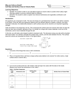


© Copyright 2026