
Document 8249
tg(ftO._.-
"'-'t'"
Appendix 2: Case Study Project
The purpose of this assignment is to facilitate students' familiarity with and understanding of neurogenic
cognitive-communicative disorders. These concepts will be reinforced via a search for and brief presentation of
individual clients with acquired neurogenic cognitive-communicative disorders.
Students will work independently to find a clinical case that fits the description of a given topic (e.g., motor
speech disorders, executive function problems, etc.). Examples of places to find such cases include: journal
articles (case studies), medical textbook cases, online stories (e.g., support group websites for a particular
disorder), famous cases (e.g., of the Diane Rehm/SD variety), experiences you've had in observing or treating
clients (NO names or identifying info please). Each student must present the following regarding their specific
case:
1. Source (e.g., AP A citation ofjournal article, name of website, or name of clinical site)
a. If your source is a journal article or website, please turn in a copy of the original case
b. Note that your case write-up should be in your own words and in laymen's terms.
2. Medical and social history summary
3. Evaluation plan and brief rationale for plan
4. Patient's estimated performance on planned evaluation (general)
5. Prognosis
Students will create a short paper (1-2 pages) including the above information that will be turned in to the
professor as well as distributed to the rest of the class (electronic distribution prior to class is fine). Each case
study will be used during class for practice in goal-writing activities. An example will be shared with the class
early in the semester. Due dates will be staggered according to the course schedule, with case studies following
current class topics. Students will sign up for their topic/due date at the beginning of the semester.
C ase StulY
d G ra d'm~ ru b riC:
.
General
Details
Content (complete)
Medical/social history summary
Evaluation plan
Evaluation rationale
Estimated performance
Prognosis
Appropriate
Matches assigned topic
Original wording, laymen's terms
Proofreading
Spelling, syntax
TOTAL POSSIBLE POINTS 50
Points
10
5
5
5
10
5
5
5
NOTE: Goal Writing Assignments
The purpose of these assignments is to facilitate students' ability to write appropriate goals for neurogenic
cognitive-communicative disorders, across severity levels, WHO levels and multiple medical settings. These
concepts will be reinforced via 10 goal-writing exercises over the course of the semester, corresponding to case
presentations.
Goal-writing assignments are worth 5 points each for a total of 50 possible points, and are graded as follows:
+5: Good (realistic, functional, specific, measurable)
+4: Adequate (at least 3/4 above criteria met)
<4: Inadequate (fewer than 3 criteria met). Re-write goals and turn in by the next class. Rewritten goals can
receive a maximum of 4 points.
7
-------------------,
COMD688
Fall 2008, Mayer
Case Study Grading rubric:
General
Details
Content (complete)
Medical/social history summary
Evaluation plan
Evaluation rationale
Estimated performance
Prognosis
Appropriate
Matches assigned topic
Proofreading
Spelling, syntax
TOTAL POSSIBLE POINTS = 50
Points
10
5
5
10
10
5
5
-\~, (so
Case 1: Mr. K
Setting: out-patient rehab
1. Medical & Social Summary
t
~X~~-~'-~
MEDICAL HISTORY:
Mr. K is an 81-yom with a diagnosis of spasmodic dysphonia. He reported that this diagnosis was
received about 8 years ago, but no form of treatment was sought at that time. Mr. Kstated that his
symptoms have worsened over the years and he now is interested in therapy. On September 4,
2008 Mr. Kcame to the NIU Speech-language-Hearing Clinic for a voice evaluation. He wanted an
alternative to Botox injections, as he did not feel comfortable with this form of treatment.
Mr. Kfirst noticed a change in his voice between 2000 and 2001. His voice had become hoarse
and he feared throat cancer. In January of 2001, he saw Dr. X, an otolaryngologist at his
commul1ity Ilospital, who made the diagnosis of spasmodic dysphonia of unknown origin. Mr. K
reported that he was did not believe this diagnosis and therefore did not seek treatment at that
time. However, as his symptoms worsened over the years he felt remorse about not pursuing
treatment and sought medical advice again. He returned to the physician as precursor to this
evaluation in September. His larynx was assessed endoscopically and therapy was recommended.
Mr. K reports that he takes Prilosec for indigestion and Advil for arthritis. He drinks one pot of
caffeinated coffee a day and Coke once or twice a day. On occasion, he drinks water and tea.
Alcohol and smoking are a part of Mr. K's daily routine. Mr. Khas no known allergies that would be
related to vocal fold dysfunction.
Mr. K reports that presently he is in relatively good health. He exercises twice a week at the indoor
community pool. Past surgeries include knee surgery, gall bladder surgery, and a hip replacement.
SOCIAL HISTORY:
Mr. K lives in Waukegan with his wife of 59 years. He has 4 grown children who all live close by
and are willing and able to care for him. Mr. Kis an active church member, participating in Bible
lessons. He sang for many years but reports that his singing voice has changed so drastically that
he is no longer able to sing.
2. Evaluation Plan
- Oral Mechanism Exam
- Hearing Screen to be performed at 25dB for the 500, 1000,2000,4000 Hz
- Assessment of slz ratio
- Vowel prolongation ability by sustaining the vowellal for as long as possible
- Onset of vowels to be evaluated by repeating "ah-ah-ah-ah-ah"
- Pitch range and voice flexibility to be subjectively assessed by two glissandos: low to high
- Intensity
- Resonance - HOlJ..) ~
- Jitter and Shimmer to be assessed by visi-pitch
- Quality
Case Study Project
September 10, 2008
- Rate of speech
\ - Subjective evaluation of vocal quality and behaviors during a reading task
3. Estimated Performance
Oral Mechanism Exam- OME revealed adequate jaw strength, symmetrical lip movement, and
adequate tongue mobility. Normal elevation of the velum was observed during la/ phonation.
Overall, rapid speech productions were WNL.
Hearing Screen- Hearing was WNl
Assessment of s/z ratio- A ratio of 1,3 was evidenced, A ratio of greater than one indicates
poor laryngeal control.
Vowel prolongation ability- Mr, Kwas able to sustain longer airflow than average despite
aphonic breaks
Onset of vowels- demonstrated no latency or glottal attacks
Pitch and voice flexibility- low pitches were difficult to achieve; however high pitches were
obtained more easily
---
Voice Flexibility- inflexible indicating a restricted range
.--
Intensity~. Mr. Kindicated that speaking loudly was difficult for him, but he was able to
display loud tone was prompted
Resonance-WI'll
-
Jitter and Shimmer- significantly highe~ than normal
Quality-WNl
t!':
Rate of speech- in aone minute conversational sample, a range of 177-160 syllables per
minute was noted indicating Mr. K's rate of speech is on the low end of an adult speaking rate5,
-N-p\u.D
Subjective evaluation of vocal quality and behaviors during a reading task- chest b~e~~h~n~ .was .
noted along with a hunched over body positioning- 1- hctU ~ vtn..l L. ~~(
4. Prognosis
Mr. Kdemonstrates spasmodic dysphonia characterized by insufficient pitch range with difficulty
reaching low tones, However, Mr. Kexhibited adequate quality, resonance, and intensity, He was
cooperative and receptive to instruction during speech tasks. Additionally he has a strong family
support system at home. His medical prognosis is!fir at this time. Speech-language therapy is
recommended to enhance Mr, K's quality of life by argeting compensatory techniques and healthy
vocal behaviors.
U-~I".bt..t.
.
5,
pr006~~-+S
1"L)~\L';'
..f--
~rt..·.Sb,
Case Study Project
September 10, 2008
Goals:
-t _/ C;D
~ ~'\r,-,,:,\ (\ ~n:.>'\.Wi\rxy~,c
,
(4 dl~((':,;"~~'CX")'
~,
Trl(}S\k- \."pt",_ L(u}ltJ\..:)
Cognitive Communication Disorders
Case Study
\,\
Ms.D
Ms. D. is a 48 yof who was admitted to Provena Mercy Hospital in Aurora with sudden
o~igh.t sided weakness and loss of speesili. It was determined she suffered aldl
asal ganglia hemorrhage, left parietal hematoma. Evaluation revealed significant ' -1
~ated blood pressUre and e em rna on CTsc~n. She was then transferred to St. of\J"\bef',~lvG . .,- .
Joseph in Joliet. Care there inclu e
0 weeks otiiehabilitation, followed by 4 weeks~t l''!.-h(>':O
~"t;l\')r-.J
the Rehabilitation Institute of Chicag Hypertension continues to be uncontrolled by
current medication. MRI completed
ce with findings as listed above. MRA (magnetic
resonance angiography) showed les th n 50% stenosis of both carotids.
i
Serial head CT demonstrated s~ble em toma without mass effects or shift. Upon
disch~ge From RI~ the~~.llE&~ste ou pa len ,reatment. .
. Broca:s 'S,;P Hr:.\J..J i'~~ (IDr,l\
:{~,hJt.U1.
11. ' '71
. :\ .(.~. ,.. . . '~( P \ _
. (
aphaSia wIth aprax~a. . ; . .
1. : "
l'
fl.-{
Ms. D's ; :
v:>r.~,,):)
pcf\A
1
"2
u
v
I
:4;yJ/.re...\,)t'-. hu:.
.
t">rtl'.
~
,
~)j •
.
',J';
'-.
:::;i:~:~stOry is remru~-kable .:;~• ..,. hio hypertension, headaChe';' ,,J-.. "to,~ H \\
oJ):)6
f'J'(
1'
(h()'\.~P ;:' 'Jst C~~\)IY'0',-
.'
kidney disease, and arthritisQyo tob~ Medical charts indicate she was non compliant to
medications and treatment program.
\
J'\" \ 'h 1.
r:r,t'tr(
()oJ
r
d'(
Lr(:.0.:.
~)
)
\7
~'rt
IS
(J
"hpy,. f;,()J't
'\
')fS'S:~''''.
't",
<r,f
.:_)
.'
Social Hx
Before the incident, Ms. D. worked in a SNF as a nurses aid!. She currently lives at home
in DeKalb with her two adult daughters. She is brought to therapy by Trans- Vac transport
system in DeKalb and Sycamore. Ms. D enjoys spending weekends'in Chicago with her
family and going to the movies.
Clinical Interview/Observation:
She reported that she has difficulty finding the correct wo!£! at the appropriate time. In
addition, she has trouble describing event~. When asked to re-cap and event, she
responded with short phrases and often asked, "what is that word?, With regard to
receptive lan ua e, she had trouble following conversations going on among family
mem ers. Following directions is also challenging. She would like to work on these
skills and writing her na.,!!!e. However, other rehab professionals report that her
attendance and motivation are inconsistent.
Evaluation Plan
, (\,;f\.
.1Q"<\) '-, , .~~
.\ \y,,,)
l.
Q
')¥-.I .
t \.
\~·~~S\ · L \ ' . r t -
~./
",\J'
~.
~
Token Test- (15-20 minutes)- to assess Ms. D's stamina and\1anguage abilities
demonstrated by following directions using 20 to~nd
commands about how to
manipulate the tokens
62
Cognitive Linguistic Quick Test (15-30 minutes)-specific subtests chosen to quickly
assess\ar~ded for effective languagel ({)(Y\r(\\)~\"CfJj)\(:r\ ,
~U)c:;,~
Boston~ng Test- (20-30 minutes) to assess Ms. D's ability to name both frequently
and non-frequently occurring objects
.~.~
lI0O\.1W(\-b
~ \.AJN',b:to R~ ~<:"J!i)S.d('>..ri) (c~i ~'rxu\~ evalUZlb.o\\
(A~·ltDJ G'~J\~ \a.eA.d. 8, A~h6S\~~~t~.. ®~~~~_~ ~
E'sfunated Perforn\,lDce
.t.® ~~ f':ktl:u 0\' (I tfl'" 01', bI.~ . 1
I
w-.
~~(~h~
Toke? Te!!t- Ms. D. was able to accurately foHow 30 of the 62 commands indicating
.(\ lmpmqnent
. -,
,..., N} ' / 4' \ . ~( 1A'i f~ r ,- 7
\.-- mit d '"? C'(1 0 ~JOJ:(6 't,Z'I '--' \.- ~ V\)~-,~ \~ ,~\.",,:> b
CLQT•
•
•
•
•
•
Confrontation Naming - moderately impaired
Story Retelling - severely impaired
Generative Naming - moderately impaired
Design Memory - severely impaired
Mazes- severely impaired
Design Generation.- moderately impaired
\
BNT- ,Impaired ~ \J\...-( '.
I-nA / /
v ~"-v ~
P~ ~ .-~
.
0
.
.
().b ,.
WCR (
.
1
o:f 1-(-" l (t
<-
~
C1lc<..\Q"
il) (---'
./cfh\S IS C\aT -tn-eSllfT( dt",,:JJ\ \:<OL (A..+~B(1e CyJt&e_..:r- . .r q11 Ms. D presents with moderate ex ressive and rece tive aphasia. Her primary deficits
appear in naming tasks, story/event re-telling, and compre ension of directions. She also
exhibits difficulty with memory and organization of language. Her prognosis is poor due
to her inconsistent particip~tiori and severity of inj\lIJ(]I0wever, with family support and
increased motivation during therapy activities, her prognosis may increase to fair.]
-Io\JlC : +~ 1:)
fC1D{ reGd ~ +//{ (5
rs;(,-d
',.-/
r,i-
rAb
This case study was adapted from a case at Unlimited Performance.
Mrs. Bishop assisted in acquiring information about this patient's
medical and social history. I worked with this client briefly over the
summer and used my experience and her diagnosis to estimate
performance on the evaluation.
,I . ~'--l
\... I.
-.
! -. \
'J
r(v·\i(~\.\t0 ..
_
4,
---1
e\J\ "re iXrt'0l1J'rj\
-I.,
G
Case Project: Mr. M
-r
;
Medical History:
Mr. Mis {j8-YOm,;J,vith a history ofCHF and HTN. He was admitted to the
hospital due to intense vertigo, nausea, vomiting, and speech and language difficulties in
September of200p. After admission, a MRI scan was administered revealing aright
} cerebellar-pontine ischemic infarcti 1 in the vascular territory of th~ superior cerebell~r
i
"al1.ery·
A). After a SPECT scan was administered five weeks sip stroke. data revealed
I a generalized ~ognitive decline and transcortical sensory aphasi J¥r. M also had
xec.u.t.ive dYSfunctions.',. disfll.pted diVI e
en lon, IStur ed ViSU.!.~l-SP. atia.l organiz.".ati.on.'
visl~al ,~r~ia, sur~~ dys~:~~a, and behavioral abn0l111alities.! " (, f/I'"" r-T "'.\ ,\ t' tr" j.fJ
0-l0
"\-, .
}
.'
\'.
'
L)
'( '~
Social Hist~~ .., ,,/ ~-" dQ151nf::
CLO{\() (' ('(lO.t \ 't;;. , 0 \it (O(}",
Mr. M is a retired restaurant manager who currently lives at home by himself. He has his ck ((j
maste(s_g~,g~in~ement. He is very active in his community and participates at
'...
th~enjor Citizens C1'lbyi.e. bingo and volunteering at the local YMCA). Mr. M's
f't 0 CC',
augtrreraffirs. on I. ive nearby to assist w.'ith his care at home even though he lives alone.
bu.u iOn., R
Mr. M is willing clnd motivated for therapy and wants to be able to communicate his
;"""-"--'.
wants and needs t~ his family members.
(;. C(\()(YVloJ i b)
~,.
f}JA,,8. sg -:
0
S Ir\,}(lJ'f icd-f(,
Evaluation Plan:
\.J
i-f"l "'t)-'l(
<: pfCcr',
\CJ ~ -Western Aphasia Battery- identifies presence, severity, and aphasia classification
U
,'j,
'") -Cognitive Linguistic Quick Test- assesses major cognitive domains and provides s.everity
rating ~ Co,1\,J hl\.\"C ~J b ~r. ~ .f:: O~
-~.""~,-- Estimated Per~ormance:
~)f1€-t.LVl \~ (i'-~,t J"'rJ-.0,;b f()( tt,f't h.,e)
t ~ ()(\)
Western AphasIa Battery scores:
.
! '
I
.' 'J
(\.
!
G
~
~
-r'b
...- ' .
\.j- '_:"'-.\.1,
"--" ..,-~ I;
\.
,,~,
---"
ol() b
--
i " "'
:?
,/,~'------Contel!t: S~yetl! ,-*" _
.\
. \"
•.r.,~1)
\" \ ,'~'
~-~\
•
".
.n
*
Jet!
('~\tlr-.r
-to.'0r::,s/1:::"'/--""¥--'
\ '+!\)t(\.L~.' OI\.~.)Ab,
. l_e
-+.
f'l uel1cy.
.,. Sever :; \N.J....0- (l.J: -; -:,-~\,-\j
~ 1\
~· .. '::::::~r
\",'
_ ...~
't".( I l ' ( uditory Comprehension: Moderate,A) severe
'
',~_
R
i .
<
.~~Uli'lia~~\' 01 ~
d'
. WN
e~ .mg.
'elf.
'
hl.S
•
•
~. X• Vv'
11 1\"
t
\
"'
~
0'
l +urihltr
-·--:r?
:'!'! (d'I _, v re.~U~FtG, chJJ It>lr_cf
.,--;c'rro!JJ.
"-- TSA «\u,Cuk p1..,
I
_'1
h1...dtk~dw,k\l~ ~-6ro k
Cognitive Linguistic Quick Test scores:
Ju\s..o-f'-bn~l(A.·.i
I
.
~
,. _
.
\
ntmg: Moderate to severe (Le. neologIsms)
•
•
•
•
,J
" l-),..;\...
.
Q
,
( ,
'
~.~~bLL
Attention: Dismpted divided attention
'. Memory: Globally decrea..sed meu.Jory. levels J."~'
Executive Fu IOjl: Severe
,
r ~It.;{t> '\::->~
~~a e: . L ),",01.0 c..r.HJ\ 'iCSll
~
'.. ISlfOspa Ia ~ 1 ,: Moderate to severe
Clock Drawing: Moderate
tf
\
'!) GJ:,
c...
Prognosis:
Mr. M presents with moderate to severe cognitive-linguistic and visuospatial
deficits impacting his daily life activities and communication. Given his relatively )::.oung
,2ge, I£9tivation, premorbid intelligence, and family support system, Mr, M's prognosis to
return honiewith moderate assistance froIn his family members or caregiver is. fair to
good with intensive multidisciplinary liP rehab. Mr. M will require OIP rehab and
supervision as well.
{ /
,
'
r ICt.se.o
xxx
CCRTEX
ELSEVIER
MASSON
(2008) [-[()
joorrre'l·homep8.\Je;.:www.eIII6vier.j:Qm/locate/cortex
Research report
Cognitive, linguistic and affective disturbances following
a right superior cerebellar artery infarction: A case study
Q
Q
Peter MarienQ,b,c,*, Hanne Baillieux , Hyo lung De Smet , Sebastiaan Engelborghsb,c,J,9,
Ineke Wilssens b, Philippe PaquierQ,d,e and Peter P. De Deyn b,C,9
aDepartment of Linguistics, Vrije Universiteit Brussel, Brussels, Belgium
bDepartment of Neurology, ZNA Middelheim Hospital, Antwerp, Belgium
"Laboratory of Neurochemistry and Behavior, Institute Born-Bunge, University of Antwerp, Antwerp, Belgium
dDepartment of Neurology and Neuropsychology, University Hospital, Erasme ULB, Brussels, Belgium
eUnit of Neurosciences, Faculty of Medicine, University of Antwerp, Antwerp, Belgium
fDepartment of Nursing Sciences, Faculty of Medicine, University of Antwerp, Antwerp, Belgium
gDepartment of Health Care Sciences, University College Antwerp (Hogeschool Antwerpen), Antwerp, Belgium
ARTICLE INFO
ABSTRACT
--~.---
Article history:
Received 26 June 2007
Reviewed 21 September 2007
Revised 19 October 2007
Accepted 6 December 2007
Action editor Gereon Fink
Published online _
Keywords:
Cerebellum
Cerebellar cognitive affective
syndrome
Visual dyslexia
SCA
SPECT
- - -..- - -..- - - - - -
...
-----~---~----
The cerebellar cognitive affective syndrome (CCAS) is a neurobehavioral syndrome that
may develop after congenital and acquired cerebellar lesions. The syndrome consists of
deficits in executive functioning, spatial cognition, visual-spatial memory and language
and also involves personality and behavioral changes. We describe a 58-year-old righthanded man who in addition to affective disturbances presented with a unique combination of cognitive and linguistic deficits fOHoWIng an Ischellilc llifarctlon in the vascular
territory of the nght supenor cerebellar artery (SeA). Neurocognitive and neurolinguistir
;;ammatlons were pertonned In the acute phase (10 days post-onset) and lesion phase .;
(four weeks post-onset) of the stroke. A Ic-99m·ECD SPECT studt was performed five
wee
ke. Acu
da revealed a eneralized cognitive decline and
tical sensory aphasia. In the lesion phase. the neuro e aVIO
was
dominated by executive ys unctIons, disrupted divided attention, disturbed visual-spatial
organization and bel'lllvi.. ra l ~alities. Neurolinguistic investigations disclosed.l.isual
dyslexia and surface dysgraphia~g of words and visual lexical decision tasks of
words and nonwords were severely defective and predominantly characterized by visual
errors. In addition, writing irregular and ambiguous words resulted in regularization errors
(phonologically plau~ors based on phoneme-grapheme correspondence rules). In
the absence of any structural damage in the supratentorial brain regions, a quantified
SPECT study showed a relative hypoperfusion in the right cerebellar hemisphere an9J!re
left medial frontal lobe. CCAS is ror the first time reported in association with visual
dyslexia and surface dysgnlEf.ua. We hypothesize that the cognitive and lmguistic deficits
might result from functional disruption of the cerebellar-encephalic pathwa s, connecting
the cerebellum to the frontal supratentorial areas w lch su serve attentional and planning
processes. This phenomenon of crossed cerebellar-cerebral diaschisis is supported by
SPECT findings revealing a hypopelrUslon m the anatomoclinically suspected brain
, Corresponding author. ZNA - A.Z. Middelheim, Department of Neurology, Lindendreef 1, B-2020 Antwerp, Belgium.
E-mail address:[email protected] (P. Marien).
0010-9452/$ see front matter © 2008 Elsevier Masson Sr!' All rights reserved.
doi:10.1016/j .cortex.2007.12.010
2
CORTEX XXX (2008)
[0
regions. The constellation of cognitive, linguistic and behavioral symptoms adds new
evidence to the multifaceted area of cerebellar neurocognition and demonstrates that
the cerebellum might playa crucial role in cognitive, linguistic, and affective processing.
@ 2008 Elsevier Masson SrI. All rights reserved.
1.
Introduction
We describe a patient who presented with a unique combination of cognitive and linguistic deficits following an ischemic infarction in the vascular territory of the right superior
cerebellar artery (SCA). From a semiological point of view
CCAS is for the first time reported in association with visual
dyslexia and surface dysgraphia. From an anatomoclinical
point of view our findings demonstrate that disruption of cognitive and linguistic skills may also follow SCA lesions.
The current interest in the role of the cerebellum in cognitive,
linguistic and affective functions is relatively new. During the
past two decades, clinical neuroscience has substantially extended the traditional view on the cerebellum as a mere coordinator of motor functions. In the late 19805, Positron
Emission Tomography (PET) studies provided preliminary evidence for cerebellar involvement in non-motor language
functions (Petersen et aI., 1988, 1989; Leiner et aI., 1989). In2.
Case report
deed, in addition to activation of Broca's area, verbal-semantic
association tasks - which are generally considered to depend
on a close cooperation between verbal and executive abilities2.1.
History
disclosed functional involvement of the inferior-lateral part of
the right cerebellum, which is functionally connected to the
RDL, a 58-year-old right-handed man, with an educational
left prefrontal language areas (Petersen et aI., 1988, 1989). Delevel of 10 years, was admitted to the hospital because of intense vertigo, nausea, vomiting and speech and language difspite variations on the original task design, several studies
ficulties. The clinical neurological examination on admission
with PET and functional Magnetic Resonance Imaging (fMRI)
revealed a discrete right hemiparesis (MRC 4+/5). Examination
have consistently reproduced crosswise cerebrocerebellar activation during semantic and phonological word generation
of coordination by finger-to-nose and heel-to-knee tests distasks in both left- and right-handers (Raichle et aI., 1994;
closed right-sided hemiataxia consisting of dysmetria and
Grabowski et aI., 1996; Chee et aI., 1998; Hubrich-Ungureanu
hypermetria of the right arm and leg that was too pronounced
to be explained by muscular weakness. Ataxia was only paret aI., 2002; Jansen et a!., 2005). In addition, clinical research
tially affected by visual clues. Gait ataxia was noticed as
has convincingly shown that focal cerebellar damage can induce a variety of non-motor linguistic deficits among which
well. Hypometric saccades were observed when eye movedisrupted articulatory and graphomotor planning (Marien
ments were tested. Tendon reflexes were slightly brisker at
the right than on the left side of the body. Plantar response
et ai., 2007), agrammatism, semantic deficits, distorted language dynamics, aphasia, and reading and writing problems
was flexor bilaterally. A discrete hypoesthesia for pinprick
wa
nd at the right side of the body. Speech was slightly
(for a review see Marien et ai., 2001; Paquier and Marien,
dysarthri
uditory-verbal comprehension and naming
2005). On the neurocognitive level cerebellar damage has
been associated with a broad spectrum of symptoms such as
wer e ective.n MRI scan of the brain showed a right cereimpaired attentional processes (Gottwald et aI., 2003), execubellar-p
e infarction in the vascular territory of the SCA.
tive dysfunctions (Schmahmann and Sherman, 1998), learnNo evidence of supratentorial damage was found (Fig. lA-H).
ing disability (Drepper et aI., 1999) and disrupted temporal
A Tc-99 m-ECD SPECT perfusion scan was performed five
and spatial processing (Salman, 2002). In addition to the growweeks post-stroke. Trans-axial images with a pixel size of
ing body of evidence for cerebellar involvement in language
3.56 mm were anatomically standardized using SPM and comand cognition, 19th century reports already anecdotically
pared to a standard normal and SD image obtained from 15
mentioned behavioral alterations following cerebellar lesions
normal ECD perfusion studies. Using a 31 ROJ template the
(Combettes, 1831; Otto, 1873). Due to a lack of standardized inZ-scores (SD) were then calculated for each region. A regional
vestigations and pathological verification these observations
Z-score of >2.0 is considered significant. In comparison to nordid not receive much attention (Dow and Moruzzi, 1958). To
mal database findings. the quantified baseline ECD SPECT
define a typical constellation of cognitive, linguistic and affecstudy revealed a relative hypoperfusion in the right cerebellar
tive symptoms following cerebellar damage Schmahmann
hemisphere (-3.15 SD below average) and decreased cerebral
and Sherman (1998) introduced the concept of Cerebellar Cogblood flow in the left medial frontal area (-2.21 SD below
nitive Affective Syndrome (CCAS). The core features of this
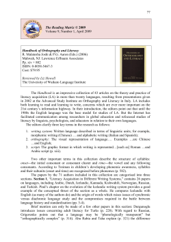
average) (Fig. 2).
syndrome consist of executive dysfunctions, disrupted spatial
cognition, impaired visual-spatial memory, language distur2.2.
Neurolinguistic assessments
bances, and personality and behavioral disorders. Since the
Formal neurolinguistic investigations were performed 10 days
first description of the syndrome, CCAS has been reported in
a number of etiologically different patients, both children
and four weeks after the stroke. The neurolinguistic examinaand adults, with acquired (e.g., Levisohn et aI., 2000; Baillieux
tion consisted of the Dutch version (Graetz et aI., 1992) of the
et aI., 2006) and congenital cerebellar damage (e.g., Duggal, -Aachener Aphasie Test (AAT) (Huber et ai., 1983), a semantic
_ verbal fluency test (animals, transportation, vegetables and
2005; Marien et a!., 2008).
CORTEX XXX (2008)
-10
3
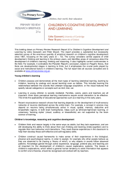
Fig. 1 - Brain MRI axial FLAIR slices (A-A) showing a right cerebellar-pontine infarction in the vascular territory of the
superior cerebellar artery (SCA) (A-D). No structural damage was observed at the supratentorial level (E-H).
clothing), and the Boston Naming Test (BNT) (Kaplan et aI.,
.I
1983; Marien et a1., 1998).
Ten days post-stroke, transcortical sensory aphasia was
objectified (Table 1). As shown by defective AAT subtest results, the patient had auditory verbal and visual comprehension deficits (total comprehension, 88/120; mean, 108.49; SO,
10.85). The score on the Token Test was nine errors (mean,
2.28; SO, 2.75). Repetition was normal. A deficient result on
he BNT (47/60, mean, 52.8; SO, 3.7) indicated disrupted visual
onfrontation nam~. The majority of nam1ng errors consisted of 'don't know responses' (10/13) indicating underlying
verbal mnestic problems. Speech was ataxic in nature and
marked by distorted consonant production, irregular articulatory breakdowns, hypophonia and fast rate. Spontaneous
speech was additionally characterized by verbal perseverations and a slightly increased verbal output. However, results
on verbal fluency tasks (Table 2) were severely impaired: semantic and phonological fluency scores were 44 (mean, 60.5;
SO, 7.14) and 16 (mean, 41; SO, 5.23), respectively. Reading
aloud was within the normal range. Writing contained
t
~
(\orl'
phonological paragraphias. Intensive semantic therapy and
articulation therapy were started on a daily basis. As reflected
by repeat AAT results, obtained four weeks post-onset neurologieal symptoms, transcortical sensory aphasia receded but
written language disturbances persisted. As demonstrated
by normal BNT results visual confrontation naming normalized but semantic verbal fluency did not improve (Table 2).
Ataxic dysarthria markedly ameliorated.
Written language skills were investigated in more detail
four weeks post-onset neurological symptoms by means of
the Outch version (Bastiaanse et aI., 1995) of the Psycholinguistic Assessments of Language Processing in Aphasia
(PALPA) (Kay et aI., 1992). Results on reading aloud and visual
lexical decision tasks are shown in Table 3. Overall reading
aloud of words was severely defective but reading accuracy
was not significantly affected by length, frequency, grammatical class, or regularity of spelling. Word imageability, however, affected reading performance. The patient obtained
a defective score of 36/40 (mean, 39.98; SO, 0.16) on reading
low imageability words. As demonstrated by maximum scores
Fig. 2 - Quantified ECD SPECT scan five weeks post-stroke showing a hypoperfusion in the right cerebellar hemisphere and
the left medial frontal area.
4
CORTEX XXX (2008) 1-10
Repetition
Confrontation naming
Total Score
1441150
147/1.50
144.1
8.07
Nouns
30/30
27/30
25/30
221313
1.001.20
30/30
3DiS13
27.92
27.69
Cqlorterms
Compound not;.n~
Se.ntenees
lanD
21/30
41/60
26/30
21/30
4Z!60
$BlUO
on letter case matching tasks ofreal words and nonwords, letter processing was normal. In addition, visual lexical decision
tasks of words and nonwords revealed a severely disrupted
performance. The patient identified 53/140 (37.9%) nonwords
as real words.
The patient's scores on the writing tests of the PALPA are
shown in Table 3. 84/144 (58.3%) real words and 17/24 (70.8%)
nonwords were correctly written. Word length clearly affected
writing. High imageable-high frequent words (9/10) were more
adequately written than low imageable-low frequent items (3/
10), whereas function words (5/5) were better written than
nouns (1/5). Regularity had a significant influence on writing
performances: regular words were written more often correctly than irregular words (14120 and 5120, respectively). In
addition, a frequency effect was also observed in each category: 5/6 regular-high frequent compared to 9/14 regular-infrequent items, and 2/3 irregular-frequent compared to 3/17
irregular-infrequent items.
2.3.
Neurocognitiue assessments
To evaluate general cognitive abilities and memory, the
Wechsler Adult Intelligence Scale-II! (WeChsler, 1997) and
Wechsler Memory Scale-R (WMS-R) (Wechsler. 1987) were administered. Executive functioning and problem-solving were
investigated by means of the Wisconsin Card Sorting Test
(WCST) (Heaton et al.. 1993), the Stroop Colour-Word Test
(Hammes. 1971). and the Trail Making Test (Reitan, 1958).
The D2 test of attention (Brickenkamp and Zillmer, 1998)
and the copy of the Rey-Osterrieth Figure (Osterrieth. 1944)
26/30
114/120
25.69
1.09.3
2.9
1.99
2.61
3.71
8,.42
28/30
1{)~120
26.49
26.79
53.28
28.3
26.91
$$;21
108.49
3.3
3.41
6.08
2.29
3.9
4.9
10.85
2. errors
2.28
2.75
28/30
28130
56/60
26/30
20/~O
4(>/60
28.04
were used to evaluate visual-motor concentration and visuoconstructive praxis (Table 2).
In the acute phase of the stroke (14 days post-onset neurological symptoms), disruption of general cognition was
reflected by a total Wechsler IQ level of 66. The verbal IQ
was 68 and the performal IQ was 71. The Wechsler Memory
Scale-R revealed globally decreased memory levels. A discrepancy of 18 index points was found between visual (index = 66)
and verbal memory (index 84). As reflected by an index of 59
(- 2. 7 SD) recent memory was severely distorted. Ataxic symptoms affected visuo-motor concentration [D2 impaired for
both working rate (Percentile 1) (Pct.) and accuracy, (Pct. 1)].
Generalized concentration disturbances were objectified by
means of non-motor tasks (e.g., WMS-R concentration
index = 60). Investigation of frontal lobe functions revealed
executive disturbances involving divided attention (Stroop:
Card I1IIIIll: Pct. 101111). mental flexibility (WCST: Pct. 1; Trail
Making A: Pct. 1). and visual-spatial planning (Trail Making
B: Pct. 1). A distorted copy of the Rey-Osterrieth figure indicated constructional apraxia (20/36; mean, 35; SD, 3). Behavior
was characterized by disinhibited actions, overfamiliarity.
confabulations and perseverations.
In addition to speech and language therapy a cognitive rehabilitation program was started. Five weeks after the stroke,
an overall improvement of cognitive functioning was found
(Table 2). Visual (index = 102) and verbal memory (index = 101)
had normalized and the discrepancy between both levels resolved. As reflected by deficient scores on the Stroop (Pct. 5/1/
1) and Trail MakingTest (Pct. 10/10). dysexecutive dysfunctions
persisted. A defective performance on concentration (WMS-R:
5
CORTEX XXX (2008) I-fO
Table 2 - Acute and lesion phase: cognitive results
Lesion phase
Norms
SD
1.3
15
.15
15
15
.102
101
100
·sa·
Language
Boston Naming<Test (BNT)
Verbal tl~ency
Semantic generation
1'btal'Ournbero('pet~e;verati()ns
Total number Qf intnisibns
Phon
44
47
3
4
o
o
1~
1
lOG
100
.100
100
15
15
15
15
52.8
3.7
60.5
7.14
41
5.23
g
P\:t.l
Pct;lO!lIl
Pct.5/l11
Pct.50/50/50
PeUO/10
Pct.50/SQ/SO
15
index = 74) and visuo-constructive praxis (Rey-Osterrieth Figure: 25/36) was found as welL Behavior and affective regulation
remained aberrant. The patient acted impulsively and displayed disinhibited behavior. He strongly confabulated and
had a severely distorted sense of reality. He forcefully denied
any cognitive, linguistic or affective problems. Consequently,
he refused additional testing and therapy and left the hospital
against the advice of family members, doctors and caregivers.
This attitude fitted in an overall behavioral profile of indifference and withdrawal. According to his relatives, the patient
had become unconcerned and emotionally labile. He withdrew
from his friends and family and became socially isolated. He
displayed irresponsible, impulsive and stereotypical behavior.
After one year, he still avoids any social contact and keeps reo
fusing medical follow-up and therapy.
3.
Discussion
After an ischemic infarction in the vascular territory of the
right SeA, our patient developed cognitive and behavioral
characteristics consistent with CCAS. In addition, persistent
visual dyslexia and surface dysgraphia were found after reo
mission of mild transcortical sensory aphasia in the acute
hase ofthe'Stroke.
Reading an writing disturbances due to cerebellar damage
are only scarcely documented in the literature (Moretti et aI.,
2002a, 2002b; Marien et aI., 2007). Analysis of RDL's readingerrors disclosed marked visual similarity between the stimulus
words and responses indicating acquired visual dyslexia
(Marshall and Newcombe, 1973). To our knowledge, this syndrome has only been recorded in nine patients and has never
been described in association with cerebellar damage (Casey
and Ettlinger, 1960; Marshall and Newcombe, 1973; Lambon
Ralph and Ellis, 1997; Cuetos and Ellis, 1999; Sinn and Blanken,
1999; Biran et aI., 2003; Crutch and Warrington, 2007). Acquired visual dyslexia is characterized by visual errors in
which at least half the number of the letters of the target
word are present (Lambon Ralph and Ellis, 1997). RDL's reading errors on the PALPA subtests were all classified as visual
errors. In 13/18 instances, errors occurred in medial word
position (72.2%) and in only 2118 (11.1%) in initial and in 3/18
i
6
CORTEX XXX (2008) I
Table 3 - PALPA subtests lesion phase: results for reading
aloud, visual lexical decision and writing to dictation
Mean',.
Test
$0
24
Word len~•.(llyUab!E!!I)
TotaL Stor~
'23124,
23.98
0.16
23-,24
19,98
20.00
0.16
19-20
~.
0.16
Table 3 (continued)
Mean
SO
Range
4/6
2/6
17/24
5.9
5.98
23.75
0.3
0.16
0.58
5-6
5-6
21-24-
lmageability and frequency
9/10
I+IF+
l+/F~
6/10
1-/1'+
4/10
1-/1'3/10
Total score
22140
9.9
9.8
9.95
9.65
39.S
0.3
0.51
0.22
0.88
1.42
9-10
115
4.98
0.4
4-5
4/5
SIS
4.98
4.93
19,88
0;16
0,4
4-5' ,
4-5
1&-'20
Test
SCraphemes
6 Graphemes
Total score
Reading aloud (palpl\~5;37. ~9. 43)
Word lengt'!:l (~~b.mE\l)
Total $core
'23124
Imageabiltty, ahd,Jreq~e~):Y
I+IF+
'20/20
20/20
19/20
17/20
10
19-20
,39{40
Grammatical class
Nouns
AdjeetWes '.
Verbs
func.tionwords
To~tlscore
~orE!
3/5
la/20
G~mmatit;;!l class and, im~!ibility
~puns
function words
Total score
5/10
8/\1.0
1.~110
5
0:16
0
9.75
9.93
19.68
0.58
0,26'
0.75
9.88
13.88
5.68
19.73
0.40
0..61
1;2'3
0.91
0.87
9-10
9-10
9-10
33-40
8-10
9-10
17-20
S.~l)ing--soundregTllarity
Regular
1-
IRG
Totalsc:ore
Total regular
Npnword~eadirig
3
4
3/6
4/6
5/13
5/6
17/24
Le~r,pr~~sing(P~a 20)
Total sl:ore'
60160 '
6.00
5.98
5B5
0.16
0.22
23,S3
0.3
0.54
59.53
G.B7
5.9
Visu~\l.~~si¢l(p~1;;23.'24"2S}
11;9&
14l~
'Z9.98
59.as
0;16
oAg
'0.16
5-6
s-:6
5-6
21-24
14-15
.12-is
Morpitology
R.eal words
Inflectipns
Qel.'j.vatiol\S
No,nwords
Inflectitms
D1!rivations
Total score
28130
HilS
14/15
6/30
5/1S
ft15
14/60
29.85
14.88
$3.83
13,1!'I
0.42
0.4
0.16
337
2.29
1$7
Writing to dictation (palpa 31,;aS. 39! 40, 42, 43)
Word length
'
3 Grapneln~
6/6
'6
Sr6
5.88
0.33
4 Graphemes
Total irregular
Total score
," %78,
8-10
7,.~O
a-:.14
3-6
15-20
9..28
1;34
3/17
213
5/20
19/40
8.88
15.10
2.85
18.10
37.38
1.71
2.64
0.36
2.93
3.53
5-10
5-10
5-17
2-3
7-20
26-40
17/24
23,33
0,96
21~24
3/10
211()
SD, standard deviation; lW. regular. IRG. irregular; 1+, high imageP+,
frequentil-, low
1'..... loVl
:.29.-30
57-60
70.:&0
156-160
'total ~~e
1'1'+
Nonwcirds
Totsl score
I~
Nlilnword~
Irregular
11+
8/10
~10
9/14
5/6
14120
28-30
13-,15
,14-15
1S-30
4-15
9-15
5-6
5-6
(16.7%) in final word position. Since initial or final letter
positions did not influence RDL's reading performance, a diag·
nosis of neglect dyslexia was excluded (Ellis et aI., 1987; War·
rington, 1991). A diagnosis of letter·by-letter reading, surface
and attentional dyslexia could also be excluded since there
was not any effect of word length, regularity, and flanking or
migration of letters. However, in addition to a visual component RDL's reading accuracy was influenced by imageability,
which is generally considered a characteristic feature of
deep dyslexia and indicative of impairments in or around
the semantic system (Plaut and Shalliee, 1994). However,
although RDL produced a few semantic paraphasias (3/6)
on the BNT, he did not make any semantic or visuo-semantic
paralexias, which are the cardinal feature of deep dyslexia.
RDL's reading ability and visual lexical decision skills were
markedly better for words than nonwords. According to
Lambon Ralph and Ellis (1997), poor performance on nonword
reading may be due to an almost exclusive reliance on lexical
processes. Biran et al. (2003) suggested that their patients
CORTEX XXX (2008)
relied on the available lexical options during word reading,
whereas in nonword reading they could only rely on the deficient visual analyzer, resulting in better reading of words over
nonwords. ROL's real word reading was marked by verbal
morphological paralexias, a type of error that also characterized reading performances of the patients described by
Lambon Ralph and Ellis (1997), Cuetos and Ellis (1999), Sinn
and Blanken (1999), and Crutch and Warrington (2007). Nonword reading was characterized by application of putative
grapheme-phoneme conversion rules (17/24); 617 nonwords
were lexicalized and in one nonword a monophthong was
diphthongized. A lexicalization effect was also present in visual lexical decision of words versus nonwords since 53/140
(37.9%) nonwords were incorrectly identified as real words.
In writing to dictation, RDL produced many neologisms
(84.5%) which frequently matched the phonetic characteristics of the target word (pseudohomophones). Paragraphias
representing real words occurred only in 9/58 instances
(15.5%). Most of ROL's writing errors stemmed from orthographic mistakes and represented phonologically plausible
errors (60.3%), characteristic of surface dysgraphia. Patients
with surface dysgraphia only apply phoneme-grapheme correspondence rules which typically results in disrupted spelling of irregular and ambiguous words. In surface dysgraphia,
spelling depends on the direct translation of the sounds of
words into those spelling patterns that are appropriate in
a given language (McCarthy and Warrington, 1990). Beauvois
and Derouesne (1981) suggested that impairment of a lexical
spelling system, without disruption of the phonological writing process, produces surface dysgraphia. Patients with
surface dysgraphia make regularization errors, which represent plausible renditions of sound-to-letter correspondences
appropriate to the language. Although RDL was able to write
some high-frequent irregular or ambiguous words, he also
produced regularization errors. Correct spelling of irregular
words may occur, suggesting some minimal preservation
and contribution from word-specific spelling (Hatfield and
Patterson, 1983).
Summing-up, on the linguistic level ROL presented with visual dyslexia and surface dysgraphia induced by a right SCA
infarction. Fabbro et al. (2000) hypothesized that cerebellar
structures, such as the right cerebellar hemisphere and
some portions of the vermis, may control written language
processes by integrating their activity with the "frontal lobe
system". Our findings seem to corroborate this view. Ouring
reading and writing, action and perception are strongly coupled, and visual perception is firmly bound to attention and
to activation ofbrain regions necessary for action preparation
(Moretti et aI., 2002b). Consequently, acquired visual dyslexia
after cerebellar lesions might be related to altered oculomotor
functioning. However, we hypothesize that ROL's reading and
writing deficits might result from damage to the cerebellarencephalic projections, connecting the cerebellum to the prefrontal supratentorial areas which subserve attentional and
planning processes (Moretti et aI., 2002a). Visual dyslexia
may follow widely distributed dominant hemisphere lesions
such as damage to the left fronto-parietal region (Cuetos and
Ellis, 1999), and bilateral frontal atrophy (Crutch and
Warrington, 2007). However, the condition has not been described in association with cerebellar damage. Clinical
<0
7
observations of patients with prefrontal lobe dysfunctions
also suggest that complex aspects of writing such as planning
and maintained attention may be disturbed (Ardila and Sur!off, 2006). In our patient, the view of a functional disruption
of the prefrontal brain regions is supported by quantified
SPECT findings which revealed crossed cerebellar-cerebral
diaschisis, reflecting the functional impact of the cerebellar lesion on a distant contralateral supratentorial region due to
a lack of excitatory impulses (Baron et aI., 1981; Marien,
et aI., 2001). Indeed, a quantified SPECT study performed five
weeks after the stroke showed a hypoperfusion in the right
cerebellar hemisphere and significantly decreased cerebral
blood flow in the left medial frontal area. This pattern of perfusion deficits was clinically also associated with a constellation of cognitive and behavioral deficits resembling CCAS
(Schmahmann and Sherman, 1998). Several studies of patients with cerebellar lesions have shown similar patterns of
neuropsychological deficits in executive functioning, visualspatial abilities and behavior (Chafetz et aI., 1996; Neau
et aI., 2000; Paulus et aI., 2004; Kalashnikova et aI., 2005). However, Gomez-Beldarrain et al. (1997), for instance, did not find
any memory disturbances, visual-spatial disabilities or executive dysfunctioning in 26 patients with cerebellar strokes. In
addition, a number of studies have documented variations
in type and severity of symptoms, leading to the conclusion
that cognitive and behavioral consequences following cerebellar lesions may vary among the population (Aarsen
et aI., 2004; Baillieux et ai., 2006). However, the crosswise
functional impact of focal cerebellar damage on distant
supratentorial regions that subserve cognitive processes
has been amply documented and contributes to the view of
a functionally lateralized and topographic organization of
the "cognitive cerebellum". In the early 19905, BotezMarquard et al. (1994) for the first time described a patient
who in the absence of a supratentorial lesion developed typical posterior right-hemisphere dysfunctions after a left
cerebellar lesion. Based on the SPECT findings which
revealed a right fronto-parietal hypoperfusion, the authors
concluded that the visual-spatial deficits were caused by
crossed cerebello-cerebral diaschisis. Further evidence of
right-hemisphere dysfunctioning following left cerebellar lesions has been provided by Chafetz et al. (1996) and Allin
et al. (2001), supporting the notion of contralateral cerebellar
involvement in cognitive modulation. On the neurolinguistic
level, Marien et al. (1996) described a patient with left hemisphere "cerebellar induced aphasia" following a right cerebellar infarction. Based on a close longitudinal follow-up of
five years with SPECT, the authors introduced the concept
of a "lateralized linguistic cerebellum" (Marien et aI., 2001),
emphasizing the involvement of the right cerebellar hemisphere in a variety of linguistic processes subserved by the
contralateral, language dominant, hemisphere. Consistent
with this evidence are the studies of Gottwald et al. (2004)
and Hokkanen et al. (2006) who showed that patients with
right-sided cerebellar lesions were generally more impaired
in the verbal domain, while left-sided cerebellar lesions
affected patients significantly in visual-spatial tasks. In addition, many functional neuroimaging studies by means of
SPECT (Baillieux et aI., 2006; Marien et aI., 2007) and fMRI
(Fink et aI., 2000; Hubrich-Ungureanu et aI., 2002; Jansen
8
<0
CORTEX XXX (2008) 1-10
et aI., 2005) have convincingly demonstrated a lateralized
functional organization of the cerebellum in the cerebellocerebral network of cognitive modulation.
Exner et al. (2004) recently investigated whether vascular
lesions in different parts of the cerebellum result in differential cognitive and affective impairments. The authors concluded that in contrast to subjects with SCA lesions
a pattern of memory impairment, executive disturbances
and emotional withdrawal is found in patients with infarcts
in the PICA territory. These findings are in concordance with
many reports in which CCAS follows from PICA lesions
(Schmahmann and Sherman, 1998; Paulus et aI., 2004; Willert
et aI., 2005). However, Neau et al. (2000) found no significant
differences between the cognitive consequences of infarcts
in PICA or SCA territory. As in our patient. Botez-Marquard
et al. (1994), and Schmahmann and Sherman (1998) reported
patients with cognitive disturbances, such as executive dysfunctions and visual-spatial deficits following SCA lesions
and Marien et al. (1996, 2000) described dynamic aphasia following a right SeA infarction. In our patient, a possible influence of the brainstem lesion on the cognitive and affective
symptoms should be considered since the stroke slightly extended into the pons. Garrard et aL (2002) reported longterm neurocognitive symptoms, mostly involving attention
and executive functions in six adult patients with isolated
vascular brainstem lesions. The authors explained the specific
pattern of symptoms as a disruption of frontal cortical functions, possibly resulting from diaschisis. Similar observations
have been made by Salgado et al. (2007). Fazekas et al. (1993)
also observed reduced blood flow in the contralateral cerebellar and ipsilateral cortical regions (most prominent in the
fronto-parietal region) in two patients with isolated vascular
lesions in the upper pons. Unfortunately, no information
with regard to the cognitive status of the patients is provided
(Fazekas et aI., 1993).
The symptoms observed in CCAS are, however, consistent with predictions derived from neuroanatomical and
neuroimaging studies, revealing extensive neural circuitries
that reciprocally connect the prefrontal, temporal, posterior
parietal and limbic cortices with the cerebellum (Desmond,
2001; Schmahmann, 2004). According to Schmahmann
(2004), these anatomical sub circuits constitute the structural
basis for functional subunits, reflecting a topographic organization of motor and cognitive functions of the cerebellum: the
anterior cerebellar lobe is mainly involved in motor functions,
while the posterior parts of the cerebellum are involved in
higher cognitive modulation. However, as variability may exist with regard to the functional organization of the cerebellum, future research is needed to further elucidate
intrigumg Issue 0 a
cerebellum".
Acknowledgements
This study was supported by grant G.0209.05 of the Fund for
Scientific Research - Flanders (F. W.O. - Vlaanderen), by
Onderzoeksraad (OZR-VUB), Nationale Vereniging tot Steun
aan Gehandicapte Personen (NVSG-ANAH), Stichting Integratie Gehandicapten (SIG), and Deloitte Belgium. SE is
a postdoctoral fellow of the Fund for Scientific Research
Flanders (F.W.O. - Vlaanderen).
REFERENCES
Aarsen FK, Van Dongen HR, Paquier PF, Van Mourik M, and
Catsman-Berrevoets CE. Long-term sequelae in children
after cerebellar astrocytoma surgery. Neurology, 62: 1311-1316,
2004.
Allin M, Matsumoto H, Santhouse AM, Nosarti C, Alasady MH,
Stexard AL, Rifkin L, and Murray RM. Cognitive and motor
functions and the size of the cerebellum in adolescents born
very pre-term. Brain, 124: 60-66, 2001.
Ardila A and Surloff C. Dysexecutive agraphia: a major executive
dysfunction sign. The International Journal of Neuroscience, 116:
653-663, 2006.
Baillieux H, De Smet HI, Lesage G, Paquier PF, De Deyn PP, and
Marien P. Neurobehavioral alterations in an adolescent
following posterior fossa tumor resection. The Cerebellum, 5:
289-295, 2006.
Baron IC, Bousser MG, Comar 0, Soussa line F, and Castaingne P.
Noninvasive tomographic study of cerebral blood flow and
oxygen metabolism in vivo. Potentials, limitations and clinical
applications in cerebral ischemic disorders. European
Neurology, 20: 273-284, 1981.
Bastiaanse R, Bosje M. and Visch-Brink EG. Psycholinguistische
Testbatterij voor de Taalverwerking van Afasiepatienten (PALPA).
Hove: Lawrence Erlbaum Associates, 1995.
Beauvois M-F and Derouesne ]. Lexical or orthographic agraphia.
Brain, 104: 21-49, 1981.
Biran M. Gvion A and Friedmann N. When read becomes a road:
two case-studies of visual dyslexia in Hebrew. Presented at
the Fourth Science of Aphasia Conference, Trieste, Italy, 2003.
Botez-Marquard T, Leveille 1. and Botez MI. Neuropsychological
functioning in unilateral cerebellar damage. Canadian Journal
of Neurological Sciences, 21: 353-357, 1994.
Brickenkamp R and Zillmer E. D2 Test of Attention. Gottingen:
Hogrefe, 1998.
Casey T and Ettlinger G. The occasional "independence" of
dyslexia and dysgraphia from dysphasia. Journal of Neurology,
Neurosurgery and Psychiatry, 23: 228-236, 1960.
Chafetz MD, Friedman AL, Kevorkian CG, and Levy JK. The
cerebellum and cognitive function: implications for
rehabilitation. Archives of Physical and Medical Rehabilitation, 77:
1303-1308, 1996.
Chee MWL, Buckner RL, and Savoy RL. Right hemisphere
language in a neurologically normal dextral: a fMRI study.
NeuroReport, 9: 3499-3502, 1998.
Combettes M. Absence complete du cervelet, des pedoncules
posterieures et de la protuberance cerebrale chez une jeune
fille morte dans sa onzieme annee. Btdletin de la Sochite
Anatomique Paris, 5: 148-157, 1831.
Crutch Sf and Warrington EK. Word form access dyslexia:
Understanding the basis of visual reading errors. Quarterly
Journal of Experimental Psychology, 60: 57-78, 2007.
Cuetos F and Ellis AW. Visual paralexias in a Spanish-speaking
patient with acquired dyslexia: a consequence of visual and
semantic impairments? Cortex, 35: 661-674, 1999.
Desmond JE. Cerebellar involvement in cognitive function:
evidence from neuroimaging. International Review of Psychiatry,
13: 283-294, 2001.
Dow RS and Moruni G. The Physiology and Pathology of the
Cerebellum. Minneapolis: University of Minnesota Press, 1958.
Drepper], Timmann 0, Kolb FP, and Diener C. Non-motor
associative learning in patients with isolated degenerative
cerebellar disease. Brain, 122: 87-97, 1999.
CORTEX XXX (2008) I
Duggal HS. Cognitive affective psychosis syndrome in a patient
with sporadic olivopontocerebellar atrophy. Journal of
Neuropsychiatry and Clinical Neurosciences, 17: 260~261, 2005.
Ellis AW, Flude BM, and Young AW. Neglect dyslexia" and the
early visual processing of letters in words and nonwords.
Cognitive Neuropsychology, 4: 439-464, 1987.
Exner C, Weniger G, and Irle E. Cerebellar lesions in the PICA but
not SCA territory impair cognition. Neurology, 63: 2132-2135,
2004.
Fabbro F, Moretti R, and Bava A Language impairments in
patients with cerebellar lesions. Journal of Neurolinguistics, 13:
173-188, 2000.
Fazekas F, payer F, Valetitsch H, Schmidt R, and Flooh E.
Brainstem infarction and diaschisis: a SPECT cerebral
perfusion study. Stroke, 24: 1162~1166, 1993.
Fink GR, Marshall IC, Shah NJ, Weiss PH, Haligan PW, GrosRuyken M, Ziemons K, Zilles K, and Freund HJ. Line bisection
judgments implicate right parietal cortex and cerebellum as
assessed by fMRl. Neurology, 54: 1324-1331. 2000.
Garrard P, Bradshaw D, Jager HR, Thompson AJ, Losseff N, and
Playford D. Cognitive dysfunction after isolated brainstem
insult. An underdiagnosed cause of long term morbidity.
Journal of Neurology, Neurosurgery and Psychiatry, 73: 191-194,
2002.
Gomez-Beldarrain M, Garcia-Monco ]C, Quintana ]M, and
Rodeno E. Diaschisis and neuropsychological performance
after cerebellar stroke. European Neurology, 37: 82~89, 1997.
Gottwald B, Mihajlovic Z, Wilde B, and Mehdom HM, Does the
cerebellum contribute to specific aspects of attention?
Neuropsychologia, 41: 1452-1460, 2003,
Gottwald B, Wilde B, Mihajlovic Z, and Mehdorn HM, Evidence·
for distinct cognitive deficits after focal cerebellar lesions.
Journal of Neurology, Neurosurgery and Psychiatry, 74:
1524-1531, 2004.
Grabowski TJ, Frank RJ, Brown CK, Damasio H, and BolesPonto LL Reliability of PET activation across statistical
methods, subject groups, and sample sizes. Human Brain
Mapping, 4: 23-46, 1996.
Graetz P, De Bleser R, and Willmes K. Akense Afasie Test. Lisse:
Swets 8< Zeitlinger, 1992.
Hammes JGW. Stroop Kleur Woord Test. Lisse: Swets 8< Zeitlinger B.
V., 1971.
Hatfield FM and Patterson KE. Phonological spelling. The Quarterly
Journal of Experimental Psychology, 35: 451--468, 1983.
Heaton RK, Chelune G], Talley IL, Kay GG, and Curtiss G. Wisconsin
Card Sorting Test: Revised and Expanded. Lutz: Psychological
Assessment Resources Inc., 1993.
Hokkanen LSK, Kauranen V, Roine RO, Salonen 0, and Kotila M.
Subtle cognitive deficits after cerebellar infarcts. European
Journal of Neurology, 13: 161-170, 2006.
Huber W, Poeck K, and Weniger D. Aachener Aphasie Test (AAT).
Gottingen: Hogrefe, 1983.
Hubrich-Ungureanu P, Kaemmerer N, Henn FA, and Braus F.
Lateralized organization of the cerebellum in a silent verbal
fluency task: a functional magnetic resonance imaging study
in healthy volunteers. Neuroscience Letters, 319: 91~94, 2002,
Jansen A, Floel A, Van Randenborgh J, Konrad C, Ratte M,
Forster A, Deppe M, and Knecht S. Crossed cerebrocerebellar language dominance. Human Brain Mapping, 24:
165-172, 2005.
Kalashnikova LA, Zueva YV, Pugacheva OV, and Korsakova NK.
Cognitive impairments in cerebellar infarcts. Neuroscience and
Behavioral Psychology, 35: 773~779, 2005.
Kaplan E, Goodglass H, and Weintraub S. Boston Naming Test. USA:
Lei and Febiger, 1983.
Kay], Lesser R. and Coltheart M. Psycholinguistics Assessments of
Language Processing in Aphasia. Hove: Lawrence Erlbaum
Associates, 1992.
10
9
Lambon Ralph MA and Ellis AW, Patterns of paralexia" revisited:
report of a case of visual dyslexia. Cognitive Neuropsychology,
14: 953-974, 1997.
Leiner HC, Leiner AL, and Dow RS. Reappraising the cerebellum:
what does the hindbrain contribute to the forebrain?
Behavioral Neuroscience, 103: 998-1008, 1989.
Levisohn L, Cronin-Golomb A, and Schmahmann JD.
Neuropsychological consequences of cerebellar tumor
resection in children: cerebellar cognitive affective syndrome
in a pediatric population. Brain, 123: 1041-1050, 2000.
Marien P, Engelborghs 5, Fabbro F, and De Deyn PP. The
lateralized linguistic cerebellum: a review and a new
hypothesis. Brain and Language, 7: 580--600, 2001,
Marien P, Engelborghs S, Pickut B, and De Deyn PP. Aphasia
following cerebellar damage: fact or fallacy? Journal of
Neurolinguistics, 13: 145-171, 2000,
Marien P, Mampaey E, Vervaet A, Saerens J. and De Deyn PP.
Normative data for the Boston Naming Test in native Dutchspeaking Belgian elderly. Brain and Language, 65: 447-467, 1998.
Marien P, Saerens ), Nanhoe R, Moens E, Nagels G, Pickut BA.
Dierckx RA, and De Deyn PP. Cerebellar induced aphasia: case
report of cerebellar induced prefrontal aphasic language
phenomena supported by SPECT findings. Journal of the
Neurological Sciences, 144: 34-43. 1996.
Marien P, Brouns R, Engelborghs S, Wackenier P, Verhoeven],
Ceulemans B, and De Deyn PP. Cerebellar cognitive affective
syndrome without global mental retardation in two relatives
with Gillespie syndrome. Cortex, 44: 54-67, 2008.
Marien P, Verhoeven], Brouns R. De Witte L. Dobbeleir A. and De
Deyn PP. Apraxic agraphia following a right cerebellar
hemorrhage. Neurology. 69: 926-929, 2007.
Marshall]C and Newcombe F, Patterns of paralexia:
a psycho linguistic approach. Journal of Psycholinguistic Research,
2: 175-199, 1973.
McCarthy RA and Warrington EK. Cognitive Neuropsychology A
Clinical Introduction. London: Academic Press, 1990.
Moretti R, Bava A, Torre P, Antonello RM, and Cazzato G. Reading
errors in patients with cerebellar vermis lesions. Journal of
Neurology, 249: 461-468, 2002a.
Moretti R, Torre P, Antonello R, and Gazzato G. Peculiar aspects of
reading and writing performances in patients with
olivopontocerebellar atrophy. Perceptual and Motor Skills, 94:
677-694, 2002b.
Neau JP, Arroyo-Anllo E, Bonnaud V, Ingrand P, and Gil R,
Neuropsychological disturbances in cerebellar infarcts. Acta
Neurologica Scandinavia, 102: 363-390, 2000.
Osterrieth PA. Rey's Complexe Figuur Test. Amsterdam: Swets &
Zeitlinger Publishers, 1944.
Otto A. Ein Fall von Verkummerung des Kleinhirns. Archives fur
Psychiatry und Nervenkranken, 4: 730--746, 1873.
Paquier PF and Marien P. A synthesis of the role of the
cerebellum. Aphasiology, 19: 3-19, 2005.
Paulus KS, Magnano I, Conti M. Galistu P, D'Onofrio M, Satta W,
and Aiello l. Pure post-stroke cerebellar cognitive affective
syndrome: a case report. Neurological Sciences, 25: 220-224.
2004.
Petersen SE, Fox PT, Posner MI, Mintun MA, and Raichle ME.
Positron emission tomographic studies of the cortical
anatomy of single-word processing. Nature, 331: 585-589, 1988.
Petersen SE, Fox PT, Posner MI. Mintun MA, and Raichle ME.
Positron emission tomographic studies of the processing of
single words, Journal of Cognitive Neuroscience. 1: 153-170, 1989.
Plaut DS and Shallice T. Connectionist Modelling in Cognitive
Neuropsychology: A Case Study. Hove: Lawrence Erlbaum
Associates, 1994.
Raichle ME, Fiez lA, Videen TO, MacLeod AM, and Pardo N,
Practice-related changes in human brain functional anatomy
during nonmotor learning. Cerebral Cortex, 4: 8-26, 1994,
10
CORTEX XXX (2008; I-IO
Reitan RM. Validity of the Trail Making Test as an indicator of
organic brain damage. Perceptual and Motor Skills, 8: 271-276,
1958.
Salgado lV, Costa-Silva M, MaUoy-Diniz LF, Siquira JM, and
Teixeira AL. Prefrontal cognitive dysfunction following
brainstem lesion. Clinical Neurology and Neurosurgery, 109:
379-382, 2007.
Salman MS. The cerebellum: it's about time! But timing is not
everything-new insights into the role of the cerebellum in timing
motor and cognitive tasks. Journal of Child Neurology, 17: 1-9,2002.
Schmahmann JD. Disorders of the cerebellum: ataxia, dysmetria of
thought and the cerebellar cognitive affective syndrome.]ournal
of Neuropsychiatry and Clinical Neuroscience, 16: 367-378, 2004.
Schmahmann JD and Sherman JC. The cerebellar cognitive
affective syndrome. Brain, 121: 561-579, 1998.
Sinn H and Blanken G. Visual errors in acquired dyslexia. Cognitive
Neuropsychology, 16: 631-653, 1999.
Warrington EK. Right neglect dyslexia: a single case study.
Cognitive Neuropsychology, 8: 193--212, 1991.
Wechsler D. Wechsler Adult Intelligence Scale - III (\VAIS-rIl). San
Antonio: The Psychological Corporation, 1997.
Wechsler D. Wechsler Memory Scale - Revised (\VMS-R). San
Antonio: The Psychological corporation, 1987.
Willert C, Schaumann-Kuchling C, Adamaszek M, and Spitzer C.
Neuropsychological dysfunction after cerebellar stroke.
Nervenartz, 76: 988-991, 2005.


© Copyright 2026