
d=1
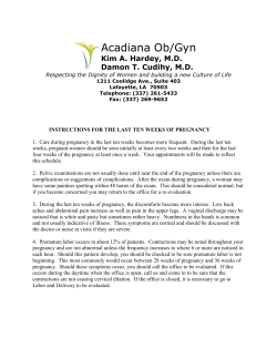
The Journal of Veterinary Science. Photon 116 (2015) 411-415
https://sites.google.com/site/photonfoundationorganization/home/the-journal-of-veterinary-science
Original Research Article. ISJN: 1784-6372: Impact Index: 5.80
The Journal of Veterinary Science
Ph ton
SERPINA 14 expression from endometrial epithelium in cows with embryonic
mortality and aborted
A. Risvanlia*, M. Yukselb, H. Bulutc, I. Sekerd, S. Kuld
a
Department of Obstetrics and Gynecology, Faculty of Veterinary Medicine, University of Firat, Elazig, Turkey
Department of Obstetrics and Gynecology, Faculty of Veterinary Medicine, University of Cumhuriyet, Sivas,
Turkey
c
Department of Virology, Faculty of Veterinary Medicine, University of Firat, Elazig, Turkey
d
Department of Zootechny, Faculty of Veterinary Medicine, Firat University, Elazig, Turkey
b
A. Risvanli, M. Yuksel, H. Bulut, I. Seker and S. Kul are
conferred with Kary Banks Mullis Research Award2015 in Veterinary Science by IASR
Article history:
Received: 15 July, 2014
Accepted: 17 July, 2014
Available online: 24 March, 2015
Keywords:
SERPINA 14, abortion, embryonic mortality, cow
Corresponding Author:
Risvanli A.*
Professor
E-mail: [email protected]
Yuksel M.
Assistant Professor
Bulut H.
Professor
Seker I.
Professor
Kul S.
Assistant Professor
Abstract
In recent years, many studies have been conducted
on SERPINA 14, which is one of the molecules
suggested to have a role in the occurrence and
continuation of pregnancy. In this study, it was
aimed to detect the presence of SERPINA 14
mRNA by the reverse transcription-polymerase
chain reaction (RT-PCR) in the endometrium of
cows in which embryonic mortality and aborted had
occurred. In this way, a novel approach can be
developed towards embryonic mortality and
abortion cases which lead to significant losses in
cattle farming. For this purpose, endometrial biopsy
samples were obtained from a total of 30 cows
which were embryonic mortality (n=10), aborted
(n=10) and normal pregnancy (n=10). The presence
of SERPINA 14 mRNA in the samples was
determined with RT-PCR. Consequently, SERPINA
14 mRNA was detected in the endometrial biopsy
samples of all groups. Therefore, it was concluded
that the role of SERPINA 14 in continuation of
pregnancy could be related to its amount rather
than its presence in cows and thereby the present
study should be supported by further studies.
Citation:
Risvanli A., Yuksel M., Bulut H., Seker I., Kul S., 2015.
SERPINA 14 expression from endometrial epithelium in cows
with embryonic mortality and aborted. The Journal of
Veterinary Science. Photon 116, 411-415.
All Rights Reserved with Photon.
Photon Ignitor: ISJN17846372D732124032015
1. Introduction
Embryonic mortalities and abortions are important
problems in animal breeding, especially dairy cattle
breeding. Embryonic mortalities and abortions lead
to great harm in the economy of cattle breeding. Be
it due to infectious or non-infectious reasons, the
ways these disorders develop are still being debated
today. It is known that intrauterine mortality of the
embryo and the fetus arises from interruption of
mechanisms enabling a healthy continuation of
pregnancy. The maternal immune system should
Ph ton
function in a healthy way for occurrence,
continuation and normal termination of pregnancy.
The maternal immune system tolerates the presence
of paternal alloantigens without affecting the antiinfectious mechanisms during pregnancy. Thus,
pregnancy is considered to continue through
establishment of immune tolerance. Incomplete
tolerance leads to abortions, embryonic mortalities
and termination of pregnancy through causing
411
other gestational pathologies (Koch and Platt,
2007; Tafuri et al., 1995).
the endometrium of cows with embryonic mortality
and aborted.
The uterine environment should be healthy for a
successful fertility. The endometrium is locally
prepared for transport of gametes and development
of the embryo during the estrous cycle. The
endometrium synthesizes and releases some
molecules for continuation of pregnancy and for
recognition by the mother. Uterine serpins/uterine
milk proteins (serpin peptidase inhibitor, clade A
[alpha-1 antiproteinase, antitrypsin], member 14,
SERPINA14) are basic glycoproteins of the serpin
super-family, which also includes serine peptidase
inhibitors (serpins). SERPINA 14 molecules are
major secretory proteins which were first described
in the 1980s as ‘ovine uterine serpins’ in the sheep
uterus, however, they were later determined to be
synthesized in the endometrium of all ruminants
during pregnancy (Bazer et al., 1979; Klein et al.,
2006; Leslie and Hansen et al., 1991; Moffatt et al.,
1987). Long term progesterone administration in
sheep and cattle are known to cause stimulation of
SERPINA 14 (Ing et al., 1989; Leslie et al., 1990;
Moffatt et al., 1987). It was observed that long term
administration of estradiol-17b and progesterone
together in sheep and cattle did not stimulate
SERPINA 14 excretion, however, administering
progesterone alone caused a decrease in SERPINA
14 mRNA excretion (Spencer et al., 1999).
2. Materials and Methods
SERPINA 14 is known to contribute to the
immunosupressive effect of progesterone (Skopets
et al., 1992). It has been suggested that SERPINA
14 inhibits lymphocyte functions in vitro and it has
been reported to play a role in the development of
maternal tolerance against the embryo (Hansen and
Newton, 1988). However, it has been suggested
that SERPINA 14 is not a predominant molecule in
cows during pregnancy, but that it should be in
significant amounts in the development of a
histotrophic environment during pregnancy as in
sheep (Segerson and Bazer, 1989). It was shown in
vitro that activation of uterine NK cells is
decreased by SERPINA 14 (Liu and Hansen, 1993,
Tekin and Hansen, 2002), whereas it has no effect
on proliferation of γδ T cells (these cells excrete
significant cytokines for continuation of
pregnancy) (Peltier et al., 2000). It has been
reported that SERPINA 14 prevents the negative
events related to antibodies which develop against
the embryo through making complexes with IgA
and IgM (Hansen and Newton, 1988). Despite all
these data, the immunomodulation mechanisms
could not be fully explained as specific receptors of
SERPINA 14 could not be detected yet.
1.1Objective of research
In the present study, it was aimed to determine the
presence of SERPINA 14 mRNA by RT-PCR in
Ph ton
2.1 Animals
This study was performed with a total of 30 cows
from various breeds and at different ages with
embryonic mortality (n=10), aborted (n=10) and
normally pregnant cows (n=10). Cows with aborted
were selected among the animals at different
periods of pregnancy, were brought to Ruminant
Clinic of Animal Hospital in Firat University,
animals which had experienced embryonic
mortality were selected from cattle breeding
enterprises and normal pregnant cows were
selected among animals in slaughterhouses in
Elazig and the surroundings, which were going to
be slaughtered without knowing that they were
pregnant.
2.2 Determination of embryonic mortalities
Embryonic mortalities were determined according
to the description by Prvanovic et al. (2009) with
ultrasonography using a 5 or 7.5 MHz linear probe
on the 17, 24, 35 and 45th days after artificial
insemination had been performed.
2.3 Obtaining endometrial biopsy samples
The samples were obtained with sterile uterine
biopsy catheters from the animals with aborted or
embryonic mortality once every week. The
endometrial samples of normal pregnant animals
were obtained from the uterus of animals which
were slaughtered in slaughterhouses. The obtained
samples were placed into eppendorf tubes without
being subjected to any procedure and stored at -80°
C until the analyses were carried out.
2.4 RNA extraction
The samples which were collected and stored at 80° C were kept at +4° C for one night to dissolve.
About 50 µg of the tissue samples were transferred
into eppendorf tubes. RNA isolations were done
using a commercial RNA isolation kit (ZR RNA
mini prep. Catalogue no: R 1065). For RNA
elution, approximately 30-40 µl of RNAaseDNAase-free water was added onto the RNA
isolation column and kept at room temperature for
2 minutes. The samples were centrifuged and RNA
was obtained.
2.5 Reverse transcription
The reverse transcription (RT) procedure was
performed in order to obtain cDNA in RNA
samples. In this study, the amplification of the 126base-pair (bp) part of SERPINA 14 gene was
aimed.
Forward:
5’ATATCATCTTCTCCCCCATGG-3’ and Reverse:
5’-GTGCACATCCAACAGTTTGG-3’
primers
412
were used to replicate the gene region. For control
the success of RT and PCR stages RNA extraction
in addition, the RT and PCR stages were realized
with
primers
(Forward:5’AAGTCTTTGGGTTCCGGG-3’ and Reverse:5’GGACATCTAAGGGCATCACA-3’)
for
amplification of 18S ribosomal RNAs in all cells.
Sterile dH2O was used as negative control samples.
approximately 100 volts. The electrophoresis
results were evaluated under UV light.
2.6 PCR
The PCR stage was realized using cDNA templates
obtained from the RT stage. While reverse primers
were used in RT stages, forward and reverse
primers were used together in the PCR stage.
3. Results and Discussion
2.7 Agarose gel electrophoresis
1.5% agarose gel was prepared in order to visualize
the amplification products following the RT-PCR
stage. The samples loaded onto the wells were
eluted for about 1 hour and 30 minutes at
2.8 Statistical analysis
A statistical calculation was not performed due to
similar SERPINA 14 mRNAs having been detected
on endometrial biopsy samples obtained from all
groups.
According to the electrophoresis results,
approximately 365 base pair (bp) of amplification
product was detected with 18S rRNA primers from
tissues of all groups included in the study
(pregnant, aborted, early embryonic mortality) by
RT-PCR. Using RNAs obtained from tissue
samples and SERPINA 14 specific primers,
approximately 126 bp of amplification product was
detected in all tissue samples in agarose gel (Figure
1).
Figure 1: RT-PCR products in 1.5% agarose gel
M; 100 bp DNA marker, Line 1-4; SERPINA 14 primers and amplification products of tissue samples obtained from the
pregnant group, Line 5-8; SERPINA 14 primers and amplification products of tissue samples obtained from the aborted
group, Line 9-13; SERPINA 14 primers and amplification products of tissue samples obtained from the early embryonic
mortality group, Line 14; SERPINA 14 primers and amplification products in which distiled water was used as template,
Line 15; 18S rRNA primers and amplification products in which distilled water was used as template, Line 16-17; 18S
rRNA primers and amplification products of tissue samples obtained in the pregnant group
SERPINA 14 is known to be excreted from the
endometrium during pregnancy and the estrous
cycle in pigs, sheep and cows, and this is known to
be regulated by estradiol and progesterone
(Bauersachs et al., 2005; Leslie et al., 1999; Padua
and Hansen, 2010). It is known that a great amount
of SERPINA 14 is excreted from the endometrium
of pregnant cows (Moffatt et al., 1987), which
Ph ton
prevents fetal rejection through inhibiting the
lymphocyte proliferation (Skopets et al., 1992) and
cytotoxic activity of NK cells (Liu and Hansen,
1993). This molecule is reported to stabilize
uteroferritin, an iron-binding protein, in pigs during
pregnancy and transports iron to the fetus (Padua
and Hansen, 2010). In cows, SERPINA 14 is
known to be low in the estrous phase of the cycle,
increase in mid-luteal phase, and reach its
413
maximum level during pregnancy (Ulbrich et al.,
2009). While SERPINA 14 secretion had been
reported to have been be regulated by only
progesterone previously, it was suggested
thereafter that it was regulated by estradiol in the
estrous cycle and by progesterone in the luteal
phase (Leslie and Hansen, 1991).
In the early periods of pregnancy, progesterone
suppresses the immune functions of the uterus by
stimulating the endometrial secretion of SERPINA
14 and thereby contributing to the occurrence and
continuation of pregnancy (Hansen, 1998).
SERPINA 14 mRNAs is up-regulated on the 18th
day of pregnancy in cows (Klein et al., 2006).
Progesterone, prolactin, placental lactogen, growth
hormone and IFNT have significant effects on the
expression of SERPINA 14 from the endometrium
(Leslie and Hansen, 1991; Moffatt et al., 1987;
Spencer and Bazer, 2002; Stewart et al., 2000).
SERPINA 14 has also been detected in uterine
fluid in the later periods of pregnancy in cows and
sheep (Padua and Hansen, 2010).
Conclusion
Despite all literature searches, no studies could be
found about endometrial expression of SERPINA
14 in cows with aborted and embryonic mortality.
In the present study, transcription of SERPINA 14
RNA was determined in all endometrial samples
obtained from cows in which aborted and early
embryonic mortality had occurred and which were
pregnant. Thus, no difference was found between
the groups for the presence of SERPINA 14 RNA.
The RT-PCR assay has sufficient sensitivity to
detect the small number of targeted RNA. Hence,
in this study, detection of SERPINA 14 RNAs in
all samples does not provide data about the amount
of SERPINA 14 RNA. We consider that the
amount, rather than the presence of SERPINA 14
RNA is important for continuation of pregnancy.
Therefore, it was concluded that determination of
the amount of SERPINA 14 will be more beneficial
than questioning the presence of SERPINA 14 in
related tissues.
Acknowledgments
This study was supported by Fırat University
Scientific Researches Projects Unit (FUBAPVF.11.05)
Authors’ Contribution and Competing Interests
All authors have read the manuscript and do not
have any conflicting of interests.
Ph ton
References
Bauersachs S., Ulbrich S.E., Gross K., Schmidt S.E.M.,
Meyer H.H.D., Einspanier R., 2005. Gene expression
profiling of bovine endometrium during the oestrous
cycle: detection of molecular pathways involved in
functional changes. Journal of Molecular Endocrinology
34, 889-908.
Bazer F.W., Roberts R.M., Basha S.M., Zavy M.T.,
Caton D., Barron D.H., 1979. Method for obtaining
ovine uterine secretions from unilaterally pregnant ewes.
Journal of Animal Science 49, 1522–1527.
Hansen P.J., Newton G.R., 1988. Binding of
immunoglobulins to the major progesterone-induced
proteins secreted by the sheep uterus. Archives of
Biochemistry and Biophysics 260, 208–217.
Hansen P.J., Tekin S., 2005. Pregnancy-associated
immunoregulatory molecules discovered in ruminants
and their possible relevance to other species. Chemical
Immunology Allergy, 88, 109–116.
Hansen P.J. 1998. Regulation of uterine immune function
by progesterone-lessons from the sheep. Journal of
Reproductive Immunology 40, 63-79.
Ing N.H., Francis H., McDonnell J.J., Amann J.F.,
Roberts R.M., 1989. Progesterone nduction of the uterine
milk proteins: major secretory proteins of sheep
endometrium. Biology of Reproduction 41, 643–654.
Klein C., Bauersachs S., Ulbrich S.E. Einspanier R.,
Meyer H.H., Schmidt S.E., Reichenbach H.D.
Vermehren M., Sinowatz F., Blum H., Wolf E., 2006.
Monozygotic twin model reveals novel embryo-induced
transcriptome changes of bovine endometrium in the
preattachment period. Biology of Reproduction 74, 253264.
Koch C.A., Platt J.L., 2007. T cell recognition and
immunity in the fetus and mother. Cellular Immunology
248, 12–17.
Leslie M.V., Hansen P.J., Newton G.R., 1990. Uterine
secretions of the cow contain proteins that are
immunochemically related to the major progesterone
induced proteins of the sheep uterus. Domestic Animal
Endocrinology 7, 517–526.
Leslie M.V., Hansen P.J., 1991. Progesterone-regulated
secretion of the serpin-like proteins of the ovine and
bovine uterus. Steroids 56, 589–597.
Liu W.J., Hansen P.J., 1993. Effect of the progesteroneinduced serpin-like proteins of the sheep endometrium
on natural-killer cell activity in sheep and mice. Biology
of Reproduction 49, 1008–1014.
Moffatt R.J., Bazer F.W., Hansen P.J. Chun P.W.,
Roberts R.M., 1987a. Purification, secretion and
immunocytochemical localization of the uterine milk
proteins, major progesterone-induced proteins in uterine
secretions of the sheep. Biology of Reproduction 36,
419–430.
414
Moffatt R.J., Bazer F.W., Roberts R.M. Thatcher W.W.,
1987b. Secretory function of the ovine uterus: effects of
gestation and steroid replacement therapy. Journal of
Animal Science 65, 1400-1410.
Padua M.B., Hansen P.J., 2010. Evolution and function
of the uterine serpins (SERPINA14). American Journal
of Reproductive Immunology 64, 265-274.
Ulbrich S.E., Frohlich T., Schulke K., Englberger E.,
Waldschmitt N., Arnold J.G., Reichenbach H.D.
Reichenbach M., Wolf E., Meyer H.D.H., Bauersach S.,
2009. Evidence for estrogen-dependent uterine serpin
(SERPINA14) expression during estrus in the bovine
endometrial glandular epithelium and lumen. Biology of
Reproduction 81, 795–805.
Peltier M.R., Liu W.J., Hansen P.J., 2000. Regulation of
lymphocyte proliferation by uterine serpin: interleukin-2
mRNA production, CD25 expression and responsiveness
to interleukin-2. Proceedings of the Society for
Experimental Biology and Medicine 223, 75–81.
Prvanović N., Tomašković A., Grizelj J., Kočila P.,
Samardžija M., 2009. Monitoring of early pregnancy and
early embryonic mortality by ultrasound and
determination of pregnancy-associated glycoproteins and
progesterone in cows. Veterinarski Arhiv 79, 259-267.
Segerson E.C., Bazer F.W., 1992. High molecular weight
basic and acidic immunosuppressive protein components
in uterine secretions of pregnant cows. Biology of
Reproduction 41, 1014–1023.
Skopets B., Li J., Thatcher W.W., Roberts R.M., Hansen
P.J., 1992. Inhibition of lymphocyte proliferation by
bovine trophoblast protein-1 (type I trophoblast
interferon) and bovine interferon-alpha. Veterinary
Immunology and Immunopathology 34, 81-96.
Spencer T.E., Bazer, F.W., 2002. Biology of
progesterone action during pregnancy recognition and
maintenance of pregnancy. Frontiers in Bioscience 7:
1879–1898.
Spencer T.E., Gray A., Johnson G.A., Taylor K.M.,
Gertler. A., Gootwine E., Ott T.L., Bazer F.W., 1999.
Effects of recombinant ovine interferon tau, placental
lactogen, and growth hormone on the ovine uterus.
Biology of Reproduction 61, 1409–1418.
Stewart M.D., Johnson. G.A., Gray C.A., Burghardt
R.C., Schuler L.A., Joyce M.M., Bazer F.W., Spencer
T.E., 2000. Prolactin receptor and uterine milk protein
expression in the ovine endometrium during the estrous
cycle and pregnancy. Biology of Reproduction 62, 1779–
1789.
Tafuri A., Alferink J., Moller P., Hammerling G.J.,
Arnold B., 1995. T cell awareness of paternal
alloantigens during pregnancy. Science 270, 630–633.
Tekin S., Hansen P.J., 2002. Natural killer-like cells in
the sheep: functional characterization and regulation by
pregnancy-associated proteins. Exp. Biol. Med.
(Maywood), 227, 803–811.
Thavaneetharajah P., 2011. Comparison of gene
expression levels in embryo, endometrium and corpus
luteum of dairy heifers and lactating dairy cows and
manipulatıon of endometrial gene expression in-vitro.
The University of British Columbia. Doctoral thesis,
Vancouver.
Ph ton
415


© Copyright 2026