
Guidelines for pre-operative cardiac risk assessment and perioperative cardiac
European Heart Journal (2009) 30, 2769–2812
doi:10.1093/eurheartj/ehp337
ESC GUIDELINES
Guidelines for pre-operative cardiac risk
assessment and perioperative cardiac
management in non-cardiac surgery
The Task Force for Preoperative Cardiac Risk Assessment and
Perioperative Cardiac Management in Non-cardiac Surgery of the
European Society of Cardiology (ESC) and endorsed by the
European Society of Anaesthesiology (ESA)
Authors/Task Force Members: Don Poldermans; (Chairperson) (The Netherlands)*;
Jeroen J. Bax (The Netherlands); Eric Boersma (The Netherlands); Stefan De Hert
(The Netherlands); Erik Eeckhout (Switzerland); Gerry Fowkes (UK);
Bulent Gorenek (Turkey); Michael G. Hennerici (Germany); Bernard Iung (France);
Malte Kelm (Germany); Keld Per Kjeldsen (Denmark); Steen Dalby Kristensen
(Denmark); Jose Lopez-Sendon (Spain); Paolo Pelosi (Italy); François Philippe
(France); Luc Pierard (Belgium); Piotr Ponikowski (Poland); Jean-Paul Schmid
(Switzerland); Olav F.M. Sellevold (Norway); Rosa Sicari (Italy);
Greet Van den Berghe (Belgium); Frank Vermassen (Belgium)
Additional Contributors: Sanne E. Hoeks (The Netherlands);
Ilse Vanhorebeek (Belgium)
ESC Committee for Practice Guidelines (CPG): Alec Vahanian; (Chairperson) (France); Angelo Auricchio
(Switzerland); Jeroen J. Bax (The Netherlands); Claudio Ceconi (Italy); Veronica Dean (France); Gerasimos Filippatos
(Greece); Christian Funck-Brentano (France); Richard Hobbs (UK); Peter Kearney (Ireland); Theresa McDonagh (UK);
Keith McGregor (France); Bogdan A. Popescu (Romania); Zeljko Reiner (Croatia); Udo Sechtem (Germany);
Per Anton Sirnes (Norway); Michal Tendera (Poland); Panos Vardas (Greece); Petr Widimsky (Czech Republic)
Document Reviewers: Raffaele De Caterina; (CPG Review Coordinator) (Italy); Stefan Agewall (Norway);
Nawwar Al Attar (France); Felicita Andreotti (Italy); Stefan D. Anker (Germany); Gonzalo Baron-Esquivias (Spain);
Guy Berkenboom (Belgium); Laurent Chapoutot (France); Renata Cifkova (Czech Republic); Pompilio Faggiano
(Italy); Simon Gibbs (UK); Henrik Steen Hansen (Denmark); Laurence Iserin (France); Carsten W. Israel
(Germany); Ran Kornowski (Israel); Nekane Murga Eizagaechevarria (Spain); Mauro Pepi (Italy); Massimo Piepoli
(Italy); Hans Joachim Priebe (Germany); Martin Scherer (Germany); Janina Stepinska (Poland); David Taggart (UK);
Marco Tubaro (Italy)
The disclosure forms of all the authors and reviewers are available on the ESC website www.escardio.org/guidelines
* Corresponding author: Don Poldermans, Department of Surgery, Erasmus Medical Center, Gravendijkwal 230, 3015 CE Rotterdam, The Netherlands. Tel: þ31 10 703 4613,
Fax: þ31 10 436 4557, Email: [email protected]
The content of these European Society of Cardiology (ESC) Guidelines has been published for personal and educational use only. No commercial use is authorized. No part of the
ESC Guidelines may be translated or reproduced in any form without written permission from the ESC. Permission can be obtained upon submission of a written request to Oxford
University Press, the publisher of the European Heart Journal and the party authorized to handle such permissions on behalf of the ESC.
Disclaimer. The ESC Guidelines represent the views of the ESC and were arrived at after careful consideration of the available evidence at the time they were written. Health
professionals are encouraged to take them fully into account when exercising their clinical judgement. The guidelines do not, however, over-ride the individual responsibility of
health professionals to make appropriate decisions in the circumstances of the individual patients, in consultation with that patient, and where appropriate and necessary the patient’s
guardian or carer. It is also the health professional’s responsibility to verify the rules and regulations applicable to drugs and devices at the time of prescription.
& The European Society of Cardiology 2009. All rights reserved. For permissions please email: [email protected].
2770
ESC Guidelines
----------------------------------------------------------------------------------------------------------------------------------------------------------Keywords
Non-cardiac surgery † Pre-operative cardiac risk assessment † Pre-operative cardiac testing † Pre-operative coronary
artery revascularization † Perioperative cardiac management † Renal disease † Pulmonary disease † Neurological
disease † Anaesthesiology † Post-operative cardiac surveillance
Table of Contents
List of acronyms and abbreviations . . . . . .
Preamble . . . . . . . . . . . . . . . . . . . . . . .
Introduction . . . . . . . . . . . . . . . . . . . . .
Magnitude of the problem . . . . . . . . .
Impact of the ageing population . . . . .
Purpose . . . . . . . . . . . . . . . . . . . . .
Pre-operative evaluation . . . . . . . . . . . . .
Surgical risk for cardiac events . . . . . .
Functional capacity . . . . . . . . . . . . . .
Risk indices . . . . . . . . . . . . . . . . . . .
Biomarkers . . . . . . . . . . . . . . . . . . .
Non-invasive testing . . . . . . . . . . . . .
Angiography . . . . . . . . . . . . . . . . . .
Risk reduction strategies . . . . . . . . . . . . .
Pharmacological . . . . . . . . . . . . . . . .
Revascularization . . . . . . . . . . . . . . .
Specific diseases . . . . . . . . . . . . . . . . . .
Chronic heart failure . . . . . . . . . . . .
Arterial hypertension . . . . . . . . . . . .
Valvular heart disease . . . . . . . . . . . .
Arrhythmias . . . . . . . . . . . . . . . . . .
Renal disease . . . . . . . . . . . . . . . . .
Cerebrovascular disease . . . . . . . . . .
Pulmonary disease . . . . . . . . . . . . . .
Perioperative monitoring . . . . . . . . . . . .
Electrocardiography . . . . . . . . . . . . .
Transoesophageal echocardiography . .
Right heart catherization . . . . . . . . . .
Disturbed glucose metabolism . . . . . .
Anaesthesia . . . . . . . . . . . . . . . . . . . . .
Intraoperative anaesthetic management
Neuraxial techniques . . . . . . . . . . . .
Post-operative pain management . . . . .
Putting the puzzle together . . . . . . . . . . .
References . . . . . . . . . . . . . . . . . . . . . .
.
.
.
.
.
.
.
.
.
.
.
.
.
.
.
.
.
.
.
.
.
.
.
.
.
.
.
.
.
.
.
.
.
.
.
.
.
.
.
.
.
.
.
.
.
.
.
.
.
.
.
.
.
.
.
.
.
.
.
.
.
.
.
.
.
.
.
.
.
.
.
.
.
.
.
.
.
.
.
.
.
.
.
.
.
.
.
.
.
.
.
.
.
.
.
.
.
.
.
.
.
.
.
.
.
.
.
.
.
.
.
.
.
.
.
.
.
.
.
.
.
.
.
.
.
.
.
.
.
.
.
.
.
.
.
.
.
.
.
.
.
.
.
.
.
.
.
.
.
.
.
.
.
.
.
.
.
.
.
.
.
.
.
.
.
.
.
.
.
.
.
.
.
.
.
.
.
.
.
.
.
.
.
.
.
.
.
.
.
.
.
.
.
.
.
.
.
.
.
.
.
.
.
.
.
.
.
.
.
.
.
.
.
.
.
.
.
.
.
.
.
.
.
.
.
.
.
.
.
.
.
.
.
.
.
.
.
.
.
.
.
.
.
.
.
.
.
.
.
.
.
.
.
.
.
.
.
.
.
.
.
.
.
.
.
.
.
.
.
.
.
.
.
.
.
.
.
.
.
.
.
.
.
.
.
.
.
.
.
.
.
.
.
.
.
.
.
.
.
.
.
.
.
.
.
.
.
.
.
.
.
.
.
.
.
.
.
.
.
.
.
.
.
.
.
.
.
.
.
.
.
.
.
.
.
.
.
.
.
.
.
.
.
.
.
.
.
.
.
.
.
.
.
.
.
.
.
.
.
.
.
.
.
.
.
.
.
.
.
.
.
.
.
.
.
.
.
.
.
.
.
.
.
.
.
.
.
.
.
.
.
.
.
.
.
.
.
.
.
.
.
.
.
.
.
.
.
.
.
.
.
.
.
.
.
.
.
.
.
.
.
.
.
.
.
.
.
.
.
.
.
.
.
.
.
.
.
.
.
.
.
.
.
.
.
.
.
.
.
.
.
.
.
.
.
.2770
.2771
.2771
.2771
.2773
.2773
.2774
.2774
.2775
.2776
.2777
.2777
.2780
.2781
.2781
.2789
.2792
.2792
.2793
.2793
.2794
.2795
.2796
.2797
.2799
.2799
.2799
.2800
.2801
.2802
.2803
.2803
.2803
.2803
.2807
List of acronyms and abbreviations
AAA
ACC
ACE
ACS
AHA
AR
ARB
AS
abdominal aortic aneurysm
American College of Cardiology
angiotensin-converting enzyme
acute coronary syndrome
American Heart Association
aortic regurgitation
angiotensin receptor blocker
aortic stenosis
AF
BBSA
BNP
CABG
CARP
CASS
CI
COX-2
COPD
CPET
CPG
CRP
CT
cTnI
cTnT
CVD
DECREASE
atrial fibrillation
b-blocker in spinal anaesthesia
brain natriuretic peptide
coronary artery bypass grafting
coronary artery revascularization prophylaxis
coronary artery surgery study
confidence interval
cyclooxygenase-2
chronic obstructive pulmonary disease
cardiopulmonary exercise testing
Committee for Practice Guidelines
C-reactive protein
computed tomography
cardiac troponin I
cardiac troponin T
cardiovascular disease
Dutch Echocardiographic Cardiac Risk Evaluating
Applying Stress Echo
DES
drug-eluting stent
DIPOM
Diabetes Postoperative Mortality and Morbidity
DSE
dobutamine stress echocardiography
ECG
electrocardiography
ESC
European Society of Cardiology
FEV1
forced expiratory volume in 1 s
FRISC
fast revascularization in instability in coronary
disease
HR
hazard ratio
ICU
intensive care unit
IHD
ischaemic heart disease
INR
international normalized ratio
LMWH
low molecular weight heparin
LQTS
long QT syndrome
LR
likelihood ratio
LV
left ventricular
MaVS
metoprolol after surgery
MET
metabolic equivalent
MI
myocardial infarction
MR
mitral regurgitation
MRI
magnetic resonance imaging
MS
mitral stenosis
NICE-SUGAR normoglycaemia in intensive care evaluation and
survival using glucose algorithm regulation
NSTEMI
non-ST-segment elevation myocardial infarction
NT-proBNP N-terminal pro-brain natriuretic peptide
NYHA
New York Heart Association
OPUS
orbofiban in patients with unstable coronary
syndromes
OR
odds ratio
PaCO2
mixed expired volume of alveolar and dead space
gas
2771
ESC Guidelines
PAH
PETCO2
PCI
PDA
POISE
QUO-VADIS
ROC
SD
SMVT
SPECT
SPVT
STEMI
SVT
SYNTAX
TACTICS
TIA
TIMI
TOE
UFH
VCO2
VE
VHD
VKA
VO2
VPB
VT
pulmonary arterial hypertension
end-tidal expiratory CO2 pressure
percutaneous coronary intervention
personal digital assistant
PeriOperative ISchaemic Evaluation trial
QUinapril On Vascular ACE and Determinants of
ISchemia
receiver operating characteristic
standard deviation
sustained monomorphic ventricular tachycardia
single photon emission computed tomography
sustained polymorphic ventricular tachycardia
ST-segment elevation myocardial infarction
supraventricular tachycardia
synergy between percutaneous coronary intervention with taxus and cardiac surgery
treat angina with aggrastat and determine cost of
therapy with an invasive or conservative strategy
transient ischaemic attack
thrombolysis in myocardial infarction
transoesophageal echocardiography
unfractionated heparin
carbon dioxide production
minute ventilation
valvular heart disease
vitamin K antagonist
oxygen consumption
ventricular premature beat
ventricular tachycardia
Preamble
Guidelines and Expert Consensus Documents aim to present management and recommendations based on the relevant evidence on
a particular subject in order to help physicians to select the best
possible management strategies for the individual patient suffering
from a specific condition, taking into account not only the impact
on outcome, but also the risk– benefit ratio of particular diagnostic
or therapeutic means. Guidelines are no substitutes for textbooks.
The legal implications of medical guidelines have been discussed
previously.1
A great number of Guidelines and Expert Consensus Documents have been issued in recent years by the European Society
of Cardiology (ESC) and also by other organizations or related
societies. Because of the impact on clinical practice, quality criteria
for development of guidelines have been established in order to
make all decisions transparent to the user. The recommendations
for formulating and issuing ESC guidelines and Expert Consensus
Documents can be found on the ESC website in the guidelines
section (www.escardio.org).
In brief, experts in the field are selected and undertake a comprehensive review of the published evidence for management and/
or prevention of a given condition. A critical evaluation of diagnostic and therapeutic procedures is performed, including assessment
of the risk–benefit ratio. Estimates of expected health outcomes
for larger societies are included, where data exist. The level of
evidence and the strength of recommendation of particular
treatment options are weighted and graded according to predefined scales, as outlined in Tables 1 and 2.
The experts of the writing panels have provided disclosure
statements of all relationships they may have which might be perceived as real or potential sources of conflicts of interest. These
disclosure forms are kept on file at the European Heart House,
headquarters of the ESC. Any changes in conflict of interest that
arise during the writing period must be notified to the ESC. The
Task Force report is entirely supported financially by the ESC
without any involvement of industry.
The ESC Committee for Practice Guidelines (CPG) supervises
and coordinates the preparation of new Guidelines and Expert
Consensus Documents produced by Task Forces, expert groups,
or consensus panels. The Committee is also responsible for the
endorsement process of these Guidelines and Expert Consensus
Documents or statements. Once the document has been finalized
and approved by all the experts involved in the Task Force, it is
submitted to outside specialists for review. The document is
revised, and finally approved by the CPG and subsequently
published.
After publication, dissemination of the message is of paramount
importance. Pocketsize versions and personal digital assistant
(PDA)-downloadable versions are useful at the point of care.
Some surveys have shown that the intended end-users are sometimes not aware of the existence of guidelines, or simply do not
translate them into practice, so this is why implementation programmes for new guidelines form an important component of
the dissemination of knowledge. Meetings are organized by the
ESC, and are directed towards its member National Societies
and key opinion leaders in Europe. Implementation meetings can
also be undertaken at national levels, once the guidelines have
been endorsed by the ESC member societies, and translated into
the national language. Implementation programmes are needed
because it has been shown that the outcome of disease may be
favourably influenced by the thorough application of clinical
recommendations.2
Thus, the task of writing Guidelines or Expert Consensus Documents covers not only the integration of the most recent research,
but also the creation of educational tools and implementation programmes for the recommendations. The development of clinical
guidelines and implementation into clinical practice can then only
be completed if surveys and registries are performed to verify its
use in real-life daily practices. Such surveys and registries also
make it possible to evaluate the impact of implementation of the
guidelines on patient outcomes. Guidelines and recommendations
should help physicians and other healthcare providers to make
decisions in their daily practice. However, the physician in charge
of his/her care must make the ultimate judgement regarding the
care of an individual patient.
Introduction
Magnitude of the problem
The present guidelines focus on the cardiological management of
patients undergoing non-cardiac surgery, i.e. patients where heart
2772
ESC Guidelines
Table 1
Classes of recommendations
Table 2
Level of evidence
disease is a potential source of complications during surgery. The
risk of perioperative complications depends on the condition of
the patient prior to surgery, the prevalence of co-morbidities,
and the magnitude and duration of the surgical procedure.3
More specifically, cardiac complications can arise in patients with
documented or asymptomatic ischaemic heart disease (IHD), left
ventricular (LV) dysfunction, and valvular heart disease (VHD)
who undergo procedures that are associated with prolonged
haemodynamic and cardiac stress. In the case of perioperative
myocardial ischaemia, two mechanisms are important: (i) chronic
mismatch in the supply-to-demand ratio of blood flow response
to metabolic demand, which clinically resembles stable IHD due
to a flow limiting stenosis in coronary conduit arteries; and (ii) coronary plaque rupture due to vascular inflammatory processes presenting as acute coronary syndromes (ACSs). Hence, although LV
dysfunction may occur for various reasons in younger age groups,
perioperative cardiac mortality and morbidity are predominantly
an issue in the adult population undergoing major non-cardiac
surgery.
The magnitude of the problem in Europe can best be understood in terms of (i) the size of the adult non-cardiac surgical
cohort; and (ii) the average risk of cardiac complications within
this cohort. Unfortunately, at a European level, no systematic
data are available on the annual number and type of operations,
nor on patient outcome. Information is collected at the national
level in several countries, but data definitions, amount of data,
and data quality vary greatly. In The Netherlands, with a population
of 16 million, throughout 1991–2005, 250 000 major surgical procedures were conducted on average annually in patients above the
age of 20 years, implying an annual rate of 1.5%.4 When applied to
Europe, with an overall population of 490 million, this figure translates into a crude estimate of 7 million major procedures annually
in patients who present with cardiac risk.
Data on cardiac outcome can be derived from the few
large-scale clinical trials and registries that have been undertaken
in patients undergoing non-cardiac surgery. Lee et al. studied
4315 patients undergoing elective major non-cardiac procedures
in a tertiary care teaching hospital throughout 1989– 1994.5 They
2773
ESC Guidelines
observed that 92 (2.1%) patients suffered major cardiac complications, including cardiac death and myocardial infarction (MI). In
a cohort of 108 593 consecutive patients who underwent
surgery throughout 1991–2000 in a university hospital in The
Netherlands, perioperative mortality occurred in 1877 (1.7%)
patients, with a cardiovascular cause being identified in 543 cases
(0.5%).6 The Dutch Echocardiographic Cardiac Risk Evaluating
Applying Stress Echo (DECREASE) -I, -II and -IV trials enrolled
3893 surgical patients throughout 1996–2008, and these comprised intermediate- and high-risk patients of whom 136 (3.5%)
suffered perioperative cardiac death or MI.7 – 9 A final piece of
evidence with respect to patient outcome is derived from the
Perioperative Ischaemic Evaluation (POISE) trial, which was conducted throughout 2002– 2007, and enrolled 8351 patients undergoing non-cardiac surgery.10 Perioperative mortality occurred in
226 patients (2.7%), of whom 133 (1.6%) suffered cardiovascular
death, whereas non-fatal MI was observed in another 367 (4.4%)
subjects. Differences in incidences between the studies are
mainly explained by patient selection and endpoint MI definitions—major non-cardiac surgery is associated with an incidence
of cardiac death of between 0.5 and 1.5%, and of major cardiac
complications of between 2.0 and 3.5%. When applied to the
population in the European Union member states these figures
translate into 150 000 –250 000 life-threatening cardiac complications due to non-cardiac surgical procedures annually.
Impact of the ageing population
Within the next 20 years, the acceleration in ageing of the population will have a major impact on perioperative patient management. It is estimated that elderly people require surgery four
times more often than the rest of the population.11 Although
exact data regarding the number of patients undergoing surgery
in Europe are lacking, it is estimated that this number will increase
by 25% by 2020, and for the same time period the elderly population will increase by .50%. The total number of surgical procedures will increase even faster because of the rising frequency
of interventions with age.12 Results of the US National Hospital
Discharge Survey show that, in general, the number of surgical procedures will increase in almost all age groups, but that the largest
increase will occur in the middle aged and elderly (Table 3).
Demographics of patients undergoing surgery show a trend
towards an increasing number of elderly patients and
co-morbidities.13 Although mortality from cardiac disease is
decreasing in the general population, the prevalence of IHD,
heart failure, and cardiovascular risk factors, especially diabetes,
is increasing. Among the significant co-morbidities in elderly
patients presenting for general surgery, cardiovascular disease
(CVD) is the most prevalent. It is estimated from primary care
data that in the 75 –84 year age group 19% of men and 12% of
women have some degree of CVD.14 Age per se, however,
seems to be responsible for only a small increase in the risk of
complications; greater risks are associated with urgency and significant cardiac, pulmonary, and renal disease. The number of affected
individuals is likely to be higher in countries with high CVD mortality, particularly in Central and Eastern Europe. These conditions
should, therefore, have a greater impact on the evaluation of
patient risk than age alone.
Table 3 Change in numbers of discharges for surgical
procedures by age for the time periods 1994/95 and
2004/05 as reported from the 2005 US National
Hospital Discharge Survey (non-federal short-stay
hospitals)15
Age (years)
Number of procedures
(in thousands)
% change
...............................
1994/95
2004/05
7311
4111
7326
5210
................................................................................
18–44
45–64
65–74
3069
3036
75 and over
18 and over
3479
17 969
4317
19 889
þ2.1
þ26.7
21.1
þ24.1
þ10.7
Purpose
Currently there are no official ESC guidelines on pre-operative risk
assessment and perioperative cardiac management. The objective
is to endorse a standardized and evidence-based approach to perioperative cardiac management. The guidelines recommend a practical, stepwise evaluation of the patient, which integrates clinical
risk factors and test results with the estimated stress of the
planned surgical procedure. This results in an individualized
cardiac risk assessment, with the opportunity to initiate medical
therapy, coronary interventions, and specific surgical and anaesthetic techniques in order to optimize the patient’s perioperative
condition. Compared with the non-surgical setting, data from randomized clinical trials, which are the ideal evidence base for the
guidelines, are sparse. Therefore, when no trials are available on
a specific cardiac management regimen in the surgical setting,
data from the non-surgical setting are used, and similar recommendations made, but with different levels of evidence. Emphasis is
placed on the restricted use of prophylactic coronary revascularization, as this is rarely indicated simply to ensure the patient survives surgery. Pre-operative evaluation requires an integrated
multidisciplinary approach from anaesthesiologists, cardiologists,
internists, pulmonologists, geriatricians, and surgeons. Anaesthesiologists, who are experts on the specific demands of the proposed surgical procedure, usually coordinate the process.
Guidelines have the potential to improve post-operative
outcome. However, as shown in an observational study of 711 vascular surgery patients from The Netherlands, adherence to guidelines is poor.16 – 18 Although 185 of a total of 711 patients (26%)
fulfilled the ACC/AHA guideline criteria for pre-operative noninvasive cardiac testing, clinicians had performed testing in only
38 of those cases (21%).16 The guideline-recommended medical
therapy for the perioperative period, namely the combination of
aspirin and statins in all patients and b-blockers in patients with
ischaemic heart disease, was followed in only 41% of cases.18
Significantly, the use of evidence-based medication during the perioperative period was associated with a reduction in 3-year mortality after adjustment for clinical characteristics [hazard ratio
(HR), 0.65; 95% confidence interval (CI), 0.45–0.94]. These data
2774
ESC Guidelines
highlight the existence of a clear opportunity for improving the
quality of care in this high-risk group of patients.
In addition to promoting an improvement in immediate perioperative care, guidelines should provide long-term advice, as
patients should live long enough to enjoy the benefits of surgery.
Following the development and introduction of perioperative
cardiac guidelines, their effect on outcome should be monitored.
The objective evaluation of changes in outcome will be an essential
part of future perioperative guideline developments.
Pre-operative evaluation
Surgical risk for cardiac events
Cardiac complications after non-cardiac surgery depend not only
on specific risk factors but also on the type of surgery and the circumstances under which it takes place.19 Surgical factors that influence cardiac risk are related to the urgency, magnitude, type, and
duration of the procedure, as well as the change in body core
temperature, blood loss, and fluid shifts.12
Every operation elicits a stress response. This response is
initiated by tissue injury and mediated by neuroendocrine
factors, and may induce tachycardia and hypertension. Fluid shifts
in the perioperative period add to the surgical stress. This stress
increases myocardial oxygen demand. Surgery also causes alterations in the balance between prothrombotic and fibrinolytic
factors, resulting in hypercoagulability and possible coronary
thrombosis (elevation of fibrinogen and other coagulation
factors, increased platelet activation and aggregation, and
reduced fibrinolysis). The extent of such changes is proportionate
to the extent and duration of the intervention. All these factors
may cause myocardial ischaemia and heart failure. Certainly in
patients at elevated risk, attention to these factors should be
given and lead, if indicated, to adaptations in the surgical plan.
Table 4
a
Although patient-specific factors are more important than
surgery-specific factors in predicting the cardiac risk for noncardiac surgical procedures, the type of surgery cannot be
ignored when evaluating a particular patient undergoing an intervention.6,20 With regard to cardiac risk, surgical interventions can
be divided into low-risk, intermediate-risk, and high-risk groups
with estimated 30-day cardiac event rates (cardiac death and MI)
of ,1, 1–5, and .5%, respectively (Table 4). Although only a
rough estimation, this risk stratification provides a good indication
of the need for cardiac evaluation, drug treatment, and assessment
of risk for cardiac events.
The high-risk group consists of major vascular interventions. In
the intermediate-risk category the risk also depends on the magnitude, duration, location, blood loss, and fluid shifts related to the
specific procedure. In the low-risk category the cardiac risk is negligible unless strong patient-specific risk factors are present.
The need for, and value of, pre-operative cardiac evaluation will
also depend on the urgency of surgery. In the case of emergency
surgical procedures, such as those for ruptured abdominal aortic
aneurysm (AAA), major trauma, or for perforated viscus, cardiac
evaluation will not change the course and result of the intervention
but may influence the management in the immediate postoperative period. In non-emergent but urgent untreated surgical
conditions such as bypass for acute limb ischaemia or treatment
of bowel obstruction, the morbidity and mortality of the untreated
underlying condition will outweigh the potential cardiac risk
related to the intervention. In these cases, cardiological evaluation
may influence the perioperative measures taken to reduce the
cardiac risk, but will not influence the decision to perform the
intervention. In some cases, the cardiac risk can also influence
the type of operation and guide the choice to less invasive interventions, such as peripheral arterial angioplasty instead of infrainguinal bypass, or extra-anatomic reconstruction instead of
aortic procedure, even when these may yield less favourable
Surgical riska estimate (modified from Boersma et al. 6)
Risk of MI and cardiac death within 30 days after surgery.
2775
ESC Guidelines
Table 5
Lee index and Erasmus model: clinical risk factors used for pre-operative cardiac risk stratification5,6
Clinical characteristics
Lee index
Erasmus model
IHD (angina pectoris and/or MI)
x
x
Surgical risk
Heart failure
High-risk surgery
x
High, intermediate-high, intermediate-low, low risk
x
...............................................................................................................................................................................
Stroke/transient ischaemic attack
x
x
Diabetes mellitus requiring insulin therapy
Renal dysfunction/haemodialysis
x
x
x
x
Age
x
IHD ¼ ischaemic heart disease; MI ¼ myocardial infarction.
results in the long term. Lastly, in some situations, the cardiac
evaluation, in as far as it can reliably predict perioperative cardiac
complications and estimate late survival, should be taken into consideration even when deciding whether to perform an intervention
or not. This is the case in certain prophylactic interventions such as
the treatment of small AAAs or asymptomatic carotid stenosis
where the life expectancy of the patient and the risk of the operation are important factors in evaluating the potential benefit of
the surgical intervention.
Vascular interventions are of specific interest, not only because
they carry the highest risk of cardiac complications, explained by
the high probability that the atherosclerotic process also affects
the coronary arteries, but also because of the many studies that
have shown that this risk can be influenced by adequate perioperative measures in these patients. Open aortic and infra-inguinal procedures have both to be considered as high-risk procedures.6
Although a less extensive intervention, infra-inguinal revascularization entails a cardiac risk similar to or even higher than aortic procedures. This can be explained by the higher incidence of
diabetes, renal dysfunction, IHD, and advanced age in this patient
group. This also explains why the risk related to peripheral artery
angioplasties, which are minimally invasive procedures, is not negligible. Several randomized trials, as well as community-based studies,
have shown that the cardiac risk is substantially lower after endovascular aortic aneurysm repair compared with open repair.21 This can
be related to the lesser tissue damage and the avoidance of aortic
cross-clamping and post-operative ileus. However, long-term survival does not seem to be influenced by the surgical technique that is
used, but is determined by the underlying cardiac disease.22 Carotid
endarterectomy is considered to be an intermediate-risk procedure.
Nevertheless, elevated cardiac risk and late survival should be taken
into account in the decision-making process and can influence the
choice between endarterectomy or stenting.
Laparoscopic procedures have the advantage of causing less
tissue trauma and intestinal paralysis compared with open procedures, resulting in less incisional pain and diminished postoperative fluid shifts related to bowel paralysis.23 On the other
hand, the pneumoperitoneum used in these procedures results
in elevated intra-abdominal pressure and a reduction in venous
return. It will result in a decrease in cardiac output and an increase
in systemic vascular resistance. Therefore, cardiac risk in patients
with heart failure is not diminished in patients undergoing
laparoscopy compared with open surgery, and both should be
evaluated in the same way. This is especially true in patients undergoing interventions for morbid obesity.24,25
Recommendation/statement on surgical risk estimate
Recommendation/statement
Classa
Levelb
Laparoscopic procedures demonstrate a cardiac
stress similar to open procedures and it is
recommended that patients be screened prior
to intervention accordingly
I
A
................................................................................
a
Class of recommendation.
Level of evidence.
b
Functional capacity
Determination of functional capacity is considered to be a pivotal
step in pre-operative cardiac risk assessment. Functional capacity is
measured in metabolic equivalents (METs). One MET equals the
basal metabolic rate. Exercise testing provides an objective assessment of functional capacity. Without testing, functional capacity
can be estimated by the ability to perform the activities of daily
living. Given that 1 MET represents metabolic demand at rest,
climbing two flights of stairs demands 4 METs, and strenuous
sports such as swimming .10 METS (Figure 1).
The inability to climb two flights of stairs or run a short distance
(,4 METs) indicates poor functional capacity and is associated
with an increased incidence of post-operative cardiac events.
After thoracic surgery, a poor functional capacity has been associated with an increased mortality (relative risk 18.7, 95% CI 5.9 –
59). However, in comparison with thoracic surgery, a poor functional status was not associated with an increased mortality after
other non-cardiac surgery (relative risk 0.47, 95% CI 0.09 –2.5).28
This may reflect the importance of pulmonary function, strongly
related to functional capacity, as a major predictor of survival
after thoracic surgery. These findings were confirmed in a study
of 5939 patients scheduled for non-cardiac surgery in which the
prognostic importance of pre-operative functional capacity was
measured in METs.29 Using receiver operating characteristic
(ROC) curve analysis, the association of functional capacity with
post-operative cardiac events or death showed an area under
2776
ESC Guidelines
Figure 1 Estimated energy requirements for various activities. km per h ¼ kilometres per hour; MET ¼ metabolic equivalent. Based on
Hlatky et al. 26 and Fletcher et al. 27
the ROC curve of just 0.664, compared with 0.814 for age.
Considering the relatively weak association between functional
capacity and post-operative cardiac outcome, what importance
should we attach to functional capacity assessment in the preoperative evaluation of the risk of non-cardiac surgery? When
functional capacity is high, the prognosis is excellent, even in the
presence of stable IHD or risk factors.30 In this case, perioperative
management will rarely be changed as a result of further cardiac
testing and the planned surgical procedure can proceed. Using
functional capacity evaluation prior to surgery, the ability to
climb two flights of stairs or run for a short distance indicated a
good functional capacity. On the other hand, when functional
capacity is poor or unknown, the presence and number of risk
factors in relation to the risk of surgery will determine preoperative risk stratification and perioperative management.
Risk indices
Effective strategies aimed at reducing the risk of perioperative
cardiac complications should involve cardiac evaluation using
medical history prior to the surgical procedure, for two main
reasons. First, patients with an anticipated low cardiac risk—after
thorough evaluation—can be operated on safely without further
delay. It is unlikely that risk reduction strategies can reduce the perioperative risk further. Secondly, risk reduction by pharmacological
treatment is most cost-effective in patients with a suspected
increased cardiac risk. Additional non-invasive cardiac imaging techniques are tools to identify patients at higher risk. However, imaging
techniques should be reserved for those patients in whom test
results would influence and change management. Obviously, the
intensity of the pre-operative cardiac evaluation must be tailored
to the patient’s clinical condition and the urgency of the circumstances requiring surgery. When emergency surgery is needed,
the evaluation must necessarily be limited. However, most clinical
circumstances allow the application of a more extensive, systematic
approach, with cardiac risk evaluation that is initially based on clinical
characteristics and type of surgery, and then extended—if indicated—to resting electrocardiography (ECG), laboratory measurements, and non-invasive (stress) testing.
During the last 30 years, several risk indices have been developed,
based on multivariable analyses of observational data, which represent the relationship between clinical characteristics and perioperative cardiac mortality and morbidity. The indices that were
developed by Goldman (1977), Detsky (1986), and Lee (1999)
became well known.5,31,32 The Lee index, which is in fact a modification of the original Goldman index, is considered by many clinicians and researchers to be the best currently available cardiac
risk prediction index in non-cardiac surgery. It was developed
using prospectively collected data on 2893 unselected patients
(and validated in another 1422 patients) who underwent a wide
spectrum of procedures. They were followed systematically throughout the post-operative phase for a range of clinically relevant cardiac
outcomes. The Lee index contains five independent clinical determinants of major perioperative cardiac events: a history of IHD, a
history of cerebrovascular disease, heart failure, insulin-dependent
diabetes mellitus, and impaired renal function. High-risk type of
surgery is the sixth factor that is included in the index. All factors
contribute equally to the index (with 1 point each), and the incidence of major cardiac complications is estimated at 0.4, 0.9, 7,
and 11% in patients with an index of 0, 1, 2, and 3 points, respectively. The area under the ROC curve in the validation data set was
0.81, indicating that the index has a high capability for discriminating
between patients with and without a major cardiac event.
However, the patients studied by Lee et al. cannot be considered
to be an average, unselected non-cardiac surgical cohort. Patients
2777
ESC Guidelines
undergoing thoracic (12%), vascular (21%), and orthopaedic surgery
(35%) were over-represented. Furthermore, despite its respectable
size, the study was too underpowered to reveal a broad range of
cardiac outcome determinants, as only 56 cardiac events were
observed in the derivation cohort. Several external validation
studies have suggested that the Lee index is probably suboptimal
for identifying patients with multiple risk factors.6 In fact, the
type of surgery was only classified as two subtypes: first, high-risk,
including intraperitoneal, intrathoracic, and suprainguinal vascular
procedures; and, secondly, all remaining non-laparoscopic procedures, mainly including orthopaedic, abdominal, and other vascular procedures. Evidence exists that a more subtle classification, such
as the Erasmus model, results in better risk discrimination.6 In this
model, an extensive description of the type of surgery and age
increased the prognostic value of the model for perioperative
cardiac events (area under the ROC curve for the prediction of cardiovascular mortality increased from 0.63 to 0.85).
Recommendations/statements on cardiac risk
stratification
Recommendations/statements
Classa
Levelb
It is recommended clinical risk indices be used for
post-operative risk stratification
I
B
It is recommended that the Lee index model
applying six different variables for perioperative
cardiac risk be used
I
A
................................................................................
a
Class of recommendation.
Level of evidence.
b
Biomarkers
A biological marker—biomarker—is a characteristic that can be
objectively measured and evaluated and which is an indicator of
abnormal biological and pathogenic processes or responses to
therapeutic interventions. In the perioperative setting, biomarkers
can be divided into markers focusing on myocardial ischaemia and
damage, inflammation, and LV function.
Cardiac troponins T and I (cTnT and cTnI) are the preferred
markers for the diagnosis of MI because they demonstrate sensitivity and tissue specificity superior to other available biomarkers.33,34 The prognostic information is independent of, and
complementary to, other important cardiac indicators of risk
such as ST deviation and LV function. The prognostic significance
of even small elevations in troponins has been independently
confirmed in community-based studies and in clinical trials
(TACTICS-TIMI 18, FRISC II, OPUS-TIMI),35,36 not only in highrisk, but also in intermediate-risk groups. cTnI and CTnT seem
to be of similar value for risk assessment in ACS in the presence
and absence of renal failure.33 The prognosis for all-cause death
in patients with end-stage renal disease and with even minor
elevations in cTnT is 2–5 times worse than for those with
undetectable values. Existing evidence suggests that even small
increases in cTnT in the perioperative period reflect clinically
relevant myocardial injury with worsened cardiac prognosis
and outcome.37 The development of new biomarkers, including
high-sensitivity troponins, will further enhance the assessment of
myocardial damage. It should be noted that troponin elevation
may be observed in many other conditions. The diagnosis of
non-ST-segment elevation myocardial infarction (NSTEMI)
should never be made solely on the basis of biomarkers.
Inflammatory markers might identify pre-operatively those
patients with an increased risk of unstable coronary plaque. Creactive protein (CRP) is an acute-phase reactant produced in the
liver. CRP is also expressed in smooth muscle cells within diseased
atherosclerotic arteries and has been implicated in many aspects of
atherogenesis and plaque vulnerability, including expression of
adhesion molecules, induction of nitric oxide, altered complement
function, and inhibition of intrinsic fibrinolysis.38 However, in the
surgical setting, no data are currently available using CRP as a
marker for the initiation of risk reduction strategies.
Brain natriuretic peptide (BNP) and N-terminal pro-BNP
(NT-proBNP) are produced in cardiac myocytes in response to
increases in myocardial wall stress. This may occur at any stage
of heart failure, independently of the presence or absence of myocardial ischaemia. Plasma BNP and NT-proBNP have emerged as
important prognostic indicators in patients with heart failure,
ACS, and stable IHD in non-surgical settings.39 – 41 Pre-operative
BNP and NT-proBNP levels have additional prognostic value for
long-term mortality and for cardiac events after major non-cardiac
vascular surgery.42 – 46
Data on pre-operative biomarker use from prospective controlled trials are sparse. Based on the present data, routine assessment of serum biomarkers for patients undergoing non-cardiac
surgery cannot be proposed for routine use as an index of cell
damage.
Recommendations/statements on biomarkers
Recommendations/statements
Classa
Levelb
NT-proBNP and BNP measurements should be
considered for obtaining independent
prognostic information for perioperative and
late cardiac events in high-risk patients.
IIa
B
Routine biomarker sampling to prevent cardiac
events is not recommended
III
C
................................................................................
a
Class of recommendation.
Level of evidence.
BNP ¼ brain natriuretic peptide; NT-proBNP ¼ N-terminal pro-brain natriuretic
peptide.
b
Non-invasive testing
Pre-operative non-invasive testing aims at providing information on
three cardiac risk markers: LV dysfunction, myocardial ischaemia,
and heart valve abnormalities, all major determinants of adverse
post-operative outcome. LV function is assessed at rest, and
various imaging modalities are available. For myocardial ischaemia
detection, exercise ECG and non-invasive imaging techniques may
be used. The overall theme is that the diagnostic algorithm for
risk stratification of myocardial ischaemia and LV function should
be similar to that proposed for patients in the non-surgical setting
with known or suspected IHD.47 Non-invasive testing should not
2778
ESC Guidelines
only be considered for coronary artery revascularization but also
for patient counselling, change of perioperative management in
relation to type of surgery, anaesthetic technique, and long-term
prognosis. Echocardiography is preferred for evaluation of valve
disease (see section on specific diseases, subheading valvular heart
disease).
MI or cardiac death.51 The limited predictive value of LV function
assessment for perioperative outcome may be related to the
failure to detect severe underlying IHD. Recommendations for
the pre-operative evaluation of (asymptomatic) patients with
cardiac murmurs are discussed in the section on VHD.
Recommendations on resting echocardiography
Non-invasive testing of cardiac disease
Electrocardiography
The 12-lead ECG is commonly performed as part of pre-operative
cardiovascular risk assessment in patients undergoing non-cardiac
surgery. In IHD patients, the pre-operative electrocardiogram contains important prognostic information and is predictive of longterm outcome independent of clinical findings and perioperative
ischaemia.48 However, the electrocardiogram may be normal or
non-specific in a patient with either ischaemia or infarction. The
routine use of ECG prior to all types of surgery is a subject of
increasing debate. A retrospective study investigated 23 036
patients scheduled for 28 457 surgical procedures; patients with
abnormal ECG findings had a greater incidence of cardiovascular
death than those with normal ECG results (1.8% vs. 0.3%). In
patients who underwent low-risk or low- to intermediate-risk
surgery, the absolute difference in the incidence of cardiovascular
death between those with and without ECG abnormalities was
only 0.5%.49
Recommendations on ECG
Recommendations
Classa
Levelb
Pre-operative ECG is recommended for patients
who have risk factor(s) and are scheduled for
intermediate- or high-risk surgery
I
B
Pre-operative ECG should be considered for
patients who have risk factor(s) and are
scheduled for low-risk surgery
Pre-operative ECG may be considered for
patients who have no risk factor and are
scheduled for intermediate-risk surgery
IIa
B
IIb
B
Pre-operative ECG is not recommended for
patients who have no risk factor and are
scheduled for low-risk surgery
III
B
................................................................................
a
Class of recommendation.
Level of evidence.
ECG ¼ electrocardiography.
b
Assessment of left ventricular function
Resting LV function can be evaluated before non-cardiac surgery
by radionuclide ventriculography, gated single photon emission
computed tomography (SPECT) imaging, echocardiography, magnetic resonance imaging (MRI), or multislice computed tomography (CT), with similar accuracy.50 Routine echocardiography is
not recommended for the pre-operative evaluation of LV function,
but may be performed in asymptomatic patients undergoing highrisk surgery. A meta-analysis of the available data demonstrated
that an LV ejection fraction of ,35% had a sensitivity of 50%
and a specificity of 91% for prediction of perioperative non-fatal
Recommendations
Classa
Levelb
Rest echocardiography for LV assessment should
be considered in patients undergoing high-risk
surgery
Rest echocardiography for LV assessment in
asymptomatic patients is not recommended
IIa
C
III
B
................................................................................
a
Class of recommendation.
Level of evidence.
LV ¼ left ventricular.
b
Non-invasive testing of ischaemic heart
disease
Physiological exercise using a treadmill or bicycle ergometer is the
preferred method for detection of ischaemia. Physiological exercise provides an estimate of functional capacity, provides blood
pressure and heart rate response, and detects myocardial ischaemia through ST-segment changes. The accuracy of exercise ECG
varies significantly among studies. Meta-analysis of the reported
studies using treadmill testing in vascular surgery patients
showed a rather low sensitivity (74%, 95% CI 60– 88%) and specificity (69%, 95% CI 60 –78%), comparable with daily clinical practice.51 The positive predictive value was as low as 10%, but the
negative predictive value was very high (98%). However, risk stratification with exercise is not suitable for patients with limited
exercise capacity due to their inability to reach an ischaemic
threshold. Furthermore, pre-existing ST-segment abnormalities,
especially in the pre-cordial leads V5 and V6 at rest, hamper reliable
ST-segment analysis. A gradient of severity in the test result relates
to the perioperative outcome: the onset of a myocardial ischaemic
response at low exercise workloads is associated with a significantly increased risk of perioperative and long-term cardiac
events. In contrast, the onset of myocardial ischaemia at high workloads is associated with significantly less risk.30 Pharmacological
stress testing with either nuclear perfusion imaging or echocardiography is more suitable in patients with limited physical capabilities.
The role of myocardial perfusion imaging for pre-operative risk
stratification is well established. In patients with limited exercise
capacity, pharmacological stress (dipyridamole, adenosine, or
dobutamine) is an alternative stressor. Images reflect myocardial
blood distribution at the time of injection. Studies are performed
both during stress and at rest to determine the presence of reversible defects, reflecting jeopardized ischaemic myocardium, or fixed
defects, reflecting scar or non-viable tissue.
The prognostic value of the extent of ischaemic myocardium,
using semi-quantitative dipyridamole myocardial perfusion
imaging, has been investigated in a meta-analysis of studies in vascular surgery patients.52 Study endpoints were perioperative
ESC Guidelines
cardiac death and MI. The authors included nine studies, totalling
1179 vascular surgery patients, with a 7% 30-day event rate.
In this analysis, reversible ischaemia in ,20% of the LV
myocardium did not change the likelihood of perioperative
cardiac events, compared with those without ischaemia. Patients
with more extensive reversible defects were at increased risk:
20–29% reversibility [likelihood ratio (LR) 1.6, 95% CI 1.0 –2.6],
30–39% reversibility (LR 2.9, 95% CI 1.6 –5.1), 40 –49% reversibility (LR 2.9, 95% CI 1.4 – 6.2), and 50% reversibility (LR 11, 95%
CI 5.8– 20).
A second meta-analysis, that assessed the prognostic value of six
diagnostic tests, reported a sensitivity of 83% (95% CI 77– 92%)
with a much lower specificity of 47% (95% CI, 41–57%) for myocardial perfusion imaging.51 – 53 The positive and negative predictive
values were 11 and 97%, respectively.
A third meta-analysis pooled the results of 10 studies evaluating
dipyridamole thallium-201 imaging in vascular surgery candidates
over a 9-year period (1985–1994).53 The 30-day cardiac death
or non-fatal MI rates were 1% in patients with normal test
results, 7% in patients with fixed defects, and 9% in patients with
reversible defects on thallium-201 imaging. Moreover, three out
of the 10 studies analysed used semi-quantitative scoring, demonstrating a higher incidence of cardiac events in patients with two or
more reversible defects.
Overall, the positive predictive value of reversible defects for
perioperative death or MI has decreased over recent years. This
is probably related to changes in perioperative management and
surgical procedures, resulting in a reduced cardiac event rate in
patients with myocardial ischaemia as detected by pre-operative
cardiac stress tests. However, because of the high sensitivity of
nuclear imaging studies for detecting IHD, patients with a normal
scan have an excellent prognosis. Myocardial perfusion imaging
using dobutamine stress has a good safety profile. Hypotension,
a systolic blood pressure decrease of 40 mmHg, occurred in
3.4%, and serious cardiac arrhythmias in 3.8% of cases, in a consecutive series of 1076 patients. All arrhythmias terminated
either spontaneously or after metoprolol administration.54
Stress echocardiography using exercise or pharmacological
(dobutamine, dipyridamole) stress has been widely used for preoperative cardiac risk evaluation. The test combines information
on LV function at rest, heart valve abnormalities, and the presence and extent of stress-inducible ischaemia.55 In one study,
530 patients were enrolled to evaluate the incremental value of
dobutamine stress echocardiography (DSE) for the assessment
of cardiac risk before non-vascular surgery.56 Multivariable predictors of post-operative events in patients with ischaemia were
found to be a history of heart failure [odds ratio (OR) 4.7,
95% CI 1.6 –14.0] and ischaemic threshold ,60% of agepredicted maximal heart rate (OR 7.0, 95% CI 2.8 –17.6). DSE
identified 60% of patients as low risk (no ischaemia), 32% as
intermediate risk (ischaemic threshold 60%), and 8% as high
risk (ischaemic threshold ,60%); post-operative event rates
were 0, 9, and 43%, respectively. A recent meta-analysis
showed that the sensitivity and specificity of DSE for perioperative cardiac death and MI are high (85 and 70%, respectively).51
DSE can be performed safely with reasonable patient tolerance
[incidence of cardiac arrhythmias and hypotension (defined as a
2779
systolic blood pressure decrease of 40 mmHg)]. DSE has
some limitations, e.g. it should not be used in patients with
severe arrhythmias, significant hypertension, large thrombus-laden
aortic aneurysms, or hypotension.
In general, stress echocardiography has a high negative predictive value (between 90 and 100%): a negative test is associated
with a very low incidence of cardiac events and indicates a safe surgical procedure. However, the positive predictive value is relatively
low (between 25 and 45%); this means that the post-surgical probability of a cardiac event is low, despite wall motion abnormality
detection during stress echocardiography.
In a meta-analysis of 15 studies comparing dipyridamole
thallium-201 imaging and DSE for risk stratification before vascular
surgery, it was demonstrated that the prognostic value of stress
imaging abnormalities for perioperative ischaemic events is comparable when using available techniques, but that the accuracy
varies with IHD prevalence.53 In patients with a low incidence of
IHD, the diagnostic accuracy is reduced compared with those
with a high incidence of IHD.
MRI can also be used for detection of ischaemia; both perfusion and wall motion can be detected during stress and at
rest.57 Ischaemia, more than IHD, is associated with adverse postoperative cardiac events. Therefore, functional testing is preferred
to the detection of anatomical stenosis. The accuracy for assessment of ischaemia is high, with a sensitivity of 83% (95% CI 79 –
88%) and specificity of 86% (95% CI 81–91%) when wall motion
is used (14 studies, 754 patients). When perfusion is added on
top of wall motion abnormalities (24 studies, 1516 patients), sensitivity in the assessment of ischaemia increases to 91% (95% CI
88 –94%); however, specificity decreases to 81% (95% CI 77–
85%). MRI with dobutamine stress was used in 102 patients
undergoing major non-cardiac surgery.58 New wall motion
abnormalities were used as a marker of ischaemia. Applying multivariable analysis, myocardial ischaemia was the strongest predictor of perioperative cardiac events (death, MI, and heart failure).
MRI enabled non-invasive angiography and meta-analysis of existing data to be undertaken, using IHD detected by coronary
angiography as a reference, and demonstrated sensitivity and
specificity of 75% (95% CI 68–80%) and 85% (95% CI 78–
90%), respectively, on a vessel basis (16 studies, 2041 vessels);
on a patient basis (13 studies, 607 subjects), sensitivity and specificity were 88% (95% CI 82 –92%) and 56% (95% CI 53–68%)
respectively.59 Currently no data are available in the setting of
pre-operative risk stratification.
CT can be used to detect coronary calcium, which reflects coronary atherosclerosis. In addition, both electron beam and multislice CT have been used for non-invasive angiography, and a
meta-analysis of existing data, using IHD detected by coronary
angiography as a reference, demonstrated a sensitivity and a specificity of 82% (95% CI 80 –85%) and 91% (95% CI 90 –92%),
respectively, on a vessel basis (eight studies, 2726 vessels); on a
patient basis (21 studies, 1570 patients), sensitivity and specificity
were 96% (95% CI 94–98%) and 74% (95% CI 65 –84%), respectively.60 Data in the setting of pre-operative risk stratification are
not yet available. A word of caution should be given with
respect to the risk of radiation.61 In patients undergoing heart
valve surgery, CT angiography has been used to exclude
2780
ESC Guidelines
concomitant IHD, thereby avoiding the need for invasive coronary
angiography.62 This approach may also be of use for pre-operative
risk stratification; however, currently no data are available in the
setting of pre-operative risk stratification.
How can these data be put into a practical algorithm? Testing
should only be performed if it changes perioperative management. Patients with extensive stress-induced ischaemia represent
a high-risk population in whom standard medical therapy appears
to be insufficient to prevent perioperative cardiac events.63 Preoperative testing may be considered in high-risk surgery patients
with fewer than three clinical risk factors. However, in these
patients, the beneficial effect of cardioprotective therapy
appears to be sufficient to preclude pre-operative stress testing.
The results of the randomized, multicentre DECREASE-II study
showed that the perioperative cardiac event rate of vascular
surgery patients on b-blocker therapy was already so reduced
that test results and subsequent alteration in perioperative management were redundant.8 No differences in cardiac death and
MI at 30 days were observed between 770 patients assigned to
no cardiac stress testing vs. testing (1.8 vs. 2.3%; OR 0.78; 95%
CI 0.28–2.1). Importantly, pre-operative testing delayed surgery
for .3 weeks. Likewise, similar recommendations are given for
intermediate-risk surgery patients, although no data from randomized trials are available. Considering the low event rate of
patients scheduled for low-risk surgery, it is unlikely that test
results in cardiac-stable patients will alter perioperative
management.
Recommendations on stress testing prior to surgery
Recommendations
Classa
Levelb
Stress testing is recommended in high-risk surgery
patients with 3 clinical factorsc
I
C
Stress testing may be considered in high-risk
surgery patients with 2 clinical factors
IIb
B
Stress testing may be considered in
intermediate-risk surgery
IIb
C
Stress testing is not recommended in low-risk
surgery
III
C
................................................................................
a
Class of recommendation.
Level of evidence.
Clinical risk factors are presented in Table 13.
b
c
Integrated assessment of
cardiopulmonary function
Cardiopulmonary exercise testing (CPET) provides a global
assessment of the integrated response to exercise involving the
pulmonary, cardiovascular, and skeletal muscle systems. CPET is
a programmed exercise test on either a cycle ergometer or a
treadmill during which inspired and expired gases are measured
through a facemask or a mouthpiece. This test provides information on oxygen uptake and utilization.64 The most commonly
used data from this test are O2 consumption at peak exercise
(VO2peak) and at anaerobic threshold (VO2AT), defined as the
point when metabolic demands exceed oxygen delivery, and
anaerobic metabolism begins to occur. The thresholds for classifying patients as low risk are usually taken as VO2peak .15 mL/kg/
min and VO2AT .11 mL/kg/min. These thresholds roughly equate
to 4 METs.65 CPET before lung resection may help in stratifying
the surgical risk and optimizing perioperative care. In a cohort of
204 consecutive patients who had undergone pulmonary lobectomy or pneumonectomy, a VO2 peak ,20 mL/kg/min was a predictor of pulmonary complications, cardiac complications, and
mortality; a VO2peak ,12 mL/kg/min was associated with a
13-fold higher rate of mortality.66 In a study of 187 elderly
patients VO2AT was measured before major abdominal
surgery.67 The overall mortality was 5.9%. Patients who had a
VO2AT ,11 mL/kg/min (n ¼ 55) had a mortality of 18% compared with those who had a VO2AT .11 mL/kg/min (n ¼ 132)
whose mortality was 0.8% (risk ratio 24, 95% CI 3.1 –183). In
patients who exhibited signs of myocardial ischaemia during
testing, the mortality was 42% for patients whose VO2AT was
,11 mL/kg/min and only 4% for those whose VO2AT was
.11 mL/kg/min (P ,0.001). CPET also carries accurate prognostic information in the setting of heart failure patients: an abnormally high relationship between minute ventilation (VE) and
carbon dioxide production (VCO2), expressed as the VE/VCO2
slope measured between the onset of loaded exercise and the
end of the isocapnic buffering period, identified by the rise in
the VE/VCO2 slope and the reduction of end-tidal expiratory
CO2 pressure (PETCO2) (or mixed expired value of alveolar
and dead space gas, PaCO2), is associated with a poor
outcome, as is an oscillatory pattern of ventilation during exercise, defined as cyclic fluctuations in minute ventilation at rest
that persist during effort.68 There are potential discrepancies
between a CPET and functional assessment using METs that preclude a widespread use of CPET. Non-cardiac and nonrespiratory factors such as skeletal muscle function and physical
training can underestimate aerobic metabolic activity. A further
consideration must be the availability of CPET testing, which at
present is not available in all centres. The role of CPET in preoperative risk assessment has not been established and CPET
should not be considered to be a substitute for stress testing
in routine practice.
Angiography
Coronary angiography is a well-established invasive diagnostic
procedure but is rarely indicated to assess the risk of noncardiac surgery. There is a lack of information derived from randomized clinical trials on its usefulness in patients scheduled for
non-cardiac surgery. Moreover, adopting an invasive coronary
angiography assessment may cause an unnecessary and unpredictable delay in an already planned surgical intervention. Nevertheless, IHD may be present in a significant number of patients
in whom non-cardiac surgery is indicated. In patients with
known IHD, indications for pre-operative coronary angiography
and revascularization are similar to angiography indications in
the non-surgical setting.47,69 – 71 The control of ischaemia
before surgery, either medically or with intervention, is recommended whenever non-cardiac surgery procedures can be
delayed.
2781
a
At 6 months.
DSE ¼ dobutamine stress echocardiography; IHD ¼ ischaemic heart disease; MI ¼ myocardial infarction.
4/459 (0.9)
0/110 (0)
0/109 (0)
152/4174 (3.6) 215/4177 (5.1)
3/462 (0.6)
72/459 (15.7)
74/462 (16.0)
1/110 (0.9)
0/109 (0)
129/4174 (3.1) 97/4177 (2.3)
IHD or 2 risk factors
IHD or atherosclerosis or
major vascular surgery or
3 risk factors
diabetes
No
Yes
No
10
30
8
12 h
.3 h
2– 4 h
Bisoprolol
Metoprolol succinate
5
41
219
8351
BBSA78
POISE10
7
921
DIPOM75
Metoprolol succinate
5/48 (10.4)
21/250 (8.4)
3/55 (5.5)
19/246 (7.7)
1/48 (2.1)
4/250 (1.6)
3/55 (5.4)
0/246 (0)
No
no
No
No
7
5
,24 h
2h
Metoprolol tartrate
Metoprolol succinate
103 100
496 100
POBBLE74
MaVS77
–
9/53 (17.0)
0/59 (0)
9/53 (17.0)
2/59 (3.4)
12/101 (11.9a) –
5/99 (5.1a)
IHD or 2 risk factors
Positive DSE
Yes
30
7 days
Bisoprolol
112 100
During the perioperative period, there is a catecholamine surge,
resulting in an increased heart rate and myocardial contractility
and subsequent increased myocardial oxygen consumption. The
main rationale for perioperative b-blocker use is to decrease myocardial oxygen consumption by reducing heart rate, resulting in a
lengthening of the diastolic filling period, and decreased myocardial
contractility.72 Additional cardioprotective factors are redistribution of coronary blood flow to the subendocardium, plaque stabilization, and an increase in the threshold for ventricular
fibrillation.72 Randomized studies have shown that b-blockers
and other drugs that lower the heart rate can reduce perioperative
myocardial ischaemia as assessed by continuous ST-segment monitoring.73 However, whether this translates into a clinical benefit
can be established only through trials analysing the incidence of
cardiovascular events. Seven multicentre randomized trials evaluating the effect of perioperative b-blockade on clinical endpoints
have been published in peer-reviewed journals (Table 6 and
Figure 2).9,10,74 – 78
DECREASE
b-Blockers
No
The occurrence of MI during the intra- or early post-operative
period is frequently preceded by prolonged or recurrent myocardial ischaemia. The stress of surgery and anaesthesia may trigger
ischaemia through an imbalance between myocardial oxygen
demand and supply. Besides specific risk reduction strategies
adapted to patient characteristics and the type of surgery, preoperative evaluation is an opportunity to check and optimize the
control of all cardiovascular risk factors.
7
Pharmacological
30 min
Risk reduction strategies
Atenolol
Class of recommendation.
Level of evidence.
STEMI ¼ ST-segment elevation myocardial infarction; NSTEMI ¼ non-STsegment elevation myocardial infarction.
b
Table 6 Summary of randomized controlled trials evaluating the effect of perioperative b-blockade on post-operative mortality and non-fatal MI
a
40
C
200
III
9
C
Mangano et al. 76
IIb
.............................................................................................................................................................................................................................................
Pre-operative angiography may be considered in
cardiac-stable patients undergoing
intermediate-risk surgery
Pre-operative angiography is not recommended in
cardiac-stable patients undergoing low-risk
surgery
Control
B
b-Blocker
IIb
Control
A
b-Blocker
I
Dose
titration
Pre-operative angiography is recommended in
patients with angina not controlled with
adequate medical therapy
Pre-operative angiography may be considered in
cardiac-stable patients undergoing high-risk
surgery
Duration
(days after)
surgery)
A
Onset
(before
surgery)
I
.............................................................................
Pre-operative angiography is recommended in
patients with NSTEMI and unstable angina
Type
A
30-day rate of non-fatal MI
(%)
I
30-day mortality (%)
Pre-operative angiography is recommended in
patients with acute STEMI
Patient selection
according to cardiac risk
Levelb
Vascular
b-Blocker
surgery (%)
Classa
................................................................................
n
Recommendations
Study
Recommendations on pre-operative coronary
angiography
.................................... ....................................
ESC Guidelines
2782
ESC Guidelines
Figure 2 Effect of b-blockers on 30-day rates of non-fatal MI and all-cause mortality as assessed from the seven randomized trials. Note: in
the trial by Mangano et al., mortality was assessed at 6 months.
Three trials targeted patients at high risk for perioperative
complications because of the type of surgery, the presence of IHD,
or risk factors for perioperative cardiac complications.9,76,78 Three
other trials did not require the presence of clinical risk factors,
except for diabetes in one case.74,75,77 The POISE trial included
patients with a wide spectrum of risk of perioperative cardiac
complications.10
The first trial randomized 200 patients with at least two risk
factors for IHD or with known IHD, who were scheduled for
non-cardiac surgery under general anaesthesia, including 40%
major vascular surgery procedures.76 Atenolol was associated
with a significant decrease in overall mortality and an increase in
event-free survival at 6 months, and this benefit was sustained for
up to 2 years. The Dutch Echographic Cardiac Risk Evaluating
Applying Stress Echo (DECREASE) trial selected 112 out of 1453
vascular surgery patients who combined at least one clinical risk
factor and positive DSE, excluding patients with extensive wall
motion abnormalities.9 Patients were randomized to standard care
or bisoprolol, which was started at least 1 week before surgery
and titrated according to heart rate. There was an 89% reduction
in cardiac mortality and/or MI in the bisoprolol group (3.4% vs.
34%, P ,0.001), which was sustained for up to 3 years.
The PeriOperative Beta-BlockadE (POBBLE) trial included 103
low-risk patients undergoing elective infrarenal vascular surgery, randomized to metoprolol tartrate or placebo.74 The incidence of
death, MI, or stroke at 30 days did not differ between the metoprolol and placebo groups (13 and 15%, respectively, P ¼ 0.78). Patients
were at low cardiac risk and those with a history of MI within the
previous 2 years were excluded. In the Metoprolol after Vascular
Surgery (MaVS) trial, 497 patients undergoing abdominal or infrainguinal vascular surgery were randomized to metoprolol succinate or
placebo.77 The combined endpoint of death, MI, heart failure,
arrhythmias, or stroke at 30 days did not differ between the metoprolol and placebo groups (10.2 and 12%, respectively, P ¼ 0.57).
The Lee index was 2 in 90% of patients and 1 in 60%.
The Diabetes Postoperative Mortality and Morbidity (DIPOM)
trial selected 921 patients with diabetes, age .39 years, and a duration of surgery of .1 h (39% low-risk surgery).75 Patients were
randomized to receive metoprolol succinate or placebo. The combined endpoint of death, MI, unstable angina, or heart failure at
30 days did not differ between the metoprolol and placebo
groups (6 and 5%, respectively, P ¼ 0.66). However, only 54% of
the patients had a history of IHD, or an additional cardiac risk
factor, and underwent high- or intermediate-risk surgery.
In the POISE trial, 8351 patients were randomized to metoprolol succinate or placebo.10 Patients were aged 45 years and were
included if they had known CVD, at least three out of seven clinical
risk factors, or were scheduled for major vascular surgery. Treatment consisted of metoprolol succinate, 100 mg 2– 4 h prior to
surgery, 100 mg during the first 6 h after surgery, but withheld if
systolic blood pressure dipped below 100 mmHg. Maintenance
therapy was started 12 h later, bringing the total dose of metoprolol succinate in the first 24 h to 400 mg, at least in a number of
patients. There was a 17% decrease in the composite endpoint,
defined as death, MI, or non-fatal cardiac arrest at 30 days (5.8%
vs. 6.9%, P ¼ 0.04). However, the 30% decrease in non-fatal MI
(3.6% vs. 5.1%, P ,0.001) was partially offset by a 33% increase
in total mortality (3.1% vs. 2.3%, P ¼ 0.03) and a 2-fold increase
in stroke (1.0% vs. 0.5%, P ¼ 0.005). Hypotension was more
frequent in patients receiving metoprolol (15.0% vs. 9.7%,
P ,0.0001). Post hoc analysis showed that hypotension had the
largest population-attributable risk for death and stroke.
Seven meta-analyses have pooled 5, 11, 6, 15, 8, 22 and 33 randomized published trials on perioperative b-blockers, totalling
respectively 586, 866, 632, 1077, 2437, 2057, and 12 306
patients.79 – 85 Five meta-analyses gave consistent results showing
a significant reduction in perioperative myocardial ischaemia and
MI in patients receiving b-blockers.79 – 83 These meta-analyses
gave consistent results showing a significant reduction in perioperative myocardial ischaemia, MI, and cardiac mortality in patients
2783
29.7 (12.4)
– 0.8 (3.7)
0.021
4.1 (3.7)
– 4.1 (2.7)
2 (0.6)
330
38.2 (14.9)
252
9 (3.6)
0.16 (0.04 –0.77)
1.08 (0.52 –2.25)
16 (1.0)
13 (1.0)
0.26 (0.09 –0.72)
5 (1.4)
Control
14 (5.2)
360
269
b-blocker
1.01 (0.60 –1.69)
31 (2.0)
27 (2.0)
1536
b-Blocker
Control
1346
Non-POISE, strokes not reported
36 (1.9)
41 (2.5)
1896
Control
1615
Non-POISE, strokes reported
4174
4177
Non-POISE
b-Blocker
129 (3.1)
97 (2.3)
1.34 (1.03 –1.75)
0.74 (0.47 –1.17)
0.027
0.017
6.4 (5.0)
–0.1 (5.2)
1536
1346
22 (1.4)
18 (1.0)
1866
4174
4177
–7.7 (3.6)
1598
75 (1.8)
58 (1.4)
0.70 (0.37 –1.31)
1.30 (0.92 –1.84)
0.086
P-value for
homogeneity of ORs
OR (95% CI)
Deaths
(%)
Benefit per
1000 (SD)
P-value for
homogeneity of ORs
OR (95% CI)
Deaths
(%)
POISE
b-Blocker
Control
.............................................................................................................................................................................................................................................
Benefit per
1000 (SD)
....................................................................................................
n
n
Cardiovascular mortality
....................................................................................................
All-cause-mortality
receiving b-blockers.84,85 Risk reduction was more marked in highrisk patients. The most recent meta-analysis concluded that
b-blockers result in 16 fewer non-fatal MIs per 1000 patients
treated, but at the expense of three non-fatal disabling strokes
and (possibly) three fatal cardiac or non-cardiac complications.83
However, it should be acknowledged that the recent POISE trial
had the greatest weight in all of the above analyses. Indeed,
80% of the deaths, MIs, and strokes in this meta-analysis are
derived from POISE, and this proportion was as high as 84% in
the trials labelled low-bias risk. Hence, a more detailed analysis
of the results of POISE compared with non-POISE trials is warranted (Table 7). First, in POISE, all-cause mortality was increased
by 34% in patients receiving b-blockers; in the non-POISE trials the
point estimate of treatment effect was consistent with a reduced,
although not statistically significant, all-cause and cardiovascular
mortality by b-blockers. The differential treatment effect seems
to be caused by the high mortality in POISE patients who are
given b-blockers (3.1% vs. 1.9% in non-POISE trials), and not by
differences in patients allocated to control therapy (2.3% vs.
2.5%). Therefore, understanding of the cause and timing of
deaths in POISE is important. Perioperative death in POISE patients
allocated to metoprolol succinate was associated with perioperative hypotension, bradycardia, and stroke. A history of cerebrovascular disease was associated with an increased risk of stroke.
Hypotension can be related to the use of a high dose of metoprolol without dose titration. It is considered that 200 mg of metoprolol has approximately the same strength of b-blockade as 100 mg
of atenolol and 10 mg of bisoprolol.
Discrepancies in the protective role of b-blockers can be
explained by differences in patient characteristics, type of surgery,
and the modalities of b-blockade (timing of onset, duration, dose
titration, and type of drug). Also, these findings may be hampered
by the inclusion of numerous trials which were not designed to
assess the effect on perioperative cardiac risk or which used only
a single b-blocker dose before anaesthesia without continuation
after surgery.84 A recent meta-analysis suggested that most differences between trials on the cardioprotective effect of b-blockers
could be attributed to the variability in heart rate response.86 In particular, the decrease in post-operative MI was highly significant when
there was tight heart rate control.
Although observational studies should be interpreted with
caution, they provide additional insights into the interactions
between risk stratification and perioperative b-blockade.
In a prospective cohort comprising 1351 patients undergoing
vascular surgery, 360 (27%) were treated using b-blockers.63 In a
study population of 1351 patients, 83% had ,3 clinical risk
factors. They experienced a lower risk of death or MI when
using b-blockers (0.8%) than without (2.3%). In the 17% of patients
who had 3 risk factors, the risk of death or MI was reduced using
b-blockers from 5.8 to 2.0% when stress-induced ischaemia was
absent and from 33 to 2.8% when stress-induced ischaemia was
limited (1–4 myocardial segments). Patients with extensive
stress-induced ischaemia (5/16 myocardial segments) had a particularly high risk of death or MI whatever the treatment used (33%
with b-blockers and 36% without). A large retrospective cohort
drawn from a quality of care database analysed 663 635 patients
undergoing non-cardiac surgery (30% high risk surgery).87 The
Table 7 Meta-analysis of perioperative effects of b-blockers in non-cardiac surgery; all-cause mortality and cardiovascular mortality95
ESC Guidelines
2784
comparison of in-hospital mortality between 119 632 patients
receiving b-blockers and 216 220 propensity-matched patients
without b-blockers showed no difference overall (2.3% vs. 2.4%,
respectively, P ¼ 0.68). However, there were marked differences
according to patient risk profile. b-Blocker use was associated
with a significant decrease in mortality when the Lee index was
3. No significant difference was observed for a Lee index of 1
or 2. Mortality was increased in the lowest risk group (Lee index
of 0).
Randomized trials selecting high-risk patients, cohort studies,
and meta-analyses provide consistent evidence supporting a
decrease in cardiac mortality and MI by b-blockers in patients
with clinical risk factors undergoing high-risk (mainly vascular)
surgery. Perioperative b-blockade is also cost-effective in these
patients. However, patients with extensive ischaemia as demonstrated by stress testing are at particularly high risk of perioperative
cardiac complications, despite perioperative b-blockers.
Conversely, randomized trials including low-risk patients and
cohort studies suggest that perioperative b-blockade does not
decrease the risk of cardiac complications in patients without clinical risk factors. The possibility of a harmful effect on mortality has
been suggested by a retrospective cohort87 and the POISE trial.10
Bradycardia and hypotension may be harmful in patients with
atherosclerosis, and possibly favour stroke.
This does not justify exposing low-risk patients to potential
side effects in the absence of proven benefit. The issue remains
debatable in intermediate-risk patients, i.e. those with one or
two clinical risk factors. Results of the DECREASE IV trial
suggest that b-blockers should also be used in patients undergoing intermediate-risk surgery.88 Patients randomized to bisoprolol (n ¼ 533) had a lower incidence of the primary efficacy
endpoint than those randomized to bisoprolol-control therapy
(2.1% vs. 6.0% events, HR 0.34, 95% CI 0.17–0.67). An increased
mortality following pre-operative b-blocker withdrawal has been
reported in observational studies.89,90 b-Blockers should be continued when prescribed for IHD or arrhythmias. When
b-blockers are prescribed for hypertension, the absence of evidence in favour of a perioperative cardioprotective effect with
other antihypertensive drugs does not support a change of
therapy. b-Blockers should not be withdrawn in patients
treated for stable heart failure due to LV systolic dysfunction.
In decompensated heart failure, b-blocker therapy may need to
be reduced, or temporarily omitted.91 If possible, non-cardiac
surgery should be deferred so that it can be performed under
optimal medical therapy in a stable condition. Contra-indications
to b-blockers (asthma, severe conduction disorders, symptomatic
bradycardia, and symptomatic hypotension) should be respected.
b-Blockers are not contra-indicated in patients with intermittent
claudication, as in randomized trials, worsening of symptoms has
not been shown to occur more frequently 92 Furthermore, a
recent study showed that cardioselective b-blockers were associated with reduced mortality in patients with chronic obstructive
pulmonary disease (COPD) undergoing vascular surgery.93 In
the absence of contra-indications, b-blocker dose should be
titrated to achieve a heart rate between 60 and 70 beats/min.
b1-Selective blockers without intrinsic sympathomimetic activity
are favoured.
ESC Guidelines
Recommendations on b-blockersa
Recommendations
Classb
Levelc
b-Blockers are recommended in patients who
have known IHD or myocardial ischaemia
according to pre-operative stress testinga
b-Blockers are recommended in patients
scheduled for high-risk surgerya
Continuation of b-blockers is recommended in
patients previously treated with b-blockers
because of IHD, arrhythmias, or hypertension
I
B
I
B
I
C
b-Blockers should be considered for patients
scheduled for intermediate-risk surgerya
IIa
B
Continuation in patients previously treated with
b-blockers because of chronic heart failure
with systolic dysfunction should be considered
b-Blockers may be considered in patients
scheduled for low-risk surgery with risk
factor(s)
IIa
C
IIb
B
Perioperative high-dose b-blockers without
titration are not recommended
III
A
b-Blockers are not recommended in patients
scheduled for low-risk surgery without risk
factors
III
B
................................................................................
a
Treatment should be initiated optimally between 30 days and at least 1 week
before surgery. Target: heart rate 60 – 70 beats/min, systolic blood pressure
.100 mmHg.
b
Class of recommendation.
c
Level of evidence.
IHD ¼ ischaemic heart disease.
Treatment onset and the choice of the optimal dose of
b-blockers are closely linked. Perioperative myocardial ischaemia
and troponin release are reduced, and long-term outcome is
improved, in patients who have a lower heart rate.94 On the
other hand, bradycardia and hypotension should be avoided. This
highlights the importance of preventing overtreatment with fixed
high initial doses. The dose of b-blockers should be titrated,
which requires that treatment be initiated optimally between 30
days and at least 1 week before surgery. It is recommended that
treatment start with a daily dose of 2.5 mg of bisoprolol or
50 mg of metoprolol succinate which should then be adjusted
before surgery to achieve a resting heart rate of between 60 and
70 beats/min with systolic blood pressure .100 mmHg. The
goal for heart rate is the same during the whole perioperative
period, using i.v. administration when oral administration is not
possible. Post-operative tachycardia should result in the first
instance in the treatment of the underlying cause, for example
hypovolaemia, pain, blood loss, or infection, rather than the
b-blocker dose simply being increased.
The optimal duration of perioperative b-blocker therapy cannot
be derived from randomized trials. The occurrence of delayed
cardiac events is an incentive to continue b-blocker therapy for
at least several months. Long-term b-blocker therapy should be
used in patients who had a positive pre-operative stress test.
Current concepts of cardioprotection have led to recommendations to use selective b1-blockers without intrinsic sympathomimetic activity and with a long half-life, e.g. bisoprolol.
2785
ESC Guidelines
Statins
3-Hydroxy-3-methylglutaryl co-enzyme A reductase inhibitors
(statins) are widely prescribed in patients with or at risk of IHD
because of their lipid-lowering effect. Patients with non-coronary
atherosclerosis (carotid, peripheral, aortic, renal) should receive
statin therapy for secondary prevention, independently of noncardiac surgery.96 Statins also induce coronary plaque stabilization
by decreasing lipid oxidation, inflammation, matrix metalloproteinase, and cell death, and by increasing tissue inhibitor of metalloproteinase and collagen. These so-called non-lipid or pleiotropic
effects may prevent plaque rupture and subsequent MI in the perioperative period.97
Multiple large clinical trials and observational studies have
demonstrated a beneficial effect of perioperative statin use.98,99
In the first prospective, randomized controlled trial, 100 patients
scheduled for vascular surgery were allocated to 20 mg of atorvastatin or placebo once a day for 45 days, irrespective of their serum
cholesterol concentration.100 Vascular surgery was performed on
average 31 days after randomization, and patients were
followed-up over 6 months. During these 6 months of follow-up,
atorvastatin significantly reduced the incidence of cardiac events
(8% vs. 26%, P ¼ 0.03). A meta-analysis of 223 010 patients from
12 retrospective and three prospective trials showed that statins
reduced mortality significantly by 44% in non-cardiac surgery and
by 59% in vascular surgery.98 The most recent randomized controlled trial was the DECREASE III study. A total of 497 vascular
surgery patients were allocated to either fluvastatin (extended
release 80 mg once daily) or placebo, starting 37 days prior to
surgery. The incidence of myocardial ischaemia in patients allocated to fluvastatin or placebo was 10.8% vs. 19.0%, respectively
(OR 0.55, 95% CI 0.34– 0.88). The incidence of cardiac death or
MI in the two study groups was 4.8% vs. 10.2%, respectively (OR
0.47, 95% CI 0.24 –0.94).101
A concern related to the use of perioperative statin therapy has
been the risk of statin-induced myopathy and rhabdomyolysis. Perioperatively, factors increasing the risk of statin-induced myopathy
are numerous, e.g. the impairment of renal function after major
surgery, and multiple drug use during anaesthesia. Furthermore,
the use of analgesic drugs and post-operative pain may mask
signs of myopathy. Failure to detect statin-induced myopathy
may then lead to the statin being continued and the subsequent
development of rhabdomyolysis and acute renal failure.
However, no studies have been published that support this
concern, except for some case reports. In a retrospective study
of 981 consecutive patients undergoing vascular surgery, no
cases of rhabdomyolysis, significantly higher creatine kinase level,
or increased incidence of myopathy were observed in statin
users.102
Recently it has been suggested that discontinuation of statins
may cause a rebound effect and be disadvantageous.99,103 A potential limitation of perioperative statin use is the lack of an i.v.
formulation.
Therefore, statins with a long half-life or extended release formulations such as rosuvastatin, atorvastatin, and fluvastatin
extended release are recommended, to bridge the period immediately after surgery when oral intake is not feasible.
Recommendations on statins
Recommendations
Classa
Levelb
It is recommended that statins be started in
high-risk surgery patients, optimally between
30 days and at least 1 week before surgery
It is recommended that statins be continued
perioperatively
I
B
I
C
................................................................................
a
Class of recommendation.
Level of evidence.
b
Nitrates
Nitroglycerin is well known to reverse myocardial ischaemia. One
small but controlled study has demonstrated decreased perioperative myocardial ischaemia in patients with stable angina given i.v.
nitroglycerin during non-cardiac surgery.104 However, no effect
was observed on the incidence of MI or cardiac death. These
observations were confirmed in a similar study, showing no
effect on either myocardial ischaemia, MI, or cardiac death.105 Furthermore, perioperative use of nitroglycerin may pose a significant
haemodynamic risk to the patients. Decreased preload may lead to
tachycardia, and hypotension.
Recommendations on nitrates
Recommendations
Classa
Levelb
Perioperative nitroglycerin use for the prevention
of adverse ischaemic events may be considered
IIb
B
................................................................................
a
Class of recommendation.
Level of evidence.
b
Angiotensin-converting enzyme
inhibitors
Independently of the blood pressure-lowering effect, angiotensinconverting enzyme (ACE) inhibitors preserve organ function.
This effect is related to improvement of endothelial function, antiinflammatory properties, and a direct interference with atherogenesis.106 The inhibition of ACE may prevent events related to myocardial ischaemia and LV dysfunction. Therefore, it seems
reasonable to suggest that perioperative treatment with ACE
inhibitors may have beneficial effects on post-operative outcome.
The QUO VADIS study compared the effect of the ACE inhibitors quinapril with that of placebo in patients undergoing cardiac
surgery. Quinapril treatment was started 4 weeks before elective
surgery and was continued up to 1 year after surgery.107 This
trial demonstrated that post-operative cardiovascular events
were significantly reduced (HR 0.23, 95% CI 0.06–0.87) in patients
treated with quinapril. The beneficial effect in the QUO VADIS
study, however, could be the result of the post-operative treatment. A recent review provided conflicting data concerning ACE
inhibitors after cardiac surgery.108
2786
ESC Guidelines
Additionally, perioperative use of ACE inhibitors carries a risk of
severe hypotension under anaesthesia, in particular following
induction and concomitant b-blocker use. Hypotension is less frequent when ACE inhibitors are discontinued the day before
surgery. Although this remains debated, ACE inhibitor withdrawal
may be considered 24 h before surgery when they are prescribed
for hypertension. They should be resumed after surgery as soon as
volume is stable. The risk of hypotension is at least as high with
angiotensin receptor blockers (ARBs) as with ACE inhibitors,
and the response to vasopressors may be impaired. In patients
with LV systolic dysfunction who are in a stable clinical condition,
it seems reasonable to continue ACE inhibitors during the perioperative period under close monitoring. When LV dysfunction is
discovered during pre-operative evaluation in untreated patients
in stable condition, surgery should be postponed, if possible, to
introduce ACE inhibitors and b-blockers as recommended by
the ESC Guidelines on heart failure.91
significance only when both endpoints were combined in a composite endpoint of death and/or MI (relative risk 0.35, 95% CI 0.08–
0.83, P ¼ 0.02). Subgroup analyses favoured diltiazem. Another
study in 1000 patients having acute or elective aortic aneurysm
surgery showed that dihydropyridine calcium channel blocker use
was independently associated with an increased incidence of perioperative mortality.110 The use of short-acting dihydropyridines, in
particular nifedipine capsules, should be avoided.
Thus, although heart rate-reducing calcium channel blockers are
not indicated in patients with heart failure and systolic dysfunction,
in patients who have contra-indications to b-blockers the continuation or the introduction of heart rate-reducing calcium channel
blockers may be considered.
Recommendations on calcium channel blockers
Classa
Levelb
It is recommended that ACE inhibitors be
continued during non-cardiac surgery in stable
patients with LV systolic dysfunction.
ACE inhibitors are recommended in
cardiac-stable patients with LV systolic
dysfunction scheduled for high-risk surgery
I
C
I
C
ACE inhibitors should be considered in
cardiac-stable patients with LV systolic
dysfunction scheduled for low-/
intermediate-risk surgery
IIa
C
Transient discontinuation of ACE inhibitors
before non-cardiac surgery in hypertensive
patients should be considered.
IIa
Classa
Levelb
It is recommended that calcium channel blockers
be continued during non-cardiac surgery in
patients with Prinzmetal angina pectoris
I
C
Heart rate-reducing calcium channel blockers, in
particular diltiazem, may be considered before
non-cardiac surgery in patients who have
contra-indications to b-blockers
IIb
C
Routine use of calcium channel blockers to reduce
the risk of perioperative cardiovascular
complications is not recommended
III
C
................................................................................
Recommendations on ACE inhibitor use
Recommendations
Recommendations
................................................................................
a
Class of recommendation.
Level of evidence.
b
C
a
Class of recommendation.
Level of evidence.
ACE ¼ angiotensin-converting enzyme; LV ¼ left ventricular.
b
Calcium channel blockers
The effect of calcium channel blockers on the balance between
myocardial oxygen supply and demand makes them theoretically
suitable for risk reduction strategies. It is necessary to distinguish
between dihydropyridines that do not act directly on heart rate
and diltiazem or verapamil that lower the heart rate.
The relevance of randomized trials assessing the perioperative
effect of calcium channel blockers is limited by their small size, the
lack of risk stratification, and the absence of the systematic
reporting of cardiac death and MI. A meta-analysis pooled 11 randomized trials totalling 1007 patients. All patients underwent noncardiac surgery under calcium channel blockers (diltiazem in
seven trials, verapamil in two, and nifedipine in one, and one
other trial incorporated three arms: control, diltiazem, and nifedipine).109 There was a significant reduction in the number of episodes of myocardial ischaemia and supraventricular tachycardia
(SVT) in the pooled analyses on calcium channel blockers.
However, the decrease in mortality and MI reached statistical
Ivabradine
Ivabradine is a specific inhibitor of the pacemaker in the sino-atrial
node and reduces heart rate independently of sympathetic activation. It does not affect blood pressure or myocardial contractility. In a randomized trial of 111 vascular surgery patients, both
ivabradine and metoprolol succinate reduced the incidence of
ischaemia and MI significantly when compared with placebo.
These preliminary findings need to be confirmed by future
studies; ivabradine might be considered for patients with strict
contra-indications to b-blockers.111
a2 Receptor agonists
a2 Receptor agonists reduce post-ganglionic noradrenaline output
and therefore might reduce the catecholamine surge during
surgery. The European Mivazerol trial randomized 1897 patients
with IHD who underwent intermediate- or high-risk non-cardiac
surgery.112 Mivazerol did not decrease the incidence of death or
MI in the whole population. However, there was a reduction of
post-operative death or MI observed in a subpopulation of 904
vascular surgery patients. A more recent study including 190
patients with clinical risk factors or IHD showed a decrease in
30-day and 2-year mortality after perioperative use of clonidine.113
However, there was no decrease in MI. A meta-analysis pooled
23 randomized trials, which included cardiac surgery in 10, vascular
surgery in eight, and non-vascular surgery in three cases.114
2787
ESC Guidelines
Perioperative use of a2 receptor agonists was associated with a
decrease in mortality and MI only in the subgroup having vascular
surgery, while there was no benefit in non-vascular surgery.
Recommendations on a2 receptor agonists
may be associated with more risks than benefits. Thus, minor,
asymptomatic electrolyte disturbances should not delay acute
surgery.
Recommendations on diuretics
Recommendations
Classa
Levelb
Recommendations
Classa
Levelb
a2 Receptor agonists may be considered to
reduce the risk of perioperative cardiovascular
complications in vascular surgery patients
IIb
B
It is recommended that electrolyte disturbances
be corrected before surgery
I
B
It is recommended that hypertensive patients
discontinue low-dose diuretics on the day of
surgery and resume orally when possible
It is recommended that diuretics be continued in
heart failure patients up to the day of surgery,
resumed intravenously perioperatively, and
continued orally when possible
I
C
I
C
................................................................................
................................................................................
a
Class of recommendation.
Level of evidence.
b
Diuretics
Diuretics are a frequent pharmacological treatment in patients with
hypertension or heart failure as underlying diseases. In hypertension, diuretics are usually used at low dose with relatively moderate blood pressure-lowering effect. In general, diuretics for
hypertension can be discontinued on the day of surgery, and
resumed orally when possible. If blood pressure reduction is
required before oral therapy can be continued, other antihypertensive agents given i.v. may be preferred. In heart failure, diuretics
are often used at high dose. Dosage increase should be considered
if signs of fluid retention are present. Dosage reduction should be
considered if there is risk of hypovolaemia, hypotension, and electrolyte disturbances. In general, diuretic treatment, if necessary to
control heart failure, should be continued up to the day of surgery,
and resumed orally when possible. In the perioperative period,
volume status in patients with heart failure should be carefully
monitored and loop diuretics may be given i.v. to control
volume overload.
In any patient given diuretics, the possibility of electrolyte disturbance should be considered, as diuretics increase renal
excretion of K and Mg. Hypokalaemia is reported to occur in up
to 34% of patients undergoing surgery (mostly non-cardiac).115
Hypokalaemia is well known to increase significantly the risk of
ventricular tachycardia (VT) and ventricular fibrillation in cardiac
disease.116 In a study of 688 patients with cardiac disease undergoing non-cardiac surgery, hypokalaemia was independently associated with perioperative mortality.117 On the other hand, in a study
of 150 patients undergoing non-cardiac surgery, no increase in
intraoperative arrhythmias was observed with hypokalaemia.115
However, this latter study was relatively small and most patients
had no evidence of cardiac disease. Significantly, the use of Kand Mg-sparing diuretics, i.e. aldosterone antagonists (spironolactone and eplerenone), is now well known to reduce mortality in
severe heart failure.118 In general, K and Mg homeostasis should
be evaluated pre-operatively. Special attention should be given to
patients on diuretics and patients prone to develop arrhythmia.
Any electrolyte disturbance—especially hypokalaemia and hypomagnesaemia—should be corrected in due time before surgery.
Dietary advice to increase intake of K and Mg should be given;
depleting drugs should, if possible, be reduced; sparing diuretics
may be added or preferred; and supplementation may be
given. Acute pre-operative repletion in asymptomatic patients
a
Class of recommendation.
Level of evidence.
b
Aspirin
Though aspirin is widely used in patients with IHD and especially
after coronary stent placement, the evidence of aspirin in the perioperative period setting is limited. In a randomized trial of
232 patients undergoing carotid endarterectomy, aspirin was
shown to be effective in preventing intraoperative and postoperative stroke, though no effect on death or MI was noted.119
A meta-analysis in 2001 demonstrated a reduction in serious vascular events and vascular death in vascular surgery patients.120
This study included 10 trials of antiplatelet treatment in lower
limb bypass surgery of which six involved aspirin treatment.
However, the benefit of antiplatelet therapy did not reach statistical significance for the combined endpoint of vascular events
(OR ¼ 0.8, 95% CI 0.5 –1.1) in this vascular surgery population.
Concerns of promoting perioperative haemorrhagic complications often led to the discontinuation of aspirin in the perioperative period. A large meta-analysis, including 41 studies in
49 590 patients, which compared perioprocedural withdrawal
vs. bleeding risks of aspirin, concluded that the risk of bleeding
complications was increased by 1.5 but that aspirin did not
lead to higher severity levels of bleeding complications.121 A
systematic review in subjects at risk of or with IHD demonstrated
that aspirin non-adherence/withdrawal was associated with a
3-fold higher risk of major adverse cardiac events (OR ¼ 3.14,
95% CI 1.8 –5.6).122 Aspirin should only be discontinued if the
bleeding risk outweighs the potential cardiac benefit. Prior to
minor surgical or endoscopic procedures, a careful consideration
should be given to the question of withdrawing antithrombotic
medications. In principle and based on individualized ‘risk to
benefit’ assessments, there is often no need for stopping the antiplatelet treatment prior to the aforementioned procedures in
patients who are taking antiplatelet medications. For patients
receiving antiplatelet therapy, i.e. aspirin, clopidogrel, or both,
with excessive or life-threatening perioperative bleeding, transfusion of platelets or administration of other prohaemostatic agents
is recommended.
2788
ESC Guidelines
Recommendations on aspirin
Recommendations
Classa
Levelb
Continuation of aspirin in patients previously
treated with aspirin should be considered in the
perioperative period
IIa
B
Discontinuation of aspirin therapy in patients
previously treated with aspirin should be
considered only in those in whom haemostasis
is difficult to control during surgery
IIa
B
................................................................................
a
Class of recommendation.
Level of evidence.
b
Anticoagulant therapy
Anticoagulant therapy is associated with increased bleeding during
non-cardiac surgery. In some patients, this risk will be outweighed
by the benefit of anticoagulant therapy, and drug therapy should be
maintained or modified, whereas in other patients with low risk of
thrombosis, therapy should be stopped in order to minimize
bleeding complications.
Patients treated with oral anticoagulant therapy with vitamin K
antagonists (VKAs) have an increased risk of periprocedural and
post-procedural bleeding. If the international normalized ratio
(INR) is ,1.5, surgery can be performed safely (Table 8).
However, in patients with a high risk of thromboembolism,
discontinuation of VKAs is hazardous and these patients will
need bridging therapy with unfractionated heparin (UFH) or
therapeutic-dose low molecular weight heparin (LMWH) i.v. or
s.c.123 – 125 A high thromboembolic risk is present among other
conditions, in patients with atrial fibrillation (AF), mechanical prosthetic heart valves, biological prosthetic heart valves or mitral valvular repair within the last 3 months, or recent venous
thromboembolism (,3 months) plus thrombophilia. Bridging
therapy is now most often performed with therapeutic-dose s.c.
LMWH. VKAs are stopped 5 days (i.e. five doses of VKA) prior
to surgery; LMWH or UFH are started 1 day after acenocoumarol
interruption, and 2 days after warfarin interruption. In high thromboembolic risk patients, 70 U/kg of antifactor Xa twice daily are
recommended and prophylactic once-daily doses in low-risk
patients (Table 9).126 The last dose of LMWH should be administered at least 12 h before the procedure. In patients with mechanical prosthetic heart valves, the evidence for i.v. UFH is more solid.
Thus, in some centres these patients are hospitalized and treated
with i.v. UFHs up until 4 h prior to surgery, and treatment with
UFH is resumed after surgery until the INR is in the therapeutic
range.124 On the day of the procedure, the INR is checked.
Table 8 Bridging therapy of VKA with UFH or LMWH in high- and low-risk
patients/procedures125
INR ¼ international normalized ratio; LMWH ¼ low molecular weight heparin; UFH ¼ unfractionated heparin.
2789
ESC Guidelines
Table 9 Anticoagulation protocols applied according to patient
thromboembolic risk126
IU ¼ international units; LMWH ¼ low molecular weight heparin; SC ¼ subcutaneous.
Consideration should be given to postponing the procedure if the
INR is .1.5. LMWH or UFH is resumed at the pre-procedural
dose 1–2 days after surgery, depending on the haemostatic
status, but at least 12 h after the procedure. Oral anticoagulants
should be resumed on day 1 or 2 after surgery depending on haemostasis sufficiency (if the patient can take oral therapy) at the preoperative maintenance dose plus a boost dose of 50% for two consecutive days; the maintenance dose should be administrated
thereafter. LMWH or UFH should be continued until the INR
returns to therapeutic levels.
Furthermore, the type of surgical procedure should be taken
into consideration, as the bleeding risk varies considerably and
affects the ability to ensure haemostatic control. Procedures with
a high risk of serious bleeding complications are those where compression cannot be performed. In these cases, discontinuation of
oral anticoagulants and bridging therapy with LMWH are warranted. In patients undergoing surgery with a low risk of serious
bleeding, such as cataract surgery, no changes in oral anticoagulation therapy are needed.
In patients who are receiving VKAs and require reversal of
the anticoagulant effect for an urgent surgical procedure,
low-dose (2.5 – 5.0 mg) i.v. or oral vitamin K is recommended.
For more immediate reversal of the anticoagulant effect of
VKAs, treatment with fresh-frozen plasma or another prothrombin concentrate in addition to low-dose i.v. or oral
vitamin K is recommended. In patients receiving UFH and
requiring reversal of the anticoagulant effect for an urgent surgical procedure, cessation of therapy is enough. When given as
an infusion, the anticoagulant effect of UFH reaches steady state
within 4 – 6 h. So on cessation of an infusion, coagulation should
be mostly normal after 4 h. When UFH is given s.c., the anticoagulant effect is more prolonged. For immediate reversal, the
antidote is protamine sulfate. However, protamine sulfate can
potentially provoke anaphylactic reactions with cardiovascular
collapse, especially if infused too quickly. The dose of protamine sulfate can be calculated by the assessment of the
amount of heparin received in the previous 2 h. The dose of
protamine sulfate for reversal for a heparin infusion then is
1 mg per 100 U of heparin sodium. If the heparin infusion
was stopped for .30 min but ,2 h, then use half the dose
of protamine sufate; if the heparin infusion was stopped for
.2 h but ,4 h, then use a quarter of the dose. The
maximum dose of protamine sulfate is 50 mg. In patients who
are receiving LMWH the anticoagulant effect may be reversed
within 8 h of the last dose because of the short half-life. If
immediate reversal is required, i.v. protamine sulfate can be
used, but anti-Xa activity is never completely neutralized
(maximum of 60 – 75%).
A summary of the recommended way to minimize bleeding and
thromboembolic events during surgery is given in Table 8.
Revascularization
The main objective of prophylactic myocardial revascularization is
the prevention of potentially lethal perioperative MI. While revascularization may be particularly effective in treating high-grade stenoses, it cannot prevent rupture of vulnerable plaques during the
stress of surgery. The latter mechanism has been advocated in at
least half of fatal cases of perioperative MI and may explain the
lack of specificity of stress imaging techniques in predicting
infarct-related coronary artery lesions.37,127
Patients who are clinically stable in the years after coronary
artery bypass grafting (CABG) have a diminished risk of cardiac
complications after subsequent non-cardiac surgery. Data from
the CASS registry indicate that this is particularly the case in
patients with triple vessel disease and/or depressed LV function
but also in the case of high-risk surgery.128 Therefore, patients
who had CABG within the previous 5 years can be sent for
surgery, if their clinical condition has remained unchanged since
their last examination.
Patients with previous percutaneous revascularization may be
at higher risk of cardiac events during or after subsequent noncardiac surgery, particularly in cases of unplanned or urgent
surgery after coronary stenting. After the introduction of angioplasty, it seemed that conventional percutaneous coronary intervention (PCI) did not worsen outcomes after surgery, even if
2790
performed as early as 11 days after PCI.129 The advent of stenting
in the mid 1990s dramatically changed the scenario. Indeed, extremely high mortality rates (up to 20%) were reported in relation
to acute stent thrombosis at the time of surgery if performed
within weeks after coronary stenting with discontinuation of antiplatelet therapy.130,131 Therefore, it is preferred that elective
surgery be postponed for a minimum period of 6 weeks and optimally up to 3 months after bare metal stent implantation and that
dual antiplatelet therapy be continued. When surgery was performed within this period, discontinuation of dual antiplatelet
therapy was associated with an increased incidence of stent
thrombosis.130,131 After 3 months, patients can be sent for noncardiac surgery, with continuation of at least aspirin therapy.132
(Figure 3).
In 2002, DESs were introduced in Europe and became widely
accepted as an efficient tool to reduce in-stent restenosis
further. However, their major drawback is the need for prolonged
dual antiplatelet therapy by aspirin and clopidogrel for at least
12 months. When surgery was performed within this period, discontinuation of dual antiplatelet therapy was associated with an
increased incidence of stent thrombosis. It is now generally
accepted that after DES implantation, elective surgery should not
take place until after at least 12 months of continuous dual antiplatelet therapy133 (Figure 3). After 12 months, patients can be sent
for non-cardiac surgery, with continuation of at least aspirin
therapy. The need for surgery in relation to its timing and the
specific pathology (e.g. malignant tumour, vascular aneurysm
repair) should be balanced against the excessive risk of stent
thrombosis during the first year following DES implantation and
a careful ‘case-by-case’ consideration is advisable. Discussion
between the surgeon, the anaesthesiologist, and the treating cardiologist about this matter is recommended in order to achieve a
reasonable expert consensus.
In patients who require temporary interruption of aspirin- or
clopidogrel-containing drugs before surgery or a procedure it is
ESC Guidelines
recommended that this treatment be stopped at least 5 days
and, preferably as much as 10 days, prior to the procedure.
Therapy can be resumed after 24 h (or the next morning)
after surgery when there is adequate haemostasis. In patients in
need of an urgent surgical or other invasive procedure, with potential excessive or life-threatening perioperative bleeding, transfusion
of platelets or administration of other prohaemostatic agents is
recommended.134
Recommendations on timing of non-cardiac surgery in
cardiac-stable/asymptomatic patients with prior
revascularization
Recommendations
Classa
Levelb
It is recommended that patients with
previous CABG in the last 5 years be sent
for non-cardiac surgery without further
delay
I
C
It is recommended that non-cardiac surgery
be performed in patients with recent bare
metal stent implantation after a minimum 6
weeks and optimally 3 months following
the intervention
It is recommended that non-cardiac surgery
be performed in patients with recent
drug-eluting stent implantation no
sooner than 12 months following the
intervention
Consideration should be given to postponing
non-cardiac surgery in patients with recent
balloon angioplasty until at least 2 weeks
following the intervention
I
B
I
B
IIa
B
................................................................................
a
Class of recommendation.
Level of evidence.
CABG ¼ coronary artery bypass grafting.
b
Figure 3 Recommendations for timing of non-cardiac surgery after PCI.133 PCI ¼ percutaneous coronary intervention.
2791
ESC Guidelines
Prophylactic revascularization in patients
with stable ischaemic heart disease
Only two randomized studies have addressed the role of prophylactic revascularization prior to non-cardiac surgery in stable
patients scheduled for vascular surgery. The Coronary Artery
Revascularization Prophylaxis (CARP) trial was the first to
compare optimal medical therapy with revascularization (by
CABG or PCI) in patients with stable IHD prior to major vascular
surgery.135 Of 5859 patients screened at 18 US Veterans Affairs
hospitals, 510 patients were randomized to one or other of the
treatment options. Patients were included on the basis of a combination of cardiovascular risk factors and the detection of ischaemia
on non-invasive testing as assessed by the consultant cardiologist.
There was no difference in the primary endpoint of long-term
mortality at 2.7 years after randomization: 22% (revascularization)
vs. 23% (no-intervention) (P ¼ 0.92). Furthermore, there was no
difference in perioperative MI: 12% vs. 14%, respectively (P ¼
0.37). The second trial, DECREASE-V, was a pilot study and
applied a different, more precise screening methodology and a
more contemporary perioperative medical management.136
A total of 1880 patients scheduled for surgery were screened for
the presence of the following risk factors: age .70 years, angina
pectoris, prior MI, compensated or a history of congestive heart
failure, drug therapy for diabetes mellitus, renal dysfunction, and
prior stroke or transient ischaemic attack (TIA). In the presence
of 3 risk factors, DSE or nuclear stress testing was performed
and in the presence of extensive ischaemia (.5/16 segments or
.3/6 walls), patients were randomized to either revascularization
or no revascularization. Importantly, b-blocker therapy was initiated
and aspirin was continued during surgery in all patients. Three-vessel
or left main disease was present in 75% of cases. Also 43% of
patients had a depressed ejection fraction of 35%. PCI was performed in 65% of patients (n ¼ 32, of whom 30 had DESs). There
was no difference in the composite primary endpoint (all-cause
mortality and non-fatal MI at 30 days): 43% for revascularization
vs. 33% for no revascularization (P ¼ 0.30).
CARP was the first trial to indicate that prophylactic revascularization prior to vascular surgery does not improve clinical outcomes in
stable patients. Nevertheless, inclusion in the trial was based on subjective indicators and the study population was a relatively low risk
group. DECREASE-V included high risk patients with extensive
stress-induced ischaemia, as assessed by non-invasive stress
testing. Despite the relatively small study cohort, DECREASE-V
extends the conclusions of CARP to a higher risk population, with
a majority of patients having three-vessel disease and a substantial
proportion having asymptomatic LV dysfunction.
Successful achievement of a vascular procedure without prophylactic revascularization in a stable coronary patient does not imply
that this patient would not need any revascularization afterwards.
The limited data from DECREASE-V indicate a potential late
catch-up phenomenon in the medically treated group.136 Despite
the lack of more scientific data, myocardial revascularization may
therefore be recommended in patients prior to foreseen noncardiac surgery without complications and who present with or
have persistent signs of extensive ischaemia, according to the
ESC Guidelines for non-surgical settings.
Both CARP and DECREASE-V have been conducted in the
setting of vascular surgery, a type of surgery presenting particular
risk to the patient with coronary heart disease. Despite this limitation, the conclusions of these trials can probably be extrapolated
to other types of surgery.
Recommendation for prophylactic revascularization in
stable/asymptomatic patients
Recommendation
Classa
Levelb
Late revascularization after successful non-cardiac
surgery should be considered in accordance
with ESC Guidelines on stable angina pectoris
IIa
C
Prophylactic myocardial revascularization prior to
high-risk surgery may be considered in patients
with proven IHD
IIb
B
Prophylactic myocardial revascularization prior to
intermediate-risk surgery in patients with
proven IHD is not recommended
III
B
Prophylactic myocardial revascularization prior to
low-risk surgery patients with proven IHD is
not recommended
III
C
................................................................................
a
Class of recommendation.
Level of evidence.
IHD ¼ ischaemic heart disease.
b
Type of prophylactic revascularization
in patients with stable ischaemic
heart disease
Occasionally, patients with stable IHD may require elective
surgery, meaning that surgery may be postponed for several
months or even up to 1 year. There are no solid data to guide
a revascularization strategy in this case, and recommendations
can therefore only be based on experts’ recommendations. Yet,
these patients may to some extent be compared with patients
who had previous revascularization. It seems therefore reasonable
to propose a cardiovascular work-up according to the ESC Guidelines on stable angina pectoris.47 CABG should be performed to
improve prognosis and relieve symptoms in patients with significant left main disease or its equivalent, for significant three-vessel
disease, in particular in the case of depressed LV function, as stated
in these guidelines. PCI should be performed to improve symptoms in stable symptomatic patients with single or multivessel
disease in whom intervention is technically suitable and in whom
the procedural risk does not outweigh the potential benefit.70
The choice between PCI and CABG, often a matter of debate, will
depend on several factors. Recently, the 1 year results of the
SYNTAX trial, in which 1800 patients with three-vessel or left
main IHD were randomized to undergo CABG or PCI, have been
published.137 They indicate that CABG remains the treatment of
choice in these patients but that PCI is a valuable alternative. As mentioned before, current guidelines on the management of stable
angina indicate a role for both treatments. Nevertheless, if PCI is performed prior to non-cardiac surgery the use of bare metal stents, in
order not to delay surgery unnecessarily, is recommended.
2792
ESC Guidelines
Specific diseases
Recommendation on type of prophylactic
revascularization in stable patients
Classa Levelb
Recommendation
................................................................................
It is recommended that PCI or CABG be performed I
according to the applicable guidelines for
management in stable angina pectoris
A
Chronic heart failure
a
Class of recommendation.
b
Level of evidence.
CABG ¼ coronary artery bypass grafting; PCI ¼ percutaneous coronary intervention.
Revascularization in patients with
unstable ischaemic heart disease
No trial has investigated the role of prophylactic revascularization in
patients with unstable angina pectoris requiring non-cardiac surgery.
Unstable angina pectoris, in particular non-ST-segment elevation
ACS, is considered to be a high-risk clinical entity and requires
prompt diagnosis, risk stratification, and revascularization. Therefore, as long as the clinical condition for non-cardiac surgery is
not life threatening, priority should be given to the diagnosis and
proper treatment of unstable angina. In this case, the recent ESC
Guidelines on the management of non-ST-segment elevation ACS
apply.69 The cornerstone of treatment includes antiplatelet and
anticoagulant therapy, b-blocking agents, and prompt revascularization. Careful attention should be paid to avoiding overt anticoagulation and/or antithrombotic management of unstable coronary
patients with concomitant surgical conditions, due to the risk of
increased bleeding tendency secondary to the background surgical
disease (malignancy, etc.). Except for the previously mentioned wellrecognized indications for emergency CABG, most patients undergo
PCI. In the exceptional situation of unstable angina and the need for
subsequent non-cardiac surgery, preference should again be given to
bare metal stents, in order not to delay surgery beyond 3 months.
Recommendations on prophylactic myocardial
revascularization in patients with unstable IHD
Recommendations
Classa
Levelb
If non-cardiac surgery can be postponed safely, it is
recommended that patients be diagnosed and
treated in line with the guidelines on unstable
angina management
I
A
In the unlikely combination of a life-threatening
clinical condition requiring urgent non-cardiac
surgery and ACS, it is recommended that
surgery be given priority
I
C
However, on follow-up, aggressive medical
treatment and myocardial revascularization
according to the guidelines on unstable angina
pectoris management is recommended
I
B
If PCI is indicated, the use of bare metal stents or
even balloon angioplasty is recommended
I
C
................................................................................
a
Class of recommendation.
Level of evidence.
ACS ¼ acute coronary syndrome; IHD ¼ ischaemic heart disease;
PCI ¼ percutaneous coronary intervention.
b
So far, the guidelines have discussed cardiac risk markers and risk
reduction strategies. However, patients presenting with specific diseases prior to surgery benefit from an integrated evaluation and management of their disease in the perioperative period. In the following
sections the most common cardiovascular diseases are discussed.
The prevalence of chronic heart failure in the adult population in
the UK has been estimated to be 1.8%, and this increases with
age. In patients .75 years the prevalence is a high as 8.0%.
The predictive value of heart failure for perioperative cardiac
events is well recognized and is an important factor of clinical
risk indices, such as Goldman’s or Detsky’s risk score.31,32
A study evaluating LV function prior to vascular surgery in 1988
found an LV ejection fraction of 35% to be an optimal predictor
of post-operative cardiac events.138 In 2008, another study confirmed these findings and concluded that elderly patients with
chronic heart failure scheduled for vascular surgery have higher
risks of operative mortality and hospital readmission than other
patients (including those with IHD) admitted for the same procedure.139 The prognostic pre-operative value of heart failure
with preserved LV ejection fraction is ill defined. Long-term
outcome is similar to that of patients with reduced LV ejection
fraction.140 These patients could present an increased cardiovascular risk when undergoing surgery. In the absence of evidence-based
studies, the committee recommends similar perioperative management in patients with preserved LV ejection fraction as in patients
with a reduced ejection fraction.
The ability to assess myocardial viability during stress testing has
allowed further risk stratification of cases with LV dysfunction. As
shown in a study of 295 patients with a LV ejection fraction ,35%
scheduled for vascular surgery, post-operative cardiac events were
related to the presence of stress-induced ischaemia and scar
tissue.141 However, there was an inverse relationship to the presence
and extent of dysfunctional but viable segments, showing an improved
function without signs of ischaemia during inotropic stimulation.
Using multivariable analysis, the number of ischaemic segments was
associated with perioperative cardiac events (OR per segment 1.6,
95% CI 1.05–1.8), whereas the number of segments with sustained
improvement was associated with improved outcome (OR per
segment 0.2, 95% CI 0.04–0.7). The stratification using stress
testing enables the physician to identify a subgroup of patients with
sustained improvement who have a relatively benign post-operative
outcome, unlike patients with a predominantly ischaemic response.
Current ESC Guidelines recommend the use of ACE inhibitors (or
ARBs in patients intolerant of ACE inhibitors) and b-blockers as
primary treatment in chronic heart failure patients, to improve morbidity and mortality.91 Unless contra-indicated or not tolerated, they
should be given in optimal doses in all patients with symptomatic
heart failure and an LV ejection fraction 40%. Either an ARB or
an aldosterone antagonist may subsequently be added, depending
on clinical condition and patient characteristics. In all patients with
an LV ejection fraction 35% who remain severely symptomatic
[New York Heart Association (NYHA) functional class III or IV],
the addition of a low dose of aldosterone antagonist should be considered (in the absence of hyperkalaemia and significant renal
2793
ESC Guidelines
dysfunction). As an alternative option, addition of an ARB is recommended in heart failure patients with an LV ejection fraction
40% who remain symptomatic despite optimal treatment with an
ACE inhibitor and b-blocker, unless also taking an aldosterone antagonist. Diuretics are recommended in heart failure patients with signs
or symptoms of congestion.
It has been concluded that the perioperative use of ACE inhibitors,
b-blockers, statins, and aspirin is independently associated with a
reduced incidence of in-hospital mortality in patients with LV dysfunction who are undergoing major non-cardiac vascular surgery.142 Thus,
it is recommended that life-saving therapies in stable heart failure
patients be continued up until the surgery and that they be reinstituted post-operatively, as soon as clinical conditions are satisfactory.
The diagnosis of post-operative heart failure is often difficult to
make since it often presents atypically and may have a different aetiology compared with the non-surgical setting. The evaluation should
include physical examination, ECG, serial biomarker measurements,
X-ray, and echocardiography. Special attention should be given to
the patient’s volume status since high-volume infusion is often
needed in the intra- and immediate post-operative setting. In the
period after surgery, fluids given during the operation may be mobilized to cause hypervolaemia and even heart failure, if not adequately
handled. Fluid overloading may cause decompensation of chronic
heart failure or development of de novo acute heart failure. Heart
failure may develop perioperatively either immediately after surgery
(due to prolonged procedure, myocardial ischaemia, rapid fluid
shift) or some days later (due to third-space fluid re-absorption).
According to the recent ESC Guidelines on heart failure, an attempt
should be made to optimize pharmacological therapy before
surgery. This may be of particular importance for b-blockers, which
are recommended in the perioperative period in all high-risk patients.
To avoid uncontrolled hypotension, routine use of i.v. b-blockers is
not recommended. Importantly, if a heart failure patient is not receiving a b-blocker, such therapy should be initiated early enough before
elective surgery to ensure optimal dose uptitration.
Once the aetiology of post-operative heart failure is diagnosed,
treatment is similar to the non-surgical setting. Patients with heart
failure have a significantly higher risk of hospital readmission after surgical procedures. This confirms the need for careful discharge planning and close follow-up, optimally using a multidisciplinary approach.
Arterial hypertension
In general, the presence of arterial hypertension is not considered
to be an independent risk factor for cardiovascular complications
in non-cardiac surgery. Pre-operative evaluation allows the identification of patients with hypertension, enables a search for target
organ damage and evidence of associated cardiovascular pathology
to be undertaken, and allows initiation of appropriate therapy. This
is particularly important for those with concomitant risk factors.
There is no clear evidence favouring one mode of antihypertensive therapy over another in patients undergoing non-cardiac
surgery. Patients with arterial hypertension should be managed
according to existing ESC Guidelines.143 However, in hypertensive
patients with concomitant IHD who are at high risk of cardiovascular complications, perioperative administration of b-blockers is
recommended. In patients with hypertension, antihypertensive
therapy should be continued up to the morning of surgery and
restarted promptly in the post-operative period.144 In patients
with grade 1 or 2 hypertension,143 there is no evidence that
delay in surgery in order to optimize therapy is beneficial. In
these cases, antihypertensive medications should be continued
during the perioperative period. In patients with grade 3 hypertension (systolic blood pressure 180 mmHg and/or diastolic blood
pressure 110 mmHg), the potential benefits of delaying surgery
to optimize the pharmacological therapy should be weighed
against the risk of delaying the surgical procedure.20,144
Valvular heart disease
Patients with VHD are at higher risk of perioperative cardiovascular
complications during non-cardiac surgery.124 Echocardiography
should be performed in patients with known or suspected VHD, to
assess its severity and consequences. On the basis of existing data,
the following recommendations are particularly applicable in these
patients.20,124
Recommendation on VHD
Recommendation
Classa
Levelb
In the presence of severe VHD it is recommended
that a clinical and echocardiographic evaluation
be performed and, if needed, treatment before
non-cardiac surgery
I
C
................................................................................
a
Class of recommendation.
Level of evidence.
VHD ¼ valvular heart disease.
b
Aortic stenosis
Aortic stenosis (AS) is the most common VHD in Europe, particularly among the elderly.145 Severe AS (defined as aortic valve area
,1 cm2, ,0.6 cm2/m2 body surface area) constitutes a well established risk factor for perioperative mortality and MI.146 In the case
of urgent non-cardiac surgery in patients with severe AS, such procedures should be performed under haemodynamic monitoring.124
In the case of elective non-cardiac surgery, the presence of symptoms is a key for decision making.
In symptomatic patients, aortic valve replacement should be
considered before elective surgery. In patients who are not candidates for valve replacement either due to high risks associated with
serious co-morbidities or those who refuse, non-cardiac surgery
should be performed only if is essential. In these patients,
balloon aortic valvuloplasty or transcatheter valve implantation
may be a reasonable therapeutic option before surgery.124,147
In asymptomatic patients, non-cardiac surgery of low to intermediate risk can be safely performed. If high-risk surgery is planned, further
clinical assessment is necessary for aortic valve replacement. In those
at high risk for aortic valve replacement, elective surgery under strict
haemodynamic monitoring should be performed only if strictly
needed. In the remaining patients, aortic valve replacement should
be considered as the initial procedure.124
Mitral stenosis
Non-cardiac surgery can be performed at relatively low risk in patients
with non-significant mitral stenosis (MS) (valve area .1.5 cm2) and in
2794
asymptomatic patients with significant MS (valve area ,1.5 cm2) and
systolic pulmonary artery pressure ,50 mmHg. Pre-operative surgical correction of MS in these patients is not indicated. It needs to be
remembered that control of heart rate is essential to avoid tachycardia, which may cause pulmonary oedema. Strict control of fluid overload is also important. Also development of AF may cause serious
clinical deterioration.20,124 With the high risk of embolism, anticoagulation control is important. In asymptomatic patients with significant
MS and systolic pulmonary artery pressure .50 mmHg and in symptomatic patients, the risk related to the non-cardiac procedure is significantly higher, and these patients may benefit from percutaneous
mitral commissurotomy (or open surgical repair) particularly before
high-risk surgery.20,124
Aortic regurgitation and mitral
regurgitation
Non-significant aortic regurgitation (AR) and mitral regurgitation
(MR) do not independently increase the risk of cardiovascular
complications during non-cardiac surgery. In asymptomatic patients
with severe AR and MR (detailed classification presented in the
ESC Guidelines124) and preserved LV function, non-cardiac
surgery can be performed without additional risk. Symptomatic
patients and those who are asymptomatic with severely impaired
LV ejection fraction (,30%) are at high risk of cardiovascular complications, and non-cardiac surgery should be performed only if
necessary.124 Patients with severe MR and AR may benefit from
optimization of pharmacological therapy to produce maximal
haemodynamic stabilization before high-risk surgery.
Patients with prosthetic valve(s)
Patients who have undergone surgical correction of VHD and have
a prosthetic valve can undergo non-cardiac surgery without
additional risk, when there is no evidence of valve or ventricular
dysfunction. In these patients, endocarditis prophylaxis is recommended and a modification of the anticoagulation regimen
needs to be considered in the perioperative period, with oral anticoagulants being temporarily replaced by i.v. UFH, s.c. UFH, or s,c
LMWH at therapeutic doses.
Prophylaxis of infective endocarditis
In patients with VHD and those with prosthetic valves who are
undergoing non-cardiac surgery at risk of bacteraemia, antibiotic
prophylaxis against infective endocarditis should be initiated. This
issue is discussed in detail in the ESC and AHA guidelines.148,149
Arrhythmias
The occurrence of perioperative arrhythmias has been reported in
70% of patients subjected to general anaesthesia for various surgical procedures.150,151 The incidence has been reported to vary
from 16 to 62% with intermittent ECG monitoring152 and 89%
with continuous Holter monitoring.153
Ventricular arrhythmias
Almost half of all high-risk patients undergoing non-cardiac surgery
have frequent ventricular premature beats (VPBs) or non-sustained
VT. There is no evidence that VPBs or non-sustained VTs alone are
ESC Guidelines
associated with a worse prognosis. ACC/AHA/ESC Guidelines for
management of patients with ventricular arrhythmias and the prevention of sudden cardiac death recommend approaches based on
large clinical trials.154 Regardless of the cause, sustained monomorphic ventricular tachycardia (SMVT) with serious haemodynamic
compromise must be treated promptly with electric cardioversion.154 I.v. amiodarone can be used for initial treatment of patients
with stable SMVT.154 It is also reasonable in patients with SMVT
that is haemodynamically unstable, refractory to conversion with
countershock, or recurrent despite other agents. In sustained polymorphic ventricular tachycardia (SPVT), if haemodynamic compromise is present, immediate electrical cardioversion should be
performed. b-Blockers are useful for patients with recurrent
SPVT, especially if ischaemia is suspected or cannot be excluded.
Amiodarone is reasonable for patients with recurrent SPVT in
the absence of long QT syndrome (LQTS).154 Torsades de
Pointes rarely occurs, and withdrawal of any offending drugs and
correction of electrolyte abnormalities are recommended. Management with magnesium sulfate is reasonable for patients with Torsades de Pointes and LQTS. b-Blockade combined with pacing is
suggested in patients who have Torsades de Pointes and sinus bradycardia. Isoproterenol is recommended in patients with recurrent
pause-dependent Torsades de Pointes who do not have congenital
LQTS.154 In the event of perioperative pulseless VT or ventricular
fibrillation, immediate defibrillation is required.
Supraventricular arrhythmias
A greater number of patients undergoing non-cardiac surgery may
suffer from SVT and AF compared with ventricular arrhythmias.153 – 158 Sympathetic activity is the primary autonomic mechanism responsible for the trigger of AF.159 Vagal manoeuvres
may terminate SVT in some cases and these arrhythmias
respond well to treatment with adenosine. When SVT is refractory
to adenosine, effective therapy for termination of the arrhythmia
includes a short-acting b-blocking agent or a non-dihydropyridine
calcium channel blocker (diltiazem and verapamil) or amiodarone
i.v.160 – 162 Verapamil should be used with care because of its negative inotropic effect. The use of calcium channel blockers is not
recommended in pre-excited SVT/AF. For perioperative AF, the
goal of management is ventricular rate control.163 b-Blockers
and non-dihydropyridine calcium channel blockers (diltiazem and
verapamil) are the drugs of choice for the rate control in AF.
Digoxin may be used as a first-line drug only in patients with
chronic heart failure, since it is not effective in high adrenergic
states such as surgery. b-Blockers have been shown to accelerate
the conversion of AF to sinus rhythm after non-cardiac surgery.164
In several studies, the pre-operative administration of b-blockers
was associated with better control of arrhythmias.165,166
Bradyarrhythmias
Severe perioperative bradyarrhythmias requiring treatment have
been reported in 0.4% of 17 021 patients, 6.4% of whom were
American Association of Anaesthesiologists physical status 3 or
4.151 These patients were monitored with routine intraoperative
and early post-operative ECG monitoring. In general, perioperative
bradyarrhythmias respond well to short-term pharmacological
therapy, non-invasive transoesophageal atrial pacing in
2795
ESC Guidelines
anaesthetized individuals, or non-invasive transcutaneous pacing in
awake or anaesthetized patients.160 Temporary cardiac pacing is
rarely required, even in the presence of pre-operative asymptomatic
bifascicular block or left bundle branch block.167 The indications for
temporary pacemakers during the perioperative period are generally the same as those for permanent pacemakers.168 Asymptomatic
bifascicular block, with or without first degree atrio-ventricular
block, is not an indication for temporary endocardial pacing.169,170
Recommendations on supraventricular arrhythmias
Recommendations
Class
Level
Anti-arrhythmic drugs are recommended for
patients with recurrent sustained VT
Continuation of amiodarone and b-blockers
before surgery is recommended
It is recommended that wide QRS tachycardia be
considered to be VT if the diagnosis is unclear
Prompt electrical cardioversion in patients with
sustained VT with haemodynamic compromise
is recommended
I
B
I
C
I
C
I
C
Anti-arrhythmic drugs for initial treatment of
patients with stable sustained monomorphic
VT should be considered
Anti-arrhythmic drugs for patients with non
sustained VT are not recommended
Anti-arrhythmic drugs for patients with VPBs are
not recommended
IIa
B
III
B
III
A
b
................................................................................
a
Class of recommendation.
Level of evidence.
VPB ¼ ventricular premature beat; VT ¼ ventricular tachycardia.
b
Levelb
Ventricular rate control is recommended in
patients with AF without haemodynamic
instability
Continuation of oral anti-arrhythmic drugs before
surgery is recommended
Electrical cardioversion when haemodynamic
instability occurs is recommended
Vagal manoeuvres and anti-arrhythmic therapy for
termination of SVT in haemodynamic stable
patients is recommended
I
A
I
C
I
C
I
C
a
Class of recommendation.
Level of evidence.
AF ¼ atrial fibrillation; SVT ¼ supraventricular tachycardia.
b
Recommendations on implantable devices
Recommendations
Classa
Levelb
Interrogation of implantable devices
pre-operatively and post-operatively is
recommended
I
C
It is recommended that the hospital management
state who is responsible for programming the
devices before and after surgery
I
C
................................................................................
a
Class of recommendation.
Level of evidence.
b
Renal disease
Recommendations on ventricular arrhythmias
a
Classa
................................................................................
Pacemaker/implantable cardioverter
defibrillator
The use of unipolar electrocautery represents a significant risk to
pacemaker-dependent patients. The electrical stimulus from electrocautery may inhibit demand pacemakers or may reprogramme
the pacemaker. However, these problems can be avoided by positioning the ground plate for the electrical circuit, such that the electrical current travels away from the generator. Keeping the
electrocautery device away from the pacemaker, giving only brief,
bursts and using the lowest possible amplitude may decrease the
interference. In many studies, the authors recommended setting
the pacemaker in an asynchronous or non-sensing mode in patients
who are pacemaker dependent and whose underlying rhythm is
unreliable, and interrogating the device after surgery to ensure
appropriate programming and sensing pacing thresholds.171 – 174
Interference with implantable cardioverter defibrillator function
can also occur during non-cardiac surgery as a result of electrical
current generated by electrocautery.175,176 The implantable cardioverter defibrillator should be turned off during surgery and switched
on in the recovery phase before discharge to the ward. In addition, it
is recommended that written instructions regarding the responsibility for surveillance and restarting of the implantable cardioverter
defibrillator should be available.
Recommendations
Reduced kidney function is an independent risk factor for adverse
post-operative cardiovascular outcomes including MI, stroke, and
progression of heart failure. In most risk indices, renal function is
taken into account. Traditionally, this function is assessed by serum
creatinine concentration. For example, the serum creatinine
cut-off value of .2.0 mg/dL (177 mmol/L) is used in the Lee
index.5 However, estimated creatinine clearance (mL/min) incorporating serum creatinine, age, and weight provides a more accurate
assessment of renal function than serum creatinine alone. Most commonly used is the Cockcroft–Gault formula {[(140 – age in years) (weight in kg)]/[72 serum creatinine in mg/dL]} (0.85 for
females).177 An evaluation of 852 subjects undergoing major vascular
surgery demonstrated an increase in mortality when serum creatinine was .2.0 mg/dL with an OR for perioperative mortality of
5.2, 95% CI 2.9 –10.8.178 However, it might be argued that patients
with less pronounced renal insufficiency also do worse compared
with patients with normal serum creatinine values. A 10 mL/min
decrease in creatinine clearance was associated with a 40% increased
risk of post-operative mortality (OR 1.4, 95% CI 1.2 –1.5; ROC area:
0.70, 95% CI 0.63–0.76). ROC curve analysis showed that the cut-off
value of 64 mL/min for creatinine clearance yielded the highest sensitivity/specificity to predict post-operative mortality.178
2796
In addition to the pre-operative renal function, worsening of
function after surgery is a prognostic factor for adverse late
outcome. In 1324 patients who underwent elective open AAA
surgery, creatinine clearance was measured pre-operatively and
on days 1, 2, and 3 after surgery.179 Patients were divided into
three groups according to the change in renal function after
surgery compared with baseline. Group 1 showed an improved
or no change (change in creatinine clearance, +10% of function
compared with baseline); group 2 showed a temporary worsening
(worsening .10% at day 1 or 2, then complete recovery
within 10% of baseline at day 3); and group 3 experienced a persistent worsening (.10% decrease compared with baseline). Mortality during 30 days after surgery was 1.3, 5.0, and 12.6% in groups
1, 2, and 3, respectively. Adjusted for baseline characteristics and
post-operative complications, 30-day mortality was highest in
patients with persistent worsening of renal function (HR 7.3,
95% CI 2.7 –19.8), followed by those with temporary worsening
(HR 3.7, 95% CI 1.4 –9.9). During 6.0 + 3.4 years of follow-up,
348 patients (36.5%) died. The risk of late mortality was 1.7
(95% CI 1.3 –2.3) in the persistent worsening group followed by
those with temporary worsening (HR 1.5, 95% CI 1.2 –1.4). This
study showed that, although renal function may recover completely after aortic surgery, temporary worsening of renal function
was associated with an increased long-term mortality.179
Identification of patients who might experience perioperative
worsening of renal function is important in order to initiate supportive measures such as maintenance of adequate intravascular volume
for renal perfusion and vasopressor use. In a large retrospective
study, risk factors for post-operative acute renal failure within
the first 7 days after major non-cardiac surgery among patients
with previously normal renal function were evaluated.180 Thirty-day,
60-day, and 1-year all-cause mortality was also assessed. A total of 65
043 cases throughout 2003 and 2006 were reviewed. Of these,
15 102 patients met the inclusion criteria; 121 patients developed
acute renal failure (0.8%), and 14 required renal replacement
therapy (0.1%). Seven independent pre-operative predictors
were identified (P ,0.05): age, emergency surgery, liver disease,
high body mass index, high-risk surgery, peripheral arterial occlusive
disease, and COPD necessitating chronic bronchodilator therapy.
Contrast-induced nephropathy, caused by renal hypoperfusion
and direct tubular toxicity, occurs in up to 15% of patients
with chronic renal dysfunction undergoing radiographic procedures.181 Between 0.5 and 12% of these patients require haemodialysis and prolonged hospitalization. A considerable
number of patients experience worsening of renal function, possibly progressing to end-stage renal failure. The cornerstone of
prevention consists of periprocedural hydration and antioxidant
drugs. Recently, three randomized studies have compared the
effects of sodium bicarbonate vs. isotonic saline in humans, resulting in an impressive reduction in contrast nephropathy in the
sodium bicarbonate group, with an incidence ,2%.182 These
results were recently evaluated in an adequately powered randomized trial comparing the efficacy of hydration with sodium
bicarbonate vs. isotonic saline in addition to oral N-acetylcysteine
for prophylaxis of contrast-induced nephropathy in a population
of patients with chronic kidney dysfunction undergoing planned
coronary angiography or intervention. A total of 502 patients
ESC Guidelines
with an estimated creatinine clearance ,60 mL/min were randomized to receive infusion of either saline (0.9% NaCl) or
sodium bicarbonate before and after administration of contrast
medium on top of N-acetylcysteı̈ne orally (600 mg b.i.d.).183
Treatment with isotonic saline consisted of 1 mL/kg/h 0.9%
sodium chloride for 12 h before and after the procedure, and
treatment with sodium bicarbonate (154 mEq/L in dextrose and
water) consisted of 3 mL/kg for 1 h before the contrast
medium, followed by an infusion of 1 mL/kg/h for 6 h after the
procedure. Contrast-induced nephropathy was defined as an
absolute increase in serum creatinine 0.5 mg/dL measured
within 5 days after contrast exposure. No difference was
observed between the two study groups; contrast-induced
nephropathy occurred in 54 patients (10.8%); 25 (10%) were
treated with sodium bicarbonate and 29 (11.5%) with saline
(P ¼ 0.60). Thus, hydration with sodium bicarbonate plus oral
N-acetylcysteine before contrast medium exposure was no
more effective than hydration with isotonic sodium chloride
plus oral N-acetylcysteine for prophylaxis of contrast-induced
nephropathy in patients with moderate renal dysfunction. The discrepancies among randomized studies might be explained by
differences in the concomitant use of N-acetylcysteine, use of
contrast medium, or baseline renal dysfunction among randomized patients. Sodium bicarbonate requires only 1 h of pretreatment and may represent an option in patients scheduled
for urgent agent injection or for outpatient procedures.
Recommendation/statement for renal function
Recommendation/statement
Classa Levelb
................................................................................
It is recommended that pre-operative renal function I
be considered as an independent cardiac risk
factor for perioperative and long-term prognosis
For patients at risk of developing contrast-induced I
nephropathy (CIN), hydration with isotonic
sodium chloride (with or without oral
N-acetylcysteine) for prophylaxis of CIN is
recommended prior to cardiac imaging
procedures that are involved with administration
of contrast medium injection (e.g. coronary and/
or peripheral angiography)
B
B
a
Class of recommendation.
Level of evidence.
b
Cerebrovascular disease
Cerebrovascular disease is the third leading cause of death in
Western countries, with 500 TIAs and 2400 new strokes per
million inhabitants. One-third of new stroke patients die within
1 year, and ,50% make a full recovery and regain independence.
An increasing number of elderly patients are referred for noncardiac surgery, including those with concomitant vascular diseases affecting the cerebral circulation. Risk factors for perioperative symptomatic or asymptomatic transient or permanent
cerebrovascular events (TIA/stroke) are embolism or haemodynamic compromise in large (aorta, carotid, vertebral, and
main cerebral arteries intracranially) or small vessels (perforating
2797
ESC Guidelines
and penetrating arterioles and capillaries). Although fatal and nonfatal stroke can be reduced significantly in symptomatic patients
with moderate/severe carotid stenosis associated with ipsilateral
symptoms, in particular if treated early (2– 4 weeks, but at least
within 3–6 months after the onset of symptoms), the benefit
of this interventional/surgical treatment is smaller in neurologically
asymptomatic subjects. Thus medical measures to prevent
stroke are of utmost general importance and include a multifaceted strategy aimed at control of hypertension, hyperlipidaemia,
diabetes, etc. The usefulness of specific antiplatelet agents or
anticoagulants has been demonstrated in many randomized
controlled trials for primary and secondary prevention, and may
even be increased in elderly subjects undergoing non-cardiac
surgery and anaesthesia.184
Apart from stroke and TIA, transient or permanent changes in
mental status characterized by disturbances of attention, orientation, memory dysfunction, illusions, hallucinations, aphasia, etc.
(the key diagnostic features of delirium) may occur, including
anxiety and depression, which are often under-recognized or misdiagnosed. They may be due to perioperative medication, surgery
itself, intraoperative hypo- or hypertension, and cerebral microembolism causing multiple small vessel occlusion and ischaemia,
evidenced by transcranial Doppler and MRI diffusion-weighted
imaging. In cardiac surgery, mental changes are common and
may be associated with transient and occasionally even permanent
cognitive dysfunction (25 –30%). It is very likely that they also
occur in the elderly high-risk patient undergoing non-cardiac
surgery.
Current concepts of perioperative stroke are summarized in
three major reviews185 – 187 which compare the incidence of
stroke for various surgical procedures (0.08– 0.07% in general
surgery, 1–5% in peripheral and carotid surgery, and 2 –10% in
cardiac surgery). Contrary to common belief, most strokes are
not related to hypoperfusion, but occur mainly in the presence
of an intact cerebral autoregulation.187 Ischaemic and embolic
mechanisms are far more common than haemodynamic compromise. Delayed stroke is mainly attributed to various sources of
cardiac embolism, followed by hypercoagulability and increased
risk of thrombogenic events. Many strokes remain undiagnosed
because of a lack of major sensory–motor symptoms or the presence of only subtle neuropsychological deficits, which are more
difficult to identify. Several patient- and procedure-related
factors are associated with an increased risk of perioperative
stroke—they should be investigated carefully to evaluate the individual risk/benefit ratio and optimize care, including appropriate
risk modification and timing of surgery. A history of recent
stroke or TIA is the strongest predictor for perioperative
stroke and should be identified after evaluating the history and
the neurological status of each patient. In such cases, and if in
doubt, additional brain and vascular images are recommended.
In patients with both carotid and cardiac disease, death rates
from cardiac causes exceed the risk of stroke; a review of the literature from 1970 to 2000 showed that patients with significant
asymptomatic carotid stenosis are at high risk for fatal and nonfatal cardiac events (8%/year), but not for stroke (1– 2%/year).96
However, the overall perioperative stroke risk tends to be overstated. There is no evidence-based recommendation to treat
carotid stenosis prior to non-cardiac surgery, but there are
exceptional cases prior to cardiac surgery.
Discontinuation of warfarin or antiplatelet agents in anticipation
of surgery exposes patients to an increased risk of perioperative
stroke. A review of perioperative outcome in patients requiring
warfarin showed 0.6% thromboembolic events in those who continued therapy vs. 7.0% in patients who received i.v. heparin as
bridging therapy.188 Whether this is due to insufficient control
or dosage of heparin administration is uncertain. In knee or hip
replacement, continued use of moderate dose warfarin therapy
during the perioperative period was safe and effective and was
similar to patients undergoing dental procedures, cataract
surgery, and diagnostic endoscopy without interrupting their antiplatelet agents or oral anticoagulants regimen. Lengthy operations
are associated with higher risks for perioperative stroke; the choice
of surgical technique is also important and the types of anaesthesia
and anaesthetic agents require additional consideration. Optimal
selection of individually guided best levels of blood pressure
during surgery and thereafter, as well as management of the
patient’s body temperature and control of blood glucose, are
suggested to reduce rates of incidental stroke and death. Pre-,
intra-, and post-operative use of antiplatelet agents is useful.
Whether or not so-called neuroprotective agents are needed is
a matter of controversy.
Recommendations on stroke/transient ischaemic attack
(TIA)
Recommendations
Classa
Levelb
If carotid stenosis is .70%, additional therapy
such as antiplatelet therapy and/or surgery is
recommended
I
A
Routine pre-operative screening for symptomatic
or asymptomatic carotid stenosis may be
considered
IIb
C
................................................................................
a
Class of recommendation.
Level of evidence.
b
Pulmonary disease
The co-existence of pulmonary disease in patients having noncardiac surgery may increase the risk of operation. Such diseases
include acute respiratory infections, COPD, asthma, cystic fibrosis,
interstitial lung disease, and other conditions causing impairment
of respiratory function. Pre-existing pulmonary disease has a significant impact on perioperative risk, but the most common
effect is to increase the risk of post-operative pulmonary complications. These complications are mainly a consequence of the
development of atelectasis during general anaesthesia. Postoperative shallow breathing, reduced lung expansion,and other
factors may cause the lung collapse to persist and promote respiratory infection. These complications occur especially after
abdominal or thoracic surgery, and the risk seems to be increased
in smokers. Specific perioperative management is required to
reduce the risks of pulmonary complications. There are some
2798
respiratory conditions which are associated with cardiovascular
abnormalities and which may require special cardiac risk assessment and management in addition to dealing with pulmonary
complications per se. Two such conditions are COPD and
pulmonary arterial hypertension (PAH).
COPD, defined as airways obstruction which is not completely
reversible, is well recognized as a major cause of morbidity and
mortality. The prevalence of COPD in adults in Europe has been
found to vary between 5 and 10%, with rates tending to be
higher in males than females. Thus, up to one in 10 patients
having non-cardiac surgery may have COPD.
Cor pulmonale with right heart failure is a direct complication of
severe COPD. However, COPD is also associated with an
increased risk of coronary heart disease. In a systematic review
of 12 population cohort studies, those with a reduced forced
expiratory volume in 1 s (FEV1) had a 75% increased risk of cardiovascular mortality compared with those with a normal FEV1.189
Reduced expiratory flow has also been associated with a higher
incidence of non-fatal coronary heart disease and stroke, carotid
stenosis, low ankle– brachial index, and cerebral white matter
lesions. These associations occur in both men and women and,
despite a strong relationship of smoking with both COPD and
CVD, are independent of traditional cardiovascular risk factors.
For every 10% decrease in FEV1, cardiovascular mortality increases
by 30% and non-fatal coronary events by 20%.
In patients undergoing aortic aneurysm repair, conflicting results
have been found with short-term mortality (often due to cardiac
complications). For example, COPD has been associated with
operative death, but not 30-day mortality. In vascular surgery
patients as a whole, COPD has not been associated with increased
30-day mortality. Thus, despite an association with CVD, there is
no convincing evidence that COPD is related to a higher risk of
perioperative cardiac complications.
PAH may be idiopathic, due to congenital heart disease, familial,
or associated with specific conditions such as collagen vascular
disease. It must be distinguished from other causes of PAH due
to COPD, thromboembolism, and congenital disease. The diagnosis is based on a mean arterial pulmonary pressure of .25 mmHg
at rest and a pulmonary wedge pressure of 15 mmHg. In surveys
in Europe, the prevalence has varied between about 15 and 50
cases per million adults. Half the cases were idiopathic. The prevalence is thus low and consequently the condition is uncommon in
surgical practice.
PAH increases surgical complications, especially right ventricular failure, myocardial ischaemia, and post-operative hypoxia. In
patients having cardiopulmonary bypass surgery, a mean preoperative arterial pressure .30 mmHg is an independent predictor of mortality. In a study of patients with pulmonary hypertension undergoing non-cardiac surgery, of whom over half had PAH,
outcome predictors included NYHA functional class II,
intermediate- to high-risk surgery, right ventricular function, and
duration of anaesthesia.190 There is a need for further research
on factors predicting poor outcomes. However, the study
above did confirm that such patients are at high risk, the
perioperative cardiopulmonary complication rate being 38% and
mortality 7%.
ESC Guidelines
Pre-existing COPD is often considered in terms of the risk of
post-operative pulmonary complications. For perioperative
cardiac risk, the lack of convincing evidence that COPD increases
risk may have arisen because in COPD patients extra care was
taken with cardiac management, thus negating any association.
Nevertheless, COPD has not been included in pre-operative
cardiac risk indices, such as Goldman, Detsky, and Lee and,
indeed, no improvement was found in the prognostic value of
the Lee index in vascular surgery patients when COPD was
included.191 For PAH, on the other hand, the condition is so
uncommon that its inclusion in an integrated risk model has not
been considered.
In patients with pulmonary disease having non-cardiac surgery,
the treatment goals pre-operatively are to optimize pulmonary
function and minimize respiratory complications. For COPD, treatment goals would include eliminating active infection with antibiotics; minimizing wheeze associated with any reversible disease
using inhaled bronchodilators or steroids; reducing right and LV
failure with diuretics; ensuring adequate oxygenation; and, finally,
encouraging smoking cessation prior to surgery. In relation to perioperative cardiac management, patients with COPD should be
managed in the same way as those without COPD and, in particular, there are no special contra-indications to the use of cardioselective b-blockers or statins in COPD patients.93,192
PAH is incurable and the treatment goal is to reduce symptoms, and improve exercise capacity and right ventricular function. Anaesthesia and surgery may be complicated by acute
right heart failure due to increase of pulmonary vascular resistance related to the impairment of lung ventilation, typical of the
operative and post-operative state of thoracic and abdominal
surgery. Specific drug therapy for PAH includes calcium channel
blockers (only for the few patients who are responders to the
acute vasoreactivity test), prostanoids, endothelin receptor antagonists, and phosphodiesterase type-5 inhibitors.143,193 Ideally,
patients with PAH should have an optimized treatment regimen
before any surgical intervention. It is recommended also that
PAH-specific drug therapy is not withheld for .12 h due to
the perioperative fasting state. In case of progression of right
heart failure in the post-operative period, it is recommended
that the diuretic dose be optimized and, if necessary, that inotropic support with dobutamine be initiated. The role of starting new
specific PAH drug therapy in the perioperative period has not
been established. In the case of severe right heart failure, not
responsive to supportive therapy, the administration of temporary
inhaled nitric oxide or i.v. epoprostenol with the guidance of a
physician experienced in the treatment of PAH may be indicated.
In this case, a period of progressive weaning from these medications may be required.
Patients with COPD and PAH have a relatively high frequency of heart failure and coronary heart disease. There is
no consistent evidence indicating that COPD patients are at
higher risk of perioperative cardiac complications and death,
so that they can be managed in the same way as patients
without COPD. On the other hand, PAH increases perioperative risk, and requires pre-operative assessment and, if severe,
perioperative treatment.
2799
ESC Guidelines
Recommendations on pulmonary diseases
Recommendations
Classa
Levelb
It is recommended that patients with pulmonary
arterial hypertension have an optimized
treatment regimen before any surgical
intervention
I
C
In case of progression of right heart failure in the
post-operative period of patients with
pulmonary arterial hypertension, it is
recommended the diuretic dose be optimized
and if necessary that inotropic support with
dobutamine be initiated
In the case of severe right heart failure not
responsive to supportive therapy the
temporary administration of inhaled nitric
oxide or i.v. epoprostenol may be considered
with the guidance of a physician experienced in
the treatment of pulmonary arterial
hypertension
Special perioperative cardiac risk management for
patients with COPD is not recommended
I
C
IIb
C
III
C
................................................................................
a
Class of recommendation.
Level of evidence.
COPD ¼ chronic obstructive pulmonary disease.
b
Perioperative monitoring
Electrocardiography
Although even a single post-operative ECG demonstrating ischaemia in the recovery room is predictive of a major cardiac complication later during the hospital stay, ECG monitoring alone is not
adequate to detect ischaemia in real time in the intensive care unit
(ICU) and intraoperative settings.194 – 196 Specifically, conventional
visual ECG monitoring for the detection of transient ST-segment
changes is inaccurate.196 Although lead V5 has been known as
the best choice for the detection of intraoperative ischaemia for
many years,197,198 one study found that lead V4 was more sensitive
and appropriate than lead V5 for detecting prolonged postoperative ischaemia and infarction.199 Leads are not specific for
ischaemic events, and, furthermore, ischaemic events are
dynamic and may not always appear in the same lead. If a single
lead is used for monitoring, there is an increased risk of missing
ischaemic events. With the use of selected lead combinations,
more ischaemic events can be precisely diagnosed in the intraoperative setting. In one study, although the best sensitivity was
obtained with lead V5 (75%), followed by lead V4 (61%), combining
leads V4 and V5 increased the sensitivity to 90%.198 In the same
study, when three leads (II, V4, and V5) were used simultaneously,
the sensitivity increased to 96%.198 Similarly, in another study in
which two or more pre-cordial leads were used, the sensitivity
of ECG monitoring was .95% for detection of perioperative
ischaemia and infarction.199 It was also shown that ECG monitoring
with fewer leads (as few as three leads) had lower sensitivity than
monitoring with 12 leads, and there was a statistically significant
association, independent of perioperative troponin values,
between perioperative ischaemia on a 12-lead ECG and long-term
mortality.200 – 202 Thus, 12-lead ECG monitoring is recommended
especially with high-risk patients.
ST-segment monitoring has been shown to be limited in patients
who have intraventricular conduction defects (e.g. left bundle
branch block) and ventricular paced rhythms.203 The secondary
ST –T changes, which were present in these patients, were due
to abnormal depolarization, which also distorted the repolarization
process. The distorted ST-segments can limit the sensitivity of the
ST-segment monitoring system.203 Because detection of
ST-segment changes of the electrocardiogram by visual inspection
is poor, computerized analysis has become standard in modern
monitors. Continuous automated ST trending monitors are
included in most new operating room ECG monitors to facilitate
ischaemia detection. Such devices increase the sensitivity of ECG
ischaemia detection.196 In one study, Holter recordings were
used as the reference standard for detection of intraoperative
ischaemia, and the ST trending monitors were found to have
overall sensitivity and specificity of 74 and 73%, respectively.
Several conditions contributed to the inaccuracy of ST trend monitoring, and additional modification of their performance was
necessary to achieve better agreement with the Holter analysis.196
In a series of studies during the past decade, the presence of ECG
changes during monitoring in high-risk cohorts has been linked to a
higher incidence of perioperative MI and cardiac events. In addition,
the duration of ST-segment changes positively correlates with the
incidence of perioperative MI.204 Therefore, when ST-segment
changes occur, the clinician should assume that myocardial ischaemia is present.205 However, it is not clear if ECG monitoring is sufficiently sensitive to identify patients at low risk.206,207 In addition,
the usefulness of this test in the general population is limited
because many studies have excluded patients with ECG findings
that preclude accurate evaluation of ischaemia.
Recommendations on 12-lead ECG monitoring
Recommendations
Classa
Levelb
12-lead ECG monitoring is recommended for all
patients undergoing surgery
I
C
Selected lead combinations for better ischaemia
detection in operation room should be
considered
IIa
B
................................................................................
a
Class of recommendation.
Level of evidence.
ECG ¼ electrocardiography.
b
Transoesophageal echocardiography
Transoesophageal echocardiography (TOE) has frequently been
used as a monitoring tool during cardiac surgery since the mid
1980s. However, few evidence-based data support TOE use in
non-cardiac surgery. TOE has several advantages over alternative
monitoring methods, such as the use of a pulmonary artery catheter. It is rapidly available, relatively non-invasive, and provides
more versatile and comprehensive information. However, although
TOE is in general a safe procedure, serious adverse events can
2800
ESC Guidelines
occur. The complication rates relate to the experience of the
operator and the presence of severe oesophageal or gastric diseases. Specific training of users is mandatory to avoid inaccurate
interpretation.
Myocardial ischaemia can be identified by abnormalities in
regional wall motion and thickening. The concordance between
intraoperative TOE and ECG is rather weak.208 Both ST-segment
changes and regional wall motion abnormalities can be present
in the absence of acute ischaemia. Wall motion abnormalities
may be difficult to interpret in the presence of left bundle
branch block, ventricular pacing, AF, or right ventricular overload.
The resolution of ischaemia is not necessarily detectable if ischaemia is followed by myocardial stunning. Episodes of new or worsened wall motion abnormalities have been shown to be
relatively infrequent (20%) in high-risk patients undergoing noncardiac surgery.208 They were more common in patients submitted
to aortic vascular surgery. Episodes were poorly correlated with
post-operative cardiac complications.208
When compared with pre-operative clinical data and intraoperative monitoring using 2-lead ECG, routine monitoring for myocardial ischaemia with TOE or 12-lead ECG during non-cardiac
surgery has little incremental clinical value in identifying patients
at high risk of perioperative ischaemic outcomes.209
differ from those present in the pre-operative evaluation. Functional and ischaemic mitral regurgitation are usually reduced
during general anaesthesia. Organic mitral regurgitation can, conversely, increase. In the setting of severe mitral regurgitation, the
LV ejection fraction overestimates LV function, and other parameters may be more accurate, such as myocardial velocities or
deformation obtained by tissue Doppler imaging or 2D speckle
tracking, an angle-independent method. These are promising techniques, but more validation is needed before they can be used routinely in this setting. In patients with severe aortic stenosis,
appropriate preload is important during surgery. Monitoring of
LV end-diastolic volume may be more accurate than that of pulmonary capillary pressure. An appropriate heart rate is crucial in
patients with mitral stenosis and aortic regurgitation: a long diastolic period in the former and shorter duration of diastole in the
latter. When inappropriate control of heart rate occurs, the consequences should be assessed: changes in transmitral mean gradient and pulmonary arterial pressures in mitral stenosis and
changes in LV volumes and indices of LV function in aortic
regurgitation.
Recommendations on intraoperative and/or
perioperative transoesophageal echocardiography in
patients with or at risk of haemodynamic instability
Recommendations on intraoperative and/or
perioperative transoesophageal echocardiography for
detection of myocardial ischaemia
Recommendations
Classa
Levelb
The use of TOE should be considered in patients
who develop ST-segment changes on
intraoperative or perioperative ECG
monitoring
The use of TOE may be considered in patients at
high risk of developing myocardial ischaemia
who undergo major non-cardiac surgery
IIa
C
C
a
Class of recommendation.
Level of evidence.
ECG ¼ electrocardiography; TOE ¼ transoesophageal echocardiography.
b
TOE is recommended if acute and severe haemodynamic
instability or life-threatening abnormalities develop during or
after surgery.210 The main advantage of TOE over pulmonary
artery catheterization is the more comprehensive evaluation of
cardiac structure and function. Information is quickly available on
regional or global, right and/or LV dysfunction, the presence of
tamponade or cardiac thrombi, and preload estimation through
the measurement of end-diastolic volume. Numerous indices of
ventricular and atrial function have been proposed. However,
most parameters are load dependent.
The role of TOE for haemodynamic monitoring in patients at
risk is more controversial. Automated analysis systems exist but
are not yet sufficiently validated. There is no evidence that haemodynamic monitoring by TOE accurately stratifies risk or predicts
outcome.
TOE can be useful in the operating room in patients with severe
valvular lesions. The loading conditions during general anaesthesia
Classa
Levelb
TOE is recommended when acute sustained
severe haemodynamic disturbances develop
during surgery or in the perioperative period
I
C
TOE monitoring may be considered in patients at
increased risk of significant haemodynamic
disturbances during and after major
non-cardiac surgery
TOE monitoring may be considered in patients
who present severe valvular lesions during
major non-cardiac surgical procedures
accompanied by significant haemodynamic
stresses
IIb
C
IIb
C
................................................................................
................................................................................
IIb
Recommendations on intraoperative and/
or perioperative TOE in patients with or at
risk of haemodynamic instability
a
Class of recommendation.
Level of evidence.
TOE ¼ transoesophageal echocardiography.
b
Right heart catherization
Most post-operative ischaemic episodes are silent and not
accompanied by changes in pulmonary capillary wedge pressure.
Right heart catheterization is not recommended for monitoring
patients with intraoperative ischaemia. Indeed, both a large observational study and a randomized multicentre clinical trial did not show
a benefit associated with the use of right heart catheterization after
major non-cardiac surgery.211,212 A case–control analysis was
carried out on a subset of patients from the observational study
who underwent pulmonary artery catheter placement and who
were matched with a similar number of patients who did not
undergo right heart catheterization. Patients, who were adjusted
for surgical procedure and propensity of catheterization,
ESC Guidelines
demonstrated a higher incidence of post-operative heart failure and
non-cardiac events in the group submitted to catheterization.211
In the randomized study, no difference in mortality and hospital
duration was found, but patients submitted to right heart catheterization had a higher incidence of pulmonary embolism.212
Disturbed glucose metabolism
Diabetes mellitus is an important risk factor for perioperative
cardiac complications and death. This condition promotes atherosclerosis, endothelial dysfunction, and activation of platelets and
proinflammatory cytokines. Surgical stress is associated with
haemodynamic stress and vasospasm and further enhances the
prothrombotic state, while inhibiting fibrinolysis. This may lead
to instability of pre-existing coronary plaques, thrombus formation,
vessel occlusion, and MI. Also, hyperglycaemia in the absence of
established diabetes plays an important role, emphasizing the
need for pre-operative management of hyperglycaemia where
possible. This is illustrated by studies on patients with pre-diabetes
glucose levels who undergo non-cardiac vascular or non-vascular
surgery, showing 2- to 4-fold increases in risk of myocardial
ischaemia, troponin release, 30-day and long-term cardiac events,
and risk of death or cardiovascular mortality in particular.213,214
Importantly, impaired glucose tolerance is often identified only
after glucose loading. Critical illness is another condition characterized by disturbed glucose homeostasis (‘stress diabetes’ or ‘diabetes of injury’), which develops independent of previously
diagnosed diabetes and has repeatedly been identified as an important risk factor for morbidity and/or mortality.
Data from the International Diabetes Foundation reveal a high
and increasing prevalence of diabetes in Europe, rising from 7.8%
in 2003 to 8.4% in 2007, with an estimated prevalence of at least
9.1% by 2025.215 More than 30% of the cases were previously
undiagnosed, pointing to underestimation of the problem. With
48 million people affected, diabetes has become one of the
main causes of morbidity and mortality in Europe. According to
the World Health Organization, 50% of these patients die of
CVDs. It has been well established that surgery in patients with diabetes is associated with longer hospital stay, higher healthcare
resource utilization, and greater perioperative mortality. More
recently, the emphasis has shifted from diabetes to hyperglycaemia
on its own. New-onset hyperglycaemia, as compared with hyperglycaemia in known diabetics, may hold a much higher risk of
adverse outcome.216
Evidence for strict blood glucose control for patients without
known diabetes undergoing non-cardiac surgery is largely derived
from studies in critically ill patients.217 In 2001 the landmark
Leuven prospective randomized controlled study demonstrated
major clinical benefits for surgical ICU patients whose blood
glucose levels were maintained normal (5.0– 5.6 mmol/L; 90 –
100 mg/dL) with intensive insulin therapy, compared with patients
who received conventional glucose management and developed
hyperglycaemia (8.3–8.9 mmol/L; 150 –160 mg/dL).218 These
benefits included lower ICU and in-hospital mortality and prevention of several critical illness-associated complications (critical
illness polyneuropathy, severe infections, acute renal failure, and
prolonged dependency on mechanical ventilation and intensive
care). Also, long-term outcome improved, as shown for the
2801
cardiac surgery subgroup. Five years later the Leuven group
reported findings from the medical ICU, showing prevention of
morbidity, but no mortality benefit from intensive glucose
control, except in a subgroup requiring critical care for 3
days.219 Based on these two trials recommendations were made
aiming at tight glucose control. Several observational implementation studies on tight glucose management or small, randomized
studies in selected ICU patient groups supported the clinical
benefits of the Leuven studies.217 Pooled analysis of the Leuven
studies revealed reduced mortality and morbidity for all major
clinical diagnostic subgroups, including cardiovascular, respiratory,
gastrointestinal/hepatic disease or surgery, active malignancy, and
sepsis upon ICU admission. Patients with known diabetes tended
to experience less morbidity but a survival benefit appeared
absent. All studies described above started glucose control after
ICU admission. Timing of initiating insulin therapy is controversial,
but a recent medical ICU study showed better outcome when
initiated within the first 48 h than after 48 h. Tight intraoperative
glucose control may provide additional benefit but appears a challenge and, so far, studies have mainly been set up for cardiac
surgery. Moderate intraoperative glycaemic control during CABG
(not continued in the ICU) resulted in decreased need for
pacing, lower incidence of AF and infections, shortening of the
ICU and hospital stay, and decreased recurrent ischaemic events
in the long-run. In contrast, implementation of glycaemic control
during cardiac surgery, superimposed upon post-operative ICU
glycaemic control, did not further reduce perioperative mortality
or morbidity.220 In an observational study, stricter glucose
control during liver transplantation was associated with a lower
infection rate and 1-year mortality than poor glycaemic control.221
Studies in the field of critical care have demonstrated the detrimental effect of hyperglycaemia, due to an adverse effect on renal
and hepatic function, endothelial function, and immune response,
particularly in patients without underlying diabetes. In the Leuven
studies, risk of death and degree of hyperglycaemia were positively
correlated. Unequivocal demonstration that glycaemic control
rather than direct insulin effects mediated the survival and most
morbidity benefits of insulin therapy was provided in a rabbit
model of critical illness.222 Several risk factors for cardiac events
after non-cardiac surgery are attenuated with strict blood
glucose control in the ICU, including endothelial injury/dysfunction,
CRP, and asymmetric dimethylarginine, apart from effects on mitochondrial damage, serum lipid profile, and the cortisol response.
No effects, or only marginal ones, were seen on cytokines, coagulation, and fibrinolysis.
Recently, the favourable outcomes of the Leuven findings using
tight glucose control were questioned. The NICE-SUGAR study
investigators randomized .6000 patients (63% medical ICU and
37% surgical ICU) to either tight glucose control (target glucose
level, 4.5 –6.0 mmol/L; 81 –108 mg/dL) or conventional glucose
control (target glucose level, 8.0 –10.0 mmol/L; 144–180 mg/
dL).223 Patients were randomized to treatment within 24 h after
admission using i.v. insulin infusions for glucose control. The
primary endpoint, death by 90 days after randomization, was
increased with intensive glucose control (27.5%) as compared
with 24.9% with conventional control. There was no morbidity
difference between the two study groups, and hence the excess
2802
mortality remains unexplained. As could be expected, hypoglycaemia (,40 mg/dL) occurred in more patients in the intensivecontrol group than in the conventional-control group (6.8% vs.
0.5%, P ,0.001). The strength of the NICE-SUGAR trial was its
large and multicentre design using a computer-guided insulin infusion protocol. However, this protocol used an if–then algorithm
based upon inaccurate and non-standardized stand-alone glucometers for blood glucose measurements. In addition, NICESUGAR had an open-label design, a small imbalance between the
groups with respect to corticosteroid therapy, and 10% of patients
randomized to intensive glucose control discontinued the study
prematurely. The differences in outcome between the two
studies should be explained.
(i) The Leuven trials were performed in a single centre with standardized care which included early parenteral nutrition supplementing enteral feeding, whereas in the NICE-SUGAR trial
enteral nutrition predominated, resulting in hypocaloric feeding
in particular during the first week after admission to ICU.
(ii) The target for initiating insulin in the standard treatment
group was different, with insulin being advocated in the
Leuven study only when blood glucose exceeded the renal
threshold of .215 mg/dL, an approach that considers hyperglycaemia as a possible beneficial adaptation, whereas in
NICE-SUGAR a target of 144 –180 mg/dL was used in the
standard group, which resulted in 70% of the patients receiving insulin and reaching an average blood glucose level of
8.0 mmol/L (144 mg/dL).
(iii) Also in the intervention group of NICE-SUGAR, the compliance to therapy was much lower than in the Leuven studies,
which resulted in an average glucose level of 6.6 mmol/L
(118 mg/dL) and a very large overlap with the glucose levels
in the control group.
(iv) The use of inaccurate glucometers in NICE-SUGAR may have
misguided the insulin therapy and may have overlooked hypokalaemia, a possible cause of excess cardiovascular mortality,
which is prevented with the use of blood gas analysers for
glucose measurement.
(v) The nurse experience with the intervention in NICE-SUGAR
was much less than in the Leuven studies, in view of the
limited number of patients recruited per centre (,15% of
all patients screened in the participating ICUs) as compared
with 70 –95% in the Leuven studies.
The results of the NICE-SUGAR trial may suggest that intensive
glucose control could harm patients admitted to the ICU, in terms
of death, when glucose levels are below the range of 7.8 –
10.0 mmol/L (140 –180 mg/dL). In contrast, evidence derived
from previous studies suggests the clinical benefit of maintenance
of normoglycaemia (4.4– 6.1 mmol/L; 80–110 mg/dL) as compared
with tolerating hyperglycaemia up to 11.9 mmol/L (215 mg/dL) for
adult critically ill patients (Table 10).
Until further data become available clarifying the reasons for the
different outcomes between the studies, it is recommended that
the management of blood glucose in the ICU be optimized, avoiding the extremes of hyperglycaemia and also hypoglycaemia. The
available data indicate that this therapy should be started immediately after ICU admission. It may be advisable to target a level of
ESC Guidelines
Table 10 Clinical benefits of intensive insulin therapy
in critically ill patients with a non-cardiac diagnosis upon
ICU admission218,219
ICU stay 3 days
.............................
CIT
(n 5 643)
P-value
IIT
(n 5 648)
................................................................................
ICU mortality
27.4%
22.7%
0.05
In-hospital mortality
38.7%
32.1%
0.01
Renal replacement therapy
Critical illness
polyneuropathya
11.2%
51.3%
7.3%
34.4%
0.02
,0.01
Bacteraemia
Mechanical ventilation (days)b
13.5%
8 (4–17)
10.6%
7 (3 –13)
0.11
0.01
ICU stay (days)b
9 (4–18)
8 (4 –15)
0.05
a
Percentage of those screened of screened.
Median (interquartile range).
CIT ¼ conventional insulin therapy; ICU ¼ intensive care unit; IIT ¼ intensive
insulin therapy.
b
8.0 mmol/L (144 mg/dL) for settings and patient populations
that are comparable with those studied in NICE-SUGAR.
Recommendations on blood glucose control
Recommendations
Classa
Levelb
Post-operative prevention of hyperglycaemia
[targeting levels at least below 10.0 mmol/L
(180 mg/dL)] with intensive insulin therapy is
recommended in adults after high-risk or
complicated major surgery requiring admission
to ICU
Intraoperative prevention of hyperglycaemia with
insulin may be considered
Post-operative prevention of hyperglycaemia with
insulin after uncomplicated elective surgery
may be considered
I
B
IIb
C
IIb
C
................................................................................
a
Class of recommendation.
Level of evidence.
ICU ¼ intensive care unit.
b
Anaesthesia
An optimal perioperative course stems from a close cooperation
between cardiologists, surgeons, pulmonologists, and anaesthesiologists. Pre-operative risk assessment and pre-operative optimization of cardiac disease should be performed jointly.
There is a paucity of strong evidence-based data supporting the
choice of a particular perioperative approach and thus several
options are available. Sufficiently powered randomized trials
addressing the potential relationship between patient outcome
and perioperative management are still lacking for cardiac patients
undergoing non-cardiac surgery.
2803
ESC Guidelines
Intraoperative anaesthetic management
The choice of the anaesthetic agent has been considered to be of
little importance with regard to patients’ outcome provided the
vital functions are adequately supported. There is conflicting evidence from cardiac surgery over whether a specific method is
advantageous in cardiac disease, but there is no evidence of superiority of any specific anaesthetic agent in non-cardiac surgery.224,225
Most anaesthetic techniques reduce sympathetic tone, leading to
vasodilatation and reduction in systemic blood pressure. Thus,
anaesthesiological management must ensure the proper maintenance of organ perfusion pressure.
Neuraxial techniques
Spinal and epidural anaesthesia also induce sympathetic blockade.
Depending on the height of the block, it induces peripheral vasodilation with fall in blood pressure. When reaching the thoracic
dermatome level 4, a reduction in cardiac sympathetic drive with
subsequent reduction in myocardial contractility, heart rate, and
change in cardiac loading conditions will appear. The speed
and strength of sympathetic blockade will depend on dosage and
drugs as well as the patient’s condition. There is conflicting evidence on the effect of neuraxial blocks on patient outcome after
non-cardiac surgery. One meta-analysis reported significantly
improved survival and reduced incidence of post-operative thromboembolic, cardiac and pulmonary complications with neuraxial
blockade compared with general anaesthesia.226 A major criticism
of this study has been the inclusion of older studies, which may
have made the results invalid for current practice. A recent analysis
of a large cohort of patients (10 564 patients without and 2253
patients with epidural) undergoing colon resection confirmed the
improved survival with epidural analgesia at 7 and 30 days after
surgery, but it was not possible to identify the cause of death.227
Also cardiac morbidity was not different between the two groups.
Randomized studies and a meta-analysis of several randomized
clinical trials in non-cardiac surgery patients, comparing outcome
with regional and general anaesthetic techniques have shown
little consistent evidence of improved outcome and reduced postoperative morbidity and mortality.228 – 230 It has been estimated
that the number of patients needed for a randomized clinical
trial to determine whether epidural anaesthesia and analgesia
would affect mortality in patients undergoing high-risk vascular
surgery would be 24 000, while enrolment of 1.2 million
would be needed in a low-risk procedure.227 Thus, present
studies are underpowered for a valid analysis of risk of death for
procedures with low surgical risk. No study has clearly demonstrated a difference in outcome with different monitoring techniques, fluid management, or transfusion strategies. Most studies
have used different pre-determined therapeutic goals, often requiring inotropic support, a factor that may have been of importance
for the results.212 The importance of skilled anaesthesiological
management in keeping adequate circulation is often underlined.231
Post-operative pain management
Post-operative pain is a major concern, reported in 5– 10% of the
patients. It may increase sympathetic drive and delay recovery.232,233 The evidence that pain causes organ complications
after surgery is less clear. Neuroaxial analgesia with local anaesthetics/opioids and/or a2-agonists, i.v. opioids alone or in combination with non-steroid anti-inflammatory drugs seems to be the
most effective. The benefit of invasive analgesic techniques
should be weighed against potential dangers. This is of special
importance when considering the use of neuraxial blockade in
patients under chronic antithrombotic therapy due to increased
potential of a neuraxial haematoma. It is beyond the scope of
these guidelines to give recommendations for the use of neuraxial
blocks in patients with coagulation disturbances.
Patient-controlled analgesia is an alternative for post-operative
pain relief. Recent meta-analyses of controlled randomized trials
show that patient-controlled analgesia has some advantage with
regard to patient satisfaction over nurse-controlled or on-demand
analgesia.234 No difference with regard to morbidity or final
outcome was demonstrated. Patient-controlled analgesia is an adequate alternative in patients and situations not suited for regional
anaesthesia. Routines for follow-up and documentation of effects
should be in place.232,235 – 237 Non-steroid anti-inflammatory drugs
and the cyclooxygenase-2 (COX-2) inhibitors have the potential
for promoting heart and renal failure as well as thromboembolic
events and should be avoided in patients with myocardial ischaemia.
The COX-2 inhibitors cause less gastrointestinal ulceration and
bronchospasm. The final role for these drugs in the treatment of
post-operative pain in cardiac patients undergoing non-cardiac
surgery has not been defined. The drugs should be avoided in
patients with renal and heart failure, elderly patients, patients on
diuretics, as well as patients with unstable haemodynamics.238
Recommendations on anaesthesia
Recommendations
Classa
Levelb
Consideration should be given to performing
thoracic epidural anaesthesia in high-risk
surgery for patients with cardiac disease
IIa
A
Use of non-steroidal anti-inflammatory drugs and
COX-2 inhibitors for post-operative pain
control is not recommended in patients with
renal and heart failure, myocardial ischaemia,
elderly patients, as well as in patients taking
diuretics or having unstable haemodynamics
III
B
................................................................................
a
Class of recommendation.
Level of evidence.
COX-2 ¼ cyclooxygenase-2.
b
Putting the puzzle together
Figure 4 presents in algorithmic form an evidence-based stepwise
approach for determining which patient benefits from cardiac
testing, coronary artery revascularization, and cardiovascular
therapy prior to surgery. For each step the committee has included
the level of the recommendations and the strength of evidence in
the accompanying Table 11.
Step 1. The urgency of the surgical procedure should be assessed. In
urgent cases, patient- or surgical-specific factors dictate the
2804
Figure 4 Summary of pre-operative cardiac risk evaluation and perioperative management.
ESC Guidelines
2805
ESC Guidelines
Table 11 Summary of pre-operative cardiac risk evaluation and perioperative management
a
Type of surgery (Table 4): risk of MI and cardiac death within 30 days after surgery.
Risk factors (Table 13): angina pectoris, MI, heart failure, stroke/transient ischaemic attack, renal dysfunction (creatinine .170 mmol/L or 2 mg/dL or a creatine
clearance of ,60 mL/min), diabetes mellitus.
c
Non-invasive testing not only for revascularization but also for patient counselling, change of perioperative management in relation to type of surgery, and anaesthesia
technique.
d
Initiation of medical therapy, but in case of emergency surgery continuation of current medical therapy. Aspirin should be continued after stent replacement.
e
In the presence of LV dysfunction (ejection fraction 40%).
f
Class I recommendations for revascularization are consistent with the 2004 ACC/AHA guidelines: 1 ¼ stable angina and significant left main disease; 2 ¼ stable angina
and three-vessel disease, especially when LV ejection fraction is ,50%; 3 ¼ stable angina and two-vessel disease with significant proximal left anterior descending
coronary artery stenosis and either LV ejection fraction ,50% or demonstrable ischaemia on non-invasive testing; 4 ¼ high-risk unstable angina or non-STEMI;
5 ¼ acute STEMI.
b
strategy, and do not allow further cardiac testing or treatment. In
these cases, the consultant provides recommendations on perioperative medical management, surveillance for cardiac events, and
continuation of chronic cardiovascular medical therapy.
Step 2. If the patients is unstable, as presented in Table 12, this condition should be clarified and treated appropriately prior to
surgery. Examples are unstable coronary syndromes, decompensated heart failure, severe arrhythmias, or symptomatic valvular disease. This usually leads to cancellation or delay of the
surgical procedure. For instance, patients with unstable angina
pectoris should be referred for coronary angiography to
assess the therapeutic options. Treatment options should be
discussed in a multidisciplinary team, involving all perioperative
care physicians, because interventions might have implications
Table 12 Unstable cardiac conditions
Unstable angina pectoris
Acute heart failure
Significant cardiac arrhythmias
Symptomatic valvular heart disease
Recent MIa and residual myocardial ischemia
a
An MI within 30 days, according to the universal definition of MI.34
for anaesthesiological and surgical care. For example, the
initiation of dual antiplatelet therapy after coronary artery
stent placement might complicate loco-regional anaesthesia or
2806
specific surgical procedures. Depending on the outcome of this
discussion, patients can proceed for coronary artery intervention, namely CABG, balloon angioplasty, or stent placement
with the initiation of dual antiplatelet therapy if the index surgical procedure can be delayed, or directly for operation if delay is
incompatible with optimal medical therapy.
Step 3. Determine the risk of the surgical procedure (Table 4). If
the estimated 30-day cardiac risk of the procedure in cardiacstable patients is low, ,1%, it is unlikely that test results will
change management and it would be appropriate to proceed
with the planned surgical procedure. The consultant can identify
risk factors and provide recommendations on lifestyle and
medical therapy according to the ESC Guidelines for postoperative care to improve long-term outcome.
Step 4. Consider the functional capacity of the patient. If an asymptomatic or cardiac-stable patient has moderate or good functional capacity, .4 METs, perioperative management is
unlikely to be changed on the basis of test results irrespective
of the planned surgical procedure. Even in the presence of clinical risk factors, it is appropriate to refer the patient for surgery.
In patients with IHD or risk factor(s), statin therapy and a
titrated low-dose b-blocker regimen can be initiated prior to
surgery, as outlined in Table 11.
Step 5. It is recommended that chronic aspirin therapy be continued. Discontinuation of aspirin therapy should be considered
only in those patients in which haemostasis is difficult to
control during surgery.
Step 6. In patients with a moderate or poor functional capacity, consider the risk of the surgical procedure, as outlined in Table 4.
Patients scheduled for intermediate-risk surgery can proceed
for surgery; statin therapy and a titrated low-dose b-blocker
regimen appears appropriate prior to surgery. In patients with systolic LV dysfunction, evidenced by LV ejection fraction ,40%,
ACE inhibitors (or ARBs in patients intolerant of ACE inhibitors)
are recommended before surgery. In patients with one or more
clinical risk factors, a pre-operative baseline ECG is recommended to monitor changes during the perioperative
period. In patients scheduled for high-risk surgery, as described
in Table 4, clinical risk factors (Table 13) are noted. In patients
with up to two clinical risk factors, statin therapy and a titrated
low-dose b-blocker regimen are recommended prior to
surgery. In patients with systolic LV dysfunction, evidenced by
LV ejection fraction ,40%, ACE inhibitors (or ARBs in patients
intolerant of ACE inhibitors) are recommended before surgery.
ESC Guidelines
Table 13 Clinical risk factors
Angina pectoris
Prior MIa
Heart failure
Stroke/transient ischaemic attack
Renal dysfunction (serum creatinine .170 mmol/L or 2 mg/dL or a
creatinine clearance of ,60 mL/min)
Diabetes mellitus requiring insulin therapy
a
According to the universal definition of MI.34
Consider non-invasive testing in patients with 3 clinical risk
factors (Table 13). Non-invasive testing can also be considered
prior to any surgical procedure for patient counselling, or
change of perioperative management in relation to type of
surgery and anaesthesia technique.
Step 7. Interpretation of non-invasive stress test results. Patients
without stress-induced ischaemia, or mild to moderate ischaemia
suggestive of one- or two-vessel disease, can proceed with the
planned surgical procedure. It is recommended that statin
therapy and a titrated low-dose b-blocker regimen be initiated.
In patients with extensive stress-induced ischaemia, as assessed
by non-invasive testing, individualized perioperative management
is recommended, taking into consideration the potential benefit
of the proposed surgical procedure compared with the predicted
adverse outcome. Also, the effect of medical therapy and/or coronary revascularization must be assessed, not only for immediate
post-operative outcome, but also for long-term follow-up. In
patients referred for percutaneous coronary artery intervention,
the initiation and duration of antiplatelet therapy will interfere
with the planned surgical procedure. In patients referred for
angioplasty, non-cardiac surgery can be performed within 2
weeks after intervention with continuation of aspirin treatment.
In patients with bare metal stent placement, non-cardiac
surgery can be performed after 6 weeks to 3 months following
intervention. Dual antiplatelet therapy should be continued for
at least 6 weeks, preferably for up to 3 months. After this
period, at least aspirin therapy should be continued. In patients
with recent DES placement, non-cardiac surgery can be performed after 12 months following intervention, before which
time dual antiplatelet therapy is recommended. After this
period, at least aspirin therapy should be continued.
The CME text ‘Guidelines for pre-operative cardiac risk assessment and perioperative cardiac management in non-cardiac surgery’ is accredited by the European Board for Accreditation in Cardiology (EBAC). EBAC works according to the quality standards of the European Accreditation Council for Continuing Medical Education (EACCME), which is an
institution of the European Union of Medical Specialists (UEMS). In compliance with EBAC/EACCME guidelines, all authors participating in this programme have disclosed potential
conflicts of interest that might cause a bias in the article. The Organizing Committee is responsible for ensuring that all potential conflicts of interest relevant to the programme are
declared to the participants prior to the CME activities.
CME questions for this article are available at: European Heart Journal http://cme.oxfordjournals.org/cgi/hierarchy/oupcme_node;ehj and European Society of Cardiology
http://www.escardio.org/guidelines.
2807
ESC Guidelines
References
1. Schwartz PJ, Breithardt G, Howard AJ, Julian DG, Rehnqvist Ahlberg N. Task
Force Report: the legal implications of medical guidelines—a Task Force of
the European Society of Cardiology. Eur Heart J 1999;20:1152 –1157.
2. Almanaseer Y, Mukherjee D, Kline-Rogers EM, Kesterson SK, Sonnad SS,
Rogers B, Smith D, Furney S, Ernst R, McCort J, Eagle KA. Implementation of
the ACC/AHA guidelines for preoperative cardiac risk assessment in a general
medicine preoperative clinic: improving efficiency and preserving outcomes. Cardiology 2005;103:24– 29.
3. Poldermans D, Hoeks SE, Feringa HH. Pre-operative risk assessment and risk
reduction before surgery. J Am Coll Cardiol 2008;51:1913 –1924.
4. http://www.prismant.nl/. Ziekenhuisstatistiek—Verrichtingen. 2008, Prismant.
5. Lee TH, Marcantonio ER, Mangione CM, Thomas EJ, Polanczyk CA, Cook EF,
Sugarbaker DJ, Donaldson MC, Poss R, Ho KK, Ludwig LE, Pedan A,
Goldman L. Derivation and prospective validation of a simple index for prediction of cardiac risk of major noncardiac surgery. Circulation 1999;100:
1043–1049.
6. Boersma E, Kertai MD, Schouten O, Bax JJ, Noordzij P, Steyerberg EW,
Schinkel AF, van Santen M, Simoons ML, Thomson IR, Klein J, van Urk H,
Poldermans D. Perioperative cardiovascular mortality in noncardiac surgery:
validation of the Lee cardiac risk index. Am J Med 2005;118:1134 – 1141.
7. Poldermans D, Bax JJ, Kertai MD, Krenning B, Westerhout CM, Schinkel AF,
Thomson IR, Lansberg PJ, Fleisher LA, Klein J, van Urk H, Roelandt JR,
Boersma E. Statins are associated with a reduced incidence of perioperative
mortality in patients undergoing major noncardiac vascular surgery. Circulation
2003;107:1848 –1851.
8. Poldermans D, Bax JJ, Schouten O, Neskovic AN, Paelinck B, Rocci G, van
Dortmont L, Durazzo AES, van de Ven LLM, van Sambeek MRHM. Should
major vascular surgery be delayed because of preoperative cardiac testing in
intermediate-risk patients receiving beta-blocker therapy with tight heart rate
control? J Am Coll Cardiol 2006;48:964 –969.
9. Poldermans D, Boersma E, Bax JJ, Thomson IR, van de Ven LL, Blankensteijn JD,
Baars HF, Yo TI, Trocino G, Vigna C, Roelandt JR, van Urk H. The effect of bisoprolol on perioperative mortality and myocardial infarction in high-risk patients
undergoing vascular surgery. Dutch Echocardiographic Cardiac Risk Evaluation
Applying Stress Echocardiography Study Group. N Engl J Med 1999;341:
1789–1794.
10. Devereaux PJ, Yang H, Yusuf S, Guyatt G, Leslie K, Villar JC, Xavier D,
Chrolavicius S, Greenspan L, Pogue J, Pais P, Liu L, Xu S, Malaga G,
Avezum A, Chan M, Montori VM, Jacka M, Choi P. Effects of extended-release
metoprolol succinate in patients undergoing non-cardiac surgery (POISE trial):
a randomised controlled trial. Lancet 2008;371:1839 –1847.
11. Naughton C, Feneck RO. The impact of age on 6-month survival in patients with
cardiovascular risk factors undergoing elective non-cardiac surgery. Int J Clin Pract
2007;61:768–776.
12. Mangano DT. Perioperative medicine: NHLBI working group deliberations and
recommendations. J Cardiothorac Vasc Anesth 2004;18:1 –6.
13. Ferguson TB Jr., Hammill BG, Peterson ED, DeLong ER, Grover FL. A decade of
change –risk profiles and outcomes for isolated coronary artery bypass grafting
procedures, 1990 –1999: a report from the STS National Database Committee
and the Duke Clinical Research Institute. Society of Thoracic Surgeons. Ann
Thorac Surg 2002;73:480 –489; discussion 489 –490.
14. Carroll K, Majeed A, Firth C, Gray J. Prevalence and management of coronary
heart disease in primary care: population-based cross-sectional study using a
disease register. J Public Health Med 2003;25:29– 35.
15. National Center for Health Statistics. Health, Unites States, 2007. 2007.
16. Hoeks SE, Scholte op Reimer WJ, Lenzen MJ, van Urk H, Jorning PJ, Boersma E,
Simoons ML, Bax JJ, Poldermans D. Guidelines for cardiac management in noncardiac surgery are poorly implemented in clinical practice: results from a peripheral vascular survey in the Netherlands. Anesthesiology 2007;107:537–544.
17. Hoeks SE, Scholte Op Reimer WJ, Schouten O, Lenzen MJ, van Urk H,
Poldermans D. Statin use in the elderly: results from a peripheral vascular
survey in The Netherlands. J Vasc Surg 2008;48:891 – 895.
18. Hoeks SE, Scholte op Reimer WJM, van Gestel YRBM, Schouten O, Lenzen MJ,
Flu W-J, van Kuijk J-P, Latour C, Bax JJ, van Urk H, Poldermans D. Medication
underuse during long-term follow-up in patients with peripheral arterial
disease. Circ Cardiovasc Qual Outcomes 2009:CIRCOUTCOMES.109.868505.
19. Wirthlin DJ, Cambria RP. Surgery-specific considerations in the cardiac patient
undergoing noncardiac surgery. Prog Cardiovasc Dis 1998;40:453 –468.
20. Fleisher LA, Beckman JA, Brown KA, Calkins H, Chaikof E, Fleischmann KE,
Freeman WK, Froehlich JB, Kasper EK, Kersten JR, Riegel B, Robb JF,
Smith SC Jr., Jacobs AK, Adams CD, Anderson JL, Antman EM, Buller CE,
Creager MA, Ettinger SM, Faxon DP, Fuster V, Halperin JL, Hiratzka LF,
Hunt SA, Lytle BW, Md RN, Ornato JP, Page RL, Riegel B, Tarkington LG,
Yancy CW. ACC/AHA 2007 Guidelines on Perioperative Cardiovascular
21.
22.
23.
24.
25.
26.
27.
28.
29.
30.
31.
32.
33.
34.
35.
36.
37.
38.
39.
40.
Evaluation and Care for Noncardiac Surgery: Executive Summary: A Report of
the American College of Cardiology/American Heart Association Task Force
on Practice Guidelines (Writing Committee to Revise the 2002 Guidelines on
Perioperative Cardiovascular Evaluation for Noncardiac Surgery): Developed
in Collaboration With the American Society of Echocardiography, American
Society of Nuclear Cardiology, Heart Rhythm Society, Society of Cardiovascular
Anesthesiologists, Society for Cardiovascular Angiography and Interventions,
Society for Vascular Medicine and Biology, and Society for Vascular Surgery. Circulation 2007;116:1971 –1996.
Schouten O, Dunkelgrun M, Feringa HH, Kok NF, Vidakovic R, Bax JJ,
Poldermans D. Myocardial damage in high-risk patients undergoing elective
endovascular or open infrarenal abdominal aortic aneurysm repair. Eur J Vasc
Endovasc Surg 2007;33:544 –549.
Blankensteijn JD, de Jong SE, Prinssen M, van der Ham AC, Buth J, van
Sterkenburg SM, Verhagen HJ, Buskens E, Grobbee DE. Two-year outcomes
after conventional or endovascular repair of abdominal aortic aneurysms. N
Engl J Med 2005;352:2398 – 2405.
Holte K, Kehlet H. Postoperative ileus: a preventable event. Br J Surg 2000;87:
1480 –1493.
Nguyen NT, Wolfe BM. The physiologic effects of pneumoperitoneum in the
morbidly obese. Ann Surg 2005;241:219 –226.
Gurusamy KS, Samraj K, Davidson BR. Abdominal lift for laparoscopic cholecystectomy. Cochrane Database Syst Rev 2008;(2):CD006574.
Hlatky MA, Boineau RE, Higginbotham MB, Lee KL, Mark DB, Califf RM,
Cobb FR, Pryor DB. A brief self-administered questionnaire to determine functional capacity (the Duke Activity Status Index). Am J Cardiol 1989;64:651 –654.
Fletcher GF, Balady GJ, Amsterdam EA, Chaitman B, Eckel R, Fleg J,
Froelicher VF, Leon AS, Pina IL, Rodney R, Simons-Morton DA, Williams MA,
Bazzarre T. Exercise standards for testing and training: a statement for healthcare professionals from the American Heart Association. Circulation 2001;104:
1694 –1740.
Biccard BM. Relationship between the inability to climb two flights of stairs and
outcome after major non-cardiac surgery: implications for the pre-operative
assessment of functional capacity. Anaesthesia 2005;60:588 –593.
Wiklund RA, Stein HD, Rosenbaum SH. Activities of daily living and cardiovascular complications following elective, noncardiac surgery. Yale J Biol Med 2001;
74:75 –87.
Morris CK, Ueshima K, Kawaguchi T, Hideg A, Froelicher VF. The prognostic
value of exercise capacity: a review of the literature. Am Heart J 1991;122:
1423 –1431.
Detsky AS, Abrams HB, Forbath N, Scott JG, Hilliard JR. Cardiac assessment for
patients undergoing noncardiac surgery. A multifactorial clinical risk index. Arch
Intern Med 1986;146:2131 –2134.
Goldman L, Caldera DL, Nussbaum SR, Southwick FS, Krogstad D, Murray B,
Burke DS, O’Malley TA, Goroll AH, Caplan CH, Nolan J, Carabello B,
Slater EE. Multifactorial index of cardiac risk in noncardiac surgical procedures.
N Engl J Med 1977;297:845–850.
Maisel AS, Bhalla V, Braunwald E. Cardiac biomarkers: a contemporary status
report. Nat Clin Pract Cardiovasc Med 2006;3:24–34.
Thygesen K, Alpert JS, White HD. Universal definition of myocardial infarction.
Eur Heart J 2007;28:2525 –2538.
Sabatine MS, Morrow DA, Giugliano RP, Murphy SA, Demopoulos LA,
DiBattiste PM, Weintraub WS, McCabe CH, Antman EM, Cannon CP,
Braunwald E. Implications of upstream glycoprotein IIb/IIIa inhibition and coronary artery stenting in the invasive management of unstable angina/
non-ST-elevation myocardial infarction: a comparison of the Thrombolysis In
Myocardial Infarction (TIMI) IIIB trial and the Treat angina with Aggrastat and
determine Cost of Therapy with Invasive or Conservative Strategy
(TACTICS)-TIMI 18 trial. Circulation 2004;109:874 –880.
Cannon CP, McCabe CH, Wilcox RG, Langer A, Caspi A, Berink P,
Lopez-Sendon J, Toman J, Charlesworth A, Anders RJ, Alexander JC, Skene A,
Braunwald E. Oral glycoprotein IIb/IIIa inhibition with orbofiban in patients
with unstable coronary syndromes (OPUS-TIMI 16) trial. Circulation 2000;102:
149 –156.
Priebe HJ. Perioperative myocardial infarction—aetiology and prevention. Br J
Anaesth 2005;95:3 –19.
Tsimikas S, Willerson JT, Ridker PM. C-reactive protein and other emerging
blood biomarkers to optimize risk stratification of vulnerable patients. J Am
Coll Cardiol 2006;47(8 Suppl):C19 –C31.
Wang TJ, Larson MG, Levy D, Benjamin EJ, Leip EP, Omland T, Wolf PA,
Vasan RS. Plasma natriuretic peptide levels and the risk of cardiovascular
events and death. N Engl J Med 2004;350:655–663.
Omland T, Sabatine MS, Jablonski KA, Rice MM, Hsia J, Wergeland R, Landaas S,
Rouleau JL, Domanski MJ, Hall C, Pfeffer MA, Braunwald E. Prognostic value of
2808
41.
42.
43.
44.
45.
46.
47.
48.
49.
50.
51.
52.
53.
54.
55.
56.
57.
B-type natriuretic peptides in patients with stable coronary artery disease: the
PEACE Trial. J Am Coll Cardiol 2007;50:205 –214.
Bibbins-Domingo K, Gupta R, Na B, Wu AHB, Schiller NB, Whooley MA.
N-Terminal fragment of the prohormone brain-type natriuretic peptide
(NT-proBNP), cardiovascular events, and mortality in patients with stable coronary heart disease. JAMA 2007;297:169 –176.
Feringa HHH, Bax JJ, Elhendy A, de Jonge R, Lindemans J, Schouten O, van den
Meiracker AH, Boersma E, Schinkel AFL, Kertai MD. Association of plasma Nterminal pro-B-type natriuretic peptide with postoperative cardiac events in
patients undergoing surgery for abdominal aortic aneurysm or leg bypass. Am
J Cardiol 2006;98:111 –115.
Dernellis J, Panaretou M. Assessment of cardiac risk before non-cardiac surgery:
brain natriuretic peptide in 1590 patients. Heart 2006;92:1645 –1650.
Rodseth RN, Padayachee L, Biccard BM. A meta-analysis of the utility of preoperative brain natriuretic peptide in predicting early and intermediate-term
mortality and major adverse cardiac events in vascular surgical patients. Anaesthesia 2008;63:1226 – 1233.
Cuthbertson BH, Card G, Croal BL, McNeilly J, Hillis GS. The utility of B-type
natriuretic peptide in predicting postoperative cardiac events and mortality in
patients undergoing major emergency non-cardiac surgery. Anaesthesia 2007;
62:875 –881.
Cuthbertson BH, Amiri AR, Croal BL, Rajagopalan S, Alozairi O, Brittenden J,
Hillis GS. Utility of B-type natriuretic peptide in predicting perioperative
cardiac events in patients undergoing major non-cardiac surgery. Br J Anaesth
2007;99:170 – 176.
Fox K, Garcia MA, Ardissino D, Buszman P, Camici PG, Crea F, Daly C, De
Backer G, Hjemdahl P, Lopez-Sendon J, Marco J, Morais J, Pepper J,
Sechtem U, Simoons M, Thygesen K. Guidelines on the management of stable
angina pectoris: executive summary: the Task Force on the Management of
Stable Angina Pectoris of the European Society of Cardiology. Eur Heart J
2006;27:1341 – 1381.
Jeger RV, Probst C, Arsenic R, Lippuner T, Pfisterer ME, Seeberger MD,
Filipovic M. Long-term prognostic value of the preoperative 12-lead electrocardiogram before major noncardiac surgery in coronary artery disease. Am Heart J
2006;151:508 – 513.
Noordzij PG, Boersma E, Bax JJ, Feringa HH, Schreiner F, Schouten O,
Kertai MD, Klein J, van Urk H, Elhendy A, Poldermans D. Prognostic value of
routine preoperative electrocardiography in patients undergoing noncardiac
surgery. Am J Cardiol 2006;97:1103 –1106.
Klocke FJ, Baird MG, Lorell BH, Bateman TM, Messer JV, Berman DS, O’Gara PT,
Carabello BA, Russell RO Jr., Cerqueira MD, St John Sutton MG, DeMaria AN,
Udelson JE, Kennedy JW, Verani MS, Williams KA, Antman EM, Smith SC Jr.,
Alpert JS, Gregoratos G, Anderson JL, Hiratzka LF, Faxon DP, Hunt SA,
Fuster V, Jacobs AK, Gibbons RJ, Russell RO. ACC/AHA/ASNC guidelines for
the clinical use of cardiac radionuclide imaging—executive summary: a report
of the American College of Cardiology/American Heart Association Task
Force on Practice Guidelines (ACC/AHA/ASNC Committee to Revise the
1995 Guidelines for the Clinical Use of Cardiac Radionuclide Imaging). J Am
Coll Cardiol 2003;42:1318 –1333.
Kertai MD, Boersma E, Bax JJ, Heijenbrok-Kal MH, Hunink MG, L’Talien GJ,
Roelandt JR, van Urk H, Poldermans D. A meta-analysis comparing the prognostic accuracy of six diagnostic tests for predicting perioperative cardiac risk in
patients undergoing major vascular surgery. Heart 2003;89:1327 –1334.
Etchells E, Meade M, Tomlinson G, Cook D. Semiquantitative dipyridamole myocardial stress perfusion imaging for cardiac risk assessment before noncardiac
vascular surgery: a metaanalysis. J Vasc Surg 2002;36:534 –540.
Shaw LJ, Eagle KA, Gersh BJ, Douglas Miller D. Meta-analysis of intravenous
dipyridamole-thallium-201 imaging (1985 to 1994) and dobutamine echocardiography (1991 to 1994) for risk stratification before vascular surgery. J Am Coll
Cardiol 1996;27:787 –798.
Elhendy A, Valkema R, van Domburg RT, Bax JJ, Nierop PR, Cornel JH,
Geleijnse ML, Reijs AE, Krenning EP, Roelandt JR. Safety of dobutamine-atropine
stress myocardial perfusion scintigraphy. J Nucl Med 1998;39:1662 –1666.
Sicari R, Nihoyannopoulos P, Evangelista A, Kasprzak J, Lancellotti P,
Poldermans D, Voigt JU, Zamorano JL. Stress Echocardiography Expert Consensus Statement—Executive Summary: European Association of Echocardiography
(EAE) (a registered branch of the ESC). Eur Heart J 2009;30:278 – 289.
Das MK, Pellikka PA, Mahoney DW, Roger VL, Oh JK, McCully RB, Seward JB.
Assessment of cardiac risk before nonvascular surgery: dobutamine stress echocardiography in 530 patients. J Am Coll Cardiol 2000;35:1647 –1653.
Nandalur KR, Dwamena BA, Choudhri AF, Nandalur MR, Carlos RC. Diagnostic
performance of stress cardiac magnetic resonance imaging in the detection of
coronary artery disease: a meta-analysis. J Am Coll Cardiol 2007;50:1343 – 1353.
ESC Guidelines
58. Rerkpattanapipat P, Morgan TM, Neagle CM, Link KM, Hamilton CA,
Hundley WG. Assessment of preoperative cardiac risk with magnetic resonance
imaging. Am J Cardiol 2002;90:416 –419.
59. Danias PG, Roussakis A, Ioannidis JP. Diagnostic performance of coronary magnetic resonance angiography as compared against conventional X-ray angiography: a meta-analysis. J Am Coll Cardiol 2004;44:1867 – 1876.
60. Hamon M, Biondi-Zoccai GG, Malagutti P, Agostoni P, Morello R, Valgimigli M,
Hamon M. Diagnostic performance of multislice spiral computed tomography of
coronary arteries as compared with conventional invasive coronary angiography:
a meta-analysis. J Am Coll Cardiol 2006;48:1896 –1910.
61. Gerber TC, Carr JJ, Arai AE, Dixon RL, Ferrari VA, Gomes AS, Heller GV,
McCollough CH, McNitt-Gray MF, Mettler FA, Mieres JH, Morin RL,
Yester MV. Ionizing radiation in cardiac imaging: a science advisory from the
American Heart Association Committee on Cardiac Imaging of the Council
on Clinical Cardiology and Committee on Cardiovascular Imaging and Intervention of the Council on Cardiovascular Radiology and Intervention. Circulation
2009;119:1056 –1065.
62. Pouleur AC, le Polain de Waroux JB, Kefer J, Pasquet A, Coche E,
Vanoverschelde JL, Gerber BL. Usefulness of 40-slice multidetector row computed tomography to detect coronary disease in patients prior to cardiac
valve surgery. Eur Radiol 2007;17:3199 –3207.
63. Boersma E, Poldermans D, Bax JJ, Steyerberg EW, Thomson IR, Banga JD, van
De Ven LL, van Urk H, Roelandt JR. Predictors of cardiac events after major vascular surgery: role of clinical characteristics, dobutamine echocardiography, and
beta-blocker therapy. JAMA 2001;285:1865 –1873.
64. ATS/ACCP Statement on Cardiopulmonary Exercise Testing. Am J Respir Crit
Care Med 2003;167:211 –277.
65. Reilly CS. Can we accurately assess an individual’s perioperative risk? Br J Anaesth
2008;101:747 –749.
66. Brunelli A, Belardinelli R, Refai M, Salati M, Socci L, Pompili C, Sabbatini A. Peak
oxygen consumption during cardiopulmonary exercise test improves risk stratification in candidates to major lung resection. Chest 2009;135:1260 – 1267.
67. Older P, Smith R, Courtney P, Hone R. Preoperative evaluation of cardiac failure
and ischemia in elderly patients by cardiopulmonary exercise testing. Chest 1993;
104:701 –704.
68. Piepoli MF, Corra U, Agostoni PG, Belardinelli R, Cohen-Solal A, Hambrecht R,
Vanhees L. Statement on cardiopulmonary exercise testing in chronic heart
failure due to left ventricular dysfunction. Recommendations for performance
and interpretation. Eur J Cardiovasc Prev Rehabil 2006;13:10 –12.
69. Bassand JP, Hamm CW, Ardissino D, Boersma E, Budaj A, Fernandez-Aviles F,
Fox KA, Hasdai D, Ohman EM, Wallentin L, Wijns W. Guidelines for the diagnosis and treatment of non-ST-segment elevation acute coronary syndromes.
Eur Heart J 2007;28:1598 –1660.
70. Silber S, Albertsson P, Aviles FF, Camici PG, Colombo A, Hamm C, Jorgensen E,
Marco J, Nordrehaug JE, Ruzyllo W, Urban P, Stone GW, Wijns W. Guidelines
for percutaneous coronary interventions. The Task Force for Percutaneous
Coronary Interventions of the European Society of Cardiology. Eur Heart J
2005;26:804 –847.
71. Van de Werf F, Bax J, Betriu A, Blomstrom-Lundqvist C, Crea F, Falk V,
Filippatos G, Fox K, Huber K, Kastrati A, Rosengren A, Steg PG, Tubaro M,
Verheugt F, Weidinger F, Weis M. Management of acute myocardial infarction
in patients presenting with persistent ST-segment elevation: the Task Force
on the management of ST-segment elevation acute myocardial infarction of
the European Society of Cardiology. Eur Heart J 2008;29:2909 –2945.
72. Cruickshank JM. Are we misunderstanding beta-blockers. Int J Cardiol 2007;120:
10 –27.
73. Raby KE, Brull SJ, Timimi F, Akhtar S, Rosenbaum S, Naimi C, Whittemore AD.
The effect of heart rate control on myocardial ischemia among high-risk patients
after vascular surgery. Anesth Analg 1999;88:477–482.
74. Brady AR, Gibbs JS, Greenhalgh RM, Powell JT, Sydes MR. Perioperative betablockade (POBBLE) for patients undergoing infrarenal vascular surgery: results
of a randomized double-blind controlled trial. J Vasc Surg 2005;41:602–609.
75. Juul AB, Wetterslev J, Gluud C, Kofoed-Enevoldsen A, Jensen G, Callesen T,
Norgaard P, Fruergaard K, Bestle M, Vedelsdal R, Miran A, Jacobsen J, Roed J,
Mortensen MB, Jorgensen L, Jorgensen J, Rovsing ML, Petersen PL, Pott F,
Haas M, Albret R, Nielsen LL, Johansson G, Stjernholm P, Molgaard Y,
Foss NB, Elkjaer J, Dehlie B, Boysen K, Zaric D, Munksgaard A, Madsen JB,
Oberg B, Khanykin B, Blemmer T, Yndgaard S, Perko G, Wang LP, Winkel P,
Hilden J, Jensen P, Salas N. Effect of perioperative beta blockade in patients
with diabetes undergoing major non-cardiac surgery: randomised placebo controlled, blinded multicentre trial. BMJ 2006;332:1482.
76. Mangano DT, Layug EL, Wallace A, Tateo I. Effect of atenolol on mortality and
cardiovascular morbidity after noncardiac surgery. Multicenter Study of Perioperative Ischemia Research Group. N Engl J Med 1996;335:1713 –1720.
2809
ESC Guidelines
77. Yang H, Raymer K, Butler R, Parlow J, Roberts R. The effects of perioperative
beta-blockade: results of the Metoprolol after Vascular Surgery (MaVS) study,
a randomized controlled trial. Am Heart J 2006;152:983 –990.
78. Zaugg M, Bestmann L, Wacker J, Lucchinetti E, Boltres A, Schulz C,
Hersberger M, Kalin G, Furrer L, Hofer C, Blumenthal S, Muller A,
Zollinger A, Spahn DR, Borgeat A. Adrenergic receptor genotype but not perioperative bisoprolol therapy may determine cardiovascular outcome in at-risk
patients undergoing surgery with spinal block: the Swiss Beta Blocker in Spinal
Anesthesia (BBSA) study: a double-blinded, placebo-controlled, multicenter
trial with 1-year follow-up. Anesthesiology 2007;107:33 –44.
79. Auerbach AD, Goldman L. b-Blockers and reduction of cardiac events in noncardiac surgery: scientific review. JAMA 2002;287:1435 –1444.
80. McGory ML, Maggard MA, Ko CY. A meta-analysis of perioperative beta blockade: what is the actual risk reduction? Surgery 2005;138:171–179.
81. Schouten O, Shaw LJ, Boersma E, Bax JJ, Kertai MD, Feringa HH, Biagini E,
Kok NF, Urk H, Elhendy A, Poldermans D. A meta-analysis of safety and effectiveness of perioperative beta-blocker use for the prevention of cardiac events
in different types of noncardiac surgery. Coron Artery Dis 2006;17:173 – 179.
82. Stevens RD, Burri H, Tramer MR. Pharmacologic myocardial protection in
patients undergoing noncardiac surgery: a quantitative systematic review.
Anesth Analg 2003;97:623 –633.
83. Bangalore S, Wetterslev J, Pranesh S, Sahney S, Gluud C, Messerli FH. Perioperative b blockers in patients having non-cardiac surgery: a meta-analysis. Lancet
2008;372:1962 –1976.
84. Devereaux PJ, Beattie WS, Choi PT, Badner NH, Guyatt GH, Villar JC, Cina CS,
Leslie K, Jacka MJ, Montori VM, Bhandari M, Avezum A, Cavalcanti AB, Giles JW,
Schricker T, Yang H, Jakobsen CJ, Yusuf S. How strong is the evidence for the
use of perioperative beta blockers in non-cardiac surgery? Systematic review
and meta-analysis of randomised controlled trials. BMJ 2005;331:313 –321.
85. Wiesbauer F, Schlager O, Domanovits H, Wildner B, Maurer G, Muellner M,
Blessberger H, Schillinger M. Perioperative beta-blockers for preventing
surgery-related mortality and morbidity: a systematic review and meta-analysis.
Anesth Analg 2007;104:27–41.
86. Beattie WS, Wijeysundera DN, Karkouti K, McCluskey S, Tait G. Does tight
heart rate control improve beta-blocker efficacy? An updated analysis of the
noncardiac surgical randomized trials. Anesth Analg 2008;106:1039 – 1048.
87. Lindenauer PK, Pekow P, Wang K, Mamidi DK, Gutierrez B, Benjamin EM. Perioperative beta-blocker therapy and mortality after major noncardiac surgery.
N Engl J Med 2005;353:349 –361.
88. Dunkelgrun M, Boersma E, Schouten O, Koopman-Van Gemert AWMM, Van
Poorten F, Bax JJ, Thomson IR, Poldermans D. Bisoprolol and fluvastin for the
reduction of perioperative cardiac mortality and myocardial infarction in
intermediate-risk patients undergoing non-cardiovascular surgery; a randomized
controlled trial (DECREASE-IV). Ann Surg 2009;249:921–926.
89. Hoeks SE, Scholte Op Reimer WJ, van Urk H, Jorning PJ, Boersma E,
Simoons ML, Bax JJ, Poldermans D. Increase of 1-year mortality after perioperative beta-blocker withdrawal in endovascular and vascular surgery patients. Eur J
Vasc Endovasc Surg 2007;33:13– 19.
90. Shammash JB, Trost JC, Gold JM, Berlin JA, Golden MA, Kimmel SE. Perioperative beta-blocker withdrawal and mortality in vascular surgical patients. Am Heart
J 2001; 41:148 –153.
91. Dickstein K, Cohen-Solal A, Filippatos G, McMurray JJ, Ponikowski P,
Poole-Wilson PA, Stromberg A, van Veldhuisen DJ, Atar D, Hoes AW,
Keren A, Mebazaa A, Nieminen M, Priori SG, Swedberg K. ESC Guidelines for
the diagnosis and treatment of acute and chronic heart failure 2008: The Task
Force for the Diagnosis and Treatment of Acute and Chronic Heart Failure
2008 of the European Society of Cardiology. Developed in collaboration with
the Heart Failure Association of the ESC (HFA) and endorsed by the European
Society of Intensive Care Medicine (ESICM). Eur Heart J 2008;29:2388 –2442.
92. Paravastu SC, Mendonca D, Da Silva A. Beta blockers for peripheral arterial
disease. Cochrane Database Syst Rev 2008;(4):CD005508.
93. van Gestel YRBM, Hoeks SE, Sin DD, Welten GMJM, Schouten O,
Witteveen HJ, Simsek C, Stam H, Mertens FW, Bax JJ, van Domburg RT,
Poldermans D. Impact of cardioselective b-blockers on mortality in patients
with chronic obstructive pulmonary disease and atherosclerosis. Am J Respir
Crit Care Med 2008;178:695 –700.
94. Feringa HHH, Bax JJ, Boersma E, Kertai MD, Meij SH, Galal W, Schouten O,
Thomson IR, Klootwijk P, van Sambeek MRHM, Klein J, Poldermans D. High-dose
beta-blockers and tight heart rate control reduce myocardial ischemia and troponin
T release in vascular surgery patients. Circulation 2006;114(1 Suppl):I-344–I-349.
95. Boersma E, Poldermans D. Beta blockers in non-cardiac surgery: haemodynamic
data needed. Lancet 2008;372:1930 – 1932.
96. Graham I, Atar D, Borch-Johnsen K, Boysen G, Burell G, Cifkova R,
Dallongeville J, De Backer G, Ebrahim S, Gjelsvik B, Herrmann-Lingen C,
Hoes A, Humphries S, Knapton M, Perk J, Priori SG, Pyorala K, Reiner Z,
97.
98.
99.
100.
101.
102.
103.
104.
105.
106.
107.
108.
109.
110.
111.
112.
113.
114.
115.
116.
117.
118.
119.
Ruilope L, Sans-Menendez S, Scholte op Reimer W, Weissberg P, Wood D,
Yarnell J, Zamorano JL. European guidelines on cardiovascular disease prevention in clinical practice: executive summary. Eur Heart J 2007;28:2375 – 2414.
Rosenson RS, Tangney CC. Antiatherothrombotic properties of statins: implications for cardiovascular event reduction. JAMA 1998;279:1643 – 1650.
Hindler K, Shaw AD, Samuels J, Fulton S, Collard CD, Riedel B. Improved postoperative outcomes associated with preoperative statin therapy. Anesthesiology
2006;105:1260 –1272; quiz 1289 –1290.
Williams TM, Harken AH. Statins for surgical patients. Ann Surg 2008;247:30– 37.
Durazzo AE, Machado FS, Ikeoka DT, De Bernoche C, Monachini MC,
Puech-Leao P, Caramelli B. Reduction in cardiovascular events after vascular
surgery with atorvastatin: a randomized trial. J Vasc Surg 2004;39:967 – 975.
Schouten O, Boersma E, Hoeks S, Benner R, van Urk H, van Sambeek MRHM,
Verhagen HJM, Khan NA, Dunkelgrun M, Bax JJ, Poldermans D. Fluvastatin XL
use is associated with improved cardiac outcome after major vascular surgery.
Results from a randomized placebo controlled trial: DECREASE III. Eur Heart J
2008;29(abstract supplement)(Hotline session ESC).
Schouten O, Kertai MD, Bax JJ, Durazzo AE, Biagini E, Boersma E, van
Waning VH, Lameris TW, van Sambeek MR, Poldermans D. Safety of perioperative statin use in high-risk patients undergoing major vascular surgery. Am J
Cardiol 2005;95:658 –660.
Schouten O, Hoeks SE, Welten GM, Davignon J, Kastelein JJ, Vidakovic R,
Feringa HH, Dunkelgrun M, van Domburg RT, Bax JJ, Poldermans D. Effect of
statin withdrawal on frequency of cardiac events after vascular surgery. Am J
Cardiol 2007;100:316 –320.
Coriat P, Daloz M, Bousseau D, Fusciardi J, Echter E, Viars P. Prevention of
intraoperative myocardial ischemia during noncardiac surgery with intravenous
nitroglycerin. Anesthesiology 1984;61:193–196.
Dodds TM, Stone JG, Coromilas J, Weinberger M, Levy DG. Prophylactic nitroglycerin infusion during noncardiac surgery does not reduce perioperative ischemia. Anesth Analg 1993;76:705 –713.
Sun YP, Zhu BQ, Browne AE, Pulukurthy S, Chou TM, Sudhir K, Glantz SA,
Deedwania PC, Chatterjee K, Parmley WW. Comparative effects of ACE inhibitors and an angiotensin receptor blocker on atherosclerosis and vascular function. J Cardiovasc Pharmacol Ther 2001;6:175 –181.
Oosterga M, Voors AA, Pinto YM, Buikema H, Grandjean JG, Kingma JH,
Crijns HJ, van Gilst WH. Effects of quinapril on clinical outcome after coronary
artery bypass grafting (The QUO VADIS Study). QUinapril on Vascular Ace and
Determinants of Ischemia. Am J Cardiol 2001;87:542 –546.
Filion KB, Pilote L, Rahme E, Eisenberg MJ. Perioperative use of cardiac medical
therapy among patients undergoing coronary artery bypass graft surgery: a systematic review. Am Heart J 2007;154:407 –414.
Wijeysundera DN, Beattie WS. Calcium channel blockers for reducing cardiac
morbidity after noncardiac surgery: a meta-analysis. Anesth Analg 2003;97:
634 –641.
Kertai MD, Westerhout CM, Varga KS, Acsady G, Gal J. Dihydropiridine
calcium-channel blockers and perioperative mortality in aortic aneurysm
surgery. Br J Anaesth 2008;101:458 – 465.
Shchukin Iu V, Vachev AN, Surkova EA, Germanov AV, Golovin EA,
D’Iachkov VA. [The role of beta-adrenoblockers and If-inhibitor ivabradine in
lowering of rate of development of cardiac complications after carotid endarterectomy]. Kardiologiia 2008;48:56 –59.
Oliver MF, Goldman L, Julian DG, Holme I. Effect of mivazerol on perioperative
cardiac complications during non-cardiac surgery in patients with coronary heart
disease: the European Mivazerol Trial (EMIT). Anesthesiology 1999;91:951 –961.
Wallace AW, Galindez D, Salahieh A, Layug EL, Lazo EA, Haratonik KA,
Boisvert DM, Kardatzke D. Effect of clonidine on cardiovascular morbidity and
mortality after noncardiac surgery. Anesthesiology 2004;101:284 –293.
Wijeysundera DN, Naik JS, Beattie WS. Alpha-2 adrenergic agonists to prevent
perioperative cardiovascular complications: a meta-analysis. Am J Med 2003;114:
742 –752.
Vitez TS, Soper LE, Wong KC, Soper P. Chronic hypokalemia and intraoperative
dysrhythmias. Anesthesiology 1985; 63:130 – 133.
Macdonald JE, Struthers AD. What is the optimal serum potassium level in cardiovascular patients? J Am Coll Cardiol 2004;43:155 – 161.
Shah KB, Kleinman BS, Rao TL, Jacobs HK, Mestan K, Schaafsma M. Angina and
other risk factors in patients with cardiac diseases undergoing noncardiac operations. Anesth Analg 1990;70:240 –247.
Domanski M, Norman J, Pitt B, Haigney M, Hanlon S, Peyster E. Diuretic use,
progressive heart failure, and death in patients in the Studies Of Left Ventricular
Dysfunction (SOLVD). J Am Coll Cardiol 2003;42:705 –708.
Lindblad B, Persson NH, Takolander R, Bergqvist D. Does low-dose acetylsalicylic acid prevent stroke after carotid surgery? A double-blind, placebocontrolled randomized trial. Stroke 1993;24:1125 –1128.
2810
120. Robless P, Mikhailidis DP, Stansby G. Systematic review of antiplatelet therapy
for the prevention of myocardial infarction, stroke or vascular death in patients
with peripheral vascular disease. Br J Surg 2001;88:787 –800.
121. Burger W, Chemnitius JM, Kneissl GD, Rucker G. Low-dose aspirin for secondary cardiovascular prevention—cardiovascular risks after its perioperative withdrawal versus bleeding risks with its continuation—review and meta-analysis.
J Intern Med 2005;257:399 –414.
122. Biondi-Zoccai GG, Lotrionte M, Agostoni P, Abbate A, Fusaro M, Burzotta F,
Testa L, Sheiban I, Sangiorgi G. A systematic review and meta-analysis on the
hazards of discontinuing or not adhering to aspirin among 50,279 patients at
risk for coronary artery disease. Eur Heart J 2006;27:2667 –2674.
123. Douketis JD, Johnson JA, Turpie AG. Low-molecular-weight heparin as bridging
anticoagulation during interruption of warfarin: assessment of a standardized
periprocedural anticoagulation regimen. Arch Intern Med 2004;164:1319 –1326.
124. Vahanian A, Baumgartner H, Bax J, Butchart E, Dion R, Filippatos G,
Flachskampf F, Hall R, Iung B, Kasprzak J, Nataf P, Tornos P, Torracca L,
Wenink A. Guidelines on the management of valvular heart disease: the Task
Force on the Management of Valvular Heart Disease of the European Society
of Cardiology. Eur Heart J 2007;28:230–268.
125. De Caterina R, Husted S, Wallentin L, Agnelli G, Bachmann F, Baigent C,
Jespersen J, Kristensen SD, Montalescot G, Siegbahn A, Verheugt FW, Weitz J.
Anticoagulants in heart disease: current status and perspectives. Eur Heart J
2007;28:880 – 913.
126. Pengo V, Cucchini U, Denas G, Erba N, Guazzaloca G, La Rosa L, De Micheli V,
Testa S, Frontoni R, Prisco D, Nante G, Iliceto S, for the Italian Federation of
Centers for the Diagnosis of Thrombosis and Management of Antithrombotic
T. Standardized low-molecular-weight heparin bridging regimen in outpatients
on oral anticoagulants undergoing invasive procedure or surgery. an inception
cohort management study. Circulation 2009;119:2920 –2927.
127. Poldermans D, Boersma E, Bax JJ, Kliffen M, van Urk H, van de Ven L,
Roelandt JR, Thomson IR. Correlation of location of acute myocardial infarct
after noncardiac vascular surgery with preoperative dobutamine echocardiographic findings. Am J Cardiol 2001;88:1413 – 1414.
128. Eagle KA, Rihal CS, Mickel MC, Holmes DR, Foster ED, Gersh BJ. Cardiac risk of
noncardiac surgery: influence of coronary disease and type of surgery in 3368
operations. CASS Investigators and University of Michigan Heart Care
Program. Coronary Artery Surgery Study. Circulation 1997;96:1882 –1887.
129. Huber KC, Evans MA, Bresnahan JF, Gibbons RJ, Holmes DR Jr. Outcome of
noncardiac operations in patients with severe coronary artery disease successfully treated preoperatively with coronary angioplasty. Mayo Clin Proc 1992;67:
15 –21.
130. Kaluza GL, Joseph J, Lee JR, Raizner ME, Raizner AE. Catastrophic outcomes of
noncardiac surgery soon after coronary stenting. J Am Coll Cardiol 2000;35:
1288 –1294.
131. Howard-Alpe GM, de Bono J, Hudsmith L, Orr WP, Foex P, Sear JW. Coronary
artery stents and non-cardiac surgery. Br J Anaesth 2007;98:560–574.
132. Nuttall GA, Brown MJ, Stombaugh JW, Michon PB, Hathaway MF, Lindeen KC,
Hanson AC, Schroeder DR, Oliver WC, Holmes DR, Rihal CS. Time and cardiac
risk of surgery after bare-metal stent percutaneous coronary intervention.
Anesthesiology 2008;109:588–595.
133. Rabbitts JA, Nuttall GA, Brown MJ, Hanson AC, Oliver WC, Holmes DR,
Rihal CS. Cardiac risk of noncardiac surgery after percutaneous coronary intervention with drug-eluting stents. Anesthesiology 2008;109:596 –604.
134. Douketis JD, Berger PB, Dunn AS, Jaffer AK, Spyropoulos AC, Becker RC,
Ansell J. The perioperative management of antithrombotic therapy: American
College of Chest Physicians Evidence-Based Clinical Practice Guidelines (8th
Edition). Chest 2008;133(6 Suppl):299S –339S.
135. McFalls EO, Ward HB, Moritz TE, Goldman S, Krupski WC, Littooy F,
Pierpont G, Santilli S, Rapp J, Hattler B, Shunk K, Jaenicke C, Thottapurathu L,
Ellis N, Reda DJ, Henderson WG. Coronary-artery revascularization before
elective major vascular surgery. N Engl J Med 2004;351:2795 – 2804.
136. Poldermans D, Schouten O, Vidakovic R, Bax JJ, Thomson IR, Hoeks SE,
Feringa HH, Dunkelgrun M, de Jaegere P, Maat A, van Sambeek MR,
Kertai MD, Boersma E. A clinical randomized trial to evaluate the safety of a
noninvasive approach in high-risk patients undergoing major vascular surgery:
the DECREASE-V Pilot Study. J Am Coll Cardiol 2007;49:1763 – 1769.
137. Serruys PW, Morice MC, Kappetein AP, Colombo A, Holmes DR, Mack MJ,
Stahle E, Feldman TE, van den Brand M, Bass EJ, Van Dyck N, Leadley K,
Dawkins KD, Mohr FW. Percutaneous coronary intervention versus
coronary-artery bypass grafting for severe coronary artery disease. N Engl J
Med 2009;360:961 –972.
138. Kazmers A, Cerqueira MD, Zierler RE. Perioperative and late outcome in
patients with left ventricular ejection fraction of 35% or less who require
major vascular surgery. J Vasc Surg 1988;8:307–315.
ESC Guidelines
139. Hammill BG, Curtis LH, Bennett-Guerrero E, O’Connor CM, Jollis JG,
Schulman KA, Hernandez AF. Impact of heart failure on patients undergoing
major noncardiac surgery. Anesthesiology 2008;108:559–567.
140. Bhatia RS, Tu JV, Lee DS, Austin PC, Fang J, Haouzi A, Gong Y, Liu PP. Outcome
of heart failure with preserved ejection fraction in a population-based study. N
Engl J Med 2006;355:260 – 269.
141. Karagiannis SE, Feringa HH, Vidakovic R, van Domburg R, Schouten O, Bax JJ,
Karatasakis G, Cokkinos DV, Poldermans D. Value of myocardial viability estimation using dobutamine stress echocardiography in assessing risk preoperatively before noncardiac vascular surgery in patients with left ventricular
ejection fraction ,35%. Am J Cardiol 2007;99:1555 –1559.
142. Feringa HH, Bax JJ, Schouten O, Poldermans D. Protecting the heart with cardiac
medication in patients with left ventricular dysfunction undergoing major noncardiac vascular surgery. Semin Cardiothorac Vasc Anesth 2006;10:25– 31.
143. Mancia G, De Backer G, Dominiczak A, Cifkova R, Fagard R, Germano G,
Grassi G, Heagerty AM, Kjeldsen SE, Laurent S, Narkiewicz K, Ruilope L,
Rynkiewicz A, Schmieder RE, Struijker Boudier HA, Zanchetti A, the Task
Force for the management of arterial hypertension of the European Society
of Hypertension, the Task Force for the management of arterial hypertension
of the European Society of C. 2007 Guidelines for the management of arterial
hypertension: The Task Force for the Management of Arterial Hypertension
of the European Society of Hypertension (ESH) and of the European Society
of Cardiology (ESC). Eur Heart J 2007;28:1462 –1536.
144. Weksler N, Klein M, Szendro G, Rozentsveig V, Schily M, Brill S, Tarnopolski A,
Ovadia L, Gurman GM. The dilemma of immediate preoperative hypertension:
to treat and operate, or to postpone surgery? J Clin Anesth 2003;15:179 –183.
145. Iung B, Baron G, Butchart EG, Delahaye F, Gohlke-Barwolf C, Levang OW,
Tornos P, Vanoverschelde JL, Vermeer F, Boersma E, Ravaud P, Vahanian A.
A prospective survey of patients with valvular heart disease in Europe: the
Euro Heart Survey on Valvular Heart Disease. Eur Heart J 2003;24:1231 – 1243.
146. Kertai MD, Bountioukos M, Boersma E, Bax JJ, Thomson IR, Sozzi F, Klein J,
Roelandt JRTC, Poldermans D. Aortic stenosis: an underestimated risk factor
for perioperative complications in patients undergoing noncardiac surgery. Am
J Med 2004;116:8 –13.
147. Vahanian A, Alfieri O, Al-Attar N, Antunes M, Bax J, Cormier B, Cribier A, De
Jaegere P, Fournial G, Kappetein AP, Kovac J, Ludgate S, Maisano F, Moat N,
Mohr F, Nataf P, Pierard L, Pomar JL, Schofer J, Tornos P, Tuzcu M, van
Hout B, Von Segesser LK, Walther T. Transcatheter valve implantation for
patients with aortic stenosis: a position statement from the European Association of Cardio-Thoracic Surgery (EACTS) and the European Society of Cardiology (ESC), in collaboration with the European Association of Percutaneous
Cardiovascular Interventions (EAPCI). Eur Heart J 2008;29:1463 –1470.
148. Habib G, Hoen B, Tornos P, Thuny F, Prendergast B, Vilacosta I, Moreillon P, de
Jesus Antunes M, Thilen U, Lekakis J, Lengyel M, Mueller L, Naber CK,
Nihoyannopoulos P, Moritz A, Zamorano GL. Guidelines on the prevention,
diagnosis, and treatment of infective endocarditis (new version 2009). Eur
Heart J; doi:10.1093/eurheartj/ehp285.
149. Wilson W, Taubert KA, Gewitz M, Lockhart PB, Baddour LM, Levison M,
Bolger A, Cabell CH, Takahashi M, Baltimore RS, Newburger JW, Strom BL,
Tani LY, Gerber M, Bonow RO, Pallasch T, Shulman ST, Rowley AH,
Burns JC, Ferrieri P, Gardner T, Goff D, Durack DT. Prevention of infective
endocarditis: guidelines from the American Heart Association: a guideline
from the American Heart Association Rheumatic Fever, Endocarditis, and Kawasaki Disease Committee, Council on Cardiovascular Disease in the Young, and
the Council on Clinical Cardiology, Council on Cardiovascular Surgery and
Anesthesia, and the Quality of Care and Outcomes Research Interdisciplinary
Working Group. Circulation 2007;116:1736 –1754.
150. Forrest JB, Cahalan MK, Rehder K, Goldsmith CH, Levy WJ, Strunin L, Bota W,
Boucek CD, Cucchiara RF, Dhamee S, Domino KB, Dudman AJ, Hamilton WK,
Kampine J, Kotrly KJ, Maltby JR, Mazloomdoost M, Mackenzie RA, Melnick BM,
Motoyama E, Muir JJ, Munshi C. Multicenter study of general anesthesia. II.
Results. Anesthesiology 1990;72:262 –268.
151. Forrest JB, Rehder K, Cahalan MK, Goldsmith CH. Multicenter study of general
anesthesia. III. Predictors of severe perioperative adverse outcomes. Anesthesiology 1992;76:3 –15.
152. Katz RL, Bigger JT Jr. Cardiac arrhythmias during anesthesia and operation.
Anesthesiology 1970;33:193 –213.
153. Bertrand CA, Steiner NV, Jameson AG, Lopez M. Disturbances of cardiac
rhythm during anesthesia and surgery. JAMA 1971;216:1615 –1617.
154. Zipes DP, Camm AJ, Borggrefe M, Buxton AE, Chaitman B, Fromer M,
Gregoratos G, Klein G, Moss AJ, Myerburg RJ, Priori SG, Quinones MA,
Roden DM, Silka MJ, Tracy C, Smith SC Jr., Jacobs AK, Adams CD,
Antman EM, Anderson JL, Hunt SA, Halperin JL, Nishimura R, Ornato JP,
Page RL, Riegel B, Blanc JJ, Budaj A, Camm AJ, Dean V, Deckers JW,
Despres C, Dickstein K, Lekakis J, McGregor K, Metra M, Morais J,
ESC Guidelines
155.
156.
157.
158.
159.
160.
161.
162.
163.
164.
165.
166.
167.
168.
169.
170.
171.
172.
Osterspey A, Tamargo JL, Zamorano JL. ACC/AHA/ESC 2006 guidelines for
management of patients with ventricular arrhythmias and the prevention of
sudden cardiac death: a report of the American College of Cardiology/American
Heart Association Task Force and the European Society of Cardiology Committee for Practice Guidelines (Writing Committee to Develop Guidelines for Management of Patients with Ventricular Arrhythmias and the Prevention of Sudden
Cardiac Death). J Am Coll Cardiol 2006;48:e247–e346.
Amar D. Perioperative atrial tachyarrhythmias. Anesthesiology 2002;97:
1618–1623.
Cox JL. A perspective of postoperative atrial fibrillation in cardiac operations.
Ann Thorac Surg 1993;56:405 – 409.
O’Kelly B, Browner WS, Massie B, Tubau J, Ngo L, Mangano DT. Ventricular
arrhythmias in patients undergoing noncardiac surgery. The Study of Perioperative Ischemia Research Group. JAMA 1992;268:217 –221.
Polanczyk CA, Goldman L, Marcantonio ER, Orav EJ, Lee TH. Supraventricular
arrhythmia in patients having noncardiac surgery: clinical correlates and effect on
length of stay. Ann Intern Med 1998;129:279 –285.
Amar D, Zhang H, Miodownik S, Kadish AH. Competing autonomic mechanisms
precede the onset of postoperative atrial fibrillation. J Am Coll Cardiol 2003;42:
1262–1268.
Atlee JL. Perioperative cardiac dysrhythmias: diagnosis and management.
Anesthesiology 1997;86:1397 –1424.
Fuster V, Ryden LE, Cannom DS, Crijns HJ, Curtis AB, Ellenbogen KA,
Halperin JL, Le Heuzey JY, Kay GN, Lowe JE, Olsson SB, Prystowsky EN,
Tamargo JL, Wann S. ACC/AHA/ESC 2006 guidelines for the management of
patients with atrial fibrillation-executive summary: a report of the American
College of Cardiology/American Heart Association Task Force on Practice
Guidelines and the European Society of Cardiology Committee for Practice
Guidelines (Writing Committee to Revise the 2001 Guidelines for the Management of Patients with Atrial Fibrillation). Eur Heart J 2006;27:1979 –2030.
Maisel WH, Rawn JD, Stevenson WG. Atrial fibrillation after cardiac surgery. Ann
Intern Med 2001;135:1061 –1073.
Balser JR. Perioperative arrhythmias: incidence, risk assessment, evaluation, and
management. Card Electrophysiol Rev 2002;6:96 –99.
Balser JR, Martinez EA, Winters BD, Perdue PW, Clarke AW, Huang W,
Tomaselli GF, Dorman T, Campbell K, Lipsett P, Breslow MJ, Rosenfeld BA.
Beta-adrenergic blockade accelerates conversion of postoperative supraventricular tachyarrhythmias. Anesthesiology 1998;89:1052 –1059.
Bayliff CD, Massel DR, Inculet RI, Malthaner RA, Quinton SD, Powell FS,
Kennedy RS. Propranolol for the prevention of postoperative arrhythmias in
general thoracic surgery. Ann Thorac Surg 1999;67:182 –186.
Jakobsen CJ, Bille S, Ahlburg P, Rybro L, Hjortholm K, Andresen EB. Perioperative metoprolol reduces the frequency of atrial fibrillation after thoracotomy for
lung resection. J Cardiothorac Vasc Anesth 1997;11:746 –751.
Mahla E, Rotman B, Rehak P, Atlee JL, Gombotz H, Berger J, List WF, Klein W,
Metzler H. Perioperative ventricular dysrhythmias in patients with structural
heart disease undergoing noncardiac surgery. Anesth Analg 1998;86:16– 21.
Vardas PE, Auricchio A, Blanc JJ, Daubert JC, Drexler H, Ector H, Gasparini M,
Linde C, Morgado FB, Oto A, Sutton R, Trusz-Gluza M. Guidelines for cardiac
pacing and cardiac resynchronization therapy: The Task Force for Cardiac
Pacing and Cardiac Resynchronization Therapy of the European Society of Cardiology. Developed in collaboration with the European Heart Rhythm Association. Eur Heart J 2007;28:2256 –2295.
Eagle KA, Berger PB, Calkins H, Chaitman BR, Ewy GA, Fleischmann KE,
Fleisher LA, Froehlich JB, Gusberg RJ, Leppo JA, Ryan T, Schlant RC,
Winters WL Jr., Gibbons RJ, Antman EM, Alpert JS, Faxon DP, Fuster V,
Gregoratos G, Jacobs AK, Hiratzka LF, Russell RO, Smith SC Jr. ACC/AHA
guideline update for perioperative cardiovascular evaluation for noncardiac
surgery –executive summary: a report of the American College of Cardiology/
American Heart Association Task Force on Practice Guidelines (Committee
to Update the 1996 Guidelines on Perioperative Cardiovascular Evaluation for
Noncardiac Surgery). J Am Coll Cardiol 2002;39:542 –553.
Gregoratos G, Cheitlin MD, Conill A, Epstein AE, Fellows C, Ferguson TB Jr.,
Freedman RA, Hlatky MA, Naccarelli GV, Saksena S, Schlant RC, Silka MJ,
Ritchie JL, Gibbons RJ, Cheitlin MD, Eagle KA, Gardner TJ, Lewis RP,
O’Rourke RA, Ryan TJ, Garson A Jr. ACC/AHA guidelines for implantation of
cardiac pacemakers and antiarrhythmia devices: a report of the American
College of Cardiology/American Heart Association Task Force on Practice
Guidelines (Committee on Pacemaker Implantation). J Am Coll Cardiol 1998;31:
1175–1209.
Erdman S, Levinsky L, Servadio C, Stoupel E, Levy MJ. Safety precautions in the
management of patients with pacemakers when electrocautery operations are
performed. Surg Gynecol Obstet 1988;167:311–314.
Potyk D, Raudaskoski P. Preoperative cardiac evaluation for elective noncardiac
surgery. Arch Fam Med 1998;7:164 –173.
2811
173. Simon AB. Perioperative management of the pacemaker patient. Anesthesiology
1977;46:127 – 131.
174. Shapiro WA, Roizen MF, Singleton MA, Morady F, Bainton CR, Gaynor RL.
Intraoperative pacemaker complications. Anesthesiology 1985;63:319 – 322.
175. Madigan JD, Choudhri AF, Chen J, Spotnitz HM, Oz MC, Edwards N. Surgical
management of the patient with an implanted cardiac device: implications of
electromagnetic interference. Ann Surg 1999;230:639 –647.
176. Pinski SL, Trohman RG. Implantable cardioverter-defibrillators: implications for
the nonelectrophysiologist. Ann Intern Med 1995;122:770 –777.
177. Cockcroft DW, Gault MH. Prediction of creatinine clearance from serum creatinine. Nephron 1976;16:31– 41.
178. Kertai MD, Boersma E, Bax JJ, van den Meiracker AH, van Urk H, Roelandt JR,
Poldermans D. Comparison between serum creatinine and creatinine clearance
for the prediction of postoperative mortality in patients undergoing major vascular surgery. Clin Nephrol 2003;59:17 –23.
179. Welten GM, Schouten O, Chonchol M, Hoeks SE, Feringa HH, Bax JJ,
Dunkelgrun M, van Gestel YR, van Domburg RT, Poldermans D. Temporary
worsening of renal function after aortic surgery is associated with higher longterm mortality. Am J Kidney Dis 2007;50:219 –228.
180. Kheterpal S, Tremper KK, Englesbe MJ, O’Reilly M, Shanks AM, Fetterman DM,
Rosenberg AL, Swartz RD. Predictors of postoperative acute renal failure after
noncardiac surgery in patients with previously normal renal function. Anesthesiology 2007;107:892 –902.
181. McCullough PA, Soman SS. Contrast-induced nephropathy. Crit Care Clin 2005;
21:261 –280.
182. Merten GJ, Burgess WP, Gray LV, Holleman JH, Roush TS, Kowalchuk GJ,
Bersin RM, Van Moore A, Simonton CA 3rd, Rittase RA, Norton HJ,
Kennedy TP. Prevention of contrast-induced nephropathy with sodium bicarbonate: a randomized controlled trial. JAMA 2004;291:2328 –2334.
183. Maioli M, Toso A, Leoncini M, Gallopin M, Tedeschi D, Micheletti C, Bellandi F.
Sodium bicarbonate versus saline for the prevention of contrast-induced
nephropathy in patients with renal dysfunction undergoing coronary angiography
or intervention. J Am Coll Cardiol 2008;52:599 –604.
184. Salem DN, Stein PD, Al-Ahmad A, Bussey HI, Horstkotte D, Miller N,
Pauker SG. Antithrombotic therapy in valvular heart disease—native and prosthetic: the Seventh ACCP Conference on Antithrombotic and Thrombolytic
Therapy. Chest 2004;126(3 Suppl):457S–482S.
185. Murkin JM. Neurologic complications in noncardiac surgery. Semin Cardiothorac
Vasc Anesth 2006;10:125 –127.
186. Newman S, Stygall J, Hirani S, Shaefi S, Maze M. Postoperative cognitive dysfunction after noncardiac surgery: a systematic review. Anesthesiology 2007;106:
572 –590.
187. Selim M. Perioperative stroke. N Engl J Med 2007;356:706–713.
188. Armstrong MJ, Schneck MJ, Biller J. Discontinuation of perioperative antiplatelet
and anticoagulant therapy in stroke patients. Neurol Clin 2006;24:607 –630.
189. Sin DDWL, Man SF. The relationship between reduced lung function and cardiovascular mortality. A population based study and a systview of the literature.
Chest2005;127:1952 –1959.
190. Ramakrishna GSJ, Ravi BS, Chandrasekaran K, McGoon MD. Impact of pulmonary hypertension on the outcomes of non cardiac surgery: predictors of perioperative morbidity and mortality. J Am Coll Cardiol 2005;45:1691 –1699.
191. Welten GM, Schouten O, van Domburg RT, Feringa HH, Hoeks SE,
Dunkelgrun M, van Gestel YR, Goei D, Bax JJ, Poldermans D. The influence
of aging on the prognostic value of the revised cardiac risk index for postoperative cardiac complications in vascular surgery patients. Eur J Vasc Endovasc Surg
2007;34:632 – 638.
192. van Gestel YR, Hoeks SE, Sin DD, Simsek C, Welten GM, Schouten O, Stam H,
Mertens FW, van Domburg RT, Poldermans D. Effect of statin therapy on mortality in patients with peripheral arterial disease and comparison of those with
versus without associated chronic obstructive pulmonary disease. Am J Cardiol
2008;102:192 – 196.
193. Galiè N, Hoeper NM, Humbert M, Torbicki A, Vachiery J-C, Barbera JA,
Beghetti M, Corris P, Gaine S, Gibbs JS, Gomez-Sanchez MA, Jondeau G,
Klepetko W, Opitz C, Peacock A, Rubin L, Zellweger M, Simonneau G. Guidelines for the diagnosis and treatment of pulmonary hypertension. The Task
Force for the Diagnosis and Treatment of Pulmonary Hypertension of the European Society of Cardiology (ESC) and the European Respiratory Society (ERS),
endorsed by the International Society of Heart and Lung Transplantation
(ISHLT). Eur Heart J 2009; in press, doi:10.1093/eurheartj/ehp297.
194. Biagini A, L’Abbate A, Testa R, Carpeggiani C, Mazzei MG, Michelassi C,
Benassi A, Riva A, Marchesi C, Maseri A. Unreliability of conventional visual electrocardiographic monitoring for detection of transient ST segment changes in a
coronary care unit. Eur Heart J 1984;5:784 – 791.
195. Leung JM, O’Kelly B, Browner WS, Tubau J, Hollenberg M, Mangano DT. Prognostic importance of postbypass regional wall-motion abnormalities in patients
2812
196.
197.
198.
199.
200.
201.
202.
203.
204.
205.
206.
207.
208.
209.
210.
211.
212.
213.
214.
215.
undergoing coronary artery bypass graft surgery. SPI Research Group. Anesthesiology 1989;71:16 –25.
Leung JM, Voskanian A, Bellows WH, Pastor D. Automated electrocardiograph
ST segment trending monitors: accuracy in detecting myocardial ischemia. Anesth
Analg 1998;87:4 –10.
Kaplan JA, King SB 3rd. The precordial electrocardiographic lead (V5) in patients
who have coronary-artery disease. Anesthesiology 1976;45:570 –574.
London MJ, Hollenberg M, Wong MG, Levenson L, Tubau JF, Browner W,
Mangano DT. Intraoperative myocardial ischemia: localization by continuous
12-lead electrocardiography. Anesthesiology 1988;69:232 –241.
Landesberg G, Mosseri M, Wolf Y, Vesselov Y, Weissman C. Perioperative myocardial ischemia and infarction: identification by continuous 12-lead electrocardiogram with online ST-segment monitoring. Anesthesiology 2002;96:264 – 270.
Kertai MD, Boersma E, Klein J, Van Urk H, Bax JJ, Poldermans D. Long-term
prognostic value of asymptomatic cardiac troponin T elevations in patients
after major vascular surgery. Eur J Vasc Endovasc Surg 2004;28:59–66.
Landesberg G, Shatz V, Akopnik I, Wolf YG, Mayer M, Berlatzky Y, Weissman C,
Mosseri M. Association of cardiac troponin, CK-MB, and postoperative myocardial ischemia with long-term survival after major vascular surgery. J Am Coll
Cardiol 2003;42:1547 –1554.
Martinez EA, Kim LJ, Faraday N, Rosenfeld B, Bass EB, Perler BA, Williams GM,
Dorman T, Pronovost PJ. Sensitivity of routine intensive care unit surveillance for
detecting myocardial ischemia. Crit Care Med 2003;31:2302 –2308.
Drew BJ, Krucoff MW. Multilead ST-segment monitoring in patients with acute
coronary syndromes: a consensus statement for healthcare professionals.
ST-Segment Monitoring Practice Guideline International Working Group. Am J
Crit Care 1999;8:372 –386; quiz 387 –378.
Landesberg G, Luria MH, Cotev S, Eidelman LA, Anner H, Mosseri M,
Schechter D, Assaf J, Erel J, Berlatzky Y. Importance of long-duration postoperative ST-segment depression in cardiac morbidity after vascular surgery. Lancet
1993;341:715 – 719.
Fleisher LA. Real-time intraoperative monitoring of myocardial ischemia in noncardiac surgery. Anesthesiology 2000;92:1183 –1188.
Mangano DT, Browner WS, Hollenberg M, London MJ, Tubau JF, Tateo IM.
Association of perioperative myocardial ischemia with cardiac morbidity and
mortality in men undergoing noncardiac surgery. The Study of Perioperative
Ischemia Research Group. N Engl J Med 1990;323:1781 –1788.
Mangano DT, Goldman L. Preoperative assessment of patients with known or
suspected coronary disease. N Engl J Med 1995;333:1750 –1756.
London MJ, Tubau JF, Wong MG, Layug E, Hollenberg M, Krupski WC, Rapp JH,
Browner WS, Mangano DT. The ‘natural history’ of segmental wall motion
abnormalities in patients undergoing noncardiac surgery. S.P.I. Research
Group. Anesthesiology 1990;73:644 – 655.
Eisenberg MJ, London MJ, Leung JM, Browner WS, Hollenberg M, Tubau JF,
Tateo IM, Schiller NB, Mangano DT. Monitoring for myocardial ischemia
during noncardiac surgery. A technology assessment of transesophageal echocardiography and 12-lead electrocardiography. The Study of Perioperative Ischemia Research Group. JAMA 1992;268:210 –216.
Practice guidelines for perioperative transesophageal echocardiography.
A report by the American Society of Anesthesiologists and the Society of
Cardiovascular Anesthesiologists Task Force on Transesophageal Echocardiography. Anesthesiology 1996;84:986 –1006.
Polanczyk CA, Rohde LE, Goldman L, Cook EF, Thomas EJ, Marcantonio ER,
Mangione CM, Lee TH. Right heart catheterization and cardiac complications
in patients undergoing noncardiac surgery: an observational study. JAMA 2001;
286:309 –314.
Sandham JD, Hull RD, Brant RF, Knox L, Pineo GF, Doig CJ, Laporta DP, Viner S,
Passerini L, Devitt H, Kirby A, Jacka M. A randomized, controlled trial of the use
of pulmonary-artery catheters in high-risk surgical patients. N Engl J Med 2003;
348:5 –14.
Feringa HH, Vidakovic R, Karagiannis SE, Dunkelgrun M, Elhendy A, Boersma E,
van Sambeek MR, Noordzij PG, Bax JJ, Poldermans D. Impaired glucose regulation, elevated glycated haemoglobin and cardiac ischaemic events in vascular
surgery patients. Diabet Med 2008;25:314 – 319.
Noordzij PG, Boersma E, Schreiner F, Kertai MD, Feringa HHH, Dunkelgrun M,
Bax JJ, Klein J, Poldermans D. Increased preoperative glucose levels are associated with perioperative mortality in patients undergoing noncardiac, nonvascular
surgery. Eur J Endocrinol 2007;156:137 –142.
International Diabetes Federation. Diabetes atlas. 2006.
ESC Guidelines
216. Umpierrez GE, Isaacs SD, Bazargan N, You X, Thaler LM, Kitabchi AE. Hyperglycemia: an independent marker of in-hospital mortality in patients with undiagnosed diabetes. J Clin Endocrinol Metab 2002;87:978 –982.
217. Vanhorebeek I, Langouche L, Van den Berghe G. Tight blood glucose control
with insulin in the ICU: facts and controversies. Chest 2007;132:268 – 278.
218. van den Berghe G, Wouters P, Weekers F, Verwaest C, Bruyninckx F, Schetz M,
Vlasselaers D, Ferdinande P, Lauwers P, Bouillon R. Intensive insulin therapy in
the critically ill patients. N Engl J Med 2001;345:1359 – 1367.
219. Van den Berghe G, Wilmer A, Hermans G, Meersseman W, Wouters PJ,
Milants I, Van Wijngaerden E, Bobbaers H, Bouillon R. Intensive insulin
therapy in the medical ICU. N Engl J Med 2006;354:449 – 461.
220. Gandhi GY, Nuttall GA, Abel MD, Mullany CJ, Schaff HV, O’Brien PC,
Johnson MG, Williams AR, Cutshall SM, Mundy LM, Rizza RA, McMahon MM.
Intensive intraoperative insulin therapy versus conventional glucose management during cardiac surgery: a randomized trial. Ann Intern Med 2007;146:
233 –243.
221. Ammori JB, Sigakis M, Englesbe MJ, O’Reilly M, Pelletier SJ. Effect of intraoperative hyperglycemia during liver transplantation. J Surg Res 2007;140:227 – 233.
222. Ellger B, Debaveye Y, Vanhorebeek I, Langouche L, Giulietti A, Van Etten E,
Herijgers P, Mathieu C, Van den Berghe G. Survival benefits of intensive
insulin therapy in critical illness: impact of maintaining normoglycemia versus
glycemia-independent actions of insulin. Diabetes 2006;55:1096 –1105.
223. Finfer S, Chittock DR, Su SY, Blair D, Foster D, Dhingra V, Bellomo R, Cook D,
Dodek P, Henderson WR, Hebert PC, Heritier S, Heyland DK, McArthur C,
McDonald E, Mitchell I, Myburgh JA, Norton R, Potter J, Robinson BG,
Ronco JJ. Intensive versus conventional glucose control in critically ill patients.
N Engl J Med 2009;360:1283 – 1297.
224. De Hert SG, Turani F, Mathur S, Stowe DF. Cardioprotection with volatile anesthetics: mechanisms and clinical implications. Anesth Analg 2005;100:1584 – 1593.
225. Slogoff S, Keats AS. Randomized trial of primary anesthetic agents on outcome
of coronary artery bypass operations. Anesthesiology 1989;70:179 –188.
226. Rodgers A, Walker N, Schug S, McKee A, Kehlet H, van Zundert A, Sage D,
Futter M, Saville G, Clark T, MacMahon S. Reduction of postoperative mortality
and morbidity with epidural or spinal anaesthesia: results from overview of randomised trials. BMJ 2000;321:1493.
227. Wu CL, Rowlingson AJ, Herbert R, Richman JM, Andrews RA, Fleisher LA. Correlation of postoperative epidural analgesia on morbidity and mortality after
colectomy in Medicare patients. J Clin Anesth 2006;18:594 –599.
228. Bode RH Jr, Lewis KP, Zarich SW, Pierce ET, Roberts M, Kowalchuk GJ,
Satwicz PR, Gibbons GW, Hunter JA, Espanola CC. Cardiac outcome after peripheral vascular surgery. Comparison of general and regional anesthesia. Anesthesiology 1996;84:3 –13.
229. Mauermann WJ, Shilling AM, Zuo Z. A comparison of neuraxial block versus
general anesthesia for elective total hip replacement: a meta-analysis. Anesth
Analg 2006;103:1018 –1025.
230. Rigg JR, Jamrozik K, Myles PS, Silbert BS, Peyton PJ, Parsons RW, Collins KS. Epidural anaesthesia and analgesia and outcome of major surgery: a randomised
trial. Lancet 2002;359:1276 –1282.
231. Birkmeyer JD, Stukel TA, Siewers AE, Goodney PP, Wennberg DE, Lucas FL.
Surgeon volume and operative mortality in the United States. N Engl J Med
2003;349:2117 –2127.
232. Liu SS, Wu CL. The effect of analgesic technique on postoperative patientreported outcomes including analgesia: a systematic review. Anesth Analg 2007;
105:789 –808.
233. White PF, Kehlet H. Postoperative pain management and patient outcome: time
to return to work! Anesth Analg 2007;104:487 –489.
234. Polomano RC, Rathmell JP, Krenzischek DA, Dunwoody CJ. Emerging trends
and new approaches to acute pain management. J Perianesth Nurs 2008;23(1
Suppl):S43 –S53.
235. Dolin SJ, Cashman JN, Bland JM. Effectiveness of acute postoperative
pain management: I. Evidence from published data. Br J Anaesth 2002;89:
409 –423.
236. Jorgensen H, Wetterslev J, Moiniche S, Dahl JB. Epidural local anaesthetics versus
opioid-based analgesic regimens on postoperative gastrointestinal paralysis,
PONV and pain after abdominal surgery. Cochrane Database Syst Rev 2000;
(4):CD001893.
237. Block BM, Liu SS, Rowlingson AJ, Cowan AR, Cowan JA Jr, Wu CL. Efficacy of
postoperative epidural analgesia: a meta-analysis. JAMA 2003;290:2455 –2463.
238. Schug SA, Manopas A. Update on the role of non-opioids for postoperative pain
treatment. Best Pract Res Clin Anaesthesiol 2007;21:15 –30.


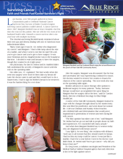


© Copyright 2026