
Tangential View and Intraoperative Three
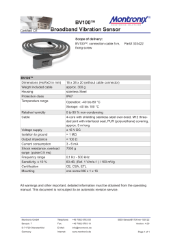
Arch Trauma Res. 2015 June; 4(2): e24622.
DOI: 10.5812/atr.4(2)2015.24622
Research Article
Published online 2015 June 20.
Tangential View and Intraoperative Three-Dimensional Fluoroscopy
for the Detection of Screw-Misplacements in Volar Plating of Distal
Radius Fractures
1,*
1
1
1
2
Sascha Rausch ; Ivan Marintschev ; Isabel Graul ; Arne Wilharm ; Kajetan Klos ; Gunther
1,3
1
O. Hofmann ; Marc Florian Gras
1Trauma Department, Jena University Hospital, Jena, Germany
2Foot and Ankle Department, Catholic Medical Center, Mainz, Germany
3Berufsgenossenschaftliche Kliniken Bergmannstrost Halle, Halle, Germany
*Corresponding author: Sascha Rausch, Trauma Department, Jena University Hospital, Jena, Germany. Tel: +49-36419322889, Fax: +49-36419322802, E-mail: [email protected]
Received: October 15, 2014; Revised: December 31, 2014; Accepted: February 28, 2015
Background: Volar locking plate fixation has become the gold standard in the treatment of unstable distal radius fractures. Juxtaarticular screws should be placed as close as possible to the subchondral zone, in an optimized length to buttress the articular surface
and address the contralateral cortical bone. On the other hand, intra-articular screw misplacements will promote osteoarthritis, while
the penetration of the contralateral bone surface may result in tendon irritations and ruptures. The intraoperative control of fracture
reduction and implant positioning is limited in the common postero-anterior and true lateral two-dimensional (2D)-fluoroscopic views.
Therefore, additional 2D-fluoroscopic views in different projections and intraoperative three-dimensional (3D) fluoroscopy were recently
reported. Nevertheless, their utility has issued controversies.
Objectives: The following questions should be answered in this study; 1) Are the additional tangential view and the intraoperative 3D
fluoroscopy useful in the clinical routine to detect persistent fracture dislocations and screw misplacements, to prevent revision surgery?
2) Which is the most dangerous plate hole for screw misplacement?
Patients and Methods: A total of 48 patients (36 females and 13 males) with 49 unstable distal radius fractures (22 x 23 A; 2 x 23 B, and 25 x
23 C) were treated with a 2.4 mm variable angle LCP Two-Column volar distal radius plate (Synthes GmbH, Oberdorf, Switzerland) during
a 10-month period. After final fixation, according to the manufactures' technique guide and control of implant placement in the two
common perpendicular 2D-fluoroscopic images (postero-anterior and true lateral), an additional tangential view and intraoperative 3D
fluoroscopic scan were performed to control the anatomic fracture reduction and screw placements. Intraoperative revision rates due to
screw misplacements (intra-articular or overlength) were evaluated. Additionally, the number of surgeons, time and radiation-exposure,
for each step of the operating procedure, were recorded.
Results: In the standard 2D-fluoroscopic views (postero-anterior and true lateral projection), 22 screw misplacements of 232 inserted screws
were not detected. Based on the additional tangential view, 12 screws were exchanged, followed by further 10 screws after performing the
3D fluoroscopic scan. The most lateral screw position had the highest risk for screw misplacement (accounting for 45.5% of all exchanged
screws). The mean number of images for the tangential view was 3 ± 2.5 images. The mean surgical time was extended by 10.02 ± 3.82
minutes for the 3D fluoroscopic scan. An additional radiation exposure of 4.4 ± 4.5seconds, with a dose area product of 39.2 ± 14.5 cGy/cm2
were necessary for the tangential view and 54.4 ± 20.9 seconds with a dose area product of 2.1 ± 2.2 cGy/cm2, for the 3D fluoroscopic scan.
Conclusions: We recommend the additional 2D-fluoroscopic tangential view for detection of screw misplacements caused by overlength,
with penetration on the dorsal cortical surface of the distal radius, predominantly observed for the most lateral screw position. The use
of intraoperative 3D fluoroscopy did not become accepted in our clinical routine, due to the technical demanding and time consuming
procedure, with a limited image quality so far.
Keywords: Distal Radius Fracture; Intraoperative; Imaging; Screw Penetration; Doenicke View
1. Background
Volar locking plate fixation became the gold standard
in operative treatment of unstable distal radius fractures
(1-3). Juxta-articular screws should to be placed as close
as possible to the subchondral zone, in an optimized
length to buttress the articular surface and address the
contralateral cortical bone, to prevent a fragment dislocation under early postoperative functional physio-
therapy, especially in dorsal comminuted fractures (4-6).
These demands, together with the use of variable angle
stable plates, are associated with an increased risk of
screw misplacements. Nevertheless, intra-articular screw
placements will promote osteoarthritis, while the penetration of the contralateral bone surface may result in
extensor tendon irritations and ruptures (7-9). If screw
Copyright © 2015, Kashan University of Medical Sciences. This is an open-access article distributed under the terms of the Creative Commons Attribution-NonCommercial 4.0 International License (http://creativecommons.org/licenses/by-nc/4.0/) which permits copy and redistribute the material just in noncommercial
usages, provided the original work is properly cited.
Rausch S et al.
misplacement is confirmed postoperatively, revision surgery is required to avoid such iatrogenic complications.
Therefore, conclusive intraoperative detection of screw
misplacements is mandatory. The standard intraoperative control of anatomic fracture reduction and implant
placement is the 2D-fluoroscopic view, in two perpendicular planes (postero-anterior and true lateral). These
projection images are not sufficient to securely detect
screw misplacements, due to the complex shaped distal
radius, especially on the dorsal side (10-18). To the best of
our knowledge, Doenicke et al. (19) were the first who described an additional 2D fluoroscopic tangential view to
detect screw penetrations on the dorsal distal radius, due
to screw overlength.
3.1. Performing the 2D-Fluoroscopic Tangential
View
The patient’s arm is placed on a radiolucent hand table,
in 90° abduction, in the glenohumeral joint. The forearm
is 70° flexed in the elbow, with a maximum volar tilt in
the wrist joint to get a central fluoroscopic beam path
perpendicular to the dorsal surface of the distal radius
(Figures 1 and 2).
2. Objectives
In recent years, intraoperative 3D fluoroscopy was introduced to acquire CT-like images for the control of
anatomic fracture reduction and prevention of implant
misplacements, predominantly in articular fractures (2023). The following two questions should be answered in
this study:
1) Are the additional tangential view and the intraoperative 3D fluoroscopy useful in the clinical routine to detect
persistent fracture dislocations and screw misplacements to prevent revision surgery?
2) Which is the most dangerous plate hole for screw
misplacement?
3. Patients and Methods
Between February and November 2013, all patients
with unstable distal radius fractures, treated with a polyaxial locking plate fixation [2.4 mm variable angle LCP
Two-Column volar distal radius plate (Synthes GmbH,
Oberdorf, Switzerland)], were prospectively included. Exclusion criteria were: I, patient’s age under 18 years; II, refusal of participation; or III, need for a dorsal approach or
use of other implants. Surgical procedure: The operation
was performed according to the manufactures technique
guide. After final fixation of the distal radius fracture including the intraoperative control of fracture reduction
and implant placement in the standard 2D fluoroscopy
(postero-anterior and true lateral), the additional 2D fluoroscopic tangential view was performed first, followed by
the final intraoperative 3D fluoroscopic scan. If any screw
misplacement was detected, an exchange of screws was
performed immediately after each image control (tangential view or 3D fluoroscopy). The senior consultant
classified the fracture types according to the AO-Müller
(AO) classification system and analyzed all intraoperative control images. The number and cause for screw
exchanges, as well as procedure time (in minutes) and
radiation exposure (no. of fluoroscopic images, fluoroscopy time and dose area product) were recorded for each
step. Additionally, the number of surgeons and their level
of education were documented.
2
Figure 1. Image of an Intraoperative Two-Dimensional Fluoroscopic Tangential View (Doenicke View)
Figure 2. Two-Dimensional Reformation Images of the Intraoperative
Three-Dimensional Fluoroscopic Scan After Volar Distal Radius Plating
In the sagittal plane, perfect juxta-articular screw placements are displayed. In the coronal plane, a screw tip perforation was detected (the image quality in the axial plane is limited).
Arch Trauma Res. 2015;4(2):e24622
Rausch S et al.
Figure 4. Intraoperative Set-up and Patients’ Arm Position for a Correctly
Performed Three-Dimensional Fluoroscopic Scan
room for the automatic scan for image acquisition. Out
of one hundred 2D-fluoroscopic images, a computed tomography like 3D data-set will be reconstructed, covering a cube dimension of approximately 12 × 12 × 12 cm. In
reformatted 2D-slice imaging, the anatomic reduction
and save screw placement were evaluated (Figure 4),
while the second surgeon closed the wound.
3.3. Statistical Analysis
Figure 3. Intraoperative Set-up and Patients’ Arm Position for the Acquisition of a 2D-Fluoroscopic Tangential View (Doenicke View)
3.2. Performing the Intraoperative Three-Dimensional Fluoroscopic Scan
The patient’s arm is placed on a radiolucent hand-table
in 90° abduction in the glenohumeral joint, with extension in the elbow and neutral position in the wrist. The carm (Ziehm Vario 3D, Ziehm Imaging GmbH, Nuremberg,
Germany) is placed in a cephalad position, parallel to the
patient’s body axis (Figure 3). After adjusting for the exact
isocenter, focused on the wrist joint, a manual orbital rotation of 136°, without radiation, is performed as controlscan, to exclude a collision of the c-arm with the hand
table. Finally, the operating team leaves the operating
Arch Trauma Res. 2015;4(2):e24622
A pre-hoc power analysis for an estimated screw misplacement rate of 20%, revealed a minimum study sample size of 46 patients (α-value of 0.05, power 0.8). The
descriptive statistical analysis was performed using SPSS
20 (SPSS Inc., Chicago, IL, USA). The study was approved
by the institutional ethics committee (No. 3630-12/2012).
4. Results
The study included 48 patients (37 women, 11 men) with
49 distal radius fractures (22 right wrist, 25 left wrist, and
one, both wrists). The patient’s characteristics are displayed
in Table 1. The most common fracture type, according to the
AO classification system, was the A3 fracture. Of the total,
15 radius fracture diagnoses were performed by senior consultants while 34 radius fractures by residents under supervision. The mean procedure time was 79 ± 20 minutes.
A total of 7.3 fluoroscopic images (14.1 ± 8.2 seconds fluoroscopic time, and 10.0 ± 19.7 cGy/cm2 dose area product)
were necessary for the standard operating procedure.
3
Rausch S et al.
Table 1. Patients’ Characteristics
Variables
All patients
Female
Male
Fracture Type Distribution
Due to AO Classification
Wrist
B
C
Left Side
Right Side
Both Sides
22
2
15
25
22
1
62.1 ± 16.3 (22 - 84)
17
2
18
20
17
0
66.7 ± 11.8 (45 - 84)
5
0
7
5
5
1
47.9 ± 16.7 (22 - 72)
4.1. Two-Dimensional Fluoroscopic Tangential
View
The additional images were 3 ± 2.5 fluoroscopic images
(4.4 ± 4.5 seconds fluoroscopy time, 2.12 ± 2.21 cGy/cm2
dose area product), which were necessary to obtain a
tangential view in an analyzable projection. Twelve of 232
screws in the distal plate-hole row had an overlength not
suspicious in the standard fluoroscopic images (posteroanterior and true lateral) and were exchanged intraoperatively.
4.2. Intraoperative Three-Dimensional Fluoroscopy
The additional procedure time for the intraoperative
3D-fluoroscopic scan was 10.02 ± 3.82 minutes. Totally, 3 ±
2.5 fluoroscopic images (54.4 ± 20.9 seconds fluoroscopy
time, 39.2 ± 14.5 cGy/cm2 dose area product) were necessary to obtain a tangential view in an analyzable projection. In four patients (8.2%) the 3D-fluoroscopic scan
could not be finished due to technical errors. Further, 10
of 232 screws (nine penetrating and one intra-articular
screw) in the distal fragments were replaced after the
3D-fluoroscopic scan, not identified as suspicious in the
three previous 2D-fluoroscopic control views (in posteroanterior, true lateral and tangential projections).
4.3. Most Dangerous
Misplacement
Screw
Position
for
In 10 cases (45.5% of all screw misplacements), the
screws in the most radial plate hole position had to be exchanged. In five patients, more than one screw had to be
replaced. The overall intraoperative revision rate for all
49 distal radius fractures was 34.7%.
5. Discussion
The anatomy of the distal radius is complex and difficult to visualize intraoperatively (14, 24, 25). The articular
surface of the radius is biconcave and tilted in different
planes. Further, the commonly used volar plating technique does not allow joint visualization and therefore,
the intra-articular fracture reduction can be controlled
by fluoroscopy, only (2). Extensive experience and detailed knowledge of the anatomy are necessary to restore
the anatomical alignment, without articular steps or
gaps, and to ensure an adequate screw length, leading
4
Mean Age in Years ± SD (Range)
A
to reliable retention of individual fragments. Standard
intraoperative postero-anterior and true lateral fluoroscopic views cannot adequately visualize the correct extra-articular subchondral screw position and length (9,
14, 17, 25). Several additional 2D-fluoroscopic projection
images have been described to solve this problem (13, 1518, 25). To the best of our knowledge, Doenicke et al. (19)
were the first in 2006, who described the tangential view
as a reliable image modality for the detection of screw
overlength (12 penetrating screws in nine patients followed by intraoperative replacement). The postoperative
CT scans revealed no residual screw penetration. Modified fluoroscopic techniques were published as “dorsal
horizon view” or “dorsal tangential view” with conclusive results (15, 16, 18). In a recent experimental cadaveric
study by Haug et al. (11), the validity of such tangential
fluoroscopic imaging was corroborated by comparison
with CT imaging and direct visual control. Maschke et
al. (13) described oblique pronosupination fluoroscopic
imaging for enhanced detection of dorsal screw prominence. A screw prominence of at least 2 to 4 mm is required, however, to achieve reproducible results. Several
authors reported the use of oblique images to rule out
intra-articular screw penetration (10, 12). Alternatively, intraoperative 3D imaging of articular fracture has become
an integrated part of several surgical procedures (20,
21, 23, 26). Different studies described an intraoperative
screw replacement rate of 10% to 20% in the treatment
of distal radial fractures, which, in the authors’ opinion,
spared the patients from further revision surgery (20,
21, 27). Multi-slice imaging allows for precise detection
of screw misplacements, poor reduction results due to
persistent articular steps and gaps. A common limitation
of most of these techniques, however, is the lack of standardization, as well as missing proof of clinical relevance
in a large patients population provided in this study. Our
findings demonstrate that the additional tangential view
(Doenicke view) is a helpful 2D-fluoroscopic image projection for the detection of screw-overlength, with penetration of the dorsal cortical surface in the distal radius.
The easy and fast acquisition, with neglectible increase
of radiation exposure, justifies the application in our
routine clinical setting. In contrast, the intraoperative
3D-fluoroscopic scan did not become accepted due to
the technical demands, as well as time and cost-intensive
procedure, despite the further detection of 10 additional
screw misplacements. Important to mention is the fact
Arch Trauma Res. 2015;4(2):e24622
Rausch S et al.
that no revision was performed to increase the reduction
quality after the intraoperative 3D-fluoroscopic control
scan, due to persistent steps or gaps. Erroneous interpretations, however, may arise from a limited image quality
of the 3D-fluoroscopy. The high intraoperative rate for
screw revisions in our study may be attributed to the
large number of operations performed by inexperienced
surgeons (34 by residents and 15 by senior consultants).
In a level 1 trauma center, most distal radius fractures are
a typical training operation for residents supervised by
the trauma-surgeons on call. The lack of tactile feed-back
for the supervised surgeon makes an adequate intraoperative imaging necessary to avoid revision surgery. The
study has several limitations:
I) A direct comparison of both image modalities was
not possible, due to the study design that primarily focused on the clinical algorithm, with stepwise exchange
of screws after each image acquisition (tangential view
and 3D-fluoroscopic scan).
II) The detected screw misplacements could not be verified by direct visualization after soft tissue dissection or
by postoperative computed tomography.
III) The observed misplacement rate due to overlength
screw cannot be directly extrapolated to potential clinical complications, like tendon synovitis and ruptures.
In conclusion, the additional tangential view (Doenicke
view) is recommended for the routine detection of screw
overlength, penetrating the posterior cortical surface of
the distal radius, which may lead to postoperative tendon irritations and ruptures. The use of the intraoperative 3D-fluoroscopy is subject for selected cases only, due
to the limitations discussed above.
8.
9.
10.
11.
12.
13.
14.
15.
16.
17.
18.
19.
Authors’ Contributions
20.
All authors have participated in the design, coordination
and execution of the study and helped to draft the manuscript. All authors read and approved the final manuscript.
21.
References
1.
2.
3.
4.
5.
6.
7.
Simic PM, Weiland AJ. Fractures of the distal aspect of the radius:
changes in treatment over the past two decades. Instr Course Lect.
2003;52:185–95.
Orbay J. Volar plate fixation of distal radius fractures. Hand Clin.
2005;21(3):347–54.
Lichtman DM, Bindra RR, Boyer MI, Putnam MD, Ring D, Slutsky
DJ, et al. Treatment of distal radius fractures. J Am Acad Orthop
Surg. 2010;18(3):180–9.
Drobetz H, Kutscha-Lissberg E. Osteosynthesis of distal radial
fractures with a volar locking screw plate system. Int Orthop.
2003;27(1):1–6.
Drobetz H, Bryant AL, Pokorny T, Spitaler R, Leixnering M, Jupiter
JB. Volar fixed-angle plating of distal radius extension fractures:
influence of plate position on secondary loss of reduction-a biomechanic study in a cadaveric model. J Hand Surg Am.
2006;31(4):615–22.
Shin EK, Jupiter JB. Current concepts in the management
of distal radius fractures. Acta Chir Orthop Traumatol Cech.
2007;74(4):233–46.
Freeland AE, Luber KT. Biomechanics and biology of plate fixation of distal radius fractures. Hand Clin. 2005;21(3):329–39.
Arch Trauma Res. 2015;4(2):e24622
22.
23.
24.
25.
26.
27.
Al-Rashid M, Theivendran K, Craigen MA. Delayed ruptures of
the extensor tendon secondary to the use of volar locking compression plates for distal radial fractures. J Bone Joint Surg Br.
2006;88(12):1610–2.
Tarallo L, Mugnai R, Zambianchi F, Adani R, Catani F. Volar plate
fixation for the treatment of distal radius fractures: analysis of
adverse events. J Orthop Trauma. 2013;27(12):740–5.
Pace A, Cresswell T. Use of articular wrist views to assess intraarticular screw penetration in surgical fixation of distal radius
fractures. J Hand Surg Am. 2010;35(6):1015–8.
Haug LC, Glodny B, Deml C, Lutz M, Attal R. A new radiological
method to detect dorsally penetrating screws when using volar
locking plates in distal radial fractures. The dorsal horizon view.
Bone Joint J. 2013;95-B(8):1101–5.
Soong M, Got C, Katarincic J, Akelman E. Fluoroscopic evaluation of intra-articular screw placement during locked volar
plating of the distal radius: a cadaveric study. J Hand Surg Am.
2008;33(10):1720–3.
Maschke SD, Evans PJ, Schub D, Drake R, Lawton JN. Radiographic evaluation of dorsal screw penetration after volar fixedangle plating of the distal radius: a cadaveric study. Hand (N Y).
2007;2(3):144–50.
Park DH, Goldie BS. Volar plating for distal radius fractures--do
not trust the image intensifier when judging distal subchondral
screw length. Tech Hand Up Extrem Surg. 2012;16(3):169–72.
Thomas AD, Greenberg JA. Use of fluoroscopy in determining
screw overshoot in the dorsal distal radius: a cadaveric study. J
Hand Surg Am. 2009;34(2):258–61.
Ozer K, Wolf JM, Watkins B, Hak DJ. Comparison of 4 fluoroscopic
views for dorsal cortex screw penetration after volar plating of
the distal radius. J Hand Surg Am. 2012;37(5):963–7.
Lee SK, Bae KW, Choy WS. Use of the radial groove view intra-operatively to prevent damage to the extensor pollicis longus tendon
by protruding screws during volar plating of a distal radial fracture. Bone Joint J. 2013;95-B(10):1372–6.
Joseph SJ, Harvey JN. The dorsal horizon view: detecting screw
protrusion at the distal radius. J Hand Surg Am. 2011;36(10):1691–3.
Dönicke T, Petrovitch A, Grubert S, Friedel R, Hofmann.. Deutscher
Kongress für Orthopädie und Unfallchirurgie 70. Jahrestagung der
Deutschen Gesellschaft für Unfallchirurgie. 2006. Available from:
http://www.egms.de/static/de/meetings/dgu2006/06dgu0249.
shtml.
Mehling I, Rittstieg P, Mehling AP, Kuchle R, Muller LP, Rommens
PM. Intraoperative C-arm CT imaging in angular stable plate
osteosynthesis of distal radius fractures. J Hand Surg Eur Vol.
2013;38(7):751–7.
Atesok K, Finkelstein J, Khoury A, Peyser A, Weil Y, Liebergall
M, et al. The use of intraoperative three-dimensional imaging (ISO-C-3D) in fixation of intraarticular fractures. Injury.
2007;38(10):1163–9.
Carelsen B, Haverlag R, Ubbink DT, Luitse JS, Goslings JC.
Does intraoperative fluoroscopic 3D imaging provide extra
information for fracture surgery? Arch Orthop Trauma Surg.
2008;128(12):1419–24.
Carelsen B, van Loon J, Streekstra GJ, Maas M, van Kemenade
P, Strackee SD. First experiences with the use of intraoperative 3D-RX for wrist surgery. Minim Invasive Ther Allied Technol.
2011;20(3):160–6.
Clement H, Pichler W, Nelson D, Hausleitner L, Tesch NP, Grechenig W. Morphometric analysis of lister's tubercle and its consequences on volar plate fixation of distal radius fractures. J Hand
Surg Am. 2008;33(10):1716–9.
Tweet ML, Calfee RP, Stern PJ. Rotational fluoroscopy assists in detection of intra-articular screw penetration during volar plating
of the distal radius. J Hand Surg Am. 2010;35(4):619–27.
Stubig T, Kendoff D, Citak M, Geerling J, Khalafi A, Krettek C, et al.
Comparative study of different intraoperative 3-D image intensifiers in orthopedic trauma care. J Trauma. 2009;66(3):821–30.
Hufner T, Stubig T, Gosling T, Kendoff D, Geerling J, Krettek
C. [Cost-benefit analysis of intraoperative 3D imaging]. Unfallchirurg. 2007;110(1):14–21.
5


© Copyright 2026