
Sudden Cardiac Death - Current Problems in Cardiology
Author's Accepted Manuscript
Sudden Cardiac Death
Vikas P. Kuriachan MD, Glen L. Sumner MD, L.
Brent Mitchell MD
www.elsevier.com/locate/buildenv
PII:
DOI:
Reference:
S0146-2806(15)00003-1
http://dx.doi.org/10.1016/j.cpcardiol.2015.01.002
YMCD293
To appear in:
Curr Probl Cardiol
Cite this article as: Vikas P. Kuriachan MD, Glen L. Sumner MD, L. Brent Mitchell MD,
Sudden Cardiac Death, Curr Probl Cardiol, http://dx.doi.org/10.1016/j.cpcardiol.2015.01.002
This is a PDF file of an unedited manuscript that has been accepted for publication. As a
service to our customers we are providing this early version of the manuscript. The
manuscript will undergo copyediting, typesetting, and review of the resulting galley proof
before it is published in its final citable form. Please note that during the production process
errors may be discovered which could affect the content, and all legal disclaimers that apply
to the journal pertain.
SUDDEN CARDIAC DEATH
Vikas P. Kuriachan MD, Glen L. Sumner MD, L. Brent Mitchell MD
Department of Cardiac Sciences
Libin Cardiovascular Institute of Alberta
Alberta Health Services and University of Calgary
No grants or financial support was used for this work.
Correspondence:
Dr. L Brent Mitchell
1403-29th St NW, Calgary, AB T2N 2T9, Canada
Tel: (403) 944-1683
Fax:
(403)944-3550
Email: [email protected]
1
Disclosure Statement:
Vikas Kuriachan has received speaker honoraria from Medtronic,
Boston Scientific, Biosense Webster, and Bayer Pharma. He also has had
research support from Medtronic acting as local principal investigator
for clinical studies
Dr. Sumner has received speaker honoraria from Medtronic and
Bayer Pharma. He also has had research support from Medtronic, Boston
Scientific and St. Jude Medical acting as local principal investigator
for clinical studies.
Dr. Mitchell has received speaker honoraria from Medtronic. He
has also had research support from Medtronic, Boston Scientific, St.
Jude Medical, and Forrest Pharma while acting as a local principal
investigator for clinical studies.
Table of contents
A. Introduction
B. Epidemiology and risk factors
C. Non-cardiac causes of sudden death
a. Neurologic disorders
b. Acute Aortic Syndromes
c. Metabolic and electrolyte derangements
d. Medications and other drugs
e. Other causes of non-cardiac sudden death
D. Cardiac causes of sudden death
a. Arrhythmic Causes (primary electrical abnormality)
i. Tachycardias
1. WPW
2. Long QT
3. Brugada
4. Short QT
5. Catecholaminergic polymorphic ventricular
tachycardia
6. Early repolarization syndrome and Idiopathic
ventricular fibrillation
7. Commotio cordis
ii. Bradycardias
2
E.
F.
G.
H.
I.
J.
b. Structural Heart disease
i. Coronary artery disease and Ischemic Cardiomyopathy
ii. Non-ischemic cardiomyopathy
iii. Congenital Heart disease
iv. Valvular heart disease
v. Hypertrophic cardiomyopathy
vi. Arrhythmogenic Right Ventricular Dysplasia
Special populations:
a. Athletes
b. Pregnancy
New Defibrillator Technologies
Driving Implications
Clinical Approach to Investigation of SCD
Risk Stratification
Conclusions
Biographical Sketch for each author:
Dr. Vikas P. Kuriachan, MD, FRCPC, FHRS
Dr. Vikas Kuriachan joined the Cardiology / Cardiac Electrophysiology
group at the Libin Cardiovascular Institute of Alberta and the
University of Calgary in July 2010 after completing a year of complex
ablation training at Brigham and Women’s Hospital / Harvard Medical
School.
He is the Program Director for the Cardiac Electrophysiology
training program. He is a Fellow of the Royal College of Physicians
and Surgeons of Canada and Heart Rhythm Society. His clinical
interests include ablation, in particular VT ablation, along with
cardiac rhythm management device implantation and complex lead
extraction.
Dr. Glen L. Sumner, MD, FRCPC
Dr. Sumner has been a Clinical Assistant Professor of Medicine in the
Department of Cardiac Sciences at the Libin Cardiovascular Institute
of Alberta, University of Calgary since 2008. He completed his
medical training at Queen’s University in Kingston (Ontario, Canada)
and practiced as a rural family physician for seven years prior to
embarking on clinical training in cardiology, echocardiography, and
cardiac electrophysiology at Dartmouth-Hitchcock Medical Center in
Lebanon, New Hampshire and McMaster University in Hamilton, Ontario.
Dr. Sumner’s main clinical and research interests are in cardiac
3
resynchronization therapy patient selection, cardiac device
implantation, and cardiac device programming.
Dr. L. Brent Mitchell, MD, FRCPC, CAHS
Dr. Mitchell is a Professor in the Department of Cardiac Sciences in
the Libin Cardiovascular Institute of Alberta at the University of
Calgary. He obtained his training in clinical cardiology at Dalhousie
University (Halifax, Nova Scotia) and his training in cardiac
electrophysiology Stanford University. He was the Inaugural Head of
the Department of Cardiac Sciences and the Inaugural Director of the
Libin Cardiovascular Institute of Alberta.
Dr. Mitchell’s clinical
practice and research interests are in the clinical science of
tachyarrhythmias – their mechanisms, their investigation, and their
management. His preferred tool in this science is the randomized
clinical trial.
Abstract:
Sudden death accounts for 300,000-400,000 deaths annually in the
United States. Most sudden deaths are cardiac and most sudden cardiac
deaths are related to arrhythmias secondary to structural heart
disease or primary electrical abnormalities of the heart.
The most
common structural disease leading to sudden death is ischemic heart
disease. Non-ischemic cardiomyopathy and other structural
abnormalities such as arrhythmogenic ventricular dysplasia and
hypertrophic cardiomyopathy may also be causative. Patients without
structural disease have a primary electrical abnormality, such as long
QT syndrome or Brugada syndrome. Severe left ventricular systolic
dysfunction is the main marker for sudden death in patients with
ischemic or non-ischemic cardiomyopathy.
In other conditions, other
markers for structural heart disease and electrical abnormalities need
to be considered. β-blocker therapy is associated with a reduction in
sudden cardiac death across a broad range of disorders. Nevertheless,
the Implantable Cardioverter Defibrillator remains the most effective
treatment strategy in selected patients.
4
Abbreviations
ACE-I – Angiotensin Converting Enzyme Inhibitor
AF – Atrial Fibrillation
ARB – Angiotensin Receptor Blocker
ARVC – Arrhythmogenic Right Ventricular Cardiomyopathy
BrP – Brugada ECG Pattern
BrS – Brugada Syndrome
CAD – Coronary Artery Disease
CHD - Congenital Heart Disease
CHF – Congestive Heart Failure
cMRI – Cardiac Magnetic Resonance Imaging
CPVT – Catecholaminergic Polymorphic Ventricular Tachycardia
EAD – Early After Depolarization
ECG – Electrocardiogram
HCM – Hypertrophic Cardiomyopathy
ICD - Implantable Cardioverter Defibrillator
LVEF – Left Ventricular Ejection Fraction
LQTS – Long QT Interval Syndrome
MVP – Mitral Valve Prolapse
SCA – Sudden Cardiac Arrest
SCD - Sudden Cardiac Death
SQTS – Short QT Interval Syndrome
VF - Ventricular Fibrillation
VT - Ventricular Tachycardia
VHD – Valvular Heart Disease
5
A) INTRODUCTION
Sudden cardiac death (SCD) is defined as unexpected, non-traumatic
death occurring within one hour of the onset of new or worsening
symptoms (witnessed arrest) or, if unwitnessed, within 24 hours of
last being seen alive(1). SCD has a multitude of potential aetiologies
but is most commonly associated with ischemic heart disease. SCD may
be preceded by symptoms such as chest pain, dyspnea, palpitations,
presyncope, and syncope but many individuals have no symptoms prior to
the event. By definition, a patient with SCD does not survive. When
the patient survives, the event is termed aborted SCD or sudden
cardiac arrest (SCA). The immediate cause of SCD in most instances is
a ventricular arrhythmia: either ventricular fibrillation (VF) or
ventricular tachycardia (VT). However, in a significant minority of
cases, asystole or pulseless electrical activity is the initial
documented rhythm.
The leading etiologies of sudden death are listed in Table 1.
Broad categories of SCD include those related to ischemic heart
disease, non-ischemic heart disease, primary electrical disease, and
non-cardiac disease.
B) EPIDEMIOLOGY AND RISK FACTORS
SCD is common with an annual incidence of 60 per 100,000 in the
United States(2). Accordingly, approximately 300,000-400,000 sudden
cardiac deaths occur in the United States each year. The incidence is
higher in men (76 per 100,000) than in women (45 per 100,000).
Using
death certificate data, it is estimated that SCD accounts for up to
15% of all deaths in western nations. However, death certificate data
may overestimate the incidence. Extrapolated data from the Oregon
Sudden Unexpected Death Study (SUDS) suggests that SCD accounts for
approximately 5.6% of all deaths in the United States(2).
SCD incidence increases with age. The incidence of SCD in younger
populations (<30 years) is 100-fold lower than that in older
individuals(1). Women are relatively protected from SCD until the
menopausal years when incidence increases to approach that of men.
However, even in younger women, conventional CAD risk factors are
predictive of SCD events. Genetic factors play a role in SCD at
multiple steps in the pathophysiologic pathway. Mutations and
polymorphisms modulate the risk of SCD associated with both CAD and
non-CAD etiologies(1). In addition, at least two studies have
described a familial propensity for SCD being the initial presentation
of CAD. Some studies have demonstrated an increased risk of SCD in
6
African Americans compared to whites while Hispanic individuals may be
at lower risk(1).
SCD is a leading cause of death. In men, SCD incidence exceeds
other causes of death including individual cancers (lung, prostate,
and colorectal), accidents, chronic respiratory diseases, diabetes,
and cerebrovascular disease(2). In women, SCD incidence is similar to
those of lung cancer and cerebrovascular disease and is higher than
those of other causes of death including breast cancer, colorectal
cancer, Alzheimer’s disease, and accidents(2).
When expressed as
years of potential life lost (YPLL), the impact of SCD is impressive.
Nearly 50% of people with SCD had no known previously diagnosed
heart disorder. In this population, risk stratification is
particularly challenging(3). Furthermore, 40% of SCD occurs in
individuals with known heart disease but with a left ventricular
ejection fraction (LVEF) greater than 40%. Our ability to predict SCD
in this population is also limited. The remaining (approximately 10%)
incidence of SCD affects individuals with known structural heart
disease and LVEF less than 40%(3). Accordingly, current risk
stratification tools and therapies such as implantable cardioverter
defibrillators (ICDs) have had a modest impact on the overall problem
of SCD. Genetic-based arrhythmic conditions account for only 2% of all
SCD. Thus, because the majority of SCD occurs in individuals without
known heart disease, efforts to prevent SCD should be directed towards
the identification and modification of conventional risk factors.
The risk factors for SCD are similar to those of ischemic heart
disease and include smoking, hypertension, dyslipidemia, and diabetes.
In the Framingham population, smoking conferred a 2-3 fold increased
risk of SCD(1). Furthermore, in one study continued smoking after
aborted SCD was associated with a recurrent cardiac arrest rate of 27%
over 3 years compared to 19% in those who stopped smoking. Obesity
also confers an increased risk of SCD in those with CAD(3). There is
conflicting data with respect to sedentary lifestyle as a risk factor
for SCD. The risk of SCD is increased (approximately 17 fold) during
vigorous physical activity especially in those who are generally
sedentary (approximately 74 fold). However, the absolute risk of SCD
during a single episode of vigorous exercise is very low (1 per 1.51
million episodes of exercise) (4). Furthermore, regular participation
in physical exercise attenuates the overall risk of SCD both during
exercise and at rest. Psychosocial stressors also increase risk of
SCD. Major disasters such as earthquake and war increase the
incidence of SCD in affected populations(5). Heavy alcohol consumption
(6 or more drinks per day) and binge drinking increase the risk of SCD
while moderate alcohol consumption (one to two drinks per day) may
7
decrease the risk(6). In contrast, caffeine intake has not been shown
to be a risk factor for SCD. Higher levels of long chain n-3
polyunsaturated fatty acids (eicosapentaenoic acid and docosahexaenoic
acid; “fish oil”) measured in plasma and in red cell membranes are
associated with a lower risk for SCD(7). Higher serum C-reactive
protein (CRP), a marker of generalized inflammation, has been shown to
be levels are associated with increased risk of SCD(8). In 97 cases
of SCD among apparently healthy men in the Physicians Health Study,
CRP serum levels in the highest quartile were associated with a 2.78
fold increased risk compared to men in the lowest quartile.
Prospective studies are required to evaluate the impact of
modification of these risk factors on the incidence of SCD.
C) NONCARDIAC CAUSES OF SUDDEN DEATH
Most studies have evaluated causes of death using medical records and
death certificates. A small population study in Japan in which 78% of
the subjects had an autopsy suggested that the major causes of sudden
death were heart disease (49%), stroke (33%), and aortic
aneurysm/dissection (12%)(9).
a) Neurologic Disorders
Central nervous system disorders that can directly (or indirectly via
cardiac interaction) result in sudden death include epilepsy, ischemic
stroke, intracranial bleeding, and traumatic head injury(10).
Uncontrolled epilepsy may lead to Sudden Unexplained Death of Epilepsy
(SUDEP). Among patients with epilepsy, the incidence of SUDEP is
6/1000 patient-years(11). SUDEP is usually triggered by a generalized
tonic-clonic seizure. It is believed that rare channelopathies may
predispose to both epilepsy and SCD. Apnea, bradycardia, and asystole
have also been implicated as has the use of certain anti-seizure
medications. Finally, associated stress-induced cardiomyopathy
(Takutsubo) may also play a role(10).
Cerebral catastrophes such as stroke and major intracranial
bleeding/injury can cause ECG changes and autonomic imbalance. It is
possible that associated arrhythmias (severe bradycardia or VT/VFs)
may cause sudden death. Patients with these neurologic disorders may
also be predisposed to SCD due to underlying cardiovascular disease
and an inflammatory state(10).
Preventative treatments include good control of seizure disorders and
measures to reduce the incidence of stroke and cerebral injuries
including blood pressure control and appropriate anticoagulation for
patients with atrial fibrillation (AF). Cardiac monitoring after
8
cerebral catastrophes is important for recognition of cardiac
arrhythmias.
b) Acute Aortic Syndromes
Acute aortic syndromes include entities that disrupt aortic integrity
with complications ranging from aortic rupture to organ ischemia and
death.
These syndromes include aortic dissection, aneurysm
expansion, trauma, penetrating ulcer, and intramural hematoma(12).
Patient risk factors include age and hypertension. Loss of tensile
strength due to Marfan’s syndrome, Ehlers-Danlos syndrome, familial
thoracic aortic aneurysm disease, and the aortopathy associated with
congenital bicuspid aortic valve also increase risk. Cocaine use is a
risk factor due to increases in blood pressure and heart rate.
Treatment imperatives include rapid diagnosis, control of blood
pressure and shear stress with β-blockers and vasodilators, and early
surgical intervention(12).
c)
Electrolyte, Metabolic, and Endocrine Derangements
Life-threatening arrhythmias may result from electrolyte imbalances
(especially hypokalemia and hypomagnesemia), metabolic derangements
(including acid-base disorders), and severe endocrine disorders
(especially hypothyroidism, hyperthyroidism, hypercatecholaminergic
states). Treatments are focused on the underlying disorder and acute
correction of electrolyte, metabolic, and endocrine imbalances.
Arrhythmias can also accompany eating disorders and the aggressive
treatments intended to correct the metabolic consequences. Weight
reduction in obesity and refeeding in anorexia should be controlled
with attention to electrolyte fluctuations(1).
One major patient group prone to metabolic and electrolyte
derangements are those on dialysis. Dialysis patients have a high
mortality rate and up to 43% of their deaths are cardiac, with most
being due to cardiac arrest(13).
Risk factors for SCD in dialysis
patients include electrolyte shifts, hyper- and hypovolemia, left
ventricular hypertrophy, systolic and diastolic heart failure, CAD
(CAD), hyperphosphatemia, sympathetic overactivity and autonomic
dysfunction.
Prevention of SCD in dialysis patients is challenging. During
dialysis, care is taken to minimize electrolyte shifts. Traditional
medical therapies for CAD, such as statins have lesser benefits in
dialysis patients suggesting that the role of coronary plaques in SCD
maybe less important in dialysis patients(14). β-blockers decrease
9
ischemia but also have antiarrhythmic and sympathoinhibitory effects
and decrease sudden deaths and increase survival of cardiac arrest in
diabetic patients on dialysis. Survival benefits from angiotensin
converting enzyme inhibitor (ACE-I) and angiotensin receptor
blocker(ARB)treatments have also been demonstrated(14).
ICDs play a major role in the prevention of SCD in many
predisposed patient populations. Many of the major trials
demonstrating the benefits of ICDs excluded patients with end-stage
renal disease. To date, no randomized trials of ICD use have been
performed in patients with end-stage renal disease. Nevertheless,
patients with end-stage renal disease are considered for an ICD if
expected to survive more than a year with good functional status
especially in patients who have survived aborted SCD(15). The
comorbidities in this population generate competing non-arrhythmic
causes of death and lead to higher ICD complication rates thereby
limiting the benefits of ICD therapy.
Although the benefits have
been difficult to document, dialysis staff should have basic lifesupport training and an external cardiac defibrillator should be onsite.
d) Medications
Many drugs can cause proarrhythmia that may lead to syncope and sudden
death. The most common mechanism of this proarrhythmia is suppression
of the rapid component of the delayed rectifier potassium current (IKr)
resulting in prolongation of the QT interval and torsades de pointes
VT(16). Risk factors for this form of proarrhythmia include other
factors that prolong the QT interval including hypokalemia,
hypomagnesaemia, bundle branch block, bradycardia, CHF, inherited long
QT abnormalities, and female sex. Accordingly, patients with a QTc
greater than 460 msec should be exposed to additional QT prolonging
drugs with great caution.
The non-cardiovascular drugs associated with this form of
proarrhythmia include certain antibiotics, psychotropic medications,
and diuretics (by magnesium and potassium depletion) (16). A webbased listing of potential culprit medications is available at
www.torsades.org.
Cardiovascular drugs may also be proarrhythmic. Vaughn Williams Class
III antiarrhythmic drugs (amiodarone, dronedarone, dofetilide,
ibutilide,and sotalol) are potassium channel blockers that prolong QT
and may produce torsades de pointes VT. Such agents are administered
with close monitoring of QT intervals, maintenance of normal potassium
and magnesium levels, and assurance of normal renal function for those
10
agents that are so excreted (dofetilide, sotalol) (16). Vaughn
Williams Class Ia antiarrhythmic agents (disopyramide, procainamide,
quinidine) are also potassium channel blockers that prolong QT and may
produce torsades de pointes VT. In addition, these drugs and other
Class I agents (lidocaine, mexiletine, propafenone, flecainide) are
sodium channel blockers that may prolong QRS duration. Class I
antiarrhythmic agents have been associated with SCD particularly in
patients with underlying structural heart disease and myocardial
fibrosis(17). Accordingly, Class I agents are used mainly in patients
with no structural heart disease. Vaughn Williams Class II
antiarrhythmic agents (β-blockers) and Class IV agents (nondihydropyridine calcium channel blockers) may result in sinus and/or
AV nodal suppression bradycardia especially if used in high doses in
combination.
Although many recreational drugs may result in SCD the one most
implicated is cocaine. Studies in Europe report that 1-4% of young
adults have used cocaine in the past year(18). Cocaine is an indirect
sympathomimetic agent and may increase blood pressure, heart rate,
increase ventricular contractility, and lower VT threshold placing
users at risk for myocardial infarction, major hemorrhages, and
arrhythmias. Up to 9% of young adults with sudden cardiac death were
recent cocaine users(18).
Clearly, the best prevention of sudden death related to drug effects
is to limit the use of implicated agents and to carefully monitor
their effects when they are used, particularly when therapy is first
prescribed and shortly after dosage increments.
e) Other causes of non-cardiac sudden death
Other causes of non-cardiac sudden death are extremely rare and
include pulmonary hemorrhage, pulmonary embolism, chronic obstructive
pulmonary disease, asthma, rupture of esophageal varices, and rupture
of ectopic pregnancy. Prevention of sudden death from these causes
mainly involves diagnosis and treatment of the underlying disorder.
D) CARDIAC CAUSES OF SUDDEN DEATH
a) Arrhythmic Causes Due to Primary Electrical Abnormalities
i) Tachycardias
1) Wolff-Parkinson-White Pattern and Syndrome
In 1930, Drs. Wolff, Parkinson, and White described group of
patients with palpitations and an ECG abnormality in the absence of
structural heart disease now recognized to be ventricular
11
preexcitation associated with an atrioventricular reentrant
supraventricular tachycardia(19). Ventricular preexcitation reflects
the presence of an accessory connection between atrial and ventricular
myocardium. If ventricular preexcitation is associated with symptoms
such as palpitations, presyncope, or syncope then the condition is
termed Wolff-Parkinson-White (WPW) syndrome. In the absence of
associated symptoms or documented tachydysrhythmia, the ECG
abnormality is termed WPW pattern or asymptomatic WPW.
The incidence of the WPW pattern in the population is 1-3/1000.
Approximately 65% of adolescents and 40% of individuals over 30 years
of age with the WPW pattern are asymptomatic(20). The mechanism of SCD
in WPW is rapid conduction of atrial flutter or atrial fibrillation
over the accessory connection that precipitates VF. Most episodes of
SCD in the setting of WPW occur in children or adolescents and in
those with symptoms. However, SCD may be the initial presentation.
The risk of SCD in one prospective study of asymptomatic adults with a
WPW-pattern ECG followed for 38 months was 4.5 per 1000 patient years.
Another study suggested that the 10 year risk of developing AF is 15%
but the risk of SCD only 1%(20).
High-risk features for SCD in patients with a WPW-pattern include age
<30, male gender, history of AF, prior syncope, associated congenital
or other heart disease, and familial WPW(20). Nevertheless, none of
these findings are particularly sensitive or specific. Additional
risk stratification is based on the electrophysiologic properties of
the accessory connection. Data from survivors of WPW-associated SCA,
properties of the AP that suggest high-risk include antegrade
conduction with a shortest preexcited R-R interval in AF less than
220-250 msec(20). The sensitivity of the cutoff of less than 220 msec
is 88-100% for predicting the risk of WPW-associated VF in adults.
However, because the incidence of SCD in WPW is very low, the positive
predictive value is only 19-38%. Similar data are provided by
measurement of the antegrade refractory period of the accessory
connection or the maximal atrial pacing rate with continuous
preexcitation during programmed atrial stimulation at an
electrophysiology study using transvenous catheters or a
transesophageal pacing catheter(20).
Non-invasive risk stratification uses Holter monitoring or exercise
testing to observe disappearance of ventricular preexcitation at
physiologic heart rates(20). If there is clear evidence of
intermittent loss of preexcitation the risk of SCD is likely low. It
may be challenging to identify loss of preexcitation with increasing
heart rates as elevated sympathetic tone may increases the speed of AV
nodal conduction such that preexcitation is masked. Accordingly, only
12
the abrupt loss of preexcitation that was clear on the previous heart
beat is accepted as a marker of low risk. The identification of
multiple accessory pathways on non-invasive evaluation by virtue of
ventricular preexcitation with multiple morphologies is a marker of
higher risk that warrants further invasive evaluation(20).
If an accessory connection has high risk features or if the patient is
symptomatic with atrioventricular reciprocating tachycardia,
transcatheter ablation of the accessory connection is considered. The
success rate for catheter ablation in this setting is greater than 95%
with a low complication rate estimated at 1-3%. A proposed algorithm
for the approach to SCD risk stratification is presented in Figure
1(20).
2) Long QT Syndrome
The congenital Long QT syndromes (LQTS) are genetically-determined
channelopathies that result in prolongation of myocardial action
potential duration. This is manifest on an ECG as a prolonged QT
interval. The QT interval varies inversely with heart rate and
therefore measurement of the QT interval is corrected (QTc) for heart
rate using various formulae. Bazett’s formula produces a corrected QT
interval by dividing the measured QT interval by the square root of
the corresponding R-R interval(in seconds). In adolescence, the normal
QTc range (370-440 msec) is the same in males and females. In
adulthood, the upper limit of normal for the QTc is 450 msec in men
and 470 msec in women(21).
Epidemiology
LQTS may be congenital or acquired. The acquired form, induced by
drugs, electrolyte, or metabolic disturbances is discussed above. It
was once believed that a genetic abnormality is required to be
susceptible to the acquired form of LQTS. However, such a genetic
abnormality has only been identified in less than 10% of cases of
acquired LQTS(22). In either congenital or acquired LQTS abnormal
prolongation of repolarization can precipitate, through early
afterdepolarizations (EADs), a specific type of polymorphic VT known
as torsades de pointes (“twisting of the points”) (Figure 2). (23)
Although torsades de pointes is often self-terminating it may also
precipitate VF and SCD.
Congenital LQTS leads to approximately 3000-4000 SCDs in childhood in
the United States(24). The incidence of congenital LQTS in the
general population is estimated at 1 in 2500. However, the incidence
may be significantly underestimated in that many genotypes have a low
penetrance and may not manifest overt QT prolongation(24). Most
13
individuals with congenital LQTS present in childhood. One registry
identified the mean age of presentation as 6.8 years; another, which
enrolled predominantly adults, reported a mean age at diagnosis of 21
years(25).
Genetics
To date, 12 gene mutations have been identified that lead to the LQTS
(Table 2) (26). Approximately 70% of individuals with a LQTS will have
an identifiable mutation on genetic testing with the majority of
mutations encoding different components of the potassium channel.
The three most common LQTS are LQT-1 (KCNQ1), LQT-2 (KCNH2), and LQT-3
(SCN5A) (26). LQTS-affected individuals may present with
palpitations, presyncope, or syncope but a high proportion are
asymptomatic. LQT-1 (KCNQ1) is responsible for 30-35% of all LQTS
cases. Individuals with LQT-1 may present with symptoms that are
precipitated by exercise or other high adrenergic states. There may
be a predilection for swimming as a precipitating event. LQT-2
(KCNH2)is responsible for 25-30% of genotyped individuals with LQTS.
They typically have symptoms precipitated by emotional stress or
sudden noise. A loud alarm clock is a commonly cited example of a
precipitating event in LQT-2. In contrast, individuals with a LQT-3
(SCN5A) genotype, which represents 7-10% of genotyped LQTS patients,
have symptoms that are typically occur at rest or while sleeping.
Cardiac arrest or SCD incidence is highest in LQT-2 (0.6%/year) and
LQT-3-affected individuals (0.56%/year) and is lowest in those with a
LQT-1 genotype (0.3%/year) (23).
Two clinical phenotypes of congenital LQTS have been described. The
Romano-Ward syndrome is transmitted as an autosomal dominant trait and
may have any of the known genetic substrates. The Jervell and LangeNeilsen syndrome is transmitted as an autosomal recessive trait and is
associated with both sensorineural deafness and LQTS. This phenotype
has only been described with mutations in KCNQ1 (LQT1) and KCNE1
(LQT5) and has a higher risk natural history(23).
Risk factors for SCD
Analysis of the natural history over 28 years of follow-up of 647
individuals with LQT-1, LQT-2, or LQT-3 showed that cardiac events
(defined as syncope, cardiac arrest, or sudden death prior to age 40
years) varied as a function of both genotypes and magnitude of
QTc(26). Cumulative event rates were highest in LQT-2 and LQT-3
genotypes at 46% and 42%, respectively. LQT-1 genotypes had the lowest
event rate at 30%. In addition, those individuals with a QTc of
>500msec in the LQT-1 and LQT-2 genotypes had higher event rates, a
14
finding not observed in LQT-3 genotypes. Finally, the risk of cardiac
events was higher for females in LQT-2 and for males in LQT-3. No
significant gender effect was seen in LQT-1(26).
Diagnosis:
The primary diagnostic criterion is a QTc interval above the normal
limits defined above. To improve the sensitivity of this measure in
patients with concealed LQTS, QTc may be measured standing, during
exercise, and following an epinephrine challenge(23). It is important
to recognize that the response of patients with LQTS to provocation
testing has significant overlap with that of normal individuals. All
individuals diagnosed with LQTS should be referred to a center with
expertise in family screening, genetic testing and counselling.
Treatment:
The cornerstone of treatment for patients with symptomatic LQTS is βblockade. The β-blockers that have been best studied for LQTS are
nadolol and propranolol although others have been used. A recent
database analysis of the efficacy of β-blocker therapy in LQT1 and
LQT2 revealed that nadolol was superior to metoprolol, atenolol, and
propranolol in LQT2 patients and of similar efficacy in LQT1 with
respect to prevention of cardiac events(27). The importance of 24hour therapeutic medication levels may influence the choice of
individual β-blockers. β-blockers are most effective in LQT-1 and LQT2. The mechanism of benefit is believed to be a reduction of
sympathetic tone that limits prolongation of the QT interval both at
rest and with activity(27).
Another effective anti-adrenergic therapy is left cardiac sympathetic
denervation. This therapy is especially useful when β-blockers are
contraindicated or when symptoms have occurred while on adequate βblocker therapy. It has also been used in infants who have extremely
prolonged QT intervals and are at high risk of SCD(23).
Implantable Cardioverter Defibrillator therapy has a limited role in
LQTS. Recently, based on the findings in a European LQTS/ICD
registry, the indications for ICD therapy have been refined to
include: cardiac arrest while on therapy; cardiac arrest while not on
therapy (exception: LQT-1); syncope on β-blockers when left cardiac
sympathetic denervation is unavailable or declined by the patient;
compound heterozygous/homozygous patients with syncope on β-blockers;
and, primary prevention in unique phenotypes with extreme QT
prolongation and other high risk features(28).
15
Lifestyle modifications are important for prevention of cardiac
events. LQTS patients must be counselled to avoid drugs that prolong
the QT interval and to maintain normal magnesium and potassium levels
especially during illnesses when electrolyte loss may occur. Sporting
activities should be restricted to low intensity, non-competitive
sports although this recommendation has been challenged by the
identification of only 2 events in 650 athlete-years of follow-up;
both events occurring in the same individual when non-adherent to βblocker therapy(23). Genotype-positive patients with normal QT
intervals may participate in competitive sports with the exception of
LQT-1 patients with respect to competitive swimming(23).
3) Brugada Syndrome
Brugada syndrome (BrS) is an autosomal dominant genetic cardiac
disorder with variable penetrance that manifests with characteristic
resting ECG abnormalities associated with VT/VF, SCA, and SCD. The
ECG patterns, which predominantly affect the right precordial leads,
have been recently classified into two characteristic types: Type I
and Type II (Figure 3) (29). The Type I pattern, also termed “coved”
type, shows ST segment elevation ≥2mm followed by an upward convexity
and abrupt descent to an inverted T wave. The Type II pattern, also
termed “saddleback” type, has a lesser degree of ST segment elevation
and resolves into an upright or biphasic T wave.
When the ECG
abnormalities occur in the absence of VT/VF or potential symptoms
thereof, the findings are termed Brugada pattern (BrP). Another
syndrome, termed Sudden Unexpected Nocturnal Death Syndrome (SUNDS)
has been described in Southeast Asians and is believed to have a
similar pathophysiology(30).
Epidemiology
The prevalence of BrP on the 12 lead ECG varies with the
population studied being most prevalent in Asian populations. In
Japan, the BrP is estimated to occur in 0.7-1.0% of the population
with 0.12-0.16% having the Type I BrP(31). In population samples from
the United States, the prevalence varied widely from 0.012-0.4%(32).
The prevalence of BrP in patients who present with unexplained VF is
up to 24%. Men are approximately nine times more frequently affected
than females. Most commonly, BrS presents in adulthood but childhood
presentations have been reported(33).
Genetics/Pathophysiology/Mechanism of SCD
An abnormality of the alpha subunit of the SCN5A sodium channel
gene is identified in 18-30% of individuals with BrS that causes a
loss of function shortening the action potential duration in affected
16
myocytes(30). The action potential duration is further shortened by
the transient outward Ito current resulting in marked heterogeneity of
action potential durations both across layers of myocardial cells and
within the ventricular epicardium. This electrical substrate
predisposes to VT/VF(34). Similar phenotypes have been observed with
mutations at other genetic loci including the cardiac L-type ion
channel, a locus on chromosome 3p22-25, and in KCNE3 and KCNE2, which
leads to a gain of function in the transient outward Ito current(35).
In addition, other SCN5A mutations have been associated with BrS often
in association with other clinical abnormalites including isolated AV
conduction defects, congenital LQTS-3, congenital sick sinus syndrome,
and familial dilated cardiomyopathy with conduction defects and a
susceptibility to AF(35).
Clinical Features and Treatment
BrP is diagnosed by the characteristic ECG features in the
absence of symptoms while BrS is defined by the presence of the ECG
abnormality associated with unexplained syncope, documented VT/VF
(most commonly VF) (30). SCA is the initial presentation in up to one
third of patients. Patients may also present with palpitations
related to the AF associated with BrS in 10-20% of cases. Nocturnal
agonal respirations may be observed and are associated with VT/VF.
Events in BrS characteristically occur at night or during periods of
rest. BrS-related arrhythmias only rarely occur in association with
exercise.
Diagnostic sensitivity for the ECG diagnosis of BrS may be
enhanced with the administration of sodium channel blocking agents
such as flecainide (2mg/kg over 10 minutes IV or 400 mg po),
procainamide (10 mg/kg over 10 minutes IV), ajmaline (1 mg/kg over 5
minutes IV), or pilsicainide (1 mg/kg over 10 minutes IV) (36). Such
testing may be used, for example, to elicit a definitive Type I ECG
BrP in asymptomatic individuals with a family history of premature SCD
and a Type II BrP ECG or in patients with unexplained SCA. Up to 2% of
patients undergoing such a drug challenge may have sustained VT/VF
that require defibrillation.
The value of electrophysiologic testing for asymptomatic
individuals with the BrP (either type I or type II) is controversial.
One study of 547 asymptomatic patients with a Type I BrP ECG showed
that inducibility of VT/VF was predictive of future arrhythmic
events(37). In contrast, in another multinational European registry,
inducible VT/VF did not predict future events and the cardiac event
rate in asymptomatic Type I BrP patient was only 0.4-0.8% per
year(38). Accordingly, many experts recommend a conservative approach
17
for asymptomatic patients with the BrP rather than recommending
invasive electrophysiologic testing for risk stratification.
Treatment
A history of SCA in BrS confers an 11-fold risk of future VT/VF
events over long-term follow-up while a history of unexplained syncope
confers a 3.4-fold risk when compared to individuals with BrP
alone(30). Accordingly, ICD implantation is recommended for secondary
prevention of SCD in BrS. Individuals who then have recurrent
appropriate ICD shocks are candidates for antiarrhythmic therapy. The
most effective agents in this setting are quinidine and amiodarone.
Quinidine is believed to exert a specific antiarrhythmic effect in BrS
by inhibition of Ito current thereby prolonging the action potential
and reducing electrical heterogeneity. Amiodarone similarly acts
favourably in BrS by prolonging action potential duration(30).
A list of drugs which may precipitate VT/VF in BrS patients appears at
www.brugadadrugs.org.
Family Screening:
Because of the autosomal dominant inheritance pattern, first-degree
relatives of patients with BrS should be screened with a history and
an ECG. No further follow-up is required in asymptomatic individuals
with a normal ECG(30). When there is a history of syncope and a Type
I BrP is present an ICD is indicated. When there is a history of
syncope and the ECG is normal or equivocal, drug challenge testing
should be performed followed by ICD implantation if a Type I BrP is
elicited. Genetic testing of the proband will identify a known BrS
mutation in only 15-30%. Nevertheless, is if a BrS mutation is
identified, family screening is simplified(30).
4) Short QT Syndrome
The short QT syndrome (SQTS) is a very rare cardiac channelopathy
first described in 2000 by Gussak and colleagues(39). Only
approximately 100 cases of SQTS have been reported worldwide. SQTS
manifests as a short ECG QT interval and is associated with AF and a
predisposition to polymorphic VT and VF and, therefore, to SCD. The
QT interval is considered short when the QTc is <330 msec. There are
environmental and metabolic factors that may lead to a short QT
interval including hyperkalemia, hypercalcemia, hyperthermia,acidosis,
high catecholamine states, and the effects of drugs such as
digitalis(40). Nevertheless, a short QT interval is rare. Although a
study of 46,129 healthy American volunteers showed a prevalence of 2%
when a short QT interval was defined as <360 msec, a Japanese cohort
18
of 114, 334 individuals suggested a prevalence of 0.4%(41, 42). In a
Finnish cohort of 10,822 individuals only 0.4% had a QT interval <340
msec while 0.1% had a QT interval <320 msec(43). Multiple
international cohorts report that an isolated short QT interval
imparts only a low risk of SCD.
Genetics and Pathophysiology
Six genetic subtypes have been described. The genes identified code
for components of either potassium or L-type calcium channels(40).
The potassium channel mutations lead to a gain of function of the
outward potassium channel current that leads to action potential
shortening and increased dispersion of repolarization predisposing to
both atrial fibrillation and VT/VFs. The calcium channel mutations
lead to a loss of function of the affected ion channel has similar
electrophysiologic effects.
Clinical Presentation/Diagnostic Criteria
Symptoms of SQTS include SCA (34%), palpitations (31%), syncope (24%)
and AF (17%). Up to 38% of individuals are asymptomatic. Symptoms
may develop in infancy in a small percentage of patients suggesting
that SQTS may be a cause of Sudden Infant Death Syndrome. ECG features
include an abnormally short QT interval (<360 msec; range 220-360
msec), absence of an ST segment, tall and peaked T waves in the
precordial leads, poor rate adaptation of the QT interval, and
prolonged Tpeak-Tend interval and Tpeak-Tend/QT ratio(40).
Diagnostic criteria for SQTS have been proposed (Table 3) (44).
Treatment
Patients who present with symptomatic VT/VF or SCA usually receive an
ICD for secondary prevention of SCD. Individuals with an isolated
short QT interval should be managed conservatively. Patients with a
family history of unexplained SCD and a short QT interval and patients
with unexplained syncope and a short QT interval should be considered
for an ICD for the primary prevention of sudden death(45). Because
large T waves are associated with SQTS, patients with ICDs in this
condition have a high prevalence of T-wave oversensing which may lead
to inappropriate shocks. Programming a decay delay function or lower
ventricular lead sensitivity may alleviate this issue.
Individuals must be counselled regarding environmental and metabolic
conditions known to further shorten QT interval (listed above). For
patients who have appropriate shocks related to VT/VF, quinidine is
19
the first line antiarrhythmic drug treatment(45). Quinidine acts
through its effect on multiple ion channels, including Ito, to increase
action potential duration and reduces dispersion of repolarization.
Disopyramide and amiodarone may act via a similar mechanism.
Family Screening:
Because of the rarity of SQTS, formal guidelines for family screening
have not been developed. However, it is recommended that first-degree
relatives of the proband undergo a history and ECG as an initial step.
The SQTS diagnostic criteria score would then be applied to each
relative to determine risk stratification and management(44).
5) Catecholaminergic Polymorphic Ventricular Tachycardia
Catecholaminergic polymorphic ventricular tachycardia (CPVT) is a
rare genetic condition characterized by exercise or emotional stressinduced polymorphic VT that occurs in the absence of structural heart
disase or an abnormal QT interval(46). CPVT usually presents in
childhood. Classically, the polymorphic VT of CPVT is bidirectional
with alternating positive and negative QRS complexes as may also be
seen with digitalis toxicity (Figure 4) (47, 48).
Genetics/Pathophysiology
Two different genetic substrates have been identified, one in the
cardiac ryanodine receptor and also in its associated calsequestrin
receptor. These entities control the release of calcium from the
sarcoplasmic reticulum(46, 47). In CPVT, hyperadrenergic states
produce excessive calcium release from the sarcoplasmic reticulum
during diastole leading to early afterdepolarizations and PMVT.
Clinical Features/Diagnostic Testing
CPVT patients present with syncope, PMVT, or VF that is associated
with exercise or emotional stress. Events associated with swimming
have been described. The ECG at rest is normal. The PMVT may be of
two types – bidirectional VT or polymorphic VT with a constantly
changing QRS morphology. Electrophysiologic testing is not useful for
diagnosis as CPVT is usually non-inducible(47).
Treatment
CPVT patients should not participate in competitive sports as an
adrenergic surge is the trigger for polymorphic VT. Patients with
stress-induced VT are treated with a β-blocker(49). β-blocker therapy
is also recommended for asymptomatic CPVT patients diagnosed in
childhood. In the largest reported series, 101 patients with CPVT were
20
followed for a mean of 7.9 years. β-blockers were prescribed to 81
patients (80%) at diagnosis. For those receiving β-blockers, the
event rate (syncope, cardiac arrest, appropriate ICD shocks, or SCD)
was 27% compared to 58% for those not receiving β-blockers(50). A
meta-analysis of 403 CPVT patients of whom 88% were prescribed a βblocker suggested an event rate (syncope, SCA, SCD) of 18.6% and 37.2%
after 4 and 8 years follow-up, respectively.
For CPVT patients who have events on β-blockers therapy, the addition
of verapamil therapy or flecainide therapy may be beneficial(51).
Finally, small studies have suggested that left cardiac stellate
denervation may provide a benefit when added to β-blocker therapy in
this setting(52).
For CPVT patients who present with SCA, ICD implantation is indicated
for secondary prevention of SCD(30). Counselling of the patient and
family is required prior to ICD placement since even appropriate ICD
shocks may lead to VT storm related to the associated adrenergic
surge. In addition, since CPVT patients are usually young, life-long
ICD therapy subjects the patient to multiple device procedures and
their potential complications. ICDs have been shown to be effective
in CPVT patients in small observational studies. In one such study, 24
CPVT patients who had an ICD implanted (mean age 13.7 years) were
followed for a median of 3.3 years. Fourteen patients (58%) received
a total of 140 shocks of which 54% were felt to be appropriate(53).
6) Early Repolarization Syndrome and Idiopathic VF
Early repolarization (ER) is defined as J-point elevation of
≥0.1mV in two contiguous ECG leads. ER is common in the general
population with a prevalence ranging from 5-13% and has been
considered to be a benign finding(54). More recently, ER has been
associated with idiopathic VF(55). However, the incidence of
idiopathic VF is very low: estimated at 10 per 100,000 population.
Isolated ER on a 12-lead ECG increases the risk of SCD (HR 1.70; 95%CI
1.19-2.42) but the absolute risk is very low (70 per 100,000 personyears) (54). Nevertheless, in patients with idiopathic VF who are
found to have diagnostic J waves, the VF is ascribed to the Early
Repolarization Syndrome.
Genetics/Pathophysiology
The genetic basis of the ER syndrome has not been clearly elucidated.
One mutation involving the KCNJ8 gene that results in a gain of
function of the ATP-sensitive potassium channel, Kir6.1, has been
implicated (56). Other mutations involve genes associated with loss of
function of the L-type calcium channel current(57). One case of a
21
mutation in SCN5A leading to loss of function through reduction in
sodium current density has been reported(58).
The basic mechanism of ER-related VF is still unknown but is believed
related to an abnormal imbalance in ion channel currents responsible
for the terminal portion of depolarization and the early portion of
repolarization.
Diagnostic Criteria/Clinical Features
In the Heart Rhythm Society/European Heart Rhythm Society consensus
statement on inherited primary arrhythmic syndromes, diagnostic
criteria for both ER and ER syndrome have been proposed (Table 4)
(30). No specific investigation or treatment is required for in
patients with isolated ER on their ECG. ER syndrome patients rarely
present with syncope and syncope is no more common in ER pattern
patients than in the general population. ER syndrome is most commonly
considered present in a survivor of SCA secondary to idiopathic VF in
the presence of an ECG ER pattern and in the absence of structural
heart disease or another cause for VF after a complete diagnostic
evaluation. Because J-waves may be transient a complete review of all
available ECGs is warranted, J-waves are often most evident just prior
to VF onset, during bradycardia, and during other periods of high
vagal tone. Arrhythmia events associated with ER syndrome typically
occur at rest or during sleep(54).
Treatment
Because of the usual benign nature of the incidental finding of the ER
pattern on an ECG, no treatment is required. For survivors of SCA in
the setting of ER syndrome the rate of recurrence is high (22-37% at
2-4 years). Current consensus guidelines recommend ICD therapy for
these patients(30).
When ER syndrome patients have acutely recurrent VF the treatment of
choice is intravenous isoproterenol(59). In one study intravenous
isoproterenol was effective for suppression of recurrent VF electrical
storm in 7 out of 7 patients(60). For chronic therapy of recurrent VF
in ER syndrome quinidine and other Vaughn Williams Class Ia drugs may
be beneficial.
7)
Commotio Cordis
Commotio cordis (Latin for “agitation of the heart”) is defined
as the relatively mild chest wall impact leading to VF and SCA. It is
one of the more common causes of SCD in athletes(61). SCD following
chest trauma has been described since the 1700s but the exact
22
incidence is unknown(62). A National Commotio Cordis Registry was
established in the United States in the 1990s permitting data
collection on over 200 confirmed cases(61). Commotio cordis occurs
predominantly at young males (mean age 15 years; 75% male) with only
9% of cases occurring in people over 25 years old. Three quarters of
the cases have occurred during a sporting event. Blunt projectiles
(baseball, lacrosse ball, hockey puck) are most commonly involved but
physical contact (football, hockey) has also been implicated. Outside
the U.S., soccer may be the most common sport associated with commotio
cordis(63). Although initially considered to carry a very high
mortality rate, recent data suggests that survival of commotio cordis
occurs in up to 58% of cases(63, 64). Improved survival may reflect
early recognition of the event, activation of emergency medical
services, effective early bystander CPR, and early defibrillation.
Mechanism of VF
The mechanism of VF induction during chest wall impact is influenced
by several factors in experimental models. The most critical element
is the timing of impact on the chest wall. Only direct impact during a
20-40 msec window on the upslope of the T wave leads to VF(65). In
addition, only impact that occurs directly on the chest wall over the
cardiac silhouette leads to VF. The projectile velocity is also
important. As velocity increases to up to 40 miles per hour the
incidence of VF increases. At impact velocities greater than 40 miles
per hour the likelihood of VF decreases. Harder objects are more
likely to cause VF. Flat objects have not been implicated in VF
induction while smaller diameter spheres are more likely to induce VF.
Following the critically-timed impact, there is activation of the ATPsensitive potassium channel which leads to dispersion of ventricular
repolarization and VF(66).
Prevention
Coaching and protective equipment are key aspects in prevention of
commotio cordis. Athletes should be coached to turn away from
oncoming projectiles to avoid chest wall contact. Softer balls may be
used for young children(67). Chest wall protectors have not been
demonstrated to provide significant protection from commotio
cordis(68).
ii) Bradycardias
Bradyarrhythmias are usually due to sinus node dysfunction or
atrioventricular (AV) block. Sinus node dysfunction or sick sinus
syndrome encompasses persistent sinus bradycardia, sinus pauses,
sinoatrial exit block, chronotropic incompetence, and tachycardia-
23
bradycardia syndrome(69). Sinus node dysfunction can present with
fatigue, exertional intolerance, syncope, or other symptoms due to
hypoperfusion. It is very rare for sinus node dysfunction to result
in sudden death unless the bradycardia precipitates a long QT
interval-related torsades de pointes VT in a susceptible individual.
Sinus node dysfunction may be the initial manifestation of diffuse
conduction system disease with an annual incidence of progression from
sinus node dysfunction to complete AV block of 0.6%(70).
Patients with AV block may present with the same symptoms as mentioned
for sinus node dysfunction. Mobitz Type II second degree block and
acquired complete AV block have a poor prognosis with one year
mortality rates of 30-50%(69).
When no reversible causes for a bradyarrhythmia can be identified, the
only long-term treatment option is a permanent pacemaker. For those
patients with irreversible LV dysfunction and CHF frequent RV pacing
may lead to interventricular dys-synchrony and biventricular pacing
may then be preferred(71). In addition, in patients with severe LV
dysfunction that is not expected to improve the pacing platform may
need to be an ICD.
b) Structural Heart Disease
i) Coronary Artery Disease and Ischemic Cardiomyopathy
Coronary artery disease (CAD) with its associated ischemic
cardiomyopathy is a major cause of SCD. VF and polymorphic VT may be
due to acute ischemia or infarction. In this setting,
revascularization can decrease VT/VF and improve survival(1). βblockers and correction of electrolytes may also reduce VT/VF in the
context of an acute ischemia. VF that occurs during or within 48
hours of an acute coronary syndrome is associated with increased inhospital mortality but not subsequent long-term mortality(1).
Accordingly, acute phase polymorphic VT or VF are not indications for
an ICD.
Mechanism of SCD
SCD remote from prior myocardial infarction is most often due to VT/VF
from the myocardial scar with or without modulation by ongoing
myocardial ischemic episodes. The scar can be an anatomic substrate
initiating ventricular tachycardia that can degenerate into
ventricular fibrillation and then asystole or pulseless electrical
activity(1).
24
Prevention
Many studies have substantiated the beneficial effects of β-blockers
for the prevention of VT/VF and SCD in patients with CAD, prior
myocardial infarction, systolic LV dysfunction, and CHF(1). Other
antiarrhythmic medications have not shown a clear benefit. Indeed,
Vaughn Williams Class I antiarrhythmic agents increased mortality in
post-MI patients in the Cardiac Arrhythmia Suppression Trials(17).
Sotalol, a β-blocker with Class III antiarrhythmic activity has not
been shown to prevent SCD but neither has it been shown to increase
SCD. The latter observation is responsible, in part, for its
preferential use for the prevention of recurrent VT/VF episodes in
patients with ICDs(1). Amiodarone has been studied in post-MI
populations and although there is a reduction in VT/VF and in SCD this
did not translate into a clear reduction in all-cause mortality(72).
The renin-angiotensin-aldosterone system (RAAS) can exert adverse
effects on the cardiovascular system through multiple mechanisms.
Angiotensin-converting enzyme inhibitors (ACE-I), angiotensin receptor
blockers (ARBs), and aldosterone antagonists are the main drugs that
are used to modulate the RAAS system. ACE-I have been studied in
patients at risk of cardiovascular disease as well as in patients with
prior myocardial infarction and CHF(1). These studies have shown that
ACE-I therapy is associated with a decrease is SCD and in all-cause
mortality. ARBs have similar effects on mortality and should be used
in this setting when ACE-I cannot be tolerated. Aldosterone
antagonists have also been shown to reduce SCD and all-cause mortality
in the post-MI population with CHF(1). Finally, HMG-CoA reductase
inhibitors (statins) not only reduce lipid levels but also have
pleomorphic effects that are associated with reductions in SCD and
all-cause mortality(1).
In the 1990’s, the ICD emerged as a viable alternative for the
prevention of SCD. Randomized trials of ICD therapy were first
accomplished in patients who had survived a cardiac arrest or had
experienced life-threatening VT/VF (secondary prevention of SCD).
These studies included the Antiarrhythmics Versus Implantable
Defibrillators(AVID)Trial, the Canadian Implantable Defibrillator
Study (CIDS), and the Cardiac Arrest Study Hamburg (CASH)(72). These
studies randomized patients to treatment with either an antiarrhythmic
drug (usually amiodarone, rarely sotalol or propafenone) or to
implantation of an ICD. A meta-analysis of these studies found that
25
ICD therapy was associated with a reduction in arrhythmic death of 50%
and an all-cause mortality reduction of 28%(72). ICD therapy benefits
were independent of β-blockers use, structural heart disease, or
surgical revascularization.
Subsequently, randomized clinical trials also evaluated the ICD for
prevention or SCD and all-cause mortality in patients who had not
experienced VT/VF or SCA but who were deemed to be at risk for doing
so (primary prevention of SCD)(Table 5). The Multicenter Automatic
Defibrillator Implantation Trial (MADIT-I) randomized patients with
CAD, nonsustained VT, LVEF <35%, and inducible VT that was not
suppressible with procainamide to receive or not to receive an ICD in
addition to standard medical therapy. ICD therapy was associated with
a 54% relative reduction in all-cause mortality(73).
The Multicenter
Unsustained Tachycardia Trial(MUSTT)enrolled patients with prior MI,
LVEF <40%, and spontaneous unsustained VT(74). Patients with
inducible VT were randomly assigned to receive antiarrhythmic therapy
or no antiarrhythmic therapy and those without inducible VT were
followed in a registry. Patients in the antiarrhythmic therapy group
underwent serial electropharmacologic testing and those who failed at
least one antiarrhythmic drug could then receive an ICD. The fiveyear all-cause mortality rate was 24% for those who received an ICD,
55% for those treated with antiarrhythmic drugs, and 48% for those
randomized to no antiarrhythmic therapy. The antiarrhythmic drugs
used in MUSTT included Class I agents, sotalol, and amiodarone and
although not statistically-significant, their use seemed to be be
associated with a higher all-cause mortality(74).
The five-year all-cause mortality rates of patients with inducible VT
and of patients without inducible VT were similar (48% and 44%,
respectively) suggesting that patients without inducible VT may also
benefit from a primary prevention ICD. Accordingly, the MADIT-II
study randomized patients with prior MI and LVEF <30% regardless of
inducibility status to receive or not to receive an ICD in addition to
standard care(75). ICD therapy was associated with a 14.2% mortality
rate compared to 19.8% in the standard care group after an average
follow up of 20 months. In these patients, the mortality rate
remained substantial for up to 15 years after MI. The Sudden Cardiac
Death-CHF Trial (SCD-HeFT) included ischemic and non-ischemic
cardiomyopathy patients with CHF and LVEF <35%(76). Patients were
randomized to placebo, amiodarone, or ICD treatment groups. Patients
randomized to receive an ICD had a 7% absolute reduction in the risk
of mortality compared to placebo. Patients randomized to receive
amiodarone had a similar mortality rate to those randomized to receive
placebo.
26
Two major ICD trials did not show benefit from ICD therapy in patients
with CAD. The Coronary Artery Bypass Graft Patch (CABG-Patch) Trial
randomized patients undergoing CABG with LVEF <35% and abnormal
signal-averaged ECG to receive or not to receive an ICD in addition to
standard care(77). No mortality benefit was seen in the ICD group in
the CABG-Patch Trial an observation that has been variable ascribed to
the benefits of revascularization overall, to the use of a thoracotomy
ICD system, and to the limited value of SAECG in selecting high-risk
patients.
The Defibrillators in Acute Myocardial Infarction Trial
(DINAMIT) randomized patients with a recent MI (6 – 40 days), LVEF
<35%, and impaired cardiac autonomic function (low heart rate
variability or high 24-hour resting heart rate) to receive or to not
receive an ICD in addition to standard care(78). No mortality benefit
was seen in the ICD group an observation variable ascribed to
competing modes of death due to CHF or recurrent ischemic events early
after MI and to the inability of impaired heart rate variability to
select patients at risk of SCD in comparison to patients at risk of
pump failure death(78). Notably, these two negative ICD trials
enrolled patients at an unstable point in the natural history of their
CAD – at the point of CABG or just after an acute MI. These trials
have affected ICD therapy guidelines. Accordingly, ICD therapy is not
recommended either early after an MI or early after
revascularization(79).
Thus, in patients with stable CAD, prior MI, reduced LVEF, and
symptomatic CHF, ICD therapy is effective at both primary and
secondary prevention of SCD. There is now also evidence that
biventricular pacing /cardiac resynchronization therapy (CRT) provides
a mortality benefit in appropriately selected patients(79).
ii) Non-Ischemic Cardiomyopathy
Non-ischemic cardiomyopathies (NICM) are a diverse group of cardiac
conditions that represent both genetic and acquired disorders. By
definition, the associated left ventricular systolic function is not
the result of CAD, valvular, or hypertensive heart disease.
Approximately 35% are familial in origin; the acquired forms secondary
to infection, toxins, autoimmune, neuromuscular, and nutritional
disorders(80). In up to 50% of cases, there is no identifiable
etiology and is therefore termed idiopathic. NICM is much less common
than ICM with an estimated annual incidence of 5-8 per 100,000
population. The overall prevalence of NICM is estimated at 36-40 per
100,000. In general, patients with NICM are younger, have a better
overall prognosis, and benefit less from ICD therapy when compared to
patients with ischemic cardiomyopathy(80).
27
Nevertheless, NICM is the second leading cause of LV systolic
dysfunction and imparts a 12-20% mortality at 3 years(81). A metaanalysis of ICD trials in this setting showed that there was a 31%
reduction in all-cause mortality with ICD therapy(82). This suggests
that VT/VF represent a significant contributor to all-cause mortality
in NICM.
Mechanism of SCD
In contrast to ischemic cardiomyopathy, the pathophysiology of
arrhythmias that lead to SCD in NICM is less well understood.
Arrhythmia mechanisms in this setting are multifactorial and include
structural changes such as scar formation/fibrosis, ventricular
dilatation, and electrophysiological changes that result in VT/VFs due
to reentry, abnormal automaticity, and triggered activity(81).
Similar to ischemic cardiomyopathy the substrate for monomorphic VT is
likely scar that facilitates a reentry mechanism. Recent cardiac
magnetic resonance imaging and electrophysiologic data has correlated
the extent of myocardial fibrosis in NICM with adverse outcomes
including appropriate ICD therapy(83).
Risk Stratification
Risk stratification for SCD in NICM is important to best define those
likely to benefit from ICD therapy. Current clinical risk
stratification tools including NYHA functional class and LVEF are
insensitive and nonspecific. Accordingly, many NICM patients at high
risk for SCD do not receive an ICD and conversely many patients
selected for ICD therapy do not receive an appropriate ICD therapy in
follow-up. A recent meta-analysis of 45 studies involving 6088 NICM
patients (mean age 52.8 ± 14.5 years; mean LVEF 30.6±11.4%) evaluated
risk predictors for SCD in NICM(81). Arrhythmic outcomes were
evaluated over a mean follow-up period of 33.6±19.9 months. The most
robust predictor of arrhythmic outcomes was the presence of a
fragmented QRS complex (OR 6.73; 95% CI 3.85-11.76) followed by the
presence of microvolt T wave alternans (OR 4.66; 95% CI 2.55-8.53).
Other predictors had lower ORs including signal-averaged ECG,
electrophysiological study, and non-sustained VT. Functional measures
including LVEF and left ventricular end diastolic dimension were
weaker predictors of arrhythmic outcomes (OR 2.87; 95% CI 2.09-3.95
and OR 3.47; 95% CI 1.90-6.35; respectively(81). Interestingly,
measures of autonomic function including heart rate variability, heart
rate turbulence, and baroreceptor sensitivity were not predictive of
arrhythmic outcomes in this population(81).
28
Measures of myocardial fibrosis including cMRI-determined late
gadolinium enhancement are also important prognostic determinants in
NICM. A prospective study of 472 patients with NICM evaluated midwall fibrosis on cMRI as a predictor of all-cause mortality and a
composite of SCD or SCA(84). Over a median follow-up of 5.3 years,
mid-wall fibrosis was associated with an absolute increase in allcause mortality by 16.2% compared to those without mid-wall fibrosis
(HR 2.96; 95% CI 1.87-4.69). Mid-wall fibrosis was also associated
with an increase in the absolute risk of the composite arrhythmia
outcome by 22.6% (HR 5.24; 95% CI 3.15-8.72. These findings remained
significant after adjustment for LVEF and other known prognostic
factors(84). Similar findings have been observed in other studies.
Primary and secondary prevention of SCD
The only Vaughn Williams antiarrhythmic drug class shown to prevent
SCD in NICM are β-blockers(85, 86). Nevertheless, in patients with
LVEF ≤35% ICDs have been shown superior to optimal medical
therapy(76).
Early, small clinical trials of ICD therapy for primary prevention of
SCD in NICM did not show a benefit. The Cardiomyopathy Trial (CAT)
randomized 104 patients with NICM and LVEF ≤30% to receive or not to
receive an ICD in addition to standard care(87). There was no
significant difference in all-cause mortality after 2 or 4 years of
follow-up.
The Amiodarone Versus Implantable CardioverterDefibrillator (AMIOVIRT) trial enrolled 103 patient with NICM, LVEF
≤35%, NYHA Class I-III symptoms, and nonsustained VT(88). Amiodarone
therapy was compared to ICD therapy (individuals were often also
amiodarone-treated). There was no difference in all-cause mortality on
follow-up. In contrast, the Defibrillators in Non-Ischemic
Cardiomyopathy Treatment Evaluation (DEFINITE) trial enrolled 458
patients with NICM, LVEF ≤35%, and non-sustained VT to receive or not
to receive an ICD in addition to standard care(89). DEFINITE reported
a trend to reduction of all-cause mortality in the ICD group (7.9%
versus 14.1%; HR 0.65; 95% CI 0.40-1.06). A DEFINITE subgroup analysis
of NYHA Class III patients showed a significant benefit (HR 0.37; 95%
CI0.15-0.9)) (89).
The largest trial of primary prevention ICD therapy in NICM was the
Sudden Cardiac Death in Heart Failure (SCD-HeFT) trial that
randomized 2521 patients (52% ICM 48% NICM) with a LVEF ≤35% and NYHA
Class II-III CHF symptoms to receive placebo, amiodarone, or an ICD in
addition to optimal CHF care(76). The principal finding was a
significant reduction in all-cause mortality in the ICD arm versus
placebo (HR 0.77; 95% CI 0.62-0.96). There was no difference between
29
amiodarone and placebo. Importantly, there were no differences in ICD
mortality benefit between ICM and NICM patient populations.
Interestingly, subgroup analysis of NYHA Class III patients showed no
mortality benefit from ICD therapy (HR 1.16). The significance of this
finding is not clear and is in contrast to the subgroup analysis in
DEFINITE that showed benefit only in Class III patients. In SCD-HeFT,
another subgroup analysis did not show a mortality benefit from ICD
therapy in patients with LVEF >30%(76). The Comparison of Medical
Therapy, Pacing, and Defibrillation in Heart Failure (COMPANION) trial
randomized 1520 patients in a with advanced heart failure and a QRS
duration >120 msec to receive either medical therapy alone, medical
therapy with a cardiac resynchronization therapy (CRT) pacemaker, or
medical therapy with a CRT defibrillator(90). A total of 903 patients
were randomized to the medical therapy versus the CRT defibrillator
comparison and 397 patients (44%) in this comparison had NICM. CRT
therapy with a defibrillator reduced all-cause mortality compared to
optimal medical therapy in patients with NICM (HR 0.50; 95% CI 0.290.88; p=0.015).
Current guidelines for primary prevention of SCD in NICM patients
recommend implantation of an ICD in patients with Class II-III heart
failure symptoms and an LVEF ≤35% after optimal medical therapy for 69 months(91).
With respect to secondary prevention of SCD, patients with NICM and
prior VT/VF are at high risk of recurrence and ICD therapy has been
shown to reduce the risk SCD compared to amiodarone in this
setting(72).
iii) Congenital Heart Disease
Congenital heart disease (CHD) occurs in 0.8% of live births and up to
half of those may require surgical intervention(92). Advances in
diagnostic imaging and in corrective surgical repairs have improved
survival such that patients born with CHD commonly live to be adults.
Nevertheless, SCD is still a problem in this population. The major
cause of SCD in patients with CHD is VT/VF. For example, in repaired
tetralogy of Fallot the prevalence of VT/VF is 3-14% and the incidence
of SCD is 2-5% per decade. Bradycardias and atrial tachyarrhythmias
are rare causes of SCD in patients with CHD(92, 93).
CHD patients at greatest risk for VT/VF include those that have had
repairs involving ventriculotomy and/or a ventricular patch. The
surgical scar may provide the substrate for macro-reentrant VT
circuits(94). VT/VF may also occur in patients without discrete
surgical scars due to ventricular systolic dysfunction or hypertrophy.
30
Nevertheless, it is not clear if LV systolic dysfunction predictors of
SCD developed in patients with ICM or NICM are useful in identifying
high-risk patients with CHD. Nevertheless, CHD patients with a
systemic ventricular EF <35% and Class II-III heart failure symptoms
are generally considered for a primary prevention ICD(93). Similarly,
patients with syncope of unclear origin and systemic ventricular
dysfunction or inducible VT/VF may also be considered for an ICD(93).
In repaired tetralogy of Fallot patients, increased QRS duration,
nonsustained VT, palliative shunt surgery, ventriculotomy incision,
older age at surgery, and inducible VT/VF may all be risk factors for
SCD(92, 93). ICD implantation in patients with CHD often presents
technical challenges due to anatomical issues and non-traditional
vascular access routes, epicardial systems, and subcutaneous ICD
systems may need to be considered. Management of concomitant
hemodynamic concerns also complicates ICD implantation procedures.
Accordingly, such patients should have their ICDs implanted and
managed in centers with experience in CHD.
In general, CHD patients presenting with symptomatic sustained VT/VF
or SCA will benefit from an ICD (secondary prevention of SCD) and have
been reported to have a high rate of appropriate shocks on followup(93). In a small subset of such patients, particularly those
without significant hemodynamic concerns, who present with a single,
stable, slow VT, catheter ablation may considered as primary therapy.
However, the usual status of ablation in such patients is to reduce
(rather than eliminate) the frequency of VT/VF in patients with an
ICD(92).
As in other settings, optimal treatment of the underlying structural
heart disease is a cornerstone in SCD prevention.
Patients with anomalous origin of the coronary arteries are found in
approximately 1% of angiography studies. Mechanisms for SCD include
acute ostial angulation, an abnormal slit-like opening, vasospasm, and
mechanical compression of anomalous coronary arteries that cross
between the aorta and pulmonary artery during exercise(95). Diagnosis
of an anomalous coronary artery leads to consideration of surgical
intervention especially in patients whose left coronary artery arises
from the right coronary cusp as such patients are at higher risk of
SCD.
However, in asymptomatic patients with the right coronary
artery arising from the left coronary cusp and no evidence of ischemia
or arrhythmias with exercise, the risk of SCD appears to be low and
prophylactic surgery is usually not recommended(95).
iii) Valvular Heart Disease and Sudden Cardiac Death
31
Patients with valvular heart disease (VHD) are at risk of both
brady- and tachy-arrhythmias that may lead to SCD. Such patients
should be managed according to the current clinical guidelines for
both the valve lesion and arrhythmia propensity in question(1, 96).
The mechanism of SCD in VHD is similar to that of other cardiac
diseases. Arrhythmias arise from secondary effects of VHD on the
myocardium including chamber dilatation, hypertrophy, systolic
dysfunction, and scarring. In addition, may VHD patients also have
CAD(1). Most of the data regarding SCD in patients with VHD arises
from knowledge about aortic valve disease. Although the overall risk
of SCD is low, aortic stenosis carries one of the highest known risks
of SCD of all valve lesions at 0.4% per year. In patients with aortic
regurgitation, the SCD risk is less than 0.2% and is similar to that
of patients with mitral valve disease 0.2%(1).
The association of myxomatous mitral valve prolapse (MVP) with SCD is
controversial but is supported by observational data. A Mayo Clinic
series evaluated 24 patients who survived unexplained out-of-hospital
SCA(97). Detailed investigation identified MVP as the only
potentially-causative abnormality in 42% of these patients. These
patients were more likely to have T wave abnormalities and complex
ventricular ectopy when compared to the other patients in the series
without MVP. Over a mean follow-up of 1.8 years, 54% of the cohort
had appropriate shocks for VT/VF. Only bileaflet MVP was associated
with recurrent VT/VF in this cohort (OR 7.2; 95% CI 1.1-48; p=0.028)
(97). The incidence of SCD in people with MVP without mitral
regurgitation has been estimated at 1.9 per 10,000 patient-years.
However, when significant mitral regurgitation is present, this risk
is 50-100 times higher (0.9%-1.9% per year) (98, 99). Other reports
that included MVP with a wide range of MR severity estimated the risk
of SCD somewhat lower at 0.2%-0.4% per year. When MVP is associated
with significant leaflet redundancy (leaflet thickness ≥5mm the SCD
risk increases to 1.6% per year compared to 0.1% per year in those
without redundant leaflets. The presence of a flail mitral leaflet is
also associated with a higher risk of SCD. The sudden death rates in
348 patients with a flail mitral leaflet at 5 and 10 years were
8.6±2.0% and 18.8±4.0%, respectively(100). Despite these risk
estimates, current guidelines do not recommend a specific riskstratification protocol for patients with MVP.
v) Hypertrophic Cardiomyopathy and Sudden Cardiac Death
Hypertrophic Cardiomyopathy (HCM) is an autosomal-dominant
genetic cardiac disease with variable penetrance and a prevalence of
approximately 1/500 of the general population(101). Fourteen hundred
32
mutations in at least 11 genes that code for elements of the cardiac
sarcomere contractile apparatus have been implicated. However, 70% of
genotype positive individuals have mutations in either the β-myosin
heavy chain or myosin-binding protein C(101). The vast majority of
individuals with HCM are asymptomatic or have minimal symptoms and
have a normal lifespan. However, a subset of individuals with HCM are
highly symptomatic with dyspnea, palpitations, exercise intolerance,
presyncope, and syncope. In some, important manifestations of their
disease include sustained VT/VF and SCD(101). HCM has achieved a
public profile because it is the most common cause of SCD in young
individuals, particularly in young atheletes.
Pathophysiology and Mechanisms of SCD
HCM is characterized by the presence of myocyte disarray,
myocardial hypertrophy, and intramyocardial scar. These factors, in
association with local myocardial ischemia and autonomic dysfunction
contribute to the pathophysiology of VT/VF in HCM(102). Cardiac MRI in
patients with HCM demonstrates the presence of fibrosis by late
gadolinium enhancement and supports the presence of an arrhythmic
substrate. In one study of 177 HCM patients both ventricular premature
beats and non-sustained VT were significantly more common in patients
with late gadolinium enhancement compared to those without that
finding (89% vs. 72% for VPBs; 28% versus 4% for NSVT) suggesting that
this tool may identify a HCM population at increased risk for
VT/VF(103).
Prevalence of Ventricular Arrhythmias and SCD
Ventricular arrhythmias are frequent in patients with HCM (VPBs
in >80% and NSVT in approximately 30%)(104). The presence of NSVT is
associated with greater degrees of LVH and the presence of severe HF
symptoms. The overall rate of SCD in all HCM patients is
approximately 1% per year. NSVT in HCM is more common with increasing
age and is a predictor of SCD risk especially in younger
patients(104).
One study of 531 HCM patients of mean age 39 years
with a prevalence of Holter-documented NSVT of 19.6% was followed for
70 months(105). In patients under 30 years of age, the five-year
probability of SCD was higher in patients with NSVT compared to those
without(22.4% vs. 5.9%; p=0.003). No significant relationship between
NSVT and SCD was evident in patients over 30 years of age. Documented
sustained VT in the pre-ICD era was relatively rare. Although
sustained VT is uncommonly demonstrated on diagnostic Holter
monitoring, ICD monitoring has clearly shown VT/VF to be the mechanism
of aborted SCD events. The overall rate of appropriate ICD discharge
in HCM patients with an ICD is approximately 7% per year. In those
33
who had their ICD implanted for a prior SCA, the rate of appropriate
discharge is approximately 11% per year while in primary prevention
patients the corresponding rate is 5% per year(106).
Risk Stratification for SCD
The majority of individuals with HCM are at relatively low risk for
SCD with an overall incidence of approximately 1% per year.
The
advent of the ICD and its success in aborting life-threatening VT/VF
has increased the importance of identifying high-risk HCM individuals.
Accordingly, patients diagnosed with HCM should undergo risk
stratification for SCD to assess their candidacy for ICD therapy.
Guidelines have summarized the known major risk factors for SCD
(Table 6) (106).
As in other settings, the most potent risk factor for SCD in patients
with HCM is prior sustained VT or cardiac arrest. A pre-ICD era study
of 33 HCM patients who had survived a SCA event evaluated the risk of
recurrent SCA after medical therapy and, in select cases, after septal
myectomy(107). The survival rates free of either recurrent SCA or
death at one, five, and ten years were 83%, 65%, and 53%,
respectively. ICD-era studies of patients with prior SCA and ICDs
estimate the risk of subsequent appropriate ICD therapy at
approximately 10-11% per year. However, individual HCM patients may
have decades-long arrhythmia-free intervals after surviving SCA(108).
Such observations emphasize the unpredictability of the arrhythmic
substrate in HCM.
A family history of SCD in a patient with HCM is a significant risk
predictor especially if SCD events occur in multiple and/or younger
family members(106). In patients with HCM, the prevalence of a family
history of SCD in at least one first degree relative is approximately
25%.
Syncope not attributable to a neurocardiogenic mechanism is also a
risk factor for SCD in patients with HCM(109). This risk factor is
especially important if the syncope is recurrent or exerciserelated(106).
As noted above, NSVT (defined as ≥3 beats at 120/minute) is associated
with increased risk of SCD. This risk is especially important in
younger patients and in those with symptomatic HCM(106).
An abnormal blood pressure response to exercise (failure to increase
systolic blood pressure from baseline by at least 20 mmHg during peak
34
exercise or a decrease in systolic blood pressure by 20 mmHg or
greater during exercise or recovery) has been identified, in some
studies, as a predictor of SCD in HCM patients(110, 111). One
prospective study of 161 consecutive patients with HCM identified 37%
of cohort with an abnormal blood pressure response to exercise.
During a mean follow-up of 44 months, SCD occurred in 15% of
individuals with an abnormal blood pressure response compared to 3% in
those with a normal blood pressure response (p<0.009). Another study
suggested that an abnormal blood pressure response in patients ≤50
years old was associated with a 4.5 fold increased risk for
cardiovascular mortality compared to those with a normal blood
pressure response. In each of these studies, the positive predictive
value for SCD was low (approximately 15%) while the negative
predictive value was relatively high (approximately 95%)(110, 111).
Accordingly, the finding of an abnormal blood pressure response to
exercise requires integration with other known risk factors.
Massive left ventricular hypertrophy, defined as a wall thickness ≥30
mm, is another major risk marker for SCD in patients with HCM(112,
113). Massive LVH is present in approximately 10% of patients with HCM
and is a stronger risk predictor in patients under 30 years of age.
In one study of 480 consecutive patients with HCM, the actuarial
probability of SCD in patients with LV wall thickness ≥30mm was close
to 40% while the SCD risk was negligible risk in patients with LV wall
thickness ≤19mm. It appears that the contribution of LV wall thickness
to the risk of SCD is best in combination with other risk factors. It
has been suggested that in patients <30 years of age the predominant
risk of massive LVH is SCD while patients ≥60 years of age the LVH
risk was related to progressive heart failure(114).
Other potential but less well-defined SCD risk factors in patients
with HCM include younger age at diagnosis, the presence of a
significant left ventricular outflow tract gradient, diastolic
dysfunction, myocardial ischemia, late gadolinium enhancement on cMRI,
and genotype(106).
The presence of multiple major risk factors as defined above has been
shown to significantly escalate the risk of SCD in HCM(106).
Indications for ICD implantation/Prevention of SCD
Current guidelines for prevention of SCD in patients with HCM
recommend placement of an ICD for secondary prevention of SCD in
patients with prior sustained VT/VF or SCA and/or primary prevention
of SCD in patients with one or more major risk factors for SCD (Table
35
6). An approach to the selection of HCM patients for ICD therapy is
presented in Figure 5(106).
Registry data suggests that the annual rate of appropriate ICD therapy
in HCM patients who received an ICD for a secondary prevention of SCD
is approximately 10-11% while the corresponding rate for those who
received an ICD for primary prevention of SCD is 4-5%(115).
At present, there is no evidence that therapy with β-blockers or
antiarrhythmic drugs such as amiodarone are effective for prevention
of SCD in high-risk patients with HCM(116). However, such therapy is
often required in patients with frequently recurrent VT/VF leading to
ICD therapy. There is some evidence that septal myectomy (but not
septal ablation) performed to reduce symptomatic left ventricular
outflow tract obstruction in patients with HCM also reduces the risk
of SCD and appropriate ICD therapy(117). Physical activity restriction
is an important component of SCD prevention in patients with HCM. All
elite or competitive athletes with HCM, regardless of the presence of
risk markers, should be counselled not to participate in competitive
sporting activity except those of low intensity such as billiards,
riflery, golf, and bowling(118). Recreational athletics should be
limited to low to moderate intensity activities such as biking,
doubles tennis, swimming laps, golf, and skating. These
recommendations apply equally to those HCM patients with and without
an ICD.
vi) Arrhythmogenic Right Ventricular Cardiomyopathy
Arrhythmogenic Right Ventricular Cardiomyopathy (ARVC) is an inherited
disorder that can cause heart failure, VT/VF, and SCD(119). It was
initially referred to as Arrhythmogenic Right Ventricular Dysplasia
but, due to the ambiguous nature of the word “dysplasia”,
“cardiomyopathy” is now preferred. It has also been suggested that
“right” be dropped as the left ventricle may also be involved but this
has not yet been widely adopted(119). The prevalence of ARVC in the
general population ranges from 1 in 2000 to 1 in 5000 with men being
affected three times more often than women(120). ARVC is most
commonly inherited as an autosomal dominant disorder with variable
penetrance and expression.
Loss of electrical coupling between
cardiac myocytes results from dysfunctional desmosomes caused by
defective cell adhesion proteins, such as plakoglobin, desmoplakin,
plakophilin, and desmoglein(120). Rarely autosomal-recessive
inheritance is seen, as in Naxos disease, which has associated wooly
hair and palmoplantarar keratoderma due to a junctional plakoglobin
mutation. The coupling disturbances can lead to death of myocytes and
fibrofatty changes enabling a substrate for arrhythmias(119, 120).
36
Patients with ARVC may present with symptoms due to premature
ventricular contraction or VT including palpitations, pre-syncope,
syncope, or SCA(121). Others are asymptomatic but have had ventricular
arrhythmias discovered incidentally. Since the arrhythmias originate
from the right ventricle they usually have left bundle branch block
morphology with a superior axis. Left bundle branch block morphology
VTs with an inferior axis are more common in patients with idiopathic
right ventricular outflow tract VT which has a good prognosis but is
diagnosis of exclusion after structural heart disease has been ruled
out. On occasion, the VT in ARVC can be very fast, 200 – 250bpm,
without significant hemodynamic instability due to the function of a
normal left ventricle..
Diagnosis
The diagnostic criteria for ARVC were initially developed in 1994 and
were then modified in 2010 to increase their sensitivity and to
incorporate new knowledge and technology(122). The modified criteria
use the same approach as the 1994 criteria by incorporating
structural, histological, electrocardiographic, arrhythmic, and
genetic features of the disease (Table 7)(122). A definite diagnosis
requires 2 major criteria, or 1 major and 2 minor criteria, or 4 minor
criteria from different categories. A borderline diagnosis is made
with 1 major and 1 minor criterion or 2 minor criteria from different
categories. A possible diagnosis requires 1 major or 2 minor criteria
from different categories. Although genotyping is a criterion,
genetic screening is only suggested in patients that have more than
one minor criterion or for mutation-specific testing in family members
of an index patient who has an identified mutation(122, 123).
Diagnosis from cardiac biopsy maybe limited by the lack of involvement
of the interventricular septum that the transvenous bioptome usually
targets. Other non-septal RV biopsy sites, guided by voltage mapping
or imaging, maybe helpful but will increase the risk of perforation.
The differential diagnosis includes RVOT-VT which is usually benign,
responds to β-blockers and calcium channel blockers, and may be cured
with catheter ablation(124). Sarcoidosis can mimic ARVC and should be
considered if there is no family history of ARVC, if concomitant
conduction system disease is present, with early involvement of the
septum, or with a known history of non-cardiac sarcoid. Patients with
ARVC and involvement of both ventricles may be difficult to
distinguish from patients with dilated cardiomyopathy(124).
37
Treatment
Treatment considerations include lifestyle modification,
antiarrhythmic drug therapy, ICD treatment, arrhythmia substrate
ablation, and cardiac transplantation(120). The main lifestyle
modification is avoiding competitive and endurance athletics. For
control of recurrent VT/VF, β-blockers, sotalol, and amiodarone may be
considered. Amiodarone is usually avoided given the young age of many
ARVC patients. When the patient has left ventricular systolic
dysfunction, standard therapies for such should be initiated. Whether
an ICD should be placed in all ARVC patients is controversial. ICDs
should be considered in patients with SCA, unexplained syncope, young
age, decreased LV systolic function, or markedly decreased RV systolic
function.
Many patients with ARVC tolerate their VT very well due to
preserved LV function and reported ICD benefits may be
overestimated(125). Shortcomings of ICD therapy that are of
importance in patients with ARVC include the expected need for many
system revisions given the young age of the ARVC population, lead
perforations and poor RV pacing and sensing function given the RV
abnormalities at the core of ARVC. ICDs should be programmed to
deliver antitachycardia pacing therapies in even fast VT zones. Over
90% of the VTs in ARVC can be pace-terminated, perhaps due to the
proximity of the RV pacing lead to the RV tachycardia(125). Catheter
ablation may be a useful therapy to help control recurrent VT. Given
the progressive nature of the underlying disease, ablation is unlikely
to be curative over the long-term(120). In patients with
uncontrollable VT/VF or unmanageable heart failure, cardiac
transplantation should be considered.
E. SPECIAL POPULATIONS
a) Athletes
SCD in either recreational or competitive athletes is rare. For
elite or competitive athletes the estimated risk of SCD is 1/50 000 to
1/300 000 individuals over a 10 to 20 year period(126, 127). For
recreational athletes, the absolute risk of SCD during any single
exercise session was estimated to be one death per 1.51 million
sessions in the Physicians Health Study of 21,481 men followed for 12
years(4). In the Nurses’ Health Study of 69,693 women, the estimated
risk was lower with one death per 36.5 million hours of exertion(128).
These low risks may be even lower if exercise is performed on a
regular basis. The most common mechanism of SCD in athletes is
related to sustained VT/VF. Only approximately 15% of SCDs in the
athlete population are related to non-cardiovascular causes(129).
38
The etiology of SCD in athletes varies by age. In athletes under
the age of 35 years, structural heart disease is present in most cases
but its form varies between North American and European cohorts. A
United States registry of 1435 young competitive athletes enrolled
between 1980 and 2005 showed that HCM was the most common etiology of
SCD and accounted for 36% of cases(129). In addition, 8% of SCD was
attributed to indeterminate LVH possibly related to HCM. The second
most common cause of SCD in this registry was coronary artery
anomalies, which accounted for 17% of cases. Other, less common
etiologies included myocarditis (6%), ARVC (4%) and mitral valve
prolapse (4%)(129). In contrast, a study from the Veneto region of
Italy evaluated 49 athletes <35 years of age who presented with SCD
and found that ARVC was the most common etiology (22.4%) while
attributing few SCDs to HCM (2%)(130). Other etiologies of SCD in
this study included CAD (18.4%) and coronary artery anomalies (12.2%).
Of note, the prevalence of ARVC in the Veneto region of Italy is known
to be high. Another registry of 6 million military recruits (mean age
19 years) in the United States identified 108 non-traumatic sudden
deaths related to exercise. Autopsies were performed in all cases.
Of the 64 cases where structural heart disease was present, the most
common etiology was coronary artery anomalies (33%). Other causes
included myocarditis (20%), CAD (16%), and HCM (13%)(131). In athletes
over the age of 35, the predominant etiology of SCD is CAD(132).
In the absence of structural heart disease the etiologies of SCD in
athletes include congenital LQTS, BrS, CPVT, idiopathic VF and
commotio cordis(129). In addition, there are case reports in young
athletes that describe a possible association between performance
enhancing androgen use and cardiac hypertrophy or myocarditis
associated with SCD(133, 134). However, causality has not been firmly
established in these cases. In addition cocaine use in athletes has
been associated with SCD(18).
Because of relatively low yield, pre-participation cardiovascular
screening of recreational or competitive athletes remains
controversial. The American Heart Association has proposed a screening
procedure for competitive athletes in the United States (Table
8)(132). This procedure involves a selective history and physical
examination but does not incorporate a screening 12-lead ECG. When
abnormalities are identified referral for further cardiovascular
evaluation is suggested.
In contrast, recommendations from both the
European Society of Cardiology and the International Olympic Committee
suggest a screening 12-lead ECG in addition to the history and
physical examination(135). The rationale for including the ECG in the
evaluation of competitive athletes is related to the observation that
39
95% of athletes with HCM will have significant abnormalities on their
12-lead ECG. In addition, the majority of athletes with ARVC will
have ECG abnormalities. In contrast, the yield of history and physical
examination alone to identify important cardiovascular conditions that
may lead to SCD is estimated to be only 3%(132, 135).
b) Pregnancy
SCA during pregnancy or at the time of delivery is a devastating event
with the potential loss of two lives. However, SCA in pregnancy is a
rare event with an incidence in the range of 1 in 12000 to 1 in
2400(136, 137). Nevertheless, this incidence is greater than that in
athletes. There are no large observational reports of SCA in pregnancy
and much of the data in that regard comes from case reports and from
small registry data. Furthermore, most of these data were derived from
patients hospitalized for delivery; data regarding out-of-hospital SCA
in pregnant patients is very sparse.
Expectant mother who are at risk for SCD include those that are older,
and obese(138). The risk factors of older age and obesity suggest
that the incidence of SCD in pregnancy will increase as the prevalence
of characteristics is increasing in the pregnant population.
Associated medical conditions that increase the risk of SCD in
pregnancy include hypertension, malignancy, liver disease, systemic
lupus erythmematous, pulmonary hypertension, and structural heart
disease(CAD, valvular heart disease, CHD) (138).
The obstetrical risks include stillbirth, cesarean delivery, severe
preeclampsia/eclampsia and placenta previa. The mnemonic “BEAUCHOPS”
is suggested to help recall the etiologies of maternal SCA:
Bleeding/DIC, Embolism (coronary, pulmonary, amniotic fluid),
Anesthetic complications, Uterine atony, Cardiac disease
(ischemia/infarct, coronary/aortic dissection, cardiomyopathy),
Hypertension/pre-eclampsia/eclampsia, Other (standard ACLS
differential diagnosis), Placenta abruption, and Sepsis(139). The
most common causes are due to hemorrhage, heart failure, amniotic
fluid embolus, and sepsis(139). In some cases the etiology of the SCA
may never be truly determined.
Given these etiologies, it is not
surprising that the initial cardiac rhythm may not be VT/VF but rather
more often is pulseless electrical activity or asystole(138).
The in-hospital SCA survival rate in this setting is better than in
other settings leading to SCA with up to 60% maternal survival and 89%
neonatal survival. Of course, survival rates depend on the etiology
with aortic dissection/rupture and trauma having the worst prognosis.
The maternal resuscitation protocol is similar to that of standard
40
adult resuscitation (including defibrillation energy and drug dosing)
but there are differences due to the pulmonary physiology changes in
pregnancy, aortocaval compression, and the need for multiple health
care teams (including obstetrics and neonatology) (139). Left uterine
displacement should be performed to help increase cardiac output
during resuscitation. If spontaneous circulation is not established
within four minutes of resuscitation, Caesarian section (definitive
treatment for aortocaval compression) should be considered
immediately. Upper airway changes, reduction in oxygen reserve,
increased intra-pulmonary shunting, and lower esophageal dysfunction
can add to the challenges of airway management. Specific guidelines
by the American Heart Association (2010) as well as consensus
statements by other organizations have been developed to address
maternal resuscitation(138, 139).
As in many rare scenarios, establishing and maintaining coordination
between multiple disciplines (using an “obstetrical code blue” instead
of a regular “code blue”), having an organized and standardized
approach, and regular simulations for the team help to achieve
positive outcomes(139).
F. NEW DEFIBRILLATOR TECHNOLOGIES
Early ICDs required epicardial leads and patches that were at the time
of a thoracotomy and impulse generators sufficiently large that they
required placement in the abdomen. Later, fully-transvenous systems
were developed with lead usually placed in the right ventricle apex
and the impulse generator usually placed in a pre- or subpectoral
left-sided pocket.
Transvenous systems still present challenges
secondary to lead fractures and insulation breaks, vascular access,
and the risks of bacteremia.
A subcutaneous (non-transvenous) ICD
(S-ICD) has been recently developed that avoids some of the problems
associated with transvenous lead systems(140). The S-ICD is placed in
the left lateral position via a lateral submammary incision and the
lead is tunneled to be positioned parasternally. Implant complications
are minor and are usually due to wound infections that can be treated
with antibiotics. The S-ICD has algorithms to discriminate SVT and VF,
can deliver shocks with efficacies similar to those of conventional
transvenous systems, but cannot provide chronic anti-bradycardia or
acute anti-tachycardia pacing other than transthoracic pacing for 30
seconds after shock delivery if necessary). Ongoing challenges with
the S-ICD include T wave oversensing and discrimination of AF with
rapid ventricular rates from VT/VF in the VF zone(140). Nevertheless,
studies have shown similar rates of inappropriate shocks comparing the
S-ICD system and standard transvenous ICD systems. At present, the S-
41
ICD is an attractive option for patients in whom transvenous lead or
septic complications are particularly likely to occur(140).
Some patients who are at risk for VT/VF may only need temporary
protection from SCD because their risk is reversible or because they
are still in the process of having their candidacy for an ICD
assessed). Such patients include those early post-MI or CABG, those
with as yet untreated CHF, those undergoing a work-up for other
potentially reversible causes of SCD, and those waiting for ICD system
infection to be cleared.
To avoid having such patients remain in
hospital or to accept a risk to stay at home, a wearable ICD has been
developed. The LifeVest®(Zoll, Pittsburgh, PA, USA) consists of a vest
with two defibrillation patch electrodes on the back, an elastic belt
with a front defibrillation patch electrode, and four non-adhesive ECG
electrodes connected to a monitoring unit capable of automatic
synchronized cardioversion or unsynchronized defibrillation(141). The
main limiting factor is that patients have to wear the garment
continuously. In trials, patients only wear the vest 19-24 hours a
day, and about 14% discontinued its use completely. Small studies
with the wearable defibrillator have been conducted in immediately
post-MI, after coronary revascularization, and in heart failure
patient populations. These studies have reported spontaneous VT/VF
events with successful resuscitation by the wearable defibrillator.
Automated External Defibrillators (AEDs) are now becoming common-place
in public areas and have saved lives even when used by laypersons.
The Home AED Trial(HAT) was a randomized, controlled trial to
determine the advantages, disadvantages, and risks of home use of an
AED(142). Patients with prior anterior MI who were not current
candidates for an ICD and who had a live-in partner that could perform
CPR, call for help, and use the AED were randomized to receive or not
to receive a home AED in addition to partner training to call for help
and to perform CPR. In HAT, the home AED was not demonstrated to
reduce all-mortality. This unexpected result has been ascribed to a
much lower mortality rate than expected with resultant loss of study
power, to benefits of the training of partners in the control group to
perform CPR, and to the observation that more than half the SCA in the
study were unwitnessed(142). Accordingly, from a public policy
perspective the home AED may be inefficient but there may be high-risk
patients that are not candidates for an ICD for whom a home AED may be
a reasonable strategy.
G. DRIVING IMPLICATIONS
With any illness that predisposes to a risk of sudden incapacitation,
consideration must be given to the appropriateness of allowing that
42
individual to drive a motor vehicle. These considerations inevitably
lead to conflict between the rights of the individual (being unable to
drive may constitute a major hardship) and the rights of society
(drivers who are suddenly incapacitated constitute a hazard).
This conflict was first addressed, in a quantitative sense, by Dr.
James Brennan et al. in 1992(143).
Reasoning that society had
accepted risks of allowing an individual who after an acute MI who was
NYHA Class I and had a negative exercise test at 7 METs to drive a
heavy truck (since such individuals were extended this right) and
recognizing that this individual had an annual risk of SCD of
approximately 1%, there were (admittedly weak) data that allowed
calculation of the risk of harm to others in this setting assuming the
risk of sudden incapacitation behind the wheel is not higher than at
other times. A risk of harm formula was developed that considered the
driver’s annual risk of sudden incapacitation, the time spent driving,
the
vehicle
being
driven,
and
the
probability
that
sudden
incapacitation would result in an accident causing death or serious
injury to others(143). This exercise suggested that a driver with a
risk of SCD of 1%/year who spent 25% of their time driving large
trucks would pose a risk of causing death or serious injury to others
of 0.00005 (1/20,000) each year.
Applying the same standard to a
driver of a car who spends 4% of their time driving results in
accepting a similar risk of harm to others if the car driver’s annual
risk of sudden incapacitation is <22%. One of the advantages of the
formula is that it permits estimation of the risks of driving in less
common situations such as driving a small vehicle 25% of the time
(acceptable if the driver’s risk of sudden incapacitation is <4%/year)
(143).
Application of these standards
review would suggest that most
as an occupation but that most
motor vehicles on an as-needed
episode of SCA in the previous
to the patients considered in this
should not drive large motor vehicles
would be permitted to drive smaller
basis at least in the absence of an
6 months(144).
It is important that medical practitioners note that their input into
the driving privileges deliberation is only advisory. Transport
regulators are responsible for issuing or withdrawing driving
licenses. Nevertheless, medical practitioners are responsible for
advising their patients of their fitness to drive and, in some
jurisdictions, are responsible for informing driving regulators when
their patient is not considered fit to drive. Medical practitioners
must be aware of the regulations in their jurisdiction(144-146).
H. CLINICAL APPROACH TO THE INVESTIGATION OF SUDDEN CARDIAC ARREST
The approach to investigation of the survivor of SCA involves the
following components:
43
1) assessment for reversible causes
2) assessment for structural heart disease
3) assessment for primary electrical disease when there is no
significant structural heart disease
4) evaluation of family members if an inheritable syndrome is
suspected or identified.
Table 9 lists some of the potentially reversible causes that are
associated with SCA(147).
Assessment of the cardiac arrest survivor begins with a complete
history and physical examination that includes evaluation of the
presence of symptoms or signs suggestive of or a previous diagnosis of
structural heart disease. The ingestion history and medication list
(both prescribed and non-prescribed) should be appraised for the
possibility of drug- or toxin–induced arrhythmias or electrolyte
disturbances. A three-generation family history should include
exploration for SCA (often called a massive heart attack by patients),
syncope, neuromuscular disorders, unexplained drowning, sudden infant
death, and sensorineural deafness(148). Laboratory testing should be
performed immediately upon presentation to assess for electrolyte,
acid-base, and metabolic abnormalities that may aid in diagnosis or
require immediate treatment. However, the natural tendency to ascribe
SCA to mild electrolyte abnormalities is to be avoided as such
abnormalities may be the result of rather than the cause of the
cardiac arrest(149).
Serial ECG evaluation should be performed to screen for myocardial
ischemia or infarction, conduction abnormalities, and repolarization
or depolarization abnormalities. Continuous ECG telemetry monitoring
may identify non-sustained and sustained arrhythmias or reversible
myocardial ischemia(148).
Invasive coronary angiography should be performed in most cardiac
arrest survivors to seek CAD that may require intervention(150). When
coronary spasm is suspected, provocative testing with either
ergonovine or acetylcholine may be performed. Non-invasive computed
tomography coronary angiography may also be of value to rule out the
presence of CAD and coronary artery anomalies when coronary
angiography is not to be performed.
Cardiac imaging is indicated to assess cardiac structure and
function, to stratify risk in terms of adverse clinical outcomes, and
to guide therapeutic intervention. Clinical experience and
experimental data indicate that there is often significant myocardial
stunning and global impairment of left ventricular function in the
44
first few days after a SCA event(151). Accordingly, in SCA survivors,
formal assessment of left ventricular systolic function should be
repeated no sooner than a week after the event.
2D echocardiography will identify many important etiologies of SCA
including CAD (focal wall motion abnormalities), HCM (asymmetric left
ventricular hypertrophy), ARVC (right ventricular wall motion
abnormalities and aneurysms), aortic stenosis, and non-ischemic
dilated cardiomyopathies (global left ventricular systolic
dysfunction). Nevertheless, cMRI is now considered the gold standard
for assessment of cardiac volumes and function(36). In addition to
those conditions listed above, cMRI may be useful in the diagnosis of
myocarditis , cardiac sarcoidosis, and cardiac amyloidosis.
Furthermore, cMRI is more sensitive for the detection of right
ventricular wall motion abnormalities of importance in the diagnosis
of ARVC. Using the technique of late gadolinium enhancement, cMRI can
also quantify the extent and distribution of myocardial scar.
Finally, T2-weighted imaging, which identifies unbound myocardial
water, characterizes myocardial edema associated with inflammatory
conditions such as myocarditis, sarcoidosis, and acute ischemia(36).
Exercise testing may be performed to provide a functional assessment
and to expose symptoms and signs of exercise-related myocardial
ischemia, myocardial dysfunction, or arrhythmia(36). Exercise testing
is of particular value in the diagnosis of catecholamine-dependent
arrhythmias including CPVT and LQTS. A failure to increase systolic
blood pressure by at least 20 mmHg with exercise identifies a
potentially high-risk individual with HCM.
Approximately 5-10% of SCA survivors have neither structural heart
disease nor a noncardiac etiology for their event. Such individuals
are considered to have a primary electrical disorder. Pharmacological
challenge testing with epinephrine and with a Class IC sodium channel
blocker (procainamide, ajmaline, or flecainide) may unmask the
otherwise subtle ECG abnormalities in conditions such as congenital
LQTS, CPVT, and BrS syndrome(36).
When a graded epinephrine infusion elicits an absolute increase in the
QT interval by at least 30 msec then congenital LQTS-1 should be
strongly considered. When the same infusion elicits polymorphic VT or
bidirectional VT, the diagnosis of CPVT is supported. Similarly, when
a Class IC antiarrhythmic drug infusion elicits a Type I BrP ECG (≥2
mm J-point elevation and coved ST-T segment elevation in V1 and V2), a
diagnosis of BrS is likely(36).
45
Transvenous catheter electrophysiologic studies (EPS) are not
routinely performed in the assessment of SCA. In patients with
structurally normal hearts, lack of inducibility of VT or VF does not
predict a favourable outcome and an ICD would be indicated regardless
of the results of testing(152). In patients with manifest WPW
syndrome, EPS may identify an accessory connection with a very short
refractory period. Ablation of the accessory connection would then be
indicated(20).
When an inheritable condition such as congenital LQTS, BrS, CPVT, or
ARVC is identified, the patient and immediate family members may
benefit from the genetic testing. One study evaluated the yield of
systematic advanced cardiac imaging and provocative testing in
patients with unexplained SCA(153). In 63 patients, a diagnosis was
obtained in 56%. Targeted genetic testing revealed causative
mutations in 47% of those diagnosed. In addition, of 64 immediate
family members screened, 24% were treated for a hitherto clinically
silent condition.
I.RISK STRATIFICATION
As noted above, SCD accounts for the deaths of more than 350,000
individuals per year in North America and the majority occur in
patients with CAD. Patients with prior MI are at higher risk and
therefore have been the principal target of the risk stratification
investigations(154). The most consistently identified predictor of
SCD risk is lower LVEF(155). Nevertheless, this predictor is
insensitive in that most patients experiencing SCD have an LVEF >30%
and nonspecific in that <20% of patients who receive an ICD for
primary prevention of SCD based upon a low LVEF receive an appropriate
ICD therapy for VT/VF (of which fewer than half of these therapies
were truly lifesaving) (154).
Many other potential predictors have been studied to enhance risk
stratification. Catheter electrophysiologic studies have predictive
capacity, particularly in the setting of CAD with previous MI and in
ARVC but the sensitivities and specificities of this predictive
capacity are strongly affected by the aggressiveness of stimulation
protocol used (two to five extrastimuli) and definitions of a positive
study (polymorphic VT, ventricular flutter, and VF are usually
considered to be nonspecific responses) (1). In the MUSTT study, the
follow-up all-cause mortality rate was very similar between the
inducible and non-inducible group, questioning the value of invasive
electrophysiologic studies in this setting(74). Heart rate
variability and a high resting heart rate are considered to be markers
of a higher risk of SCD were included as an enrollment criterion in
46
the DINAMIT study (that did not show a survival advantage with ICDs)
(78). Similarly, the signal-averaged ECG, which identifies
ventricular late potentials that represent slow conduction through
scar was included as an enrollment criterion in the CABG-Patch study
(that did not show a survival benefit with ICD therapy) (77).
Accordingly, these potential predictors are not commonly used with the
exception of the signal-averaged ECG in patients with ARVC. Microvolt
T-wave alternans, due to alternating beat-to-beat T wave changes, is a
measure of dispersion of repolarization linked to abnormal
intracellular calcium handling. T wave alternans appears to be a
possible marker of SCD risk but enough evidence does not exist to
support its use to guide early ICD implantation(155). The Risk
Estimation Following Infarction, Noninvasive Evaluation (REFINE)study
showed that many risk stratification tests (T-wave alternans, heart
rate variability, signal-averaged ECG, baroreceptor sensitivity) are
not predictive of SCD when measured in the first 2-4 weeks after
MI(156). More recently, cMRI and genetic testing have emerged as
potential SCD risk stratifiers(155). cMRI can measure infarct size
and define viable myocardium, infarct core, and per-infarct zone. In
patients with inherited SCD syndromes, genetic testing is being
evaluated for its role in risk stratification with the expectation of
predictive value.
Susceptibillity to life-threatening VT/VF generally requires a
myocardial structural abnormality, impaired autonomic modulation, and
a trigger(155). For many, one of these abnormalities alone may not
provide sufficient information for risk stratification. The ongoing
REFINE-ICD Trial(ClinicalTrials.gov NCT00673842) is enrolling post-MI
patients with LVEFs of 36-49%, abnormal heart rate turbulence, and Twave alternans to receive or not to receive an ICD. Programmed
Ventricular Stimulation to Risk Stratify for Early CardioverterDefibrillator Implantation to Prevent Tachyarrhythmias following Acute
Myocardial Infactions (Protect ICD Study - Australian New Zealand
Clinical Trials 12614000042640) will evaluate EPS-guided ICD
implantation in post-MI patients with LV dysfunction. A subgroup in
PROTECT-ICD will also undergo cMRI to identify scar characteristics
with inducibility and arrhythmia endpoints. The results of these
studies and future research will further refine our ability to predict
SCD risk.
J) CONCLUSION
As is evident from the foregoing, the past 35 years have seen
remarkable advances in the identification of patients at risk
for SCD and in the management of this risk. Nevertheless,
although these advances have saved many lives and have been of
47
great importance to those whose lives have been saved, these
advances have only just begun to impact the much greater global
population problem of SCD. Much more needs to be done and will
undoubtedly be done over both the short-term and the longer-term
future.
References
1. Zipes DP, Camm A, Borggrefe M, et al. ACC/AHA/ESC 2006 guidelines for the management of patients
with ventricular arrhythmias and the prevention of sudden cardiac death: A report of the american
college of cardiology/american heart association task force and the european society of cardiology
committee for practice guidelines (writing committee to develop guidelines for management of patients
with ventricular arrhythmias and the prevention of sudden cardiac death). J Am Coll Cardiol.
2006;48:e247.
2. Stecker EC, Reinier K, Marijon E, et al. Public health burden of sudden cardiac death in the united
states. Circ Arrhythm Electrophysiol. 2014;7:212-7.
3. Wellens HJ, Schwartz PJ, Lindemans FW, et al. Risk stratification for sudden cardiac death: Current
status and challenges for the future. Eur Heart J. 2014;35:1642-51.
4. Albert CM, Mittleman MA, Chae CU, Lee IM, Hennekens CH, Manson JE. Triggering of sudden death
from cardiac causes by vigorous exertion. N Engl J Med. 2000;343:1355-61.
5. Kark J, Goldman S, Epstein L. Iraqi missle attacks on israel. the association of mortality with a lifethreatening stressor. JAMA. 1995;273:1208.
6. Albert CM, Manson JE, Cook NR, Ajani UA, Gaziano JM, Hennekens CH. Moderate alcohol
consumption and the risk of sudden cardiac death among US male physicians. Circulation.
1999;100:944-50.
48
7. Albert CM, Campos H, Stampfer MJ, et al. Blood levels of long-chain n-3 fatty acids and the risk of
sudden death. N Engl J Med. 2002;346:1113-8.
8. Albert CM, Ma J, Rifai N, Stampfer MJ, Ridker PM. Prospective study of C-reactive protein,
homocysteine, and plasma lipid levels as predictors of sudden cardiac death. Circulation.
2002;105:2595-9.
9. Nagata M, Ninomiya T, Doi Y, et al. Temporal trends in sudden unexpected death in a general
population: The hisayama study. Am Heart J. 2013;165:932,938.e1.
10. Finsterer J, Wahbi K. CNS-disease affecting the heart: Brain-heart disorders. J Neurol Sci. 2014.
11. Ryvlin P, Nashef L, Tomson T. Prevention of sudden unexpected death in epilepsy: A realistic goal?
Epilepsia. 2013;54 Suppl 2:23-8.
12. Bonaca MP, O'Gara PT. Diagnosis and management of acute aortic syndromes: Dissection,
intramural hematoma, and penetrating aortic ulcer. Curr Cardiol Rep. 2014;16:536,014-0536-x.
13. Kanbay M, Afsar B, Goldsmith D, Covic A. Sudden death in hemodialysis: An update. Blood Purif.
2010;30:135-45.
14. Ritz E, Wanner C. The challenge of sudden death in dialysis patients. Clin J Am Soc Nephrol.
2008;3:920-9.
15. Lehrich RW, Pun PH, Tanenbaum ND, Smith SR, Middleton JP. Automated external defibrillators and
survival from cardiac arrest in the outpatient hemodialysis clinic. J Am Soc Nephrol. 2007;18:312-20.
49
16. Li EC, Esterly JS, Pohl S, Scott SD, McBride BF. Drug-induced QT-interval prolongation: Considerations
for clinicians. Pharmacotherapy. 2010;30:684-701.
17. Echt DS, Liebson PR, Mitchell LB, et al. Mortality and morbidity in patients receiving encainide,
flecainide, or placebo. the cardiac arrhythmia suppression trial. N Engl J Med. 1991;324:781-8.
18. Morentin B, Ballesteros J, Callado LF, Meana JJ. Recent cocaine use is a significant risk factor for
sudden cardiovascular death in 15-49-year-old subjects: A forensic case-control study. Addiction. 2014.
19. Wolff L, Parkinson J, White PD. Bundle-branch block with short P-R interval in healthy young people
prone to paroxysmal tachycardia. 1930. Ann Noninvasive Electrocardiol. 2006;11:340-53.
20. Cohen MI, Triedman JK, Cannon BC, et al. PACES/HRS expert consensus statement on the
management of the asymptomatic young patient with a wolff-parkinson-white (WPW, ventricular
preexcitation) electrocardiographic pattern. Heart Rhythm. 2012;9:1006.
21. Goldenberg I, Moss AJ. Long QT syndrome. J Am Coll Cardiol. 2008;51:2291-300.
22. Mahida S, Hogarth AJ, Cowan C, Tayebjee MH, Graham LN, Pepper CB. Genetics of congenital and
drug-induced long QT syndromes: Current evidence and future research perspectives. J Interv Card
Electrophysiol. 2013;37:9-19.
23. Abrams DJ, Macrae CA. Long QT syndrome. Circulation. 2014;129:1524-9.
24. Ruan Y, Liu N, Napolitano C, Priori SG. Therapeutic strategies for long-QT syndrome: Does the
molecular substrate matter? Circ Arrhythm Electrophysiol. 2008;1:290-7.
50
25. Odening KE, Brunner M. Risk stratification in long QT syndrome: Are we finally getting closer to a
mutation-specific assessment of an individual patient's arrhythmogenic risk? Heart Rhythm.
2013;10:726-7.
26. Priori SG, Schwartz PJ, Napolitano C, et al. Risk stratification in the long-QT syndrome. N Engl J Med.
2003;348:1866-74.
27. Abu-Zeitone A, Peterson DR, Polonsky B, McNitt S, Moss AJ. Efficacy of different beta-blockers in the
treatment of long QT syndrome. J Am Coll Cardiol. 2014;64:1352-8.
28. Schwartz PJ, Spazzolini C, Priori SG, et al. Who are the long-QT syndrome patients who receive an
implantable cardioverter-defibrillator and what happens to them?: Data from the european long-QT
syndrome implantable cardioverter-defibrillator (LQTS ICD) registry. Circulation. 2010;122:1272-82.
29. Bayes de Luna A, Brugada J, Baranchuk A, et al. Current electrocardiographic criteria for diagnosis of
brugada pattern: A consensus report. J Electrocardiol. 2012;45:433-42.
30. Priori SG, Wilde AA, Horie M, et al. HRS/EHRA/APHRS expert consensus statement on the diagnosis
and management of patients with inherited primary arrhythmia syndromes: Document endorsed by
HRS, EHRA, and APHRS in may 2013 and by ACCF, AHA, PACES, and AEPC in june 2013. Heart Rhythm.
2013;10:1932-63.
31. Miyasaka Y, Tsuji H, Yamada K, et al. Prevalence and mortality of the brugada-type
electrocardiogram in one city in japan. J Am Coll Cardiol. 2001;38:771-4.
32. Patel SS, Anees S, Ferrick KJ. Prevalence of a brugada pattern electrocardiogram in an urban
population in the united states. Pacing Clin Electrophysiol. 2009;32:704-8.
51
33. Remme CA, Wever EF, Wilde AA, Derksen R, Hauer RN. Diagnosis and long-term follow-up of the
brugada syndrome in patients with idiopathic ventricular fibrillation. Eur Heart J. 2001;22:400-9.
34. Yan GX, Antzelevitch C. Cellular basis for the brugada syndrome and other mechanisms of
arrhythmogenesis associated with ST-segment elevation. Circulation. 1999;100:1660-6.
35. Clancy CE, Kass RS. Defective cardiac ion channels: From mutations to clinical syndromes. J Clin
Invest. 2002;110:1075-7.
36. Krahn AD, Healey JS, Chauhan V, et al. Systematic assessment of patients with unexplained cardiac
arrest: Cardiac arrest survivors with preserved ejection fraction registry (CASPER). Circulation.
2009;120:278-85.
37. Brugada J, Brugada R, Brugada P. Determinants of sudden cardiac death in individuals with the
electrocardiographic pattern of brugada syndrome and no previous cardiac arrest. Circulation.
2003;108:3092-6.
38. Probst V, Veltmann C, Eckardt L, et al. Long-term prognosis of patients diagnosed with brugada
syndrome: Results from the FINGER brugada syndrome registry. Circulation. 2010;121:635-43.
39. Gussak I, Brugada P, Brugada J, et al. Idiopathic short QT interval: A new clinical syndrome?
Cardiology. 2000;94:99-102.
40. Patel U, Pavri BB. Short QT syndrome: A review. Cardiol Rev. 2009;17:300-3.
41. Mason JW, Ramseth DJ, Chanter DO, Moon TE, Goodman DB, Mendzelevski B. Electrocardiographic
reference ranges derived from 79,743 ambulatory subjects. J Electrocardiol. 2007;40:228-34.
52
42. Miyamoto A, Hayashi H, Yoshino T, et al. Clinical and electrocardiographic characteristics of patients
with short QT interval in a large hospital-based population. Heart Rhythm. 2012;9:66-74.
43. Anttonen O, Junttila MJ, Rissanen H, Reunanen A, Viitasalo M, Huikuri HV. Prevalence and prognostic
significance of short QT interval in a middle-aged finnish population. Circulation. 2007;116:714-20.
44. Gollob MH, Redpath CJ, Roberts JD. The short QT syndrome: Proposed diagnostic criteria. J Am Coll
Cardiol. 2011;57:802-12.
45. Cross B, Homoud M, Link M, et al. The short QT syndrome. J Interv Card Electrophysiol. 2011;31:2531.
46. Priori SG, Napolitano C, Tiso N, et al. Mutations in the cardiac ryanodine receptor gene (hRyR2)
underlie catecholaminergic polymorphic ventricular tachycardia. Circulation. 2001;103:196-200.
47. Hayashi M, Denjoy I, Extramiana F, et al. Incidence and risk factors of arrhythmic events in
catecholaminergic polymorphic ventricular tachycardia. Circulation. 2009;119:2426-34.
48. Chapman M, Hargreaves M, Schneider H, Royle M. Bidirectional ventricular tachycardia associated
with digoxin toxicity and with normal digoxin levels. Heart Rhythm. 2014;11:1222-5.
49. van der Werf C, Zwinderman AH, Wilde AA. Therapeutic approach for patients with
catecholaminergic polymorphic ventricular tachycardia: State of the art and future developments.
Europace. 2012;14:175-83.
50. Rosso R, Kalman JM, Rogowski O, et al. Calcium channel blockers and beta-blockers versus betablockers alone for preventing exercise-induced arrhythmias in catecholaminergic polymorphic
ventricular tachycardia. Heart Rhythm. 2007;4:1149-54.
53
51. van der Werf C, Kannankeril PJ, Sacher F, et al. Flecainide therapy reduces exercise-induced
ventricular arrhythmias in patients with catecholaminergic polymorphic ventricular tachycardia. J Am
Coll Cardiol. 2011;57:2244-54.
52. Odero A, Bozzani A, De Ferrari GM, Schwartz PJ. Left cardiac sympathetic denervation for the
prevention of life-threatening arrhythmias: The surgical supraclavicular approach to cervicothoracic
sympathectomy. Heart Rhythm. 2010;7:1161-5.
53. Miyake CY, Webster G, Czosek RJ, et al. Efficacy of implantable cardioverter defibrillators in young
patients with catecholaminergic polymorphic ventricular tachycardia: Success depends on substrate.
Circ Arrhythm Electrophysiol. 2013;6:579-87.
54. Obeyesekere MN, Klein GJ, Nattel S, et al. A clinical approach to early repolarization. Circulation.
2013;127:1620-9.
55. Haissaguerre M, Derval N, Sacher F, et al. Sudden cardiac arrest associated with early repolarization.
N Engl J Med. 2008;358:2016-23.
56. Haissaguerre M, Chatel S, Sacher F, et al. Ventricular fibrillation with prominent early repolarization
associated with a rare variant of KCNJ8/KATP channel. J Cardiovasc Electrophysiol. 2009;20:93-8.
57. Burashnikov E, Pfeiffer R, Barajas-Martinez H, et al. Mutations in the cardiac L-type calcium channel
associated with inherited J-wave syndromes and sudden cardiac death. Heart Rhythm. 2010;7:1872-82.
58. Li N, Wang R, Hou C, Zhang Y, Teng S, Pu J. A heterozygous missense SCN5A mutation associated
with early repolarization syndrome. Int J Mol Med. 2013;32:661-7.
54
59. Haissaguerre M, Sacher F, Nogami A, et al. Characteristics of recurrent ventricular fibrillation
associated with inferolateral early repolarization role of drug therapy. J Am Coll Cardiol. 2009;53:612-9.
60. Belhassen B, Viskin S, Fish R, Glick A, Setbon I, Eldar M. Effects of electrophysiologic-guided therapy
with class IA antiarrhythmic drugs on the long-term outcome of patients with idiopathic ventricular
fibrillation with or without the brugada syndrome. J Cardiovasc Electrophysiol. 1999;10:1301-12.
61. Maron BJ, Gohman TE, Kyle SB, Estes NA,3rd, Link MS. Clinical profile and spectrum of commotio
cordis. JAMA. 2002;287:1142-6.
62. Maron BJ, Doerer JJ, Haas TS, Estes NA,3rd, Link MS. Historical observation on commotio cordis.
Heart Rhythm. 2006;3:605-6.
63. Maron BJ, Ahluwalia A, Haas TS, Semsarian C, Link MS, Estes NA,3rd. Global epidemiology and
demographics of commotio cordis. Heart Rhythm. 2011;8:1969-71.
64. Maron BJ, Haas TS, Ahluwalia A, Garberich RF, Estes NA,3rd, Link MS. Increasing survival rate from
commotio cordis. Heart Rhythm. 2013;10:219-23.
65. Link MS, Wang PJ, Pandian NG, et al. An experimental model of sudden death due to low-energy
chest-wall impact (commotio cordis). N Engl J Med. 1998;338:1805-11.
66. Maron BJ, Estes NA,3rd. Commotio cordis. N Engl J Med. 2010;362:917-27.
67. Link MS. Commotio cordis: Ventricular fibrillation triggered by chest impact-induced abnormalities in
repolarization. Circ Arrhythm Electrophysiol. 2012;5:425-32.
55
68. Weinstock J, Maron BJ, Song C, Mane PP, Estes NA,3rd, Link MS. Failure of commercially available
chest wall protectors to prevent sudden cardiac death induced by chest wall blows in an experimental
model of commotio cordis. Pediatrics. 2006;117:e656-62.
69. Vogler J, Breithardt G, Eckardt L. Bradyarrhythmias and conduction blocks. Rev Esp Cardiol (Engl Ed).
2012;65:656-67.
70. Epstein AE, Dimarco JP, Ellenbogen KA, et al. ACC/AHA/HRS 2008 guidelines for device-based
therapy of cardiac rhythm abnormalities: Executive summary. Heart Rhythm. 2008;5:934-55.
71. Manlucu J, Tang AS. Whom should I refer in 2014 for cardiac resynchronization? Can J Cardiol.
2014;30:675-8.
72. Connolly SJ, Hallstrom AP, Cappato R, et al. Meta-analysis of the implantable cardioverter
defibrillator secondary prevention trials. AVID, CASH and CIDS studies. antiarrhythmics vs implantable
defibrillator study. cardiac arrest study hamburg . canadian implantable defibrillator study. Eur Heart J.
2000;21:2071-8.
73. Moss AJ, Hall WJ, Cannom DS, et al. Improved survival with an implanted defibrillator in patients
with coronary disease at high risk for ventricular arrhythmia. multicenter automatic defibrillator
implantation trial investigators. N Engl J Med. 1996;335:1933-40.
74. Buxton AE, Lee KL, Hafley GE, et al. Relation of ejection fraction and inducible ventricular tachycardia
to mode of death in patients with coronary artery disease: An analysis of patients enrolled in the
multicenter unsustained tachycardia trial. Circulation. 2002;106:2466-72.
56
75. Moss AJ, Zareba W, Hall WJ, et al. Prophylactic implantation of a defibrillator in patients with
myocardial infarction and reduced ejection fraction. N Engl J Med. 2002;346:877-83.
76. Bardy GH, Lee KL, Mark DB, et al. Amiodarone or an implantable cardioverter-defibrillator for
congestive heart failure. N Engl J Med. 2005;352:225-37.
77. Bigger JT,Jr. Prophylactic use of implanted cardiac defibrillators in patients at high risk for ventricular
arrhythmias after coronary-artery bypass graft surgery. coronary artery bypass graft (CABG) patch trial
investigators. N Engl J Med. 1997;337:1569-75.
78. Hohnloser SH, Kuck KH, Dorian P, et al. Prophylactic use of an implantable cardioverter-defibrillator
after acute myocardial infarction. N Engl J Med. 2004;351:2481-8.
79. Epstein AE, DiMarco JP, Ellenbogen KA, et al. 2012 ACCF/AHA/HRS focused update incorporated into
the ACCF/AHA/HRS 2008 guidelines for device-based therapy of cardiac rhythm abnormalities: A report
of the american college of cardiology foundation/american heart association task force on practice
guidelines and the heart rhythm society. Circulation. 2013;127:e283-352.
80. Betensky BP, Dixit S. Sudden cardiac death in patients with nonischemic cardiomyopathy. Indian
Heart J. 2014;66 Suppl 1:S35-45.
81. Goldberger JJ, Subacius H, Patel T, Cunnane R, Kadish AH. Sudden cardiac death risk stratification in
patients with nonischemic dilated cardiomyopathy. J Am Coll Cardiol. 2014;63:1879-89.
82. Desai AS, Fang JC, Maisel WH, Baughman KL. Implantable defibrillators for the prevention of
mortality in patients with nonischemic cardiomyopathy: A meta-analysis of randomized controlled trials.
JAMA. 2004;292:2874-9.
57
83. Klem I, Weinsaft JW, Bahnson TD, et al. Assessment of myocardial scarring improves risk
stratification in patients evaluated for cardiac defibrillator implantation. J Am Coll Cardiol. 2012;60:40820.
84. Gulati A, Jabbour A, Ismail TF, et al. Association of fibrosis with mortality and sudden cardiac death
in patients with nonischemic dilated cardiomyopathy. JAMA. 2013;309:896-908.
85. Packer M, Bristow MR, Cohn JN, et al. The effect of carvedilol on morbidity and mortality in patients
with chronic heart failure. U.S. carvedilol heart failure study group. N Engl J Med. 1996;334:1349-55.
86. Packer M, Fowler MB, Roecker EB, et al. Effect of carvedilol on the morbidity of patients with severe
chronic heart failure: Results of the carvedilol prospective randomized cumulative survival
(COPERNICUS) study. Circulation. 2002;106:2194-9.
87. Bansch D, Antz M, Boczor S, et al. Primary prevention of sudden cardiac death in idiopathic dilated
cardiomyopathy: The cardiomyopathy trial (CAT). Circulation. 2002;105:1453-8.
88. Strickberger SA, Hummel JD, Bartlett TG, et al. Amiodarone versus implantable cardioverterdefibrillator:Randomized trial in patients with nonischemic dilated cardiomyopathy and asymptomatic
nonsustained ventricular tachycardia--AMIOVIRT. J Am Coll Cardiol. 2003;41:1707-12.
89. Kadish A, Dyer A, Daubert JP, et al. Prophylactic defibrillator implantation in patients with
nonischemic dilated cardiomyopathy. N Engl J Med. 2004;350:2151-8.
90. Bristow MR, Saxon LA, Boehmer J, et al. Cardiac-resynchronization therapy with or without an
implantable defibrillator in advanced chronic heart failure. N Engl J Med. 2004;350:2140-50.
58
91. Epstein AE, DiMarco JP, Ellenbogen KA, et al. 2012 ACCF/AHA/HRS focused update incorporated into
the ACCF/AHA/HRS 2008 guidelines for device-based therapy of cardiac rhythm abnormalities: A report
of the american college of cardiology foundation/american heart association task force on practice
guidelines and the heart rhythm society. Circulation. 2013;127:e283-352.
92. Walsh EP. Interventional electrophysiology in patients with congenital heart disease. Circulation.
2007;115:3224-34.
93. Mondesert B, Khairy P. Implantable cardioverter-defibrillators in congenital heart disease. Curr Opin
Cardiol. 2014;29:45-52.
94. Zeppenfeld K, Schalij MJ, Bartelings MM, et al. Catheter ablation of ventricular tachycardia after
repair of congenital heart disease: Electroanatomic identification of the critical right ventricular isthmus.
Circulation. 2007;116:2241-52.
95. Gersony WM. Management of anomalous coronary artery from the contralateral coronary sinus. J
Am Coll Cardiol. 2007;50:2083-4.
96. Nishimura RA, Otto CM, Bonow RO, et al. 2014 AHA/ACC guideline for the management of patients
with valvular heart disease: A report of the american college of cardiology/american heart association
task force on practice guidelines. J Thorac Cardiovasc Surg. 2014;148:e1-e132.
97. Sriram CS, Syed FF, Ferguson ME, et al. Malignant bileaflet mitral valve prolapse syndrome in
patients with otherwise idiopathic out-of-hospital cardiac arrest. J Am Coll Cardiol. 2013;62:222-30.
98. Kligfield P, Levy D, Devereux RB, Savage DD. Arrhythmias and sudden death in mitral valve prolapse.
Am Heart J. 1987;113:1298-307.
59
99. Duren DR, Becker AE, Dunning AJ. Long-term follow-up of idiopathic mitral valve prolapse in 300
patients: A prospective study. J Am Coll Cardiol. 1988;11:42-7.
100. Grigioni F, Enriquez-Sarano M, Ling LH, et al. Sudden death in mitral regurgitation due to flail
leaflet. J Am Coll Cardiol. 1999;34:2078-85.
101. Maron BJ, Maron MS. Hypertrophic cardiomyopathy. Lancet. 2013;381:242-55.
102. Basso C, Thiene G, Corrado D, Buja G, Melacini P, Nava A. Hypertrophic cardiomyopathy and
sudden death in the young: Pathologic evidence of myocardial ischemia. Hum Pathol. 2000;31:988-98.
103. Adabag AS, Maron BJ, Appelbaum E, et al. Occurrence and frequency of arrhythmias in
hypertrophic cardiomyopathy in relation to delayed enhancement on cardiovascular magnetic
resonance. J Am Coll Cardiol. 2008;51:1369-74.
104. Monserrat L, Elliott PM, Gimeno JR, Sharma S, Penas-Lado M, McKenna WJ. Non-sustained
ventricular tachycardia in hypertrophic cardiomyopathy: An independent marker of sudden death risk in
young patients. J Am Coll Cardiol. 2003;42:873-9.
105. Adabag AS, Casey SA, Kuskowski MA, Zenovich AG, Maron BJ. Spectrum and prognostic significance
of arrhythmias on ambulatory holter electrocardiogram in hypertrophic cardiomyopathy. J Am Coll
Cardiol. 2005;45:697-704.
106. Gersh BJ, Maron BJ, Bonow RO, et al. 2011 ACCF/AHA guideline for the diagnosis and treatment of
hypertrophic cardiomyopathy: A report of the american college of cardiology foundation/american
heart association task force on practice guidelines. developed in collaboration with the american
association for thoracic surgery, american society of echocardiography, american society of nuclear
60
cardiology, heart failure society of america, heart rhythm society, society for cardiovascular angiography
and interventions, and society of thoracic surgeons. J Am Coll Cardiol. 2011;58:e212-60.
107. Cecchi F, Maron BJ, Epstein SE. Long-term outcome of patients with hypertrophic cardiomyopathy
successfully resuscitated after cardiac arrest. J Am Coll Cardiol. 1989;13:1283-8.
108. Maron BJ, Haas TS, Shannon KM, Almquist AK, Hodges JS. Long-term survival after cardiac arrest in
hypertrophic cardiomyopathy. Heart Rhythm. 2009;6:993-7.
109. Spirito P, Autore C, Rapezzi C, et al. Syncope and risk of sudden death in hypertrophic
cardiomyopathy. Circulation. 2009;119:1703-10.
110. Sadoul N, Prasad K, Elliott PM, Bannerjee S, Frenneaux MP, McKenna WJ. Prospective prognostic
assessment of blood pressure response during exercise in patients with hypertrophic cardiomyopathy.
Circulation. 1997;96:2987-91.
111. Olivotto I, Maron BJ, Montereggi A, Mazzuoli F, Dolara A, Cecchi F. Prognostic value of systemic
blood pressure response during exercise in a community-based patient population with hypertrophic
cardiomyopathy. J Am Coll Cardiol. 1999;33:2044-51.
112. Spirito P, Bellone P, Harris KM, Bernabo P, Bruzzi P, Maron BJ. Magnitude of left ventricular
hypertrophy and risk of sudden death in hypertrophic cardiomyopathy. N Engl J Med. 2000;342:1778-85.
113. Elliott PM, Gimeno Blanes JR, Mahon NG, Poloniecki JD, McKenna WJ. Relation between severity of
left-ventricular hypertrophy and prognosis in patients with hypertrophic cardiomyopathy. Lancet.
2001;357:420-4.
61
114. Sorajja P, Nishimura RA, Ommen SR, Ackerman MJ, Tajik AJ, Gersh BJ. Use of echocardiography in
patients with hypertrophic cardiomyopathy: Clinical implications of massive hypertrophy. J Am Soc
Echocardiogr. 2006;19:788-95.
115. Maron BJ, Spirito P, Shen WK, et al. Implantable cardioverter-defibrillators and prevention of
sudden cardiac death in hypertrophic cardiomyopathy. JAMA. 2007;298:405-12.
116. Melacini P, Maron BJ, Bobbo F, et al. Evidence that pharmacological strategies lack efficacy for the
prevention of sudden death in hypertrophic cardiomyopathy. Heart. 2007;93:708-10.
117. McLeod CJ, Ommen SR, Ackerman MJ, et al. Surgical septal myectomy decreases the risk for
appropriate implantable cardioverter defibrillator discharge in obstructive hypertrophic
cardiomyopathy. Eur Heart J. 2007;28:2583-8.
118. Heidbuchel H, Corrado D, Biffi A, et al. Recommendations for participation in leisure-time physical
activity and competitive sports of patients with arrhythmias and potentially arrhythmogenic conditions.
part II: Ventricular arrhythmias, channelopathies and implantable defibrillators. Eur J Cardiovasc Prev
Rehabil. 2006;13:676-86.
119. Saguner AM, Brunckhorst C, Duru F. Arrhythmogenic ventricular cardiomyopathy: A paradigm shift
from right to biventricular disease. World J Cardiol. 2014;6:154-74.
120. Azaouagh A, Churzidse S, Konorza T, Erbel R. Arrhythmogenic right ventricular
cardiomyopathy/dysplasia: A review and update. Clin Res Cardiol. 2011;100:383-94.
62
121. Link MS, Laidlaw D, Polonsky B, et al. Ventricular arrhythmias in the north american
multidisciplinary study of ARVC: Predictors, characteristics, and treatment. J Am Coll Cardiol.
2014;64:119-25.
122. Marcus FI, McKenna WJ, Sherrill D, et al. Diagnosis of arrhythmogenic right ventricular
cardiomyopathy/dysplasia: Proposed modification of the task force criteria. Eur Heart J. 2010;31:806-14.
123. Ackerman MJ, Priori SG, Willems S, et al. HRS/EHRA expert consensus statement on the state of
genetic testing for the channelopathies and cardiomyopathies: This document was developed as a
partnership between the heart rhythm society (HRS) and the european heart rhythm association (EHRA).
Europace. 2011;13:1077-109.
124. Marcus FI, Abidov A. Arrhythmogenic right ventricular cardiomyopathy 2012: Diagnostic challenges
and treatment. J Cardiovasc Electrophysiol. 2012;23:1149-53.
125. McGarry TJ, Narayan SM. Defining arrhythmic risk and defibrillator therapy in ARVC: Shocking
rhythm? J Am Coll Cardiol. 2014;64:126-8.
126. Maron BJ, Shirani J, Poliac LC, Mathenge R, Roberts WC, Mueller FO. Sudden death in young
competitive athletes. clinical, demographic, and pathological profiles. JAMA. 1996;276:199-204.
127. Maron BJ, Poliac LC, Roberts WO. Risk for sudden cardiac death associated with marathon running.
J Am Coll Cardiol. 1996;28:428-31.
128. Whang W, Manson JE, Hu FB, et al. Physical exertion, exercise, and sudden cardiac death in women.
JAMA. 2006;295:1399-403.
63
129. Maron BJ, Haas TS, Murphy CJ, Ahluwalia A, Rutten-Ramos S. Incidence and causes of sudden death
in U.S. college athletes. J Am Coll Cardiol. 2014;63:1636-43.
130. Corrado D, Basso C, Schiavon M, Thiene G. Screening for hypertrophic cardiomyopathy in young
athletes. N Engl J Med. 1998;339:364-9.
131. Eckart RE, Scoville SL, Campbell CL, et al. Sudden death in young adults: A 25-year review of
autopsies in military recruits. Ann Intern Med. 2004;141:829-34.
132. Maron BJ, Thompson PD, Ackerman MJ, et al. Recommendations and considerations related to
preparticipation screening for cardiovascular abnormalities in competitive athletes: 2007 update: A
scientific statement from the american heart association council on nutrition, physical activity, and
metabolism: Endorsed by the american college of cardiology foundation. Circulation. 2007;115:1643455.
133. Kennedy MC, Lawrence C. Anabolic steroid abuse and cardiac death. Med J Aust. 1993;158:346-8.
134. Hausmann R, Hammer S, Betz P. Performance enhancing drugs (doping agents) and sudden death-a case report and review of the literature. Int J Legal Med. 1998;111:261-4.
135. Corrado D, Pelliccia A, Bjornstad HH, et al. Cardiovascular pre-participation screening of young
competitive athletes for prevention of sudden death: Proposal for a common european protocol.
consensus statement of the study group of sport cardiology of the working group of cardiac
rehabilitation and exercise physiology and the working group of myocardial and pericardial diseases of
the european society of cardiology. Eur Heart J. 2005;26:516-24.
64
136. Baghirzada L, Balki M. Maternal cardiac arrest in a tertiary care centre during 1989-2011: A case
series. Can J Anaesth. 2013;60:1077-84.
137. Mhyre JM, Tsen LC, Einav S, Kuklina EV, Leffert LR, Bateman BT. Cardiac arrest during
hospitalization for delivery in the united states, 1998-2011. Anesthesiology. 2014;120:810-8.
138. Lipman S, Cohen S, Einav S, et al. The society for obstetric anesthesia and perinatology consensus
statement on the management of cardiac arrest in pregnancy. Anesth Analg. 2014;118:1003-16.
139. Jeejeebhoy F, Windrim R. Management of cardiac arrest in pregnancy. Best Pract Res Clin Obstet
Gynaecol. 2014;28:607-18.
140. Lambiase PD, Srinivasan NT. Early experience with the subcutaneous ICD. Curr Cardiol Rep.
2014;16:516,014-0516-1.
141. Klein HU, Goldenberg I, Moss AJ. Risk stratification for implantable cardioverter defibrillator
therapy: The role of the wearable cardioverter-defibrillator. Eur Heart J. 2013;34:2230-42.
142. Bardy GH, Lee KL, Mark DB, et al. Home use of automated external defibrillators for sudden cardiac
arrest. N Engl J Med. 2008;358:1793-804.
143. Assessment of the cardiac patient for fitness to drive. Can J Cardiol. 1992;8:406-19.
144. Simpson C, Dorian P, Gupta A, et al. Assessment of the cardiac patient for fitness to drive: Drive
subgroup executive summary. Can J Cardiol. 2004;20:1314-20.
145. Epstein AE, Baessler CA, Curtis AB, et al. Addendum to "personal and public safety issues related to
arrhythmias that may affect consciousness: Implications for regulation and physician recommendations:
65
A medical/scientific statement from the american heart association and the north american society of
pacing and electrophysiology": Public safety issues in patients with implantable defibrillators: A scientific
statement from the american heart association and the heart rhythm society. Circulation.
2007;115:1170-6.
146. Banning AS, Ng GA. Driving and arrhythmia: A review of scientific basis for international guidelines.
Eur Heart J. 2013;34:236-44.
147. Eisenberg MS, Mengert TJ. Cardiac resuscitation. N Engl J Med. 2001;344:1304-13.
148. Lim HS, Subbiah RN, Leong-Sit P, et al. How to diagnose the cause of sudden cardiac arrest. Cardiol
J. 2011;18:210-6.
149. Salemo D, Asinger R, Elsperger J, Ruiz E, Hodges M. Frequency of hypokalemia after successfully
resuscitated out-of-hospital cardiac arrest compared with that in acute transmural myocardial
infarction. Am J Cardiol. 1987;59:84.
150. Spaulding CM, Joly LM, Rosenberg A, et al. Immediate coronary angiography in survivors of out-ofhospital cardiac arrest. N Engl J Med. 1997;336:1629-33.
151. Kern KB, Hilwig RW, Rhee KH, Berg RA. Myocardial dysfunction after resuscitation from cardiac
arrest: An example of global myocardial stunning. J Am Coll Cardiol. 1996;28:232-40.
152. DiCarlo LA,Jr, Morady F, Schwartz AB, et al. Clinical significance of ventricular fibrillation-flutter
induced by ventricular programmed stimulation. Am Heart J. 1985;109:959-63.
153. Krahn AD, Sanatani S, Gardner MJ, Arbour L. Inherited heart rhythm disease: Negotiating the
minefield for the practicing cardiologist. Can J Cardiol. 2013;29:122-5.
66
154. Kuriachan V, Exner DV. Role of risk stratification after myocardial infarction. Curr Treat Options
Cardiovasc Med. 2009;11:10-21.
155. Zaman S, Kovoor P. Sudden cardiac death early after myocardial infarction: Pathogenesis, risk
stratification, and primary prevention. Circulation. 2014;129:2426-35.
156. Exner DV, Kavanagh KM, Slawnych MP, et al. Noninvasive risk assessment early after a myocardial
infarction the REFINE study. J Am Coll Cardiol. 2007;50:2275-84.
67
Figure 1. Approach to asymptomatic pre-excitation.
(Reproduced with permission from Cohen MI, Triedman JK, Cannon BC, et al. PACES/HRS
expert consensus statement on the management of the asymptomatic young patient with a
wolff-parkinson-white electrocardiographic pattern. Heart Rhythm. 2012;9:1006.)
68
Figure 2. Torsades de Pointes
Reproduced with permission from Chokr MO, Darrieux FC, Hardy CA, et al. Short-coupled
variant of "torsades de pointes" and polymorphic ventricular tachycardia. Arq Bras Cardiol.
2014;102(6):e60-4.
69
Figure 3. ECG patterns of Brugada Syndrome in V1 and V2
Reproduced with permission from Bayes de Luna A, Brugada J, Baranchuk A, et al. Current
electrocardiographic criteria for diagnosis of brugada pattern: A consensus report. J
Electrocardiol. 2012;45(5):433-442.
70
Figure 4. Bidirectional Ventricular Tachycardia
Reproduced with permission from Chapman M, Hargreaves M, Schneider H, Royle M.Bidirectional
ventricular tachycardia associated with digoxin toxicity and with normal digoxin levels. Heart Rhythm
2014:11(7): 1222-1225
71
Figure 5. An approach to selection of hypertrophic cardiomyopathy patients for implantable defibrillator
therapy.
(Reproduced with permission from Gersh B et al. J Am Coll Cardiol. 2011;58(25):e212-60.
72
Table 1 - Major Causes of Sudden Cardiac Death
Ischemic Heart Disease
Coronary artery disease with myocardial infarction or angina
Coronary artery embolism
Non-atherogenic coronary artery disease (arteritis, dissection, congenital coronary anomalies)
Coronary artery spasm
Non-Ischemic Heart Disease
Hypertrophic cardiomyopathy
Dilated cardiomyopathy
Valvular heart disease
Congenital heart disease
Arrhythmogenic right ventricular cardiomyopathy
Myocarditis
Cardiac tamponade
Acute myocardial rupture
Aortic dissection
No Structural Heart Disease
Idiopathic ventricular fibrillation/J wave syndrome
Brugada syndrome
Long QT syndrome with torsades de pointes
Preexcitation syndrome
High grade atrioventricular block with torsades de pointes
Familial sudden cardiac death
73
Commotio cordis
Non-Cardiac Disease
Pulmonary embolism
Intracranial hemorrhage
Drowning
Pickwickian syndrome
Drug overdose/toxicity
Central airway obstruction
Sudden infant death syndrome
74
Table 2. Long QT syndrome genotypes, mechanism of effect, and treatment strategies.
(Reproduced with permission from Priori SG, Schwartz PJ, Napolitano C, et al. Risk stratification in the
long-QT syndrome. N Engl J Med. 2003;348(19):1866-1874
75
Table 3. Proposed Diagnostic Criteria for Short QT Syndrome
(Reproduced with permission from Gollob MH, Redpath CJ, Roberts JD. The short QT syndrome:
Proposed diagnostic criteria. J Am Coll Cardiol. 2011;57(7):802-812.
76
Table 4. Consensus Statement on Early Repolarization Syndrome Diagnosis
Reproduced with permission from Priori SG, Wilde AA, Horie M, et al. Executive summary:
HRS/EHRA/APHRS expert consensus statement on the diagnosis and management of
patients with inherited primary arrhythmia syndromes. Heart Rhythm. 2013;15(10):13891406.
Expert Consensus Statement on Diagnosis
1. ER syndrome is diagnosed in the presence of J-point elevation ≥ 1mm in ≥2 contiguous
inferior and/or lateral leads of a standard 12 lead ECG in a patient resuscitated from
otherwise unexplained VF/polymorphic VT
2. ER syndrome can be diagnosed in a SCD victim with a negative autopsy and medical chart
review with a previous ECG demonstrating J-point elevation ≥1mm in ≥2 contiguous
inferior and/or lateral leads of a standard 12 lead ECG.
3. ER pattern can be diagnosed in the presence of J-point elevation ≥1mm in ≥2 contiguous
leads of a standard 12 lead ECG
77
Table 5 - Selected Trials of ICD therapy for Primary Prevention
Reproduced with permission from Klein MH, Gold MR. Use of traditional and biventricular implantable
cardiac devices for primary and secondary prevention of sudden death. Cardiol Clin. 2008;26(3):419-31,
vi-vii.
78
Table 6 – Risk factors for SCD in Hypertrophic Cardiomyopathy
(Reproduced with permission from Maron BJ, Maron M. Hypertrophic Cardiomyopathy. Lancet
2013;381:242-255)
79
Table 7 - Original and Revised Task Force Criteria for Arrhythmogenic Right Ventricular Cardiomyopathy
Reproduced with permission from Marcus FI, McKenna WJ, Sherrill
D, et al. Diagnosis of arrhythmogenic right ventricular
cardiomyopathy/dysplasia: Proposed modification of the task
force criteria. Eur Heart J. 2010;31(7):806-814.
80
81
Table 8 – Screening Recommendations for Athletes. With permission, from Maron BJ, Thompson PD,
Ackerman MJ, et al. Recommendations and considerations related to preparticipation screening for
cardiovascular screening in competitive athletes: 2007 update. A scientific statement from the american
heart association council on nutrition, physical activity, and metabolism. Endorsed by the american
college of cardiology. Circulation 2007;115: 1643.
82
Table 9. Reversible Causes of Sudden Cardiac Arrest:
Acidosis
Cardiac Tamponade
Hypothermia
Hypovolemia, Hemorrhage, Anemia
Hypoxia
Hypomagnesemia
Hyperkalemia and Hypokalemia
Myocardial Infarction
Poisoning
Pulmonary embolism
Tension Pneumothorax
83
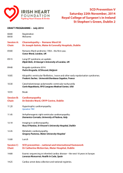


© Copyright 2026