
The benefit of docosahexaenoic acid for the adult brain in aging and
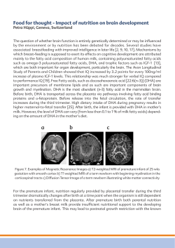
Prostaglandins, Leukotrienes and Essential Fatty Acids 92 (2015) 15–22
Contents lists available at ScienceDirect
Prostaglandins, Leukotrienes and Essential
Fatty Acids
journal homepage: www.elsevier.com/locate/plefa
The benefit of docosahexaenoic acid for the adult brain
in aging and dementia
Norman Salem Jra,n, Milene Vandal b,c,d, Frederic Calon b,c,d
a
Nutritional Lipids, DSM Nutritional Products, Columbia, MD, USA
Center de recherche du center Hospitalier de l'Université Laval (CHUL), Québec, QC, Canada
c
Faculté de pharmacie, Université Laval, Quebec, Canada
d
Institut des Nutraceutiques et des Aliments Fonctionnels, Universite Laval, Quebec, Canada
b
a b s t r a c t
Keywords:
Docosahexaenoic acid
Dementia
Aging
Alzheimer's Disease
ApoE
Cognition
A brief overview of the evidence for omega-3 fatty acids and, in particular, of docosahexaenoic acid
(DHA), involvement in cognition and in dementia is given. Two studies are presented in this regard in
which the key intervention is a DHA supplement. The fist, the MIDAS Study demonstrated that DHA can
be of benefit for episodic memory in healthy adults with a mild memory complaint. The second, the
ADCS AD trial found no benefit of DHA in the primary outcomes but found an intriguing benefit for
cognitive score in ApoE4 negative allele patients. This leads to a consideration of the mechanisms of
action and role of ApoE and its modulation by DHA. Given the fundamental role of ApoE in cellular lipid
transport and metabolism in the brain and periphery, it is no surprise that ApoE affects n-3 PUFA brain
function as well. It remains to be seen to what extent ApoE4 deleterious effect in AD is associated with n3 PUFA-related cellular mechanisms in the brain and, more specifically, whether ApoE4 directly impairs
the transport of DHA into the brain, as has been suggested.
& 2014 The Authors. Published by Elsevier Ltd. This is an open access article under the CC BY-NC-ND
license (http://creativecommons.org/licenses/by-nc-nd/3.0/).
1. Introduction
A fairly substantial literature now underpins the positive
relationship between long chain n-3 fatty acid intake and protection from cognitive decline as well as dementia. For example, a
lower plasma concentration of docosahexaenoic acid (DHA, 22:6n3) has been associated with cognitive decline in both healthy
elderly people [1,2] as well as in patients with Alzheimer's disease
(AD) [3,4]. The Framingham study related decreased plasma
phosphatidylcholine DHA content to increased cognitive decline
and rates of dementia [4]. Another aspect of the relationship
between long chain n-3 fatty acids and cognition is the literature
relating fish intake to cognitive decline. There have been many
such epidemiological studies, some quite large with both positive
results and some null studies. The VA Normative Aging Study
found an association between fish intake and the Mini Mental
State Examination (MMSE) in over 1000 healthy men of median
Abbreviations: DHA, docosahexaenoic acid; AD, Alzheimer's disease; ApoE,
apolipoprotein E; CNS, central nervous system; LDLR, low-density lipoprotein
receptor; VLDLR, very low-density lipoprotein receptor; LRPs, LDLR-related proteins; BBB, blood brain barrier; CSF, cerebrospinal fluid
n
Correspondence to: Nutritional Lipids, DSM Nutritional Products, 6480 Dobbin
Rd, Columbia, MD 21045. Tel: 443 542 2370.
E-mail address: [email protected] (N. Salem Jr).
age 68 [5]. The PAQUID Study observed a 35% reduction in risk of
AD in 1600 French adults over 68 years of age who consumed
only one fish meal a week or more [6]. Morris et al. have also
shown an association of decreased AD risk with fish eating in
Americans [7].
Intervention studies of fish oil or EPA/DHA supplementation
have recently been reviewed [8]. A recent meta-analysis of 10
randomized, controlled trials that included studies of aging
healthy adults as well as those with mild dementia or AD primarily
with DHA as the intervention indicated positive results for
patients with mild cognitive impairment [9]. Several epidemiological studies have suggested that ApoE4 allele negative patients
benefit from DHA supplementation and that ApoE4 allele patients
may not [10–12]. In addition, some recent imaging studies have
shown an association of brain structural features with omega-3
intake. Raji et al. observed that weekly fish consumption was
positively associated with gray matter volumes in several substructures including hippocampus and cingulate and orbitofrontal
cortices [13]. The use of fish oil supplements was associated with
higher mean hippocampal and cerebral cortex gray matter
volumes as well as better cognitive scores (ADAS-cog and MMSE)
in those with normal cognition [14]. Such cross-sectional studies
are not conclusive but the associations that they suggest are then
good candidates for interventional studies using randomized
clinical trial methodology.
http://dx.doi.org/10.1016/j.plefa.2014.10.003
0952-3278/& 2014 The Authors. Published by Elsevier Ltd. This is an open access article under the CC BY-NC-ND license (http://creativecommons.org/licenses/by-nc-nd/3.0/).
16
N. Salem Jr et al. / Prostaglandins, Leukotrienes and Essential Fatty Acids 92 (2015) 15–22
2. Results
One key study in the domain of benefits of DHA for brain function
is the MIDAS study, an acronym for Memory Improvement after DHA
Study [15]. This was a randomized, controlled, multi-center trial of
cognitive outcome in 485 healthy elderly patients. The intervention
was 900 mg of algal DHA/d vs. corn/soy oil placebo capsules over a 24
week period. The primary endpoint was the score in the Paired
Associate Learning (PAL) test from CANTAB. This is a computer
generated, objective test that is sensitive to early episodic memory
changes. Secondary endpoints included various tests of cognitive
function, Activity of Daily Living (ADL) skills, plasma fatty acid analysis,
safety and tolerability. Subjects were 455 years of age and the
population had a mean age of 70 years. The subjects had a subjective
memory complaint and were screened for age related cognitive
decline by means of the Logical Memory subtest of the Wechsler
Memory Scale and also the MMSE. The cut offs for these tests were as
follows: subjects had an immediate (r28) or delayed (r15) recall
score that was Z1 standard deviation below the mean of younger
subjects of 25–35 years, and also had an MMSE 426. Subjects were
excluded who reported taking omega-3 supplements, consuming
4200 mg/d DHA in their diets, used medications for AD, major
anti-psychotic or anti-depressant medications, had major medical
conditions or who abused alcohol or drugs.
There was a significant improvement in the primary outcome,
the PAL test, in the DHA supplemented group (Table 1). After 24
weeks, there were 4.5 fewer errors made in the DHA group while
the practice effect produced only 2.4 fewer errors in the placebo
group. There were significant differences also in two of the
measures of verbal recognition memory, the immediate and
delayed recall, but not in the free recall (Table 1). No differences
were detected between groups in tests for pattern recognition or
spatial working memory nor in geriatric depression [15].
This was an impressive demonstration in a RCT of considerable
size that DHA provided a benefit to the elderly for episodic
memory and measures of visual recognition memory. When
compared to normative data vs age, it could be estimated that
the improvement in the PAL measure corresponded to that of
being 7 years younger, whilst the controls appeared to be 3.6 years
younger. In addition, the cognitive changes (PAL score) were
significantly correlated with plasma DHA content which was
increased from baseline values to the extent of 3.2 percentage
points, expressed as a weight percent of total fatty acids. No
erythrocyte data was obtained in this study.
Adverse events and serious adverse events were carefully
monitored in this study and there were no significant differences
between groups in this regard, nor were there differences in
hematological or clinical chemistry measures. There was however
a significant decrease in heart rate observed in the DHA group of
3.2 bpm (vs. 1 in the placebo group).
Since the MIDAS Study was published in 2010, there have been
five studies of healthy adults involving memory related measures
and with DHA/EPA as the key intervention and with the number of
subjects greater than 100. Of these, three studied young adults of
age 35 years or less [16–18]and two studied middle aged to elderly
individuals [19,20]. Dangour et al. reported no changes in cognitive
measure such as the California Verbal Learning Test in 70–79 year
old adults after providing an experimental treatment of 700 mg/d
of EPA/DHA [19]. However, as the authors noted, over the 24
months period of the study, the subjects experienced no decline in
cognitive function; thus, it does not seem possible to measure a
benefit of a supplement on declining cognitive function [21]. In a
study of 176 healthy young adults of 18–45 years of age who were
given 1.16 g/d of DHA or placebo, Stonehouse et al. that reaction
times for both episodic and working memory improved and
episodic memory improved in women and working memory in
men [20]. In a rather large study of 285 young women, age 18–25,
Benton et al. gave 400 mg/d of DHA and found no differences in a
series of measures of mood and cognitive function [16]. Jackson
et al. tested 1 g/d of EPA- or DHA- rich fish oils in 18–35 year old
healthy adults on a series of cognitive tests and found effects in
both directions on the Stroop test and some apparent benefit of
the EPA-rich fish oil on subjective mental fatigue [17]. It is worth
mentioning that Lee et al., when providing a larger dose of 1.74 g/d
of EPA/DHA for 12 months found a benefit for several measures of
memory function in a small group of 36 more elderly patients with
mild cognitive impairment [22]. Also, Witte et al. studied executive functions and neuroimaging in a group of 65 healthy subjects
age 50–75 years of age given 2.2 g/d of n-3 PUFA in a randomized,
controlled trial design [23]. They observed a benefit in executive
function including verbal fluency. They also observed changes in
white matter microstructural integrity which they interpreted to
be beneficial as well as increases in gray matter volume in the
frontal, temporal, parietal and limbic areas primarily within the
left hemisphere. These authors demonstrated an increase in
the omega-3 index (erythrocyte EPA þ DHA) as well as in the
erythrocyte EPA content.
It appears that benefits of DHA are best observed during aging
where there is some decrement in cognition, e.g., a mild cognitive
impairment or memory complaint or perhaps when a person is
exposed to certain chronic physical or mental stressors. A limitation of the these interventional studies is that they can only
address limited aspects of memory. In the case of the MIDAS
study, the PAL task is considered to be a measure of episodic
memory, one of many different aspects of the clinical construct of
memory.
There has also been a major trial of DHA in patients with AD as
reported by Quinn et al. in 2010 [18]. This study was performed
under the auspices of the National institutes of Health by
the Alzheimer's Disease Cooperative Study group and so was a
Table 1
Positive results in cognitive testing in the MIDAS trial.
Cognitive test
Baseline score mean 7 SD Wk 24 score mean7 SD Wk 24, change from baseline mean7 SE P value
CANTAB PAL
DHA
Placebo
Verbal recognition memory, immediate, (total correct)
DHA
Placebo
Verbal recognition memory, delayed, (total correct)
DHA
Placebo
Taken from Reference [13] ,Table 2.
13.4 7 11.6
12.17 10.9
8.8 7 9.9
9.7 7 10.4
4.57 0.64
2.47 0.62
0.032
10.8 7 1.5
10.9 7 1.5
11.0 7 1.4
10.9 7 0.0
0.27 0.11
0.47 0.11
0.018
10.4 7 1.8
10.5 7 1.8
10.7 7 1.5
10.7 7 1.8
0.37 0.11
0.17 0.11
0.012
N. Salem Jr et al. / Prostaglandins, Leukotrienes and Essential Fatty Acids 92 (2015) 15–22
multi-center trial involving 51 centers and a total of 555 patients
screened and 402 randomized with a 60:40 distribution across the
DHA and placebo groups. It was a randomized, controlled trial in
mild to moderate severity of AD as defined by a MMSE score
within the 14–26 range. The dosage was 2 g of algal DHA/d given
over an 18 months period and a corn/soy oil placebo. The primary
outcome measures were the Alzheimer's Disease Assessment Scale
(ADAS-cog) and the Clinical Dementia Rating (CDR) sum of boxes.
Secondary measures included the MMSE and the ADCS Activities
of Daily Living (ADCS–ADL) scale. There was a preplanned subanalysis of the ApoE4 negative allele group.
The results of the Quinn et al. study indicated that DHA had no
significant benefit for the cognitive tests, the global score, activities of daily living and, in a subset, of brain atrophy although
compliance was believed to be good. There were no significant
increases in adverse events with the DHA treatment. The fascinating finding in this study came from the preplanned comparison of
those patients with and without the ApoE4 allele. Patients in the
ApoE4 negative group had a significantly (p ¼0.03) lower decline
in the mean change in ADAS-cog score over 18 months [18]. This
was observed in 61 patients in the DHA group vs. 48 in the placebo
group; a subset size that demonstrates the magnitude of this
effect. Indeed, it is indicated to redo this study in just ApoE4
negative group to test whether cognition can in fact be observed in
AD patients of mild to moderate severity. This study implicates
ApoE4 as a key modulator of the disease in regards to the efficacy
of DHA treatment and raises many questions about the mechanism
of action and the role of this system. This relationship has been
observed in other studies as well. For example, the APOE3/E4
status has also been shown to influence the results of clinical trials
with medium chain triglycerides [24], intravenous immunoglobulins [25], and bapineuzumab [26]. Interactions between treatment
and APOE status were also initially reported with glitazone, statins
and cholinesterase inhibitors but not confirmed in larger studies
[27–31]. One limitation of AD studies thus far has been the lack of
studies specifically targeted to APOE variants or possibly other
genotypes as a selection criteria for the patient group.
3. Discussion
3.1. ApoE: molecular aspects in AD
Despite overwhelming evidence of its key role in AD pathogenesis, the exact mechanisms by which ApoE4 accelerates the onset
of cognitive impairment continue to elude us. Nevertheless, very
important molecular clues have been gathered over the years.
Three common human ApoE polymorphisms have been identified,
designated E2, E3, and E4, which differ by single amino acid
interchanges at residues 112 and 158. The prevalence of the E2, E3
and E4 alleles coding for these ApoE isoforms in the general
population is 7, 78 and 15%, respectively [32–35]. The
arginine residue at position 112 in ApoE4 reduces stability of the
protein and impairs binding to receptors and other proteins
[33,36]. In the brain, ApoE is primarily synthesized by astrocytes
[37], although neurons also produce the protein under select
conditions [38]. Because ApoE does not readily cross the blood
brain barrier (BBB) due to its large size, the liver-produced
peripheral pool of ApoE is distinct from CNS-synthesized ApoE
[33]. ApoE can interact with various receptors, such as low-density
lipoprotein receptor (LDLR), LDLR-related proteins (LRPs), SORL1/
LR11, and very low-density lipoprotein receptor (VLDLR), which
are all present in the brain, in neurons, astrocytes and microglia of
the brain parenchyma, but also in capillary endothelial cells at the
blood–brain barrier (BBB) [33,37,39]. Thus, circulating ApoE can
influence the brain by interacting with BBB-expressed receptors,
17
whereas CNS-borne ApoE may act directly on receptors located on
pericytes, astrocytes, neurons and microglia.
Not surprisingly, to decipher the role of ApoE in AD, most early
research was centered on the role of ApoE on the production,
accumulation and clearance of Aβ, given the importance of this
peptide in the amyloid hypothesis of AD [33,34,40,41]. The
observation of ApoE-amyloid complexes in AD brain [42], the
strong binding avidity of ApoE to Aβ [43,44] and the relationship
detected between the ApoE genotype and brain levels of plaques/
Aβ [45–47] initially fueled the hypothesis that ApoE4 acts by
binding Aβ, subsequently increasing its neurotoxicity. To directly
study the role of ApoE alleles, lines of transgenic mice expressing
either one of the three human ApoE isotypes have been developed
and quickly emerged as invaluable tools [33,36,48]. Interestingly,
the association between ApoE4 and increased Aβ concentrations
in cerebral tissue has been confirmed in these knock-in mice [49–
51]. Additional in vivo and in vitro data support a scheme in which
ApoE binds to Aβ and promote its clearance and degradation, a
mechanism becoming less effective in the presence of either lower
total ApoE levels, ApoE4 genotype, or both [40,52–56]. A more
recent report suggests however that molecular interactions
between ApoE and Aβ are not needed for ApoE4 to further Aβ
clearance from the brain [57]. Still, these data are hard to reconcile
with recently reported results showing that genetically increasing
human ApoE levels, regardless of isoform status, rather increased
Aβ accumulation in transgenic mice [58,59]. Neuroimaging studies
add to the orchestra of conflicting data on the link between ApoE
expression and brain amyloid load [60–63]. Finally, it should be
kept in mind that the relationship between ApoE and Aβ, if it does
exist, might be consequential or accidental rather than causal,
since reduced ApoE levels (see below) and increased Aβ are both
observed in AD, and may be independently associated with its
pathogenesis.
3.2. ApoE: gain or loss of function?
An inescapable question in APOE research is whether the
expression of ApoE4 increases AD risk though a gain or a loss of
function, or a combination of both [33,40].
In support of a possible gain of function, ApoE4- specific
neurotoxic truncated fragments generated after proteolysis of the
full length protein have been shown to cause mitochondrial
dysfunction, cytoskeletal alterations, as well as tau hyperphosphorylation [41,64,65]. Studies comparing ApoE3/E4 bigenic mice
versus ApoE4 single-transgenic mice also support a dominant
negative effect of ApoE4 [66]. Other sets of experiments suggest
isoform-specific interactions of human ApoE to various form of Aβ
or tau may underlie the deleterious impact of ApoE4 in AD,
compared to other isoforms [53,56,67–69]. To add more complexity to gain of function hypotheses, any effect interpreted as
putative deleterious effects of ApoE4 may also be explained in
part by a lack of protective effect of ApoE3 or E2 in ApoE4 carriers,
as exemplified in different studies [55,70–72].
Indeed, up to now, the evidence pointing toward a loss of
neuroprotective function of ApoE in ApoE4 carriers is difficult to
cast away. It was discovered a long time ago that human ApoE4
carriers display lower concentrations in total ApoE in the brain, a
post-mortem finding that has been replicated many times [45,73–
76]. Consistent observations have been made in the plasma [77–
79]. Such a loss of ApoE has been replicated in human ApoE4targeted replacement mice when compared to ApoE2 or ApoE3, as
measured in brain tissue, plasma and CSF [51,80–84]. Interestingly,
alteration in the ApoE protein is not a consequence of changes in
mRNA expression, which is rather increased or unchanged in ApoE
carriers [85–87], hinting toward post-translational mechanisms.
Indeed, animal and in vitro experiments have suggested that
18
N. Salem Jr et al. / Prostaglandins, Leukotrienes and Essential Fatty Acids 92 (2015) 15–22
enhanced degradation or defective lipidation of the ApoE4 protein
compared with ApoE3 stand as possible explanations for a reduction in total ApoE in tissue [81,88]. Brain ApoE concentrations are
also decreased in AD brain [45,74,89] and plasma [90], further
arguing for a loss of ApoE activity in AD. Again, ApoE mRNA levels
remain increased or unchanged in AD compared to age-matched
controls [89,91,92], arguing against the specific death of ApoEexpressing cells or a change in mRNA expression. The fact that
ApoE concentrations are lower in AD and in ApoE4 carriers is no
surprise since the ApoE4 allele is more frequent in the AD
population [32,35]. Lastly, a recent report showed that hippocampal ApoE concentrations correlate with spatial memory retention
in ApoE knock-in mice [80]. Whatever the underlying mechanism
may be, the key conclusion is that the magnitude of the ApoE4induced decrease in total ApoE concentrations is so important that
it cannot be dissociated from any specific function of ApoE4 in AD.
3.3. ApoE and lipids
From the scientific knowledge accumulated thus far, it has
become clear that ApoE is the primary apolipoprotein synthesized
within the CNS, where it plays a critical role in lipid transport and
metabolism. For example, ApoE and its receptor pathways appear
to be essential to fulfill cholesterol requirements of the majority of
brain cells [34]. Cholesterol is present in high concentrations in the
brain [93], where it is essential in maintaining neuronal health,
particularly during synaptogenesis and related processes
[34,94,95]. Although CNS cholesterol comes from de novo synthesis and not from the periphery, ApoE is implicated in its transport
and recycling [34,37]. ApoE is also involved in the removal of
excess cholesterol and its oxidized products like 24Shydroxycholesterol out of the CNS through the BBB [33,34].
Since neurons rapidly produce ApoE during an injury response
[41], it has been hypothesized that ApoE plays a critical role in
delivering cholesterol to regenerating neurons [34,96]. It would be
interesting to see whether a similar mechanism applies to n-3
A
PUFA as well. Indeed, the role of n-3 PUFA in synapse health is
quite well established. In vitro data show that exposing neurons to
n-3 PUFA promotes neurite outgrowth and membrane expansion
[9,97–99]. Supporting in vivo data is also available from animals
fed with DHA-enriched diets [100,101] or transgenetically engineered to produce endogenous DHA [102]. Evidence of a relationship between neuron size and brain DHA content has also been
reported in animals as determined by electrophysiology [103] and
histochemical readouts [104]. Finally, there are numerous recent
studies showing that n-3 PUFA induce neuroprotection against
centrally acting neurotoxins [105], while upregulating neurotrophic factors [106,107] and neurogenesis [108]. However,
whether a parallel can be drawn between cholesterol and n-3
PUFA as substrates of the lipid-based mechanism underlying ApoE
impact on the brain, and, more specifically, on AD pathogenesis,
remains unclear. Although preliminary investigations show that
ApoE deficiency can alter the relative prevalence of DHAcontaining phospholipids in synaptic plasma membranes [109],
data linking ApoE with the effects of n-3 PUFA on neuronal tissue
is still missing. Thus, studies focusing on how ApoE may be
involved in n-3 PUFA-induced neuroprotection or neuroregeneration would clearly be warranted at this time.
Brain lipids are particularly vulnerable to oxidative damage.
Intriguingly, the ApoE4 genotype is associated with higher lipid
peroxidation indices in the plasma [110,111] and in the brain
[76,112,113] in humans and in animal models. However, no specific
post-mortem relationship was detected between ApoE4 status and
concentrations of DHA derivatives such as neuroprostanes in the
brain of individuals with AD [114]. Nevertheless, ApoE deficiency
in mice contributes to increased cerebral DHA oxidation as
determined by neuroprostane quantification with gas chromatography coupled with mass spectrometry [115]. Such a role of ApoE
on lipid oxidation is likely to have important implications since
lipid peroxidation in diseased regions has been well established in
advanced AD [116]. However, the underlying mechanisms remain
unclear and deserve further investigation.
B
Fig. 1. (A) The nutragenetics and pharmacogenetics of DHA: APOE genotype and possibly other genetic factors may influence the clinical outcomes of DHA supplementation
trials; (B) Proposed mechanism by which the APOE status affects brain–blood barrier properties, brain DHA bioavailability and, ultimately, cognition and the risk of
developing Alzheimer's disease. Abbreviations: ABCA7: ATP-binding cassette sub-family A member 7; AD: Alzheimer's disease; APOE: Apolipoprotein E; BBB: brain-blood
barrier; CEPT: Cholesterylester transfer protein; CLU: Clusterin; DHA: docosahexaenoic acid; FADS: Fatty acid desaturase; SORL1: Sortilin-related Receptor, L.
N. Salem Jr et al. / Prostaglandins, Leukotrienes and Essential Fatty Acids 92 (2015) 15–22
3.4. Does ApoE regulate DHA metabolism and its transport to the
brain?
As a lipid transporter, one simple way by which ApoE4 may
impact n-3 PUFA cerebral function is by ultimately limiting DHA
transport in the brain. Indeed, markers of AD neuropathology are
sensitive to n-3 PUFA in the CNS. A high n-3 PUFA content in brain
tissue is associated with reduced Aβ levels [117,118], reduced
soluble hyperphosporylated tau [103,117,119], higher synaptic
marker levels [120–122] and improved cognitive performance
[103,120,123] in animal models of AD.
Thus, ApoE4 could impact AD pathogenesis by decreasing
circulating DHA concentrations or by interfering with its transport
in the brain (Fig. 1). The impact of ApoE on circulating PUFA has
been the subject of recent investigations. Although higher baseline
EPA and DHA levels have been observed in plasma tryglycerides
from E4 carriers compared to ApoE3 subjects [124], another study
did not detect an association between ApoE genotype and plasma
DHA concentrations [125]. Comparison between old ApoE4 carriers versus noncarriers show a shorter DHA whole-body half-life
in the former after an oral dose of [13C]–DHA [126], consistent
with a faster metabolism. Thus, it has been hypothesized that
ApoE4 carriers accumulate DHA in the blood, partly due to
reduced metabolism. However, it is debatable whether higher n3 PUFA plasma concentrations reflect a higher tissue bioavailability. Taking advantage of the APOE knock-in mouse, we recently
reported an accumulation of DHA in the blood associated with
lower concentrations in cerebral tissue of ApoE4 mice, in comparison to ApoE2 animals [83]. Such an inverse relationship between
plasma and brain levels indicate that plasma concentrations may
reflect defective distribution into the brain rather that being a
good correlate of cerebral concentrations. Further supporting the
role of ApoE in regulating cerebral DHA levels, a significant
association between DHA content and ApoE concentrations was
detected in the brain of AD patients [83]. Therefore, based on the
evidence accumulated so far, it can be postulated that ApoE4 leads
to reduced transport of DHA into the brain (Fig. 1).
Obviously, to access the brain, DHA has to cross the BBB.
Despite controversies stemming from evidence suggesting the
expression and functional role of fatty acid transporters at the
BBB [127,128], n-3 PUFA can enter the brain by simple diffusion
through the BBB [129,130]. For example, it was shown that the
brain uptake of [14C]–DHA and [14C]–EPA through the BBB is not
saturable, using in situ cerebral perfusion [130]. Despite this, longterm exposition to dietary n-3 PUFA still influence the uptake rate
of [14C]–DHA in the brain, suggesting that regulatory mechanisms
of DHA transport across the BBB do exist [130]. After n-3 PUFA
supplementation, incorporation of EPA and DHA in the plasma free
fatty acid pool was impaired in ApoE4 carriers compared to noncarriers [124]. This information may be important as free DHA
unbound to plasma lipoproteins is expected to cross the BBB more
readily [130–132]. Our recent work demonstrates that mice carrying ApoE4 display lower brain uptake of [14C]–DHA through the
BBB and lower brain DHA concentrations [83], providing a clear
mechanistic explanation for reduced brain levels of DHA in ApoE4
mice. Indeed, ApoE isoforms differ in their binding affinity for their
receptor, including the low density lipoprotein receptor (LDL-R)
[37,133]. Thus, it could be readily postulated that 3D molecular
conformational changes could affect the capacity of ApoE to
transport DHA throughout the blood circulation and, ultimately,
into the brain.
Alternatively, ApoE4-induced changes in BBB function, resulting in DHA transport failure and lower brain accumulation,
remains an intriguing possibility (Fig. 1). A collection of recent
evidence suggests that ApoE is essential for the maintenance of
BBB integrity. Disruption of the BBB accompanied by increased
19
permeability as a function of age has been reported in ApoE
knockout [134,135]. Changes in morphology of the cerebral microvasculature, such as a thickening of the basal membrane found in
AD [136–138] and in animals models [139,140], was also shown to
depend on ApoE genotype in humans [141,142]. More recent work
confirms that the lack of ApoE leads to BBB breakdown, whereas
ApoE4 increases BBB susceptibility to injury in mice [143]. The
ApoE4-induced BBB disruption is proposed to trigger neuronal
dysfunction and perhaps initiate neurodegenerative changes [143].
Decreased interaction of ApoE4 with low density lipoprotein
receptor-related protein 1 (LRP1) at the BBB and tight junction
impairments are among the main suspected mechanisms
[143,144]. Our recent data in ApoE mice also support the hypothesis that the ApoE4 genotype induces changes in the BBB, leading
to reduced DHA uptake by the brain [83] (Fig. 1). Therefore, in the
light of data linking ApoE with lipid transport and BBB dysfunction, it could be speculated that ApoE4 expression leads to
defective transport of lipids into the brain. The main long-term
consequence would be reduced distribution of n-3 PUFA in the
brain [83]. Interestingly, a lower brain bioavailability of DHA could
explain the lack of protective effect of DHA seen in clinical studies.
It could also explain the lack of association between high n-3 PUFA
concentration and low AD incidence reported in prospective
epidemiological analyses.
In conclusion, it is becoming increasingly clear that one of the
greatest challenges of AD pharmaceutical care is to intervene as early
as possible in the disease, before widespread neurodegeneration
begins [145,146]. This issue has been particularly raised with antiamyloid agents [147]. Because of its relative safety compared to most
other drugs in development, DHA and other nutraceuticals seem
better positioned to be used as preventive tools during AD prodromal
states [148]. Preventing age-related cognitive decline is likely to
require intervention with a low safety risk as well. Recent advances
in nutragenetics and in the neuropathology of cognitive impairment
suggest that the identification of the subgroups of persons who are
most likely to benefit from DHA will be the way forward.
References
[1] M.A. Beydoun, J.S. Kaufman, J.A. Satia, et al., Plasma n-3 fatty acids and the
risk of cognitive decline in older adults: the atherosclerosis risk in communities study, Am. J. Clin. Nutr. 85 (4) (2007) 1103–1111.
[2] B. Heude, P. Ducimetière, C. Berr, Cognitive decline and fatty acid composition of erythrocyte membranes – the EVA study, Am. J. Clin. Nutr. 77 (4)
(2003) 803–808.
[3] J.A. Conquer, M.C. Tierney, J. Zecevic, et al., Fatty acid analysis of blood
plasma of patients with Alzheimer's disease, other types of dementia, and
cognitive impairment, Lipids 35 (12) (2000) 1305–1312.
[4] E.J. Schaefer, V. Bongard, A.S. Beiser, et al., Plasma phosphatidylcholine
docosahexaenoic acid content and risk of dementia and Alzheimer disease:
the Framingham Heart Study, Arch. Neurol. 63 (11) (2006) 1545–1550.
[5] O. van de Rest, A. Spiro, E. Krall-Kaye, et al., Intakes of (n-3) fatty acids and
fatty fish are not associated with cognitive performance and 6-year cognitive
change in men participating in the Veterans Affairs Normative Aging Study, J.
Nutr. 139 (12) (2009) 2329–2336.
[6] P. Barberger-Gateau, L. Letenneur, V. Deschamps, et al., Fish, meat, and risk of
dementia: cohort study, Br. Med. J. 325 (7370) (2002) 932–933.
[7] M.C. Morris, D.A. Evans, J.L. Bienias, et al., Consumption of fish and n-3 fatty
acids and risk of incident Alzheimer disease, Arch. Neurol. 60 (7) (2003)
940–946.
[8] T. Cederholm, N. Salem Jr, J. Palmblad, Omega-3 fatty acids in the prevention
of cognitive decline in humans, Adv. Nutr. (Bethesda, Md) 4 (6) (2013)
672–676.
[9] J.R. Marszalek, H.F. Lodish, Docosahexaenoic acid, fatty acid-interacting
proteins, and neuronal function: breastmilk and fish are good for you, Annu.
Rev. Cell Dev. Biol. 21 (2005) 633–657.
[10] P. Barberger-Gateau, C. Raffaitin, L. Letenneur, et al., Dietary patterns and risk
of dementia: the three-city cohort study, Neurology 69 (20) (2007)
1921–1930.
[11] T.L. Huang, P.P. Zandi, K.L. Tucker, et al., Benefits of fatty fish on dementia risk
are stronger for those without APOE epsilon4, Neurology 65 (9) (2005)
1409–1414.
20
N. Salem Jr et al. / Prostaglandins, Leukotrienes and Essential Fatty Acids 92 (2015) 15–22
[12] L.J. Whalley, I.J. Deary, J.M. Starr, et al., n-3 Fatty acid erythrocyte membrane
content, APOE epsilon 4, and cognitive variation: an observational follow-up
study in late adulthood, Am. J. Clin. Nutr. 87 (2) (2008) 449–454.
[13] C.A. Raji, K.I. Erickson, O.L. Lopez, L.H. Kuller, H.M. Gach, P.M. Thompson,
et al., Regular fish consumption and age-related brain gray matter loss, Am. J.
Prev. Med. 47 (2014) 444–451.
[14] L.A. Daiello, A. Gongvatana, S. Dunsiger, R.A. Cohen, B.R. Ott, Association of
fish oil supplement use with preservation of brain volume and cognitive
function, Alzheimers Dement. (2014) (Epub ahead of print).
[15] K. Yurko-Mauro, D. Mccarthy, D. Rom, et al., Beneficial effects of docosahexaenoic acid on cognition in age-related cognitive decline, Alzheimers
Dement. 6 (6) (2010) 456–464.
[16] D. Benton, R.T. Donohoe, D.E. Clayton, et al., Supplementation with DHA and
the psychological functioning of young adults, Br. J. Nutr. 109 (1) (2013)
155–161.
[17] P.A. Jackson, M.E. Deary, J.L. Reay, et al., No effect of 12 weeks and apos;
supplementation with 1 g DHA-rich or EPA-rich fish oil on cognitive function
or mood in healthy young adults aged 18–35 years, Br. J. Nutr. 107 (8) (2012)
1232–1243.
[18] J.F. Quinn, R. Raman, R.G. Thomas, et al., Docosahexaenoic acid supplementation and cognitive decline in Alzheimer disease: a randomized trial, J. Am.
Med. Assoc. 304 (17) (2010) 1903–1911.
[19] A.D. Dangour, E. Allen, D. Elbourne, et al., Effect of 2-y n-3 long-chain
polyunsaturated fatty acid supplementation on cognitive function in older
people: a randomized, double-blind, controlled trial, Am. J. Clin. Nutr. 91 (6)
(2010) 1725–1732.
[20] W. Stonehouse, C.A. Conlon, J. Podd, et al., DHA supplementation improved
both memory and reaction time in healthy young adults: a randomized
controlled trial, Am. J. Clin. Nutr. 97 (5) (2013) 1134–1143.
[21] C. Stough, L. Downey, B. Silber, et al., The effects of 90-day supplementation
with the omega-3 essential fatty acid docosahexaenoic acid (DHA) on
cognitive function and visual acuity in a healthy aging population, Neurobiol.
Aging 33 (4) (2012).
[22] L.K. Lee, S. Shahar, A.-V. Chin, et al., Docosahexaenoic acid-concentrated fish
oil supplementation in subjects with mild cognitive impairment (MCI): a 12month randomised, double-blind, placebo-controlled trial, Psychopharmacology. (Berl). 225 (3) (2013) 605–612.
[23] A.V. Witte, L. Kerti, H.M. Hermannstadter, J.B. Fiebach, S.J. Schreiber, J.
P. Schuchardt, et al., Long-chain omega-3 fatty acids improve brain function
and structure in older adults, Cereb. cortex (2013) ([Epub ahead of print]).
[24] S.T. Henderson, J.L. Vogel, L.J. Barr, F. Garvin, J.J. Jones, L.C. Costantini, Study of
the ketogenic agent AC-1202 in mild to moderate Alzheimer's disease: a
randomized, double-blind, placebo-controlled, multicenter trial, Nutr.
Metab. (Lond) 6 (2009) 31.
[25] N. Relkin, Clinical trials of intravenous immunoglobulin for Alzheimer's
disease, J. Clin. Immunol. 34 (2014) S74–S79.
[26] S. Salloway, R. Sperling, N.C. Fox, K. Blennow, W. Klunk, Raskind, M. Sabbagh,
et al., Two phase 3 trials of bapineuzumab in mild-to-moderate Alzheimer's
disease, N. Engl. J. Med. 370 (2014) 322–333.
[27] M.E. Risner, A.M. Saunders, J.F. Altman, G.C. Ormandy, S. Craft, I.M. Foley, M.
E. Zvartau-Hind, D.A. Hosford, A.D. Roses, Efficacy of rosiglitazone in a
genetically defined population with mild-to-moderate Alzheimer's disease,
Pharmacogenomics J. 6 (2006) 246–254.
[28] L.A. Donnelly, C.N. Palmer, A.L. Whitley, C.C. Lang, A.S. Doney, A.D. Morris, P.
T. Donnan, Apolipoprotein E genotypes are associated with lipid-lowering
responses to statin treatment in diabetes: a Go-DARTS study, Pharmacogenet. Genomics. 18 (2008) 279–287.
[29] C. Harrington, S. Sawchak, C. Chiang, J. Davies, C. Donovan, A.M. Saunders,
M. Irizarry, et al., Rosiglitazone does not improve cognition or global
function when used as adjunctive therapy to AChE inhibitors in mild-tomoderate Alzheimer's disease: two phase 3 studies, Curr. Alzheimer Res. 8
(2011) 592–606.
[30] M. Sano, K.L. Bell, D. Galasko, J.E. Galvin, R.G. Thomas, C.H. van Dyck, P.
S. Aisen, A randomized, double-blind, placebo-controlled trial of simvastatin
to treat Alzheimer disease, Neurology 77 (2011) 556–563.
[31] M. Noetzli, C.B. Eap, Pharmacodynamic, pharmacokinetic and pharmacogenetic aspects of drugs used in the treatment of Alzheimer's disease, Clin.
Pharmacokinet. 52 (2013) 225–241.
[32] L.A. Farrer, L.A. Cupples, J.L. Haines, et al., Effects of age, sex, and ethnicity on
the association between apolipoprotein E genotype and Alzheimer disease. A
meta-analysis. APOE and Alzheimer Disease Meta Analysis Consortium, J.
Am. Med. Assoc. 278 (16) (1997) 1349–1356.
[33] P.S. Hauser, V. Narayanaswami, R.O. Ryan, E. Apolipoprotein, from lipid
transport to neurobiology, Prog Lipid Res. 50 (1) (2011) 62–74.
[34] V. Leduc, S. Jasmin-Belanger, J. Poirier, APOE and cholesterol homeostasis in
Alzheimer's disease, Trends Mol. Med. 16 (10) (2010) 469–477.
[35] A. Ward, S. Crean, C.J. Mercaldi, et al., Prevalence of apolipoprotein E4
genotype and homozygotes (APOE e4/4) among patients diagnosed with
Alzheimer's disease: a systematic review and meta-analysis, Neuroepidemiology 38 (1) (2012) 1–17.
[36] B. Teter, Life-span influences of apoE4 on CNS function, Neurobiol. Aging 28
(5) (2007) 693–703.
[37] R.E. Pitas, J.K. Boyles, S.H. Lee, et al., Lipoproteins and their receptors in the
central nervous system. Characterization of the lipoproteins in cerebrospinal
fluid and identification of apolipoprotein B, E (LDL) receptors in the brain, J.
Biol. Chem. 262 (29) (1987) 14352–14360.
[38] K. Aoki, T. Uchihara, N. Sanjo, et al., Increased expression of neuronal
apolipoprotein E in human brain with cerebral infarction, Stroke 34 (4)
(2003) 875–880.
[39] G. Bu, Apolipoprotein E and its receptors in Alzheimer's disease: pathways,
pathogenesis and therapy, Nat. Rev. Neurosci. 10 (5) (2009) 333–344.
[40] J. Kim, J.M. Basak, D.M. Holtzman, The role of apolipoprotein E in Alzheimer's
disease, Neuron 63 (3) (2009) 287–303.
[41] R.W. Mahley, Y. Huang, Apolipoprotein e sets the stage: response to injury
triggers neuropathology, Neuron 76 (5) (2012) 871–885.
[42] J. Näslund, J. Thyberg, L.O. Tjernberg, et al., Characterization of stable
complexes involving apolipoprotein E and the amyloid beta peptide in
Alzheimer's disease brain, Neuron 15 (1) (1995) 219–228.
[43] W.J. Strittmatter, D.Y. Huang, R. Bhasin, et al., Avid binding of beta A amyloid
peptide to its own precursor, Exp. Neurol. 122 (2) (1993) 327–334.
[44] W.J. Strittmatter, K.H. Weisgraber, D.Y. Huang, et al., Binding of human
apolipoprotein E to synthetic amyloid beta peptide: isoform-specific effects
and implications for late-onset Alzheimer disease, Proc. Natl. Acad. Sci. 90
(17) (1993) 8098–8102.
[45] U. Beffert, J.S. Cohn, C. Petit-Turcotte, et al., Apolipoprotein E and betaamyloid levels in the hippocampus and frontal cortex of Alzheimer's disease
subjects are disease-related and apolipoprotein E genotype dependent, Brain
Res. 843 (1-2) (1999) 87–94.
[46] T. Pirttila, H. Soininen, P.D. Mehta, et al., Apolipoprotein E genotype and
amyloid load in Alzheimer disease and control brains, Neurobiol. Aging 18
(1) (1997) 121–127.
[47] P. Tiraboschi, L.A. Hansen, E. Masliah, et al., Impact of APOE genotype on
neuropathologic and neurochemical markers of Alzheimer disease, Neurology 62 (11) (2004) 1977–1983.
[48] P.M. Sullivan, H. Mezdour, S.H. Quarfordt, et al., Type III hyperlipoproteinemia and spontaneous atherosclerosis in mice resulting from gene replacement of mouse Apoe with human Apoen2, J. Clin. Invest. 102 (1) (1998)
130–135.
[49] M. Buttini, G.-Q. Yu, K. Shockley, et al., Modulation of Alzheimer-like synaptic
and cholinergic deficits in transgenic mice by human apolipoprotein E
depends on isoform, aging, and overexpression of amyloid beta peptides
but not on plaque formation, J. Neurosci. 22 (24) (2002) 10539–10548.
[50] I. Dolev, D.M. Michaelson, A nontransgenic mouse model shows inducible
amyloid-beta (Abeta) peptide deposition and elucidates the role of apolipoprotein E in the amyloid cascade, Proc. Natl. Acad. Sci. 101 (38) (2004)
13909–13914.
[51] N.F. Fitz, A.A. Cronican, M. Saleem, et al., Abca1 deficiency affects Alzheimer's
disease-like phenotype in human ApoE4 but not in ApoE3-targeted replacement mice, J. Neurosci. 32 (38) (2012) 13125–13136.
[52] J.M. Castellano, J. Kim, F.R. Stewart, et al., Human apoE isoforms differentially
regulate brain amyloid-beta peptide clearance, Sci. Trans. Med. 3 (89) (2011)
(89ra57).
[53] E. Cerf, A. Gustot, E. Goormaghtigh, et al., High ability of apolipoprotein E4 to
stabilize amyloid-beta peptide oligomers, the pathological entities responsible for Alzheimer's disease, Fed. Am. Soc. Exp. Biol. 25 (5) (2011)
1585–1595.
[54] R. Deane, A. Sagare, K. Hamm, et al., apoE isoform-specific disruption of
amyloid beta peptide clearance from mouse brain, J. Clin. Invest. 118 (12)
(2008) 4002–4013.
[55] W.P. Esler, J.R. Marshall, E.R. Stimson, et al., Apolipoprotein E affects amyloid
formation but not amyloid growth in vitro: mechanistic implications for
apoE4 enhanced amyloid burden and risk for Alzheimer's disease, Amyloid 9
(1) (2002) 1–12.
[56] N.C. Stratman, C.K. Castle, B.M. Taylor, et al., Isoform-specific interactions of
human apolipoprotein E to an intermediate conformation of human Alzheimer amyloid-beta peptide, Chem. Phys. Lipids. 137 (1-2) (2005) 52–61.
[57] P.B. Verghese, J.M. Castellano, K. Garai, et al., ApoE influences amyloid-beta
(Abeta) clearance despite minimal apoE/Abeta association in physiological
conditions, Proc. Natl. Acad. Sci. 110 (19) (2013) E1807–E1816.
[58] N. Bien-Ly, A.K. Gillespie, D. Walker, et al., Reducing human apolipoprotein E
levels attenuates age-dependent Abeta accumulation in mutant human
amyloid precursor protein transgenic mice, J. Neurosci. 32 (14) (2012)
4803–4811.
[59] J. Kim, H. Jiang, S. Park, et al., Haploinsufficiency of human APOE reduces
amyloid deposition in a mouse model of amyloid-beta amyloidosis, J.
Neurosci. 31 (49) (2011) 18007–18012.
[60] A. Drzezga, T. Grimmer, G. Henriksen, et al., Effect of APOE genotype on
amyloid plaque load and gray matter volume in Alzheimer disease, Neurology 72 (17) (2009) 1487–1494.
[61] M. Lehmann, P.M. Ghosh, C. Madison, et al., Greater medial temporal
hypometabolism and lower cortical amyloid burden in ApoE4-positive AD
patients, J. Neurol. Neurosurg. Psychiatry. 85 (3) (2014) 266–273.
[62] R. Ossenkoppele, W.M. Van Der Flier, M.D. Zwan, et al., Differential effect of
APOE genotype on amyloid load and glucose metabolism in AD dementia,
Neurology 80 (4) (2013) 359–365.
[63] C.C. Rowe, K.A. Ellis, M. Rimajova, et al., Amyloid imaging results from the
Australian Imaging, Biomarkers and Lifestyle (AIBL) study of aging, Neurobiol. Aging 31 (8) (2010) 1275–1283.
[64] W.J. Brecht, F.M. Harris, S. Chang, et al., Neuron-specific apolipoprotein e4
proteolysis is associated with increased tau phosphorylation in brains of
transgenic mice, J. Neurosci. 24 (10) (2004) 2527–2534.
N. Salem Jr et al. / Prostaglandins, Leukotrienes and Essential Fatty Acids 92 (2015) 15–22
[65] T.T. Rohn, Proteolytic cleavage of apolipoprotein E4 as the keystone for the
heightened risk associated with Alzheimer's disease, Int. J. Mol. Sci. 14 (7)
(2013) 14908–14922.
[66] M. Buttini, H. Akeefe, C. Lin, et al., Dominant negative effects of apolipoprotein E4 revealed in transgenic models of neurodegenerative disease, Neuroscience 97 (2) (2000) 207–210.
[67] M.J. Ladu, M.T. Falduto, A.M. Manelli, et al., Isoform-specific binding of
apolipoprotein E to beta-amyloid, J. Biol. Chem. 269 (38) (1994)
23403–23406.
[68] W.J. Strittmatter, A.M. Saunders, D. Schmechel, et al., Apolipoprotein E: highavidity binding to beta-amyloid and increased frequency of type 4 allele in
late-onset familial Alzheimer disease, Proc. Natl. Acad. Sci. 90 (5) (1993)
1977–1981.
[69] W.J. Strittmatter, A.M. Saunders, M. Goedert, et al., Isoform-specific interactions of apolipoprotein E with microtubule-associated protein tau: implications for Alzheimer disease, Proc. Natl. Acad. Sci. 91 (23) (1994) 11183–11186.
[70] O. Aboud, R.E. Mrak, F. Boop, et al., Apolipoprotein epsilon 3 alleles are
associated with indicators of neuronal resilience, BMC Med. 10 (2012) 35.
[71] S.B. Dumanis, H.-J. Cha, J.M. Song, et al., ApoE receptor 2 regulates synapse
and dendritic spine formation, PLoS ONE 6 (2) (2011) e17203.
[72] A. Sen, D.L. Alkon, T.J. Nelson, Apolipoprotein E3 (ApoE3) but not ApoE4
protects against synaptic loss through increased expression of protein kinase
C epsilon, J. Biol. Chem. 287 (19) (2012) 15947–15958.
[73] T. Arendt, C. Schindler, M.K. Bruckner, et al., Plastic neuronal remodeling is
impaired in patients with Alzheimer's disease carrying apolipoprotein
epsilon 4 allele, J. Neurosci. 17 (2) (1997) 516–529.
[74] P. Bertrand, J. Poirier, T. Oda, et al., Association of apolipoprotein E genotype
with brain levels of apolipoprotein E and apolipoprotein J (clusterin) in
Alzheimer disease, Brain Res. Mol. Brain Res. 33 (1) (1995) 174–178.
[75] T. Pirttila, H. Soininen, O. Heinonen, et al., Apolipoprotein E (apoE) levels in
brains from Alzheimer disease patients and controls, Brain Res. 722 (1-2)
(1996) 71–77.
[76] C. Ramassamy, D. Averill, U. Beffert, et al., Oxidative insults are associated
with apolipoprotein E genotype in Alzheimer's disease brain, Neurobiol. Dis.
7 (1) (2000) 23–37.
[77] J.M. Ringman, D. Elashoff, D.H. Geschwind, et al., Plasma signaling proteins in
persons at genetic risk for Alzheimer disease: influence of APOE genotype,
Arch. Neurol. 69 (6) (2012) 757–764.
[78] F. Schiele, D. De Bacquer, M. Vincent-Viry, et al., Apolipoprotein E serum
concentration and polymorphism in six European countries: the ApoEurope
Project, Atherosclerosis 152 (2) (2000) 475–488.
[79] P. van Vliet, R.G.J. Westendorp, P. Eikelenboom, et al., Parental history of
Alzheimer disease associated with lower plasma apolipoprotein E levels,
Neurology 73 (9) (2009) 681–687.
[80] L.A. Johnson, R.H.J. Olsen, L.S. Merkens, et al., Apolipoprotein E-low density
lipoprotein receptor interaction affects spatial memory retention and brain
ApoE levels in an isoform-dependent manner, Neurobiol. Dis. 64 (2014)
150–162.
[81] D.R. Riddell, H. Zhou, K. Atchison, et al., Impact of apolipoprotein E (ApoE)
polymorphism on brain ApoE levels, J. Neurosci. 28 (45) (2008)
11445–11453.
[82] J.D. Ulrich, J.M. Burchett, J.L. Restivo, et al., In vivo measurement of
apolipoprotein E from the brain interstitial fluid using microdialysis, Mol.
Neurodegener. 8 (2013) 13.
[83] M. Vandal, W. Alata, C. Tremblay, et al., Reduction in DHA transport to the
brain of mice expressing human APOE4 compared to APOE2, J. Neurochem.
129 (3) (2014) 516–526.
[84] S.E. Wahrle, H. Jiang, M. Parsadanian, et al., Overexpression of ABCA1 reduces
amyloid deposition in the PDAPP mouse model of Alzheimer disease, J. Clin.
Invest. 118 (2) (2008) 671–682.
[85] N.J. Bray, L. Jehu, V. Moskvina, et al., Allelic expression of APOE in human
brain: effects of epsilon status and promoter haplotypes, Hum. Mol. Genet.
13 (22) (2004) 2885–2892.
[86] W.B. Growdon, B.S. Cheung, B.T. Hyman, et al., Lack of allelic imbalance in
APOE epsilon 3/4 brain mRNA expression in Alzheimer's disease, Neurosci.
Lett. 272 (2) (1999) 83–86.
[87] J.C. Lambert, J. Perez-tur, M.J. Dupire, et al., Distortion of allelic expression of
apolipoprotein E in Alzheimer's disease, Hum. Mol. Genet. 6 (12) (1997)
2151–2154.
[88] S. Arold, P. Sullivan, T. Bilousova, et al., Apolipoprotein E level and cholesterol
are associated with reduced synaptic amyloid beta in Alzheimer's disease
and apoE TR mouse cortex, Acta Neuropathol. 123 (1) (2012) 39–52.
[89] L.M. Bekris, N.M. Galloway, T.J. Montine, et al., APOE mRNA and protein
expression in postmortem brain are modulated by an extended haplotype
structure, Am. J. Med. Genet. B. Neuropsychiatr. Genet. 153B (2) (2010)
409–417.
[90] G. Siest, P. Bertrand, B. Qin, et al., Apolipoprotein E polymorphism and serum
concentration in Alzheimer's disease in nine European centres: the ApoEurope study. ApoEurope group, Clin. Chem. Lab. Med. 38 (8) (2000) 721–730.
[91] T. Matsui, M. Ingelsson, H. Fukumoto, et al., Expression of APP pathway
mRNAs and proteins in Alzheimer's disease, Brain Res. 1161 (2007) 116–123.
[92] K. Yamagata, K. Urakami, K. Ikeda, et al., High expression of apolipoprotein E
mRNA in the brains with sporadic Alzheimer's disease, Dement. Geriatr.
Cogn. Disord. 12 (2) (2001) 57–62.
[93] P.S. Sastry, Lipids of nervous tissue: composition and metabolism, Prog. Lipid
Res. 24 (2) (1985) 69–176.
21
[94] G.E. Handelmann, J.K. Boyles, K.H. Weisgraber, et al., Effects of apolipoprotein
E, beta-very low density lipoproteins, and cholesterol on the extension of
neurites by rabbit dorsal root ganglion neurons in vitro, J. Lipid Res. 33 (11)
(1992) 1677–1688.
[95] D.H. Mauch, K. Nagler, S. Schumacher, et al., CNS synaptogenesis promoted
by glia-derived cholesterol, Science 294 (5545) (2001) 1354–1357.
[96] J. Poirier, Apolipoprotein E in animal models of CNS injury and in Alzheimer's disease, Trends Neurosci. 17 (12) (1994) 525–530.
[97] F. Calderon, H.-Y. Kim, Role of RXR in neurite outgrowth induced by
docosahexaenoic acid, Prostaglandins. Leukot. Essent. Fatty Acids 77 (5-6)
(2007) 227–232.
[98] F. Darios, B. Davletov, Omega-3 and omega-6 fatty acids stimulate cell
membrane expansion by acting on syntaxin 3, Nature 440 (7085) (2006)
813–817.
[99] R.E. Martin, J.Q. Wickham, A.S. Om, et al., Uptake and incorporation of
docosahexaenoic acid (DHA) into neuronal cell body and neurite/nerve
growth cone lipids: evidence of compartmental DHA metabolism in nerve
growth factor-differentiated PC12 cells, Neurochem. Res. 25 (5) (2000)
715–723.
[100] L.G. Robson, S. Dyall, D. Sidloff, et al., Omega-3 polyunsaturated fatty acids
increase the neurite outgrowth of rat sensory neurones throughout development and in aged animals, Neurobiol. Aging 31 (4) (2010) 678–687.
[101] R.J. Wurtman, M. Cansev, I.H. Ulus, Synapse formation is enhanced by oral
administration of uridine and DHA, the circulating precursors of brain
phosphatide, J. Nutr. Health Aging 13 (3) (2009) 189–197.
[102] C. He, X. Qu, L. Cui, et al., Improved spatial learning performance of fat-1 mice
is associated with enhanced neurogenesis and neuritogenesis by docosahexaenoic acid, Proc. Natl. Acad. Sci. 106 (27) (2009) 11370–11375.
[103] D. Arsenault, C. Julien, C. Tremblay, et al., DHA improves cognition and
prevents dysfunction of entorhinal cortex neurons in 3xTg-AD mice, PLoS
ONE 6 (2) (2011) e17397.
[104] A. Ahmad, M. Murthy, R.S. Greiner, et al., A decrease in cell size accompanies
a loss of docosahexaenoate in the rat hippocampus, Nutr. Neurosci. 5 (2)
(2002) 103–113.
[105] M. Bousquet, M. Saint-Pierre, C. Julien, et al., Beneficial effects of dietary
omega-3 polyunsaturated fatty acid on toxin-induced neuronal degeneration
in an animal model of Parkinson's disease, Fed. Am. Soc. Exp. Biol. 22
(4) (2008) 1213–1225.
[106] M. Bousquet, C. Gibrat, M. Saint-Pierre, et al., Modulation of brain-derived
neurotrophic factor as a potential neuroprotective mechanism of action of
omega-3 fatty acids in a parkinsonian animal model, Prog. Neuropsychopharmacol. Biol. Psychiatry 33 (8) (2009) 1401–1408.
[107] A. Wu, Z. Ying, F. Gomez-Pinilla, Docosahexaenoic acid dietary supplementation enhances the effects of exercise on synaptic plasticity and cognition,
Neuroscience 155 (3) (2008) 751–759.
[108] E. Kawakita, M. Hashimoto, O. Shido, Docosahexaenoic acid promotes
neurogenesis in vitro and in vivo, Neuroscience 139 (3) (2006) 991–997.
[109] U. Igbavboa, J. Hamilton, H.-Y. Kim, et al., A new role for apolipoprotein E:
modulating transport of polyunsaturated phospholipid molecular species in
synaptic plasma membranes, J. Neurochem. 80 (2) (2002) 255–261.
[110] T. Hayek, J. Attias, J. Smith, et al., Antiatherosclerotic and antioxidative effects
of captopril in apolipoprotein E-deficient mice, J. Cardiovasc. Pharmacol. 31
(4) (1998) 540–544.
[111] J.D. Smith, M. Miyata, S.E. Poulin, et al., The relationship between apolipoprotein E and serum oxidation-related variables is apolipoprotein E phenotype dependent, Int. J. Clin. Lab. Res. 28 (2) (1998) 116–121.
[112] C.N. Bassett, T.J. Montine, Lipoproteins and lipid peroxidation in Alzheimer's
disease, J. Nutr. Health Aging 7 (1) (2003) 24–29.
[113] C.M. Chinnici, Y. Yao, T. Ding, et al., Absence of 12/15 lipoxygenase reduces
brain oxidative stress in apolipoprotein E-deficient mice, Am. J. Pathol. 167
(5) (2005) 1371–1377.
[114] E.E. Reich, W.R. Markesbery, L.J. Roberts, et al., Brain regional quantification of
F-ring and D-/E-ring isoprostanes and neuroprostanes in Alzheimer's disease,
Am. J. Pathol. 158 (1) (2001) 293–297.
[115] E.E. Reich, K.S. Montine, M.D. Gross, et al., Interactions between apolipoprotein E gene and dietary alpha-tocopherol influence cerebral oxidative
damage in aged mice, J. Neurosci. 21 (16) (2001) 5993–5999.
[116] T.J. Montine, J.D. Morrow, Fatty acid oxidation in the pathogenesis of
Alzheimer's disease, Am. J. Pathol. 166 (5) (2005) 1283–1289.
[117] M. Lebbadi, C. Julien, A. Phivilay, et al., Endogenous conversion of omega-6
into omega-3 fatty acids improves neuropathology in an animal model of
Alzheimer's disease, J. Alzheimers Dis. 27 (4) (2011) 853–869.
[118] G.P. Lim, F. Calon, T. Morihara, et al., A diet enriched with the omega-3 fatty
acid docosahexaenoic acid reduces amyloid burden in an aged Alzheimer
mouse model, J. Neurosci. 25 (12) (2005) 3032–3040.
[119] K.N. Green, H. Martinez-Coria, H. Khashwji, et al., Dietary docosahexaenoic
acid and docosapentaenoic acid ameliorate amyloid-beta and tau pathology
via a mechanism involving presenilin 1 levels, J. Neurosci. 27 (16) (2007)
4385–4395.
[120] F. Calon, G.P. Lim, F. Yang, et al., Docosahexaenoic acid protects from dendritic
pathology in an Alzheimer's disease mouse model, Neuron 43 (5) (2004)
633–645.
[121] F. Calon, G.P. Lim, T. Morihara, et al., Dietary n-3 polyunsaturated fatty acid
depletion activates caspases and decreases NMDA receptors in the brain of a
transgenic mouse model of Alzheimer's disease, Eur. J. Neurosci. 22 (3)
(2005) 617–626.
22
N. Salem Jr et al. / Prostaglandins, Leukotrienes and Essential Fatty Acids 92 (2015) 15–22
[122] S.E. Perez, B.M. Berg, K.A. Moore, et al., DHA diet reduces AD pathology in
young APPswe/PS1 Delta E9 transgenic mice: possible gender effects, J.
Neurosci. Res. 88 (5) (2010) 1026–1040.
[123] C.R. Hooijmans, CEEM Van der Zee, P.J. Dederen, et al., DHA and cholesterol
containing diets influence Alzheimer-like pathology, cognition and cerebral
vasculature in APPswe/PS1dE9 mice, Neurobiol. Dis. 33 (3) (2009) 482–498.
[124] M. Plourde, M.-C. Vohl, M. Vandal, et al., Plasma n-3 fatty acid response to an
n-3 fatty acid supplement is modulated by apoE epsilon4 but not by the
common PPAR-alpha L162V polymorphism in men, Br. J. Nutr. 102 (8) (2009)
1121–1124.
[125] C. Samieri, S. Lorrain, B. Buaud, et al., Relationship between diet and plasma
long-chain n-3 PUFAs in older people: impact of apolipoprotein E genotype, J.
Lipid Res. 54 (9) (2013) 2559–2567.
[126] R. Chouinard-Watkins, C. Rioux-Perreault, M. Fortier, et al., Disturbance in
uniformly 13C-labelled DHA metabolism in elderly human subjects carrying
the apoE epsilon4 allele, Br. J. Nutr. 110 (10) (2013) 1751–1759.
[127] J. Edmond, Essential polyunsaturated fatty acids and the barrier to the brain:
the components of a model for transport, J. Mol. Neurosci. 16 (2-3) (2001)
181–193.
[128] R.W. Mitchell, G.M. Hatch, Fatty acid transport into the brain: of fatty acid
fables and lipid tails, Prostaglandins Leukot. Essent. Fatty Acids 85 (5) (2011)
293–302.
[129] J.A. Hamilton, K. Brunaldi, A model for fatty acid transport into the brain, J.
Mol. Neurosci. 33 (1) (2007) 12–17.
[130] M. Ouellet, V. Emond, C.T. Chen, et al., Diffusion of docosahexaenoic and
eicosapentaenoic acids through the blood–brain barrier: An in situ cerebral
perfusion study, Neurochem. Int. 55 (7) (2009) 476–482.
[131] J.A. Hamilton, New insights into the roles of proteins and lipids in membrane
transport of fatty acids, Prostaglandins Leukot. Essent. Fatty Acids 77 (5-6)
(2007) 355–361.
[132] A.A. Spector, Plasma free fatty acid and lipoproteins as sources of polyunsaturated fatty acid for the brain, J. Mol. Neurosci. 16 (2-3) (2001) 159–165.
[133] K.H. Weisgraber, T.L. Innerarity, R.W. Mahley, Abnormal lipoprotein receptorbinding activity of the human E apoprotein due to cysteine–arginine
interchange at a single site, J. Biol. Chem. 257 (5) (1982) 2518–2521.
[134] S.M. Fullerton, G.A. Shirman, W.J. Strittmatter, et al., Impairment of the
blood–nerve and blood–brain barriers in apolipoprotein e knockout mice,
Exp. Neurol. 169 (1) (2001) 13–22.
[135] A. Hafezi-Moghadam, K.L. Thomas, D.D. Wagner, ApoE deficiency leads to a
progressive age-dependent blood–brain barrier leakage, Am. J. Physiol. Cell
Physiol. 292 (4) (2007) C1256–C1262.
[136] R.N. Kalaria, A.B. Pax, Increased collagen content of cerebral microvessels in
Alzheimer's disease, Brain Res. 705 (1-2) (1995) 349–352.
[137] X.-K. Tong, N. Nicolakakis, A. Kocharyan, et al., Vascular remodeling versus
amyloid beta-induced oxidative stress in the cerebrovascular dysfunctions
associated with Alzheimer's disease, J. Neurosci. 25 (48) (2005) 11165–11174.
[138] C. Zarow, E. Barron, H.C. Chui, et al., Vascular basement membrane pathology
and Alzheimer's disease, Ann. NY Acad. Sci. 826 (1997) 147–160.
[139] F. Bourasset, M. Ouellet, C. Tremblay, et al., Reduction of the cerebrovascular
volume in a transgenic mouse model of Alzheimer's disease, Neuropharmacology 56 (4) (2009) 808–813.
[140] M.A. Gama Sosa, R.D. Gasperi, A.B. Rocher, et al., Age-related vascular
pathology in transgenic mice expressing presenilin 1-associated familial
Alzheimer's disease mutations, Am. J. Pathol. 176 (1) (2010) 353–368.
[141] S. Salloway, T. Gur, T. Berzin, et al., Effect of APOE genotype on microvascular
basement membrane in Alzheimer's disease, J. Neurol. Sci. 203–204 (2002)
183–187.
[142] B.D. Zipser, C.E. Johanson, L. Gonzalez, et al., Microvascular injury and blood–
brain barrier leakage in Alzheimer disease, Neurobiol. Aging 28 (7) (2007)
977–986.
[143] R.D. Bell, E.A. Winkler, I. Singh, et al., Apolipoprotein E controls cerebrovascular integrity via cyclophilin A, Nature 485 (7399) (2012) 512–516.
[144] K. Nishitsuji, T. Hosono, T. Nakamura, et al., Apolipoprotein E regulates the
integrity of tight junctions in an isoform-dependent manner in an in vitro
blood–brain barrier model, J. Biol. Chem. 286 (20) (2011) 17536–17542.
[145] V.O. Emery, Alzheimer disease: are we intervening too late? Pro, J. Neural.
Transm. 118 (2011) 1361–1378.
[146] J.L. Cummings, R. Doody, C. Clark, Disease-modifying therapies for Alzheimer
disease: challenges to early intervention, Neurology 69 (2007) 1622–1634.
[147] P.S. Aisen, B. Vellas, H. Hampel, Moving towards early clinical trials for
amyloid-targeted therapy in Alzheimer's disease, Nat. Rev. Drug Discov. 12
(2013) 324.
[148] F. Calon, Omega-3 polyunsaturated fatty acids in Alzheimer's Disease: key
questions and partial answers, Curr. Alzheimer. Res. 8 (2011) 470–478.


© Copyright 2026