
Document 92625
1
The Biology of Hair Growth
Valerie Anne Randall and Natalia \I: Botchkareva
Centre of Skin Sciences, School of Life Sciences, University of Bradford, Bradford, UK
1.1 Introduction
1.2 The Functions of Hair
1.3 Hair Follicle Anatomy
1.3.1 The Hair Shaft
1.3.2 The Inner Root Sheath
1.3.3 The Outer Root Sheath
1.3.4 The Dermal Papilla
1.4 Changing the Hair Produced by a Follicle via the Hair Growth Cycle
1.4.1 Telogen-The Resting Phase
1.4.2 Anagen-The Growth Phase
1.4.3 Catagen-The Regressive Phase
1.4.4 Exogen-Hair Shedding
Hair
Pigmentation
1.5
1.6 Seasonal Changes in Hair Growth
1.6.1 Hormonal Coordination of Seasonal Changes in Animals
1.6.2 Seasonal Variation in Human Hair Growth
1.7 Hormonal Regulation of Human Hair Growth
1.7.1 Pregnancy
1.7.2 Androgens
1.7.2.1 Human Hair Follicles Show Paradoxically Different Intrinsic
Responses to Androgens
1.7.2.2 The Mechanism of Androgen Action in Hair Follicles
4
4
7
7
7
8
8
9
10
11
11
13
13
15
15
16
18
18
18
18
21
Gurpreet S. Ahluwalia (ed.), Cosmetic Applications of Laser and Light-Based Systems, 3-35,
0 2009 William Andrew Inc.
3
4
BASICTECHNOLOGY AND TARGETS
FOR LIGHT-BASED
SYSTEMS
1.8 Treatment of Hair Growth Disorders
References
25
26
1.I Introduction
The hair follicle is a highly dynamic organ found only in mammals. Although frequently
overlooked, the follicle is fascinating from many viewpoints. For cell and developmental
biologists it has an almost unique ability in mammals to regenerate itself, recapitulating
many embryonic steps en route [1,2]. For zoologists, it is a mammalian characteristic, significant for their evolutionary success and crucial for the survival of many mammals-loss
of fur or faulty colouration leads to death from cold or predation. Human follicles also pose
a unique paradox for endocrinologists as the same hormones, androgens, cause stimulation
of hair growth in many areas, while simultaneously inhibiting scalp follicles causing balding [3,4]. In contrast, hair is often seen as rather irrelevant medically, as human hair loss is
not life threatening. Nevertheless, hair is very important for most people [ 5 ] .Many men
spend significant time shaving daily and vast amounts are spent on hair products; a ‘bad
hair day’ is a common expression for days when everything goes wrong! This reflects the
important role hair plays in human communication in both social and sexual contexts and
explains why hair disorders such as hirsutism (excessive hair growth) or alopecia (hair loss/
balding) cause serious psychological distress [6].
Hair growth is co-ordinated by hormones, usually in parallel to changes in the individual’s
age and stage of development or environmental alterations like day-length [7]. Hormones
instruct the follicle to undergo appropriate changes so that during the next hair cycle, the new
hair produced differs in colour andor size. This chapter will review the functions of hair, its
structure and the processes occurring during the hair growth cycle, the changes which can
occur with the seasons, and the importance of the main regulator of human hair growth, the
androgens. Throughout the chapter, the main emphasis will be on human hair growth.
1.2 The Functions of Hair
Mammalian skin produces hair everywhere except for the glabrous skin of the lips,
palms, and soles. Although obvious in most mammals, human hair growth is so reduced
with tiny, virtually colourless vellus hairs in many areas, that we are termed the “naked
ape”. Externally hairs are thin, flexible tubes of dead, fully keratinised epithelial cells; they
vary in colour, length, diameter, and cross-sectional shape. Inside the skin hairs are part of
individual living hair follicles, cylindrical epithelial downgrowths into the dermis and subcutaneous fat, which enlarge at the base into the hair bulb surrounding the mesenchymederived dermal papilla (Fig. 1.1) [8].
In many mammals, hair’s important roles include insulation for thermoregulation, appropriate colour for camouflage [9], and a protective physical barrier, for example, from ultraviolet light. Follicles also specialise as neuroreceptors (e.g. whiskers) or for sexual
communication like the lion’s mane [ lo]. Human hair’s main functions are protection and
communication; it has virtually lost insulation and camouflage roles, although seasonal
variation [ 11-13] and hair erection when cold indicate the evolutionary history. Children’s
1: BIOLOGY
OF HAIRGROWTH,
RANDALL
& BOTCHKAREVA
5
Hair medulla-
Figure 1.1 The hair follicle. The right-hand side of this diagram shows a section through
the lower hair follicle while the left represents a three-dimensional view cut away to reveal
the various layers. Drawing by Richard J. Dew. Reproduced from Randall [3].
hairs are mainly protective; eyebrows and eyelashes stop things entering the eyes, while
scalp hair probably prevents sunlight, cold, and physical damage to the head and neck [ 141.
Scalp hair is also important in social communication. Abundant, good-quality hair signals
good health, in contrast to sparse, brittle hair indicating starvation or disease [ 151. Customs
involving head hair spread across many cultures throughout history. Hair removal generally
has strong depersonalising roles (e.g. head shaving of prisoners and Christian/Buddhist
monks), while long uncut hair has positive connotations like Samson’s strength in the Bible.
Other human hair is involved in sexual communication. Pubic and axillary hair development signals puberty in both sexes [16-181, and sexually mature men exhibit masculinity
with visible beard, chest, and upper pubic diamond hair (Fig. 1.2). The beard’s strong signal and its potential involvement in a display of threatening behaviour, like the lion’s mane,
[5,10,14] may explain its common removal in “Westernised” countries. This important
communication role explains the serious psychological consequences and impact on quality of life seen in hair disorders like hirsutism, excessive male pattern hair growth in women,
and hair loss, such as alopecia areata, an autoimmune disease affecting both sexes [19].
Common balding, androgenetic alopecia or male pattern hair loss [20], also causes negative,
effects, even among men who have never sought medical help [6]. Its high incidence in
Caucasians and occurrence in other primates suggest a natural phenomenon, a secondary
6
BASICTECHNOLOGY AND TARGETS
FOR LIGHT-BASED
SYSTEMS
Figure 1.2 Human hair distribution under differing endocrine conditions. Normal patterns
of human hair growth are shown in the upper panel. Visible (i.e. terminal) hair with
protective functions normally develops in children on the scalp, eyelashes, and eyebrows.
Once puberty occurs, further terminal hair develops on the axilla and pubis in both sexes
and on the face, chest, limbs, and often back in men. In people with the appropriate
genetic tendency, androgens may also stimulate hair loss from the scalp in a patterned
manner causing androgenetic alopecia. The various androgen insufficiency syndromes
(lower panel) demonstrate that none of this occurs without functional androgen receptors
and that only axillary and female pattern of lower pubic triangle hairs are formed in the
absence of 5a-reductase type-2. Male pattern hair growth (hirsutism) occurs in women
with abnormalities of plasma androgens or from idiopathic causes and women may also
develop a different form of hair loss, female androgenetic alopecia. Reproduced from
Randall 12211.
1: BIOLOGY
OF HAIRGROWTH,
RANDALL
& BOTCHKAREVA
7
sexual characteristic, rather than a disorder. Marked balding would identify the older male
leader, like the silver-backed gorilla or the senior stag’s largest antlers. Other suggestions
include advantages in fighting, as flushed bald skin would look aggressive or offer less hair
for opponents to pull [14]. If any of these were evolutionary pressures to develop balding,
the lower incidence among Africans [21] suggests that any possible advantages were outweighed by hair’s important protection from the tropical sun. Whatever the origin, looking
older is not beneficial in the industrialised world’s current youth-orientated culture.
1.3 Hair Follicle Anatomy
The hair follicle can be divided into three anatomical compartments: the infundibulum,
isthmus, and the inferior segment. The upper follicle is permanent, whereas the lower follicle, the inferior segment, regenerates with each hair follicle cycle. The infundibulum
extends from the skin surface to the sebaceous duct. The isthmus, the permanent middle
portion, extends from the duct of sebaceous gland to the exertion of arrector pilli muscle.
The inferior segment consists of the suprabulbar area and the hair bulb. The hair bulb consists of extensively proliferating keratinocytes and pigment-producing melanocytes of the
hair matrix that surround the pear-shaped dermal papilla, which contains specialised fibroblast-type cells embedded in an extracellular matrix and separated from the keratinocytes
by a basement membrane [22]. The hair matrix keratinocytes move upwards and differentiate into the hair shaft, as well as into the inner root sheath; the melanocytes transfer pigment into the developing hair keratinocytes to give the hair its colour. The epithelial portion
of the hair follicle is separated from the surrounding dermis by the perifollicular connective tissue or dermaE sheath. This consists of an inner basement membrane called the hyaline or glassy membrane and an outer connective tissue sheath. The major compartments of
the hair follicle from the innermost to the outermost include the hair shaft, the inner root
sheath, the outer root sheath, and the connective tissue sheath (Fig. 1.1).
1.3.1 The Hair Shaft
The hair shaft consists of the medulla, cortex. Immediately above the matrix cells, hair
shaft cells begin to express specific hair shaft keratins in the prekeratogenous zone [23].
The medulla is a central part of larger hairs, such as beard hairs, and a specific keratin
expressed in this layer of cells can be controlled by androgens [24]. The cortex is composed
of longitudinally arranged fibres. The hair shaft cuticle covers the hair, and its integrity and
properties have a great impact on the appearance of the hair. It is formed by a layer of scales
that interlock with opposing scales of the inner root sheath, which allows the hair shaft and
the inner root sheath to move upwards together.
1.3.2 The Inner Root Sheath
The inner root sheath consists of four layers: the cuticle, Huxley’s layer, Henle’s layer,
and the companion layer. The cells of the inner root sheath cuticle partially overlap with the
cuticle cells of the hair shaft, anchoring the hair shaft tightly to the follicle. Inner root
8
BASICTECHNOLOGY AND TARGETSFOR LIGHT-BASED
SYSTEMS
sheath cells produce keratins 1/10 and trichohyalin that serve as an intracellular “cement”
giving strength to the inner root sheath to support and mould the growing hair shaft, as well
as guide its upward movement. The transcription factor GATA-3 is critical for inner
root sheath differentiation and lineage. Mice lacking this gene fail to form an inner root
sheath 1251. The inner root sheath separates the hair shaft from the outer root sheath, which
forms the external concentric layer of epithelial cells in the hair follicle.
1.3.3 The Outer Root Sheath
The outer root sheath contains a heterogeneous cell population including keratinocytes
expressing keratins 5 and 14, keratinocyte and melanocyte stem cell progeny migrating
downward to the hair matrix, and differentiating melanocytes [26-291. Between the insertion of the arrector pili muscle and duct of the sebaceous gland the outer root sheath forms
a distinct bulge, which has been identified as a reservoir of multipotent stem cells [30].
These cells are biochemically distinct and can be identified by long-term retention of
BrdU or by immunodetection of cytokeratins 15 and 19, CD 34 (in mice), and CD 200 (in
humans) 131-34]. In addition, these cells are characterised by their low proliferative rate
and their capacity for giving rise to several different cell types including epidermal keratinocytes, sebaceous gland cells, and the various different types of epithelial cells of the
lower follicle [35]. This area also contains melanocyte stem cells 1361. Moreover, recently
nestin, the neural stem cell marker protein, was also shown to be expressed in the bulge
area of the hair follicle. Nestin-positive stem cells isolated from this area could differentiate
into neurons, glia, smooth muscle cells, and melanocytes in vitro. Experiments in mice
confirmed that nestin-expressing hair follicle stem cells can differentiate into blood vessels
and neural tissue after transplantation to the subcutis of nude mice [37]. These experiments
suggest that hair-follicle bulge-area stem cells may provide an accessible source of undifferentiated multipotent stem cells for therapeutic applications [37].
1.3.4 The Dermal Papilla
The hair bulb encloses the follicular dermal papilla, which comprises a group of mesenchyme-derived cells, the dermal papilla cells, mucopolysaccharide-rich stroma, nerve
fibres, and a single capillary loop. The follicular papilla is believed to be one of the most
important drivers to instruct the hair follicle to grow and form a particularly sized and pigmented hair shaft. Several experiments have shown that the dermal papilla has powerful
inductive properties. Dermal papilla cells transplanted into non-hair-bearing epidermis are
able to induce the formation of new hair follicles [38,39]. The dermal papilla is an essential
source of paracrine factors critical for hair growth and melanogenesis; it is believed to be the
interpreter of circulating signals such as hormones to the follicle (discussed in Section 1.7).
Specific examples of factors produced by the dermal papilla that influence hair growth
include noggin, which exerts a hair growth-inducing effect by antagonising bone morphogenetic protein (BMP) signalling and activation of the BMP receptor IA expressed in the
follicular epithelium [40]. Keratinocyte growth factor (KGF) is also produced by the
anagen dermal papilla, and its receptor, FGFR2, is found predominantly in the matrix keratinocytes. The activation of this pathway by injections of KGF into nude mice induces hair
1: BIOLOGY
OF HAIRGROWTH,
RANDALL
& BOTCHKAREVA
9
growth at the site of injection [41]. Dermal papilla cells also express hepatocyte growth
factor (HGF) [42]. Transgenic mice overexpressing HGF display accelerated hair follicle
development [42]. Insulin-like growth factor-I (IGF-I) found in the dermal papilla also
serves as an important morphogen in the hair follicle [43]. In addition, stem cell factor
(SCF) produced by the dermal papilla [44] is essential for proliferation, differentiation, and
melanin production by follicular melanocytes expressing its receptor c-kit [26]. The dermal
papilla also displays unusually strong alkaline phosphatase activity during the entire hair
cycle [45]. Although a role for alkaline phosphatase remains obscure, hair growth is reduced
when inhibitors of alkaline phosphatase are applied [46].
Interestingly, recent studies suggested that follicle dermal papilla and connective (or
dermal) sheath cells may act as stem cells for both follicular and interfollicular dermis.
Moreover, the stem cell potential of follicle dermal cells extends beyond the skin. Jahoda
and colleagues have demonstrated that rodent hair follicle dermal cells have haematopoietic stem cell activity [47] and can also be directed towards adipocyte and osteocyte phenotypes (reviewed in [48]).
1.4 Changing the Hair Produced by a Follicle via
the Hair Growth Cycle
To fulfil all the roles described in Section 1.2, the hair produced by a follicle often needs
to change and follicles possess a unique mechanism for this, the hair growth cycle [1,2]
(Fig. 1.3). This involves destruction of the original lower follicle, and its regeneration to
form another, which can produce hair with different characteristics. Thus, post-natal follicles retain the ability to recapitulate the later stages of follicular embryogenesis throughout
life. Exactly how differently sized a hair can be to its immediate predecessor is currently
unclear because many changes take several years (e.g. growing a full beard) [49]. Hairs are
produced in anagen, the growth phase. Once a hair reaches full length, a short apoptosisdriven involution phase, catagen, occurs, where cell division and pigmentation stops, the
hair becomes fully keratinised with a swollen “club” end and moves up in the skin with the
regressed dermal papilla. After a period of rest, telogen, the dermal papilla cells and associated keratinocyte stem cells reactivate and a new lower follicle develops downwards inside
the dermal sheath which surrounded the previous follicle. The new hair then grows up into
the original upper follicle (Fig. 1.3). The existing hair is generally lost; although previously
thought to be due to the new hair’s upward movement, a further active shedding stage,
exogen, is now proposed [50-531.
Hair follicle regeneration is characterised by dramatic changes in its microanatomy and
cellular activity. Hair follicle transition between distinct hair cycle stages is governed by
epithelial-mesenchymal interactions between the keratinocytes of the follicular epithelium
and the dermal papilla fibroblasts. Cell fate during hair follicle growth and involution is
controlled by numerous growth regulators that induce survival and/or differentiation or
apoptosis. During hair follicle active growth and hair production, the activity of factors
promoting proliferation, differentiation, and survival predominates, while hair follicle
regression is characterised by activation of various signalling pathways that induce apoptosis in hair follicle cells [53-551.
10
Anagen
BASICTECHNOLOGY AND TARGETS
FOR LIGHT-BASED
SYSTEMS
Catagen
Telogen
-
Early mid anagen
Anagen
Figure 1.3 The hair follicle growth cycle. Hair follicles go through well established
repeated cycles of development and growth (anagen), regression (catagen), and rest
(telogen) [1,2] to enable the replacement of hairs, often by another of differing colour
or size. An additional phase, exogen, has been reported where the resting club hair is
released [87,88]. Modified from Randall [3].
1.4.1 Telogen-The
Resting Phase
Telogen hair follicles are very short in length. They are characterised by a lack of pigment-producing melanocytes and the inner root sheath. Their compact ball-shaped dermal
papilla is closely attached to a small cap of secondary hair germ keratinocytes containing
hair follicle stem cells. A balance of local growth stimulators and inhibitors in the proximal
part of the telogen hair follicle appears to be critical for the initiation of the telogen-anagen
transition. In particular, activation of the Shh pathway induces hair follicle transition from
telogen to anagen [56]. The high sensitivity of telogen hair follicles to Shh pathways was
confirmed by the initiation of anagen by a single topical application of synthetic, nonpeptidyl small molecule agonists of the Hh pathway [57].On the other hand, telogen skin
has been suggested to contain inhibitors of hair growth [58]. Bone morphogenetic protein
4 (BMP4) has been identified as one of these inhibitors, as antagonising the BMP4 pathway
by its endogenous inhibitor, noggin, induces active hair growth in post-natal telogen skin
in vivo [26]. Interestingly, noggin increased Shh mRNA in the hair follicle, while BMP4
downregulated Shh [26].
Cell proliferation in the germinative compartment of the telogen hair follicle can also be
activated by applying mechanical or chemical stimuli. For instance, removing the hair shaft
from telogen follicles by epilation results in a new hair growth wave [59]. The molecular
mechanisms underlying this induction remain largely unknown. However, plucking-induced
anagen is widely used as a model for studying the hair cycle in mice to evaluate the expression pattern of genes of interest at distinct hair cycle stages, although there is always the
possibility of abnormal effects due to the wounding caused by plucking. In addition, telogenanagen transformation of mouse hair follicles can also be induced by the administration of
immunosuppressants such as cyclosporin A and FK506 [60,61]. Indeed, the stimulation of
unwanted hair growth is one of the most common dermatological side effects of immunosuppressive cyclosporine A therapy, seen in transplantation medicine and in the treatment
of autoimmune diseases [62].
1: BIOLOGY
OF HAIRGROWTH,
RANDALL
& BOTCHKAREVA
1.4.2 Anagen-The
11
Growth Phase
Anagen can be divided into six stages. During early phases, hair progenitor cells proliferate, envelope the growing dermal papilla, grow downwards into the skin and begin to
differentiate into the hair shaft and inner root sheath. In mid anagen, melanocytes located
in the hair matrix show pigment-producing activity, and the newly formed hair shaft begins
to develop. In late anagen, full restoration of the hair fibre-producing unit is achieved,
which is characterised by the formation of the epithelial hair bulb surrounding the dermal
papilla, located deep in the subcutaneous tissue, and the new hair shaft emerges from the
skin surface [59,63,64]. During anagen, active signal exchanges occur between the epithelial cells of the hair bulb and the fibroblasts of the dermal papilla. Actively proliferating and
postmitotic keratinocytes of the hair matrix express receptors andor intracellular signalling components of a variety of signalling pathways (P-catenidef-1, c-kit, c-met, FGFR2,
IGF-IR), while the corresponding ligands are expressed in the dermal papilla (WntSa, SCF,
HGF, FGF7, IGF-1) (reviewed in [54,63]).
In addition to hair follicle tissue remodelling, skin innervation and vascular networks
also undergo substantial changes with the progression of the anagen stage [65,66]. Perifollicular vascularisation is significantly increased during anagen. It correlates with the upregulation of the expression of vascular endothelial growth factor (VEGF) mRNA, a potent
angiogenic growth factor, produced by keratinocytes of the outer root sheath. In transgenic
mice overexpressing VEGF, perifollicular vascularisation was strongly induced, which
resulted in accelerated hair growth and increased size of hair follicles and hair shafts [67].
In contrast, application of suppressors of angiogenesis leads to hair growth reduction [68].
Therefore, cutaneous vasculature may have a great impact on the hair shaft producing
activity of hair follicle cells.
1.4.3 Catagen-The
Regressive Phase
Anagen is followed by a phase of hair follicle involution, catagen. Catagen was first
characterised in detail by Kligman [69] and Straile [70]. At the beginning of catagen, proliferation and differentiation of hair matrix keratinocytes reduces dramatically, the pigment-producing activity of melanocytes ceases, and hair shaft production is completed.
During catagen, the follicle compartments involved in hair production are reduced to sizes
that allow them to regenerate in the next hair cycle after receiving the appropriate stimulation. The hair follicle shortens in length by up to 70%. Although catagen is often considered a regressive event, it is an exquisitely orchestrated, energy-requiring remodelling
process, whose progression assures renewal of a further generation of the hair follicle.
Morphologically and functionally, catagen is divided into eight sub-stages [59]. During
catagen, a specialised structure, the club hair is formed. The keratinised brush-like structure at the base of the club hair is surrounded by epithelial cells of the outer root sheath and
anchors the hair in the telogen follicle. During catagen, the dermal papilla is transformed
into a cluster of quiescent cells that are closely adjacent to the regressing hair follicle epithelium and travel from the subcutis to the dermishbcutis border to maintain contact with
the distal portion of the hair follicle epithelium including the secondary hair germ and
bulge. Catagen is characterised by several simultaneously occurring and tightly coordinated cellular programs. The most important characteristic feature is a well-coordinated
12
BASICTECHNOLOGY AND TARGETS
FOR LIGHT-BASED
SYSTEMS
apoptosis occurring in the proximal part of the hair follicle. Apoptosis is regulated differently in each follicle compartment and distinct cell populations show different abilities to
undergo apoptosis [55]. The majority of the follicular epithelial cells and melanocytes
are very susceptible to apoptosis, while dermal papilla fibroblasts and the populations
of keratinocytes and melanocytes selected for survival display a high resistance [7 1,721.
The physiological involution of the hair follicle may be triggered by the withdrawal
of dermal papilla-derived growth factors that maintain cell proliferation and differentiation
in the anagen hair follicle, and by a variety of stimuli, including signalling via death receptors (Fig. 1.4).
One of the candidate molecules mediating apoptosis in hair matrix keratinocytes after
growth factor withdrawal is p53. Mice lacking p53 showed significantly retarded catagen
progression, compared with control mice confirming a pro-apoptotic role for p53 in the
hair follicle [26]. The delicate proliferation-apoptosis balance, essential for follicle cyclic
behaviour, can also be controlled by survivin [73]. Survivin, a member of the apoptosis
inhibitor protein family, is implicated in the control of cell proliferation as well as the inhibition of apoptosis [74]. Survivin, expressed in the proliferating keratinocytes of the anagen
hair matrix and outer root sheath, disappears with the progression of catagen [73]. Before or
during catagen, outer root sheath keratinocytes produce several important catagen-promoting
secreted molecules: fibroblast growth factor-5 short isoform, neurotrophins, transforming
growth factor-p1/2 (TGF-P1/2), IGF binding protein 3, and thrombospondin-1 [75-781.
Several important growth factors were discovered as modulators of catagen development
Figure 1.4 Molecular mechanisms of apoptosis control in the distinct hair follicle
compartments. Scheme demonstrates the expression pattern of anti- and pro-apoptotic
molecules (shown in brown and black respectively) in the hair follicle.
1: BIOLOGY
OF HAIRGROWTH,
RANDALL
& BOTCHKAREVA
13
by gene knockout studies. The most remarkable phenotype was seen in mice lacking the
fibroblast growth factor-5 ( F g f s ) gene whose hair was 50% longer than their wild type littermates, giving an “angora-like’’ phenotype [79]. Neurotrophins and TGF-PI also induce
premature catagen onset. Mice overexpressing distinct members of the neurotrophin family (BDNF, NT-3) show premature catagen development in part by stimulation of proapoptotic signalling through the p75 kD neurotrophin receptor in the outer root sheath [75].
TGF-P 1 knockout mice display delayed catagen onset [76]. Neurotrophins and TGF-P2
also exert catagen-promoting effects on human hair follicles in organ culture [80,8I].
Catagen can also be initiated by several other molecules, such as endothelin-I, insulinlike growth factor binding proteins-3/4/5, interleukin- 1, vitamin D receptor (reviewed in
[82]), prolactin [83,84], endocannabinoids [85],or thrombospondin-1 [78].
1.4.4 Exogen-Hair Shedding
An additional phase of the hair cycle called exogen was recently recognised; this involves
hair shaft shedding from the telogen follicle [86], an active process, accompanied by the
activation of proteolytic processes in the follicular root [87]. Exogen was also recently
characterized in human follicles. It was shown that while anagen and telogen hairs are
firmly anchored to the follicle, exogen hairs are passively retained within the follicles. In
addition, exogen clubs do not retain remnants of the outer root sheath, in contrast to plucked
telogen hairs 1881.
The new hair formed during the next anagen may resemble its predecessor, like most
human scalp hair, or may differ markedly like the brown summer and white winter hairs of
Scottish hares [9]. The type of hair produced depends on the regulatory dermal papilla
[89,90] although the cell biology and biochemistry of their mechanisms are not fully understood. The duration of hair cycle stages varies in different body areas. Human scalp hair
follicles have the longest anagen phase, which can last up to several years; they also display
a relatively short catagen phase (1-2 weeks) followed by a telogen phase lasting several
months. The majority of scalp hair follicles are in anagen (80-SS%), with the rest either in
catagen (2%) or telogen (10-15%). The anagen phase of follicles in other body regions is
substantially shorter, for example on the arms, legs, and thighs it ranges from 3 to 4 months
[26]. It is clear that anagen length generally determines hair length; long scalp hairs are
produced by follicles with anagens over 2 years, while short finger hairs only grow for
around 2 months [91].
1.5 Hair Pigmentation
The colour of hair is variable. It is important in many mammals for camouflage and in
human beings for making hair visible, such as the increased colour of sexually related hair
after puberty [ 17,181. Loss of hair pigment resulting in greying and whitening of hair is one
of the first characteristics of ageing. Within the hair follicle, neural crest-derived melanocytes in the hair bulb produce and transport melanin to the keratinocytes of the precortical
zone that differentiate to form the pigmented hair shaft. The hair follicle pigmentary unit in
the bulb cyclically regenerates synchronously with the hair follicle during the hair cycle.
The melanogenic activity of the follicular melanocytes is strictly coupled to the anagen
14
BASICTECHNOLOGY AND TARGETS
FOR LIGHT-BASED
SYSTEMS
stage, decreases during late anagen and early catagen, and ceases during late catagen and
telogen [26,92,93].
In the anagen hair follicle, melanocytes are divided into three distinct subpopulations. The
first population is located in the hair follicle bulge and represents melanocyte stem cells that
repopulate the melanocytes in the new hair bulb formed at the onset of anagen [26,36,94].
The second population is located in the hair follicle outer root sheath and represents differentiating melanocytes. The third is located in the hair matrix above the dermal papilla and
actively produces melanin [26,93] (Fig. 1.5). Melanogenesis is controlled by several key
enzymes that are uniquely expressed in the melanocytes (reviewed in [95]). Tyrosinase
catalyses the rate-limiting initial events of melanogenesis, and mutations in tyrosinase gene
lead to loss of pigment [96]. Tyrosinase-related proteins (TRP) 1 and TRP2 share 4045%
amino acid identity with tyrosinase and are also critically important for melanogenesis,
functioning as downstream enzymes in the melanin biosynthetic pathway [97].
Hair pigmentation is tightly regulated by several hormones and growth factors. Androgens play a major role in causing alterations of human hair colour, including increase of
pigment during vellus to terminal hair switches in many regions such as the beard after
puberty, or the converse on the scalp during male pattern balding [98]. Changes in anagenassociated melanogenesis are accompanied by changes in the gene expression of melanocortin 1 receptor (MCl-R) activated by POMC-derived ACTH and MSH peptides [99], and
ACTH and a-MSH are able to promote human follicular melanocyte differentiation by
Figure 1.5 Hair follicle melanocyte distribution. Schematic drawing represents
localisation of different subpopulations of melanocytes in the anagen hair follicle.
Melanocyte stem cells are located in the bulge, the differentiating melanocyte are mostly
located in the outer root sheath, while differentiated melanogenically active melanocytes
are present in the hair bulb.
1 : BIOLOGY
OF HAIRGROWTH, RANDALL
& BOTCHKAREVA
15
up-regulating melanogenesis, dendricity, and proliferation in less differentiated melanocyte subpopulations [ 1001. SCF/c-kit signalling is required for cyclic regeneration of the
hair pigmentation unit. Pharmacological inhibition of SCF/c-kit signalling in vivo leads to
the production of depigmented hairs in rodents [26]. In addition, other proteins known to
be involved in melanocyte biology, including agouti signal protein, the endothelin family,
fibroblast growth factor 2, and hepatocyte growth factor may be important for modulating
the activity of hair follicle melanocytes during the hair cycle (reviewed in [101,102]).
1.6 Seasonal Changes in Hair Growth
Hair follicles are under hormonal regulation due to the importance of coordinating alterations in insulative and colour properties of a mammal’s coat to the environment or visibility
to changes in sexual development. Seasonal changes usually occur twice a year in temperate
regions with coordinated waves of growth and moulting to produce a thicker, warmer winter
coat and shorter summer pelage. These are linked to day-length, and to a lesser extent to
temperature, like seasonal breeding activity [7,103]; nutrient availability can also affect hair
type because of the high metabolic requirements of hair production [ 1041.
1.6.1 Hormonal Coordination of Seasonal Changes in Animals
Studies in many species, including sheep, hamsters, mink, and ground squirrels [ 105,106],
show that long daylight hours initiate short periods of daily melatonin secretion by the
pineal gland and summer coat development, while short (winter) day-length increases
melatonin secretion and stimulates a longer, warmer pelage [7,103]. The pineal gland acts
as a neuroendocrine transducer converting nerve impulses stimulated by daylight to reduced
secretion of melatonin, normally secreted in the dark. Melatonin signals are generally
translated to the follicle by the hypothalamus-pituitary route; for example, melatonin
administration into the sheep hypothalamus stimulates short day responses [ 1071. However, although disconnecting the hypothalamus and pituitary removes seasonal changes in
body weight and the wool’s normal cycling pattern, long days stimulate a minor moult [103].
Prolactin levels continue to cycle, suggesting melatonin also acts directly on the pituitary
prolactin secretion. Since both growth hormone and IGF-I levels are also reduced, this may
prevent prolactin’s full effect as IGF-1 receptors are present in goat follicles [I081 and
IGF-1 can stimulate human hair growth in vitro [109].
There is strong evidence for prolactin’s involvement in seasonal coat changes in Djungarian hamsters [ 1061, goats [ 1081, mink [ 1 101, sheep [ 110,ll I], and deer [ 1121. Increased
prolactin levels in long daylight correspond to low summer growth and low prolactin
concentrations during short days with increased winter growth; moulting occurred in
sheep after maximal prolactin levels [ 1031. Prolactin infusion inhibits goat hair growth
locally [ 1131 and prolactin receptors are located in rodent [ 114,1151 and mink [I 161 skin
and the dermal papilla and epithelial compartments of sheep follicles [ 1171. Interestingly,
sheep [ 1 111, mink [ 1161, and non-seasonal laboratory rodent [ 1151 follicles also express
prolactin mRNA.
Other hormones implicated in regulating mammalian hair growth cycles include the
sex steroids, oestradiol and testosterone, and the adrenal steroids; these delay anagen in
16
BASICTECHNOLOGY AND TARGETS
FOR LIGHT-BASED
SYSTEMS
rats [7,118], while gonadectomy in rats and adrenalectomy in rats and mink [7,118,119]
advance it. Topical application of 17P-oestradiol to mice skin inhibits hair growth and
accelerates catagen, while antioestrogens promote early anagen [ 120-1 241. Rat dermal
papillae take up oestradiol [125] and both oestrogen receptors a (ERa) and p (ERP) are
detected in human follicles [ 1261 and cultured dermal papilla cells [ 1271. Testosterone also
delays seasonal hair growth in badgers [ 1281, while urinary cortisol levels are negatively
correlated with hair loss in rhesus macaque monkeys [ 1291. In contrast, thyroid hormones
advance anagen while thyroidectomy or propythiouracil delay it [7,118]. How these circulating hormones interact is still unclear, but the main drivers in seasonal coat changes are
light, melatonin, and prolactin.
1.6.2 Seasonal Variation in Human Hair Growth
Seasonal changes are much less obvious in human beings, where follicle cycles are generally unsynchronised after age one, except in groups of three follicles called Demeijkre
trios [26]. Regular annual cycles in human scalp [ 1 1-13], beard, and other body hair [ 1 11
were only recognised relatively recently.
Seasonal changes in hair growth were evident in 14 healthy Caucasian men aged 18-39
years studied for 18 months in Sheffield, UK (latitude 53.4"N); these men also showed
pronounced seasonal behaviour, spending much more time outside in summer, despite their
indoor employment [ 111. Scalp hair showed a single annual cycle with over 90% of follicles in anagen in the spring falling to around 80% in the autumn; the number of hairs shed
in the autumn also more than doubled [ 111 (Fig. 1.6). Similar increased head-hair shedding
in New York women [ 121 indicates an autumnal moult. Since scalp hair usually grows for
at least 2-3 years [91], detection of an annual cycle indicates a strong response of any follicles able to react, presumably those in later stages of anagen.
Changes also occurred in male characteristic, androgen-dependent body hair [ 1I].
Winter beard and thigh hair growth rate were low, but increased significantly in the summer
(Fig. 1. 6). French men showed similar summer peaks in semen volume, sperm count, and
mobility [ 1301 suggesting androgen-related effects; their luteinising hormone (LH), testosterone, and 17P-oestradiol levels showed autumnal peaks. Low winter testosterone and
higher summer levels were also reported in European men [ 131,1321 and pubertal boys
[ 1331. Testosterone changes probably alter beard and thigh hair growth rate, but they are
less likely to regulate scalp follicles as seasonal changes also occur in women. However,
androgens do inhibit some scalp follicles in genetically susceptible individuals causing
balding [ 1341 and dermal papilla cells derived from non-balding scalp follicles contain
low levels of androgen receptors making such a response possible [135]. Annual fluctuations of thyroid hormones, with peaks of T3 in September and free T4 in October [136],
could also influence scalp growth, but hypothyroidism is normally associated with hair
loss [137].
In contrast to these single cycles, thigh follicles showed biannual changes in anagen,
with 80% of follicles growing in May and November, falling to around 60% in March and
August [ 111 (Fig. 1. 6).This pattern is similar to the spring and autumn moults of many
temperate mammals [7] and may reflect such seasonal moulting from our evolutionary
past. Presumably these cycles are controlled like those in Section 1.6.1. Human beings can
respond to altered day-length by changing melatonin, prolactin, and cortisol secretion, but
1: BIOLOGY
OF HAIRGROWTH,
RANDALL& BOTCHKAREVA
17
Figure 1.6 Seasonal changes in human hair growth. Hair follicles on the scalp (left) and
body (right) of British men with indoor occupations living in the north of England show
significant seasonal variation. Scalp hair (upper panel) has a single annual cycle with
most follicles in anagen in spring, with anagen numbers falling in autumn; the number
of hairs shed (lower panel) paralleled this. Facial (upper panel) and thigh hair (lower
panel) grows significantly faster in the summer months and more slowly in the winter.
Measurements are mean SEM for Caucasian men (13 scalp and beard, 14 thigh); there
is wide variation in beard heaviness in individual men [49]. Statistical analysis was carried
out using runs (RT), turning points (TP), and phase length (PL) tests. Data from Randall
and Ebling [ l l ] , redrawn from Randall VA [221].
*
the artificially manipulated light of urban environments suppress these responses [ 1381.
Nevertheless, people in Antarctica [ 1391 and those with seasonal affective disorder [ 1401
maintain melatonin rhythms and Randall and Ebling’s study population definitely exhibited seasonal behaviour despite indoor occupations [ 111.
These annual changes are important for any investigations of scalp or androgen-dependent
hair growth, particularly in individuals living in temperate zones. For hair loss patients, any
condition may be exacerbated during the increased autumnal shedding. They also have
important implications for any assessments of new therapies or treatments to stimulate,
inhibit or remove hair; to be accurate measurements need to be carried out over a year to
avoid natural seasonal variations.
18
BASICTECHNOLOGY
AND TARGETS
FOR
LIGHT-BASED
SYSTEMS
1.7 Hormonal Regulation of Human Hair Growth
Apart from seasonal changes (Section 1.6), the most obvious regulators of human hair
growth are androgens, as long as individuals have good nutrition [ 15,1411 and normal thyroid function [ 137,1421. Pregnancy hormones also effect hair growth causing diffuse hair
loss post-partum.
1.7.1 Pregnancy
Lynfield [143] found more scalp follicles were in anagen during the second and third
trimesters (95%) and for about a week after birth; by six weeks this fell to about 76%,
remaining low for 3 months. Pregnancy hormones maintain follicles in anagen, but after
birth many enter catagen and telogen, causing a synchronised partial shedding or moult.
This may be particularly noticeable in autumn due to seasonal shedding (Section 1.6.2).
Which hormones are involved is uncertain, although oestrogen and prolactin are possibilities. Human follicles have prolactin [ 1441 and 17P-oestradiol[ 126,1271receptors, but 17poestradiol inhibits cultured human follicles [ 1451, and rodent hair growth, accelerating
catagen onset [ 121-1231, the opposite of the pregnancy effect. Prolactin reduces human
follicular growth in vitro [ 1441 supporting a role in post-partum shedding.
1.7.2 Androgens
1.7.2.1 Human Hair Follicles Show Paradoxically Different
Intrinsic Responses to Androgens
Androgens’ dramatic stimulation of hair growth is seen first in puberty with pubic and
axillary hair development in both sexes [16-181. These changes parallel the rise in
plasma androgens, occurring later in boys than girls [ 146,1471. Testosterone stimulates
beard growth in eunuchs and elderly men [148] and castration inhibits beard growth [49]
and male pattern baldness [ 1491, but individuals with complete androgen insufficiency
(i.e. without functional androgen receptors) highlight the essential involvement of androgens [150]. As they cannot respond to androgen, these XY individuals develop a femaletype phenotype, but without any pubic or axillary hair or any androgenetic alopecia
(Fig. 1. 2). Growth hormone is also required for the full androgen response as sexual hair
development is inhibited in growth hormone deficiency [ 1511.
Androgens stimulate tiny vellus follicles producing fine, virtually colourless, almost
invisible hairs to transform into larger, deeper follicles forming longer, thicker, more pigmented hairs (Fig. 1.7). Follicles must pass through the hair cycle, regenerating the lower
follicle to carry out such changes (Section 1.4). Although androgens stimulate hair growth
in many areas, causing greater hair growth on the face, upper pubic diamond, chest, etc. in
men [49], they can also have the opposite effect on specific scalp areas, often in the same
individual, causing balding [57]. This involves the reverse transformation of large, deep
follicles producing long, often heavily pigmented terminal scalp hairs to miniaturised vellus follicles forming tiny, almost invisible hairs (Fig. 1.7).
During puberty, the hairline is usually straight across the top of the forehead. In
many men this frontal hairline progressively regresses in two wings and thinning occurs
1: BIOLOGY
OF HAIRGROWTH,
RANDALL& BOTCHKAREVA
19
Figure 1.7 Androgens have paradoxically different effects on human hair follicles
depending on their body site. In many areas, androgens stimulate the gradual
transformation of small follicles producing tiny, virtually colourless, vellus hairs to terminal
follicles producing longer, thicker, and more pigmented hairs during and after puberty
(upper panel) [49]. These changes involve passing through the hair cycle (see Fig. 1.3).
At the same time many follicles in the scalp and eyelashes continue to produce the same
type of hairs, apparently unaffected by androgens (middle panel). In complete contrast,
androgens may cause inhibition of follicles on specific areas of the scalp in genetically
susceptible individuals causing the reverse transformation of terminal follicles to vellus
ones and androgenetic alopecia [134]. Diagram reproduced from Randall [221].
20
BASICTECHNOLOGY AND TARGETSFOR LIGHT-BASED
SYSTEMS
mid-vertex [ 1341. These areas gradually expand in a precise pattern exposing ‘bare’ scalp
[134,152]; the lower sides and back normally retain terminal hair (Fig. 1.2). Androgenetic
alopecia is reviewed thoroughly elsewhere [20,153]. Similar hair loss, considered androgendependent, can occur in women, but the pattern differs; the frontal hairline is normally
retained while generalised thinning progresses on the vertex until it appears bald [ 1541.
In contrast, androgens appear to have no effect on other hairs like the eyelashes (Fig. 1.7).
This is an intriguing and unique biological paradox. How does one hormone stimulate an
organ, the hair follicle, in many areas, but have no effect in another, while at the same time,
cause inhibition in the same organ in another part of the body, often in the same individual?
There are also significant differences between androgen-stimulated follicles. Axillary
and lower pubic follicles enlarge in response to female levels of androgens, while other
follicles require male levels [146,147]. Follicles also differ in their sensitivity, or speed of
response. Facial follicles enlarge first above the mouth (moustache) and on the chin in boys
and hirsute women; this spreads gradually over the face and neck [18]. This progression
resembles the patterned inhibition during balding [ 134,1521. Many androgen responses are
gradual, with some follicles taking years to show the full response. Beard weight increases
dramatically during puberty but continues rising until the mid-thirties, while terminal
hairs may only be visible on the chest and ear canal years later [49] and the miniaturisation
processes of androgenetic alopecia continue well into old age [ 134,1521. This delay
parallels the late onset of androgen-dependent benign prostatic hypertrophy and prostatic
carcinoma [ 1351.
Another demonstration of the intrinsic behaviour of human follicles is the contrast
between beard and axillary hair growth. Although both increase rapidly during puberty,
beard growth remains heavy, while axillary hair is maximal in the mid-twenties before falling rapidly in both sexes [49].This is another paradox; why do follicles in some areas no
longer show their androgenic responses, while in many others they maintain or extend them?
These contrasts are presumably due to differential gene expression within individual follicles, since all follicles are exposed to the same circulating hormones and, from the complete androgen insensitivity syndrome, require the same receptor. [ 1501. Follicles’ retention
of their original androgen response when transplanted, the basis of corrective cosmetic
surgery confirms this [ 1561. Presumably, this genetic programming occurs, in the patterning processes during development. Interestingly, the dermis of the chick’s frontal parietal
scalp, which parallels human balding regions, develops from the neural crest, while the
occipital-temporal region, our non-balding area, arises from the mesoderm [ 1571. The
molecular mechanisms involved in forming different types of follicles during embryogenesis are unclear, but secreted signalling factors, such as Eda, sonic hedgehog, Wnt, and
various growth factor families (e.g. BMPs, nuclear factors), including various homeobox
genes, and others such as Hairless and Tabby, plus transmembrane and extracellular matrix
molecules are all implicated [ 158,1591.
Human follicles require androgens not only for their initial transformation, but also need
them to maintain many of the effects. If men are castrated after puberty neither beard
growth nor male pattern balding return to prepubertal levels [22,134] suggesting that some
altered gene expression does not require androgens for maintenance or lower levels can
maintain some effect. Nevertheless, beard growth increases in the summer [ 1 11 (Fig. 1.6),
probably in response to increased circulating androgens (Section 1.6), antiandrogen treatment reduces hair growth in hirsutism [I601 and more selective blockers of androgen
1: BIOLOGY
OF HAIRGROWTH,
RANDALL& BOTCHKAREVA
21
action, Sa-reductase inhibitors such as finasteride, can cause regrowth in androgenetic alopecia [ 161,1621.This suggests that androgens are required to maintain most of the responses,
as well as initiating progression.
These intrinsic differences in hair follicle androgen responses have important consequences for anyone wishing to investigate androgen action. It is essential to study follicles
which respond appropriately in vivo for the question being addressed. Unfortunately, this
means that the most available human material, non-balding scalp, is often inappropriate.
Genetics also appears important in androgen-dependent hair growth. Male pattern
baldness [149,163,164] and heavy beard growth [49] run in families, Caucasian men and
women generally have greater hair growth than Japanese [49], despite similar testosterone
levels [165], and African men exhibit much less baldness [21]. Several genes have been
investigated for association with androgenetic alopecia. Interestingly, women with polycystic ovaries and their brothers with early balding exhibit links to one allele of the steroid
metabolism gene, CYP17 [ 1661. No association was found with neutral polymorphic
markers of genes for testosterone metabolising enzymes 5a-reductase type- 1 or -2 in balding [167,168]; however, Stu I restriction fragment length polymorphism (RFLP) in exon 1
of the androgen receptor was present in young (98%) and older (92%) balding men,
although also in 77% of older controls [169]. Although single triplet repeats of CAG or
GAC were unaltered, shortlshort polymorphic CAG/GGC haplotypes were significantly
higher in balding subjects. Interestingly, Spanish girls with precocious puberty (i.e. before
8 years) showed smaller numbers of CAG repeats [ 1701 and shorter triplet repeat lengths
are associated with another androgen-dependent condition, prostate cancer [ 1711. Whether
this has functional significance like increased androgen sensitivity or simply reflects linkage disequilibrium with a causative mutation is unclear. However, increased sensitivity is
not supported by the similarity of steroid binding capability between androgen receptors
from balding and non-balding follicle dermal papilla cells [ 1721.
1.7.2.2 The Mechanism of Androgen Action in Hair Follicles
Specijic effects of androgens on hair follicle cells. Androgens must alter many aspects of
follicular cell activity to cause these changes in follicle and hair type. They must alter the
ability of epithelial matrix cells to divide, determine whether they should differentiate into
medulla (found in some large hairs), and regulate the pigment produced and/or transferred
by follicular melanocytes. They must also alter dermal papilla size which has a constant
relationship with the hair and follicle size [173,174], and ensure the dermal sheath surrounding follicles expands to accommodate larger follicles. These responses are also quite
complex; for example, altering hair length could involve changing cell division rate, that is,
hair growth rate, and/or the actual growing period, anagen. Anagen length seems the most
important. Thigh hair is three times longer in young men than women, but grows only
slightly faster for a much longer period [ 1751. Androgens do cause such alterations as antiandrogen treatment reduces hair diameter, growth rate, length, pigmentation, and medullation in hirsute women [ 1761, while blocking Sa-reductase activity increases many of these
aspects in alopecia [161]. This raises the question: are androgens acting on each target cell
individually or operating through one coordinating system with indirect effects on other
cell types?
22
BASICTECHNOLOGY AND TARGETSFOR LIGHT-BASED
SYSTEMS
General mechanism of action of androgens. Androgens, like other steroid hormones, diffuse through cell membranes to act on target cells by binding to specific intracellular receptors. These hormone-receptor complexes undergo conformational changes exposing DNA
binding sites and bind to specific hormone response elements (HRE) in the DNA, often in
combination with accessory (coactivating) proteins, promoting expression of specific, hormone-regulated genes [ 1771. Androgen action is more complex than other steroids. Testosterone, the main male circulating androgen, binds receptors in some tissues (e.g. skeletal
muscle). However, in others, including secondary sexual tissues like the prostate, testosterone is metabolised intracellularly by Sa-reductase enzymes to Sa-dihydrotestosterone, a
more potent androgen, which binds more strongly to the androgen receptor to activate gene
expression [ 1781.
Androgen-dependent follicles require androgen receptors to respond as highlighted by
the absence of adult body hair in complete androgen insensitivity (Fig. 1.2) [ 1501, but the
need for Sa-reductase varies with body region. Men with Sa-reductase type-2 deficiency
only produce female patterns of pubic and axillary hair growth, although their body shapes
become masculinised [ 1791 (Fig. 1.2). Therefore, Sa-dihydrotestosterone appears necessary for follicles characteristic of men, including beard, chest, and upper pubic diamond,
while testosterone itself can stimulate the axilla and lower pubic triangle follicles also
found in women. Since androgenetic alopecia is not seen in Sa-reductase type-2 deficient
men and the Sa-reductase type-2 inhibitor, finasteride, can restore hair growth [85,86],
Sa-reductase type-2 also seems important for androgen-dependent balding.
Why some follicles need 5a-dihydrotestosterone and others testosterone to stimulate the
same types of cell biological changes that lead to larger hairs is unclear; presumably, the
cells use different intracellular coactivating proteins to act with the receptor.
Current model for androgen action in hair follicles. Hair follicle growth is complex but
rarely abnormal, indicating a highly controlled system. This suggests that androgen action
is coordinated through one part of the follicle. The current hypothesis, proposed in 1990 by
Randall et al. [180], focuses on the dermal papilla with androgens acting directly on dermal
papilla cells where they bind to androgen receptors and then initiate the altered gene expression of regulatory factors which influence other target cells (Fig. 1.8). These factors could
be soluble paracrine factors and/or extracellular matrix factors; extracellular matrix forms
much of the papilla volume, and dermal papilla size corresponds to hair and follicle
size [173,174]. In this model the dermal papilla is the primary direct target, while other
cells such as keratinocytes and melanocytes are indirect targets.
This hypothesis evolved from several concepts reviewed elsewhere [3,180] including
dermal papilla determination of the type of hair produced [89]; adult follicle cycles
partially recapitulating their embryogenic development; strong parallels in androgen
dependency and age-related changes between hair follicles and the prostate; and androgens
acting on embryonic prostate epithelium through the mesenchyme [155]. There is now
strong experimental support for this model. Androgen receptors are found in the dermal
papilla [ 126,181 ] and in cultured dermal papilla cells derived from androgen-sensitive follicles including beard [ 1351,balding scalp [ 1721, and deer manes [ 1821.Cells from androgensensitive sites contain higher levels of specific, saturable androgen receptors than
androgen-insensitive non-balding scalp in vitro [ 135,172,1831. Importantly, beard, but not
pubic or non-balding scalp cultured dermal papilla cells metabolise testosterone to 5adihydrotestosterone in vitro [ 184-1 861 reflecting hair growth in Sa-reductase deficiency;
1: BIOLOGY
OF HAIRGROWTH,
RANDALL
& BOTCHKAREVA
23
Figure 1.8 The current model for androgen action in the hair follicle. In this model
androgens from the blood enter the hair follicle via the dermal papilla’s blood supply. They
are bound by androgen receptors in the dermal papilla cells causing changes in their
production of regulatory paracrine factors; these then alter the activity of dermal papilla
cells, follicular keratinocytes, melanocytes, etc. T = testosterone; ? = unknown paracrine
factors. Reproduced from Randall [221].
Sa-reductase type-2 gene expression also supports this [183]. These results led to wide
acceptance of this hypothesis.
However, some recent observations suggest minor modifications. The dermal sheath,
which isolates the follicle from the dermis, now seems to have other important roles as
well, as it can form a new dermal papilla and stimulate follicle development [ 1871. Cultured dermal sheath cells from beard follicles contain similar levels of androgen receptors
to dermal papilla cells (personal observations) and balding dermal sheath and dermal
papilla express mRNA for Sa-reductase type-2 [ 1881. This indicates that the dermal sheath
can respond directly to androgens without the dermal papilla acting as an intermediary. The
sheath may be a reserve to replace a lost dermal papilla’s key roles because of hair’s essential role for mammalian survival and/or dermal sheath cells may respond directly to androgens to facilitate alterations in sheath, or even dermal papilla, size in forming a differently
sized follicle.
Recently, a very specialised keratin, hHa7, was found in the medulla of hairs from beard,
pubis, and axilla [189]. The medulla is formed by central hair cells which develop large
air-filled spaces. Beard medulla cells showed coexpression of keratin hHa7 and the androgen receptor. Since the hHa7 gene promoter also contained sequences with high homology
to the androgen response element (ARE), keratin hHa7 expression may be androgenregulated. However, no stimulation occurred when the promoter was transfected into prostate cells and keratin hHa7 with the same promoter is also expressed in androgen-insensitive
body hairs of chimpanzees [ 1901 making the significance unclear. Nevertheless, the current
model needs modification to include possible specific, direct action of androgens on lower
dermal sheath and medulla cells.
24
BASICTECHNOLOGY
AND
TARGETS
FOR LIGHT-BASED
SYSTEMS
The alteration of signalling molecules in the hair follicle by androgens. The final part of
the mechanism of androgen action involves the alteration of paracrine signalling factors
produced by dermal papilla cells. There is great interest in paracrine signalling in developing and cycling follicles, aiming to understand hair follicles as dynamic organs (see
Sections 1.2 and 1.3) [90,190]. Unfortunately, there are few practical animal models for
studying androgen effects [191] because of the special effects of androgens on human follicles. Fortunately, cultured dermal papilla cells from follicles with different sensitivities to
androgens offer a useful model in which to study androgen effects due to the dermal papilla’s
central role, their abilities to be grown from small skin samples, to stimulate hair growth in
vivo at low passage numbers [89,90], and to retain characteristics in vitro which reflect
their androgen responses in vivo [ 1911 (discussed earlier). They secrete both extracellular
matrix [ 1921and soluble, proteinaceous factors which stimulate growth in other dermal papilla
cells [ 180,1931, outer root sheath cells [ 194,1951, and transformed epidermal keratinocytes
[ 1961. Soluble factors from human cells can cross species affecting rodent cell growth in
vitro and in vivo [197], paralleling the ability of human dermal papillae to induce hair
growth in vivo in athymic mice [198].
Importantly, physiological levels of testosterone in vitro increase the ability of beard
cells to promote increased growth of other beard dermal papilla cells [193], outer root
sheath cells [195], and keratinocytes [ 1961 in line with the hypothesis. Interestingly, testosterone had no effect on non-balding scalp cells and only beard cells responded to the soluble factors produced [ 1931, suggesting they have different receptors to non-balding scalp
cells. This implies that an autocrine mechanism is involved in androgen-stimulated beard
cell growth; androgen-mediated changes do involve alterations in dermal papilla cell numbers as well as the amount of extracellular matrix [174]. A need to modify the autocrine
production of growth factors could contribute to the slow androgenic response, which often
takes many years to reach full effect [22,134]. In contrast to the beard cell stimulation,
testosterone decreased the mitogenic capacity of androgenetic alopecia dermal papilla cells
from both men [ 1961 and stump-tailed macaques [ 1991.All these results support the dermal
papilla based model and demonstrate that the paradoxical androgen effects observed in
vivo are reflected in vitro, strengthening the use of cultured dermal papilla cells as a model
system for studying androgen action in vitro.
The main priority now is to identify the factors that androgens alter. So far, only IGF-1
is identified as secreted by beard cells under androgens in vitro [181]. IGF-1 is a potent
mitogen which maintains anagen in cultured human follicles [ 109,200] and abnormal hair
growth occurs in the IGF-I receptor deficient mouse [201] supporting its importance. Beard
cells also secrete more SCF than non-balding scalp cells, although this is unaltered by
androgens in vitro [202]. Since SCF plays important roles in epidermal [203] and hair pigmentation development [204], the dermal papilla probably provides local SCF for follicular melanocytes [202]. Androgens in vivo presumably increase scf expression by facial
dermal papilla cells to cause hair darkening when boys’ vellus hairs transform to adult
beard. Recently DNA microarray methods also revealed that three genes, sfrp-2, mn l , and
atpl pl, were expressed at significantly higher levels in beard than normal scalp cells, but
no changes were detected due to androgen in vitro [205].
Although androgenetic alopecia dermal papilla cells are even more difficult to culture
than normal follicles [206], androgens inhibit their expression of protease nexin- 1, a potent
inhibitor of serine proteases, which regulate cellular growth and differentiation in many
1: BIOLOGY
OF HAIRGROWTH,
RANDALL
& BOTCHKAREVA
25
tissues [207]. Androgens also stimulate their production of TGF-P and TGF-P2 [208,209].
TGF-P is a strong candidate for an inhibitor of keratinocyte activity in alopecia because it
inhibits human follicle growth in vitro promoting catagen-like changes in human beings
[ 111,2101 and mice [2 111; a probable TGF-PI suppressor delays catagen in mice [212] and
follicular keratinocytes have receptors for TGF-P [213]. However, in a limited DNA macroarray analysis TGF-P2 and TNF-a were actually slightly reduced in balding cells [214].
Balding scalp-cell conditioned media also inhibits human and rodent dermal papilla cell
growth in vitro and delays mouse hair growth in vivo suggesting active secretion of inhibitory
factors [197]. This is unlikely to involve TGF-P which is associated with the transition from
anagen to catagen [210,211] and whose receptors are only detected on keratinocytes [213].
Thus, studying dermal papilla cells implicates several factors already: IGF- 1 in enlargement, SCF in increased pigmentation, and nexin- 1 and TGF-P in miniaturisation. Alterations in several factors are probably necessary to precisely control the major cell biological
rearrangements required when follicles change size. Further research into such factors
should help clarify the complex follicular cell interactions and the pathogenesis of androgendependent disorders.
1.8 Treatment of Hair Growth Disorders
Because human hair plays important roles in social and sexual communication (discussed
in Section 1.2), hair where it is unwanted or hair loss is a source of embarrassment and psychological distress. A variety of methods are available to help control both excess hair growth
and hair loss. The earliest methods used to remove hair were physical means such as shaving, followed by depilatory creams, waxes, or sugars; new developments include
the use of lasers (see Chapter lo), and chemical inhibitors of hair growth such as Vaniqua
[2 16,2151. Many substances have been suggested to stimulate hair growth over the years
[20,217] with one of the most recent also being laser treatment. However, the most established promoters are topical applications of minoxidil (Regaine) or oral finasteride (Propecia) a 5a-reductase inhibitor used to block androgen effects in androgenetic alopecia [ 1611.
The mechanism of action of minoxidil, an antihypertensive agent that promoted hair growth
as an unacceptable side effect, has been a mystery despite its use for over 20 years; recent
research supports action via potassium channels in the dermal papilla [218,219]. The most
effective method remains transplanting androgen-independent hair follicles from the base of
the scalp to the affected areas where they retain their intrinsic independence to androgens
and maintain terminal hair [ 1561. Current research includes attempts to culture cells from
hair follicles to amplify the individual’s donor follicles.
Despite this range of treatments, neither excess hair growth nor hair loss are fully controlled; since much unwanted hair growth or hair loss is potentiated by androgens, any
treatment has to be applied frequently and continually to counteract the constant supply of
hormonal stimulation. Recently, successful clinical response to finasteride was related to
increased dermal papilla expression of IGF- 1 [220], confirming the importance of dermal
papilla-produced paracrine factors and emphasising the dermal papilla’s key role in androgen action. Greater understanding should lead to exciting new ways to treat hair disorders,
as molecular pharmacology can devise very specific drugs and transport through the skin
can target particular areas.
26
BASICTECHNOLOGY AND TARGETS
FOR LIGHT-BASED
SYSTEMS
References
1.
2.
3.
4.
Dry FW. The coat of the mouse (Mus musculus). J Genet 1926; 16: 32-35.
Kligman AG. The human hair cycle. J Invest Dermatol 1959; 33: 307-3 16.
Randall VA. Androgens and human hair growth. Clin Endocrinol 1994; 40: 439457.
Randall VA. Androgens and hair: a biological paradox. In: Nieschlag E, Behre HM, editors,
Testosterone: Action, Deficiency, Substitution, 3rd Ed. Berlin: Cambridge University Press,
2004, pp. 207-23 1.
5. Jansen VAA, van Baalen M. Altruism through beard chromodynamics. Nature 2006; 440:
663466.
6. Girman CJ, Rhodes T, Lilly FRW, Guo SS, Siervogal RM, Patrick DL, Chumlea WC. Effects
of self-perceived hair loss in a community sample of men. Dermatol 1998; 197: 223-229.
7. Ebling FG, Hale PA, Randall VA. Hormones and hair growth. In: Goldsmith LA, editor,
Biochemistry and Physiology of the Skin, 2nd .Ed. Oxford: Clarendon Press, 1991,
pp. 660-690.
8. Montagna W, Van Scott EJ. The anatomy of the hair follicle. In: Montagna W, Ellis RA, editors, The Biology of Hair Growth. New York: Academic Press, 1958, pp. 39-64.
9. Flux JEC. Colour change of mountain hares (Lepus timidus scotius) in north-east Scotland.
Zoology 1970; 162: 345-358.
10. West PM, Packer C. Sexual selection, temperature and the lion’s mane. Science 2002; 297:
1339- 1343.
11. Randall VA, Ebling EJG. Seasonal changes in human hair growth. Br J Dermatol 1991; 124:
146-15 1.
12. Orentreich N. Scalp hair replacement in men. In: Montagna W, Dobson RL, editors, Advances
in Biology of Skin. Vol. 9: Hair Growth. Oxford: Pergamon Press, 1969, pp. 99-108.
13. Courtois M, Loussouarn G, Howseau S, et al. Periodicity in the growth and shedding of hair.
Br J Dermatol 1996; 134: 47-54.
14. Goodhart CB. The evolutionary significance of human hair patterns and skin coloring. Adv Sci
1960; 17: 53-58.
15. Bradfield RB. Protein deprivation: comparative response of hair roots, serum protein and urinary nitrogen. Am J Clin Nutr 1971; 24: 405410.
16. Reynolds EL. The appearance of adult patterns of body hair in man. Ann NY Acad Sci 1951;
53: 576-584.
17. Marshall WA, Tanner JM. Variations in pattern of pubertal change in girls. Arch Dis Child
1969; 44: 291-303.
18. Marshall WA, Tanner JM. Variations in the pattern of pubertal changes in boys. Arch Dis Child
1970; 45: 13-23.
19. Randall VA. Is alopecia areata an autoimmune disease? Lancet 2001; 358: 1922-1924.
20. Randall VA. Physiology and pathophysiology of androgenetic alopecia. In: Degroot LJ,
Jameson JL, editors, Endocrinology, 5th Ed. Section XIV, Burger HG, editor, Male Reproduction. Philadelphia: WB Saunders Co., 2005, pp. 3295-3309.
21. Setty LR. Hair patterns of the scalp of white and negro males. Am J Phys Anthrop 1970;
33: 49-55.
22. Nutbrown M, Randall VA. Differences between connective tissue-epithelial junctions in
human skin and the anagen hair follicle. J Invest Dermatol 1995; 104: 90-94.
23. Langbein L, Schweizer J. Keratins of the human hair follicle. Int Rev Cytol 2005; 243: 1-78.
24. Jave-Suarez LF, Langbein L, Winter H, Praetzel S, Rogers MA, Schweizer J. Androgen regulation of the human hair follicle: the type I hair keratin hHa7 is a direct target gene in trichocytes.
J Invest Dermatol2004; 122: 555-564.
25. Kaufman CK, Zhou P, Pasolli HA, Rend1 M, Bolotin D, Lim KC, Dai X, Alegre ML, Fuchs E.
GATA-3: an unexpected regulator of cell lineage determination in skin. Genes Dev 2003; 17:
2108-2 122.
26. Botchkareva NV, Khlgatian M, Longley BJ, Botchkarev VA, Gilchrest BA. SCF/c-kit signaling is required for cyclic regeneration of the hair pigmentation unit. FASEB J 2001; 15:
645-658.
1: BIOLOGY
OF HAIRGROWTH,
RANDALL& BOTCHKAREVA
27
27. Byrne C, Fuchs E. Probing keratinocyte and differentiation specificity of the human K5 promoter in vitro and in transgenic mice. Mol Cell Biol 1993; 13: 31763190.
28. Coulombe PA, Kopan R, Fuchs E. Expression of keratin K14 in the epidermis and hair follicle:
insights into complex programs of differentiation. J Cell Biol 1989; 109: 2295-23 12.
29. Oshima H, Rochat A, Kedzia C, Kobayashi K, Barrandon Y. Morphogenesis and renewal of
hair follicles from adult multipotent stem cells. Cell 2001; 104: 233-245.
30. Cotsarelis G, Sun TT, Lavker RM. Label-retaining cells reside in the bulge area of pilosebaceous unit: implications for follicular stem cells, hair cycle, and skin carcinogenesis. Cell
1990; 61: 1329-1337.
3 1. Commo S, Bernard BA. Immunohistochemical analysis of tissue remodelling during the
anagen-catagen transition of the human hair follicle. Br J Dermatol 1997; 137: 31-38.
32. Lyle S, Christofidou-Solomidou M, Liu Y, Elder DE, Albelda S, Cotsarelis G. The C8/144B
monoclonal antibody recognizes cytokeratin 15 and defines the location of human hair follicle
stem cells. J Cell Sci 1998; 111: 3 179-3 188.
33. Michel M, Torok N, Godbout MJ, Lussier M, Pierrete G, Royal A, Germain L. Keratin 19 as a
biochemical marker of skin stem cells in vivo and in vitro: keratin 19 expressing cells are differentially localized in function of anatomic sites, and their number varies with donor age and
culture stage. J Cell Sci 1996; 109: 1017-1028.
34. Ohyama M, Terunuma A, Tock C, Radonovich M, Pise-Masison C, Hopping S, Brady J, Udey
M, Vogel JC. Characterization and isolation of stem cell-enriched human hair follicle bulge
cells. J Clin Invest 2006; 116: 249-260.
35. Cotsarelis G. Epithelial stem cells: a folliculocentric view. J Invest Dermatol 2006; 126:
1459- 1468.
36. Nishimura EK, Jordan SA, Oshima H, Yoshida H, Osawa M, Moriyama M, Jackson IJ,
Barrandon Y, Miyachi Y, Nishikawa S. Dominant role of the niche in melanocyte stem-cell fate
determination. Nature 2002; 4 16: 854-860.
37. Hoffman RM. The potential of nestin-expressing hair follicle stem cells in regenerative medicine. Expert Opin Biol Ther 2007; 7: 289-29 1.
38. Jahoda CA, Horne KA, Oliver RF. Induction of hair growth by implantation of cultured dermal
papilla cells. Nature 1984; 31 1: 560-562.
39. Reynolds AJ, Lawrence C, Cserhalmi-Friedman PB, Christian0 AM, Jahoda CA. Trans-gender
induction of hair follicles. Nature 1999; 402: 33-34.
40. Botchkarev VA, Botchkareva NV, Nakamura M, Huber 0, Funa K, Lauster R, Paus R,
Gilchrest BA. Noggin is required for induction of hair follicle growth phase in postnatal skin.
FASEB J 2001; 15: 2205-2214.
41. Danilenko DM, Ring BD, Yanagihara D, Benson W, Wiemann B, Stames CO, Pierce GF.
Keratinocyte growth factor is an important endogenous mediator of hair follicle growth, development, and differentiation. Normalization of the nu/nu follicular differentiation defect and
amelioration of chemotherapy-induced alopecia. Am J Pathol 1995; 147: 145-154.
42. Lindner G, Menrad A, Gherardi E, Merlino G, Welker P, Handjiski B, Roloff B, Paus R. Involvement of hepatocyte growth factorkatter factor and met receptor signaling in hair follicle
morphogenesis and cycling. FASEB J 2000; 14: 3 19-332.
43. Rudman SM, Philpott MP, Thomas GA, Kealey T. The role of IGF-I in human skin and its
appendages: morphogen as well as mitogen? J Invest Dermatol 1997; 109: 770-777.
44. Hibberts NA, Messenger AG, Randall VA. Dermal papilla cells derived from beard hair
follicles secrete more stem cell factor (SCF) in culture than scalp cells or dermal fibroblasts.
Biochem Biophys Res Commun 1996; 222: 401-405.
45. Handjiski BK, Eichmuller S, Hofmann U, Czarnetzki BM, Paus R. Alkaline phosphatase activity and localization during the murine hair cycle. Br J Dermatol 1994; 131: 303-310.
46. Styczynski P, Ahluwalia G. Reduction of hair growth, USA, 2001, US6299865.
47. Lako M, Armstrong L, Cairns PM, Harris S, Hole N, Jahoda CA. Hair follicle dermal cells
repopulate the mouse haematopoietic system. J Cell Sci 2002; 115: 3967-3974.
48. Richardson GD, Arnott EC, Whitehouse CJ, Lawrence CM, Reynolds AJ, Hole N, Jahoda CA.
Plasticity of rodent and human hair follicle dermal cells: implications for cell therapy and tissue engineering. J Invest Dermatol Symp Proc 2005; 10: 180-183.
28
BASICTECHNOLOGY AND TARGETS
FOR LIGHT-BASED
SYSTEMS
49. Hamilton JB. Age, sex and genetic factors in the regulation of hair growth in man: a comparison of Caucasian and Japanese populations. In: Montagna W, Ellis RA, editors, The Biology
of Hair Growth. New York: Academic Press, 1958, pp. 399-433.
50. Chase HB. Growth of the hair. Physiol Rev 1954; 34: 113-126.
51. Chase HB, Eaton GJ. The growth of hair follicles in waves. Ann NY Acad Sci 1959; 83: 365-368.
52. Stenn KS, Combates NJ, Eilertsen KJ, Gordon JS, Pardinas JR, Parimoo S, Prouty SM. Hair
follicle growth controls. Dermatol Clin 1996; 14: 543-558.
53. Kligman AM. The human hair cycle. J Invest Dermatol 1959; 33: 307-316.
54. Botchkarev VA, Kishimoto J. Molecular control of epithelial-mesenchymal interactions during
hair follicle cycling. J Invest Dermatol Symp Proc 2003; 8: 46-55.
55. Botchkareva NV, Ahluwalia G, Shander D. Apoptosis in the hair follicle. J Invest Dermatol
2006; 126: 258-264.
56. Sat0 N, Leopold PL, Crystal RG. Induction of the hair growth phase in postnatal mice by localized transient expression of Sonic hedgehog. J Clin Invest 1999; 104: 855-864.
57. Paladini RD, Saleh J, Qian C, Xu GX, Rubin LL. Modulation of hair growth with small molecule agonists of the hedgehog signaling pathway. J Invest Dermatol 2005; 125: 638-646.
58. Paus R, Stenn KS, Link RE. Telogen skin contains an inhibitor of hair growth. Br J Dermatol
1990; 122: 777-784.
59. Muller-Rover S, Handjiski B, van der Veen C, Eichmuller S, Foitzik K, McKay IA, Stenn KS,
Paus R. A comprehensive guide for the accurate classification of murine hair follicles in distinct hair cycle stages. J Invest Dermatol 2001; 117: 3-15.
60. Iwabuchi T, Maruyama T, Sei Y, Adachi K. Effects of immunosuppressive peptidyl-prolyl cistrans isomerase (PPIase) inhibitors, cyclosporin A, FK506, ascomycin and rapamycin, on hair
growth initiation in mouse: immunosuppression is not required for new hair growth. J Dermatol
Sci 1995; 9: 64-69.
61. Paus R, Stenn KS, Link RE. The induction of anagen hair growth in telogen mouse skin by
cyclosporine A administration. Lab Invest 1989; 60: 365-369.
62. Lutz G. Effects of cyclosporin A on hair. Review. Skin Pharmacol 1994; 7: 101-104.
63. Paus R, Cotsarelis G. The biology of hair follicles. N Engl J Med 1999; 341: 491-497.
64. Stenn KS, Paus R. Controls of hair follicle cycling. Physiol Rev 2001; 81: 449-494.
65. Botchkarev VA, Eichmuller S, Johansson 0, Paus R. Hair cycle-dependent plasticity of skin
and hair follicle innervation in normal murine skin. J Comp Neurol 1997; 386: 379-395.
66. Mecklenburg L, Tobin DJ, Muller-Rover S, Handjiski B, Wendt G, Peters EM, Pohl S, Moll I,
Paus R. Active hair growth (anagen) is associated with angiogenesis. J Invest Dermatol 2000;
114: 909-916.
67. Yano K, Brown LF, Detmar M. Control of hair growth and follicle size by VEGF-mediated
angiogenesis. J Clin Invest 2001; 107: 409-417.
68. Ahluwalia G, Styczynski P, Shander D. Inhibition of hair growth. USA, 2000, US6093748.
69. Kligman AG. The human hair cycle. J Invest Dermatol 1959; 33: 307-316.
70. Straile WE, Chase HB, Arsenault C. Growth and differentiation of hair follicles between periods of activity and quiescence. J Exp Zoo1 196l ; 148: 205-22 l.
71. Lindner G, Botchkarev VA, Botchkareva NV, Ling G, van der Veen C, Paus R. Analysis of
apoptosis during hair follicle regression (catagen). Am J Pathol 1997; 15 1: 1601-1617.
72. Seiberg M, Marthinuss J, Stenn KS. Changes in expression of apoptosis-associated genes in
skin mark early catagen. J Invest Dermatol 1995; 104: 78-82.
73. Botchkareva NV, Kahn M, Ahluwalia G, Shander D. Survivin in the human hair follicle.
J Invest Dermatol2007; 127: 479-482.
74. Altieri DC. Survivin, versatile modulation of cell division and apoptosis in cancer. Oncogene
2003; 22: 858 1-8589.
75. Botchkarev VA, Botchkareva NV, Peters EM, Paus R. Epithelial growth control by neurotrophins: leads and lessons from the hair follicle. Prog Brain Res 2004; 146: 493-5 13.
76. Foitzik K, Lindner G, Mueller-Roever S, Maurer M, Botchkareva N, Botchkarev V, Handjiski
B, Metz M, Hibino T, Soma T, Dotto GP, Paus R. Control of murine hair follicle regression
(catagen) by TGF-PI in vivo. FASEB J 2000; 14: 752-760.
1: BIOLOGYOF HAIRGROWTH,RANDALL
& BOTCHKAREVA
29
77. Suzuki S, Ota Y, Ozawa K, Imamura T. Dual-mode regulation of hair growth cycle by two
Fgf-5 gene products. J Invest Dermatol2000; 114: 456-463.
78. Yano K, Brown LF, Lawler J, Miyakawa T, Detmar M. Thrombospondin- 1 plays a critical role
in the induction of hair follicle involution and vascular regression during the catagen phase.
J Invest Dermatol2003; 120: 14-19.
79. Hebert JM, Rosenquist T, Gotz J, Martin GR. FGF5 as a regulator of the hair growth cycle:
evidence from targeted and spontaneous mutations. Cell 1994; 78: 1017-1025.
80. Tsuji Y, Denda S, Soma T, Raftery L, Momoi T, Hibino T. A potential suppressor of TGF-P
delays catagen progression in hair follicles. J Invest Dermdtol Symp Proc 2003; 8: 65-68.
8 1. Peters EM, Stieglitz MG, Liezman C, Overall RW, Nakamura M, Hagen E, Klapp BF, Arck P,
Paus R. p75 Neurotrophin receptor-mediated signaling promotes human hair follicle regression (catagen). Am J Pathol 2006; 168: 221-234.
82. Stenn KS, Paus R. Controls of hair follicle cycling. Physiol Rev 200 1 ; 8 1: 449-494.
83. Foitzik K, Krause K, Conrad F, Nakamura M, Funk W, Paus R. Human scalp hair follicles are
both a target and a source of prolactin, which serves as an autocrine and/or paracrine promoter
of apoptosis-driven hair follicle regression. Am J Pathol2006; 168: 748-756.
84. Pearson AJ, Ashby MG, Wildermoth JE, Craven AJ, Nixon AJ. Effect of exogenous prolactin
on the hair growth cycle. Exp Dermatol 1999; 8: 358-360.
85. Telek A, Bir6 T, Bod6 E, T6th BI, Borbir6 I, Kunos G, Paus R. Inhibition of human hair follicle
growth by endo- and exocannabinoids. FASEB J 2007; 21: 3534-3541.
86. Stenn KS, Parimoo S, Prouty SM. Growth of the hair follicle, a cycling and regenerating
biological system. In: Chuong C-M, editor, Molecular Basis of Epithelial Appendage Morphogenesis. Austin, TX: RG Landes Co., 1998, pp. 11 1-130.
87. MilnerY, Sudnik J, Filippi M, Kizoulis M, Kashgarian M, Stenn K. Exogen, shedding phase of the
hair growth cycle: characterization of a mouse model. J Invest Dermatol2002; 119: 639-644.
88. Van Neste D, Leroy T, Conil S. Exogen hair characterization in human scalp. Skin Res Techno1
2007; 13: 4 3 M 4 3 .
89. Reynolds AJ, Jahoda CAB. Cultured human and rat tooth papilla cells induce hair follicle
regeneration and fibre growth. Differentiation 2004; 72: 566-575.
90. Waters JM, Richardson GD, Jahoda CA. Hair follicle stem cells. Semin Cell Dev Biol 2007;
18: 245-254.
91. Saitoh M, Sakamoto M. Human hair cycle. J Invest Dermatol 1970; 54: 65-81.
92. Slominski A, Paus R, Costantino R. Differential expression and activity of melanogenesisrelated proteins during induced hair growth in mice. J Invest Dermatol 1991; 96: 172-179.
93. Tobin DJ, Slominski A, Botchkarev V, Paus R. The fate of hair follicle melanocytes during the
hair growth cycle. J Invest Dermatol 1999; 4: 323-332.
94. Osawa M, Egawa G, Mak SS, Moriyama M, Freter R, Yonetani S, Beermann F, Nishikawa S.
Molecular characterization of melanocyte stem cells in their niche. Development (Cambridge,
England) 2006; 132: 5589-5599.
95. Hearing VJ. Biochemical control of melanogenesis and melanosomal organization. J Invest
Dermatol Symp Proc 1999; 4: 24-28.
96. Beermann F. The tyrosinase related protein- 1 (Tyrpl) promoter in transgenic experiments:
targeted expression to the retinal pigment epithelium. Cell Mol Biol 1999; 45: 961-968.
97. Winder A, Kobayashi T, Tsukamoto K, Urabe K, Aroca P, Kameyama K, Hearing VJ. The
tyrosinase gene family-interactions of melanogenic proteins to regulate melanogenesis. Cell
Mol Biol Res 1994; 40: 613-626.
98. Randall VA. Hormonal regulation of hair follicles exhibits a biological paradox. Semin Cell
Dev Biol. 2007; 18(2): 274-285.
99. Ermak G, Slominski A. Production of POMC, CRH-R 1, MC 1, and MC2 receptor mRNA and
expression of tyrosinase gene in relation to hair cycle and dexamethasone treatment in the
C57BL/6 mouse skin. J Invest Dermatol 1997; 108: 160-165.
100. Kauser S, Thody AJ, Schallreuter KU, Gummer CL, Tobin DJ. A fully functional proopiomelanocortin/melanocortin-1 receptor system regulates the differentiation of human scalp hair
follicle melanocytes. Endocrinology 2005; 146: 532-543.
30
BASICTl5CHNOLOGY AND TARGETS
FOR LIGHT-BASED
SYSTEMS
101. Slominski A, Wortsman J, Plonka PM, Schallreuter KU, Paus R, Tobin DJ. Hair follicle pigmentation. J Invest Dermatol 2005; 124: 13-21.
102. Tobin DJ, Hordinsky M, Bernard BA. Hair pigmentation: a research update. J Investig
Dermatol Symp Proc. 2005; 10:275-9. Review
103. Lincoln GA, Richardson M. Photo-neuroendocrine control of seasonal cycles in body weight,
pelage growth and reproduction: lessons from the HPD sheep model. Comp Biochem Physiol
Part C 1998; 119: 283-294.
104. Johnson E. Seasonal changes in the skin of mammals. Symp Zool SOCLand 1977; 39: 373404.
105. Santiago-Moreno J, Lopez-Sebastian A, del Campo A, Gonzalez-Bulnes A, Picazo R, GomezBrunet A. Effect of constant-release melatonin implants and prolonged exposure to a long day
photoperiod on prolactin secretion and hair growth in mouflon (Ovis gmelini musimon).
Domest Anim Endocrinol2004; 26: 303-3 14.
106. Duncan MJ, Goldman BD. Hormonal regulation of the annual pelage color cycle in the Djungarian hamster, Phodopus surgorus. 11. Role of prolactin. J Exp Zoo1 1984; 230: 97-103.
107. Lincoln GA. Effects of placing micro-implant of melatonin in the pars tuberalis, pars distalis
and the lateral septum of the forebrain on the secretion of follicle stimulating hormone and
prolactin and testicular size in rams. J Endocrinol 1994; 142: 267-276.
108. Dicks P, Morgan CJ, Morgan PJ, Kelly D, Williams LM. The localisation and characterisation of
insulin-like growth factor-1 receptors and the investigation of melatonin receptors on the hair follicles of seasonal and non-seasonal fibre-producing goats. J Neuroendocrinol 1996; 151: 5 5 4 3 .
109. Philpott M. The roles of growth factors in hair follicles: investigations using cultured hair
follicles. In: Camacho FM, Randall VA, Price VH, editors, Hair and Its Disorders: Biology,
Pathology and Management. London: Martin Dunitz, 2000, pp. 103-1 13.
110. Rougeot J, Allain D, Martinet L. Photoperiodic and hormonal control of seasonal coat changes in
mammals with special reference to sheep and mink. Acta Zoologica Fennica 1984; 171: 13-18.
1 1 1. Nixon AJ, Ford CA, Wildermouth JE, Craven AJ, Ashby MG. Regulation of prolactin receptor
expression in ovine skin in relation to circulating prolactin and wool follicle growth status.
J Endocrinol 2002; 172: 605-614.
112. Curlewis JD, Loudon AS, Milne JA, McNeilly AS. Effects of chronic long-acting bromocriptine treatment on liveweight, voluntary food intake, coat growth and breeding season in nonpregnant red deer hinds. J Endocrinol 1988; 119: 413420.
113. Puchala R, Pierzynowski SG, Wuliji T, Goetsch AL, Soto-Navarro SA, Sahlu T. Effects of prolactin administered to a perfused area of the skin of Angora goats. J Anim Sci 2003; 81: 279-284.
114. Outit A, Morel G, Kelly PA. Visualisation of gene expression of short and long forms of prolactin receptor in the rat. Endocrinol 1993; 133: 135-144.
115. Foitzik K, Krause K, Nixon AJ, Ford CA, Ohnemus U, Pearson AJ, Paus R. Prolactin and its
receptor are expressed in murine hair follicle epithelium, show hair cycle-dependent expression, and induce catagen. Am J Pathol2003; 162: 161 1-1621.
116. Rose J, Garwood T, Jaber B. Prolactin receptor concentrations in the skin of mink during the
winter fur growth cycle. J Exp Zool 1995; 271: 205-210.
117. Choy VJ, Nixon AJ, Pearson AJ. Distribution of prolactin receptor immuno-reactivity in ovine
skin and changes during the wool follicle growth cycle. J Endocrinol 1977; 155: 265-275.
118. Johnson E. Qualitative studies of hair growth in the albino rat. 11. The effects of sex hormones.
J Endocrinol 1958; 16: 351-359.
119. Rose J. Bilateral adrenalectomy induces early onset of summer fur growth in mink (Mustela
vison). Comp Biochem Physiol C Parmacol Toxic01 Endocrinol 1995; 11 1: 243-247.
120. Oh HS, Smart RC. An estrogen receptor pathway regulates the telogen-anagen hair follicle
transition and influences epidermal cell proliferation. Proc Natl Acad Sci USA 1996; 93:
12525-1 2530.
121. Smart RC, Oh HS, Chanda S, Robinette CL. Effects of 17-P-estradiol and ICI 182 780 on hair
growth in various strains of mice. J Invest Dermatol Symp Proc 1999; 4: 285-289.
122. Chanda S, Robinette CL, Couse JF, Smart RC. 17P-estradiol and ICI-182780 regulate the hair
follicle cycle in mice through an estrogen receptor-a pathway. Am J Physiol Endocrinol Metab
2000; 278: E202-E210.
1: BIOLOGY OF HAIRGROWTH,
RANDALL& BOTCHKAREVA
31
123. Movtrare S, Lindberg MK, Faergemann J, Gustafsson JA, Ohlsson C. Estrogen receptor alpha,
but not estrogen receptor beta, is involved in the regulation of the hair follicle cycling as well
as the thickness of epidermis in male mice. J Invest Dermatol. 2002;119: 1053-8.
124. Ohnemus U, Uenalan M, Conrad F, Handjiski B, Mecklenburg L, Nakamura M, et al. Hair
cycle control by estrogens: catagen induction via ERa is checked by ERP signalling. Endocrinol2005; 145: 1214-1225.
125. Bidmon HJ, Pitts JD, Solomon HF, Bondi JV, Stumpf WE. Estradiol distribution and penetration in rat skin after topical application, studied by high resolution autoradiography. Histochem
1990; 95: 43-54.
126. Thornton MJ, Taylor AH, Mulligan K, Al-Azzawi F, Lyon CC, O’Driscoll J, et al. The distribution of estrogen receptor P is distinct to that of estrogen receptor a and the androgen receptor
in human skin and thepilosebaceous unit. J Invest Dermatol Symp Proc 2003; 8: 100-103.
127. Thornton MJ, Nelson LD, Taylor AH, Birch MP, Laing I, Messenger AG. The modulation of
aromatase and estrogen receptor a in cultured human dermal papilla cells by dexamethasone:
a novel mechanism for selective action of estrogen via estrogen receptor P? J Invest Dermatol
2006; 126: 2010-2018.
128. Maurel D, Coutant C, Boissin J. Thyroid and gonadal regulation of hair growth during the
seasonal moult in the male European badger, Meles meles L. Gen Comp Endocrinol 1987; 65:
3 17-327.
129. Steinmetz HW, Kaumanns W, Dix I, Heistermann M, Fox M, Kaup F-J. Coat condition,
housing condition and measurement of faecal cortisol metabolites-a non-invasive study about
alopecia in captive rhesus macaques (Mucucu muluttu). J Med Primatol2006; 35: 3-1 1.
130. Reinberg A, Smolensky MH, Hallek M, Smith KD, Steinberger E. Annual variation in semen
characteristics and plasma hormone levels in men undergoing vasectomy. Fertil Steril 1988;
49: 309-315.
131. Reinberg A, Lagoguey M, Chauffourinier JM, Cesselin F. Circannual and circadian rhythms in
plasma testosterone in five healthy young Parisian males. Acta Endocrinol 1975; 80: 732-743.
132. Smals AGH, Kloppenberg PWC, Benrad THJ. Circannual cycle in plasma testosterone levels
in man. J Clin Endocrinol Metabol 1976; 42: 979-982.
133. Bellastella A, Criscuoco T, Mango A, Perrone L, Sawisi AJ, Faggiano M. Circannual rhythms
of LH, FSH, testosterone, prolactin and cortisol during puberty. Clin Endocrinol 1983; 19:
453-459.
134. Hamilton JB. Patterned loss of hair in man; types and incidence. Ann NY Acad Sci 1951; 53:
708-728.
135. Randall VA, Thornton MJ, Messenger AG. Cultured dermal papilla cells from androgendependent human hair follicles (e.g. beard) contain more androgen receptors than those from
non-balding areas of scalp. J Endocrinol 1992; 3: 141-147.
136. Pasquali R, Baraldi G, Casimirri F, Mattioli L, Capelli M, Melchionda N, Capani F, Lab0 G.
Seasonal variations of total and free thyroid hormones in healthy men: a chronobiological
study. Acta Endocimol (Copenh) 1984; 107: 42-48.
137. Eckert J, Church RE, Ebling FJ, Munro DS. Hair loss in women. Br J Dermatol 1967; 79:
543-548.
138. Wehr TA. Effects of seasonal changes in daylength on human neuroendocrine function. Horm
Res 1998; 49: 118-124.
139. Yoneyama S, Hashimoto S, Honma K. Seasonal changes of human circadian rhythms in Antarctica. Am J Physiol 1999; 227: R 1091-RI 097.
140. Wehr TA, Duncan WC Jr, Sher L, Aeschbach D, Schwartz PJ, Turner EH, Postolache TT,
Rosenthal NE. A circadian signal of change of season in patients with seasonal affective disorder. Arch Gen Psychiatry 2001; 58: 11 15-1 116.
141. Rushton HD. Commentary: Decreased serum ferritin and alopecia in women. J Invest Dermato1 2003; 12 l: xvii-xviii.
142. Jackson D, Church RE, Ebling FJG. Hair diameter in female baldness. Br J Dermatol 1972; 87:
361-367.
143. Lynfield YL. Effect of pregnancy on the human hair cycle. J Invest Dermatol 1960; 35: 323-327.
32
BASICTECHNOLOGY AND TARGETS
FOR LIGHT-BASED
SYSTEMS
144. Fiotzik K, Krause K, Conrad F, Nakamura M, Funk W, Paus R. Human scalp hair follicles are
both a target and a source of prolactin, which serves as an autocrine and/or paracrine promoter
of apoptosis-driven hair follicle regression. Am J Pathol 2006; 168: 748-756.
145. Conrad F, Ohnemus U, Bod0 E, Bettermann A, Paus R. Estrogens and human scalp hair
growth-still more questions than answers. J Invest Dermatol2004; 122: 840-842.
146. Winter JSD, Faiman C. Pituitary-gonadal relations in male children and adolescents. Paed Res
1972; 6: 125-135.
147. Winter JSD, Faiman C. Pituitary-gonadal relations in female children and adolescents. Paed
Res 1973; 7: 948-953.
148. Chieffi M. Effect of testosterone administration on the beard growth of elderly males. J Gerontol
1949; 4: 200-204.
149. Hamilton JB. Effect of castration in adolescent and young adult males upon further changes in
the proportions of bare and hairy scalp. J Clin Endocrinol Metabol 1960; 20: 1309-13 18.
150. McPhaul MJ. Mutations that alter androgen function; androgen insensitivity and related disorders. In: Degroot LJ, Jameson JL, editors, Endocrinology, 5th Ed. Section XIV, Burger HG,
editor, Male Reproduction. Philadelphia: WB Saunders Co., 2005, pp. 3 139-3 157.
151. Blok GJ, de Boer H, Gooren LJ, van der Veen EA. Growth hormone substitution in adult
growth hormone-deficient men augments androgen effects on the skin. Clin Endocrinol 1997;
47: 29-36.
152. Norwood OTT. Male pattern baldness. Classification and incidence. South Med J 1975; 68:
1359- 1370.
153. Randall VA. The biology of androgenetic alopecia. In: Camacho FM, Randall VA, Price VH,
editors, Hair and Its Disorders: Biology, Pathology and Management. London: Martin Dunitz,
2000, pp. 123-136.
154. Ludwig E. Classification of the types of androgenic alopecia (common baldness) arising in the
female sex. Br J Dermatol 1977; 97: 249-256.
155. Hayward S, Donjacour AA, Bhowmick NA, Thomson AA, Cunha GR. Endocrinology of the
prostate and benign prostatic hyperplasia. In: Degroot LJ, Jameson JLB, editors, Endocrinology, 5th Ed. Section XIV, Burger HG, editor, Male Reproduction. Philadelphia: WB Saunders
CO.,2005, pp. 331 1-3324.
156. Orentreich N, Durr NP. Biology of scalp hair growth. Clin Plast Surg 1982; 9: 197-205.
157. Ziller C. Pattern formation in neural crest derivatives. In: Van Neste D, Randall VA, editors,
Hair Research for the Next Millennium. Amsterdam: Elsevier Science, 1996, pp. 1-5.
158. Wu-Kuo T, Chuong C-M. Developmental biology of hair follicles and other skin appendages.
In: Camacho FM, Randall VA, Price VH, editors, Hair and Its Disorders: Biology, Pathology
and Management. London: Martin Dunitz, 2000, pp. 17-37.
159. Mou C, Jackson B, Schneider P, Overbeek PA, Headon DJ. Generation of the primary hair follicle pattern. Proc Natl Acad Sci USA 2006; 103: 9075-9080.
160. Fruzetti F. Treatment of hirsutism: antiandrogen and Sa-reductase inhibitor therapy. In: Azziz
R, Nestler JE, Dewailly D, editors, Androgen Excess Disorders in Women. Philadelphia:
Lippincott-Raven, 1997, pp. 787-797.
161. Kaufman KD, Olsen EA, Whiting D, Savi R, De Villez R, Bergfeld W, the Finasteride Male
Pattern Hair Loss Study Group. Finasteride in the treatment of men with androgenetic alopecia. J Am Acad Dermatol 1998; 39: 578-589.
162. Whiting DA, Olsen EA, Savin R, Halper L, Rodgers A, Wang L, Hustad C, Palmisano J, Male
Pattern Hair Loss Study Group. Efficacy and tolerability of finasteride 1 mg in men aged 41 to
60 years with male pattern hair loss. Eur J Dermatol 2003; 13: 150-160.
163. Birch MP, Messenger AG. Genetic factors predispose to balding and non-balding in men.
Eur J Dermatol 2001; 11: 309-3 14.
164. Ellis JA, Harrap SB. The genetics of androgenetic alopecia. Clin Dermatol2001; 19: 149-154.
165. Ewing JA, Rouse BA. Hirsutism, race and testosterone levels: comparison of East Asians and
Euro-Americans. Hum Biol 1978; 50: 209-215.
166. Carey AH, Chan KL, Short F, White D, Williamson R, Franks S. Evidence for a single gene effect causing polycystic ovaries and male pattern baldness. Clin Endocrinol 1993; 38: 653-658.
1: BIOLOGYOF HAIRGROWTH.RANDALL
& BOTCHKAREVA
33
167. Ellis JA, Stebbing M, Harrap SB. Genetic analysis of male pattern baldness and the 5areductase genes. J Invest Dermatol 1998; 110: 849-853.
168. Ha SJ, Kim JS, Myung JW, Lee HJ, Kim JW. Analysis of genetic polymorphisms of steroid
Sa-reductase type 1 and 2 genes in Korean men with androgenetic alopecia. J Dermatol Sci
2003; 31: 135-141.
169. Ellis JA, Stebbing M, Harrap SB. Polymorphism of androgen receptor gene is associated with
male pattern baldness. J Invest Derm 2000; 116: 452455.
170. Ibanez L, Ong KK, Mongan N, Jaaskelainen J, Marcos MV, Hughes IA, De Zegher F, Dunger
DB. Androgen receptor gene CAG repeat polymorphism in the development of ovarian hyperandrogenism. J Clin Endocr Metab 2003; 88: 3333-3338.
171. Stanford JL, Just JJ, Gibbs M, Wicklund KG, Neal CL, Blumenstein BA, Ostrander EA. Polymorphic repeats in the androgen receptor gene: molecular markers of prostate cancer risk.
Cancer Res 1997; 57: 1194-1 198.
172. Hibberts NA, Howell AE, Randall VA. Dermal papilla cells from human balding scalp hair
follicles contain higher levels of androgen receptors than those from non-balding scalp.
J Endocrinol 1998; 156: 59-65.
173. Van Scott EJ, Eke1 TM. Geometric relationships between the matrix of the hair bulb and its
dermal papilla in normal and alopecic scalp. J Invest Dermatol 1958; 31: 281-287.
174. Elliot K, Stephenson TJ, Messenger AG. Differences in hair follicle dermal papilla volume are
due to extracellular matrix volume and cell number: implications for the control of hair follicle
size and androgen responses. J Invest Dermatol 1999; 113: 873-877.
175. Seago SV, Ebling FJG. The hair cycle on the thigh and upper arm. Br J Dermatol 1985; 113:
9-16.
176. Sawers RA, Randall VA, Iqbal MJ. Studies on the clinical and endocrine aspects of antiandrogens. In: Jeffcoate JL, editor, Androgens and Antiandrogen Therapy. Current Topics in Endocrinology, Vol. 1. Chichester: John Wiley, 1982, pp. 145-168.
177. Handelsman DJ. Androgen action and pharmacologic uses. In: DeGroot LJ, Jameson JL,
editors, Endocrinology, 5th Ed. Section XIV, Burger HG, editor, Male Reproduction. Philadelphia: WB Saunders Co., 2005, pp. 3121-3138.
178. Randall VA. The role of 5a-reductase in health and disease. Baillikres Clin Endocrinol
Metabol 1994; 8: 405431.
179. Wilson JD, Griffin JE, Russell DW. Steroid 5a-reductase 2 deficiency. Endocr Rev 1993; 14:
577-593.
180. Randall VA, Thornton MJ, Hamada K, Redfern CPF, Nutbrown M, Ebling FJG, Messenger
AG. Androgens and the hair follicle: cultured human dermal papilla cells as a model system.
Ann NY Acad Sci 199 1; 642: 355-375.
181. Itami S, Kurata S, Takayasu S. Androgen induction of follicular epithelial cell growth is mediated via insulin-like growth factor I from dermal papilla cells. Biochem Biophys Res Commun
1995; 212: 988-994.
182. Thornton MJ, Hibberts NA, Street T, Brinklow BR, Loudon AS, Randall VA. Androgen receptors are only present in mesenchyme-derived dermal papilla cells of red deer (Cervus eluphus)
neck follicles when raised androgens induce a mane in the breeding season. J Endocrinol2001;
168: 401418.
183. Ando Y, Yamaguchi Y, Hamada K, Yoshikawa K, Itami S. Expression of mRNA for androgen
receptor, Sa-reductase and 17fLhydroxysteroid dehydrogenase in human dermal papilla cells.
Br J Dermatol 1999; 141: 840-845.
184. Itami S, Kurata S, Takayasu S. Sa-Reductase activity in cultured human dermal papilla cells
from beard compared with reticular dermal fibroblasts. J Invest Dermatol 1990; 94: 150-152.
185. Thornton MJ, Liang I, Hamada K, Messenger AG, Randall VA. Differences in testosterone
metabolism by beard and scalp hair follicle dermal papilla cells. Clin Endocrinol 1993; 39:
633-639.
186. Hamada K, Thornton MJ, Liang I, Messenger AG, Randall VA. Pubic and axillary dermal
papilla cells do not produce 5a-dihydrotestosterone in culture. J Invest Dermatol 1996; 106:
1017- 1022.
34
BASICTECHNOLOGY AND TARGETS
FOR LIGHT-BASED
SYSTEMS
187. Reynolds AJ, Lawrence C, Cserhalmi-Friedman PB, Christiano AM, Jahoda CAB. Trans-gender induction of hair follicles. Nature 1999; 402: 33-34.
188. Asada Y, Sonoda T, Ojiro M, Kurata S, Sat0 T, Ezaki T, Takayasu S. 5a-Reductase type 2 is
constitutively expressed in the dermal papilla and connective tissue sheath of the hair follicle
in vivo but not during culture in vitro. J Clin Endocrinol Metab 2001; 86: 2875-2880.
189. Jave-Suarez LF, Langbein L, Winter H, Praetzel S, Rogers MA, Schweizer J. Androgen regulation of the human hair follicle: the type 1 hair keratin hHa7 is a direct target gene in trichocytes. J Invest Dermatol 2004; 122: 555-564.
190. Rend1 M, Lewis L, Fuchs E. Molecular dissection of mesenchymal-epithelial interactions in
the hair follicle. PLoS Biol2005; 3( 11): e33 1.
191. Randall VA, Sundberg JP, Philpott MP. Animal and in vitro models for the study of hair follicles. J Invest Dermatol Symp Proc 2003; 8: 39-45.
192. Messenger AG, Elliott K, Temple A, Randall VA. Expression of basement membrane proteins
and interstitial collagens in dermal papillae of human hair follicles. J Invest Dermatol 1991;
96: 93-97.
193. Thornton MJ, Hamada K, Messenger AG, Randall VA. Beard, but not scalp, dermal papilla
cells secrete autocrine growth factors in response to testosterone in vitro. J Invest Dermatol
1998; 111: 727-732.
194. Limat A, Hunziker T, Waelti ER, Inaebrit SP, Wiesmann U, Brathen LR. Soluble factors from
human hair papilla cells and dermal fibroblasts dramatically increase the clonal growth of
outer root sheath cells. Arch Dermatol Res 1993; 285: 205-210.
195. Itami S, Kurata S, Sonada T, Takayasu S: Interactions between dermal papilla cells and follicular epithelial cells in vitro: effect of androgen. Br J Dermatol 1995; 132: 527-532.
196. Hibberts NA, Randall VA. Testosterone inhibits the capacity of cultured cells from human
balding scalp dermal papilla cells to produce keratinocyte mitogenic factors. In: Van Neste DV,
Randall VA, editors, Hair Research for the Next Millennium. Amsterdam: Elsevier Science,
1996, pp. 303-306.
197. Hamada K, Randall VA. Inhibitory autocrine factors produced by the mesenchyme-derived hair
follicle dermal papilla may be a key to male pattern baldness. Br J Dermatol2006; 154: 609-6 18.
198. Jahoda CA, Oliver RF, Reynolds AJ, Forrester JC, Gillespie JW, Cserhalmi-Friedman PB,
Christiano AM, Home KA. Trans-species hair growth induction by human hair follicle dermal
papillae. Exp Dermatol 2001; 10: 229-237.
199. Obana N, Chang C, Uno H. Inhibition of hair growth by testosterone in the presence of dermal
papilla cells from the frontal bald scalp of the post-pubertal stump-tailed macaque. Endocrinol
1997; 138: 356-361.
200. Philpott MP, Sanders DA, Kealey T. Effects of insulin and insulin-like growth factors on cultured human hair follicles; IGF-1 at physiologic concentrations is an important regulator of
hair follicle growth in vitro. J Invest Dermatol 1994; 102: 857-86 1.
201. Liu JP, Baker J, Perkins AS, Robertson EH, Efstratiadis A. Mice carrying null mutations of the
genes encoding insulin-like growth factor I (IGF-1) and type 1 IGF receptor (IGF lr). Cell
1993; 75: 59-72.
202. Hibberts NA, Messenger AG, Randall VA. Dermal papilla cells derived from beard hair
follicles secrete more stem cell factor (SCF) in culture than scalp cells or dermal fibroblasts.
Biochem Biophys Res Commun 1996; 222: 401-415.
203. Williams DE, de Vries P, Namen AE, Widmer MB, Lyman SD. The steel factor. Dev Biol
1992; 151: 368-376.
204. Fleischman RA, Saltman DL, Stastry V, Zneimer S. Deletion of the c-kit proto-oncogene in the
human developmental defect piebald trait. Proc Natl Acad Sci USA 1991; 88: 10885-10889.
20.5. Rutberg SE, Kolpak ML, Gourley JA, Tan G, Henry JP, Shander S. Differences in expression
of specific biomarkers distinguish human beard from scalp dermal papilla cells. J Invest
Dermatol 2006; 126: 2583-2595.
206. Randall VA, Hibberts NA, Hamada K. A comparison of the culture and growth of dermal
papilla cells derived from normal and balding (androgenetic alopecia) scalp. Br J Dermatol
1996; 134: 437-444.
1: BIOLOGYOF HAIRGROWTH,
RANDALL& BOTCHKAREVA
35
207. Sonada T, Asada Y, Kurata S, Takayasu S. The mRNA for protease nexin- 1 is expressed in human dermal papilla cells and its level is affected by androgen. J Invest Dermatol 1999; 113:
308-3 13.
208. Inui S, Fukuzato Y, Nakajima F, Yoshikawa K, Itami S. Androgen-inducible TGF-PI from
balding dermal papilla cells inhibits epithelial cell growth: a clue to understand paradoxical
effects of androgen on human hair growth. FASEB J 2002; 16: 1967-1969.
209. Hibino T, Nishiyama T. Role of TGF-P2 in the human hair cycle. J Dermatol Sci 2004; 35: 9-18.
210. Soma T, Tsuji Y, Hibino T. Involvement of transforming growth factor-P2 in catagen induction
during the human hair cycle. J Invest Dermatol 2002; 118: 993-997.
211. Soma T, Dohrmann CE, Hibino T, Raftery LA. Profile of transforming growth factor-p
responses during the murine hair cycle. J Invest Dermatol2003; 121: 969-975.
212. Tsuji Y, Denda S, Soma T, Raferty L, Momoi T, Hibino T. A potential suppressor of TGF-P
delays catagen progression in hair follicles. J Invest Derm Symp Proc 2003; 8: 65-68.
213. Wollina U, Lange D, Funa K, Paus R. Expression of transforming growth factor beta isoforms
and their receptors during hair growth phases in mice. Histol Histopathol 1996; 11: 431436.
214. Midorikawa T, Chikazawa T, Yoshino T, Takada K, Arase S. Different gene expression profile
observed in dermal papilla cells related to androgenic alopecia by DNA macroarray analysis.
J Dermatol Sci 2004; 36: 25-32.
215. Azziz R. The evaluation and management of hirsutism. Obstet Gynecol2003; 101: 995-1007.
216. Ross EK, Shapiro J. Management of hair loss. Dermatol Clin 2005; 23: 227-243.
217. Randall VA, Lanigan S, Hamzavi I, Chamberlain James L. New dimensions in Hirsutism. Lasers Med Sci 2006; 21: 126-133.
218. Davies GC, Thornton MJ, Jenner TJ, Chen YJ, Hansen JB, Carr RD, Randall VA. Novel and
established potassium channel openers stimulate hair growth in vitro: implications for their
modes of action in hair follicles. J Invest Dermatol 2005; 124: 686-694.
219. Shorter K, Farjo NP, Picksley SM, Randall VA. The human hair follicle contains two forms of
ATP-sensitive potassium channels, only one of which is sensitive to minoxidil. FASEB J 2008;
22: 1725-1736.
220. Tang L, Bemardo 0, Bolduc C, Lui H, Madani S, Shapiro J. The expression of insulin-like
growth factor 1 in follicular dermal papillae correlates with therapeutic efficacy of finasteride
in androgenetic alopecia. J Am Acad Dermatol 2003; 49: 229-233.
221. Randall VA. Androgens: the main regulator of human hair growth. In: Camacho FM, Randall
VA, Price VH, editors, Hair and Its Disorders: Biology, Pathology and Management. London:
Martin Dunitz, 2000, pp. 69-82.

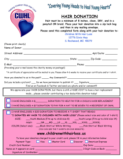


© Copyright 2026