
Introduction to the Annual Report
ANNUAL REPORT July 1, 2014 to June 30, 2015 1 Table of Contents Introduction. . . . . . . . . . . . . . . . . . . . . . . . . . . . . . . . . . . . . . . . . . . . . . . . . . . . . . . . .Page 5 Annual Scientific Conference Agenda. . . . . . . . . . . . . . . . . . . . . . . . . . . . . . . . . . . .Page 9 Institutional Information. . . . . . . . . . . . . . . . . . . . . . . . . . . . . . . . . . . . . . . . . . . . . . Page 35 - Research Summaries - Key Personnel Project Progress Reports . . . . . . . . . . . . . . . . . . . . . . . . . . . . . . . . . . . . . . . . . . . . . . Page 68 - Project Progress Reports by Institution 2014-2015 Publications, Manuscripts, & Grants - Publications and Manuscripts. . . . . . . . . . . . . . . . . . . . . . . . . . . . . . . . . . . . . . . Page 210 - Current and Pending Grants. . . . . . . . . . . . . . . . . . . . . . . . . . . . . . . . . . . . . . . . .Page 237 Poster Abstracts. . . . . . . . . . . . . . . . . . . . . . . . . . . . . . . . . . . . . . . . . . . . . . . . . . . . . . Page 280 Institutional Budgets and Justifications . . . . . . . . . . . . . . . . . . . . . . . . . . . . See companion report 2 In memory of our dear colleague Jason J. Corneveaux (1981-2014) 3 4 Introduction to the Annual Report Background The Arizona Alzheimer’s Consortium is the nation’s leading model of statewide collaboration in Alzheimer’s disease (AD) research. It includes about 150 researchers and support staff from seven principal organizations, including Arizona State University, Banner Alzheimer’s Institute, Banner Sun Health Research Institute, Barrow Neurological Institute, Mayo Clinic Arizona, the Translational Genomics Research Institute, and the University of Arizona, and from several affiliated organizations, including Midwestern University, the Critical Path Institute, and the University of Arizona College of Medicine, Phoenix Campus. Established in 1998, the Consortium is intended to make a major difference in the scientific fight against AD, to engage Arizona’s underserved and understudied Native American and Latino communities, and to help address the unmet needs of patients and family caregivers. The Consortium’s major themes are the early detection and prevention of AD. Its primary goal is to find effective treatments to stop and end AD as quickly as possible. The Consortium is widely recognized as a model of multi-institutional collaboration in biomedical research. It capitalizes on complementary resources and expertise from different disciplines and organizations to address scientific problems in the most impactful way. Its researchers receive critical support from the state of Arizona (through the Arizona Department of Health Services [ADHS] and its Arizona Biomedical Research Commission [ABRC]), the participating institutions, a competitive Arizona AD Center (ADCC) grant from the National Institute on Aging (NIA), and many other grants and contracts. Dr. Eric Reiman is the Director of the Consortium and the NIA-sponsored ADCC, Dr. Richard Caselli is the ADCC’s Associate Director, and Dr. Carol Barnes is Chairperson of the Consortium’s 25-member Internal Scientific Advisory Committee. Leaders from each of the seven principal institutions serve on the Consortium’s Board of Directors. The Consortium’s external advisors include Drs. Marilyn Albert, Zaven Khachaturian, and Bruce Miller, who are recognized for their pioneering contributions and leadership roles in the study of AD and related disorders. They conduct an annual site visit, review the progress and productivity of the Consortium and the ADCC, and provide formal feedback and recommendations to the researchers, NIA and state. The Arizona Alzheimer’s Consortium capitalizes on the state’s strengths in brain imaging, genomics, the computational and mathematical analysis of complex data sets, the basic, cognitive and behavioral neurosciences, clinical, experimental therapeutics, and neuropathology research. It has made major contributions to the scientific understanding, unusually early detection and tracking of AD, the accelerated evaluation of putative AD prevention therapies, and the scientific understanding of the aging mind and brain. It has introduced promising new 5 care models for patients and family caregivers. It has introduced new ways for different stakeholders to work together and it has provided data, biological samples and interested research participants for researchers inside the state and around the world. It continues to attract new researchers and clinicians to its participating institutions and support other biomedical research developments in the state. Indeed, it has helped to make Arizona a destination center for the advancement of AD research and care. State and institutional matching funds continue to provide the “glue” for this geographically distributed research program, the “fuel” needed to launch new research initiatives, and the framework needed to reach the Consortium’s over-arching goals. Funds are used to support dozens of research projects each year, almost all of which involve researchers from different scientific disciplines, and about half of which include researchers from different institutions. The Arizona ADCC has received competitive NIA grant support since 2001. The ADCC’s Administrative, Clinical, Data Management and Statistics, Neuropathology, and Education and Information Transfer Cores support researchers and projects inside and outside of the state. The ADCC’s progress, productivity and plans were recently described in our annual report to the NIA and will be reviewed at our advisors’ annual site visit on May 15. The Consortium’s statewide collaborative model, scientific productivity and cores were recently presented to leaders from the NIA and its AD Centers in a four-hour “virtual site visit” in Washington, DC, and the ADCC will submit its next five-year competitive renewal grant application to the NIA in September 2015. Productivity and Impact The Arizona Alzheimer’s Consortium is the leading statewide AD Center in the nation and one of the most productive AD research programs in the world. Since the Consortium’s inception in 1998, its researchers have generated more than 4,000 publications, 1,000 research grants and contracts, and a billion dollars in new investments, including more than half of those investments in the last 3 years. Consortium researchers continue to make pioneering contributions to the scientific fight against AD, related disorders, and the aging brain. • • • They have helped clarify genetic and non-genetic risk factors and disease mechanisms, offered targets at which to aim new AD treatments, provided new insights about the pathological changes associated with AD and related disorders, and proposed promising ways to treat and prevent the clinical onset of AD. They have played leadership roles in the earliest detection and tracking of AD and the accelerated evaluation of putative prevention therapies, and they have the use of amyloid PET and other imaging techniques in the research and clinical settings. They continue to clarify how different brain cells, regions, and networks, and mental operations orchestrate memory and other thinking abilities and how they are affected by AD and normal aging. They have played leadership roles in the study of normal cognitive aging. 6 • • They continue to develop groundbreaking research methods and strategies, collaborative models and data-sharing paradigms to support these and other research endeavors. With several hundred million dollars in NIA, philanthropic, foundation and industry funding, the “Alzheimer’s Prevention Initiative (API)” has helped launch a new era in AD prevention research, set the stage for the field to rapidly evaluate the range of promising but unproven prevention therapies, and find ones that work as quickly as possible. API includes prevention trials in cognitively unimpaired persons who, based on their genetic background and age, are at the highest imminent risk for the clinical onset of AD; exceptionally large registries to support enrollment in prevention trials; and paradigm-changing scientific, collaborative, data-sharing, and consensus-building elements for the advancement of AD prevention research. During the past year, the Consortium provided research support for other Arizona organizations, including Midwestern University (e.g., to study mitochondrial activity in AD), the University of Arizona College of Medicine (to advance the study of traumatic brain injury), and the Critical Path Institute (to develop FDA-supported data standards for AD trials). It launched new programs for the study of AD in patients with Down syndrome, the study of chronic traumatic encephalopathy (CTE) in retired NFL players, and the assessment of cellular changes in the intact rodent brain. It supported the recruitment of productive AD researchers and increased the number of AD researchers, clinicians, and programs at several of our organizations, and additional recruitments are underway. Plans During the next few years, organizations in the Consortium will further enhance Arizona’s position as a destination center for the advancement of AD research and care. For instance, the recently announced Arizona State University-Banner Neurodegenerative Disease Research Center (NDRC) will establish one of the world’s largest basic scientific research programs for the study of AD and other neurodegenerative disease on the University’s Tempe Campus. Plans are underway to increase the number of AD researchers and clinicians and further develop programs to advance research and/or care at almost all of the Consortium’s participating organizations–and to do so in ways that build upon and strengthen our existing collaborations. During the next few years, the Consortium will continue to advance several new scientific and clinical initiatives. We will do everything we can to advance the scientific fight against AD, related disorders and the aging brain, and do so in a way that served the needs of our patients, families, and underserved communities. We will try to set a new standard of dementia care for patients and family caregivers within the next five years, and we will try to find treatments to postpone, reduce, or completely prevent the clinical onset of AD within a decade. While there is no guarantee that we will meet those goals, we have an unprecedented opportunity to do just that. We are extremely grateful to the state of Arizona and the NIA and to our institutional leaders, collaborators, research participants and other supporters. More than ever, we are proud of our progress and excited about our plans. We are determined to make a transformational difference in the fight against AD, and do so together. 7 8 Arizona Alzheimer’s Consortium 17th Annual Conference – Thursday May 14, 2015 Arizona State University Old Main, 400 E. Tyler Mall Tempe, Arizona 85281 POSTER PRESENTATION SET-UP CONTINENTAL BREAKFAST 7:30 – 8:50AM WELCOME William Petuskey, Ph.D. Associate Vice President & Professor Office of Knowledge Enterprise Development Arizona State University 8:50 – 9:00AM INTRODUCTION Eric M. Reiman, M.D. Director, Arizona Alzheimer's Consortium 9:00 – 9:15AM LEON THAL MEMORIAL LECTURE "Is Beta-Amyloid Bad for the Brain? Insights from Human Imaging Studies” William Jagust, M.D. Professor of Public Health & Neuroscience, University of California, Berkeley Faculty Senior Scientist, Lawrence Berkeley National Laboratory 9:15 – 10:30AM ORAL RESEARCH PRESENTATIONS – SESSION I 10:30 – 11:45AM POSTER SESSION I & LUNCH 12:00 – 1:00PM POSTER SESSION II & LUNCH 1:00 – 2:00PM ORAL RESEARCH PRESENTATIONS – SESSION II 2:00 – 3:15PM CLOSING REMARKS Eric M. Reiman, M.D. 3:15 – 3:30PM 9 Arizona Alzheimer’s Consortium Oral Research Presentations SESSION I Moderator: Richard Caselli, M.D. 10:30 – 10:43AM Subjective cognitive complaint, quality of life and APOE genotyping among Latinos in Phoenix, Arizona: the Sangre por salud study. Janina Krell-Roesch. Mayo Clinic, Scottsdale; Arizona Alzheimer’s Consortium. 10:44 – 10:57AM The Alzheimer’s Prevention Initiative. Pierre Tariot. Banner Alzheimer’s Institute, Translational Genomics Research Institute, University of Arizona, University of Antioquia; Arizona Alzheimer’s Consortium. 10:58 – 11:11AM Individual differences in aerobic fitness influence the regional pattern of brain volume in healthy aging. Gene E. Alexander. University of Arizona; Arizona Alzheimer’s Consortium. 11:12 – 11:25AM Age-related changes in high-frequency local field activity in the rodent hippocampus during ripple and inter-ripple periods. Jean-Paul Wiegand. University of Arizona; Arizona Alzheimer’s Consortium. 11:26 – 11:39AM Comprehensive profiling of DNA methylation differences in patients with Alzheimer’s and Parkinson’s disease. Travis Dunckley. Translational Genomics Research Institute; Mayo Clinic, Scottsdale; Texas A&M University; Arizona Alzheimer’s Consortium. 10 Arizona Alzheimer’s Consortium Oral Research Presentations SESSION II Moderator: Heather Bimonte-Nelson, Ph.D. 2:00 – 2:13PM Prevalence of submandibular gland synucleinopathy in Parkinson’s disease, dementia with Lewy bodies, and other Lewy body disorders. Thomas G. Beach. Banner Sun Health Research Institute; Mayo Clinic Scottsdale; Arizona Alzheimer’s Consortium. 2:14 – 2:27PM Extracellular small RNA profiles from cerebrospinal fluid and serum of Alzheimer's, Parkinson's, and neurologically normal control subjects. Kendall Van-Keuren Jensen. Translational Genomics Research Institute; Banner Sun Health Research Institute; Mayo Clinic, Scottsdale; Arizona Alzheimer’s Consortium. 2:28 – 2:41PM Using bioengineering approaches to dissect the mechanisms of Alzheimer’s disease with human induced pluripotent stem cells. David Brafman. Arizona State University; Arizona Alzheimer’s Consortium. 2:42 – 2:55PM Molecular distribution following Fus-mediated BBB opening. Michael Valdez. University of Arizona; University of Notre Dame; Arizona Alzheimer’s Consortium. 2:56 – 3:09PM Resilience of precuneus neurotrophic signaling despite amyloid pathology in mild cognitive impairment. Elliot Mufson. Barrow Neurological Institute, St. Joseph’s Hospital and Medical Center; Arizona Alzheimer’s Consortium. 11 Arizona Alzheimer’s Consortium Poster Presentations 1. Impact of family history of cardiovascular risk factors on executive functions in younger adults. Addy J, Ryan L. University of Arizona; Arizona Alzheimer’s Consortium. 2. Individual differences in aerobic fitness influence the regional pattern of brain volume in healthy aging. Alexander GE, Fitzhugh MC, Raichlen DA, Bharadwaj PK, Haws KA, Torre GA, Trouard TP, Hishaw GA. University of Arizona; Arizona Alzheimer’s Consortium. 3. fMRI correlates of successful encoding and retrieval in response to increasing difficulty during an episodic memory task. Baena E, Ryan L. University of Arizona; Arizona Alzheimer’s Consortium. 4. Early surgical menopause in rats is associated with brain regional functional changes in specific learning and memory circuits. Ballina LE, Mennenga SE, Perkins M, Patel S, Bimonte-Nelson HA, Valla J. Midwestern University; Arizona State University; Arizona Alzheimer’s Consortium. 5. Prevalence of submandibular gland synucleinopathy in Parkinson’s disease, dementia with Lewy bodies, and other Lewy body disorders. Beach TG, Adler CH, Serrano G, Sue LI, Walker DG, Dugger BN, Shill HA, Driver-Dunckley E, Caviness JN, Intorcia A, SaxonLabelle M, Filon J, Pullen J, Scroggins A, Scott S, Garcia A, Hoffman B, Jacobson SA, Belden CM, Davis KJ, Sabbagh MN; Banner Sun Health Research Institute; Mayo Clinic Scottsdale; Arizona Alzheimer’s Consortium. 6. White matter rarefaction is a significant and independent predictor of final MMSE score in elderly autopsied subjects. Beach TG, Scott S, Sue LI, Serrano G, Intorcia A, Saxon-Labelle M, Filon J, Pullen J, Scroggins A, Garcia A, Hoffman B, Jacobson SA, Belden CM, Davis KJ, Long KE, Gale LD, Nicholson LR, Belden CM, Long KE, MalekAhmadi M, Powell JJ, Cline C, Gale LD, Nicholson LR, Sabbagh MN. Banner Sun Health Research Institute; Mayo Clinic, Scottsdale; Arizona Alzheimer’s Consortium. 7. Detection of striatal amyloid plaques with [18F] flutemetamol: validation with postmortem histopathology. Beach TG, Thal DR, Zanette M, Smith A, Buckley C. Banner Sun Health Research Institute; University of Ulm, Germany; GE Healthcare; Arizona Alzheimer’s Consortium. 12 8. Reduced default network functional connectivity and verbal learning in cognitively unimpaired late middle-aged and older adults: exploratory findings from the Arizona APOE cohort study. Beck IR, Chen K, Roontiva A, Wroolie TE, Bauer III R, Dunbar C, Peshkin E, Bandy D, Rasgon N, Caselli R, Reiman EM. Banner Alzheimer’s Institute; University of Arizona; Arizona State University; Translational Genomics Research Institute; Mayo Clinic, Scottsdale; Arizona Alzheimer’s Consortium. 9. A case study characterizing changes in discourse complexity preceding Alzheimer’s diagnosis: comparing the press conferences of presidents Ronald Reagan and George Herbert Walker Bush. Berisha V, Wang S, LaCross A, Liss J. Arizona State University; Arizona Alzheimer’s Consortium. 10. The contribution of amyloid pet imaging to clinical decision-making in the differential diagnosis of Alzheimer’s disease: a case series. Bhalla N, Chen K, Burke A, Weinstein D, Belden CM, Powell JJ, Devadas V, Roontiva A, Thiyyagura P, Sabbagh MN. Banner Sun Health Research Institute; Banner Alzheimer’s Institute; University of Arizona College of Medicine-Phoenix; Arizona Alzheimer’s Consortium. 11. Evaluation of an automated white matter hyperintensity segmentation method in healthy aging. Bharadwaj PK, Hishaw GA, Haws KA, Nguyen LA, Trouard TP, Alexander GE. University of Arizona; Arizona Alzheimer's Consortium. 12. Dr. Kanner’s first patient is an octogenarian: cognitive and brain aging in autism. Braden BB, Smith CJ, Glaspy TK, Baxter LC. Barrow Neurological Institute; Southwest Autism Research & Resource Center; Arizona Alzheimer’s Consortium. 13. Dissecting the mechanisms of Alzheimer’s disease using human induced pluripotent stem cells. Brafman D. Arizona State University; Arizona Alzheimer’s Consortium. 14. Reducing ribosomal protein s6 kinase 1 ameliorates Alzheimer’s disease-like cognitive and synaptic deficits by reducing BACE-1 translation. Caccamo A, Branca C, Turner D, Shaw DM, Talboom JS, Messina A, Sara KC, Wu J, Oddo S. Banner Sun Health Research Institute; University of Catania, Italy; Barrow Neurological Institute, St. Joseph's Hospital and Medical Center; University of Cincinnati; University of Arizona College of MedicinePhoenix; Arizona Alzheimer’s Consortium. 15. Early functional effects of APOE4 on expression of TREM2 and microglial phenotype in young adult targeted replacement mice. Castro M, De Vera C, Ho A, Chavira B, Valla J, Jentarra G, Jones TB. Midwestern University; Arizona Alzheimer’s Consortium. 16. Novel method for behavior-driven molecular and structural investigation in rodent whole brain. Chawla MK, Gray DT, Comrie AE, Baggett BK, Utzinger U, Barnes CA. University of Arizona; Arizona Alzheimer’s Consortium. 13 17. Generation of human induced pluripotent stem cell-derived A9 dopamine neurons for modeling idiopathic Parkinson's disease. Corenblum MJ, Sherman SJ, Curiel CN, Sligh JE, Schwartz PH, Brick DJ, Nethercott HE, Madhavan L. University of Arizona; University of Arizona Cancer Center; Children’s Hospital of Orange County; Arizona Alzheimer’s Consortium. 18. Dextromethorphan/quinidine (AVP-923) efficacy and safety for treatment of agitation in persons with Alzheimer’s disease: results from a Phase 2 study (NCT01584440). Cummings J, Lyketsos C, Tariot PN, Peskind ER, Nguyen U, Knowles N, Shin P, Siffert J. Cleveland Clinic; Lou Ruvo Center for Brain Health; Johns Hopkins Medicine; Banner Alzheimer’s Institute; University of Washington School of Medicine; Avanir Pharmaceuticals, Inc.; Arizona Alzheimer’s Consortium. 19. Poor safety and tolerability hamper reaching a potentially therapeutic dose in the use of thalidomide for Alzheimer’s disease: results from a double-blind, placebo-controlled trial. Decourt B, Drumm-Gurnee D, Wilson J, Jacobson S, Belden C, Sirrel S, Ahmadi M, Shill H, Powell J, Walker A, Gonzales A, Macias M, Sabbagh MN. Banner Sun Health Research Institute; Arizona State University; Arizona Alzheimer’s Consortium. 20. Feasibility of peripheral tau detection to determine Braak neurofibrillary tangle stage. Dugger BN, Whiteside CM, Maarouf CL, Beach TG, Dunckley T, Meechoovet B, Roher AE. Banner Sun Health Research Institute; Translational Genomics Research Institute; Arizona Alzheimer’s Consortium. 21. Neuropathological comparisons of amnestic and non-amnestic mild cognitive impairment. Dugger BN, Davis K, Malek-Ahmadi M, Hentz JG, Sandhu S, Beach TG, Adler CH, Caselli RJ, Johnson TA, Serrano G, Shill HA, Belden C, Sue LI, Jacobson S, Powell J, Caviness J, Driver-Dunckley E, Sabbagh MN. Banner Sun Health Research Institute; Mayo Clinic, Scottsdale; Arizona Alzheimer’s Consortium. 22. Comprehensive profiling of DNA methylation differences in patients with Alzheimer’s and Parkinson’s disease. Dunckley T, Meechoovet B, Caselli RJ, Hua J, Driver-Dunckley E. Translational Genomics Research Institute; Mayo Clinic, Scottsdale; Texas A&M University; Arizona Alzheimer’s Consortium. 23. Gamma secretase activating protein (GSAP) levels during the progression of AD: correlation with amyloid and tau pathology. Farrell EK, Perez SE, Nadeem M, He B, Mufson EJ. Barrow Neurological Institute, St. Joseph’s Hospital and Medical Center; Rush University Medical Center; Arizona Alzheimer’s Consortium. 24. Amyloid beta peptide (Aβ)-formed Ca2+ permeable channels in pancreatic acinar cells of APP mice. Gao M, Yin J, Hou B, Gao F, Shi J, Wu J. Barrow Neurological Institute, St. Joseph’s Hospital and Medical Center; Arizona Alzheimer’s Consortium. 14 25. Selective changes in inhibitory networks of the medial temporal lobe correlate with behavioral and electrophysiological deficits in aged rhesus macaques. Gray DT, Thome A, Erickson CA, Lipa P, Takamatsu CL, Comrie AE, Barnes CA. University of Arizona; Metropolitan State University of Denver; Arizona Alzheimer’s Consortium. 26. PACAP expression is downregulated in aged nonhuman primates. Han P, Permenter MR, Vogt JA, Engle JR, Barnes CA, Shi J. Barrow Neurological Institute, St. Joseph’s Hospital and Medical Center; University of California, Davis; University of Arizona; Arizona Alzheimer’s Consortium. 27. Suppression of non-specific Bielschowsky silver staining by pretreatment with potassium permanganate and oxalic acid. Intorcia A, Filon J, Pullen J, Scott S, Serrano G, Sue LI, Beach TG. Banner Sun Health Research Institute; Arizona Alzheimer’s Institute. 28. Cholinergic dysfunction and muscarinic receptor uncoupling in Alzheimer’s disease. Jones DC, Potter PE, Hamada M, Beach TG. Midwestern University; Sun Health Research Institute; Arizona Alzheimer’s Consortium. 29. Comparing regional activations between older and younger adults on an fMRI taskswitching and memory updating paradigm. Kawa K, Cardoza J, Stickel A, Schmit M, Lozano M, Glisky E, Ryan L. University of Arizona; Arizona Alzheimer’s Consortium. 30. Multifunctional radical quenchers: therapeutic agents for mitochondrial and neurodegenerative diseases. Khdour OM, Alam MP, Arce PM, Chen Y, Roy B, Dey S, Hecht SM. Arizona State University; Arizona Alzheimer’s Consortium. 31. Caloric intake and the risk of mild cognitive impairment: a population-based cohort study. Krell-Roesch J, Pink A, Roberts RO, Mielke MM, Christianson TJ, Knopman DS, Stokin GB, Petersen RC, Geda YE. Mayo Clinic, Scottsdale; Mayo Clinic, Rochester; St. Anne’s University Hospital Brno; Arizona Alzheimer’s Consortium. 32. Subjective cognitive complaint, quality of life and APOE genotyping among Latinos in Phoenix, Arizona: the Sangre Por Salud Study. Krell-Roesch J, Shaibi G, Mandarino LJ, Caselli RJ, Singh DP, Velgos SN, Stokin GB, Geda YE. Mayo Clinic, Scottsdale; Arizona Alzheimer’s Consortium. 33. Correlation between apoe4 genotype and hippocampal atrophy on Arizona APOE cohort: a surface multivariate tensor-based morphometry study. Li B, Mcmahon T, Shi J, Gutman BA, Thompson PM, Baxter LC, Chen K, Reiman EM, Caselli RJ, Wang Y. Barrow Neurological Institute, St. Joseph’s Hospital and Medical Center; Banner Alzheimer’s Institute; Mayo Clinic, Scottsdale; Arizona Alzheimer’s Consortium. 34. Longitudinal study of genetic influence of APOE e4 on hippocampal atrophy with conformal geometry. Li B, Shi J, Gutman BA, Thompson PM, Caselli RJ, Wang Y. Barrow Neurological Institute, St. Joseph’s Hospital and Medical Center; Mayo Clinic, Scottsdale; Arizona Alzheimer’s Institute. 15 35. Increases in amyloid beta and phosphorylated alpha synuclein are linked in Parkinson’s disease with dementia in the absence of Alzheimer’s disease. Lue L-F, Caviness J, Walker DG, Schmitz CT, Serrano G, Sue LI, Beach TG. Banner Sun Health Research Institute; Mayo Clinic, Scottsdale; Arizona Alzheimer’s Consortium. 36. Biochemical assessment of precuneus and posterior cingulate gyrus in the context of brain aging and Alzheimer’s disease. Maarouf CL, Kokjohn TA, Walker DB, Whiteside CM, Kalback WM, Whetzel A, Sue LI, Serrano G, Jacobson SA, Sabbagh MN, Reiman EM, Beach TG, Roher AE. Banner Sun Health Research Institute; Midwestern University; Banner Alzheimer’s Institute; Arizona Alzheimer’s Consortium. 37. The contraceptive estrogen ethinyl estradiol impairs spatial working memory in young adult, ovary-intact rodents. Mennenga SE, Hewitt LT, Carson C, Poisson M, BimonteNelson HA. Arizona State University; Arizona Alzheimer’s Consortium. 38. Non-linear optical imaging: a powerful new technique for acquiring high-resolution brain images and possible application for identifying cell types and neuronal activity. Miller MA, Mehravar S, Gray DT, Koshy A, Cabra C, Chawla MK, Kieu KQ, Barnes CA, Cowen SL, Peyghambarian N. University of Arizona; Arizona Alzheimer’s Consortium. 39. Single cell systems biology with cleavable fluorescent probes. Mondal M, Liao R, Xiao L, Guo J. Arizona State University; Arizona Alzheimer’s Consortium. 40. Consensus clinical data standards for Alzheimer’s disease: focus on prevention trials. Neville J, Kopko S, Romero K, Aviles E, Stephenson D. the Coalition Against Major Diseases (CAMD); Critical Path Institute; CDISC; Arizona Alzheimer’s Consortium. 41. Mitochondrial peptide levels in the young adult neocortex differ by APOE allele: a role for TOMM40 polymorphisms? Perkins M, Shonebarger D, Henderson L, Valla J. Midwestern University; Arizona Alzheimer’s Consortium. 42. The effect of varying 17β-estradiol treatment frequency on cognitive performance in middle-aged ovariectomized rats. Prakapenka AV, Quihuis AM, Mennenga SE, Hiroi R, Koebele SV, Sirianni RW, Bimonte-Nelson HA. Arizona State University; Arizona Alzheimer’s Consortium; Barrow Neurological Institute, St. Joseph’s Hospital and Medical Center. 43. Associations between subjective memory complaints and hippocampal volume in preclinical early-onset Alzheimer’s disease. Quiroz YT, Amariglio R, AguirreAcevedo DC, Opoka S, Pulsifer B, Jaimes SY, Castrillon G, Tirado V, Munoz C, Sperling RA, Lopera F. Universidad de Antioquia; Massachusetts General Hospital; Harvard Medical School; Brigham and Women's Hospital; Boston University; Instituto de Alta Tecnologia Medica; the Athinoula A Martinos Center for Biomedical Imaging; Banner Alzheimer’s Institute. 16 44. Gene expression profiling of human astrocytes treated with bexarotene and related compounds shows an increase in the neuroprotective cytokine GMCSF. Richholt RF, Piras IS, Persico AM, Huentelman MJ. Translational Genomics Research Institute; Arizona Alzheimer's Consortium; University of Arizona; University Campus Bio-Medico. 45. FDG-PET of the brain and neuropsychiatric symptoms in normal cognitive aging: the Mayo Clinic Study of Aging. Ruider H, Krell-Roesch J, Stokin GB, Lowe V, Roberts RO, Mielke MM, Knopman DS, Christianson TJ, Jack CR, Petersen RC, Geda YE. Mayo Clinic, Scottsdale; Arizona Alzheimer’s Consortium. 46. Aging is associated with altered intrinsic neural dynamics in the basolateral complex of the amygdala. Samson RD, Lester AW, Lipa P, Barnes CA. University of Arizona; Arizona Alzheimer’s Consortium. 47. Endogenous cannabinoid receptor type 2 expression and regulation in Alzheimer’s disease. Schmitz CT, Serrano G, Sue LI, Beach TG, Wu J, Walker DG, Lue L-F. Banner Sun Health Research Institute; Barrow Neurological Institute, St. Joseph’s Hospital and Medical Center; Arizona Alzheimer’s Consortium. 48. The predictive value of assessing cognitive and processing tasks in the risk assessment of Alzheimer’s disease in young adults. Schrauwen I, Gupta A, Corneveaux JJ, Siniard AL, Richholt R, de Both M, Peden J, Reiman EM, Caselli RJ, Ryan L, Glisky E, Huentelman MJ. Translational Genomics Research Institute; Arizona Alzheimer’s Consortium; University of Antwerp; Banner Alzheimer’s Institute; Mayo Clinic, Scottsdale; University of Arizona. 49. Positive Florbetapir PET amyloid imaging in a subject with frequent cortical neuritic plaques and frontotemporal lobar degeneration with TDP43-positive inclusions. Serrano GE, Sabbagh MN, Sue LI, Hidalgo JA, Schneider JA, Bedell BJ, Van Deerlin VM, Suh E, Akiyama H, Joshi AD, Pontecorvo MJ, Mintun MA, Beach TG. Banner Sun Health Research Institute; Rush University Medical Center; Biospective Inc.; McGill University; University of Pennsylvania; Tokyo Institute of Psychiatry; Avid Radiopharmaceuticals; Arizona Alzheimer’s Consortium. 50. Family history of Alzheimer’s disease predicts white and gray matter brain volumes in older adults with no signs of dementia. Singh P, Stickel A, Kawa K, Buller A, Ryan L. University of Arizona; McGill University; Arizona Alzheimer’s Consortium. 51. A cognitive evaluation of the forgotten estrogen: evaluating bioidentical estriol as a potential hormone therapy option in an animal model of surgical menopause. Stonebarger GA, Koebele SV, Bimonte-Nelson HA. Arizona State University; Arizona Alzheimer’s Consortium. 52. Chemogenetic facilitation of neuronal depolarization ameliorates Alzheimer’s diseaselike cognitive deficits. Talboom JS, Orr M, Caccamo A, Oddo S. Banner Sun Health Research Institute; UT Health Science Center; Arizona Alzheimer’s Consortium. 17 53. Novel epigenetic modulators for treatment of Alzheimer’s disease. Tran N, Iacoban P, Rowles J, Olsen M. Midwestern University; Arizona Alzheimer’s Consortium. 54. PRISM II: An open-label study to assess the safety, tolerability, and effectiveness of Dextromethorphan 20 mg/Quinidine 10 mg for treatment of pseudobulbar affect secondary to dementia, stroke, or traumatic brain injury: results from the Alzheimer’s Disease/Dementia Cohort. Trifilo M, Doody RS, D’Amico S, Cutler AJ, Shin P, Ledon F, Yonan C, Siffert J. Baylor College of Medicine; Cornerstone Medical Group; Florida Clinical Research Center, LLC; Avanir Pharmaceuticals, Inc. 55. Molecular distribution following Fus-mediated BBB opening. Valdez M, Yuan S, Liu Z, Helquist P, Matsunaga T, Witte R, Furenlid L, Romanowski M, Trouard T. University of Arizona; University of Notre Dame; Arizona Alzheimer’s Consortium. 56. Young adult carriers of the Alzheimer’s disease risk factor APOE4 show broad dysregulation in cortical energy metabolism pathways. Valla J, Perkins M, Shonebarger D, Pangle P, Ballina L, Chavira B, Vallejo J, Jentarra G. Midwestern University; Arizona Alzheimer’s Consortium. 57. Colony stimulating factor-1 receptor ligands and receptor expression in Alzheimer’s disease. Walker DG, Huentelman MJ, Whetzel AM, Lue L-F. Banner Sun Health Research Institute; Translational Genomics Research Institute; Arizona Alzheimer’s Consortium. 58. How does age and Alzheimer’s disease pathology affect O-GlcNAc-ylation, O-GlcNActransferase and O-GlcNAc-ase in human brain tissues: a study of postmortem brain tissue? Walker DG, Whetzel AM, Lue L-F. Banner Sun Health Research Institute; Arizona Alzheimer’s Consortium. 59. How does inflammation affect O-GlcNAcylation, O-GlcNAc-transferase and GGlcNAcase in human microglia: an in vitro study? Walker DG, Whetzel AM, Lue L-F. Banner Sun Health Research Institute; Arizona Alzheimer’s Consortium. 60. Immune phenotyping of microglia in human brains: endoglin (cd105) identifies a population of activated microglia in Alzheimer’s disease and Parkinson’s disease brains. Walker DG, Whetzel AM, Lue L-F. Banner Sun Health Research Institute; Arizona Alzheimer’s Consortium. 61. A 3D volumetric Laplace-Beltrami operator based cortical thickness computation method. Wang G, Zhang X, Su Q, Shi J, Caselli RJ, Wang Y. Ludong University; Arizona State University; Mayo Clinic, Scottsdale; Arizona Alzheimer’s Consortium. 62. Prevalence and incidence of cognitive impairment and depression disorder in the elderly in shanghai, china. Wang T, Yang C, Dong S, Cheng Y, Li X, Wang J, Zhu M, Yang F, Li G, Su N, Liu Y, Dai J, Chen K, Xiao S. Shanghai Jiao Tong University School of Medicine; Med-X Research Institution; Banner Alzheimer’s Institute; University of Arizona; Arizona State University; Arizona Alzheimer’s Consortium. 18 63. Age is associated with a reduction in ripple oscillation frequency and neuronal variability in the CA1 region of the hippocampus. Wiegand J-PL, Gray DT, Schimanski LA, Lipa P, Barnes CA, Cowen SL. University of Arizona; Arizona Alzheimer’s Consortium. 64. Age-related changes in high-frequency local field activity in the rodent hippocampus during ripple and inter-ripple periods. Wiegand J-P, Gray DT, Schimanski LA, Lipa P, Barnes CA, Cowen SL. University of Arizona; Arizona Alzheimer’s Consortium. 65. Self–imagining improves memory in older adults. Woolverton C, Crawford M, Grilli M, Glisky E. University of Arizona; VA Boston Healthcare System; Arizona Alzheimer’s Consortium. 66. Sirtuin 3 is down-regulated in apolipoprotein e4 carriers with Alzheimer’s disease. Yin J, Han P, Caselli RJ, Beach TG, Serrano GE, Reiman EM, Shi J. Barrow Neurological Institute, St. Joseph’s Hospital and Medical Center; Mayo Clinic, Scottsdale; Banner Sun Health Research Institute; Banner Alzheimer's Institute; Arizona Alzheimer’s Consortium. 67. An automatic surface-based ventricular morphometry pipeline and its application in Alzheimer’s disease research. Zhang W, Shi J, Chen K, Baxter LC, Reiman EM, Caselli RJ, Wang Y. Arizona State University; Banner Alzheimer’s Institute and Banner Good Samaritan PET Center; Barrow Neurological Institute, St. Joseph’s Hospital and Medical Center; Mayo Clinic, Scottsdale; Arizona Alzheimer's Consortium. STUDENT POSTER PRESENTATIONS 68. Extracellular small RNA profiles from cerebrospinal fluid and serum of Alzheimer's, Parkinson's, and neurologically normal control subjects. Allen S, Burgos K, Malenica I, Yeri A, Courtright A, Beach TG, Shill H, Adler C, Sabbagh M, Craig DW, Van KeurenJensen K. Translational Genomics Research Institute; Banner Sun Health Research Institute; Mayo Clinic, Scottsdale; Arizona Alzheimer’s Consortium. 69. The role of apolipoprotein E on the renin-angiotensin system in a sporadic Alzheimer’s disease mouse model. De Vera C, Ho A, Castro M, Chavira B, Jones CB, Valla J, Jentarra G, Jones TB. Midwestern University; Arizona Alzheimer’s Consortium. 70. The association of serum cholesterol level with the default mode network connectivity in cognitively normal subjects. Dunbar CA, Peshkin ER, Beck IR, Roontiva A, Bauer III RJ, Lou J, Reiman EM, Chen K. Columbia University, Duke University, Banner Alzheimer’s Institute, Translational Genomics Research Institute, University of Arizona, Arizona State University, Arizona Alzheimer Consortium. 19 71. Cerebral glucose metabolic rate differences between subjects with subjective memory concerns and healthy controls. Ehrhardt T, Mix M, Lee W, Bauer R, Thiyyagura P, Devadas V, Roontiva A, Luo X, Reiman EM, Chen K, the Alzheimer’s Disease Neuroimaging Initiative. Banner Alzheimer's Institute; University of Arizona; Arizona State University; Translational Genomics Research Institute; Arizona Alzheimer’s Consortium. 72. In vitro morphologic and spatial dynamics of primary skin fibroblasts from idiopathic Parkinson’s disease patients. Flores AJ, Corenblum MJ, Curiel C, Sherman SJ, Madhavan L. University of Arizona; Arizona Alzheimer’s Consortium. 73. The role of APOE-deficiency on expression of TREM2 and microglial functional phenotype. Ho A, De Vera C, Castro M, Jones TB. Midwestern University; Arizona Alzheimer’s Consortium. 74. Cognitive changes across the menopause transition in a rat model: a longitudinal evaluation of the impact of age and ovarian status on spatial memory. Koebele SV, Mennenga SE, Hiroi R, Hewitt LT, Quihuis AM, Poisson ML, Mayer LP, Dyer CA, Bimonte-Nelson HA. Arizona State University; Arizona Alzheimer’s Consortium; SenesTech Inc. 75. Further confirmation of correlation of FDG-PET measured cerebral hypometabolism with APOE4 status and cognitive functions using larger ADNI dataset. Lu S, Chen K. Mesquite High School; Banner Alzheimer’s Institute; University of Arizona; Arizona State University; Arizona Alzheimer’s Consortium. 76. Analysis of motor/olfactory deficits in homozygous Parkin mutant Drosophila with nicotine treatment. Mannett BT, Grose JM, Pearman K, Buhlman LM, Call GB. Midwestern University; Arizona Alzheimer’s Consortium. 77. A novel isometry-invariant descriptor for detection of brain cortical surface deformation affected by Alzheimer’s disease. Mi L, Su Z, Gu X, Wang Y. Arizona State University; State University of New York at Stony Brook; Arizona Alzheimer’s Consortium. 78. The use of a white matter reference region for standard uptake value ratios in the detection of cross-sectional fibrillar amyloid β-burden. Mix M, Ehrhardt T, Lee W, Roontiva A, Thiyyagura P, Bauer R, Devadas V, Luo J, Reiman EM, Chen K. Banner Alzheimer’s Institute; University of Arizona; Arizona State University; Translational Genomics Research Institute; Arizona Alzheimer’s Consortium. 79. Demographics and cognitive impairment as defined by the Montreal Cognitive Assessment in a Phoenix community memory screen. Parsons C, Dougherty J, Yaari R. University of Arizona College of Medicine Phoenix; Banner Alzheimer’s Institute; Arizona Alzheimer’s Consortium. 20 80. Using hypometabolic convergence index to detect longitudinal metabolic decline and its association with cognitive degeneration. Pendyala N*, Pandya S*, Luo J, Bauer R, Thiyyagura P, Roontiva A, Ayutyanont N, Reiman EM, Chen K. Banner Alzheimer’s Institute; Arizona State University; University of Arizona; Translational Genomics Research Institute; Georgia Institute of Technology; University of Arizona College of Medicine; Arizona Alzheimer’s Consortium. 81. Understanding the relationship between fasting serum glucose levels and default mode network connectivity. Peshkin ER, Dunbar CA, Beck IR, Roontiva A, Bauer III RJ, Lou J, Devadas V, Reiman EM, Chen K. Duke University; Columbia University; Banner Alzheimer’s Institute; Arizona Alzheimer’s Consortium; Translational Genomics Research Institute; University of Arizona; Arizona State University. 82. Characterization of genomic alterations in circulating mitochondrial DNA in Alzheimer’s disease. Sekar S, Adkins J, Serrano G, Beach TG, Jensen K, Liang WS. Translational Genomics Research Institute; Arizona Alzheimer’s Consortium; Banner Sun Health Research Institute. 83. Statistical modeling of plasma proteins as identifiers of dementia in Parkinson’s disease. Snyder NL, Schmitz CT, Shill H, Chen K, Lue L-F. Arizona State University; Banner Alzheimer’s Institute; Sun Health Research Institute; University of Arizona College of Medicine-Phoenix; Arizona Alzheimer’s Consortium. 84. Non-pharmacological therapy for the management of neuropsychiatric symptoms of Alzheimer’s disease: linking evidence to practice. Staedtler A. Arizona State University; Arizona Alzheimer’s Consortium. 85. Novel rock inhibitors developed for both cognitive enhancement and blockade of pathological tau phosphorylation. Turk MN, Adams MD, Wang T, Dunckley T, Huentelman MJ. Translational Genomics Research Institute; Arizona State University; Arizona Alzheimer’s Consortium, University of Arizona; Midwestern University. 86. A novel approach to Alzheimer's prevention: A proposal for investigating the effects of inhibition of natural antisense transcripts for ADAM10 in order to overexpress ADAM10 as a competitive inhibitor to BACE1 for less amyloid-β production. Dave N. Arizona Alzheimer’s Consortium. 21 2015 Oral Research Presentation Abstracts 22 SUBJECTIVE COGNITIVE COMPLAINT, QUALITY OF LIFE AND APOE GENOTYPING AMONG LATINOS IN PHOENIX, ARIZONA: THE SANGRE POR SALUD STUDY. Krell-Roesch J, Shaibi G, Mandarino LJ, Caselli RJ, Singh DP, Velgos SN, Stokin GB, Geda YE. Mayo Clinic, Scottsdale; Arizona Alzheimer’s Consortium. Background: Despite the fact that Latinos are one of the fastest growing populations in the US, they remain disproportionately under-represented in research. It is of public health importance to investigate health and disease among Latinos particularly because of the high prevalence of cardiometabolic disorders which may increase the risk of cognitive impairment and dementia in this population. Therefore, we need to investigate biological and psychosocial risk factors for cognitive disorders. Methods: We conducted a cross-sectional study derived from the ongoing Sangre Por Salud (Blood for Health) Biobank, a collaborative project between Mayo Clinic Center for Individualized Medicine and Mountain Park Health Center in Phoenix, Arizona. Data were available for 499 self-identified Latinos who were enrolled into the study between June 2013 and April 2014. The participants completed a health examination including a self-administered, Spanish speaking coordinator-assisted survey on general health and functioning, medical history, health behaviors, and demographics. Blood samples were used for APOE genotyping determined from DNA using a polymerase chain reaction amplification. Subjective cognitive complaint, quality of life as well as physical exercise data were acquired from the self-reported questionnaires. We performed descriptive statistics, logistic regression analyses and two-sample t-tests using JMP software (SAS Institute Inc., Cary, NC). Results: Of the 499 study participants, 370 (74%) were females and 129 (26%) were males. Mean age was 41.4 ±12.9 years (range 18.1 to 85.9). The mean BMI was 30.9 ± 6.0 kg/m2 for females [3 (1%) were underweight, 64 (17%) were normal, 112 (30%) were overweight, and 190 (51%) were obese]. For males, the mean BMI was 30.7 ± 6.1 kg/m2 [18 (14%) were normal, 48 (37%) were overweight, and 63 (49%) were obese]. The frequency of APOE genotypes were as follows: 12 (2%) participants were APOEε4 homozygotes, 94 (19%) were ε4 heterozygotes, and 393 (79%) were ε4 non-carriers. Subjective cognitive complaint data were available for 222 participants of whom 161 (73%) had cognitive complaints. Physical exercise data were available for 223 participants of whom 78 (35%), 85 (38%), and 53 (24%) participants reported engaging in mild, moderate, and strenuous physical exercise, respectively, for at least 15 minutes on three or more days per week. Preliminary analyses indicated that neither APOEε4 genotype nor physical exercise was associated with subjective cognitive complaint. Whereas study participants with subjective cognitive complaint reported a decreased quality of life compared to participants without subjective cognitive complaint (p .0001). Conclusions: In this preliminary analysis, we observed that subjective cognitive complaint was common in this Latino study sample and was significantly associated with an impaired quality of life. The frequency of APOEε4 in this sample is consistent with the reported prevalence of APOEε4 in the US. One major limitation of this study pertains to the assessment of subjective cognitive complaint and physical exercise based on a self-reported questionnaire which may have led to recall bias. Our findings should thus be considered as preliminary until confirmed by future analyses on a larger sample size of the Sangre Por Salud study or by a prospective cohort study. 23 THE ALZHEIMER’S PREVENTION INITIATIVE. Tariot PN, Lopera F, Langbaum JB, Rios SR, Jakimovich L, Langlois C, High N, Reiman EM. Banner Alzheimer’s Institute, Translational Genomics Research Institute, University of Arizona, University of Antioquia; Arizona Alzheimer’s Consortium. The Alzheimer’s Prevention Initiative (API) is a collaborative research program involving the Banner Alzheimer’s Institute (BAI) and key partners that evaluate promising treatments with the ultimate goal to someday postpone, reduce the risk of, or prevent the clinical onset of Alzheimer’s disease (AD). API’s preclinical treatment studies focus on cognitively normal people who, based on their age and genetic background, are at the highest imminent risk of developing AD symptoms. Aims include establishing whether cognitive decline can be slowed; relating a treatment’s biomarker effects to clinical outcome; robustly testing the amyloid hypothesis; giving persons at highest risk access to investigational treatments; creating large prevention registries for these and other preclinical trials; and complementing other prevention initiatives. API includes the following. a) The API Autosomal Dominant Alzheimer’s Disease (ADAD) Trial, conducted in partnership with Genentech and the University of Antioquia (UdeA), launched in 2013. It is evaluating crenezumab in unimpaired ADAD mutation carriers at certain risk for developing early onset AD. b) The API APOE4 Trial, which will launch in late 2015 in partnership with Novartis, will evaluate a BACE inhibitor and an active amyloid-modifying immunization therapy in unimpaired 60-75 year-old apolipoprotein E4 homozygotes, who are at particularly high risk for developing AD at older ages. c) The Colombian API Registry, created by the UdeA in conjunction with BAI and Genentech, includes >3,300 members of a very large ADAD kindred, providing a resource for enrollment into the API ADAD Trial and for biomarker and cognitive studies of ADAD. d) The Alzheimer’s Prevention Registry (www.endALZnow.org), is a web-based resource that aims to enroll > 250,000 individuals (over 1220oo enrolled this far) who are interested in Alzheimer’s prevention research, facilitates enrollment into AD prevention studies, and provides regular communication to members. 24 INDIVIDUAL DIFFERENCES IN AEROBIC FITNESS INFLUENCE THE REGIONAL PATTERN OF BRAIN VOLUME IN HEALTHY AGING. Alexander GE, Fitzhugh MC, Raichlen DA, Bharadwaj PK, Haws KA, Torre GA, Trouard TP, Hishaw GA. University of Arizona; Arizona Alzheimer’s Consortium. Background: Individual differences in aerobic fitness levels may be an important factor affecting heterogeneity in brain aging and associated age-related cognitive decline. We recently proposed that increased demands for physical activity helped to support the evolution of long human lifespans and healthy brain aging (Raichlen and Alexander, Trends Neurosci, 2014). In this study, we sought to evaluate how individual differences in aerobic fitness levels effect regional brain volumes that are altered in the context of healthy aging. Methods: Quantitative measures of aerobic fitness (VO2max) during a graded exercise treadmill test were acquired in 155 healthy, community-dwelling adults, 50 to 89 years of age to determine whether those brain regions showing reductions in volume with increasing age are also altered by individual differences in VO2max. Participants (85M/70F; mean ± sd age = 69.6 ± 10.0; mean ± sd Mini-Mental State Exam = 29.0 ± 1.3) were medically screened to exclude neurological and psychiatric illnesses that could impact cognitive function. Results: Regional patterns of brain volume were assessed using Freesurfer software with T1weighted 3T volumetric magnetic resonance imaging (MRI) scans to identify the regional patterns of gray matter associated with age and VO2max. The results showed a regional pattern of gray matter reductions with increasing age that included bilateral superior, middle, and inferior frontal, superior and middle temporal, fusiform/lingual gyri, insula, inferior parietal, paracentral, cuneus, and precuneus regions (FDR correction, p < 0.05). After we controlled for the effects of aging in the cohort, greater levels of VO2max were associated with greater volumes in distinct regions of bilateral medial frontal, anterior cingulate, lateral occipital, and entorhinal cortices. In addition, greater levels of VO2max were associated with increased volumes in several of the regions directly affected by aging in this cohort, including bilateral middle frontal, insula, fusiform/lingual gyri, and precuneus regions (FDR correction, p < 0.05). Conclusions: These findings provide initial support for a regionally distributed pattern of brain volume associated with individual differences in aerobic fitness levels in neurologically healthy middle-aged to elderly adults, suggesting that having higher levels of physical activity may help compensate for regional differences in gray matter volume often associated with brain aging. 25 AGE-RELATED CHANGES IN HIGH-FREQUENCY LOCAL FIELD ACTIVITY IN THE RODENT HIPPOCAMPUS DURING RIPPLE AND INTER-RIPPLE PERIODS. Wiegand J-P, Gray DT, Schimanski LA, Lipa P, Barnes CA, Cowen SL. University of Arizona; Arizona Alzheimer’s Consortium. Background: Sharp-wave ripples (SPW-Rs) are brief (20-150 ms), high-frequency (140-180 Hz) oscillations in the local field potential in the hippocampus (O’Keefe et al., 1978, Buzsaki et al., 1992, Sullivan et al., 2011). Given the hypothesized association between ripples, memory consolidation, and homeostatic plasticity, we investigated whether there might be age-associated changes in ripple characteristics that contribute to age-related memory loss. Methods: To investigate this, local field potentials were recorded from CA1 during rest sessions before and after rats performed a place-dependent eyeblink-conditioning task. High-frequency (80-500 Hz) oscillatory activity in the hippocampus of old (n=6) and young (n=6) male F344 rats during ripple events and during inter-ripple periods was recorded. Results: Two features of these local field potentials were found to differ between the young and old animals. Specifically, during hippocampal ripple periods the mean frequency in old rats was 6 Hz lower than that observed in young rats. Additionally, during the inter-ripple periods, old rats showed greater local field potential power in high-frequency bands (150 to 500 Hz). Conclusions: Compared to young rats, old rats showed a decrease in ripple frequency and an increase in power at high frequencies in inter-ripple intervals. 26 COMPREHENSIVE PROFILING OF DNA METHYLATION DIFFERENCES IN PATIENTS WITH ALZHEIMER’S AND PARKINSON’S DISEASE. Dunckley T, Meechoovet B, Caselli RJ, Hua J, Driver-Dunckley E. Translational Genomics Research Institute; Mayo Clinic, Scottsdale; Texas A&M University; Arizona Alzheimer’s Consortium. Background: Many complex sporadic neurodegenerative disorders are the phenotypic expression of interactions between environmental influences and an individual’s inherent genetic risk. Specific molecular mechanisms mediating the differential impact of environmental factors on susceptible individuals leading to the development (and prevention) of neurodegenerative disease remain unclear. Epigenetic changes to DNA methylation patterns at specific genomic loci have been found in individuals with AD and PD, even in peripheral tissues such as blood. A more complete genome-wide characterization of the methylation events in AD and PD could add new insights into the etiology of these disorders. Methods: Methylation profiles were obtained on blood samples from 15 neurologically normal controls, from 15 AD and from 15 non-demented PD patients using the Illumina Infinium 450K Methylation BeadChip. We obtained robust data on over 480,000 CpG methylation sites in the form of beta values, which represent the ratio of methylated CpG to the sum of methylated plus nonmethylated CpG at a given site. Thus, these values range from 0 (unmethylated) to 1 (fully methylated). Results: We identified 84 methylation sites in AD vs controls with statistically significant changes to the beta value greater than 0.2. In PD vs controls, there were 83 sites with a beta value larger than the 0.2 threshold. However, of these sites, only 7 were shared between AD and PD. Thus, patients with either AD or PD exhibit numerous unique methylation events in peripheral blood DNA. Conclusions: Methylation profiles in the blood of individuals with AD or PD and healthy controls show distinct differences in the patient sample sets examined. Further validation efforts on larger sample sets, and characterization of methylation status in patients at varying stages of disease, will help to establish whether methylation status at specific loci could be leveraged as therapeutic targets or biomarkers to track disease progression or aid in disease diagnosis. 27 PREVALENCE OF SUBMANDIBULAR GLAND SYNUCLEINOPATHY IN PARKINSON’S DISEASE, DEMENTIA WITH LEWY BODIES, AND OTHER LEWY BODY DISORDERS. Beach TG, Adler CH, Serrano G, Sue LI, Walker DG, Dugger BN, Shill HA, Driver-Dunckley E, Caviness JN, Intorcia A, Saxon-Labelle M, Filon J, Pullen J, Scroggins A, Scott S, Garcia A, Hoffman B, Jacobson SA, Belden CM, Davis KJ, Sabbagh MN. Banner Sun Health Research Institute; Mayo Clinic Scottsdale; Arizona Alzheimer’s Consortium. Background: Low clinical diagnostic accuracy, particularly at early disease stages, is a critical roadblock to finding new therapies for Parkinson’s disease (PD) and dementia with Lewy bodies (DLB). Brain biopsy has been avoided but biopsy of a peripheral site might provide improved diagnostic accuracy. Previously, we have reported, on the basis of results from a large autopsy survey and a clinical trial of needle core biopsy, that the submandibular gland is a promising and safe biopsy site. Here, we report an extension of these studies, in submandibular gland from 200 autopsied subjects in the Arizona Study of Aging and Neurodegenerative Disorders (AZSAND). Methods: Lewy-type synucleinoapathy (LTS) was demonstrated by immunohistochemical staining for alpha-synuclein phosphorylated at serine-129. There were 118 cases with CNS LTS, including 46 PD, 28 DLB, 9 incidental Lewy body disease (ILBD), 33 Alzheimer’s disease with Lewy bodies (ADLB) and 2 with progressive supranuclear palsy and Lewy bodies (PSPLB). Control subjects, defined as those without CNS LTS, included 51 normal elderly subjects, 15 AD, 12 PSP, 2 CBD and 2 multiple system atrophy (MSA). Results: Submandibular gland LTS was found in 42/46 (91%) PD subjects, 20/28 (71%) DLB, 4/33 (12%) ADLB and 1/9 (11%) ILBD subjects but none of the 82 non-LTS control subjects. Concurrent AD histopathology was present in the brain in all LTS and non-LTS control cases. Conclusions: These results provide further support for clinical trials of in vivo submandibular gland diagnostic biopsy for PD and DLB. In addition to aiding subject selection for clinical trials, an accurate peripheral biopsy diagnosis would also be advantageous when selecting subjects for invasive therapies or for verifying other biomarker studies. 28 EXTRACELLULAR SMALL RNA PROFILES FROM CEREBROSPINAL FLUID AND SERUM OF ALZHEIMER'S, PARKINSON'S, AND NEUROLOGICALLY NORMAL CONTROL SUBJECTS. Allen S, Burgos K, Malenica I, Yeri A, Courtright A, Beach TG, Shill H, Adler C, Sabbagh M, Craig DW, Van Keuren-Jensen K. Translational Genomics Research Institute; Banner Sun Health Research Institute; Mayo Clinic, Scottsdale; Arizona Alzheimer’s Consortium. Background: Extracellular RNAs (exRNAs) have recently been heralded as novel mediators of health and disease. We anticipate that exRNAs can be used for the diagnosis of disease, for monitoring treatment efficacy and disease progression, and targeted therapies. This field is still in an early period of development, making it necessary to acquire basic information about the distribution and categories of exRNA present in different biological sources and how disease alters the exRNA profile. RNAs originating from hard to access tissues, such as neurons within the brain and spinal cord, have the potential to get to the periphery where they can be detected non-invasively. The formation and extracellular release of microvesicles and RNA binding proteins have been found to carry RNA from cells of the central nervous system to the periphery and protect the RNA from degradation. Therefore, exRNAs in peripheral circulation may be able to provide information about cellular changes associated with Alzheimer’s and Parkinson’s diseases. We previously reported miRNA profiles in Alzheimer’s, Parkinson’s patients, and neurologically normal controls. We found that there were distinct profiles of miRNA that matched plaque and tangle burden, pathology quantified from autopsies performed at Banner Sun Health Research Institute. Methods: We isolated cell-free RNA from 1 mL of serum and cerebrospinal fluid using the miRVana PARIS kit with a second phenol chloroform extraction. We used TruSeq Small RNA library preparation. We sequenced the samples on a single read flowcell, and after trimming of the adaptors, aligned the samples using the UEA Small RNA Workbench. Each exRNA type was quantified. Samples were normalized using DeSeq2 and were assessed for differential expression. Results: In the current study we assess other small RNA types identified in the samples, snRNA, snoRNA, tRNA, etc. We profiled the extracellular small RNA content from 69 patients with Alzheimer’s disease, 67 with Parkinson’s disease and 78 neurologically normal controls using next generation sequencing (NGS). We report the average abundance of each detected small exRNA in cerebrospinal fluid and in serum. We correlated changes in exRNA expression with disease pathology. A thorough examination of these other small RNA types has not been reported in the literature. Conclusions: There are interesting small RNA changes in serum and cerebrospinal fluid in patients with Alzheimer's, Parkinson's diseases compared with controls. It will be important to begin to understand what role, if any, these exRNAs play in the initiation and progression of the disease. 29 USING BIOENGINEERING APPROACHES TO DISSECT THE MECHANISMS OF ALZHEIMER’S DISEASE WITH HUMAN INDUCED PLURIPOTENT STEM CELLS. Brafman D. Arizona State University; Arizona Alzheimer’s Consortium. Animal models that overexpress specific Alzheimer’s disease (AD)-related proteins or have familial AD (FAD)-related mutations introduced into the animal genome have provided important insights into AD. Nonetheless, these animal models do not display important ADrelated pathologies and have not been useful in modeling the complex genetics associated with sporadic AD (SAD). The use of AD human induced pluripotent stem cell (hiPSC)-derived neurons has provided new opportunities to study the disease in a simplified and accessible system. However, current studies using AD hiPSCs have been limited by: (1) Use of 2-D culture systems that do not mimic the architecture of in vivo neural tissue, (2) Analysis of AD-related phenotypes in heterogeneous cell populations consisting of multiple neuronal subtypes, thereby limiting the identification of all the molecular and cellular hallmarks associated with the disease and (3) Inability to observe phenotypes associated with late-onset of the disease in aging adults, such as synaptic and neuronal loss. To that end, we have engineered a 3-D hiPSC-based tissue culture model that reflects the complexity of in vivo neural tissues. In addition, we have developed a robust protocol that allows for the differentiation of hiPSCs into pure populations of cortical neurons, the neuronal subtype most heavily affected in AD. Finally, we are employing a progerin-based method to generate ‘aged’ hiPSC-derived cortical neurons that will enable the identification and analysis of disease phenotypes that require aging. In the future, the ability to generate reproducible in vitro models of AD that mimic the various phenotypes of the disease that are observed in aging adults will allow us to investigate AD pathology, progression, and mechanism as well as develop and screen potential therapeutic compounds. 30 MOLECULAR DISTRIBUTION FOLLOWING FUS-MEDIATED BBB OPENING. Valdez M, Yuan S, Liu Z, Helquist P, Matsunaga T, Witte R, Furenlid L, Romanowski M, Trouard T. University of Arizona; University of Notre Dame; Arizona Alzheimer’s Consortium. Background: Treatment of neurological disorders is often hampered by the inability of therapeutics to cross the blood-brain barrier (BBB). Over the last several years, novel techniques have been developed that use focused ultrasound (FUS) energy in combination with microbubble (µB) contrast agents to temporarily open up the BBB. Foundational studies have been carried out in animal models, where BBB opening is clearly visible via contrast-enhanced MRI. While MRI allows assessment of BBB opening to contrast agents, it does not directly show the distribution of therapeutics within the brain. In this work, we have employed MRI, SPECT, CT, autoradiography, and fluorescence microscopy to compare the distribution of both contrast agents and model therapeutics within the brains of mice following FUS-mediated BBB opening. Methods: BBB opening was carried out in anesthetized mice with the following procedure: Mice were anesthetized (1.5% isofluorane gas in oxygen) and placed in a supine position into a custom made positioning apparatus which held a FUS transducer (20 mm diameter, 19 mm focal length) such that its focal spot was within the midbrain of the mouse. A 150 µL bolus of perfluorocarbon (PFC) gas-filled µBs was injected into the vein. FUS was immediately applied via twenty 3second sonications (37% duty cycle, 6 ms pulse width, 40 W per sq cm) separated by 3 second pauses. Following an IP injection of Gd-DTPA, MRI was carried out on the mice using T1weighted spin-echo sequences. All MRI was carried out on a 7T Bruker BioSpec MRI system utilizing a 72 mm ID birdcage coil for excitation and a 4-channel phased array coil for reception. During imaging, mice were secured with ear and bite bars and maintained at body temperature. Within 3 hours of the MRI procedure, pairs of mice were injected with pertechnetate (Tc-99m ion), Tc-99m DTPA, or Tc-99m-labeled cyclodextrin and imaged on a custom built dualmodality SPECT/CT imaging system to determine the distribution of the radiotracer in the brain. Following SPECT/CT, mice were sacrificed and autoradiography was carried out on coronal brain sections. Other mice underwent the same BBB opening and MRI procedures followed by the IV injection of 100 µL 70kD FITC-labeled dextran. Twenty minutes post injection, the mice were perfused transcardially with 10 mL of saline followed by 10 mL of 4% PFA. Brains were extracted, snap-frozen, sectioned horizontally and imaged with an Olympus MVX10 fluorescence microscope. Liposome-coated microbubbles were made by conjugating 100 nm diameter carboxyfluorescein-loaded liposomes, including DPSE-PEG-maleimide to PFC microbubbles including DPPE-PEG-SPDP in the lipid shell. The particles were imaged on an Olympus IX71 inverted microscope. Results: MRI and fluorescence microscopy images following BBB opening show a strong colocalization of MRI signal enhancement with the larger (70kD) dextran molecule. However, as expected, the distribution of the relatively small Gd-DTPA is consistently larger than that of the dextran molecules. MRI was used to confirm BBB opening in the mouse brain. SPECT/CT images of the same mouse following an injection of pertechnetate demonstrate a slight uptake of radiotracer into the brain that is co-localized with the MRI enhancement. Autoradiography revealed increased radioactivity in the same BBB region. The activity of all tracers within the brain, however, was extremely small compared to the activity observed in the body of the mouse. Conclusions: The results of these studies validate the ability of FUS-mediated BBB opening to allow molecules to enter the brain. However, they also emphasize the need to develop more 31 efficient methods with which to introduce drugs to the specific site of BBB opening without exposing the rest of the body to excessively high concentrations of drug. Drug-loaded liposomes conjugated to acoustically active µBs could prove very useful in this regard. Example images of such macromolecular constructs are shown in Fig. 3 where fluorescently-labeled liposomes have been conjugated to µBs. While the BBB opening technique described herein is intended to increase drug delivery to the brain for neurological disorders (e.g. Alzheimer’s and Parkinson’s), these imaging experiments could also be utilized to evaluate the loss of BBB integrity caused by other pathologies such as brain tumors, TBI, and viral infections. 32 RESILIENCE OF PRECUNEUS NEUROTROPHIC SIGNALING DESPITE AMYLOID PATHOLOGY IN MILD COGNITIVE IMPAIRMENT. Mufson E, Perez SE. Barrow Neurological Institute, St. Joseph’s Hospital and Medical Center; Arizona Alzheimer’s Consortium; Rush University Medical Center. Background: Reduction of precuneus choline acetyltransferase activity co-occurs with greater beta-amyloid (Aβ) in Alzheimer's disease (AD). Whether this cholinergic deficit is associated with alteration in nerve growth factor (NGF) signaling and its relation to Aβ plaque and neurofibrillary tangle (NFT) pathology during disease onset is unknown. Methods: Precuneus NGF upstream and downstream signaling levels relative to Aβ and NFT pathology were evaluated using biochemistry and histochemistry in 62 subjects with a premortem diagnosis of non-cognitively impaired (NCI; n = 23), mild cognitive impairment (MCI; n = 21), and mild to moderate AD (n = 18). Results: Immunoblots revealed increased levels of proNGF in AD subjects but not MCI subjects, whereas cognate receptors were unchanged. There were no significant differences in protein level for the downstream survival kinase-signaling proteins Erk and phospho-Erk among groups. Apoptotic phospho-JNK, phospho-JNK/JNK ratio, and Bcl-2 were significantly elevated in AD subjects. Soluble Aβ1-42 and fibrillar Aβ measured by [(3)H] Pittsburgh compoundB ([(3)H]PiB) binding were significantly higher in AD subjects compared with MCI and NCI subjects. The density of plaques showed a trend to increase, but only 6-CN-PiB-positive plaques reached significance in AD subjects. AT8-positive, TOC-1-positive, and Tau C3-positive NFT densities were unchanged, whereas only AT8-positive neuropil thread density was statistically higher in AD subjects. A negative correlation was found between proNGF, phospho-JNK, and Bcl-2 levels and phospho-JNK/JNK ratio and cognition, whereas proNGF correlated positively with 6-CN-PiB-positive plaques during disease progression. Conclusions: Data indicate that precuneus neurotrophin pathways are resilient to amyloid toxicity during the onset of AD. 33 34 Institutional Information Research Summaries and Key Personnel from Each Participating Institution 35 36 ARIZONA STATE UNIVERSITY Institutional Abstract Over the past decade, ASU has committed to the model of the New American University, focused on academic excellence, inclusiveness to a broad demographic, and maximum societal impact. With Alzheimer’s disease affecting roughly one in nine people 65 years old and over, and one in three people 85 years old and over, research on Alzheimer’s disease exemplifies the type of endeavor that ASU seeks to promote. For the Arizona Alzheimer’s Consortium, ASU provides the Data Management and Biostatistics Core as well as the Education and Information Transfer Core, serving researchers throughout the state as part of the Consortium’s NIA-sponsored Arizona Alzheimer’s Disease Core Center. The ASU team includes leaders in the development of antibody and novel compound strategies for the treatment of Alzheimer’s and other neurodegenerative diseases (Sierks, Hecht, and Johnson laboratories), in the development and use of animal models to characterize the influence of reproductive senescence and hormonal influences on brain aging and cognition (Bimonte-Nelson laboratory), in the development of mice as a model for odor learning and discrimination to understand pathologies and novel markers of neurodegenerative disease (Smith laboratory), in the development and implementation of computational image analysis and biomathematical techniques to increase the power to detect and track Alzheimer’s disease (Chen and Wang laboratories), and in the development of improved care models for patients and family caregivers, including the HOPE memory partner program to explore the role of community health workers in Alzheimer’s disease research and clinical practice (led by David Coon). In addition, we are thrilled that in January of 2015 Dr. David Brafman joined the ASU Arizona Alzheimer’s Consortium team; his cutting edge laboratory uses interdisciplinary approaches, including work with stem cells, to determine the mechanisms of, and design targeted therapies to treat, Alzheimer’s and other diseases. It is noteworthy that ASU has numerous scientific research domains that are being further developed and strengthened to additionally bolster the impact on Alzheimer’s disease and aging research, with a focus on discovery and action to move forward for trajectories, diagnosis, and treatment. These include, but are not limited to, the neurosciences, health outcomes research, and focused translational research realms that pose hypothesis-driven questions approached from a systems and interdisciplinary perspective. Collectively, ASU has a solid framework and varied strengths that are poised to make great strides in the scientific fight against Alzheimer’s disease as well as to optimize the trajectory of brain aging. Moreover, it is noteworthy that the strengths in the research programs at ASU within the Arizona Alzheimer’s Consortium represent a range of colleges and institutes across ASU. Training, mentoring, and education are prodigious strengths at ASU. ASU offers graduate degrees in Statistics and Biomedical Informatics, the Behavioral Neuroscience Program within the Department of Psychology, as well as the Interdisciplinary Graduate Program in Neuroscience. The latter two programs emphasize approaches that integrate several levels of analysis using a systems approach – cellular, behavioral, and cognitive – to investigate preclinical, clinical, and translational questions about brain and behavior relationships. Importantly, the laboratories at ASU that are involved with the Arizona Alzheimer’s Consortium work to engage and train future generations of scientists, including high school students, undergraduate students, graduate students, and postdoctoral fellows. The approach is hands-on, 37 multifaceted, interdisciplinary, and dimensional, with the goal to engage future exemplary scientists in goal-driven aging and neurodegenerative research to yield maximal impact on research discovery at ASU. Moreover, ASU and Banner Health just announced a research alliance to advance the scientific understanding, treatment and prevention of Alzheimer’s disease, and continue to build relationships with each of the other organizations in the Arizona Alzheimer’s Consortium under the leadership of Dr. Eric Reiman. The agreement calls for development of the ASU-Banner Neurodegenerative Disease Research Center (NDRC) on the Tempe Campus and close working relationships with ASU’s Biodesign Institute, other research programs at ASU, and clinical research programs at Banner Alzheimer’s Institute (BAI) and Banner Sun Health Research Institute. The NDRC will recruit an internationally recognized basic neuroscientist to lead the Center, start with a nucleus of leading basic neuroscientists, and continue to recruit numerous productive basic scientists as new space for the Center becomes available. The agreement between the two organizations calls for the NDRC to become one of the world’s largest and most productive basic neuroscience programs for the fight against Alzheimer’s disease, Parkinson’s disease and other neurodegenerative diseases. The new director will report to Dr. Eric Reiman and have a close working relationship with Dr. Ray DuBois, executive director of the Biodesign Institute . 1 http://researchmatters.asu.edu/stories/alzheimers-z-2639#sthash.S5YSQXX4.dpuf https://psychology.clas.asu.edu/bn 3 http://neuroscience.asu.edu/ 2 38 ARIZONA STATE UNIVERSITY Key Personnel Name (Last, First) Petuskey, William Bimonte-Nelson, Heather Brafman, David Chen, Grace Coon, David Wayne Gonzalez, Graciela Hecht, Sidney Michael Johnston, Stephen Marchant, Gary Renaut, Rosemary Rittmann, Bruce Sierks, Michael Richard Smith, Brian H. Verrelli, Brian Wang, Yalin West, Steve Xue, Guoliang Chen, Yana He, Ping Williams, Stephaine Xin, Wei Shi, Jie Zhang, Wen Liang, Mi Duyan, Ta Gerkins, Richard Sinakevitch, Irina Ryoko, Hiroi DuBay Sheri Degree Sc.D. Role on Project Institutional PI Ph.D. PI Ph.D. Ph.D. Ph.D. Ph.D. PI PI PI PI Ph.D. PI Ph.D. Ph.D., J.D. Ph.D. Ph.D. PI PI PI PI Ph.D. PI Ph.D. Ph.D. Ph.D. Ph.D. Ph.D. Ph.D. Ph.D. Ph.D. Ph.D. Ph.D. Ph.D. Ph.D. Ph.D. Ph.D. Ph.D. PI PI PI PI Professor Postdoc Postdoc Postdoc Postdoc Postdoc Postdoc Postdoc Postdoc Research Assistant Professor Research Assistant Professor PhD. Research Assistant Professor 39 Name (Last, First) Bhalla, Amol Moreno, Alan Wojtulewics, Laura Beth Quihuis, Alicia Brackney, Ryan Koebele, Stephanie Mennenga, Sarah Prakapenka, Alesia Alam, Mohammad Parvez Li Sr, Bolun Mi, Liang Venkataraman, Lalitha Zhang, Wen Bolun, Li Zheng, Chang McMahon, Travis Lavery, Courtney Plumley, Zachary Granger, Steve Poisson, Mallori Patel, Shruti Carson, Catie Palmer, Justin Mousa, Abeer Torres, Laura Stonebarger, Gail Weyrich, Giulia Melikian, Ryan Ciaramitaro, Vincent Gang, Wang Carbajal, Berta TBN Coordinator Degree MD BA Role on Project Research Specialist Research Specialist Research Specialist Research Specialist Graduate Research Assistant Graduate Research Assistant Graduate Research Assistant Graduate Research Assistant BS/MS BA MS BA Graduate Research Assistant Graduate Research Assistant Graduate Research Assistant MS Graduate Research Assistant AA Graduate Research Assistant Student Student Student Student Student Student Student Student Student Student Student Student Student Student Student Student Visiting Scholar Director, HOPE Network BA/BS Coordinator MS MS Undergraduate Undergraduate Undergraduate Undergraduate Undergraduate Undergraduate Undergraduate Undergraduate Undergraduate Undergraduate Undergraduate Undergraduate Undergraduate High School Schulz, Phillip Lab Technician 40 BANNER ALZHEIMER’S INSTITUTE Institutional Abstract The Banner Alzheimer’s Institute (BAI) has three goals: To end Alzheimer’s disease (AD) without losing a generation, to set a new standard of care for patients and families, and to promote a model of multi-institutional collaboration in biomedical research. It is intended to promote the evaluation of promising investigational AD treatments and accelerate the identification of treatments to postpone, reduce or completely prevent the clinical onset of AD as quickly as possible. It is intended to leverage its brain imaging resources and expertise to advance the scientific study, early detection, tracking, diagnosis, treatment and prevention of AD and related disorders. It is intended to address both the medical and non-medical needs of patients and families to the fullest extent possible and help to establish a new standard of dementia care in the emerging population-based healthcare financing system. Finally, it is intended to complement, enhance, and benefit from close working relationships with its institutional partners inside and outside of the Arizona Alzheimer’s Consortium (AAC). BAI’s Stead Family Memory Center includes a Memory Clinic, Family and Community Services Program and Clinical Trials Program. The Memory Center provides a range of services for patients and family caregivers, helping to address their medical and non-medical needs throughout the patient’s illness. It provides educational, outreach and research enrollment programs for Arizona’s Native American and Latino communities, it evaluates and follows Native Americans in the NIA-sponsored Arizona AD Center’s Clinical Core, and it oversees an Annual Conference on AD and Dementia in Native Americans. It conducts numerous clinical trials of investigational treatments, and helps to support programs in the Alzheimer’s Prevention Initiative (API) and Banner Dementia Care Initiative (DCI). Its state-of-the-art NIH-supported Imaging Center includes two PET systems, a 3T MRI, cyclotron, radiochemistry laboratory, and computational image analysis laboratory. It provides imaging resources and expertise, research PET tracers, image-analysis methods, data and biological samples for researchers inside and outside of Arizona. In collaboration with Mayo Clinic, it includes a longitudinal brain imaging study of cognitively unimpaired persons with two copies, one copy, and no copies of the APOE ε4 allele, reflecting three levels of genetic risk for late-onset AD, and image-analysis techniques with improved power to characterize subtle brain changes over time. In collaboration with the University of Antioquia and a Harvard post-doctoral student, it also includes a study of PSEN1 E280A mutation carriers and non-carriers from the world’s largest autosomal dominant AD kindred in Colombia. It is a member of the AD Neuroimaging Initiative (ADNI) PET Core, where it is responsible for the development, testing and use of voxel-based image analysis techniques with improved power to detect and track AD. It has played pioneering roles in the study of preclinical AD. AARC funds complement research activities supported by competitive grant awards from several NIA-sponsored research grants, private foundation grants, and clinical trials. In conjunction with our NIA-sponsored ADCC, subjects, images, other data, and image-analysis techniques from our study of cognitively normal APOE ε4 carriers and non-carriers provide a core resource for interested investigators inside and outside of Arizona. With several hundred million dollars in NIH, philanthropic and industry support, BAI’s Alzheimer’s Prevention Initiative (API) has helped to launch a new era in AD prevention research. In its partnership with the University of Antioquia, Genentech and the NIH, the API 41 Autosomal Dominant AD (ADAD) trial is evaluating a passive amyloid-β (Aβ) immunotherapy in 300 cognitively unimpaired 30-60 year-old PSEN1 E280A mutation carriers and non-carriers in the world’s largest ADAD kindred, located in Antioquia Colombia. In its partnership with Novartis and the NIH, the international API APOE4 trial will evaluate an active Aβ immunotherapy and BACE-1 inhibitor in >1,300 60-75 year-old APOE ε4 homozygotes. The 5year trials are intended to evaluate the investigational treatments in potentially license-enabling prevention trials; to provide a better test of the amyloid hypothesis than trials in the later preclinical or clinical stages of AD; establish the extent to which a treatment’s different biomarker effects are associated with a clinical benefit and provide evidence to support their use as reasonably likely surrogate endpoints in future 24-month prevention trials; provide a shared resource of data and biological fluids for the research community after the trial is over; complement, support and providing a foundation for other prevention trials; to help clarify the benefits, risks and role of APOE genetic test disclosure in the era of Alzheimer’s prevention trials; support the advancement of Alzheimer’s prevention research in the Collaboration for Alzheimer’s Prevention; and empower persons at highest risk in the scientific fight against AD. API also includes exceptionally large registries to support interest and possible enrollment in prevention studies. In partnership with the University of Antioquia, the API Colombian Registry, in collaboration now includes >4,000 members of the PSEN1 E280A mutation kindred, including almost 1,000 mutation carriers, who have provided their DNA and had clinical and neuropsychological evaluations. The web-based Alzheimer’s Prevention Registry (www.endALZnow.org) now provides information about advances in prevention research and opportunities to enroll in prevention trials to >125,000 people, it aims to enroll >250,000 people and to help the field enroll interested and eligible participants in prevention trials. BAI has several specific aims: 1. To leverage our imaging resources in the early detection, tracking, and diagnosis of AD, the clarification of genetic and non-genetic risk factors, and other collaborative research studies inside and outside of Arizona. 2. To leverage our imaging resources in the early detection and tracking of related diseases (e.g., chronic traumatic encephalopathy [CTE] and AD in patients with Down syndrome) 3. To implement, test and use PET radiotracer techniques (e.g., for the assessment of amyloid and tau pathology) in the study of AD and related disorders 4. To develop image analysis techniques and composite cognitive test scores with improved power to detect and track AD and evaluate AD-modifying and prevention therapies. 5. To accelerate the evaluation of AD prevention therapies through API’s preclinical AD trials and enrollment registries. 6. To share data and biological fluid samples with the research community, and advance the complementary research goals of our partners inside and outside Arizona. 7. To provide a care model that more fully address the needs of patients and families and BAI, and to develop and test the cost-effectiveness of a dementia care program that better addresses the needs of patients and family caregivers in the Banner Health Accountable Care Organization in the Banner Dementia Care Initiative. 8. To support the clinical research and Native American outreach, education and enrollment goals of the Arizona ADCC. 42 BANNER ALZHEIMER’S INSTITUTE Key Personnel Name (last, first) Degree Role on project Reiman, Eric MD Executive Director, BAI, Director, AAC Tariot, Pierre MD Director, BAI Anderson, Darin Research Operations Director PET Technical Director and Sr. Scientist Physician Assistant, Memory Disorders Center Bandy, Dan MS, CNMT Brand, Helle PA Burke, Anna MD Burke, William MD Chen, Kewei PhD Dougherty, Jan RN, MS Hall, Geri PhD, ARNT, CS, FAAN High, Nellie MS Jakimovich, Laura RN Langbaum, Jessica PhD Langlois, Carolyn MA Lee, Wendy MS Clinical Research Program Manager Assistant Director, Computational Brain Imaging Native American Outreach Representative Lopez, Ashley MS Clinical Trials Operations Director Perrin, Allison MD Physician Dementia Specialist Protas, Hillary PhD Associate Scientist Riggs, Garrett MD Dementia Specialist Seward, James PhD Neuropsychologist Weidman, David MD Physician Dementia Specialist Dementia Specialist Director, Stead Family Memory Center Director, Computational Image Analysis & Sr. Scientist Director, Family & Community Services Clinical Nurse Specialist, Family & Community Services Research Project Coordinator Multi-Center Clinical Trials Manager Associate Director, API, Principal Scientist Lomay, Nicole 43 BANNER SUN HEALTH RESEARCH INSTITUTE Institutional Abstract The Banner Sun Health Research Institute (BSHRI) has capitalized a) on the state’s largest basic science program for the study of AD, b) a world leading brain and body donation program for the study of AD (through the Arizona ADCC), Parkinson’s disease (PD, through a non-overlapping NINDS U24 grant), related disorders and normal aging, c) clinical and clinical trials programs for AD, PD and related disorders, d) an longevity cohort for the study of aging and age-related disorders in older and oldest-old adults, and e) numerous research collaborative research projects that are supported by the state-supported Arizona Alzheimer’s Research Center, the NIA-sponsored Arizona, ADCC, and other research grants and contracts. Banner Sun Health Research Institute (BSHRI) plays a key role in the AARC in several different areas, from longitudinal clinical assessments and tissue banking to basic research and from basic research to clinical trials. In 2014, the BSHRI Brain and Body Donation Program, which functions as the brain bank for the Arizona Alzheimer's Disease Center (ADC) and the AARC, enrolled 104 subjects performed 54 autopsies, the vast majority of which were antemortem-evaluated under ADC guidelines, and continued its track record of 2 hours 50 minutes average postmortem interval. In that same year, the BSHRI Clinical Center logged over 6783 patient visits, and was engaged in 19 Alzheimer's-related protocols. BSHRI scientists published over 58 basic and clinical research papers related to Alzheimer's. BSHRI's research program has several specific aims: 1. Underlying mechanisms of Alzheimer's disease pathogenesis. Major areas of emphasis include vascular changes in Alzheimer's, epigenetics, soluble RAGE, Aβ metabolism and clearance, transgenic mouse models, BACE1 detection, genomics of tangle-bearing neurons, tau splicing, development of pharmacological treatments for AD and neuroinflammation. Dr. Walker received an NIH R21 investigating neuroinflammation (Toll-Like Receptor 3 Signaling in AD). Dr Oddo increased his lab in 2014 and has expanded vivarium activity with multiple TG models for AD. Dr. Boris DeCourt was awarded a K01 in 2015 for investigating lenalidomide as a potential treatment for AD. Dr. Diego Mastroeni received an award from the Arizona ABRC to test a model of Alzheimer’s pathophysiology. Dr. Brittany Dugger received an award from the Arizona ABRC to determine the relationship of AB levels within human liver and brain. 2. Alzheimer's diagnostics. Major areas of emphasis include CSF and serum proteomics, blood assays of complement-adherent erythrocyte Aβ, and lymphocyte markers using a highly sophisticated canonical multivariate statistical approach. This was also developed under the purview of ADNI. Many investigators at BSHRI including DeCourt, Coleman, Lue, and Walker, receive samples to develop and test novel diagnostics. Additionally, many collaborations were established with external investigators (Rogers, Joyce, Granholm) and biotech companies (Amarantus). 3. Clinical trials. New Alzheimer's therapeutics, including multiple Alzheimer's Disease Cooperative Study (RI, DHA, homocysteine, HBA) and industry protocols, have been completed. There are several prevention (Takeda, A4) that have started and treatment studies (Neuronix TMS, Roche Marguerite AD, Lilly Expedition 3, Functional Neuromodulation DBS, Merck 019 BACE for MCI, and ADCS TCAD). 4. Neuroimaging. Imaging capabilities are being exploited through the Alzheimer's Disease Neuroimaging Initiative. This project takes advantage of the large, highly research-motivated 44 elderly population in the BSHRI service area. Added in 2014 is DOD-ADNI. Several imaging protocols have been conducted including Avid, Bayer, GE, and MNI. Recently, an imaging study of Down syndrome was completed with results to be published in Alzheimer’s and Dementia. Another phase III trial of an amyloid imaging compound has been completed (Navidea). Dr Sabbagh received a grant from the Arizona ABRC to do longitudinal follow up on the Down Syndrome Cohort. In 2014, Avid AV1451 for tau imaging was added. Pilot studies funded by the Arizona Alzheimer’s Consortium are underway for Down Syndrome and Chronic Traumatic Encephalopathy. 5. Outreach. Student and minority outreach projects are being pursued. For example, BSHRI's Student Intern program, now in its 15th year, provided two months hands-on research training for 20 high school and college students who are interested in biomedical careers. Registration and evaluation of a large aging cohort, the Longevity study, to be comprised of normal subjects each at every decade from 50 to 100 years old, is underway and has enrolled 1100 subjects. This cohort should become an invaluable resource not simply for studies of aging, but also for studies of the antecedents of age-related disorders such as Alzheimer's. 45 BANNER SUN HEALTH RESEARCH INSTITUTE Key Personnel Name (last, first) Degree Sabbagh, Marwan MD Beach, Thomas MD, Ph.D. Belden, Christine PsyD. Neuropsychologist Coleman, Paul Ph.D. Study PI, Senior Scientist Dugger, Brittany Ph.D. Study PI, Associate Scientist Davis, Kathryn B.A., CSP, CRC Site Clinical Core Coordinator Ph.D. Study PI, Staff Scientist Lue, Lih-Fen Ph.D. Study PI, Senior Scientist Mastroeni, Diego Ph.D. Study PI, Associate Scientist Nieri, Walter M.D. Director, Center for Healthy Aging Oddo, Salvatore Ph.D. Study PI, Senior Scientist Roher, Alex MD, Ph.D. Study PI, Senior Scientist Schmitt, Andrea B.S. ADCC Administrative Director Serrano, Geidy Ph.D. Anatomist Shill, Holly M.D. Parkinson’s Research Director Sue, Lucia B.S Neuropathology Core Coordinator Talboom, Joshua Ph.D. Post Doctoral Fellow Velazquez, Ramon Ph.D. Post Doctoral Fellow Walker, Douglas Ph.D. Study PI, Senior Scientist Decourt, Boris Role on project 46 Director BSHRI, Clinical Core Site PI, Study PI Neuropathology Core Director, Study PI, Senior Scientist BARROW NEUROLOGICAL INSTITUTE at St. Joseph’s Hospital and Medical Center Institutional Abstract The Barrow Neurological Institute focuses on human and animal research that can translate to clinical care. The BNI focus in Alzheimer’s Disease and aging is in prevention, early detection and defining mechanisms of AD. On the cellular level, the Cellular Metabolism laboratory (Dr. Jiong Shi) studies the role of energy metabolism and, more specifically, mitochondrial function, in brain aging and age-related neurological disorders, primarily Alzheimer’s disease. Dr. Elliot Mufson has joined the BNI faculty in the past year. He is an internationally known molecular neuroanatomist in the area of dementia in the aged and diseased brain, and is one of the hundred most cited authors in the ISI Web of Knowledge. His focus is on gene expression patterns in Alzheimer’s disease and has conducted a study this year comparing familial to sporadic AD. Dr. Leslie Baxter continues as the BNI Site PI for the Arizona Alzheimer’s Disease Core Center grant. In addition, she has launched a novel research program studying aging in Autistic Spectrum Disorders (ASD), one of the first ever studies of the interaction of aging and ASD. Alzheimer’s disease biomarker studies in the Cellular Metabolism Laboratory The focus of the laboratory is to study and identify biomarkers in brain aging and age-related neurological disorders, primarily Alzheimer’s disease. Presently, our efforts are aimed at understanding the role of the PACAP-AMPK-Sirtuin3 pathway in the pathogenesis of Alzheimer’s disease, at characterizing the neuroprotective properties and at identifying underlying molecular mediators that will be amenable to pharmacological intervention. We rely on a variety of techniques, including cognitive testing, recording of electrical brain activity, anatomy and microscopy studies, magnetic resonance imaging, biochemical energy measurements and genetic manipulations using specialized viruses to introduce desired DNA into neurons. Alzheimer's Disease Research Laboratory The focus of Dr. Mufson’s Alzheimer’s Disease Research Laboratory is to study the mechanisms underlying the selective vulnerability of neuronal populations which degenerate in people with mild cognitive impairment, Alzheimer's disease and Parkinson's disease. Human Brain Mapping Laboratory The Human Brain Mapping Lab utilizes both cognitive and neuroimaging data to study brain and behavioral changes associated with normal and pathological aging. In the current year, we have focused on an under-studied area of aging research: age-related changes in Autistic Spectrum Disorders. We are currently obtaining cognitive and structural, resting-state and task-based functional MRI data on a group of high-functioning older ASD. We are testing the model that aging and ASD may interact, altering the anterior-posterior gradient of brain changes associated with aging. 47 Cognitive Disorders Program We are an active contributor to the Arizona Alzheimer’s Disease Core Center study. The focus of the BNI is to provide clinical services and referrals to research studies to Hispanics in the Phoenix area. We have 2 full-time bilingual/bicultural staff members who participate in the ADCC to recruit and assess Hispanic patients. We continue to partner with the Latino community through a Promotore program and outreach activities. We continue to expand this program to include greater inclusion of local participants through our outreach efforts with the City of Phoenix senior centers which has improved both our Latino recruitment as well as the ADCC’s relationship with the local City of Phoenix community. This has considerably enhanced the AARC and ADCC efforts in reaching out to underserved communities in Phoenix. Over one third of the BNI’s ADCC participants are Hispanic, we have approximately 53 Hispanics enrolled, with Hispanics enrolled for as long as 11 epochs. We are currently completing data analysis of the differences in retention and patient profiles of Hispanics who are recruited through Neurology compared to those that are volunteers from the local Hispanic community. When data analysis is completed, we will submit these findings for publication. 48 BARROW NEUROLOGICAL INSTITUTE at St. Joseph’s Hospital and Medical Center Key Personnel Name (last, first) Degree Role on Project Baxter, Leslie PhD Principal Investigator Shi, Jiong MD, PhD Neurologist Mufson, Elliot PhD Neuroscientist Wu, Jie MD, PhD Neuroscientist Braden, Blair PhD Post-Doctoral Fellow Yin, Junxiang PhD Post-Doctoral Fellow Han, Pengcheng PhD Post-Doctoral Fellow Mar, Lily MS Clinical Coordinator Amaya, Vanessa BS Clinical Coordinator Arriola, Anel BS Clinical Coordinator Pipe, James PhD Debbins, Josef PhD Steinke, Kyle MS 49 Director: Keller Center for Imaging Innovation MR Engineer: Keller Center for Imaging Innovation Research Assistant CRITICAL PATH INSTITUTE Institutional Abstract Critical Path Institute (C-Path) serves as a catalyst in the development of new approaches to advance medical innovation and regulatory science by leading teams that share data, knowledge and expertise to produce sound, consensus-based science. C-Path presently has eight distinct consortia and despite the fact that each consortium focuses on distinct areas, all C-Path consortia share the common mission of sharing data to enhance the understanding of human disease. Multiple C-Path consortia have been successful in securing contributions of experiment-level preclinical and subject-level clinical data to support consortia regulatory goals. C-Path developed a database system and instituted data management processes to ensure the responsible use of clinical data. Additionally, C-Path positions itself as a trusted and neutral third party that is able to convene consortia of industry, academia, patient stakeholders, regulators, and government in precompetitive collaborations where development of consensus data standards and responsible data sharing is a core strength. Critical Path Consortia related to AAC collaboration: (1) Coalition Against Major Diseases (CAMD) Critical Path Institute’s Coalition Against Major Diseases (CAMD) was formed in 2008 with the mission of streamlining and de-risking drug development for Alzheimer’s disease (AD) and Parkinson’s disease (PD) CAMD convenes pharmaceutical companies, research and patient advocacy organizations, regulatory and other government agencies, and academia to address data sharing, modeling & simulation and biomarkers. Since its origin, the consortium has achieved several milestones, including development of consensus data standards for AD and PD, a unified clinical trial database comprised of placebo data from AD therapeutic trials and regulatory endorsement of drug development tools. With the Clinical Data Interchange Standards Consortium (CDISC), CAMD developed the first therapeutic-area standards for AD, currently used by industry. These standards were used to remap control-arm patient-level data to populate the CAMD database into a single unified database. While there are other AD databases available to researchers, this is the first available to qualified researchers, with currently 24trials ~6500 subjects (Neville et al., 2015). The CAMD AD database was fundamental to develop the firstever regulatory-endorsed quantitative clinical trial simulation tool for AD [Romero et al., 2015. With endorsement from both the FDA and EMA, this tool is being adopted to design clinical trials for candidate therapeutics in AD. Integration of data from interventional trials enables analyses of the aggregate data sets which enable more power to understand the diseaseprogression continuum and probe the relationship between drug effects on biomarkers and drug effects on outcome measures. CAMD has three distinct biomarker teams focused on regulatory qualification of biomarkers for use in clinical trials at the early stages of AD and PD. Both neuroimaging [hippocampal volume (HV)] and Cerebral Spinal Fluid (CSF) biomarkers are promising candidates as prognostic biomarkers for identifying early symptomatic patients with a high likelihood of cognitive decline and progression to dementia in a period of time that is consistent with current clinical trials. CAMD successfully qualified with EMA the use of low baseline HPC volume for enrichment of predementia AD trials (Hill et al., 2014). SPECT imaging of the dopamine transporter is being advanced to qualification as a prognostic biomarker for identifying patients in the early stages of Parkinson’s disease as an enrichment tool for clinical trials. Challenges in 50 successful regulatory qualification by FDA are linked to challenges with obtaining biomarker data from relevant clinical trials and challenges with technical reliability and reproducibility of biomarkers in clinical trials. (2) Patient Reported Outcome (PRO) Consortium The main objectives of the PRO Consortium are to identify and prioritize the medical product development areas where PRO instruments are needed, establish an overarching framework for collaboration and infrastructure for a PRO Consortium, and submit qualification dossiers to regulatory agencies for qualification of PRO instruments for use as primary and/or secondary endpoints in clinical trials to document treatment benefit. The Cognition Working Group of the PRO Consortium is at the consultation and advice stage with FDA for qualification of a patientreported outcome (PRO) instrument to assess complex activities of daily living and interpersonal functioning in patients with mild cognitive impairment due to suspected Alzheimer's disease. The patient reported outcome survey developed by the PRO consortium is currently being implemented in the A4 trial. (3) Coalition for Accelerating Standards and Therapies (CFAST) CFAST, a joint initiative of CDISC, was established in June 2012, to accelerate clinical research and medical product development by facilitating the establishment and maintenance of data standards for conducting research in therapeutic areas important to public health. The common CDISC standards developed through a collaborative process provide a consistent way to collect and submit clinical trial data, allowing researchers, drug developers and regulatory agencies to aggregate and analyze clinical data more efficiently. Since 2011, the collective work of CDISC and C-Path has resulted in 12 new therapeutic area (TA) standards that are available today for Alzheimer’s disease, asthma, cardiovascular disease, diabetes, influenza, multiple sclerosis, pain, Parkinson’s disease, polycystic kidney disease, QT studies, tuberculosis, and virology. C-Path consortia apply CDISC standards developed by CFAST, to achieve regulatory goals. Data collected and analyzed according to an agreed-upon research plan are available to FDA and other regulatory agencies. C-Path/Banner AD Collaboration: Through the Banner/C-Path alliance, CAMD and C-Path intend to leverage its core strengths in developing consensus data standards, responsible data sharing and innovation in regulatory science to de-risk Alzheimer’s disease therapies and accelerate treatments for this devastating disease. CAMD’s mission and activities aligns with the following specific aims of the Banner Alzheimer’s Initiative: 1. To detect and track the FDG PET, PIB PET and volumetric MRI changes associated with the predisposition to AD, normal aging, and their interaction. 2. To establish the role of brain imaging techniques in the evaluation of promising Alzheimer’s disease-slowing and prevention therapies, the evaluation of putative genetic and non-genetic AD risk factors for AD and the differential diagnosis of AD. 3. To conduct presymptomatic treatment trials of promising experimental treatments. 4. To develop, test, and apply improved image-analysis techniques for these endeavors. 5. To establish an extraordinarily productive clinical research site for the evaluation of promising disease-slowing, risk-reducing and primary prevention therapies. 6. Foster mechanisms to promote and expand core resources and collaborations for interested investigators inside and outside of Arizona. 51 CRITICAL PATH INSTITUTE Key Personnel Name (last, first) Stephenson, Diane Romero, Klaus Neville, Jon Shane, Robin Degree PhD MD PSM Role on project Executive Director, Coalition Against Major Diseases Director, Clinical Pharmacology Assistant Director, Data Standards and Management Project Coordinator Emerson, Melanie Grants Administrator 52 MAYO CLINIC ARIZONA Institutional Abstract The main goal of this research program is to determine the correlation between genetic risk for Alzheimer’s disease (apolipoprotein E [APOE] genotype) and the effect of normal aging on certain measures of cognitive function, brain volume, brain metabolism, cerebral amyloid deposition, and potential plasma biomarkers (APOE fragments and others). The principal institutions involved in this collaborative research effort are Mayo Clinic Arizona (primary site), Banner Alzheimer Institute, Barrow Neurological Institute, Arizona State University, and Translational Genomics Research Institute though as the program has matured, it has evolved into a core resource for investigators from these and other institutions as well. Our research program capitalizes on the clinical and neuropsychological expertise of the Behavioral Neurologists and Neuropsychologists at Mayo Clinic Arizona, in conjunction with the genetic expertise of Dr. Rosa Rademakers at Mayo Clinic Jacksonville. During the initial phase of our program, data were analyzed in cross sectional correlations between APOE genetic status, physical and psychological stressors, cognition, and brain imaging measures. Since then, the bulk of our efforts have been dedicated to longitudinal analyses, and we have shown the neuropsychologically defined onset of Alzheimer’s disease begins during our 50’s in APOE e4 carriers, it is confined to memory during the early preclinical phase but there is an increase in self-awareness of decline that is not mirrored by informant observation. In later stages of preclinical Alzheimer’s disease, as patients get within a few years of incident MCI conversion, executive measures begin to decline and informant observations begin to parallel self-reports of decline. Finally, by the time MCI emerges, memory, executive skills, and in some cases visuospatial skills begin to decline. And missing from this preclinical profile is any indication of depression as a preclinical harbinger. To date we have: 1. analyzed the longitudinal trajectories of all our measures, identified those showing significantly greater acceleration of decline in APOE e4 carriers relative to noncarriers, and developed a cognitive profile of APOE e4 driven pathological aging that defines the cognitive profile of preclinical Alzheimer’s disease. 2. compared our incident cases of mild cognitive impairment (MCI) to a clinical (prevalent) group of matched patients to further define an early and late preclinical/early clinical phase in which we begin to see decline in non-memory measures, especially those sensitive to executive functions. 3. characterized the significance of subjective impairment as voiced by one’s self as well as by one’s informant and showed that both reflect an early stage of decline in a small subset, but that stress related symptoms overshadow the cognitive changes so that subjective impairment alone is an unreliable indicator of imminent decline. 4. showed that personality traits that increase one’s proneness to stress further speed up agerelated memory decline, and this effect is more apparent in APOE e4 carriers reflecting their inherent predilection for Alzheimer’s disease. In contrast we found that the developmental sexbased cognitive advantages of women over men regarding verbal memory and men over women regarding visual memory do not buffer the rate of decline associated with APOE e4. 53 5. presented an initial analysis of a computer-based cognitive task developed by Mario Parro sensitive to memory “binding” of different stimulus properties (e.g., shape and color), but we did not find this to be more sensitive than conventional neuropsychological measures of declarative memory. 6. completed a survey both online as well as among members of our cohort examining attitudes about predictive testing for Alzheimer’s disease (genetic and biomarker based) and found there is considerable interest in having such testing even in the absence of definitive therapy, but that roughly 12% and 6% respectively envision suicidal ideation should they be found at high risk for Alzheimer’s disease. These results are informing the design of test disclosure methods in forthcoming trials. These types of analyses will continue well into the future permitting us to achieve our longer term goals of: 1. correlating changes in brain function with structure, metabolism, and pathology 2. determining rates of symptomatic conversion from preclinical Alzheimer’s disease to MCI, and from MCI to dementia 3. developing a predictive model based on presymptomatic parameters for the timing of symptomatic conversion 4. develop primary prevention strategies 5. provide a core resource to all our collaborative partners 6. correlating nontraditional measures of neuropsychiatric status such as intellectual achievement, sleep patterns, and personality factors with presymptomatic cerebral amyloid levels Specific goals for this fiscal year include: 1. expand our biobanking efforts to include all those with young onset Alzheimer’s disease 2. complete our analyses of personality factors’ influence on age-related cognitive trajectories 3. continue data analysis within our large cross sectional study of multiple MRI-based structural, physiological, and vascular measures across the entire adult lifespan (20’s-90’s), and their correlation with neuropsychological test scores 4. continue to compare and analyze the sensitivity of nontraditional neuropsychological tests with existing state-of-the-art measures in the earliest possible detection of Alzheimer’s disease 5. Completion of a study evaluating a cognitive “stress test” based upon TOMM40 genotype to further test the proposal that TOMM40 is another genetic risk factor for AD This research proposal has been peer reviewed and approved by the Mayo Clinic Institutional Review Board. 54 MAYO CLINIC ARIZONA Key Personnel Name Degree Caselli, Richard MD Woodruff, Bryan MD Locke, Dona PhD Co Investigator, Neuropsychologist Stonnington, Cynthia MD Co Investigator, Psychiatrist Geda, Yonas MD Co Investigator, Psychiatrist Hoffman-Snyder, Charlene DNP Nurse Practitioner Henslin, Bruce BA Study Coordinator Johnson, Travis BA Adler, Charles MD Dodick, David MD Wethe, Jennifer PhD Duffy, Amy BA Study Coordinator PI, Chronic Traumatic Encephalopathy Project Co-PI, Chronic Traumatic Encephalopathy Project CoInvestigator, Neuropsychologist, Chronic Traumatic Encephalopathy project Study Coordinator, Chronic Traumatic Encephalopathy Project 55 Role on Project Principal Investigator, Clinical Core Director, Associate Director, Behavioral Neurologist Co Investigator, Behavioral Neurologist MIDWESTERN UNIVERSITY Institutional Abstract Midwestern University offers a diverse group of scientific researchers and clinicians from a variety of disciplines opportunities to contribute to the efforts of the Arizona Alzheimer’s Consortium. These contributions can be broadly characterized as efforts to understand modes and mechanisms for the early detection, tracking, and treatment of Alzheimer’s disease and associated disorders. The goals of Midwestern University are to leverage this diversity of expertise and establish a common core of investigators that contribute to our understanding of neurodegenerative disorders and aging, to inspire collaboration within Midwestern and with investigators at other institutions, and to complement and enhance the efforts of other Consortium institutions and investigators around the state. The Midwestern Alzheimer’s Advisory Committee (MAAC) was established to lead the efforts of the Alzheimer’s Consortium at Midwestern University. The MAAC holds an annual open intramural competition for the Consortium funding allocated to Midwestern to enhance the diversity of thought while contributing productively to the Consortium. The MAAC currently consists of investigators from 13 different departments across six different colleges. Midwestern University is currently engaged in a major expansion of its research programs and capability, and the Consortium effort is well-situated to leverage this expansion as well as contribute significantly to its completion. Future goals for Midwestern University’s Consortium efforts include broader roles in basic science understanding, patient evaluation and treatment mechanisms, education and outreach, and clinical recruitment. Midwestern University investigators can capitalize on support mechanisms that enhance their ability to conduct Consortium-relevant research. For instance, faculty salaries are not dependent on extramural support, most of the research technicians are University-funded, the University provides generous funding for capital equipment, and multiple intramural funding mechanisms are available. This allows the MAAC to focus funds on enhancements, productivity, and new directions for research and to meaningfully fund a larger group of investigators than would otherwise be possible. The overall Consortium-related research program at MWU has several specific aims: 1) Continue to develop and test functional assays for the early detection of Alzheimer’s disease and other neurodegenerative diseases. 2) Continue to analyze the early effects of the APOE4 genotype and other emerging risk factors in young adult human carriers of the genes, as well as in transgenic animals, to deduce mechanisms and modes for the reduction of the risk each presents for neurological disorders. 3) Establish and enhance new neuroscientific techniques, such as CLARITY, and leverage these to increase our understanding of disease mechanisms and pathology. 4) Support the development and validation of new pharmacological treatments that could have a positive impact on Alzheimer’s disease and other neurological conditions, and support research on the cellular and subcellular targeted delivery of relevant treatments. 5) Continue to evaluate the dysfunction within and contribution of various neurotransmitter systems in Alzheimer’s disease and related disorders, such as Parkinson’s disease, prominently including the nicotinic and muscarinic receptor systems of the brain. 56 6) Continue work to deduce the causes and effects of mitochondrial dysfunction in neurodegenerative disorders, including energetic dysfunction, fission, fusion, and trafficking of mitochondria, and mitophagy. 7) Support research in the involvement of inflammatory molecules in the pathophysiology of Alzheimer’s disease, related disorders, and CNS injury. 8) Support ongoing efforts in the psychological evaluation of and intervention within the aging population. 9) Enhance the ability of the Midwestern Clinics to recruit, evaluate, and intervene in geriatric populations; assist in the efforts of diverse specialties to contribute to the treatment and care of patients suffering from neurodegenerative disorders and the well-being of their caregivers. 57 MIDWESTERN UNIVERSITY Key Personnel Name (last, first) Degree Valla, Jonathan PhD Jentarra, Garilyn PhD Role on project Administrative PI; Associate Professor, Biochemistry Assistant Professor, Biochemistry Kaufman, Jason PhD Associate Professor, Anatomy Olsen, Mark PhD Associate Professor, Pharmaceutical Sciences Jones, Doug PhD Assistant Professor, Pharmacology Vallejo, Johana PhD Associate Professor, Physiology Jones, T.B. PhD Associate Professor, Anatomy Bae, Nancy PhD Assistant Professor, Biochemistry Hernandez, Jose PhD Associate Professor, Biochemistry Gadagkar, Sudhindra PhD Assistant Professor, Biomedical Sciences Jones, Carleton PhD Associate Professor, Biomedical Sciences Amin, Kiran PhD Professor, Clinical Psychology Flint, Melissa PsyD Kokjohn, Tyler PhD Acting Director, Clinical Training, Clinical Psychology Professor, Microbiology & Immunology Potter, Pam PhD Chair, Pharmacology Carroll, Chad PhD Associate Professor, Physiology Yevseyenkov, Vladimir OD, PhD Assistant Professor, Optometry Veltri, Charles PhD Assistant Professor, Pharmaceutical Sciences Knudsen Gerber, Dawn PharmD Associate Professor, Pharmacy Practice Eckman, Delrae PhD Assistant Professor, Biomedical Sciences Nithman, Robert DPT Assistant Professor, Physical Therapy Myers, Kent MD Associate Professor, Podiatry Weingand, Kurt DVM/PhD Associate Dean, Veterinary Medicine 58 TRANSLATIONAL GENOMICS RESEARCH INSTITUTE Institutional Abstract The Translational Genomics Research Institute (TGen) is a non-profit biomedical research institute whose mission is to make and translate genomic discoveries into advances in human health. TGen is dedicated to bringing the breakthroughs in genomics research to the bedside and benefit of patients. Its focus on translational research involves coupling, in novel ways, basic and clinical research with emerging molecular technologies to accelerate the development of therapeutics for human disease. Part of the unique nature of TGen is its partnering relationships with academic institutions, clinical practices and corporate entities, each aimed at accelerating the movement of discovery-based research toward clinical application. TGen is organized into several research Divisions including: Cancer and Cell Biology, Clinical Translational Research, Computational Biology, Diabetes, Cardiovascular, & Metabolic Diseases, Genetic Basis of Human Disease, Integrated Cancer Genomics, Neurogenomics, Pathogen Genomics, and Pharmaceutical Genomics. The Neurogenomics Division is the home of Alzheimer’s disease (AD) and aging research within TGen. AD and aging has been a focus of the Division since its inception and every laboratory within the Division performs research related to aging or AD. The Neurogenomics Division is subdivided into several disease-oriented research clusters. Each cluster represents a unique cross-pollination between basic researchers and clinicians with the endgame being successful clinical trials that ultimately lead to improved treatments. These clusters include geneticists, molecular and cellular biologists, brain imaging researchers, proteomics researchers and other experts. The Division has accomplished several milestones in AD research including: (1) the first high-density genome screen to identify common heritable risk factors for AD, (2) the identification of a key genetic driver of episodic memory function in healthy individuals, (3) the first large-scale study identifying the genes differentially expressed in pathology-containing and pathology-free neurons in the brains of AD patients and control donors, (4) the identification of protein kinase targets responsible for phosphorylation of the tau protein which contributes to AD pathology, and (5) the collaborative discovery of a novel cognitive enhancing agent based on the genetic finding in episodic memory. Recently the focus within several laboratories in the Division is in the area of biomarker development for the early assessment of AD and/or dementia risk. 59 TRANSLATIONAL GENOMICS RESEARCH INSTITUTE Key Personnel Name (last, first) Adkins, Jonathan Allen, Sean Courtright, Amanda Craig, David Cuyugan, Lori Degree BS BS BS PhD MS Role on project Research Associate III Intern Research Associate II Director and Professor, Neurogenomics Research Associate III DeBoth, Matt BS Bioinformatician De La Torre, Therese Dunckley, Travis B.S.ChE., M.B.A PhD Ghaffari, Layla Neurogenomics Project Manager Co-Investigator Intern Henderson-Smith, Adrienne BS Research Associate Huentelman, Matthew PhD Lechuga, Cynthia MBA,CRA Liang, Winnie PhD Principal Investigator Sr. Grants Administrator, Neurogenomics Co-Investigator Malencia, Ivana BA Bioinformatician Meechoovet, Bessie BS Research Associate III Reiman, Eric MD Consultant Van Keuren-Jensen, Kendall Schrauwen, Isabelle Siniard, Ashley Sekar, Shobana PhD PhD BS MS Co-Investigator Postdoctoral Fellow Research Associate III Graduate student Richholt, Ryan BS Research Associate II Turk, Mari BS Graduate Student Wolfe, Amanda BS Research Associate I 60 UNIVERSITY OF ARIZONA Institutional Abstract Researchers at the University of Arizona (UA) are engaged in collaborative, multidisciplinary programs of research focused on advancing our understanding of the major risk factors for brain aging and age-related neurodegenerative disease, their underlying neural substrate, and ways to delay or prevent cognitive aging and dementia. To accomplish these goals, UA investigators representing 11 departments and institutes, including researchers in the fields of neuroimaging, cognitive and behavioral neuroscience, neuropsychology, neurology, and statistical analysis are involved in these research programs. Projects apply a range of scientific approaches from basic neuroscience to cognitive science to clinical intervention, including studies that translate findings across species with humans and non-human animal models of aging and age-related disease. A major component of this research uses advanced magnetic resonance imaging (MRI) as a cross-cutting methodology to measure brain function, structure, and connectivity in aging and age-related, neurodegenerative disease. A translational approach to research is undertaken that spans multiple laboratories and methodologies to address clinical and basic research aims concerning the effects of healthy and pathological aging, including 1) to investigate the neural systems and associated cognitive processes that are altered in the context of aging and age-related disease, 2) to track brain changes and cognitive abilities during aging, 3) to evaluate how genetic and other health risk factors influence brain aging and cognitive decline, 4) to develop and test new imaging methods to aid early detection and the tracking of brain changes due to aging and disease, 5) to develop and test strategies to improve cognitive function during aging, and 6) to provide information to the community to advance understanding about aging, cognitive decline, and age-related neurodegenerative disease. Program-related activities at the UA include three major areas of research: 1. Imaging methods development. Our researchers are developing and implementing new magnetic resonance imaging techniques and statistical analysis methods that may prove useful in examining brain structure, function, connectivity, and pathology in both human and non-human animal models of aging and age-related disease. Methods are developed with high resolution MRI for quantitative, non-invasive measurements in humans, non-human primates, and wild-type and transgenic rodents. 2. fMRI studies of memory and aging. These studies utilize functional MRI in order to better understand the neural basis of memory and other cognitive changes across the normal adult lifespan, and compensatory or adaptive strategies that lead to better memory function. 3. Early detection of healthy and pathological aging. The application of several MR methods including high-resolution anatomical imaging, diffusion MRI, perfusion MRI, and MRI measures of functional connectivity for the early detection, diagnosis, and treatment of cognitive and psychological impairments associated with cognitive aging and Alzheimer’s disease (AD). The projects focus on identifying early neurocognitive and biological markers that may signal the early effects of AD prior to the onset of cognitive symptoms. MR methods are also being applied to understand factors that increase risk for AD, including genetics, familial risk, health factors such as hypertension, head injury, and obesity, and those that may decrease risk for AD, such as exercise, education, and the use of anti-inflammatory drugs. 61 This program of research is complemented by interactions with other UA investigators and programs. Other complementary areas of activity at the UA include research on the underlying biological mechanisms of normal age-related alterations in memory as part of the Arizona Evelyn F. McKnight Brain Institute, studying the longitudinal effects of aging on memory processes in older adults with and without increased risk for AD, investigating the cognitive effects of Down syndrome as a cohort with increased genetic risk for the development of AD pathology, and the development of novel radiotracer imaging methods to detect pathology in transgenic animal models of AD. In addition, UA researchers participate in complementary efforts to support the Arizona ADC with recruitment and longitudinal follow up of individuals with mild cognitive impairment, AD, and other forms of dementia, with administrative support for a pilot grant program and the center Internal Scientific Advisory Committee, with an Annual Conference on Successful Aging to support education and outreach in the Tucson community and with a Diversity Outreach Program to enhance community outreach, education, and research participation by underserved minority groups in Arizona. 62 UNIVERSITY OF ARIZONA Key Personnel Name (last, first) Degree Ahern, Geoffrey MD Alexander, Gene PhD Bhattacharjee, Sandipan PhD Barnes, Carol PhD Beeson, Pagie Billheimer, Dean Edgin, Jamie Erickson, Robert Furenlid, Lars PhD PhD PhD MD PhD Glisky, Elizabeth PhD Hay, Meredith PhD Hishaw, G. Alex MD Kaszniak, Alfred PhD Konhilis, John Koshy, Anita Liang, Ron Matsunaga, Terry PhD MD PhD PhD Nadel, Lynn PhD Peterson, Mary Raichlen, David PhD PhD Role on project Investigator; Neurology, Psychology, Evelyn F. McKnight Brain Institute Investigator; Psychology, Neuroscience & Physiological Sciences Programs, Evelyn F. McKnight Brain Institute Investigator; Pharmacy Practice and Science Investigator; Psychology, Neurology, Neuroscience & Physiological Sciences Programs, Evelyn F. McKnight Brain Institute Investigator; Speech and Hearing Sciences Investigator; Biometry, Statistics Program Investigator; Psychology Investigator; Pediatrics Investigator; Medical Imaging Investigator; Psychology, Evelyn F. McKnight Brain Institute Investigator; Physiology, Evelyn F. McKnight Brain Institute Investigator; Neurology Investigator; Psychology, Neurology, Psychiatry, Neuroscience Program, Evelyn F. McKnight Brain Institute Investigator; Physiology Investigator; Neurology and Immunobiology Investigator; Optical Sciences Investigator; Medical Imaging Investigator; Psychology, Neuroscience Program, Evelyn F. McKnight Brain Institute Investigator; Psychology Investigator; Anthropology 63 Name (last, first) Degree Rapcsak, Steven MD Romanowski, Marek PhD Ryan, Lee PhD Serio, Tricia Sweitzer, Nancy PhD MD, PhD Trouard, Theodore PhD Utzinger, Urs Wilson, Stephen Witte, Russ PhD PhD PhD Role on project Investigator; Neurology, Psychology, Evelyn F. McKnight Brain Institute Investigator; Biomedical Engineering Investigator; Psychology, Neurology, Neuroscience Program, Evelyn F. McKnight Brain Institute Investigator; Molecular and Cellular Biology Investigator; Cardiology Investigator; Biomedical Engineering, Evelyn F. McKnight Brain Institute Investigator; Biomedical Engineering Investigator; Speech and Hearing Sciences Investigator; Medical Imaging 64 UNIVERSITY OF ARIZONA COLLEGE OF MEDICINE – PHOENIX Institutional Abstract The University of Arizona (UA) College of Medicine is the only MD granting medical school in the state of Arizona. It has two campuses: the Tucson campus located at the Arizona Health Sciences Center and University Medical Center, and the Phoenix campus located at the historic Phoenix Union High School campus. This site is part of the Phoenix Biomedical Campus that includes the Translational Genomics Research Institute. The UA College of Medicine Phoenix is part of the University of Arizona, and is governed by the Arizona Board of Regents. The UA College of Medicine – Phoenix mission is to inspire and train exemplary physicians, scientists and leaders to optimize health and healthcare in Arizona and beyond. The college uniquely positioned to accelerate the biomedical and economic engines in Phoenix and the State by leveraging our vital relationships with key clinical and community partners. The UA College of Medicine – Phoenix, founded in 2007, is a full, four-year program. It was initially established as a branch campus of the long-standing UA College of Medicine in Tucson, but is now has been separately accredited by the accrediting body, the Liaison Committee on Medical Education (LCME). The standard curriculum is a four-year program which currently graduates approximately 80 students per year. The inaugural class for the Phoenix campus was 24 students and graduated in 2011. Enrollment at the Phoenix campus will ultimately be increased to 120 students per class. To inspire life-long learning the UA College of Medicine - Phoenix requires that each student conduct a Scholarly Research Project over their four years of training. Students are paired with a physician/scientist mentor to conduct this project and culminates with a thesis as part of the graduating requirement. As part of this goal, the UA College of Medicine - Phoenix has developed a number of cooperative agreements and collaborations with local institutions. Examples of these interactions have resulted in the development of a Translational Neurotrauma Research Program between the UA College of Medicine - Phoenix, Phoenix Children’s Hospital, Barrow Neurological Institute of St. Joseph’s Hospital and Medical Center and the Phoenix Veterans Administration to become the destination for neurotrauma research, training and clinical care. In the area of wellness for women, a grant funded by the Flinn Foundation based on collaborative efforts between St. Joseph’s Hospital and Medical Center, UA College of Medicine - Phoenix will evaluate the vaginal microbiome that may be responsible for pathological conditions such as cancer or sexually transmitted diseases. More recently this program is engaging with other partners at Scottsdale Healthcare Shea Medical Center, Maricopa Integrated Health System, and Banner University Medical Center Phoenix (formerly Banner Good Samaritan). Since the collaborative research community continues to evolve in a robust fashion, there is now a need to develop an infrastructure to support this growing enterprise. The UA College of Medicine - Phoenix is providing the leadership in developing a series of research cores. The Flow Cytometry Immunology Core was the first core developed. This core is designed to support research investigators, provide education to medical and graduate students, residents and fellows and services to the medical and industry communities. Aside from offering routine flow services the research component of this core is developing panels for the diagnosis and prognosis of 65 lymphoid malignancies. Currently, most hospitals in this state have been sending patient samples out of state for testing and this core will bring this expertise back to the Arizona community. Like other projects or programs this endeavor is a collaboration between Molecular Medicine at St. Joseph’s Hospital, Phoenix Children’s Hospital and UA College of Medicine Phoenix. Other cores in development that include partnering with institutions include a Microscopy/Imaging Core and a Histology Biorespository Core. 66 UNIVERSITY OF ARIZONA COLLEGE OF MEDICINE – PHOENIX Key Personnel Name (last, first) Lifshitz, Jonathan Oddo, Salvatore Rowe, Rachel K. Ziebell, Jenna M, Degree PhD PhD PhD PhD Role on Project Investigator Investigator Post-doctoral fellow Post-doctoral fellow Griffiths, Daniel Antonella Caccamo Darren Shaw BS MS MS Research Assistant Research Associate Research Associate 67 Project Progress Reports 68 Project Progress Reports Arizona State University 69 ARIZONA ALZHEIMER’S CONSORTIUM 2014-2015 Scientific Progress Report Cognitive efficacy of three bio-identical, endogenously circulating estrogens given as hormone therapy after surgical menopause. Heather Bimonte-Nelson, PhD. Arizona State University; Arizona Alzheimer’s Consortium. Specific Aim: The specific aim of this study is to directly compare the three naturally circulating estrogens, given as bio-identical estrogen therapy, for cognitive effectiveness after surgical menopause in the rat. Background and Objectives: Menopause onset in women has been linked to many symptoms that affect quality of life, and hormone therapy is given to attenuate these symptoms (Curtis & Martins, 2006; Sherwin, 1988). In 2010, a NIA-sponsored workshop was held to better understand effects of the menopause transition on cognition and mood (Maki et al., 2010). Outcomes were several-fold, including that “identifying a cognitively neutral or beneficial combination therapy for the treatment of menopausal symptoms in naturally menopausal women is an important goal for future research” (Maki et al., 2010, p2). A critical step toward this goal is defining the optimal hormones to be used in hormone therapy treatments during menopause. This is important now more than ever given recent controversies driving many new hypotheses regarding personalization of hormone therapy, and finding hormone therapies that provide the health benefits without the risks. Recently, the importance of utilizing bio-identical hormone therapy, that is, using natural, endogenously-occurring estrogens instead of chemically similar substances, has led many women to seek custom-made alternatives due to the lack of commercially available bio-identical hormone treatments. Options for bio-identical estrogen treatments include the three endogenous estrogens: estradiol (E2), estrone (E1), and estriol (E3). E2 is the most potent of the naturally circulating estrogens, binding with the strongest affinity to the estrogen receptor, followed by E2’s metabolites E1 and E3 (Kuhl, 2005). The current study examined the effects of E1, E2 and E3 administration on cognition and brain in a rodent model of menopause. Progress to Date: At this time we have ordered and received the rats and hormones for the study. We have also completed the ovariectomy surgeries, hormone treatment regimens, behavioral testing, and data processing. Preliminary analysis of the behavioral data suggests that the three bio-identical estrogen treatments have differential effects on cognition, depending on the type of memory tested. We are currently in the process of submitting the serum samples collected at the end of the behavioral study to analyze the level of circulating hormones in the rats. The values from the serum hormone assays will be used to confirm our hormone treatments and to correlate individual hormone levels (including metabolites of administered hormones) to cognitive scores. Behavioral data are currently being analyzed and formatted for publication. 70 ARIZONA ALZHEIMER’S CONSORTIUM 2014-2015 Scientific Progress Report The HOPE Memory Partner Program. David Coon, PhD, Berta Carbajal, HOPE Network Director. Arizona State University; Arizona Alzheimer’s Consortium. Specific Aims: The Helping Other Providers Excel (HOPE) Memory Partner Program explores the role of community health workers (CHWs) in Alzheimer’s Disease or Related Dementia (ADRD) research and clinical practice. Through a series of focus groups and focused interviews, the program explores the perspectives of administrators and staff from health and social service agencies, as well as from CHWs themselves, regarding the growing roles of community health workers. The aims are to develop strategies that a) increase awareness about health issues (ADRD in particular) and educate and promote appropriate services to address them; b) identify the availability of programs and services for target populations (and encouraging development where gaps exist); c) help minimize or remove barriers (transportation, language barriers, costs) to service accessibility; and, d) champion acceptability in terms of culturally responsive and sustainable services. Background and Significance: In Arizona, 11% of seniors have Alzheimer’s and Arizona is expected to have the 3rd greatest state increase by 2025. Already the 5th leading cause of death in Arizona, the cost of caring for those with ADRD is staggering- a total of $214 billion is estimated to be spent this year caring for ADRD patients nationwide. In addition, ADRD impacts not only the individual but also those close to them, often leading to family caregiver burden with another $9.3 billion in associated health care costs. The project leverages HOPE and its community partners to examine ways to minimize or eliminate gaps, barriers or delays between health care systems that assess and treat people with ADRD and community based organizations that provide services to support the independence and quality of life of people with ADRD and their family caregivers. The focus groups to be formed will help identify ways to sustain CHWs in health and social service systems by examining their role related to the Triple Aim in the ADRD arena in terms of: 1) improving the experience of care for both people with ADRD and their family caregivers; 2) improving the health of populations underrepresented in ADRD services; and, 3) reducing health care costs by earlier intervention and connection to appropriate services (clinical and social services versus emergency room visits). Preliminary Data and Plan: At least 3 focus groups as well as a series of focused one-on-one interviews will be held with a total of 24 providers as well as a series of focused interviews with providers. These focus groups and interviews will help to identify ways to sustain CHWs in health and long term supportive services networks. Proposed One-Year and Long-Term Outcomes: 1) Continue to grow lasting relationships with HOPE and other community health worker networks to reach underserved populations regarding ADRD. 71 2) Identify ways to sustain vital CHW roles in ADRD-related community outreach and education; available, accessible and acceptable ADRD-related services; and, ADRD research efforts and opportunities. 3) Use the data gathered in the project as part of NIH grant submissions that will test the feasibility and acceptability of and gather initial cost analyses for system change interventions for ADRD care that integrate CHWs. Year End Progress Summary: This project began in February, 2015. Progress has been successful in identifying key organizations and individuals as potential participants in the focus groups (e.g., promotoras from the CHW networks; the AAA personnel; DSW Alzheimer’s Association Chapter; Barrow Neurological Institute; Adelante; Maricopa County Public Health; etc.) and in submitting an IRB protocol for the project. The focus groups, transcription and analyses of data gathered from the focus groups will be completed by June 30, 2015. 72 ARIZONA ALZHEIMER’S CONSORTIUM 2014-2015 Scientific Progress Report Building an Evidence-based Intervention for Family Caregivers in LTC. David W. Coon, PhD, Bronwynne Evans, PhD. Arizona State University; Arizona Alzheimer’s Consortium. Specific Aims: a) Through a series of focus groups and focused interviews (one on one), gather critical information on the needs of family caregivers who have placed loved ones with ADRD into a long term care (LTC) facility; b) Transcribe and code the data collected in “a” through an established thematic analysis approach to identify key themes and sub-themes; c) Collect quantitative sociodemographic and work-life data through a structured questionnaire distributed to participants prior to the focus group; d) Combine the information gathered and analyzed in “a” through “c” with data previously collected from staff in LTC to develop an intervention for those family caregivers (potentially for the LTC staff). Background and Significance: Over 85% of all help provided to older adults in the US if from family members. In 2013, family caregivers provided an estimated 17.7 billion hours of informal (unpaid) care. The impact of this care includes increased depression and other mood disorders, exacerbated health conditions, wage losses and increased health care utilization. Caregivers who place their loved ones typically do so after providing many years of direct care. Evidence from the NIH funded REACH and REACH II trials demonstrates that family caregiver distress does not end with placement of the care recipient into a long term care facility (assisted living, nursing home, dementia care unit, etc.). In fact, family caregivers of people with Alzheimer’s disease and related dementias (ADRD) reported similar levels of both depression and anxiety before and after placement. Several factors were found to be associated with sustained levels of distress including a) being a spousal caregiver and b) visiting the facility more often. Placement comes with a new set of issues and concerns that are not simply extensions of the concerns faced by caregivers of community dwelling ADRD individuals. Preliminary Data and Plan: To date, very little is known about how to promote family and formal caregiver partnerships in long term care settings (LTC) that will help positively impact quality of life for both family caregivers and their loved ones with advanced dementia. In order to address these issues and concerns, the proposed project will build on data already collected by my lab through focus groups and structured demographic/work life questionnaires with a variety of LTC staff at 5 facilities in the Valley. The data collected thus far focuses on staff’s perceived needs of family caregivers of individuals with dementia in LTC settings as well as the staff’s own needs to better interact with these family members. Key themes from the staff data suggest poor education for family caregivers on placement and placement related issues; high levels of stress and distress; poor communication skills between staff and family caregivers as well as among family members themselves. The methodology for this project is: (1) Recruit and enroll up to 40 family caregivers of individuals with ADRD placed into LTC into the project. (2) Conduct a series of up to 5 audiotaped focus groups to identify key needs of family caregivers in their situation, experiences with LTC and its staff, and ways to overcome barriers experienced including barriers to service 73 utilization. Provide focused interviews for eligible participants who cannot attend the focus groups. (3) Transcribe and verify the transcriptions and double entry verify the structured data. (4) Conduct qualitative and mixed method analyses to integrate the data collected. Our approach encompasses both constant comparative analysis using grounded theory and content analysis. To do this, we will assemble and compare all text references to a concept. Four types of analysis will be conducted: a) descriptive analyses that focus on asking who these participants are that helps differentiate among groups (e.g., spouse/non-spouse); b) thematic analyses that elaborate the structures of the basic constructs (e.g., mistrust of the LTC) c) comparative analyses that clarify differences among subgroups (e.g., differences in values and beliefs); and d) theory building that occurs once the conceptual scheme is suitably differentiated. Theory-building reexamines all the preceding analyses to revise the conceptual frameworks that encouraged the study’s development and the elaboration of higher level theoretical issues (e.g., relationship of analyses to theories/models of service utilization, and family caregiving). (5) Utilize the information to develop an intervention for this population. Proposed One-Year and Long-Term Outcomes: 1) Data analyses would yield both professional presentations at meetings like the Gerontological Society of America, the American Society on Aging or American Psychological Association as well as the submission of a publication related to the integration of the data to venues like the Clinical Gerontologist or The Gerontologist (Practice Concepts Section). 2) The team plans to submit either an R21 or an R01 in Fall 2015 or Spring 2016, depending on the project’s findings. For example, if the findings suggest that a number of skill-building activities developed in other projects by Dr. Coon would be applicable but need to only be modified for this population, the earlier submission date would be most likely. However, if the findings suggest the need to pilot a variety of new components, we would need to pilot the new intervention in the Fall of 2015 and submit in Spring 2016. 3) Long Term Outcomes would include sponsored project pursuits to develop, implement, test in an RCT, and then ultimately translate a skills-based intervention to enhance a variety of clinical (e.g., depression, anger, worry), quality of life (coping, positive aspects of care, physical health), social validity (satisfaction with the intervention, approval ratings of the LTC, etc.) and social significance (e.g., costs) outcomes for both LTC staff and family members of people with ADRD in long term care settings. Year End Progress Summary: Additional focus group recruitment and data analyses are underway with the goal of completing all focus groups and transcriptions by June 30, 2015. Initial findings that warrant additional investigation across the remaining focus groups include: 1) Information/Education on ADRD and related facility programs should be the norm rather than the exception, recognizing that orientation and education is for both staff and families. 2) Public awareness campaigns about dementia as well as LTC and community resources are sorely lacking. 3) Tools to change public perceptions about LTC are needed given ongoing negative media images. 4) Referrals: Connecting to the Outside World are available but not utilized fostering a general lack of understanding about who, where, and sometimes even why to refer families to outside resources. 5) Communication skills must be sharpened to help family members more effectively communicate with their loved ones; to deal effectively with difficult family members; and to enhance communication between staff and family about changes, issues or problems with their loved one; and, to mediate between family members. 74 ARIZONA ALZHEIMER’S CONSORTIUM 2014-2015 Scientific Progress Report In vivo testing of metabolically stabilized multifunctional radical quenchers. Sidney M. Hecht, PhD. Arizona State University; Arizona Alzheimer’s Consortium. Specific Aims: • identifying metabolically stable compounds by testing in a liver microsome assay • evaluating the most promising compounds in mice for tolerability and pharmacokinetic parameters (after oral administration), particularly CNS bioavailability. Project Achievements: During the project period we prepared several new multifunctional radical quenchers (MRQs) and tested them for their ability to quench reactive oxygen species and lipid peroxidation, for their ability to maintain mitochondrial membrane potential and the viability of cultured cells placed under oxidative stress, and for their ability to augment the mitochondrial synthesis of ATP. A few of the compounds proved to have exceptionally good properties in the foregoing assays. We also tested several of the compounds for metabolic stability in a liver microsome assay, and found a few that were stable. Three compounds were tested in mice by oral administration and found to have reasonable bioavailability in blood after a single oral dose. One of the compounds had somewhat better bioavailability than the others, and was dosed multiple times by a collaborator at Banner Health (Dr. Salvatore Oddo), who also has a grant from the Arizona Alzheimer’s Consortium. The compound has very good bioavailability in mouse brain, consistent with its potential utility as a neuroprotective agent. We have prepared multigram quantities of this compound for testing in animal models. Anticipated Impact: The compound identified under this grant is just entering a mouse model study of Alzheimer’s Disease being run at Banner Health by Salvatore Oddo. We are preparing enough material to support a rat aging study to be run at Univ. Arizona by Dr. Carol Barnes. We hope to have the compound tested for other indications as well. The compound is bioavailable in heart tissue, suggesting its possible utility in treating Friedreich’s Ataxia. If these tests are positive, we will identify a mechanism to initiate full preclinical studies, and explore an industrial partnership. List of Publications and Patents: A study partially supported by this grant is in press and others will follow: D. Mastroeni; O. M. Khdour; P. M. Arce; S. M. Hecht; P. D. Coleman, Novel Antioxidants Protect Mitochondria from the Effects of Oligomeric Amyloid Beta and Contribute to the Maintenance of Epigenome Function, ACS Chem. Neurosci., 6, in press. A provisional patent application has been filed: S. M. Hecht; O. M. Khdour, M. Alam; S. Dey; Y. Chen, Multifunctional Radical Quenchers Exhibiting Metabolic Stability and Bioavailability, AzTE invention ID M15-134L, provisional patent filed. List of Proposal/Grant Activity: As indicated in the grant proposal, we have recently submitted a grant to NIH in collaboration with Dr. Paul Coleman, Banner Health (Normalization of Cellular Mitochondria and Epigenetics in Early Alzheimer's), and another collaborative effort will soon be submitted to the Alzheimer Drug Discovery Foundation. 75 ARIZONA ALZHEIMER’S CONSORTIUM 2014-2015 Scientific Progress Report Developing morphology specific nanobodies for use as imaging and therapeutic agents for AD. Michael Sierks, PhD. Arizona State University; Arizona Alzheimer’s Consortium. Specific Aims: In order to achieve these goals we have devised the following specific aims: 1. Modify the nanobody gene to introduce a disulfide bond to stabilize the protein for in vivo applications. 2. Modify the nanobody reagents with a peptide tag to facilitate transfer into the brain. We will utilize two different peptide sequences that have been shown to facilitate transfer across the blood brain barrier and determine which one crosses the BBB more efficiently. Background and Significance: The two major hallmarks of Alzheimer’s disease (AD) are extracellular amyloid plaques containing fibrillar aggregates of the amyloid beta protein (Aß), and intra-neuronal neurofibrillary tangles (NFTs) containing aggregates of tau protein. While many studies have focused on the presence of fibrillar Aß and tau aggregates in AD, increasing evidence suggests that smaller aggregates that form earlier in the aggregation pathway are key species involved in the onset and progression of AD. Cortical levels of soluble Aß correlate well with the cognitive impairment and loss of synaptic function. Small, soluble aggregates of Aß are neurotoxic, inhibit long term potentiation (LTP), disrupt neural connections and cognitive function in transgenic animal models of AD. In addition, elevated levels of oligomeric Aß are found in transgenic mouse models of AD and in human AD brain and CSF samples. Similarly, oligomeric tau also plays a critical role in AD as the pathological structures of tau most closely associated with AD progression are tau oligomers. Therefore there is substantial evidence that oligomeric forms of both Aß and tau play critical roles in the onset of AD. We have developed unique capabilities that enable us to generate conformation specific ligands that target specific protein morphologies and to tailor these ligands for in vivo imaging or therapeutic studies. We generated highly selective antibody based reagents (nanobodies) that specifically recognize individual aggregate species of Aß or tau that have been implicated in AD. We have shown that these nanobodies not only label human AD brain tissue, but can be used to readily distinguish human AD CSF and serum samples from cognitively normal or other neurodegenerative disease samples. Since these nanobody reagents recognize toxic protein species selectively present in human AD tissue, CSF and serum samples, they have great potential as either imaging or therapeutic agents. We can modify the genes coding for the nanobodies to increase their stability in vivo and to facilitate transfer across the blood brain barrier (BBB). The nanobody constructs we use (single chain antibody fragments) are inherently unstable and prone to misfolding and aggregation in vivo. Nanobody stability for in vivo applications can be increased by constructing appropriately place disulfide bonds to stabilize the heavy and light chain domains of the nanobody. Transport of proteins into the brain can also be increased by addition of tags that facilitate transfer across the BBB. Peptide tags that facilitate transfer across the BBB include the ApoB lipoprotein receptor and the secretion/penetration tag of the homeodomain protein. In order to demonstrate the potential value of our nanobody reagents for potential imaging and therapeutic applications, we first need to demonstrate that the nanobodies can get into the brain in reasonable amounts for these applications, which is the goal of this proposal. 76 Preliminary Data and Plan: We generated the nanobody gene with a disulfide bond and verified the sequence. We then verified that the nanobody still maintained binding specificity. We plan on constructing a nanobody gene with a peptide tag to facilitate transport across the blood brain barrier. Long range plans are to determine if the disulfide bond stabilizes nanobody folding in vivo and to determine if the peptide tag facilitates transport of the tagged nanobody across the blood brain barrier into the brain. Proposed One-Year and Long-Term Outcomes: Proposed one year outcome is to demonstrate that the disulfide bond does not alter nanobody specificity. We will also determine if it facilitates purification of functional nanobody protein. A second outcome is to demonstrate whether tagged nanobodies can cross the blood brain barrier and accumulate in the brain using a mouse model. Proposed Long term outcomes. Longer term outcomes are to show that stabilized and tagged nanobodies represent promising therapeutic and imaging agents for studying neurodegenerative diseases such as Alzheimer’s disease. Year End Progress Summary: Aim 1). We generated the nanobody gene with a disulfide bond to stabilize the protein. We verified that the modified nanobody maintains binding specificity. Aim 2). We are in the process of generating nanobodies with the peptide tag to facilitate transfer across the blood brain barrier. Proposals: I submitted a letter of intent to DOD to develop the novel therapeutics for treating ALS. We were invited to submit a full proposal, however the full proposal did not get funded because we did not have enough preliminary data demonstrating that the nanobodies against the target protein can get into the brain. Commercialization potential: We are in the process of starting a company to use our nanobodies as potential diagnostics and therapeutics for neurodegenerative diseases. Along with Brian Spencer, neuroscientist (UCSD), we are in discussions with potential CEOs to develop a path forward. 77 ARIZONA ALZHEIMER’S CONSORTIUM 2014-2015 Scientific Progress Report Plasticity of odor coding in the mouse olfactory bulb. Brian H. Smith, PhD. Arizona State University; Arizona Alzheimer’s Consortium. Specific Aims: Some of the earliest and predictive manifestations of age-related neurological disorders are manifested in declines in olfactory perception. These declines correlate to early manifestations of aberrant proteins, which are clinical hallmarks of these diseases, in the Olfactory Bulb (OB). Yet we do not yet understand how these proteins disrupt OB neural networks to interfere with olfactory processing. This understanding will be important to help manage the inevitable decline in quality of life experienced by people who suffer from these diseases. The focus of this proposal is to transition the PIs laboratory into work with a mouse model such that the work can be applied to mouse models of neurological diseases (Alzheimer’s and Parkinson’s diseases). NIH-funded research in the PI’s laboratory on the honey bee as a model for olfactory processing has led to the identification of how plasticity in early processing, identical to plasticity identified to date in the OB, may be critical to enhance detection and discrimination of odors. The broader hypothesis is that aberrant proteins in the OB disrupt the modulatory pathways in the OB that drive this plasticity, which leads to the decline in olfactory perception. Experiments combining behavioral conditioning protocols with electrophysiological recordings from the mouse OB will test these hypotheses derived from the honey bee work in the mouse. Goals: (Aim 1) Develop behavioral conditioning protocols for studying odor learning and discrimination in the mouse; (Aim 2) Train awake, behaving mice with simultaneous electrophysiological recording of activity from the olfactory bulb mitral cell layer. Background and Significance: The two most rapidly growing segments of the US population are the 44-65 and 65+ age groups. Together these age groups grew by 45% between 2000 and 2010. With this changing demographic comes an increasing challenge for the biomedical and health care communities presented by diseases associated with an aging population. In 2007 there were over 5 million people in the US living with Alzheimer’s disease (AD), the vast majority of which were over the age of 65. By mid-century this number could soar to over 7 million people. Currently, over 500,000 people suffer from Parkinson’s disease (PD), with 50,000 new cases reported annually. The two diseases have different symptoms and progressions, but both cause significant impairment and loss of quality of life. Moreover, the financial strain placed on the healthcare system from Alzheimer’s alone is currently $148 billion annually. Early diagnosis of dementias will be a key factor in development of effective treatments to manage the conditions as they progress and to prevent or slow the decline in the quality of life. Impairment of the sense of smell is one of the earliest clinical manifestations of AD and PD4. AD and PD patients are relatively more impaired on odor identification and recognition than on detection, and PD patients are more impaired than AD patients on detection. These early impairments in detection and/or discrimination of odors correlate to early manifestation of inclusions such as neurofibrillary tangles (NFT) or Lewy bodies (LB) in the Olfactory Bulb (OB), which is the first synaptic relay for olfactory sensory cells in the mammalian brain. NFT’s and LB’s are diagnostic of AD and PD, respectively, as well as sports related head injury and pugilistic dementias. Accumulation of these inclusions is ultimately toxic 78 to neurons. NFTs in the OB are strongly associated with the earliest stages of AD (Braak stages 0 and 1) and PD. Accumulation of alpha-synuclein, the soluble protein that forms LB’s, in central and peripheral nervous systems is a hallmark of PD, and mice that overexpress the human form of alpha-synuclein, show olfactory deficits. The percentage of human subjects with NFT’s in the OB increases linearly after approximately 50 years of age. Therefore the OB is a “canary in the coalmine” – OB inclusions and olfactory deficits are among the earliest indicators of these diseases. Anticipated impact: The combination of behavior, electrophysiology and manipulation of modulation will contribute to development of a powerful model for testing a new hypothesis of the impact of Parkinson’s disease on modulation early olfactory processing. We fully anticipate development of more sophisticated computational models of the olfactory bulb based on this work, which will contribute to development of early diagnosis of PD and implementation of effective treatments to combat the decline in the sense of smell associated with PD. Preliminary Data and Plan: We have developed a training protocol and routinely train students to help run mice and collect data. The procedure is currently be used by two or three undergraduate students to collect data for their honors research projects. We house three different genetic strains of mice for this work. We currently also house two to three mice with chronic electrophysiological implants into the olfactory bulb, which is enabling us to make progress on Aim 2. Proposed One-Year and Long-Term Outcomes: Publish current behavioral work and develop/publish behavior on track ball with head-fixed mice. Test hypotheses regarding plasticity of processing in the olfactory bulb by making chronic electrophysiological recordings from the mitral cell (output) layer before, during and after behavioral conditioning. Test hypotheses about the role of acetyl choline modulation in olfactory bulb processing and plasticity using the genetic line we developed that expresses channel rhodopsin in the cholinergic modulatory fibers that project into the olfactory bulb. Further develop a new line of study* of expression of alpha-synuclein in the human olfactory bulb. A proposal to fund this work will be submitted to the Michael J Fox Foundation in April and to NIH in June. Year End Progress Summary: Aim 1: We have trained three different mouse genetic lines to discriminate odors using a new way for training and testing odor mixtures. We show that we can manipulate the perceptual similarity of the mixtures to be more versus less similar. This achievement has been difficult in other labs to date because of the reputed excellent olfactory acuity of mice. Manuscript on the behavioral work is currently under review for publication. We have shown this behavior in a more typical ‘skinner box’ design as well as with mice freely walking on a ball while their heads are fixed in place (more amenable to electrophysiology) Aim 2: We now successfully perform electrophysiological recordings from the olfactory bulb mitral cell layer while mice are performing odor guided behavioral choices. We have developed a transgenic mouse line that expresses channel rhodopsin in the main cholinergic pathway that innervates the olfactory bulb. This will allow us to optically manipulate cholinergic modulation in awake behaving mice in conjunction with electrophysiology. New project*: We have begun a project in collaboration with BSHRI and May to study the distribution of alpha-synuclein (the 79 clinical hallmark of Parkinson’s disease) in the human olfactory bulb. We have documented the distribution of alpha-synuclein in the olfactory bulb. We are modeling scores of human PD patients on a standardized smell test * Funding for this project is independent of the AZ Alzheimer’s project funding. However, it is listed here as an important spinoff of the AZALZ funding. 80 ARIZONA ALZHEIMER’S CONSORTIUM 2014-2015 Scientific Progress Report Discovering a Single Preclinical Alzheimer’s Disease Risk Index with Structural MRI Analysis. Yalin Wang, PhD. Arizona State University; Arizona Alzheimer’s Consortium. Specific Aims: We will develop a single preclinical Alzheimer’s disease risk index (PADRI) based on structural MRI (sMRI) analysis, to describe the potential AD severity and provide a clinically convenient way to permit the identification of AD at younger ages than previously shown by the prevailing standard research technique, thus facilitating disease prevention strategies that may be most effective during pre-symptomatic stages. Background and Significance: The decline of cognitive skills to a functionally disabling degree heralds the clinical onset of Alzheimer’s disease (AD), but optimizing disease modification strategies requires early intervention against appropriate therapeutic targets that may vary with disease stage. Current therapeutic failures in patients with symptomatic memory loss might reflect intervention that is too late, or else targets that represent secondary effects that are less relevant to disease initiation and early progression. For therapy to be successful, timing may be critical. Currently, in patients that are presymptomatic determining whether AD is present is still challenging. Missing at this time is a widely available, highly objective brain imaging biomarker capable of identifying preclinical individuals at high risk for AD. With much success in group difference study, including our own recent work, eventually one would like a method sensitive enough to apply to individual patients, compared against a normative sample. There is a dearth of studies to develop surface based single imaging index systems with strong statistical power to detect preclinical biomarkers for AD in presymptomatic individuals. Researchers in Arizona Alzheimer Consortium (AAC) pioneered the apolipoprotein E (APOE) e4 effect research in preclinical population. We have rich experience in surface mAb research. Together we are uniquely positioned to develop a preclinical AD risk system to identify MRI biomarkers in preclinical stage AD populations. The AAC grant, once available, will be leveraged to produce more exciting preliminary results. Our proposed project will enrich our R21 outcomes and make the planned R01 proposal submission more competitive. Our work derived novel computational algorithms (e.g. hyperbolic Ricci flow and heat kernel), designed and developed sophisticated neuroimaging software and deployed the software package to process thousands of human brain images. The sMRI based PADRI will help identify factors that either resist or promote AD development, and those that help AD diagnosis and prognosis, and will also help identify new mechanisms and drug targets for AD treatment. Preliminary Data and Plan: Multivariate tensor-based morphometry (mTBM) and structured sparse learning Deeply rooted in advanced geometry research, a total of 6 brain surface conformal mapping methods were published by our group. We computed multivariate tensorbased morphometry (mTBM) and heat kernel based grey matter morphology system to detect local geometric shape changes in population-based studies. We actively advocated the structured sparse learning in AD research (some example results are shown in Fig. 1). An R21 with data from AAC (Co-PI: Drs. Caselli and Baxter) was awarded in July 2013. Since then, we have published a total of 7 AD journal papers. 81 Fig. 1 Illustration of heat kernel based cortical thickness analysis and surface multivariate tensor-based morphometry on AD research, which are from our prior AD work. Experimental Designs and Methods Datasets. We will use three datasets. (1). The Alzheimer’s Disease Neuroimaging Initiative (ADNI) is a publicly available large multi-center longitudinal MRI and FDG-PET study. (2). Under the leadership of Dr. Reiman, Banner Alzheimer’s Institute (BAI) has collected longitudinal FDG-PET and volumetric MRI data from over 200 health subjects with 0, 1 or two copies of APOE e4 alleles. (3). Barrow Neurological Institute (BNI) also collected volumetric MRI data from 105 health subjects with various APOE e4 alleles. We will use these three datasets for experiments. Preclinical Alzheimer’s Disease Risk Index (PADRI). Based on our multivariate tensor-based morphometry system on cortical, hippocampal and ventricular surfaces and sparse learning research, we propose a sparse structure estimation approach to identify the statistical significant areas to increase statistical power for brain imaging research. We will formulate it with a group Lasso formulation and use stability selection to optimize the model estimation. Inspired by hypometabolic convergence index (HCI), the preclinical AD risk index is used as a single index to measure the AD severity by analyzing volumetric MRI data. Specifically, on the identified regions from the three morphometry studies, i.e. cortical, hippocampal and ventricular surface morphometry studies, we define two scores with Mahalanobis distances on each vertex to measure their similarities to the normal population, 𝑠𝑛! , and the AD population, 𝑠𝑎! , where 𝑖 = 1,2,3, represents grey matter, hippocampal and ventricular morphometry, respectively. The final preclinical AD risk index is computed as the vertex-wise summation across all the selected surface vertices, divided by some constant, e.g. 30,000, (to reduce the index to be in two-digit range), 𝑃𝐴𝐷𝑅𝐼 = ! !!! !! ! !!! !!!" !",!!! !!!!!" , where 𝑛 is the total selected vertex numbers determined in the previous step and 𝜆! are regularity parameters satisfying !!!! 𝜆! = 1 and 𝜆! ≥ 0. The values of 𝜆s will be learned and refined through K-fold validation experiments. Applications of the PADRI System in Preclinical AD Research. To validate our system, we will apply it to study a wide variety of applications in preclinical AD research, including: (1). correlation with APOE genotype; (2) prediction on mild cognitive impairment (MCI) conversion; (3) correlation with other AD imaging index, e.g. HCI; et al. We expect our results will outperform other available AD severity index, e.g., and may offer potential surrogate biomarkers for use in trials of new treatment. Our clinical collaborators, Drs. Caselli and Stonnington, will provide us their independent evaluation for our biological discoveries based on their clinical expertise (see Drs. Caselli and Stonnington’s support letters). 82 Proposed One-Year and Long-Term Outcomes: We expect to build a strong collaborative relationship with AAC researchers and publish multiple joint journal and conference papers during the funding period. After the current project period, we will continue to refine our work and pursue more publications. With the preliminary results obtained from this project, we plan to submit R01 grant proposals to National Institute of Aging. AD research problems provide significant push-pull on my research: the computational theory and algorithm development is pushed by these urgent AD research problems and in turn, once it is developed, it pulls the AD research to a higher level. The PI expects to build a long-term and productive collaboration relationship between his laboratory and AAC researchers. We will keep developing novel and rigorous computational algorithms and software packages for preclinical AD diagnosis and AD prevention research. Progress Summary: Studying Ventricular Abnormalities in Mild Cognitive Impairment (MCI) We developed a novel ventricular morphometry system based on the hyperbolic Ricci flow method and TBM statistics. In collaboration with AAC researchers (Drs. Reiman, Caselli, Stonnington, Chen and Baxter), we applied our system to the baseline sMRI scans of a set of MCI subjects, our fine-grained surface analysis revealed significant differences between them and normal control population in the ventricular regions close to the temporal lobe and posterior cingulate, structures that are affected early in AD. Besides, we also demonstrated the proposed work has significant correlations with another established AD imaging index, HCI. Multi-scale Heat Kernel based Gray Matter Morphology Signature We introduced a novel multi-scale heat kernel based regional shape statistical approach that may be capable of exploiting all available brain gray matter morphological information, thereby improving statistical power on brain sMRI analysis. Collaborated with Dr. Caselli, we showed that our work outperformed a popular brain imaging software, FreeSurfer, and demonstrated its potential as an effective PADRI. Genetic Influence of APOE4 Genotype on Hippocampi We examined the longitudinal effect of apolipoprotein E (APOE) e4 on hippocampal morphometry with our novel surface fluid registration and multivariate TBM system. In collaboration with Dr. Caselli, we discovered that the APOE4 homozygotes have faster morphological deformation than heterozygotes. Our findings extended previous findings with our proposed morphometry that will enhance diagnostic sensitivity so as to detect abnormalities earlier that in turn will facilitate prevention therapies. During the project period, the PI has published a total of 8 journal papers. Besides, 2 journal papers are under major revision and 1 journal papers is under review. The PI published 6 conference papers, 13 peer-reviewed conference abstracts, including 5 abstracts to Arizona Alzheimer’s Consortium 2015 Annual Scientific Conference. During the project period, besides the existing NIH grant (R21AG043760, “MRI Biomarker Discovery for Preclinical Alzheimer’s Disease with Geometry Methods”, $409,038.00), the PI was awarded with 2 new NSF grants as the PI (DMS-1413417, “Quantifying Human Retinotopic Mapping by Conformal Geometry”, $208,000.00; IIS-1421165, “Multi-modal Neuroimaging Data Fusion and Analysis with Harmonic Maps Under Designed Riemannian Metric”, $418,114.00) and a new NIH grant as the PI on ASU site (U54EB020403, “ENIGMA Center for Worldwide Medicine, Imaging, and Genomics”, about $170,933.00). The PI was also very productive in new grant development and submission. During the funding period, He has submitted 3 NIH R01 proposals (co-PI on two of them), including one R01 application submitted to National Institute of Aging. More proposals are planned to be submitted in the coming year. 83 Project Progress Reports Banner Alzheimer’s Institute 84 ARIZONA ALZHEIMER’S CONSORTIUM 2014-2015 Scientific Progress Report Voxel-based image analysis techniques for the analysis of tau PET images. Kewei Chen, PhD, Eric M. Reiman, MD. Banner Alzheimer’s Institute; Arizona Alzheimer’s Consortium. Project Description: The recent development of tau PET methodology provides an unprecedented opportunity to detect and track neurofibrillary tangle burden, a cardinal feature of Alzheimer’s disease (AD) and certain other “tauopathies” in the living human brain. While we and others have recently implemented AV1451 (also known as T807) PET to detect and track fibrillar Aβ PET, the optimal ways to detect and track these changes and evaluate clinical and preclinical AD treatments remains to be clarified. We previously developed voxel-based image analysis techniques to detect and track the FDG PET and amyloid-β PET changes associated with AD with improved statistical power and freedom from the Type 1 error associated with multiple comparisons. For instance, our hypometabolic convergence index (HCI) permits us to characterize the magnitude and spatial extent of AD-related cerebral glucose hypometabolism from an individual’s FDG PET measurements with improved power and in a single summary metric (Chen et al, NeuroImage 2011); our empirically pre-specified statistical region of interest (SROI) method permits us to track metabolic declines and evaluate AD modifying treatments from serial FDG PET images with improved power and in a summary metric (Chen et al, NeuroImage 2010); and our cerebralto-white matter standard uptake value ratio (SUVR) method permits us to track fibrillar Aβ accumulation and evaluate AD modifying treatments from serial florbetapir PET images with improved power (Chen et al, J Nucl Med 2015 [in press]; Reiman et al, CTAD abstr 2014). In this project, we will ask our colleagues from Avid Radiopharmaceuticals to provide access to our the cross-sectional AV1451 PET images generated in our lab and, when they become available, longitudinal AV1451 PET images that they are acquiring at multiple centers. We will then use these data to begin to 1) characterize the optimal reference region-of-interest to detect and track neurofibrillary tangle burden, 2) develop a voxel-based image analysis technique similar to our HCI approach to characterize the magnitude and spatial extent of tau burden in each person’s PET image, 3) develop a voxel-based image analysis technique similar to our SROI approach to track these changes and evaluate tau-modifying treatments with improved power and in a single measurement, and 4) provide a foundation for the use of these techniques in our anticipated studies of persons at differential genetic risk for AD, our Alzheimer’s Prevention Initiative (API) trials, and studies that we are about to begin in patients with Down syndrome (who commonly develop a form of AD) and in retired National Football League (NFL) players with suspected chronic traumatic encephalopathy (CTE). To help accomplish the second goal, we will identify the cluster of voxels in a person’s image associated with significant z-scores, when compared to that in a group of healthy younger adults and then compute a weighted sum of the voxel z-scores above that threshold. Progress will depend in part on the timing of our access to the tau-PET images 85 Progress Report (February 13, 2015): We have been in close communication with Avid, submitted a request for access to our PET Center’s AV1451 data and expect to have a signed material transfer agreement to begin analyzing those data this month. We have also requested access to the longitudinal data from their ongoing registration trial and have been informed that it will be available in the second half of 2015. In the meantime, we have reviewed the relevant literature, reviewed the most recent scientific developments in tau PET methodology at the January 2015 Human Amyloid Imaging, and discussed our proposed strategy with leaders in the field. We have also begun to develop the software needed to conduct our proposed analyses, providing a foundation to help fulfill the promise of tau PET imaging in the study of AD and related disorders. 86 ARIZONA ALZHEIMER’S CONSORTIUM 2014-2015 Scientific Progress Report Alzheimer’s Prevention Initiative. Eric M. Reiman, MD, Pierre N. Tariot, MD, Jessica B. Langbaum, PhD, Kewei Chen, PhD, Napatkamon Ayutanont, PhD. Banner Alzheimer’s Institute; Arizona Alzheimer’s Consortium. Project Description: The Alzheimer’s Prevention Initiative (API) is a multi-partner, multiinstitutional collaborative program established and directed by Drs. Reiman and Tariot at the Banner Alzheimer’s Institute. The API was created to help advance a new era in Alzheimer’s disease (AD) prevention research; evaluate promising investigational preclinical AD treatments in people who, based on their genetic background and age, are at high imminent risk of clinical progression; and further develop the biomarker and cognitive endpoints and accelerated regulatory approval pathway needed to rapidly evaluate the range of putative treatments(1-3). (We define preclinical AD treatments as those intended to postpone, reduce the risk of or completely prevent progression to clinical stages of AD.) It currently consists of two complementary preclinical treatment trial programs/surrogate marker development programs in cognitively normal individuals who are (1) autosomal dominant AD (ADAD) mutation carriers within 15 years of their estimated age at clinical onset, and (2) apolipoprotein E (APOE) ε4 homozygotes close to their estimated median age at clinical onset. The current project will help to lay the foundation for these and other prevention treatment trials/surrogate marker development trials, including refining trial design and outcome measures. Specific Aims: Aim 1: To conduct a preclinical trial/surrogate marker development program in ADAD mutation carriers within 15 years of their estimated age at clinical onset. Aim 2: To conduct a preclinical trial/surrogate marker development program in APOE ε4 homozygotes ages 60-75. Aim 3: To further refine trial designs for other preclinical treatment trial programs/surrogate marker development programs in cognitively normal individuals who are for ADAD or LOAD. Aim 4: To continue to develop registries to support future preclinical treatment trials. Aim 5: To continue to conduct biomarker studies of ADAD mutation carriers to assist in designing future preclinical treatment trial programs/surrogate marker development programs 2014-2015 Progress: 1) API’s first prevention treatment trial in cognitively unimpaired autosomal dominant AD mutation carriers continued to meet its stated goals in 2014 (Clinicaltrials.gov Identifier: NCT01998841). This trial, which includes $15.3 million in NIH funding, $15 million in philanthropic funding; and about $100 million in cash and in-kind funding from Genentech, has several aims: It will evaluate the amyloid antibody agent crenezumab in the prevention of AD and (based on public statements by regulatory officials) could provide the evidence to support its use in the prevention of autosomal dominant AD. It will provide a better test of the amyloid hypothesis than trials in clinically affected patients, either supporting the use of anti-amyloid agents in the prevention of AD or providing a compelling reason for drug discovery efforts to target other elements of the disease. It will help to establish whether a treatment’s 2-year effects on biomarkers predict a clinical benefit in order to determine 87 whether the biomarkers could be used to evaluate promising therapies in 2-year label-enabling trials. We secured an unprecedented agreement from Genentech to release the trial data and biological samples to the research community after the trial is over to help in the preclinical study of AD and the development new, faster methods for evaluating therapies. We are working with other groups to provide a foundation for the conduct of future prevention trials. It has empowered family members at the highest risk in the fight against AD. 2) In September 2013 we were awarded a $33.2 million grant from the NIH for our second prevention trial in cognitively unimpaired 60-75 year-olds with two copies of the APOE4 allele, the major genetic risk factor for developing AD at older ages. In July 2014 we announced that Novartis was selected as the industry partner for this trial, and instead of studying 1 drug in 650 individuals, we would be studying 2 drugs – an active immunotherapy (CAD106) and an oral BACE inhibitor in approximately 1,300 individuals. The Accelerating Medicines Partnership (AMP) will proved several million dollars in additional funding (the exact dollars to be determined) to incorporate a promising new imaging technique called tau PET into this trial. As with our first trial, we anticipate contributing $10-$15 million in philanthropic funding. 3) Two manuscripts published (Ayutyanont et al., J Clinical Psychiatry, 2014; Langbaum et al., Alzheimer’s & Dementia, 2014) describing our work using to empirically derive a composite cognitive test score that is sensitive to detecting and tracking the preclinical stages of AD, anticipate clinical onset, and can be used to evaluate preclinical AD treatments. The API composite cognitive test score continues to be refined and extended to other longitudinal cohorts, it will serve as the primary endpoint in the API’s first preclinical AD trial, and it is now being considered by several other groups as they plan their own preclinical AD treatment trials. 4) We continue to expand the Alzheimer’s Prevention Registry, a web-based registry focused on encouraging enrollment into North American-based prevention studies. The Registry, which aims to enroll 250,000 currently has over 110,000 enrollees, is intended to be an online community of individuals who want to stay informed and engaged about Alzheimer’s prevention research, including receiving email notifications about study opportunities, providing a shared resource to accelerate enrollment in other prevention trials. We are honored to have leaders in the field serve on the Registry’s executive committee. 5) We exceeded our ambitious goals for the Colombian API Registry, to date having enrolled over 4,100 autosomal dominant kindred members, including 973 mutation carriers, into this Registry, which includes extensive information on memory and thinking tests, DNA and genetic test results. Registry expansion will continue throughout 2015. 6) We completed a two-year longitudinal follow-up MRI, FDG PET, amyloid PET, CSF and plasma biomarker data from a subset of 24 young adult mutation carriers and non-carriers (individuals who would not be eligible for our prevention trial) as well as clinically affected carriers. These findings have already had a major impact on the field’s understanding of the earliest biological and cognitive changes associated with the risk of AD. Data is currently being analyzed and manuscript preparation is underway. 88 ARIZONA ALZHEIMER’S CONSORTIUM 2014-2015 Scientific Progress Report Native American Outreach Program. Anna Burke, MD, Jan Dougherty, RN, MS, Nicole Lomay, Richard Caselli, MD, Marwan Sabbagh, MD, Eric Reiman, MD, Pierre N. Tariot, MD. Banner Alzheimer’s Institute, Mayo Clinic Scottsdale, Banner Sun Health Research Institute, University of Arizona, Arizona Alzheimer’s Consortium. Specific Aims: 1) To forge a close working relationship with members of our Native American Community in the awareness, care, and scientific understanding of AD through educational and service-related outreach activities. 2) To support the participation of interested Native Americans in the ADCC clinical core and research studies of interest to them without detracting from our other outreach and partnership-development goals. 3) To work with our Native American partners to identify and begin to prepare for one or more research studies that advance the understanding of AD and/or service to patients and families from this understudied, underserved population. Background and Significance: Native Americans facing the problem of Alzheimer’s disease (AD) constitutes the most underserved and understudied population in the United States. Since 2006, we have established an outreach program to help address the educational and clinical needs of patients, families and health care professionals; developed culturally sensitive educational and service programs; and demonstrated to the Native American communities our strong interest in serving these needs whether or not they participate in research studies. We have continued to attract a number of interested participants from the Urban Native American community to participate in the Arizona Alzheimer’s Disease Core Center (ADCC) Clinical Core. Preliminary Data and Plan: To date, 47 Native Americans have been followed through the ADCC and whose clinical findings are reported in a national database. As of March 2015, there are 29 are active participants, 5 have withdrawn, and 1 died. Over 3100 Native Americans have participated in education and outreach efforts. We continue working relationships with numerous Arizona tribes and have had participation in the outreach efforts from tribes outside of Arizona including New Mexico, Colorado, California and Oklahoma. We will host the 11th Annual conference on AD in Native Americans in October in Phoenix, AZ and are currently formulating a plan for a National conference on Alzheimer’s disease in Native Americans for the fall 2015. We will also host a pre-conference intensive for Arizona Tribal, Urban and IHS leaders and identified tribal health professionals prior to the annual conference in order to discuss/describe the unique issues of AD in urban and reservation communities. We will hold at least six public events to promote awareness in Urban and reservation communities and will continue refining/testing a new cognitive assessment tool. Our outstanding Native American personnel and our other colleagues will continue to establish close working relationships with stakeholders from different tribes and nations. 89 Proposed One-Year and Long-Term Outcomes: 1. Continue outreach efforts to general Native American communities and education of health care providers for American Indians that will decrease the disparity related to diagnosis and treatment of AD in both reservation and urban dwelling Natives. 2. Retain the 29 Native American cohorts in the ADCC trial in the next 12-months with a goal of recruiting 8 new participants. 3. Continue to study efficacy of a new cognitive assessment tool that is reliable/valid and sensitive to early cognitive changes in Native Americans. Funds will be used in a way that complement but do not overlap with funding provided by the National Institute on Aging (NIA, which supports some of our outreach and clinical core enrollment activities), the Ottens Foundation (which provides partial support for our Annual Conference), and funds from Tohono O’odham Nation and Gila River Indian Community to support development of culturally sensitive memory screening/brain health programs. Year End Progress Summary: Aim 1: A variety of education/outreach programs reached 2,236 community participants and another 837 professional staff. These efforts also include completion of the 11th annual conference on AD in Natives. Aim 2: A total of assessments have been completed: 29 in total including 9 new visits. 1 participant died and 5 participants have withdrawn after repeated attempts to call without response back. The goal is to recruit/enroll another 8 participants in the coming year. Aim 3: We have revised and continue to test a new cognitive screening tool, Southwest Indigenous Cognitive Assessment (SWICA) in three separate memory screenings and will continue to revise to create a more sensitive tool for detection of early changes. We will identify next steps to further test this tool in identified tribal communities. In addition, we are actively working with 3 separate work groups to formulate a Native Brain Health program for children, community at large and professionals. Once completed both memory screening and brain health materials will be widely replicated in Native American communities. 90 ARIZONA ALZHEIMER’S CONSORTIUM 2014-2015 Scientific Progress Report The characterization of Tau protein presentation in the brain of patients with Alzheimer’s disease in comparison to young healthy controls for cross-sectional group differences and longitudinal accumulation. Kewei Chen, PhD, Eric M. Reiman, MD. Banner Alzheimer’s Institute; Arizona Alzheimer’s Consortium. The recent introduction of the Tau PET imaging technique to the study of AD created enthusiastic curiosity in its use for clinical diagnosis, prognosis and effect evaluation of treatments that target Tau alone or in conjunction of amyloid. These great potentials can be realized should proper analytic tool developed and corresponding quantitative measures (indices) established. We previously constructed an index, the hypometabolic convergence index (HCI) for FDG-PET for characterizing the glucose hypometabolic pattern similarity of an individual subject to that of typical AD (Chen et al, NeuroImage 2011). Separately, we proposed the use of cerebral white matter reference region for the computation of standard uptake value ratio (SUVR) to track the amyloid accumulation over time (Chen, et al., JNM 2015). With the increased power and optimized trade-off between the statistical Type-1 and Type-2 errors we obtained in these studies, we plan to investigate the establishment of a global index, similar to HCI, for Tau PET imaging data based on SUVR measurement with the best possible reference region, recognizing the individual variability of the spatial distributions of Tau protein.. This methodology development will focus on the tracer T807 or AV-1451. We have been in discussion with Dr. Mintun, Avid and he agreed to give us the access to the Tau PET imaging data. A formal request to Avid was initiated with such request access to Tau PET data from participants who were AD patients or healthy young adults with their baseline Tau PET scan and possibly at least one follow-up scan. After determining the optimal reference region, we plan to construct and examine an index similar to HCI and to the index developed by Herholz et al (Herholz, et al. Neuroimage, 2002) but also in consideration of the individual spatial variability of tau protein. Instead of computing the extent to which the pattern and magnitude of [cerebral hypometabolism in a person’s FDG PET] image corresponds to that in AD patients, we will identify the set of brain regions where the difference between a given individual and the healthy young adults is significant. The summed differences (or summed of corresponding t-scores) will be used as a global index. Such cross-sectional index will be extended to longitudinal one. Progress Report on 2/13/2015: At the time we submitted our application for the additional funding, we started communications with Avid, our long-term industry partner, requesting that they share the AV-1451 PET data. Since this Tau imaging is at its early development phase, no data has been ever collected for any of our own research projects. We are pleased to report that, at the time of the report, we received positive response from Avid. While we are awaiting the data to be made available to us, we have started literature search and talked to researchers who have acquired and analyzed AV-1451 data. Dr. Chen and Dr. Reiman both attended the Human Amyloid Imaging (HAI) annual meeting at Miami this year. Also, while we are awaiting the data to be made available to us, we started developing the computer programs for the analytic algorithms. With the data sharing agreement being signed by both Avid and our institute, we 91 anticipate we will have the data by the end of this month the latest. And we feel we are well positioned to devote our time and efforts supported by this funding in full speed on our method development and implementation. 92 ARIZONA ALZHEIMER’S CONSORTIUM 2014-2015 Scientific Progress Report Advanced image analysis techniques for the detection and tracking of Alzheimer’s disease and its prevention. Kewei Chen, PhD, Eric M. Reiman, MD. Banner Alzheimer’s Institute; Arizona Alzheimer’s Consortium. Specific Aims: 1) To continue our efforts to develop, test and apply our voxel-based image analysis techniques for the early detection, tracking, and differential diagnosis of AD and the evaluation of ADmodifying treatments in the symptomatic and presymptomatic stages of the disorder. 2) To make our data analysis algorithms available to laboratories inside and outside Arizona. 3) To develop user friendly platform for easy sharing our APOE4 project data. Background and Significance: Our lab has been working over years on the developments, refinements and testing of image analysis and processing techniques to improve powers to detect and track the brain changes associated with AD and the risks to AD before its onset and to use these techniques to advance the scientific understanding, diagnosis, treatment, and prevention of AD. In this project, we will use Consortium funds to further refine the image analysis techniques we developed, demonstrate their improved power to detect and track AD, continue to develop new ones and share our methods with other laboratories. This resource for the development and use of sophisticated computational techniques has already had a profound impact, and we intend to leverage this resource to the fullest extent possible. Preliminary Data and Plan: Data. This project will capitalize on multi-modal imaging data generated in ADNI, in APOE ε4 gene dose project, in API-BIO, and in various collaborative projects. HCI improvements. We will finalize our modification of HCI for its use for tracking longitudinal changes permitting us to evaluate disease-modifying treatments; and (using other grant funds), we will compare it to other methods developed in our laboratory in terms of its statistical power to track AD-related FDG PET changes and evaluate AD-modifying treatments in AD dementia and MCI patients from ADNI and in our cognitively normal APOE ε4 homozygotes and heterozygotes. MMPLS extenstion: we plan to evaluate its use to functional connectivity based on resting state fMRI data. Refinement of the cerebral white matter reference region for longitudinal amyloid PET SUVR: With the results we obtained just recently, we will examine the use of the cerebral white matter reference region in our AD-related convergence index strategy to amyloid PET images. Hypotheses: We predict that these modifications will be associated with improved statistical power to track AD and detect AD-modifying treatment effects. Distribution of image-analysis techniques: We will continue to share our methodology procedures with collaborators inside and outside Arizona, as we continue to forge new collaborative relationships and have the greatest possible impact on the field. Platform construction for data sharing: We will use some of the state funds to support our continued efforts on this important aspect of our research Proposed One-Year and Long-Term Outcomes: During the one-year funding period, we will complete the new HCI, MMPLS and the white matter reference refinements noted above, and we 93 (using other funds) will finish to compare the modified methods to existing approaches for the detection and tracking of AD and to further prepare for the presymptomatic AD trial proposed in the API—and for which the BAI has obtained the NIH funding (16 million and with additional funding in-kind philanthropic and industry funding). Our long term goal is to use these techniques to evaluate promising presymptomatic AD treatments in the most rapid and rigorous way. Our methods developed have been used as our strengths in a number of grant applications submitted or awarded (including methodology development ones). Year End Progress Summary: Aim 1: We developed new techniques to estimate ages of abnormality onset for progressive biomarker changes and their temporal relationship to clinical onset (Fleisher et al, JAMA Neurol 2015). Continued our last year’s efforts, the findings on a cerebral white matter reference region to track longitudinal increases in Aβ is now published by Journal of Nuclear Medicine. We are attempting to improve this approach by examining the effects of atrophy. We are evaluating the MMPLS to explore the possibility to use multiple seed regions of interest (seed ROIs) to construct functional connectivity map. Our HCI was further refined by using a better characterized cognitively normal control database and a more inclusive whole brain mask. We examined the refined the HCI trajectory separately for AD, MCI and normal. Such trajectory provided a background against which an individual patient’s HCI can be compared to. Aim 2: We shared our cerebral white matter reference region procedure with our Avid industrial partner. Our implemented multi-index based classification technique using MATLAB codes were made available to researcher in Beijing Normal University, China. We shared our HCI computation MATLAB codes with researchers in Italy. Another form of sharing our imaging analysis expertise is to process imaging data from researchers in academic and in industries. With the state funding, we were able to process and analyze data from Brown University, data from Genentech and from Novartis among several others. Aim 3: we have been sharing our data with researchers from other laboratories, helping them to development innovative image-analysis techniques, and helping to support the analysis of data, we recently selected XNAT as a platform for a user-friendly database and will be recruiting a database professional. 94 ARIZONA ALZHEIMER’S CONSORTIUM 2014-2015 Scientific Progress Report Statistical and Neuroimaging Core Resources Serving the Consortium members for the Alzheimer’s disease and prevention related studies. Kewei Chen, PhD, Eric M. Reiman, MD. Banner Alzheimer’s Institute, Arizona Alzheimer’s Consortium. Specific Aims: 1. To facilitate and prompt optimal imaging pre-processing and voxel-based/ROI-based statistical analyses by providing assistances to our Consortium members 2. To offer professional and sound statistical services for various data analysis needs 3. To make our data analysis assistants parts of research publication efforts. Background and Significance: Our consortium has made significant contributions nationally for the study of AD due to the fact that researchers from multiple institutes work together closely. With the same spirit, our image analysis la at BAI has been trying our best to assist many researchers in the Consortium. With requests from our Consortium members coming to us more often, we feel the need to request additional supports from the Consortium. We feel confident that our expertise on the image processing and statistics to our Consortium members will contribute their productivity and quality as documented by our track records. For this project, we will NOT use the support from Consortium to develop new methods or refine the existing ones (supported separately), but take advantages of their availability together with other widely used software packages to provide helps or to carry out the analyses directly for the projects our Consortium member needs. Preliminary Data and Plan: Data. This project will involve various neuroimaging data and a great variety of non-imaging data. Image processing and image-based statistics: We will share our knowledge, of various settings for each of a number of neuroimaging software packages. Statistical service: The statistical services will be either consultation or directly analyses by our team members. Hypotheses: We believe that our core resource will fill the needs from many Consortium members and increase the productivity and quality of the researches in the form of peer-reviewed journal papers, conference abstracts and grant submissions. Project identification: We have two parts as listed below: A) and B) A) projects identified so far In discussion with a number of the Consortium members earlier this year (2014), Drs. Roher, Sabbagh, Stonnington, Baxter, Shi and Lue each expressed their strong interests and enthusiasm for us to share more our expertize as core resources in their research. As their requests for our services increased greatly recently, we have been trying our best, using the team members’ own times, to provide comprehensive statistical/imaging analysis assistances. B) Drs. Reiman and Chen plan to proactively continue our on-going in-depth discussions with the Consortium institutes from which no project is listed on A) above yet. Proposed One-Year and Long-Term Outcomes: The outcomes in the forms of publication in peer-reviewed journals will be our focus. Our long term goal is to be part of the efforts to 95 evaluate presymptomatic AD treatments, to evaluate AD-modifying treatments in clinically affected patients, to clarify mechanisms associated with the presymptomatic stages of AD, and to help in the differential diagnosis and management of AD. Year End Progress Summary: Collectively for Aims 1, 2 and 3: We provided inputs on way to analyze PET data for Dr. Carol Barnes’ animal studies). We processed the imaging data for Dr. Marwan Sabbagh’s Down Syndrome (DS) study which generated a publication and helped to form the base for a subsequent ABRC grant application for a 3-year DS study. Working with Prof. Li and Prof. Wu, who took the lead analyzing the imaging data from Dr. Baxter, we provided statistical and imaging processing inputs. We helped Dr. Shi for a statistical power analysis for his R21 grant application. We have been working with Dr. LihFen Lu, for her multipanel protein data. Working with Dr. Cynthia Stonington, Dr. Richard Caselli, we assisted Prof. Wang of ASU for his methodology development published on Neuroimage. We were also involved in Dr. Alex Roher’s grant application on the cardiovascular risks for AD. We are extending our within Consortium collaborations to research on other diseases such as stroke (Dr. Ronald Korn), cognitive impairment due to cardiac by-pass surgery (Prof. Meredith Hay), We are working with Dr. Elliott Mufson, recently transferred to Barrow Neurological Institute from Rush University to be part of his research and to assist him on his statistical data analysis needs. 96 Project Progress Reports Banner Sun Health Research Institute 97 ARIZONA ALZHEIMER’S CONSORTIUM 2014-2015 Scientific Progress Report Pre-clinical assessment of lenalidomide potency on brain tau pathology.. Boris Decourt, Ph.D., Banner Sun Health Research Institute; Arizona Alzheimer’s Consortium. Specific Aim: The goal of our study is to test whether the immunomodulator lenalidomide can alter brain tau pathology. Mounting genetic and pharmacological evidence suggests that inflammation stimulates tau hyperphosphorylation and aggregation (see Background and Significance below). Thus, anti-inflammatory therapies appear promising to treat tau pathology found in several neurodegenerative diseases, including Alzheimer’s. The results of our study will provide critical information about the potential of immunomodulators to reduce brain tau pathology. Background and Significance: Hyperphosphorylated tau and tau tangles not only occurs in Alzheimer’s disease (AD), but also in other neuro-degenerative disorders such as Pick’s disease, progressive supranuclear palsy, and frontotemporal dementia and parkinsonism linked to chromosome 17. This indicates that tau pathology can occur in absence of Aβ. Recent evidence suggests that inflammation is a driving force in the development of tau pathology. For example, overexpression of IL-1β in 3xTg AD mice exacerbated tau pathology. Furthermore, antiinflammatory agents were shown to reduce tau pathology in 3xTg-AD mice, including rapamycin and a thalidomide analog. In addition, the anti-inflammatory action of some acetylcholinesterase inhibitors improved tau pathology in PS19 mice. Altogether, these results strongly suggest that controlling brain inflammation may alter tau pathology. Our preliminary results showed that the immunomodulator lenalidomide significantly improves cognitive functions and decreases amyloid deposits in the brain of APP23 mice, a model of amyloid pathology. Based on literature and our current data, we hypothesize that, in addition to lowering Aβ pathology, lenalidomide could also reduce tau pathology. This is highly significant for AD research because, if our hypothesis is verified, we would have evidence that lenalidomide exerts pleiotropic effects on inflammation, amyloid, and tau pathologies. In addition, if this project is successful lenalidomide could rapidly be repurposed for Phase II clinical studies on AD subjects since it is FDA-approved for use in human cancer treatment. Preliminary Data and Plan: Our preliminary data showed that lenalidomide improves cognition, and reduces brain inflammation and amyloid plaques in the brain of APP23 mice. Inflammation exacerbates brain tau pathology in 3XTg-AD and PS19 mice. Thus, we propose to study the potency of the immunomodulator lenalidomide to alter tau pathology in absence of Aβ pathology. All experiments will be conducted in collaboration with Dr. S. Oddo (BSHRI) who has prior experience with PS19 mice, behavioral tests, and brain tau analysis. We will use PS19 mice in this study (founders purchased from Jackson Laboratories). These mice overexpress the human tau mutant P301S. They start developing tau pathology at 4-5 months of age, and up to 80% animals die before 12 months of age [5]. Consequently, we will administer lenalidomide (0, 1, and 10 mg/kg) daily to 3 month old PS19 (i.e. prior to pathology onset) and WT control (B6C3F1/J strain; also acquired from Jackson Laboratories) mice. Because of the high death rate 98 at older ages, we will sacrifice all animals at 9 months of age, i.e. after 6 months of treatment. Based on Dr. Oddo’s experience, we will use 10 animals per group (total 60 mice), including both males and females. The lenalidomide doses were chosen based on our experiments on APP23 mice. 10 mg/kg is equivalent to the dose used in human cancer treatment. All mice will undergo behavioral testing three times: prior to drug administration at 3 months of age, at 6 months of age, and prior to termination at 9 month of age. The tests will consist in motor assessment via rotarod because tau pathology affects motor control, and cognitive assessment via the Morris and 8-arm radial water mazes, as routinely carried out by Dr. Oddo. After sacrifice, brains are isolated and one hemisphere is immediately frozen, while the second hemisphere is fixed. Fixed brains will be used for immunostainings with antibodies against human tau (HT7; Pierce), phospho-tau pSer202/Thr205 (AT8; Pierce), phospho-tau pThr231 (AT180; Pierce), phospho-tau pThr181 (AT270; Pierce), misfolded tau (MC1; gift from Peter Davies), and the markers of gliosis GFAP and CD11b. Most of the tau antibodies have been used by Dr. Oddo [3]. Some sections will be counterstained with thioflavin S to detect intracellular neurofibrillary tangles [5]. Frozen brains will be thawed in sterile HBSS and cortex, hippocampus and cerebellum dissected. Each subregion will be split into two fractions: one fraction for qPCR and one fraction for protein analysis. Given the scarcity of tissue collected per mouse, we will limit the number of proteins analyzed. Thus, we will conduct qPCR for human tau, TNFα, IL-1β, IL-6, and IL-10. Western blots will detect total tau and tau phosphorylation sites (same antibodies as above). Statistical analysis: With the assistance of Dr. Chen (BAI), we will conduct careful data analysis. We will adopt the random-effect modeling approach for longitudinal data (behavioral testing). Under this model, the longitudinal changes within subjects are modeled linearly, and the resultant rate of change (per week) estimated for each behavioral measure. The effects of drug dose, genotype (PS19 versus WT), and their interactions (pair-wise or among all of them), will be variables of interest in the model (conceptually can be viewed as the factors in the 3-way ANOVA) with the rate of change being predicted. The behavioral measures to be tested are fall latency (rotarod); total time and distance traveled (Morris maze); and working memory errors, working corrects, and total time (8-arm maze). The biological outcome measures will be microglial activation, astrocyte reactivity, total and phosphorylated tau. In addition, TNFα, IL1β, IL-6, IL-10 mRNA levels will also be evaluated. We realize the number of statistical tests is high, thus we set TNα mRNA and phosphorylated tau levels as our primary biological measures. For a given behavior or biological measure (either our primary focused or after multiple comparison correction for the rest), if significant main effects or a significant interaction are obtained, the analyses will be followed by specific a priori t-tests and other statistical tools. A positive result would translate into significantly improved performance on the various maze outcome measures accompanied by decreased neuroinflammation and tau phosphorylation in lenalidomide-injected PS19 mice. Challenges and alternative approaches: The tau pathology is very aggressive in PS19 mice, and some may die during the course of the project. To plan for such eventuality, we will breed PS19 mice for several months, and in case of death we will replace the animals to reach N=10 per group. We realize that the quantity of tissue collected for each brain region is going to limit the number of analyses. However, our experience with APP23 mice shows that the number of qPCR (Fig. 1 B) and Western blot analyses planned here are feasible. 99 Proposed One-Year and Long-Term Outcomes: The data generated during the course of this project will be utilized to prepare grant applications submitted to the Alzheimer’s Association and the NIH in the 12 months following completion of the study. For information, this project is a sub-Aim of an NIH K01 application awarded to PI which started in January 2015. Year End Progress Summary: At this point in time (February 2015), all 60 mice planned for the study have been generated and are being treated with either vehicle or lenalidomide 100 mg/kg. Half of the sample population is wild type (WT), and the second half bears the human tau P301S mutation (confirmed by genotyping). Due to a shortage in manpower, the first two sessions of behavioral testing that should have been conducted at baseline and after 3 months of treatment were not carried out. However, the most important testing is the third and final assessment at 9 months of age (i.e. after 6 months of treatment), which we will be carried out as planned in April and May 2015. The PI will then have all the resources to perform the proposed tests. Shortly after the behavioral testing (mid-May-June 2015), all animals will be sacrificed, their brain isolated and analyzed for both inflammatory and tau pathology markers as planned. All experiments and data collection should be completed by the end of June 2015. 100 ARIZONA ALZHEIMER’S CONSORTIUM 2014-2015 Scientific Progress Report Histological evaluation of Aβ in human liver and its relation to APOE genotype. Brittany Dugger, PhD, Douglas Walker, PhD, Alex Roher, MD, Thomas Beach MD, PhD. Banner Sun Health Research Institute; Arizona Alzheimer’s Consortium. Specific Aims: 1) Determine if Aβ histological staining in the liver is associated with APOE genotype. 2) Determine the relationship of Aβ histological staining in the liver as compared to the brain. Background and Significance: It has been established for nearly 30 years that Apolipoprotein E (APOE) genotype alters the risk of developing Alzheimer’s disease (AD).1, 2 Although great strides have been made in understanding these alterations in the brain, it has not been extensively examined with respect to how this genotype may affect the periphery in the setting of AD. This proposal utilizes an innovative approach by examining the liver, which is known to synthesize ApoE and is the major clearing point for Aβ, one of the main protein aggregates in AD.3, 4 Cerebrospinal fluid and blood have been extensively examined for Aβ, but results are variable.5-9 This may be due to a number of factors revolving around Aβ’s amphipathic and promiscuous nature that can confound attempts at protein estimation during sampling. This work will use a solid state organ potentially leading to more reliable measures. We have already secured funding from the Arizona Biomedical Research Commission using a biochemical approach (enzymelinked immunosorbent assay and western blot) to analyze whether Aβ species are dependent on APOE genotype in non-demented (ND) and AD individuals and how these proteins in the liver relate to brain, this complementary proposal will examine the same question with a histochemical approach utilizing post-mortem human liver and brain potentially strengthening any results found. Preliminary Data and Plan: The brain is intimately connected to all organs in the body and many proteins involved in AD pathogenesis, have major roles in the periphery. One of these, ApoE, participates in the transport of cholesterol and other lipids to various tissues throughout the body.10, 11 APOE has three alleles (ε2, ε3, ε4) that generate a combination of 6 genotypes.12 APOE ε4 is a known 2. Western blot of Aβ40, Aβ42, C-terminal risk factor for AD, whereas APOE ε2 may be Figure end of APP and ApoE in ND and AD liver tissues protective.2, 13 A number of studies suggest that each with different APOE genotypes. D=dimer, FL= full length, CTF= c-terminal isoform has a differential binding to Aβ, thus altering m=monomer, fragment. Aβ clearance and/or deposition.10, 14-21 Although the brain has the second largest concentration of ApoE mRNA, it is approximately one third the levels seen in liver making the liver the major producer of ApoE.3, 22 The liver is also the major organ responsible for Aβ plasma clearance.4, 19 Even though the liver primarily synthesizes ApoE and is the major clearing point for Aβ understanding ApoE and Aβ in human liver 101 and their relation to brain has yet to be tested. Preliminary Aβ western blots of 5 AD and 5 ND livers show a distinct pattern based on APOE genotype, especially comparing an ApoE 2/2 case to the 4/4 case, that warrants further investigation (Figure 1). We proposed to utilize at least 20 formalin fixed paraffin embedded postmortem human liver of individuals carrying an APOE4 or APOE 2 allele. Tissue then will be subject to immunohistochemistry utilizing antibodies for 6E10, Aβ40 and Aβ42 as well as Thioflavin-S staining. Once staining is performed, we will examine each tissue section and perform semi-quantitative analysis of the density of staining (none, mild, moderate, severe) using standard CERAD templates.23 For brain tissue, scores have already been generated for Aβ plaque burdens and will be compared to results generated from liver. Proposed One-Year and Long-Term Outcomes: Funds will be used in a way that complement but do not overlap with funding provided by the Arizona Biomedical Research Commission. We anticipate by the end of this study to generate working knowledge of the relationship of APOE genotypes to Aβ in the liver and how this relationship relates to the brain. The results will be used as preliminary data for a larger NIH grant which will investigate other proteins involved in the Aβ pathway between liver and brain. We plan for at least 1 publication resulting from this research. Figure 2. Staining of 5µm formalin fixed paraffin embedded hepatic tissues for immunohistochemistry using A) 6E10 (Covance SIG-39320), B) AB40 (Invitrogen Cat #44348A), C) AB42 (Invitrogen 700254) as well as D) Thioflavin-S staining. All photos taken at 40x magnification. Year End Progress Summary: As of February 2015, all tissues have been process and stained. Immunohistochemistry for 6E10 revealed punctate intracellular staining concentrated in hepatocytes close to central veins (Fig 2A), this was also apparent in thioflavin-S (Fig. 2D).With AB42, there was diffuse staining throughout the perikarya sparing stromal and vessel tissues (Fig. 2C); while Aβ40 revealed no hepatic staining (Fig. 2B). Given the concentration of hemosiderin within hepatocyte cytoplasm, and the tendency for the immunohistochemistry substrate (DAB) to bind to hemosiderin, we are current in the process of troubleshooting our methods to note if the staining seen is indeed indicative of Aβ and not artifactual as well as understanding the differential staining based on antibody specificity before embarking on analyzes. References 1. Corder EH, et al.Science. 1993;261(5123):921-923. 2. Roses AD.Annu Rev Med. 1996;47:387-400. 3. Blue ML, et al.Proc Natl Acad Sci U S A. 1983;80(1):283-287. 102 4. 5. 6. 7. 8. 9. 10. 11. 12. 13. 14. 15. 16. 17. 18. 19. 20. 21. 22. 23. Ghiso J, et al.J Biol Chem. 2004;279(44):45897-45908. Mattsson N, et al.Alzheimers Dement. 2013;9(3):251-261. Song F, et al.J Alzheimers Dis. 2011;26(2):365-375. Andersson C, et al.Dement Geriatr Cogn Disord. 2007;23(2):87-95. Galasko D, et al.Arch Neurol. 1998;55(7):937-945. Engelborghs S, et al.Brain. 2007;130(Pt 9):2320-2326. Mahley RW, et al.Annu Rev Genomics Hum Genet. 2000;1:507-537. Mahley RW.Science. 1988;240(4852):622-630. Sing CF, et al.Am J Hum Genet. 1985;37(2):268-285. Strittmatter WJ, et al.Proc Natl Acad Sci U S A. 1995;92(11):4725-4727. Ye S, et al.Proc Natl Acad Sci U S A. 2005;102(51):18700-18705. Wisniewski T, et al.Am J Pathol. 1994;145(5):1030-1035. Ma J, et al.Nature. 1994;372(6501):92-94. Sanan DA, et al.J Clin Invest. 1994;94(2):860-869. Strittmatter WJ, et al.Proc Natl Acad Sci U S A. 1993;90(17):8098-8102. Hone E, et al.J Alzheimers Dis. 2003;5(1):1-8. Mahley RW, et al.Proc Natl Acad Sci U S A. 2006;103(15):5644-5651. LaDu MJ, et al.J Biol Chem. 1994;269(38):23403-23406. Elshourbagy NA, et al.Proc Natl Acad Sci U S A. 1985;82(1):203-207. Mirra SS, et al.Neurology. 1991;41(4):479-486. 103 ARIZONA ALZHEIMER’S CONSORTIUM 2014-2015 Scientific Progress Report Alzheimer’s disease plasma biomarker validation using MagQu immunomagnetic reduction technology. Lih-Fen Lue, PhD, Marwan Sabbagh, MD, Eric M. Reiman, MD, MingJang Chiu, MD, Charles Yang, PhD. Banner Sun Health Research institute; Banner Alzheimer’s Institute; National Taiwan University Hospital; MagQu Co. Ltd.; Arizona Alzheimer’s Consortium. Specific Aims: To validate that the immunomagnetic reduction (IMR) assays developed by MagQu Co, Ltd can measure the levels of plasma Tau, Abeta (Aβ)42, and Aβ40; and the results from these measurements can distinguish normal controls (NC) from Alzheimer’s disease (AD) and from young healthy controls (HC). Background and Significance: Reliable and sensitive biomarkers that reflect the pathology of Alzheimer's disease (AD) are urgently needed for both early and accurate diagnosis and prediction of the disease. As blood is the most accessible, and repeatable sampling source of specimens for disease biomarkers, much research effort have been devoted to the search of blood-based proteins as potential AD biomarkers [current reviews in (1-13)]. The efforts have led to discovery of many “candidate” proteins. However, many of these candidate proteins have not been able to be validated independently in different cohorts and in longitudinal studies with usable specificity and sensitivity. Currently, the most promising biofluid markers for prediction of AD-pathology associated cognitive decline are Aβ and Tau in cerebrospinal fluid (CSF). The concentrations of Tau, phosphorylated Tau (pTau), and Ab42 in combination or ratios have been demonstrated to correlate with cognitive changes in amnestic MCI and AD, predict the conversion of amnestic MCI to AD, and to AD disease progression (14-19). They are considered as the core CSF biomarkers of AD. Why haven’t levels of Aβ and Tau in the blood been useful as biomarkers for AD? Firstly, current literature showed that the concentrations of Aβ and Tau in plasma are approximately 10-fold less than the concentrations in CSF [examples in (20;21)]. The low values limit reliable and accurate detection by most commonly used singlex or multiplex ELISA resulting in disagreement in absolute values and trend of changes (22;23). Secondly, plasma proteins could interfere with detection accuracy; for examples, albumin, transferrin, and lipoproteins bind with Aβ (24;25). Recently, the MagQu immunomagnetic reduction (IMR) technology stands out among several modern technologies developed for improving detection of plasma Tau and Aβ. Research using this technology has repeatedly shown high sensitivity and specificity in using plasma Tau and Aβ42 to distinguish ND from MCI and AD in the Taiwanese cohorts. The significance of this project is that we will validate the MagQu IMR assays, for the first time, in the plasma samples collected from the participants who live in US. This is a crucial step before these assays could be further pursued in future longitudinal study that compares plasma data with CSF Tau and Ab42 and neuroimaging measurement. Preliminary Data: The development and application of MagQu IMR technology for Ab and Tau assays in human plasma have been published in years 2013 and 2014 (26-28). A summary of the findings are shown in the following Table. 104 Table. Plasma Tau and A β levels in normal controls (NC), mild cognitive impairment (MCI), and Alzheimer's disease (AD) subjects measured by MagQu IMR technology Publication 1. Analytes Human Brain Mapping 2014, 35, 3132-‐3142 Tau epub in 2013 Groups (N=) NC (30) MCI (20) AD (10) Mean values (pg/ml) Standard Deviation 15.6 6.9 32.7 5.8 53.9 11.7 Publication 2. Analytes Groups ACS Chemical Neuroscience 2013, 4, 1530-‐1536 Aβ 42 NC vs. Patients MCI vs. AD NC vs. Patients MCI vs. AD NC vs. Patients MCI vs. AD Groups (N=) NC (20) MCI (11) AD (14) NC (20) MCI (11) AD (14) Tau Aβ 42xTau Publication 3. ACS Chemical Neuroscience 2014, 5, 830-‐836 Analytes Aβ 42 Tau Threshold (pg/ml) sensitivity (%) 16.33 91 17.65 69 23.89 97 38.18 78 455.49 96 642.59 80 Mean values (pg/ml) Standard Deviation 15.9 0.3 33.5 0.3 46.7 0.3 13.5 5.5 33.5 2.2 46.7 2.0 Between group, P NC vs. AD, P<0.01 NC vs. MCI, P<0.01 MCI vs AD, P<0.01 Specificity (%) 88 68 91 82 97 82 Between group, P P<0.05 between any two groups P<0.05 between any two groups Experimental Designs and Methods: Subjects: In total we will recruit 45 participants, including 15 elderly normal controls (NC, age >65 year old), 15 age-matched AD with Clinical Dementia Rating (CDR) = 1-3, and 15 young healthy controls [HC, 30< age (year old) <50]. Gender will be balanced in each group of participants and age will be 65 and older. The recruitment will take place in the Cleo Roberts Center for Clinical Research. Subject recruitment will be overseen by Dr. Sabbagh. Inclusion criteria: All of the elderly subjects (NC and AD) enrolled in this study will be 65 and older, in both genders and with secondary school education and have no depressive symptoms. The MCI subjects meet the 2011 NIA-AA diagnostic guideline for mild cognitive impairment due to Alzheimer’s disease and very mild Alzheimer’s disease dementia that the memory impairment tested by WEMS-III and score of any subtest below 4th percentile and maintaining normal activities of daily living and AD subjects meet the 2011 NIA-AA diagnostic guideline for probable AD dementia. Young healthy controls (HC) who have normal memory function will be enrolled. Exclusion criteria for all participants: Subjects with major systemic diseases that possibly affect cognitive function, such as cardiopulmonary failure, hepatic or renal failure, poor control diabetes (HbA1C > 8.5), head injury, stroke or other neurodegenerative disease will be excluded. Subjects with the diagnosis of mild cognitive impairment or dementia for NC will be excluded. Neuropsychological test battery: For NC and AD participants who have enrolled in the Banner Brain and Body Donation Program (BBDP), neuropsychological tests will not be specially requested for this project. The MMSE and CDR results obtained 24 or less months before participation will be used in data analysis. However, NC and AD participants who are not enrolled in BBDP will not be excluded from the study. HC subjects will be screened for Informed consent: The recruitment and informed consenting process will be conducted by qualified personnel in the Cleo Roberts Center for Clinical Research. The subjects who participate in this study will be scheduled for an appointment to collect blood by an experienced phlebotomist. 105 Whole blood specimen collection and processing: The blood draw will be performed by venipuncture by a designated phlebotomist. 20 ml of whole blood is collected in an EDTA blood collection tube (K3 EDTA, lavender-top tube). Sample tubes are assigned with a serial number and picked up within 15 minutes by Dr. Lue’s staff after collection. The whole blood will be centrifuged at 2,500xg for 15 minutes at room temperature. Plasma samples will be carefully taken out and aliquoted immediately in various volumes and frozen at -80oC. White blood cell layers will be collected and further spun to obtain pellets. Cell pellets will be stored at -80oC for ApoE genotyping and determination of other AD-related mutations. Plasma samples will be shipped on dry ice to MagQu Co. Ltd. located in Taipei using dry ice shipment service of World Courier. The disease group information will be blind to MagQu Co. Immunomagnetic reduction assays and statistical analysis: The technology for assaying plasma Aβ1-40, Aβ1-42, tau protein is based on the principles of immunomagnetic reduction (IMR). The mechanism, reagents, analyzer, operating processes, and specifications of IMR for assaying plasma Aβ1-40, Aβ1-42, and tau protein are as described in details in the publications. Briefly, plasma samples will be thawed and spun at high speed to remove the debris. Cleared samples will be mixed with prepared nanomagnetic beads that coated with the detection antibodies to Tau, Aβ42. After binding, samples are subjected to SQUID analysis. The reduction of the oscillation under magnetic field will correspond to the magnitude of the binding with the proteins bound to the antibodies. After assays and initial data calculation, data will be sent to Dr. Lue to perform statistical analysis to determine (1) group differences, (2) ROC curves respective for Aβ42 and Tau to discriminate AD from NC, and (3) plasma Aβ42 × Tau ROC curve to discriminate AD from NC. The results of the statistical analysis will be verified by a Biostatistician. Future Direction: If validated, we will pursue a NIH grant application for a longitudinal study that compare these assays with CSF core biomarkers. Mid-term Progress: Since approval of the funding in January of 2015, we have made the following progress: 1.A meeting with the Legal Department of Banner Research for initiating the draft of a contract between MagQu Corp, Ltd. and Banner Research was called on January 9. The contract draft was done on February 28. The contract was fully executed on March 3. 2.Application for IRB protocol: The PI wrote the application and protocol after funding was approved. The application of the protocol was submitted on February 12 to WIRB for review by the officers of IRB at Cleo Roberts Center and approved by WIRB on February 22. 3.Working with Clinical staffs, we have now completed the preparatory steps for recruitment. We anticipate recruitment to be completed before the end of May. The proposed assays will be completed by the third week of May. The project will be completed in its entirety at the end of funding period. References (1) SNYDER HM, CARRILLO MC, GRODSTEIN F et al. Developing novel blood-based biomarkers for Alzheimer's disease. Alzheimers Dement 2014; 10:109-114. (2) PATERSON RW, TOOMBS J, SLATTERY CF, SCHOTT JM, ZETTERBERG H. Biomarker modelling of early molecular changes in Alzheimer's disease. Mol Diagn Ther 2014; 18:213227. (3) EVIN G, LI QX. Platelets and Alzheimer's disease: Potential of APP as a biomarker. World J Psychiatry 2012; 2:102-113. 106 (4) KIDDLE SJ, SATTLECKER M, PROITSI P et al. Candidate blood proteome markers of Alzheimer's disease onset and progression: a systematic review and replication study. J Alzheimers Dis 2014; 38:515-531. (5) SUTPHEN CL, FAGAN AM, HOLTZMAN DM. Progress update: fluid and imaging biomarkers in Alzheimer's disease. Biol Psychiatry 2014; 75:520-526. (6) DORVAL V, NELSON PT, HEBERT SS. Circulating microRNAs in Alzheimer's disease: the search for novel biomarkers. Front Mol Neurosci 2013; 6:24. (7) REMBACH A, RYAN TM, ROBERTS BR et al. Progress towards a consensus on biomarkers for Alzheimer's disease: a review of peripheral analytes. Biomark Med 2013; 7:641-662. (8) HENRIKSEN K, O'BRYANT SE, HAMPEL H et al. The future of blood-based biomarkers for Alzheimer's disease. Alzheimers Dement 2014; 10:115-131. (9) GUPTA VB, SUNDARAM R, MARTINS RN. Multiplex biomarkers in blood. Alzheimers Res Ther 2013; 5:31. (10) FLETCHER LC, BURKE KE, CAINE PL et al. Diagnosing Alzheimer's disease: are we any nearer to useful biomarker-based, non-invasive tests? GMS Health Technol Assess 2013; 9:Doc01. (11) CLARK LF, KODADEK T. Advances in blood-based protein biomarkers for Alzheimer's disease. Alzheimers Res Ther 2013; 5:18. (12) GALASKO D, GOLDE TE. Biomarkers for Alzheimer's disease in plasma, serum and blood - conceptual and practical problems. Alzheimers Res Ther 2013; 5:10. (13) TOLEDO JB, SHAW LM, TROJANOWSKI JQ. Plasma amyloid beta measurements - a desired but elusive Alzheimer's disease biomarker. Alzheimers Res Ther 2013; 5:8. (14) O'BRYANT SE, XIAO G, BARBER R et al. A blood-based screening tool for Alzheimer's disease that spans serum and plasma: findings from TARC and ADNI. PLoS One 2011; 6:e28092. (15) FAGAN AM, ROE CM, XIONG C, MINTUN MA, MORRIS JC, HOLTZMAN DM. Cerebrospinal fluid tau/beta-amyloid(42) ratio as a prediction of cognitive decline in nondemented older adults. Arch Neurol 2007; 64:343-349. (16) SUTPHEN CL, FAGAN AM, HOLTZMAN DM. Progress update: fluid and imaging biomarkers in Alzheimer's disease. Biol Psychiatry 2014; 75:520-526. (17) STOMRUD E, HANSSON O, BLENNOW K, MINTHON L, LONDOS E. Cerebrospinal fluid biomarkers predict decline in subjective cognitive function over 3 years in healthy elderly. Dement Geriatr Cogn Disord 2007; 24:118-124. (18) LI G, SOKAL I, QUINN JF et al. CSF tau/Abeta42 ratio for increased risk of mild cognitive impairment: a follow-up study. Neurology 2007; 69:631-639. (19) BUCHHAVE P, MINTHON L, ZETTERBERG H, WALLIN AK, BLENNOW K, HANSSON O. Cerebrospinal fluid levels of beta-amyloid 1-42, but not of tau, are fully changed already 5 to 10 years before the onset of Alzheimer dementia. Arch Gen Psychiatry 2012; 69:98-106. (20) RITCHIE C, SMAILAGIC N, NOEL-STORR AH et al. Plasma and cerebrospinal fluid amyloid beta for the diagnosis of Alzheimer's disease dementia and other dementias in people with mild cognitive impairment (MCI). Cochrane Database Syst Rev 2014; 6:CD008782. (21) RICHENS JL, VERE KA, LIGHT RA et al. Practical detection of a definitive biomarker panel for Alzheimer's disease; comparisons between matched plasma and cerebrospinal fluid. Int J Mol Epidemiol Genet 2014; 5:53-70. (22) BLENNOW K, HAMPEL H, WEINER M, ZETTERBERG H. Cerebrospinal fluid and plasma biomarkers in Alzheimer disease. Nat Rev Neurol 2010; 6:131-144. (23) ZETTERBERG H. Is plasma amyloid-beta a reliable biomarker for Alzheimer's disease? Recent Pat CNS Drug Discov 2008; 3:109-111. (24) MILOJEVIC J, MELACINI G. Stoichiometry and affinity of the human serum albuminAlzheimer's Abeta peptide interactions. Biophys J 2011; 100:183-192. (25) RADITSIS AV, MILOJEVIC J, MELACINI G. Abeta association inhibition by transferrin. Biophys J 2013; 105:473-480. 107 (26) CHIU MJ, CHEN YF, CHEN TF et al. Plasma tau as a window to the brain-negative associations with brain volume and memory function in mild cognitive impairment and early Alzheimer's disease. Hum Brain Mapp 2014; 35:3132-3142. (27) TZEN KY, YANG SY, CHEN TF et al. Plasma Abeta but not tau is related to brain PiB retention in early Alzheimer's disease. ACS Chem Neurosci 2014; 5:830-836. (28) CHIU MJ, YANG SY, HORNG HE et al. Combined plasma biomarkers for diagnosing mild cognition impairment and Alzheimer's disease. ACS Chem Neurosci 2013; 4:1530-1536. 108 ARIZONA ALZHEIMER’S CONSORTIUM 2014-2015 Scientific Progress Report Neuroimmune-modulating mechanisms of microglial cannabinoid receptor 2 in Alzheimer’s disease. Lih-Fen Lue, PhD, Douglas Walker, PhD, Salvatore Oddo, PhD, Paul Coleman, PhD, Thomas Beach, MD. PhD, Jie Wu, MD, PhD. Banner Sun Health Research Institute; Barrow Neurological Institutute; Arizona Alzheimer’s Consortium. Objectives: This project is to investigate the immune-modulatory and neuroprotective roles of microglial cannabinoid receptor type 2 (CB2) and the outcome of activating this receptor with selective agonist for protecting against amyloid and tau toxicity in Alzheimer’s disease (AD) brain. The long-term goal of our research is to characterize the mechanisms of human brain microglia that increase neuroprotection and neuronal plasticity and to assess their ptoentials for preventing neuroinflammatory damages in AD. Specific Aims: Aim 1: To establish that the abnormality of CB2 receptor expression is a pathological correlate in Alzheimer’s disease (AD) brain. Aim 2: To determine whether the findings in Aim 1 could be recapitulated in APP/PS1 transgenic mice that model amyloid-driven pathogenesis and to determine if restoring autophage receptor p62 expression in APP/PS1 mice normaizes CB2 signaling pathway. Aim 3: To model in vitro the outcomes of activating CB2 receptor against Aβ-induced abnormality of microglia and neurotoxicity. Background and Significance: The endocannabinoid system consists of two major receptors, cannabinoid receptor type 1 (CB1) and type 2 (CB2); ligands; and ligand-metabolizing enzymes. Originally, CB2 receptor was established as a peripheral receptor, mainly expressed on the immune cells, whereas CB1 as a central receptor, expressed mainly on neurons and glia [6, 9]. Thus, the pharmacological evidence of the involvement of endocannabinoid signaling in regulation of cognition is mostly obtained in the context of CB1. However, in last decade, new evidence has shown that although basal level of the CB2 is low or undetectable, CB2 is upregulated in response to diseased conditions, including AD [1, 8]. These studies reported elevated expression of CB2 in amyloid plaque-associated microglia and pathology-affected brain regions. Other component in the endocannabinoid system, such as ligand metabolizing enzyme, the fatty acid amide hydrolase (FAAH), was also shown elevated in AD brain. FAAH is responsible for breaking down the ligand arachidonoyl ethanolamine (AEA), and is possibly the causes for reduction of AEA availability in AD brain. Reduced availability of the ligand could mean that even there is upregulation of receptor, the receptor signaling is not properly activated. The other enzyme, the monoacyl glycerol lipase (MAGL), is responsible for catabolizing the other ligand, 2-arachidonoyl glycerol (2-AG). Its expression is upregulated in the microglia surrounding amyloid plaques [3]. Taken together, this evidence supports that the endocannabinoid system is abnormally altered in chronic inflammatory and neurodegenerative 109 environment. The idea that CB2 could be a potential therapeutic target for treating AD has been proposed, based on the findings in animal model research. In Tg APP2576 mice, prolonged oral treatment with cannabinoids that selectively activated CB2 reduced cognitive impairment [5]. In APP J20 mice crossed with CB2 knockout mice, amyloid accumulation and microglia associated with plaques were significantly increased [4]. It is known that the psychotropic effect of cannabinoids is mainly linked to the activation of CB1 receptor on neurons. Thus, the use of CB2 agonist as therapeutics could be a more favorable choice. We have proposed three aims to delineate the abnormality of CB2-mediated mechanisms in activated microglia in AD. The innovation of this project includes investigation on the effects of improving neuronal p62autophage pathway on the microglial CB2 in Aim 2 and the use of human microglia and the microglia derived from CB2 knockout mice in Aim 3. Currently how CB2 activation could lead to modulation of the properties and functions of microglia is unclear. We expect the outcome of this study will provide answers to these issues. Preliminary Data and Methods: 1. CB2 antibody characterization in human brain cortical extracts. Because we will focus on CB2 protein detection, we began the characterization of CB2 antibodies in human brain samples. In adjacent figure, the specificity of the antibody was demonstrated in A by the absence of 45 kD band after neutralization of a monoclonal CB2 antibody with full length CB2 protein. In B, the CB2 bands were shown in all 4 brain samples. 2. Demographic (Table 1) and pathological features (Table 2) of MTG brain tissues for Table 2 Pathological features of the disease groups CB2 study. Path_Dx NC Poss AD AD Mean SEM Mean SEM Mean SEM 4 kd Abeta 0.03 0.01 0.15 0.07 0.16 0.08 Caspase 3 active 0.53 0.05 0.56 0.04 0.73 0.05 p-Tau 0.06 0.01 0.12 0.01 0.36 0.08 IBA1 0.54 0.06 0.49 0.05 0.74 0.07 PSD95 0.20 0.01 0.19 0.01 0.15 0.01 SNAP25 0.20 0.01 0.17 0.01 0.15 0.01 To carry out the proposed aims, we will use the techniques already established in our laboratory. The cannabinoids specific for the CB1 and CB2 receptors will be used to treat monocultures and mixed cultures of human microglia and neurons to unravel novel cannabinoid-dependent neuroprotective and inflammatory pathways. Because none of the CB2 selective agonist is completely CB2-specific the selectivity is only displayed within a finite dose range. Because there is no previous human microglia study before, the agonists (purchased from Tocris) to be used are ACEA that has 1400-fold selectivity for CB1 over CB2 receptors (Ki for CB1, 1.4 nM), JWH133 that has a 200-fold selectivity for CB2 over CB1 receptors (Ki for CB2, 3.4 nM and >10 µM for CB1). Human microglia will be isolated from autopsy cases according to our published procedure during the first 9 months of funding period. Human neurons used for testing neuroprotection will be grown from the cryoprotected neurons from Cellular Dynamics International (Madison, WI). CB2 and CB1 knockout mice will be provided by Dr. Wu in Barrow Neurological Institute. 110 Proposed One-Year and Long-Term Outcomes: Milestones: The timeline for each aim was indicated above. The results from this funding will provide foundation for the RO1 submission in October of 2015 and publication in September of 2015. Mid-Term Progress: Aim 1: To establish that the abnormality of CB2 receptor expression is a pathological correlate in Alzheimer’s disease (AD) brain. To best use the brain samples provided by the Brain and Whole Body Donation Program for other research, we included three series in this study. During this funding period, we analyzed the protein expression of CB2R in three brain regions, the superior frontal cortex from Alzheimer’s disease (AD), Parkinson’s disease (PD), frontotemporal dementia (FTD), amyotrophic lateral sclerosis (ALS), and non-demented normal controls (ND); mid-temporal cortex from AD and NC cases; and cingulate cortical tissues from AD with diabetes mellitus (ADDM), AD without diabetes (AD), NC with diabetes (NDDM), and NC without diabetes (ND). The major findings from this aim are described in the followings: 1. As the primary goal of this aim is to determine whether CB2R levels are altered due to disease pathology, we first compared the expression between AD and ND groups. CB2R expression levels were significantly elevated in cingulate tissue homogenates in AD group. The expression levels in AD were 1.96 and 2.5 folds respectively of the levels of cingulate and mid-temporal cortical tissues of ND. In superior frontal cortical tissues, CB2R levels were not significantly different between AD and ND groups. When all other neurodegenerative diseases such as FTD, ALS, and PD were analyzed together in SFG cases, the levels of CB2R were significantly elevated only in the Dis FTD group. When the cases with diabetes mellitus were included in the statistical analysis, the CB2R levels in AD with or without diabetes were all significantly higher than ND cases without diabetes. There was a trend of increases in CB2R expression in ND with DM group, as comparing to ND without DM group. These data demonstrate that CB2R upregulation was associated with disease conditions (Right bar graph). 2. To further investigate whether CB2R expression correlated with neuropathology, biochemical measures of AD pathology, and synaptic proteins, we performed correlation analysis. In mid-temporal cortical tissues, we detected positive correlations of CB2R (P<0.05) with 4-kilo Dalton Abeta, AT8 immunoreactive phospho-tau, Braak’s Stage, total plaque score, and total tangle score; but negative correlation (P<0.05) with pre-synaptic protein SNAP25 and post-synaptic protein PSD95. Interestingly, the levels of CB2R were significantly higher in the MTG from the cases with two ApoE4 alleles than with one allele and noncarrier of ApoE4 (right bar graph). 3. In cingulate cortical series, we detected significant correlations between CB2R levels and 4kilo Dalton Abeta, phospho-tau, total plaque score, and vascular inflammatory marker 0.6 0.5 CB2R/actin 0.4 0.3 0.2 0.1 0.0 1 ND 2 ND+DM 3 Dis code by # AD 4 AD+DM 4.0 3.5 3.0 CB2R/actin 2.5 2.0 1.5 1.0 0.5 0.0 0 111 1 apoE4 2 ICAM-1. When groups with diabetes were removed from analysis, Abeta and total tangle scores significantly correlated with CB2R levels. These data support that upregulation of CB2R in cingulate cortical tissues were also linked to severity of the AD pathology. Across brain regions, 4-kilo Dalton Abeta was a consistent correlate with CB2R expression. This provided a rationale for investigating the cause-and-effect between Abeta and induction of CB2R in in vitro system. 4. We also characterize the expression of CB2R in fixed human postmortem brain tissues. The CB2R immunoreactivities were observed in a subset of neurons, microglia and astrocytes. These results indicated that the upregulation of CB2R detected in brain homogenates reflected the global changes in the brain region. Aim 2: To determine whether the findings in Aim 1 could be recapitulated in APP/PS1 transgenic mice that model amyloid-driven pathogenesis and to determine if restoring autophage receptor p62 expression in APP/PS1 mice normaizes CB2 signaling pathway. Progress: Currently, the experiments in this Aim is still undegoing. Brain homogenates from Tg mice without any manipulation have been prepared for CB2R expression analysis by western blot. According to the proposed plan, we will receive brain homogenates that Dr. Oddo provide to analyze for CB2R. This aim is our current focus. 0.3 CB2R 0.2 0.1 Aim 3: To model in vitro the outcomes of activating CB2 receptor against Aβ-induced abnormality of microglia and neurotoxicity. Progress: In this aim we plan to use human microglia isolated from autopsy cases to determine if CB2R-mediated signaling pathway is involved in altering Aβ-induced neurotoxicity. Three autopsy used to isolate microglia were used for different treatments. We used inflammatory stimuli including 50 ng/ml lipopolysaccharide (LPS), 2 uM Abeta 1-42, and 1 uM alpha-synuclein for 24 hour stimulation. It turned out that LPS is the most potent inducer of CB2R protein expression among three stimuli (top right graph). Curcumin has been shown in animal and cell models to exert anti-Abeta aggregation and anti-inflammatory effects. In the right bottom graph, we showed the effects of 2 µM curcumin on induction of CB2R in control group, IL1treated, and Abeta-treated microglia. Because curcumin has been considered as a potential therapeutic agent for AD, our data suggested a potential mechanism of action. Interestingly, we detected a significant correlation between the expression levels of CB2R and TREM2, a microglia anti-inflammatory phagocytic receptor (Spearman’s coefficient=0.596, P<0.0001). The curcumin was a very potent induction effect on TREM2 protein in human microglia. 0.0 Abeta 2 uM asyn 1 uM Control Treatment 1.2 LPS 50 ng/ml P<0.05 P<0.05 1.0 CB2R/actin 0.8 P<0.05 0.6 0.4 0.2 24 hour treatment 112 IL 1b Cu rc um in IL 1b aS yn Cu rc + Cu rc + Ab et a co nt ro l Cu rc + aS yn Ab et a 0.0 In the following months, we will perform experiment in microglia derived from autopsy cases to determine whether CB2R activation with agonist affect neuronal toxicity mediated by human microglia. Because LPS is the most potent inducer of CB2R, we will also access whether CB2R activation reduces TLR4-mediated inflammatory responses in human microglia and cytotoxicity to human neurons. 113 ARIZONA ALZHEIMER’S CONSORTIUM 2014-2015 Scientific Progress Report Are Microglia the Same In Different Brain Regions or Poles Apart? Diego Mastroeni Ph.D.. Banner Sun Health Research Institute; Arizona Alzheimer’s Consortium. Specific Aims: In this proposal we dig deep into the molecular biology of a single class of cells and attempt to profile the expression of this select class of cells in select brain regions and diseases. Although one could argue that nerve cells are the most important class of cells in the pathobiology of Alzheimer’s disease, in this proposal we focus on the “little guy”, the microglia. Although small, microglia pack the biggest punch in active immune defense, a process that has been a primary focus for our lab for quite some time. In this proposal we will profile laser captured microglia by RNA sequencing in affected and unaffected brain regions and diseases. Specific Aim 1: Isolate 300 microglia/region/case. Microglia will be captured in two distinct regions: CA1 of hippocampus and substantia nigra from NC (n=6), AD (n=6) and PD brains (n=6). Total RNA will be extracted from laser-captured microglia. Specific Aim 2: RNA isolated in Aim 1 will be subjected to RNA sequencing and QPCR validation. The relationship between genes expressed by microglia, brain region, and disease will be analyzed. Gene expression data will be correlated with brain region and disease using multivariate statistics, especially multivariate regression which will allow us to estimate the role of combinations of variables. Background and Significance: Alzheimer’s disease (AD) is multi-factorial neurological disorder classically characterized by amyloid beta plaque and neurofibrillary tangles. AD also features many signs of chronic inflammation. Microglia, the resident immune cell within the central nervous system are key regulators of the inflammatory cascade, which has been proposed as an early event in AD. It has been known for a decade that microglia have the ability to release neuro-toxic inflammatory factors. These pro-inflammatory factors have prompted hundreds of studies and clinical trials to suppress their function, but none have been successful to date. Although there are several explanations listed in the literature on why these clinical studies failed to recapitulate in vitro findings, we hypothesize that these studies failed due to the inability to address probable heterogeneity among microglia as a function of brain region and disease. For example, therapeutic agents are designed and directed toward a specific target (i.e. selective serotonin reuptake inhibitors) which is expressed by specific neurons (i.e. serotonergic neurons) which are located in specific regions (i.e. brain stem). Like neurons, we hypothesize microglia exhibit different profiles of gene expression depending on location. This proposal is aimed at obtaining Preliminary Data that will form the basis for a more extensive proposal aimed at formally testing this hypothesis. We believe this proposal will lay the foundation for future development of therapeutic targets and answer basic biological questions regarding microglial function based on location. Preliminary Data and Plan: Our expression array data show large changes in expression of hundreds of microglial-specific genes in Alzheimer’s disease (AD) compared to normal controls (NC). These findings are important first steps in determining whether significant changes exist in microglia between disease and non-disease. However, the fact that homogenates were used to 114 obtain the array data introduced un-wanted complication because of the enormous number of cell types analyzed. Therefore, we propose here to work with laser captured microglia from select regions of AD, NC and Parkinson’s disease (PD) cases to determine if 1) Microglia in different brain regions will have different expression profiles in healthy brain, and 2) microglia in different brain regions will react differentially to different diseases. With the most up to date laser technology (AS-LMD laser) and the collaborative efforts from Dr. Liang at TGEN, this proposal is poised for success. Proposed One-Year and Long-Term Outcomes: The findings and funds from this research will be used in a way that complements but does not overlap with other funding; like the funds provided by the Alzheimer’s Association, where I will be looking at astrocytes in the same individuals and regions. In addition these findings will compliment my funded ABRC grant where I will be looking at neurons in the same brain regions and individuals. The Goal is to be able to determine the ability of one class of cells to influence the expression of another cell class. The crosstalk or the potential miscommunication between cell classes is an important step in the disease process that has yet to be fully explained. The data obtained will serve as preliminary data for applications to NIH in response to an announcement of support for studies of single cells. We also expect that the data will serve as preliminary data for a collaborative proposal with Lih-Fen Lue and Doug Walker, collaborators at Banner Sun Health Research Institute. Year End Progress Summary: Aim 1: All samples have been immunoreacted and cut using laser capture system. RNA is being processed for each laser captured sample (done by Feb 28). Aim 2: Samples will be sent to TGEN in the next month (March 1) where Dr. Liang will perform the RNAsequencing (approximately 10-20 million reads) and the bioinformatics, which uses a Wald statistic as a measure of significance and the Benjamin and Hochberg False Discovery Rate will be used for multiple testing corrections. Genes with an adjusted, or corrected, P < 0.05 will be evaluated. 115 ARIZONA ALZHEIMER’S CONSORTIUM 2014-2015 Scientific Progress Report Establishing a transgenic mouse core for the Arizona Alzheimer’s consortium. Salvatore Oddo, Ph.D. Banner Sun Health Research Institute; Arizona Alzheimer’s Consortium. Project Description: Frequently, laboratories with the necessary expertise to develop potential new therapeutics for Alzheimer’s disease (AD) are not equipped to perform pre-clinical studies in animal models. Along these lines, laboratories that have the scientific and technical capability to work with animal models lack the necessary equipment and/or do not have the expertise to develop new therapeutics. To bridge these two fields and facilitate preclinical testing of new potential compounds, we propose the generation of a mouse core that will serve investigators within the Arizona Alzheimer Consortium (AAC). We envision that the mouse core will become a unique and invaluable resource as it will maintain and provide animal models for AAC investigators. More importantly, the core will provide scientific guidance for preclinical studies, some of which will be designed and conducted by Dr. Oddo (the director of the animal core) and his scientific team. Overall, the collaborations that will occur through the mouse core will have a highly translational value and decrease the time for a new potential therapeutic for AD to enter the clinical trial phase. Aims and Objectives: As an initial effort to instigate this collaborative process, we propose collaborations with several drug design specialists. We identified two classes of compounds, developed and initially characterized by Drs. Hecht and Coleman and Drs. Hulme and Dunckle. We selected these compounds based on their highly meritorious scientific potentials as new therapeutics for AD, and because the developers of the compounds were not equipped to perform in vivo studies in mouse models of AD. The overall objective of Project 1 is to test whether chronic administration of a MRQ has any effect on the onset and progression of AD-like pathology in APP/PS1 mice, a mouse model of AD. MRQs are a multifunctional radical quenchers developed by Dr. Hecht at Arizona State University. In collaboration with Dr. Hecht the MRQ selected for this study is MPA-VI-161. Mice will be 4 months of age at the beginning of the treatment, which will last for 2 months. The overall objective of Project 2 is to test whether chronic inhibition of the dual specificity tyrosine phosphorylation-regulated kinase 1A (DYRK1A) has beneficial effects on AD-like pathology developed by 3xTg-AD mice, a widely used animal model of AD. We selected two lead DYRK1A inhibitors (219 and 266), which have been developed and synthetized by Drs. Hulme and Dunckle, at the University of Arizona and the Translational Genomics Research Institute, respectively. Mice will be 10 months of age at the beginning of the treatment, which will last for 2 months. Progress to date: Project 1. We have conducted basic pharmacological studies to assess MPA-VI-161 toxicity, and determine whether it crosses the blood brain barrier. Wild type mice were dosed daily for five days with 100 mg/kg MPA-VI-161, administered by oral gavage. Treated mice did not display 116 any signs of toxicity. Mice were then sacrificed and we measured the concentration of MPA-VI161 in brain and heart. We found that MPA-VI-161 crossed the blood brain barrier, as it is detectable in the brain by mass spectroscopy (Fig. 1). We have synthetized the amount of MPAVI-161 needed for the chronic studies. Also, we have obtain all the mice needed (30 APP/PS1 and 30 wild type), which are currently aging. We will start the 2-month treatment by mid-March and expect to complete the project by the end of June 2015. C o n c e n t r a t io n ( n M /g ) 10 M e a n = 6 .6 4 6 4 B r a in H e a rt 8 M e a n = 2 .3 9 2 S u b je c t 3 2 1 3 2 1 0 Figure 1. The figure displays the concentration of MPA-VI-161 in whole brain and heart samples of wild type mice. Tissue was collected six hours post-treatment after the last of five consecutive daily treatment of 100mg/kg via oral gavage. The data reveal detectable concentrations of MPA-VI161 in both the brain and heart. Treated mice did not displayed any signs of toxicity. Project 2. We have conducted basic pharmacological studies to assess 219 and 266 toxicity, and determine whether they cross the blood brain barrier. Wild type mice were dosed daily for five days with 25 mg/kg 219 or 25 mg/kg 266; both drugs were administered by intraperitoneal injections. Treated mice did not displayed any signs of toxicity. Mice were then sacrificed and we measured the effects on tau phosphorylation by western blot. We found that both compounds crossed the blood brain barrier and decreased brain tau phosphorylation (Fig. 2). We have synthetized the amount of 219 and 266 needed for the chronic studies. Also, we have obtain all the mice needed (45 3xTg-AD and 45 wild type), which are currently aging. We will start the 2-month treatment by mid-March and expect to complete the project by the end of June 2015. Figure 2. The figure displays the levels of phosphorylated tau in mice that received daily intraperitoneal injections of 216, 266, or vehicle (CTL) for five consecutive days (n = 3/group). Tau levels were measured by western blot. 117 ARIZONA ALZHEIMER’S CONSORTIUM 2014-2015 Scientific Progress Report Elucidating the role of p62 in Alzheimer’s disease pathogenesis. Salvatore Oddo, Ph.D. Banner Sun Health Research Institute; Arizona Alzheimer’s Consortium. Project Description: Growing evidence shows that reduction in protein degradation may play a role in the pathogenesis of Alzheimer’s disease (AD) and related disorders. We and others reported that autophagy is a key catabolic process involved in amyloid-β (Aβ) degradation. p62 is an adaptor protein involved in the regulation of cellular signaling and protein trafficking, aggregation and degradation. p62 binds ubiquitinated proteins and targets them for autophagic degradation. Additionally, by directly binding to other autophagy related proteins, it regulates autophagy induction. p62 accumulates in autophagy-deficient mice suggesting that there is a reciprocal interaction between autophagy induction and p62 levels. Notably, p62 levels are upregulated in neuronal cells during accumulation of ubiquitinated proteins, suggesting a protective role against protein accumulation. Physiologically, there is indirect evidence that p62 expression is epigenetically regulated; however, more needs to be done to fully understand how and which epigenetic changes control p62 expression, and whether these mechanisms are altered in AD. In AD, p62 is associated with neurofibrillary tangles while p62 knockout mice accumulate hyperphosphorylated tau and neurodegeneration, further highlighting a link between p62 and AD pathogenesis. We proposed to test the following hypothesis: epigenetic changes in the p62 gene reduce its expression. In turn, the low p62 levels facilitate Aβ accumulation and the associated cognitive deficits by reducing Aβ turnover. Specific Aims: Specific Aim 1. Defining the extent to which epigenetic mechanisms may be associated with the reduced expression of p62 in Alzheimer’s disease. Specific Aim 2. Will restoring p62 levels ameliorate Aβ pathology and cognitive deficits? Progress to date: Specific Aim 1. We have obtained middle temporal gyrus samples from five AD (Braak IV) and five age-matched controls (Braak 0). We have processed the tissue for western blot, ELISA, qRT-PCR, and chromatin structure analysis. In order to determine chromatin accessibility, we are designing and testing primers flanking the binding sites of FXR and Nrf2 on the p62 gene. All the proposed experiments will be completed by June 2015. Specific Aim 2. We generated an adeno-associated virus (AAV) overexpressing the following construct: --EF1a-p62-ires-GFP--. We injected 1.5 x 10⁷ IU of the p62 virus bilaterally in the ventricles of 1-day-old APP/PS1 and wild type mice (n=15/genotype). 15 additional mice/genotype were injected with a virus expressing GFP only. Currently mice are 3 months of age. We plan to age them three additional months after which, we will assess their cognitive phenotype. 118 ARIZONA ALZHEIMER’S CONSORTIUM 2014-2015 Scientific Progress Report Quantitative and Morphological Assessment of REST in Alzheimer’s Disease and Normal Aging. Alex E. Roher, MD, PhD. Banner Sun Health Research Institute; Arizona Alzheimer’s Consortium. Project Description Specific Aims. At present there are 5.5 million Americans with Alzheimer’s disease (AD). Recent epidemiological studies indicate that this neurodegenerative disorder is the 3rd major cause of death since it kills about 600,000 individuals annually in the USA [1]. Therefore, it is urgent to find effective diagnostic tools as well as successful interventions to prevent, delay the onset or modify the course of AD. It has been recently discovered that induction of the repressor element 1-silencing transcription factor (REST) declines in the brains of individuals suffering mild cognitive impairment and AD [2]. REST is expressed during embryonic development where it aids in neuronal differentiation and derepressed de novo during successful aging of the brain playing an important neuroprotective function by repressing genes participating in cell death and oxidative stress [2]. REST also promotes longevity and protects against cognitive damage by repressing pro-apoptotic genes that render neurons sensitive to hydrogen peroxide and Aβ1-42. Intriguingly, REST is apparently involved in autophagy in neurodegenerative disorders, since this molecule co-localizes with Aβ, tau and αsynuclein in LC3-positive autophagosomes. REST knockout mice developed neurodegeneration at about 8 months of age, suggesting its key role in successful aging [2]. These multiple age-related neuroprotective functions make REST a promising potential target for therapeutic intervention. In view of these outstanding observations regarding the neuroprotective activities REST, and the magnitude of its neurodegenerative correlations [3], we decided to investigate the levels of REST in groups of clinically and neuropathologically characterized elderly individuals with and without AD as well as young-adult subjects. We hypothesize that neuroprotective REST levels are expressed and maintained in individuals exhibiting cognitive function preservation and that these positive differential levels will be correlated with better brain perfusion. For the present study, we will utilize quantitative ELISA and qualitative Western Blots of frontal cortex homogenates extracted by strong protein solubilizing methodologies. The presence and cellular location of REST will be also characterized by immunohistochemistry (IHC). To achieve these goals we propose the following Specific Aims: Specific Aim 1. We will study REST in a population of 30 oldest-old (OO) individuals, mean age 94 years, composed of 10 neuropathologically diagnosed uncomplicated AD cases (OO-AD), 10 neuropathologically diagnosed as high pathology control cases (OO-HPC; individuals with large quantities of brain amyloid deposits sufficient to be classified as AD, but without dementia) and 10 non-demented control (OO-NDC) individuals with none or very low amyloid plaque content. REST will be quantified by Western blot and ELISA and histologically characterized by IHC. Specific Aim 2. We will analyze REST in a population of 29 young-old (YO) individuals, mean age 72 years, composed of 10 neuropathologically diagnosed uncomplicated AD cases (OO-AD), 9 neuropathologically diagnosed as high pathology control cases (YO-HPC) and 10 non-demented control (YO-NDC) individuals by WB, ELISA and IHC. 119 Specific Aim 3. We will investigate REST in a group of 10 young adults, mean age 25 years, who died as the result of motor vehicle-related accidents and thus potentially without AD or other agerelated neurodegenerative disorders, by Western blot, ELISA and IHC. This young group will serve as a control frame of reference. The proposed investigation will not only validate the recent and thorough REST observations made by the Harvard scientists [2], but will add a new dimension by rigorously quantifying the expression levels of this molecule in meticulously characterized populations with short postmortem delays. Due to the established relationships between REST and brain ischemia, all 59 subjects will be neuropathologically assessed for brain-related cerebrovascular lesions (e,g. microvascular lesions, infarcts, atherosclerosis) and their clinical records especially assessed for cardiovascular disease history. The results of these vascular-related clinical and neuropathological investigations will be correlated with REST protein expression. Background and Significance: REST is a nuclear repressor silencing transcription factor that is ubiquitously distributed in neuronal and non-neuronal tissues. A large cohort of genes identified in the frontal cortex of elderly individuals carry the REST motif binding domain, highlighting the importance of REST in the genetic/epigenetic and environmental expression of aging. A review of the literature related to REST function is extensive as revealed by the entry of ‘REST transcription factor’ in PubMed which displays 453 publications since 1995. Nevertheless, the transcriptional regulatory functions of REST in the CNS are multiple, since REST participates in neurogenesis, gliogenesis, neural connectivity, neuronal subtype specification, neural homeostasis, neural fate, cell lineage restriction and neural stem cell maintenance [3,4]. The participation of the cardiovascular and cerebrovascular system in the pathogenesis and pathophysiology of AD through impaired hemodynamic mechanisms that lead to chronic brain hypoperfusion, cognitive deficits and dementia has been clearly established [5-20]. Intriguingly, the mechanisms of REST action are related to brain ischemia. Under physiological conditions the neuronal casein kinase-1 (CK1) binds and phosphorylates the C-terminal region of REST which is recognized by the E3 ubiquitin protein ligase β-TrCP and targets REST for proteosome degradation, which results in the expression of REST target proteins. Under ischemic conditions, CK1 and β-TrCP are reduced, resulting in the in the increase of REST in hippocampal CA1 neurons which binds the RE1 within the promoter of target genes such as GluA2 which then recruits other cofactors (mSin3A, CoREST, HDAC 1 and 2, G9a and MeCP2). The REST co-repressor complex promotes epigenetic remodeling of core histone proteins (such as H3K9me2 and H3K9ac) at the promoter of target genes and represses transcription of REST target genes [21-23]. Brain ischemic insults up-regulate REST mRNA in vulnerable rat CA1 hippocampal neurons and this increase correlates with the decrease of histone acetylation and gene silencing of GluA2. Acute knockdown of the REST gene by antisense oligonucleotides rescues oxygen/glucose deprived CA1 neurons by preventing GluA2 down-regulation and neuronal death, thereby establishing a causal relationship between REST and the expression of neuronal death [24-26]. A recent study found CK1 is an effector that regulates REST cellular abundance. In a hippocampal model of ischemia there is a decrease of CK1 and an increase of REST in CA1 neurons. Administration of pyrvinium, a CK1 activator immediately after the ischemic insult, restores CK1 activity, suppresses REST expression and rescues neurons destined to die, suggesting CK1 as a target for hippocampal injury and potential protective therapeutic agent against cognitive deficits associated with global ischemia [21]. 120 Under conditions in which brain perfusion is impaired due to inevitable age-related cardiovascular system decline, the levels of REST may be a key factor in maintaining cognitive function [2]. REST may serve as a critical proxy biomarker of neuronal malfunction signaling brain ischemic status which reveals the existing or impending risk of dementia development. REST levels may reflect perfusion adequacy and clinicians alerted to early or impending insufficiency may be able to implement proactive measures to increase circulatory system function. REST may also serve as the natural focus for hypothesis-driven molecular investigations into dementia production mechanism(s). Detailed investigation of REST may enable the specific pathways leading to cognitive failure to be delineated by straightforward molecular approaches which may then be confirmed by direct examination of AD and other dementia subjects. Preliminary Data and Plan: Our proposed study is the first of its kind executed at BSHRI involving a meticulously clinico-pathologically selected human population and rigorous quantitative analyses of REST (ng/mg of brain total protein) utilizing strong chaotropic agents to insure REST extraction and solubilization. The only preliminary data on the characterization of REST function in AD have been mentioned above. Gray matter (500 mg) will be homogenized in 10 ml of 90% glass-distilled formic acid (GDFA) using the Omni TH tissue grinder (Kennesaw, GA). After high speed centrifugation, supernatants will be dialyzed in 1000 MWCO tubing with deionized H2O followed by 0.1 M ammonium bicarbonate, lyophilized and submitted to Western blot/scanning densitometry or ELISA. Nuclei will be isolated from brain tissue using the Nuclei Enrichment Kit for Tissue (Pierce, Rockford, IL). ELISA: Proteins will be reconstituted in 5 M guanidine-hydrochloride, 50 mM Trizma, pH 8.0, centrifuged and submitted to ELISA with a REST kit from MyBiosource (San Diego, CA, #MBS922211). Western Blot: Lyophilized samples will be reconstituted in 5% SDS, 5 mM EDTA, 20 mM Tris-HCl, pH 7.8, submitted to electrophoresis and transfer to nitrocellulose membranes. All ELISA and Western blot samples will have total protein determined with Pierce’s Micro BCA protein assay kit (Rockford, IL). For complete details for sample preparation, ELISA and Western blot protocols please see Roher et al, 2013 [27]. Immunohistochemistry (IHC): Coronal sections (40 µm thick) will be probed with REST antibodies from Bethyl Laboratories (Montgomery, TX, #IHC-00141) or Santa Cruz (Dallas, Texas, sc-15118) and visualized following standard diaminobenzidine (DAB) labelling protocols [2]. Proposed One-Year and Long-Term Outcomes: We will produce 1-2 publications before the end of the first year tenure of the grant. We will submit an NIH grant proposal with the preliminary results from the present project. In the future NIH grant we will characterize, by a custom designed ELISA microarray, the following proteins: AMPA receptor GluA2, CK1 and molecules that participate in cell death like MAPK11, FAS, FADD, TRADD, BAX, BID, DAXX, BBC3, SLC2A4 and cytochrome c, and the neuroprotective molecules BCL2, SOD1, catalase and FOXO as well as molecules related to AD pathology that had previously been only investigated in cell culture models. In this search for new AD biomarkers and novel pathophysiological pathways involved in AD, all the aforementioned molecules will be assessed in different regions of the brain such as the precuneus, posterior cingulate, hippocampus, frontal cortex, etc. We are planning to utilize NDC humans aged 38 to 100 years and individuals with mild cognitive impairment, moderate sporadic AD and familial AD with presenilin 1 and presenilin 2 mutations. In addition to the evaluation of REST in the aging brain, we will assess whether or not REST is present in peripheral tissues and body fluids which may open venues for very 121 much needed neurodegenerative biomarkers. A very important issue that will be investigated in future studies is the expression of REST in individuals with uncomplicated vascular dementia. Year End Progress Summary: We tested a variety of extraction methods of frontal cortex to determine the most effective treatment for REST quantification using the only commercially available kit from MyBiosource. These methods included homogenization in glassdistilled formic acid (GDFA) followed by dialysis, lyophilization and reconstitution in 5 M guanidine-hydrochloride, 50 mM Tris, pH 8.0 or 5% SDS, 5 mM EDTA, 20 mM Tris-HCl, pH 7.8. In addition, homogenization was performed directly in RIPA buffer; 5% SDS, 5 mM EDTA, 20 mM Tris-HCl, pH 7.8; 50 mM Tris, pH 8.0 and phosphate buffered saline (PBS). None of these 6 different methods yielded detectable results. Funding and time restraints prohibit the development of an in-house ELISA for this proposal. We also tested 7 different REST antibodies for Western blotting: Bethyl Laboratories/A300-540A, FabGennix/REST-101AP, EMD Millipore/07-579, Abcam/ab75785, Santa Cruz/sc-15118, Santa Cruz/sc-15120 and LifeSpan BioSciences/LC-C145441. The antibodies from FabGennix and Millipore detected full-length REST (~200 kDa) while the antibody from Bethyl detected 60 kDa and 30 kDa fragments (specificity confirmed by antibody preabsorbtion with the blocking peptide - the band below 60 kDa is non-specific (NS). Preliminary results show increased levels of the 60 kDa peptide in the NDC group relative to the AD group and similar levels of the 30 kDa band in both cohorts (Figure 1). On theoretical grounds, at this early stage of our research, one can assume that the 60 kDa results from the C-terminal proteolysis of the holoREST and that the 30 kDa is also derived from the ~200 kDa REST, rather than from the 60 kDa REST. Proteolysis of REST at the C-terminal region may be very important from a functional point of view since this is the REST domain that phosphorylates by the neuronal CK1, thus tagging REST for proteasome digestion. A reduction of functional neuronal REST in the nuclear compartment may damage the ability of the neuron to repress oxidative stress and prevent neuronal death. We have tested different antibodies to detect REST by immunohistochemistry. Most of the antibodies tested yielded poor results. However, successful reactions were best achieved using the anti-REST from Bethyl Laboratories (IHC-00141) at a dilution of 1:200. MAP2 (Abcam, AB11267) was used to visualize neurons and nuclei were detected with ProLong® Diamond Antifade Mountant with DAPI. Figure 2 show examples from a NDC and an AD case taken at 400X. When all AD and NDC cases will be studied, we will utilize the ImageJ algorithm (National Institutes of Health) for accurate immunofluorescence quantitative analysis. We have also extended our studies to Parkinson’s disease (PD) cases and have observed more cells with mislocalized cytoplasmic REST relative to normal controls. We will also look for this phenomenon in our AD analyses. Now that we have established sound methods for Western blotting and immunofluorescence, we will proceed with processing the series of cases described in the specific aims: 10 OO-AD, 10 OOHPC, 10 OO-NDC, 10 YO-AD, 9 YO-HPC, 10 YO-NDC and 10 young adults. 122 Figure 2 – Immunofluorescence chemistry from a NDC (A-F) and AD (G-L) case stained with anti-REST antibody. Frontal cerebral cortex sections were stained with anti-REST antibody (red: B and H), DAPI (to demonstrate nuclei in blue: A and G) and anti-MAP2 to delineate neurons (green: C and I). D and J show an overlap between DAPI and REST; E and K show an overlap between REST and MAP2 and F and L show and overlap between REST, DAPI and MAP2. All micrographs were taken at 400X magnification. References [1] Weuve J, Hebert LE, Scherr PA, Evans DA. Deaths in the United States among persons with Alzheimer's disease (2010-2050). Alzheimers Dement 2014;10(2):e40-e46. PMCID:PMC3976898 [2] Lu T, Aron L, Zullo J, Pan Y, Kim H, Chen Y, Yang TH, Kim HM, Drake D, Liu XS, Bennett DA, Colaiacovo MP, Yankner BA. REST and stress resistance in ageing and Alzheimer's disease. Nature 2014;507(7493):448-54. [3] Qureshi IA, Gokhan S, Mehler MF. REST and CoREST are transcriptional and epigenetic regulators of seminal neural fate decisions. Cell Cycle 2010;9(22):4477-86. PMCID:PMC3048046 [4] Ballas N, Grunseich C, Lu DD, Speh JC, Mandel G. REST and its corepressors mediate plasticity of neuronal gene chromatin throughout neurogenesis. Cell 2005;121(4):645-57. [5] Roher AE, Cribbs DH, Kim RC, Maarouf CL, Whiteside CM, Kokjohn TA, Daugs ID, Head E, Liebsack C, Serrano G, Belden C, Sabbagh MN, Beach TG. Bapineuzumab alters abeta composition: implications for the amyloid cascade hypothesis and anti-amyloid immunotherapy. PLoS One 2013;8(3):e59735. PMCID: PMC3605408 [6] Roher AE, Debbins JP, Malek-Ahmadi M, Chen K, Pipe JG, Maze S, Belden C, Maarouf CL, Thiyyagura P, Mo H, Hunter JM, Kokjohn TA, Walker DG, Kruchowsky JC, Belohlavek M, 123 [7] [8] [9] [10] [11] [12] [13] [14] [15] [16] [17] [18] [19] [20] [21] [22] Sabbagh MN, Beach TG. Cerebral blood flow in Alzheimer's disease. Vasc Health Risk Manag 2012;8:599-611. PMCID: PMC3481957 Weller RO, Massey A, Newman TA, Hutchings M, Kuo YM, Roher AE. Cerebral amyloid angiopathy: amyloid beta accumulates in putative interstitial fluid drainage pathways in Alzheimer's disease. Am J Pathol 1998;153(3):725-33. Weller RO, Massey A, Kuo YM, Roher AE. Cerebral amyloid angiopathy: accumulation of A beta in interstitial fluid drainage pathways in Alzheimer's disease. Ann N Y Acad Sci 2000;903:110-7. Roher AE, Tyas SL, Maarouf CL, Daugs ID, Kokjohn TA, Emmerling MR, Garami Z, Belohlavek M, Sabbagh MN, Sue LI, Beach TG. Intracranial atherosclerosis as a contributing factor to Alzheimer's disease dementia. Alzheimers Dement 2011;7(4):436-44. PMCID: PMC3117084 Beach TG, Wilson JR, Sue LI, Newell A, Poston M, Cisneros R, Pandya Y, Esh C, Connor DJ, Sabbagh M, Walker DG, Roher AE. Circle of Willis atherosclerosis: association with Alzheimer's disease, neuritic plaques and neurofibrillary tangles. Acta Neuropathol 2007;113(1):13-21. Belohlavek M, Jiamsripong P, Calleja AM, McMahon EM, Maarouf CL, Kokjohn TA, Chaffin TL, Vedders LJ, Garami Z, Beach TG, Sabbagh MN, Roher AE. Patients with Alzheimer disease have altered transmitral flow: echocardiographic analysis of the vortex formation time. J Ultrasound Med 2009;28(11):1493-500. Hunter JM, Kwan J, Malek-Ahmadi M, Maarouf CL, Kokjohn TA, Belden C, Sabbagh MN, Beach TG, Roher AE. Morphological and pathological evolution of the brain microcirculation in aging and Alzheimer's disease. PLoS One 2012;7(5):e36893. PMCID: PMC3353981 Kalback W, Esh C, Castano EM, Rahman A, Kokjohn T, Luehrs DC, Sue L, Cisneros R, Gerber F, Richardson C, Bohrmann B, Walker DG, Beach TG, Roher AE. Atherosclerosis, vascular amyloidosis and brain hypoperfusion in the pathogenesis of sporadic Alzheimer's disease. Neurol Res 2004;26(5):525-39. Kokjohn TA, Maarouf CL, Roher AE. Is Alzheimer's disease amyloidosis the result of a repair mechanism gone astray? Alzheimers Dement 2012;8(6):574-83. PMCID:PMC3984046 Roher AE, Esh C, Kokjohn TA, Kalback W, Luehrs DC, Seward JD, Sue LI, Beach TG. Circle of Willis atherosclerosis is a risk factor for sporadic Alzheimer's disease. Arterioscler Thromb Vasc Biol 2003;23(11):2055-62. http://atvb.ahajournals.org/content/23/11/2055.long Roher AE, Esh C, Rahman A, Kokjohn TA, Beach TG. Atherosclerosis of cerebral arteries in Alzheimer disease. Stroke 2004;35(11 Suppl 1):2623-7. http://stroke.ahajournals.org/content/35/11_suppl_1/2623.long Roher AE, Kuo YM, Esh C, Knebel C, Weiss N, Kalback W, Luehrs DC, Childress JL, Beach TG, Weller RO, Kokjohn TA. Cortical and leptomeningeal cerebrovascular amyloid and white matter pathology in Alzheimer's disease. Mol Med 2003;9(3-4):112-22. PMCID: PMC1430731 Roher AE, Garami Z, Alexandrov AV, Kokjohn TA, Esh CL, Kalback WM, Vedders LJ, Wilson JR, Sabbagh MN, Beach TG. Interaction of cardiovascular disease and neurodegeneration: transcranial Doppler ultrasonography and Alzheimer's disease. Neurol Res 2006;28(6):672-8. Roher AE, Kokjohn TA, Beach TG. An association with great implications: vascular pathology and Alzheimer disease. Alzheimer Dis Assoc Disord 2006;20(1):73-5. Roher AE, Garami Z, Tyas SL, Maarouf CL, Kokjohn TA, Belohlavek M, Vedders LJ, Connor DJ, Sabbagh MN, Beach TG, Emmerling MR. Transcranial Doppler ultrasound blood flow velocity and pulsatility index as systemic indicators for Alzheimer's disease. Alzheimers Dement 2011;7(4):445-55. PMCID: PMC3117072 Kaneko N, Hwang JY, Gertner M, Pontarelli F, Zukin RS. Casein Kinase 1 Suppresses Activation of REST in Insulted Hippocampal Neurons and Halts Ischemia-Induced Neuronal Death. J Neurosci 2014;34(17):6030-9. Frescas D, Pagano M. Deregulated proteolysis by the F-box proteins SKP2 and beta-TrCP: tipping the scales of cancer. Nat Rev Cancer 2008;8(6):438-49. PMCID:PMC2711846 124 [23] Noh KM, Yokota H, Mashiko T, Castillo PE, Zukin RS, Bennett MV. Blockade of calciumpermeable AMPA receptors protects hippocampal neurons against global ischemia-induced death. Proc Natl Acad Sci U S A 2005;102(34):12230-5. PMCID:PMC1189338 [24] Calderone A, Jover T, Noh KM, Tanaka H, Yokota H, Lin Y, Grooms SY, Regis R, Bennett MV, Zukin RS. Ischemic insults derepress the gene silencer REST in neurons destined to die. J Neurosci 2003;23(6):2112-21. [25] Formisano L, Noh KM, Miyawaki T, Mashiko T, Bennett MV, Zukin RS. Ischemic insults promote epigenetic reprogramming of mu opioid receptor expression in hippocampal neurons. Proc Natl Acad Sci U S A 2007;104(10):4170-5. PMCID:PMC1820727 [26] Noh KM, Hwang JY, Follenzi A, Athanasiadou R, Miyawaki T, Greally JM, Bennett MV, Zukin RS. Repressor element-1 silencing transcription factor (REST)-dependent epigenetic remodeling is critical to ischemia-induced neuronal death. Proc Natl Acad Sci U S A 2012;109(16):E962E971. PMCID:PMC1820727 [27] Roher AE, Maarouf CL, Malek-Ahmadi M, Wilson J, Kokjohn TA, Daugs ID, Whiteside CM, Kalback WM, Macias MP, Jacobson SA, Sabbagh MN, Ghetti B, Beach TG. Subjects harboring presenilin familial Alzheimer's disease mutations exhibit diverse white matter biochemistry alterations. Am J Neurodegener Dis 2013;2(3):187-207. PMCID:PMC3783832 125 ARIZONA ALZHEIMER’S CONSORTIUM 2014-2015 Scientific Progress Report Florbetapir PET, FDG PET, and MRI in Down Syndrome Individuals with and without Alzheimer’s Dementia. Marwan Sabbagh, MD, Kewei Chen, PhD, Jamie Edgin, PhD, Eric M. Reiman, MD. Banner Sun Health Research Institute; Banner Alzheimer’s Institute; Arizona Alzheimer’s Consortium. Project Description: In order to treat individuals with Down syndrome (DS) better and more effeciently and to gain more insights on its relation to Alzheimer’s disease (AD), a comprehensive understanding is needed for its progression in the early or preclinical phase using various biomarkers. In this new proposal, we will assess the longitudinal changes of various biomarkers in a cohort of individuals previously examined cross-sectionally. Specific Aims: (a) To characterize the fibrillar amyloid (Aβ) accumulation over time in DS patients with AD (DSAD) and without AD (DS) using florbetapir PET. We expect Aβ burden increase in DS subjects before clinically detectable decline. (b) To quantify the change of relative regional cerebral metabolic rate for glucose (rCMRgl) measured by FDG-PET over time in DS DSAD patients. We expect rCMRgl change will be synchronous with Aβ accumulation (c) To characterize volumetric brain changes in DS and DSAD patients using vMRI. We expect longitudinal volumetric changes in DS and DSAD subjects may be synchronous with amyloid increase or rCMRgl decline (d) To characterize the relations among Aβ, rCMRgl and regional gray matter volume changes. We expect anticipated statistical power increases in detecting the occurrence of the early abnormalities. (e) To examine the relationship between the neuroimaging data and cognitive measure and clinical ratings. We expect imaging based biomarkers will be more sensitive detecting abnormality before dementia onset. (f) To begin to provide the longitudinal data and sample size estimates needed to evaluate investigational treatments in preclinical AD DS patients. We will be able to approximate the SUVR threshold where DS subjects convert from negative to positive on florbetapir scans to propose the estimated age for recruitment of subjects into a secondary prevention trial and to estimate the sample size needed for that trial. (g) To define differences in sleep patterns between DSAD, DS, and NC subjects. we will be able to detect differences in sleep patterns and relate them to neuroimaging based biomarker measurements. Background and Significance: See full proposal Preliminary Data: See full proposal Experimental Design and Methods: See full proposal (note funding for scans for 17 subjects is requested) 126 Methods and Procedures: This study will provide a follow-up assessment of the cohort described in the preliminary data section with 3 experimental groups: The DS (adult) group will consist of 10 DS subjects aged 21 and older who do not qualify for the diagnosis of dementia at the beginning of the study. The DSAD group will consist of 10 DS subjects aged 40 and older who do qualify for the diagnosis of dementia by DSM-IV criteria. Diagnoses will be by standard consensus review of all cases. A third group, NC (adult), will consist of 10 cognitively normal, non-DS individuals, age-matched to the DS group. The DS group will be confirmed for trisomy 21. Saliva will be collected to assess ApoE genotype. Exclusionary criteria for all groups will include: Previous or current diagnosis of a neurodegenerative disorders other than AD or DS, including, but not limited to Parkinson’s disease, Pick’s disease, fronto-temporal dementia, Huntington’s chorea, Creutzfeldt-Jacob disease, normal pressure hydrocephalus, and progressive supranuclear palsy; previous or current diagnosis of clinically significant infarct or possible multi-infarct dementia as defined by the NINDS-AIREN criteria; previous or current clinically significant psychiatric disease, as judged by (DSM-IV) criteria. All participants will undergo the Dementia Questionnaire for People with Learning Disabilities (DLD) [Evenhuis 2006], the mini-mental state exam (MMSE) [Folstein 1975], the Brief Praxis Test [Aylard 1997 ], the severe impairment battery (SIB), and the Vineland Adaptive Behavior Scale, Second edition [Sparrow 2005], and a new battery of neuropsychological tests specifically designed for lifespan cognitive assessment in Down syndrome, the Arizona Memory Assessment for Special Populations [Edgin]. This battery includes neuropsychological tests of hippocampal, temporal, frontal, and parietal function. Subjects and/or their caregivers/legal guardians will have procedures thoroughly explained to them and will sign an Informed Consent approved by the independent Western Institutional Review Board before testing. FBP PET: Subjects will undergo a 10-minute frame emission scan, 50 minutes after intravenous injection of 10 mCi (370 MBq) of FBP F 18 on a Siemens trio 16 slice PET/CT scanner. The images will be reconstructed immediately after the 10 minute scan, and if significant patient motion is detected, another 10 minute scan will be acquired. Standard attenuation correction using CT scan data will be applied. The SUVr map with pons as the reference region will be generated for each subject after the FBP scan was linearly and nonlinearly warped to the Montreal Neurological Institute (MNI) template. In addition, SUVr for pre-defined regions of interest [7] (frontal cortex, temporal cortex, parietal cortex, anterior cingulate gyrus, posterior cingulate gyrus, and the precuneus region) will be computed. The mean SUVr over all six regions will be combined to derive mean cortical uptake as the primary measure for FBP measurement of whole brain fibrillar amyloid burden. FDG PET: Participants will be injected with 5mci of FDG and scanned using 6 five-minute frames 30 minutes after injection on the same PET/CT scanner and the same attenuation correction and image reconstruction procedure for the realigned and averaged images over these 6 frames. Based on the findings of our pilot study, the precuneus CMRgl will be the primary measure together with the global hypometabolic convergence index [8], which is free of multiple comparison concerns and sensitive in differentiating AD, MCI, normal controls and among cognitively normals with 0, 1 or 2 copies of the APOE4 allele. MRI: Participants will receive MRI scans on a GE 3T Excite scanner. Sequences will included a high resolution 3D T1-weighted magnetization prepared rapid acquisition gradient echo (MPRAGE). For clinical safety and secondary measures, a T2 weighted FLAIR image sensitive to strokes and edema, and long TE gradient echo acquisition for microhemorrhage will 127 be performed. Hippocampal, ventricular and whole brain volumes will be determined by automated segmentation using Freesurfer. Voxelwise measures of regional gray matter volumes (corrected for the total intracranial volume) were determined using voxel-based morphometry in SPM8 and the longitudinal whole brain atrophy was estimated using iterative principal component analysis [9]. The whole brain atrophy and hippocampal volume will be our primary volumetric MRI measure. FBP PET, FDG-PET and MRI integration: We will use MMPLS to integrate the volumetric PET and FBP PET data and investigate the potential for increased sensitivity to examine the longitudinal changes related to both brain structure, glucose metabolism and fibrillar amyloid depositions. The MMPLS based subject scores, which is free from multiple comparisons, will be the primary measure integrating information from multi imaging modalities. Statistical analysis: we will examine the within-group longitudinal changes and time by group interactions for the imaging based biomarkers. Finally, we will also examine the ageassociation difference (and the ages at which the significant differences between NC and DS (including DS/AD). Relationship between cognitive/clinical measures with imaging based biomarkers will be examined using parametric or non-parametric correlation analysis. These primary measures will be used each to compute the number of DS or DS/AD patients in the treatment and in the placebo arms needed for a 12-month clinical trial with 80% power, two-tailed p=0.05 and assumed treatment effects of 25%. These estimated samples are indicative of each biomarker’s sensitivity. Sleep Assessment: Sleep will be evaluated using actigraphy (Actiwatch 2, Phillips Respironics Mini-Mitter, Bend, OH, USA), a non-intrusive method of evaluating sleep-wake patterns. The Actiwatch-2 has previously been validated against polysomnographic scoring (Meltzer et al., 2012). All subjects will wear the actigraph on the non-dominant wrist for 7 consecutive days. A portable pulse oximeter will determine oxygen desaturations, a marker of sleep apnea. Caregivers will complete a sleep log for the 7-day period, which is used as supplemental data to evaluate discrepancies between parental report and actigraphic data. Justification of Methods and Experimental Design: The unique cohort of young to elderly subjects is available via the investigators’ longstanding relationships with several large DS programs and from the cohort followed in the preliminary study. This study will allow us to track and detect amyloid accumulation by FBP PET, and MRI changes in DS before the onset of symptoms. It will allow us to narrow down a window of age where a prevention trial would be logical to deploy. Given the preliminary data and assuming at least the same longitudinal changes observed from previous studies in cognitively normal APOE4 carriers in a separate study of our own, the number of patients we propose in this study is with enough power to detect meaningful differences between groups. Timeline for Project – 1 year, 7/1/14-6/30/15: Q1-2 2014 (7/1/14-12/30/14): Obtain IRB approval and AVID funding for tracer. Q3-4 2015 (1/1/15-6/30/15): Contact original cohort and schedule for florbetapir PET, FDGPET, and MRI. Recruit additional subjects and schedule for florbetapir PET, FDG PET, and MRI. Preliminary Data and Plan: Western Institutional Review Board approval has been obtained and staff training has occurred. Subject recruitment is underway with testing expected in the next months. 128 Proposed One-Year and Long-Term Outcomes: This study was initiated to set the framework for a longitudinal analysis of biomarkers in a cohort of individuals previously examined crosssectionally. It also will provide a one-year sleep assessment of the cohort. Short-term outcomes will be leveraged in the planned longitudinal studies. Anticipated outcomes include the following: • With actigraphy, pulse oximetry, and caregiver report we will be able to detect differences in sleep patterns. • We expect that DS subjects will increase fibrillar amyloid burden even before that could correlate with clinically detectable decline in cognition. • We expect rCMRgl will change longitudinally in DS and DSAD compared to NC and this change will be synchronous with fibrillar amyloid accumulation as measured by florbetapir PET. • We expect longitudinal volumetric changes in DS and DSAD subjects may be synchronous with amyloid increase or rCMRgl decline. • We anticipate seeing statistical power increases in detecting the occurrence of the early abnormalities. • We expect longitudinal imaging and cognitive assessment will allow detection of the interaction between imaging biomarkers and cognitive changes. • With serial scans performed longitudinally, we will be able to approximate the SUVR threshold where DS subjects convert from negative to positive on florbetapir scans to propose the estimated age for recruitment of subjects into a secondary prevention trial and will be able to estimate the sample size needed for that trial. Year End Progress Summary: Because of the prominence of this program, our research collaborations have begun to expand in several ways. We measure this by listing the below. New Program Direction Upon initiation of this grant, funding from AVID was secured to provide the imaging contrast agents for florbetapir. After project initiation, a major advance was reported in the field: The discovery of the ability to image tau in vivo allows for monitoring and assessment of a second biomarker in AD. Tau is perceived to be more likely to change with clinical progression. It is so novel that it has never been applied in Down syndrome research. Our group is poised to be the first in the world to image DS subjects with tau imaging. The work performed under this grant will allow us to expand our research program to include tau imaging. In terms of the funded AARC proposal we have gained IRB approval and are now collecting data. The staff at Banner have been trained on the sleep and cognitive protocols by Dr. Edgin’s team. We anticipate rapid recruitment in the following year with access to Dr. Edgin’s resources and through advertisement of the study at the National Down Syndrome convention in Phoenix in June, which will draw thousands of families from around the country and locally. Dr. Edgin’s group will recruit participants from this venue and solicit participation during a talk she will deliver there. Invitations and Collaborations • Dr. Ann-Charlotte Granholm of the Medical University of South Carolina has asked us to collaborate with her by drawing biospecimens for her Alzheimer’s Association DS study. • Dr. Sabbagh has been asked to chair a session at the AAIC on biomarkers for DS, which stems from this work. 129 • • Dr. Edgin was invited to chair a session on cognition in Down syndrome for the first annual Trisomy 21 Society meeting in Paris in June. She will also deliver a talk on her work on sleep in Down syndrome to the National Down Syndrome Medical Interest Group. Dr. Sabbagh and Dr. Edgin have been invited to participate in a workshop in Chicago in May to advance the field, and Dr. Edgin is one of 11 investigators that have been selected for a working group on cognitive outcome measures for Down syndrome at the National Institute of Health this Spring. Funded Grant Dr. Sabbagh applied for and was awarded a grant through the Arizona Biomedical Research Commission (ABRC), to provide a 3-year longitudinal follow up in our population. This was made possible in part by the preliminary studies approved in this grant. Jamie Edgin received a continuation of a LuMind and Research Down syndrome innovation award, which helped her to design a new cognitive test battery for aging individuals with DS. New Proposals Dr. Sabbagh submitted as subgrantee to the University of Pittsburgh, the Medical University of South Carolina, and the University of California San Diego to become a site for these three multicenter proposals that were submitted in response to the NIH RFA-AG-15-011 – Biomarkers of Alzheimer’s Disease in Down Syndrome. Our work on the AARC grant and newly funded ABRC grant is providing the follow-up we need to strengthen our program and enhance the likelihood of our being invited to participate in proposals of this nature. Dr. Edgin submitted a Crnic Application through the AD Foundation. Publications 1. Hartley D, Blumenthal T, Carrillo M, DiPaolo G, Esralew L, Gardiner K, Granholm AC, Iqbal K, Krams M, Lemere C, Lott I, Mobley W, Ness S, Nixon R, Potter H, Reeves R, Sabbagh M, Silverman W, Tycko B, Whitten M, Wisniewski T. Down syndrome and Alzheimer's disease: Common pathways, common goals. Alzheimer's Dement. 2014 Dec 12. pii: S1552-5260(14)02860-X. doi: 10.1016/j.jalz.2014.10.007. 2. Danna Jennings, John Seibyl, Marwan Sabbagh, Florence Lai, William Hopkins, Santi Bullich, Corneila Reininger, Barbara Putz, Andrew Stephens, Ana Catafau, and Ken Marek. Age dependence of brain β-amyloid deposition in Down syndrome: a [18F]florbetaben PET study. Neurology. 2015 Jan 7. pii: 10.1212/WNL.0000000000001212. [Epub ahead of print] 3. Marwan N. Sabbagh, MD1,2,, Kewei Chen, PhD2,3,4,, Joseph Rogers, PhD5, Adam S. Fleisher, MD2,3,6,, Carolyn Liebsack, RN1,2,, Dan Bandy, MS2,3,, Christine Belden, PsyD1,2,, Pradeep Thiyyagura,MS 2,3,, Xiaofen Liu, MS 2,3,, Auttawut Roontiva, MS 2,3,, Sandra Jacobson, MD1,2,, Michael Malek-Ahmadi, MSPH1,2, , Stephanie Parks, BS2,3,, Jessica Powell PsyD1,2,, Eric M. Reiman, MD2,3,7, Florbetapir PET, FDG PET, and MRI in Down Syndrome Individuals with and without Alzheimer’s Dementia. Alzheimer’s and Dementia 2014 (accepted) 130 4. Mason, G.*, Spanò, G, & Edgin, J.O. (2015). Symptoms of Attention Deficit Hyperactivity Disorder in Down syndrome: effects of the dopamine receptor D4 gene, American Journal on Intellectual and Developmental Disabilities. 5. Edgin, J., Clark, C. & Karmiloff-Smith, A. (invited contribution in preparation). Building an adaptive brain across development: Targets for neurorehabilitation in Down syndrome, Frontiers in Behavioral Neuroscience. 131 ARIZONA ALZHEIMER’S CONSORTIUM 2014-2015 Scientific Progress Report “How do O-GlcNAc protein modifications affect neurodegenerative disease processes?” Douglas G. Walker, PhD, Lih-Fen Lue, PhD, Paul Coleman, PhD, Salvatore Oddo, PhD, Barry Boyes, Ron Orlando. Banner Sun Health Research Institute; Glycoscientific Inc.; Arizona Alzheimer’s Consortium. Theme: “Innovative mechanisms of neuroprotection for Alzheimer’s disease (AD)”. Project Description: Specific Aim 1. To correlate changes in overall levels of O GlcNAcylation, levels of O-GlcNAc modified tau, APP, synaptophysin and NFκB with progression of Alzheimer’s disease pathology. Specific Aim 2. To determine the effects of increasing levels of O-GlcNAc-modified proteins on properties of human neurons and human microglia. Hypothesis: Reduction of O-GlcNAc modification of key proteins related to Alzheimer’ disease correlates with increased neuronal and inflammatory pathology. Significance: It has been well established that excessive phosphorylation of key proteins plays a role in neurodegenerative processes in Alzheimer’s disease (AD) and Parkinson disease (PD), with tau and α-synuclein being the most widely studied proteins [1-3]. O-glycosylation on serine and threonine of nuclear and cytoplasmic proteins by a single β-N-acetylglucosamine moiety (OGlcNAcylation) is now considered as being to some extent the reverse of phosphorylation [4], although the two modifications are not mutually exclusive [5]. O-GlcNAcylation often affects protein phosphorylation as the same amino acids can be modified, and these modifications have extensive crosstalk in the regulation of cellular signaling. This type of glycosylation has been identified on 1500 human proteins, including tau, amyloid precursor protein (APP), synaptophysin, α-synuclein and many cellular signaling and epigenetic factors [6]. Aberrations in O-GlcNAcylation are involved in many different human diseases [7-24]. In AD brains, there is a reciprocal decrease in O-GlcNAc modified tau with increasing T o t a l O - G lc N A c r e a c t iv ity phosphorylation [25, 26]. Therapeutic consequences of M TG modulating O-GlcNAc protein levels in brain have been demonstrated. Chronic treatment of tau mouse model rTg4510 with an O-GlcNAcase (OGA) inhibitor, which prevents the removal of O-GlcNAc residues, strongly increased tau OGlcNAcylation, reduced the number of dystrophic neurons, and A reduced formation of pathological tau species [27]. Using the P301L tau mouse model, inhibition of OGA resulted in higher levels of O-GlcNAc-modified proteins with significant improvement in behavior and reduced mortality [28]. Treatment of 5x FAD amyloid AD mice with an OGA inhibitor reduced plaque levels, neuroinflammation and cognitive decline through a mechanism where O-GlcNAc modification of nicastrin resulted in reduced gamma secretase activity [29]. There have been few studies of O-GlcNAc modification in controlling inflammation, but it was shown that NFκB activity, a key transcription factor 0 .6 0 .4 0 .2 D A o n 0 .0 C R e la t iv e U n its ( O G lc N A c / - a c t in ) 0 .8 132 activated by inflammatory agents, is reduced if modified by O-GlcNAcylation [30], as is activation of the PI3/Akt pathway [31]. Preliminary Data: The following pieces of preliminary data illustrate the feasibility of the proposed studies. Firstly, in panel A, there is a reduction of overall levels of O-GlcNAcmodified proteins in human AD brains [26]. In panel B, we illustrate the immunoprecipitation (IP) and western blot B method that is key for this project to detect protein specific changes in O-GlcNAc. In this panel, the relative levels of O-GlcNAc-tau are shown compared to phosphorylated tau in the same samples. In panel C, we illustrate expression of O-GlcNAcase (OGA) mRNA by human microglia. OGA is O -G lc N A c a s e m R N A C m ic r o g lia the enzyme that removes O-GlcNAc from protein. Interleukin (IL)-4 treatment of microglia reduced its expression suggesting that higher levels of O-GlcNAc-modified proteins could be present in treated microglia with an anti-inflammatory phenotype, while tumor necrosis factor-α (TNF-α) increased OGA mRNA expression suggesting the opposite. Potential Impact: Compared to protein phosphorylation, the overall literature on O-GlcNAc modifications is still quite limited. The field is inherently complex as O-GlcNAc modifications are also linked to cellular glucose metabolism, which is aberrant in diabetes and AD [21, 32, 33]. This project will utilize well-established biochemical methodology combined with access to high quality human brain tissue, transgenic mice brain tissue and human cellular models to further characterize the interrelationship between O-GlcNAc-modified proteins and disease pathology or disease mechanisms, including effects of modulating O-GlcNAcylation on microglial-mediated inflammation. 2 .5 * P < 0 .0 5 - a c t in ) (O G A / R e la t iv e U n it s * P < 0 .0 5 2 .0 1 .5 1 .0 0 .5 0 1 T N F F T G 6 IL IL 4 N IL IF C O N 0 .0 T r e a tm e n ts Proposed One-Year and Long-Term Outcomes: Milestones: We anticipate collecting sufficient data from the two aims of this project to address the proposed hypothesis, and which will allow the preparation of a competitive federal application as well as 2 manuscripts. Year End Progress Summary: Two distinct aspects of this project have been undertaken that have produced interesting results. Aim 1: Human Brain Studies: Correlations Plaques Tangles Microglia Using a series of human brain cases OGA r = - r = -0.2566 r = - 0.196 from non-demented and Alzheimer’s 0.3643 p=0.072 p= 0.174 disease donors, we complete mRNA p=0.0093 expression studies for OGA and OGT. OGT r = -0.256 r =-0.3488 r = -0.407 Our data showed that there were p= 0.072 p=0.013 p= 0.0037 actually no significant expression O-GlcNAc r=-0.258 r =-0.3542 r = -0.276 differences with disease. We P=0.0732 p=0.012 p=0.052 followed these studies with protein analysis for OGA, OGT and using antibodies that recognize all forms of O-GlcNAc modified proteins (RL2 and CTD110.6). Correlation analysis with histological plaque and tangle scores produced the following results. 133 There were significant negative correlation for OGA expression with plaque scores; significant negative correlations for OGT and tangle score and microglia load, and significant negative correlations between O-GlcNAc protein levels and tangle scores. The interesting feature was the negative correlation between OGT and microglia. This suggested that as microglia levels (inflammation) increase, OGT expression decreases. Immunohistochemistry for OGA, OGT and O-GlcNAc is underway. Preliminary results identify OGA to be primarily localized in neurons (A and B) and vessels (C). The proposed immunoprecipitation/O-GlcNAc analyses will be initiated soon now that our antibody reagents have been verified. Aim 2: In vitro Studies: This approach focused on microglia and endothelial cells. We were examining whether inflammation affected O-GlcNAcylation and the controlling enzymes OGA and OGT in these cells. As presented in preliminary data for the application, it appeared that most cytokines did not affect expression. We extended these findings using the toll-like receptor-3 ligand poly IC. The results for mRNA expression studies are shown in the adjacent charts. Firstly it shows that the lower dose of poly IC affected OGA (C) and OGT expression in the same manner, but higher doses did not. The other finding is shown in panels E and F where treatment of microglia with the promising agent curcumin had different effects on OGA (increased expression) while OGT expression was decreased. This suggests some signaling pathways that can be further investigated. The other part of this aim concerned the use of the OGA inhibitor Thiamet G (ThG) on microglia and endothelial cells. We wished to understand the consequences of chronic OGA O -G lc N A c m ic r o g lia inhibition on inflammatory function. The figures show that OGA inhibition resulted in strong induction of OGA expression, but not for OGT. As expected, cells treated with ThG showed increased levels of O-GlcNAcylated proteins. Representative western blot images are shown. Similar results were obtained with brain endothelial cells (not shown). What is noticeable from O G A m ic r o g lia 0 .4 R e la t iv e U n its 0 .1 0 0 .0 5 0 .0 0 * p < 0 .0 5 0 .3 0 .2 0 .1 /A G h G A o n T G h T T h C /A G h T A o n 0 .0 C R e la t iv e U n its ( O G A / - a c t in ) * * * p < 0 .0 0 1 ( O - G lc N A c / - a c t in ) 0 .1 5 134 both sets of experiments is that treatment of microglia (or endothelial cells) with Aβ peptide had no effect on O-GlcNAcylation features. Outcomes: We have submitted an R21 exploratory proposal to NIH for further funding of this project. The application is titled “How is O-GlcNAcylation involved in inflammation and Alzheimer's disease”. This grant will be reviewed by July 2015. There have been no publications as yet, but we have submitted two abstracts on this work for the Arizona Alzheimer’s Consortium meeting in May 2015. To be completed: To understand the significance of these studies, focused analyses by immunoprecipitation/western blots for assessing O-GlcNAcylation in inflammatory proteins from ND and AD cases will carried out. 135 Project Progress Reports Barrow Neurological Institute 136 ARIZONA ALZHEIMER’S CONSORTIUM 2014-2015 Scientific Progress Report Alzheimer’s disease biomarker studies. Jiong Shi, MD, PhD. Barrow Neurological Institute; Arizona Alzheimer’s Consortium. The focus of my laboratory is to study and identify biomarkers in brain aging and age-related neurological disorders, primarily Alzheimer’s disease. Presently, our efforts are aimed at understanding the role of the PACAP-AMPK-Sirtuin3 pathway in the pathogenesis of Alzheimer’s disease, at characterizing the neuroprotective properties and at identifying underlying molecular mediators that will be amenable to pharmacological intervention. We rely on a variety of techniques, including cognitive testing, recording of electrical brain activity, anatomy and microscopy studies, magnetic resonance imaging, biochemical energy measurements and genetic manipulations using specialized viruses to introduce desired DNA into neurons. Specific Aims: Aim 1: To examine whether PACAP levels differ between NC, MCI and AD subjects in postmortem brains. Aim 2: To examine whether PACAP levels correlate with AD pathology. Aim 3: To examine the specificity of PACAP in other neurodegenerative conditions. Aim 4: To examine the cause-effect relationship of PACAP and AD pathology in a transgenic animal model of AD. Background and Significance: Biomarkers in MCI and AD. MCI describes a syndrome of cognitive impairment beyond ageadjusted norms that is not severe enough to impair daily function or fulfill clinical criteria for dementia (Petersen et al., 1999). Longitudinal studies have shown that 15% of MCI patients progress to AD per year (Ewers et al., 2012; Landau et al., 2010). Amnestic MCI (aMCI) has the highest conversion rate among all subgroups (Fischer et al., 2007). Current therapy provides limited symptomatic benefit in MCI patients, and disease-modifying therapy will likely be most effective when the disease is diagnosed early. Biomarkers that accurately predict disease progression would ameliorate prevailing uncertainties regarding which MCI patients will develop AD and aid in early treatment. CSF Aß and p-tau biomarkers for MCI and AD. CSF Aß42, total tau and phosphorylated tau (p-tau) are most commonly used biomarkers. Many studies have shown that compared to NC, AD patients have lower CSF Aß42 levels and higher total tau and phosphorylated tau (p-tau) levels. As mildly demented AD patients show elevated tau protein levels (Galasko et al., 1997; Riemenschneider et al., 1996) and decreased Aß1-42 levels (Andreasen et al., 1999; Galasko et al., 1998; Motter et al., 1995) in CSF compared to NC, altered CSF tau and Aß1-42 levels have been proposed as putative early diagnostic markers for MCI subjects at high risk of developing AD. Patients who converted from MCI to AD showed significantly higher tau levels at baseline compared to NC (Arai et al., 1997). Moreover, subjects with MCI who later develop AD can be 137 identified by the combination of decreased CSF concentrations of Aß1-42 and increased levels of tau (Andreasen et al., 1999; Riemenschneider et al., 2002), suggesting that CSF tau and Aß1-42 may be valuable to detect the preclinical stages of AD. However, the specificity is relatively low since up to 20% of NC subjects may have abnormal CSF AD biomarkers. Therefore, more specific biomarkers are needed. PACAP is such a potential biomarker. We will determine whether changes in PACAP levels in MCI patients correlate with clinical progression and conversion to AD. Amyloid PET imaging. The molecular imaging of brain amyloid in living AD patients with florbetapir 18F PET closely correlates with Aß neuritic plaque burden in post-mortem AD brains (Clark et al., 2012). A study from the Alzheimer's Disease Neuroimaging Initiative (ADNI) examined agreement and disagreement between two biomarkers of Aß deposition (florbetapir 18F PET image and CSF Aß1-42) in cognitively normal, MCI and AD participants. They were in agreement in 86% of subjects. Among subjects showing disagreement, the florbetapir+/CSF Aßgroup was larger and was made up of only normal and early MCI subjects; suggesting florbetapir 18 F PET image is more sensitive at early stage (Murphy et al., 2013). We have considerable experience in amyloid PET imaging. We were one of the clinical sites for the original Avid trial on florbetapir 18F PET and are currently participating in two other clinical trials utilizing amyloid PET imaging. PACAP as a novel biomarker. We have discovered reduced PACAP level in the brains of patients with AD compared to controls (Han et al., 2015; Han et al., 2014). ADCYAP1 (the PACAP gene) expression was significantly reduced in the Middle Temporal Gyrus (MTG), Superior Frontal Gyrus (SFG), and Primary Visual Cortex (PVC), while its protein levels were reduced in all three regions plus the Entorhinal cortex (ENT). PACAP protein levels were correlated with higher CERAD amyloid plaque score in the ENT and SFG but not in the MTG or PVC. In terms of neurofibrillary tangles, PACAP levels were reduced in Braak stage V-VI (all AD cases) than in stage III-IV. Therefore, PACAP expression was inversely associated with both pathological hallmarks of AD. Furthermore, the PACAP level in CSF was correlated with that of the brain and was reduced in AD as compared with CN. This reduction in PACAP is specific for AD since PACAP levels in Parkinson Disease with Dementia (PDD) and in Frontotemporal Lobe Dementia (FTLD) were comparable to that of CN. Hence, downregulation of PACAP may be an early pathogenic factor in AD. Therefore, early detection of reduced PACAP levels in the CSF may be indicative of underlying AD pathology in patients with MCI and in those with an increased risk of developing AD. Preliminary Work: We showed that PACAP mRNA and protein levels of the brain were decreased in AD patients compared to NC. PACAP reduction in different brain areas was associated with CERAD amyloid plaque burden and Braak stage. PACAP levels in CSF reflected its levels in brain and its reduction was specific to AD. Furthermore, we showed that lower PACAP was associated with poorer cognitive performance. In a 3xTG mouse model of AD, We have shown it had apparent amyloid plaques and neurofibrillary tangles mimicking human AD pathology (Billings et al., 2005; Oddo et al., 2003; Oddo et al., 2008). In 3xTG mice, PACAP expression in the brain cortex was reduced compared to their wild type controls. We tested the possibility that Aβ, a key player in AD pathogenesis, may interfere with PACAP expression. In cultured primary neurons, we added Aβ42 at a dose 138 range of 0.05 ~ 0.5 µM but did not observe a reduction of PACAP. Nontoxic APPα (0.5 µM) did not affect PACAP expression either. Thus, it is more likely that PACAP deficits may cause neuronal vulnerability to toxic milieu in AD. Year End Progress Summary: Aim 1: We have shown that PACAP levels decreases as the disease progresses from NC to MCI to AD. PACAP levels in MCI are between its levels in NC and AD. This gradual decrease in PACAP correlates well with the neuropsychological performance, especially in the frontal and temporal areas that are commonly affected by AD. Aim 2: We have shown that PACAP levels correlate well with Aβ plaques and Braak staging. Since the MCI and AD cases we included in our study have definitive AD pathology, this correlation of PACAP and AD pathology suggest PACAP is involved in the pathogenesis of AD. Aim 3: Regarding specificity of PACAP, it only reduces in AD cases, not in other neurodegenerative diseases, such as FTLD and PDD. The good specificity and sensitivity make PACAP a potential biomarker in diagnosis and in monitoring disease progression. Aim 4: We have established a colony of 3xTG mice (with three mutant alleles: Psen1 mutation, APPSwe and TauP301L) and controls (129/sv × C57BL6 background strain). Proposed One-Year and Long-Term Outcomes: 1) We will examine whether CSF PACAP levels differ between NC, MCI and AD subjects. We will first compare PACAP from patients with AD (meeting NINCDS-ADRDA criteria, (1997)) to age-matched NC individuals. Since the diagnosis of MCI is applied to individuals who experience cognitive decline but do not meet the clinical criteria of dementia (Petersen et al., 1999), we will determine whether PACAP levels in MCI patients are of an intermediate level between that of AD and NC individuals. Furthermore, we will use florbetapir 18F PET image as a gold standard to correlate amyloid burden with PACAP levels in the same population. 2) We will examine whether longitudinal changes in CSF PACAP levels correlate with conversion from MCI to AD. Because of the need for early diagnosis, we will apply several types of analyses to evaluate longitudinally whether PACAP levels predict risk of cognitive impairment and progressive decline based on cognitive test scores and/or CDR sum of boxes. Specifically, we will follow subjects > 65 years of age with annual evaluations, and we will investigate whether there is a decrease of PACAP in NC and MCI subjects that precedes the clinical diagnosis of AD. 3) We will examine the specificity of PACAP in other neurodegenerative conditions. To test its specificity, we will include cases of Parkinson’s disease (without dementia), Amyotrophic Lateral Sclerosis, Frontotemporal Lobe Degeneration and related tauopathies. These neurodegenerative pathologies differ from AD, and so will show whether reduction in PACAP is specific to AD or universal in neurodegeneration. 4) We will examine the cause-effect relationship of PACAP and AD pathology in a transgenic animal model of AD. We have identified PACAP as a marker of AD, but the causeeffect relationship of PACAP and Aβ/ p-Tau is unknown. We will use a triple transgenic model of AD to exam the PACAP levels over a time course of 12-month to delineate its relationship with Aβ/ p-Tau. 139 ARIZONA ALZHEIMER’S CONSORTIUM 2014-2015 Scientific Progress Report Genetic signature of NFT cortical neurons in FAD and sporadic AD. Elliott Mufson. Barrow Neurological Institute; Arizona Alzheimer’s Consortium. Specific Aims: We will test the hypothesis that classes of transcripts related to neuronal apoptosis, cell adhesion, calcium metabolism, and tau regulation are altered in early NFTbearing frontal cortical neurons in PS1-linked FAD compared to SAD and age matched non cognitively impaired cases. Background and Significance: Two primary forms of AD have been identified: Sporadic AD (sAD), which effect 98% of the population and familial AD (FAD), which accounts for approximately 1-2% of those with the disease. However, whether the classes of gene related to neuronal survival are the same of different in people with sAD and FAD remain unclear. Mitochondrial perturbations are a consistent feature of sAD including impairment in cellular respiration, mitochondrial oxidative stress, increased fission/fusion dysfunction, and mitophagy supporting the concept that pathogenic alterations in fundamental mitochondrial processes contribute to selective neuronal vulnerability during disease progression. The mechanisms that initiate these perturbations are unclear, yet one potential nexus for this spectrum of mitochondrial defects could be an imbalance in mitochondrial proteostasis. Interestingly, during the past decade studies indicate that a specialized unfolded protein response (UPR), similar to that described for the endoplasmic reticulum, is also present in mitochondria. Specifically, this mitochondrial UPR (mtUPR) is activated upon the aberrant accumulation of misfolded or unfolded proteins in the cellular matrix, which in turns triggers a mitochondria-to-nuclear signal that up-regulates several key genes involved in mitochondrial proteostasis, including those encoding chaperones hsp60 (chaperonin), hsp10, and mitochondrial hsp40/Tid1, mitochondrial proteases ClpP and YME1L1; mitochondrial import protein Timm17 and the mitochondrial enzymes thioredoxin-2 [5, 6]. However, whether mtUPR dysregulation seen in sporadic AD is mimicked in familial AD (FAD) remains unknown. We were awarded pilot funds from the AZADC to investigate this question. Research Plan: To determine whether mtUPR gene activation plays a similar or different a role in AD pathogenesis, we are performing real-time quantitative PCR (qPCR) on postmortem samples of frozen frontal cortex (Brodmann area 10) from subjects classified as sporadic AD and FAD linked to presenilin-1 (mutations = T115C, I143T, G209V, A260V, A431E) or cognitively intact controls provided by Dr. Thomas Montine from the University of Washington Alzheimer’s Disease Research Center Brain Bank. qPCR is performed using Taqman hydrolysis probe primer sets specific for amplification of the following transcripts: clpp (ClpP mitochondrial protease), dnaja3 (mitochondrial hsp40, ot tid1), hspa5 (GRP78, or BiP), hspa9 (mitochondrial hsp70, or mortalin), hspd1 (hsp60), hspe1 (hsp10), lonp1 (lon 1 mitochondrial peptidase), txn2 (thioredoxin 2), or yme1l1 (mitochondrial YME1-like ATPase 1). Primer sets specific for citrate synthase or GAPDH served as control housekeeping transcripts. 140 2014-2015 Progress: Preliminary Data: Preliminary findings indicate that compared to controls, sporadic AD subjects exhibited an ~40-60% increase in frontal cortex expression levels of select genes associated with activation of the mtUP), including mitochondrial chaperones dnaja3, hspd1, and hspe1, mitochondrial proteases clpp, yme1l1. On the other hand, frontal cortex levels of these mtUPR genes were up regulated by ~70-90% in FAD, and in most instances these expression levels were significantly higher compared to sporadic AD (Fig. 1). By contrast, two mitochondrial genes not up-regulated by the mtUPR, hspa9 (the mortalin hsp70 chaperone) and lon1p1 (the Lon1 mitochondrial protease), as well as the endoplasmic reticulum stress-mediated UPR gene hspa5 (BiP) [5, 7], were unchanged across groups. These data support the concept that both sporadic AD and FAD are characterized by chronic mtUPR activation that may impact disease pathophysiology. 141 ARIZONA ALZHEIMER’S CONSORTIUM 2014-2015 Scientific Progress Report Cognitive and Neural Correlates of Aging in Autism Spectrum Disorder. Leslie C. Baxter, PhD, Jiong Shi, PhD, Blair Braden, PhD, Jieping Ye, PhD, Christopher Smith, PhD. Barrow Neurological Institute; University of Michigan; Southwest Autism Resource and Research Center; Arizona Alzheimer’s Consortium. Specific Aims: There are very few studies of the effects of aging in Autism Spectrum Disorder (ASD). Young adults with ASD struggle with executive functions, such as working memory, inhibition, and set shifting [1]. Conversely, ASD individuals often have preserved or enhanced visuospatial skills, such as embedded figure recognition and detail processing [2]. Atrophic changes associated with brain aging is more pronounced in the frontal lobe, and the cognitive profile of normal aging reflects these structural brain changes with impairment in some frontal lobe mediated functions, such working memory and set shifting [3, 4]. Given that ASD individuals struggle with many cognitive functions that are related to frontal lobe integrity in young adulthood, and that the frontal lobe is susceptible to normal age-related changes, there may be an exacerbation of deficits beyond normal aging in ASD. The present study expands the limited prior research in aging and autism by assessing cognitive functioning in middle-aged ASD individuals using tasks that represent both intact and impaired domains in younger patients. Further, we will correlate cognitive results with measures of functional and structural brain integrity Specific Aim 1: Do middle-aged (40-60 y.o.) ASD individuals show cognitive deficits as compared to age-matched controls? Hypothesis: Middle-aged ASD individuals show selective cognitive deficits, performing worse on executive tasks than age-matched Controls, with preservation of semantic memory and visuospatial tasks of detailed local processing. Specific Aim 2: Do middle-aged ASD individuals recruit brain networks differently during task-based fMRI than age-matched Controls? Do the differences correlate with cognitive profile? • Hypothesis 1: On fluency, working memory and inhibition fMRI tasks, middle-aged ASD individuals will exhibit a more diffuse pattern of frontal lobe activation and will recruit additional posterior brain regions to perform these tasks, as compared to age-matched controls. • Hypothesis 2: Connectivity differences will be observed comparing middle-aged ASD individuals to age matched controls, indicating reduced functional connectivity between areas of the frontal cortex and association cortices (parietal, temporal, and occipital). • Hypothesis 3: Using multi-task learning techniques, combining cognitive and imaging (connectivity and gray matter/white matter integrity) will show different profiles based on group status, and that weaker connectivity of the frontal lobe with more posterior regions will correlate with greater impairment on executive functioning cognitive tasks. Research Plan: This project is capitalizing on a multi-institutional group of Arizona researchers who have expertise and interest in aging and ASD. We are partners with Dr. Christopher Smith, 142 research director of the Southwest Autism Research and Resource Center (SARRC), and he will assist with study development and participant recruitment. Dr. Woodruff at Mayo Clinic Arizona (MCA) has also worked with Dr. Smith in a study of cognitive abilities in a group of 50 ASD adults ranging in age from 20 to 58 years, and he will participate in the conceptualization and manuscript preparation of this study. Dr. Caselli, also at MCA, has incorporated measures of ASD in his longitudinal APOE cohort. We are also partnering with Arizona State University researchers, Drs. Jieping Ye and Corianne Rogalsky. Dr. Ye develops state of the art statistical packages for imaging analyses and Dr. Rogalsky studies functional and structural networks of frontal lobe functioning and language. Drs. Rogalsky and Baxter partner in imaging studies, and will share an incoming graduate student through the ASU-BNI Neurosciences program. This study will benefit from the combined clinical and imaging expertise of this group. We will recruit 16 ASD and 16 typically developing (TD) Control males, ages 40-60, who are right-handed, and the same number of young adult ASDs and controls (age 18-25) to perform a battery of cognitive testing with a focus on frontal lobe/executive abilities and also undergo structural, functional (resting state and task-based) imaging. 2014-2015 Progress: Preliminary Data: Data were collected as the first wave of a longitudinal/cross-sectional cognitive aging study in ASD. We examined preliminary differences between 10 middle-age (40-65 years) high-functioning ASD and 10 age-matched typically developing (TD) control males on a cognitive battery including executive function, verbal memory, and visuospatial domains. Brain white mater integrity differences were assessed via fractional anisotropy (FA) data obtained from diffusion tensor imaging. Preliminary Results: Executive functioning differences were observed, with ASD participants making more perseverative errors on the Wisconsin Card Sorting Task than TD participants (p=0.04). Delayed recall on the Rey Auditory Verbal Learning Test was significantly lower in the ASD group (p=0.05), with no differences in new learning (i.e. total words). Brain areas with reduced FA values in the ASD group versus the TD group were found in the right corona radiata, genu of the corpus callosum, and left cerebellum (p<0.05, corrected). Year End Progress Summary: • Obtained outside funding from the Department of Defense “Cognitive and Neural Correlates of Aging in Autism Spectrum Disorder” • Will present preliminary data as an Oral Presentation at the 2015 annual meeting of the International Meeting for Autism Research in May, 2015. • Manuscript of first 32 participants will be prepared for submission, estimated in May 2015. Proposed One-Year and Long-Term Outcomes: • Continue acquiring data from a cohort of at least 25 each ASD elderly and younger adults, and their age-matched controls. • Apply for further funding 143 Project Progress Reports Critical Path Institute 144 ARIZONA ALZHEIMER’S CONSORTIUM 2014-2015 Scientific Progress Report Enabling Pooling of Alzheimer’s disease Prevention Study Data by Application of CDISC Standards. Diane Stephenson, PhD, Klaus Romero, MD, Jon Neville, PSM; Coalition Against Major Diseases (CAMD); Critical Path Institute; Banner Alzheimer’s Institute; Arizona Alzheimer’s Consortium. Specific Aims: The aims of this project are to 1) proactively position specific prevention trials for Alzheimer’s disease (AD) to enable the gaining of additional scientific insights through data aggregation, 2) de-risk the successful reliable and reproducible implementation of biomarkers in AD prevention trials, and 3) prepare the way for expedited regulatory review of drug development programs by successful implementation of clinical data standards. Background and Significance: The Banner Alzheimer’s Institute and Arizona Alzheimer’s Consortium (AAC) are leading the field in the execution of clinical trials aimed at the prevention of AD. The Colombian Medellín study is transformational in many ways. This study is being conducted in a genetically-defined cohort (presenilin-1 mutation) with a design carefully informed by the longitudinal Dominantly Inherited Alzheimer’s Network (DIAN) study. Landmark achievements of the Colombian study to-date include the agreement to share all data from this study, including patient-level data from both the active treatment and control arms, as well as detailed biomarker data results. A second prevention study in asymptomatic ApoE4 carrier subjects was also recently announced and will be led by Banner Alzheimer’s Institute. Additional prevention studies underway include the TOMMORROW study (Takeda), the A4 trial (R. Sperling et al.) and DIAN (Dominantly Inherited Alzheimer’s Network). Current precompetitive efforts have been initiated to facilitate sharing of information across these studies (CAP) and expanded implementation of biomarkers (AMP). Data standards: C-Path has, through partnership with CDISC, successfully developed therapeutic-area-specific data standards for AD, Parkinson’s disease (PD), polycystic kidney disease (PKD), tuberculosis (TB), multiple sclerosis (MS) and influenza. The Alzheimer’s disease CDISC standards represented the first such disease-specific standards. These therapeutic area standards were developed with funding support from FDA and represent the preferred format by regulatory agencies for expedited review of new drug submissions. In 2017, FDA will begin requiring CDISC standards for all new drug application submissions, suggesting that clinical trials initiating at the present time should adopt these standards. The adoption of CDISC disease-specific data standards in trials will aid in improving the efficiency of regulatory reviews for any novel candidate in development, independent of sponsor or the mechanism of action of the therapeutic candidate. Data sharing: Implementation of globally accepted CDISC clinical data standards facilitates aggregation of clinical data from diverse sources, enabling the analyses of integrated data. To date, the C-Path On-line Data Repository (CODR) for Alzheimer’s disease represents the only CPath database available for use by qualified researchers who are not consortium members. The use of this database continues to grow with examples of novel findings and advances that aim to catalyze our understanding of disease pathophysiology and placebo response. At present the CAMD database consists of clinical trial data from the placebo arms of 24 clinical trials of nine member companies comprising 6,500 patients, all which have been remapped into the AD 145 CDISC data standard. Since the June 2010 launch, this database has been made available to more than 200 qualified researchers. A private data repository has been established to accommodate clinical trial data, including biomarker data, that have been contributed in support of CAMD’s regulatory submissions, but that are not available for public dissemination. With the growing interest in big data healthcare and analytics, CAMD is consulted on a continual basis to share best practices and lessons learned with other data sharing initiatives being proposed by diverse consortia and stakeholders. Significance: Broad impact: The current partnership/collaboration between C-Path and AAC is laying the foundation for additional data standardization and integration of data from prevention trials to facilitate other innovative approaches that will enhance our overall understanding of AD. Such global initiatives currently underway include Global Alzheimer’s Association Interactive Network (GAAIN), Innovative Medicines Initiative European medicines information framework, Alzheimer’s disease (EMIF-AD), IMI European Platform to facilitate Proof of Concept for prevention in Alzheimer’s Disease (EPAD), Consortium for Alzheimer’s Prevention (CAP), Global Alzheimer’s Platform (GAP), and Accelerating Medicines Partnership (AMP). Preliminary Data and Plan: The purpose of this project is to facilitate the preparation of data from the Banner AAC Consortium prevention trials for regulatory review; while proactively positioning the data from these studies for additional insights through analyses of aggregated data. To this end, the objective of the C-Path/Banner proposal is to complete a thorough annotation of the case report forms from the pivotal Banner AAC Consortium prevention trials with the existing Clinical Trial Data Interchange Standards Consortium (CDISC) foundational, as well as AD-specific therapeutic data standards. CDISC standards are the preferred format for all new drug applications and will be required of all clinical data supporting regulatory submissions starting 2017. By partnering with sponsors (Novartis and Genentech), C-Path is aiming to facilitate the implementation of the CDISC Study Data Tabulation Model (SDTM) data standards in the Columbian and ApoE4 trials. Once these trials are successfully completed and data are analyzed and released, incorporation of these data into the C-Path CODR database will enable additional analyses of aggregated data. Engagement of CAP and other stakeholders (IMI EPAD/GAP) is catalyzing similar investments in other prevention trials. Active collaboration of CAMD industry members (Genentech and Novartis) is underway. Actively engaging industry sponsors is key to identify the clinical outcome measures and biomarkers that are being employed in the two key prevention trials. Careful alignment with existing AD CDISC standards is a key aim of this project. Where possible, annotating the case report forms from these trials with existing CDISC foundational and AD-specific clinical data standards will be undertaken. If CRFs are not available, other strategies are being taken. Domains uniquely collected in the two prevention trials will be identified when possible to define novel standards that will be the focus of future CDISC standards development. To date, there have been multiple conference calls with each sponsor. A confidential disclosure agreement, mutual has been successfully executed with Novartis, and we are now actively engaged in working teleconferences to address the SDTM implementation strategies for the planned data collection, including the APCC. Our data standards lead has been invited to participate in weekly calls with Novartis’ contractors, who are addressing data acquisition, and will attend these calls on an as-needed basis. The process with Genentech is not as far along, but we have (at their request) submitted a list of required information (imaging charter, planned 146 COAs, etc.) to enable assessment of the effort required to achieve CDISC SDTM conformance. Additionally we have submitted a list of important imaging and CSF biomarker parameters that should be recorded and controlled for the data acquisition phases. Proposed One-Year and Long-Term Outcomes: 1. Develop and expand relationships with CAMD industry members Genentech and Novartis to enable success of the project and awareness of benefit to sponsors. 2. Identify existing AD CDISC clinical data standards including biomarker concept maps relevant to the two AD prevention trials. Where possible, identify gaps where clinical and biomarker standards are not covered by current AD CDISC Standards in order to define those parameters/measures in need for further development. 3. Promote and foster expanded educational awareness of AD CDISC standards. Abstracts, publications, and meetings all represent opportunities for engagement. Publication for submission in peer reviewed journal highlighting the importance of clinical data standards for AD with focus on AD CDISC standards development, implementation to-date and impact for future. Outreach and engagement of stakeholders is underway to catalyze similar recognition and attention to standards in other prevention initiatives (IMI EPAD/GAP). The long term goal is to ultimately enable aggregation of data from AD prevention trials into the CDISC standards format. These data then can be integrated with other AD clinical trial data that reside in the CAMD C-Path Online Data Repository (CODR). Once these trials are complete and data are analyzed and released, valuable knowledge can be gained though aggregation and analyses of the integrated data. Year End Progress Summary: Aim 1: Proactively position specific prevention trials for Alzheimer’s disease (AD) to enable the gaining of additional scientific insights through data aggregation. Aim 2: De-risk the successful reliable and reproducible implementation of biomarkers in AD prevention trials. Aim 3: Prepare the way for expedited regulatory review of drug development programs by successful implementation of clinical data standards. Progress to date: Active communications with CAMD member organizations Novartis and Genentech are well underway. Progress with Novartis has been catalyzed by putting in place a new confidentiality agreement (in addition to the Novartis CAMD membership agreement) that supports integration of C-Path into the Novartis team meetings. C-Path’s data expert, Jon Neville, has now been integrated into the clinical study team and specific data standards discussions are taking place focused on outcome measures and biomarkers. C-Path staff intends to document any gaps in the CDISC SDTM clinical data standard as they apply to AD prevention trials being conducted by Novartis/Genentech. Working with subject matter experts and CDISC, the team is currently focused on identifying which components of AD patient data elements are already covered by the CDISC SDTM foundational standard and AD v2.0 CDISC therapeutic area standard focused on MCI. To-date, it’s been clear that many of the biomarkers standards are already developed. For clinical outcome measures, some, but not all, scales are in SDTM CDISC format. Additional communication with team experts and 147 clinical leaders are required as well as willingness to share components of novel composite measures with CAMD. New confidentiality agreements and approvals are being discussed. C-Path staff is currently reviewing, with our industry partners, the therapeutic area concept map diagrams that will be developed to define and document relationships between data elements that need to be incorporated into the standard. The maps form a “bridge” from clinical and scientific concepts for AD to CDISC SDTM data relationships that need to be represented in the published standard. More detailed information sharing is underway to separate out concepts that are key to define at the early planning stages of the study. Specific factors such as scanner type, preanalytical factors, time of day and others are important to own and understand at the early stages of trial design and recruitment. Educational awareness of data standards has been a key focus of this project from its launch. Communications C-Path and Industry sponsors of Banner prevention trials: Since the start of the AAC/C-Path collaboration, direct communications between CAMD and industry stakeholders have taken place. Specifically, a total of four teleconferences with Genentech, two with Novartis and one face-to-face meeting with each company have transpired with more planned at increasing frequency. Key learnings from these discussions indicate that there is a lack of recognition and awareness of CDISC standards value and impact, specific misunderstanding of when standards are important to implement, unfamiliarity of the challenges of employing biomarkers consistently and reliably in clinical trials and an overall concern of sharing of proprietary information such as CRFs. As a result, C-Path is aggressively pursuing, in conjunction with our FDA partners, expanded ways to communicate as well as targeted messaging for relevant stakeholders. Communications C-Path and CAMD members/external stakeholders: Messaging as to the impact of AD CDISC standards has been aggressively pursued by embarking on communication outreach in the following forums: CTAD 2014, CAMD annual meeting (Oct 2014), AD Summit (Feb 2015), AAN (abstract accepted), ADPD2015 (GAP/EPAD participation), CAP discussions, AAC retreat (March 2015). Three abstracts were submitted to scientific conferences that focus on CDISC standards for brain diseases including: AAN, April 2015 (Oral session presentation) DEVELOPMENT OF THERAPEUTIC AREA-SPECIFIC DATA STANDARDS FOR BRAIN DISEASES. Jon Neville1, Steve Kopko2, Bess LeRoy1, Mark Forrest Gordon3, Susan De Santi4, Andreas Jeromin5, Ellen Mowry6, Mark Austin7, Patricia Cole8, Ken Marek9, Jerry Novak10, Klaus Romero1, Bob Stafford1, Emily Hartley1, Amy Palmer2, Rhonda Facile2, Kewei Chen11, Adam Fleisher11, Joanne Odenkirchen12, Enrique Avilés1, Fred Lublin13, Eric Reiman11, Geoffrey Manley14, Lynn Hudson1, Diane Stephenson1. 1 – Critical Path Institute, Tucson, AZ; 2 – Clinical Data Interchange Standards Consortium, Austin, TX; 3 – Boehringer Ingelheim Pharmaceuticals, Inc., Ridgefield, CT; 4 – Piramal Pharma Inc, Boston, MA; 5 – Quanterix, Lexington, MA; 6 – Johns Hopkins University School of Medicine, Baltimore, MD; 7 – Ixico, London, UK; 8 – Takeda Pharmaceuticals U.S.A., Inc., Deerfield, IL; 9 – Institute for Neurodegenerative Disorders, New Haven, CT; 10 – J&J PRD, Titusville, NJ; 11 – Banner Alzheimer’s Institute, Phoenix, AZ; 12 – National Institute for Neurological Disorders and 148 Stroke, Bethesda, MD; 13 – Mount Sinai Medical Center, New York, NY; 14 – University of California, San Francisco, San Francisco, CA Movement Disorders Society, June 2015, (pending acceptance) IMPACT OF THERAPEUTIC PARKINSON’S DISEASE AREA-SPECIFIC DATA STANDARDS FOR Jon Neville1, Steve Kopko2, Joanne Odenkirchen3, Wendy Galpern3, Ken Marek4, David Burn5, Yaov Ben Shlomo6, Donald G. Grosset7, Matthew Farrer8, Klaus Romero1, Enrique Avilés1, Sue Dubman9, Mark Forrest Gordon10, Arthur Roach11, Diane Stephenson1. 1 – Critical Path Institute, Coalition Against Major Diseases, Tucson, AZ; 2 – Clinical Data Interchange Standards Consortium, Austin, TX; 3 – National Institute of Neurological Disorders and Stroke, Bethesda, MD; 4 – Institute of Neurodegenerative Diseases, New Haven, CT; 5 – Institute of Neuroscience, Newcastle University, Newcastle upon Tyne, UK; 6 – University of Bristol, UK; 7 – Institute of Neurological Sciences and University of Glasgow, Scotland; 8 – University of British Columbia, Canada; 9 – University of California, San Francisco, San Francisco, CA; 10 – Boehringer Ingelheim Pharmaceuticals, Inc., Ridgefield, CT; 11 – Parkinson’s UK, London, UK Arizona Alzheimer’s Consortium, May 2015 CONSENSUS CLINICAL DATA STANDARDS FOR ALZHEIMER’S DISEASE: FOCUS ON PREVENTION TRIALS. Jon Neville1, Steve Kopko2, Klaus Romero1, Enrique Aviles1, Diane Stephenson1 for the Coalition Against Major Diseases (CAMD). 1-Critical Path Institute, Tucson, AZ; 2-CDISC, Austin, TX. Communications from FDA: FDA has in parallel reached out in the following ways to proactively and aggressively communicate the importance of CDISC standard implementation (biomarker qualification presentations to CAMD members, letters of support for biomarker qualification (near final drafts), evidentiary standards/biomarkers federal register notification, FDA/C-Path IMI meeting (Dec 2015). Proactive planning of peer reviewed publication: We have scoped into the project the preparation and submission of a publication focused on AD CDISC clinical data standards as a deliverable of this project, thanks to the additional funding for our Banner/C-Path alliance (awarded December 2014). 149 Project Progress Reports Mayo Clinic Arizona 150 ARIZONA ALZHEIMER’S CONSORTIUM 2014-2015 Scientific Progress Report Predicting Cognitive Decline in Cognitively Normal Individuals. Cynthia M. Stonnnington, MD. Mayo Clinic Arizona; Arizona Alzheimer’s Consortium. Project Description: Cognitively normal individuals age 21-99 (most age 45-70) undergo 1) APOE genotyping to categorize their relative risk for developing Alzheimer’s disease; 2) longitudinal neuropsychological and behavioral assessments; and 3) serve to create a biorepository for DNA, serum, plasma, viable frozen lymphocytes, and immortalized cell lines to determine what factors divert individuals from normal to pathological aging/Alzheimer’s disease with the intent of identifying optimal timing of treatment and new potential therapeutic targets for preventing this divergence (prevention of Alzheimer’s disease). This project will capitalize on the existing longitudinal data base of imaging, neuropsychological testing, and genetic testing to establish how a clinician might use a combination of such data to identify pre-clinical predictors of disease and to determine the probability of developing disease for any given individual patient. Specific Aims: 1. To identify participants in our longitudinal study of aging who have baseline imaging and have shown evidence of cognitive decline but are still cognitively normal. 2. To identify participants in our longitudinal study of aging who have baseline imaging and have shown evidence of cognitive decline by having developed incident MCI. 3. To preprocess MRI scans using cortical thickness, i.e., Freesurfer, and grey matter volume, i.e., SPM, methods. Compare region of interest and whole brain differences between decliners and nondecliners for each of the methods. 4. To develop methods to predict decline using FDG PET, MRI, amyloid imaging, genetic, and neuropsychological data by creating training sets of baseline data from participants with decline and from participants who have at least two epochs of data and show no decline. a. Examine the statistical power in distinguishing the two groups using Receiver Operating Curve (ROC). b. Examine prediction accuracy by using machine learning methods. Background and Significance: Even at the earliest clinical stages of Alzheimer’s disease (AD), amyloid pathology has nearly peaked yet neither symptoms nor brain atrophy correlate well with amyloid burden. Anti-amyloid therapies have all fallen well short of expectations to date, for the generally held reason that they are started too late, and that for a disease modifying agent to be effective it must be started during an earlier, preclinical stage, i.e., before patients develop symptomatic memory loss. Preclinical AD is superficially indistinguishable from normal aging. We therefore plan to develop methods to differentiate normal from pathological aging by combining imaging based biomarkers, neuropsychological, and genetic data to better identify those individuals on the cusp of symptoms and therefore most likely to benefit from treatment. 151 Preliminary Data: 1. From a total of 139 ADNI participants who were diagnosed as MCI and had baseline FDG PET and MRI imaging data, 78 (75.8±7.0 years old) developed incident AD during the subsequent 36 months, and the remaining (75.3±8.0) did not during the same period. FDG PET measured glucose uptake, MRI measured hippocampal volume and ADASmod at baseline all distinguished MCI converters from non-converters, but, using ROC, the sensitivity and specificity showed increased statistical power when these modalities were combined (sensitivity=82%, and specificity=80%). 2. From our longitudinal APOE data base of cognitively normal individuals, we have identified 21 individuals with baseline FDG PET and MRI and neuropsychological data who subsequently developed incident MCI, along with 180 in the same age cohort who remain cognitively normal also had FDG PET and MRI and neuropsychological data. 3. From our longitudinal APOE data base of 180 cognitively normal individuals with baseline FDG PET and MRI and neuropsychological data, we have identified 18 who show evidence of cognitive decline but have not yet developed MCI or AD. 4. From our longitudinal APOE data base, we identified 14 individuals with amyloid imaging data who also had evidence of cognitive decline but remained cognitively normal and matched by age, sex, APOE status, and education to 14 individuals who did not show any cognitive decline. At P<.005 (uncorrected), decliners had significantly greater evidence of fibrillar Aβ burden in comparison to nondecliners (1) Experimental Designs and Methods: From our ongoing, longitudinal normal and pathological aging study, identify: 1) all participants with baseline imaging exhibiting cognitive decline according to definitions used in our prior studies; and 2) all participants with baseline imaging who developed incident MCI. Both the FDG PET and PiB PET Distribution Volume Ratio (DVR) baseline images will be coregistered to MRI baseline images, and the MRI Dartel normalization will be used to normalize the MRI and PET data. For PiB PET scan data, the well-known graphical analysis Logan method and an automatically labeled cerebellar region-of-interest will be used to compute parametric brain images of the PiB DVR, a measure of fibrillar Aβ burden. Together with the effects of age and sex, partial volume effect corrected PET kernel matrices will be created separately for segmented grey matter, cortical thickness, Dartel normalized MRI and PET images, APOE e4 genotype, and cognitive test score data. Regions of interest will be determined from published data that used a data set independent of ours. Firstly, we will examine the statistical power in distinguishing the two groups using Receiver Operating Curve method. Secondly, we will apply machine learned decision trees to various sets of features from brain imaging, genetic, and neuropsychological data. We will then test diagnostic and prognostic performance using different maximum number of features. Proposed One-Year and Long-Term Outcomes: Produce computerized systems capable of diagnosis or prognosis for individuals who are cognitively normal based on chains of reasoning that a clinician can evaluate. 152 References 1. Stonnington CM, Chen K, Lee W, Locke DE, Dueck AC, Liu X, Roontiva A, Fleisher AS, Caselli RJ, Reiman EM. Fibrillar amyloid correlates of preclinical cognitive decline. Alzheimers Dement. 2013 Apr 11. [Epub ahead of print] PMID:23583233. DOI:10.1016/j.jalz.2013.01.009. Budget Justification: Principal Investigator (6% Salary and Benefits). Cynthia Stonnington, MD Associate professor of Psychiatry will oversee all aspects of this study including procurement of scans, image analysis, coordination of data acquisition and analysis from the APOE cohort with Dr. Caselli, preparation of presentations, manuscripts, and progress reports, and compliance with all institutional and ethical guidelines. Other budgetary items overlap with the project, Normal and Pathological Aging. Progress report: 1. We have completed specific aim #1 and #2 as noted above in preliminary data section. We continue to track and update groups regarding diagnosis of MCI. 2. For aim #3, we worked with Yalin Wang at ASU to develop a method that we can then apply to the APOE cohort for the purpose of specific aim #4. This uses a fine-grained surface analysis, which revealed significant differences in the ventricular regions close to the temporal lobe and posterior cingulate for MCI patients who later converted to AD. This method achieved good correlation with neuropsychological tests and FDG-PET. a. Shi J, Stonnington CM, Thompson PM, Chen K, Gutman B, Reschke C, Baxter LC, Reiman EM, Caselli RJ, Wang Y, Alzheimer's Disease Neuroimaging Initiative. Studying ventricular abnormalities in mild cognitive impairment with hyperbolic Ricci flow and tensor-based morphometry. Neuroimage. 2015 Jan 1; 104:1-20. Epub 2014 Oct 05. PMID:25285374. PMCID:4252650. DOI:10.1016/j.neuroimage.2014.09.062. 3. Professor Wang’s group has recently also developed a new algorithm to estimate cortex thickness. Based on his preliminary data, we expect this algorithm to improve the prediction results when using measurements from ventricular, HP, and cortical thickness together. We are working with Professor Wang to apply this method to the APOE cohort. 4. We have been working with Jieping Ye at ASU to apply machine learning techniques to the same ADNI data set used in the recent NeuroImage publication. Then we plan to apply machine learning approach to the APOE data set as outlined above. 153 ARIZONA ALZHEIMER’S CONSORTIUM 2014-2015 Scientific Progress Report Normal and Pathological Aging (Preclinical Alzheimer’s Disease). Richard J. Caselli, MD, Dona E.C. Locke, PhD. Mayo Clinic Arizona; Arizona Alzheimer’s Consortium. Project Description: Cognitively normal individuals age 21-99 (most age 45-70) undergo 1) APOE genotyping to categorize their relative risk for developing Alzheimer’s disease; 2) longitudinal neuropsychological and behavioral assessments; and 3) serve to create a biorepository for DNA, serum, plasma, viable frozen lymphocytes, and immortalized cell lines to determine what factors divert individuals from normal to pathological aging/Alzheimer’s disease with the intent of identifying optimal timing of treatment and new potential therapeutic targets for preventing this divergence (prevention of Alzheimer’s disease). This “APOE Cohort” also serves as a core resource for multiple collaborative projects within our site and for the consortium. Mayo collaborators and their projects supported by this budget include 1) Bryan Woodruff, MD, Correlating APOE genotype with cognitive and behavioral outcomes among adults with an autism spectrum disorder, 2) Yonas Geda, MD, APOE genotypes, Brain Derived Neurotropic Factor and cognitive function among Hispanics in Phoenix, Arizona, 3) Cynthia Stonnington, MD, Predicting Cognitive Decline in Cognitively Normal Individuals. Separate project descriptions will be included for these three investigators. Specific Aims: A. To maintain and grow a unique cohort of human aging in which we characterize the effect of APOE gene dose (a risk factor for Alzheimer’s disease) on age-related changes in: 1. Mentation (neuropsychological measures of cognition and behavior; subjective assessments by observers and self; sleep parameters) 2. Brain Imaging (structural brain changes [MRI], functional [FDG-PET], amyloid-PET) B. To correlate longitudinal changes on each of these measures with clinical outcomes (mild cognitive impairment, Alzheimer’s dementia, non-Alzheimer’s dementia) C. To characterize the influence of other demographic, genetic, epigenetic, and health factors on cognitive aging trajectories D. To create a biobank of serum, plasma, DNA, frozen viable lymphocytes, and immortalized cell lines of this cohort. E. To function as a core resource collaboratively supporting other investigators Background and Significance: Even at the earliest clinical stages of Alzheimer’s disease (AD), amyloid pathology has nearly peaked yet neither symptoms nor brain atrophy correlate well with amyloid burden. Failed anti-amyloid therapies have been blamed on being started too late, resulting in new disease modifying strategies that begin during the preclinical, asymptomatic stage. Our work to date has helped to define and characterize the preclinical stage of AD, differentiating normal from pathological aging. Themes of our current research include 1) identification of preclinical disease modifying attributes (genetic, medical, demographic, and others), 2) extension of preclinical testing and precision medicine into the clinical practice domain, and 3) integration of multiple data sources into predictive algorithms. 154 Preliminary Data: To date we have completed APOE genetic testing on over 2500 participants from which were selected our study population for further testing. We have completed one or more epochs of neuropsychological testing on 706 individuals including 409 APOE e4 noncarriers, 215 e4 heterozygotes, and 82 e4 homozygotes. Of these, 566 have completed two or more epochs of testing, providing data for longitudinal studies. Through December 2013, we have nearly 3000 plasma and serum samples on roughly 375 individuals, and DNA on all. 497 have immortalized cell lines established including all with brain imaging. We established memory aging trajectories for each of 3 APOE genotypes (1), and subsequently on all remaining cognitive domains (2) providing a baseline upon which we are able to distinguish normal aging from preclinical Alzheimer’s disease, and the differential impact of modifying factors such as cardiovascular risk factors (3) and preclinical amyloid deposition (4) thus generating new hypotheses about amyloid’s pathophysiologic role. We have further published TOMM40 related memory trajectories and have found a qualitatively and quantitatively different effect than for APOE (5). In addition to continuing our ongoing 2013-2014 goals described in last year’s progress report, we are progressing in our 2014-2015 goals: 1. survey public attitudes regarding preclinical genetic and biomarker testing for Alzheimer’s disease in the absence of a highly effective therapy to help guide preclinical investigation and intervention that will may entail disclosure of such results to participants (6) 2. compare incident MCI with “clinical MCI”, that is, the relative cognitive profile and severity of MCI identified during the course of longitudinal testing compared with that seen in patients presenting with symptomatic memory loss to an outpatient Neurology department. (abstract to be presented at the Alzheimer Association International Conference July 2014 [7]) 3. do traditional neuropsychological tests or newer computerized cognitive assessment tools correlate better with neuroimaging-derived AD biomarkers? (abstract submitted for the Alzheimer Association International Conference) 4. Whole genome and epigenetic analyses of unexpectedly young onset AD patients: patients recruited, samples collected and analyses underway which have already resulted in the discovery of a novel PS1 mutation in a patient lacking a similarly affected relative. Experimental Designs and Methods: Responders to local media ads undergo APOE genotyping (a blood test); APOE e4 carriers are matched by age, gender, and education to a noncarrier. Screening tests (Folstein MMSE, Hamilton Depression Scale, Neurologic exam, psychiatric interview) confirm reported normality. Blood for the biorepository is obtained at entry for storage of plasma, serum, DNA, and frozen viable lymphocytes. Immortalized cell lines are established for those undergoing brain imaging (collaborative study with Dr. Eric Reiman). Neuropsychological (and related) testing is performed every 2 years under age 80 and annually over age 80. Individuals developing MCI or AD are trolled over into the NIA-ADCC study. Proposed One-Year and Long-Term Outcomes: In addition to maintaining the ongoing evaluation of this important cohort, our goals for the next one year include: 1. identify personal traits that correlate with attitudes and envisioned reactions especially consideration of suicide) to presymptomatic testing 2. Complete our study comparing incident with prevalent MCI 3. Complete our analysis of computerized cognitive assessment tools 4. Complete our whole genome/epigenetic analyses of unexpectedly young onset AD patients 155 5. Examine the effects of gender (a biological model of cognitive reserve) on memory decline 6. Support our collaborative Mayo projects (Woodruff, Geda, Stonnington-see project descriptions) as well as non-Mayo collaborators (Reiman, Baxter, Huentelman, Coleman, Gonzalez, Rapcsak) Year End Progress Summary: A. To maintain and grow a unique cohort of human aging in which we characterize the effect of APOE gene dose (a risk factor for Alzheimer’s disease) on age-related changes in mentation: a. In addition to continuing our enrollment (13 new and roughly 500 continuing participants in 2014) we completed an analysis of the effects of gender-based memory advantages on memory trajectories as a test of the cognitive reserve hypothesis, (manuscript in press in Journal of the International Neuropsychological Society, 2015), and showed that while baseline advantages were maintained over the course of normal aging, there was no attenuation of age-related memory decline in either APOE e4 carriers or noncarriers arguing that cognitive reserve does not protect against either normal age-related memory decline or preclinical stage Alzheimer’s disease. b. We also began analyzing the effects of stress proneness on cognitive aging and will be presenting our initial findings at the American Academy of Neurology in April, 2015. In short, high “neuroticism” is associated with steeper age-related memory decline and this effect is significantly greater in APOe4 carriers (and so may be a factor contributing to earlier age of onset of Alzheimer’s disease). B. To correlate longitudinal changes on each of these measures with clinical outcomes (mild cognitive impairment, Alzheimer’s dementia, non-Alzheimer’s dementia). We now have 43 participants who entered our study as cognitively normal but who have since developed incident mild cognitive impairment or dementia. As this important cohort grows we will be able to more accurately assess the earliest changes that are associated with the future development of Alzheimer’s disease. C. To characterize the influence of other demographic, genetic, epigenetic, and health factors on cognitive aging trajectories. As noted above, we have now assessed gender related memory differences and are in the process of evaluating stress effects on cognitive aging with resulting manuscripts and presentations in 2015. D. To create a biobank of serum, plasma, DNA, frozen viable lymphocytes, and immortalized cell lines of this cohort.. While we have been storing DNA and frozen lymphocytes, the plasma and serum biobank created at Mayo Clinic Arizona for consortium support is our newest addition. To date we have banked plasma and serum on 262 of our participants (Mayo 162, BSHRI 60, U of AZ/UMC 27, BNI 7, BAI 6) comprising 1712 stored samples. E. To function as a core resource collaboratively supporting other investigators. We continue to provide data and biospecimens to other investigators within the consortium as well as outside of the consortium including the following institutions in 2014: ASU, TGen, BAI, BNI, BSHRI, University of North Carolina Greensboro, National Alzheimer’s Coordinating Committee related projects. References 1. Caselli RJ, Dueck AC, Osborne D, Sabbagh MN, Connor DJ, Ahern GL, Baxter LC, Rapcsak SZ, Shi J, Woodruff BK, Locke DE, Snyder CH, Alexander GE, Rademakers R, Reiman EM. 156 Longitudinal modeling of age-related memory decline and the APOE epsilon4 effect. N Engl J Med. 2009; 361(3):255-63. 2. Caselli RJ, Locke DE, Dueck AC, Knopman DS, Woodruff BK, Hoffman-Snyder C, Rademakers R, Fleisher AS, Reiman EM. The neuropsychology of normal aging and preclinical Alzheimer's disease. Alzheimers Dement. 2014; 10(1):84-92. 3. Caselli RJ, Dueck AC, Locke DE, Sabbagh MN, Ahern GL, Rapcsak SZ, Baxter LC, Yaari R, Woodruff BK, Hoffman-Snyder C, Rademakers R, Findley S, Reiman EM. Cerebrovascular risk factors and preclinical memory decline in healthy APOE epsilon4 homozygotes. Neurology. 2011; 76(12):1078-84. 4. Caselli RJ, Dueck AC, Locke DE, Hoffman-Snyder CR, Woodruff BK, Rapcsak SZ, Reiman EM. Longitudinal modeling of frontal cognition in APOE epsilon4 homozygotes, heterozygotes, and noncarriers. Neurology. 2011; 76(16):1383-8. 5. Caselli RJ, Dueck AC, Huentelman MJ, Lutz MW, Saunders AM, Reiman EM, Roses AD. Longitudinal modeling of cognitive aging and the TOMM40 effect. Alzheimers Dement. 2012; 8(6):490-5. 6. Caselli RJ, Langbaum J, Marchant GE, Lindor RA, Hunt KS, Henslin BR, Dueck AC, Robert JS. Public perceptions of presymptomatic testing for Alzheimer’s disease. Mayo Clin Proc 2014 (in press). 7. Schlosser-Covell G, Caselli RJ. Executive dysfunction in prevalent vs incident amnestic mild cognitive impairment. To be presented at the Alzheimer Association International Conference, July 2014 in Copenhagen, Denmark. 157 ARIZONA ALZHEIMER’S CONSORTIUM 2014-2015 Scientific Progress Report Apolipoprotein E (APOE) Genotypes, Brain Derived Neurotropic Factor (BDNF), and Cognitive Function Among Hispanics in Phoenix, Arizona. Yonas E. Geda, PhD, Janina Krell-Roesch. Mayo Clinic Arizona; Arizona Alzheimer’s Consortium. Background: This project has two complementary parts: 1) To correlate plasma brain derived neurotropic factor (BDNF) levels with Apolipoprotein E (APOE) genotype on up to 500 Hispanic members of the Sangre Por Salud Biobank, a collaborative project between Mayo Clinic Arizona and Mountain Park Health Center; and 2) To compare cognitive status and APOE genotype between three groups: Sangre Por Salud Cohort members (many of whom are immigrants), Hispanic members of the Arizona APOE Cohort (very few of whom are immigrants), and non-Hispanic members of the Arizona APOE Cohort (very few of whom are immigrants) in order to determine whether these three different levels of naturalization in the U.S. correlate with APOE genotype distribution as well as cognitive status. Proceeding from these specific goals, we hope to build a closer relationship with Mountain Park Health Center that may help to engage more members of this under-served population into our research program. Specific Aims: 1) To determine serum BDNF, and perform BDNF genotyping in a large Hispanic cohort, and to correlate BDNF levels with BDNF and APOE genotypes. 2) To compare APOE genotypes, particularly ε4 prevalence in immigrant Hispanic, nonimmigrant Hispanic, and non-Hispanic cohorts. 3) To correlate the relationship between APOE and cognitive status in each of these three cohorts looking for possible interactions reflecting either genetic, demographic, or social influences. Background and Significance: APOE ε4 is a major risk factor for Alzheimer’s disease whose prevalence varies worldwide, being higher in indigenous populations, lower in Mediterranean and Asian countries, and intermediate in North America (1-3). The prevalence of APOE ε4 among Mexican immigrants to the U.S. is currently unknown, but may significantly impact future health care needs, particularly in members of vulnerable under-served populations whose health care ranges from tenuous to government-subsidized. In addition to APOE, BDNF has also been identified as contributing to dementia risk as well as related aging effects in Caucasian populations (4). However, BDNF levels and genotypes in other populations have been less well studied. Hispanic participation in research trials has been limited by a variety of factors, and immigrant Hispanics in particular remain vastly under-represented. Preliminary Data: Between 2009 and 2011, Mayo Clinic investigators collaborated with Mountain Park Health Center leadership who serve a large proportion of the Latino community of Phoenix, Arizona (regardless of insurance and other factors that traditionally have been barriers to health care in this population) to assemble a registry of self-identified Latino persons for the purpose of investigating cardiometabolic diseases that are disproportionately more 158 prevalent in the Latino community. As part of their study, they administered a comprehensive health survey and biobanked plasma, DNA, RNA, and immortalized lymphoblastoid cell lines in Mayo Clinic’s Arizona biorepository. Accrual continues and is approaching 500 members with banked specimens. Additionally, to date, we have recruited and evaluated 89 Hispanic participants, 27 of whom are APOE ε4 carriers in the Arizona APOE cohort. Experimental Designs and Methods: We will assemble a cohort of 500 adult Hispanics. We will collect blood specimens in order to perform BDNF assays and genotyping to correlate with APOE genotypes that will be performed with separate funding. Health survey data, BDNF, and APOE results will be entered into a JMP database for statistical analysis and ultimately compared with APOE Cohort data. For direct cognitive measurement of the Mountain Park Health Center cohort we plan to implement CogState, a computer-based test of executive and memory ability that is language and culture neutral, and is also being administered to the Arizona APOE Cohort. The CogState will be done during face-to-face evaluations when participants go to Mountain Park Health Center for other medical- and research-related appointments so that a separate visit will not be needed. The CogState takes 20-30 minutes and will be administered during the 2 hours of down time when they undergo a 2-h glucose tolerance test as part of an unrelated research study being done collaboratively with other Mayo Clinic investigators. Proposed One-Year and Long-Term Outcomes: 1) To analyze the serum BDNF assay and examine its correlation with APOE genotype, physical exercise, and cognitive health data among immigrant Hispanics in Phoenix, Arizona. 2) To sequence the BDNF gene and explore whether the BDNF genotype mediates the association between serum BDNF, memory complaints, and physical activity. 3) To investigate the association between BDNF plasma levels, BDNF genotype, and APOE genotype. 4) To compare APOE ε4 prevalence in three different groups: immigrant Hispanics, nonimmigrant Hispanics, and non-immigrant non-Hispanics. 5) Based upon the above, to begin developing an estimate of future dementia burden in the immigrant Hispanic population. 6) To build upon the current relationship with Mountain Park Health Center in a way that serves their needs and offers participation to this underserved and under-represented population. 7) To implement CogState, a computerized language/culture neutral attention and memory task that is also being administered to the Arizona APOE Cohort, for the Mountain Park Health Center cohort. Funds will be use in a way that complement but do not overlap with funding provided by the National Institute on Aging (which supports some of our outreach and clinical core enrollment activities), the Ottens Foundation (which provides partial support for our annual conference), and the Gila River Indian Community and Tohono O’odham Nation for targeted memory screening/brain health programs. Year-End Progress Summary: 1) In 2014, the National Institute on Aging issued a program announcement for a supplement to extend research into the minority population, “Research Supplements for Aging Research on Health Disparities Link: http://grants.nih.gov/grants/guide/pa-files/PA-14256.html”. Accordingly, we were successfully funded. The principal investigator of the parent 159 grant (# 5P30 AG019610-10) is Dr. Eric Reiman. The co-principal investigator is Dr. Richard Caselli. The site principal investigator for the supplement is Dr. Yonas Geda. 2) The following steps were completed towards initiation of the grant: a. We completed a 2-month process of getting approval from the biobank oversight committee of the Mayo Clinic in order to launch the study involving Latinos that receive medical care at Mountain Park Hospital in Phoenix, Arizona. b.The Institutional Review Board agreement from Mountain Park Hospital is still pending. c. We have translated the consent, telephone script, and follow-up information into Spanish. It is now undergoing the final proof and approval process by Mountain Park Hospital. d.We have hired a Spanish speaking study coordinator who has a well-established record of working with the Latino population. 3) We conducted a preliminary analysis of the data on APOE ε4 (Principal Investigator – Dr. Richard Caselli) acquired from the Latino population in the past. The post-doctoral fellow (Dr. Janina Krell-Roesch) won a travel award to present the results at the Kogod Aging Center at Mayo Clinic in Rochester, Minnesota. 160 ARIZONA ALZHEIMER’S CONSORTIUM 2014-2015 Scientific Progress Report Correlating APOE genotype with cognitive and behavioral outcomes among adults with an autism spectrum disorder (ASD). Bryan K. Woodruff, MD, Richard J. Caselli, MD, Amylou C. Dueck, PhD. Mayo Clinic Arizona; Arizona Alzheimer’s Consortium. Project Description: Expand our cohort to include individuals with autism spectrum disorder (ASD), and examine the interaction of APOE genotype on cognitive and behavioral performance Specific Aims: Perform APOE genotype testing on a cohort of well characterized adults with ASD, and explore possible relationships between APOE genotype, cognitive, behavioral, and functional performance. Background and Significance: We will begin to examine the relationship of AD-related genetic risk to cognitive, behavioral, and functional performance measures in individuals with ASD. Research from members of the consortium have disclosed that even infants exhibit metabolic alterations that correlate with APOE genotype. We will explore the possibility that ASD individuals may have poorer outcomes that correlate with APOE e4 gene dose prior to the added neurological burden of AD in later life. Alternatively, an over representation of the APOE e2 allele has been described among autistic individuals which we could confirm in our cohort. Preliminary Data: Dr. Woodruff is working collaboratively with a local autism research group (Southwest Autism Research and Resource Center) as part of an intramurally-funded Mayo career development award and has access to a relatively large ASD adult population. Experimental Designs and Methods: All data for the projects detailed above were collected as part of the Normal and Pathological Aging longitudinal research project or Dr. Woodruff's intramural award. Statistical analyses will be conducted by the project statistician, Amylou C. Dueck, PhD. Proposed One-Year and Long-Term Outcomes: 1. Gather cross-sectional neuropsychological and behavioral assessments in a well-characterized cohort of adults on the autism spectrum. 2. Collect data regarding autistic traits utilizing the Autism Spectrum Questionnaire[6] in our Normal and Pathological Aging cohort as well as collect APOE genotype data in a group of autistic adults who are enrolled in a separate project, "Cognitive, Occupational and Psychosocial Outcomes of Adults on the Autism Spectrum", recognizing that the autism spectrum disorders represent an increasingly prevalent group, and that outcomes among autistics in adulthood are understudied. This knowledge gap in particular applies to late life health outcomes for autistic adults such as the development of dementia. One-Year Progress: We have successfully recruited 49 subjects (target n = 50) for the cohort of adults on the autism spectrum. Administration of the Autism Spectrum Questionnaire has been conducted in 226 participants in the Normal and Pathological Aging cohort. Comparison of APOE genotype effects on neuropsychological performance and autistic traits in both cohorts will be performed. 2015 Progress Update: Analysis of the Normal and Pathological Aging Cohort failed to demonstrate any correlation between higher AQ scores and APOE genotype. Additionally the age differences between the adult ASD cohort and the Normal and Pathological Aging Cohort make comparisons between the two groups problematic. We anticipate that further effort expended on this project will not yield relevant results. ARIZONA ALZHEIMER’S CONSORTIUM 2014-2015 Scientific Progress Report Arizona Alzheimer’s Disease Center Biorepository for Plasma and Serum. Richard Caselli, MD, Geoffrey Ahern, MD, PhD, Leslie Baxter, PhD, Steven Rapcsak, MD, Marwan Sabbagh, MD, Roy Yaari, MD. Mayo Clinic Arizona; University Medical Center; Barrow Neurological Institute; Banner Sun Health Research Institute; Banner Alzheimer Institute; Arizona Alzheimer’s Consortium. Project Description: In response to the recommendations of our external scientific advisory committee members, we are establishing a longitudinal biorepository of plasma and serum samples that will serve as a core resource for investigators supporting biomarker discovery and related research. Specific Aims: A. To create a biobank of serum and plasma of this cohort. B. To function as a core resource collaboratively supporting other investigators Background and Significance: The Clinical Core of the Arizona Alzheimer's Disease Center (ADC) is a consortium of five recruitment sites that function as a standardized unit under a single Clinical Core Director. The Clinical Core maintains a target of 500 participants at all stages of the aging-dementia spectrum including 200 normal controls, 100 patients with mild cognitive impairment (MCI), and 200 with Alzheimer's disease (AD) and other forms of degenerative dementia. Embedded within these diagnostic categories are defined Latino and Native American cohorts. In addressing goal 5 of our strategic aims (articulated in our original grant proposal), “to procure and maintain biospecimens for clinical and translational research including but not limited to DNA, brain and related tissues,” we now seek to create a shared biorepository resource, housed at Mayo Clinic Arizona (MCA) where there exists such a facility currently serving such a function for MCA researchers to provide a new service to our research participants that is the storage of plasma and serum. Progress to date: This project began collecting its first samples in May 2013 and is intended to continue longitudinally. For the brief funding period 119 unique patients contributed 938 plasma and serum samples. The biorepository has already been utilized by an ADC investigator, Dr. Jiong Shi to who samples were sent on 93 patients for his Pituitary Adenylate Cyclase Activating Polypeptide (PACAP) biomarker study. Experimental Designs and Methods: New participants (and existing participants for whom this will be a new procedure) will be drawn for a total of 20 mls. Tubes will be one lavender EDTA (10 mls), one Red top (10 mls) will be drawn/collected at the respective sites, shipped to MCA and prepped for Plasma/Serum storage at the MCA Biorepository. MCA will incur the costs for this service that will be charged back to the MCA portion of the ADC budget. Requests for biospecimen utilization and sharing will be managed by the MCA biorepository as an ongoing service. Participants will be consented at their respective sites by the local investigators. Samples will be collected only once at this time with the possibility of increasing sample frequency to annual visits pending future funding arrangements. Requests for sample use will be adjudicated by the Clinical Core site PI’s who meet on alternate weeks at the Diagnostic 162 Consensus Conference, the forum in which scientific decisions of this nature (typically requests for clinical/cognitive data or DNA until this point) are handled. Proposed One-Year and Long-Term Outcomes: Our proposed one year goal is to collect serum and plasma samples on all existing members of the cohort as they return for their scheduled annual evaluations. Year End Progress Summary: To date we have banked plasma and serum on 262 of our participants comprising 1712 stored samples: BAI 6 BNI 7 BSHRI 60 Mayo 162 UMC 27 Total 262 Samples have been utilized by investigators at BNI for their PACAP project and R21 grant submission (J. Shi, PI). 163 ARIZONA ALZHEIMER’S CONSORTIUM 2014-2015 Scientific Progress Report Clinical Core Supplement. Steven Rapcsak, MD, Jiong Shi, MD, Bryan Woodruff, MD. University of Arizona; Barrow Neurological Institute; Mayo Clinic Scottsdale; Arizona Alzheimer’s Consortium. University of Arizona Steven Z. Rapcsak, M.D. is the Site-PI for the ADCC project. He is responsible for patient recruitment, performance of neurological examinations, diagnostic assessment, and the scientific, clinical, and administrative oversight of all study-related activities. We request 12% salary and benefits for Dr. Rapcsak’s effort to enhance the recruitment of new patients into the Clinical Core. With these funds, we intend to enroll and follow longitudinally 20 additional participants with a particular emphasis on patients with FTLD spectrum disorders, given the University of Arizona’s local research strengths related to these understudied individuals. Kim Corley, B.A.is the Study Coordinator for this project. We request 7% salary and benefits for her additional effort that includes testing new patients enrolled into the Clinical Core, collection and handling of biological specimens, and data entry/management. Barrow Neurological Institute Jiong Shi, M.D, Co-investigator is the site neurologist who performs neurologic exams, attends the diagnostic consensus conference, assists in the analysis of data, provides critical reviews as well as writing of manuscripts and presentations for which we supported 0.6 calendar months. Lily Mar is the bilingual, bicultural study coordinator who is the lead person in BNI’s Latino outreach efforts, as well as site research coordinator for which we supported 0.96 calendar months. Mayo Clinic Scottsdale Woodruff, Bryan MD, Co-Investigator. Effort equals 0.6 calendar months. Dr. Woodruff recruits and evaluates patients for the clinical core, attends all ADCC related meetings, and leads (with Dr. Locke) the UDS-FTLD project. 164 ARIZONA ALZHEIMER’S CONSORTIUM 2014-2015 Scientific Progress Report Reallocation Progress Report. Bryan Woodruff, MD, Yonas E. Geda, PhD, Richard J. Caselli, MD. Mayo Clinic Scottsdale; Arizona Alzheimer’s Consortium. State supplemental dollars were used to provide modest salary support for our PI’s whose time on the match-budget projects was limited. Full project reports are included as part of our match project renewal documents: Bryan K. Woodruff, MD, PI, “Correlating APOE Genotype with Cognitive and Behavioral Outcomes Among Adults with an Autism Spectrum Disorder”, 2% salary and benefits oversees all aspects of this study including recruitment and assessment of patients with autism spectrum disorder, collaboration with non-Mayo ASD researchers at the Southwest Autism Research and Resource Center (SARRC), coordination of data acquisition and analysis from the APOE cohort with Dr. Caselli, preparation of presentations, manuscripts, and progress reports, and compliance with all institutional and ethical guidelines. Other budgetary items overlap with the project, Normal and Pathological Aging (R.J. Caselli, PI). Yonas E. Geda, MD, PI, „Apolipoprotein E (APOE) genotypes, Brain Derived Neurotropic Factor (BDNF) and cognitive function among Hispanics in Phoenix, Arizona,” 1.8% salary and benefits, oversees all aspects of this study including BDNF plasma assays and genotyping, implementation of CogState with Mountain Park, coordination of data acquisition and analysis from the APOE cohort with Dr. Caselli, preparation of presentations, manuscripts, and progress reports, and compliance with all institutional and ethical guidelines. Richard J. Caselli, MD, PI, “Normal and Pathological Aging”, 1% salary and benefits, is responsible for all aspects of Mayo Clinic Arizona’s research evaluations including neurologic and cognitive assessments, biospecimens, genetic and epigenetic testing. Further he is responsible for progress reports, preparation of presentations and papers, and compliance with all institutional and ethical guidelines. He works closely with Dr. Locke who supervises neuropsychological testing, as well as with all collaborators at Mayo and outside of Mayo. He and Dr. Locke review participant performances and determine whether any clinically significant changes have occurred, in which case he contacts patients to offer an initial (no charge) clinical examination. 165 Project Progress Reports Midwestern University 166 ARIZONA ALZHEIMER’S CONSORTIUM 2014-2015 Scientific Progress Report Modes and mechanisms for the early detection, tracking, and treatment of Alzheimer’s disease and associated disorders. Jonathan Valla PhD, Garilyn Jentarra PhD, T.Bucky Jones PhD, J. Kaufman PhD, M. Olsen PhD, D. Jones PhD, P. Potter PhD, J. Vallejo PhD. Midwestern University; Arizona Alzheimer’s Consortium. Funded Project Descriptions: 1) Functional Assays for the Early Diagnosis of Neurodegenerative Disease (Valla) Specific Aims: To finalize and clinically test a promising early diagnostic blood test for AD. Background and Significance: The ongoing research in our laboratory and others has indicated that mitochondrial functional declines specific to AD brain can be detected in peripheral cells, including and especially platelets. Our work has also indicated that these same changes can be detected in platelets from subjects who have been diagnosed with MCI. Preliminary Data and Plan: Based on our previous findings, we have been working to establish a simple and low-cost blood-based assay for early screening and/or diagnosis of AD. We have also founded a company for the development and commercialization of this patentpending assay, using a grant from the Arizona Commerce Authority and ASU SkySong, a technology incubator. Proposed One-Year and Long-Term Outcomes: Work continues to increase our ability to reliably differentiate subject classes, primarily by increasing enzyme reaction velocity. Subsequently, we will complete another small clinical trial next year, investigate SBIR/STTR mechanisms for potential submission, and ultimately initiate a larger (N=~300) clinical trial across multiple subject classes for final sensitivity and specificity data. Year End Progress Summary: In the last year, we completed a small and preliminary clinical trial utilizing ADCC subjects and an improved assay methodology. While this sample was small and the spread between clinical groups was not significant, resulting sensitivity for AD and MCI was 100%, while specificity was 63% (83% for all impaired/demented subjects). 2) Mechanisms of AD Risk in Young-Adult APOE4 Carriers (Valla, Jentarra, Vallejo, TB Jones) Specific Aims: Multi-investigator collaboration to determine the foundation of the AD risk associated with the APOE4 genotype, utilizing postmortem human tissue and mouse models. Background and Significance: Previous published work in the Consortium has demonstrated that young-adult APOE4 carriers show dysfunction in energy metabolism in AD-vulnerable areas of the cortex. The underlying mechanism is unknown but would be a promising target. Preliminary Data and Plan: Previous work has shown that young-adult APOE4 carriers demonstrate significant reductions in energy metabolism in posterior cingulate cortex. This decrease was most prominent in the outermost lamina (Layers I-III) of the cortex, a pattern similar to that seen in AD. Protein expression, gene expression, and histology will be assessed, particularly in regard to energy metabolism and inflammation. Proposed One-Year and Long-Term Outcomes: We have initiated a colony of mice modeling the expression of APOE3 and APOE4 and have begun to analyze the APOE-related changes occurring in the brains of these mice. We are currently targeting the June 2015 deadline for submission of a new NIH R21 proposal. Year End Progress Summary: Our preliminary data from young-adult human cortex supported an NIH R01 grant proposal in 2014 and a manuscript is prepared for submission 167 pending final review. This data demonstrates significant disruption in multiple pathways of intermediary metabolism in young APOE4 carriers, as well as in neuroinflammatory pathways. We also collaborated with TGen to produce comprehensive RNASeq data on the human samples; these data are under analysis. Analysis of the murine model has also begun, and a new set of human tissues has arrived to act as a replication and extension set. 3) A combined CLARITY/MRI investigation in the mouse (Kaufman, Jentarra) Specific Aims: Establish the CLARITY protocol for mouse brain, ensure that chemical optimization of tissue for MRI will not interfere with CLARITY and subsequent IHC, and localize known markers using CLARITY-based IHC and register these data with MRI. Background and Significance: CLARITY is a pioneering advance in obtaining highresolution histological data while maintaining large-scale anatomical topography. Since CLARITY has not been applied to AD tissue, there is a significant opportunity to validate this new method in AD. Preliminary Data and Plan: This new method for clearing and staining intact, whole brains was only recently introduced, but work here is already underway. A CLARITY system has been custom-built and tested. In-depth pathology studies will be conducted using pathologyexpressing transgenic mouse models. Proposed One-Year and Long-Term Outcomes: Our priority is to jump-start a CLARITYbased research program, taking advantage of existing resources at MWU. Subsequently, we will validate the CLARITY procedure in AD models and tissue. 5XFAD, tauGFP, and control mice will be aged to different time points, developing plaques and tangles that we will detect using CLARITY and verify with histochemistry. Additional reallocated funds were used to purchase these animal models. Planned completion of this study is Summer 2015. Year End Progress Summary: Working with both mice and rats, we have been able to harvest brain tissue, embed the brain in hydrogel, and clear it of lipids to produce a near-transparent tissue-hydrogel matrix. PI also attended a CLARITY workshop held at Stanford University and meetings with colleagues from Stanford, MIT, and Caltech about the newest revisions to the CLARITY protocol. We have established a relationship with ASU to leverage their core microscope facility after discovering that our confocal microscope does not have a compatible configuration for use long-working-distance immersion objective lenses that are optimal for CLARITY. 4) Synthesis and Evaluation of Novel FTO Inhibitors for the Treatment of Alzheimer’s Disease (Olsen) Specific Aims: To evaluate existing FTO inhibitors in an AD cell assay and to synthesize and evaluate new FTO inhibitors with tailored anti-AD activities. Background and Significance: FTO is a highly expressed 2-oxoglutarate-utilizing enzyme in the brain involved in the demethylation of RNA N6-methyladenosine (m6A) residues, which are associated with microRNA binding sites. These agents are safe, brain-penetrating, and neuroprotective. Preliminary Data and Plan: Tetronimide FTO inhibitors have already been synthesized and evaluated for FTO inhibition. One has also been demonstrated to modulate microRNAs. These compounds are the subject of a provisional patent filed (US #61/704,014). Cells will be treated with FTO inhibitors, and cellular viability will be monitored. FTO inhibitors that enhance neuroblastoma cell survival will be identified, and potentially advanced to an animal model of AD held by Dr. Shi at Barrow Neurological Institute. Proposed One-Year and Long-Term Outcomes: A new LC-MS/MS is being installed this month (March 2015) to further enable our analyses. A publication describing FTO inhibitors is in 168 submission and a manuscript reviewing the potential for epigenetic drug agents for the treatment of neurological disease is in preparation. Year End Progress Summary: Novel FTO molecules have been designed and synthesized, and submitted for inhibition of related enzymes, including TET-1 at the University of Chicago and FIH at the University of Massachusetts–Amherst. Another family of computationallydesigned FTO inhibitors was also tested using additional reallocated funds. Two papers on related molecules have been published (2014, 2015), and an R21 grant has been submitted in collaboration with the University of Oklahoma and Rhode Island Hospital. 5) Muscarinic Receptor Dysfunction in AD (Jones, Potter) Specific Aims: To characterize dysfunction and uncoupling of muscarinic receptor-mediated cell signaling in AD and cell and animal models of AD. Background and Significance: Although cholinesterase inhibitors alleviate some of the cognitive deficits in AD their effectiveness is limited by two factors: continuing degeneration of cholinergic neurons and uncoupling of muscarinic receptors from G-proteins and cell signaling. New therapeutic strategies can directly target post-synaptic muscarinic receptors. Preliminary Data and Plan: Previous work has demonstrated that muscarinic receptor dysfunction is positively correlated to the extent of plaque formation and cognitive deficits in AD. Uncoupling of receptors will be assessed by examining displacement curves of an antagonist (3H pirenzepine) by an agonist (oxotremorine-M) in the presence and absence of GppNHp, along with Western blotting and ELISA. Proposed One-Year and Long-Term Outcomes: We will examine the relationship between muscarinic receptor uncoupling and β-amyloid aggregation, including assessing the 3xTG AD mouse model and treating it to decrease uncoupling, and assessing signaling dysfunction and cognitive deficits; this will be the focus of a planned R15 in Summer 2015. Year End Progress Summary: Samples were recently obtained from the BSHRI Brain and Tissue Bank for this analysis, and we have begun a breeding colony of 3xTG mice from which we have harvested tissue. We have completed the initial detection testing of β-arrestin1 and 2, GRK2 and 5, and, with additional reallocated funds, established and tested our amyloid ELISA. Results in our APP-overexpressing cell line suggest greater muscarinic uncoupling. Results from this work are being presented at the Society of Toxicology Annual meeting in March 2015. 169 Project Progress Reports Translational Genomics Research Institute 170 ARIZONA ALZHEIMER’S CONSORTIUM 2014-2015 Scientific Progress Report Next generation exome sequencing of individuals demonstrating “exceptional” phenotypes associated with Alzheimer’s disease. Matthew Huentelman, PhD, Eric Reiman, MD, Richard Caselli, MD, Bryan Woodruff, MD, Paul Coleman, PhD, Thomas Beach, PhD, Ashley Siniard, BS, Jason Corneveaux, BS. Translational Genomics Research Institute; Banner Alzheimer’s Institute; Mayo Clinic Scottsdale; Banner Sun Health Research Institute; Arizona Alzheimer’s Consortium. Note: this project may also include collaborators from the National Cell Repository for Alzheimer’s Disease (NCRAD) and possibly other sites for the supply of the DNA samples associated with the proposed work. Specific Aim: Identify genetic factors associated with uncharacteristic or exceptional phenotypes of health and disease that are relevant to Alzheimer’s disease biology. Background and Significance: The ability to sequence the entire human exome – those regions of the genome that encompass canonical protein coding genes – has only recently become available and even in such a short period of time has led to the identification of several novel genetic mutations that cause rare human disease. This fact highlights the utility of genome sequencing in general but also suggests that the initial advances may be achieved through the study of rare diseases and, by extension, rare human phenotypes. This is likely because these rare diseases and phenotypes are also driven by rare genetic changes and as such these changes are much more easily identified and prioritized within any given genome sequence particularly due to the ability to now compare new genome sequences against a public database of over 7,000 genomes. To leverage this enhanced ability to examine rare human phenotypes we identified two groups to study: (1) individuals with early onset AD (EOAD, before age 65) who had no known family history of EOAD – these individuals likely harbor novel mutations that would increase risk for Alzheimer’s disease. The identification of these mutations would shed new light on the putative biological processes that could be altered on the path to AD; (2) individuals who lived to age 80 but who demonstrated low levels or a lack of amyloid plaques in their brain upon death. A significant percentage of individuals die with appreciable amyloid deposits in their brains. This group was selected because they likely harbor factors that may be protective against the amyloid deposition process. Identification of those factors would lead to potential novel drug developments. We will exome sequence a total of 24 individuals comprising one of these groups. Experimental Design and Methods: We will perform whole exome sequencing at an approximate average depth of 100X per basepair per study subject on those individuals detailed above. Exomes will be analyzed using our standardized approach that has been in use for the last three years. This approach continues to adapt as novel tools and databases of variants become available. Additionally, we will leverage TGen’s internal exome database of over 1,000 sequenced exomes to enable variant filtering. Highest priority will be given to variants that are very rare in the general population (less than 3% frequency) and are predicted to potentially influence protein coding sequence and or protein function. Proposed One-Year and Long-Term Outcomes: The end result of this study will be a rank ordered list of genes and their variants that may account for the observed phenotypic trait. Next 171 steps will include the validation of these genes in other subjects as well as the biological investigation of them in vitro and in vivo – both of these next steps are beyond the scope of this proposed pilot study. Note that the results of this study will be shared openly and freely with the Alzheimer’s disease community via TGen’s Neurogenomics Division data page in concert with the initial publication or within one year following the end of the study, whichever comes first. The long-term outcome will be an improved study of the genetic factors involved in amyloid deposition and Alzheimer’s disease risk and their potential influence on dementia progression. Year End Progress Report: We have completed the sequencing for all individuals. Analysis has identified several variants in genes with putative biological links to AD, amyloid deposition, and cognition. Each of these variants are being explored further using multiple in vitro models including primary neurons and three dimensional cell culture. If validated these variants will represent novel targets for treatment or novel genes to examine for further rare genetic variants for risk for AD. Lastly, we recently did obtain samples from NCRAD (n=12). These samples are from individuals with demonstrable amyloid deposition but no dementia. These samples have been sequenced and are currently under analysis to identify variants associated with those findings. During the coming months we aim to publish our findings for at least one of the variants that validate in vitro. Additionally, we plan to submit a federal grant application to examine the validated variants in a much larger sample of AD as well as examine their functional influence on AD pathology and learning/memory in general. 172 ARIZONA ALZHEIMER’S CONSORTIUM 2014-2015 Scientific Progress Report Validation of RNA targets identified in serum and CSF of healthy control subjects, patients with Alzheimer’s disease, and patients with Parkinson’s disease. Layla Ghaffari, Stephanie Casey, Amanda Courtright, Kendall Van Keuren-Jensen, PhD. Translational Genomics Research Institute; Arizona Alzheimer’s Consortium. Specific Aims: 1) To validate the findings of our recently published paper examining extracellular miRNAs in serum and cerebrospinal fluid in subjects with Alzheimer’s disease, Parkinson’s disese, and neurologically normal controls. We found several miRNAs that were associated with features of pathology as well as cognition. The samples from the initial cohort were obtained from postmortem subjects at Banner Sun Health Research Institute in Sun City. The samples from the validation cohort will be from patients currently living with the disease. We will explore new isolation methods for dividing the RNA in the samples into different categories (total cell-free RNA, vesicular RNA, RNA-binding proteins). 2) To determine what cell types are expressing the miRNAs of interest and where in the brain they are located. We will test in situ probes targeted for specific miRNAs. We will also examine brain-clearing protocols for the ability to keep RNA intact. 3) Assess compartments of biofluids for enrichment of specific miRNAs (vesicles, RNAbinding proteins). Background and Significance: Identification of extracellular RNAs (exRNAs) in blood serum and cerebrospinal fluid can provide us with an opportunity to monitor diseases of the central nervous system. ExRNAs, within either extracellular vesicles or RNA-carrier proteins, contain information specific and relevant to the tissues from which they were released, and can include disease-related information. We recently examined total extracellular miRNAs in serum and cerebrospinal fluid from subjects confirmed at autopsy to have either Alzheimer’s or Parkinson’s disease, or to be normal controls. We identified miRNAs that were highly correlated with pathological features of the diseases (plaques, tangles, Lewy Bodies; Burgos et al., 2014). We also found a number of extracellular miRNAs that were associated with cognition. Next, we will try to verify that the altered levels of miRNAs we detected in biofluids are also altered in the brain. We are particularly interested in identifying cell types whose miRNA expression reflects the changes we detected in the periphery. This would be important evidence to demonstrate that these miRNAs play a role in the disease, and that the miRNAs are part of disease-relevant mechanisms that are measurable in biofluids. Additionally, within biofluids we would like to determine where the miRNAs of interest are enriched (vesicles, carrier proteins). This would allow us to isolate RNA in a more targeted way that may decrease variability in measuring expression values and increase signal-to-noise ratios. Preliminary Data and Plan: We have sectioned tissue and examined several locked nucleic acid probes for our miRNA targets. We have been able to get several of the probes to work, however, we need to tweak the protocols further to make the in situ results more reliable. We are currently working to make the necessary improvements. We have also examined brain-clearing protocols, such as CLARITY. We have experimented with ways to keep the RNA intact for downstream analysis and for in situs within larger pieces of the tissue. We have collected new 173 patient samples from which we have tested new isolation methods. We are about to sequence these samples and assess which fractions are enriched for the miRNAs we are interested in. Proposed One-Year and Long-Term Outcomes: The data obtained will be used to inform our ongoing studies. We have several profiling projects that will directly benefit from the knowledge we gain about miRNA enrichment. We will publish those results within the year. We are continuing to improve protocols for the in situs and brain-clearing techniques; to keep RNA intact and allow us to assess cell-type specific information. We will learn more about miRNA regulation of disease mechanisms, if we can identify the affected cell types. We want to understand how miRNAs contribute to disease pathogenesis and progression. A key component of this is which cell types have the most altered miRNA levels and play a role in driving the disease. Year End Progress Summary: 1) We have collected samples for validation of our original findings. 2) We have been examining best methods for enrichment of target miRNAs 3) We are making progress in visualization of cell-types that display deregulated miRNA associated with disease pathology 174 ARIZONA ALZHEIMER’S CONSORTIUM 2014-2015 Scientific Progress Report The DYRK1A gene. Travis Dunckley, PhD, Bessie Meechovet, BS, Adrienne HendersonSmith, BS. Translational Genomics Research Institute; Arizona Alzheimer’s Consortium. Specific Aims: We have developed two new high affinity inhibitors of DYRK1A that we plan to test in the 3xTg-AD mouse model of Alzheimer's disease. We will administer these compounds to aged mice and assess their ability to prevent or lessen cognitive decline following the three month treatment. We will also assess effects on hippocampal neurofibrillary and amyloid pathologies via western and immunohistochemistry approaches. Background and Significance: The DYRK1A gene has been implicated in the aberrant hyperphosphorylation of Tau protein that is associated with aggregation of the protein into neurofibrillary tangles. Inhibition of DYRK1A activity reduces tau phosphorylation at key AD related sites in cell lines and in primary neuronal cultures. DYRK1A is also overexpressed in neurons containing neurofibrillary tangles in the brains of patients with AD. Moreover, we have shown that compounds that inhibit DYRK1A improve memory performance in rodents. Thus, inhibition of this kinase may offer a new therapeutic option to slow or prevent the underlying neurofibrillary degeneration associated with AD. Year End Progress Summary: Most of this year was spent breeding and aging sufficient numbers of female 3X-TgAD mice for testing. During this time, we finalized compound solvent for maximum solubility and minimal toxicity (40% PEG, 10% ethanol, 50% water). We then injected a small cohort of mice (n=3) once daily with either DYR219 (25 mg/kg), DYR266 (25 mg/kg), or vehicle and assessed toxicity and tau effects over a one week period. In this small sample size and limited treatment time, there was a clear trend toward reductions to phosphorylated tau protein (CP13 epitope) with both compounds. For DYR219, we noted a 65% reduction compared to vehicle (p=0.23) and for DYR266 there was a 58% reduction compared to vehicle treated mice(p=0.28). Importantly, this experiment confirms predictions from the in vitro properties of these compounds: (1) both compounds cross the blood brain barrier, (2) both compounds engage the intended target (DYRK1A), (3) both compounds elicit the anticipated effect in reducing phosphorylated tau protein. Based on these results and the now aged cohort of 3X-TgAD animals, we have begun prolonged injections of aged 3X mice and will assess tau and cognitive effects following extended treatment in larger numbers of animals. We maintain our original prediction that the DYR compounds will reduce pTau and improve cognition in these animals. 175 ARIZONA ALZHEIMER’S CONSORTIUM 2014-2015 Scientific Progress Report Detection and characterization of circulating mitochondrial DNA in Alzheimer’s disease. Winnie S Liang, PhD, Shobana Sekar, MS, Jonathan Adkins, BS, Geidy Serrano, PhD, Thomas Beach, MD, Eric Reiman, MD. Translational Genomics Research Institute; Banner Sun Health Research Institute; Banner Alzheimer’s Institute; Arizona Alzheimer’s Consortium Background and Significance: Given the rapidly growing need to improve earlier diagnostics and preventative treatments for Alzheimer’s disease (AD), significant efforts have been devoted towards identification of reliable and discrete biomarkers of the disease. Both circulating proteins and microRNAs (miRs) in CSF (cerebrospinal fluid) and serum have been evaluated in AD patients. Additionally, the presence of the combination of Abeta (1-42), total tau, and phospho-tau-181 in CSF has been shown to be a biomarker of sporadic AD with high sensitivity and specificity. One area that has not been explored widely is circulating mitochondrial DNA (mtDNA) in AD. Circulating mtDNA has been reported in healthy individuals with respect to gender and age, and low levels of circulating mtDNA in the CSF of pre-clinical AD subjects has previously been reported [1]. Given the need to identify more easily assayable biomarkers, the goal of this study is to use an unbiased approach to characterize circulating mtDNA in serum, a more accessible sample type, collected from late-onset AD (LOAD) subjects and controls, and to evaluate circulating mtDNA at base-resolution to determine if genomic alterations may characterize mtDNAs in LOAD. Preliminary Data and Plan: We previously confirmed the presence of mtDNA in serum samples collected from 5 LOAD subjects by performing DNA extractions, mtDNA amplification, and mtDNA verification using PCR and mtDNA-specific primers. Based on this confirmation, we next performed a test analysis by extracting genomic DNA from serum collected from four LOAD subjects. We performed mtDNA enrichments using the Nugen Ovation Target Enrichment Mitochondrial kit on these samples, and generated mtDNA sequencing libraries which were sequenced on the Illumina MiSeq. Sequencing reads were aligned against the human mitochondrial genome and due to heteroplasmy, assembly was performed on each sample to identify the most prominent mtDNA sequences from which we will call genomic events (base substitutions, small insertion/deletions). Assembly analysis was initially performed using Velvet [2] to evaluate the number of contigs that can be generated from each data set and combined assembly and variant calling was performed using Platypus [3]. Using Platypus, three of the four LOAD serums demonstrated a base substitution change at the same location (chrM:73:A>G). This mutation falls within the mitochondrial D-loop region and has been previously reported in Alzheimer’s and aged control brains [4] as well as in cancer [5,6]. Upon closer inspection of the fourth serum sample for which the mutation was not called, we found evidence of sequencing reads supporting this event (to demonstrate that this event was not called due to a lower number of supporting reads). With optimization of our analysis workflow during this test phase, we will expand our analyses to additional serum samples collected from LOAD (n=20) and healthy elderly subjects (n=17). Final libraries will be pooled and sequenced on the Illumina MiSeq. Data will be analyzed to determine if there are recurrent events in LOAD subjects and in control subjects and determine if unique events are present in LOAD samples. We will additionally use qRT-PCR to measure the abundance of mtDNA in each serum sample. 176 Year End Progress Summary: To optimize wet-lab and post-sequencing bioinformatics workflows, we have performed DNA extractions from four serum samples from LOAD subjects. mtDNA next generation sequencing libraries were then prepared using Nugen’s Ovation Target Enrichment System that enriches for mtDNA. Final libraries were equimolarly pooled and sequenced on the Illumina MiSeq for a 140bp single read run. FASTQ files for each sample were generated, reads were trimmed to 100bp, and PCR duplicates removed using Nugen’s N6 PCR duplicate barcoding strategy. Data was aligned against the human mitochondrial genome. Table 1 lists sequencing and assembly metrics (from Velvet) for these samples. We have additionally completed construction of mtDNA libraries from genomic DNA extractions on serum collected from an additional 20 LOAD and 17 healthy elderly control subjects. Sequencing completed in March 2015. We anticipate that data analysis, qRT-PCR to measure mtDNA abundance, and validation will be completed by May 2015. Table 1: Sequencing and assembly metrics Sample AD Serum 1 AD Serum 2 AD Serum 4 AD Serum 5 #mapped reads 84,117 124,709 1,665,680 109,551 Coverage 710.7 752.7 7349.3 661.1 #contigs assembled 20,387 101,863 93,802 23,904 References 1. Podlesniy P, Figueiro-Silva J, Llado A, Antonell A, Sanchez-Valle R, et al. (2013) Low cerebrospinal fluid concentration of mitochondrial DNA in preclinical Alzheimer disease. Ann Neurol 22: 23955. 2. Zerbino DR, Birney E (2008) Velvet: algorithms for de novo short read assembly using de Bruijn graphs. Genome Res 18(5):821-9. 3. Rimmer A, Phan H, Mathieson I, Iqbal Z, Twigg SR; WGS500 Consortium, Wilkie AO, McVean G, Lunter G (2014) Integrating mapping-, assembly- and haplotype-based approaches for calling variants in clinical sequencing applications. Nat Genet 46(8):912-8. 4. Coskun PE, Beal MF, Wallace DC (2004) Alzheimer's brains harbor somatic mtDNA controlregion mutations that suppress mitochondrial transcription and replication. Proc Natl Acad Sci 101(29):10726-31. 5. Chen JZ, Gokden N, Greene GF, Mukunyadzi P, Kadlubar FF (2002) Extensive somatic mitochondrial mutations in primary prostate cancer using laser capture microdissection. Cancer Res 62(22);6470-4. 6. Máximo V, Soares P, Lima J, Cameselle-Teijeiro J, Sobrinho-Simões M (2002) Mitochondrial DNA somatic mutations (point mutations and large deletions) and mitochondrial DNA variants in human thyroid pathology: a study with emphasis on Hürthle cell tumors. Am J Pathol 160(5):1857-65. 177 ARIZONA ALZHEIMER’S CONSORTIUM 2014-2015 Scientific Progress Report Characterization of mitochondrial pseudogenes in Alzheimer’s disease. Winnie S. Liang, PhD, Jonathan Valla, PhD, Shobana Sekar, MS, Syndia Marxer, BS. Translational Genomics Research Institute; Midwestern University; Arizona Alzheimer’s Consortium. Background and Significance: In our previous 2013 ADCC pilot project, we used laser capture microdissection (LCM) to isolate healthy ALDH1L1+ astrocytes from the posterior cingulate (PC) of Alzheimer’s disease (AD) patients and healthy elderly controls. We generated RNA sequencing (RNAseq) libraries for each sample and performed paired end next generation sequencing of all libraries. Transcriptional analysis based on disease status led to the identification of differentially expressed immune response genes including CLU, C3, and CD74, as well as genes involved in mitochondrial processes including TRMT61B and FASTKD2 [1]. Transcriptional analysis taking into account APOE genotype, whereby we compared AD APOEε3/4 subjects (n=5) versus AD APOEε3/3 subjects (n=5), led to the identification of differentially expressed mitochondrial pseudogenes. More recent research has implicated a growing role of pseudogenes and long non-coding RNAs in gene expression regulation and pathogenesis of multiple diseases. However, our understanding of mitochondrial pseudogenes remains limited. In order to increase the statistical strength of our findings, the goal for this study to perform additional analyses on higher numbers of PC samples from AD subjects. Preliminary Data: In our initial study, we identified dysregulated expression of mitochondrial pseudogenes in APOEε4 carriers in AD subjects: Ensembl ID Log2 fold P-value Gene Name Description ENSG00000227714 ENSG00000230158 11.71962479 10.89856191 0.00381 0.02717 MTND6P18 MTND1P28 MT-ND6 pseudogene 18 MT-ND1 pseudogene 28 ENSG00000232373 -8.321985003 0.02108 MTCYBP3 MT-CYB pseudogene 3 ENSG00000240824 5.77105633 0.02173 MTND3P7 MT-ND3 pseudogene 7 ENSG00000249770 ENSG00000251407 8.206504154 3.437116207 0.00322 0.02192 MTND6P17 MTND1P22 MT-ND6 pseudogene 17 MT-ND1 pseudogene 22 ENSG00000254156 5.609144934 0.03968 MTND6P20 MT-ND6 pseudogene 20 ENSG00000254565 6.739341094 0.02367 MTND2P26 MT-ND2 pseudogene 26 Given these findings, we have requested additional PC samples from Dr. Thomas Beach, who directs the Brain and Body Donation Program at Banner Sun Health Research Institute, in order to perform additional analyses. Year End Progress Summary: We have received additional PC samples acquired from eight AD APOEε3/3 subjects and eight AD APOEε3/4 subjects from Dr. Thomas Beach and have performed initial RNA extractions from whole sections from each subject to evaluate RNA quality. Samples had an average DV200 of 78%, demonstrating that high quality RNA was extracted. For each sample, healthy ALDH1L1+ astrocytes will be microdissected, RNA extracted using the Picopure RNA Extraction kit (including DNase treatment), and each RNA will be used to generate RNAseq libraries. All libraries will be equimolarly pooled and 178 sequenced on the Illumina HiSeq. As of March 31, 2015, two samples have been LCMed and we anticipate completion of LCMing, RNA library construction, and sequencing by May 2015. Reference 1. Sekar S, McDonald J, Cuyugan L, Aldrich J, Kurdoglu A, Adkins J, Serrano G, Beach TG, Craig DW, Valla J, Reiman EM, Liang WS (2015) Alzheimer's disease is associated with altered expression of genes involved in immune response and mitochondrial processes in astrocytes. Neurobiology of Aging 36(2):583-91. 179 Project Progress Reports University of Arizona 180 ARIZONA ALZHEIMER’S CONSORTIUM 2014-2015 Scientific Progress Report Patient Recruitment and Outreach for Alzheimer’s Disease and Related-Disorders. Geoffrey Ahern, MD, Steven Rapcsak, MD, University of Arizona; Arizona Alzheimer’s Consortium. Specific Aims: This proposal requests complementary support to enhance ongoing efforts for participant recruitment and outreach efforts as part of the UA site of the Arizona Alzheimer’s Disease Center (ADC). The Arizona ADC is part of a multi-institutional state-wide consortium that links together the major research institutions in Arizona to advance efforts in the early detection, tracking of progression, and evaluation of treatments and prevention therapies for Alzheimer’s disease (AD) and related disorders. As part of the Clinical Core of the Arizona ADC, Drs. Ahern and Rapcsak lead efforts in the participant recruitment for patients with AD, mild cognitive impairment (MCI), and healthy elderly controls in the Tucson-metro area. In addition, they have been actively involved in the recruitment and clinical assessment of patients with other less common forms of dementia afflicting the elderly, including frontotemporal lobar dementia spectrum disorders and the occurrence of AD dementia with an early age-at-onset. This proposal will support the following primary specific aims: AIM 1) to recruit, enroll, and evaluate patients with dementia, cognitive impairment, and healthy controls for inclusion in the Arizona ADC; AIM 2) to support Arizona ADC outreach efforts, providing the Tucson-metro area community with educational information on AD and related disorders and the opportunity to participate in related research, including clinical trials. Background and Significance: The older adult population is expected to grow rapidly over the next two decades. In the United States, the number of elderly persons will reach over 70 million (US Census Bureau, 16), and public health programs will increasingly need to respond to this escalating growth. Associated with the dramatic increase in the elderly will be an increase in the occurrence of AD and associated cognitive decline. It will be essential to identify new effective treatments and prevention therapies to address the increasing needs of elderly adults with increased risk for dementia. The Arizona Alzheimer’s Consortium is a state-wide, multiinstitutional research center focused on advancing research to enhance early detection, tracking of disease progression, and evaluating potential treatments for AD. As investigators in the Clinical Core of the Arizona ADC since its inception, Drs. Ahern and Rapcsak have been actively engaged in research to advance understanding of the clinical effects of AD and other age-related neurodegenerative diseases as part of the Arizona Alzheimer’s Consortium [see Literature Cited for selected recent publications (1-15,17)]. Geoffrey Ahern, M.D., Ph.D., holds the Bruce and Lorraine Cumming Endowed Chair in Alzheimer’s Research and is Professor of Neurology, Psychology, and Psychiatry at the University of Arizona. Steven Rapcsak, M.D. is Professor in the Departments of Neurology, Psychology, and Speech, Hearing, and Language Pathology at the University of Arizona. Proposed One-Year and Long-Term Outcomes: The primary one year outcomes for this project include increasing the number of new participants enrolled in the Clinical Core of the 181 Arizona ADC as well as to continue to follow currently enrolled participants on a yearly basis to characterize and track changes in cognitive functions and behavior. In addition, we plan to continue and expand our participation in outreach efforts to support our ongoing patient recruitment goals and to provide information to the Tucson-metro area community concerning current research efforts on AD, dementia, and age-related cognitive decline. For example, Dr. Ahern provided a presentation on new directions in the treatment and prevention of AD at the 2nd Annual Conference on Successful Aging (ACoSA), a conference developed and organized by Drs. Alexander and Ryan, collaborating Arizona AAC investigators at the University of Arizona, to provide the most up to date information on aging and the risk for AD to community members in the Tucson-metro area. The focus of this past year’s ACoSA meeting was Successful Aging: Reducing your Risk for Alzheimer’s disease, and planning for the next conference is underway. Year End Progress Report: We have increased our recruitment efforts and have enrolled new study participants from the following diagnostic categories: Alzheimer’s disease (AD), frontotemporal dementia (FTD), and normal controls with family history of AD. We have several individuals on the waiting list to join the study. We have continued with our outreach efforts to neurological colleagues, including the Neuromuscular Division at the University of Arizona in order to recruit individuals with FTD/ALS. Dr. Rapcsak has given 5 lectures on the topic of AD and dementia, including presentations at educational events sponsored by the Arizona Alzheimer’s Association, Pima Council on Aging, Western Area Council of Governments/Yuma, Tucson VA Medical Center, and the Department of Medicine, University of Arizona. Dr. Ahern gave invited lectures on “Alzheimer’s Disease: What Women Need to Know” at the 13th Annual Women’s Health Symposium (5/9/2014)and “Dementia: Diagnosis and Treatment Options” at the 2015 Update on Psychiatry (2/18/2015), both sponsored by the UA Dept of Psychiatry. Our plans to project conclusion are to increase activities in all the areas listed above. 182 ARIZONA ALZHEIMER’S CONSORTIUM 2014-2015 Scientific Progress Report Risk Factors for Brain Aging & Preclinical Alzheimer’s Disease. Gene Alexander, PhD, Elizabeth Glisky, PhD, G. Alex Hishaw, MD, Matthew Huentelman, PhD, David Raichlen, PhD, Lee Ryan, PhD, Ted Trouard, PhD. University of Arizona; Translational Genomics Research Institute; Arizona Alzheimer’s Consortium. Specific Aims: This proposal requests support to conduct a multi-disciplinary research project with the goal of advancing our understanding of how common health-related factors in the elderly impact brain aging and the preclinical risk for Alzheimer’s disease (AD). To accomplish this goal, we have a multi-disciplinary collaborative team of Arizona Alzheimer’s Consortium (AAC) investigators, including researchers in the fields of neuropsychology, neurology, neuroimaging, neuroscience, genetics, biomedical engineering, and biological anthropology. This hypothesis-driven, research proposal will use “state-of-the-art” methods for testing human cognition, imaging of brain structure, function, and connectivity, genetics, and behavioral measures of lifestyle, physical activity, and sleep quality. This integrative approach will support efforts to investigate health-related factors, including hypertension and cerebrovascular risk, exercise/physical activity and sleep quality, and traumatic brain injury (TBI) on the neural systems supporting cognitive function during aging and their impact on the preclinical risk for AD. Our overall hypothesis is that the common health risk factors of hypertension and mild TBI, as well as the beneficial effects of exercise/physical activity and sleep influence brain aging and the preclinical risk for AD by altering the structure and function of brain networks important for cognitive processes that depend on frontal and temporal brain regions and the integrity of connecting white matter. Further, we expect that the brain-based effects of these health factors will be affected by genetic variation related to the preclinical risk for AD. In our proposed study, we plan to address the following primary specific aims: 1) to investigate how the health factors of hypertension, mild TBI, exercise/physical activity, and sleep quality affect brain structure, function, and connectivity in the elderly and 2) to determine how the effects of hypertension, mild TBI, exercise/physical activity, and sleep quality influence cognitive performance on measures sensitive to the early effects of cognitive aging and preclinical AD (i.e., memory, executive function, and processing speed). Additional Goals: This study will provide substantial added value by 1) acquiring a battery of neuroimaging scans to advance new multi-modal image analysis methods to detect the earliest effects of preclinical AD, 2) exploring how differences in normal genetic variation related to risk for AD and cognitive decline influence brain aging and cognitive performance in the elderly, 3) developing and submitting new external collaborative grant proposals on brain aging and preclinical AD, and 4) supporting community outreach and recruitment with our Annual Conference on Successful Aging (ACoSA) and Southern Arizona Healthy Aging Registry (SAHAR). Background and Significance: The population of older adults is expected to grow rapidly over the next two decades and public health programs will increasingly need to respond to this escalating growth. Associated with this increase in the elderly will be an increase in Alzheimer’s dementia and associated cognitive decline. One important and highly prevalent health risk factor for the development of cognitive decline in the elderly is hypertension. Hypertension is estimated to occur in almost two-thirds of those over the age of 60 and increases the risk for 183 cerebrovascular disease and AD. The occurrence of TBI represents another important health risk factor in the elderly. Approximately 1.4 million people in the United States sustain a TBI annually and a large proportion of these patients are elderly, with 70% of TBIs being mild in severity. Importantly, the cognitive changes typically associated with aging reflect those cognitive domains that can often be affected in mild TBI, including disturbances in processing speed, executive functions, and memory. Identifying the regional pattern of brain changes associated with cerebrovascular risk due to hypertension and mild TBI during brain aging represents an essential step toward distinguishing the effects of these risk factors from those of healthy cognitive aging and for developing effective interventions to enhance function and reduce risks for AD. In contrast to these health risks, exercise may help mitigate or improve cognition and brain function during the lifespan. Studies have shown that long-term aerobic exercise can improve cognition during aging, especially executive function and memory, and can reduce the risk of developing dementia and AD. In older individuals, high levels of physical activity are correlated with increased brain volume, as well as increased functional connectivity needed for efficient cognitive processing. Studies investigating brain structure, function and connectivity in older adults are critically needed to help determine the potential for exercise as an intervention to support healthy brain aging. In addition, the importance of variability in sleep quality is an emerging area of research that may reflect an important factor influencing the course of healthy aging and the risk for AD. Preliminary Data and Plan: We previously reported patterns of MRI gray matter volume associated with healthy aging using a multivariate model of regional covariance, the scaled subprofile model (SSM; Alexander and Moeller, 1994; Alexander et al., 2006, 2008, 2012). We recently found a pattern of gray matter related to APOE e4 in young to early middle aged adults, suggesting longstanding brain morphological differences related to this genetic risk for AD (Alexander et al., 2012). Using SSM network analysis, we found a pattern of gray matter volume associated with age (p<1e-26), with greater effects of brain aging related to poorer cognitive performance and the presence of hypertension. These findings support the use of MRI to evaluate the effects of health and genetic risk factors for preclinical AD. In addition, we recently proposed a new hypothesis in Fig. 1. Aerobic fitness related to an article featured on the cover of Trends in Neuroscience suggesting gray matter volume demands for physical activity helped to support the evolution of the long human lifespan and healthy brain aging (Raichlen and Alexander, 2014). This multidisciplinary approach has the potential to enhance our understanding of the role of exercise as an intervention for developing AD and cognitive aging. Further, we recently found that greater regional gray matter volume in healthy middle-aged to elderly adults was related to higher levels of aerobic fitness after controlling for age and total intracranial volume (FDR corrected, p < 0.05; Fig. 1). Proposed One-Year and Long-Term Outcomes: The one-year outcomes for this project include the opportunity to identify new findings on the effects of hypertension and exercise/physical activity on brain structure, function, and connectivity, as well as associations with cognitive performance. In addition, this work will be leveraged to support a complementary project investigating the effects of TBI on brain structure, function, and connectivity. These studies reflect collaborations focused on developing externally funded grant proposals to investigate how cerebrovascular risk factors, differing levels of aerobic fitness, and TBI impact brain aging and the preclinical risk for AD. The proposed research will provide novel and rich datasets with which to publish findings that will advance our understanding of the brain changes 184 associated with multiple health-related factors that may either enhance or diminish the risk for dementia and age-related cognitive decline. It is expected that this dataset will provide essential pilot data to support new applications for external funding to NIH, NSF, and other external funding sources. Specifically, this project will provide key data and methodological developments to support pending and planned grant applications by the project investigators, including applications to investigate the effects of differences in exercise/physical activity, mild TBI, and sleep quality on brain aging and cognitive function, and to evaluate how hypertension and other cerebrovascular risk factors interact with genetic risk for preclinical AD to affect brain aging and cognitive decline. In addition, we plan to continue our ACoSA and SAHAR to provide for enhanced community outreach, education, and subject recruitment in support of our ongoing studies of brain aging and the preclinical risk for AD, as well as outreach efforts of the Arizona ADC. Year End Progress Summary: Over the past year, we have made significant progress in our studies on the influence of individual differences in risk factors and lifestyle characteristics for brain aging and preclinical AD. Analysis from our healthy aging cohort investigated the relation of mild subjective memory complaints in older adults, 70 to 89 years of age to hypertension status. In this study, an interaction was observed with poorer memory performance in those with mild complaints and hypertension compared to non-complaining hypertensives. Importantly, among non-hypertensive elderly in the cohort, no relationship with mild memory complaints was found. Together, these findings suggest that in the context of treated hypertension, even mild memory concerns may be an important indicator of cognitive differences and risk for decline during aging. A manuscript from this work has been submitted for publication (Nguyen et al., submitted). In addition, a study of ambulatory blood pressure in this healthy elderly cohort revealed that those not showing the typical 10-15% nocturnal dip in blood pressure demonstrate greater cognitive difficulties and that the combination of hypertension with nocturnal nondipping blood pressure was associated the poorer cognitive performance than the other groups. This finding suggests that nocturnal variation in blood pressure may be an important factor influencing cognitive decline during aging. A manuscript of these findings has been submitted for publication (Haws et al., submitted). To evaluate the effects of hypertension and its associated risk for cognitive decline to brain structure, we have begun to implement and test automated methods to measure volumes of white matter lesions in MRI. Currently, there are no widely used or accepted methods to evaluate this important brain biomarker of vascular disease. Work currently underway in this project shows promise in the use of automated lesion segmentation methods optimized for our healthy aging cohort to measure the volume of MRI white matter hyperintensities in comparison to those measured using manual segmentation with consensus by an expert rater. Preliminary results of this work was submitted for presentation at the annual meeting of the Arizona Alzheimer’s Consortium (Bharadwaj et al., submitted), and we plan to continue to expand on this effort to develop an approach that can be applied to our entire older adult cohort to evaluate its relation to tests of cognition and other brain markers. The human hypertension studies have been extended to a new translational research study with a transgenic rodent model of hypertension in an NIH R01 grant funded this year (Multiple PIs: Alexander, Barnes, Coleman). This five-year study, which is now underway, will use “state of the art” epigenetics, cognitive measures, and neuroimaging methods to evaluate the effects of hypertension on the molecular status of brain regions affected by brain aging. Work from the current AAC study has also supported the development of a new multi-site collaborative project that was funded this year by the McKnight Brain Research Foundation (Multiple PIs: Alexander, Cohen, Visscher, Wright) to study the effects of cognitive and brain function in generally healthy advanced older adults, ages 85 to 100+. This study is currently 185 underway and reflects ongoing collaborations between the University of Arizona, the University of Florida, the University of Alabama, and the University of Miami. We plan to expand this multi-site effort with a new proposal for submission this year to enhance the development and implementation of novel cognitive measures to assess cognitive decline in this advanced elderly cohort. In addition, work continues in the recruitment of older adults with mild traumatic brain injury by Dr. Hishaw for comparison to our healthy older adults cohort to evaluate the effects brain injury in the context of healthy aging. A study evaluating the effects of differences in aerobic capacity measured by the maximum volume of oxygen consumption (VO2max) among older adults was performed, showing that after controlling for the effects of age and total intracranial volume, higher levels of aerobic capacity is associated with greater brain volume in key brain regions affected by aging. This finding supports the potential benefits of aerobic training and exercise to reduce the effects of brain aging. This work was presented at this year’s meeting of the Society for Neuroscience and a manuscript of these findings for publication is in preparation (Alexander et al., in preparation). During this past year, Drs. Alexander and Raichlen developed a novel exercise training method for applications in cognitive aging and the risk for AD. Both Bio5 Fellowship (Multiple PIs: Alexander, Raichlen) and Tech Launch Arizona Wheelhouse Proof of Concept (Multiple PIs: Alexander, Raichlen) grants were funded to support this work and to conduct an initial intervention study, which is now underway. We expect to submit new NIH and NSF grant proposals to expand on our findings with exercise and brain aging during the coming months. In addition, Dr. Alexander established a new collaboration with Dr. Phil Kuo in Medical Imaging at the University of Arizona during this past year, serving as a co-investigator on an funded NIH grant (PI: Zubal, UA Subcontract PI: Kuo) to help develop and test an automated method for the analysis and detection of amyloid deposition using positron emission tomography (PET). We expect this work will lead to new grant proposals that will apply this and related methods for the analysis of PET amyloid imaging. Drs. Alexander and Ryan continued to spearhead the implementation of our Annual Conference on Successful Aging (ACoSA), providing members of the Tucson-metro area community with up to date information and new research findings on ways to enhance and support cognitive functions as we age. We had our third annual daylong conference in February, 2015 with the topic of “Finding Balance: Enhancing Physical, Emotional, and Social WellBeing”. The conference for this year was very well attended by members of the community and plans for our next year’s conference are currently underway. 186 ARIZONA ALZHEIMER’S CONSORTIUM 2014-2015 Scientific Progress Report Arizona Traumatic Brain Injury Research Planning Workgroup. Gene Alexander, PhD, P. David Adelson, MD, Javier Cardenas, MD, Steven Erickson, MD, Jonathan Lifshitz, PhD, Jorge Rango, MD, Danielle Brown, RN, MSN. University of Arizona; Barrow Neurological Institute; Banner University Medical Center; Arizona Alzheimer’s Consortium. Specific Aims: This proposal requests support to establish the Arizona Traumatic Brain Injury Research Planning Workgroup. The overarching goal of this workgroup is to advance research on the detection, evaluation, and treatment of traumatic brain injury (TBI) by leveraging the multi-disciplinary clinical and research expertise represented in the Phoenix and Tucson-metro areas. We plan to bring together experts in the clinical evaluation, treatment, and research of TBI and related conditions to develop and foster areas of shared interest that will support plans for multi-disciplinary collaborative research, leading to the submission of new externally-funded research projects. Disciplines represented at this workgroup meeting will include neurology, sports medicine, neuropsychology, neuroimaging, and neuroscience. The specific aim of this workgroup is to establish meetings that will provide a critical venue for the discussion of clinical and research efforts currently underway, as well as the development of new efforts planned to provide the basis for new collaborative research proposals focused on advancing the field of TBI research and improving the quality of life of patients and their families in Arizona and nationally. Background and Significance: Approximately 1.4 million people in the United States sustain a TBI annually, making it a common event that has a substantial impact on public health and on quality of life for the afflicted patients and their families. An estimated 70-80% of TBIs evaluated in emergency departments are mild in severity, with the remaining 20-30% in the moderate and severe categories. Several lines of research have begun to link TBI with long-term effects and health risks, including Alzheimer’s disease (AD) and chronic traumatic encephalopathy (CTE). Such studies have pointed to epidemiological associations of TBI with the later development of AD and brain autopsy studies have shown both acute and chronic neurodegenerative pathology following TBI. Understanding whether and how TBIs contribute to the development of cognitive aging and the risk for dementia is an important public health concern. Whereas it has been suggested that the majority of adults with mild TBI typically have favorable short-term outcomes with observable cognitive difficulties often resolving within one to three months post-injury, it is estimated that 10 to 20% of these patients have persistent cognitive, physical, and emotional symptoms. This may, however, represent a gross underestimate of the sustained effects of brain injury, as many mild symptoms are unreported and mild injuries can often remain undiagnosed. For example, in the elderly, the symptoms of mild TBI can appear similar to the effects commonly observed during cognitive aging, potentially leading to under-diagnosis and under-treatment. Yet, the long-term impact for those with both favorable and unfavorable short-term outcomes has not been fully investigated. There is growing concern that even mild TBIs may lead to long-term health effects, especially with repeated events during the lifespan and in the context of aging. Given the high prevalence of TBIs of all levels of severity in the population, it is of critical importance to understand the short and long-term impact of these brain injuries. Identifying sensitive, quantitative, and reliable methods that can aid early detection of TBI effects, track changes over time, and evaluate efficacy of interventions are critically needed. It is expected that such methods will provide an essential step toward elucidating how and for whom outcomes from TBI are likely to lead to 187 long-term negative consequences for cognitive aging and the risk for dementia, as well as who may benefit most from early interventions. Preliminary Data and Plan: An initial meeting of Arizona clinicians and researchers with an interest in collaborative TBI research from the Phoenix and Tucson-metro areas was held in May, 2012 at Banner Alzheimer’s Institute with representation from Arizona State University, Banner Health, Barrow Neurological Institute and St. Joseph’s Hospital and Medical Center, the Mayo Clinic Scottsdale, the A.T. Still University, Translational Genomics Research Institute, and the University of Arizona. During this meeting, there was broad consensus in support of establishing a venue to discuss opportunities for research collaboration with a focus toward developing plans for new TBI research project proposals and initiatives. In addition, Dr. Alexander subsequently met with Dr. Erickson and discussed plans with Dr. Cardenas to develop the Arizona TBI Research Planning Workgroup, with an emphasis on identifying ways to maximize inclusion and diversity among Arizona clinical researchers for those that have an interest in linking clinical and research efforts in Arizona to advance our understanding of the effects of TBI on brain function, cognition, and behavior. In the past year, Dr. Alexander participated with Dr. Adelson in the TBI Neuroscience Subcommittee at the University of Arizona to discuss opportunities and plans for clinical translational research in TBI. Designs and Methods: We plan to initiate opportunities for discussion and ongoing collaborative interactions among key clinicians and researchers in the Phoenix and Tucson-metro areas to identify potential collaborative groups that will focus on the development and submission of research projects on TBI. Specifically, we will have regular group meetings to provide an opportunity for each member of the workgroup to present their areas of interest, expertise, and plans, as well as a set of current research questions they believe will be important to address in the context of TBI research. From these discussions, we plan to develop a set of thematic issues and research questions that emerge. We will focus on developing plans and implementing a timeline for research proposal development and submission. Topics for consideration will include, but are not limited to, the links between TBI, aging and the preclinical risk for AD; evaluating the short and longer term impact of sports-related concussion; new methods for the detection and tracking of TBI-related effects; and developing and evaluating effective interventions for TBI. Interactions among workgroup participants will be encouraged with an emphasis on maximizing inclusion and diversity in the development of proposals and outcomes. Proposed One-Year and Long-Term Outcomes: The one-year outcomes for this proposal will include establishing regular meetings with broad representation from the major Arizona TBI clinical and research programs to facilitate interactions and the development of plans for submission of external grant proposals. It is expected that thematic topics and associated subgroups will be formed to focus efforts in the development of project proposals. Discussions will include identifying potential for collection of useful pilot data to support proposal plans, the development and use of patient registries, and the development and availability of specialized methods and measures to aid detection, tracking, and evaluation of interventions for TBI across differing levels of severity and different patient populations. The long-term outcomes will include the development of specific project proposals for submission to external grant funding agencies, as well as the potential development of research or review articles for publication from collaborative work, which may emerge from the workgroup meetings. Additionally, it is expected that outcomes will include opportunities to enhance community education and outreach concerning the diagnosis, evaluation, and treatment of TBI. 188 Year End Progress Summary: During this project period, a group of investigators from Barrow Neurological Institute, Banner Good Samaritan Medical Center, and the University of Arizona at Tucson and Phoenix campuses have been engaged in regular phone conference call meetings to discuss current issues, challenges, and opportunities in the state of Arizona for advancing research and clinical work in the area of TBI across the lifespan and for a wide range of TBI severity. The group of participating clinicians and researchers includes Drs. Adelson, Alexander, Cardenas, Erickson, Lifshitz, and Arango, and Danielle Brown, RN, MSN, CNRN. During our meetings, we have discussed current efforts that are underway for research and clinical work across institutions in the Phoenix and Tucson metro areas, including current work on sportsrelated injuries in children and adolescents, clinical and research interests on the effects of concussion in adult populations, potential opportunities for working with veterans with ongoing effects of TBI combined with post-traumatic stress disorder, interests in translational research with non-human animal models and how this could be potentially integrated with human research efforts, and the opportunities for expanding interest and work into the study of the impact of mild and moderate brain injury in the context of aging and the risk for Alzheimer’s disease. We plan to continue our regular group meetings and discussions to further address areas of common interest and shared focus for developing new pilot studies and larger scale projects to investigate the effects of TBI on brain structure and function, cognition, and behavior. Further, we plan to explore ways to utilize currently available databases and measures in existing cohorts to provide initial pilot data to support new collaborative externally funded grant proposals. We also plan to further develop the goal of establishing a new state-wide registry for TBI that will allow for the collection and tracking of patient outcomes across the state and to potentially provide for the collection and testing of DNA and other biomarker samples to aid efforts in developing quick and efficient methods for detecting the effects of TBI in the acute and chronic stages. In addition, we plan to discuss and explore the ways to enhance outreach efforts to engage and educate the community across the Phoenix and Tucson-metro areas about TBI and its effects across the lifespan, as well as issues of early detection and prevention. We fully expect that these discussions will lead to new research proposals, including from NIH, NSF, and related foundations, to support and extend our current and planned state-wide efforts, with the possibility of connecting to larger scale national efforts for expanded TBI research and data repositories. 189 ARIZONA ALZHEIMER’S CONSORTIUM 2014-2015 Scientific Progress Report Animal models of normative human aging: from rodents to nonhuman primates. Carol A. Barnes, PhD, Sidney M. Hecht, PhD, Paul Coleman, PhD, Gene E. Alexander, PhD, Theodore Trouard, PhD. University of Arizona, Arizona State University, Banner Sun Health Research Institute; Arizona Alzheimer’s Consortium. Overall Project Description: This project has six specific Aims for the conduct of research on two distinct topics: Project 1) Evaluation of multifunctional radical quenchers for cognitive and neuroprotective effects in aged rats with Sid Hecht and Paul Coleman, and Project 2) Understanding individual differences in normative aging models, and in models of hypertension with Ted Trouard and Gene Alexander, Project 1 Project Description: Over the past few years, one of us (Hecht) has developed compounds denoted multifunctional radical quenchers (MRQs), which may be regarded as coenzyme Q analogues. They have been shown to suppress reactive oxygen species (ROS) and lipid peroxidation, preserve mitochondrial membrane potential and confer cytoprotection in oxidative stressed cultured cells. The MRQs therefore show great promise for treating cognitive dysfunction that occurs during normative aging. We wish to study the effects of the MRQs on gene expression in an in vivo model of brain and cognitive aging. The three Specific Aims of this first project include: Aim 1: identifying MRQs that having properties suitable for animal testing (which Hecht will apply for funding to do via ASU State funding or other sources); Aim 2: evaluate the effects of MRQs on cognition of aged rats (Barnes, UA, this request); Aim 3: test for alterations in the expression of genes in the brains of the treated and untreated old rats, particularly ones responsible for epigenetic modifications, such as histone acetyl transferase (Coleman via Banner Sun Health Institute State funding or other sources). Preliminary Data. The synthesis and cellular evaluation of our MRQs has been described in numerous recent publications. The best MRQs are effective in cells at low nanomolar concentrations, apparently through a catalytic cycle; they restore ATP levels in cells from patients with mitochondrial diseases. In differentiated SH-SY5Y cells subjected to sublethal Aβ142, MRQ treatment mitigated Aβ effects on ROS and ATP production, and recovered the expression of > 90% of the epigenetic genes analyzed. Four MRQs have been administered to mice in a single oral dose (100 mg/kg) without ill effects. Repeated dosing of one MRQ in a mouse model of FRDA did not result in toxicity, but tissue analysis suggested that the MRQ may undergo significant metabolism, presumably in the liver. We wish to select MRQs optimal for oral dosing, for the purposes of feeding the compound to aged rats for extended periods (see below). This will allow us to test rats at ages equivalent to older humans (i.e., ~70 years). For the present experiment we will begin by comparing old rats that have been treated with the most promising compound, and old rats that have been given an inactive compound in their food. Proposed One-Year and Long-Term Outcomes: The results of this study will identify of one or more MRQs suitable for more extensive animal testing to establish the behavioral, cognitive, physiological, morphological, molecular and epigenetic effects of the compounds in aged rats 190 and animal models of Alzheimer’s disease. The results should also enable funding of the work through R01 and U01 mechanisms at NIH. Project 2 Project Description: We have undertaken several studies of animal models of normative brain aging in rats and non-human primates (bonnet macaques), and in a rat transgenic animal model of hypertension to begin to understand in more detail the biological basis of normal age-related memory loss, and memory changes that may be due to a combination of normal brain aging and the effects of hypertension. Background, Preliminary Data Collected. The Alzheimer’s consortium has supported several experiments that have served as Pilot Projects to feed preliminary data into grant applications and to result in collaborative publications among Consortium faculty. The largest of these was a grant that was developed as a Program Project, with Gene Alexander as PI. There were 4 projects proposed, two that focused on humans, and two on rat models of normal aging and of hypertension. While the Program Project grant (PPG) did not get funded as a whole, it appears that the two animal projects may be paid as separated RO1s, although we do not have official word of this yet. Barnes was PI on Project 3 of the PPG that was designed to obtain cognitive measures from 3 age groups (across the lifespan of the rat), aimed at identifying the underlying mechanisms of individual differences in cognitive ability across the lifespan of the inbred F344 rat strain. The animals will be given a cognitive test battery, and through this, screened and separated into groups of high, average, or low cognitive scores for their respective age group. These cognitively well-characterized animals, will also undergo MRI scans on the 7T magnet as well as genome sequencing. For pilot data for Project 3, we obtained funds to do 7T MRI scans on a group of young and a group of rats. Trouard implemented these scans on the 7T magnet, and Gene Alexander analyzed some of these data so that they could be included as preliminary data for the submission of Project 3. For Project 4 of the PPG, we collaborated with Ken Mitchell to obtain his transgenic rat model of hypertension. The Barnes’ lab acquired the rats, induced hypertension in the treatment group, and gave sham diets to the control group. Trouard scanned these animals on the 7T magnet before and after hypertension was induced. Gene Alexander partially analyzed these data, and we used these preliminary data to support the imaging part of Project 4. In the past year, the Consortium funded scanning of an additional group of rats for an experiment designed to provide a control for the weight loss that occurred in the transgenic rats group that was fed the diet to induce hypertension. Barnes acquired the rats for this control study, and implemented weight reduction over a six week period that matched the weight loss that was experienced by the rats fed the hypertension-inducing diet. We were successful in mimicking the weight reduction in half the animals, and Trouard scanned these rats just as in the hypertension experiments. No data analysis has been conducted on these scans. Additionally, Barnes received partial funding from the Consortium to support two different scans of her aging colony of bonnet macaques, which have been given an extensive cognitive test battery. This aging model is an outstanding bridge between the rat experiments and the human studies conducted by Alexander and other investigators in the Consortium on normal older individuals. The first of these scans obtained from these nonhuman primates was a structural scan, for which we would like to apply voxel-based morphometric methods that Alexander uses in his human aging participants. In the second set of scans Trouard and Alexander designed a DTI sequence to administer so that we could also measure white matter in these animals. We also need to complete the analysis of the DTI scans, and to see if we can combine these studies 191 into a comprehensive assessment of the young and aged primates in relation to the cognitive tests that have been administered to these animals. To complete these animals experiments, each of which can be written up for publication, we need a dedicated 50% time postdoctoral fellow who has substantial expertise in image analysis, and who can devote themselves to finishing the imaging portion of these projects. This is high priority for a number of reasons, including the fact that, assuming the two RO1s are awarded this summer, we would be able report early progress on each grant, and so that we can build our track record of collaborative publications on aging projects across species. Barnes will provide the Fellow with the background and overview of the questions we are asking in these animal experiments, so that they understand the literature and what has been done so far. Barnes, Trouard and Alexander will have meetings with the Fellow to give guidance with the approach taken to the analysis projects. These analysis procedures can be complex, but Alexander has particular expertise that should be invaluable for guiding these imaging analyses to completion so that the entire body of data that have been collected from these animals can be put together for publication. The three Specific Aims for Project 2 include: Aim 4: To finish the MRI analysis of the normal aging study in young and old rats that have been cognitively characterized, and to prepare the manuscript from these data. Aim 5: To analyze the MRI control data from the hypertensive transgenic rat study in which we manipulated the weight of the animal to match the weight loss observed in the rats who treated so that they would become hypertense. This will allow us to publish this manuscript. Aim 6: To analyze the structural MRI data for the young and old bonnet macaques, so that SPM and voxel based morphometric analysis can be done. To analyze the diffusion tensor imaging MRI scans, so that these data can be published along with the data from the structural scans, and in relation to the cognitive test battery that these animals have undergone. Proposed One-Year and Long-Term Outcomes By completing the analyses for the study, we will be able to: 1) for Aim 4 show progress on our expected RO1 (disaggregated Project 3), by publishing the results of the structural scans of the young and old rats in relation to their behavior; 2) for Aim 5 show progress on our expected RO1 (disaggregated Project 4), by publishing the data on the transgenic hypertension model, with appropriate controls included; 3) for Aim 6 we will be able to extend our cognitive aging findings from rats and humans to nonhuman primates, and to publish an important paper from these results. Project #1 and #2 Year End Progress Report: We have made good progress during the past year on all of these projects. For the multifunctional radical quenchers (MRQs). With the compound called MPA-V1-161, we were able to show that this compound was well tolerated in mice (not toxic), was available in the blood at micromolar concentrations, and in the brain at 2-3 micromolar concentration, suggesting that the compound is bioavailable and penetrates the brain. This is encouraging, as this compound is easier to synthesize (another compound, UMDF-18, we tried was just too expensive to synthesize in the quantities needed for rat behavior testing), and it also makes ATP. We should be able to gear up and do the bioavailability study now in rats. The main good news for the preliminary data analysis that was supported by this award is that both the study that proposed to examine individual differences in cognitive ability throughout life in the F344 rat, and the study that proposed to examine a rat model of hypertension in aging were funded. These two RO1s will now be able to fully support the studies in which we only had pilot data – and these grants are under way amongst Consortium laboratories. 192 ARIZONA ALZHEIMER’S CONSORTIUM 2014-2015 Scientific Progress Report Technologies to visualize cells, pathways and molecular circuits in intact brains: interrogating networks in normal and pathological brains. Carol A. Barnes, PhD, Matthew Huentelman, PhD, Anita Koshy, MD, Ron Liang, PhD, Urs Utzinger, PhD. University of Arizona; Translational Genomics Research Institute; Arizona Alzheimer’s Consortium. Specific Aims: One impediment to progress in understanding the critical differences between brains of normal versus diseased individuals is the difficulty of reconstructing, from sectioned tissue, wiring diagrams that reveal structural or molecular circuit differences. Methods capable of studying intact brains with cellular and molecular resolution have been rapidly evolving since Tuchin et al. (1996) began to develop optical clearing methods that allow increased tissue penetration. More recent biomedical photonic approaches have been particularly impressive, enabling 3-dimensional imaging of tissue at greater depths than before and even the ability to penetrate whole organs (e.g., Erturk et al., 2012; Zhu et al., 2013) and whole brains (Chung et al., 2013). The new “CLARITY” method developed in Deisseroth’s laboratory (Chung et al., 2013) is particularly exciting, as it allows identification of cells, their long range projections, as well as protein and RNA composition of these networks. This is accomplished by infusing acrylamide or hydrogel monomers that preserve the structure of brain tissue but render it optically transparent. Electric fields are used to facilitate tissue penetration and good molecular preservation is compatible with the technique. The method was not only shown to be effective in whole mouse brain, but also in sections of long-stored human brain tissue. Remarkably, structural and molecular phenotypes were revealed in an autism case versus a control brain, allowing identification of human dendritic neurofilament abnormalities in autism within an immunohistochemically-identified neuronal population. With further refinement, these methods may result in a paradigm shift in the kinds of questions that can be asked about normal and abnormal brain structural and chemical wiring. The goal of this proposal is to improve the CLARITY method so that it can be used on larger brains and tissue sections. expand the applications of the technique. To do this we have assembled a team of investigators with interests in systems and molecular neuroscience, as well as optical and electrical engineering at the University of Arizona, and at Translational Genomics. Initial work will begin with YFP mice, which enables straight forward identification of neurons, providing a platform to enable the group to move to animal models of aging and age-related diseases, with a long term goal of examining more extensive regions of human brain. Background and Significance: Replacement of painstaking serial section brain reconstruction technologies with intact brain imaging methods has long been a goal of interdisciplinary work in neuroscience, physics, optics and electrical engineering. For the most impact, this method should also have single cell and multiple molecular marker capability. Because biological tissue scatters and limits the penetration of light, it has been challenging for optical methods to maintain resolution when imaging through increasing depths of tissue. Two insights were particularly important in guiding the success of the CLARITY method developed at Stanford: 1) that the lipid bilayers of cells were a significant contributor to image degradation at greater tissue depth, and 2) that the bilayers could be removed but physically stabilized to retain structural integrity and molecular information. Although this is not an in vivo method, the first attempts to ask experimental questions with this technique have been quite impressive, as mentioned above in the case of human autism. There are, however, a number of components of the approach that 193 could be improved, including its use in larger brains, eventually including more extensive areas of human brain. The goal of the present group of investigators is to set up the method as originally described, and to work together to refine its implementation the in several ways (see below). This will enable us to demonstrate that we have this new technology in hand, and that we are able to deploy it to answer important questions about cognitive aging, Alzheimer’s or other age-related diseases. Preliminary Data - N/A Proposed One-Year and Long-Term Outcomes: The data obtained in our Aims will allow us to demonstrate that we are not only able to implement the CLARITY method, but can also contribute to technical improvements of the method, including the development of a new type of microscope lens and system for Neuroscientists. The conduct of this project will lead to the collection of the preliminary data necessary to submit R01 grants U01 grants and others. Year End Progress Report: We have made good progress this year on improving the methods for brain clearing (“CLARITY”). We were able to submit a UO1 grant with our preliminary data. We got very favorable reviews concerning the concept of the novel microscope design, and our progress on developing methods for better RNA probe penetration. In the end, however, we were not among those funded. We submitted a proposal to the University of Arizoan for a Keck Foundation grant – and we were chosen, both by the University, and then as the only proposal the University was given permission by the Keck Foundation to go forward with the next level of application. We are very excited about this new development, and are preparing our grant with the help of UA Foundation staff (as there are match requirements that the Foundation is involved with). 194 ARIZONA ALZHEIMER’S CONSORTIUM 2014-2015 Scientific Progress Report APOE genotype, sleep apnea, and cognitive development in Down Syndrome. Jamie Edgin, PhD. University of Arizona; Arizona Alzheimer’s Consortium. Specific Aims: 1) To complete collection of APOE gene status on a cohort of individuals with DS ages 7-18 years (n = 70, current genetics n = 64), who were assessed with sleep studies (polysomnography) and administered the Arizona Cognitive Test Battery (Edgin et al., 2010), 2) to determine age-related longitudinal changes in cognitive and behavioral function in those with DS, I plan to conduct a follow-up assessment of the cohort’s cognition, behavior, and sleep (using actigraphy and pulse oximetry). Time since baseline to the proposed assessment is 5 years on average, 3) To compare APOE e4 carriers (3/4 or 4/4, current n = 13) to non-carriers across the baseline and follow-up examination; in this comparison I will control for background medical factors in the examination of any progression, by comparing age, gender, ethnicity, and sleep apnea matched carriers (at baseline) and non-carriers of APOE e4. Preliminary results suggest caregiver-reported behavioral differences in the sample carrying APOE e4 in comparison to a matched non-carrier sample; this study will involve a follow-up assessment to determine if these results persist across 5 years. Background and Significance: It is becoming clear that the sequence of AD disease processes will be best understood by examining the course of the illness as early as possible. DS is a population with heightened risk for AD related neuropathology and decline very early in adulthood, ultimately resulting in up to 75% prevalence after age 60 years (Lai & Williams, 1989). Partly due to the triplication of the APP gene on chromosome 21, those with DS are prone to early and persistent Aβ accumulation and deposition of plaques and neurofibrillary pathology is universally found by age 35 years (Mann & Esiri, 1989; Wisniewski et al., 1995). A number of risk factors are likely to moderate the progression of AD in DS, including additional genetic risk (i.e., APOE e4 genotype) and poor health (BMI, obstructive sleep apnea) (Fernandez & Edgin, 2013). Given the numbers of individuals with DS transitioning to AD early in adulthood, DS could serve as a model to develop early prophylactic treatments applicable to the broader population. It is not yet clear when in development individuals with DS may begin to display differences due to AD progression. Finding this transition point could help to determine time windows for effective treatment of AD in DS. This progression may occur before adulthood, but few studies have examined risk factors for AD in young cohorts of individuals with DS and no longitudinal investigations exist with baseline assessments before age 20 years. My colleagues and I have collected data on a cohort of 70 individuals with DS ages 7 to 18 years who were tested between 2007-2012. Using this cohort, I will expand the genotyping of the group to identify the largest number of APOE e4 carriers possible. Tracking that group in relation to matched controls may help to reveal the earliest signs of disease. In the current cohort, I have acquired APOE status for 64 individuals. Eight individuals are carrying at least one e2 allele, 43 individuals are homozygous for e3, and 13 cases have at least one e4 allele. Preliminary results show that caregivers rate children with e4 alleles as poorer in emotional and inhibitory control on the BRIEF scales of executive function. This year, I proposed to assess if these differences are present across a 5-year longitudinal follow-up. Proposed One-Year and Long-Term Outcomes: Over the course of the year I aim to expand 195 the data on this cohort to support the submission of a manuscript; this paper will be substantially stronger with the proposed follow-up assessment. These data will be used to apply for additional funds, including an NIH RO1. The continued assessment of this cohort as they age could help benefit other efforts of the AAC consortium, including future investigations using neuroimaging (Sabbagh and colleagues). Year End Progress Summary: This year, I have collected 5-year long-term follow-up data on 5 APOE e4 allele carriers and 5 non-carriers (homozygous e3). Age did not differ in the two samples (e4 M(SD) age years =19.90 (3.65), e3 M(SD) age =19.33 (5.02)). Similar to our baseline results, this young sample did differ on measures of memory and inattention, including differences that were found on the BRIEF scale of executive function, the Observer Memory Questionnaire, and the Conners Scale of Symptoms of Inattention and Hyperactivity. APOE e4 carriers were more impaired. I now have longitudinal data at two time points, 5 years apart, that suggest memory and attention differences in young individuals with DS carrying e4. This year we will try to increase these numbers to achieve a convincing result with a publishable n. I am currently part of a consortium study that has collected genetics and cognitive data (on the same set of measures as I have in my cohort) on 175 school-age and young adult participants with DS (i.e., the “Down Syndrome Phenotype Project”, PI Sherman, Emory University). Based on my results, we will now examine the APOE alleles of this larger sample in relation to cognition and behavior. This analysis may allow for an independent verification of these results and a stronger publication. Further, I have continued to collaborate with Marwan Sabbagh and have trained his staff on neuropsychological and sleep assessment to be used alongside our joint projects. Invitations • I was invited to chair a session on cognition in Down syndrome for the first annual Trisomy 21 Society meeting in Paris in June. I will also deliver a talk on my work on sleep in Down syndrome to the National Down Syndrome Medical Interest Group and for parent groups in Canada and Mexico. • I have been invited to participate in a workshop in Chicago in May to advance the field of AD in DS, and I am one of 11 investigators that have been selected for a working group on cognitive outcome measures for Down syndrome at the National Institute of Health this Spring. Funded Grants I received a continuation of an innovation award from the LuMind Foundation and Research Down syndrome, which helped my group to design a new cognitive test battery for aging individuals with DS. This battery is now being implemented in my joint project with Marwan Sabbagh. Further, I have received new grants from the Bill and Melinda Gates Foundation, the Lejeune Foundation, and the Mollie Lawson Foundation. I plan on adding APOE allele data collection to a longitudinal study of infant development funded by these new projects. 196 ARIZONA ALZHEIMER’S CONSORTIUM 2014-2015 Scientific Progress Report The impact of family history for Alzheimer’s disease on cognition and brain function. Lee Ryan, PhD, Matt Huentelman, PhD, Elizabeth Glisky, PhD. University of Arizona; Translational Genomics Institute; Arizona Alzheimer’s Consortium. Project Description: One class of risk factors for Alzheimer’s disease (AD) is an individual’s genetic profile. One gene in particular, apolipoprotein E e4 allele (APOE-4) has been reliably associated with increased risk for Alzheimer’s disease. A positive family history of Alzheimer’s disease also increases the risk to develop AD, and family history and APOE-4 genetic risks highly co-occur (Zintl et al. 2009). However, recent studies suggest that family history effects are often dissociable from APOE-4 effects. Few studies, however, have considered the impact of these risk factors on brain structure and function. Drawing on a large cohort of previously genotyped participants, the present study will acquire pilot data for a larger grant focusing on the independent impact of APOE-4 and family history on cognitive functioning and the integrity of brain structure and function using MRI. Specific Aims: The goal of the project is to identify the cognitive and neural correlates of family history of AD and the APOE-4 allele in a group of 60 older adults, ages 65-75. 1) Examine the neuropsychological profiles in a group of 60 participants with and without a first order family member with AD, controlling for APOE-4 status. We hypothesize that family history will be related more strongly to executive functioning, while APOE status will be related to memory function. 2) Examine brain integrity in 30 participants with and without family history, controlling for APOE-4 status, with a combination of MRI measurements that include structural, diffusion tensor, perfusion, and vascular reactivity. We hypothesize that age-related changes in these measures will be accelerated by family history and APOE status. In particular, measures of vascular integrity (perfusion, reactivity, diffusion) will be most sensitive to the combination of these risk factors. Background and Significance: To date, there is a relatively small literature examining the relation between family history, genetic factors, and cognitive functioning in healthy older adults. Taken together, the findings suggest that family history and APOE-4 may be associated with different cognitive profiles, with family history influencing executive functions, whereas APOE-4 may preferentially influence memory tasks (Debette et al., 2009; Donix et al., 2012). A growing number of studies have utilized MRI to study the functional and structural correlates of family history and its relationship to APOE status. Functional MRI studies during cognitive tasks have provided a confusing picture, with APOE-4 and family history associated with both decreases and increases in BOLD signal (Bookheimer et al., 2000; Johnson et al., 2006). However, structural MRI investigations show clear evidence of independent and/or additive effects of family history and APOE-4. Okonkwo et al. (2012) found that family history increased gray matter atrophy in the left posterior hippocampus, which was exacerbated by APOE-4. In non-carriers, individuals with a family history showed greater atrophy in the right posterior hippocampus than those individuals without family history. Interestingly, family history had no effect in the APOE-4 carriers. 197 Diffusion tensor imaging (DTI) may be particularly sensitive to age-related changes in white matter associated with genetic and family risk for AD. In a recent study in our laboratory (Ryan et al, 2011), DTI showed significant age-related reductions that were greater in APOE-4 carriers than non-carriers. Further, the diffusion measures predicted composite measures of memory and executive function in a region-specific way that was also influenced by APOE-4 status. Bendlin and colleagues (2010) investigated APOE status and family history risk separately using diffusion tensor MRI. Family history was associated with lower fractional anisotropy in several regions including the hippocampus, with an additive effect of APOE-4 and family history. Taken together, these findings suggest that family history is an important risk factor independent of APOE-4. The present study will provide the opportunity to further understand the impact of family history and APOE-4 risk on neural and cognitive functions. Year and Long-Term Outcomes: Based on experience from the PI’s prior research projects, we anticipate that data collection will be completed well within the one-year project timeline. The study, combined with prior publications from Drs. Ryan’s laboratory on this topic, will provide a strong basis for an RO1 grant. The investigators intend to submit a grant on this topic during the year following completion of the study. Year End Progress Summary: Aim 1. We have now collected data from 80 older adults with and without family history of AD. The groups are matched on age, education, gender, and e4 status. All participants have neuropsychological testing as well as structural MRI. Preliminary analyses using voxel-based morphometry indicates that individuals with a family history of AD have greater age-related declines in both gray and white matter volumes, particularly in posterior regions including the parietal cortex, lateral and medial temporal lobes, and white matter in both frontal and temporal regions. The results of the preliminary study have been accepted for presentation at the upcoming Cognitive Neuroscience meeting. A manuscript should be ready for submission by June. Aim 2. Data collection for Aim 2 is scheduled to begin April 1st and will be completed by early June. A new functional MRI paradigm is currently being pilot tested on young and older adults. We will include this paradigm in scanning session for participants described in Aim 2. This is a memory-for-objects paradigm that evaluates the association of visual contexts and objects, and should therefore be sensitive to hippocampal functioning. If the paradigm is successful, we will consider including it in a grant focusing on the impact of family history on memory functioning. 198 ARIZONA ALZHEIMER’S CONSORTIUM 2014-2015 Scientific Progress Report Reducing neuroinflammation in heart failure patients with probiotic therapy. Lee Ryan, PhD, Meredith Hay, PhD, John Konhilis, PhD, Nancy Sweitzer, MD, Theodore Trouard, PhD, Carol Barnes, PhD. University of Arizona; Arizona Alzheimer’s Consortium. Project Description: This pilot study will obtain data to support an RO1 grant application that will evaluate the effect of a probiotic supplement targeting the inflammatory cascades that impair cognitive performance in animal models of HF. In addition to the randomized clinical trial, the proposal will include a concurrent study of the molecular and cellular mechanisms of the effects of probiotic therapy on cognitive performance using a complementary mouse model of HF. The combination of clinical and model studies has the potential to yield tremendous insight into structural and functional brain changes and mechanisms of cognitive impairments in patients with HF, as well as provide insight regarding a therapy targeted to modulate systemic and brain inflammation, decrease cognitive impairment, and ultimately improve medication adherence and patient self-care. Background and Significance: Heart failure (HF) is the major cardiovascular disease that continues to grow in prevalence, largely due to aging of the population. Cognitive impairment is common in HF patients, with age-adjusted performance in spatial and memory tasks particularly impaired. Cognitive impairment is associated with medication non-compliance, poor self-care, recurrent hospitalization, and higher mortality. In the few studies to date that have examined brain function in HF patients, multiple mechanisms have been implicated, including cerebral blood flow, microembolization, and inflammation. Successful completion of this work will greatly enhance the competitiveness of an RO1 submission. The proposed studies have the potential to improve understanding of the role of inflammation in heart failure, particularly as it affects cognitive performance and memory. Year End Progress Report: The mouse model portion of the pilot project is underway, with animals in all four groups currently receiving probiotic treatment. We anticipate that the animal model portion of the study will be completed by June 30th. Regarding the HF patient study, IRB approval at UA has been obtained, and all methods and procedures are ready for data collection. We have coordinated patient recruitment through the Sarver Heart Center. We are currently negotiating with the company that produces the probiotic for access to the supplement. We anticipate this should be in place in the next month which will allow us to begin data collection. 199 ARIZONA ALZHEIMER’S CONSORTIUM 2014-2015 Scientific Progress Report Chaperone Saturation During Aging As A Mechanism of Asymmetric Segregation of Misfolded Proteins. Tricia R. Serio. University of Arizona; Arizona Alzheimer’s Consortium. Specific Aims: The misfolding of normal proteins and their assembly into amyloid aggregates has been linked to a wide-variety of human diseases, including Alzheimer’s, Huntington’s and Parkinson’s diseases, which are exacerbated by aging. In a variety of organisms, the ability to maintain protein homeostasis declines with age, providing a molecular explanation for this link. Importantly, the threshold of misfolded proteins that can be managed by quality control pathways is reached more quickly than might be expected given the frequency of protein misfolding in vivo because once arising, these aberrant species are asymmetrically retained in dividing cells. Our long-term goals are to identify the limits on the protein homeostasis network that allow metastable, disease-associated proteins to misfold and to identify interventions to circumvent the collapse of these protective pathways. Toward this end, we will use the tools that we have developed to study the in vivo dynamics of the yeast prion protein Sup35, as a robust model for the amyloidogenic proteins that are associated with human neurodegenerative diseases, to determine the mechanism(s) underlying the asymmetric retention of misfolded proteins. Specifically, we will: Aim 1: Determine the effects of chaperone availability on aggregate transmission Aim 2: Determine the mechanisms by which modifiers of spatial quality control affect aggregate transmissibility Background and Significance: Many genetic modifiers of misfolded protein asymmetry have been identified in the yeast Saccharomyces cerevisiae. For example, Sir2 (an NAD-dependent histone deaceytylase), Hsp104 (a molecular chaperone), and components of the polarisome have been identified as key players in the establishment of age-dependent asymmetry in the segregation of damaged proteins in yeast. Live cell imaging studies have detected the retrotranslocation of Hsp104-GFP marked foci and their subsequent capture by similar foci in mother cells, leading to the suggestion that daughter cells are actively cleared of transmitted aggregates.7 However, this process occurs on a time scale that approximates the yeast division time, which is extremely slow for active transport. Alternatively, the slow diffusion of Hsp104-GFP may reflect the complete engagement of the chaperone with substrate. In this scenario, chaperone saturation due to the increasing load of damaged and misfolded proteins, which accumulate with age, could indirectly establish segregation asymmetry by allowing protein aggregates in mothers to increase in size and decrease in mobility. Consistent with this idea, we have uncovered a size threshold for the transmission of aggregates of the Sup35 prion protein in yeast, and the size-dependent transfer of extrachromosomal plasmids can also be explained by a diffusion-based model. In addition, chaperone engagement with these substrate may further limit their mobility. In this scenario, suppression of asymmetry associated with the genetic modifiers could promote segregation by indirectly reducing aggregate load and/or by providing a new substrate to titrate chaperones such as Hsp104 away from age-dependent aggregates. A likely alternative substrate is actin itself, as the treatments, which suppress aggregate retention, are all associated with changes in actin polymerization. Thus, passive retention of senescence factors may occur when the diffusional 200 thresholds set by normal aspects of cell biology have been exceeded, providing a new molecular paradigm for segregation asymmetry. Preliminary Data: Our previous studies have established methods to monitor protein aggregate size biochemically and mobility in vivo, using quantitative fluorescence microscopy techniques, such as FRAP and FLIP. In addition, we have recently developed a fluorescence microscopybased microfluidics assay to monitor chaperone retention in dividing yeast cells. As shown at the right, the Hsp104 chaperone is asymmetrically retained in cells (A, black) in a manner that is proportional to its substrate load (B). In addition, pharmacological inhibition of Hsp104 (A, red) reverses this asymmetry. Proposed One-Year and Long-Term Outcomes: Together, our studies will test a novel molecular paradigm for establishing the age-dependent asymmetric segregation of damaged and misfolded proteins and identify specific molecular processes as targets for therapeutic strategies to perturb into protein-based aging disorders. The studies proposed here will generate preliminary data to support an NIH R01 application to explore the links between spatial quality control and protein misfolding thresholds. Year End Progress Report: During the past year, we have developed assays and conditions for determining the effects of chaperone availability on aggregate transmission (Aim1), including yeast strains expressing a fluorescently-marked substrate (firefly luciferase-mCherry) or fluorescently-marked chaperones (Hsp104, Ssa1, Ssb1, Sis1), an immunoprecipitation protocol for monitoring chaperone-substrate interactions, and stress conditions that induce protein misfolding but that do not compromise viability, including treatments with KCl, H2O2, ethanol, and amino-acid analogs. Our preliminary studies have identified distinct behavior among the chaperones in response to these treatments, and we are currently developing objective means to quantify these differences in fluorescent pattern before moving on to their impact on protein transmission. In addition, we have created strains lacking known modifiers of spatial quality control (Aim2, SIR2 and BNI1), confirmed their effects on the transmission of stress-induced aggregates and determined their impact on prion propagation in response to these stresses. Both factors are protective of prion propagation, consistent with their increased transmission of chaperones following stress. These preliminary studies have been published in eLife, and our ongoing investigations seek a quantitative analysis of the mechanisms underlying these observations. 201 ARIZONA ALZHEIMER’S CONSORTIUM 2014-2015 Scientific Progress Report Enhanced Delivery of Therapy to the Brain. Ted Trouard, PhD, Lars Furenlid, PhD, Robert Erickson, MD. University of Arizona; Arizona Alzheimer’s Consortium. Specific Aims: The ability to deliver therapeutic drugs to the brain is often hindered by the blood brain barrier (BBB) and, because of this, therapies that work in cell cultures often do not provide benefit in humans. A relatively new technology that uses focused ultrasound (FUS) in conjunction with magnetic resonance imaging (MRI) guidance (referred to as Magnetic Resonance-guided Focused Ultrasound, MRgFUS), has potential to address this problem. When used in conjunction with FDA-approved microbubble (µB) ultrasound contrast agents, MRgFUS has been shown to be able temporarily make the BBB permeable to drugs in the vascular system. In this one-year project, we propose to refine the method of delivering drugs to the brains of NPC mice to establish the safety and efficacy of the technique. We will also continue development of a new nanoparticle drug delivery system that should allow more efficient delivery of drugs to the brain while minimizing their exposure to peripheral organs. Specific Aim 1. Develop and evaluate MRgFUS for the delivery of drug in mice. We will optimize techniques to locally, temporarily, and reliably open the BBB in mice using MRgFUS. MRI will be used to evaluate the BBB opening to contrast agents. High resolution computed tomography (CT) and single photon emission computed tomography (SPECT) will be used Specific Aim 2. Synthesize and evaluate liposome-µB nanoparticles for enhancing the delivery of cyclodextrin to the brain. Liposomes that can be internally loaded with therapeutics, e.g. cyclodextrin, will be conjugated to the lipid monolayer of µBs. These liposome-µB nanoparticles will then be used in the MRgFUS experiments to deliver therapy to the brain. Background and Significance: The effectiveness of drugs for treating neurological diseases is continually hindered by the inability of drugs to cross the blood brain barrier (BBB) and unless significant advancements are made to safely deliver drugs to the brain, drug development for neurological disease will remain limited. Existing techniques for circumventing the BBB have had limited success. Intraventricular infusion is a poor method of delivering drugs to the brain because it requires skull penetration and introduces the risk of infection. In addition, drugs are rapidly cleared by the CSF and diffusion of drugs into tissue can be minimal. MRgFUS techniques use focused ultrasound (FUS) in combination with FDA-approved intravascular microbubble (µB) contrast agents to locally and temporarily open the BBB to intravenous drugs. In combination with magnetic resonance imaging (MRI), local regions of FUS delivery can be accurately determined so that drug delivery to specific brain regions can be achieved. While most studies to date have focused on efficacy and safety in rodents and non-human primates, clinical trials using MRgFUS in humans have been initiated. Preliminary Data: Over the last year, we have developed the capability to carry out MRgFUS in mice to deliver drugs to the brain. We have designed and built an MRI compatible FUS system in collaboration with Synergy Electronics (Tempe, AZ) that fits within the bore of our 7T smallanimal MRI magnet and will allow targeting and monitoring of the delivery of FUS and the opening of the BBB. Bench top experiments have been carried out that demonstrate the ability 202 of the FUS system and the MRgFUS technique to safely and consistently open the BBB in mice and monitor the opening to MRI contrast agent. We have also initiated development of more efficient drug delivery systems to be used in MRgFUS. In our initial work in this area, we have developed the facilities and techniques necessary to fabricate liposome-µB nanoparticles. Their structure has been verified by microscopy and we have utilized them in preliminary MRgFUS experiments in mice. Proposed One-Year and Long-Term Outcomes: The µB-liposome complexes as well as the in vivo SPECT imaging of the kinetics and distribution of drugs delivered to the brain are very novel and innovative and will be the focus of at least one manuscript. Using the data obtained in this pilot project, at least one grant will be submitted to the NIH. We are currently talking with investigators in the area of Parkinson’s and Alzheimer’s Diseases. Additional grants will be submitted to foundations relevant to specific neurological disorders, e.g. the Ara Parseghian Medical Research Foundation and the Michael J Fox Foundation. Year End Progress Summary: Grants. The results of this project have provided the preliminary data for two grant submissions. An R01 entitled “Ultrasound-mediated Delivery of Neurotherapy in Alzheimer's and NPC Disease” was submitted to the NIH on February 5, 2015 ($1,518,562 total costs over 4 years). This grant is currently under review. As small grant to the Ara Parseghian Medical Research Foundation was also submitted (an initial letter of intent was accepted) but was not awarded. Publications. An abstract on this work was submitted to the International Society for Magnetic Resonance in Medicine Annual Scientific Meeting and was accepted for an oral presentation. Valdez M, Yuan S, Liu Z, Helquist P, Matsunaga T, Witte R, Furenlid L, Romanowski M, and Trouard T (2015) Comparison of MRI Contrast Enhancement with Molecular Distribution Following FUS-Mediated BBB Opening. Proc. International Society for Magnetic Resonance in Medicine, Toronto 203 Project Progress Reports University of Arizona College of Medicine – Phoenix 204 ARIZONA ALZHEIMER’S CONSORTIUM 2014-2015 Scientific Progress Report Cognitive decline associated with enduring inflammation in the wake of traumatic brain injury over the rodent lifespan. Jonathan Lifshitz, PhD, Salvatore Oddo, PhD. UA College of Medicine Phoenix; Barrow Neurological Institute at Phoenix Children’s Hospital; Phoenix Veterans Administration Healthcare System; Banner Sun Health Research Institute; Arizona Alzheimer’s Consortium. Specific Aims: For this proposal, we test the hypothesis that diffuse brain injury accelerates cognitive decline in a manner that is dependent on age-at-injury. To accomplish this, we exposed rats, wild-type mice, and mice that are a transgenic model of Alzheimer’s disease to a single diffuse TBI by midline fluid percussion injury at one of five time points during their lifespan. Cognitive performance will be measured longitudinally at 4-6 time points (see figure below). Histology on all animals will be conducted at 10 months of age to determine the consequences of prior exposure to brain injury in the presence and absence of genetic predisposition to cognitive decline and neuropathology. Impact: The impact of the proposed work relates to the world-wide prevalence of TBI in the context of increasing lifespan. Modern lifestyle adaptations and biomedical advancements have increased lifespan around the world. Living longer means that more individuals are susceptible to age-related neurological conditions, particularly cognitive decline, dementia and Alzheimer’s disease. In addition, over this lifetime, recreational and organized activities have inherent and hidden risks for sustaining a TBI, where over 1.4 million TBIs occur annually in the United States alone. For professional football players, 25-30 years worth of helmet-to-helmet contact in practice and competition have shown adverse outcome in terms of suicide, depression and cognitive decline. The ravages of TBI resemble the histological hallmarks of Alzheimer’s disease, suggesting a correlation between TBI and age-related neurological conditions. The same processes may be at play for any single TBI, whether a result of car accident, fall, sports or combat. For these reasons, we explore the progressive behavioral performance and terminal histopathology in rodents exposed to TBI at different times throughout their lifespan. Preliminary Data and Plan: The funding period for this 10 month study began in July 2014. Shortly thereafter, ~100 inbred rats and ~100 mice (wild-type C57Bl/6 mice, triple transgenic Alzheimer model mice (3xTgAD)) were obtained and entered into the study (Fig 1). At 2-5 different time points throughout their lifespan, diffuse brain injury was induced by midline fluid percussion. As controls, groups of animals either remained naïve or subjected to a sham brain injury. In 5 rat cohorts, body weight as a Figure 1: Illustration of the study design for rats and mice. physiological measure of growth 205 and beam walk motor performance have been conducted up to 6 months of age. In 2 mouse cohorts, initial cognitive function has been evaluated at 6 months of age (3 months post-injury). Since all rodents are reared together, only the age at which brain injury was induced differs between cohorts. Based on a priori power analyses and experience with the behavioral tests, group sizes of 12-15 were sought. Body weight in brain-injured rats: Weekly, body weight was measured as a physiological indicator of overall health. On average, all groups were similar in terms of body weight over time (Fig 2). On further analysis of individual animals, it becomes evident that weight gain over the lifespan is personalized, with cases where growth remains below or above the standard deviation of the naïve animals. In no cases, does brain injury dramatically change the trajectory of weight gain over the lifespan of the animals. Motor function in brain-injured rats: At regular intervals across the lifespan, all animals were evaluated for motor performance using the beam walk task. Performance is measured in terms Figure 2: Rat weights from arrival until week 26 of age. Rats of latency to traverse the beam and gained weight at a similar rate over time (F(24, 1920) = 7793, p<0.0001, two-way RM ANOVA); regardless of age-at-injury (F5, the number of foot faults (slips 80 = 0.8694, p=0.5055). Black lines represent mean ± standard from the beam) while traversing deviation of naïve rats and the red lines indicate the time of injury. the beam. Animals from the naïve group were serially drawn to populate the later brain-injury groups. No differences were detected in the latency to traverse the beam between groups, but significantly more foot faults were detected in brain-injured groups, primarily early in life, regardless of when brain injury was induced. Cognitive function in brain-injured mice: Wild-type and transgenic mice, with and without diffuse brain injury were evaluated for learning performance in the cognitive Morris water maze (Figure 4). Mice are measured in terms of the latency to find a hidden platform, where latencies are expected to decrease over days (multiple trials per day). All groups demonstrated shorter latencies over time, however no group was significantly different from any other. Since these animals received a brain injury at 3 months of age and were evaluated at 6 months of age, no additional Alzheimer’s pathology is predicted and the 3 mo post-injury time point is expected to represent a natural recovery of cognitive function. 206 Beam balance at 1 month. At this time point (1 month of age plus 10 days) two sets of animals had been injured one at post-natal day 17 (PND17; 17 d), the other at PND34 (34 d). Rats were able to traverse the beam at similar speeds despite injury (F(2, 89) = 0.3941, p = 0.6755, one-way ANOVA). However injured animals made significantly more foot faults (F(2, 89) = 26.23, p<0.0001, one-way ANOVA; **** = p<0.0001 versus naïve, Tukeys multiple comparison). Beam balance at 3 months. By three months of age (plus 10 days) another group of animals had received braininjury. Again, all rats traversed the beam at a similar rate (F(3, 86) = 0.3041, p = 0.8224, one-way ANOVA), however injured rats made significantly more foot faults (F(3,86) = 23.21, P<0.0001, one-way ANOVA; **** = p<0.0001; ** = p<0.001 versus naïve, Tukeys multiple comparison). Beam balance at 5 months. At 5 months of age (plus 10 days), rats took similar times to traverse the beam regardless of injury (F(4, 83) = 0.8265, p = 0.5120). Only rats injured at PND 34 had significantly more foot faults compared to PND 17 rats (F(4, 83) = 2.971, p<0.0240 one-way ANOVA; * = p<0.05 PND 34 versus PND 17 Tukeys multiple comparison). Figure 3: Beam balance performance at 3 times over the lifespan of brain-injured rats. Remainder of proposed work: All groups have been fully populated, having completed all surgeries and injuries. As animals approach 10 months of age, behavioral performance will continue to be evaluated on current and additional tests. The behavioral battery will focus on cognitive function, but include anxiety and somatic behaviors as well. At 10 months of age, all rodents will be transcardially perfused for histological analysis, akin to post-mortem histopathology, rather than serial pathology. Tissue sections will be stained by histochemistry and immunofluorescence to identify neuropathology and inflammatory markers. Digital micrographs will be acquired by light, fluorescent or confocal microscope of the cortex, hippocampus and thalamus. These regions of interest have been chosen due to involvement in cognitive function and the presence of neuropathology following diffuse TBI. Histological results are targeting the extent of cellular senescence and injury-induced inflammation as a function of injury at particular stages of development. 207 Long term plan: At the conclusion of the study, data will indicate injury mechanics across rodent lifespan, serial weight measurements as an indicator of physiological health, repeated prospective behavioral testing (including cognitive and anxiety behaviors) and histopathology for inflammatory markers. The effects of age at the time of injury and genetic susceptibility to Alzheimer’s disease with Figure 4: Learning in a cognitive hidden platform task for respect to cognitive decline in naïve, sham and brain-injured mice at 6 months of age (3 TBI will be elucidated, and months post-injury). No learning dysfunction was observed. will provide insight to longterm neuropathology. The data generated from this proposal will support publication of new knowledge and establish an active collaboration to seek continued extramural funding. We have leveraged our expertise into several areas of neuroscience, including development, inflammation, TBI, aging, Alzheimer’s disease and cognition. To this end, the data generated can support funding mechanisms through NINDS, NICHHD, NIA and NIMH for individual and collaborative proposals to affect healthy aging in the wake of TBI. As such, we seek the long term goal to establish an Arizona Brain Injury Consortium, modeled from the Arizona Alzheimer’s Consortium framework. Additional FY14-15 funds allowed the team to purchase new novel object recognition behavioral boxes suited to the larger adult rats in the study. These boxes permit the conduct of an additional assessment of cognitive function – recall memory – in the evaluation of the chronic effects of diffuse brain injury. 208 2014– 2015 Publications, Manuscripts, & Grants 209 2014 Publications and Manuscripts Acosta J, Geda Y, Stokin G, Fleisher A, Reschke C, Bauer R, Pradeep T, Lu B, Caselli R, Weiner M, Reiman E, & Chen K. Brain-derived neurotrophic factor (BDNF) polymorphisms are associated with differential rates of amyloid accumulation and cognitive decline in cognitively normal older adults, Alzheimer’s Association International Conference, Copenhagen, Denmark. 2014 Jul 14 Adler CH, Beach TG, Hentz JG, Shill HA, Caviness JN, Driver-Dunckley E, Sabbagh MN, Sue LI, Jacobson SA, Belden CM, Dugger BN. Low clinical diagnostic accuracy of early vs advanced Parkinson disease: clinicopathologic study. Neurology. 2014 Jul 29;83(5):406-12. doi: 10.1212/WNL.0000000000000641. Epub 2014 Jun 27. PubMed PMID: 24975862; PubMed Central PMCID: PMC4132570. Adler CH, Dugger BN, Hinni ML, Lott DG, Driver-Dunckley E, Hidalgo J, Henry-Watson J, Serrano G, Sue LI, Nagel T, Duffy A, Shill HA, Akiyama H, Walker DG, Beach TG. Submandibular gland needle biopsy for the diagnosis of Parkinson disease. Neurology. 2014 Mar 11;82(10):858-64. doi: 10.1212/WNL.0000000000000204. Epub 2014 Feb 5. PubMed PMID: 24500652; PubMed Central PMCID: PMC3959757. Aihara A, Huang CK, Olsen MJ, Lin Q, Chung W, Tang Q, Dong X, & Wands JR. (2014) A Cell surface β -Hydroxylase is a biomarker and therapeutic target for hepatocellular carcinoma. Hepatology June 20:epub ahead of print. Ash JA, Velazquez, R., Kelley, C. M., Powers, B.E., Ginsberg, S. D., Mufson, E.J. and Strupp, B. J.: Maternal choline supplementation improves spatial mapping and increases basal forebrain cholinergic neuron number and size in aged Ts65Dn mice, Neurobiol. Dis.,70: 32–42, 2014. Ayutyanont N, Langbaum J, Hendrix S, Chen K, Fleisher A, Friesenhahn M, Ward M, Aguirre C, Acosta-Baena N, Madrigal L, Muñoz C, Tirado V, Moreno S, Tariot P, Lopera F, & Reiman E. The Alzheimer’s prevention initiative composite cognitive test score: sample size estimates for the evaluation of preclinical Alzheimer’s disease treatments in presenilin-1 E280A mutation carriers, The Journal of Clinical Psychiatry. 04/2014; 75(6):652-60 DOI:10.4088/JCP.13m08927 Baron EP, Markowitz SY, Lettich A, Hastriter E, Lovell B, Kalidas K, Dodick DW, Schwedt TJ, American Headache Society Heache Fellows Research Consortium. Triptan Education and Improving Knowledge for Optimal Migraine Treatment: An Observational Study. Wiley Online Library. 12 FEB 2014:10. DOI:1111/head12286. Baron EP, Markowitz SY, Lettich A, Hastriter E, Lovell B, Kalidas K, Dodick DW, Schwedt TJ. Triptan education and improving knowledge for optimal migraine treatment: an observational study. Headache: The Journal of Head & Face Pain. 2014 Apr; 54(4):686-97. Beach TG, Adler CH, Sue LI, Serrano G, Shill HA, Walker DG, Lue L, Roher AE, Dugger BN, Maarouf C, Birdsill AC, Intorcia A, Saxon-Labelle M, Pullen J, Scroggins A, Filon J, Scott S, Hoffman B, Garcia A, Caviness JN, Hentz JG, Driver-Dunckley E, Jacobson SA, Davis KJ, Belden CM, Long KE, Malek-Ahmadi M, Powell JJ, Gale LD, Nicholson LR, Caselli RJ, Woodruff BK, Rapscak SZ, Ahern GL, Shi J, Burke AD, Reiman EM, Sabbagh MN. Arizona 210 Study of Aging and Neurodegenerative Disorders and Brain and Body Donation Program. Neuropathology. 2015 Jan 26. doi: 10.1111/neup.12189. [Epub ahead of print] Beach TG, Carew J, Serrano G, Adler CH, Shill HA, Sue LI, Sabbagh MN, Akiyama H, Cuenca N; Arizona Parkinson's Disease Consortium. Phosphorylated α-synuclein-immunoreactive retinal neuronal elements in Parkinson's disease subjects. Neurosci Lett. 2014 Jun 13;571:34-8. doi: 10.1016/j.neulet.2014.04.027. Epub 2014 Apr 28. PubMed PMID: 24785101. Beach TG, Schneider JA, Sue LI, Serrano G, Dugger BN, Monsell SE, Kukull W. Theoretical impact of Florbetapir (18F) amyloid imaging on diagnosis of Alzheimer dementia and detection of preclinical cortical amyloid. J Neuropathol Exp Neurol. 2014 Oct;73(10):948-53. doi: 10.1097/NEN.0000000000000114. PubMed PMID: 25192053; PubMed Central PMCID: PMC4169306. Beecham GW, Hamilton K, Naj AC, Martin ER, Huentelman M, Myers AJ, Corneveaux JJ, Hardy J, Vonsattel JP, Younkin SG, Bennett DA, De Jager PL, Larson EB, Crane PK, Kamboh MI, Kofler JK, Mash DC, Duque L, Gilbert JR, Gwirtsman H, Buxbaum JD, Kramer P, Dickson DW, Farrer LA, Frosch MP, Ghetti B, Haines JL, Hyman BT, Kukull WA, Mayeux RP, PericakVance MA, Schneider JA, Trojanowski JQ, Reiman EM; Alzheimer's Disease Genetics Consortium (ADGC), Schellenberg GD, Montine TJ. Genome-wide association meta-analysis of neuropathologic features of Alzheimer's disease and related dementias. PLoS Genet. 2014 Sep 4;10(9):e1004606. doi:10.1371/journal.pgen.1004606. eCollection 2014 Sep. PubMed PMID: 25188341; PubMedCentral PMCID: PMC4154667. Belden CM, Kahlon V, Malek-Ahmadi M, Tsai A, Sabbagh MN. Clinical Characterization of Mild Cognitive Impairment as a Prodrome to Dementia With Lewy Bodies. Am J Alzheimers Dis Other Demen. 2014 Jul 9. pii: 1533317514542642. [Epub ahead of print] PubMed PMID: 25013117. Berchtold NC, Sabbagh MN, Beach TG, Kim RC, Cribbs DH, Cotman CW. Brain gene expression patterns differentiate mild cognitive impairment from normal aged and Alzheimer's disease. Neurobiol Aging. 2014 Sep;35(9):1961-72. doi: 10.1016/j.neurobiolaging.2014.03.031. Epub 2014 Apr 2. PubMed PMID: 24786631; PubMed Central PMCID: PMC4067010. Berk C, Paul G, Sabbagh M. Investigational drugs in Alzheimer's disease: current progress. Expert Opin Investig Drugs. 2014 Jun;23(6):837-46. doi: 10.1517/13543784.2014.905542. Epub 2014 Apr 5. Review. PubMed PMID: 24702504. Bhattacharya R, Ajmera M, Bhattacharjee S, Sambamoorthi U. (2014) Use of antidepressants and statins and short-term risk of new-onset diabetes among high risk adults. Diabetes Res Clin Pract, 105: 251-60. Bimonte-Nelson H. A., editor (in press) The Maze Book: Theories, Practice, and Protocols for Testing Rodent Cognition. Neuromethods Series. Vol. 94, Springer, Humana Press, NY. 457 pages. Branca C, Wisely EV, Hartman LK, Caccamo A, Oddo S. Administration of a selective β2 adrenergic receptor antagonist exacerbates neuropathology and cognitive deficits in a mouse model of Alzheimer's disease. Neurobiol Aging. 2014 Dec;35(12):2726-35. doi: 211 10.1016/j.neurobiolaging.2014.06.011. Epub 2014 Jun 16. PubMed PMID: 25034342; PubMed Central PMCID: PMC4252846. Burden CM, Smith BH. (2014) The Probosics Extension Response (PER) procedure for investigation s of behavioral plasticity in insects: Applications to basic, biomedical and agricultural research. J Visualized Expts Sep ; (91):e51057. Burgos K, Malenica I, Metpally R, Courtright A, Rakela B, Beach T, Shill H, Adler C, Sabbagh M, Villa S, Tembe W, Craig D, Van Keuren-Jensen K. Profiles of Extracellular miRNA in Cerebrospinal Fluid and Serum from Patients with Alzheimer's and Parkinson's Diseases Correlate with Disease Status and Features of Pathology. PLoS One. 2014; 9(5):e94839. Epub 2014 May 05. PMID:24797360. PMCID:4010405. DOI:10.1371/journal.pone.0094839. Burgos KL, Van Keuren-Jensen K. RNA isolation for small RNA Next-Generation Sequencing from acellular biofluids. Methods Mol Biol. 2014;1182:83-92. doi:10.1007/978-1-4939-10625_8. PubMed PMID: 25055903. Burke SN, Maurer, AP, Nematollahi, S, Uprety A, Wallace JL, Barnes CA. (2014) Advanced age dissociates dual functions of the perirhinal cortex. Journal of Neuroscience, 34: 467-480. Burke SN, Thome A, Plange K, Engle JR, Trouard TP, Gothard KM, Barnes CA. (2014) Orbitofrontal cortex volume in area 11/13 predicts reward devaluation, but not reversal learning performance, in young and aged monkeys. Journal of Neuroscience, 34: 9905-9916. Burstein AH, Grimes I, Galasko DR, Aisen PS, Sabbagh M, Mjalli AM. Effect of TTP488 in patients with mild to moderate Alzheimer's disease. BMC Neurol. 2014 Jan 15;14:12. doi: 10.1186/1471-2377-14-12. PubMed PMID: 24423155; PubMed Central PMCID: PMC4021072. Caccamo A, De Pinto V, Messina A, Branca C, Oddo S. Genetic reduction of mammalian target of rapamycin ameliorates Alzheimer's disease-like cognitive and pathological deficits by restoring hippocampal gene expression signature. J Neurosci. 2014 Jun 4;34(23):7988-98. doi: 10.1523/JNEUROSCI.0777-14.2014. PubMed PMID: 24899720; PubMed Central PMCID: PMC4044255. Caroli A, Prestia A, Wade S, Chen K, Ayutyanont K, Landau S, Madison C, Haense C, Herholz K, Reiman E, Jagust W, & Frisoni G. Alzheimer's disease biomarkers as outcome measures for clinical trials in MCI, Alzheimer Disease and Associated Disorders, 2014; 00(00): 1-9 Caselli R, Chen K, Locke D, Lee W, Roontiva A, Bandy D, Fleisher A, & Reiman E. Subjective cognitive decline: self and informant comparisons. Alzheimer’s Dement. 2014; 10(1):93-8. Epub 2013 Apr 03. PMID:23562429. PMCID:3732500. DOI:10.1016/j.jalz.2013.01.003. Caselli R, Langbaum J, Marchant G, Lindor R, Hunt K, Henslin B, Dueck A, & Robert J Predictive testing for Alzheimer's disease: suicidal ideation among healthy participants. Alzheimer's Association International Conference, Copenhagen, Denmark. 2014 Jul 14. Caselli R, Langbaum J, Marchant G, Lindor R, Hunt K, Henslin B, Dueck A, & Robert J. Public perceptions of presymptomatic testing for Alzheimer disease. Mayo Clin Proc. 2014 Oct; 89(10):1389-96. Epub 2014 Aug 26. PMID:25171823. DOI:10.1016/j.mayocp.2014.05.016. 212 Caselli R, Locke D, Dueck A, Knopman D, Woodruff B, Hoffman-Snyder C, Rademakers R, Fleisher A, & Reiman E. The neuropsychology of normal aging and preclinical Alzheimer's disease. Alzheimers Dement. 2014 Jan; 10(1):84-92. Epub 2013 Mar 26. PMID:23541188. PMCID:3700591. DOI:10.1016/j.jalz.2013.01.004. Caselli RJ, Wang Y (2015) Studying ventricular abnormalities in mild cognitive impairment with hyperbolic Ricci flow and tensor-based morphometry. Neuroimage. Caviness JN, Hentz JG, Belden CM, Shill HA, Driver-Dunckley ED, Sabbagh MN, Powell JJ, Adler CH. Longitudinal EEG Changes Correlate with Cognitive Measure Deterioration in Parkinson's Disease. J Parkinsons Dis. 2014 Nov 24. [Epub ahead of print] PubMed PMID: 25420672. Charles PD, Adler CH, Stacy M, Comella C, Jankovic J, Manack Adams A, Schwartz M, Brin MF. Cervical dystonia and pain: Characteristics and treatment patterns from CD PROBE (Cervical Dystonia Patient Registry for Observation of OnabotulinumtoxinA Efficacy). J Neurol. 2014; 261(7):1309-19. Chen J-Y, Marachlian E, Assisi C, Herta R, Smith BH, Locatelli F, Bazhenov M (2015) Learning modifies odor mixture processing to improve detection of relevant components. J Neurosci 35: 179-197. Chen K, Roontiva A, Thiyyagura P, Lee W, Liu X, Ayutyanont N, Protas H, Luo J, Bauer R, Reschke C, Bandy D, Koeppe RA, Fleisher AS, Caselli RJ, Landau S, Jagust WJ, Weiner MW, Reiman EM for the Alzheimer’s Disease Neuroimaging Initiative, Improved power to characterize longitudinal amyloid-β PET changes and evaluate amyloid-modifying treatments using a cerebral white matter reference region, Journal Nuclear Medicine (accepted), 2014. Chen K, Roontiva A, Thiyyagura, P, Lee W, Liu X, Ayutyanont N, Protas H, Landau S, Luo J, Bauer R, Reschke C, Bandy D, Koeppe R, Fleisher AS, Caselli RJ, Jagust WJ, Weiner MW, Reiman EM, and the Alzheimer’s Disease Neuroimaging Initiative, Improving the power to track fibrillar amyloid pet measurements and evaluate amyloid-modifying treatments using a cerebral white matter reference region-of-interest, AAIC abstract, Alzheimer’s & Dementia10 (4), Supplement, P894, 2014 Chen Y, Chen K, Zhang J, Li X, Shu N, Wang J, Zhanjun Z, & Reiman E. [Paper #NPP-141049R], Disrupted functional and structural networks in cognitively normal elderly subjects with the APOE ε4 allele, Neuropsychopharmacology, accepted 2014 Chong CD, Dodick DW, Schlaggar BL, Schwedt TJ. Atypical age-related cortical thinning in episodic migraine. Cephalalgia. 2014 Dec; 34(14):1115-24. Epub 2014 Apr 29. PMID:24781111. DOI:10.1177/0333102414531157. Corrigan B, Ito, K., Rogers, J., Polhamus, D., Stephenson, D., & Romero, K. (2014). Clinical Trial Simulation in Alzheimer’s Disease. In Applied Pharmacometrics (pp. 451–476). Springer. Schmidt, S, Derendorf, H (Eds.) AAPS Advances in the Pharmaceutical Sciences Series Volume 14, 2014, pp 451-476 213 Counts SE, Alldred, M. J., Che, S., Ginsberg, S. D. and Mufson, E. J.: Synaptic gene dysregulation within hippocampal CA1 pyramidal neurons in mild cognitive impairment. Neuropharmacology: Synaptic Basis of Neurodegenerative Disorders, Neuropharm., 79: 172179, 2014. Counts SE, Perez, S.E., and Mufson, E.J.: Intravenous immunoglobulin reduces tau pathology and preserves neuroplastic gene expression in the 3xTg mouse model of Alzheimer’s disease, Current Alzheimer Research, in press. Counts SE, Ray. B., Mufson, E. J., Perez, S. E., He, B., and Lahiri, D. K.: Intravenous Immunoglobulin (IVIG) Treatment Exerts Antioxidant and Neuropreservatory Effects in Preclinical Models of Alzheimer's Disease. J Clin Immunol. 2014, Apr 24. [Epub ahead of print] Curtis C, Gamez JE, Singh U, Sadowsky CH, Villena T, Sabbagh MN, Beach TG, Duara R, Fleisher AS, Frey KA, Walker Z, Hunjan A, Holmes C, Escovar YM, Vera CX, Agronin ME, Ross J, Bozoki A, Akinola M, Shi J, Vandenberghe R, Ikonomovic MD, Sherwin PF, Grachev ID, Farrar G, Smith AP, Buckley CJ, McLain R, Salloway S. Phase 3 Trial of Flutemetamol Labeled With Radioactive Fluorine 18 Imaging and Neuritic Plaque Density. JAMA Neurol. 2015 Jan 26. doi: 10.1001/jamaneurol.2014. 4144. [Epub ahead of print] Das S, Fleisher AS, Yaari R, Seward JD, Burke AD, Brand H, Tariot PN. Out of sight, out of mind: the case of a 62-year-old man with visual complaints and cognitive decline. Prim Care Companion CNS Disord. 2014;16 Dean DC 3rd, Jerskey BA, Chen K, Protas H, Thiyyagura P, Roontiva A, O'Muircheartaigh J, Dirks H, Waskiewicz N, Lehman K, Siniard AL, Turk MN, Hua X, Madsen SK, Thompson PM, Fleisher AS, Huentelman MJ, Deoni SC, Reiman EM. Brain differences in infants at differential genetic risk for late-onset Alzheimer disease: a cross-sectional imaging study. JAMA Neurol. 2014 Jan;71(1):11-22. doi:10.1001/jamaneurol.2013.4544. PubMed PMID: 24276092. Dilli E, Halker R, Vargas B, Hentz J, Radam T, Rogers R, Dodick D. Occipital nerve block for the short-term preventive treatment of migraine: A randomized, double-blinded, placebocontrolled study. Cephalalgia. 2014 Dec 12. PMID:25505035. DOI:10.1177/0333102414561872. Dodick DW, Goadsby PJ, Silberstein SD, Lipton RB, Olesen J, Ashina M, Wilks K, Kudrow D, Kroll R, Kohrman B, Bargar R, Hirman J, Smith J, ALD403 study investigators. Safety and efficacy of ALD403, an antibody to calcitonin gene-related peptide, for the prevention of frequent episodic migraine: a randomised, double-blind, placebo-controlled, exploratory phase 2 trial. Lancet Neurol. 2014 Nov; 13(11):1100-7. Epub 2014 Oct 05. PMID:25297013. DOI:10.1016/S1474-4422(14)70209-1. Dodick DW, Goadsby PJ, Spierings EL, Scherer JC, Sweeney SP, Grayzel DS. Safety and efficacy of LY2951742, a monoclonal antibody to calcitonin gene-related peptide, for the prevention of migraine: a phase 2, randomised, double-blind, placebo-controlled study. Lancet Neurol. 2014 Sep; 13(9):885-92. Epub 2014 Aug 10. PMID:25127173. DOI:10.1016/S14744422(14)70128-0. Dodick DW, Silberstein SD, Reed KL, Deer TR, Slavin KV, Huh B, Sharan AD, Narouze S, Mogilner AY, Trentman TL, Ordia J, Vaisman J, Goldstein J, Mekhail N. Safety and efficacy of 214 peripheral nerve stimulation of the occipital nerves for the management of chronic migraine: Long-term results from a randomized, multicenter, double-blinded, controlled study. Cephalalgia. 2014 Jul 30. PMID:25078718. DOI:10.1177/0333102414543331. Doraiswamy PM, Sperling RA, Johnson K, Reiman EM, Wong TZ, Sabbagh MN, Sadowsky CH, Fleisher AS, Carpenter A, Joshi AD, Lu M, Grundman M, Mintun MA, Skovronsky DM, Pontecorvo MJ; AV45-A11 Study Group; AV45-A11 Study Group. Florbetapir F 18 amyloid PET and 36-month cognitive decline: a prospective multicenter study. Mol Psychiatry. 2014 Sep;19(9):1044-51. doi:10.1038/ mp.2014.9. Epub 2014 Mar 11. PubMed PMID: 24614494; PubMed Central PMCID: PMC4195975 Driver-Dunckley E, Adler CH, Hentz JG, Dugger BN, Shill HA, Caviness JN, Sabbagh MN, Beach TG; Arizona Parkinson Disease Consortium. Olfactory dysfunction in incidental Lewy body disease and Parkinson's disease. Parkinsonism Relat Disord. 2014 Nov;20(11):1260-2. doi: 10.1016/j.parkreldis.2014.08.006. Epub 2014 Aug 16. PubMed PMID: 25172126. Dugger BN, Adler CH, Shill HA, Caviness J, Jacobson S, Driver-Dunckley E, Beach TG; Arizona Parkinson's Disease Consortium. Concomitant pathologies among a spectrum of parkinsonian disorders. Parkinsonism Relat Disord. 2014 May;20(5):525-9. doi: 10.1016/j.parkreldis.2014.02.012. Epub 2014 Feb 22. PubMed PMID: 24637124; PubMed Central PMCID: PMC4028418. Dugger BN, Hentz JG, Adler CH, Sabbagh MN, Shill HA, Jacobson S, Caviness JN, Belden C, Driver-Dunckley E, Davis KJ, Sue LI, Beach TG. Clinicopathological outcomes of prospectively followed normal elderly brain bank volunteers. J Neuropathol Exp Neurol. 2014 Mar;73(3):24452. doi: 10.1097/NEN.0000000000000046. Erratum in: J Neuropathol Exp Neurol. 2014 Jul;73(7):732. PubMed PMID: 24487796; PubMed Central PMCID: PMC4066814. Dubey, R., J. Zhou, Y. Wang, P.M. Thompson, and J. Ye, Analysis of sampling techniques for imbalanced data: An n = 648 ADNI study. Neuroimage, 2014. 87: p. 220-41. PMID: 24176869. Escott-Price V, Bellenguez C, Wang LS, Choi SH, Harold D, Jones L, Holmans P, Gerrish A, Vedernikov A, Richards A, DeStefano AL, Lambert JC, Ibrahim-Verbaas CA, Naj AC, Sims R, Jun G, Bis JC, Beecham GW, Grenier-Boley B, Russo G, Thornton-Wells TA, Denning N, Smith AV, Chouraki V, Thomas C, Ikram MA, Zelenika D, Vardarajan BN, Kamatani Y, Lin CF, Schmidt H, Kunkle B, Dunstan ML, Vronskaya M; United Kingdom Brain Expression Consortium, Johnson AD, Ruiz A, Bihoreau MT, Reitz C, Pasquier F, Hollingworth P, Hanon O, Fitzpatrick AL, Buxbaum JD, Campion D, Crane PK, Baldwin C, Becker T, Gudnason V, Cruchaga C, Craig D, Amin N, Berr C, Lopez OL, De Jager PL, Deramecourt V, Johnston JA, Evans D, Lovestone S, Letenneur L, Hernández I, Rubinsztein DC, Eiriksdottir G, Sleegers K, Goate AM, Fiévet N, Huentelman MJ, Gill M, Brown K, Kamboh MI, Keller L, BarbergerGateau P, McGuinness B, Larson EB, Myers AJ, Dufouil C, Todd S, Wallon D, Love S, Rogaeva E, Gallacher J, George-Hyslop PS, Clarimon J, Lleo A, Bayer A, Tsuang DW, Yu L, Tsolaki M, Bossù P, Spalletta G, Proitsi P, Collinge J, Sorbi S, Garcia FS, Fox NC, Hardy J, Naranjo MC, Bosco P, Clarke R, Brayne C, Galimberti D, Scarpini E, Bonuccelli U, Mancuso M, Siciliano G, Moebus S, Mecocci P, Zompo MD, Maier W, Hampel H, Pilotto A, FrankGarcía A, Panza F, Solfrizzi V, Caffarra P, Nacmias B, Perry W, Mayhaus M, Lannfelt L, Hakonarson H, Pichler S, Carrasquillo MM, Ingelsson M, Beekly D, Alvarez V, Zou F, Valladares O, Younkin SG, Coto E, Hamilton-Nelson KL, Gu W, Razquin C, Pastor P, Mateo I, 215 Owen MJ, Faber KM, Jonsson PV, Combarros O, O'Donovan MC, Cantwell LB, Soininen H, Blacker D, Mead S, Mosley TH Jr, Bennett DA, Harris TB, Fratiglioni L, Holmes C, de Bruijn RF, Passmore P, Montine TJ, Bettens K, Rotter JI, Brice A, Morgan K, Foroud TM, Kukull WA, Hannequin D, Powell JF, Nalls MA, Ritchie K, Lunetta KL, Kauwe JS, Boerwinkle E, Riemenschneider M, Boada M, Hiltunen M, Martin ER, Schmidt R, Rujescu D, Dartigues JF, Mayeux R, Tzourio C, Hofman A, Nöthen MM, Graff C, Psaty BM, Haines JL, Lathrop M, Pericak-Vance MA, Launer LJ, Van Broeckhoven C, Farrer LA, van Duijn CM, Ramirez A, Seshadri S, Schellenberg GD, Amouyel P, Williams J; Cardiovascular Health Study (CHS). Gene-wide analysis detects two new susceptibility genes for Alzheimer's disease. PLoS One. 2014 Jun 12;9(6):e94661. doi:10.1371/journal.pone.0094661. eCollection 2014. PubMed PMID: 24922517;PubMed Central PMCID: PMC4055488. Evans, B., Coon, D.W., & Belyea, M. (2014). Worry among Mexican American caregivers of community-dwelling elders. The Hispanic Journal of Behavioral Sciences, 36, 344-365. Farais D, Davis C, Wilson SM. (2014) Treating apraxia of speech with an implicit protocol that activates speech motor areas via inner speech. Aphasiology, 28: 515-32. Files JA, Schwedt TJ, Mayer AP, David PS, Vargas BB, Chang YH, Hunt M, Patel S, Ko MG, Tozer BS, Burstein R, Dodick DW. Imploding and exploding migraine headaches: comparison of methods to diagnose pain directionality. Headache. 2014 Jun; 54(6):1010-8. Epub 2014 Apr 25. PMID:24527766. DOI:10.1111/head.12335. Fitzsimons CP, van Bodegraven E, Schouten M, Lardenoije R, Kompotis K, Kenis G, van den Hurk M, Boks MP, Biojone C, Joca S, Steinbusch HW, Lunnon K, Mastroeni DF, Mill J, Lucassen PJ, Coleman PD, van den Hove DL, Rutten BP. Epigenetic regulation of adult neural stem cells: implications for Alzheimer's disease. Mol Neurodegener. 2014 Jun 25;9:25. doi: 10.1186/1750-1326-9-25. Review. PubMed PMID: 24964731; PubMed Central PMCID: PMC4080757. Fleisher AS, Chen K, Quiroz TY, Jakimovich L, Gomez MG, Langois MC, Langbaum J, Roontiva A, Thiyyagura P, Lee W, Ayutyanont N, Liliana Lopez, Moreno S, Munoz C, Tirado V, Acosta-Baena N, Fagan AM, Giraldo M, Garcia G, Huentelman MJ, Tariot PN, Lopera F, Reiman EM, for the Alzheimer’s Prevention Initiative, Associations Between Biomarkers and Age in the Presenilin 1 E280A Autosomal Dominant Alzheimer Disease Kindred: A Crosssectional Study JAMA Neurology, 2015, Epub ahead of print: http://www.ncbi.nlm.nih.gov/pubmed/25580592 Galasko D, Bell J, Mancuso JY, Kupiec JW, Sabbagh MN, van Dyck C, Thomas RG, Aisen PS; Alzheimer's Disease Cooperative Study. Clinical trial of an inhibitor of RAGE-Aβ interactions in Alzheimer disease. Neurology. 2014 Apr 29;82(17):1536-42. doi: 10.1212/WNL.0000000000000364. Epub 2014 Apr 2. PubMed PMID: 24696507; PubMed Central PMCID: PMC4011464. Geda YE, Roberts RO, Mielke MM, Knopman DS, Christianson TJ, Pankratz VS, Boeve BF, Sochor O, Tangalos EG, Petersen RC, Rocca WA. (2014). Baseline neuropsychiatric symptoms and the risk of incident mild cognitive impairment: a population-based study. American Journal of Psychiatry, 171(5), 572-581. PMCID: PMC4057095 [Available on 2015/5/1] 216 Guo X, Chen K, Zhang Y, Wang Y, & Yao L. Regional covariance patterns of gray matter alterations in Alzheimer’s disease and its replicability evaluation. Journal of Magnetic Resonance Imaging 2014, 39(1):143-149. Guo X, Wang Y, Chen K, Wu X, Zhang J, Li K, Jin Z, & Yao L. Characterizing structural association alterations within brain networks in normal aging using Gaussian Bayesian Networks. Front Computational Neuroscience 2014;8:122. http://www.ncbi.nlm.nih.gov/pmc/articles/PMC4179716/ Guo X, Wang Y, Guo T, Chen K, Zhang J, Li K, Jin Z, & Yao L. Structural covariance networks across healthy young adults and their consistency. Journal of Magnetic Resonance Imaging 2014. DOI: 10.1002/jmri.24780. http://www.readcube.com/articles/10.1002%2Fjmri.24780 Gutman, B.A., Y. Wang, I. Yanovsky, X. Hua, A.W. Toga, C.R. Jack, Jr., M.W. Weiner, and P.M. Thompson, Empowering imaging biomarkers of Alzheimer's disease. Neurobiol Aging, 2015. 36 Suppl 1: p. S69-80. PMID: 25260848 Hales, C. M., Seyfried, N. T. Dammer, E. B. Duong, D., Yi, H. Gearing, M., Troncoso, J. C., Mufson, E. J., Thambisetty, M., Levey, A. I., and Lah, J. J.: U1 small nuclear ribonucleoproteins (snRNPs) aggregate in 1 Alzheimer’s disease due to autosomal dominant genetic mutations and trisomy 21. Molecular Neurodeg., 9: 15, 2014. Halliday GM, Leverenz JB, Schneider JS, Adler CH. The neurobiological basis of cognitive impairment in Parkinson's disease. Mov Disord. 2014; 29(5):634-50. Han P, Caselli RJ, Baxter L, Serrano G, Yin J, Beach TG, Reiman EM, Shi J. Association of Pituitary Adenylate Cyclase-Activating Polypeptide With Cognitive Decline in Mild Cognitive Impairment Due to Alzheimer Disease. JAMA Neurol. 2015 Jan 19. doi: 10.1001/jamaneurol.2014.3625. [Epub ahead of print] Han P, Liang W, Baxter L, Yin J, Tang Z, Beach T, Caselli R, Reiman E, & Shi J. Pituitary adenylate cyclase-activating polypeptide is reduced in Alzheimer disease. Neurology. 2014 May 13; 82(19):1724-8. Epub 2014 Apr 09. PMID:24719484. PMCID:4032204. DOI:10.1212/WNL.0000000000000417. Han P, Tang Z, Yin J, Maalouf M, Beach TG, Reiman EM, Shi J. Pituitary adenylate cyclaseactivating polypeptide protects against β-amyloid toxicity. Neurobiol Aging. 2014 Sep;35(9):2064-71. doi: 10.1016/j.neurobiolaging.2014.03.022. Epub 2014 Mar 22. PubMed PMID: 24726470. Hartley D, Blumenthal T, Carrillo M, DiPaolo G, Esralew L, Gardiner K, Granholm AC, Iqbal K, Krams M, Lemere C, Lott I, Mobley W, Ness S, Nixon R, Potter H, Reeves R, Sabbagh M, Silverman W, Tycko B, Whitten M, Wisniewski T. Down syndrome and Alzheimer's disease: Common pathways, common goals. Alzheimers Dement. 2014 Dec 12. pii: S15525260(14)02860-X. doi: 10.1016/j.jalz.2014.10.007. [Epub ahead of print] PubMed PMID: 25510383. 217 Hepp Z, Dodick DW, Varon SF, Gillard P, Hansen RN, Devine EB. Adherence to oral migrainepreventive medications among patients with chronic migraine. Cephalalgia. 2014 Aug 27. PMID:25164920. DOI:10.1177/0333102414547138. Henry ML, Wilson SM, Ogar JM, Sidhu MS, Rankin KP, Cattaruzza T, Miller BL, GornoTempini ML, Seeley W. (2014) Neuropsychological, behavioral, and anatomical evolution in right temporal variant frontotemporal dementia: A longitudinal and post-mortem single case analysis. NeuroCase, 20: 100-9. Henry ML, Wilson SM, Rapcsak SZ. Primary progressive aphasia. In: Nair A, Sabbagh M, editors. Geriatric Neurology. Wiley-Blackwell; 2014. Hill, D. L. G., Schwarz, A. J., Isaac, M., Pani, L., Vamvakas, S., Hemmings, R., Stephenson, D. (2014). Coalition Against Major Diseases/European Medicines Agency biomarker qualification of hippocampal volume for enrichment of clinical trials in predementia stages of Alzheimer’s disease. Alzheimer’s & Dementia: The Journal of the Alzheimer’s Association, 10(4), 421–429. Hoscheidt SM, Labar KS, Ryan L, Jacobs WJ, Nadel L. (2014). Encoding negative events under stress: High subjective arousal is related to accurate emotional memory despite misinformation exposure. Neurobiol Learn Mem, 112: 237-47. International Genomics of Alzheimer's Disease Consortium (IGAP). Convergent genetic and expression data implicate immunity in Alzheimer's disease. Alzheimers Dement. 2014 Dec 20. pii: S1552-5260(14)02492-3. doi:10.1016/j.jalz.2014.05.1757. [Epub ahead of print] PubMed PMID: 25533204. Jacobson SA, Morshed T, Dugger BN, Beach TG, Hentz JG, Adler CH, Shill HA, Sabbagh MN, Belden CM, Sue LI, Caviness JN, Hu C; Arizona Parkinson's Disease Consortium. Plaques and tangles as well as Lewy-type alpha synucleinopathy are associated with formed visual hallucinations. Parkinsonism Relat Disord. 2014 Sep;20(9):1009-14. doi: 10.1016/j.parkreldis.2014.06.018. Epub 2014 Jun 28. PubMed PMID: 25027359; PubMed Central PMCID: PMC4143433. Jacobson SA, Simpson RG, Lubahn C, Hu C, Belden CM, Davis KJ, Nicholson LR, Long KE, Osredkar T, Lorton D. Characterization of fibromyalgia symptoms in patients 55-95 years old: a longitudinal study showing symptom persistence with suboptimal treatment. Aging Clin Exp Res. 2014 May 25. [Epub ahead of print] PubMed PMID: 24859821; PubMed Central PMCID: PMC4244273. Jankovic J, Adler CH, Charles D, Comella C, Stacy M, Schwartz M, Manack Adams A, Brin MF. Primary results from the Cervical Dystonia Patient Registry for Observation of OnabotulinumtoxinA Efficacy (CD PROBE). J Neurol Sci. 2015 Feb 15; 349(1-2):84-93. Epub 2014 Dec 27. PMID:25595221. DOI:10.1016/j.jns.2014.12.030. Jones TB. (2014) Lymphocytes and autoimmunity after spinal cord injury. Experimental Neurology 258C:78-90. 218 Joshi SH, R.T. Espinoza, T. Pirnia, J. Shi, Y. Wang, B. Ayers, A. Leaver, R.P. Woods, and K.L. Narr (2015) Structural plasticity of the hippocampus and amygdala induced by electroconvulsive therapy in major depression. Biol Psychiatry. Kelley, C. M., Powers, B. E., Velazquez, R., Ash, J. A., Ginsberg, S. D., Strupp, B.J., and Mufson, E. J.: Maternal choline supplementation differentially alters the basal forebrain cholinergic system of young-adult Ts65Dn and disomic mice, J. Comp. Neurol., 522:1390-1410, 2014. Kellie JF, Higgs RE, Ryder JW, Major A, Beach TG, Adler CH, Merchant K, Knierman MD. Quantitative measurement of intact alpha-synuclein proteoforms from post-mortem control and Parkinson's disease brain tissue by intact protein mass spectrometry. Sci Rep. 2014; 4:5797. Epub 2014 Jul 23. PMID:25052239. PMCID:4107347. DOI:10.1038/srep05797. Klaips, CL, Hochstrasser ML, Langlois CR, Serio TR. (2014) Spatial Quality Control Bypasses Cell-Based Limitations on Proteostasis to Promote Prion Curing. eLife 3: e04288. Lancaster JJ, Juneman E, Arnce SA, Johnson NM, Qin Y, Witte R, Thai H, Kellar RS, Ek Vitorin J, Burt J, Gaballa MA, Bahl JJ, Goldman S. An electrically coupled tissue-engineered cardiomyocyte scaffold improves cardiac function in rats with chronic heart failure. J Heart Lung Transplant. 2014 Apr;33(4):438-45. doi: 10.1016/j.healun.2013.12.004. Epub 2013 Dec 17. PubMed PMID: 24560982; PubMed Central PMCID: PMC3966928. Lane R, Ryan L, Nadel L, Greenberg L. (2014). Memory Reconsolidation, Emotional Arousal and the Process of Change in Psychotherapy: New Insights from Brain Science. Behavioral and Brain Sciences, 15: 1-80. Langbaum JB, Fleisher AS, Chen K, Ayutyanont N, Lopera F, Quiroz YT, Caselli RJ, Tariot PN, Reiman EM. Ushering in the study and treatment of preclinical Alzheimer disease. Nat Rev Neurol. 2013 Jul;9(7):371-81. PubMed PMID: 23752908. Langbaum J, Hendrix S, Ayutyanont N, Chen K, Fleisher A, Shah R, Barnes L, Bennett D, Tariot P, & Reiman E. An empirically derived composite cognitive test score with improved power to track and evaluate treatments for preclinical Alzheimer's disease, Alzheimer's & dementia: the journal of the Alzheimer's Association (Impact Factor: 14.48). 04/2014; 10(6):66674 ;DOI:10.1016/j.jalz.2014.02.002 Lao, Y., Y. Wang, J. Shi, R. Ceschin, M.D. Nelson, A. Panigrahy, and N. Lepore, Thalamic alterations in preterm neonates and their relation to ventral striatum disturbances revealed by a combined shape and pose analysis. Brain Struct Funct, 2014. PMID: 25366970 Li He, Chenlong LV, Zhang Ting, Chen Kewei, Chen Chuansheng, Gai Guozhong, Hu Liangping, Wang Yongyan and Zhang Zhanjun, Trajectories of Age-Related Cognitive Decline and Potential Associated Factors of Cognitive Function in Senior Citizens of Beijing, Current Alzheimer Research 2014, 11 Liang Y, He Li, Chenlong Lv, Ni Shu, Kewei Chen, Xin Li, Junying Zhang, Liangping Hu, and Zhanjun Zhang, Sex moderates the effects of the SORL1 gene rs2070045 polymorphism on 219 cognitive impairment and disruption of the cingulum integrity in healthy elderly [Paper #NPP14-1262R], accepted by Neuropsychopharmacology, Dec. 2014 Lim HJ, Joo S, Oh S, Jackson JD, Eckman DM, Bledsoe TM, Pierson CR, Childers MK, Atala A & Yoo JJ. (2014) Syngeneic myoblast transplantation improves muscle function in a murine model of X-linked myotubular myopathy. Cell Transplantation July 25:epub ahead of print. Lin TP, Adler CH, Hentz JG, Balcer LJ, Galetta SL, Devick S. Slowing of number naming speed by King-Devick test in Parkinson's disease. Parkinsonism Relat Disord. 2014 Feb; 20(2):226-9. Epub 2013 Oct 18. PMID:24269283. PMCID:3946616. DOI:10.1016/j.parkreldis.2013.10.009. Lin TP, Rigby H, Adler JS, Hentz JG, Balcer LJ, Galetta SL, Devick S, Cronin R, Adler CH. Abnormal Visual Contrast Acuity in Parkinson's Disease. J Parkinsons Dis. 2014 Nov 25. PMID:25425583. DOI:10.3233/JPD-140470. Little JP, Simtchouk S, Schindler SM, Villanueva EB, Gill NE, Walker DG, Wolthers KR, Klegeris A. Mitochondrial transcription factor A (Tfam) is a pro-inflammatory extracellular signaling molecule recognized by brain microglia. Mol Cell Neurosci. 2014 May;60:88-96. doi: 10.1016/j.mcn.2014.04.003. Epub 2014 Apr 23. PubMed PMID: 24769106. Lue LF, Schmitz CT, Serrano G, Sue LI, Beach TG, Walker DG. TREM2 Protein Expression Changes Correlate with Alzheimer's Disease Neurodegenerative Pathologies in Post-Mortem Temporal Cortices. Brain Pathol. 2014 Sep 3. doi:10.1111/bpa.12190. [Epub ahead of print] PubMed PMID: 25186950. Maarouf CL, Kokjohn TA, Walker DG, Whiteside CM, Kalback WM, Whetzel A, Sue LI, Serrano G, Jacobson SA, Sabbagh MN, Reiman EM, Beach TG, & Roher AE. Biochemical assessment of precuneus and posterior cingulate gyrus in the context of brain aging and Alzheimer's disease. PLoS One 2014;9(8):e105784. http://journals.plos.org/plosone/article?id=10.1371/journal.pone.0105784 Macias MP, Gonzales AM, Siniard AL, Walker AW, Corneveaux JJ, Huentelman MJ, Sabbagh MN, Decourt B. A cellular model of amyloid precursor protein processing and amyloid-β peptide production. J Neurosci Methods. 2014 Feb 15;223:114-22. doi: 10.1016/j.jneumeth.2013.11.024. Epub 2013 Dec 12. PubMed PMID: 24333289;PubMed Central PMCID: PMC3931259. Malek-Ahmadi M, Patel A, Sabbagh MN. KIF6 719Arg Carrier Status Association with Homocysteine and C-Reactive Protein in Amnestic Mild Cognitive Impairment and Alzheimer's Disease Patients. Int J Alzheimers Dis. 2013;2013:242303. doi: 10.1155/2013/242303. Epub 2013 Dec 24. PubMed PMID: 24455405; PubMed Central PMCID: PMC3884607. Mastroeni D; O. M. Khdour; P. M. Arce; S. M. Hecht; P. D. Coleman (in press) Novel Antioxidants Protect Mitochondria from the Effects of Oligomeric Amyloid Beta and Contribute to the Maintenance of Epigenome Function, ACS Chem Neurosci. Mathew PG, Pavlovic JM, Lettich A, Wells RE, Robertson CE, Mullin K, charleston L, Dodick DW, Schwedt TJ. Education and Decision Making at the Time of Triptan Prescribing: Patient Expectations vs Actual Practice. Wiley Online Liberary. 2014 Feb 11. DOI:10.1111/head12308. 220 Mathew PG, Pavlovic JM, Lettich A, Wells RE, Robertson CE, Mullin K, Charleston L IV, Dodick DW, Schwedt TJ. Education and decision making at the time of triptan prescribing: patient expectations vs actual practice. Headache: The Journal of Head & Face Pain. 2014 Apr; 54(4):698-708. Maurer AP, Lester AW, Burke SN, Ferng JJ, Barnes CA. (2014) Back to the future: Preserved hippocampal network activity during reverse ambulation. Journal of Neuroscience, 34: 1502215031. Mennenga S.E., Baxter L.C., Grunfeld I.S., Brewer G.A., Aiken L.S., Engler-Chiurazzi E.B., Camp B.W., Acosta J.I., Braden B.B., Schaefer K.R., Gerson J.E., Lavery C.N., Tsang C.W., Hewitt L.T., Kingston M.L., Koebele S.V., Patten K.J., Ball B.H., McBeath M.K. BimonteNelson H.A. (2014) Navigating to new frontiers in behavioral neuroscience: traditional neuropsychological tests predict human performance on a rodent-inspired radial-arm maze. Frontiers in Behavioral Neuroscience, 8. PMID: 25249951. Mennenga S.E., Koebele S.V., Mousa A.A., Alderete T.J., Tsang C.W., Acosta J.I., Camp B.W., Demers L.M., Bimonte-Nelson H.A. (2014) Pharmacological blockade of the aromatase enzyme, but not the androgen receptor, reverses androstenedione-induced cognitive impairments in young surgically menopausal rats. Steroids. PMID: 25159107. Mennenga, S.E., Gerson J.E., Dunckley T., Bimonte-Nelson H.A. (2015) Harmine treatment enhances short-term memory in old rats: Dissociation of cognition and the ability to perform the procedural requirements of maze testing. Physiology & Behavior, 138, 260-265. PMID: 25250831. Mielke MM, Savica R, Wiste HJ, Weigand SD, Vemuri P, Knopman DS, Lowe VJ, Roberts RO, Machulda MM, Geda YE, Petersen RC, Jack CR Jr. (2014). Head trauma and in vivo measures of amyloid and neurodegeneration in a population-based study. Neurology, 82(1), 70-76. PMCID:3873622 Monsell S, Caselli R, Kukull W, Beach T, Montine T, & Reiman E. APOE4 Non-carriers with the clinical diagnosis of Alzheimer's dementia and minimal amyloid histopathology. Alzheimer Association International Conference, Copenhagen, Denmark. 2014 Jul 13 Naj AC, Jun G, Reitz C, Kunkle BW, Perry W, Park YS, Beecham GW, Rajbhandary RA, Hamilton-Nelson KL, Wang LS, Kauwe JS, Huentelman MJ, Myers AJ, Bird TD, Boeve BF, Baldwin CT, Jarvik GP, Crane PK, Rogaeva E, Barmada MM, Demirci FY, Cruchaga C, Kramer PL, Ertekin-Taner N, Hardy J, Graff-Radford NR, Green RC, Larson EB, St George-Hyslop PH, Buxbaum JD, Evans DA, Schneider JA, Lunetta KL, Kamboh MI, Saykin AJ, Reiman EM, De Jager PL, Bennett DA, Morris JC, Montine TJ, Goate AM, Blacker D, Tsuang DW, Hakonarson H, Kukull WA, Foroud TM, Martin ER,Haines JL, Mayeux RP, Farrer LA, Schellenberg GD, Pericak-Vance MA; Alzheimer Disease Genetics Consortium, Albert MS, Albin RL, Apostolova LG, Arnold SE,Barber R, Barnes LL, Beach TG, Becker JT, Beekly D, Bigio EH, Bowen JD, Boxer A, Burke JR, Cairns NJ, Cantwell LB, Cao C, Carlson CS, Carney RM, Carrasquillo MM, Carroll SL, Chui HC, Clark DG, Corneveaux J, Cribbs DH, Crocco EA, DeCarli C, DeKosky ST, Dick M, Dickson DW, Duara R, Faber KM, Fallon KB, Farlow MR, Ferris S, Frosch MP, Galasko DR, Ganguli M, Gearing M, Geschwind DH, Ghetti B, Gilbert JR, Glass JD, Growdon JH, Hamilton RL, Harrell LE, Head E, Honig LS, Hulette CM, Hyman BT, Jicha GA, Jin LW, 221 Karydas A, Kaye JA, Kim R, Koo EH, Kowall NW, Kramer JH, LaFerla FM, Lah JJ, Leverenz JB, Levey AI, Li G, Lieberman AP, Lin CF, Lopez OL, Lyketsos CG, Mack WJ, Martiniuk F, Mash DC, Masliah E, McCormick WC, McCurry SM, McDavid AN, McKee AC, Mesulam M, Miller BL, Miller CA, Miller JW, Murrell JR, Olichney JM, Pankratz VS, Parisi JE, Paulson HL, Peskind E, Petersen RC, Pierce A, Poon WW, Potter H, Quinn JF, Raj A, Raskind M, Reisberg B, Ringman JM, Roberson ED, Rosen HJ, Rosenberg RN, Sano M, Schneider LS, Seeley WW, Smith AG, Sonnen JA, Spina S, Stern RA, Tanzi RE, Thornton-Wells TA, Trojanowski JQ, Troncoso JC, Valladares O, Van Deerlin VM, Van Eldik LJ, Vardarajan BN, Vinters HV, Vonsattel JP, Weintraub S, Welsh-Bohmer KA, Williamson J, Wishnek S, Woltjer RL, Wright CB, Younkin SG, Yu CE, Yu L. Effects of multiple genetic loci on age at onset in late-onset Alzheimer disease: a genome-wide association study. JAMA Neurol. 2014 Nov;71(11):1394404. doi: 10.1001/jamaneurol.2014.1491. Erratum in: JAMA Neurol. 2014 Nov;71(11):1457. PubMed PMID: 25199842; PubMed Central PMCID: PMC4314944. Nithman RW. (2014) The relationship between ankle dorsiflexion range of motion and elderly fall risks. The Arizona Geriatrics Society Journal 1(19). Nugent S, Tremblay S, Chen KW, Ayutyanont N, Roontiva A, Castellano CA, Fortier M, Roy M, Courchesne-Loyer A, Bocti C, Lepage M, Turcotte E, Fulop T, Reiman EM, & Cunnane SC. Brain glucose and acetoacetate metabolism: a comparison of young and older adults. Neurobiol Aging 2014;35(6):1386-1395. Nural-Guvener HF, Zakharova L, Nimlos J, Popovic S, Mastroeni D, Gaballa MA. HDAC class I inhibitor, Mocetinostat, reverses cardiac fibrosis in heart failure and diminishes CD90+ cardiac myofibroblast activation. Fibrogenesis Tissue Repair. 2014 Jul 2;7:10. doi: 10.1186/1755-15367-10. eCollection 2014. PubMed PMID: 25024745; PubMed Central PMCID: PMC4094898. Orr ME, Salinas A, Buffenstein R, Oddo S. Mammalian target of rapamycin hyperactivity mediates the detrimental effects of a high sucrose diet on Alzheimer's disease pathology. Neurobiol Aging. 2014 Jun;35(6):1233-42. doi: 10.1016/j.neurobiolaging.2013.12.006. Epub 2013 Dec 14. PubMed PMID: 24411482; PubMed Central PMCID: PMC3973159. OuYang X, Chen K, Yao L, Wu X, Zhang J, Li K, Jin Z, & Guo X. Independent component analysis-based identification of covariance patterns of microstructural white matter damage in Alzheimer’s disease. PLOS ONE (accepted) 2014. Parsaik AK, Singh B, Roberts RO, Pankratz S, Edwards KK, Geda YE, Gharib H, Boeve BF, Knopman DS, Petersen RC. (2014). Hypothyroidism and risk of mild cognitive impairment in elderly persons: a population-based study. JAMA Neurology, 71(2), 201-207. PMCID:4136444. Paris D, Beaulieu-Abdelahad D, Ait-Ghezala G, Mathura V, Verma M, Roher AE, Reed J, Crawford F, Mullan M. (2014) Anatabine Attenuates Tau Phosphorylation and Oligomerization in P301S Tau Transgenic Mice. Brain Disord Ther 3:126. http://omicsgroup.org/journals/anatabine-attenuates-tau-phosphorylation-and-oligomerization-inps-tau-transgenic-mice-2168-975X.1000126.pdf Parkinson Study Group QE3 Investigators, Beal MF, Oakes D, Shoulson I, Henchcliffe C, Galpern WR, Haas R, Juncos JL, Nutt JG, Voss TS, Ravina B, Shults CM, Helles K, Snively V, Lew MF, Griebner B, Watts A, Gao S, Pourcher E, Bond L, Kompoliti K, Agarwal P, Sia C, Jog 222 M, Cole L, Sultana M, Kurlan R, Richard I, Deeley C, Waters CH, Figueroa A, Arkun A, Brodsky M, Ondo WG, Hunter CB, Jimenez-Shahed J, Palao A, Miyasaki JM, So J, Tetrud J, Reys L, Smith K, Singer C, Blenke A, Russell DS, Cotto C, Friedman JH, Lannon M, Zhang L, Drasby E, Kumar R, Subramanian T, Ford DS, Grimes DA, Cote D, Conway J, Siderowf AD, Evatt ML, Sommerfeld B, Lieberman AN, Okun MS, Rodriguez RL, Merritt S, Swartz CL, Martin WR, King P, Stover N, Guthrie S, Watts RL, Ahmed A, Fernandez HH, Winters A, Mari Z, Dawson TM, Dunlop B, Feigin AS, Shannon B, Nirenberg MJ, Ogg M, Ellias SA, Thomas CA, Frei K, Bodis-Wollner I, Glazman S, Mayer T, Hauser RA, Pahwa R, Langhammer A, Ranawaya R, Derwent L, Sethi KD, Farrow B, Prakash R, Litvan I, Robinson A, Sahay A, Gartner M, Hinson VK, Markind S, Pelikan M, Perlmutter JS, Hartlein J, Molho E, Evans S, Adler CH, Duffy A, Lind M, Elmer L, Davis K, Spears J, Wilson S, Leehey MA, Hermanowicz N, Niswonger S, Shill HA, Obradov S, Rajput A, Cowper M, Lessig S, Song D, Fontaine D, Zadikoff C, Williams K, Blindauer KA, Bergholte J, Propsom CS, Stacy MA, Field J, Mihaila D, Chilton M, Uc EY, Sieren J, Simon DK, Kraics L, Silver A, Boyd JT, Hamill RW, Ingvoldstad C, Young J, Thomas K, Kostyk SK, Wojcieszek J, Pfeiffer RF, Panisset M, Beland M, Reich SG, Cines M, Zappala N, Rivest J, Zweig R, Lumina LP, Hilliard CL, Grill S, Kellermann M, Tuite P, Rolandelli S, Kang UJ, Young J, Rao J, Cook MM, Severt L, Boyar K. A randomized clinical trial of high-dosage coenzyme Q10 in early Parkinson disease: no evidence of benefit. JAMA Neurol. 2014 May; 71(5):543-52. PMID:24664227. DOI:10.1001/jamaneurol.2014.131. Patterson D, Van Petten C, Beeson PM, Rapcsak SZ, Plante E. (2014) Bidirectional iterative parcellation of diffusion-weighted imaging data: separating cortical regions connected by the arcuate fasciculus and external capsule. Neuroimage, 102:704-716. Perez, S. E., Bin He, Nadeem, M., Wuu, J., Scheff, S. W., Abrahamson, E. E., Ikonomovic, M. D., and Mufson, E. J: Resilience of precuneus neurotrophic signaling pathways despite amyloid pathology in prodromal Alzheimer’s disease, Biol. Psych., Epub, 2014. Pontzer H, Raichlen DA, Gordon AD, Schroepfer-Walker KK, Hare B, O'Neill MC, Muldoon KM, Dunsworth HM, Wood BM, Isler K, Burkart J, Irwin M, Shumaker RW, Lonsdorf EV, Ross SR. (2014) Primate energy expenditure and life history. Proc Natl Acad Sci USA, 111 :1433-7. Pontzer H, Raichlen DA, Rodman PS. (2014) Bipedal and quadrupedal locomotion in chimpanzees. J Hum Evol, 66: 64-82. Protas H, Chen K, Luo J, Rausch JH, Bauer III R, Goodwin S, Richter N, Bandy D, Caselli R, Fleisher A, Reiman EM, Early-frame PiB PET perfusion and FDG PET glucose metabolism comparison in cognitively normal persons at three levels of genetic risk for Alzheimer’s disease, poster P25, Human Amyloid Imaging, 2014, Miami Raichlen, D., Alexander, G.E. (2014) Exercise, APOE genotype, and the evolution of the human lifespan. Trends in Neuroscience, 37: 247-55. (Featured Article on Journal Cover) Raichlen DA, Wood BM, Gordon AD, Mabulla AZ, Marlowe FW, Pontzer H. (2014) Evidence of Levy walk foraging patterns in human hunter-gatherers, Proc Natl Acad Sci USA, 111: 72833. 223 Ramanan VK, Risacher SL, Nho K, Kim S, Swaminathan S, Shen L, Foroud TM, Hakonarson H, Huentelman MJ, Aisen PS, Petersen RC, Green RC, Jack CR, Koeppe RA, Jagust WJ, Weiner MW, Saykin AJ; Alzheimer’s Disease Neuroimaging Initiative. APOE and BCHE as modulators of cerebral amyloid deposition: a florbetapir PET genome-wide association study. Mol Psychiatry. 2014 Mar;19(3):351-7. doi:10.1038/mp.2013.19. Epub 2013 Feb 19. PubMed PMID: 23419831; PubMed Central PMCID: PMC3661739. Reiman EM, Langbaum JB, & Tariot PN. Endpoints in preclinical Alzheimer's disease trials. J Clin Psychiatry 2014;75(6):661-662. Reiman EM, Chen K, Cho W, Clayton D, Honigberg L, Rabe C, Friesenhahn M, Ward M, Ho C, Paul R. Analysis of Aβ PET changes from the crenezumab phase 2 trial using a pre-specified cerebral white matter reference region-of-interest. Clinical Trials on Alzheimer’s Disease (CTAD) Abstract, 2014. Richardson A, Galvan V, Lin AL, Oddo S. How longevity research can lead to therapies for Alzheimer's disease: The rapamycin story. Exp Gerontol. 2014 Dec 3. pii: S05315565(14)00349-0. doi: 10.1016/j.exger.2014.12.002. [Epub ahead of print] PubMed PMID: 25481271. Roberts RO, Knopman DS, Mielke MM, Cha RH, Pankratz VS, Christianson TJ, Geda YE, Boeve BF, Ivnik RJ, Tangalos EG, Rocca WA, Petersen RC. (2014). Higher risk of progression to dementia in mild cognitive impairment cases who revert to normal. Neurology, 82(4), 317325. PMCID:3929198 Roberts RO, Knopman DS, Przybelski SA, Mielke MM, Kantarci K, Preboske GM, Senjem ML, Pankratz VS, Geda YE, Boeve BF, Ivnik RJ, Rocca WA, Petersen RC, Jack CR Jr. (2014). Association of type 2 diabetes with brain atrophy and cognitive impairment. Neurology, 82(13), 1132-1141. PMCID:3966799 Roberts RO, Knopman DS, Cha RH, Mielke MM, Pankratz VS, Boeve BF, Kantarci K, Geda YE, Jack CR Jr, Petersen RC, Lowe VJ. (2014). Diabetes and elevated hemoglobin A1c levels are associated with brain hypometabolism but not amyloid accumulation. Journal of Nuclear Medicine, 55(5), 759-764. PMCID:4011952 Roher AE, Maarouf CL, Kokjohn TA, Whiteside CM, Kalback WM, Serrano G, Belden C, Liebsack C, Jacobson SA, Sabbagh MN, Beach TG. Neuropathological and biochemical assessments of an Alzheimer's disease patient treated with the γ-secretase inhibitor semagacestat. Am J Neurodegener Dis. 2014 Dec 5;3(3):115-33. eCollection 2014. PubMed PMID: 25628963; PubMed Central PMCID: PMC4299724. Romero, K., Sinha, V., Allerheiligen, S., Danhof, M., Pinheiro, J., Kruhlak, N., Colatsky, T. (2014). Modeling and simulation for medical product development and evaluation: highlights from the FDA-C-Path-ISOP 2013 workshop. Journal of Pharmacokinetics and Pharmacodynamics, 41(6), 545–552. Stephenson, D., & Sauer, J.-M. (2014). The Predictive Safety Testing Consortium and the Coalition Against Major Diseases. Nature Reviews Drug Discovery, 13(11), 793–794. 224 Ryan L, & Walther K. (2014) White matter integrity in older females is altered by increased body fat. Obesity, 22: 2039-46. Ryman, DC, Acosta-Baena N, Aisen, PS, Bird, T, Danek, A, Fox, NC, Goate, A, Frommelt, P, Ghetti ,B, Langbaum, JB, Lopera, F, Martins, R, Masters, CL, Mayeux, RP, McDade, E, Moreno, S, Reiman, EM, Rignman, JM, Salloway, S, Schofield, PR, Sperling, R, Tariot, PN, Xiong, C, Morris, JC, Bateman, RJ, Dominantly Inherited Alzheimer Network. Symptom onset in autosomal dominant Alzheimer disease: a systematic review and meta-analysis. Neurology, epublished, 2014. Salloway S, Sperling R, Fox NC, Blennow K, Klunk W, Raskind M, Sabbagh M, Honig LS, Porsteinsson AP, Ferris S, Reichert M, Ketter N, Nejadnik B, Guenzler V, Miloslavsky M, Wang D, Lu Y, Lull J, Tudor IC, Liu E, Grundman M, Yuen E, Black R, Brashear HR; Bapineuzumab 301 and 302 Clinical Trial Investigators. Two phase 3 trials of bapineuzumab in mild-tomoderate Alzheimer's disease. N Engl J Med. 2014 Jan 23;370(4):322-33. doi: 10.1056/NEJMoa1304839. PubMed PMID: 24450891; PubMed Central PMCID: PMC4159618. Samson RD, Venkatesh A, Patel DH, Lipa P, Barnes CA. (2014) Enhanced performance of aged rats in contingency degradation and instrumental extinction tasks. Behavioral Neuroscience, 128: 122-133. Schwedt TJ, Chong CD, Chiang CC, Baxter L, Schlaggar BL, Dodick DW. Enhanced paininduced activity of pain-processing regions in a case-control study of episodic migraine. Cephalalgia. 2014 Oct; 34(12):947-58. Epub 2014 Mar 13. PMID:24627432. PMCID:4163130. DOI:10.1177/0333102414526069. Serrano GE, Sabbagh MN, Sue LI, Hidalgo JA, Schneider JA, Bedell BJ, Van Deerlin VM, Suh E, Akiyama H, Joshi AD, Pontecorvo MJ, Mintun MA, Beach TG. Positive florbetapir PET amyloid imaging in a subject with frequent cortical neuritic plaques and frontotemporal lobar degeneration with TDP43-positive inclusions. J Alzheimers Dis. 2014;42(3):813-21. doi: 10.3233/JAD-140162. PubMed PMID: 24927705; PubMed Central PMCID: PMC4167919. Shapiro LJ, Cole WG, Young JW, Raichlen DA, Robinson SR, Adolph KE. (2014) Human quadrupeds, primate quadrupedalism, and Uner Tan Syndrome. PLoS One, 16: 9. Shen L, Thompson PM, Potkin SG, Bertram L, Farrer LA, Foroud TM, Green RC, Hu X, Huentelman MJ, Kim S, Kauwe JS, Li Q, Liu E, Macciardi F, Moore JH, Munsie L, Nho K, Ramanan VK, Risacher SL, Stone DJ, Swaminathan S, Toga AW, Weiner MW, Saykin AJ; Alzheimer’s Disease Neuroimaging Initiative. Genetic analysis of quantitative phenotypes in AD and MCI: imaging, cognition and biomarkers. Brain Imaging Behav. 2014 Jun;8(2):183-207. doi: 10.1007/s11682-013-9262-z. Review. PubMed PMID: 24092460; PubMed Central PMCID: PMC3976843. Shi J, Baxter, LC, Kuniyoshi, S. Pathological and imaging correlates of cognitive deficits in multiple sclerosis: changing the paradigm of diagnosis and prognosis. Cogn Behav Neurol. 2014 Mar;27(1):1-7. Shi J, Collignon O., L. Xu, G. Wang, Y. Kang, F. Lepore, Y. Lao, A.A. Joshi, N. Lepore, and Y. Wang (2015) Impact of Early and Late Visual Deprivation on the Structure of the Corpus 225 Callosum: A Study Combining Thickness Profile with Surface Tensor-Based Morphometry. Neuroinformatics. PMID: 25649876. Shi J, Han P, Kuniyoshi SM. Cognitive Impairment in Neurological Diseases: Lessons from Apolipoprotein E. J Alzheimers Dis. 2014; 38(1):1-9. Shi J, N. Lepore, B.A. Gutman, P.M. Thompson, L.C. Baxter, R.L. Caselli, and Y. Wang (2014) Genetic influence of apolipoprotein E4 genotype on hippocampal morphometry: An N = 725 surface-based Alzheimer's disease neuroimaging initiative study. Hum Brain Mapp, PMID: 24453132. Shi J, C.M. Stonnington, P.M. Thompson, K. Chen, B. Gutman, C. Reschke, L.C. Baxter, E.M. Reiman, R.J. Caselli, and Y. Wang (2015) Studying ventricular abnormalities in mild cognitive impairment with hyperbolic Ricci flow and tensor-based morphometry. Neuroimage, 104: p. 120. PMID: 25285374. Shill HA, Adler CH, Hentz JG. Reply: Purkinje cell loss in essential tremor. Mov Disord. 2014 Sep;29(10):1330-1. doi: 10.1002/mds.25954. Epub 2014 Aug 1. PubMed PMID: 25087570. Siderowf A, Pontecorvo MJ, Shill HA, Mintun MA, Arora A, Joshi AD, Lu M, Adler CH, Galasko D, Liebsack C, Skovronsky DM, Sabbagh MN. PET imaging of amyloid with Florbetapir F 18 and PET imaging of dopamine degeneration with 18F-AV-133 (florbenazine) in patients with Alzheimer's disease and Lewy body disorders. BMC Neurol. 2014 Apr 9;14:79. doi: 10.1186/1471-2377-14-79. PubMed PMID: 24716655; PubMed Central PMCID: PMC4027995. Silberstein SD, Dodick DW, Aurora SK, Diener HC, DeGryse RE, Lipton RB, Turkel CC. Per cent of patients with chronic migraine who responded per onabotulinumtoxinA treatment cycle: PREEMPT. J Neurol Neurosurg Psychiatry. 2014 Dec 12. PMID:25500317. DOI:10.1136/jnnp2013-307149. Silberstein SD, Lipton RB, Dodick DW. Operational diagnostic criteria for chronic migraine: expert opinion. Headache: The Journal of Head & Face Pain. 2014 Jul; 54(7):1258-66. Singh B, Mielke MM, Parsaik AK, Cha RH, Roberts RO, Scanlon PD, Geda YE, Christianson TJ, Pankratz VS, Petersen RC. (2014). A prospective study of chronic obstructive pulmonary disease and the risk for mild cognitive impairment. JAMA Neurology, 71(5), 581-588. PMCID:4020948 Smith R, Braden B, Chen K, Ponce F, Lane R, & Baxter L. The neural basis of attaining conscious awareness of sad mood. Brain Imaging and Behavior 09/2014. http://link.springer.com/article/10.1007/s11682-014-9318-8# Smith R, Braden, B.B., Handley, P., Chen, K., Grove, G.A., Ponce, F.A., Spetzler, R.F., and Baxter, L.C. A novel fMRI paradigm for assessing the neural mechanisms underlying dynamic shifts in sad mood. Brain Imaging and Behavior. 2014; In press. 226 Sollberger M, Rosen H, Shany-Ur T, Ullah J, Stanley C, Laluz V, Weiner M, Wilson SM, Miller B, Rankin KP. (2014) Neural substrates of socioemotional self-awareness in neurodegenerative disease. Brain Behav, 4: 201-14. Spencer, B, Emadi, S, Desplats, P, Eleuteri, S, Michael, S, Kosberg, K, Shen, J, Rockenstein, E, Patrick, C, Adame, A, Gonzalez, T, Sierks, M, mediated Uptake and Degradation of Brain-targeted alpha-synuclein. Molecular Therapy, 35 (10), 1753-67. Sun F, Gao, X., & Coon, D. W. (in press) Perceived threat of Alzheimer’s disease (AD) among Chinese American older adults: The role of AD literacy. Journals of Gerontology: Psychological Sciences. Sun X, Liang, Ying, Wang, Jun , Chen, Kewei, Chen, Yaojing, Zhou, Xiaoqing, Jia, Jianjun, and Zhan, Zhanjun, Early frontal structural and functional changes in mild white matter lesions relevant to cognitive decline, Journal of Alzheimer’s Disease, 2014 Sun F, Mutlu, A., & Coon, D.W. (2014). Service barriers faced by Chinese American families with a dementia relative: Perspectives from family caregivers and service professionals. Clinical Gerontologist, 37, 120-138. Symanski C, Shill HA, Dugger B, Hentz JG, Adler CH, Jacobson SA, Driver-Dunckley E, Beach TG. Essential tremor is not associated with cerebellar Purkinje cell loss. Mov Disord. 2014 Apr;29(4):496-500. doi: 10.1002/mds.25845. Epub 2014 Feb 15. PubMed PMID: 24532134. Szysaka P, Gerkin R, Galizia CG, Smith BH. (2014) High speed order transduction and pluse tracking by insect olfactory receptor neurons. Proc National Acad Sci 111 (47): 16925-30 Talboom J.S., West S.G., Engler-Chiurazzi E.B., Enders C.K., Crain I., Bimonte-Nelson H.A. (2014) Learning to remember: cognitive training-induced attenuation of age-related memory decline depends on sex and cognitive demand, and can transfer to untrained cognitive domains. Neurobiology of Aging, 35(12), 2791-2802. PMID: 25104561. Tan H, Wu Z, Wang H, Bai B, Li Y, Wang X, Zhai B, Beach TG, Peng J. Refined phosphopeptide enrichment by phosphate additive and the analysis of human brain phosphoproteome. Proteomics. 2014 Oct 11. doi: 10.1002/pmic.201400171. [Epub ahead of print] PubMed PMID: 25307156. Tapia-Rojo R, Mazo JJ, Hernandez JA, Peleato ML, Fillat MF & Falo F. (2014) Mesoscopic model and free energy landscape for protein-DNA binding sites: Analysis of cyanobacterial promoters. Public Library of Science Computational Biology 10(10):e1003835. Tariot, PN. When should neuroprotective drugs move from mice to men. Lancet Neurology, epublished, 2014 Tariot, PN, Wirth, Y, Graham, SM, Tocco, M, Fokkens, J, Important error in 'systematic review and meta-analysis of combination therapy with cholinesterase inhibitors and memantine in Alzheimer's disease and other dementias' by muayqil and camicioli. Dement Geriatr Cogn Dis Extra, 2014 227 Truran S, Franco DA, Roher AE, Beach TG, Burciu C, Serrano G, Maarouf CL, Schwab S, Anderson J, Georges J, Reaven P, Migrino RQ. Adipose and leptomeningeal arteriole endothelial dysfunction induced by β-amyloid peptide: a practical human model to study Alzheimer's disease vasculopathy. J Neurosci Methods. 2014 Sep 30;235:123-9. doi: 10.1016/j.jneumeth.2014.06.014. Epub 2014 Jul 5. PubMed PMID: 25004204; PubMed Central PMCID: PMC4175686. van Blitterswijk M, Mullen B, Heckman MG, Baker MC, DeJesus-Hernandez M, Brown PH, Murray ME, Hsiung GY, Stewart H, Karydas AM, Finger E, Kertesz A, Bigio EH, Weintraub S, Mesulam M, Hatanpaa KJ, White CL 3rd, Neumann M, Strong MJ, Beach TG, Wszolek ZK, Lippa C, Caselli R, Petrucelli L, Josephs KA, Parisi JE, Knopman DS, Petersen RC, Mackenzie IR, Seeley WW, Grinberg LT, Miller BL, Boylan KB, Graff-Radford NR, Boeve BF, Dickson DW, Rademakers R. Ataxin-2 as potential disease modifier in C9ORF72 expansion carriers. Neurobiol Aging. 2014 Oct;35(10):2421.e13-7. doi: 10.1016/j.neurobiolaging.2014.04.016. Epub 2014 May 2. PubMed PMID: 24866401; PubMed Central PMCID: PMC4105839. van Blitterswijk M, Mullen B, Wojtas A, Heckman MG, Diehl NN, Baker MC, DeJesusHernandez M, Brown PH, Murray ME, Hsiung GY, Stewart H, Karydas AM, Finger E, Kertesz A, Bigio EH, Weintraub S, Mesulam M, Hatanpaa KJ, White CL 3rd, Neumann M, Strong MJ, Beach TG, Wszolek ZK, Lippa C, Caselli R, Petrucelli L, Josephs KA, Parisi JE, Knopman DS, Petersen RC, Mackenzie IR, Seeley WW, Grinberg LT, Miller BL, Boylan KB, Graff-Radford NR, Boeve BF, Dickson DW, Rademakers R. Genetic modifiers in carriers of repeat expansions in the C9ORF72 gene. Mol Neurodegener. 2014 Sep 20;9:38. doi: 10.1186/1750-1326-9-38. PubMed PMID: 25192053; PubMed Central PMCID: PMC4190282. Vemuri P, Lesnick TG, Przybelski SA, Machulda M, Knopman DS, Mielke MM, Roberts RO, Geda YE, Rocca WA, Petersen RC, Jack CR Jr. (2014). Association of lifetime intellectual enrichment with cognitive decline in the older population. JAMA Neurology, 71(8), 1017-1024. PMCID:4266551 Vongvaivanich K, Lertakyamanee P, Silberstein SD, Dodick DW. Late-life migraine accompaniments: A narrative review. Cephalalgia. 2014 Dec 12. PMID:25505036. DOI:10.1177/0333102414560635. Walker DG, Whetzel AM, Lue LF. Expression of suppressor of cytokine signaling genes in human elderly and Alzheimer's disease brains and human microglia. Neuroscience. 2014 Oct 5. pii: S0306-4522(14)00803-3. doi: 10.1016/j.neuroscience.2014.09.052. [Epub ahead of print] PubMed PMID: 25286386. Wang G, X. Zhang, Q. Su, J. Shi, R.J. Caselli, and Y. Wang (2015) A Novel Cortical Thickness Estimation Method based on Volumetric Laplace-Beltrami Operator and Heat Kernel. Med Image Anal 22(1):p. 1-20. Wang LS, Naj AC, Graham RR, Crane PK, Kunkle BW, Cruchaga C, Murcia JD, CannonAlbright L, Baldwin CT, Zetterberg H, Blennow K, Kukull WA, Faber KM, Schupf N, Norton MC, Tschanz JT, Munger RG, Corcoran CD, Rogaeva E, Lin CF, Dombroski BA, Cantwell LB, Partch A, Valladares O, Hakonarson H, St George-Hyslop P, Green RC, Goate AM, Foroud TM, Carney RM, Larson EB, Behrens TW, Kauwe JS, Haines JL, Farrer LA, Pericak-Vance MA, Mayeux R, Schellenberg GD; for the National Institute on Aging–Late-Onset Alzheimer’s 228 Disease (NIA-LOAD) Family Study; Alzheimer’s Disease Genetics Consortium, Albert MS, Albin RL, Apostolova LG, Arnold SE, Barber R, Barmada MM, Barnes LL, Beach TG, Becker JT, Beecham GW, Beekly D, Bennett DA, Bigio EH, Bird TD, Blacker D, Boeve BF, Bowen JD, Boxer A, Burke JR, Buxbaum JD, Cairns NJ, Cao C, Carlson CS, Carroll SL, Chui HC, Clark DG, Cribbs DH, Crocco EA, DeCarli C, DeKosky ST, Demirci FY, Dick M, Dickson DW, Duara R, Ertekin-Taner N, Fallon KB, Farlow MR, Ferris S, Frosch MP, Galasko DR, Ganguli M, Gearing M, Geschwind DH, Ghetti B, Gilbert JR, Glass JD, Graff-Radford NR, Growdon JH, Hamilton RL, Hamilton-Nelson KL, Harrell LE, Head E, Honig LS, Hulette CM, Hyman BT, Jarvik GP, Jicha GA, Jin LW, Jun G, Kamboh MI, Karydas A, Kaye JA, Kim R, Koo EH, Kowall NW, Kramer JH, Kramer P, LaFerla FM, Lah JJ, Leverenz JB, Levey AI, Li G, Lieberman AP, Lopez OL, Lunetta KL, Lyketsos CG, Mack WJ, Marson DC, Martin ER, Martiniuk F, Mash DC, Masliah E, McCormick WC, McCurry SM, McDavid AN, McKee AC, Mesulam MM, Miller BL, Miller CA, Miller JW, Montine TJ, Morris JC, Murrell JR, Olichney JM, Parisi JE, Perry W, Peskind E, Petersen RC, Pierce A, Poon WW, Potter H, Quinn JF, Raj A, Raskind M, Reiman EM, Reisberg B, Reitz C, Ringman JM, Roberson ED, Rosen HJ, Rosenberg RN, Sano M, Saykin AJ, Schneider JA, Schneider LS, Seeley WW, Smith AG, Sonnen JA, Spina S, Stern RA, Tanzi RE, Thornton-Wells TA, Trojanowski JQ, Troncoso JC, Tsuang DW, Van Deerlin VM, Van Eldik LJ, Vardarajan BN, Vinters HV, Vonsattel JP, Weintraub S, Welsh-Bohmer KA, Williamson J, Wishnek S, Woltjer RL, Wright CB, Younkin SG, Yu CE, Yu L. Rarity of the Alzheimer Disease-Protective APP A673T Variant in the United States. JAMA Neurol. 2014 Dec 22. doi: 10.1001/jamaneurol.2014.2157. [Epub ahead of print] PubMed PMID: 25531812. Wang Y, Chen K, Zhang J, Yao L, Li K, Jin Z, Ye Q, & Guo X. Aging influence on grey matter structural associations within the default mode network utilizing Bayesian network modeling. Front. Aging Neurosci. 2014, 6(105):1-7. Wilson SM. (2014) The impact of vascular factors on language localization in the superior temporal sulcus. Hum Brain Mapp, 35: 4049-63. Wilson SM, Brandt TH, Henry ML, Babiak M, Ogar JM, Salli C, Wilson L, Peralta K, Miller BL, Gorno-Tempini ML. (2014) Inflectional morphology in primary progressive aphasia: An elicited production study. Brain Lang, 136: 58-68. Wilson SM, DeMarco AT, Henry ML, Gesierich B, Babiak M, Mandelli ML, Miller BL, GornoTempini ML. (2014) What role does the anterior temporal lobe play in sentence-level processing? Neural correlates of syntactic processing in semantic PPA. J Cogn Neurosci, 26: 970-85. Wisely EV, Xiang YK, Oddo S. Genetic suppression of β2-adrenergic receptors ameliorates tau pathology in a mouse model of tauopathies. Hum Mol Genet. 2014 Aug 1;23(15):4024-34. doi: 10.1093/hmg/ddu116. Epub 2014 Mar 13. PubMed PMID:24626633; PubMed Central PMCID: PMC4082366. Xu L, Fan Tingting, Wu Xia, Chen Kewei, Guo Xiaojuan, Zhang Jiacai, Yao Li, A poolingLiNGAM algorithm for effective connectivity analysis of fMRI data, Frontiers in Computational Neuroscience, 2041/9 229 Yin J, Turner GH, Coons SW, Maalouf M, Reiman EM, Shi J. Association of amyloid burden, brain atrophy and memory deficits in aged Apolipoprotein ε4 Mice. Current Alzheimer Research 2014 Mar;11(3):283-90. Yoshimaru E, Totenhagen J, Alexander GE, Trouard TP. (2014) Design, manufacture, and analysis of customized phantoms for enhanced quality control in small animal MRI systems. Magn Reson Med, 71: 880-84. Yu, P., Sun, J., Wolz, R., Stephenson, D., Brewer, J., Fox, N. C., Coalition Against Major Diseases and the Alzheimer’s Disease Neuroimaging Initiative. (2014). Operationalizing hippocampal volume as an enrichment biomarker for amnestic mild cognitive impairment trials: effect of algorithm, test-retest variability, and cut point on trial cost, duration, and sample size. Neurobiology of Aging, 35(4), 808–818. Schwedt TJ, Zhou J, Dodick DW. Sporadic hemiplegic migraine with permanent neurological deficits. Headache. 2014 Jan; 54(1):163-6. Epub 2013 Oct 10. PMID:24117121. PMCID:4220590. DOI:10.1111/head.12232. Zakharova L, Nural-Guvener H, Feehery L, Popovic S, Nimlos J, Gaballa MA. Retrograde coronary vein infusion of cardiac explant-derived c-Kit+ cells improves function in ischemic heart failure. J Heart Lung Transplant. 2014 Jun;33(6):644-53. doi: 10.1016/j.healun.2014.03.006. Epub 2014 Mar 28. PubMed PMID: 24746638. Zelikowsky M, Hersman S, Chawla MK, Barnes CA, Fanselow MS. (2014) Neuronal ensembles in amygdala, hippocampus and prefrontal cortex track differential components of contextual fear. The Journal of Neuroscience, 34: 8462-8466. Zhan, L., J. Zhou, Y. Wang, Y. Jin, N. Jahanshad, G. Prasad, T.M. Nir, C.D. Leonardo, J. Ye, and P.M. Thompson (in press) Comparison of 9 Tractography Methods for Detecting Structural Brain Network Abnormalities in Alzheimer’s Disease. Frontiers in Aging Neuroscience. 230 2015 Publications and Manuscripts Aihara A, Huang CK, Olsen MJ, Lin Q, Chung W, Tang Q, Dong X & Wands JR. (2014) A cellsurface β-hydroxylase is a biomarker and thera-peutic target for hepatocellular carcinoma. Hepatology 60(4):1302-1313. Beach TG, Adler CH, Sue LI, Serrano G, Shill HA, Walker DG, Lue L, Roher AE, Dugger BN, Maarouf C, Birdsill AC, Intorcia A, Saxon-Labelle M, Pullen J, Scroggins A, Filon J, Scott S, Hoffman B, Garcia A, Caviness JN, Hentz JG, Driver-Dunckley E, Jacobson SA, Davis KJ, Belden CM, Long KE, Malek-Ahmadi M, Powell JJ, Gale LD, Nicholson LR, Caselli RJ, Woodruff BK, Rapscak SZ, Ahern GL, Shi J, Burke AD, Reiman EM, Sabbagh MN. Arizona Study of Aging and Neurodegenerative Disorders and Brain and Body Donation Program. Neuropathology. 2015 Jan 26. doi: 10.1111/neup.12189. [Epub ahead of print] PubMed PMID: 25619230. Beeson PM, & Rapcsak SZ. (2015) Clinical diagnosis and treatment of spelling disorders. In A.E. Hillis (Ed.), Handbook of Adult Language Disorders: Integrating Cognitive Neuropsychology, Neurology, and Rehabilitation (2nd Edition). Philadelphia: Psychology Press, in press. Bonakdarpour B, Beeson PM, DeMarco AT, Rapcsak SZ. Variability in blood oxygen level dependent (BOLD) signal in patients with stroke-induced and primary progressive aphasia. Neuroimage: Clinical, under review. Burke SN and Barnes CA. The neural representation of 3-dimensional objects in rodent memory circuits. Behavioral Brain Research, in press. Burke AD, Hall G, Tariot PN, Fleisher A, Yaari R, Dougherty J, Young J, Brand H. Pocket Reference to Alzheimer's Disease Management Springer Healthcare, 2015 Carroll C, Martineau K, Arthur KA, Huynh RT, Volper BD & Broderick TL. (2015) The effect of chronic treadmill exercise and acetaminophen on collagen and cross-linking in rat skeletal muscle and heart. American Journal of Physiology: Regulatory, Integrative and Comparative Physiology 308(4):R294-R299. Cedarbaum, J. M., Stephenson, D., Rudick, R., Carrillo, M. C., Stebbins, G., Kerr, D., Walton, M. K. (2015). Commonalities and challenges in the development of clinical trial measures in neurology. Neurotherapeutics: The Journal of the American Society for Experimental NeuroTherapeutics, 12(1), 151–169. Curtis C, Gamez JE, Singh U, Sadowsky CH, Villena T, Sabbagh MN, Beach TG, Duara R, Fleisher AS, Frey KA, Walker Z, Hunjan A, Holmes C, Escovar YM, Vera CX, Agronin ME, Ross J, Bozoki A, Akinola M, Shi J, Vandenberghe R, Ikonomovic MD, Sherwin PF, Grachev ID, Farrar G, Smith AP, Buckley CJ, McLain R, Salloway S. Phase 3 Trial of Flutemetamol Labeled With Radioactive Fluorine 18 Imaging and Neuritic Plaque Density. JAMA Neurol. 2015 Jan 26. doi: 10.1001/jamaneurol.2014.4144. [Epub ahead of print] PubMed PMID: 25622185. 231 Dodick DW, Turkel CC, DeGryse RE, Diener HC, Lipton RB, Aurora SK, Nolan ME, Silberstein SD. Assessing clinically meaningful treatment effects in controlled trials: chronic migraine as an example. J Pain. 2015 Feb; 16(2):164-75. Epub 2014 Nov 15. PMID:25464159. DOI:10.1016/j.jpain.2014.11.004. Edgin JO. et al. Sleep Disturbance and Expressive Language Development in Preschool-Age Children, Child Development, invited revision. Edgin J, Clark C, Karmiloff-Smith A. Building an adaptive brain across development: Targets for intellectual disability start in infancy, Frontiers in Behavioral Neuroscience, invited special topic contribution. Fleisher AS, Chen K, Quiroz YT, Jakimovich LJ, Gutierrez Gomez M, Langois CM, Langbaum JB, Roontiva A, Thiyyagura P, Lee W, Ayutyanont N, Lopez L, Moreno S, Muñoz C, Tirado V, Acosta-Baena N, Fagan AM, Giraldo M, Garcia G, Huentelman MJ, Tariot PN, Lopera F, Reiman EM. Associations Between Biomarkers and Age in the Presenilin 1 E280A Autosomal Dominant Alzheimer Disease Kindred: A Cross-sectional Study. JAMA Neurol. 2015 Mar 1;72(3):316-24. doi:10.1001/jamaneurol.2014.3314. PubMed PMID: 25580592. Gomez, R & Edgin, JO. Sleep as a window into early neural development: Shifts in sleepdependent memory formation across early childhood, Child Development Perspectives, invited revision. Gray DT and Barnes CA. Adaptability and vulnerability in the aging hippocampus. Neuroscience, in press. Haws KA, Bergfield KL, Hanson KD, Minopoli EM, Hishaw GA, Lin L, Luecken LJ, Alexander GE. Effects of nocturnal blood pressure variation on neuropsychological performance in cognitive aging, submitted. Han P, Caselli RJ, Baxter L, Serrano G, Yin J, Beach TG, Reiman EM, Shi J. Association of Pituitary Adenylate Cyclase-Activating Polypeptide With Cognitive Decline in Mild Cognitive Impairment Due to Alzheimer Disease. JAMA Neurol. 2015 Jan 19. doi: 10.1001/jamaneurol.2014.3625. [Epub ahead of print] PubMed PMID:25599520. Insel N and Barnes CA. Differential activation of fast-spiking and regular-firing neuron populations during movement and reward in the dorsal medial frontal cortex. Cerebral Cortex, in press. Jennings D, Seibyl J, Sabbagh M, Lai F, Hopkins W, Bullich S, Gimenez M, Reininger C, Putz B, Stephens A, Catafau AM, Marek K. Age dependence of brain β-amyloid deposition in Down syndrome: An [18F]florbetaben PET study. Neurology. 2015 Feb 3;84(5):500-7. doi: 0.1212/WNL. 0000000000001212. Epub 2015 Jan 7. PubMed PMID: 25568295. Kellie JF, Higgs RE, Ryder JW, Major A, Beach TG, Adler CH, Merchant K, Knierman MD. Quantitative measurement of intact alpha-synuclein proteoforms from post-mortem control and Parkinson's disease brain tissue by intact protein mass spectrometry. Sci Rep. 2014 Jul 23;4:5797. doi: 10.1038/srep05797. PubMed PMID: 25027359; PubMed Central PMCID: PMC4107347. 232 Kern KC, Wright CB, Bergfield KL, Fitzhugh MC, Chen K, Moeller JR, Nabizadeh N, Elkind MSV, Sacco RL, Stern Y, DeCarli C, Alexander GE. Blood Pressure Control is Linked to Microvascular Disease-associated Regional Cerebral Atrophy: The North Manhattan Study, submitted. Kim ES, Rising K, Rapcsak SZ & Beeson PM. Concurrent treatment for the combined syndrome of letter-by-letter reading and surface agraphia. Journal of Speech, Language and Hearing Research, under review. Kurth T, Dodick DW. Migraine and the vascular system: Much has been learned but still a long way to go. Cephalalgia. 2015 Feb; 35(2):83-4. PMID:25613014. DOI:10.1177/0333102414566050. Lal D, Rounds A, Dodick DW. Comprehensive management of patients presenting to the otolaryngologist for Sinus pressure, pain, or headache. Laryngoscope. 2015 Feb; 125(2):303-10. Epub 2014 Sep 12. PMID:25216102. DOI:10.1002/lary.24926. Mason G, Spanò G, Edgin JO. (2015). Symptoms of Attention Deficit Hyperactivity Disorder in Down syndrome: effects of the dopamine receptor D4 gene. American Journal on Intellectual and Developmental Disabilities, 120: 58-71. Mennenga SE, Gerson JE, Dunckley T, Bimonte-Nelson HA. Harmine treatment enhances shortterm memory in old rats: Dissociation of cognition and the ability to perform the procedural requirements of maze testing. Physiol Behav. 2015 Jan;138:260-5. doi: 10.1016/j.physbeh.2014.09.001. Epub 2014 Sep 22. PubMed PMID:25250831. Nael K, Trouard TP, Lafleur SR, Krupinski EA, Salamon N, Kidwell CS. (2015) White Matter Ischemic Changes in Hyperacute Ischemic Stroke: Voxel-Based Analysis Using Diffusion Tensor Imaging and MR Perfusion. Stroke, 46:413-8. Neville, J., Kopko, S., Broadbent, S., Avilés, E., Stafford, R., Solinsky, C. M., Coalition Against Major Diseases. (2015). Development of a unified clinical trial database for Alzheimer’s disease. Alzheimer’s & Dementia doi: 10.1016/j.jalz.2014.11.005. [Epub ahead of print] Neuville J & Yevseyenkov V. (2015) Spectral domain OCT imaging techniques in tamoxifen retinopathy. Optometry and Vision Science 92(2). Nguyen LA, Haws KA, Fitzhugh MC, Hishaw GA, Alexander GE. Interactive Effects of Subjective Memory Complaints and Hypertension on Cognitive Performance in the Elderly, submitted. Nho K, Kim S, Risacher SL, Shen L, Corneveaux JJ, Swaminathan S, Lin H, Ramanan VK, Liu Y, Foroud TM, Inlow MH, Siniard AL, Reiman RA, Aisen PS, Petersen RC, Green RC, Jack CR Jr, Weiner MW, Baldwin CT, Lunetta KL, Farrer LA; MIRAGE (Multi-Institutional Research on Alzheimer Genetic Epidemiology) Study, Furney SJ, Lovestone S, Simmons A, Mecocci P, Vellas B, Tsolaki M, Kloszewska I, Soininen H; AddNeuroMed Consortium, McDonald BC, Farlow MR, Ghetti B; Indiana Memory and Aging Study, Huentelman MJ, Saykin AJ; Alzheimer's Disease Neuroimaging Initiative. Protective variant for hippocampal atrophy 233 identified by whole exome sequencing. Ann Neurol. 2015 doi:10.1002/ana.24349. Epub 2015 Feb 14. PubMed PMID: 25559091. Mar;77(3):547-52. Perry, D., Sperling, R., Katz, R., Berry, D., Dilts, D., Hanna, D., Bens, C. (2015). Building a roadmap for developing combination therapies for Alzheimer’s disease. Expert Review of Neurotherapeutics, 15(3), 327–333. Orr SL, Aube M, Becker WJ, Davenport WJ, Dilli E, Dodick D, Giammarco R, Gladstone J, Leroux E, Pim H, Dickinson G, Christie SN. Canadian Headache Society systematic review and recommendations on the treatment of migraine pain in emergency settings. Cephalalgia. 2015 Mar; 35(3):271-84. Epub 2014 May 29. PMID:24875925. DOI:10.1177/0333102414535997. Olsen MJ & Wands J. (2014) Aspartate β-hydroxylase expression promotes a malignant pancreatic cellular phenotype. Targeted Oncology November 26:epub ahead of print. Ouyang, Kewei Chen, Li Yao, Xia Wu, xxx, Xiaojuan Guo, Independent component analysisbased identification of covariance patterns of microstructural white matter damage in Alzheimer's disease, accepted by PLOS One, 2015. Raichlen DA, Gordon AD, Foster AD, Webber JT, Sukhdeo SM, Scott RS, Gosman JH, Ryan TM. An ontogenetic framework linking locomotion and trabecular bone architecture with applications for reconstructing hominin life history. J Hum Evol, in press. Rapcsak SZ, & Beeson PM. (2015) Neuroanatomical correlates of spelling and writing. In A.E. Hillis (Ed.), Handbook of Adult Language Disorders: Integrating Cognitive Neuropsychology, Neurology, and Rehabilitation (2nd Edition). Philadelphia: Psychology Press, in press. Raval AD, Zhou S, Wei W, Bhattacharjee S, Miao R, Sambamoorthi U. 30-Day Readmission Among Elderly Medicare Beneficiaries with Type 2 Diabetes. Popul Health Manag, in press. Roberts DJ, Lambon Ralph MA, Kim ES, Tainturier M-J, Beeson PM, Rapcsak SZ, Woollams AM. Processing deficits for familiar and novel faces in patients with left posterior fusiform lesions. Cortex, in press. Roher AE, Maarouf CL, Kokjohn TA, Whiteside CM, Kalback WM, Serrano G, Belden C, Liebsack C, Jacobson SA, Sabbagh MN & Beach TG. (2015) Neuropathological and biochemical assessments of an Alzheimer’s disease patient treated with the γ-secretase inhibitor semagacestat. American Journal of Neurodegenerative Disease 3(3):115-133. Romero, K., Ito, K., Rogers, J. A., Polhamus, D., Qiu, R., Stephenson, D., Corrigan, B. (2015). The future is now: Model-‐based clinical trial design for Alzheimer’s disease. Clinical Pharmacology & Therapeutics, 97(3), 210–214. Sabbagh M, K. Chen, xxx, Florbetapir PET, FDG PET, and MRI in Down Syndrome Individuals with and without Alzheimer's Dementia, accepted 2015, Alzheimer’s & Dementia Samson RD, Venkatesh A, Lester AW, Weinstein AT, Lipa P, Barnes CA. Age differences in strategy selection and risk preference during risk-based decision making. Behavioral Neuroscience, in press. 234 Schimanski LA and Barnes CA. Insights into age-related cognitive decline: Coupling neurophysiological and behavioral approaches. In: H.A. Bimonte-Nelson (Ed) The Maze Book: Your Guidebook to Theories, Practice, and Protocols for Testing Rodent Cognition. Springer: New York, in press. Schwedt TJ, Chiang CC, Chong CD, Dodick DW. Functional MRI of migraine. Lancet Neurol. 2015 Jan; 14(1):81-91. PMID:25496899. DOI:10.1016/S1474-4422(14)70193-0. Sekar S, McDonald J, Cuyugan L, Aldrich J, Kurdoglu A, Adkins J, Serrano G, Beach TG, Craig DW, Valla J, Reiman EM, Liang WS. Alzheimer's disease is associated with altered expression of genes involved in immune response and mitochondrial processes in astrocytes. Neurobiol Aging. 2015 Feb;36(2):583-91. doi: 10.1016/j.neurobiolaging.2014.09.027. Epub 2014 Oct 2. PubMed PMID: 25448601; PubMed Central PMCID: PMC4315763. Siniard AL, Corneveaux JJ, DeBoth M, Chawla M., Barnes CA, Huentelman MJ. RNA sequencing from laser capture microdissected brain tissue to study normal aging and Alzheimer’s disease. In: Applied Neurogenomics. Springer: New York, in press. Spano G, Peterson M, Nadel L, Rhoads C, Edgin JO. Seeing Can Be Remembering: Interactions Between Memory and Perception in Typical and Atypical Development, Clinical Psychological Science, invited revision. Stephenson, D., Brumfield, M., Woodcock, J., Reiman, E., Tanner, C., Mohs, R., Koroshetz, W., Nicholas, T., Bain, L., Romero, K., Hill, D., Shaw, L., Luthman, J., Ropacki, M., Meibach, R., Loupos, P., Zineh, I., Marek, K., Hendrix, J., Karran, E., Vradenburg, G., Motohashi, K., Cedarbaum, J., Gordon, M. (2015) Alzheimer's and Parkinson’s disease face common challenges in therapeutic development: Role of the precompetitive consortium. Journal of Alzheimer’s Disease & Parkinsonism, submitted. Stephenson, D., Hu, M., Romero, R., Breen, K., Burn, D., Ben-Shlomo, Y., Bhattaram, A., Isaac, M., Venuto, C., Kubota, K., Little, M., Friend, S., Lovestone, S., Morris, H., Grosset, D., Sutherland, M., Gallacher, J., Williams-Gray, C., Bain, L., Avilés, E., Marek, K., Toga, A., Stark, Y., Gordon, M., Ford, S. (2015). Precompetitive data sharing as a catalyst to address unmet needs in Parkinson’s disease. Journal of Parkinson’s Disease, submitted. Stephenson, D., Perry, D., Bens, C., Bain, L. J., Berry, D., Krams, M., Katz, R. (2015). Charting a path toward combination therapy for Alzheimer’s disease. Expert Review of Neurotherapeutics, 15(1), 107–113. Walker D, Lue LF, Paul G, Patel A, Sabbagh MN. Receptor for advanced glycation endproduct modulators: a new therapeutic target in Alzheimer's disease. Expert Opin Investig Drugs. 2015 Jan 14:1-7. [Epub ahead of print] PubMed PMID: 25586103. Walker DG, Whetzel AM, Serrano G, Sue LI, Beach TG, Lue LF. Association of CD33 polymorphism rs3865444 with Alzheimer's disease pathology and CD33 expression in human cerebral cortex. Neurobiol Aging. 2015 Feb;36(2):571-82. doi: 10.1016/j.neurobiolaging.2014.09.023. Epub 2014 Oct 2. PubMed PMID: 25448602; PubMed Central PMCID: PMC4315751. 235 Weise x, Thiyyagura P, Reiman E, Chen K, Krakoff J: A Potential Role for the Midbrain in Integrating Fat-Free Mass Determined Energy Needs – An H215O PET Study, accepted by HBM, 2015 Williams JK, Eckman D, Dean A, Moradi M, Allickson J, Cline JM, Yoo JJ & Atala A. (2015) The dose-effect safety profile of skeletal muscle precursor cell therapy in a dog model of intrinsic urinary sphincter deficiency. Stem Cells Translational Medicine 4:1-9. 236 Current and Pending Grants Current Grants Bimonte-Nelson, Heather (PI) 9/1/2013 – 5/31/2018 HHS-NIH-NIA $1,609,782 Arizona State University Title: Variations in Hormones during Menopause: Effects on Cognitive and Brain Aging Bimonte-Nelson, Heather (PI) Arizona State University, CLAS NS-SS-GRG Seed Funding: Impact of exogenous hormone treatment on agerelated memory changes. 1/1/13 – 1/1/14 $25,000 Bimonte-Nelson, Heather (Co-I), Hiroi (PI) 9/16/13 – 9/15/14 Institute for Mental Health Research $20,000 Sex-specific effects of an antidepressant treatment on cognition in aged rats. Bimonte-Nelson, Heather and Sirianni, Rachael, Graduate student stipend of $32,000/yr (3yrs) Co-mentors; Graduate Student (PI): Alesia Prakapenka, National Science Foundation (NSF) Graduate Research Fellowship: Development of targeted delivery of estrogen to examine its effect on cognitive function. Bimonte-Nelson, Heather; Baxter, Leslie; McBeath, Mike 1/1/2014 – 3/1/15 (Co-Is); Wynne, Clive (PI) Arizona State University, CLAS $25,000 NS-SS-GRG Seed Funding. Adapting a commonly-used rodent maze task for use in the dog: Systematically testing an increasing working memory load during aging. Bimonte-Nelson, Heather (PI); Undergraduate student mentored on the project: Courtney Lavery. CLAS Undergraduate Summer Enrichment Award for Laboratory Research. 5/1/14 – 8/1/14 $2400 Bimonte-Nelson, Heather and Handa, Robert, Co-mentors; Postdoctoral fellow PI: Hiroi, Ryoko, F32 MH093145, Postdoctoral 07/01/11 – 06/30/14 NRSA, National Institute of Mental Health. Regulation of the Tryptophan Hydroxylase-2 Promoter by Estrogen. 9/1/2013 – 5/31/2018 $1,609,782 Bimonte-Nelson, Heather (PI) AADC 7/1/2014 – 6/30/2015 Arizona State University $27,720 Title: FY15: Arizona Alzheimer’s Consortium Coon, David Wayne HHS-NIH-NINR 7/1/2014 – 6/30/2015 Arizona State University $262,642 Title: Transdisciplinary Training in Health Disparities Science (TTHDS) Yr. 4 237 Coon, David Wayne HHS-NIH-NINR 7/1/2014 – 6/30/2015 Arizona State University $37,383 Title: Jiggins TTHDS Transdisciplinary Training in Health Disparities Science Yr. 4 Karen Jiggins Pre-doc Coon, David Wayne HHS-NIH-NINR 7/1/2014 – 6/30/2015 Arizona State University $37,383 Title: Lober-Campbel TTHDS Transdisciplinary Training in Health Disparities Science Yr. 4 Angela Lober-Campbel Pre-doc Coon, David Wayne HHS-NIH-NINR 7/1/2014 – 6/30/2015 Arizona State University $37,382 Title: Hutchens TTHDS Transdisciplinary Training in Health Disparities Science Yr. 4 Amy Hutchens Pre-doc Coon, David Wayne HHS-NIH-NINR 7/1/2014 – 6/30/2015 Arizona State University $37,383 Title: Bond TTHDS Transdisciplinary Training in Health Disparities Science Yr. 4 Angela Bond Pre-doc Coon, David Wayne HHS-NIH-NINR 7/1/2014 – 6/30/2015 Arizona State University $54,702 Title: Benitez TTHDS Transdisciplinary Training in Health Disparities Science Yr. 4 Tanya Benitez Pre-doc Coon, David Wayne HHS-NIH-NINR 7/1/2014 – 6/30/2015 Arizona State University $58,409 Title: Joseph TTHDS Transdisciplinary Training in Health Disparities Science Yr. 4 Rodney Joseph Pre-doc Coon, David Wayne BANNER HEALTH Arizona State University Title: AADC FY-15-Ed Core Leader 7/1/2014 – 3/30/2015 $92,677 Coon, David Wayne AADC Arizona State University Title: FY15: Arizona Alzheimer’s Consortium 7/1/2014 – 6/30/2015 $23,467 238 Coon, David Wayne AADC 7/1/2014 – 6/30/2015 Arizona State University $20,000 Title: FY15: Arizona Alzheimer’s Consortium- Supplement- Ed Core Latino Hispanic Outreach Enrollment Coon, David Wayne 8/20/2014 – 6/30/2015 Arizona State University $100,568 Title: Phoenix Symphony ADRD Project- An Interprofessional Collaboration Gonzalez, Graciela H. BANNER HEALTH Arizona State University Title: Yr. 15 Alzheimer’s Disease Core Center (ADCC) 7/1/2014 – 6/30/2015 $142,252 Gonzalez, Graciela H. HHS-NIH-NLM 9/10/2012 – 8/31/2016 Arizona State University $1,031,369 Title: Mining Social Network Postings for Mentions of Potential Adverse Drug Reactions Gonzalez, Graciela H. UNIV OF MARYLAND 3/1/2013 – 8/31/2014 Arizona State University $93,125 Title: Extracting mentions of adverse effects of nutritional supplements from social network postings by consumers Gonzalez, Graciela H. HHS-NIH-NIAID 8/2/2013 – 7/31/2015 Arizona State University $451,478 Title: Text Processing and Geospatial Uncertainty for Phylogeography of Zoonotic Viruses Gonzalez, Graciela H. AADC Arizona State University Title: FY15: Arizona Alzheimer’s Consortium 7/1/2014 – 6/30-2015 $18,333 Hecht, Sidney Michael HHS-NIH-NCI Arizona State University Title: Molecular Recognition by Bleomycin 1/18/2010 – 12/31/2015 $1,679,274 Hecht, Sidney Michael HHS-NIH-NIGMS Arizona State University Title: Dynamic Properties that Enhance Enzyme Function 4/1/2010 – 3/31/2015 $1,273,285 239 Hecht, Sidney Michael HHS-NIH-NIDA 4/15/2013 – 2/29/2016 Arizona State University $3,347,581 Title: Rational design and targeted selection of effective DNA-scaffolded nicotine vaccines (Chang) Hecht, Sidney Michael HHS-NIH-NIBIB 9/13/2013 – 8/31/2016 Arizona State University $575,274 Title: Selection of Modified Ribosomes Using Novel Puromycins Hecht, Sidney, Michael HHS-NIH Arizona State University Title: FY15: Arizona Alzheimer’s Consortium 7/1/2014 – 6/30/2015 $28,527 Sierks, Michael Richard HHS-NIH-NIA 8/1/2012 – 7/31/2015 Arizona State University $438,825 Title: Developing Diagnostic Nanobodies Against Aggregated TDP-43 Species Sierks, Michael Richard HHS-NIH-NIA 9/30/2013 – 6/30/2015 Arizona State University $431,464 Title: Nanobodies selective for oligomeric Tau species isolated from AD brain Sierks, Michael Richard HHS-NIH-NICHD 9/15/2014 – 8/31/2019 Arizona State University $2,317,500 Title: Detecting and Treating Traumatic Brain Injury Pathology Progression from the Inside Out Sierks, Michael Richard DOD-ARMY-USAAMRMC 9/25/2012 – 3/24/2015 Arizona State University $339,424 Title: Oligomeric Neuronal Protein Aggregates as Biomarkers for Traumatic Brain Injury (TBI) and Alzheimer Disease (AD) Sierks , Michael Richard DOD-ARMY-USAMRAA 9/15/2014 – 9/14/2017 Arizona State University $707,733 Title: Oligomeric Protein Variants as Biomarkers for AD and TBI Susceptibility Sierks, Michael Richard MAYO CLINIC, FLA. 2/1/2013 – 1/31/2015 Arizona State University $50,000 Title: Development of Nanobodies that target amyloid-oligomers for individual diagnosis in Alzheimers disease 240 Sierks, Michael Richard AADC Arizona State University Title: FY15: Arizona Alzheimer’s Consortium 7/1/2014 – 6/30/2015 $18,333 Sierks, Michael Richard UNIV OF ALABAMA BIRMINGHAM 8/1/2014 – 7/31/2015 Arizona State University $9,014 Title: 14-3-3s as Regulators of Prion-like Mode of Alpha-Synuclein Toxicity Smith, Brian H. UNIV CA- SAN DIEGO 7/01/2010 – 6/30/2015 Arizona State University $663,111 Title: YR 1-2: Collaborative Research: Dynamic and Distributed Memory of Olfaction Smith, Brian H. UC RIVERSIDE 4/15/2012 – 4/14/2015 Arizona State University $55,000 Title: Contaminant Accumulation in Plants and Biotransfer to the Honey Bee (Apis Mellifera L.): Implications for Insect Behavior, Honey Bee Survival and PE Smith, Brian H. NSF-CISE-IIS Arizona State University Title: 2014 CRCNS PI Conference 9/01/2015 – 8/31/2015 $29,813 Smith, Brian H. AADC Arizona State University Title: FY15: Arizona Alzheimer’s Consortium 7/01/2014 – 8/31/2015 $24,154 Wang, Yalin HHS-NIH-NIA 7/15/2013 – 6/30/2015 Arizona State University $409,038 Title: MRI Biomarker Discovery for Preclinical Alzheimers disease with Geometry Methods Wang, Yalin NSF-MPS-DMS 7/1/2014 – 6/30/2017 Arizona State University $208,000 Title: Collaborative Research: Quanitfying Human Retinotopic Mapping by Conformal Geometry Wang, Yalin NSF-CISE-IIS 8/1/2014 – 7/31/2017 Arizona State University $418,114 Title: III: Small: Multi-modal Neuroimaging Data Fusion and Analysis with Harmonic Maps Under Designed Riemannian Metric 241 Wang, Yalin AADC Arizona State University Title: FY15: Arizona Alzheimer’s Consortium 7/1/2014 – 6/30/2015 $34,466 Wang, Yalin UNIV OF SOUTHERN CA 6/1/2015 – 9/30/2018 Arizona State University $148,418 Title: ENIGMA Ceneter for Worldwide Medicine, Imaging, and Genomics Burke, Anna (PI) 7/1/14 – 6/30/15 State of Arizona DHS Caselli (PI) $6,942 Annual DC Clinical Core Enhancement and Arizona Alzheimer’s Disease Center Biorepository for Plasma and Serum Burke, William (Site PI) 1/1/15 – 5/31/15 NIH R01AG046543 (Mintzer) $46,249 Annual DC Apathy in Alzheimer’s Disease Methyphenidate Trial II (ADMET II) Chen, Kewei (PI) 7/1/14 – 6/30/15 Arizona Alzheimer’s Research Consortium $67,500 Annual DC State of Arizona Advanced Image Analysis Techniques for the Detection and Tracking of Alzheimer’s disease and its prevention Chen, Kewei (PI) 7/1/14 – 6/30/15 Arizona Alzheimer’s Research Consortium $75,000 Annual DC State of Arizona Statistical and Neuroimaging Core Resources Serving the Consortium members of the Alzheimer’s disease and prevention related studies. Chen, Kewei (Co Investigator) 4/1/12 – 3/31/17 NIH/NINDS R01NS075075 Rogalski (PI) $64,478 Annual DC Determinants of Neurodegenerative Decline in Primary Progressive Aphasia Chen, Kewei (Co Investigator) 7/1/14 – 6/30/15 State of Arizona DHS $35,000 Annual DC Long-Term Consequences of Repetitive Brain Injury in Athletes: A Longitudinal Study with Eventual Brain Donation Chen, Kewei (Co Investigator) 7/1/14 – 6/30/15 State of Arizona DHS $75,000 Annual DC Florbetapir PET, and MRI in Down Syndrome Individuals with and without Alzheimer’s Dementia Dougherty, Jan, Burke, Anna AARC State of Arizona Native American Outreach and Native American Clinical Core 242 7/1/14 – 6/30/15 $45,000 Annual DC Langbaum, Jessica (PI) AARC State of Arizona Alzheimer’s Prevention Registry 7/1/14 – 6/30/15 $20,000 Annual DC Langbaum, Jessica (PI) Alzheimer’s Disease Core Center Pilot API APOE4 Genotyping and Disclosure Study 7/1/14 – 6/30/15 $27,504 Annual DC Reiman, Eric; Tariot, Pierre; Lopera, Francisco (Multi-PI) NIH RF1AG041705 Alzheimer’s Prevention Imitative 5/18/12 – 4/30/17 $12,302,690 Total DC Reiman, Eric; Tariot, Pierre NIH UF1AG046150 Alzheimer’s Prevention Initiative APOE4 Trial 9/20/13 – 8/31/18 $22,280 Total DC Reiman, Eric (PI) NIA P30AG19610 Arizona Alzheimer’s Disease Core Center 9/30/01 – 6/30/16 $1,114,066 Annual DC Reiman, Eric (PI) AARC State of Arizona (Match from Banner Alzheimer’s Foundation) Alzheimer’s Prevention Initiative 7/1/14 – 6/30/15 $175,000 Annual DC Reiman, Eric (PI) NIH/NIA 5RO1AG031581 PET, APOE, and the Preclinical Course of Alzheimer Disease 5/01/08 – 3/31/19 $1,110,690 Annual DC Reiman, Eric (PI) TGEN Professional Services Agreement 7/1/08 – 6/30/15 $41,872 Annual Costs Reiman, Eric (Co Investigator) NIH/NIA U01AG024904 Weiner (PI) Amyloid Imaging, VMCI and Analysis, for ADNI 8/1/14 – 7/31/15 $98,984 Annual DC Reiman, Eric (PI) 2/20/14 – 2/20/15 DOD W81XWH-12-5-0012 Weiner (PI) $19,152 Annual DC Effects of traumatic brain injury and post-traumatic stress disorder on Alzheimer’s disease (AD) in Veterans using ADNI Reiman, Eric (PI) 9/30/14 – 9/29/15 DOD W81XWH-12-5-0259 Weiner (PI) $6,631 Annual DC Effects of traumatic brain injury and post-traumatic stress disorder on Alzheimer’s disease (AD) in Veterans using ADNI 243 Reiman, Eric (PI) NIH 4UH3TR000967-02 Strittmatter/Van Dyck (PI) Fyn Inhibition by AZD0530 for Alzheimer ’s disease 6/18/13 – 5/31/16 $92,943 Annual DC Reiman, Eric; Tariot, Pierre; Langbaum, Jessica; Chen, Kewei 1/1/15 – 12/31/15 Flinn Foundation $450,000 Annual Costs Alzheimer’s Prevention Initiative Clinical Trials Arizona Infrastructure Beach, Thomas U24 NS072026 (Beach) 9/1/11 – 6/30/16 NIH $1,142,961 Annual DC National Brain and Tissue Resource for Parkinson’s Disease and Related Disorders Beach, Thomas (Core Leader) P30 AG019610 (Reiman) NIH/NIA Core Arizona Alzheimer’s Disease Core Center 7/1/11 – 6/30/16 $168,756 Annual DC NP Beach, Thomas U01 (Scherzer) 9/30/12 – 8/31/15 Brigham and Women’s Hospital $22,598 Annual DC Biomarkers for early detection and intervention in Parkinson’s disease Beach, Thomas Michael J. Fox Foundation for Parkinson’s Research. Adler (PI) 9/01/11 – 12/01/14 Michael J. Fox Foundation for Parkinson’s Research. $71,000 Annual DC Salivary Gland Biopsies as a Diagnostic Test for Parkinson’s Disease Beach, Thomas MJFF Research Grant 2013 (Beach/Derkinderen) 11/25/13 – 11/30/15 Michael J. Fox Foundation for Parkinson’s Research $132,686 Annual Direct Costs Evaluation of alpha-synuclein immunohistochemical methods for the detection of Lewy-type synucleinopathy in gastrointestinal biopsies. Beach, Thomas GE Healthcare (Beach) 8/1/10 to present Postmortem Correlation for Amyloid Imaging Ligand GE-067-007 $89,724 (Beach Total Costs) Beach, Thomas Schering-Bayer Pharmaceuticals, Inc. (Beach) 11/1/09 to present Schering-Bayer Pharmaceuticals, Inc. $98,000 (Beach total project direct costs) Postmortem Correlation for Amyloid Imaging Ligand Bay 94-9172 Beach, Thomas Avid Radiopharmaceuticals, Inc. Beach (PI) Avid Radiopharmaceuticals, Inc. 1/10/09 to present 244 Postmortem Correlation for the Amyloid Imaging Ligand AV45-A07 and AV45-A16. Beach, Thomas MJFF (Adler) 3/18/13 – 9/30/14 Michael J. Fox Foundation for Parkinson ’s Research $71,000 Annual Direct Costs Transcutaneous Submandibular Gland Biopsy: A Diagnostic Test for Early Parkinson’s Disease Beach, Thomas MJFF (Adler/Beach) Michael J. Fox Foundation for Parkinson’s Research Resources Utilization Grants Program Beach, Adler (co-PIs) 1/01/12 – ongoing $50,000 total Beach, Thomas MJFF Research Grant 2013 (Beach) Michael J. Fox Foundation for Parkinson’s Research Search for Specific Retinal Biomarkers of Parkinson’s Disease 5/01/13 – 3/28/14 $17,906 Annual Direct Costs Beach, Thomas R21 (Sierks) 9/1/13 – 6/30/14 NIH via ASU $8,000 Annual Direct Costs Nanobodies selective for oligomeric Tau species isolated from AD brain Beach, Thomas Navidea Biopharmaceuticals 4/01/2014 – (ongoing) Dr. Beach is Leader of the Central Neuropathology Site for this imaging-to-autopsy Phase III clinical trial of an amyloid imaging agent for diagnostic usage. Beach, Thomas Janssen Research & Development 9/01/2014 – (ongoing) A Brain Donation Study for Subjects Who Participated in Clinical Trials FOR the Alzheimer Immunotherapy Program Beach, Thomas MJFF (Multi PI Cuenca, Beach, Walker, Adler) 9/1/14 – 8/31/15 MJFF $90,120 Annual DC Retinal Pathology in Parkinson’s Disease: Implications for Vision and Biomarkers Beach, Thomas R01 AG044372-01A1 (PI: Kanaan) 9/30/14 – 4/30/19 NIH via Michigan State University $12,300 Annual DC Tau-induced axonal degeneration in Alzheimer's disease and tauopathies Beach, Thomas R01 AG044723-01A1 (PI: Migrino) NIH via Phoenix VA Human Vascular model to study Alzheimer’s Disease Beach, Thomas ABRC ESI (Mastroeni) Arizona Biomedical Research Commission 245 9/15/14 – 8/31/16 $2,532 Annual DC 10/23/14 – 10/22/17 $68,167 Annual Direct Costs A Novel Compound to Protect Mitochondria against Oligomeric Abeta Toxicity, Implications for the Synapse Beach, Thomas AARC (Reiman, Project PI: Adler) 7/1/14 – 6/30/15 AZ DHS via AARC $75,000 Annual Direct Costs Long-Term Consequences of Repetitive Brain Injury in Athletes: A Longitudinal Study with Eventual Brain Donation Belden, Christine P30 AG019610 (Reiman) NIH/NIH Arizona Alzheimer’s Disease Core Center – Clinical Core 7/1/11 – 6/30/16 $104,787Annual DC Clinic Belden, Christine U24NS072026 (Beach) 9/1/11 – 6/30/16 NIH $320,707 Annual DC Clinic National Brain and Tissue Resource for Parkinson’s Disease and Related Disorders Coleman, Paul R01 AG036400-01 9/15/09 – 8/31/15 NIH/NIA $339,377 Annual DC DNA Methylation in Alzheimer’s disease and normally aged brain. Coleman, Paul R01 AG049464 (Alexander, Barnes, Coleman (multi-PI)) 8/1/14 – 3/31/19 NIH via University of Arizona $412,301 Annual DC Epigenetic, Neuroimaging & Behavioral Effects of Hypertension in the Aging Brain Coleman, Paul AARC (Reiman, Project PI’s Coleman, Oddo) AZ DHS via AARC and SHRI Match Elucidating the role of P62 in Alzheimer's disease pathogenesis 7/1/14-6/30/15 $59,000 Annual DC Coleman, Paul AARC (Reiman, Project PI: Coleman) 7/1/14 – 6/30/15 AZ DHS via AARC $5,000 Annual DC Biomarker project for unexpectedly early onset Alzheimer’s disease Coleman, Paul AARC (Reiman, Pilot PI: Mastroeni) 7/1/14 – 6/30/15 AZ DHS via AARC and SHRI Match $20,000 Annual DC AAC Pilot: Are Microglia the Same In Different Brain Regions or Poles Apart? Coleman, Paul ABRC (Mastroeni) 10/23/14 – 10/22/17 Arizona Biomedical Research Commission $68,167 Annual DC A Novel Compound to Protect Mitochondria against Oligomeric Abeta Toxicity, Implications for the Synapse 246 DeCourt, Boris NIRG-12-237512 (Decourt) 10/1/12 – 6/30/15 Alzheimer’s Association $45,577 Annual DC Pre-clinical testing of lenalidomide as anti-amyloid treatment for AD DeCourt, Boris AARC (Decourt) 7/1/13 – 6/30/14 State of Arizona and Sun Health Foundation (match) $54,258 Annual DC Comparison of Alzheimer’s disease and Down syndrome post-mortem brain markers DeCourt, Boris AARC (Decourt) 7/1/14 – 6/30/15 State of Arizona and Sun Health Foundation (match) $20,000 Annual DC Pre-clinical testing of lenalidomide potency on brain tau pathology DeCourt, Boris K01 (DeCourt) 1/15/15 – 12/31/20 NIH $92,170 Annual DC Pre-clinical testing of lenalidomide as pleitropic therapeutics for AD Dugger, Brittany U24 NS072026 (Beach) 9/01/11 – 6/30/13 NIH/NINDS $1,142,961Annual DC National Brain and Tissue Resource for Parkinson’s Disease and Related Disorders Dugger, Brittany Michael J. Fox Foundation for Parkinson’s Research (Adler) 3/18/13 – 9/30/14 MJFF $71,000 Annual Direct Costs Transcutaneous Submandibular Gland Biopsy: A Diagnostic Test for Early Parkinson’s Disease Dugger, Brittany AARC (Reiman, Project PI Dugger) 7/1/14 – 6/30/15 State of Arizona and Sun Health Foundation (match) $20,000 Annual DC AAC Pilot: Histological evaluation of AB in human liver and its relation to APOE genotype Dugger, Brittany Grant (Dugger) 10/1/13 – 9/31/15 Parkinson’s Action Network $10,000 Total Award 2013 Parkinson’s Action Network Postdoctoral Advocacy Award Dugger, Brittany Grant (Dugger) Cure PSP Tau in Peripheral Tissues of PSP and CBD 10/1/14 – 9/30/16 $28,838 Annual DC Dugger, Brittany New Investigator Grant (Dugger) ABRC 11/1/14 – 10/31/17 $68,030 Annual DC 247 The effects of APOE genotype on APP/Aβ levels in human liver and brain Lue, Lih-Fen MJFF Cognition Biomarkers RFA (Lue/Walker/Caviness) 1/1/14 – 12/31/15 Michael J. Fox Fdn for Parkinson’s Research $56,407 Annual Direct Costs Validation of Novel and Traditional Quantitative EEG Biomarkers Lue, Lih-Fen R21AG034409-01A1 (Walker) 8/15/10 – 7/31/14 NIH $120,000 direct costs year 1 Are the suppressors of cytokine signaling involved in Alzheimer's disease? Lue, Lih-Fen 1R21AG044068-01 (Walker) 9/30/12 – 8/31/15 NIH $150,000 Annual Direct Costs Is Toll-like receptor-3 signaling involved in Alzheimer's disease? Lue, Lih-Fen ADHS13-031241 (Lue/Walker) 6/01/13 – 09/30/14 AZ DHS - Arizona Biomedical Research Commission (ABRC) $159,000 Annual Direct Costs Longitudinal Changes in Circulating TAU Correlates with Changing Cognitive Performance Lue, Lih-Fen AARC (Reiman, Project PI Lue) 7/1/14 – 6/30/15 State of Arizona and Sun Health Foundation (match) $65,555 Annual Direct Costs Neuroimmune-modulating Mechanisms of microglial cannabinoid receptor 2 in Alzheimer's disease Lue, Lih-Fen AARC (Reiman, Project PI Walker) 7/1/14 – 6/30/15 State of Arizona and Sun Health Foundation (match) $65,555 Annual Direct Costs How do O-GlcNAc protein modifications affect neurodegenerative disease processes? Lue, Lih-Fen AARC (Reiman, Project PI Lue) 1/1/15 – 6/30/15 State of Arizona via AARC $7,643 Annual Direct Costs Alzheimer’s disease plasma biomarker validation using MagQu immunomagnetic reduction technology Mastroeni, Diego R01 AG 036400 (Coleman) 9/15/09 – 8/31/15 NIH $339,377 Annual Direct Costs DNA methylation in Alzheimer’s disease and normally aging brain Mastroeni, Diego AARC (Mastroeni) 7/1/14 – 6/30/15 248 AZ DHS via AARC and SHRI Match $20,000 Annual DC AAC Pilot: Are Microglia the Same In Different Brain Regions or Poles Apart? Mastroeni, Diego ABRC (Mastroeni) 10/23/14 – 10/22/17 Arizona Biomedical Research Commission $68,178 Annual DC A Novel Compound to Protect Mitochondria against Oligomeric Abeta Toxicity, Implications for the Synapse Mastroeni, Diego Material Transfer Agreement Maastricht University DNA and RNA Material Transfer 7/1/14 – 6/30/15 $9,494 Annual DC Mastroeni, Diego NIRG (Mastroeni) Alzheimer's Association Profiling the glia-ome in Alzheimer’s disease 10/23/14 – 10/22/17 $43,640 Annual DC Mastroeni, Diego AARC (Reiman, Project PI: Adler) 7/1/14 – 6/30/15 AZ DHS via AARC $75,000 Annual DC Long-Term Consequences of Repetitive Brain Injury in Athletes: A Longitudinal Study with Eventual Brain Donation Oddo, Salvatore AARC (Reiman, Project PI’s Coleman, Oddo) AZ DHS via AARC and SHRI Match Elucidating the role of P62 in Alzheimer's disease pathogenesis 7/1/14 – 6/30/15 $59,000 Annual DC Oddo, Salvatore AARC (Reiman, Project PI: Oddo) AZ DHS via AARC AARC Transgenic Mouse Core 7/1/14 – 6/30/15 $40,000 Annual DC Oddo, Salvatore AARC (Reiman, Project PI: Oddo, Lifshitz) 7/1/14 – 6/30/15 AZ DHS via AARC and University of Arizona $30,000 Annual DC Cognitive decline associated with enduring inflammation in the wake of traumatic brain injury over the rodent lifespan Oddo, Salvatore R01 AG037637 (Oddo) 8/1/11 – 5/31/16 NIH/NIA $205,000 Annual DC Molecular interplay between Abeta, tau and mTOR: Mechanisms of neurodegeneration Oddo, Salvatore ADDF (Oddo) Alzheimer’s Drug Discovery Foundation 8/1/2013 – 7/31/2015 $121,000 Annual DC 249 Reducing mTOR activity as a treatment for Alzheimer’s disease Oddo, Salvatore K01 (DeCourt) 1/15/15 – 12/31/20 NIH $92,170 Annual DC Pre-clinical testing of lenalidomide as pleitropic therapeutics for AD Oddo, Salvatore Grant (Han) 7/1/14 – 6/30/15 Barrow Neurological Neurological Institute and Department of Basic Medical Sciences COMPhoenix PACAP deficit and the pathogenesis of Alzheimer's disease Roher, Alex 1R21 AG035078 (Roher) 2/15/10 – 1/31/14 NIH/NIA $102,128 Annual DC Beta-amyloid peptides in the oldest-old: A biochemical profile of successful aging Roher, Alex R01 AG19795 (Roher) 8/1/01 – 5/31/14 NIH $186,208 Annual DC APP/Abeta/Tau biochemistry in transgenic mice, familial and sporadic AD Roher, Alex R01 AG044723-01A1 (PI: Migrino) NIH via Phoenix VA Human Vascular model to study Alzheimer’s Disease 9/15/14 – 8/31/16 $2,532 Annual DC Roher, Alex AARC (Reiman, Project PI: Roher) 7/1/14 – 6/30/15 AZ DHS via AARC and University of Arizona $30,000 Annual DC Quantitative and Morphological Assessment of REST in Alzheimer’s Disease and Normal Aging Sabbagh, Marwan 5P30 AG019610 (Reiman) NIH/NIH Arizona Alzheimer’s Disease Core Center – Clinical Core 7/1/11 – 6/30/16 $104,787 Annual DC Clinic Sabbagh, Marwan U24 NS072026 (Beach) 9/1/11 – 6/30/16 NIH $1,142,961 Annual DC Clinic National Brain and Tissue Resource for Parkinson’s Disease and Related Disorders Sabbagh, Marwan ADHS13-031241 (Lue/Walker) 6/01/13 – 09/30/14 AZ DHS - Arizona Biomedical Research Commission (ABRC) $159,000 Annual Direct Costs Longitudinal Changes in Circulating TAU Correlates with Changing Cognitive Performance 250 Sabbagh, Marwan AARC (Reiman, Project PI: Sabbagh) 7/1/14 – 6/30/15 AZ DHS via AARC $75,000 Annual Direct Costs Florbetapir PET, and MRI in Down Syndrome Individuals with and without Alzheimer’s Dementia Sabbagh, Marwan AARC (Reiman, Project PI: Adler) 7/1/14 – 6/30/15 AZ DHS via AARC $75,000 Annual Direct Costs Long-Term Consequences of Repetitive Brain Injury in Athletes: A Longitudinal Study with Eventual Brain Donation Sabbagh, Marwan 2R01AG007367-21 (Rogers) 8/1/12 – 5/31/15 NIH/NIA subcontract (Sabbagh) $33,198 Annual DC Alzheimer's disease: a blood diagnostic and biomarker of disease progression Sabbagh, Marwan NIRG-12-237512 (DeCourt) 10/1/12 – 6/30/15 Alzheimer’s Association $45,577 Annual DC Pre-clinical testing of lenalidomide as anti-amyloid treatment for AD Sabbagh, Marwan Grant IIRG Pro-00026835 (lead PI: Granholm-Bentley) Alzheimer’s Association via MUSC BDNF and Downs Syndrome 6/1/14 – 5/31/16 $9,091 Annual DC Sabbagh, Marwan Grant 1907 Flinn Foundation via University of Arizona Precision Medicine: Arizona Aging and Cognition Collaborative 7/1/13 – 6/30/15 $49,187 Annual DC Sabbagh, Marwan BIG Grant (Sabbagh) 11/1/14 – 10/31/17 ABRC $85,073 Annual DC Longitudinal Assessment of Florbetapir PET, FDG PET, Tau, and MRI in Down Syndrome Individuals with and without Alzheimer's Disease Sabbagh, Marwan AARC (Reiman, Project PI Lue) 1/1/15 – 6/30/15 State of Arizona via AARC $14,600 Annual Direct Costs Alzheimer’s disease plasma biomarker validation using MagQu immunomagnetic reduction technology Shill, Holly IETF 7/1/12 – present International Essential Tremor Foundation $35,000 Annual DC A Feasibility study for an Essential Tremor Brain Bank at the Arizona Study of Aging and Neurodegenerative Disorders 251 Shill, Holly Grant (Shill) 7/2013 – present Sun Health Foundation $97,300 Annual Direct Costs Feasibility study of an early wellness program in Parkinson’s disease and impact on quality of life Shill, Holly Contract W81XWH-11-1-0310 (Adler/Shill) 7/1/12 – 3/31/14 USAMRAA via The Parkinson’s Institute $18,729 Annual DC Validating Diagnostic and Screening Procedures for Pre-Motor Parkinson’s Disease Shill, Holly U24 NS072026 (Beach) 9/1/11 – 6/30/16 NIH $1,142,961Annual DC National Brain and Tissue Resource for Parkinson’s Disease and Related Disorders Shill, Holly Grant (Shill) Fountain Hills Walking Group PD support 12/1/13 – present $15,000 total award costs Shill, Holly Grant (Shill) Consolidated Anti-Aging Foundation 2014 1/1/14 – 12/31/14 $19,048 Annual Direct costs Shill, Holly Grant (Shill) Consolidated Anti-Aging Foundation 2015 1/1/15 – 12/31/15 $28,571 Annual Direct costs Shill, Holly Clinical Trial (Shill) 6/2013 – present UCB Biosciences A multicenter, multinational, double-blind, placebo controlled, 3-arm, phase 4 study to evaluate the efficacy of rotigotine on Parkinson’s disease-associated apathy, motor symptoms and mood (BRIGHT) Shill, Holly 18F-AV-133-B04 (Shill) 6/2012 – present Avid Radiopharmaceuticals An open label, multicenter study, evaluating the safety and efficacy of 18F-AV-133 PET imaging to identify subjects with dopaminergic degeneration among subjects presenting to a movement disorders specialty clinic with an uncertain diagnosis Shill, Holly Clinical Trial (Shill) 12/2013 – present Teva Neuroscience A Multicenter, Double-Blind, Randomized, Placebo-Controlled, Add-on, Parallel Group Study to Assess the Effect of Rasagiline on Cognitive Abilities in Patients with Parkinson’s Disease 252 Walker, Douglas 6R21AG044068-03 9/30/12 – 8/31/15 NIH $150,000 Annual DC Is Toll-like receptor-3 signaling involved in Alzheimer's disease? Walker, Douglas 6R21AG034409-03 8/15/10 – 7/31/14 NIH $120,000 Annual DC Are the suppressors of cytokine signaling involved in Alzheimer's disease? Walker, Douglas 5U24NS072026 (Beach) 9/1/11 – 6/30/16 NIH – NINDS $1,142,961 Annual DC National Brain and Tissue Resource for Parkinson's Disease and Related Disorders Walker, Douglas 5 P30 AG019610 (Reiman) NIH/NIA Core Arizona Alzheimer’s Disease Core Center 7/01/12 – 06/30/16 $168,756 Annual DC NP Walker, Douglas ADHS13-031241 (Lue/Walker) 6/01/13 – 09/30/14 AZ DHS - Arizona Biomedical Research Commission (ABRC) $159,000 Annual Direct Costs Longitudinal Changes in Circulating TAU Correlates with Changing Cognitive Performance Walker, Douglas MJFF Cognition Biomarkers RFA (Lue/Walker/Caviness) 1/1/14 – 12/31/15 Michael J. Fox Fdn for Parkinson’s Research $56,407 Annual Direct Costs Validation of Novel and Traditional Quantitative EEG Biomarkers Walker, Douglas AARC (Reiman, Project PI Lue) 7/1/14 – 6/30/15 State of Arizona and Sun Health Foundation (match) $65,555 Annual Direct Costs Neuroimmune-modulating Mechanisms of microglial cannabinoid receptor 2 in Alzheimer's disease Walker, Douglas AARC (Reiman, Project PI Walker) 7/1/14 – 6/30/15 State of Arizona and Sun Health Foundation (match) $65,555 Annual Direct Costs How do O-GlcNAc protein modifications affect neurodegenerative disease processes? Walker, Douglas AARC (Reiman, Project PI Dugger) 7/1/14 – 6/30/15 State of Arizona and Sun Health Foundation (match) $20,000 Annual DC Histological evaluation of AB in human liver and its relation to APOE genotype 253 Walker, Douglas New Investigator Grant (Dugger) 11/1/14 – 10/31/17 ABRC $68,030 Annual DC The effects of APOE genotype on APP/Aβ levels in human liver and brain Walker, Douglas MJFF (Multi PI Cuenca, Beach, Walker, Adler) 9/1/14 – 8/31/15 MJFF $90,120 Annual DC Retinal Pathology in Parkinson’s Disease: Implications for Vision and Biomarkers Han, Peng Cheng (PI) BNI-UA-Phoenix Joint Translation Neuroscience PACAP deficit in AD. 2013 – 2015 $50,000 (DC) Shi, Jiong (PI) 2009 – 2015 Eli Lilly $510,036 (TC) Effect of LY2062430, an Anti-Amyloid Beta Monoclonal Antibody, on the Progression of Alzheimer’s Disease as Compared with Placebo (H8A-MC-LZAM). Shi, Jiong (PI) 2013– 2015 Avanir Pharmaceuticals, Inc $180,000 (TC) A Prospective, Open-label Study to Assess the Safety and Efficacy of Nuedexta (Dextromethorphan/Quinidine) in the Treatment of Pseudobulbar Affect (PBA) in Patients with Alzheimer’s Disease. Shi, Jiong (PI) 2014– 2016 Merck & Co. $460,500 (TC) A Phase III, Randomized, Placebo-Controlled, Parallel-Group, Double Blind Clinical Trial to Study the Efficacy and Safety of MK-8931 (SCH900931) in Subjects with Amnestic Mild Cognitive Impairment due to Alzheimer's Disease (prodromal AD). Shi, Jiong (PI) 2010– 2015 GE Healthcare $86,437 (TC) A Principal Open-label Study to Assess the Prognostic Usefulness of Flutemetamol (18F) Injection for Identifying Subjects with Amnestic Mild Cognitive Impairment Who Will Convert to Probable Alzheimer's Disease. Shi, Jiong (PI) 2012– 2015 Navidea Biopharmaceuticals $307,625 (TC) A Phase 2 Clinical Trial to Evaluate the Efficacy and Safety of [18F] AZD4694 PET in the Detection of Beta Amyloid in Subjects with Probable Alzheimer’s Disease, Older Healthy Volunteers, and Young Healthy Volunteers. Department of Defense (Baxter PI) 6/01/2015 – 5/30/2018 W81XWH-14-ARP-IDA $358,757 (TC) “Cognitive and Neural Correlates of Aging in Autism Spectrum Disorder” The major goal of this project is to study the brain-behavior relationship in executive functioning using cognitive and imaging correlates in aging ASD individuals and a TD age-matched control group. 254 Baxter, Leslie (co-PI; Theodore BNI PI; Sierkes PI) 9/25/12 – 6/24/15 Oligomeric Neuronal Protein Aggregates as Biomarkers for Traumatic Brain Injury (TBI) and Alzheimer’s disease (AD) $339,424 27,999 (BNI: DC) Baxter, Leslie (co-PI; Reiman PI) NIA AG019610 Arizona Alzheimer’s Disease Core Center 07/01/11 – 06/30/16 $53,272 (TC) Baxter, Leslie (PI) State of Arizona/Barrow Subcontract Using multimodal MRI to investigate early brain changes Match) in presymptomatic APOE ε4 Carriers 07/01/11 – 06/30/12 $150,000 (TC) $150,000 (BNI Baxter, Leslie (Consultant; Wang PI) NIA AG043760-01A1 “MRI Biomarker Discovery for Preclinical Alzheimer’s disease with Geometry Methods” Baxter, Leslie (Co-Pi PI: Pipe, Schmainda) RO1 CA092500 “MRI contrast agent methods in GBM” 2012 – 2017 Baxter, Leslie (PI), Jiong Shi, Elliot Mufson State of Arizona/Barrow Subcontract Alzheimer’s Disease and Aging Studies at BNI Match) in presymptomatic APOE ε4 Carriers 07/01/14 – 06/30/15 $150,000 (TC) $150,000 (BNI Baxter, Leslie (PI) State of Arizona Supplemental funding for Arizona ADCC 7/1/14 – 6/30/15 $ 42,535.99 $28,373 (DC) Brumfield (PI) 1U18FD005320 9/5/14 – 8/31/19 Funder: FDA Title/Description: Critical Path Public-Private Partnerships Role: Staff Scientist; Executive Director, CAMD ($2.1 million/yr x five years for all C-Path programs) ADLER, CHARLES, MD, PhD ADHS12-010553 State of Arizona, DHS (Adler) 7/1/14 – 6/30/15 Arizona Alzheimer’s Research Center (Consortium) $35,000 Long-Term Consequences of Repetitive Brain Injury in Athletes: A Longitudinal Study with Eventual Brain Donation, Role: Principal Investigator Rapid Response Award Michael J. Fox Foundation (Adler) 05/31/13 – 03/1/15 $67,528 255 Transcutaneous Submandibular Gland Biopsy: A Diagnostic Test for Early Parkinson’s Disease Role: Principal Investigator U54NS065701 (Jinnah) 03/01/12 – 02/28/15 NIH/NINDS $24,313 Dystonia Coalition The major goals of this project are to develop a biorepository database and to develop comprehensive rating tools for cervical dystonia. Role: Site Investigator U24NS072026 (Beach) 09/01/11 – 06/30/16 NIH/NINDS $274,767 National Brain and Tissue Resource for Parkinson's Disease and Related Disorders Role: Site-Director/Co-Investigator DODICK, DAVID, MD ADHS12-010553 State of Arizona, DHS (Adler) 7/1/14 – 6/30/15 Arizona Alzheimer’s Research Center (Consortium) $35,000 Long-Term Consequences of Repetitive Brain Injury in Athletes: A Longitudinal Study with Eventual Brain Donation Role: Co-Investigator Hernandez, Jose (PI) Bill & Melinda Gates Foundation subcontract with the Universidad Politécnica de Madrid, Spain NF Cereals-Engineering N2 Fixation in Mitochondria 3/1/24 – 7/31/14 $14,000 Jones, T. Bucky (consultant) NIH-R01 Subcontract with St. Joseph’s Hospital and Medical Center Motoneuron pool plasticity following spinal cord injury 5/1/14 – 4/30/15 $4,798 Kokjohn, Tyler (PI) 5/10/11 – 5/31/14 NIH Subcontract with Banner Research Institute $20,000 App/Aβ/Tau Biochemistry in Transgenic Mice, Familial and Sporadic Alzheimer’s Disease Ahern, Geoff (co-PI) 2 P30 AG019610-13 Arizona Alzheimer’s Disease Core Center (UA Clinical Core) 7/1/14 – 6/30/15 $67,835 DC Ahern, Geoff (Co-PI) 7/1/14 – 6/30/15 State of Arizona, DHS Grant $13,000 Annual DC Arizona Alzheimer’s Consortium - Patient Recruitment and Outreach for Alzheimer’s Disease and Related-Disorders Ahern, Geoff (PI) 2011 – present Pfizer $56,031/patient A Phase 2, Multicenter, 24-Month, Randomized, Third-Party Unblinded, Placebo-Controlled, Parallel-Group Amyloid Imaging Positron Emission Tomography (PET) and Safety Trial of 256 AAC-001 and QS-21 Adjuvant in Subjects with Early Alzheimer’s Disease. Protocol # B2571010. Ahern, Geoff (PI) 2013 – present Eisai $107,194 patient A Placebo-controlled, Double-blind, Parallel-group, Bayesian Adaptive Randomization Design and Dose Regimen-finding Study to Evaluate Safety, Tolerability and Efficacy of BAN2401 in Subjects With Early Alzheimer’s Disease. Protocol # BAN2401-G000-201. Ahern, Geoff (PI) 2013 – present Lilly Pharmaceuticals $32,863 / patient Effect of Passive Immunization on the Progression of Mild Alzheimer’s Disease: Solanezumab (LY2062430) versus Placebo. Protocol # H8A-MC-LZAX. Ahern, Geoff (PI) 2013 – present EnVivo Pharmaceuticals $37,069/patient A Randomized, Double-Blind, Placebo-Controlled, Parallel-Group, 26-Week, Phase 3 Study of Two Doses of EVP-6124 or Placebo in Subjects with Mild to Moderate Alzheimer’s Disease Currently or Previously Receiving an Acetylcholinesterase Inhibitor Medication. Protocol # EVP-6124-025 Ahern, Geoff (PI) 2013 – present EnVivo Pharmaceuticals $27,944 / patient A Randomized, Double-Blind, Placebo-Controlled, Parallel-Group, 26-Week, Phase 3 Study of Two Doses of EVP-6124 or Placebo in Subjects with Mild to Moderate Alzheimer’s Disease Currently or Previously Receiving an Acetylcholinesterase Inhibitor Medication. Protocol # EVP-6124-025 Alexander, Gene (PI, multi-PI) 08/01/14 – 3/31/19 NIH/NIA 1 RO1 AG049464 $412,301 Annual DC Epigenetic, Neuroimaging and Behavioral Effects of Hypertension in the Aging Brain Alexander, Gene (PI, multi-PI) 1/1/15 – 12/31/18 McKnight Brain Research Foundation $381,587 Annual DC McKnight Inter-institutional Neuroimaging Core and Brain Aging Registry Alexander, Gene (PI) 7/1/14 – 6/30/15 State of Arizona, DHS Grant $67,562 Annual DC Arizona Alzheimer’s Consortium – Risk Factors for Brain Aging and Cognitive Health. Alexander, Gene (PI) 7/1/14 – 6/30/15 State of Arizona, DHS Grant $30,000 Annual DC Arizona Alzheimer’s Consortium – Arizona Traumatic Brain Injury Research Planning Workgroup Alexander, Gene (Co-Investigator) 08/01/14 – 3/31/19 NIH/NIA 1 RO1 AG049465 $545,494 Annual DC Neural System Dynamics and Gene Expression Supporting Successful Cognitive Aging 257 Alexander, Gene (PI, UA Subcontract) 08/01/14 – 3/31/19 NIA/Banner Health Subcontract 3 RO1 AG031581 $9,614 Subcont DC Brain Imaging, APOE and the Preclinical Course of Alzheimer’s Disease Alexander, Gene (PI, multi-PI) University of Arizona BIO5 Fellowship BIO5FLW2014-03 Cognitive Training to Enhance Brain Aging 11/1/14 – 5/31/16 $30,000 DC Alexander, Gene (PI, multi-PI) 2/5/15 – 4/31/16 TLA Wheelhouse $76,298 TC Evaluation of the aerobic and cognitive training system for enhancing cognitive performance in older adults Alexander, Gene (Co-Investigator) NIH R42NS055475 Alzheimer's Disease Evaluation of Radiotracers (ADER) 10/1/13 – 09/30/15 $335,962 Subcont TC Barnes, Carol A. (PI) NIH/NIA 1 R37 AG012609 Cell Assemblies, Pattern Completion and the Aging Brain 7/1/09 – 6/30/15 $184,347 Annual DC Barnes, Carol A. (PI) NIH/NIA 5 RO1 AG003376 Neurobehavioral Relations in Senescent Hippocampus 5/10/10 – 6/30/15 $597,557 Annual DC Barnes, Carol A. (co-PI) NIH/NIA 5 P30 AG019610 Arizona Alzheimer’s Disease Core Center Ad Hoc Review Program 7/1/11 – 6/30/15 $12,611 Annual DC Barnes, Carol A. (co-PI) NIH/NIA 5 P30 AG019610 Evelyn F. McKnight Inter-Institutional Bio-Informatics Core 12/1/13 – 12/1/15 $150,000 Annual DC Barnes, Carol A. (PI) 8/01/14 – 3/31/19 NIH/NIA 1 RO1 AG049465 $545,494 Annual DC Neural System Dynamics and Gene Expression Supporting Successful Cognitive Aging Barnes, Carol A. (PI, multi-PI) 8/01/14 – 3/31/19 NIH/NIA 1 RO1 AG049464 $412,301 Annual DC Epigenetic, Neuroimaging and Behavioral Effects of Hypertension in the Aging Brain Barnes, Carol A. (PI, multi-PI) 9/30/14 – 5/31/18 NIH/NIA 1 RO1 AG048907 $245,145 Annual DC CATT: Development and Application of a Neuronal Cell Activity-Tagging Toolbox Barnes, Carol A. (PI) 7/1/14 – 6/30/15 State of Arizona, DHS Grant $60,063 Annual DC Arizona Alzheimer’s Consortium – Animal models of normative human aging: from rodents to nonhuman 258 Barnes, Carol A. (PI) 7/1/14 – 6/30/15 State of Arizona, DHS Grant $100,000 Annual DC Arizona Alzheimer’s Consortium – Technologies to visualize cells, pathways and molecular circuits in intact brains Edgin, Jamie (Co-PI, UA Subcontract PI) NIH/NICHD Expressive language sampling as an outcome measure 2013 – 2018 $595,927 TC Edgin, Jamie (Co-PI) 7/1/14 – 6/30/15 State of Arizona, DHS Grant $10,000 Annual DC Arizona Alzheimer’s Consortium--Florbetapir PET, and MRI in Down syndrome Individuals with and without Alzheimer’s Dementia Edgin, Jamie (PI) 7/1/14 – 6/30/15 State of Arizona, DHS Grant $10,000 Annual DC Arizona Alzheimer’s Consortium-- APOE Genotype, Sleep apnea, and cognitive development in Down syndrome Edgin, Jamie (Co-Investigator) Sonoran University Center for Excellence in Developmental Disabilities Administration on Intellectual and Developmental Disabilities 2012 – present $1,070,000 TC Edgin, Jamie (PI) Bill and Melinda Gates Foundation Sleep quality as a marker of neural development 2015 – 2016 $100,000 TC Edgin, Jamie (Co-Investigator) 2015 – 2016 F. Hoffman-La Roche Ltd. $96,145 TC Pre-clinical cognitive measurement validation study for school age children with Down syndrome Edgin, Jamie (Co-Investigator) Lejeune Foundation Sleep and cognition in toddlers with Down syndrome 2015 – 2017 $54,000 TC Edgin, Jamie (PI) 2014 – 2015 Molly Lawson Foundation $18,000 TC Request to establish the “Molly Lawson graduate fellowship in Down syndrome research” Edgin, Jamie (Subcontract PI) Lumind Foundation The Down syndrome phenotype project 2014 – 2015 $42,000 TC Edgin, Jamie (PI) Lumind Foundation Neuropsychology of Down syndrome 2014 – 2015 $195,000 Annual DC 259 Edgin, Jamie (PI) Research Down Syndrome Neuropsychology of Down syndrome 2014 – 2015 $55,000 Annual DC Raichlen, David (PI, multi-PI) The National Science Foundation The evolutionary basis of human inactivity physiology. 2014 – 2017 $299,707 TC Raichlen, David (PI, multi-PI) University of Arizona BIO5 Fellowship BIO5FLW2014-03 Cognitive Training to Enhance Brain Aging 11/1/14 – 5/31/16 $30,000 DC Raichlen, David (PI, multi-PI) 2/5/15 – 4/31/16 TLA Wheelhouse $76,298 Evaluation of the aerobic and cognitive training system for enhancing cognitive performance in older adults Rapcsak, Steven (PI, Multi-PI) 5/1/13 – 4/31/17 VA Merit Review Medial Temporal Lobe Contributions to Future Thinking: Evidence from Amnesia Rapcsak, Steven (Site PI) 7/1/12 – 6/30/16 NIH/NIA 5P30AG19610-12 Arizona Alzheimer’s Disease Core Center (ADCC) $67,605 Annual TC Rapcsak, Steven (Co-PI) 7/1/14 – 6/30/15 State of Arizona, DHS Grant $13,000 Annual DC Arizona Alzheimer’s Consortium - Patient Recruitment and Outreach for Alzheimer’s Disease and Related-Disorders Rapcsak, Steven (Co-Investigator) 2/1/11 – 1/31/16 NIH/NIDCD 2RO1DC07646-06 $38,693 Annual TC Developing Evidence-Based Treatment Continuum for Spoken and Written Language Rapcsak, Steven (Co-Investigator) 6/2014 – 5/2019 R01 DC013270 $2,098,505 National Institute on Deafness and Other Communication Disorders (NIDCD) Neural correlates of recovery from aphasia after acute stroke Ryan, Lee (PI) 7/1/14 – 6/30/15 State of Arizona, DHS Grant $67,562 Annual DC Arizona Alzheimer’s Consortium - The impact of family history for Alzheimer’s disease on cognition and brain function Ryan, Lee (PI) State of Arizona, DHS Grant 7/1/14 – 6/30/15 $19,962 Annual DC 260 Arizona Alzheimer’s Consortium - Reducing neuroinflammation in heart failure patients with probiotic therapy Serio, Tricia R. (PI) National Institutes of Health (NIGMS)R01 GM069802 Prion Cycle Regulation In Vivo 2/01/06 – 5/31/15 $306,788 Annual TC Serio, Tricia R. (PI) National Institutes of Health (NIGMS) R01 GM100740 The Role of Competitive Forces in Prion Propagation and Appearance 9/1/12 – 8/31/16 $270,717 Annual TC Serio, Tricia R. (PI) 7/1/14 – 6/30/15 State of Arizona, DHS Grant $10,000 Annual DC Arizona Alzheimer’s Consortium – Chaperone Saturation During Aging as a Mechanism of Asymmetric Segregation of Misfolded Proteins Trouard, Theodore (PI) 7/01/14 – 6/30/15 State of Arizona, DHS Grant $36,717 Annual DC Arizona Alzheimer’s Consortium - Enhanced Delivery of Neurotherapeutics via FUS, MRI and SPECT Trouard, Theodore (Co-Investigator) 07/01/14 – 07/31/18 DOD W81XWH-12-1-0386 $553,327 Annual DC Model for Predicting Cognitive and Emotional Health from Functional Neurocircuitry. Trouard, Theodore (Co-Investigator) NIH/NBIB T32-EB000809 Graduate Training in Biomedical Imaging and Spectroscopy 07/01/06 – 6/30/18 N/A Trouard, Theodore (Co-Investigator) 08/01/14 – 3/31/19 NIH/NIA 1 RO1 AG049465 $545,494 Annual DC Neural System Dynamics and Gene Expression Supporting Successful Cognitive Aging Trouard, Theodore (Co-Investigator) 08/01/14 – 3/31/19 NIH/NIA 1 RO1 AG049464 $412,301 Annual DC Epigenetic, Neuroimaging and Behavioral Effects of Hypertension in the Aging Brain ADHS14-3606 (Lifshitz) 07/01/14 – 06/30/2015 Arizona Alzheimer’s Consortium $95,025 Cognitive decline associated with enduring inflammation in the wake of traumatic brain injury over the rodent lifespan Role: PI Bisgrove Post-doctoral Fellowship (Lifshitz) 06/01/14 – 05/31/2016 Science Foundation Arizona $200,000 Post-doctoral support for Rachel Rowe: Endocrine Dysfunction after Brain Injury Role: PI Project #: 1R01AG037637-01 08/2011 – 07/2016 261 Funding Agency: NIH – National Institute on Aging $1,527,250.00 Title: Molecular interplay between Abeta, tau and mTOR: Mechanisms of neurodegeneration Status: Active Role: Principal Investigator - Oddo Funding Agency: Alzheimer’s Drug Discovery Foundation Title: Reducing mTOR activity as a treatment for Alzheimer’s disease Status: Active Role: Principal Investigator - Oddo 08/2013 – 07/2015 $242,000 Funding Agency: Barrow Neurological Institute and Department of Basic Medical Sciences COM-Phoenix Title: PACAP deficit and the pathogenesis of Alzheimer's disease 07/2014 – 06/2015 Status: Active $50,000 Role: Co-Principal Investigator - Oddo Funding Agency: Arizona Alzheimer’s Consortium 07/2014 – 06/2015 Title: Establishing a transgenic mouse core for the Arizona Alzheimer’s Consortium Role: Principal investigator - Oddo $40,000 Funding Agency: Arizona Alzheimer’s Consortium Title: Elucidating the role of p62 in Alzheimer’s disease pathogenesis Role: Co-Principal investigator - Oddo Huentelman, Matthew 2 P30 AG019610 (Eric Reiman) NIH Arizona Alzheimer's Disease Core Center Role: Co-Investigator 07/2014 – 06/2015 $65,000 07/01/11 – 6/30/16 $13,294 Huentelman, Matthew SU2C-AACR-DT0612 (Jeffrey Trent/ Pat LoRusso) 04/01/12 – 3/31/15 American Association for Cancer Research $1,302,921 Personalized Medicine for Patients with BRAF wild-type (BRAFwt) Cancer Role: Investigator Huentelman, Matthew 1R01AG041232 (Amanda Myers) 07/01/13 – 4/30/18 NIH $125,000 APOEomic: Searching for APOE interacting risk factors using omics data Role: Co-Investigator. Huentelman, Matthew UH2TR0000891 (Matthew Huentelman) NIH/Trans-NIH Research exRNA signatures predict outcomes after brain injury Role: Principal Investigator Huentelman, Matthew 262 08/01/13 – 7/31/18 $242,183 Grant (Matthew Huentelman) AARC AARC FY 15 : Alzheimer’s Projects Role: Principal Investigator Huentelman, Matthew 5U01AG032984-05 (Gerard D Schellenberg) NIH Alzheimer's Disease Genetics Consortium Role: Co-Investigator 07/01/14 – 06/30/15 $85,000 4/01/14 – 3/31/15 $8,134 Huentelman, Matthew 1R01AG048907-01 (Matthew Huentelman) 9/15/14 – 9/14/18 NIH $66,898 CATT: Development and Application of a Neuronal Cell Activity-Tagging Toolbox Role: Multi-PI Huentelman, Matthew 1 RO1 AG049465-01 (Carol Barnes) 8/01/14 – 3/31/19 NIH/NIA $145,718 Neural System Dynamics and Gene Expression Supporting Successful Cognitive Aging Role: Co-Investigator Huentelman, Matthew 1 RO1 AG049464-01 (Coleman/Barnes/Alexander) 8/01/14 – 7/31/19 NIH/NIA $66,363 Epigenetic, Neuroimaging and Behavioral Effects of Hypertension in the Aging Brain Role: Co-Investigator Dunckley, Travis Grant (Matthew Huentelman) AARC AARC FY 15 : Alzheimer’s Projects Role: Principal Investigator 7/01/14 – 06/30/15 $85,000 Dunckley, Travis Grant 8934.01 (Travis Dunckley) 4/01/15 – 5/31/15 MJFox Foundation $9,623 Independent Data analysis of Complementary Parkinson’s Disease Methylation Profiling data Sets., Role: Principal Investigator Liang, Winnie S. Contract (John Carpten) 9/01/11 – 8/31/19 MMRF $304,504 Longitudinal, Observation Study in Newly Diagnosed Multiple Myeloma (MM) Patients to Assess the Relationship between Patient Outcomes, Treatment Regimens and Molecular Profiles (The MMRF Longitudinal Study) Role: Investigator 263 Liang, Winnie S. SU2C-AACR-DT0612 (Jeff Trent/ Patricia LoRusso) 4/01/12 – 03/31/15 American Association for Cancer Research $870,876 Personalized Medicine for Patients with BRAF wild-type (BRAFwt) Cancer Role: Investigator Liang, Winnie S. Grant (Matt Huentelman) AzDHS AARC FY 15 : Alzheimer’s Projects Role: Investigator 7/01/14 – 6/30/15 $10,000 Liang, Winnie S. KG111063PP (Wicha/LoRusso/Trent) Susan G. Komen for the Cure Role: Investigator 4/01/11 – 3/31/16 $275,451 Liang, Winnie S. 5R01CA159871 (Brian Kaetzel) NIH Suppression of Melanoma Initiation and Progression by NM23-H1 Role: Staff Scientist Liang, Winnie S. UM1CA186689 (Pat LoRusso) NIH ViKTriY Early Clinical Trials Consortium (ECTC) Role: Jr. Faculty Investigator Van Keuren- Jensen, Kendall 1UH2TR000891 (Matthew Huentelman) NIH/Trans-NIH Research exRNA signatures predict outcomes after brain injury Role: Co-Investigator 1/01/14 – 6/30/15 $72,941 2/01/14 – 1/31/19 $88,161 8/01/13 – 7/31/15 $242,183 Van Keuren- Jensen, Kendall Grant (Johan Skog) 10/15/13 – 9/31/15 MJFF $61,422 Identification of enriched RNAs in AGO2 and exosome pellets compared to the RNA found in the whole sample Role: Co-Investigator Van Keuren- Jensen, Kendall Grant (Kendall Jensen) MJFF miRNA biomarkers of dementia Role: Principal Investigator 4/03/14 – 4/02/16 $61,422 Van Keuren- Jensen, Kendall Grant (Kendall Jensen) 1/01/14 – 3/31/15 264 University of Arizona –BNI seed grant Assay development for rapid analysis of miRNA targets Role: Co-Principal Investigator with Drs. Kalani and Zenhausern Van Keuren- Jensen, Kendall W81XWH-14-1-03 (Ashkan Javaherian) 9/04/15 – 8/31/16 DoD $10,991 Exosome-Mediated Transmission of Neurodegeneration in Amyotrophic Lateral Sclerosis Using Patient Induced Pluripotent Stem Cell-Derived Neurons and Astrocytes Role: Co-Investigator Van Keuren- Jensen, Kendall 2R56NS061867-07 (Robert Bowser) NIH Peptide and protein biomarkers for amyotrophic lateral sclerosis (ALS) Role: Investigator 9/01/14 – 8/31/15 $156,000 Pending Grants Bimonte-Nelson, Heather AZ ADCC Arizona State University Title: Impact of Stress on age-related memory deficits and recovery 7/1/14 – 6/30/15 $41,763 Bimonte-Nelson, Heather HHS-NIH-NIA 4/1/15 – 3/31/17 Arizona State University Title: Menopause and hormone therapy: Learning, memory, and brain sites of action Bimonte-Nelson, Heather HHS-NIH Arizona State University Title: Estrogen, memory, and hippocampal plasticity 4/1/15 – 3/31/17 $137,119 Bimonte-Nelson, Heather HHS-NIH Arizona State University Title: Bidirectional relations between nicotine and impulsivity 9/1/15 – 8/31/20 $1,778,421 Bimonte-Nelson, Heather HHS-NIH Arizona State University Title: Stress and Resilience: Biobehavioral and aging outcomes 7/1/15 – 6/30/20 $1,903,531 Bimonte-Nelson, Heather HHS-NIH Arizona State University Title: Mechanisms of alcohol and methamphetamine co-abuse 7/1/15 – 6/30/20 $1,879,006 265 Bimonte-Nelson, Heather HHS-NIH 4/1/15 – 3/31/17 Arizona State University $108,000 Title: Sex-dependent effects of early life stress and MeCP2 on methamphetamine intake Bimonte-Nelson, Heather HHS-NIH 7/1/15 – 6/30/17 Arizona State University $154,500 Title: Sex, age, and antidepressants: Outcomes on affective behavior and serotonin Coon, David Wayne HHS-NIH 12/1/14 – 11/30/19 Arizona State University $3,767,794 Title: EPIC: A Group-based intervention for early-stage AD dyads in diverse communities Coon, David Wayne Alzheimer’s Association Arizona State University Title: Dementia Capability Grant $412,663 Coon, David Wayne 7/1/14 – 6/30/19 HBIN Contract for Consultation $12,373 Title: Content expertise in design, evaluation and translation of psychosocial intervention in adults facing chronic illness and Alzheimer’s disease Coon, David Wayne RESILIENCE 7/1/14 – 12/31/16 Arizona State University $550,000 Title: Advancing person-centered care through on-line social intelligence training Coon, David Wayne HHS-NIH 12/1/14 – 11/30/19 Arizona State University $3,853,100 Title: An early palliative and end of life care intervention with Hispanic/Latino families Coon, David Wayne DOD-ARMY-USAMRAA 7/1/15 – 6/30/17 Arizona State University $299,264 Title: Developing and testing a dyadic skills-building intervention for caregivers and veterans in the early stage of Alzheimer’s disease Coon, David Wayne RESILIENCE 4/1/15 – 9/30/15 Arizona State University $50,318 Title: Advancing person-centered care through on-line social intelligence training Coon, David Wayne NORTHERN AZ UNIV 7/1/14 – 6/30/16 266 Arizona State University $38,335 Title: LGBT KAP in Alzheimer’s care: Voices of care professionals and care recipients Coon, David Wayne UNIV OF AZ- COLL OF MED Arizona State University Title: Healthy Brain Initiative Network (HBIN) Collaborating Centers 9/30/14 – 12/31/19 $61,859 Gonzalez, Graciela H. HHS-NIH 4/1/15 – 3/31/19 Arizona State University $1,975,019 Title: Tracking evolution and spread of viral genomes by geospatial observation error Gonzalez, Graciela H. HHS0NIH 7/1/15 – 6/30/20 Arizona State University $3,764,127 Title: Developmental gene-culture interplay in Mexican American alcohol and drug abuse and addiction Hecht, Sidney Michael HHS-NIH Arizona State University Title: Developing a novel tool to detect HIV at the earliest stage 7/1/15 – 6/30/17 $440,750 Hecht, Sidney Michael HHS-NIH Arizona State University Title: Molecular recognition by bleomycin 4/1/15 – 3/31/20 $1,924,301 Hecht, Sidney Michael Banner Health 9/1/15 – 8/31/20 Arizona State University $749,995.00 Title: Normalization of cellular mitochondria and epigenetics in early Alzheimer’s Hecht, Sidney Michael HHS-NIH Arizona State University Title: Dynamic properties that enhance enzyme function 7/1/15 – 6/30/20 $1,921,127.00 Hecht, Sidney Michael ASU FDN Arizona State University Title: Fluorescent protein sensor to diagnose HIV at low cost 1/1/15– 12/31/16 $1,000,000.00 Hecht, Sidney Michael HHS-NIH 12/16/14 – 12/15/16 Arizona State University $160,232.00 Title: Fluorescent ENF peptide sensor to diagnose HIV at an early state Sierks, Michael Richard 267 ADDF 3/1/15 – 2/29/16 Arizona State University $150,000.00 Title: Toxic oligomeric protein species as early CSF and serum biomarkers for FTD Sierks, Michael Richard DOD-ARMY_USAMRAA 1/1/15 – 12/31/16 Arizona State University $618,000.00 Title: Selective clearing of aggregated TDP-43 as a novel therapeutic for ALS Sierks, Michael Richard Alzheimer’s Assn. 1/1/15 – 12/31/17 Arizona State University $449,999.00 Title: Identifying specific beta and tau morphologies diagnostic for Alzheimer’s Sierks, Michael Richard 4/1/15 – 3/31/18 Arizona State University $371,170.00 Title: Disease specific protein variants in CSF and serum as early biomarkers of ALS Sierks, Michael Richard Univ. of Alabama Birmingham 4/1/15 – 3/31/20 Arizona State University $164,268.00 Title: Regulation of cellular release of proteins in Parkinson neurodegeneration Smith, Brian H. HHS-NIH 4/01/15 – 3/31/19 Arizona State University $2,577,798 Title: Multiscale model of exploration-exploitation tradeoff: from genes to collectives Smith, Brian H. UNIV CA-SAN DIEGO Arizona State University Title: Dynamic and distributed memory in olfaction 7/01/15 – 6/30/20 $1,004,250 Smith, Brian H. ASU FDN 1/01/15 – 12/31/15 Arizona State University $60,000 Title: Correlation of olfactory bulb synucleinopathy and olfaction in human subjects with and without Parkinson’s disease Smith, Brian H. HHS-NIH Arizona State University Title: Plasticity of odor coding ensembles in the mouse olfactory bulb 7/01/15 – 6/30/18 $154,662 Smith, Brian H. NSF 6/01/15 – 5/31/19 Arizona State University $480,000 Title: RI: Medium: Collaborative Research: On the role inhibitory circuits for robust classification of multisensory inputs 268 Smith, Brian H. HFSPO 1/01/15 – 12/31/17 Arizona State University $337,500 Title: Odor-background segregation and source localization using fast olfactory processing Wang, Yalin Univ of Michigan Arizona State University Title: Multi-Source sparse learning to identify MCI and predict decline 9/1/15 – 8/31/19 $792,315.00 Wang, Yalin CHDRN HOSPITAL LA 9/1/15 – 8/31/20 Arizona State University $628,176.00 Title: Neurocranial shape development and its relationship to brain anatomy from MRI Wang, Yalin UNIV of SOUTHERN CA 4/1/15 – 3/31/20 Arizona State University $380,587.00 Title: Long term effects of subcortical shape and network abnormalities from prematurity Wang, Yalin HHS-NIH-NIA 12/1/15 – 11/20/17 Arizona State University $380,587.00 Title: Empowering Diffusion MRI Measures by Integrating White and Grey Matter Morphology Chen, Kewei 9/1/15 – 8/31/17 NIH via Barrow Neurological Institute $20,000 Total DC Pituitary adenylate cyclase activating polypeptide and Sirtuin 3: novel biomarkers for Alzheimer’s disease Reiman, Eric 6/1/15 – 5/30/22 NIH via Boston University $109,539 Annual DC Detect, Define and Measure Progression of Chronic Traumatic Encephalopathy Reiman, Eric 7/1/15 – 6/30/20 Department of Defense via Cleveland Clinic $109,723 Total DC Who Gets What? Biomarkers of Alzheimer’s and Associated Disorders in Individuals Exposed to Repetitive Head Trauma Reiman, Eric; Burke, William 11/1/15 – 10/31/20 NIH via UCLA $455,003 Total DC Early and long-term health outcomes of molecular cerebral imaging in incipient dementia Beach, Thomas R01 (Roher) 10/1/15 – 9/30/20 NIH $634,015 Annual DC Interdisciplinary Study to Reveal Cardiovascular Contributions to AD Pathogenesis Roher (Co-Is: Beach, Chen, Reiman, Sabbagh) 269 Beach, Thomas R01 (Coleman/Mastroeni/Hecht) 9/1/15 – 8/31/20 NIH $250,000 Annual DC Normalization of Cellular Mitochondria and epigenetics in early Alzheimer’s Beach, Thomas MJFF 11/1/14 – 10/31/16 MJFF $750,000 Can Peripheral a-Synuclein be a Diagnostic Biomarker for Parkinson’s Disease? Biochemical Characterization of Peripheral a-Synuclein Species” Walker, Lue, Beach, Bundin (VARI), Ma (VARI), Derkinderin (France), Outeiro(Germany) Beach, Thomas R01 7/1/14 – 6/30/19 NIH $10,000/year tissue Do CD33 and TREM-2 interactions alter microglial function in Alzheimer's disease? Lue / Walker (Multiple PI) Beach, Thomas R01 (Lue) 7/1/14 – 6/30/19 NIH $10,000/year tissue Are neuritic and axonal proteins involved in human microgilia TREM2 dysfunction Lue (Collaborators Walker/Beach) Beach, Thomas R21 7/1/14 – 6/30/17 NIH via TGEN $16,000 Annual DC BSHRI Identification of pathogenic mechanisms important in multiple system atrophy TGEN (Huentelman) Beach, Thomas MJFF 7/1/15 – 6/30/18 MJFF via Mayo Clinic $42,718 Annual DC BSHRI Transcutaneous submandibular gland needle biopsy in idiopathic REM Behavior Disorder: A Potential Biomarker for Development of Parkinson’s Disease Mayo Clinic (Iannotti/Adler) Beach, Thomas, MJFF 7/1/15 – 6/30/18 MJFF via Mayo $19,798 Annual DC BSHRI Correlation of olfactory bulb synucleinopathy and olfaction in human subjects with and without Parkinson's disease Mayo Clinic (Adler), ASU (Smith) Beach, Thomas 270 R01 (Walker/Lue) 10/1/15 – 9/30/20 NIH $250,000 Annual DC Neuronal-Microglial cross-regulation of inflammation: Role of CD200R and TREM2 Walker/Lue MULTIPLE PI, collaborators Beach, Sabbagh Coleman, Paul R01 (Coleman/Mastroeni/Hecht) 9/1/15 – 8/31/20 NIH $250,000 Annual DC Normalization of Cellular Mitochondria and epigenetics in early Alzheimer’s Coleman, Paul R21(Mastroeni) NIH Predicting precursor fate through the actions of 5-hydroxymethylation Co-I on Prime 7/1/15 – 6/30/17 $150,000 Annual DC Coleman, Paul U01 (Galbraith) 9/1/14 – 8/31/19 NIH via University of Arizona $252,939 Annual DC A Molecular Census of Individual Cells and Circuits within Select Brain Regions Coleman, Paul R01 (Xing) 7/1/15 – 6/30/17 NIH via Massachussetts General $21,501 Annual DC Comparative Gene Expression in Human vs Rodent Translational Stroke Models Coleman, Paul R01 (Oddo) 7/1/15 – 6/30/20 NIH $327,111 Annual DC Dissecting the mechanisms of cognitive deficits in Alzheimer’s disease Dugger, Brittany The Esther A. & Joseph Klingnstein Fund, Inc. 7/1/14 – 6/30/17 Fellowship Awards in Neurosciences $75,000 Annual DC The effects of APOE genotype on amyloid beta metabolites in human liver and brain Lue, Lih-Fen R21 (Roher) 4/1/15 – 3/31/17 NIH $150,000 Annual DC Posttranslationally modified Abeta, microglial responses and neurotoxicity in AD Lue, Lih-Fen MJFF 11/1/14 – 10/31/16 MJFF $750,000 Can Peripheral a-Synuclein be a Diagnostic Biomarker for Parkinson’s Disease? Biochemical Characterization of Peripheral a-Synuclein Species” Walker, Lue, Beach, Bundin (VARI), Ma (VARI), Derkinderin (France), Outeiro(Germany) Lue, Lih-Fen R01 (Lue/Walker Multiple PI) NIH 7/1/14 – 6/30/19 $121,738 Annual DC 271 Do CD33 and TREM-2 interactions alter microglial function in Alzheimer's disease? Lue, Lih-Fen R01 (Lue) 7/1/14 – 6/30/19 NIH $250,000 Annual DC Are neuritic and axonal proteins involved in human microgilia TREM2 dysfunction Lue, Lih-Fen MJFF pre-proposal (Lue) 7/1/15 – 6/30/18 MJFF Novel immunomagnetic reduction assays for alph-synuclein proteins in human biofluids Lue, Lih-Fen R01 (Walker/Lue) 10/1/15 – 9/30/20 NIH $250,000 Annual DC Neuronal-Microglial cross-regulation of inflammation: Role of CD200R and TREM2 Walker/Lue MULTIPLE PI, collaborators Beach, Sabbagh Mastroeni, Diego R01 (Coleman/Mastroeni/Hecht) 9/1/15 – 8/31/20 NIH $250,000 Annual DC Normalization of Cellular Mitochondria and epigenetics in early Alzheimer’s Mastroeni, Diego R21 (Mastroeni) NIH Predicting precursor fate through the actions of 5-hydroxymethylation 7/1/15 – 6/30/17 $150,000 Annual DC Mastroeni, Diego R01 (Oddo) NIH Dissecting the mechanisms of cognitive deficits in Alzheimer’s disease 7/1/15 – 6/30/20 $327,111 Annual DC Oddo, Salvatore R21 (Oddo) 4/1/15 – 3/31/17 NIH $250,000 Annual DC Chemogenetic tools to remotely stimulate neuronal networks in Alzheimer's disease Oddo, Salvatore DOD Grant 7/1/15 – 6/30/17 DOD via University of Catania, Italy ALSRP Program $44,091 Annual DC Interactions Of Mutated Sod1 Aggregates At The Surface Of Mitochondria: Characterization, Relevance, Relief Oddo, Salvatore R01 (Oddo) NIH Dissecting the mechanisms of cognitive deficits in Alzheimer’s disease 272 7/1/15 – 6/30/20 $327,111 Annual DC Oddo, Salvatore R01 (Oddo) (Competitive Renewal) 9/1/15 – 8/31/20 NIH $250,000 Annual DC Molecular interplay between Aβ, tau and mTOR: Mechanisms of neurodegeneration Oddo, Salvatore P301AG019610-16 (Reiman, Pilot PI Velazquez) 7/1/15 – 6/30/16 NIH via ADCC Pilot $30,000 Annual DC Maternal choline supplementation as a potential preventative treatment option for Alzheimer's disease Oddo, Salvatore P301AG019610-16 (Reiman, Pilot PI Talboom) 7/1/15 – 6/30/16 NIH via ADCC Pilot $30,000 Annual DC Chemogenetic tools to remotely stimulate select neuronal networks in AD Roher, Alex ABRC BIG (Roher) 7/1/14 – 6/30/17 Arizona Biomedical Research Commission $226,923 Annual DC Translating structural, hemodynamic and biochemical markers into comprehensive Alzheimer’s dementia risk assessment tools Roher, Alex R21 (Roher) 4/1/15 – 3/31/17 NIH $150,000 Annual DC Posttranslationally modified Abeta, microglial responses and neurotoxicity in AD Roher, Alex R01 (Roher) 7/1/15 – 6/30/20 NIH $332,335 Annual DC Assessments of the complex biochemical alterations in human PSEN mutations Roher, Alex R01 (Roher) 10/1/15 – 9/30/20 NIH $634,015 Annual DC Interdisciplinary Study to Reveal Cardiovascular Contributions to AD Pathogenesis Sabbagh, Marwan R01 (Walker/Lue) 10/1/15 – 9/30/20 NIH $250,000 Annual DC Neuronal-Microglial cross-regulation of inflammation: Role of CD200R and TREM2 Walker/Lue MULTIPLE PI, collaborators Beach, Sabbagh Sabbagh, Marwan R01 (Roher) 10/1/15 – 9/30/20 NIH $634,015 Annual DC Interdisciplinary Study to Reveal Cardiovascular Contributions to AD Pathogenesis Roher (Co-Is: Beach, Chen, Reiman, Sabbagh) 273 Sabbagh, Marwan R01 (Dunckley) 9/1/15 – 8/31/20 NIH via TGEN $118,472 Annual DC DNA Methylation as a Molecular Risk Assessment Tool for Parkinson's Disease Sabbagh, Marwan R01 (Granholm-Bentley) 10/1/15 – 9/30/20 NIH via Medical University of South Carolina $68,730 Annual DC BSHRI International Down Syndrome Biorepository: Biological Mechanisms for AD in DS Sabbagh, Marwan R01 (Handen) 10/1/15 – 9/30/20 NIH via University of Pittsburgh $306,050 Annual DC BSHRI Neurodegeneration in Aging Down Syndrome (NiAD): A Longitudinal Study of Cognition and Biomarkers of Alzheimer's Disease Sabbagh, Marwan R01 (Rafii) 10/1/15 – 9/30/20 NIH via University of California, San Diego $75,000 Annual DC BSHRI Down Syndrome Biomarker Initiative: A Natural History Study of Alzheimer’s Disease in Down Syndrome Walker, Douglas MJFF 11/1/14 – 10/31/16 MJFF $750,000 Can Peripheral a-Synuclein be a Diagnostic Biomarker for Parkinson’s Disease? Biochemical Characterization of Peripheral a-Synuclein Species” Walker, Lue, Beach, Bundin (VARI), Ma (VARI), Derkinderin (France), Outeiro(Germany) Walker, Douglas R01 (Walker/Lue) 10/1/15 – 9/30/20 NIH $250,000 Annual DC Neuronal-Microglial cross-regulation of inflammation: Role of CD200R and TREM2 Walker/Lue MULTIPLE PI, collaborators Beach, Sabbagh Walker, Douglas R01 (Lue) 7/1/14 – 6/30/19 NIH $17,650 Annual DC Are neuritic and axonal proteins involved in human microgilia TREM2 dysfunction Lue (Collaborators Walker/Beach) Walker, Douglas MJFF pre-proposal (Lue) 7/1/15 – 6/30/18 MJFF Novel immunomagnetic reduction assays for alph-synuclein proteins in human biofluids 274 Walker, Douglas R01 (Walker/Lue) 10/1/15 – 9/30/20 NIH $250,000 Annual DC Neuronal-Microglial cross-regulation of inflammation: Role of CD200R and TREM2 Walker/Lue MULTIPLE PI, collaborators Beach, Sabbagh Baxter, Leslie (PI) Institute for Mental Health Research “Depression and anxiety in the aging autism spectrum disorders cohort” Traumatic Brain Injury (TBI) Endpoint Development Project DoD TED grant subcontract to C-Path Role: Executive Director, CAMD; Regulatory Consultant yrs 7/1/15 – 8/1/15 $20,000(submitted) 3/15 – 3/17 $150,000/yr for two Adler, Charles, MD, PhD NIH (Stern) 06/01/15 – 05/31/22 Chronic Traumatic Encephalopathy: Detection, Diagnosis, Course, and Risk Factors Role: Clinical Site Principal Investigator DODICK, DAVID, MD NIH (Stern) WETHE, JENNIFER, PhD NIH (Stern) 06/01/15 – 05/31/22 $220,020 Chronic Traumatic Encephalopathy: Detection, Diagnosis, Course, and Risk Factors Role: Co-Investigator 06/01/15 – 05/31/22 $220,020 Chronic Traumatic Encephalopathy: Detection, Diagnosis, Course, and Risk Factors Role: Co-Investigator Carroll, Chad (PI) 12/1/15 – 11/30/18 NIH-R15 $468,640 Novel effects of genistein on tendon remodeling in a model of estrogen-deficiency Olsen, Mark (PI-sub) 4/1/15 – 3/31/20 NIH-R01 subcontract with University of Rhode Island $514,855 Second Generation Beta-Hydroxylase Inhibitor for the Treatment of Hepatocellular Carcinoma Olsen, Mark (PI-sub) NIH-R21 Subcontract with University of Oklahoma Health Sciences Center Targeted Therapy of Pancreatic Cancer Invasion and Metastasis 7/1/15 – 6/30/17 $56,490 Veltri, Charles (PI) 7/1/15 – 6/30/16 American Society of Pharmacognosy $5,000 Evaluation of Iron-dependent Dioxygenase Inhibition by Grape Derived Polyphenols 275 Alexander, Gene E. (PI, muli-PI) NIA Augmenting Cognitive Training in Older Adults 09/01/14 – 08/31/19 $358,510 Subcont DC Barnes, Carol A. (PI) NIH NIA 1 RO1 AG050548 Cell Assemblies, Brain Adaptation and Cognitive Aging 7/1/15 – 6/30/20 $356,855 Annual DC Barnes, Carol A. (PI) NIH/NIA 1 RO1 AG003376 Neurobehavioral Relations in Senescent Hippocampus 9/1/15 – 8/31/20 $659,016 Annual DC Ryan, Lee (Co-PI) 11/1/15 – 10/31/18 NIH U01 (PAR-13-358) $929,076 DC Evaluation of the safety and efficacy of Angiotensin1-7 to enhance cognitive function in participants undergoing coronary artery bypass surgery Wilson, Stephen M. (PI) 6/2014 – 5/2019 R01 DC013270 $2,098,505 National Institute on Deafness and Other Communication Disorders (NIDCD) Neural correlates of recovery from aphasia after acute stroke Wilson, Stephen M. and Kidwell, Chelsea S. (co-PIs) BIO5 Fellowship to Develop Collaborative Life Sciences Project University of Arizona Neuroimaging correlates of recovery of language function after stroke 8/2015 – 12/2015 $30,000 Wilson, Stephen M. (PI) 1/2015 – 5/2015 Western Alliance to Expand Student Opportunities (WAESO) $2,000 Mapping language regions in the superior temporal sulcus (competitive renewal) Wilson, Stephen M. (PI) Western Alliance to Expand Student Opportunities (WAESO) Mapping language regions in the superior temporal sulcus Huentelman, Matthew R01 (Eric Reiman) NIH Brain Imaging APOE & Preclinical Course of Alzheimer's Disease Role: Investigator 8/2014 – 12/2014 $2,000 4/01/14 – 3/31/16 $88,561 Huentelman, Matthew R01 (Gene Alexander) 7/01/15 – 6/30/19 NIH $19,607 NREM Sleep in the Aging Brain, Cognitive Decline & Preclinical Risk for AD Role: Co-Investigator Huentelman, Matthew BAA-AFOSR-2014-0001 (Xanthopoulos) 2/01/15 – 1/31/19 276 AFOSR $22,448 Neurobiological Mechanisms of Enhanced Multitasking Performance by low intensity direct current stimulation Role: Co-Investigator Huentelman, Matthew Grant (Huentelman) 12/08/14 – 12/07/15 American Kennel Club Canine Health Foundation $11,111 Identification of disease causing mutations in Dalmatian littermates with hearing loss Role: Principal Investigator Huentelman, Matthew R21 (Huentelman) 7/01/15 – 6/30/17 NIH $200,000 Identification of pathogenic mechanisms important in multiple system atrophy Role: Principal Investigator Huentelman, Matthew R01 (Oddo) NIH Dissecting the mechanisms of cognitive deficits in Alzheimer’s disease Role: Investigator Huentelman, Matthew R01 (Madhavan ) NIH Nrf2 as a regulator of neural stem cell function during aging Role: Co-Investigator Huentelman, Matthew Grant (Schrauwen) Az Alzheimer’s Consortium TREM2 agonism: a new approach for Alzheimer’s disease therapy Role: Mentor 7/01/15 – 6/30/20 $54,907 7/01/15 – 6/30/20 $3,254 7/01/15 – 6/30/16 $30,000 Huentelman, Matthew R21 (Huentelman) 9/01/15 – 8/31/17 NIH $150,000 Identification of TREM2 agonists as a therapeutic avenue for Alzheimer’s disease Role: Principal Investigator Huentelman, Matthew R21 (Myers) 9/01/15 – 8/31/17 NIH $87,500 StemBrain: Generation of iPSC lines with autopsy confirmed profiles as a rapid model system for neurologic disease Role: Co-Investigator Dunckley, Travis 277 R01 (Travis Dunckley) 9/01/15 – 8/31/2020 NIH $766,353 DNA Methylation as a molecular risk assessment tool for Parkinson’s Disease Role: Principal Investigator Dunckley, Travis SBIR (Patrick MDonough) NIH A high throughput assay to test chemicals for Parkinson’s toxicity Role: Investigator Dunckley, Travis Grant (Travis Dunckley) ADDF Testing of selective DYRK1A inhibitors as a novel treatment for AD. 09/01/15 – 06/14/16 $26,882 09/15/14 – 2/14/16 $64,135 Role: Principal Investigator Liang, Winnie R21 (Nhan Tran) NIH Genome Guided therapy for GBM Role: Co-Investigator 7/01/15 – 6/30/17 $68,425 Liang, Winnie R01 (Muhammed Murtaza) 9/01/15 – 8/31/20 NIH $407,596 Circulating tumor DNA analysis as a personalized biomarker for metastatic melanoma Role: Co- Investigator Liang, Winnie U54 (Michael Berens) NIH Center for Translational Nanotherapeutics of Pediatric Brain Tumors Role: Project 2 Lead Liang, Winnie Grant (Peng Cheng Han) Barrow Neurological Foundation ADCYAP1 gene polymorphism in Alzheimer’s disease Role Co-Investigator 9/01/15 – 8/31/20 $158,388 1/01/16 – 12/31/16 $48,483 Liang, Winnie Grant (Andrew Little) 1/01/16 – 12/31/16 Barrow Neurological Foundation Feasibility of using molecular profiling to guide a personalized treatment plan in chordoma patients Role: Co-Investigator 278 Van-Keuren Jensen, Kendall Grant (Jeffrey Trent) Flinn Foundation Grant Role: Co Investigator 10/01/14 – 3/30/15 $100,000 Van-Keuren Jensen, Kendall Grant (Kendall Jensen) ALS Foundation Assessment of extracellular vesicle contents in patients with ALS Role: Principal Investigator Van-Keuren Jensen, Kendall Grant (Aarthi Narayanan) Burroughs Wellcome Fund Interactions of Chikungunya virus with the human host Role Co-Investigator Van-Keuren Jensen, Kendall R21 (M Yashar S. Kalani) NIH Extracellular RNAs a Biomakers of Cerebal Ischemia Role: Co-Investigator Van-Keuren Jensen, Kendall R21 (Nicolas Theodore) NIH RNA-based Biomarker Analysis of Acute Spinal Cord Injury Role: Co-Investigator 8/01/15 – 7/31/18 $80,000 7/01/15 – 6/30/17 $17,500 9/01/15 – 8/31/17 $98,512 9/01/15 – 8/31/17 $98,512 Van-Keuren Jensen, Kendall Grant (Kendall Jensen) 8/01/15 – 7/31/18 ALS Foundation $80,000 Examination of FGGY’s role in ALS and in response to acute nerve in nerve injury Role: Principal Investigator 279 . Arizona Alzheimer’s Consortium 17th Annual Scientific Conference Thursday, May 14, 2015 Arizona State University Tempe, Arizona Poster Abstracts 280 Poster 1 IMPACT OF FAMILY HISTORY OF CARDIOVASCULAR RISK FACTORS ON EXECUTIVE FUNCTIONS IN YOUNGER ADULTS. Addy J, Ryan L. University of Arizona; Arizona Alzheimer’s Consortium. Background: There are several prominent risk factors that negatively impact executive functioning. Past studies have suggested that in addition to normal aging; there are other factors that influence executive functioning. Those other factors are: obesity, hypertension, diabetes, and cardiovascular disease. In the present study, we investigated whether a family history of these other factors would affect executive functioning earlier in life. We examined performance on executive functioning tasks (i.e., tasks that measure one’s ability to inhibit automatic responses, update one’s memories based on relevance, and switch between different rules of a task) in 39 young adults (18 – 32 years old). We hypothesized that a family history of cardiovascular risk would negatively influence executive functions in younger adults. We found that while having any combination of risk factors resulted in worse performance on some measures of executive functions; the effects were not universal across all measures. The differences in effects across executive functions highlight the need for further investigations between these risk factors and executive functioning abilities. Methods: Qualifying participants were administered three executive function tasks, and one reaction time task. The executive function tasks consisted of the Simon, Keep Track, and Global Local. Results: In the updating task, there were no significant differences between the two groups in each of the four categories. The results for the shifting task did show that as the task got more complex, both groups did take a longer time to response to the task. However, there was no significant difference between the two groups. In the inhibiting task, we start to see a difference between the two groups’ response times.When it came to the congruent part of the task, there was no significance between the groups. However, when the task got more complex (incongruent), the group with the cardiovascular risk seemed to take longer to respond. Paired-samples t-test comparing performance between congruent and incongruent conditions in participants with a family history showed that the median reaction time for incongruent responses was significantly slower than congruent responses: t(20) = 6.5, p < .001. The Deary-Liewald task, which tested response time, did not show a significant difference between the two groups. However, based on a scatter plot that was done, it does show that the group with cardiovascular risk is heading in the direction of slower response times when the task becomes more complex. Conclusions: These results suggest that the group with a family history of cardiovascular risk dampen down inhibition when they have to deal with information that interferes with the task (i.e. the task is more complex). Interestingly, for inhibiting tasks and reaction time tasks, differences between groups were detected only when the task became more difficult. This suggests that family history of CVD risk may have a greater negative impact when tasks become more difficult, which can even be detected in young adulthood. 281 Poster 2 INDIVIDUAL DIFFERENCES IN AEROBIC FITNESS INFLUENCE THE REGIONAL PATTERN OF BRAIN VOLUME IN HEALTHY AGING. Alexander GE, Fitzhugh MC, Raichlen DA, Bharadwaj PK, Haws KA, Torre GA, Trouard TP, Hishaw GA. University of Arizona; Arizona Alzheimer’s Consortium. Background: Individual differences in aerobic fitness levels may be an important factor affecting heterogeneity in brain aging and associated age-related cognitive decline. We recently proposed that increased demands for physical activity helped to support the evolution of long human lifespans and healthy brain aging (Raichlen and Alexander, Trends Neurosci, 2014). In this study, we sought to evaluate how individual differences in aerobic fitness levels effect regional brain volumes that are altered in the context of healthy aging. Methods: Quantitative measures of aerobic fitness (VO2max) during a graded exercise treadmill test were acquired in 155 healthy, community-dwelling adults, 50 to 89 years of age to determine whether those brain regions showing reductions in volume with increasing age are also altered by individual differences in VO2max. Participants (85M/70F; mean ±sd age = 69.6 ± 10.0; mean ± sd Mini-Mental State Exam = 29.0 ± 1.3) were medically screened to exclude neurological and psychiatric illnesses that could impact cognitive function. Results: Regional patterns of brain volume were assessed using Freesurfer software with T1weighted 3T volumetric magnetic resonance imaging (MRI) scans to identify the regional patterns of gray matter associated with age and VO2max. The results showed a regional pattern of gray matter reductions with increasing age that included bilateral superior, middle, and inferior frontal, superior and middle temporal, fusiform/lingual gyri, insula, inferior parietal, paracentral, cuneus, and precuneus regions (FDR correction, p < 0.05). After we controlled for the effects of aging in the cohort, greater levels of VO2max were associated with greater volumes in distinct regions of bilateral medial frontal, anterior cingulate, lateral occipital, and entorhinal cortices. In addition, greater levels of VO2max were associated with increased volumes in several of the regions directly affected by aging in this cohort, including bilateral middle frontal, insula, fusiform/lingual gyri, and precuneus regions (FDR correction, p < 0.05). Conclusions: These findings provide initial support for a regionally distributed pattern of brain volume associated with individual differences in aerobic fitness levels in neurologically healthy middle-aged to elderly adults, suggesting that having higher levels of physical activity may help compensate for regional differences in gray matter volume often associated with brain aging. 282 Poster 3 FMRI CORRELATES OF SUCCESSFUL ENCODING AND RETRIEVAL IN RESPONSE TO INCREASING DIFFICULTY DURING AN EPISODIC MEMORY TASK. Baena E, Ryan L. University of Arizona; Arizona Alzheimer’s Consortium. The aim of the present study was to delineate the neural substrates engaged in successful memory encoding (subsequent memory effect) and successful retrieval (recognition effect) in an episodic memory task, as task difficulty increases. The present study evaluated patterns of activation in younger (ages 18-24) and older (ages 62-83) adults. During encoding, subjects judged whether the words in a pair were synonyms or antonyms without any expectation of a memory test. Memory for the word pairs was tested with yes/no recognition judgments. Difficulty was manipulated by increasing word frequency, such that task difficulty increases as word frequency increases. Behavioral results indicated that younger adults were more accurate than older adults. In an fMRI analysis of successful encoding (subtracting forgotten items from remembered items), younger and older adults both activated left-lateralized regions in the inferior prefrontal, middle temporal, and middle frontal/anterior cingulate gyrus. When comparing age-related activations, consistent with previous research, older adults showed less activation than young adults in bilateral parahippocampal gyri and more activation than young adults in the middle frontal cortex. Additionally, older adults showed increased activation in the left posterior parahippocampal gyrus and left inferior parietal lobule. During successful retrieval, both young and older adults recruited similar areas for all successfully retrieved items. However, bilateral increases in activation due to difficulty were observed in frontal and parietal regions for the older adults, whereas young adults showed increases in posterior and medial regions. Our results support the hypothesis that increased prefrontal activations in older adults during successful retrieval are compensatory, because of an effort to maintain performance in the face of increasing cognitive difficulty and also because of decreases in medial-temporal activations during encoding compared to younger adults. Support: Arizona Alzheimer’s Research Consortium and McKnight Brain Institute 283 Poster 4 EARLY SURGICAL MENOPAUSE IN RATS IS ASSOCIATED WITH BRAIN REGIONAL FUNCTIONAL CHANGES IN SPECIFIC LEARNING AND MEMORY CIRCUITS. Ballina LE, Mennenga SE, Perkins M, Patel S, Bimonte-Nelson HA, Valla J. Midwestern University; Arizona State University; Arizona Alzheimer’s Consortium. Background: Oophorectomy has been associated with accelerated cognitive decline in women, and ovary removal (ovariectomy; ovx) in young rodents generates cognitive deficits, specifically in tasks testing spatial memory. In contrast, ovx in older rodents has been shown to be beneficial for spatial memory. The neural mechanisms contributing to these changes have not yet been elucidated. In this study, we utilized cytochrome oxidase (CO) histochemistry, an endogenous mitochondrial marker of chronic neural activity, to map the brain regions displaying significant functional change in the context of early vs late ovx in a rat model. Methods: Young (3-month-old) and aged (18-month-old) F344 rats underwent either bilateral ovx or sham surgery and were then sacrificed, without further intervention, 2 months later. Their brains were coronally sectioned, stained for CO activity, and the relative activity of 54 anatomically-defined regions-of-interest (ROIs) was densitometrically quantified. Results: 2x2 ANOVA showed several significant (p<0.05) main effects of ovx in regions of the anterior basal forebrain (dorsal lateral septum, ventral lateral septum, intermediate lateral septum, medial septum, and vertical diagonal band) as well as hippocampal regions (CA2, CA3, dentate gyrus) and perirhinal and entorhinal cortex. Significant interactions between age and ovariectomy were localized in the frontal cortex. Post-hoc Student’s t-tests revealed that the young ovx animals exhibited higher CO activity versus young sham animals in several ROIs, most significantly in the frontal cortex as well as in the nuclei of the anterior basal forebrain. Aged ovx rats showed moderately higher activity in perirhinal cortex and amygdala, and strong indications of the same patterns in hippocampal subfields and entorhinal cortex, versus aged sham rats. Conclusions: These results indicate that young ovx rats exhibit a significantly different pattern of chronic functional changes in brain regions that have been linked to spatial learning and memory. These sites may be particularly vulnerable to the systemic hormonal imbalance induced by surgical ovarian loss, especially at this young age, and they will serve as sites for future investigation of the underlying mechanisms. 284 Poster 5 PREVALENCE OF SUBMANDIBULAR GLAND SYNUCLEINOPATHY IN PARKINSON’S DISEASE, DEMENTIA WITH LEWY BODIES, AND OTHER LEWY BODY DISORDERS. Beach TG, Adler CH, Serrano G, Sue LI, Walker DG, Dugger BN, Shill HA, Driver-Dunckley E, Caviness JN, Intorcia A, Saxon-Labelle M, Filon J, Pullen J, Scroggins A, Scott S, Garcia A, Hoffman B, Jacobson SA, Belden CM, Davis KJ, Sabbagh MN. Banner Sun Health Research Institute; Mayo Clinic Scottsdale; Arizona Alzheimer’s Consortium. Background: Low clinical diagnostic accuracy, particularly at early disease stages, is a critical roadblock to finding new therapies for Parkinson’s disease (PD) and dementia with Lewy bodies (DLB). Brain biopsy has been avoided but biopsy of a peripheral site might provide improved diagnostic accuracy. Previously, we have reported, on the basis of results from a large autopsy survey and a clinical trial of needle core biopsy, that the submandibular gland is a promising and safe biopsy site. Here, we report an extension of these studies, in submandibular gland from 200 autopsied subjects in the Arizona Study of Aging and Neurodegenerative Disorders (AZSAND). Methods: Lewy-type synucleinoapathy (LTS) was demonstrated by immunohistochemical staining for alpha-synuclein phosphorylated at serine-129. There were 118 cases with CNS LTS, including 46 PD, 28 DLB, 9 incidental Lewy body disease (ILBD), 33 Alzheimer’s disease with Lewy bodies (ADLB) and 2 with progressive supranuclear palsy and Lewy bodies (PSPLB). Control subjects, defined as those without CNS LTS, included 51 normal elderly subjects, 15 AD, 12 PSP, 2 CBD and 2 multiple system atrophy (MSA). Results: Submandibular gland LTS was found in 42/46 (91%) PD subjects, 20/28 (71%) DLB, 4/33 (12%) ADLB and 1/9 (11%) ILBD subjects but none of the 82 non-LTS control subjects. Concurrent AD histopathology was present in teh brain in all LTS and non-LTS control cases. Conclusions: These results provide further support for clinical trials of in vivo submandibular gland diagnostic biopsy for PD and DLB. In addition to aiding subject selection for clinical trials, an accurate peripheral biopsy diagnosis would also be advantageous when selecting subjects for invasive therapies or for verifying other biomarker studies. 285 Poster 6 WHITE MATTER RAREFACTION IS A SIGNIFICANT AND INDEPENDENT PREDICTOR OF FINAL MMSE SCORE IN ELDERLY AUTOPSIED SUBJECTS. Beach TG, Scott S, Sue LI, Serrano G, Intorcia A, Saxon-Labelle M, Filon J, Pullen J, Scroggins A, Garcia A, Hoffman B, Jacobson SA, Belden CM, Davis KJ, Long KE, Gale LD, Nicholson LR, Belden CM, Long KE, Malek-Ahmadi M, Powell JJ, Cline C, Gale LD, Nicholson LR, Sabbagh MN. Banner Sun Health Research Institute; Mayo Clinic, Scottsdale; Arizona Alzheimer’s Consortium. Background: Cerebral white matter rarefaction (WMR), also known as leukoaraiosis and subcortical arteriolosclerotic leukoencephalopathy, is a common autopsy finding in elderly subjects and has been reported to be more common in subjects with Alzheimer’s disease (AD) than in non-demented age-similar subjects. Despite many studies, the cause(s) and clinical impact of WMR are still unclear. Circulatory insufficiency, axonal degeneration secondary to neurodegenerative disease and primary oligodendrocytic degeneration have all been hypothesized as causes, as well as combinations of these. Methods: The database of the Banner Sun Health Research Institute Brain and Body Donation Program was searched to allow statistical testing, including logistic regression models of: 1) the effect of total WMR on the final Mini Mental State Examination (MMSE) score, with age, cortical neuritic plaque density, Braak neurofibrillary tangle stage, Lewy-type synucleinopathy (LTS) total brain load, cerebral amyloid angiopathy (CAA) and number of cortical microscopic infarcts as covariates. 2) the effect of several common brain pathologies on frontal lobe WMR score, including circle of Willis (cW) atherosclerosis score, Braak stage, neuritic plaque density, LTS total brain load, total cerebral infarct volume and number of cortical microinfarcts. For a subset of cases, large 3 x 5 cm 40-80 micron sections of frontal, temporal, parietal and occipital lobe were stained for myelin associated glycoprotein and neurofilament, to determine the presence or absence of myelin and axonal loss, respectively. Results: We confirm that WMR is more common in subjects with Alzheimer’s disease (AD) than in non-demented age-similar subjects (67% vs 50%; p = 0.002). WMR was a significant (p < 0.01) and independent predictor of MMSE score (OR = 1.4), as was neuritic plaque density (OR = 1.4), Braak stage (OR = 4.0) and LTS brain load (OR = 1.4). Neuritic plaque density (OR = 1.2; p = 0.04) and Braak stage (OR = 1.4; p = 0.01) were the only significant and independent predictors of WMR. Section staining indicated that areas with WMR had both myelin and axonal loss. Conclusions: In this elderly autopsy population, WMR was a significant predictor of MMSE score, even after controlling for the effects of neuritic plaques, neurofibrillary tangles and LTS brain load. Neuritic plaque density and Braak tangle stage were the only significant and independent predictors of WMR. Vascular factors, including circle of Willis atherosclerosis and the presence of cerebral infarcts, were not significant predictors of WMR. 286 Poster 7 DETECTION OF STRIATAL AMYLOID PLAQUES WITH [18F] FLUTEMETAMOL: VALIDATION WITH POSTMORTEM HISTOPATHOLOGY. Beach TG, Thal DR, Zanette M, Smith A, Buckley C. Banner Sun Health Research Institute; University of Ulm, Germany; GE Healthcare; Arizona Alzheimer’s Consortium. Background: Amyloid imaging represents a major advance for Alzheimer’s disease (AD) research and clinical practice but is critically limited by an inconsistent relationship between cerebral cortex beta-amyloid plaques and dementia. Autopsy studies suggest that the presence of both cortical and striatal beta-amyloid plaques may be more strongly correlated with the presence of dementia than cortical beta-amyloid plaques alone. Striatal plaques are reportedly identifiable by amyloid imaging but the accuracy and reliability of striatal amyloid imaging has never been tested against postmortem histopathology. Additionally, as amyloid is initially deposited in the cerebral cortex and only later appears in the striatum, the ability to detect a striatal amyloid signal would allow, for the first time, pathology-based clinical staging of AD. Methods: This study was a secondary analysis of data from a Phase III clinical trial comparing, in 68 subjects, the presence of a positive [18F] flutemetamol PET cortical amyloid imaging signal with the presence of postmortem, histologically-demonstrated cortical neuritic plaques. This secondary analysis compared the presence of a qualitatively-positive [18F] flutemetamol PET striatal amyloid imaging signal, as determined by the majority decision of five radiologists, with the presence of postmortem striatal beta-amyloid-immunoreactive plaques as determined by two neuropathologists. Results: The sensitivity of [18F] flutemetamol PET striatal amyloid imaging, for several defined density levels of histological striatal beta-amyloid deposits, ranged between 69% and 87% while the specificity ranged between 96% and 100%. Sensitivity increased with higher histological density thresholds while the reverse was found for specificity. In general, as compared with PET alone, PET with CT had slightly higher sensitivities but slightly lower specificities. Conclusions: A majority-positive striatal [18F] flutemetamol PET signal predicted the presence of striatal beta -amyloid plaques with reasonably high accuracy. This potentially allows pathology-based clinical staging of AD. 287 Poster 8 REDUCED DEFAULT NETWORK FUNCTIONAL CONNECTIVITY AND VERBAL LEARNING IN COGNITIVELY UNIMPAIRED LATE MIDDLE-AGED AND OLDER ADULTS: EXPLORATORY FINDINGS FROM THE ARIZONA APOE COHORT STUDY. Beck IR, Chen K, Roontiva A, Wroolie TE, Bauer III R, Dunbar C, Peshkin E, Bandy D, Rasgon N, Caselli R, Reiman EM. Banner Alzheimer’s Institute; University of Arizona; Arizona State University; Translational Genomics Research Institute; Mayo Clinic, Scottsdale; Arizona Alzheimer’s Consortium. Background: Epidemiological studies have implicated that metabolic syndrome (defined as the presence of 3 out of 5 factors: abdominal obesity, elevated triglycerides, reduced HDL-C, elevated blood pressure, and elevated fasting glucose) is associated with the risk of age-related cognitive decline and the clinical of Alzheimer’s Disease (AD). In this study, we explored the possibility that the metabolic syndrome is associated with altered default mode network (DMN) functional connectivity, memory and learning in cognitively unimpaired late middle-aged and older adults with two, one and no copies of the apolipoprotein E (APOE4) allele, the major genetic risk factor for late-onset AD. Methods: Resting state functional connectivity magnetic resonance images (fcMRIs) and auditory verbal learning test (AVLT) long-term recall, immediate recall, and learning test scores were analyzed in 98 cognitively unimpaired 65±4 year old adults with a reported first degree family history of dementia, including 16 APOE4 homozygotes, 26 heterozygotes, and 56 noncarriers with similar ages, gender distributions, and educational levels. A DMN map was created from each person’s fcMRIusing the right angular gyrus as the seed region of interest. Results: Metabolic syndrome was associated with reduced resting state functional connectivity several DMN regions, including right precuneus, lateral parietal cortex, right and left hippocampal, lateral temporal, frontal and occipital cortex locations, and with increased connectivity in other DMN regions, including temporal, frontal, and occipital locations (P<0.001, uncorrected for multiple regional comparisons). It was also associated with reduced AVLT learning scores (P=0.03, uncorrected for the 3 AVLT comparisons). Functional connectivity, memory and learning findings were not significantly associated with APOE4 gene dose. Conclusion: While our findings should be considered exploratory, they support the possibility that metabolic syndrome is associated with age-related declines in learning and the risk for AD, and they illustrate how functional connectivity MRI can be used as a preclinical endophenotype of AD. 288 Poster 9 A CASE STUDY CHARACTERIZING CHANGES IN DISCOURSE COMPLEXITY PRECEDING ALZHEIMER’S DIAGNOSIS: COMPARING THE PRESS CONFERENCES OF PRESIDENTS RONALD REAGAN AND GEORGE HERBERT WALKER BUSH. Berisha V, Wang S, LaCross A, Liss J. Arizona State University; Arizona Alzheimer’s Consortium. Background: Alzheimer’s disease (AD) onset can span a decade or more, in which mild cognitive decline precedes more pronounced clinical presentation (Howieson et al., 1997; Riley et al., 2005; Snowdon et al., 1996). The appearance of a more precipitous decline is likely the result of failing compensatory strategies, wherein one is eventually unable to mask the underlying deficit (Amieva et al., 2005). Spontaneous discourse, such as a non-scripted question/answer session, is one such task that puts pressure on the cognitive-linguistic system since it requires immediate formulations of carefully structured responses. In the context of neurological disease, compensatory strategies include reliance on highly over-learned/overpracticed phrases and lexical choices (Nicholas et al., 1985; Smith et al., 1989; Holm et al., 1994), with a reliance on non-specific indefinite nouns (e.g. something, anything, etc.) and highfrequency low-imageability verbs (e.g. get, give, go, have, etc.) Bird et al., 2000). President Ronald Reagan (RR) was diagnosed with AD in 1994 - six years after he left office; however speculation of his cognitive decline while in office has been the subject of both academic scholarship and popular debate (Gottschalk et al., 1988). The availability of official archives of presidential transcripts offers an opportunity to address questions regarding linguistic changes over time as a bellwether for AD. Inspired by the previous research of Bird et al (2000) and Le et al (2011), we investigate the claims of RR’s cognitive decline through statistical analysis of the transcripts of the President’s press conferences. Specifically, we estimate linguistic complexity by measuring the number of unique words in each transcript, analyzing the specificity of nouns and verbs used, and counting the number of conversational fillers. Results show that our algorithms detect changes in RR's linguistic complexity from his transcripts; however no such changes were detected for GHWB. Methods: Materials. The American Presidency Project(http://www.presidency.ucsb.edu) hosts a wide selection of historic documents from former presidents, including press conference transcripts. Every press conference transcript of news conferences starts with a prepared statement from the president and is followed by a Q&A sessions between the media and the Presidents. We focus on only the spontaneous Q&A session. Data Processing. The answers from each President were extracted and combined into a single document. Each transcript was further refined by removing annotations (e.g. "laughter"). The transcripts were shortened to the length of the shortest transcript. The first 1,400 words was use to analyze since the type-to-token-ratio is correlated with the length of the candidate text (Le et al., 2011). Stemming of each word is also performed. 46 of 46 news conferences (from 1981 to 1988) from Reagan was used. 101 news conferences from Bush (from 1989 to 1993) was used. Text Statistics. We use the Natural Language Process Toolkit (NLTK) in the Python programming language to write scripts analyzing each transcript to measure (1) unique words, (2) non-specific words, (3) filler words, and (4) low-imagebility verbs. 289 Results: Linear Regression between the transcript index (a proxy for transcript date) and the count of unique words shows a significant negative trend in Reagan's discourse analysis p=0.002). Linear Regression between the transcript index and the count of filler words plus non-specific nouns shows a significant positive trend in Reagan's discourse analysis p=0.017). These are statistics that have been shown to correlate with Alzheimer's disease progression in Le et al (2011). These statistics yield no significant trends for GHWB. Conclusions: President Reagan was not diagnosed with AD until August of 1994, but the results of our analyses suggest that changes in speaking patterns were becoming detectable years prior to clinical diagnosis. Analysis of his transcripts revealed significant differences in variables known to be associated with the onset of dementia. We found that the use of unique words in the discourse of RR declined over time, and the use of non-specific nouns and fillers increased over time. As such, the approach outlined here potentially represents a valuable, noninvasive diagnostic tool that may be adaptable to different neurological conditions and a variety of discourse types. 290 Poster 10 THE CONTRIBUTION OF AMYLOID PET IMAGING TO CLINICAL DECISIONMAKING IN THE DIFFERENTIAL DIAGNOSIS OF ALZHEIMER’S DISEASE: A CASE SERIES. Bhalla N, Chen K, Burke A, Weinstein D, Belden CM, Powell JJ, Devadas V, Roontiva A, Thiyyagura P, Sabbagh MN. Banner Sun Health Research Institute; Banner Alzheimer’s Institute; University of Arizona College of Medicine-Phoenix; Arizona Alzheimer’s Consortium. Preclinical biomarkers that may predict progression from normal cognition to MCI and Alzheimer’s disease were included in the latest criteria and guidelines for the diagnosis of Alzheimer’s disease from the National Institute on Aging and the Alzheimer’s Association [1]. One such biomarker is positron emission tomography (PET) amyloid β (Aβ) imaging. When conducted according to established protocols, a positive PET Aβ scan is taken as consistent with moderate to frequent Aβ as visualized on post-mortem exam, while a negative scan is consistent with sparse to no Aβ [2]. As with other biomarkers, PET Aβ imaging can be positive in the earliest disease stages, before clinical symptoms. As there is a developing consensus that early diagnosis and treatment are essential for effective intervention, PET Aβ imaging is poised to have a significant impact in the clinic and in the selection of patients for clinical trials aimed at developing disease-specific therapies [1]. In the fall of 2013, the Centers for Medicare & Medicaid Services (CMS) issued a decision memorandum stating that the evidence was insufficient to conclude that PET Aβ imaging was reasonable and necessary for the clinical diagnosis or treatment of Alzheimer’s disease, such that Medicare would not cover the procedure as a routine clinical test. Other third-party payers followed Medicare’s lead. Thus, although the procedure has been approved by the FDA for clinical use and is widely available, in clinical practice it can be utilized only by those able to afford the approximately $3,000 out-of-pocket expense. The CMS memorandum posed several questions meriting further study, including whether PET Aβ imaging leads to meaningfully improved health outcomes, including avoidance of futile treatment or tests, improving (or slowing the decline of) quality of life, and survival. We present here a series of four cases involving patients seen at two memory clinic locations affiliated with the University of Arizona: Banner Alzheimer’s Institute and Banner Sun Health Research Institute. All patients gave written informed consent for inclusion of case data in the manuscript. Patients underwent a standard clinical evaluation by a neurologist or psychiatrist specializing in dementia care. Details of the work-ups are included in each case description. A pre-scan diagnosis was made by the individual clinician, and the follow-up schedule decided according to that clinic’s routine. Each of these cases illustrates how knowledge of PET Aβ imaging results was useful in directing further diagnostic work-up and deciding disease-specific treatment. In two of the cases, imaging results facilitated decisions about employment. The wide availability of PET Aβ imaging coupled with its high out-of-pocket cost is leading us to a two-tiered system of care. As illustrated by the cases presented, PET Aβ imaging has real291 world advantages for those patients able to afford it. Although measurement of CSF Aβ42 has similar diagnostic accuracy to PET Aβ imaging, the latter has greater specificity [3]. As these cases suggest, PET Aβ imaging can be helpful diagnostically and therapeutically, and can also reduce stress by aiding decision-making regarding employment. [1] Sperling RA, Aisen PS, Beckett LA, Bennett DA, Craft S, Fagan AM, et al. Toward defining the preclinical stages of Alzheimer's disease: recommendations from the National Institute on Aging-Alzheimer's Association workgroups on diagnostic guidelines for Alzheimer's disease. Alzheimer's & dementia : the journal of the Alzheimer's Association 2011;7:280-92. [2] Zannas AS, Doraiswamy PM, Shpanskaya KS, Murphy KR, Petrella JR, Burke JR, et al. Impact of (1)(8)F-florbetapir PET imaging of beta-amyloid neuritic plaque density on clinical decision-making. Neurocase 2014;20:466-73. [3] Mattsson N, Insel PS, Landau S, Jagust W, Donohue M, Shaw LM, et al. Diagnostic accuracy of CSF Ab42 and florbetapir PET for Alzheimer's disease. Annals of clinical and translational neurology 2014;1:534-43. 292 Poster 11 EVALUATION OF AN AUTOMATED WHITE MATTER HYPERINTENSITY SEGMENTATION METHOD IN HEALTHY AGING. Bharadwaj PK, Hishaw GA, Haws KA, Nguyen LA, Trouard TP, Alexander GE. University of Arizona; Arizona Alzheimer's Consortium. Background: White matter hyperintensities (WMH) are frequently observed in the T2-FLAIR magnetic resonance images of healthy older adults, as well as in individuals with vascular and neurological disorders. Rating the severity of these hyperintensities is often performed using subjective visual rating scales as an alternative to the more time consuming quantitative assessment of WMH volume with manual segmentation. Numerous automated methods of lesion segmentation have emerged, providing the potential for high throughput and bias free segmentation of WMH. However, these methods have yet to be fully evaluated in relation to manually segmented WMH volumes in the context of healthy aging. We evaluate the performance of one automated method by comparing its measures of total lesion volume with those obtained from manual segmentation and the semi-quantitative Scheltens’ rating scale for WMH (Scheltens et al., 1993) in a sample of healthy older adults. Methods: Structural T1 and T2-FLAIR magnetic resonance images were obtained from 15 healthy older adults (Mean Age ± SD = 70.30 ± 11.20 years) selected from a larger cohort of 210 participants recruited as part of a longitudinal study on healthy cognitive aging. The automated segmentation routine was performed using the SPM8 Lesion Segmentation Toolbox (LST; Schmidt et al., 2012), which uses the FLAIR and T1 images to create grey and white matter lesion belief maps. These maps were binarized by thresholding them at kappa values ranging from 0.05 - 0.55 to create seed regions for a region growing algorithm whose target was a unified WMH belief map. This process yielded lesion probability maps at each kappa threshold. The manual segmentation of WMH was performed using MRIcron (Rorden et al., 2000) with lesion boundaries placed for each FLAIR image slice which were reviewed to consensus with an expert rater. Correlation analyses were performed to compare the volumes obtained from the LST automated segmentation at different kappa thresholds with those from the manual segmentation and their Scheltens’ ratings. Results: The correlation between the manual segmentation volumes and the LST automated binarized lesion probability maps ranged from 0.68 to 0.95 for kappa values ranging from 0.05 to 0.55, in increments of 0.05. The best performance was observed for kappa values ranging from 0.25 (R2=0.88, p=2.06E-7) to 0.45 (R2=0.90, p=6.82E-8). The correlation values between the Scheltens’ ratings and volumes from the LST varied from 0.67 at kappa = 0.25 to 0.59 at kappa = 0.45, with the highest correlation at kappa= 0.10 (R2= 0.57, p = 1.1E-3). Conclusions: Our results showed that WMH volumes generated by the LST accounted for a very high proportion of the variance observed in manually segmented WMH. The use of multi-modal neuroimaging information with LST may contribute to the observed robust performance. The correlation between the automated segmentation volumes and the Scheltens’ ratings, though more moderate, were also highly statistically significant. Together these findings provide preliminary support for the LST method in characterizing the WMH burden in healthy older adults. Further validation studies in larger samples of healthy older adults and across multiple scanning sites would help in additionally refining and optimizing this multi-spectral automated approach for segmenting WMH in the context of healthy aging. 293 Poster 12 DR. KANNER’S FIRST PATIENT IS AN OCTOGENARIAN: COGNITIVE AND BRAIN AGING IN AUTISM. Braden BB, Smith CJ, Glaspy TK, Baxter LC. Barrow Neurological Institute; Southwest Autism Research & Resource Center; Arizona Alzheimer’s Consortium. Background: The adult population of autism spectrum disorder (ASD) is rapidly growing and continuing to get older, yet there are few studies of aging in ASD to inform appropriate care and biological targets for intervention. Little is known about age-related cognitive changes and associated risk for pathological aging in ASD. ASD and normal aging have similarities; executive functioning deficits are commonly associated with ASD and declines are also observed in normal aging. Reduced integrity of white matter fibers supporting the frontal lobe has been implicated in cognitive deficits in both populations. Methods: Data were collected as the first wave of a longitudinal/cross-sectional cognitive aging study in ASD. We examined preliminary differences between 10 middle-age (40-65 years) highfunctioning ASD and 10 age-matched typically developing (TD) control males on a cognitive battery including executive function, verbal memory, and visuospatial domains. Brain white mater integrity differences were assessed via fractional anisotropy (FA) data obtained from diffusion tensor imaging. Results: Executive functioning differences were observed, with ASD participants making more perseverative errors on the Wisconsin Card Sorting Task than TD participants (p=0.04). Delayed recall on the Rey Auditory Verbal Learning Test was significantly lower in the ASD group (p=0.05), with no differences in new learning (i.e. total words). Brain areas with reduced FA values in the ASD group versus the TD group were found in the right corona radiata, genu of the corpus callosum, and left cerebellum (p<0.05, corrected). Conclusions: We found executive function deficits in middle-age in ASD, similar to those observed in younger cohorts. There was also a reduction in delayed verbal memory performance in the ASD group, as compared to TD, which is not typically seen in younger high-functioning ASD. In this small sample, cognitive deficits are worrisome, as these domains are also affected early on in dementia. Whole brain analysis indicated decreased white matter integrity specific to the frontal lobe, which aligns with cognitive findings. Through our longitudinal study, we will further investigate whether: 1) these declines are indicative of pathological aging, and 2) the relationship between cognitive and brain changes can guide biological targets for intervention. 294 Poster 13 DISSECTING THE MECHANISMS OF ALZHEIMER’S DISEASE USING HUMAN INDUCED PLURIPOTENT STEM CELLS. Brafman D. Arizona State University; Arizona Alzheimer’s Consortium. Background: Animal models that overexpress specific Alzheimer’s disease (AD)-related proteins or have familial AD (FAD)-related mutations introduced into the animal genome have provided important insights into AD. Nonetheless, these animal models do not display important ADrelated pathologies and have not been useful in modeling the complex genetics associated with sporadic AD (SAD). The use of AD human induced pluripotent stem cell (hiPSC)-derived neurons have provided new opportunities to study the disease in a simplified and accessible system. However, current studies using AD hiPSCs have been limited by: (1) Analysis of ADrelated phenotypes in heterogeneous cell populations consisting of multiple neuronal subtypes, thereby limiting the identification of all the molecular and cellular hallmarks associated with the disease and (2) Inability to observe phenotypes associated with late-onset of the disease in aging adults, such as synaptic and neuronal loss. To that end, we have developed a robust protocol that allows for the generation of pure populations of cortical neurons, the neuronal subtype most heavily affected in AD, from hiPSCs. In addition, we are employing a progerin-based method to generate ‘aged’ hiPSC-derived cortical neurons that will enable the identification and analysis of disease phenotypes that require aging. Methods: See Results Results: Neuronal cultures derived from hiPSCs using ‘standard’ protocols exhibit significant heterogeneity with respect to regional identity despite uniform expression of pan-neuronal markers such as MAP2 and β3T. For example, these neuronal cultures express markers of all central nervous system (CNS) regions, including the forebrain, cortex, midbrain, hindbrain, and spinal cord. To that end, we have developed a protocol in which exogenous manipulation of WNT signaling, through either activation or inhibition, during neural differentiation of hiPSCs eliminates this heterogeneity and promotes the formation of regionally homogenous neuronal cultures. Specifically, addition of the WNT antagonist IWP2 (‘Forebrain-Inducing’ conditions), low levels of the WNT agonist CHIR 98014 (‘Midbrain-Inducing’ conditions), or high levels of CHIR 98014 (‘Hindbrain/Spinal Cord-Inducing’ conditions) during neural induction of hiPSCs results in the generation of homogenous neuronal cultures of specific regional identify. Gene expression and immunofluorescence of day 40 neurons for regionally specific markers revealed that IWP2 treatment leads to an increase in the expression of forebrain-related markers, low levels of CHIR 98014 treatment leads to an increase in expression of midbrain-related markers, and high levels of CHIR 98014 treatment leads to an increase in expression of hindbrain/spinal cord-related markers. Importantly, our ‘forebrain inducing’ conditions lead to the generation of near homogenous neuronal populations that express forebrain- and cortical-related markers such as FOXG1 (a marker of neurons with telencephalic identity), SATB2 (labels cortical neurons of layers II/III), CTIP2 (expressed by striatal medium spiny neurons), EMX1 (a marker for pyramidal neurons of the cerebral cortex), CUX1 (expressed in layer IV-II late-born/upper-layer cortical neurons), and TBR1 (labels cortical neurons, especially those associated with layer VI). Because the cognitive decline of AD is directly related to the loss of synapses and neurons in the forebrain and cortex our ability to generate pure neuronal cultures of cortical identify is critical in using hiPSCs-derived neurons to model and elucidate the molecular underpinnings of AD. 295 In parallel, detailed genetic analysis of hiPSC-derived cortical neurons revealed a high degree of similarity between hiPSC-derived neurons and fetal neurons. Moreover, markers of neuronal aging such as downregulation of heterochromatin protein 1γ expression (HP1γ), or upregulation of markers associated with axon degeneration/regeneration, protein misfolding/aggregation, and oxidative stress are absent in hiPSC-derived cortical neurons. These findings are consistent with reports by others that hiPSC-derived neurons resemble those found in fetal, rather than adult, tissue. We hypothesize that the lack of consistent neurodegenerative phenotypes in cells generated from AD-hiPSCs is directly related to their immature phenotype. To that end, we are employing a method that uses the overexpression of progerin, a truncated form of lamin A associated with premature aging, to induce aging-related phenotypes in hiPSC-derived neurons. Currently, we are using biochemical, cellular, and genetic methods to determine if the overexpression of progerin in AD-iPSC-derived neurons induces AD-related phenotypes that have not been previously observed. Conclusions: In the future, the ability to generate reproducible in vitro models of AD that mimic the various phenotypes of the disease that are observed in aging adults will allow us to address important research questions such as: (1) Do the pathologies associated with AD occur in a cell autonomous or non-autonomous manner? (2) What is the genetic contribution to disease onset and progression? (3) What are the phenotypic effects of genetic risk variants such as APOE4 and SORL1? (4) Are there new risk variants associated with observed in vitro phenotypes? (5) Is there a link between observed phenotypes, genetic background, and responsiveness to therapies? 296 Poster 14 REDUCING RIBOSOMAL PROTEIN S6 KINASE 1 AMELIORATES ALZHEIMER’S DISEASE-LIKE COGNITIVE AND SYNAPTIC DEFICITS BY REDUCING BACE-1 TRANSLATION. Caccamo A, Branca C, Turner D, Shaw DM, Talboom JS, Messina A, Sara KC, Wu J, Oddo S. Banner Sun Health Research Institute; University of Catania, Italy; Barrow Neurological Institute, St. Joseph's Hospital and Medical Center; University of Cincinnati; University of Arizona College of Medicine-Phoenix; Arizona Alzheimer’s Consortium. Background: Aging is the major risk factor associated with the development of AD; thus, it is plausible that alterations in selective pathways associated with aging may facilitate the development of this insidious disorder. The ribosomal protein S6 kinase 1 (S6K1) is a ubiquitously expressed protein involved in several cellular processes, including protein translation and glucose homeostasis, both of which are altered in AD. Overwhelming evidence shows S6K1 as a key regulator of lifespan and healthspan. For example, deletion of S6K1 in mice is sufficient to increase lifespan and decrease the incident of age-dependent motor dysfunction and insulin sensitivity. Similarly, lack of S6K1 protects mice against age-induced obesity. Given the role of S6K1 in aging and age-related diseases, in this study we sought to determine whether decreasing S6K1 levels could prevent AD-like phenotype developed by the 3xTg-AD mice, a widely used mouse model of AD. Methods: Our experimental design used a mouse model of AD, 3xTg-AD, as a genetic approach to study the effects of S6K1 expression. These AD mice were bred with another transgenic mouse to reduce the expression of S6K1. Using a multidisciplinary approach, we have collected data from normal phenotype, diseased, and S6K1 reduced brains. These mice have been aged to a time point late enough to exhibit hallmark indicators of AD. Results: Here we show that S6K1 expression is upregulated in the brains of AD patients. Using a mouse model of AD, we found that genetic reduction of S6K1 improved synaptic plasticity and cognitive deficits, and reduced the accumulation of amyloid-β and tau, the two neuropathological hallmarks of AD. Mechanistically, these changes were linked to reduced translation of the betasite APP cleaving enzyme 1, a key enzyme in the formation of amyloid-β. Conclusions: Our results implicate S6K1 dysregulation as a previously unidentified molecular mechanism underlying synaptic and cognitive deficits in AD. These findings further suggest that therapeutic manipulation of S6K1 could be a valid approach to mitigate AD pathology. 297 Poster 15 EARLY FUNCTIONAL EFFECTS OF APOE4 ON EXPRESSION OF TREM2 AND MICROGLIAL PHENOTYPE IN YOUNG ADULT TARGETED REPLACEMENT MICE. Castro M, De Vera C, Ho A, Chavira B, Valla J, Jentarra G, Jones TB. Midwestern University; Arizona Alzheimer’s Consortium. Background: Current research in various CNS disease models has brought to light the importance of the interplay between neurons and microglia. Communication between neuronal and microglial cells occurs in part through microglial receptors, including triggering receptor expressed on myeloid cells 2 (TREM2). TREM2 normally functions as a regulator of the balance between phagocytic and pro-inflammatory activity in microglia, and loss-of-function of this receptor is consistent with an enhanced inflammatory state, characteristic of Alzheimer disease (AD) brain. Mutations in TREM2 confer increased risk for AD, suggesting that expression of this receptor is protective in the CNS. Previously, the greatest known risk factor predisposing individuals to sporadic AD is the apolipoprotein E ε4 (APOE4) allele; thus, we sought to determine whether expression of humanized apoE4 protein in mice was associated with alterations in TREM2 and further, whether this was associated with changes in microglial polarization state. Methods: In this pilot study, young adult (~4 months old) ApoE4-targeted replacement (APOE4TR) and APOE3-targeted replacement (APOE3-TR) mice were transcardially perfused with sterile PBS (pH 7.4). The brain was removed and the cortex and hippocampus dissected from the right hemisphere. These tissues were snap-frozen and stored at -80°C for q-RT-PCR analyses. The left hemisphere was kept intact and immersed in 4% paraformaldehyde for 24 hours at 4°C, and then processed for protein and mRNA analyses (qPCR). Results: Expression of TREM2 mRNA in the cortex was decreased in APOE4-TR mice compared APOE3-TR. Arginase 1 (Arg1) and iNOS (NOS2) are prototypical enzymes expressed in anti-(M2) and pro-(M1) inflammatory microglia, respectively. Arg1 mRNA was decreased in the cortex of APOE4-TR mice, indicating a potentially pro-inflammatory s tate while NOS2 expression was unaltered. In contrast to what was observed in the cortex, TREM2 expression was increased in hippocampal homogenates of APOE4-TR mice, indicating regional heterogeneity to the inflammatory changes that may be relevant to AD. With regard to microglial polarization in the hippocampus, there was no effect of APOE4 expression on Arg1, while NOS2 was significantly decreased in APOE4-TR mice. Conclusions: The purpose of this preliminary study was to determine whether two known risk alleles, APOE4 and TREM2, may be acting synergistically to enhance risk for developing lateonset AD. Our preliminary data support previous research suggesting that a loss of apoEmediated immunoregulation may account for heightened microglial activation. Of greater impact, our results indicate that expression of humanized apoE4 protein in targeted-replacement mice is associated with functional differences in microglia in AD-vulnerable regions of the brain. In this study, we show a shift in the relative M1/M2 balance in the cortex toward an inflammatory microglial phenotype, which is consistent with reduced TREM2 expression also observed in the cortex of APOE4-TR mice. Surprisingly, a similar pattern was not observed in the hippocampus. Instead, there was increased TREM2 expression in the hippocampus of ApoE4-TR mice that was accompanied by changes in microglial polarization enzymes toward a relatively anti-inflammatory, M2 phenotype. It is interesting to speculate that these early changes in TREM2 expression occur in response to ongoing synaptic and metabolic changes known to exist in these mice at this age. In conclusion, these data suggest that the APOE4 allele is associated with region-specific alterations in TREM2 and shifts in microglial polarization. 298 Poster 16 NOVEL METHOD FOR BEHAVIOR-DRIVEN MOLECULAR AND STRUCTURAL INVESTIGATION IN RODENT WHOLE BRAIN. Chawla MK, Gray DT, Comrie AE, Baggett BK, Utzinger U, Barnes CA. University of Arizona; Arizona Alzheimer’s Consortium. Background: Methods for identifying the regional distribution of neuronal activity within the brain during specific behaviors are labor intensive, time consuming and suffer from the necessity to prepare thin (20 micron) sections when in situ hybridization methods are employed. To circumvent these limitations, we are currently developing a novel method that allows behaviorinduced activity markers to be imaged in intact brain tissue. This involves combining a recently developed whole brain clarification method (CLARITY; Chung et al., 2013) that provides the capacity to image deep into intact brain tissue, with a gene expression, cellular activity marker method (catFISH; Guzowski et al., 1999) that labels only those cells active in a given behavioral experience. Supported by: McKnight Brain Research Foundation; UA BIO5 Institute Keywords: multiphoton, CLARITY, immediate-early genes Methods: Prior to using spatial exploration behavior we used a rat that was given maximal electro-convulsive shock treatment (that enables rapid transcription of immediate early genes) to maximize Arc expression. Current experiments are being done using exploratory behavior. Brain was quick frozen in isopentane that was cooled in a dry-ice ethanol slurry. The frozen brain was then placed in a 50 ml centrifuge tube containing hydrogel solution for post-fixation which allows cross linkage with formaldehyde in the presence of hydrogel monomers, covalently linking tissue elements to monomers that are then polymerized into a hydrogel mesh via thermal initiation. An electric field (25 volts) was applied across the sample in ionic detergent in an electrophoretic chamber which actively transports micelles through the tissue, which removes brain lipids, leaving the fine structure and cross linked biomolecules in place. The cleared brain tissue (~2 mm slab) was then processed for in situ hybridization using full length Arc digoxigenin tagged cRNA probe (Chawla et al., 2005) followed by CY3 TSA amplification. Tissue was counterstained with DAPI and submerged in 85% glycerol for imaging. Results: Images were collected using an advanced intravital multi-photon microscope and a 3-D rendering of the collected images was performed. Cell nuclei with Arc transcription foci and cytoplasmic Arc were clearly visible up to ~300 µm deep in the tissue. Conclusions: These results provide evidence for the first time that we can combine Arc in situ hybridization with CLARITY methods in a slab of cleared brain. Future experiments will be carried out to increase signal penetration, imaging depth, and eventually be applied in whole brains of animals that have undergone exploratory behaviors to visualize the activity of entire circuits. 299 Poster 17 GENERATION OF HUMAN INDUCED PLURIPOTENT STEM CELL-DERIVED A9 DOPAMINE NEURONS FOR MODELING IDIOPATHIC PARKINSON'S DISEASE. Corenblum MJ, Sherman SJ, Curiel CN, Sligh JE, Schwartz PH, Brick DJ, Nethercott HE, Madhavan L. University of Arizona; University of Arizona Cancer Center; Children’s Hospital of Orange County; Arizona Alzheimer’s Consortium. Background: Human induced pluripotent stem cells (iPSCs) offer the potential to study otherwise inaccessible cell types, such as midbrain dopaminergic neurons that degenerate during disease progression in Parkinson’s Disease (PD), which are usually unobtainable until post-mortem. The goal of this study was to generate iPSCs, and subsequently drive them to neural stem cell (NSC), and midbrain dopamine neuron fates, from individuals with idiopathic PD and unaffected agematched control subjects to support the development of a human cellular model system for the investigation of PD-related etiology. Methods: Fibroblasts are the most commonly used primary somatic cell type for the generation of iPSCs. We therefore first obtained dermal biopsies via 4mm skin punch, from patients with PD as well as age-matched controls, and isolated fibroblasts in culture. Once a mature population of fibroblasts was obtained, patient-specific iPSCs were induced by reprogramming with Sendai virus containing transcription factors OCT4, SOX2, KLF4, and c-MYC. The resulting iPSC clones were expanded and further differentiated, via a dual SMAD inhibition method and lineage specific growth factor addition, into neural stem cells and the A9 subtype of dopamine (DA) neurons which is the specifically vulnerable midbrain DA neuron population in PD. Results: Patient specific iPSCs were successfully generated and characterized for pluripotency properties, including the expression of antigens Tra-1-81, Stage-specific embryonic antigen-4 (SSEA4), sex determining region Y-box 2 (Sox2) and Embryonic stem cell specific homeobox protein (Nanog). We then demonstrated robust neural induction of the iPSCs with resulting cells found to be positive for the NSC marker Nestin. Further, highly efficient differentiation into A9 DA neurons was confirmed via the detection of both tyrosine hydroxylase (TH) and G proteinactivated inward rectifier potassium channel 2 (Girk2) antigens in a large proportion of obtained neurons. Conclusions: Here we demonstrate methods for the generation of patient specific iPSCs, NSCs and A9 neurons from PD and age-matched control subjects. The establishment of these characterized iPSC lines, from patients of known clinical histories, now sets the stage for further disease relevant studies. Similarly, these mature iPSC-derived dopaminergic neurons provide a neurophysiological model of previously inaccessible and vulnerable SNc dopaminergic neurons, the use of which might help to bridge the gap between animal models and clinical PD. 300 Poster 18 DEXTROMETHORPHAN/QUINIDINE (AVP-923) EFFICACY AND SAFETY FOR TREATMENT OF AGITATION IN PERSONS WITH ALZHEIMER’S DISEASE: RESULTS FROM A PHASE 2 STUDY (NCT01584440). Cummings J, Lyketsos C, Tariot PN, Peskind ER, Nguyen U, Knowles N, Shin P, Siffert J. Cleveland Clinic; Lou Ruvo Center for Brain Health; Johns Hopkins Medicine; Banner Alzheimer’s Institute; University of Washington School of Medicine; Avanir Pharmaceuticals, Inc.; Arizona Alzheimer’s Consortium. Background: Agitation/aggression is common in AD, increasing caregiver burden and risk for institutionalization. Methods: Multicenter, double-blind, 2-stage Sequential Parallel Comparison Design (SPCD) 10week study. Stage 1 (weeks 1-5): patients with probable AD and clinically meaningful agitation were randomized (4:3; total N=220) to placebo or AVP-923 titrated to 30/10 mg BID. Stage 2 (weeks 6-10): patients randomized to AVP-923 continued the same dose; placebo patients were stratified according to response and re-randomized 1:1 (total N=119) to placebo or AVP-923. Primary endpoint: change from baseline on NPI agitation/aggression domain, using standard SPCD methodology. Secondary endpoints: change in total NPI, individual NPI domains/domain clusters, NPI Caregiver Distress, Clinical and Patient Global Impression of Change (ADCSCGIC, PGI-C), Caregiver Strain Index (CSI), MMSE, and Cornell Scale for Depression in Dementia (CSDD). Results: 220 patients enrolled; 194 (88%) completed. Stage 1 mITT population: AVP-923 (n=93); placebo (n=125). Stage 2: included those on AVP-923 (n=83) and placebo nonresponders (n=89) and responders (n=30) re-randomized from Stage 1. Using SPCD analysis, the NPI agitation/aggression domain improved significantly with AVP-923 vs placebo (P≤0.001); results were also significant for Stage 1 and 2 analyzed separately (ANCOVA; P<0.001, P=0.021, respectively). Secondary endpoints including the ADCS-CGIC agitation, PGI-C, NPI total score, NPI-4D, NPI-4A, NPI Caregiver Distress Score, CSDD, and CSI improved significantly with AVP-923 vs placebo. AEs occurred in 61.2% and 43.3%, led to discontinuation in 5.3% and 3.1%, and were serious in 7.9% and 4.7% of patients taking AVP923 vs placebo, respectively. No clinically meaningful between-group ECG differences were observed; AVP-923 was not associated with sedation/somnolence or cognitive decline (P=0.053 favoring AVP-923 on MMSE). Conclusions: AVP-923 significantly improved AD-associated agitation, reduced caregiver burden, and was generally well tolerated. 301 Poster 19 POOR SAFETY AND TOLERABILITY HAMPER REACHING A POTENTIALLY THERAPEUTIC DOSE IN THE USE OF THALIDOMIDE FOR ALZHEIMER’S DISEASE: RESULTS FROM A DOUBLE-BLIND, PLACEBO-CONTROLLED TRIAL. Decourt B, Drumm-Gurnee D, Wilson J, Jacobson S, Belden C, Sirrel S, Ahmadi M, Shill H, Powell J, Walker A, Gonzales A, Macias M, Sabbagh MN. Banner Sun Health Research Institute; Arizona State University; Arizona Alzheimer’s Consortium. Background: To date there is no cure for Alzheimer’s disease (AD). After amyloid beta immunotherapies have failed to meet primary endpoints of slowing cognitive decline in AD subjects, the inhibition of the beta-secretase BACE1 appears as a promising therapeutic approach. Pre-clinical data obtained in APP23 mice suggested that the anti-cancer drug thalidomide decreases brain BACE1 and Aβ levels. This prompted us to develop an NIHsupported Phase IIa clinical trial to test the potential of thalidomide for AD. We hypothesized that thalidomide can decrease or stabilize brain amyloid deposits, which would result in slower cognitive decline in drug-treated versus placebo-treated subjects. Methods: This was a 24-week, randomized, double-blind, placebo-controlled, parallel group study with escalating dose regimen of thalidomide with a target dose of 400 mg daily in patients with mild to moderate AD. The primary outcome measures were tolerability and cognitive performance assessed by a battery of tests. Results: A total of 185 subjects have been pre-screened, and 25 were randomized. Mean age of the sample at baseline was 73.64 (7.20) years; mean education was 14.24 (3.10) years; mean MMSE score was 21.00 (5.32); mean GDS score was 2.76 (2.28). Among the 25 participants, 14 (56%) terminated early due to adverse events, dramatically decreasing the power of the study. In addition, those who completed the study (44%) never reached the estimated therapeutic dose of 400 mg/day thalidomide because of reported adverse events. The cognitive data showed no difference between the treated and placebo groups at the end of the trial. Conclusions: This study demonstrates AD patients have poor tolerability for thalidomide, and are unable to reach a therapeutic dose felt to be sufficient to have effects on BACE. Because of poor tolerability, this study fails to demonstrate a beneficial effect on cognition. 302 Poster 20 NEUROPATHOLOGICAL COMPARISONS OF AMNESTIC AND NON-AMNESTIC MILD COGNITIVE IMPAIRMENT. Dugger BN, Davis K, Malek-Ahmadi M, Hentz JG, Sandhu S, Beach TG, Adler CH, Caselli RJ, Johnson TA, Serrano G, Shill HA, Belden C, Sue LI, Jacobson S, Powell J, Caviness J, Driver-Dunckley E, Sabbagh MN. Banner Sun Health Research Institute; Mayo Clinic, Scottsdale; Arizona Alzheimer’s Consortium. Background: Although there are studies investigating the pathologic origins of mild cognitive impairment (MCI), these have revolved around comparisons to normal elderly individuals or those with Alzheimer’s disease (AD) or other dementias. There are few studies directly comparing the comprehensive neuropathology of amnestic (aMCI) and non-amnestic (naMCI) MCI. Methods: The current study queried the database of the Arizona Study of Aging and Neurodegenerative Disorders, a longitudinal clinicopathological study of normal aging and neurodegenerative disorders, for subjects who were carrying a diagnosis of aMCI or naMCI at their last clinical visit before death. Neuropathological lesions, including neuritic plaques, neurofibrillary tangles (NFTs), Lewy bodies (LBs), cerebral white matter rarefaction (CWMR), cerebral amyloid angiopathy (CAA), as well as concurrent major clinicopathological diagnoses including Parkinson’s disease (PD) were analyzed. Results: Thirty four subjects with aMCI and 15 naMCI met study criteria. Subjects with aMCI were older at death (88 vs. 83 years of age, p = 0.03). Individuals with naMCI contained higher densities of LBs within the temporal lobe (p =0.04) while subjects with aMCI had a propensity for increased NFTs in parietal and temporal lobes (p values =0.07). Other regional pathology scores for plaques, NFTs, and LBs were similar between groups. Subjects met clinicopathological criteria for co-existent PD in 24% aMCI and 47% naMCI while neuropathological criteria for Alzheimer’s disease were met in equal percentages of aMCI and of naMCI cases (53%); these proportional differences were not significant in this small sample (p values > 0.35). Other common pathologies in aMCI and naMCI were CWMR (65% aMCI and 67% naMCI) and CAA (67% aMCI and 47% naMCI). Conclusions: This study confirms previous reports in that multiple pathologies are present in MCI. Through the midst of the plethora of pathologies, naMCI had a propensity for increased LBs and aMCI increased NFTs in select anatomic regions. 303 Poster 21 FEASIBILITY OF PERIPHERAL TAU DETECTION TO DTERMINE BRAAK NEUROFIBRILLARY TANGLE STAGE. Dugger BN, Whiteside CM, Maarouf CL, Beach TG, Dunckley T, Meechoovet B, Roher AE. Banner Sun Health Research Institute; Translational Genomics Research Institute; Arizona Alzheimer’s Consortium. Background: Tau is one of the main protein aggregates that define Alzheimer’s disease (AD). Tau has been extensively studied within the central nervous system; however, it has not been well characterized within peripheral organs in humans. The purpose of our study to determine the presence of tau in human peripheral tissues and its associated with Braak neurofibrillary tangle (NFT) stage. Methods: Cases were chosen from a previous publication that contained phosphorylated tau deposits through the extent of their spinal cords (N=18; 16 Alzheimer’s disease and 2 nondemented cases). From these cases, five peripheral postmortem human tissues samples (sigmoid colon, scalp, abdominal skin, liver, and submandibular gland) were subject to Western blot and ELISA assays for total tau species. Frontal gray matter was used as a frame of reference. In the peripheral area containing the highest total tau levels, a second confirmatory set of tissues having a variety of Braak NFT stages was examined (N=36; 12 Braak 0-II, 14 Braak III-IV, 10 Braak VVI). Results: The brain had the highest total tau levels, with 7889±2742ng/mg (average± standard deviation) of total protein, followed by the submandibular gland (120±25.9ng/mg), sigmoid colon (22±9.0ng/mg), abdominal skin (21±16.1ng/mg), scalp (14±5.7ng/mg), and liver (13±4.6ng/mg). Additional submandibular glands, having a variety of Braak NFT stages, showed a significant inverse association between Braak NFT stage and total tau levels (rho= - 0.55, P=0.002). Conclusions: These results demonstrate that tau is present in measurable quantities within peripheral tissues but not to the levels that are detected within the brain. Of potential importance, there is the strong and significant inverse correlation between submandibular gland tau levels and Braak NFT stage. This study gives a glimpse into the etiopathogenesis of Alzheimer’s disease from a peripheral perspective. 304 Poster 22 COMPREHENSIVE PROFILING OF DNA METHYLATION DIFFERENCES IN PATIENTS WITH ALZHEIMER’S AND PARKINSON’S DISEASE. Dunckley T, Meechoovet B, Caselli RJ, Hua J, Driver-Dunckley E. Translational Genomics Research Institute; Mayo Clinic, Scottsdale; Texas A&M University; Arizona Alzheimer’s Consortium. Background: Many complex sporadic neurodegenerative disorders are the phenotypic expression of interactions between environmental influences and an individual’s inherent genetic risk. Specific molecular mechanisms mediating the differential impact of environmental factors on susceptible individuals leading to the development (and prevention) of neurodegenerative disease remain unclear. Epigenetic changes to DNA methylation patterns at specific genomic loci have been found in individuals with AD and PD, even in peripheral tissues such as blood. A more complete genome-wide characterization of the methylation events in AD and PD could add new insights into the etiology of these disorders. Methods: Methylation profiles were obtained on blood samples from 15 neurologically normal controls, from 15 AD and from 15 non-demented PD patients using the Illumina Infinium 450K Methylation BeadChip. We obtained robust data on over 480,000 CpG methylation sites in the form of beta values, which represent the ratio of methylated CpG to the sum of methylated plus nonmethylated CpG at a given site. Thus, these values range from 0 (unmethylated) to 1 (fully methylated). Results: We identified 84 methylation sites in AD vs controls with statistically significant changes to the beta value greater than 0.2. In PD vs controls, there were 83 sites with a beta value larger than the 0.2 threshold. However, of these sites, only 7 were shared between AD and PD. Thus, patients with either AD or PD exhibit numerous unique methylation events in peripheral blood DNA. Conclusions: Methylation profiles in the blood of individuals with AD or PD and healthy controls show distinct differences in the patient sample sets examined. Further validation efforts on larger sample sets, and characterization of methylation status in patients at varying stages of disease, will help to establish whether methylation status at specific loci could be leveraged as therapeutic targets or biomarkers to track disease progression or aid in disease diagnosis. 305 Poster 23 GAMMA SECRETASE ACTIVATING PROTEIN (GSAP) LEVELS DURING THE PROGRESSION OF AD: CORRELATION WITH AMYLOID AND TAU PATHOLOGY. Farrell EK, Perez SE, Nadeem M, He B, Mufson EJ. Barrow Neurological Institute, St. Joseph’s Hospital and Medical Center; Rush University Medical Center; Arizona Alzheimer’s Consortium. Background: Gamma secretase activating protein (GSAP) has recently been shown to be involved in production of Aβ by stabilizing the gamma secretase-APP interaction, thus making it a potential therapeutic target with high specificity for anti-Aβ accumulation. Despite its potential role in Aβ production, little is known about its regulation during the progression of Alzheimer’s disease (AD). Therefore, we quantified GSAP protein levels in hippocampal tissue obtained from people who died with a premortem clinical diagnosis of no cognitive impairment (NCI), mild cognitive impairment (MCI) and AD. GSAP levels were correlated with clinical and pathological findings. Methods: The study included 41 cases with antemortem clinical diagnoses of NCI (N = 10), MCI (N =12), early AD (N = 10) from the Rush Religious Order Study and severe AD (sAD, N = 9) from the Rush Alzheimer’s Disease Center, Chicago, IL. Clinical and neuropathological evaluation was performed as previously described (Mufson et al, J Neuropathol Exp Neurol, 2012). Hippocampal GSAP levels were evaluated by Western blot using an antibody against a synthetic peptide conjugated to KLH, corresponding to a region within C terminal amino acids 536-565 of human PION (Abcam, Cambridge, MA), the precursor protein for GSAP, at a dilution of 1:500. Samples were quantified using Kodak 1D image analysis software, and βtubulin levels were used as an internal control for protein loading. Each sample was analyzed 3 times in independent experiments. Statistical analyses included ANOVA and a Bonferoni posthoc test with a p value threshold of 0.05. Results: No association was observed between GSAP levels, age, sex, education, ApoE status, or brain weight. Quantitative western blotting failed to reveal differences in GSAP levels between NCI, MCI and early AD. However, there was a statistically significant increase in GSAP levels in sAD compared to NCI and MCI (p<0.0001) but not early AD. A negative correlation was observed between GSAP and cognitive function measured by Mini Mental Status Exam (r = 0.55, p = 0.001) and episodic memory Z score (r = -0.56, p = 0.001). Positive associations were observed between GSAP and CERAD (r = -0.41, p = 0.02), but not Braak or Reagan pathological criteria, as well as all Aβ species (40/42, soluble and insoluble), particularly Aβ42. Additionally, using data derived from earlier studies using the same hippocampal samples (Perez et al., in press, Mufson et al., 2012), positive relationships were found between GSAP and levels of the early endosomal marker rab5, the autophagic protein molecular target of rapamycin (total mTOR), and the intracellular neuronal survival molecule phospho-Akt. Conclusions: The present findings indicate that GSAP levels are stable during the early stages of AD. By contrast, GSAP levels are significantly elevated only in severe AD, suggesting that this protein likely does not play a major role during the early pathogenic stages of AD. Interestingly, we observed a positive association between GSAP and Aβ, rab5, mTOR, pAkt suggesting that this protein may activate downstream events related to endosomal/autophagy pathways associated with Aβ degradation and cell survival. Whether GSAP is a potential drug target for early AD remains questionable. 306 Poster 24 AMYLOID BETA PEPTIDE (AΒ)-FORMED CA2+ PERMEABLE CHANNELS IN PANCREATIC ACINAR CELLS OF APP MICE. Gao M, Yin J, Hou B, Gao F, Shi J, Wu J. Barrow Neurological Institute, St. Joseph’s Hospital and Medical Center; Arizona Alzheimer’s Consortium. Background: AD is a neurodegenerative dementia characterized by increased accumulation of beta-amyloid peptides (Aβ), gradual degeneration of brain neurons and progressive deficits in learning and memory. It has long been hypothesized that Aβ exerts toxic effects on neurons but the precise mechanisms are still unclear. One intriguing hypothesis is Aβ channels, in which Aβ forms Ca2+-permeable channels in the plasma membrane, results in a dysfunction of neuronal Ca2+ signaling, leads to an initial decline in memory and then progresses to a later phase of neurodegeneration. This is based on the observation that Aβ oligomers form Ca2+ conducting channels in bilayer membranes in vitro. Further evidence indicates that Aβ shares perforating properties with gramicidin and causes pore formation to induce neurotoxicity by a Ca2+dependent mechanism in culture neurons. However, the predominant lines of evidence supporting this hypothesis were collected using artificial membrane or cell culture at a nascent stage, and mostly come from studies that have used exogenous Aβ1-40 with high concentrations. Thus far, it is still unknown whether Aβ-formed Ca2+ permeable channels can be detected in native cells in AD model animals. This knowledge gap significantly diminishes enthusiasm to this hypothesis. Methods: In this study, we used patch-clamp and cell biological approaches to test the Aβformed Ca2+-permeable channels in pancreatic acinar cells freshly isolated from aged amyloid precursor protein (APP) AD mice. We choose pancreatic acinar cells as our experimental model based on four reasons: (1) in APP AD mice, in addition to brain, Aβ proteins are also locally deposited and accumulated in pancreatic tissues, including acinar cells; (2) acinar cells do not possess voltage or ligand dependent-Ca2+ channels in plasma membrane; (3) acinar cell is a well-studied cell model for intracellular Ca2+ signaling; and (4) technically, acinar cells freshly dissociated from aged mice are still maintained excellent conditions for patch clamp recording. Results: We hypothesize that in pancreatic acinar cells of aged APP mice, the deposited Aβ can form Ca2+ permeable channels, which can be tested by measuring extracellular Ca2+ triggered Ca2+ signals. We found that in acinar cells freshly isolated from aged APP (J19) mice, but not from age-matched wild-type mice, the spontaneous Ca2+ oscillations were recorded by patchclamp perforated whole-cell recordings. Importantly, these spontaneous Ca2+ oscillations were dependent on extracellular Ca2+ because experimental removal of extracellular Ca2+ abolished this Ca2+ oscillation, suggesting the calcium ions influx into cells and trigger the Ca2+ oscillatory response. Since pancreatic acinar cells do not possess any Ca2+ channels (voltagegated or ligant-gated Ca2+ channels), these results suggest that in aged APP pancreatic acinar cells, extracellular Ca2+ may influx into cells through the Aβ-formed Ca2+-permeable channels and trigger the intracellular Ca2+ responses. Then, we compared agonist (ACh)-induced Ca2+ oscillations between acinar cells isolated from aged APP mice and age-matched WT mice, and found a significant enhancement of Ca2+ oscillations occurred in acinar cells of aged APP mice, suggesting a dysfunction of intracellular Ca2+ signals. Finally, we asked whether we can mimic Aβ-formed Ca2+-permeable channels in WT acinar cells. To address this question, we put Aβ into recording pipette, and test whether Aβ can perforate pores by measuring cell membrane 307 conductance. Our data showed that Aβ formed pore indicated by gradually increased membrane conduction. Conclusions: Collectively, our results provide new evidence, for the first time, that Aβ can form Ca2+-permeable channels in the pancreatic acinar cells from aged APP mice, and these Aβformed Ca2+-permeable channels may play a critical role in Aβ cell toxicity. 308 Poster 25 SELECTIVE CHANGES IN INHIBITORY NETWORKS OF THE MEDIAL TEMPORAL LOBE CORRELATE WITH BEHAVIORAL AND ELECTROPHYSIOLOGICAL DEFICITS IN AGED RHESUS MACAQUES. Gray DT, Thome A, Erickson CA, Lipa P, Takamatsu CL, Comrie AE, Barnes CA. University of Arizona; Metropolitan State University of Denver; Arizona Alzheimer’s Consortium. Background: Hippocampal neurons have been shown to encode features of episodic memory in multiple animal models. These representations rely on complex neural codes, and basic electrophysiological characteristics of neurons in the hippocampus have been suggested to underlie the ability of these networks to encode and retrieve information. In rodent models, activity of neurons in specific subregions of the hippocampus increases with age, and this hyperexcitability correlates with performance deficits in various medial temporal lobe-dependent behaviors. While the origins of this increased neural output remain poorly understood, several studies suggest that inhibitory circuits in select hippocampal regions change with age. No study to date has examined the relationship between the behavioral, electrophysiological, and molecular components of these deficits in the same animals. Furthermore, it is unknown whether similar age-related alterations occur in non-human primates. Methods: Ensemble single unit electrophysiological recordings and counts of parvalbumin- and somatostatin-expressing GABAergic interneurons were obtained from various subregions of the temporal lobe in three middle aged and two aged rhesus macaques which were behaviorally characterized in a delayed nonmatching-to-sample task. Results: Age-related behavioral deficits were significantly correlated with both neural hyperexcitability and decreases in the number of somatostatin-containing interneurons in the CA3and CA1 region of the hippocampus, but not in the perirhinal cortex. These changes were not global across the hippocamps, but rather regionally selective to the stratum oriens lamina. Conclusions: Together, these findings are consistent with data suggesting that age-related declines in episodic memory are due, at least in part, to network dysfunction in specific regions of the medial temporal lobe which arise from losses in a specific classes of interneurons. These data are, to our knowledge the first confirmation of these effects in non-human primate models. The selective decrease of somatostatin interneurons but not parvalbumin cells within a specific lamina of the hippocampus yield both regional and cell-type specific targets for future investigations and therapeutic interventions. 309 Poster 26 PACAP EXPRESSION IS DOWNREGULATED IN AGED NONHUMAN PRIMATES. Han P, Permenter MR, Vogt JA, Engle JR, Barnes CA, Shi J. Barrow Neurological Institute, St. Joseph’s Hospital and Medical Center; University of California, Davis; University of Arizona; Arizona Alzheimer’s Consortium. Background: Pituitary adenylate cyclase activating polypeptide (PACAP) is considered to be a potent neurotrophic and neuroprotective peptide. PACAP exists in two forms. The 38-amino acid form (PACAP-38) is the major form in the brain and peripheral organs such as pancreas and respiratory tract. The shorter 27-amino acids form corresponds to the N-terminal of PACAP-38 (PACAP-27) that contains the biological active region and is preserved during evolution. Both forms of PACAP bind to and activate G protein-coupled receptors (PAC1, VPAC1, and VPAC2). We characterized changes in PACAP in human Alzheimer’s disease cortex and in AD transgenic mice and found significant decreases in PACAP protein and mRNA levels (Han et al., 2014, Neurobiol Aging). As aging has been identified as the most significant risk factor for AD, it is essential to understand how PACAP changes during the aging process. Because postmortem brain tissue from young humans is rarely available, in this pilot study we examined PACAP expression in nonhuman primates (Macaca mulatta) from mature adult to aged (12 to 30 years, equivalent to 36 to 90 human years). Methods: For the immunohistochemistry, the tissues were mounted on glass slides and treated with 3% H2O2 in methanol. After blocking with goat serum, the primary PACAP antibody was added to incubate in a humid chamber overnight at 4ºC. The slides were washed and incubated with a secondary antibody for 1 hour at room temperature, followed by fluorescent imaging. We used the size exclusion method in Image J software to select neuronal populations, measure the immunodensity and quantify the PACAP (+) cells. Results: We analyzed the neurons from the visual cortex, the parietal cortex, the post-cingulate gyrus, the hippocampal formation, and the temporal cortex. PACAP expression was high in the temporal cortex particularly in younger monkeys, very low in parietal cortex and not detectable in hippocampus or postcingulate gyrus. PACAP expression is not linearly correlated with aging. However, in the advanced age (>20 years in monkey), the level of PACAP expression decreases with aging and correlates with neuro-cognitive performance. Conclusions: While larger numbers of animals will be needed to confirm our preliminary results, it appears that PACAP expression is inversely related to aging in nonhuman primates with advanced age. 310 Poster 27 SUPPRESSION OF NON-SPECIFIC BIELSCHOWSKY SILVER STAINING BY PRETREATMENT WITH POTASSIUM PERMANGANATE AND OXALIC ACID. Intorcia A, Filon J, Pullen J, Scott S, Serrano G, Sue LI, Beach TG. Banner Sun Health Research Institute; Arizona Alzheimer’s Institute. Background: The Bielschowsky silver method has been the most commonly used histological stain, since the time of Alzheimer, for demonstrating the senile plaques and neurofibrillary tangles that define the pathology of the disease named for him. The US FDA has required the usage of Bielschowsky staining for neuritic plaques as the “standard of proof” for licensing of PET amyloid ligands. Many modifications of the Bielschowsky stain have been published1 but it is still known to frequently generate artifactual staining that at times mimics the appearance of senile plaques. This most often occurs when the section is devoid of either true plaques or tangles. We sought to eliminate these artifacts. Methods: Reasoning that the artifactual staining was due to in-situ tissue compounds with readily-available electrons, we pretreated paraffin-embedded 6 micron tissue sections, prior to the initial silver nitrate step, with the oxidizing agents potassium permanganate and oxalic acid (0.25% and 2%, respectively, for 2 minutes each, times selected after testing several intervals). The remainder of the procedure was identical to that of Yamamoto and Hirano (1986)2. The new method was employed on multiple sections, primarily of striatum and cerebellum that had previously and repetitively shown artifactual staining. Results: In all sections, the new method eliminated all artifactual staining. Additionally, in sections with abundant plaques and tangles, there was less diffuse background staining, making it easier to appreciate both neuritic plaques and neurofibrillary tangles. Additional analyses will determine whether the new method results in differences in plaque or tangle density estimates. Conclusions: Pretreatment of paraffin sections with potassium permanganate and oxalic acid eliminated troublesome artifactual staining with the Bielschowsky stain. Quantitative studies are needed further a comparison with the unmodified method, but the qualitative appearance suggests that this modification will considerably improve the reliability and usefulness of the Bielschowsky stain. 1. Uchihara T. Acta Neuropathol 2007:113: 483-499. 2. Yamamoto T, Hirano A. Neuropathol Appl Neurobiol 1986:12: 3-9. 311 Poster 28 CHOLINERGIC DYSFUNCTION AND MUSCARINIC RECEPTOR UNCOUPLING IN ALZHEIMER’S DISEASE. Jones DC, Potter PE, Hamada M, Beach TG. Midwestern University; Sun Health Research Institute; Arizona Alzheimer’s Consortium. Background: The major purpose of this study was to characterize the mechanism underlying muscarinic receptor uncoupling in Alzheimer’s disease (AD) in a neuroblastoma cell model (SH5YSY cells). A secondary goal was to begin studies examining muscarinic uncoupling in an AD mouse model (3xTG-AD mouse). Muscarinic receptor signaling is terminated by GRK phosphorylation, followed by β-arrestin binding, which begins the process of receptor uncoupling and internalization We have demonstrated that muscarinic receptors were uncoupled from G-proteins in brains of patients with Alzheimer’s disease (AD), as well as in non-demented controls with substantial β-amyloid deposition and neuritic plaque formation. Levels of βarrestin were examined in four groups: patients diagnosed with Alzheimer’s (AD), age matched controls with many amyloid plaques (MP), age matched controls with sparse plaques (SP), and age-matched controls with no plaques (NP). The extent of plaque formation was measured using an ELISA kit specific for β-amyloid and was positively correlated with loss of cholinergic neurons as assessed by choline acetyltransferase (ChAT) activity. Western analysis sowed increased β-arrestin levels. Methods: ELISA, Western Immunoblot Analysis, Transgenic Mice - 3xTG-AD Results: In the current study, using a neuroblastoma cell line that overexpresses APP695 shows that exposure to β-amyloid for 24 hrs caused a both a decrease in GRK-2 and an increase in βarrestin levels indicating alterations in the coupling of the muscarinic receptor to its g-protein. The extent of uncoupling is positively correlated with an increase in β-amyloid plaque formation as assed by ELISA. Cells overexpressing APP695 had greater levels of muscarinic uncoupling and plaque formation indicating that increased levels of processing of β-amyloid may contribute to the uncoupling of the muscarinic receptor. Finally, we began to examine muscarinic receptor uncoupling in the 3xTg-AD mouse model of AD. Preliminary results indicate differences in both ChAT activity and muscarinic uncoupling at 3 months of age compared to non-TG control mice. Conclusions: We plan to continue to examine these animals and expect the uncoupling to get more pronounced as the mice age. It is likely that alterations in GRK, coupled with a decrease in β-arrestin, could impair muscarinic receptor and g-protein recycling and contribute to the cholinergic dysfunction associated with AD. Thus, it may be very important to attempt to circumvent impairment of signal transduction by addressing cholinergic dysfunction in the treatment of Alzheimer’s disease. 312 Poster 29 COMPARING REGIONAL ACTIVATIONS BETWEEN OLDER AND YOUNGER ADULTS ON AN FMRI TASK-SWITCHING AND MEMORY UPDATING PARADIGM. Kawa K, Cardoza J, Stickel A, Schmit M, Lozano M, Glisky E, Ryan L. University of Arizona; Arizona Alzheimer’s Consortium. Background: According to Miyake et al. (2000), executive functions are a set of three relatively independent processes comprised of shifting between multiple tasks or mental sets, updating and monitoring information in working memory, and inhibition of prepotent or dominant responses elicited by a task. Although executive functions tend to decline in older adulthood, there is a wide range of variability in performance. Research in our laboratory has shown that some older adults perform as well or better than younger adults on measures of executive functions. Additionally, while previous studies have investigated the neural mechanisms underlying single measures of executive functions, e.g., task-switching (Madden et al., 2010), fewer studies have investigated multiple measures of executive functions to determine the degree to which individuals vary across executive functions. Using the framework proposed by Miyake et al. (2000), we investigated two measures of executive functions, updating and shifting, using running span (updating) and task-switching (shifting) fMRI paradigms. Methods: In the running span task, participants were presented with series of numbers and asked to continually remember the last three numbers. The length of the series of numbers varied randomly from 3 to 5 to 7, in order to test for effects of task difficulty. In the task-switching session, participants were presented with letters of the alphabet in either a square shape or diamond shape. If the letter was presented in a square, they responded whether the letter was printed in upper case or lower case. If the letter was presented in a diamond, they responded whether the letter was a consonant or a vowel. Three types of blocks of trials were presented: 1) upper/lower case trial blocks, 2) consonant/vowel trial blocks, and 3) mixed blocks that included both upper/lower case and consonant/vowel trials. Results: We found that older adults showed similar regions of activation in medial frontal and lateral temporal cortices as younger adults during the running span task. However, the older adults showed greater extents of activation in these regions. Furthermore, older adults recruited additional regions in the parietal cortices and basal ganglia, suggesting that older adults are engaging more resources for the task. Regarding task-switching, the two groups showed different networks of activation during mixed-trial blocks – younger adults engaging greater frontal, parietal, and basal ganglia regions, while older adults engaged lateral frontal and visual cortices. Conclusions: Our results suggest that older and younger adults recruit different networks of regions depending on the type of executive functioning task being performed. The differential recruitment highlights the importance of examining multiple executive functions to tease apart which aspects of executive functioning are changing with age. 313 Poster 30 MULTIFUNCTIONAL RADICAL QUENCHERS: THERAPEUTIC AGENTS FOR MITOCHONDRIAL AND NEURODEGENERATIVE DISEASES. Khdour OM, Alam MP, Arce PM, Chen Y, Roy B, Dey S, Hecht SM. Arizona State University; Arizona Alzheimer’s Consortium. Background: Mitochondrial dysfunction is a major hallmark of various neurodegenerative disorders including Alzheimer’s disease, Parkinson’s disease, and Huntington’s disease. Severe metabolic deficit has been shown to be a prominent feature of Alzheimer’s disease in human brain, animal models, and in vitro models. Given the evidence suggesting that mitochondrial dysfunction and oxida¬tive damage play a role in neurodegeneration, therapies based on agents that enhance mitochondrial health may eventually play a role in the treatment of Alzheimer’s disease and other neurodegenerative conditions. Previously we have shown that coenzyme Q10 analogues can protect against amyloid beta induced neuronal toxicity in a differentiated SHSY5Y cellular model. The hypothesis tested is that coenzyme Q10 analogues can be designed and prepared to suppress oxidative stress, and diminish the degradation of cellular macromolecules, in addition to supporting ATP synthesis in a broad range of mitochondrial and neurological diseases. Because they suppress one-electron trafficking in dysfunctional mitochondria, with multiple beneficial effects, we denote them multifunctional radical quenchers (MRQs). It was therefore of interest to extend these latter studies to identify compounds having the same efficacy but with sufficient metabolic stability to be useful in vivo. Compounds with such properties may find utility in treating mitochondrial and neurodegenerative diseases such as Friedreich’s Ataxia and Alzheimer's disease. Methods: A number of coenzyme Q10 analogues (MRQs) were prepared and tested for their ability to suppress ROS formation, restore ATP production and confer cytoprotection to several different cell lines from a wide spectrum of mitochondrial and neurological disease patients. The metabolic stability of the compounds was evaluated in vitro using bovine liver microsomes before testing their bioavailability in mice. Results: The coenzyme Q10 analogues were found to be excellent ROS scavengers, and to protect the cells from oxidative stress induced by glutathione depletion. The metabolic stability of an analogue containing an azetidine and cyclobutoxy group has afforded a promising candidate for studies in animal models of neurodegenerative and mitochondrial diseases. Conclusions: We have succeeded in preparing coenzyme Q10 analogues that protect against oxidative stress and preserving mitochondrial function, while retaining metabolic stability in microsomes and were orally bioavailable in mice. Agents with such properties may find utility in treating mitochondrial and neurodegenerative diseases such as Friedreich’s Ataxia and Alzheimer's disease. 314 Poster 31 CALORIC INTAKE AND THE RISK OF MILD COGNITIVE IMPAIRMENT: A POPULATION-BASED COHORT STUDY. Krell-Roesch J, Pink A, Roberts RO, Mielke MM, Christianson TJ, Knopman DS, Stokin GB, Petersen RC, Geda YE. Mayo Clinic, Scottsdale; Mayo Clinic, Rochester; St. Anne’s University Hospital Brno; Arizona Alzheimer’s Consortium. Background: Cross-sectional studies have reported that high caloric intake is associated with mild cognitive impairment (MCI). However, little is known about the risk of incident MCI as predicted by baseline caloric intake among cognitively normal persons aged 70 years and older. Methods: We conducted a prospective cohort study derived from the population-based Mayo Clinic Study of Aging. At baseline, caloric consumption was assessed by using a Food Frequency Questionnaire. Furthermore, we classified the caloric intake data in tertiles, i.e., low (the reference group), moderate, and high caloric intake groups. We then made the following comparisons (high vs. low caloric intake; moderate vs. low caloric intake) in predicting incident MCI. The outcome of incident MCI was measured by an expert consensus panel based on published criteria, after reviewing neurological, psychometric, and other pertinent data. We assessed the association between caloric intake and the risk of incident MCI using hazard ratios (HR) and 95% confidence intervals (95% CI) calculated from Cox proportional hazards model, after adjusting for age, sex, education, medical comorbidity, depression, and body mass index. We also conducted stratified analyses by Apolipoprotein E (APOE) ε4 genotype. Results: There were 902 cognitively normal persons at baseline, with a median age (interquartile range; IQR) of 79.5 (75.5, 83.9) years, and 50% were females. 238 participants developed incident MCI during a median (IQR) follow-up of 5.2 [2.6, 6.6] years. Neither moderate nor high caloric intake was associated with the risk of incident MCI. However, high caloric intake was significantly associated with an increased risk of incident MCI among APOE ε4 carriers (HR, 2.17; 95% CI, 1.05–4.51). On the other hand, high caloric intake was associated with decreased risk of incident MCI among APOE ε4 non-carriers (HR, 0.46; 95% CI, 0.22–0.96). Conclusions: High caloric intake significantly increased the risk of new onset MCI only in APOE ε4 carriers. Surprisingly, high caloric intake in APOE ε4 non-carriers is associated with decreased risk of incident MCI. This finding indicates that a biological risk for MCI may have a key role in influencing the association between a lifestyle factor (high caloric intake) and the risk of incident MCI. 315 Poster 32 SUBJECTIVE COGNITIVE COMPLAINT, QUALITY OF LIFE AND APOE GENOTYPING AMONG LATINOS IN PHOENIX, ARIZONA: THE SANGRE POR SALUD STUDY. Krell-Roesch J, Shaibi G, Mandarino LJ, Caselli RJ, Singh DP, Velgos SN, Stokin GB, Geda YE. Mayo Clinic, Scottsdale; Arizona Alzheimer’s Consortium. Background: Despite the fact that Latinos are one of the fastest growing populations in the US, they remain disproportionately under-represented in research. It is of public health importance to investigate health and disease among Latinos particularly because of the high prevalence of cardiometabolic disorders which may increase the risk of cognitive impairment and dementia in this population. Therefore, we need to investigate biological and psychosocial risk factors for cognitive disorders. Methods: We conducted a cross-sectional study derived from the ongoing Sangre Por Salud (Blood for Health) Biobank, a collaborative project between Mayo Clinic Center for Individualized Medicine and Mountain Park Health Center in Phoenix, Arizona. Data were available for 499 self-identified Latinos who were enrolled into the study between June 2013 and April 2014. The participants completed a health examination including a self-administered, Spanish speaking coordinator-assisted survey on general health and functioning, medical history, health behaviors, and demographics. Blood samples were used for APOE genotyping determined from DNA using a polymerase chain reaction amplification. Subjective cognitive complaint, quality of life as well as physical exercise data were acquired from the self-reported questionnaires. We performed descriptive statistics, logistic regression analyses and two-sample t-tests using JMP software (SAS Institute Inc., Cary, NC). Results: Of the 499 study participants, 370 (74%) were females and 129 (26%) were males. Mean age was 41.4 ±12.9 years (range 18.1 to 85.9). The mean BMI was 30.9 ± 6.0 kg/m2 for females [3 (1%) were underweight, 64 (17%) were normal, 112 (30%) were overweight, and 190 (51%) were obese]. For males, the mean BMI was 30.7 ± 6.1 kg/m2 [18 (14%) were normal, 48 (37%) were overweight, and 63 (49%) were obese]. The frequency of APOE genotypes were as follows: 12 (2%) participants were APOEε4 homozygotes, 94 (19%) were ε4 heterozygotes, and 393 (79%) were ε4 non-carriers. Subjective cognitive complaint data were available for 222 participants of whom 161 (73%) had cognitive complaints. Physical exercise data were available for 223 participants of whom 78 (35%), 85 (38%), and 53 (24%) participants reported engaging in mild, moderate, and strenuous physical exercise, respectively, for at least 15 minutes on three or more days per week. Preliminary analyses indicated that neither APOEε4 genotype nor physical exercise was associated with subjective cognitive complaint. Whereas study participants with subjective cognitive complaint reported a decreased quality of life compared to participants without subjective cognitive complaint (p .0001). Conclusions: In this preliminary analysis, we observed that subjective cognitive complaint was common in this Latino study sample and was significantly associated with an impaired quality of life. The frequency of APOEε4 in this sample is consistent with the reported prevalence of APOEε4 in the US. One major limitation of this study pertains to the assessment of subjective cognitive complaint and physical exercise based on a self-reported questionnaire which may have led to recall bias. Our findings should thus be considered as preliminary until confirmed by future analyses on a larger sample size of the Sangre Por Salud study or by a prospective cohort study. 316 Poster 33 CORRELATION BETWEEN APOE4 GENOTYPE AND HIPPOCAMPAL ATROPHY ON ARIZONA APOE COHORT: A SURFACE MULTIVARIATE TENSOR-BASED MORPHOMETRY STUDY. Li B, Mcmahon T, Shi J, Gutman BA, Thompson PM, Baxter LC, Chen K, Reiman EM, Caselli RJ, Wang Y. Barrow Neurological Institute, St. Joseph’s Hospital and Medical Center; Banner Alzheimer’s Institute; Mayo Clinic, Scottsdale; Arizona Alzheimer’s Consortium. Background: The apolipoprotein E (APOE) e4 allele is the most prevalent risk factor for AD [1,2], and is present in roughly 20-25% of North Americans and Europeans [3]. This discovery has made it possible to study large numbers of genetically at-risk individuals before the onset of symptomatic memory impairment and has led to the concept of preclinical stage AD [4-6]. With a novel surface based approach, here we examined the genetic influence of APOE e4 on hippocampal morphometry in cognitively normal Members of our Arizona APOE Cohort. Methods: Since January 1, 1994, cognitively normal residents of Maricopa County age 21 years and older were recruited through local media ads into the Arizona APOE cohort, a longitudinal study of cognitive aging [6]. Demographic, family, and medical history data were obtained on each individual undergoing APOE genotyping, and their identity was coded. Genetic determination of APOE allelic status was performed using a polymerase chain reaction (PCR) based assay. In this current report, we used longitudinal volumetric MRI data from a subset of health study participants with 0, 1 or 2 copies of APOE e4 alleles, obtained at the BAI (Banner Alzheimer’s Institute) [7]. This BAI dataset includes two scan cohorts: subjects had their follow up scans two years after their baseline ones. Subjects with one e2 allele, i.e., e2/e3 and e2/e4, are excluded from our study due to the possible protective effect of e2 allele for AD. As a result, the baseline scan cohort had data from 152 subjects, including 57 non-carriers of e4 (e3/e3, mean age is 57.47), 45 heterozygous subjects (e3/e4, mean age is 56.51) and 33 homozygous subjects (e4/e4, mean age is 56.51). The follow up scan cohort had data from 123 subjects, including 43 non-carriers of e4 (e3/e3, mean age is 57.47), 38 heterozygous subjects (e3/e4, mean age is 56.86) and 27 homozygous subjects (e4/e4, mean age is 56.02). We applied our surface multivariate tensor-based morphometry procedure [8,9] to analyze hippocampal morphometry. First, we segmented the MRI data to obtain hippocampal substructures. Then, we generated a conformal grid on each hippocampal surface [8] with the holomorphic 1-form. Finally, we registered the feature images using a fluid registration algorithm [9] for morphometry study. For statistical analysis, we used Hotelling’s T^2 test on multivariate tensor-based morphometry (mTBM) [10] and radial distance mapping [11]. mTBM combines complementary information on deformation within surfaces, and radial distance, which measures hippocampal size in the surface normal direction. We used the multivariate statistics [8,9,12] to study cross sectional shape differences between groups with different APOE e4 doses on the two scan cohorts separately. Results: In both first and second cohorts, we found no cognitive performance differences among the 3 groups. We also did not find any significant differences between groups in the first scan cohort. In the second scan cohort, however, we found significant differences between the APOE e4 non-carriers (e3/e3) and carriers (e3/e4 and e4/e4) overall at the left hippocampus (p =0.0086), but not in right part (p =0.5489). Similar only left hippocampal significant differences 317 held between the APOE e4 non-carriers (e3/e3) and the APOE e4 heterozygotes (e3/e4) (p = 0.0165), and between the APOE e4 non-carriers (e3/e3) and the APOE e4 homozygotes (e4/e4) of the left part of hippocampus (p =0.0192) but not in right side (p = 0.4778, p = 0.0916 for heterozygotes and homozygotes respectively compared to non-carriers). Conclusions: Two years after baseline scans on average, e4 non-carriers in the second scan cohort of BAI dataset, had significant hippocampal surface differences on the left side compared to the e4 carriers, e4 heterozygous or e4 homozygous, even there was no detectable cognitive difference. In our future work, we will a careful study on longitudinal interval hippocampal morphometry changes in this dataset. 318 Poster 34 LONGITUDINAL STUDY OF GENETIC INFLUENCE OF APOE E4 ON HIPPOCAMPAL ATROPHY WITH CONFORMAL GEOMETRY. Li B, Shi J, Gutman BA, Thompson PM, Caselli RJ, Wang Y. Barrow Neurological Institute, St. Joseph’s Hospital and Medical Center; Mayo Clinic, Scottsdale; Arizona Alzheimer’s Institute. Background: The apolipoprotein E (APOE) e4 genotype is the most prevalent known genetic risk factor for Alzheimer’s disease (AD), so APOE genotyping is considered critical in clinical trials of AD. Here we examined the longitudinal effect of APOE e4 on hippocampal morphometry. Methods: We studied longitudinal brain MRI from the Alzheimer’s Disease Neuroimaging Initiative (ADNI), a study of brain aging and AD carried out at sites across North America. In our experiments, we analyzed a baseline and 6-month follow-up dataset consisting of brain MRI scans from adults, aged 55 to 90, including 214 elderly healthy controls (CTL), 359 participants with mild cognitive impairment (MCI) and 165 AD patients. The 12-month dataset included 203 CTL, 338 MCI and 144 AD patients. The 24-month dataset included 178 CTL, 254 MCI and 111 AD patients. Based on this set of MRI scans (at 6-months, 1- and 2-years), we segmented them to obtain hippocampal substructures. Then, we generated a conformal grid on each hippocampal surface with the holomorphic 1-form. Finally, we registered the feature images using a fluid registration algorithm for morphometry study. For statistical analysis, we used multivariate tensor-based morphometry (mTBM) and radial distance mapping. mTBM combines complementary information on deformation within surfaces, and radial distance, which measures hippocampal size in the surface normal direction. We used the multivariate statistics to study longitudinal shape differences between groups with different APOE e4 dose. Results: In our experiments, using Hotelling’s T^2 test, firstly we found significant differences between the APOE e4 noncarriers (e3/e3) and carriers (e3/e4 and e4/e4) in the full ADNI cohort at 6-months, 12-months and 24-months (p < 0.0001, p < 0.0001 and p < 0.0005, respectively) and in the nondemented cohort at 6-months, 12-months and 24-months (p < 0.001, p < 0.0005 and p < 0.0015, respectively). Secondly, we found significant differences between the heterozygous APOE e4 carriers (e3/e4) and the homozygous APOE e4 carriers (e4/e4) in the full ADNI cohort at 6-months, 12-months and 24-months (p < 0.0117, p < 0.0024 and p < 0.0959, respectively) and in the nondemented cohort at 6-months, 12-months and 24-months (p < 0.1351, p < 0.0204 and p < 0,187, respectively). Thirdly, we found significant differences between the APOE e4 noncarriers (e3/e3) and the homozygous APOE e4 carriers (e4/e4) in the full ADNI cohort at 6-months, 12-months and 24-months (p < 0.0001, p < 0.0001 and p < 0.0001, respectively) and in the nondemented cohort at 6-months, 12-months and 24-months (p < 0.0035, p < 0.001 and p < 0.0077, respectively). Lastly, we found significant differences between the APOE e4 noncarriers (e3/e3) and the heterozygous APOE e4 carriers (e3/e4) in the full ADNI cohort at 6-months, 12-months and 24-months (p < 0.0116, p < 0.0039 and p < 0.0003, respectively) and in the nondemented cohort at 6-months, 12-months and 24-months (p < 0.0058, p < 0.1191 and p < 0.011, respectively). Conclusions: We found significant hippocampal surfaces differences between APOE e4 carriers and noncarriers in both full ADNI and nondemented cohorts, and, carrying more APOE e4 copies was associated with greater longitudinal hippocampal atrophy. 319 Poster 35 INCREASES IN AMYLOID BETA AND PHOSPHORYLATED ALPHA SYNUCLEIN ARE LINKED IN PARKINSON’S DISEASE WITH DEMENTIA IN THE ABSENCE OF ALZHEIMER’S DISEASE. Lue L-F, Caviness J, Walker DG, Schmitz CT, Serrano G, Sue LI, Beach TG. Banner Sun Health Research Institute; Mayo Clinic, Scottsdale; Arizona Alzheimer’s Consortium. Background: Dementia occurs at a 5.9 times greater risk in Parkinson’s disease (PD) patients than in the normal population. The cumulative prevalence for PD patients who survive for more than 10 years is 75%. The development of both cognitive and motor dysfunctions during the course of PD not only significantly reduces quality of life, increases burdens of finances and care-givers, but also increases the mortality. Currently, treatment for PD with dementia (PDD) is inadequate. Identification of molecular and cellular mechanisms for cognitive decline in PDD patients will facilitate therapeutic development. This could further be complicated by the presence of Alzheimer’s disease (AD) pathology. Nevertheless, some of the PDD patients do not have pathology meeting AD diagnosis. It is important to distinguish these two groups because the potential influence of AD pathology on disease process may lead to different therapeutic strategy. In this study, we investigated the relationship between the levels of amyloid beta, phosphorylated tau, and phosphorylated alpha synuclein at serine 129 (p129-asyn) in pathologically and clinically defined PDD and PD cases. P129-asyn is not only the critical pathogenic factor for dopaminergic neurotoxicity but also the major building blocks of Lewy pathology in PD. We analyzed cingulate cortical tissues for the levels of amyloid beta, phosphorylated tau, and p129-asyn by western blot. Our hypothesis is that increase in amyloid beta level is crucial to the increases of p129--asyn in the cases of PDD. Methods: Postmortem cingulate cortical tissues were obtained from a series of clinically and neuropathologically diagnosed patients, including PD without dementia (PD) and PD with dementia (PDD), which further classified as PDD without AD (PDD-AD) or PDD with AD (PDD+AD). These patients were participants of the Brain and Whole Body Donation Program of Banner Sun Health Research Institute. Brain tissues were homogenized and fractionated by different types of reagents and centrifugation, followed by gel electrophoresed according to our previously published procedure. This procedure generated Tris-buffered saline-soluble cytosolic fraction, total RIPA buffer-soluble fraction, RIPA buffer-extracted membrane fraction, and ureaextracted, RIPA-insoluble fraction. Western blots of the proteins contained in these fractions were detected with well characterized antibodies, and immunoreactive bands quantified by densitometry software. Results were statistically analyzed using Spearman’s rank correlation and One Way ANOVA. Results: We detected, in all of the fractions except urea fraction, significantly higher p129-asyn levels in PDD+AD than in PD and PDD-AD. The increases in p129-asyn in membrane fraction was 1.5X while there were increases in membrane-associated amyloid beta at 4.6X in PDD+AD case. As we assessed the relationship between the levels of amyloid beta, phosphorylated tau, p129-asyn and AD pathology, we found that in PDD-AD there was a significant correlation between membrane-associated 4 kilo Dalton amyloid beta and membrane-associated p129-asyn (r=0.495, P=0.039). In the PDD+AD cases, no significant correlation between these two measures was observed. In PDD-AD cases, the membrane p129-asyn level also positively correlated with total amyloid plaque counts. These data suggest that in the absence of AD pathology, membrane-associated amyloid beta could still be important to p129-asyn. 320 Concomitant increases in these two molecules could exert synergistically adverse effects on neurons. The precise mechanisms for how these two molecules interact at membrane level need to be further investigated. Conclusions: Our finding that p129-asyn in cellular membrane was significantly associated with amyloid beta in PDD-AD, but not in PDD+AD cases, suggest that concomitant shift of amyloid beta and p129-asyn from cytosol to membrane could be synergistic mechanism in the development of dementia in the PD brains. 321 Poster 36 BIOCHEMICAL ASSESSMENT OF PRECUNEUS AND POSTERIOR CINGULATE GYRUS IN THE CONTEXT OF BRAIN AGING AND ALZHEIMER’S DISEASE. Maarouf CL, Kokjohn TA, Walker DB, Whiteside CM, Kalback WM, Whetzel A, Sue LI, Serrano G, Jacobson SA, Sabbagh MN, Reiman EM, Beach TG, Roher AE. Banner Sun Health Research Institute; Midwestern University; Banner Alzheimer’s Institute; Arizona Alzheimer’s Consortium. Background: Defining the biochemical alterations that occur in the brain during ‘‘normal’’ aging is an important part of understanding the pathophysiology of neurodegenerative diseases and of distinguishing pathological conditions from aging-associated changes. Methods: Three groups were selected based on age and on having no evidence of neurological or significant neurodegenerative disease: 1) young adult individuals, average age 26 years (n = 9); 2) middle-aged subjects, average age 59 years (n = 5); 3) oldest-old individuals, average age 93 years (n = 6). Using ELISA and Western blotting methods, we quantified and compared the levels of several key molecules associated with neurodegenerative disease in the precuneus and posterior cingulate gyrus, two brain regions known to exhibit early imaging alterations during the course of Alzheimer’s disease. Results: Our experiments revealed that the bioindicators of emerging brain pathology remained steady or decreased with advancing age. One exception was S100B, which significantly increased with age. Along the process of aging, neurofibrillary tangle deposition increased, even in the absence of amyloid deposition, suggesting the presence of amyloid plaques is not obligatory for their development and that limited tangle density is a part of normal aging. Conclusions: Our study complements a previous assessment of neuropathology in oldest-old subjects, and within the limitations of the small number of individuals involved in the present investigation, it adds valuable information to the molecular and structural heterogeneity observed along the course of aging and dementia. This work underscores the need to examine through direct observation how the processes of amyloid deposition unfold or change prior to the earliest phases of dementia emergence. 322 Poster 37 THE CONTRACEPTIVE ESTROGEN ETHINYL ESTRADIOL IMPAIRS SPATIAL WORKING MEMORY IN YOUNG ADULT, OVARY-INTACT RODENTS. Mennenga SE, Hewitt LT, Carson C, Poisson M, Bimonte-Nelson HA. Arizona State University; Arizona Alzheimer’s Consortium. Background: Ethinyl estradiol (EE), a synthetic form of 17β-Estradiol (E2), is the most common estrogen in hormonal contraceptives (HCs), and is also found in hormone therapies (HTs) for women during the menopausal transition (Shively, 1998; Hoffman et al., 2012). An estimated 10.6 million women between 2006 and 2010 (Jones et al., 2012), and 17.3% of all women between 2006 and 2008 (Mosher and Jones, 2010), used oral HCs. Thus, understanding the cognitive impact of contraceptive hormones is critical, as women are exposed to these exogenous hormones throughout the lifespan via both HCs and HTs. While EE is a synthetic analogue to natural E2, these estrogens have different pharmacological profiles (Coelingh Bennink et al., 2004); EE is more biologically active than E2 (Dickson and Eisenfeld, 1981), and EE cannot be converted to other weaker estrogens (Fotherby, 1996) whereas E2 can (Prokai-Tatrai, et al., 2005). Our lab has previously shown that a high dose of EE treatment, outside of current clinically used doses, impaired spatial working memory relative to vehicle treatment in ovariectomized (Ovx) young adult rats. Additionally, medium and high doses of EE were sufficient to decrease choline acetyltransferase-positive cell populations in the basal forebrain, with a negative correlation between this cell population estimate and working memory errors realized (Mennenga et al., 2015). An animal model using ovary intact female rats is necessary as the next step in this work, given that most women retain their ovaries for the majority of their lives, and most women taking EE have their ovaries. Methods: The goal of the current study was to determine how the administration of EE affects cognition in ovary intact rats. Three-month old female rats were randomly assigned to receive either vehicle or EE treatment, and were then evaluated on a battery of spatial and non-spatial memory tasks. Behavioral testing consisted of: water radial arm maze (WRAM), Morris water maze (MM), open field (OF), and object recognition (OR) tasks. Results: EE treatment in ovary-intact young adult rats produced impairments on the WRAM, had no impact on MM performance, and did not produce anxiolytic or anxiogenic effects on the OF, but did reduce total mobility. OR task performance is still be quantified. EE treatment also increased serum androstenedione levels, decreased E2 levels, and increased FSH levels, relative to vehicle treatment. Conclusions: We have shown cognitive-impairing effects of EE in Ovx animals previously (Mennenga et al., 2015) and the current study extends this finding to ovary-intact animals. We have also shown in several studies that elevated androstenedione levels are associated with impaired performance on several maze tasks (Acosta et al., 2009, 2010; Camp et al., 2012, Mennenga et al., 2014), implicating this elevated hormone level in the cognitive impairments seen here. Further evaluations to further explore this possibility are underway. 323 Poster 38 NON-LINEAR OPTICAL IMAGING: A POWERFUL NEW TECHNIQUE FOR ACQUIRING HIGH-RESOLUTION BRAIN IMAGES AND POSSIBLE APPLICATION FOR IDENTIFYING CELL TYPES AND NEURONAL ACTIVITY. Miller MA, Mehravar S, Gray DT, Koshy A, Cabra C, Chawla MK, Kieu KQ, Barnes CA, Cowen SL, Peyghambarian N. University of Arizona; Arizona Alzheimer’s Consortium. Background: Current imaging techniques for whole-brain visualization such as confocal or electron microscopy are costly, time consuming and labor intensive. Furthermore, using such techniques for the identification of cell types and markers of neuronal activity typically require thin (~40 uM) slices of brain tissue in conjunction with slow and costly techniques such as immunohistochemistry. Methods: Recent advances in nonlinear optics by the College of Optical Sciences at the University of Arizona have allowed harmonic frequency and resonance changes by Raman amplification to be used to image thick (1mm) brain slices (cortex and hippocampus) with submicron resolution. Ongoing analyses of these images suggest that the technique may also produce non-linear optical signatures corresponding to specific cell types (e.g. interneurons or glial cells). Results: To investigate this possibility, we are combining this technique with standard immunohistochemistry and signal co-registration analysis to identify recently-activated neurons using the immediate early gene Arc. In addition, we are using other markers such as parvalbumin, GFAP and tyrosine hydroxylase to determine whether this imaging technique produces signatures that correspond to specific cell-types. Conclusions: If effective, this imaging method could open up the possibility of rapidly identifying patterns of neuronal activation and cell type distributions in entire brains (rodent and primate) at subcellular resolution. 324 Poster 39 SINGLE CELL SYSTEMS BIOLOGY WITH CLEAVABLE FLUORESCENT PROBES. Mondal M, Liao R, Xiao L, Guo J. Arizona State University; Arizona Alzheimer’s Consortium. Background: The ability to profile the comprehensive molecular states in single cells is crucial for our understanding of neurobiology. However, existing single cell genomics and proteomics technologies carried out on isolated and amplified biomolecules mask the spatial complexity of biomolecules. Other in situ imaging based methods are limited by a small number of parallel analyses. Methods: We developed novel cleavable fluorescent probes to enable highly multiplexed singlecell in situ analysis. Upon reiterative cycles of target binding by fluorescent probes, fluorescence imaging, and removal of fluorescence signals, this approach can quantify the identities, positions and abundances of a large number of different DNA, RNA and protein molecules in individual cells in situ. Results: Using this approach, we analyzed 12 different proteins and 7 varied mRNAs in single cells in situ. The cell heterogeneity and expression correlation of different genes were studied. Conclusions: This approach has the potential to detect over 100 varied biomolecules in single cells in situ, which will have wide applications in studies of systems biology, molecular diagnosis and targeted therapies. 325 Poster 40 CONSENSUS CLINICAL DATA STANDARDS FOR ALZHEIMER’S DISEASE: FOCUS ON PREVENTION TRIALS. Neville J, Kopko S, Romero K, Aviles E, Stephenson D. the Coalition Against Major Diseases (CAMD); Critical Path Institute; CDISC; Arizona Alzheimer’s Consortium. Background: Alzheimer’s disease clinical trials targeting the presymptomatic stages rely on the appropriate use of biomarkers and outcome measures for accurate decision making. Reliable and reproducible methodologies to assure confidence in biomarker implementation are facilitated by adoption of clinical data standards that can be employed throughout the duration of the lengthy trial. Critical Path Institute’s Coalition Against Major Diseases (CAMD) in partnership with the Banner Alzheimer’s Prevention Initiative (API) aims to enable the use of Clinical Data Interchange Standards Consortium (CDISC) consensus data standards in two key ongoing AD amyloid modifying treatment trials. It is important to think of these standards as fulfilling two purposes. An intuitive one is the remapping of existing data for aggregation purposes, but more importantly, the prospective collection of data in standardized form is also critical, not just to seamlessly integrate additional data into aggregated databases, but also for simplifying regulatory submissions. In Alzheimer’s disease clinical trials, there is an urgent need for sponsors to understand key parameters of data standards that are required to capture and record particularly at the early stages of planning of the long costly trials. Methods: A coalition of academic experts, industry members, regulatory agencies, in conjunction with ADNI leaders collectively developed data standards in partnership with CDISC that included brain imaging, cerebrospinal fluid (CSF), and cognitive endpoints. With input from clinical subject matter experts (SMEs), working groups of data standards experts mapped clinical concepts relevant to AD to the CDISC Study Data Tabulation Model (SDTM) and developed controlled terminology to support the construction of standardized databases for research and regulatory submission in AD clinical trials. These standards were reviewed with respect to prevention trials with a focus on the following biomarkers: imaging (structural/MRI, PET) and CSF analytes. Specific concepts were reviewed and those parameters that should be defined at the planning and acquisition phase of clinical trials were extracted. Results: Comprehensive and thorough review of the AD CDISC standard therapeutic area user guide (TAUG) selectively identified core domains that are relevant at the acquisition phase of AD trials. Key concepts identified with respect to MRI include specific image acquisition parameters such as pulse sequences, image weighting, and magnet strength. For PET or PET/CT, the concepts identified include radiolabeled tracer used, uptake time, time of scan, anatomical region-of-interest and reference anatomical location (for SUVR), and various parameters relevant to image acquisition and correction For all types of imaging, scanner identification and software versions (scanner operating system and analysis software) are also important to capture. For cerebrospinal fluid (CSF) biomarkers, the relevant parameters identified include time and date of lumbar puncture, specific anatomical location of lumbar puncture (e.g., L3-L4 intervertebral space), gauge of spinal needle and storage tube type. A comprehensive list of all parameters relevant to imaging and CSF that were identified as factors to capture were defined and shared with CAMD member sponsors of the two prevention API trials. Items in biomarkers and cognitive outcome measures for the prevention trials that are not captured in the AD v2.0 standards are being targeted to incorporate in future revisions to the AD data standards. 326 Conclusions: The use of consensus data standards maximizes efficiency in regulatory review and facilitates analyses across diverse studies. Importantly, CDISC standards will be required by FDA for regulatory submission as early as fiscal year 2017. These standards foster the collection of clinical trial data and the integration and analysis of existing or anticipated data across various stakeholders’ systems independent of the particular platform. It’s important that sponsors plan to employ the use of key elements described by the AD CDISC standards at the onset of the study and even prior to study initiation to ensure poolable data are produced, to maximize efficiency, and to streamline regulatory review. Finally the use of AD CDISC standards serves to maximize the ability to analyze across distinct AD trials in the future. The AD CDISC TAUG is readily available to sponsors, data scientists and researchers for wide implementation (http://www.cdisc.org/therapeutic). 327 Poster 41 ASSOCIATIONS BETWEEN SUBJECTIVE MEMORY COMPLAINTS AND HIPPOCAMPAL VOLUME IN PRECLINICAL EARLY-ONSET ALZHEIMER’S DISEASE. Quiroz YT, Amariglio R, AguirreAcevedo DC, Opoka S, Pulsifer B, Jaimes SY, Castrillon G, Tirado V, Munoz C, Sperling RA, Lopera F. Universidad de Antioquia; Massachusetts General Hospital; Harvard Medical School; Brigham and Women's Hospital; Boston University; Instituto de Alta Tecnologia Medica; the Athinoula A Martinos Center for Biomedical Imaging; Banner Alzheimer’s Institute. Background: There is increasing evidence that subjective memory complaints (SMC) may be one of the earliest clinical signs of preclinical Alzheimer’s disease (AD) in late-onset AD. Understanding the relevance of SMC in early-onset AD is relatively underexplored. Our goal was to examine self-reported and informant-based SMC in young cognitively-intact individuals who carry the E280A mutation in presenilin1 (PSEN1) gene and age-matched non-carriers. Furthermore, we sought to examine the association between SMC with hippocampal volume. Methods: Participants were 51 cognitively-intact volunteers from a Colombian kindred with autosomal dominant early-onset AD; 25 were positive for the AD-associated PSEN1 mutation (mean age 34 +/7 years), whereas 26 were non-carriers (mean age 37 +/6 years). All participants underwent comprehensive clinical and neuropsychological assessments, and structural MRI scans. Participant-informant dyads were asked to complete a 15item questionnaire of SMC (Acosta-Baena, et al. 2011). We compared groups using t-test analyses and calculated Cohen effect size (d). Pearson correlation coefficients (R) were performed to explore the associations between ratings of SMC and hippocampal volume. Results: Groups did not differ in age, education, ratio of men to women, or performance on cognitive measures (e.g. memory, language, visuospatial and executive functioning). There were no differences between groups in hippocampal volume. Self-reported ratings of SMC were higher in the carrier group compared to the non-carrier group (d= 0.72, p-value= 0.01), whereas there were no differences across groups for the informant-based ratings. In the carriers alone, informant-based ratings of SMC were significantly correlated with hippocampal volume (R= 0.47, p-value=0.04). Conclusions: These findings suggest that self-reported ratings of SMC may be the earliest sign of subtle cognitive changes in preclinical familial Alzheimer’s disease. By contrast, informantbased ratings may be more relevant for diagnosis as the AD-related limbic neurodegeneration progresses. Further research is needed to determine whether ratings of SMC could be useful for identifying individuals at high risk to develop AD. 328 Poster 42 MITOCHONDRIAL PEPTIDE LEVELS IN THE YOUNG ADULT NEOCORTEX DIFFER BY APOE ALLELE: A ROLE FOR TOMM40 POLYMORPHISMS? Perkins M, Shonebarger D, Henderson L, Valla J. Midwestern University; Arizona Alzheimer’s Consortium. Background: The mechanism by which the apolipoprotein E ε4 (APOE4) allele increases risk for development of late-onset sporadic Alzheimer’s disease (AD) is not known. However, young adult carriers of this genetic risk factor display significant functional deficits in cerebral glucose metabolism as well as mitochondrial oxidative metabolism, with features similar to AD patients. These functional changes in young adults were apparent in the absence of any significant changes in AD-related neuropathology such as amyloid or tau deposition. Recently, variants of another gene in linkage disequilibrium with APOE, TOMM40, which encodes the pore-forming subunit of the protein translocase of the outer mitochondrial membrane, were identified as a putative risk factor for late-onset AD. To determine whether APOE4 related to mitochondrial translocase function, we analyzed the levels of 4 nuclear-encoded mitochondrial proteins and 1 mitochondrially-encoded electron transport chain (ETC) protein in cortical lysates and in isolated mitochondria. Methods: Subjects consisted of 18-40 year-old APOE4 carriers (N=12) and APOE4 noncarriers (N=12). Frozen posterior cingulate/precuneus cortex samples were received from the NICHD Brain and Tissue Bank for Developmental Disorders (University of Maryland, Baltimore). A portion of the cortical block was homogenized in RIPA buffer. A second portion was homogenized in a sucrose-Tris-ATP buffer, the mitochondria were labeled with anti-TOMM22 paramagnetic beads, and then isolated in a magnetic field (Miltenyi Biotec). Both samples were similarly solubilized in RIPA buffer and subjected to SDS-PAGE and Western blotting. An antibody cocktail against select subunits of mitochondrial oxidative phosphorylation (Complexes I-V) was applied. Results: In the isolated mitochondria, in accordance with the functional declines reported previously, each of the ETC subunit levels trended slightly lower in APOE4(+) subjects. In stark contrast, the whole-tissue lysates from the same APOE4(+) subjects showed highly significant (P<0.05) increases in these same markers. Conclusions: While these findings can be interpreted in the context of mitochondrial protein translocation (i.e., a TOMM40 functional defect), the mitochondrially-encoded protein subunit showed the same effect. A number of explanations can be posited for these changes, including a role for a TOMM40 defect indirectly interfering with the TOMM22-based mitochondrial isolation. Alternative explanations also include overexpression and translation of nuclear pseudogenes, but this appears to be an extremely rare event. 329 Poster 43 THE EFFECT OF VARYING 17Β-ESTRADIOL TREATMENT FREQUENCY ON COGNITIVE PERFORMANCE IN MIDDLE-AGED OVARIECTOMIZED RATS. Prakapenka AV, Quihuis AM, Mennenga SE, Hiroi R, Koebele SV, Sirianni RW, BimonteNelson HA. Arizona State University; Arizona Alzheimer’s Consortium; Barrow Neurological Institute, St. Joseph’s Hospital and Medical Center. Background: There is accumulating evidence that, in both humans and rats, ovarian hormone loss contributes to cognitive decline, and that estrogen treatment can impact cognitive function. The factors impacting the realization and direction of estrogen’s effects on the brain and its function are numerous. Temporal parameters of administration are likely critical to efficacy. The current study evaluated how the frequency of estrogen administration impacts cognitive performance in surgically menopausal rats. Methods: Ovariectomized middle-aged rats were subcutaneously injected with a vehicle treatment, or a daily, weekly, or bi-weekly (every other week) 17β-estradiol (E2) treatment. Rats were then tested on the water radial arm maze to test spatial working and reference memory, and the Morris water maze to assess spatial reference memory. Results: Results indicated that any E2 treatment regimen was beneficial for spatial memory performance at some level, but with varied effectiveness. Notably, the daily E2 treatment group exhibited the best performance overall, especially during initial learning. E2 levels in blood serum, brain, and organs are currently being assayed to determine how varying the treatment regimen affects E2 levels present in the body, and how they relate to cognitive change. Conclusions: Results from this analysis will be employed to further study the effects of estrogen on cognitive performance utilizing brain-targeted nanoparticles as the delivery platform. 330 Poster 44 GENE EXPRESSION PROFILING OF HUMAN ASTROCYTES TREATED WITH BEXAROTENE AND RELATED COMPOUNDS SHOWS AN INCREASE IN THE NEUROPROTECTIVE CYTOKINE GMCSF. Richholt RF, Piras IS, Persico AM, Huentelman MJ. Translational Genomics Research Institute; Arizona Alzheimer's Consortium; University of Arizona; University Campus Bio-Medico. Background: Characteristic neuropathology of Alzheimer's disease (AD) includes the accumulation of extracellular amyloid plaques in the brain. These plaques are thought to be formed by an imbalance between beta-amyloid (Aβ) production and clearance. Recent studies in multiple AD mouse models show that treatment with the RXR agonist bexarotene (BEX) restores cognitive functions and in some models results in reduced soluble and oligomeric Aβ. These observations position BEX as a potential agent for AD prevention therapy. RXR and LXR activation has been shown to increase expression of the cholesterol transporters ABCA1 and ABCG1, as well as APOE. These increases were attributed to the benefits of the BEX treatment on Aβ, but they also caused concern regarding its potential use in patients carrying the epsilon 4 allele variant of APOE. How these molecules facilitate AB clearance is not fully understood; therefore we utilized gene expression profiling to investigate BEX and related RXR/LXR agonists in human cells. Methods: Human primary astrocytes (Lonza) were treated for 48 hours with 100nM concentrations of the following RXR/LXR agonists – BEX, honokiol, and 9-cis retinoic acid (RA). Gene expression analysis was conducted with Illumina HumanHT-12 v4 BeadChips and differential expression analysis was performed with the R package Limma. Hierarchical clustering and gene ontology analysis was also conducted. Results: BEX and RA significantly upregulated ABCA1 and ABCG1 (p<0.01, validated by qRTPCR), but APOE was unaffected by any of the three drug treatments. Cluster analysis identified a group of immune response genes that were upregulated at three hours by all drugs. Among these genes, BEX increased granulocyte-macrophage colony stimulating factor (GMCSF) (Log2 fold change 1.64, p<0.01), a cytokine that is known to be neuroprotective. Additionally, treatment of cultured human microglia with BEX demonstrated a significant increase in GMCSF across a similar time course. Conclusions: This study is the first to examine the molecular effects of BEX in human cells. Our results suggest that BEX treatment does not upregulate APOE expression and therefore should remain a strong candidate for anti-amyloid therapy in humans. Additionally, our results demonstrate that BEX may act at least in part via upregulation of GMCSF. Several studies show that upregulation of GMCSF can reverse both cognitive impairment and amyloidosis. BEX likely represents a novel approach to upregulate GMCSF in the central nervous system. 331 Poster 45 FDG-PET OF THE BRAIN AND NEUROPSYCHIATRIC SYMPTOMS IN NORMAL COGNITIVE AGING: THE MAYO CLINIC STUDY OF AGING. Ruider H, Krell-Roesch J, Stokin GB, Lowe V, Roberts RO, Mielke MM, Knopman DS, Christianson TJ, Jack CR, Petersen RC, Geda YE. Mayo Clinic, Scottsdale; Arizona Alzheimer’s Consortium. Background: Biomarkers are critically important in the investigation of presymptomatic Alzheimer’s disease. Furthermore, there are extensive and reciprocal neuronal connections between structures that mediate emotion and the epicenter of cognition. Therefore, we need to understand the relationship between NPS and glucose metabolism as measured by FDG-PET in normal cognitive aging. Methods: We conducted a cross-sectional study derived from the population-based Mayo Clinic Study of Aging, involving 668 cognitively normal subjects aged 70 to 90 years that underwent FDG-PET and neuropsychiatric assessment (NPI-Q). Normal cognitive aging was classified by an expert panel based on published criteria and after reviewing three sources of data (neurological evaluation, risk factor assessment, and neuropsychological testing). An abnormal FDG-PET was defined as Jagust ratio < 1.3. Standard techniques were used to determine APOE genotypes. Multi-variable logistic regression analyses were performed to calculate odds ratios (OR) and 95% confidence intervals after adjusting for age, sex and education. Results: Of 668 cognitively normal subjects (median age = 78.1 years, 54.3 % males), 205 had abnormal FDG-PET. Depression was associated with an abnormal FDG-PET (OR = 2.12; 1.233.64) and the odds were further elevated for APOEε4 carriers (OR = 2.59; 1.00-6.69). Additionally, the point estimates for anxiety (OR = 1.61; 0.76-3.42), irritability (OR = 1.38; 0.71-2.68), agitation (OR = 1.21; 0.38-3.79), appetite change (OR = 1.44; 0.62-3.36), and nighttime behavior (OR = 1.32; 0.67-2.59) were also elevated, but none reached statistical significance regardless of APOEε4 carrier status. Conclusions: Depression, particularly among APOEε4 carriers, is the only NPS which is significantly associated with an abnormal FDG-PET in this sample. A larger cohort-study is needed to confirm these preliminary findings. 332 Poster 46 AGING IS ASSOCIATED WITH ALTERED INTRINSIC NEURAL DYNAMICS IN THE BASOLATERAL COMPLEX OF THE AMYGDALA. Samson RD, Lester AW, Lipa P, Barnes CA. University of Arizona; Arizona Alzheimer’s Consortium. Background: Emotion regulation is thought to be preserved, if not improved with aging. That is, older adults show a “positivity effect”, as shown by a greater attention to and memory for positive items or events as compared to young adults. Currently, two competing theories argue that the positivity effect is as result of either a decline in amygdala function, or decreased prefrontal cortical control over the amygdala (Nashiro et al., 2012). Methods: To further our understanding of how aging impacts the function of the amygdala, we recorded from the amygdala of 19 rats (10 young and 9 old) and have analyzed both intrinsic neuronal properties of its cells as well as their short time-scale interactions. The patterns in firing dynamics, was used to separate neurons of the basolateral complex of the amygdala (BLA) into regular, irregular, irregular-bursting and highly bursting neurons. Results: In line with the idea that amygdala networks are altered with aging, we found neurons of this structure to be disinhibited in aged rats. Indeed, we found that the average firing frequency of BLA neurons in aged rats to be greater than that of young rats. In contrast, neurons recorded in the cortical area ventral to the BLA did not differ across age. The age difference in firing frequency of BLA cells could in part be explained by the finding that a larger proportion of neurons fired at less than 2 Hz in young rats, whereas a greater proportion of cells fired above 5 Hz in aged rats. We found that the firing frequency of non-bursting neurons specifically (regular and irregular firing) was greater in aged rats and that the burst properties of BLA cells, such as intra- and inter burst frequency did not differ between age groups. Conclusions: Thus far, our results suggest that amygdala networks in rats do change with aging, and may actually be overactive. It remains to be determined, however, whether this change is detrimental to amygdala function or serves as a compensatory mechanism to maintain activity in downstream targets in aged rats. 333 Poster 47 ENDOGENOUS CANNABINOID RECEPTOR TYPE 2 EXPRESSION AND REGULATION IN ALZHEIMER’S DISEASE. Schmitz CT, Serrano G, Sue LI, Beach TG, Wu J, Walker DG, Lue L-F. Banner Sun Health Research Institute; Barrow Neurological Institute, St. Joseph’s Hospital and Medical Center; Arizona Alzheimer’s Consortium. Background: The endogenous cannabinoid system (ECS), consisting of two major receptors, cannabinoid receptor type 1 (CB1R) and type 2 (CB2R); ligands; and ligand-metabolizing enzymes, is expressed in the brain. Recent studies have suggested that the ECS is abnormally altered in chronic inflammatory and neurodegenerative environment. Thus, it could have potential as therapeutic targets in various neurodegenerative diseases. It is known that the psychotropic effect of cannabinoids is mainly linked to the activation of CB1 receptor on neurons. Thus, the use of CB2R agonist as therapeutics could be a more favorable choice. In Alzheimer’s disease (AD) transgenic mouse models, prolonged oral treatment with cannabinoids that selectively activated CB2R reduced cognitive impairment in APP2576 mice. In APP J20 mice crossed with CB2 knockout mice, amyloid accumulation and microglia associated with plaques were significantly increased. However, the expression and roles of CB2R in neurodegenerative diseases are still not well understood. In this project, we characterize the protein expression levels of CB2R in postmortem human brains with and without neurodegenerative diseases to provide basis for further investigation of the abnormality of CB2R in diseases. Methods: Postmortem brains in three series of the previous research projects were used in this study. The brain regions included superior frontal gyrus, mid-temporal cortices, and posterior cingulate cortices. Brain tissue homogenates were extracted for western blot detection whereas paraformaldehyde-fixed brain sections were used for immunohistochemical identification of CB2R expressing cell types. Human microglia derived from autopsy brains were used to model the regulation of CB2R expression. Results: We have detected significantly increases in CB2R proteins in AD-pathology enriched brain regions (Mid-temporal cortex and cingulate cortex) but not in superior frontal gyrus. CB2R expression in superior frontal gyrus was elevated in Frontotemporal cortex. We further analyzed the relationship between CB2R and AD pathological hallmarks. Interestingly, CB2R levels in mid-temporal cortex significantly correlated with total neurofibrillary tangle scores and Braak’s stages but not total plaque scores. CB2R levels were not associated with age and gender. But, there is Apolipoprotein E allele 4 dose effect on CB2R expression levels; with highest CB2R levels in Apolipoprotein E allele 4/4 cases. By Immunohistochemistry, CB2R immunoreactivity was observed in neurons, microglia and astrocytes. In microglia, the association of CB2R with inflammatory phenotype is rare. This is consistent with CB2R expressing microglia are mostly alternatively activated microglia. We also model how CB2R expression was regulated in human microglia. Among inflammatory stimulants we tested, lipopolysaccharides (LPS) exerted the most potent induction of CB2R expression, which was opposite to its effect on the other microglia anti-inflammatory receptor, the triggering receptor expressed on myeloid cells 2 (TREM2). LPS suppressed potently TREM2 expression in human microglia when compared to amyloid beta 1-42 and alpha-synuclein. These results demonstrated that the regulation of CB2R expression depends on the types and context of inflammatory stimuli. 334 Conclusions: Our findings that the expression of CB2R protein is upregulated in diseased brain region and correlated with tangle pathology suggested that CB2R may respond to the distress and neurodegenerative neurons by increasing its expression for protective mechanisms. Ongoing mechanistic analysis is to provide further insights. 335 Poster 48 THE PREDICTIVE VALUE OF ASSESSING COGNITIVE AND PROCESSING TASKS IN THE RISK ASSESSMENT OF ALZHEIMER’S DISEASE IN YOUNG ADULTS. Schrauwen I, Gupta A, Corneveaux JJ, Siniard AL, Richholt R, de Both M, Peden J, Reiman EM, Caselli RJ, Ryan L, Glisky E, Huentelman MJ. Translational Genomics Research Institute; Arizona Alzheimer’s Consortium; University of Antwerp; Banner Alzheimer’s Institute; Mayo Clinic, Scottsdale; University of Arizona. Background: The symptoms of Alzheimer’s disease are preceded by a long period of gradual accrual of pathological changes and there is currently great interest in molecularly defining and characterizing the preclinical stages of the disease. Understanding the timing and spatial order by which different changes occur in presymptomatic AD is a fundamental issue for the field. Additionally, a growing literature shows that young individuals who are at elevated risk for AD, due to the presence of a first-degree relative diagnosed with the disease or because they are known to carry the APOE-E4 genetic risk allele, demonstrate subtle deficits in cognition when compared to matched control individuals. In recent work that we collaborated on, these findings were extended even further into differences in white matter myelin fraction and gray matter volume in healthy infants who were carriers of the E4 allele. Methods: Most of these studies have suffered from small sample sizes and therefore an inability to fully control for the myriad of demographic factors that could be contributing to these differences. To address this concern and to also investigate the effect of AD risk factors on cognitive performance in healthy volunteers, we developed a web-based visual reaction time (RT) and paired associates episodic memory task (PAL) and have recruited 50,029 participants over a wide age range (18-85) that completed these tasks and who answered 23 questions on demographics, lifestyle, and disease risk factors. Results: A significant association was found in individuals aged 18-50 with a first-degree relative with AD compared to individuals without a first-degree relative, as they perform less well in both the RT (18-50; p = 4.1x10⁻⁷) and PAL task (18-40; p=1.3x10⁻⁴). Significance of this effect was tested by fitting a multiple regression model accounting for other demographic and lifestyle variables, including gender, age and education among others. The decrease in performance is small (-2.4% for RT and -3.3% for PAL) but significant. This is of great interest as it provides further evidence, in a well-powered cohort, that family-based risk factors for AD influence cognition in early to mid-adulthood. In future work, we will use several classification methods, including logistic regression, Artificial Neural Networks (ANN), random forests and support vector machine (SVM; Gaussian and Polynomial kernels) methods for prediction purposes. Classification results will be presented at the meeting. Additionally, studies are ongoing to assess APOE genotypic status of the individuals included in this study. Conclusions: In short, mounting evidence suggests that AD is perhaps a life-long progressive disease with demonstrable differences in even young healthy individuals. 336 Poster 49 POSITIVE FLORBETAPIR PET AMYLOID IMAGING IN A SUBJECT WITH FREQUENT CORTICAL NEURITIC PLAQUES AND FRONTOTEMPORAL LOBAR DEGENERATION WITH TDP43-POSITIVE INCLUSIONS. Serrano GE, Sabbagh MN, Sue LI, Hidalgo JA, Schneider JA, Bedell BJ, Van Deerlin VM, Suh E, Akiyama H, Joshi AD, Pontecorvo MJ, Mintun MA, Beach TG. Banner Sun Health Research Institute; Rush University Medical Center; Biospective Inc.; McGill University; University of Pennsylvania; Tokyo Institute of Psychiatry; Avid Radiopharmaceuticals; Arizona Alzheimer’s Consortium. Background: Abnormal neuronal accumulation and modification of TAR DNA binding protein 43 (TDP-43) have recently been discovered to be defining histopathological features of particular subtypes of frontotemporal dementia and amyotrophic lateral sclerosis, and are also common in aging, particularly coexisting with hippocampal sclerosis and Alzheimer's disease pathology. Methods: This case report describes a 72 year old Hispanic male who was imaged with a Positron emission tomography imaging using the amyloid ligand 18F florbetapir (Amyvid) and presented at age 59 with obsessive behavior, anxiety, agitation, and dysphasia. Results: Positron emission tomography imaging using the amyloid ligand 18F florbetapir (Amyvid) was positive. Postmortem examination revealed frequent diffuse and neuritic amyloid plaques throughout the cerebral cortex, thalamus, and striatum, Braak stage II neurofibrillary degeneration, and frequent frontal and temporal cortex TDP-43-positive neurites with rare nuclear inclusions. Conclusions: The case is unusual and instructive because of the co-existence of frequent cortical and diencephalic amyloid plaques with extensive TDP-43-positive histopathology in the setting of early-onset dementia and because it demonstrates that a positive cortical amyloid imaging signal in a subject with dementia does not necessarily establish that Alzheimer's disease is the sole cause. 337 Poster 50 FAMILY HISTORY OF ALZHEIMER’S DISEASE PREDICTS WHITE AND GRAY MATTER BRAIN VOLUMES IN OLDER ADULTS WITH NO SIGNS OF DEMENTIA. Singh P, Stickel A, Kawa K, Buller A, Ryan L. University of Arizona; McGill University; Arizona Alzheimer’s Consortium. Background: Family history of Alzheimer’s disease (AD) and Apolipoprotein E 4 status are risk factors for developing AD. Although the relationships between ApoE 4 and brain volume have been well studied, there are fewer studies on the role of family history of AD on the brain. Methods: The present study examines this relationship in 81 cognitively normal late middle age and older adults (49-89 years). Participants included 40 individuals with a first degree relative with AD (+FH) and 41 age, gender, education, and ApoE e4 matched controls without a family history of AD (-FH). Voxel based morphometry was used to analyze structural MRIs. Segmented gray and white matter images were analyzed to determine regions in which +FH volumes were significantly smaller than in -FH individuals, controlling for intracranial volume and age. Results: Significant gray matter regions included bilateral frontal, temporal, occipital, cerebellar, and thalamic regions while significant white matter regions included bilateral frontal, parietal, and cerebellar regions. Conclusions: These results suggest that FH of AD does affect brain structure in older adults with no signs of dementia. Future studies should investigate genetic contributions and interactions to these relationships. 338 Poster 51 A COGNITIVE EVALUATION OF THE FORGOTTEN ESTROGEN: EVALUATING BIOIDENTICAL ESTRIOL AS A POTENTIAL HORMONE THERAPY OPTION IN AN ANIMAL MODEL OF SURGICAL MENOPAUSE. Stonebarger GA, Koebele SV, BimonteNelson HA. Arizona State University; Arizona Alzheimer’s Consortium. Background: The most widely used estrogen component of hormone therapy in the United States, conjugated equine estrogens (CEE, trade name Premarin©), is comprised of approximately 50% estrone sulfate. CEE also contains numerous estrogens that are not endogenous to the human body, but rather are equine-specific. Recent preclinical research from our and other laboratories indicate that CEE is detrimental or beneficial to cognition depending on specific parameters of administration and baseline hormone milieu. In recent years, many women have begun to seek alternative hormone therapy options during the menopause transition that consist of hormones that circulate naturally in the human body, also known as bioidentical hormones. Estradiol, estrone, and estriol are the three main estrogens circulating in the body of women and rats. Of these endogenous estrogens, estriol, a metabolite of estradiol and estrone, has been given little consideration as a viable option for hormone therapy. In fact, few endocrine textbooks or other forums entertain much discussion of this estrogen in this sense; it is considered the “weakest” of the estrogens and significant mainly during pregnancy when elevated levels occur from placenta origin. However, estriol treatment is given in Japan to women to combat bone density loss, and there are a small handful of studies that suggest estriol may be promising for the brain and its function via its observed beneficial effects on multiple sclerosis symptoms, as well as synaptic functioning and neuroprotective effects in the hippocampus. Thus, we hypothesized that estriol would benefit cognition. Here, we tested estriol’s effect on learning and memory in an animal model to further consider it as a novel bioidentical hormone therapy treatment. To our knowledge, this is the first test of estriol for its cognitive effects in an animal model. Methods: Estriol doses used in this study were based on doses currently prescribed in Japan as a treatment for bone density loss (adjusting for body weight), which is often a symptom associated with menopause. Middle-aged female Fischer-344 rats underwent bilateral ovariectomy to remove the primary source of naturally circulating gonadal hormones. Animals received a subcutaneous Alzet osmotic pump containing one of the following treatments: vehicle, low-dose estriol, or high-dose estriol. Animals were evaluated on the water radial-arm maze (WRAM), Morris water maze (MM), and delayed-match-to-sample water maze (DMS), which are all tasks assessing spatial memory. To test delayed memory retention, the WRAM and DMS included a final day during which a 6-hour delay occurred between trials. Moreover, in order to test strategy as well as the learning of the task, the last trial of the final day of MM testing included a probe trial, in which the platform was removed, and animals swam freely for 60 seconds. Results: We predicted that estriol treatment would have a beneficial effect, and we would observe enhanced cognition in the estriol groups relative to the vehicle group. However, preliminary data analyses indicate that estriol given at these tonic doses impaired spatial working and reference memory relative to control. Final analyses will be presented at the conference. Conclusions: A small handful of studies suggest that estriol might be beneficial for the brain and its function. Here, however, we found estriol to impair spatial cognition. These detrimental effects could be specific to the administration parameters, maze tests, and animal profiles used in 339 our experiment. Additional studies will consider estriol and other endogenous estrogens (independent and in combination) for their potential for bioidentical hormone therapy. Evaluations will include effects of variations of doses, routes of administration, timing of hormone administration, and background reproductive senescence. These assessments will be critical to discovering an effective and safe bioidentical hormone therapy. 340 Poster 52 CHEMOGENETIC FACILITATION OF NEURONAL DEPOLARIZATION AMELIORATES ALZHEIMER’S DISEASE-LIKE COGNITIVE DEFICITS. Talboom JS, Orr M, Caccamo A, Oddo S. Banner Sun Health Research Institute; UT Health Science Center; Arizona Alzheimer’s Consortium. Background: The depolarization of hippocampal neurons, in a mouse model of Alzheimer’s disease (AD), was artificially facilitated during learning to improve memory. Methods: The human muscarinic subtype 3 designer receptor exclusively activated by a designer drug (DREADD) Gq-coupled receptor, known as hM3Dq was used. An adeno-associated virus (AAV) expressing the hM3Dq receptor was stereotaxically infused in the hippocampi of 6month-old APP/PS1 mice. Different cohorts of APP/PS1 and wild type (NonTg) mice were injected with a control AAV containing only green fluorescent protein (GFP). The hM3Dq’s agonist, clozapine-N-oxide (CNO), was then administered to all the groups during spatial learning on the Morris water maze (MWM). Two weeks after their initial exposure to the MWM, mice were retested on the MWM in an altered spatial environment without exposure to CNO. Results: During the spatial learning portion of the MWM, APP/PS1 mice expressing hM3Dq (APP/PS1-hM3Dq) trended towards better performance on last day of testing when compared to APP/PS1 mice infused with the control AAV (APP/PS1-GFP). Twenty-four hours after the last learning trial, we tested spatial memory. APP/PS1 mice expressing hM3Dq performed significantly better than APP/PS1-GFP mice. Two weeks later, the APP/PS1-hM3Dq group performed significantly better during both spatial learning and memory portions of the altered MWM when compared to the APP/PS1-GFP group. Conclusions: The direct facilitation of neuronal depolarization ameliorates cognitive deficits in the APP/PS1 mouse model of AD. 341 Poster 53 NOVEL EPIGENETIC MODULATORS FOR TREATMENT OF ALZHEIMER’S DISEASE. Tran N, Iacoban P, Rowles J, Olsen M. Midwestern University; Arizona Alzheimer’s Consortium. Background: Genetic variation in FTO has been linked with Alzheimer's Disease (AD) in human studies, and patients with variant FTO are also associated with decreased brain volume. FTO is a highly expressed 2-oxoglutarate utilizing enzyme in the brain involved in the demethylation of RNA N6-methyladenosine (m6A) residues. A closely related enzyme, TET-1, involved in the conversion of 5-methycytosine into 5-hydroxymethylcytosine, and has recently been demonstrated to be signficiantly over-expressed in early and late onset Alzheimer’s disease. TET-1 is also essential for memory extinction, and is a known tumor suppressor. Methods: Novel FTO and TET-1 inhibitors were designed and synthesized. Neuroblastoma cells were cultured and treated with vehicle or a novel FTO inhibitor. Following vehicle or drug treatment, mRNA was isolated, degraded, and A,G,C,T and m6A were quantified by HPLC. Neuroblastoma cells were also cultured and treated with vehicle or FTO inhibitor, total mRNA was isolated, labeled with Cy5, and analyzed by microarray and by Digital Gene Expression. Neuroblastoma and primary cortical neurons were also treated with rationally designed TET-1 inhibitors and evaluated for phenotypic effects. Results: Numerous microRNAs were either up-regulated or down-regulated by the novel FTO inhibitor. Analysis of modulated mRNA includes protein folding chaperones, energy associated mitochondrial proteins, and SNORDs. Potential TET-1 inhibiting compounds modulating the phenotype of neuroblastoma and primary cortical neurons similar to tumor suppressor inhibition were identified and further evaluated. Conclusions: FTO variation has been identified as a risk factor for AD. A novel blood-brain barrier penetrating FTO inhibitor has demonstrated the ability to increase cellular m6A residues, and subsequent modulation of microRNA. The pattern of microRNA modulation suggests that mitochondrial transport may be altered in treated cells relative to control. Potential TET-1 inhibitors have been identified, and are being evaluated for selectivity against other irondependent dioxygenases. Future studies investigating the modulation of microRNA in CNS cell types may be useful in evaluating the potential of a FTO inhibitor in CNS disease states, including AD. 342 Poster 54 PRISM II: AN OPEN-LABEL STUDY TO ASSESS THE SAFETY, TOLERABILITY, AND EFFECTIVENESS OF DEXTROMETHORPHAN 20 MG/QUINIDINE 10 MG FOR TREATMENT OF PSEUDOBULBAR AFFECT SECONDARY TO DEMENTIA, STROKE, OR TRAUMATIC BRAIN INJURY: RESULTS FROM THE ALZHEIMER’S DISEASE/DEMENTIA COHORT. Trifilo M, Doody RS, D’Amico S, Cutler AJ, Shin P, Ledon F, Yonan C, Siffert J. Baylor College of Medicine; Cornerstone Medical Group; Florida Clinical Research Center, LLC; Avanir Pharmaceuticals, Inc. Background: Pseudobulbar affect (PBA) is characterized by frequent, uncontrollable laughing/crying episodes that are generally incongruent with social context/mood state and are associated with negative QOL impact. PRISM II evaluates the effectiveness, safety, and tolerability of dextromethorphan/quinidine (DM/Q) 20/10 mg twice daily for PBA secondary to dementia, stroke, or traumatic brain injury; the dementia cohort has completed and results are reported. Methods: Open-label, 12-week, multicenter, US trial in patients with clinical diagnoses of dementia and PBA, and baseline Center for Neurologic Study Lability Scale (CNS-LS) ≥13. Primary endpoint was change in CNS-LS from baseline to Day 90/early withdrawal. Additional endpoints: PBA episodes/week, QOL-VAS, Clinical and Patient/Caregiver’s Global Impression of Change (PGIC and CGIC), Patient Health Questionnaire-9 (PHQ-9), MMSE, and AEs. Results: 134 patients enrolled, 28 (20.9%) discontinued, 14 (10.4%) due to AEs. At baseline, mean (SD) CNS-LS score was 20.1 (4.2) and PBA episodes/week was 25.8 (23.2). CNS-LS scores and PBA weekly episodes improved at Day 30 and 90; mean (SD) change in CNS-LS at Day 90 was -7.2 (6.0) and PBA weekly episodes were reduced 67.7% (both P<0.001 vs. baseline). Over 75% of patients/caregivers and clinicians deemed PBA much or very much improved based on PGIC and CGIC scores. Depression symptoms (PHQ-9) and QOL-VAS also improved significantly. AEs, mostly mild/moderate intensity, were reported by 49 (36.6%) patients; 16 (11.9%) were deemed treatment-related. Most common AEs were headache (7.5%), urinary tract infection (4.5%), and diarrhea (3.7%). Fourteen patients (10.4%) had serious AEs; none considered treatment-related. Conclusions: DM/Q was generally well tolerated and associated with clinically meaningful PBA symptom reduction rated by patients, caregivers, and clinicians. PBA improvement was consistent with previous controlled trials in patients with PBA due to ALS and MS, supporting DM/Q effectiveness irrespective of PBA etiology. 343 Poster 55 MOLECULAR DISTRIBUTION FOLLOWING FUS-MEDIATED BBB OPENING. Valdez M, Yuan S, Liu Z, Helquist P, Matsunaga T, Witte R, Furenlid L, Romanowski M, Trouard T. University of Arizona; University of Notre Dame; Arizona Alzheimer’s Consortium. Background: Treatment of neurological disorders is often hampered by the inability of therapeutics to cross the blood-brain barrier (BBB). Over the last several years, novel techniques have been developed that use focused ultrasound (FUS) energy in combination with microbubble (µB) contrast agents to temporarily open up the BBB. Foundational studies have been carried out in animal models, where BBB opening is clearly visible via contrast-enhanced MRI. While MRI allows assessment of BBB opening to contrast agents, it does not directly show the distribution of therapeutics within the brain. In this work, we have employed MRI, SPECT, CT, autoradiography, and fluorescence microscopy to compare the distribution of both contrast agents and model therapeutics within the brains of mice following FUS-mediated BBB opening. Methods: BBB opening was carried out in anesthetized mice with the following procedure: Mice were anesthetized (1.5% isoflurane gas in oxygen) and placed in a supine position into a custom made positioning apparatus which held a FUS transducer (20 mm diameter, 19 mm focal length) such that its focal spot was within the midbrain of the mouse. A 150 µL bolus of perfluorocarbon (PFC) gas-filled µBs was injected into the tain vein. FUS was immediately applied via twenty 3second sonications (37% duty cycle, 6 ms pulse width, 40 W per sq cm) separated by 3 second pauses. Following an IP injection of Gd-DTPA, MRI was carried out on the mice using T1weighted spin-echo sequences. All MRI was carried out on a 7T Bruker BioSpec MRI system utilizing a 72 mm ID birdcage coil for excitation and a 4-channel phased array coil for reception. During imaging, mice were secured with ear and bite bars and maintained at body temperature. Within 3 hours of the MRI procedure, pairs of mice were injected with pertechnetate (Tc-99m ion), Tc-99m DTPA, or Tc-99m-labeled cyclodextrin and imaged on a custom built dualmodality SPECT/CT imaging system to determine the distribution of the radiotracer in the brain. Following SPECT/CT, mice were sacrificed and autoradiography was carried out on coronal brain sections. Other mice underwent the same BBB opening and MRI procedures followed by the IV injection of 100 µL 70kD FITC-labeled dextran. Twenty minutes post injection, the mice were perfused transcardially with 10 mL of saline followed by 10 mL of 4% PFA. Brains were extracted, snap-frozen, sectioned horizontally and imaged with an Olympus MVX10 fluorescence microscope. Liposome-coated microbubbles were made by conjugating 100 nm diameter carboxyfluorescein-loaded liposomes, including DPSE-PEG-maleimide to PFC microbubbles including DPPE-PEG-SPDP in the lipid shell. The particles were imaged on an Olympus IX71 inverted microscope. Results: MRI and fluorescence microscopy images following BBB opening show a strong colocalization of MRI signal enhancement with the larger (70kD) dextran molecule. However, as expected, the distribution of the relatively small Gd-DTPA is consistently larger than that of the dextran molecules. MRI was used to confirm BBB opening in the mouse brain. SPECT/CT images of the same mouse following an injection of pertechnetate demonstrates a slight uptake of radiotracer into the brain that is co-localized with the MRI enhancement. Autoradiography revealed increased radioactivity in the same BBB region. The activity of all tracers within the brain, however, was extremely small compared to the activity observed in the body of the mouse. 344 Conclusions: The results of these studies validate the ability of FUS-mediated BBB opening to allow molecules to enter the brain. However, they also emphasize the need to develop more efficient methods with which to introduce drugs to the specific site of BBB opening without exposing the rest of the body to excessively high concentrations of drug. Drug-loaded liposomes conjugated to acoustically active µBs could prove very useful in this regard. Example images of such macromolecular constructs are shown in Fig. 3 where fluorescently-labeled liposomes have been conjugated to µBs. While the BBB opening technique described herein is intended to increase drug delivery to the brain for neurological disorders (e.g. Alzheimer’s and Parkinson’s), these imaging experiments could also be utilized to evaluate the loss of BBB integrity caused by other pathologies such as brain tumors, TBI, and viral infections. 345 Poster 56 YOUNG ADULT CARRIERS OF THE ALZHEIMER’S DISEASE RISK FACTOR APOE4 SHOW BROAD DYSREGULATION IN CORTICAL ENERGY METABOLISM PATHWAYS. Valla J, Perkins M, Shonebarger D, Pangle P, Ballina L, Chavira B, Vallejo J, Jentarra G. Midwestern University; Arizona Alzheimer’s Consortium. Background: The apolipoprotein E ε4 (APOE4) allele dramatically increases risk for development of late-onset sporadic Alzheimer’s disease (AD). Imaging studies using FDG PET showed that young-adult APOE4 carriers display a regional pattern of reduced cerebral glucose metabolism similar to that in AD patients. Similarly, we have previously demonstrated that young-adult (age 18-40y) APOE4 carriers show postmortem functional declines in cortical oxidative metabolism in a laminar pattern closely resembling that of AD patients. These functional changes were apparent in the absence of any significant changes in AD-related neuropathology such as amyloid or tau deposition. Methods: To determine the foundation of these changes, we analyzed select targets in glucose uptake and metabolism, ketone uptake and metabolism, and mitochondrial oxidative metabolism in a subset of these subjects via SDS-PAGE, Western blotting and qPCR. Subjects consisted of N=12 APOE4 carriers and N=12 APOE4 noncarriers, 18-40 years of age. Frozen posterior cingulate/precuneus cortex samples received from the NICHD Brain and Tissue Bank for Developmental Disorders (University of Maryland, Baltimore) were processed for Western blotting and RNA extraction. Results: Analysis of protein levels demonstrated statistically significant (p<0.05) increases in neuronal glucose transport (GLUT3) and phosphorylation (HEX1), neuronal monocarboxylate (ketone/lactate) transport (MCT2) and ketone catabolism (succinyl CoA transferase [SCOT]), and the apparent expression of select subunits of mitochondrial oxidative phosphorylation (Complexes I-V). Several of these changes have been further validated via qPCR. In contrast, the primary blood-brain barrier monocarboxylate transporters (MCT1, MCT4) showed significantly lower protein expression in the APOE4 carriers. GLUT1 and caveolin-1 (CAV1) as well as the metabolic transcriptional co-regulator PGC-1α showed no expression differences between groups. Conclusions: These findings confirm that brain bioenergetic dysregulation may contribute significantly to the risk APOE4 confers for future AD, although it is not yet known what mechanism confers this vulnerability. One potential candidate is the decrease of brain ketone transport and subsequent compensation for the loss of this important brain fuel, which may have connotations for recent attempts to ameliorate AD-related cognitive symptoms via ketogenic supplementation. 346 Poster 57 COLONY STIMULATING FACTOR-1 RECEPTOR LIGANDS AND RECEPTOR EXPRESSION IN ALZHEIMER’S DISEASE. Walker DG, Huentelman MJ, Whetzel AM, Lue L-F. Banner Sun Health Research Institute; Translational Genomics Research Institute; Arizona Alzheimer’s Consortium. Background: Recent identification of interleukin (IL)-34 as a ligand for macrophage colony stimulating factor-1 receptor (CSF-1R), along with its significant expression in brain indicates it may have critical and unique roles in regulating the health of microglia and controlling inflammatory processes in pathology affected brains. Although lacking similarities in protein structure features, IL-34 shares functional properties with macrophage colony stimulating factor (CSF-1). Both cytokines bind to the CSF-1 receptor (CSF-1R) and activate signaling processes that can lead to macrophage/microglial proliferation. IL-34 has a molecular weight of 27 kD compared to 60 kD for CSF-1. Studies have shown that the cell signaling pathways activated by IL-34 binding to CSF-1R have some differences with those activated by CSF-1. It appears that IL-34 has a normal function of maintaining the homeostasis of macrophages/microglia and controlling inflammation by activating anti-inflammatory signaling, while CSF-1 is involved in microglia activation and proliferation under pathological conditions. We sought to understand how IL-34 and CSF-1 levels could be altered in human brains from cases with Alzheimer’s disease (AD) and determine whether human microglia derived from such cases respond differently to CSF-1 compared to IL-34. Methods: This study utilized RNA human brain tissue samples from control and AD cases, and also human microglia derived from human autopsy brains that had been stimulated in vitro with IL-34 or CSF-1. The techniques included quantitative polymerase chain reaction (qPCR) to measure expression of CSF-1, CSF-1R and IL-34 messenger RNA (mRNA) in brain samples. RNA from microglia that had been stimulated for 24 hours with CSF-1 or IL-34 (n=4 separate cases) were analyzed by NextGen RNA sequencing to identify all transcripts expressed by these cells. Results: In agreement with current concepts about the differences in IL-34 function compared to CSF-1, we showed a significant reduction in IL-34 mRNA expression in AD temporal cortex samples compared to control samples, but not between cerebellum samples. Temporal cortex is severely affected in AD while cerebellum is not. By comparison, there were significant increased expression of CSF-1 and CSF-1R mRNA in the AD temporal cortex samples. RNA sequencing analyses of microglia RNA comparing IL-34 stimulation to CSF-1 stimulation showed that most identified changes in gene expression were common to bother CSF-1R ligands. The data obtained have identified some novel genes whose expression is controlled by CSF-1R activation. Several candidate gene of interest that showed differential expression between IL-34 and CSF1R were identified. These are being validated to confirm these changes. Conclusions: Current understanding of microglia biology indicates that it is necessary to maintain a fine balance between keeping these cells functional and preventing them from becoming activated. It appears that CSF-1R signaling is important in this regard. Our data support a different role for IL-34 and CSF-1 in AD but further studies are needed to provide a mechanism. Funded by grants from the Arizona Alzheimer’s Consortium and from the Sun Health Foundation. 347 Poster 58 HOW DOES AGE AND ALZHEIMER’S DISEASE PATHOLOGY AFFECT OGLCNAC-YLATION, O-GLCNAC-TRANSFERASE AND O-GLCNAC-ASE IN HUMAN BRAIN TISSUES: A STUDY OF POSTMORTEM BRAIN TISSUE? Walker DG, Whetzel AM, Lue L-F. Banner Sun Health Research Institute; Arizona Alzheimer’s Consortium. Background: The O-linked addition of β-N-acetylglucosamine to serine and threonine residues on certain nuclear and cytoplasmic proteins, a reversible post-translational modification referred to as O-GlcNAcylation, is becoming appreciated as a significant modifier of properties of proteins. O-GlcNAcylation of proteins has been shown to counteract the consequences of phosphorylation for many proteins. Unlike phosphorylation, which is due to the action of many different kinases, the addition of the N-acetylglucosamine to a protein is controlled by the enzyme O-GlcNAc transferase (OGT) and the removal of this residue is controlled by the enzyme β-N-acetyl-glucosaminidase (O-GlcNAc-ase – OGA). While protein phosphorylation has been extensively studied for many years, the study of O-GlcNac modified proteins is still at the beginning. OGT and OGA appear to be expressed at relatively high levels in brain. One brain protein that has been studied for O-GlcNAcylation is the microtubule-associated protein tau, which becomes extensively phosphorylated in Alzheimer’s disease (AD) tangles. Hyperphosphorylation of tau been shown to correlate with reduced levels of O-GlcNAcylation, and in vitro O-GlcNAcylation of recombinant tau prevented its aggregation. The aim of this study was to determine how AD pathology affects overall O-GlcNAcylation of brain proteins and the expression of OGT and OGA in a series of non-demented and AD cases Methods: This study utilized RNA and protein extracts from human brain tissue samples (inferior temporal cortex and middle temporal cortex) from ND and AD cases. The techniques included quantitative polymerase chain reaction (qPCR) to measure expression of OGT and OGA messenger RNA (mRNA) and related molecules. Antibodies (RL2 and CTD110.6) that detect O-GlcNAcylation of proteins and antibodies to OGT, OGA and IBA-1 were used in western blots to measure their expression in brain samples, and by immunohistochemistry to identify cellular localization. Results: The major findings so far in this study are the demonstration that AD pathology does not significantly affect expression of OGA and OGT mRNA in inferior temporal cortex or middle temporal cortex samples. Data from gene profiling studies also indicated OGA and OGT mRNA levels in brain do not change with increasing age. Correlation analysis of OGA, OGT and OGlcNAc-modified protein levels with brain plaque and tangles scores and microglia levels showed significant negative correlation between OGA and plaque load (r=-0.36, p=0.0093), between OGT and tangle load (r=-0.35, p=0.013) and between O-GlcNAc-modified proteins and tangles (r=-0.35, p=0.012). We also showed a significant negative correlation between OGT and microglia levels (r=-0.407, p=0.0037), but not between OGA and microglia levels. Immunohistochemistry with an antibody to OGA demonstrated significant neuronal staining of cytoplasm and in some neurons there was strong nuclear staining. There was noticeable less OGA neuronal staining in AD cases. Endothelial staining for OGA was also observed. Immunohistochemistry of OGT requires further studies with only a subset of microglia showing positive results. Strong staining of O-GlcNAc modified staining was noticed in white matter. Conclusions: Changes in O-GlcNAcylation of key brain proteins has been implicated as a mechanism for AD. Key findings of changes in O-GlcNAcylation in tau and APP have been 348 shown but how these changes affect other aspects of brain pathology requires further investigations. We have taken a systematic approach to examine components of OGlcNAcylation in relation to AD pathology in neuropathologically characterized human brains. Supported by a grant from the Arizona Alzheimer’s Consortium and matching funds from the Sun Health Foundation 349 Poster 59 HOW DOES INFLAMMATION AFFECT O-GLCNACYLATION, O-GLCNACTRANSFERASE AND O-GLCNACASE IN HUMAN MICROGLIA: AN IN VITRO STUDY? Walker DG, Whetzel AM, Lue L-F. Banner Sun Health Research Institute; Arizona Alzheimer’s Consortium. Background: The O-linked addition β-N-acetylglucosamine to serine and threonine residues on certain nuclear and cytoplasmic proteins, a reversible post-translational modification referred to as O-GlcNacylation, is becoming appreciated as a significant modifier of properties of proteins. O-GlcNAc-ylation of proteins has been shown to counteract the consequences of phosphorylation for many proteins. Unlike phosphorylation, which is due to the action of many different kinases, the addition of the N-acetylglucosamine to a protein is controlled by the enzyme O-GlcNAc transferase (OGT) and the removal of this residue is controlled by the enzyme β-N-acetyl-glucosaminidase (O-GlcNAc-ase – OGA). While protein phosphorylation has been extensively studied for many years, the study of O-GlcNAc modified proteins is still at the beginning. O-GlcNAcylation controls many facets of cellular function, but its involvement in inflammation was recently described as “a vast territory to explore”. O-GlcNAcylation of inflammatory signaling proteins, for example NFkappaB, STAT3 and PI3K, can reduce activation and reduce the consequences of phosphorylation. O-GlcNAcylation of other inflammatory associated proteins needs to be investigated. The aim of this study was to determine how proinflammatory and anti-inflammatory factors affect expression of OGA, OGT and OGlcNAcylation in microglia. Methods: This study utilized RNA and protein extracts derived from human microglia isolated from human brains and cultured in vitro. Cultures were exposed to different inflammatory agents, includingAbeta peptide, the anti-inflammatory agent curcumin and the OGA inhibitor Thiamet G. The techniques included quantitative polymerase chain reaction (qPCR) to measure expression of OGT and OGA messenger RNA (mRNA) and related molecules. Western blots were used with antibodies (RL2 and CTD110.6) that detect O-GlcNAcylation of proteins. Levels of OGT and OGA were measured by western blots. Results: Several new features of OGlcNAc and related proteins OGA and OGT have been identified. It was shown that OGA and OGT mRNA expression by microglia was only changed by treatment with the cytokines interleukin (IL)-4 and tumor necrosis factor (TNF)-alphaand by the toll-like receptor-3 ligand poly IC. Each of these agents modulated OGA and OGT mRNA expression in the same manner. IL-4 treatment significantly decreased both, while TNF-alpha and pIC significantly elevated expression of both. We also started to test known antiinflammatory agents and showed that the agent curcumin (20 microM), increased expression of OGA but reduced expression of OGT. Following on from this, we examined the effect of OGA inhibition with Thiamet G on OGT, OGA and O-GlcNAc protein levels in microglia. OGA inhibition resulted in strong induction of OGA protein levels in microglia, but not OGT levels. A similar response was observed for brain endothelial cells. In relation to AD, we showed that microglial expression of OGA and OGT mRNA or protein levels were not affected by Abeta treatment. The significance of OGA inhibition to inflammatory responses will be investigated as this is central to determining if OGA inhibition could be a viable therapeutic approach to AD. Conclusions: Enhancement of levels of O-GlcNAC-modified proteins has therapeutic potential for AD, but as this modification affects many different cellular processes, including 350 inflammation, further investigations are required. Based on these findings, it appears that OGA and OGT expression levels, and by implication, levels of O-GlcNAcylated proteins are not strongly modulated by inflammation, but can be modulated in a specific manner with an agent such as curcumin;however further studies of a wider range of agents are indicated. Supported by a grant from the Arizona Alzheimer’s Consortium and matching funds from the Sun Health Foundation 351 Poster 60 IMMUNE PHENOTYPING OF MICROGLIA IN HUMAN BRAINS: ENDOGLIN (CD105) IDENTIFIES A POPULATION OF ACTIVATED MICROGLIA IN ALZHEIMER’S DISEASE AND PARKINSON DISEASE BRAINS. Walker DG, Whetzel AM, Lue L-F. Banner Sun Health Research Institute; Arizona Alzheimer’s Consortium. Background: Inflammation is a prominent feature of Alzheimer’s disease (AD) and Parkinson’s disease (PD) affected brains and has been a target investigated from many aspects to determine if stopping inflammation was the best way of halting the loss of neurons in both diseases. There are needs for new markers to describe features of microglia in human brains. The current widely used markers to identify activated microglia in brain show changes in shape, but provide little evidence for their function. Endoglin (CD105) is a multifunctional cell surface protein with a role in cellular adhesion, but is a co-receptor for transforming growth factor (TGF)-beta and related molecules. Endoglin was identified as an activated/differentiated macrophage marker, but is more widely used as an endothelial cell marker. Increased endoglin expression can interfere with anti-inflammatory and cellular responses to TGF-beta-1 by human macrophages. It is this known interaction of endoglin with TGFbeta-1 that has sparked this investigation. TGFbeta-1 is a potent deactivator of microglia, present at high levels in tissue where microglia are chronically activated; so the question is why has TGFbeta-1 signaling failed? Blocking endoglin on microglia might restore TGFbeta-1 anti-inflammatory signaling and ultimately reduce neurotoxicity. In this study, we characterize the unique expression of endoglin by a subpopulation of activated microglia in human brains in pathology affected regions of AD and PD brains. Methods: Immunohistochemistry methods were used to stain human brain sections from nondemented and AD cortex, and from control and PD substantia nigra. The results to be presented were only obtained with the use of one particular antibody to endoglin (R&D Systems, MAB10972). Other antibodies to endoglin were employed for comparative purposes. Biochemical studies were carried out using western blot methods to determine the nature and specificity of this particular antibody. Results: A series of tissue sections from ND and AD cortex samples were stained by immunohistochemistry using antibody MAB10972. Surprisingly, this antibody primarily recognized a subset of microglia associated with pathology structures. A similar feature was observed in the substantia nigra of PD cases with strong staining in areas of free neuromelanin and cell death. In non-diseased brains, only a small subset of microglia was reactive with this antibody, but there were higher percentages in sections from diseased brains. This antibody reacted with vessels but very weakly. We tested other endoglin antibodies to endoglin on these tissue sections, but strong staining of vessels was the main result and they could not identify microglia. Western blot analyses indicated that MAB1972 was reacting with a band of around 70 kDa in microglia and brain, while the predominant band identified by other antibodies was 80 kDa. The protein patterns suggest that a form of endoglin with less glycosylation is more abundant in microglia, but the higher molecular weight form is predominantly the endothelial form. Conclusions: Phenotyping of microglia in human brains aid in identifying different functions of these pleotropic cells. The current markers used (HLA-DR and IBA-1) for activated microglia are non-specific for identifying function. This unique antibody to endoglin may prove useful as a 352 pathological reagent to assign function to microglia or at least refine the identification of activated cells. These results also suggest a means for further investigating TGFbeta signaling in human brains. Supported by a grant from the Arizona Alzheimer’s Consortium and matching funds from the Sun Health Foundation 353 Poster 61 A 3D VOLUMETRIC LAPLACE-BELTRAMI OPERATOR BASED CORTICAL THICKNESS COMPUTATION METHOD. Wang G, Zhang X, Su Q, Shi J, Caselli RJ, Wang Y. Ludong University; Arizona State University; Mayo Clinic, Scottsdale; Arizona Alzheimer’s Consortium. Background: Alzheimer's disease (AD) is the most common form of cognitive disability in older people. As one of major AD symptoms on clinical anatomy, the partial atrophy in the cerebral cortex of the patients is a biomarker of AD progress. To check and monitor the cortical atrophy, a number of research has focused on an accurate estimation of cortical thickness. Here we propose to adopt 3D volumetric Laplace-Beltrami operator to compute a heat kernel, which can trace the streamlines between inner and outer cortical surfaces and compute the cortical thickness correctly. Our method is applied on MR images from ADNI. The results show that our proposed method provides a computationally efficient and statistically powerful cortical thickness solution. Methods: First we fill the MRI space with the cubic background voxels with binvox software. Secondly, the cubic voxel containing the boundary surface and the internal voxel are split into the tetrahedrons using smoothing modules in software package CGAL. The obtained tetrahedral mesh needs to be corrected to improve the quality and the smoothness based on harmonic function minimization which regularizes the mesh generation by minimizing an energy term which consists of elastic term, smoothness term, fidelity term on the shape regularity. Then we define the tetrahedral mesh of the cortex as the finite solution space and the interior nodes and boundary nodes. After we compute the local stiffness matrix according to the specific tetrahedron mesh, we can add the contribution of the local stiffness matrix to global stiffness matrix and construct the discrete Laplace-Beltrami operator under the Dirichlet boundary condition. Then we can translate solving the Laplace equation problem into solving a sparse linear system problem. The solution, also called as harmonic field, is also the internal temperature distribution inside the cortex. After we compute the harmonic field, we can construct the isothermal surfaces. With the defined volumetric Laplace-Beltrami operator, it is straightforward to compute the heat kernel from the specific point on an isothermal surface to a different point on the next isothermal surface. According to the theory of the spectral analysis, for Riemann manifold M, the heat kernel can be interpreted as the transition density function of the Brownian motion on the manifold. The connection direction of the maximum transition probability is the direction of the temperature gradient. By repeating this process, a streamline of the cortex will be obtained by finding out the maximum heat transition probability between the isothermal surfaces. And the cortical thickness is estimated as the total length of the streamline. Results: Our data set consists of 51 patients of Alzhermer's disease (AD), 45 patients of mild cognitive impairment (MCI) and 55 healthy controls. For comparison, the thicknesses estimated by our method and FreeSurfer were also linearly interpreted to the same surface template. We run a permutation test where we randomly assign subjects to groups (5000 random assignments). We compare the results (t-test values) from true labels to the distribution generated from the randomly assigned ones. In each case, the covariate (group membership) was permuted 5000 times and a null distribution was developed for the area of the average surface with groupdifference statistics above the pre-defined threshold (0.05) in the significance p-maps. All group difference p-maps were corrected for multiple comparisons using the widely-used false discovery rate method (FDR). As expected, we found very strong thickness differences between 354 AD and control groups (q-value: 0.0385 with our method and 0.0281 with FreeSurfer software), strong thickness differences between MCI and control groups (q-value: 0.0289 with our method and 0.0133 with FreeSurfer software) and relatively less thickness differences between AD and MCI groups (q-value: 0.0247 with our method and 0.0101 with FreeSurfer software). Conclusions: We discussed the influences of the tetrahedral resolution and the heat transition time interval on the thickness estimation accuracy. And we have done a direct comparison against the finite difference method in 3D volumetric grid. The empirical results demonstrated the potential that our thickness measurement may achieve greater statistical power. In the future, we plan to apply our heat kernel diffusion algorithm to depict the geometrical characteristics of the local and global cortical regions. 355 Poster 62 PREVALENCE AND INCIDENCE OF COGNITIVE IMPAIRMENT AND DEPRESSION DISORDER IN THE ELDERLY IN SHANGHAI, CHINA. Wang T, Yang C, Dong S, Cheng Y, Li X, Wang J, Zhu M, Yang F, Li G, Su N, Liu Y, Dai J, Chen K, Xiao S. Shanghai Jiao Tong University School of Medicine; Med-X Research Institution; Banner Alzheimer’s Institute; University of Arizona; Arizona State University; Arizona Alzheimer’s Consortium. Background: To estimate the prevalence and incidence rates of Amnesia mild cognitive impairment (aMCI), Vascular mild cognitive impairment (vMCI), Alzheimer’s disease (AD), Vascular dementia (VD), mixed dementia (MD), sub-clinical depression (SCD), depressive disorder (DD) and successful aging (SA) in the elderly in Shanghai communities. Methods: In this one-year longitudinal survey, a target group of 1,302 individuals aged over 60 years old was randomly selected from each of three candidate communities. Two diagnostic methods were used to determine depression disorder and cognitive impairments and successful aging: clinical assessments and/or sections of the Structured Clinical Interview for DSM. Prevalence were determined by dividing the total number of cases diagnosed at the baseline visit by the total number of participants (expressed as percentages). Incidence rates are expressed as the number of cases per 1,000 person-years. Results: The total prevalence of psychological and cognitive impairment in those above 60 years of age were: aMCI 22.3%, vMCI 4.1%, AD 4.8%, VD 2.4%, mixed dementia 1.3%, other dementia 2.9%, SCD 1.3%, DD 0.6%, and SA 3.5%. The incidence rates of per 1,000 personyears were: aMCI 102.11, vMCI 21.13, AD 21.13, VD 10.56, SCD 3.52, DD 7.04, total 165.49. aMCI conversion rates were: aMCI non-conversion 55.0%, vMCI 2.7%, AD 11.7%, VD 1.8%, normal aging 26.1%, DD 1.8%, and others 0.9% in one year. Conclusions: The present study indicates that depression and dementia are a considerable health problem. Special attention should be given to developing strategies to postpone or even prevent the development of new cases of depression and dementia. 356 Poster 63 AGE IS ASSOCIATED WITH A REDUCTION IN RIPPLE OSCILLATION FREQUENCY AND NEURONAL VARIABILITY IN THE CA1 REGION OF THE HIPPOCAMPUS. Wiegand J-PL, Gray DT, Schimanski LA, Lipa P, Barnes CA, Cowen SL. University of Arizona; Arizona Alzheimer’s Consortium. Background: Sharp-wave ripples (SPW-Rs) are brief (~70 ms), high-frequency (140 - 180 Hz) oscillations generated in the hippocampus that have been reliably and causally associated with memory performance. Even though age is associated with memory decline, its neural basis is not understood and virtually nothing is known regarding the effect of age on SPW-Rs. We explored the general hypothesis that normal aging alters features of the ripple oscillation such as the rate of occurrence and oscillatory frequency. We also explored the hypothesis that age is associated with a reduction in the temporal precision by which individual neurons respond to SPW-R events. Methods: These questions were explored through the analysis of single-unit and local-field activity surrounding SPW-R events recorded in the CA1 region of the hippocampus of old (n = 6) and young (n = 6) F344 rats. SPW-Rs were recorded during periods of rest preceding and following performance on a place-dependent eyeblink conditioning task in which rats ran on a semi-circular maze. Results: Neural responses in aged rats differed from responses in young rats in three ways. First, the peak frequency of the ripple oscillation was lower in aged animals (Aged: 135 Hz, Young: 145 Hz). Second, the rate of occurrence of SPW-Rs was reduced significantly in aged rats, even after correcting for the reduction in frequency. Third, and contrary to our original prediction, neurons in aged animals responded more consistently to SPW-R events relative to young animals and also responded to a narrower range of phases of the ripple oscillations. Conclusions: These results suggest that the CA1 network is more rigid in aged animals and may have a more limited “vocabulary” of representational states. It is also conceivable that the increase in the precision of neuronal responses in aged animals represents an adaptive mechanism by which the aging brain adjusts to reduced neuronal efficiency. 357 Poster 64 AGE-RELATED CHANGES IN HIGH-FREQUENCY LOCAL FIELD ACTIVITY IN THE RODENT HIPPOCAMPUS DURING RIPPLE AND INTER-RIPPLE PERIODS. Wiegand J-P, Gray DT, Schimanski LA, Lipa P, Barnes CA, Cowen SL. University of Arizona; Arizona Alzheimer’s Consortium. Background: Sharp-wave ripples (SPW-Rs) are brief (20-150 ms), high-frequency (140-180 Hz) oscillations in the local field potential in the hippocampus (O’Keefe et al., 1978, Buzsaki et al., 1992, Sullivan et al., 2011). Given the hypothesized association between ripples, memory consolidation, and homeostatic plasticity, we investigated whether there might be age-associated changes in ripple characteristics that contribute to age-related memory loss. Methods: To investigate this, local field potentials were recorded from CA1 during rest sessions before and after rats performed a place-dependent eyeblink-conditioning task. High-frequency (80-500 Hz) oscillatory activity in the hippocampus of old (n=6) and young (n=6) male F344 rats during ripple events and during inter-ripple periods was recorded. Results: Two features of these local field potentials were found to differ between the young and old animals. Specifically, during hippocampal ripple periods the mean frequency in old rats was 6 Hz lower than that observed in young rats. Additionally, during the inter-ripple periods, old rats showed greater local field potential power in high-frequency bands (150 to 500 Hz). Conclusions: Compared to young rats, old rats showed a decrease in ripple frequency and an increase in power at high frequencies in inter-ripple intervals. 358 Poster 65 SELF–IMAGINING IMPROVES MEMORY IN OLDER ADULTS. Woolverton C, Crawford M, Grilli M, Glisky E. University of Arizona; VA Boston Healthcare System; Arizona Alzheimer’s Consortium. Background: Previous studies in older adults have demonstrated that encoding information in reference to the self provides positive benefits for memory—the self-reference effect (SRE). Recently, in a series of studies of individuals with traumatic brain injury, we showed that a form of self-referential processing called self-imagination, provided an even greater mnemonic benefit than the standard procedure. The present study extends that research to a group of normallyaging older adults. Methods: Thirty-five healthy older adults (ages 65-92) and 31 younger adults (aged 18-22) encoded neutral and emotional sentences under three processing conditions: baseline, semantic, and self-imagining, followed by a yes/no recognition memory test. Results: Results indicated a self-imagination effect (SIE): recognition memory was greatest in the self-imagination condition. There was also a main effect of emotion, which interacted with condition. Emotional sentences were recognized better than neutral sentences in the baseline and semantic conditions but not in the self-imagination condition. Young adults performed better than older adults, and age group did not interact with any of the other variables. A secondary analysis showed that older adults who were carriers of the APoE e4 allele also showed the SIE although they failed to show the emotion effect. Conclusions: These results demonstrate that although older adults performed more poorly overall than young adults, self-imagination provided an equivalent memory benefit for both age groups. It also benefited both APoE e4 carriers and non-carriers equivalently. Interestingly, however, APoE e4 carriers did not show the same benefits of emotion as non-carriers. 359 Poster 66 SIRTUIN 3 IS DOWN-REGULATED IN APOLIPOPROTEIN E4 CARRIERS WITH ALZHEIMER’S DISEASE. Yin J, Han P, Caselli RJ, Beach TG, Serrano GE, Reiman EM, Shi J. Barrow Neurological Institute, St. Joseph’s Hospital and Medical Center; Mayo Clinic, Scottsdale; Banner Sun Health Research Institute; Banner Alzheimer's Institute; Arizona Alzheimer’s Consortium. Background: APOE4 is the major genetic risk factor for late-onset AD. APOE4 carriers have characteristic and progressive reductions in regional measurements of cerebral glucose metabolism, beginning prior to the clinical onset of AD. SIRT3 plays an important role in energy metabolism. Little is known regarding the relationship between SIRT3 and APOE4 in the pathogenesis of AD. Methods: Frozen postmortem human cerebral frontal cortex was obtained from the Banner Sun Health Research Institute, including 15 subjects with a clinicopathological diagnosis of AD and 12 age-matched non-demented subjects without the neuropathological criteria for AD or other neurodegenerative disorders. Brain cortical tissues from 3xTg AD model mice and APOE4 mice were also used. SIRT3 level was determined using western blot. Results: SIRT3 was down-regulated in frontal cortices of human AD postmortem brains (60.4± 4.0%, N=15) compared to non-demented subjects (82.9 ± 5.0%, N = 12, p< 0.01). In these AD brain tissues, SIRT3 was further reduced in APOE4 carriers (50.2 ± 5.4 %, N=6), p<0.01 compared with non-carriers (67.9 ± 3.1 %, N=9). SIRT3 protein level was also down-regulated in brain cortical tissues of 3xTg AD model mice and APOE4 mice. The relative protein levels of SIRT3 were 9.2 ± 0.4% in 3xTg mice, (N=3), significantly reduced compared to control (12.4 ± 0.7%, N=3, p<0.05); SIRT3 level was 53.0±5.1 % in APOE4 mice, N=5) compared to 80.77±1.96 % in APOE3 mice, N=5, p<0.01). Conclusions: Further studies are needed to clarify the extent to which SIRT3 reduction in human AD postmortem frontal cortical brain tissues of APOE4 carriers and APOE4 mice may be related to the detection, tracking, the pathogenesis, treatment, and prevention of AD. 360 Poster 67 AN AUTOMATIC SURFACE-BASED VENTRICULAR MORPHOMETRY PIPELINE AND ITS APPLICATION IN ALZHEIMER’S DISEASE RESEARCH. Zhang W, Shi J, Chen K, Baxter LC, Reiman EM, Caselli RJ, Wang Y. Arizona State University; Banner Alzheimer’s Institute and Banner Good Samaritan PET Center; Barrow Neurological Institute, St. Joseph’s Hospital and Medical Center; Mayo Clinic, Scottsdale; Arizona Alzheimer's Consortium. Background: Ventricular changes are associated with a variety of human diseases such as HIV/AIDS, hydrocephalus, vascular dementia, diabetes mellitus, drug addiction, and Alzheimer’s disease (AD). Currently available automated ventricular segmentation programs have been of limited use for surface-based ventricular morphometry. In this study, we present a novel automated atlas-based ventricular segmentation algorithm that reduces both the computational time and expertise required for manual segmentation. The combined holomorphic 1-form based surface analysis and this newly introduced procedure constitutes a complete automated ventricular morphometry system for structural MRI analysis. In the current research, we set out to apply our software to Alzheimer’s disease imaging research. Methods: To use the atlas-based ventricular segmentation to obtain a volumetric mask for each subject, the T1-weighted MR images of all the subjects were first linearly registered to MNI space using FSL/FLIRT. Then voxel-based morphometry processing (VBM) in SPM8 toolbox was used to automatically segment each aligned T1 image to three tissue classes (GM, WM and CSF). After warping the CSF probabilistic mask to the binary mask, we applied the geodesic shooting to merge all CSF masks to create a group averaged atlas in which the nonlinear deformation for each CSF mask was estimated. We then apply the ALVIN (Automatic Lateral Ventricle delIneatioN) ventricle binary mask to exclude CSF that is outside the lateral ventricles. To obtain the individual volumetric ventricular mask in standard MNI space, the group average ventricular atlas is then warped back by referring the nonlinear deformation estimated in the geodesic shooting registration. Finally, we use a topology-preserving level set method to build a surface mesh from the binary ventricular mask. The conformal grids of each ventricular surface were generated by the holomorphic 1-forms algorithm and the zero point on the surface was found as the starting point to further segment the ventricle into three parts. After surface registration on each part, we merged them together for the extraction of surface features such as mutivariate tensor-based morphometry (mTBM) and radial distance at each surface point. Results: 149 normal subjects from Arizona APOE cohort were included in this study including 70 e4 non-carriers (e3/e3), 46 APOE e4 heterozygotes (e3/e4) and 33 APOE e4 homozygotes (e4/e4). The results of atlas-based segmentation of lateral ventricle showed consistency and stability among all the subjects that anatomical characteristics were clearly depicted on three ventricular horns. Our ongoing work is applying the obtained multivariate statistics as geometry features to study the genetic influence of APOE e4 on ventricular surfaces. Conclusions: In this study, by combining a new atlas-based segmentation algorithm with our prior surface-based holomorphic 1-forms work, we develop an automatic surface-based ventricular morphometry pipeline. When applying to a large normal database, it worked efficiently and precisely on construction of the lateral ventricular surface. Our work may provide a convenient tool to study the genetic influence of APOE e4 in a preclinical population. 361 STUDENT POSTER PRESENTATIONS 362 Poster 68 EXTRACELLULAR SMALL RNA PROFILES FROM CEREBROSPINAL FLUID AND SERUM OF ALZHEIMER'S, PARKINSON'S, AND NEUROLOGICALLY NORMAL CONTROL SUBJECTS. Allen S, Burgos K, Malenica I, Yeri A, Courtright A, Beach TG, Shill H, Adler C, Sabbagh M, Craig DW, Van Keuren-Jensen K. Translational Genomics Research Institute; Banner Sun Health Research Institute; Mayo Clinic, Scottsdale; Arizona Alzheimer’s Consortium. Background: Extracellular RNAs (exRNAs) have recently been heralded as novel mediators of health and disease. We anticipate that exRNAs can be used for the diagnosis of disease, for monitoring treatment efficacy and disease progression, and targeted therapies. This field is still in an early period of development, making it necessary to acquire basic information about the distribution and categories of exRNA present in different biological sources and how disease alters the exRNA profile. RNAs originating from hard to access tissues, such as neurons within the brain and spinal cord, have the potential to get to the periphery where they can be detected non-invasively. The formation and extracellular release of microvesicles and RNA binding proteins have been found to carry RNA from cells of the central nervous system to the periphery and protect the RNA from degradation. Therefore, exRNAs in peripheral circulation may be able to provide information about cellular changes associated with Alzheimer’s and Parkinson’s diseases. We previously reported miRNA profiles in Alzheimer’s, Parkinson’s patients, and neurologically normal controls. We found that there were distinct profiles of miRNA that matched plaque and tangle burden, pathology quantified from autopsies performed at Banner Sun Health Research Institute. Methods: We isolated cell-free RNA from 1 mL of serum and cerebrospinal fluid using the miRVana PARIS kit with a second phenol chloroform extraction. We used TruSeq Small RNA library preparation. We sequenced the samples on a single read flowcell, and after trimming of the adaptors, aligned the samples using the UEA Small RNA Workbench. Each exRNA type was quantified. Samples were normalized using DeSeq2 and were assessed for differential expression. Results: In the current study we assess other small RNA types identified in the samples, snRNA, snoRNA, tRNA, etc. We profiled the extracellular small RNA content from 69 patients with Alzheimer’s disease, 67 with Parkinson’s disease and 78 neurologically normal controls using next generation sequencing (NGS). We report the average abundance of each detected small exRNA in cerebrospinal fluid and in serum. We correlated changes in exRNA expression with disease pathology. A thorough examination of these other small RNA types has not been reported in the literature. Conclusions: There are interesting small RNA changes in serum and cerebrospinal fluid in patients with Alzheimer's, Parkinson's diseases compared with controls. It will be important to begin to understand what role, if any, these exRNAs play in the initiation and progression of the disease. 363 Poster 69 THE ROLE OF APOLIPOPROTEIN E ON THE RENIN-ANGIOTENSIN SYSTEM IN A SPORADIC ALZHEIMER DISEASE MOUSE MODEL. De Vera C, Ho A, Castro M, Chavira B, Jones CB, Valla J, Jentarra G, Jones TB. Midwestern University; Arizona Alzheimer’s Consortium. Background: Neuroinflammation is a major contributor to pathology in numerous CNS disorders, including Alzheimer Disease (AD). Pharmaceutical manipulation of the renin angiotensin system (RAS) modifies the inflammatory profile in post-mortem AD brains as well as reduces the rate of cognitive decline in AD patients, an effect generally attributed to the activation of the angiotensin II type 2 receptors (AT2R). Previous studies suggest that the CNSspecific RAS exerts pro-inflammatory effects, generally deemed to be deleterious, via angiotensin II type 1 receptors (AT1R). The presence of the apoEε4 allele is the greatest susceptibility factor for developing AD, yet few studies consider the apoE genotype when evaluating the RAS in the CNS. Therefore the objective of our study was to evaluate the role of apoE on the expression of RAS components in the CNS. Methods: Four groups of mice differing in apoE status were used: apoE deficient (ApoE-KO), ApoE4-targeted replacement (APOE4-TR), ApoE3-targeted replacement (APOE3-TR), and C57BL/6 (wild-type; WT) mice. A subset of mice were injected with a single dose of LPS (i.p., 100 µg) or saline 24 hours before sacrifice and aged to either six or nine months. Tissues were processed for qRT-PCR (ren1, agtr1a, agtr2) or immunohistochemistry (IHC; AT1R). Results: Expression of agtr1a at six months was increased in LPS-treated WT, but not LPStreated ApoE-KO mice compared with saline-treated mice. At nine months of age, while there were no differences in agtr1a expression in any group, agtr2 expression was decreased in LPStreated ApoE-KO compared to saline-treated apoE-KO mice. Similarly, agtr2 was decreased in APOE4-TR mice compared with APOE3-TR mice. Qualitative IHC analyses of AT1R in the six month cohort revealed pronounced AT1R immunoreactivity (IR) in the cingulate and sensory cortices but a lack of staining in the hippocampus and striatum of unstimulated WT mice. LPS treatment enhanced AT1R IR specifically in the sensory cortex of WT mice. In contrast, compared with WT, ApoE-KO mice exhibited overall enhanced AT1R IR in cingulate and sensory cortices both at baseline (i.e., unstimulated) and following LPS administration. Conclusions: As expected, LPS-stimulation in mice expressing WT apoE protein (i.e. C57BL/6J) was associated with enhanced expression of agtr1a, consistent with the role of this receptor subtype in pro-inflammatory signaling. In mice lacking endogenous apoE protein (i.e. ApoEKO), there was no effect of LPS stimulation on agtr1a expression, however, these mice did demonstrate unstimulated (e.g., saline-treated) and LPS-stimulated enhancement of AT1R IR in brain regions known to be vulnerable in AD. Moreover, agtr2 expression was decreased in ApoE-deficient mice and in mice expressing humanized APOE4. Collectively, these data suggest that the loss of ApoE signaling may mediate detrimental effects of RAS activation via decreasing its action at AT2R. 364 Poster 70 THE ASSOCIATION OF SERUM CHOLESTEROL LEVEL WITH THE DEFAULT MODE NETWORK CONNECTIVITY IN COGNITIVELY NORMAL SUBJECTS. Dunbar CA, Peshkin ER, Beck IR, Roontiva A, Bauer III RJ, Lou J, Reiman EM, Chen K. Columbia University, Duke University, Banner Alzheimer’s Institute, Translational Genomics Research Institute, University of Arizona, Arizona State University, Arizona Alzheimer Consortium. Background: The default mode network (DMN) is a set of inter-connected brain regions that are active under resting conditions, and is one of the brain networks preferentially affected by Alzheimer’s disease (AD) (Buckner, et al.). The connectivity among the DMN core brain regions in cognitively normal subjects carrying the apolipoprotein E ε4 (APOE) allele, a genetic risk factor to AD, is also reportedly reduced (Damoiseaux, et al.). Separately, some recent studies suggested abnormal cholesterol levels additionally contributed to the AD risks (Gamba, et al.). Using resting-state functional magnetic resonance imaging (fMRI) data, this study examined the association between the increased cholesterol and the reduced DMN connectivity in cognitively normal (CN) elderly subjects who are non-carriers (NC), heterozygotes (HT) or homozygotes (HM) of ε4 allele. Methods: Data from 103 CN subjects, all participants of our NIH sponsored 19 year APOE risk investigation, aged 49 to 79 (58 NC, 28 HT, and 17 HM) were included in this study. Cholesterol was measured after at least 4-hours of fasting. We used Data Processing Assistant for Resting State fMRI (DPARSF, www.rmri.org/DPARSF) and SPM (www.fil.ion.uclac.uk/spm) to preprocess fMRI data and to generate voxel-wise serum cholesterol/DMN association map based on general linear model approach for simple comparison and for covariating out ApoE e4 effects. Significance was not corrected for multiple comparisons at p=0.005. Results: Overall, we found that cholesterol was negatively associated with the DMN connectivity at the para-hippocampal formation, fusiform gyrus, inferior temporal lobe, and superior frontal lobe. Furthermore, separate examination showed such association was common to ε4 NC, HT and HM groups. Conclusions: Our results demonstrated the negative impact of cholesterol on brain DMN connectivity. Further studies are needed to confirm our findings and to investigate the ε4 effects on the serum cholesterol and DMN associations. References Buckner, R. L., J. R. Andrews-Hanna, and D. L. Schacter. "The Brain's Default Network: Anatomy, Function, And Relevance To Disease." Annals of the New York Academy of Sciences 1124.1 (2008): 1-38. Print. Damoiseaux, J. S., J. H. Kramer, B. L. Miller, H. J. Rosen, A. Karydas, G. Coppola, W. R. Shirer, J. Zhou, W. W. Seeley, and M. D. Greicius. "Gender Modulates the APOE Â 4 Effect in Healthy Older Adults: Convergent Evidence from Functional Brain Connectivity and Spinal Fluid Tau Levels." Journal of Neuroscience 32.24 (2012): 8254-8262. Print. Gamba, P., G. Testa, B. Sottero, S. Gargiulo, G. Poli, and G. Leonarduzzi. "The link between altered cholesterol metabolism and Alzheimer's disease." Annals of the New York Academy of Sciences 1259.1 (2012): 54-64. Print. Chao-Gan, Y., DPARSF: A MATLAB Toolbox for “Pipeline”. Front Syst Neurosci. 2010;5:1–7. Raichle, M. E.. "Inaugural Article: A default mode of brain function." Proceedings of the National Academy of Sciences 98.2 (2001): 676-682. Print. 365 Poster 71 CEREBRAL GLUCOSE METABOLIC RATE DIFFERENCES BETWEEN SUBJECTS WITH SUBJECTIVE MEMORY CONCERNS AND HEALTHY CONTROLS. Ehrhardt T, Mix M, Lee W, Bauer R, Thiyyagura P, Devadas V, Roontiva A, Luo X, Reiman EM, Chen K, the Alzheimer’s Disease Neuroimaging Initiative. Banner Alzheimer's Institute; University of Arizona; Arizona State University; Translational Genomics Research Institute; Arizona Alzheimer’s Consortium. Background: In an effort to explore early possible preclinical abnormal changes in the study of Alzheimer’s disease (AD), a subgroup in the cognitively normal subjects characterized as subjective memory concern (SMC), was introduced. SMC subjects score normally on cognitive tests, but personally express concerns of memory loss. In the present study, differences in cerebral metabolic rate of glucose metabolism (CMRgl) measured by flourodeoxyglucose positron emission tomography (FDG-PET) technique were examined between cognitively normal subjects with SMC and those without. Methods: FDG-PET scans of 105 SMC subjects and 336 cognitively normal controls (NL) from the Alzheimer’s Disease Neuroimaging Initiative were included in this study. FDG-PET scans were pre-processed and analyzed using statistical parametric mapping (SPM8) to compare CMRgl in SMC and NL subjects on a voxel-by-voxel basis. Age and APOE status were included as covariates in post-hoc analysis. Results: Except for the younger age than NL subjects (p=2.2e-05), SMC subjects did not differ in education, gender, or APOE4 ratio and MMSE score from NL (p>0.05). Interestingly, SMC subjects had higher overall CMRgl than NL subjects in several brain regions such as the frontal, parietal, temporal, precuneus, occipital and fusiform all which were AD affected areas1,2. Furthermore, these hypermetabolism results remained even after the SMC subjects’ younger ages were corrected in our post-hoc analysis (uncorrected p<0.005). In addition, hypometabolism was also observed in SMC subjects within in fewer brain regions sub-regions in precuneus, cuneus, occipital, and fusiform areas (uncorrected p<0.005) and smaller spatial extents. Conclusion: SMC subjects had higher overall CMRgl in a number of brain regions affected by AD even with their younger ages corrected. Additional studies are needed to further confirm our findings and to explore possible causes of such hypermetabolism. References 1. Langbaum J, Fleisher A, Chen K, et al (2013) Ushering in the Study and Treatment of Preclinical Alzheimer Disease. Nature Reviews Neurology. 2013; 9: 371-381 2. Langbaum J, Kewei C, Lee W, et al (2009) Categorical and correlational analyses of baseline fluorodeoxyglucose positron emission tomography images from the Alzheimer's Disease Neuroimaging Initiative (ADNI). NeuroImage. 2009; 45: 1107-1116 366 Poster 72 IN VITRO MORPHOLOGIC AND SPATIAL DYNAMICS OF PRIMARY SKIN FIBROBLASTS FROM IDIOPATHIC PARKINSON’S DISEASE PATIENTS. Flores AJ, Corenblum MJ, Curiel C, Sherman SJ, Madhavan L. University of Arizona; Arizona Alzheimer’s Consortium. Background: Human primary skin fibroblasts are easily accessible peripheral cells that have Parkinson’s disease (PD)-relevant biochemical and gene expression profiles. They also constitute a system which reflects the chronological aging of patients in the context of their polygenic predisposition and environmental etiopathology, to provide a patient-specific model of the disease. Recent data suggest a role for cytoskeletal and metabolic alterations—which contribute to determining cellular morphology—in the PD degenerative process. On this basis, we have begun to systematically analyze the morphology and growth dynamics of primary dermal fibroblasts from individuals diagnosed with late onset PD. Methods: Primary fibroblasts were generated from 4 mm skin punch biopsies obtained from idiopathic PD patients and age-matched controls. From a cohort of 3 patients in each experimental group, cells were analyzed in terms of their viability and growth rate (time to reach 90-100% confluence and total cell count at this stage). Measurements occurred across five passages (P6-P10) for each patient cell line assessed. Additionally, morphologic parameters including cell area, nuclear-cytoplasmic ratio, shape parameters including aspect ratio and circularity, as well as cellular spatial organization are being qualitatively and quantitatively examined. For visualization, cells were stained with a fluorescent phalloidin probe to label Factin, and were counterstained with DAPI, allowing precise analysis of morphological features. Results: Comparisons of fibroblast lines from Control and PD groups showed no significant differences in cell viability, time to confluence, and total cell count. However, preliminary qualitative observations indicate that differences in morphology and spatial growth patterns may exist. Specifically, the cell lines from PD patients appear to exhibit greater morphologic variability in terms of shape and size, and less stable patterns of growth. A detailed quantitative analysis using CellProfiler (www.cellprofiler.org) and ImageJ software is currently in progress. Conclusions: These studies provide a foundation for investigating PD-relevant structural and metabolic cellular alterations in a patient-specific manner, and will complement future assessments in induced pluripotent stem cell (iPSC)-derived midbrain dopamine neurons obtained from PD patients. 367 Poster 73 THE ROLE OF APOE-DEFICIENCY ON EXPRESSION OF TREM2 AND MICROGLIAL FUNCTIONAL PHENOTYPE. Ho A, De Vera C, Castro M, Jones TB. Midwestern University; Arizona Alzheimer’s Consortium. Background: The apolipoprotein E ε4 allele (APOE4) is the most significant risk factor for sporadic Alzheimer disease (AD). Carriers of this allele exhibit enhanced microglial activation and production of pro-inflammatory mediators in the CNS. Triggering receptor expressed on myeloid cells 2 (TREM2) is expressed on microglia and plays an important role in microglianeuron communication. The importance of microglial-neuron signaling in protection from AD is highlighted by the recent finding that TREM2 genetic variants confer enhanced risk of sporadic AD. TREM2 expression is associated with enhanced phagocytosis and an immunoregulatory M2 phenotype. Few studies have evaluated the relationship between apoE-deficiency and the expression of TREM2. Thus, our objective was to determine the effect of apoE-deficiency on the expression of TREM2 and further, whether this was associated with alterations in microglial phenotype. Methods: In this pilot study, two groups of mice differing in apoE status were used: apoEdeficient (ApoE-KO), and wild type (WT; C57BL/6) mice. A subset of mice from each group was injected with a single dose of LPS (i.p., 100 µg) or saline 24 hours before sacrifice at six or nine months of age. Tissues were processed for quantitative reverse transcriptase - PCR (qRTPCR; trem2, arg1, and nos2) or immunohistochemistry (IHC; Iba1, TREM2). Results: There were no baseline effects of apoE deficiency on either trem2 or nos2 expression at either six or nine months of age. Expression of arg1, however, was upregulated in ApoE-KO mice, at six, but not nine months of age. In response to LPS stimulation, trem2 expression was downregulated at six and nine months, while arg1 expression was upregulated at six months. Immunohistochemical analyses of Iba1 confirm that mice deficient in apoE demonstrate morphological indices of microglial activation. Importantly, TREM2 immunoreactivity (IR) is heterogeneous in its distribution; not simply across different regions of the brain but also within different layers of specific regions, i.e. hippocampal formation (HPF). Specifically, in unstimulated (i.e., saline-treated) mice, TREM2 IR in the pyramidal layer of the HPF was decreased in mice lacking apoE compared with WT, while there were no observed differences in TREM2 IR within the stratum layer of the HPF. In response to LPS, in WT mice, TREM2 IR was decreased in the stratum, but not pyramidal layer of the HPF. Conversely, following LPS stimulation in ApoE-KO mice, there was no effect in the stratum, but in the pyramidal layer, which exhibited reduced TREM2 IR at baseline, showed a further reduction in TREM2 IR. Conclusions: Mice lacking endogenous apoE protein have previously been shown to exhibit a basal level of neuroinflammation; these findings were confirmed in the present study by Iba1 IHC. We hypothesized that these changes were due to decreased trem2 expression; however, this was not observed in brain homogenates of ApoE-KO mice. This could be due to the heterogeneity of TREM2 protein distribution in various regions of the brain, highlighted by our IHC analyses. In response to LPS stimulation, both WT and ApoE-KO mice exhibited global downregulation of trem2 at six and nine months; interestingly, in ApoE-KO, but not WT mice, the reduction in trem2 was associated with enhanced arg1 expression at six, but not nine months of age. A similar effect of apoE-deficiency on arg1 expression was present in unstimulated mice again, at six, but not nine months of age. It is interesting to speculate whether this is a compensatory response to the loss of endogenous apoE-mediated immunomodulation that is not maintained as the mice age. 368 Poster 74 COGNITIVE CHANGES ACROSS THE MENOPAUSE TRANSITION IN A RAT MODEL: A LONGITUDINAL EVALUATION OF THE IMPACT OF AGE AND OVARIAN STATUS ON SPATIAL MEMORY. Koebele SV, Mennenga SE, Hiroi R, Hewitt LT, Quihuis AM, Poisson ML, Mayer LP, Dyer CA, Bimonte-Nelson HA. Arizona State University; Arizona Alzheimer’s Consortium; SenesTech Inc. Background: Female mammals typically experience reproductive senescence prior to the end of the lifespan. For women, the transition into menopause characteristically occurs in the fifth decade of life, long before the end of the average human lifespan. As such, women now live a significant proportion of their lives in a post-menopausal state. Aging and the menopause transition are each associated with cognitive changes during mid-life and beyond. Clinical literature suggests that the age at which women experience the onset of the menopause transition may impact cognitive abilities in the post-menopausal life stage. The multifaceted relationship among physiological, behavioral, and brain changes that occur during the natural transition to menopause, which is typically comorbid with aging, has only begun to be explored. The purpose of this experiment was to investigate these relationships, and to better understand the trajectory of cognitive change with menopause status and normal aging in a rodent model of transitional menopause via follicular depletion of the ovaries. Methods: A rodent model of transitional menopause was employed utilizing the industrial chemical 4-vinylcyclohexene diepoxide (VCD), which selectively depletes ovarian follicles, allowing for retention of post-menopausal ovarian tissue. Because most women who experience transitional menopause maintain their reproductive organs, the VCD model allows us to create a clinically relevant model of ovarian follicular depletion and reproductive senescence. Here, ovary-intact young and middle-aged Fischer-344 rats were trained on the spatial working and reference memory water radial-arm maze task. After training, subjects received either VCD or Vehicle treatment, and were subsequently tested on the water radial-arm maze repeatedly across a four-month span to assess the cognitive effects of transitional menopause via VCD-induced follicular depletion over time, as well as potential interactions with age. Results: Both age and menopause status interacted with performance on the water radial-arm maze during the time period evaluated. During the early- to mid- follicular depletion time point, VCD treatment impacted maze performance, particularly in young animals. In the post-follicular depletion time point, old age impacted performance, regardless of VCD or Vehicle treatment. Ongoing analyses of serum hormone levels, brain, and ovarian tissue will provide insight into behavioral correlates. Conclusions: The observed interactions of age and treatment with spatial memory performance over time merit further research in order to determine the impact of each of these factors on spatial learning, memory, and maintenance during aging. Additionally, understanding how and when the transition into a follicular-deplete state affects cognitive performance may reveal an optimal time point for hormone therapy interventions to provide the most efficacious effects on cognitive performance in the post-menopausal life stage and maintain quality of life during normal aging. 369 Poster 75 FURTHER CONFIRMATION OF CORRELATION OF FDG-PET MEASURED CEREBRAL HYPOMETABOLISM WITH APOE4 STATUS AND COGNITIVE FUNCTIONS USING LARGER ADNI DATASET. Lu S, Chen K. Mesquite High School; Banner Alzheimer’s Institute; University of Arizona; Arizona State University; Arizona Alzheimer’s Consortium. Background: Previous studies have demonstrated the typical spatial profile of cerebral hypometabolism related to Alzheimer’s disease (AD) and measured by the neuroimaging technique Fludeoxyglucose positron emission tomography (FDG-PET). This pattern was also observed in cognitively normal subjects who are apolipoprotein e4 (APOE4) carriers. These studies also demonstrated close correlation of the hypometabolism with cognitive performances. Using a single global index to characterize the spatial hypometabolic profile to serve as an FDGPET based single biomarker, we introduced HCI (hypometabolic convergency index, Chen, et al., 2011). HCI illustrates the extent to which the pattern of the brain alterations in AD patients correspond to the one for a given study participant. Using the publically available dataset provided by the Alzheimer’s Disease Neuroimaging Initiative (ADNI) and HCI, this study is to reconfirm these previous findings. Methods: Pooling all ADNI FDG-PET baseline and followup scans together, HCI was calculated for a total of 2871 scans. Statistical analysis was conducted using a statistical analysis computer program, JMP. Among the cognitive tests, we used mini-mental state exam (MMSE) to investigate the cognition/HCI correlation. Results: HCI in patients with AD differed significantly from normal controls (p<0.01). In addition, HCI was different between e4 carriers and non-carriers (p<0.01) and significantly correlated with e4 gene dose (p<0.01). The correlation between MMSE and HCI posed a significant negative correlation (p<0.01), which confirmed the consistency of both indicators. Conclusions: HCI can serve as a reliable neuroimaging biomarker for Alzheimer’s disease. 370 Poster 76 ANALYSIS OF MOTOR/OLFACTORY DEFICITS IN HOMOZYGOUS PARKIN MUTANT DROSOPHILA WITH NICOTINE TREATMENT. Mannett BT, Grose JM, Pearman K, Buhlman LM, Call GB. Midwestern University; Arizona Alzheimer’s Consortium. Background: Parkinson’s disease (PD) is a neurodegenerative disorder in which the dopaminergic neurons from the substantia nigra pars compacta undergo degeneration that leads to motor and olfaction deficits in PD patients. A Drosophila melanogaster PD model with a mutant parkin gene has shown remarkable similarities to PD, including motor deficiencies, olfaction loss, dopaminergic neuron loss and a reduced lifespan (Chambers et al. 2013, Whitworth et al. 2005). Nicotine treatment has been found to improve motor deficits in flight and climbing, and has even been found to rescue olfaction loss in heterozygous parkin-deficient flies (park25/+) (Chambers et al. 2013). Methods: Here we study nicotine treatment in homozygous parkin-deficient Drosophila melanogaster model (park25/park25) by measuring climbing in a newly developed assay utilizing a multibeam activity monitor to more quantitatively assess climbing deficiencies in these flies. Additionally, we have used this multibeam monitor horizontally to assess chemoattraction to food in a newly developed olfactory assay. Finally, we have performed the standard flight assay to determine if individual flies are able to fly in a 30cm plexiglass cube. Results: When measuring climbing behaviors over a 20 minute period, 20-day-old park25 mutants attempt to climb the same number of times as control (w1118) flies. However, the park25 flies have reduced total height climbed, a reduced number of climbs that reach the top of the climbing vial, and a reduced average height climbed along with deficits in other climbing metrics analyzed. When given nicotine (3 µg/ml or 4.5 µg/ml) in their food from day 0 posteclosion (pe), some climbing parameters show improvement, but not if nicotine treatment is delayed. For flight, homozygous park25 mutants have an extreme loss of flight phenotype that improves with nicotine treatment. This improvement occurred when nicotine was given from days 0 to 5 pe, which is different from the beneficial effects of nicotine observed with climbing. Finally, park25 homozygotes also show an olfaction deficit in response to food. This is similar to the olfactory deficit observed in park25 heterozygotes on day 5 pe (Chambers et al. 2013), but in homozygous flies it is present from day 1. Conclusions: Altogether, the homozygous park25 flies have stronger phenotypes in which early nicotine treatment appears to benefit supporting our previous assertion that nicotine therapy might have potential benefits in PD if given early enough in the disease. 371 Poster 77 DEMOGRAPHICS AND COGNITIVE IMPAIRMENT AS DEFINED BY THE MONTREAL COGNITIVE ASSESSMENT IN A PHOENIX COMMUNITY MEMORY SCREEN. Parsons C, Dougherty J, Yaari R. University of Arizona College of Medicine Phoenix; Banner Alzheimer’s Institute; Arizona Alzheimer’s Consortium. Background: Memory screening in the community promotes early detection of memory problems, as well as Alzheimer’s disease (AD), and encourages appropriate intervention. The Montreal Cognitive Assessment (MoCA) is a rapid and sensitive screening tool for cognitive impairment that can be readily employed at the clinical level, but little is known about its utility as a community screening tool. Also, little is known regarding the demographics of the population that presents for a community screen. The research aims to evaluate the demographics of participants that attended Phoenix community memory screens, the prevalence of screen positives using the MoCA, and variables that correlated with higher scores. Descriptive analysis and statistical tests were employed to evaluate for significant relationships between demographic variables and MoCA scores. The population (n=346) had a mean age of 72 (SD =10.7), was primarily female (70%), primarily Caucasian (68%) and 86% had greater than a high school education. There was a 58% prevalence of cognitive impairment as defined by the MoCA. Increased age, male gender, and non-Caucasian race correlated with lower scores. Lower education correlated with lower scores despite the inherent educational correction in the MoCA. Further research is needed to ascertain if this is a confounding factor and one that can be rectified with a different correction. Cognitive impairment was more prevalent in certain races, however it is unclear if other variables such as cultural factors necessitate score adjustment or adjustment in the test itself. The screen was likely worthwhile given the validity of the MoCA in discerning cognitive impairment and supports more routine use of community memory screens. Variables identified that were associated with increased cognitive impairment better describe the population at risk and can be utilized to focus future screening efforts. Introduction: Memory screens targeting a broader portion of the community than that in clinic populations are not well documented in the literature, especially with the MoCA due to its less frequent use than the Mini-Mental State Examination (MMSE). The research aims to evaluate the demographics of the population that attended community memory screens in Phoenix, and to evaluate the prevalence of screen positives using the MoCA. Demographic analysis is important to better understand the population that attends a community memory screen. This information can also be utilized to improve recruitment. Characterizing the screen positives can help describe the most at risk population for cognitive impairment. The prevalence of screen positives implicates the utility of community memory screening given the high sensitivity and relatively high specificity of the MoCA for detecting cognitive impairment. Methods: Memory screens employing the MoCA were conducted at ten Phoenix locations after the event was advertised through local media. Formally trained volunteers conducted the screens using standardized verbal instructions. The MoCA was available in English, Chinese and Spanish and there were multilingual administrators. Gender, family history of AD, history of diabetes, age, years of education, race, and MoCA score were collected for each participant. Analysis was conducted using a combined data set as well as by the individual location samples. The sample includes 346 subjects: 200 with MoCA scores < 26 (defined as cognitive impairment) and 146 with scores ≥ 26. The sample size provides satisfactory power to detect any relevant difference in demographic characteristics between these two groups. Univariate analysis 372 and stepwise linear and logistic regression to evaluate the effect of variables on MoCA score were performed. Results: 58% of the population was cognitively impaired as defined by the MoCA. Participants with scores <26 had a greater mean age of 3 years and 1 year less of education compared to those with scores ≥26. An increase in 10 years of age yields a 1 point drop in score and the odds of having cognitive impairment is 29% greater. For each year increase in education, the score increases by 1/3 of a point and there is a 9% decrease in the odds of having cognitive impairment. 62% of males and 56% of females were considered cognitively impaired. MoCA scores are 1 point less for males compared to females, however there was a significant interaction between age and gender. 65% of participants with a PMH of DM had scores <26 compared to 54% of those without diabetes; however there was not a significant relationship between a PMH of DM and MoCA score. There was not a significant relationship between a FH of AD and MoCA score. Caucasian, African American, Hispanic, and Asian participants had a 53%, 83%, 80%, and 58% prevalence of cognitive impairment. Scores were 3 points less for non-Caucasian races. Screening location had a significant effect on score, however, the relationships between variables and MoCA score in the individual site samples generally align with those in the combined data set. Conclusions: The high prevalence of cognitive impairment as defined by the MoCA in the screened population is interesting. There is little in the literature on a similar population to compare. The prevalence of cognitive impairment as assessed by the MoCA was similarly high at 62% in an ethnically diverse population (n= 2,653).3 Another point of interest is the positive association found between higher education and MoCA scores despite the correction implicit to the test, implying that a low level of education is not fully accounted for by the MoCA. The significant effect of race on scores was also noteworthy. This may indicate that the MoCA or its protocol is not appropriately translated or free of cultural influences. The two non-English versions of the MoCA employed in the study have not received the same validation as the English version.4,5,6,7 The study results of demographic analysis better characterize the population that attends a community memory screen as well as help characterize groups with cognitive impairment as measured by the MoCA in similar populations. The high prevalence of study screen positives implicates that community memory screens may be a worthwhile effort. Determining the number of true positives following methodical diagnosis would be useful to better assess efficacy. 373 Poster 78 A NOVEL ISOMETRY-INVARIANT DESCRIPTOR FOR DETECTION OF BRAIN CORTICAL SURFACE DEFORMATION AFFECTED BY ALZHEIMER’S DISEASE. Mi L, Su Z, Gu X, Wang Y. Arizona State University; State University of New York at Stony Brook; Arizona Alzheimer’s Consortium. * The first and second authors have equal contribution. Background: The thickness of the brain cortical surface has been considered as an essential morphometric feature in the surface-based morphometric analysis of the structural magnetic resonance images (MRI) of human brain. Recent works, however, showed that the area of the cortical surface might be independent to its thickness and provides a unique morphometric feature. The measurements of cerebral atrophy in the structural MRI are widely used as the biomarkers of the progression Alzheimer’s disease (AD) and its pathology. The cerebral atrophy not only occurs in surface area; it has anisotropic directions. Thus a good morphological measurement containing the information of both the area and the angle deformation is demanded and is a potential biomarker. This report represents our recent work on encoding the area and angles changes on surface deformation. It is based on a novel area-preserving mapping and Beltrami coefficient. Experiments have been conducted on brain cortical surfaces, which demonstrates the efficacy and efficiency of our proposed method on selecting the most affected brain functional areas by AD. Methods: Our method is two folded. First we conduct a novel area-preserving mapping between the original 3D cortical surface and a unit disk. After that, we use the conformal mapping method to obtain another unit disk map of the same surface and then compute the Beltrami coefficients between the two mapping results. The output of the algorithm is called isometry invariant shape descriptors, which serve as an indicator to distinguish the difference among different brain cortical surfaces. Area-preserving mapping Given a simply connected surface S with total area π, we select a fixed interior point p0 and a fixed boundary point p1. According to Riemann mapping theorem, there is a unique conformal mapping between surface S and a unit disk D, such that the Riemannian metric of the surface, g, can be represented by g = e^(2λ)(dx^2+dy^2). The conformal factor defines a measure on the unit disk, which is µ = e^(2λ)dxdy. Then there exists a unique Brénier mapping, τ, from (D, dxdy) to (D, µ). Thus the composition mapping τ^(-1) is an area-preserving mapping. Isometry Invariant Shape Descriptor The distortion or dilation of a map is given by function only containing the Beltrami coefficient. Thus Beltrami coefficient carries rich information about surface deformation. In our work, first we cut along some consistent surface curves (e.g. the boundary of unlabeled subcortical region), turning the cortical surface into a genus zero surface with on open boundary. Then we conduct its conformal mapping and area-preserving mapping and compute the Beltrami coefficients between the two mapping results and output them as the isometry invariant shape descriptors. Results: To validate our method in detecting the minor deformation in the brain functional areas for the use of prediction and detection of AD, we have conducted series of experiments with the Alzheimer's Disease Neuroimaging Initiative (ADNI) dataset which contains baseline T1374 weighted MRI images from 100 AD subjects and 100 non-AD control (CTL) subjects. For each of the brain cortical surface, we label 36 functional areas on the left hemisphere cortical surface. The area-preserving mapping and conformal mapping for the whole surface are computed respectively and the Beltrami coefficients between the two mappings are then obtained. To discover the potential delicate difference in each function area between AD and CTL subjects, we use the norms of Beltrami coefficients in each area to construct the feature set. After that, we use feature selection methods to discover the feature(s) with the highest prediction power, indicating the area(s) that are most affected by AD. The results show that 10-Middle Temporal area, 25-Fusiform area, 27-Superior Frontal area, 32-Inferior Parietal area and 34-Superior Temporal area are the functional areas with the largest number of the “most affected features”. Conclusions: We propose a novel area-preserving mapping method. Combining our mapping method and the Beltrami coefficient, we discuss a novel method to compute a rigorous and practical shape descriptor for comparing surface deformation between two different groups. We apply our method on brain cortical surface for the purpose of detecting the region(s) that are most affected by Alzheimer’s disease. By using feature selection methods, we discover five brain functional areas that are statistically related to AD. 375 Poster 79 THE USE OF A WHITE MATTER REFERENCE REGION FOR STANDARD UPTAKE VALUE RATIOS IN THE DETECTION OF CROSS-SECTIONAL FIBRILLAR AMYLOID-β BURDEN. Mix M, Ehrhardt T, Lee W, Roontiva A, Thiyyagura P, Bauer R, Devadas V, Luo J, Reiman EM, Chen K. Banner Alzheimer’s Institute; University of Arizona; Arizona State University; Translational Genomics Research Institute; Arizona Alzheimer’s Consortium. Background: The assessment of cerebral fibrillar amyloid-β (Aβ) burden based on florbetapir positron emission tomography (PET) has primarily been quantified in terms of cerebral-tocerebellar or pontine standardized uptake value ratios (SUVR) in cross-sectional settings to optimally distinguish patients with probable Alzheimer’s disease (pAD), patients with mild cognitive impairment (MCI) and cognitively normal subjects (CN) based on threshold determined for histopathologically confirmed AD cases [1, 3]. Unfortunately, however, high variability of such SUVR existed in longitudinal studies. We recently proposed the use of a white-matter (WM) reference region and demonstrated much reduced variability in detecting cerebral-to-WM SUVR change over time [2]. Using florbetapir PET data from the Alzheimer’s Disease Neuroimaging Initiative (ADNI), we sought to investigate the cross-sectional use of this cerebral-to-WM SUVR to distinguish pAD, MCI and CN in comparison to SUVRs with a cerebellar or pontine reference region. Methods: We utilized baseline florbetapir PET scans from 31 patients with pAD, 187 patients with MCI and 114 CNs to compute SUVRs using the automatically generated cerebellar, pontine, and WM (eroded corpus callosum/centrum semiovale) reference ROIs. Using the baseline cerebral/cerebellar SUVR threshold of 1.18, subjects were also stratified into Aβ+ and Aβ- groups [1,3]. We then examined the SUVR differences among the pAD, MCI, and CN groups and between Aβ+ and Aβ- sub-groups. Receiver operator characteristic (ROC) analysis were performed to estimate and compare the cut-off value, sensitivity, specificity and overall accuracy, distinguishing CN and pAD subjects for SUVR using each of the three regions. Results: Cerebral-to-WM SUVR was found to distinguish among pAD/MCI/CN groups and between Aβ+/Aβ- group pair in each of the three diagnostic groups equally well as the cerebralto-cerebellar or pontine SUVR (all p<0.001). In the ROC analysis, SUVR with the use of each of the 3 reference regions demonstrated accuracy of greater than 80%. Specially, the cerebral-toWM SUVR exhibited a sensitivity of 87% and a specificity of 81% with a calculated cut-off value of 0.818 for distinguishing AD and NC. Conclusions: The SUVR measures have equal power in the detection of cross-sectional fibrillar amyloid-β burden differences using WM, cerebellar or pontine reference regions. Additional studies with data from post-mortem histopathologically confirmed Aβ+ AD patients and Aβ- CN are needed to eventually confirm our findings. References 1. A.S. Fleisher, et al., Apolipoprotein E ε4 and age effects on florbetapir positron emission tomography in healthy aging and Alzheimer disease, Neurobiology Aging, 34(1) (2013), 1-12. 2. K. Chen, et al., Improving the power to track fibrillar amyloid PET measurements and evaluate amyloidmodifying treatments using a cerebral white matter reference region-of-interest, International Alzheimer’s Convention, (2014). 3. C.M. Clark, et al., Cerebral PET with florbetapir compared with neuropathology at autopsy for detection of neuritic amyloid-β plaques: a prospective cohort study, Lancet Neurol., 11(8) (2012), 669-78. 376 Poster 80 USING HYPOMETABOLIC CONVERGENCE INDEX TO DETECT LONGITUDINAL METABOLIC DECLINE AND ITS ASSOCIATION WITH COGNITIVE DEGENERATION. Pendyala N*, Pandya S*, Luo J, Bauer R, Thiyyagura P, Roontiva A, Ayutyanont N, Reiman EM, Chen K. Banner Alzheimer’s Institute; Arizona State University; University of Arizona; Translational Genomics Research Institute; Georgia Institute of Technology; University of Arizona College of Medicine; Arizona Alzheimer’s Consortium. *Authors contributed equally. Background: The hypometabolic convergence index (HCI) was previously introduced to characterize the extent to which a person’s spatial hypometabolic pattern measured by fluorodeoxyglucose (FDG) positron emission tomography (PET) matches with that seen in patients with probable Alzheimer’s disease (AD). In cross-sectional studies, baseline HCI data was used to adequately distinguish AD patients, mild cognitively impaired (MCI) patients, and cognitively normal controls (NL) in addition to predicting conversion from MCI to AD [1]. The relationship between hypometabolism and amyloid-beta (Aβ) load, a hallmark of AD [2], raises the possibility of there being an association between HCI and established biomarkers of Aβ characterization, such as CSF amyloid-beta1-42 (Aβ1-42) levels and florbetapir PET measurements of mean cortical standardized uptake value ratios (SUVR). Using data from the AD Neuroimaging Initiative (ADNI), this study investigates the capacity of HCI to longitudinally track AD-related metabolic decline in three diagnostic stages (NL, MCI, and AD), as well as its relationship with Aβ positivity and cognitive degeneration. Methods: HCI was calculated using a whole brain reference region for 82 NL, 144 MCI and 73 AD subjects, who had baseline and 12.0±0.9-month follow-up FDG PET data, and for 168 NL, 87 MCI and 40 AD subjects, who had baseline and 23.9±1.4-month follow-up FDG PET data as well as a baseline florbetapir PET scan. HCI rate for each subject was calculated as the difference in HCI scores between his/her baseline and follow-up visits divided by the time interval. The differences in HCI rate among NL, MCI, and AD groups were analyzed for all subjects with 12-month follow-up data. All subjects with 24-month follow-up data and baseline florbetapir PET scans were combined with 163 subjects with 12-month follow-up data and baseline CSF Aβ1-42 measurements. The pooled subjects were then divided into amyloid negative (Aβ-) and amyloid positive (Aβ+) subgroups based on either their baseline CSF Aβ1-42 levels or their baseline cerebellum-to-cerebellum SUVR, using a CSF Aβ1-42 threshold value of 192 pg/mL [3] and a SUVR threshold value of 1.18 [4]. Subjects with both CSF Aβ1-42 levels and SUVRs were excluded from the analysis. HCI rate was compared between Aβ+/- subgroups in NL, MCI, and AD. Finally, HCI rate was correlated with rate of change in cognitive measures for subjects with 12-month follow-up scans in NL, MCI, and AD. Statistical inferences were examined using analysis of variance (ANOVA), independent two-sample t-tests, and Pearson’s R correlation analysis Results: Monotonic HCI rate increase was observed in the order of NC<MCI<AD (linear trend, p=3.91e-10). Overall group pairwise difference in HCI rate between Aβ+/- subjects was significant (p=5.58e-15). The Aβ+/- pair-wise difference in MCI group was also significant (p=6.30e-5). The association between HCI rate and rate of change in each of the cognitive measures was significant over all subjects. HCI rate and decline in MMSE scores were significantly correlated in AD group (r=-.247, p=.035), as well. Conclusions: The results of this investigation support the feasibility of using HCI as a potential biomarker to longitudinally track AD-related hypometabolic changes in both clinical and preclinical stages. Additional studies are needed to further confirm these results. 377 Poster 81 UNDERSTANDING THE RELATIONSHIP BETWEEN FASTING SERUM GLUCOSE LEVELS AND DEFAULT MODE NETWORK CONNECTIVITY. Peshkin ER, Dunbar CA, Beck IR, Roontiva A, Bauer III RJ, Lou J, Devadas V, Reiman EM, Chen K. Duke University; Columbia University; Banner Alzheimer’s Institute; Arizona Alzheimer’s Consortium; Translational Genomics Research Institute; University of Arizona; Arizona State University. Background: The default mode network (DMN) is a set of inter-connected brain regions remaining active under task free conditions and is reportedly preferentially affected by Alzheimer’s disease (AD). Decreases in connectivity of the components of the DMN, often associated with memory processing, have been shown to be associated with the increased risk of Alzheimer’s disease (AD) (Buckner et. al 2008). Separately, some recent studies suggested abnormal glucose levels also contributed to AD risk (Burns et. al 2013). Using resting-state functional magnetic resonance imaging (fMRI) data, this study examined the association between the increased serum glucose levels and the reduced DMN connectivity in cognitively normal (CN) elderly subjects who are non-carriers (NC), heterozygotes (HT) or homozygotes (HM) of apolipoprotein (APOE) ε4 allele, a genetic risk factor to AD. Methods: Data from 103 CN subjects, all participants of our NIH sponsored 19 years long APOE risk investigation, aged 49 to 79 (58 NC, 28 HT, and 17 HM) were included in this study. Blood glucose was measured after at least 4-hours of fasting. We used Data Processing Assistant for Resting State fMRI (DPARSF, www.rmri.org/DPARSF) and SPM (www.fil.ion.uclac.uk/spm) to pre-process fMRI data and to generate the general linear model based voxel-wise association map with p=0.005 uncorrected with multiple comparisons. Results: Overall, we found that glucose level was negatively associated with the DMN connectivity at hippocampus, parahippocampal formation, fusiform gyrus, posterior cingulum, and medial temporal lobe (p=0.001 uncorrected for multiple comparisons). Furthermore, no significant interaction of such association with ) ε4 dose was detected. Conclusions: Our results demonstrated the negative impact of serum glucose level on brain DMN connectivity in cognitively normal subjects. Further studies are needed to confirm our findings and to confirm the ε4 dose independent serum glucose effects on DMN. References Buckner, R. L., Annals of the New York Academy of Sciences 1124.1 (2008): 1-38. Web. Burns, C. M., American Academy of Neurology, 2013 Kim, Insub, Journal of Alzheimer's Disease 19: 1371-1376. Web. Chao-Gan, Y., DPARSF: A MATLAB Toolbox for “Pipeline” Data Analysis of Resting-State fMRI. Front Syst Neurosci. 2010;5:1–7. 378 Poster 82 CHARACTERIZATION OF GENOMIC ALTERATIONS IN CIRCULATING MITOCHONDRIAL DNA IN ALZHEIMER’S DISEASE. Sekar S, Adkins J, Serrano G, Beach TG, Jensen K, Liang WS. Translational Genomics Research Institute; Arizona Alzheimer’s Consortium; Banner Sun Health Research Institute. Background: Given the rapidly growing need to improve earlier diagnostics and preventative treatments for Alzheimer’s disease (AD), significant efforts have been devoted towards identification of reliable and discrete biomarkers of the disease. Both circulating proteins and microRNAs (miRs) in CSF (cerebrospinal fluid) and serum have been evaluated in AD patients. Additionally, the presence of the combination of Abeta (1-42), total tau, and phospho-tau-181 in CSF has been shown to be a biomarker of sporadic AD with high sensitivity and specificity. One area that has not been explored widely is circulating mitochondrial DNA (mtDNA) in AD. Circulating mtDNA has been reported in healthy individuals with respect to gender and age, and low levels of circulating mtDNA in the CSF of pre-clinical AD subjects has previously been reported. Given the need to identify more easily assayable biomarkers, the goal of this study is to use an unbiased approach to characterize circulating mtDNA in serum, a more accessible sample type, collected from late-onset AD (LOAD) subjects and controls. Methods: To optimize wet lab and bioinformatics workflows, we first performed DNA extractions from serum collected from four LOAD subjects. MtDNA was enriched from each sample using Nugen’s Ovation Target Enrichment Mitochondrial kit and sequenced on the Illumina MiSeq for single end 140bp reads. Sequences were aligned against the mitochondrial genome and PCR duplicates were removed using Nugen’s N6 barcoding strategy. For each sample, aligned reads were assembled and genomic variants in each set of consensus contigs were called using Platypus. Results: We generated over 3.5 million sequence reads across all samples. 407.7X to 7349.3X mapped coverage was achieved across the LOAD serum samples and we generated 1616 to 101,863 contigs (median=19,538) across all samples. Following variant calling, we identified one single nucleotide variant in three of four LOAD serum samples. This base substitution event is chrM:73 (A>G), which falls within the mitochondrial D-loop region and that has been previously reported in Alzheimer’s disease and elderly control brains. Conclusions: In our preliminary analysis of four LOAD subjects, we successfully demonstrated our ability to identify genomic events in mtDNA. We have additionally completed DNA extractions from 20 LOAD and 17 healthy elderly control samples, and will perform mitochondrial enrichment and sequencing on these samples to increase the size of our cohort. Such additional analysis will allow us to determine if recurrent genomic events in circulating mtDNA in LOAD subjects differentiate them from healthy elderly individuals. 379 Poster 83 STATISTICAL MODELING OF PLASMA PROTEINS AS IDENTIFIERS OF DEMENTIA IN PARKINSON’S DISEASE. Snyder NL, Schmitz CT, Shill H, Chen K, Lue LF. Arizona State University; Banner Alzheimer’s Institute; Sun Health Research Institute; University of Arizona College of Medicine-Phoenix; Arizona Alzheimer’s Consortium. Background: Dementia is a frequent clinical feature of Parkinson’s disease (PD). The risk in PD is 5.9 times greater than in the normal population. The cumulative prevalence for PD patients survived for more than 10 years is 75%. The development of both cognitive and motor dysfunctions during the course of PD significantly reduces quality of life, increases financial burden, results in additive care-giver strains, and increases mortality. Currently available treatment for dementia in PD is inadequate. Identification of sensitive molecular and cellular biomarkers for cognitive decline for PD patients will facilitate earlier and more accurate diagnosis of cognitive impairment, identify disease mechanisms, and provide novel targets for therapies. We hypothesize that plasma biomarkers of inflammatory signaling activation in the blood distinguish PD dementia (PDD) from PD with no dementia (PDND) and that the implicated biomarkers may ultimately help predict which PD patients progress to PDD. Based on this hypothesis, we conducted a plasma-based biomarker discovery study using multiplex arrays. We report here the statistical modeling of the levels of inflammatory and cellular activation factors as the predictors of PDD. Methods: The blood samples of 52 PDND and 22 PDD subjects were collected in EDTA-coated tubes and separated by centrifugation within 30 minutes to obtain plasma, based on standardized procedure in agreeable with recommendations of Parkinson’s Progression Biomarker Initiatives (PPMI). Aliquoted plasma samples were stored at -80oC until analysis. Plasma aliquots were subjected to measurement using Quantibody Human Cytokine Array 3000 (Cat#QAH-CAA3000) (RayBiotech, Norcross, GA). This array simultaneously measured 160 proteins from one sample. In addition, amounts of alpha synuclein in plasma samples were measured by Meso Scale Discovery ELISA. Statistical analysis and modeling were performed using statistical package SPSS. Group differences were identified using T-tests and general linear models for inclusion of any covariates. We used linear discriminant analysis to distinguish between PDND and PDD groups, for which all classification results were found using leave-one-out crossvalidation. Results: The PDND and PDD groups differed in age and cognitive measurements (MMSE, ALTM-a7, and CDR scores). In fact, PDD patients were older (PDD: 78.73+7.5 years; PDND: 72.94+9.04 years). Before adjusting for age, 12 plasma proteins were significantly different between disease groups: BMP-7, FGF-7, GDNF, insulin, NT-4m PIGF, SCF, and TGFb3 were higher in PDND plasma samples, whereas TARC, IGFBP-1, PDGF-AA, and uPAR were higher in PDD plasma samples. After adjusting for age, the disease differences in TARC and PDGF-AA remained significant. The levels of plasma alpha synuclein were significantly correlated with 26 proteins; among them, PDGF-AA, TARC, PDGF-AA and EGF were highly significant, with P values ranged from 10-7 to10-4. Linear discriminant analysis (LDA) with leave-one-out scheme identified a total of 24 proteins via stepwise selection which could predict PDD with sensitivity of 0.909 and specificity of 1.0. We then entered these 24 proteins into a new LDA model for further reduction of predictors using stepwise selection. The resulting model consisted of a combination of 9 proteins that retained a high AUC of 0.848, with sensitivity of 0.773 and specificity of 0.923. 380 Conclusions: Here we reported a panel of plasma markers that have potential to distinguish PDD from PD. These findings will need to be further validated in a different and larger cohort. However, our results supported our hypothesis that the biochemical changes in blood can reflect the pathological process of the dementia in Parkinson’s disease (Funding for this research by MJJF to Lue; funding for statistical modeling by AARC to Chen). 381 Poster 84 NON-PHARMACOLOGICAL THERAPY FOR THE MANAGEMENT OF NEUROPSYCHIATRIC SYMPTOMS OF ALZHEIMER’S DISEASE: LINKING EVIDENCE TO PRACTICE. Staedtler A. Arizona State University; Arizona Alzheimer’s Consortium. Special thanks to Johannah Uriri-Glover PhD, RN for assistance with statistical analysis Background: Neuropsychiatric symptoms of Alzheimer’s disease affect up to 90% of patients at some point during their dementing illness. Anti-depressants, anti-anxiety, and anti-psychotic medications are most often utilized as treatment, since they have been shown to produce a mild but significant short term affect. Unfortunately, they are also associated with significant side effects including falls, extra pyramidal symptoms, metabolic effects, infections, and further cognitive decline. The purpose of this evidence-based practice project is to explore the effects of non-pharmacological therapy (NPT) as an adjunctive treatment to manage agitation in dementia, to address barriers of NPT implementation, and to conclude with implications for practice that serve to facilitate patient-caregiver relationships and embrace patient humanity. Methods: A multicomponent caregiver education colloquium was implemented in a 194-bed, long term care facility with formal caregivers. The ‘PLST’ conceptual framework was used to design the intervention, which focused on increasing awareness of agitation as a consequence of dementia, avoiding agitation triggers during patient care, and the implementation of nonpharmacological therapies to control agitation symptoms. The short term goals of this project encompassed improving knowledge of and attitudes towards non-pharmacological therapies along with increasing caregiver perceived self-efficacy regarding implementation. Results: Results of a thorough literature review demonstrate that NPT is safe and effective and a preferred treatment among caregivers and patients when implemented on an individualized basis. Furthermore, outcomes of the clinically applied project support the proposal of a structured training platform for caregivers to provide them with increased knowledge, stronger support systems, and a wider range of clinical skills. NPT, tailored to individual needs and preferences, has the potential to decrease agitation and thus improve patient outcomes and increase caregiver satisfaction. Conclusions: A need for future research includes the measurement of intermediate and long term practice implications, including decreased patient agitation, improved patient outcomes, increased caregiver satisfaction, decreased healthcare costs, diminished staff turnover, and reduced caregiver burnout. 382 Poster 85 NOVEL ROCK INHIBITORS DEVELOPED FOR BOTH COGNITIVE ENHANCEMENT AND BLOCKADE OF PATHOLOGICAL TAU PHOSPHORYLATION. Turk MN, Adams MD, Wang T, Dunckley T, Huentelman MJ. Translational Genomics Research Institute; Arizona State University; Arizona Alzheimer’s Consortium, University of Arizona; Midwestern University. Rho-associated protein kinase (ROCK) is an enzyme that plays important roles in neuronal cells including mediating actin organization, critical for cell migration and axon path-finding, as well as dendritic spine morphogenesis. The ROCK inhibitor (ROCK-i) Fasudil has been shown to increase learning and working memory in aged rats, but another ROCK-i, Y27632, was shown to impair learning and memory in rats. These observations suggest different mechanisms of action for these two albeit structurally different ROCK inhibitors. Thirteen different ROCK-i, five of which are commercially available and eight of which were newly designed and synthesized for this study, were used to treat human neuroglioma cells overexpressing 4-repeat tau (H4-tau) across a 96-hour time course. The IC-10 dosage, at which 10% of H4-tau cells are no longer viable after 120 hours in the presence of drug, was used and fresh drug-containing media was applied every 24 hours. The ratio of Serine 396 phosphorylated tau (p-tau) to total tau was measured using ELISA at each of 8 time points. All drug treatments were compared against the corresponding time point for vehicle-treated cells. Fasudil was the only commercial drug to decrease the p-tau to total tau ratio (p=0.0004). Of note, Y27632 did not decrease this ratio (p=0.218). Several of the novel ROCK-i significantly decreased the p-tau total tau ratio; of these, T343 had the greatest significance (p=0.003). T299, another newly designed ROCK-i, displayed no change in the p-tau total tau ratio despite its similarity to T343 in ROCK1 and ROCK2 inhibition. The results for these four drugs were replicated in H4-tau cells using the higher IC-50 dosage in order to ensure that the dose alone was not playing a significant role in the observed effect. While results of treatment with Fasudil, Y27632, and T343 at IC-50 were significant in the same direction of effect (p=0.01; 0.08; 0.003), T299 at the IC-50 dosage in contrast significantly increased the p-tau total tau ratio (p=0.0009). These findings detail several drugs with differential effects on tau. We have treated triple transgenic Alzheimer’s mice with these four drugs in a pilot study to determine their palatability for further studies. The results present a unique opportunity to utilize these molecules to dissect the changes associated with each, and perhaps further fine-tune the drugs to more effectively target p-tau. Phosphorylation of tau at Serine 396 decreases tau mobility and the ability of tau to bind microtubules, perhaps contributing to the tauopathy of Alzheimer’s disease. The differential effects of ROCK inhibitors on the p-tau to total tau ratio, as well as on learning and memory in healthy animals are compelling. Further research is necessary to parse out whether the effects of Fasudil on learning and memory are mediated through changes in p-tau to total tau expression, or through other on- or off-target effects. 383 Poster 86 A NOVEL APPROACH TO ALZHEIMER'S PREVENTION: A PROPOSAL FOR INVESTIGATING THE EFFECTS OF INHIBITION OF NATURAL ANTISENSE TRANSCRIPTS FOR ADAM10 IN ORDER TO OVEREXPRESS ADAM10 AS A COMPETITIVE INHIBITOR TO BACE1 FOR LESS AMYLOID-Β PRODUCTION. Dave N. Arizona Alzheimer’s Consortium. Background: The amyloid beta peptide, a hallmark of Alzheimer’s disease, is generated as a result of an enzymatic pathway in the brain involving the two enzymes beta-secretase (BACE1) and alpha-secretase (ADAM10). When amyloid precursor protein (APP) is cleaved by ADAM10 and then by gamma-secretase, a healthy peptide called p3 is created. However, when APP is cleaved by BACE1 followed by gamma-secretase, the amyloid beta peptide is formed. These 40 to 42 amino acid peptides aggregate together to form senile plaques or amyloid beta plaques which may contribute to memory loss and neurodegeneration. Hypothetically, if the ADAM10 protein was overexpressed or the ADAM10 gene was upregulated, the ADAM10 protein would competitively inhibit the BACE1 protein in the brain, which would result in more p3 proteins and less toxic amyloid beta peptides. The body naturally produces its own miRNA sequences called Natural Antisense Transcripts (NATs) which are pieces of single stranded RNA which bind to certain mRNA sequences making the mRNA sequences double stranded and unable to be translated. The body does this to regulate protein production, and it does this for the ADAM10 protein. Because of this mechanism, the overexpression of ADAM10 can potentially be achieved through the use of antisense oligonucleotides (lab-synthesized miRNA) which bind to the Natural Antisense Transcripts (NAT) for ADAM10 in the brain. When the binding occurs, it makes the NATs already double stranded, and therefore unable to bind to ADAM10 mRNA. This inhibits the NAT’s mechanism of regulating ADAM10 protein production, and allows the ADAM10 protein to be produced in larger quantities, which may theoretically result in competitive inhibition of the BACE1 protein. In order to get these antisense oligonucleotides into the nucleus of the cell where they can bind to the ADAM10 NATs, I propose the use of a nonimmunogenic PEGylated Immunoliposome which encapsulates a lab-synthesized plasmid which codes for antisense oligonucleotides. The Immunoliposome is able to penetrate the Blood Brain Barrier (BBB), cell membrane, and nuclear membrane of the neurons due to the use of monoclonal antibodies (MAb) which are tethered to the PEG strands. Antibodies specific to the transferrin receptor (8D3 MAb) and the human insulin receptor (83-14 MAb) will be used to allow the Immunoliposome to diffuse through the BBB and the cell membrane/nuclear membranes respectively. These PEGylated Immunoliposome can be a potential preventive drug for Alzheimer’s and can be injected intravenously in a saline buffer. Methods: Method of Testing in vitro: The lab-synthesized immunoliposome carrying the plasmid can be tested in vitro using iPS neurons (induced pluripotent stem cells). iPS neuron cells can be grown or purchased with FAD (Familial Alzheimer's Disease) mutations such as mutations in the Presenilin 1 gene or the APP gene. Then, certain doses of the immunoliposome will be exposed and put in contact with the Alzheimer's Neurons. After days of constant immunoliposome input, the amount of Amyloid Beta Plaques can be seen through the use of immunohistochemistry. This can be put in comparison to a control dish of FAD iPS neurons. Method of Testing in vivo: Similar to the work of Yun Zhang, Chunni Zhu, and William M. Pardridge in their research titled, “Antisense Gene Therapy of Brain Cancer with an Artificial Virus Gene Delivery System,” doses of this gene therapy would be administered weekly for 384 approximately 9 months, which is when Alzheimer's onset begins in Tg2576 mice. The doses would be administered intravenously through the tail vein of the mice. Mice with mutations that because the onset to begin earlier may also be chosen for quicker results. Once the onset begins, extracts of cerebrospinal fluid will be taken and examined using ELISA (enzyme linked immunosorbent assay). The treated mice’s cerebrospinal fluid will be compared with the CSF of the control FAD mice, to examine the difference in amyloid beta peptides. Finally, post-mortem brain samples can be extracted, sliced, and examined for amyloid beta plaques and peptides using immunohistochemistry. Results: Projected Results (in vitro): Here, it could be predicted that the cells of the control FAD neurons would have produced far more amyloid beta plaques in comparison to the ADAM10 NAT-combating antisense oligonucleotide treated FAD neurons. Projected Results (in vivo): The hypothetical projected results from ELISA of CSF are that the Amyloid Beta 40-42 found in the control mice would be about five times as much as the Amyloid Beta found in the treated mice. The hypothetical results from the immunohistochemistry tests are that there would be far less plaques stained on the treated mice brain samples than on the control mice brain samples. Conclusions: My proposal provides a novel therapeutic strategy for Alzheimer’s prevention. In fact, there is currently a PEGylated liposome pharmaceutical out on the market called Doxorubicin Liposomal, used for treating cancer. The next step is to conduct both experiments (in vitro and in vivo) and analyze the data. The concept of pumping these immunoliposomes intravenously at the first sign of Alzheimer's or neurodegeneration could be a possibly lifesaving therapy in preventing later stages of Alzheimer's. It could also be potentially used in patients with hereditary histories of Alzheimer's as they go into their later ages as a preventive measure. 385 Additional Abstracts 386 THE ANXIOLYTIC EFFECTS OF CHRONIC CITALOPRAM TREATMENT IN THE MIDDLE-AGED FEMALE RATS DEPEND ON ENVIRONEMTAL CONDITIONS. Hiroi R, Quihuis AM, Plumley Z, Lavery CN, Granger SJ, Carson CG, Bimonte-Nelson HA. Arizona State University; Arizona Alzheimer’s Consortium. Background: Aging and the menopausal transition are each associated with affective disorders such as depression and anxiety, which are often co-morbid with cognitive impairment amongst elderly patients. Antidepressants, particularly selective serotonin reuptake inhibitors (SSRIs), are commonly prescribed to alleviate symptoms of depression and anxiety. Although preclinical studies have investigated the antidepressant effects of SSRIs in adults, anxiolytic, antidepressant, and cognitive effects of SSRIs in the aged population are not well characterized, especially regarding sex differences. Recently, we found that chronic treatment with citalopram, an SSRI, had cognitive effects that depended on sex and memory domain. Citalopram improved memory retention in females, despite the impaired learning during acquisition of the task for WRAM working memory. Due to the highly co-morbid nature of cognitive and affective disorders, it is important to understand the effects of chronic citalopram on cognitive, as well as anxiety- and depressive- like, behaviors. Therefore, in the present study, we evaluated the effects of chronic citalopram on cognitive, anxiety-, and depressive- like behaviors in middle-aged female rats. Methods: The effects of chronic citalopram treatment were assessed using the water radial arm maze (WRAM) in middle-aged female rats. To assess the highly co-morbid nature of cognitive and affective disorders, we also evaluated the effects of citalopram on anxiety- and depressivelike behaviors, using the open field test (OFT) and forced swim test (FST), respectively. Results: Preliminary results demonstrated that chronic citalopram impaired working memory during acquisition of the WRAM task at the highest working memory load, replicating our previous findings. We also found that chronic citalopram decreased anxiety-like behaviors, as measured by increased number of entries into the center of the open field box, illuminated with dim infrared lighting. In contrast, when another cohort of animals were tested under a higher stress environment (i.e., bright light illuminating the open field box), chronic citalopram had no effect on anxiety-like behaviors, suggesting that highly stressful conditions may reverse the beneficial effects of citalopram. We are currently analyzing the data from the FST. Conclusions: The results from the current study replicated our previous findings that chronic citalopram impairs working memory performance in the WRAM in middle-aged female rats. We also found that chronic citalopram decreases anxiety-like behavior under a low stress environment (i.e., dimly lit open field); however, this anxiolytic effect was abolished when tested under a high stress environment (i.e., brightly lit open field). Collectively, these findings suggest that the effects of citalopram depend on various parameters. Further evaluations testing cognition under different stress conditions should shed light on potential cognitive x stress interactions. 387 ANATABINE ATTENUATES TAU PHOSPHORYLATION AND OLIGOMERIZATION IN P301S TAU TRANSGENIC MICE. Paris D, Beaulieu-Abdelahad D, Ait-Ghezala G, Mathura V, Verma M, Roher AE, Reed J, Crawford F, Mullan M. Roskamp Institute; Rock Creek Pharmaceuticals; Banner Sun Health Research Institute; Arizona Alzheimer’s Consortium. Background: We have previously shown that the natural alkaloid anatabine displays some antiinflammatory and Alzheimer amyloid (Aβ) lowering properties in the central nervous system associated with reduced STAT3 and NFkB activation. Methods: We investigated the impact of a chronic oral treatment with anatabine in P301S mutant human Tau transgenic mice (Tg Tau P301S), a model of tauopathy. Motor and behavioral assessments were made with the hind-limb extension reflex test, an accelerating rotarod apparatus and an elevated plus maze. Phosphorylated tau and conformers of tau were biochemically assessed by dot blot and Western blot. Results: We found that anatabine reduces the incidence of paralysis and abnormal hind limb extension reflex while improving rotarod performances in Tg Tau P301S mice, suggesting that anatabine delays the disease progression in this model of tauopathy. Analyzes of brain and spinal cord homogenates reveal that anatabine reduces tau phosphorylation at multiple pertinent Alzheimer’s disease (AD) epitopes and decreases the levels of pathological tau conformers/oligomers in both detergent soluble and insoluble fractions. Pathological tau species reduction induced by anatabine was accompanied by decreased Iba1 expression suggesting a diminution of microgliosis in the brain and spinal cord of Tg Tau P301S mice. In addition, we found that anatabine administration increases phosphorylation of the inhibitory residue (Ser9) of glycogen synthase kinase-3β, a primary tau kinase, associated with AD pathology, providing a possible mechanism for the observed reduction of tau phosphorylation. Conclusions: These data support further exploration of anatabine as a possible disease modifying agent for neurodegenerative tauopathies and, in particular AD, since anatabine also displays Aβ lowering properties. 388 NEUROPATHOLOGICAL AND BIOCHEMICAL ASSESSMENTS OF AN ALZHEIMER’S DISEASE PATIENT TREATED WITH THE γ-SECRETASE INHIBITOR SEMAGACESTAT. Roher AE, Maarouf CL, Kokjohn TA, Whiteside CM, Kalback WM, Serrano G, Belden C, Liebsack C, Jacobson SA, Sabbagh MN, Beach TG. Banner Sun Health Research Institute; Midwestern University; University of Arizona College of Medicine-Phoenix; Arizona Alzheimer’s Consortium. Background: Amyloid deposition has been implicated as the key determinant of Alzheimer’s disease (AD) pathogenesis. Interventions to antagonize amyloid accumulation and mitigate dementia are now under active investigation. Methods: We conducted a combined clinical, biochemical and neuropathological assessment of a participant in a clinical trial of the γ-secretase inhibitor, semagacestat. This patient received a daily oral dose of 140 mg of semagacestat for approximately 76 weeks. Levels of brain amyloidβ (Aβ) peptides were quantified using enzyme-linked immunosor¬bent assays (ELISA). Western blot/scanning densitometry was performed to reveal BACE1, presenilin1, amyloid precursor protein (APP) and its proteolysis-produced C-terminal peptides APP-CT99 and APP-CT83 as well as several γ-secretase substrates. To serve as a frame of reference, the ELISA and Western analyses were performed in parallel on samples from neuropathologically confirmed nondemented control (NDC) and AD subjects who did not receive semagacestat. Results: Neuropathology findings confirmed a diagnosis of AD with frequent amyloid deposits and neurofibrillary tangles in most areas of the cortex and subcortical nuclei as well as cerebellar amyloid plaques. Mean levels of Tris-soluble Aβ40 and glass-distilled formic acid (GDFA)/guanidine hydrochloride (GHCl)-extractable Aβ40 in the frontal lobe and GDFA/GHCl-soluble Aβ40 in the temporal lobe were increased 4.2, 9.5 and 7.7-fold, respectively, in the semagacestat-treated subject compared to those observed in the non-treated AD group. In addition, GDFA/GHCl-extracted Aβ42 was increased 2-fold in the temporal lobe relative to non-treated AD cases. No major changes in APP, β- and γ-secretase and CT99/CT83 were observed between the semagacestat-treated subject compared to either NDC or AD cases. Furthermore, the levels of γ-secretase substrates in the semagacestat-treated subject and the reference groups were also similar. Interestingly, there were significant alterations in the levels of several γ-secretase substrates between the NDC and non-treated AD subjects. Conclusions: This is the first reported case study of an individual enrolled in the semagacestat clinical trial. The subject of this study remained alive for ~7 months after treat¬ment termination, therefore it is difficult to conclude whether the outcomes observed represent a consequence of semagacestat therapy. Additional evaluations of trial participants, including several who expired during the course of treatment, may provide vital clarification regarding the impacts and aftermath of γ-secretase inhibition. 389 ADIPOSE AND LEPTOMENINGEAL ARTERIOLE ENDOTHELIAL DYSFUNCTION INDUCED BY β-AMYLOID PEPTIDE: A PRACTICAL HUMAN MODEL TO STUDY ALZHEIMER’S DISEASE VASCULOPATHY. Truran S, Franco DA, Roher AE, Beach TG, Burciu C, Serrano G, Maarouf CL, Schwab S, Anderson J, Georges J, Reaven P, Migrino RQ. Phoenix Veterans Affairs Health Care System; Banner Sun Health Research Institute; University of Arizona College of Medicine; Barrow Neurological Institute, St. Joseph’s Hospital and Medical Center; Arizona Alzheimer’s Consortium. Background: Evidence points to vascular dysfunction and hypoperfusion as early abnormalities in Alzheimer’s disease (AD); probing their mechanistic bases can lead to new therapeutic approaches. We tested the hypotheses that β-amyloid peptide induces endothelial dysfunction and oxidative stress in human microvasculature and that response will be similar between peripheral adipose and brain leptomeningeal arterioles. Methods: Abdominal subcutaneous arterioles from living human subjects (n = 17) and cadaver leptomeningeal arterioles (n = 6) from rapid autopsy were exposed to Aβ1–42 (Aβ) for 1 h and dilation response to acetylcholine/papaverine were measured and compared to baseline response. Adipose arteriole reactive oxygen species (ROS) production and nitrotyrosine content were measured. These methods allow direct investigation of human microvessel functional response that cannot be replicated by human noninvasive imaging or postmortem histology. Results: Adipose arterioles exposed to 2µ M Aβ showed impaired dilation to acetylcholine that was reversed by antioxidant polyethylene glycol superoxide dismutase (PEG-SOD) (Aβ -60.9 ± 6%, control-93.2 ± 1.8%, Aβ+PEGSOD-84.7 ± 3.9%, both p < 0.05 vs. Aβ). Aβ caused reduced dilation to papaverine. Aβ increased adipose arteriole ROS production and increased arteriole nitrotyrosine content. Leptomeningeal arterioles showed similar impaired response to acetylcholine when exposed to Aβ (43.0 ± 6.2% versus 81.1 ± 5.7% control, p < 0.05). Conclusions: Aβ exposure induced adipose arteriole endothelial and non-endothelial dysfunction and oxidative stress that were reversed by antioxidant treatment. Aβ-induced endothelial dysfunction was similar between peripheral adipose and leptomeningeal arterioles. Ex vivo living adipose and cadaver leptomeningeal arterioles are viable, novel and practical human tissue models to study Alzheimer’s vascular pathophysiology. 390 AMYLOID IMAGING IN THERAPEUTIC TRIALS: THE QUEST FOR THE OPTIMAL REFERENCE REGION. Villemagne VL, Bourgeat P, Doré V, Macaulay L, Williams R, Ames D, Martins RN, Salvado O, Chen K, Reiman EM, Masters CL, Rowe CC. Austin Health; The Florey Institute of Neuroscience and Mental Health; CSIRO; National Ageing Research Institute; Sir James McCusker Alzheimer's Disease Research Unit (Hollywood Private Hospital); Banner Alzheimer's Institute; Arizona Alzheimer’s Consortium. Background: The reference region (RR) approach was initially proposed for the kinetic analysis of neuroimaging radiotracers to remove the need of a metabolite-corrected plasma input function. The RR was described as a brain region with similar cellular and blood flow characteristics as the target region but lacking specific (saturable) binding sites. It relied on two basic assumptions: that the degree of nonspecific binding and the volume of distribution of the free compartment were the same in the RR and target regions. The use of a RR was later applied to semiquantitative tissue ratio approaches, as the ones widely used for Aβ imaging studies. With the advent of therapeutic anti-Aβ trials there has been a renewed interest in optimizing outcomes by reducing the variance of Aβ burden measurements. The purpose of this study was to first assess the stability of different RR and then look at the stable RR that yielded the lowest variance for Aβ burden estimates obtained with three FDA-approved Aβ imaging tracers. Methods: 653 participants were evaluated (258 w/flutemetamol-FLUTE-; 184 w/florbetapirFBP- and 211 w/florbetaben-FBB-) where 237 had longitudinal scans (81 w/FLUTE; 87 w/FBP and 69 w/FBB). We assessed the SUV of 11 either pure grey (GM) or white matter (WM) RR, and their combinations (Table 1) across clinical conditions, across Aβ status, and across time. Stable RR were then used to assess the variance of the Aβ burden estimates. Results: For FLUTE, GM and most WM RR were stable under all conditions, with the composite SWM+pons yielding the lowest Aβ burden variance both cross-sectionally and longitudinally. While a subcortical WM RR (SWMKCER, extending from the centrum semiovale to the corpus callosum as proposed by Kewei Chen and Eric Reiman) was stable under all conditions for FBP, Cerebellar GM (CbGM) was the only stable RR for FBB. The Aβ burden variances obtained with the aforementioned RR were similar for all tracers. Conclusions: SWM+pons for FLUTE, CbGM for FBB, and SWMKCER for FBP remained stable across the examined conditions, yielding the lowest variance of the Aβ burden estimates. To optimize outcomes in ongoing therapeutic trials, tracer-specific RR should be applied. 391


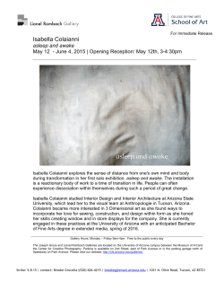

© Copyright 2026