
Screening for abdominal aortic aneurysm myocardial infarction using portable
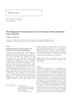
European Heart Journal – Cardiovascular Imaging (2012) 13, 574–578
doi:10.1093/ejechocard/jer260
Screening for abdominal aortic aneurysm
in coronary care unit patients with acute
myocardial infarction using portable
transthoracic echocardiography
1
Cardiology Department, AP-HP, Bichat Hospital, 46 rue Henri Huchard, 75018 Paris, France; 2INSERM U698, Paris, France; and 3University Paris 7, Paris, France
Received 30 August 2011; accepted after revision 31 October 2011; online publish-ahead-of-print 29 November 2011
Aims
Patients with acute myocardial infarction (AMI) represent a high-risk population in which screening for abdominal
aortic aneurysm (AAA) is recommended but only occasionally performed. Transthoracic echocardiography (TTE)
may offer the unique opportunity to evaluate the cardiac function and to screen for AAA during the same examination. We aimed to evaluate the feasibility of AAA screening at bedside using a portable cardiac ultrasound (US) echo
machine and to determine the prevalence of AAA in population with AMI.
.....................................................................................................................................................................................
Methods
The AA diameter was measured at bedside at the end of a regular TTE performed in consecutive patients admitted
for AMI in the coronary care unit using a portable echo machine (Vividi, General Electric). AAA was defined by a
and results
transverse diameter of ≥30 mm. We prospectively enrolled 193 patients (65 + 11 years, 77% male). Measurement
of the AA diameter was feasible in 93% and the duration was 3 + 1 min. An AAA was observed in nine patients
(4.7%) and the prevalence increased with age (7.7% after 60 years and 9.2% after 65 years). No AAA was observed
in patients under 50 years old. Inter-observer variability between cardiologists using the portable US system was excellent (mean difference 1.8 + 2.0 mm) as well as the accuracy compared with measurements performed by a radiologist using a dedicated vascular US system (mean difference 1.5 + 1.3 mm).
.....................................................................................................................................................................................
Conclusion
Overall, the prevalence of AAA was 4.7%, increased with age, and seems higher than expected in the ‘same-aged
population’. In regard to the simplicity, accuracy, and feasibility, screening for AAA during TTE (one cardiovascular
shot) may be of value after AMI especially in elderly patients.
----------------------------------------------------------------------------------------------------------------------------------------------------------Keywords
Abdominal aortic aneurysm † Screening † Acute myocardial infarction † Echocardiography
Introduction
Abdominal aortic aneurysm (AAA) is the 14th leading cause of death
almost from dramatic rupture. The overall mortality of ruptured
AAA is 80 –90% compared with a 30-day post-operative mortality
of 3% after elective surgery. Considering the balance between incidence and cost-effectiveness,1 even if screening for AAA in asymptomatic patients combined with increased numbers of elective
aneurysm surgery may reduce the mortality rate, the relatively low
prevalence of AAA in the general population (5.5% of men over 65
years old2) implies to target specific high-risk population. As AAA
and coronary heart disease share common risks factors, patients
with acute myocardial infarction (AMI) represent a high-risk population in which screening for another atherosclerotic location is
recommended but often neglected.3 In addition, the prevalence of
AAA in this population has never been properly evaluated.
* Corresponding author. Tel: +33 1 40 25 66 01; fax: +33 1 40 25 67 32, Email: [email protected]
Published on behalf of the European Society of Cardiology. All rights reserved. & The Author 2011. For permissions please email: [email protected]
Downloaded from http://ehjcimaging.oxfordjournals.org/ by guest on June 9, 2014
Caroline Cueff 1, Niall G. Keenan 1, Laura Krapf 1, Philippe Gabriel Steg 1,2,3,
Claire Cimadevilla 1, Gregory Ducrocq 1, Jean-Baptiste Michel2,3, Alec Vahanian 1, and
David Messika-Zeitoun1,2,3*
575
Screening for AAA in AMI patients
Ultrasound (US) scanning using a 3.5 MHz probe is a valid firstline method of screening. It is fast and safe with a sensitivity and
specificity close to 100% with a low intra- and inter-observer variability. Previous studies have suggested that the 2.5 MHz probe
used during routine transthoracic echocardiography (TTE) may
be adequate for the detection of AAA with a sensitivity ranging
from 91 to 94% using quick-screen programmes and the subcostal
view.4,5 As most patients with AMI undergo a TTE when hospitalized in the cardiology coronary care unit (CCU), we aimed to
evaluate the feasibility of AAA screening by the cardiologist at
bedside during TTE using a portable cardiac echo machine and
to determine the prevalence of AAA in this population.
Methods
Study design
Figure 1 Methodology of abdominal aortic diameter measurements. External antero-posterior outside-to-outside wall diameter was measured in subcostal views (thin arrow). The large
arrow indicates mural thrombus in a patient with an aortic
abdominal aneurysm.
Clinical investigation
Baseline characteristics were collected including age, gender, body
mass index, history of atherothrombotic risk factors: smoking status
(current or past smokers vs. non-smokers), diabetes mellitus (fasting
glucose ≥7 mmol/L and/or the use of diet or oral hypoglycaemic
agents or insulin treatment), hypertension (patients receiving antihypertensive medications or having known but untreated blood pressure
≥140/90 mmHg) and hypercholesterolaemia (fasting serum total cholesterol ≥220 mg/dL or treated hypercholesterolaemia). Physical exam
consisted in measurement of bilateral brachial and ankle blood pressure to calculate the ankle– brachial index (ABI) and screen for peripheral arterial disease and abdominal examination (deep palpation in the
left upper quadrant/epigastrium using the right hand). If the aorta was
palpable, the examiner, using deep palpation with the left hand,
attempted to ascertain whether the patients had AAA or simply a
normal palpable aorta.
Echocardiography
Scanning of an AA was performed directly at bedside using a portable
echocardiographic US system (Vivid I from General Electrics) and a
2.5 MHZ cardiac probe. No abdominal preparation was required. Analysis of the aorta was made at the end of the regular TTE examination
performed routinely in patients with AMI, using the subcostal view
while the patient lying supine. The entire AA was first visualized in
the transverse and longitudinal planes. Measurements were carried
out in the transverse plane, antero-posterior outside-to-outside wall
at both the suprarenal aorta level (next to the celiac vessel) and the
infrarenal aorta level (just below the renal arteries) (Figure 1). External
antero-posterior diameter was measured in each site three times and
then averaged. Quality was defined as ‘good’ when the vessel was analysable easily, ‘correct’ when limits of the vessel were analysable but
with less resolution, ‘average’ when limits were imprecise but distinguishable, and ‘poor’ when the vessel could not be seen. AAA was
defined as an aortic external antero-posterior diameter of
≥30 mm.3 Patients with suspected AAA were referred to the vascular
department to undergo a specific vascular Doppler echography using a
dedicated US system and if needed were advised to consult a vascular
surgeon.
Statistical analysis
Continuous variables were expressed as mean + standard deviation
(SD). Categorical variables are expressed as absolute numbers and
percentages. The prevalence of AAA was calculated as the number
of AAA diagnosed divided by the number of patients included in the
study. Feasibility was the percentage of patients in whom the aorta
was analysable. Reproducibility was assessed as the difference
between measurements performed at bedside by two cardiologists
using the portable US system several days apart blinded of previous
results. Accuracy was assessed as the difference between measurements performed at bedside by a cardiologist using the portable ultrasonographic system and measurements performed blindly by a
radiologist using a dedicated vascular US machine. Correlations
between measurements were evaluated using linear regression. All
tests were two-sided. A value of P , 0.05 was considered statistically
significant.
Results
Population
One hundred and nineteen-three patients were consecutively and
prospectively enrolled on two periods from February to October
2008 (9 months) and from June to September 2009 (4 months),
including weekends, totalizing 13 months. The mean age was
65 + 11 years (27–98) and 150 (77%) were males. The mean
Downloaded from http://ehjcimaging.oxfordjournals.org/ by guest on June 9, 2014
Consecutive patients admitted for AMI in the CCU of Bichat Hospital,
a large tertiary academic centre, were prospectively enrolled in the
present study. Inclusion criteria were a clinical diagnosis of myocardial
infarction using an elevation of biomarkers (troponin, CK) in the presence of chest pain and/or electrocardiographic changes in at least two
derivations (persistent or transient ST-segment elevation or depression, T-waves inversion, flat T-waves, or pseudo-normalization of
T-waves). There were no exclusion criteria. An oral informed
consent was obtained in all patients.
576
C. Cueff et al.
Table 1 Baseline characteristics of the overall
population and of patients with abdominal aortic
aneurysm
Characteristics
Overall (N 5 193)
Abdominal aortic
aneurysm (N 5 9)
................................................................................
Age (years)
Male gender
65 + 14
150 (77%)
85 + 3
7 (78%)
141 (73%)
95 (51%)
7 (78%)
6 (67%)
Risk factors
Smokers
Hypertension
Dyslipidaemia
85 (44%)
5 (56%)
Diabetes
Body mass index
46 (24%)
26 + 4
3 (33%)
27 + 4
Ankle–brachial
index
1.03 (0.6– 1.5)
0.8 (0.6– 1)
AMI
Stroke
49 (25%)
17 (9%)
5 (55%)
2 (22%)
Claudicating
13 (7%)
3 (33%)
PCI
CABG
22 (12%)
13 (7%)
1 (11%)
3 (33%)
Type of acute myocardial infarction
ST elevation
myocardial
infarction
107 (55%)
3 (33%)
Non-ST elevation
myocardial
infarction
77 (40%)
5 (56%)
Bundles block or
paced rhythm
9 (5%)
1 (11%)
Extends of CAD
1-vessel disease
69 (38%)
5 (14%)
2-vessel disease
54 (30%)
2 (28%)
3-vessel disease
No angiography
55 (30%)
15
4 (57%)
2
Abdominal aortic diameter
Infrarenal
Suprarenal
17 + 5 (9– 45 mm)
19 + 3 (13– 28 mm)
35 + 5 (30– 45 mm)
19 + 3 (13– 24 mm)
ABI was 1.03 + 0.16 (0.6 –1.5) and 16 patients (19%) had an ABI of
≤0.9. No patient had a previously known AAA. Patients’ characteristics are summarized in Table 1.
Feasibility of screening for abdominal
aortic aneurysm
The feasibility of screening using the portable US system was 93%;
13 patients (7%) had a poor echocardiographic window. Quality
was average in 69 patients (36%), correct in 59 (30%), and
good in 52 (27%). The duration of screening was 3 + 1 min
(1– 10 min). Reproducibility, assessed in 37 patients, was good
(18 + 6 vs. 18 + 5 mm, P ¼ 0.86; mean difference 1.8 + 2mm; r
¼ 0.88, P , 0.0001) and accuracy, assessed in 30 unselected
patients, was excellent (18 + 7 vs. 19 + 7 mm; mean difference
Prevalence of abdominal aortic aneurysm
in patients with acute myocardial
infarction
The mean diameter of the AA was 19 + 3 mm (13–28 mm) at the
suprarenal level and 17 + 5 mm (9–45 mm) at the infrarenal level.
Nine patients had an AAA (diameter ≥30 mm) yielding an overall
prevalence of 4.7% (none was previously known). The mean diameter was 35 + 6 mm (30– 45 mm). The aortic diameter was
between 30 and 35 mm in six patients, between 36 and 40 mm in
one patient, and .40 mm in two patients. The prevalence of AAA
increased with age, 5.5% after 50 years (n ¼ 162), 7.7% after 60
years (n ¼ 117), and 9.2% after 65 years (n ¼ 97) (Figure 3). All
aneurysms were located at the infrarenal level and were partially
thrombosed. Clinical characteristics of the nine patients with AAA
are presented in Table 1 (Right column). Our results did not
change if we used the ratio of suprarenal/infrarenal aorta diameter
≥1.5 as a definition for AAA: the nine patients with a diameter of
≥30 mm were all identified as well as an additional patient with a
27 mm AA diameter.
Discussion
In this series of consecutive patients hospitalized for acute coronary syndrome in our CCU, the analysis of the AA by a cardiologist
using a portable cardiac ultrasonography equipment at the bedside
was highly feasible (93%), accurate, reproducible, and fast. As the
prevalence detected over 65-year-old patients (9.2%) is higher
than that expected in the general ‘same-aged population’ (5.5%),2
screening of AAA may be recommended during TTE after AMI,
as one ‘cardiovascular shot’, especially in the elderly population.
Rupture is a dreaded complication of AAA directly correlated to
the antero-posterior diameter with an increasing risk over 50 mm
of diameter.6 Screening programmes target the populations most
likely to benefit of this strategy, maximize cost-effectiveness, and
minimize inconvenience and risk to the healthy population. As
the prevalence of AAA in the general population is relatively
low,2 the current guidelines recommend to screen specific population subsets: at least once men over 65 years old, women or men
with familial history of AAA over 50 years old, and by extension
patients with cardiovascular disease.3,6 Echography is the key
exam using a vascular 3.5 MHz probe and has an excellent sensibility and specificity (close to 100%)7 including when ‘quick-screen
analysis protocols’ are used.5
Patients hospitalized for AMI routinely undergo TTE. TTE may
offer the unique opportunity to evaluate, during the same examination, the cardiac function and to screen for AAA. A cardiac probe
has already been evaluated for AAA screening with a sensitivity
between 91 and 96%.4 Our study was first performed using portable US systems directly at the patient bedside, at the end of a
routine TTE (2.5 MHz probe). Feasibility was excellent (93%), independently of the time since the last meal, suggesting that no specific abdominal preparation is required. Inter-observer variability
Downloaded from http://ehjcimaging.oxfordjournals.org/ by guest on June 9, 2014
Prior medical history
1.5 + 1.3 mm; r ¼ 0.98, P , 0.0001) (Figure 2). Using clinical palpation for screening, 31 patients (16%) had a palpable aorta of whom
only 2 (6%) had an aneurysm confirmed using US.
577
Screening for AAA in AMI patients
bedside and by the radiologist using a dedicated ultrasound system. (B) Quality control plots using the Altman and Bland analysis for the two
methods. The middle line represents the mean and the upper and lower lines +2 SD.
Figure 3 Prevalence of aortic abdominal aneurysms in the
overall population and according to age.
was good and consistent with previous reports. Scan duration was
short, 3 min in average to be added to the cardiac imaging time. No
extra cost or additional equipment (same examinator, same material) was required. Our results suggest that US scanning is simple,
quick, accurate, and reproducible when performed by a cardiologist using small portable systems. It is worth noting that abdominal
palpation was neither sensitive nor specific for the diagnostic of
AAA but remains essential in clinical practice to detect
complications.
The prevalence of coronary artery disease in patients with AAA
is well known and is one of the main causes of death.8,9 On the
opposite, the prevalence of AAA in patients with coronary
artery disease is less evaluated but seems higher and varies from
7 to 18% in patients with stable coronary artery disease. This
large range may be due to differences in AAA definition and
patient selection.10 – 12 Recently, Dupont et al. 13 found a prevalence of AAA of 6.9% in patients undergoing coronary artery
bypass surgery. In the present study, the prevalence was 4.7% in
the overall population and increased with age, 9.2% after 65
years which is twice the expected prevalence for the age-adjusted.
The present study has several clinical implications. For the cardiologist, we suggest that at the end of the regular TTE, a quick analysis
of the aorta by the subcostal view should be systematically performed. For the patient, they may benefit at least once of the screening, with respect to guidelines, especially in patients with risk factors.
In the setting of an acute coronary syndrome, the diagnosis of large
AAA before coronary angiography has implication for the treatment
strategy (e.g. bare-metal stent vs. drug-eluting stent) and on the
choice of the vascular access for angiography and percutaneous coronary intervention (radial vs. femoral). In addition, detection of small
AAA is a strong incentive for intensive therapy.14
Our study deserves several comments. First, quick-screen protocol and complete analysis of the aorta by the specialist should not be
regarded as competitive but as complementary methods in order to
optimize the screening programmes. Secondly, in the absence of
control group, we cannot definitively establish the higher prevalence
of AAA among AMI, compared with patients without AMI. Nevertheless, large previous studies suggested a lower prevalence in the
general same-aged population. Finally, this was an observational
study and was not intended nor powered to demonstrate the clinical
impact of this screening programme.
Conclusion
This is the first study to highlight the interest of a systematic AAA
screening by the cardiologist in patients admitted in the CCU for
AMI using portable echocardiographic US systems during routine
Downloaded from http://ehjcimaging.oxfordjournals.org/ by guest on June 9, 2014
Figure 2 (A) Correlation between abdominal aortic diameter measured by the cardiologist using the portable cardiac ultrasound device at
578
TTE at the bedside. In regard to the simplicity, accuracy, and feasibility of this screening of AAA, it may considered as a complement
of the routine TTE examination as one cardiovascular shot screening strategy especially in the elderly population.
Conflict of interest: none declared.
Funding
C.C. and N.G.K. were supported by a grant from the Association de
Cardiologie d’Ile de France (ACIF-SFC) and D.M.-Z. was supported
by a contrat d’interface INSERM.
References
4. Roshanali F, Mandegar MH, Yousefnia MA, Mohammadi A, Baharvand B. Abdominal aorta screening during transthoracic echocardiography. Echocardiography
2007;24:685 –8.
5. Lee TY, Korn P, Heller JA, Kilaru S, Beavers FP, Bush HL et al. The costeffectiveness of a ‘quick-screen’ program for abdominal aortic aneurysms.
Surgery 2002;132:399 –407.
6. Powell JT, Greenhalgh RM. Clinical practice. Small abdominal aortic aneurysms. N
Engl J Med 2003;348:1895 – 901.
7. Lederle FA. Ultrasonographic screening for abdominal aortic aneurysms. Ann
Intern Med 2003;139:516 –22.
8. Hertzer NR, Beven EG, Young JR, O’Hara PJ, Ruschhaupt WF 3rd, Graor RA et al.
Coronary artery disease in peripheral vascular patients. A classification of 1000 coronary angiograms and results of surgical management. Ann Surg 1984;199:223 –33.
9. United Kingdom Small Aneurysm Trial Participants. Long-term outcomes of immediate repair compared with surveillance of small abdominal aortic aneurysms.
N Engl J Med 2002;346:1445 –52.
10. Bergersen L, Kiernan MS, McFarlane G, Case TD, Ricci MA. Prevalence of abdominal aortic aneurysms in patients undergoing coronary artery bypass. Ann Vasc Surg
1998;12:101 –5.
11. Madaric J, Vulev I, Bartunek J, Mistrik A, Verhamme K, De Bruyne B et al. Frequency of abdominal aortic aneurysm in patients .60 years of age with coronary
artery disease. Am J Cardiol 2005;96:1214 –6.
12. Nevelsteen A, Kim Y, Meersman A, Suy R. Routine screening for unsuspected
aortic aneurysms in patients after myocardial revascularization: a prospective
study. Acta Cardiol 1991;46:201 –6.
13. Dupont A, Elkalioubie A, Juthier F, Tagzirt M, Vincentelli A, Le Tourneau T et al.
Frequency of abdominal aortic aneurysm in patients undergoing coronary artery
bypass grafting. Am J Cardiol 2010;105:1545 –8.
14. Baumgartner I, Hirsch AT, Abola MT, Cacoub PP, Poldermans D, Steg PG et al.
Cardiovascular risk profile and outcome of patients with abdominal aortic
aneurysm in out-patients with atherothrombosis: data from the Reduction of
Atherothrombosis for Continued Health (REACH) Registry. J Vasc Surg 2008;
48:808 – 14.
Downloaded from http://ehjcimaging.oxfordjournals.org/ by guest on June 9, 2014
1. Bergqvist D, Bjorck M, Wanhainen A. Abdominal aortic aneurysm—to screen or
not to screen. Eur J Vasc Endovasc Surg 2008;35:13–8.
2. Lindholt JS, Norman P. Screening for abdominal aortic aneurysm reduces
overall mortality in men. A meta-analysis of the mid- and long-term effects of
screening for abdominal aortic aneurysms. Eur J Vasc Endovasc Surg 2008;36:
167 –71.
3. Hirsch AT, Haskal ZJ, Hertzer NR, Bakal CW, Creager MA, Halperin JL et al.
ACC/AHA guidelines for the management of patients with peripheral arterial
disease (lower extremity, renal, mesenteric, and abdominal aortic): a collaborative
report from the American Associations for Vascular Surgery/Society for
Vascular Surgery, Society for Cardiovascular Angiography and Interventions,
Society for Vascular Medicine and Biology, Society of Interventional Radiology,
and the ACC/AHA Task Force on Practice Guidelines (writing committee to
develop guidelines for the management of patients with peripheral arterial
disease)—summary of recommendations. J Vasc Interv Radiol 2006;17:1383 – 97;
quiz 1398.
C. Cueff et al.


© Copyright 2026