
Possible functional links among brain- and skull-related
Language Sciences
Possible functional links among brain- and skull-related genes selected in
modern humans
Antonio Benítez-Burraco and Cedric Boeckx
Journal Name:
Frontiers in Psychology
ISSN:
1664-1078
Article type:
Hypothesis & Theory Article
Received on:
29 Jan 2015
Accepted on:
26 May 2015
Provisional PDF published on:
26 May 2015
Frontiers website link:
www.frontiersin.org
Citation:
Benítez-burraco A and Boeckx C(2015) Possible functional links
among brain- and skull-related genes selected in modern humans.
Front. Psychol. 6:794. doi:10.3389/fpsyg.2015.00794
Copyright statement:
© 2015 Benítez-burraco and Boeckx. This is an open-access article
distributed under the terms of the Creative Commons Attribution
License (CC BY). The use, distribution and reproduction in other
forums is permitted, provided the original author(s) or licensor
are credited and that the original publication in this journal is
cited, in accordance with accepted academic practice. No use,
distribution or reproduction is permitted which does not comply
with these terms.
This Provisional PDF corresponds to the article as it appeared upon acceptance, after rigorous
peer-review. Fully formatted PDF and full text (HTML) versions will be made available soon.
Possible functional links among brain- and skull-related genes selected in modern humans
A. Benítez-Burraco1 and C. Boeckx2,3
1. University of Huelva, Huelva, Spain
2. Catalan Institute for Research and Advanced Studies (ICREA)
3. Universitat de Barcelona, Barcelona, Spain
Abstract: The sequencing of the genomes from extinct hominins has revealed that changes in
some brain-related genes have been selected after the split between anatomically-modern humans
and Neanderthals/Denisovans. To date, no coherent view of these changes has been provided.
Following a line of research we initiated in Boeckx and Benítez-Burraco (2014a), we hypothesize
functional links among most of these genes and their products, based on the existing literature for
each of the gene discussed. The genes we focus on are found mutated in different cognitive
disorders affecting modern populations and their products are involved in skull and brain
morphology, and neural connectivity. If our hypothesis turns out to be on the right track, it means
that the changes affecting most of these proteins resulted in a more globular brain and ultimately
brought about modern cognition, with its characteristic generativity and capacity to form and
exploit cross-modular concepts, properties most clearly manifested in language.
Keywords: Language-ready brain, skull morphology, hominin ontogeny, human evolution,
Neanderthals/Denisovans, anatomically modern humans, paleogenomics, neuroscience,
biolinguistics, AUTS2, FOXP2, RUNX2.
1
1. Introduction
The successful retrieval of ancient DNA and subsequent reconstruction of archaic human
genomes (Pääbo 2014a) surely qualify as a major breakthrough in our attempt to understand the
evolution of our species. Ancient genomes constitute fossils of a new kind, potentially far more
revealing than the more traditional fossils scientists have manipulated for decades. This research
generates candidate genes to shed light on changes that led to the emergence of modern human
cognitive capacities (Green et al., 2010; Meyer et al. 2012; Prüfer et al. 2014; Pääbo 2014b). To
date, no coherent view of these changes has been yet provided. In particular, no functional link
between these changes has been established. In this paper, we would like to rely on the existing
literature for each of the gene involved and hypothesize functional links among them. In other
words, we claim that some of the key genetic changes related to the brain in modern humans did
not occur independently of one another, but rather in a related fashion.
Before delving into these genes, we would like to briefly remind the reader that as important as it
is to be able to provide a list of genes that affect the brain, we must ultimately be able to
characterize what roles the cellular consequences of these gene products have for brain circuits
and their dynamics and how those in turn give rise to specific cognitive phenotypes.
The psychological sciences already offer good characterizations of the human cognitive
phenotype. Interestingly, for many of these, the scholars that put them forward tend to implicate
our language capacity to a significant extent. For example, Spelke (2003) takes language to be
crucial in the establishment of productive, systematic exchange of information across ‘core
knowledge’ systems shared with other species. Likewise, Marcus (2001) sees in language the
clearest illustration of what makes our mind ‘algebraic’ (as opposed to merely ‘symbolic’). There
is indeed a growing consensus among scientists of various disciplines --- from paleoanthropology and archeology (Mithen 1996, Coolidge and Wynn 2005) to philosophy (Carruthers
2006, Pietroski 2012) and linguistics (Reinhart 2006, Boeckx 2011) --- that our brain’s languagereadiness, understood not only as a communication system but also and perhaps most importantly
as the generator of a particular conceptual format, is the central piece of the puzzle of our cognitive
modernity.
If on the right track, the centrality of language in delimiting our cognitive phenotype would allow
us to take advantage of the decades of results in formal linguistics to come up with a detailed,
computationally explicit description of the ‘human cognome’ (Poeppel 2012) --- the set of
elementary mental representations and operations needed to generate the anlage of our rich
cognitive life (e.g., Samuels 2011, Boeckx 2014). Taken as a whole, these elementary
representations and operations would constitute the proper target of neurobiological
investigations.
To those who have already ventured in this direction, it is clear that a successful marriage of the
relevant disciplines will require a serious rethinking of the neurobiological foundations of
language processing (Poeppel 2014, Hagoort 2014, Fedorenko and Thompson-Schill 2014). The
age of the “classical model” of brain and language, developed in the 19th century by pioneers like
Broca, Wernicke and Lichtheim is over. As reviewed in Petersson et al. (2012), the language
network is more extended than the classical language regions and includes, next to Broca’s region,
adjacent cortex in the left inferior and middle frontal region, as well as substantial parts of superior
and middle temporal cortex, inferior parietal cortex, as well as subcortical structures such as the
basal ganglia, the cerebellum, the hippocampus, and the thalamus.
As it slowly replaces the classical model, this new ‘neurobiology of language’ (Poeppel et al.
2012) approximates distributed networks implicated in cognition more generally (working
memory models, the default network, the multiple demand system, the global neuronal workspace
model, etc.; see Boeckx and Benítez-Burraco 2014a), thereby becoming a better candidate for the
2
hypothesis that language-readiness is central to modern cognition, as opposed to just another
encapsulated module of the mind.
It is with this background in mind that we want to examine the genetic changes that separate us
from our extinct closest relatives, the Neanderthals and the Denisovans. To do so, we have
decided to rely on our (2014a) proposal, centered around RUNX2.
Briefly summarizing our (2014a) paper, we put forward a set of genes involved in skull
morphogenesis, thalamic development and the specification, migration and interconnection of
GABAergic neurons within the forebrain. The most salient members of this set of genes were
RUNX2, the DLX suite (in particular DLX1, DLX2, DLX5, DLX6), genes of the BMP family
(BMP2, BMP7), and interacting partners like SHH and FGF8. We hypothesized that the
evolutionary modification of this network may account for our species-specific globular head
shape (compared to Neanderthals; Bruner 2004, Gunz et al. 2010, 2012, Neubauer et al. 2010)
and for the concomitant rewiring of different connections between cortical and sub-cortical
(specifically, thalamic) structures, which we claimed provide the scaffolding for our speciesspecific mode of cognition (see Boeckx and Benítez-Burraco 2014a for details; see also BenítezBurraco and Boeckx 2015).
Once this initial set of genes was established, we decided to examine potential links between these
genes and the rich body of work on FOXP2 (a robust candidate for language disorders in our
species) and its network (Vernes et al. 2011, Graham and Fisher 2013). The results were presented
in Boeckx and Benítez-Burraco 2014b. There, we paid special attention to ROBO1, which appears
to be critical to aspects of language such as vocal learning (Pfenning et al. 2014). As we will
highlight below, the new set of genes discussed below further strengthen the links between our
initial set of genes and the FOXP2 network.
Figure 1 provides a schematic representation of our strategy.
FIGURE 1 ABOUT HERE
Figure 1. Objective of the present study: Focus on the intersection of the sets of genes highlighted
in Boeckx and Benítez-Burraco 2014a,b with the currently available list of genes showing signs
of positive selection in Anatomically Modern Humans.
At this point we would like to highlight a few considerations that will bear in the discussion to
follow. Specifically, the three considerations we now turn to helped us select both the genes
discussed in the next section, and the information we present for each.
First, we think that a detailed investigation of the genetic underpinnings of our species-specific
brain-growth trajectory that gives rise to a more globular braincase configuration ought to pay
attention as well to changes in the visual system, and their cognitive implications, something
which we did not do in our original (2014a) study. The globularization of the brain is likely to
affect the occipital lobe, which provides the roots of the visual system. In addition, a recent study
by Pearce et al. (2013), focusing on the fact that Neanderthals had larger eyes than our species,
suggest that more of their brain was devoted to seeing in the long, dark nights in Europe, at the
expense of high-level cognitive processing. This is so because larger eyes entail a much larger
visual processing area at the back of their brains. In other words, more of the Neanderthal brain
would have been dedicated to vision and body control. A reduction of the visual area in
Anatomically Modern Humans (AMHs) has been independently supported by Sherwood et al.
(2008), and it may have led to an expansion of the parietal region, and a re-allocation of the
computational power of the pulvinar, the part of the dorsal thalamus that modulates cortical visual
processing (Saalmann et al., 2012), in service of other cognitive domains, such as language. The
fact that visual deficits either predispose towards or protect against mental diseases like
schizophrenia for which language is crucially implicated (Silverstein et al. 2013, Leivada and
3
Boeckx 2014), suggest that the visual system and visual cognition ought to play a role in any
account of the brain’s language-readiness and the emergence of modern cognition more generally.
Some of the findings reported below are meant to bear directly on this hypothesis.
Second, an important aspect of language-readiness --- the ability to form and maintain longdistance connections across the whole brain to form and exploit cross-modular concepts --- is
likely to require the recruitment of thalamic nuclei such as the medio-dorsal nucleus and the
pulvinar in addition to neocortical areas. Accordingly, both cortical and subcortical
reconfigurations must be taken into account. In particular, we think that it is crucial to focus on
new sources of inhibition in light of neo-cortical and pulvinar expansion. In this context, Rakic’s
hypothesis concerning new sources of interneurons due to molecular changes (Letinic and Rakic
2001, Rakic 2009; Geschwind and Rakic 2013) strike us as particularly relevant. Incidentally, our
concern for sources of inhibition as well as the establishment and maintenance of long-distance
cortical connections naturally led us to pay special attention to mental diseases like autism, where
the factors just mentioned appear to be targeted (Just et al. 2004, Yizhar et al. 2011, Khan et al.
2013, Le Magueresse and Monyer 2013, Zikopoulos and Barbas 2013). Hence the frequent
mention of autism below (see also Benítez-Burraco and Boeckx 2015).
Third, in addition to the precise establishment of cortico-thalamic and thalamocortical
connections acting as cognitive pacemakers, it is also important to examine any changes
concerning interhemispheric connectivity (Theofanopoulou 2015). Although neurolinguistic
studies have traditionally been lopsided (focus on the left-hemisphere), recent reviews reporting
progress in the neurobiology of language emphasize the importance of the right hemisphere and
interhemispheric integration for language (see Petersson et al. 2012; Poeppel et al. 2012; Poeppel
2014; Theofanopoulou 2015). This also led us to pay attention to mental conditions in which
interhemispheric connectivity patterns and language abilities are altered: again autism turns out
to be a key source of information (Verly et al. 2014).
Armed with all these considerations, we turn to brain-related changes revealed by discoveries
based on archaic human genomes, and examine their possible implications for the brain
organization (and head shape) of AMHs.
2. Methods
Our goal in this paper is to establish potential functional connections with the initial gene set
discussed in Boeckx and Benítez-Burraco (2014a), the FOXP2 interactome as currently known
(reviewed in Boeckx and Benítez-Burraco 2014b), and the literature on selected genes in AMHs.
Our modus operandi has been as follows.
a. We searched the literature via PubMed (http://www.ncbi.nlm.nih.gov/pubmed) looking for
symptoms of interest for us linked to the mutation of the “brain-related” genes frequently
mentioned in publications addressing the list of changes selected for in AMHs (e.g. Crisci et al.
2011, Lalueza-Fox 2013, Lombard et al. 2013). We also relied on the most recent and more
exhaustive list of genes showing signals of positive selection in AMHs given in Pääbo 2014b
(Table S1). Some of the key search terms used were: “language disorder”, cognitive disorder”,
“intellectual disability”, “syntax deficit”, “semantic deficit”, “phonological deficit”, “speech
deficit”, “dyslexia”, “schizophrenia”, “autism”, “autism spectrum disorder”, etc.
b. When a gene of interest was identified, we looked for potential links with the set of genes as
advanced in Boeckx and Benítez-Burraco (2014a) via String 9.1. (http://string-db.org/). String 9.1
predicts direct/physical and indirect/functional associations between proteins that derive from
four sources: genomic context, high-throughput experiments, conserved coexpression, and the
knowledge previously gained from text mining (Franceschini et al. 2013). We considered links
with a confidence value above .0400. This score means that one has a 40% probability that a
4
predicted link exists between two enzymes in the same metabolic map in the KEGG database
(http://www.genome.jp/kegg/pathway.html). We used this medium confidence value because the
highest one (0.700) was too restrictive and we did not get many links (as is typical of any
approaches relying on String). Importantly, we checked all the predicted links against the existing
literature and discarded all the links for which we could not find strong evidence, in order to
achieve a reliable set of candidates for our potential network.
c. We did not systematically gather information beyond more than three connection levels. Thus,
our limit was of the sort ‘A is connected to B which is in turn connected to C’. We considered
additional levels only if the identified genes were more closely connected to some other gene(s)
of interest previously reviewed. For example, ‘A is connected to B which is in turn connected to
C which is in turn connected to D (but D is connected to RUNX2)’.
d. We also used PubMed to look for genetic information related to brain areas, circuits, neural
processes, neurotransmitters, etc. of interest for us; specifically, the neurological considerations
of Boeckx and Benitez-Burraco (2014a) highlighted in section 1, as well as those neural substrates
emphasized in the skull and brain literature. For this, we used search terms like “thalamus”,
“thalamo-cortical connection”, “interhemispheric connection”, etc.
Because the amount of literature on single genes varies greatly, we did not necessarily discard
any gene because the number of manuscripts was under a value we may have pre-selected.
In addition to PubMed, we also relied on the following databases, which we also used for our
(2014a) paper.
1) the microarray database of the Allen Brain Atlas (http://human.brainmap.org/microarray/search)
2) the Prenatal LMD Microarray search engine
(http://www.brainspan.org/lcm/search/index.html)
3) the Developmental Transcriptome browser
(http://www.brainspan.org/rnaseq/search/index.html) of the Allen Brain Atlas.
4) OMIM for the linguistic and cognitive deficits linked to the mutation of genes of interest
(http://www.ncbi.nlm.nih.gov/omim/)
Additionally, as we did in Boeckx and Benítez-Burraco (2014b), we have exploited the
information provided in Kuhlwim et al. (2013), where 691 genes were found to be differentially
expressed after RUNX2 transfection in neuroblastomic SH-SY5Y cells, as well as the detailed
FOXP2 targets list provided in Konopka et al. (2009), Spiteri et al. (2007) and Vernes et al. (2007).
While collecting information on each new gene of interest, we paid special attention to its
potential role not only in language, but also vision, in the establishment of cortico-thalamic and
thalamo-cortical connections, and in the balance between excitation and inhibition in the context
of brain growth. This focus is reflected in the information we present in the next section.
3. RUNX2 and brain-related genes selected in AMHs
Because of the centrality of RUNX2 in our (2014a) proposal, we decided to start this section by
briefly reviewing the nature and relevance of this gene, focusing on information that was not
available at the time we completed our initial study.
RUNX2
5
There is solid evidence of a selective sweep in RUNX2 after our split from Neanderthals (Green
et al., 2010). Although the aminoacid sequence of RUNX2 is conserved between AMHs,
Neanderthals and Denisovans, its promoter region presents two derived alleles in modern humans
(Perdomo-Sabotal et al. 2014). As Perdomo-Sabotal et al. point out, this might have caused gene
regulatory changes with implications for cranial, skeletal and bone development in modern
humans, given the well-established fact that RUNX2 controls different aspects of the morphology
of the upper body and the cranium: closure of cranial sutures, clavicle development, rib cage
formation, and dental growth (Stein et al. 2004). In particular, the action of RUNX2 under the
control of DLX5 (Holleville et al. 2007) and TLE1 (Wang et al. 2004) appears crucial for the
integration of the parietal bone (Depew et al. 1999, Stephens 2006), heavily implicated in the
globularization of the AMH brain/skull (Bruner 2004).
Although RUNX2 does not figure among the canonical “brain-related” genes in publications
addressing the list of changes selected for in AMHs (e.g. Crisci et al. 2011, Lalueza-Fox 2013,
Lombard et al. 2013), unlike genes like AUTS2 or DYRK1A, which we discuss below, we think
that it should be included among genes of great relevance for our cognitive phenotype. Here is
why.
To begin with, Jeong et al. (2008) and Reale et al. (2013) demonstrate that RUNX2 expression
plays an important role at the brain level (thalamus, hypothalamus, hippocampus), and the gene
is repeatedly mentioned as a candidate for several mental diseases, including schizophrenia and
bipolar disorders (Benes et al. 2007, Subburaju and Benes 2012, Talkowski et al. 2012, Ruzicka
et al. 2015). Recently, RUNX2 has been shown to interact with several key candidate genes for
autism, specifically with SMURF1 (De Rubeis et al. 2014), a gene involved in the regulation of
axonogenesis (Cheng et al. 2011, Kannan et al. 2012).
More indirectly, RUNX2 (and “skull-related” genes more generally) provides crucial information
about brain growth and connectivity, as the very signals sent to build the brain case, and thus
influencing RUNX2 expression, come from the same signaling molecules that have been
independently argued to be responsible for brain organization (WNT, FGF, SHH, DLX, etc.).
Especially in the first year of life, when globularity is established in AMHs, brain and skull exhibit
an extremely tight fit (Lieberman 2011), which may well explain why mental retardation and
craniofacial dysmorphism co-occur, even more so that one suspects (see Boeckx and BenítezBurraco 2014a for review). Studies on other species such as dogs demonstrate how skull shape
and brain connectivity influence one another (Roberts et al. 2010), so we feel entitled to apply
this logic to the case of AMHs. Incidentally, this also applies to FOXP2, which interacts with
genes that not only affect brain connectivity but also craniofacial bones (e.g. ROR2, COL9A1;
Konopka et al. 2009). As a matter of fact, direct physical interaction between RUNX2 and FOXP2
has recently been experimentally demonstrated (Zhao et al. 2015).
There is also mounting evidence in favor of bone affecting cognition. As reviewed in Oury et al.
(2013), the regulation of bone mass exerted by subcortical structures like the hypothalamus
(Driessler and Baldock 2010) suggests the existence of bone-derived signals modulating this
regulation or other functions of the brain. As Oury et al. show, the osteoblast-derived hormone
osteocalcin crosses the blood-brain barrier, binds to neurons of the brainstem, midbrain, and
hippocampus, enhances the synthesis of monoamine neurotransmitters, inhibits GABA synthesis,
prevents anxiety and depression, and favors learning and memory independently of its other
metabolic functions at the bone level. In addition to these postnatal functions, maternal
osteocalcin crosses the placenta during pregnancy and prevents neuronal apoptosis before
embryos synthesize this hormone. Thus, the authors conclude, the skeleton via osteocalcin
influences cognition and contributes to the maternal influence on fetal brain development.
Interestingly, RUNX2 is deeply implicated in the regulation of osteocalcin, including at the brain
level (Vladimirova et al. 2008). RUNX2 has been shown to interact with the 1alpha,25dihydroxyvitamin D3 receptor (encoded by VDR) to up-regulate rat Bglap (encoding osteocalcin)
6
expression in osteoblastic cells (Paredes et al. 2004). We take this interaction between RUNX2
and VDR to be extremely relevant. Stephens and Morrison (2014) have shown that RUNX2 and
VDR, which becomes active when bound by its ligand 1,25-dihydroxyvitamin D3 (VD3), unite
to cooperatively regulate the expression of numerous genes. Among the genes identified are
SPAG5, which is among the genes selected by AMHs (Green et al. 2010), and to which we return
below, and also SRGAP3. The latter has been shown to play an important role in severe mental
retardation and absence of speech (Endris et al. 2002). Srgap3-/- mice present a
neurodevelopmental disorder with schizophrenia-related intermediate phenotypes (Waltereit et
al. 2012), and the gene has been shown to be implicated in childhood onset schizophrenia in
humans (Wilson et al. 2011). In addition, Srgap2 acts through Srgap3 to modulate neuronal
differentiation and neurite outgrowth of mouse neuroblastoma cells (Ma et al. 2013). We highlight
this fact because SRGAP2 is a gene that has duplicated three times in humans (Sudmant et al.
2010). One of the duplicated forms encodes a truncated form of the protein that binds to the
nontruncated protein encoded by the ancestral gene (Dennis et al., 2012). When expressed in
mouse neuronal precursor cells, the truncated protein variant results in increased density of longer
neuronal spines (Charrier et al., 2012). More generally, SRGAP3-mediated cytoskeletal
reorganisation has an important influence on a variety of neurodevelopmental processes, which
may be required for normal cognitive function. (Endris et al. 2011, Bacon et al. 2013, Radaei et
al. 2014). In particular, SRGAP2 and SRGAP3 interact with ROBO1 and affect the SLIT/ROBO
pathway (Wong et al. 2001), which is critical for a range of brain connectivity patterns, including
in the context of language (see Boeckx and Benítez-Burraco 2014b for review, and especially
Pfenning et al. 2014, where SLIT-mediated interaction between FOXP2/foxp2 and ROBO1/robo1
is claimed to be critical to the establishment of vocal learning pathways across species).
Like RUNX2, SRGAP3 is heavily expressed in the hippocampus (Waltereit et al. 2012).
Hippocampal network alterations are well attested in a variety of human cognitive diseases, such
as schizophrenia (Heckers 2001), bipolar disorder (Frey et al. 2007), Alzheimer's disease (Schuff
et al. 2009) and Down syndrome (Pennington et al. 2003). In the context of Alzheimer's, it may
be worth pointing out that RUNX2 interacts with APOE (Vaes et al. 2006, Kuhlwim et al. 2013),
a gene that has been consistently related to some of the metabolic changes that allowed bigger
brains, and eventually enhanced cognitive capacities, to evolve within hominins (Bufill and
Carbonell, 2006), even if one of its alleles (ε4) appears to predispose its bearers to Alzheimer’s.
Incidentally, the link between RUNX2 and APOE may well reinforce the hypothesis put forth by
Bruner and Jacobs (2013) that Alzheimer's disease is intimately connected to the globularization
of the AMH skull/brain.
In a similar vein, Shen and Christakos (2005) have shown that the Vdr, Runx2, and the Notch
signaling pathway cooperate in the transcriptional regulation of Spp1, which encodes osteopontin,
which is also important for the brain. Osteopontin-deficient mice have been shown to suffer from
thalamic neurodegeneration (Schroeter et al. 2006). A disregulation of osteopontin is known to
cause intracranial arteriovenous malformations (Xu et al. 2012).
Finally, vitamin D (Patrick and Ames 2014) and other RUNX2 interacting partners (e.g., CREB;
Oury et al. 2010) also regulate serotonin synthesis. Not only does brain serotonin regulate bone
mass through its release in ventromedial hypothalamic neurons, it also affects thalamocortical
axonal system development (van Kleef et al. 2012), and has been claimed to be implicated in
autism (Patrick and Ames 2014).
As a final note, we wish to point out that brain expression of Foxp2 was shown to be significantly
altered by vitamin D deficiency (Hawes et al. 2015).
Having discussed RUNX2, we now turn our attention to more canonical “brain-related” genes that
show evidence of selection in AMHs. We begin with AUTS2.
AUTS2
7
According to Green et al (2012) the first half of AUTS2 (roughly, the section of the gene
encompassing exons 1 to 4 [chr7:68,662,946-69,274,862 (hg18)]) displays the strongest signal of
positive selection in AMHs compared to Neanderthals (within this region, two of the 293 SNPs
that are ancestral in Neanderthals are coding variants, although they are not fixed in AMHs).
Perhaps for this reason AUTS2 has been subject of recent attention (see Oksenberg and Ahituv
2013 for review).
Three noncoding intronic regions of interest in evolutionary terms have been found within
AUTS2: the human accelerated region HAR31, located in intron 4 (Prabhakar et al. 2006), and the
human accelerated conserved non-coding sequences HACNS 369 and HACNS 174, located in
introns 1 and 4, respectively (Pollard et al. 2006). Interestingly, these regions contain enhancers
that seem to be active in the brain. Within the region under selective sweep Oksenberg et al.
(2013) found six enhancers that show expression in the brain and four mouse enhancers that are
active in the midbrain and the eye. Some of these enhancers are also found inside the HAR or
HACNS regions. It may be the case that some of the enhancers encompassing the AUTS2
regulatory landscape affect other genes, perhaps to some of the genes located within the Williams
Syndrome critical region (see Oksenberg and Ahituv 2013 for discussion). Williams syndrome is
a developmental disorder involving cognitive and linguistic deficits (see Martens et al. 2008, and
Mervis and Becerra 2007 for reviews), which results from a microdeletion in one copy of the
chromosome 7 that affects roughly two dozen genes (Korenberg et al. 2008). Among these genes
one finds GTF2I, related to craniofacial abnormalities and cognitive problems (Morris et al.,
2003; Tassabehji et al., 2005), which represses RUNX2 (Lazebnik et al., 2009) and is a functional
partner of USF1 (Roy et al., 1997), both of them central pieces in our (2014a) paper. Incidentally,
the regulatory region of USF1 has undergone 30 fixed or high frequency changes after our split
from Denisovans (Meyer et al., 2012).
Of particular interest to us is the fact that AUTS2 is a candidate for several neurodevelopmental
disorders, including autism spectrum disorder (ASD), intellectual disability, and developmental
delay (see Oksenberg and Ahituv 2013 for discussion). Cognitive impairment can co-occur with
epilepsy, skeletal abnormalities, brain malformations, and/or dysmorphic features―the so-called
AUTS2 syndrome (Beunders et al. 2013). Copy-number variation and genome wide associations
studies have related AUTS2 with other ASD-independent disordered conditions, like
schizoaffective disorder (Hamshere et al., 2009), bipolar disorder (Hattori et al. 2009), differential
processing speed (Luciano et al. 2010), or even dyslexia (Girirajan et al. 2011). According to
Talkowski et al. (2012) the AUTS2 locus is also linked to microcephaly, macrocephaly, ataxia,
visual impairment, motor delay, or Rubinstein–Taybi syndrome. Recently, Amarillo et al. (2014)
have found a deletion encompassing exon 6 of AUTS2 in a subject with severe speech and
language problems (the individual was unable to make full sentences at the age of 4;6). During
growth AUTS2 is expressed in the human brain throughout the telencephalon (primarily in the
frontal, parietal, and temporal lobes), but also in some other regions, including the cerebellum
and the basal ganglia (caudate and putamen nuclei) (Sultana et al. 2002, Lepagnol-Bestel et al.
2008). The gene is also highly expressed in the ocular tissues (Wagner et al. 2013). In mice, Auts2
has proved to be expressed in the developing thalamus, the cerebellum and the cerebral cortex, in
neurons of several types, including glutamatergic, GABAergic, and dopaminergic neurons
(Bedogni et al. 2010a). Interestingly, Auts2 is expressed in the thalamus both prenatally (in the
dorsal thalamus) and postnatally (in the anterior thalamic nuclei and in ventrolateral/ventromedial
nuclei). In the cortex Auts2 is only expressed in the frontal areas after birth (Bedogni et al. 2010a).
The knockdown of auts2 in zebrafish causes a decrease in neuronal cells which results in
microcephaly. Craniofacial abnormalities are also observed, as well as motor problems (plausibly
due to a reduced number of motor neurons and/or sensory neurons) and a reduced size of the eye
(Beunders et al., 2013; Oksenberg et al. 2013).
In sum, available evidence points to AUTS2 as an important gene for neuro-cranial development
and human evolution. However, the putative link between this gene, the changes that occurred at
8
the brain level to give rise to AMHs, and the emergence of modern cognition and the language
abilities we have, remain unclear. What follows is an attempt to offer a hypothesis that could
guide future experimental investigations seeking to address this issue.
Our attempt starts with the noteworthy finding that AUTS2 figures among the genes found to be
differentially expressed after RUNX2 transfection in neuroblastomic SH-SY5Y cells (Kuhlwim
et al 2013). Even more so is the fact that both Runx2 and Foxp2 are found among Auts2 regulatory
targets (Oksenberg et al. 2014), as is Cntnap2. CNTNAP2 is a well-known FOXP2 target (Vernes
et al. 2008) and a candidate for language delay and language impairment (Petrin et al. 2010,
Sehested et al. 2010), intellectual disability (Gregor et al. 2011), and autism (Alarcón et al. 2008,
Bakkaloglu et al. 2008), The AMH CNTNAP2 exhibits a fixed change (Ile345Val) compared to
the Denisovan protein (Meyer et al. 2012). Moreover, CNTNAP2 is related to NFASC, a protein
involved in neurite outgrowth and the formation of postsynaptic components (Kriebel et al. 2012)
that shows a fixed change (T987A) in AMHs compared to Neanderthals/Denisovans (Pääbo,
2014b, table S1).
Interestingly, AUTS2 regulation of RUNX2 also affects CBL. Mutations in the latter gene cause
Noonan syndrome-like disorder, a condition characterized by facial dysmorphism, a reduced
growth, and a variety of cognitive deficits, among other symptoms (Martinelli et al. 2010). Of
interest to us is the fact that CBL is located in a region showing signal of a strong selective sweep
(20-fold enrichment over random) in AMHs compared to Altai Neanderthals (Prüfer et al. 2014).
Needless to say, AUTS2 has been associated with many other proteins that play a key role at the
brain level. Many of these are also candidate for autism or for developmental disorders affecting
cognitive/language abilities. These include TBR1, RELN, SATB2, GTF2I, ZMAT3, or PRC1
(reviewed by Oksenberg and Ahituv 2013). We will discuss some of these genes in detail here,
but we will focus in their connections with both RUNX2 and FOXP2, and explore additional
connections with other AMH-selected genes.
TBR1
In mice Auts2 and Tbr1 are coexpressed mostly in glutamatergic neurons of the forebrain
(Bedogni et al. 2010a). In fact, Auts2 is a direct target of Tbr1 in the developing neocortex
(Bedogni et al. 2010b). Microdeletions causing the disruption of TBR1 give rise to severe speech
and language deficits, autistic-like problems, and moderate to severe intellectual disability, while
larger deletions encompassing TBR1 cause delayed or absent speech and language and intellectual
disability (Palumbo et al. 2012, 2014; Traylor et al. 2012). Microcephaly is also a reported
(Palumbo et al. 2012), plausibly due to the functional link between TBR1 and ASPM, a robust
candidate for microcephaly, and candidate for positive selection in human lineage (Bond et al.
2002; Zhang 2003).
TBR1 has been shown to interact with FOXP2 (Enard et al. 2009, Deriziotis et al. 2014), as
Ferland et al. (2003) had anticipated in mice. Specifically, Ferland et al. had speculated that
disruption of Tbr1 disrupts Foxp2 expression in layer VI of the cortex, thereby altering the
projections of layer VI neurons to the dorsal thalamus. TBR1 indeed plays a key role in the
organization of the corticothalamic tracts, which originate mostly from layer VI of the cortex
(Hevner et al. 2001). Srinivasan et al. (2102) have shown that in mice the expression of Tbr1
consistently correlates with subcortical axons that innervate the dorsal thalamus. After birth, Trb1
expression is upregulated in different upper-layer neurons and this upregulation is required for
the expression of Auts2 (Bedogni et al. 2010b; Srinivasan et al. 2012). The knockout of the gene
almost abolishes neocortical connectivity with the thalamus (which is, among other things, a
critical aspect of the language-ready brain, if we are right in our (2014a) hypothesis), and
misroutes corticothalamic axons of neurons from layer VI and the subplate towards the spinal
cord (Han et al., 2011; McKenna et al., 2011).
9
In addition to this role in corticofugal connectivity, Tbr1 is also involved in the establishment of
intercortical connections. Specifically, callosal axons fail to cross the midline in the absence of
Tbr1 (Hevner et al. 2001). Interestingly, the integrity of the corpus callosum is frequently reported
to be affected in people suffering from ASD (Kumar et al., 2010; Shukla et al. 2010; Nagae et al.
2012); also, as we advanced in section 1, a decrease in interhemispheric connections seems to be
a hallmark of this neurodevelopmental condition (Shukla et al. 2010; Ingalhalikar et al. 2011;
Weinstein et al. 2011). Moreover, in mice Tbr1 haploinsufficiency also results in defective axonal
projections of amygdalar neurons, which give rise to a deficit in ultrasonic vocalization, social
interaction, and associative memory and cognitive flexibility (Huang et al. 2014).
FEZF2
Much of the function performed by TBR1 seemingly results from its effect on FEZF2 expression.
Fezf2 has been claimed to be a key orchestrator of gene activity giving rise to neuronal subtypes
(Lodato et al. 2014). In mice the knockdown of Tbr1 results in the ectopic upregulation of Fezf2
(Han et al. 2011, McKenna et al. 2011). In fishes, fezf2 is one of the components (together with
six3, shh, irx1b, and wnt1) of the gene suite controlling the relative size of the telencephalon
versus the thalamus and ultimately, the differences in brain architecture among species (Sylvester
et al. 2010). In mice Fezf2 is expressed exclusively in the corticofugal projection neurons of the
deep cortical layers, mostly in subcerebral neurons from layer V, and also, but less strongly, in
corticothalamic neurons from layer VI (Chen et al. 2005a, Molyneaux et al. 2005). Fezf2 directs
the differentiation of neural stem cells in the subventricular zone toward specific cortical
phenotypes (Zuccotti et al. 2014), and it has been shown to regulate the gene suite that define the
glutamatergic corticospinal neurons (Lodato et al. 2014) and the fate of pyramidal neurons in
deep layers of the cortex.
In the absence of Fezf2 expression neurons express Satb2 and project to other regions of the
cortex. Moreover, the number of interneurons of several subtypes--- critical for the correct balance
between excitation and inhibition in the neocortex--- becomes reduced in layer V of the cortex
(Lodato et al., 2011). Conversely, the presence of Fezf2 causes neurons to project to subcortical
regions (Chen et al. 2008). Specifically, the gene controls the development and the
corticothalamic projections of neurons from layer VI (Chen et al. 2005, Molyneaux et al. 2005).
Interestingly, FEZF2 (and also its partner CTIP2) is highly expressed in von Economo neurons
(Cobos and Seeley 2013). These neurons, located in layer V of the anterior cingulate and the
fronto-insular cortices, are important for social behavior and are only found in species that are
able of self-awareness (great apes, elephants, or cetaceans) (Butti et al. 2009; Allman et al. 2010).
Cortical areas containing von Economo neurons form a network of frontoparietal functional
connectivity, encompassing four different sub-networks, involved in saliency detection, sensorymotor behavior, and attention (Cauda et al. 2013). Significantly, autistics show a higher ratio of
Von Economo neurons to pyramidal neurons in the frontoinsular cortex, which has been related
to an enhanced interoception (Santos et al., 2011).
Not surprisingly, then, FEZF2, like TBR1, is a candidate for autism (Wang et al., 2009).
Interestingly, the SNP found by Wang et al. to be associated with this condition, located within
the proximal promoter region, represents a reversion to the ancestral non-primate allele (the
primate allele has been strongly selected in our clade). Interestingly also, FEZF2 is required for
the expression of FOXP2 (Molyneaux et al., 2005).
DYRK1A
Another promising partner of TBR1 is DYRK1A, a gene often mentioned alongside AUTS2 in the
context of human evolution, given that it contains a region identified to have strong signals of
selective sweep in AMHs compared to Neanderthals (Green et al. 2012). TRB1 regulates RELN
(Chen et al. 2002), an important gene controlling neural migration and also a candidate for
lissencephaly with language loss (Hong et al. 2000) and for autism (Wang et al. 2014). In turn,
10
RELN is upregulated by FOXO1 (Daly et al. 2004). FOXO1 is a target of both RUNX2 (Kuhlwilm
et al. 2013) and FOXP2 (Vernes et al. 2011), and encodes a protein that is phosphorylated by
DYRK1A (Huang and Tindall, 2007).
DYRK1A is located within the Down Syndrome Critical Region on chromosome 21. Mutations
on DYRK1A give rise to microcephaly, facial dysmorphisms, mental retardation, and absence of
speech (Van Bon et al. 2011, Courcet et al. 2012). The gene is expressed during development, but
also in the adult brain, and it seems to be involved in learning and memory (Hämmerle et al.
2003). This role is explained by the effect of DYRK1A on synaptic plasticity and on the
expression of genes encoding GABAergic and glutaminergic related proteins. This effect
ultimately alters the balance between excitation and inhibition, which we believe is very important
for cognitive function. Specifically, DYRK1A overexpression, as observed in Down syndrome,
affects pathways involved in synaptogenesis and synaptic plasticity, and moves the
excitation/inhibition balance towards inhibition (Souchet et al. 2014).
Interestingly, increasing expression of Dyrk1a in mice upregulates GAD1 (Souchet et al. 2014).
GAD1 is a target of FOXP2 (Konopka et al. 2009). Moreover, RUNX2, DLX1, and DLX2, three
key genes in our (2014a) hypothesis concerning globularity and the emergence of the languageready brain, are also key components of the GAD1 regulatory network, which is important for the
normal development of GABAergic neurons within the hippocampus (Pleasure et al. 2000,
Ruzicka et al. 2015). Additionally, in mice Dyrk1a has been shown to play a central role in the
balance between cortical and thalamic neurons: in particular, Dyrk1a copy number seems to be
directly related to neuron density in the thalamic nuclei and indirectly related to neuron density
in the cortex (Guedj et al. 2012).
Finally, DYRK1A directly phosphorylates SIRT1 and also promotes deacetylation of TP53.
SIRT1 is involved in the regulation of different neural processes, including neural precursor
activity and differentiation (Saharan et al. 2013) and axon formation and elongation (Li et al.
2013). As we discussed in Boeckx and Benítez-Burraco (2014a), a potential link exists between
FOXP2 and RUNX2 via SIRT1. Moreover, SIRT1 directly regulates RUNX2: it both upregulates
RUNX2 and deacetylates RUNX2, ultimately promoting osteoblast differentiation (Shakibaei et
al. 2012, Srivastava et al. 2012). In this context, it may be worth mentioning that SIRT1 is an
effector of several genes that are under selection in modern populations and that show non-fixed
changes in their coding regions compared to Neanderthals and Denisovans, like BAZ2A and
NR1H2 (Prüfer et al., 2014). Interestingly also, SIRT1 regulates some of the genes controlling
biological noise, like H2A.Z (Baptista et al. 2013). This may have helped buffer the molecular
noise resulting from the changes occurred in the RUNX2 network and that brought about the
language-ready brain (Benítez-Burraco, 2015).
Regarding TP53, it is a candidate for schizophrenia (Ni et al, 2005). A non-fixed change (Pro72
to Arg72) has been found in the AMH protein compared to Neanderthals/Denisovans (Paskulin
et al. 2012). Last, but not least, as we also review in Boeckx and Benitez-Burraco (2014a), TP53
is related to many of the genes encompassing the language-ready brain network, including SIRT1,
USF1 (mentioned above), CDH1, ASPM (also mentioned above) and PTEN.
SATB2
An additional partner of TBR1 and AUTS2 we wish to highlight in this part of the paper is SATB2.
Together with Fezf2, Ctip2, and Tbr1, Satb2 controls the identity of stereotypic projections in the
cortex. Specifically, in conjunction with Ctip2, Satb2 regulates Tbr1 expression in neurons from
cortical layers II to V in order to produce callosal projections. Together with Fezf2, Satb2
regulates Auts2 (Srinivasan et al. 2012). Sequence and copy number variations in SATB2 have
been found in patients with ASD, intellectual disability, and developmental and language delays,
as well as craniofacial defects (see Kwan 2013 and especially Liedén et al. 2014 for discussion).
Accordingly, SATB2 is also involved in osteogenesis, where it directly interacts with RUNX2
11
(Hassan et al. 2010). For example, lentiviral-mediated-Satb2-transduced cells from the mouse
bone marrow stroma overproduce Satb2 and upregulates Runx2 (Gong et al. 2014). Finally, an
interesting link also exists between SATB2 and FOXP2. Hence, Dobreva et al. (2006) have found
that Satb2 represses the expression of several Hox genes, including Hoxa2. This gene encodes an
inhibitor of bone formation and regulator of branchial arch patterning. According to Konopka et
al. (2009) HOX2A is among FOXP2’s targets. Also, the phenotype linked to chromosome 2q32q33 deletions and to the happloinsufficiency of SATB2 can be mimicked by the haploinsufficiency
of GTF3C3, a gene that is also a target of FOXP2 (Konopka et al., 2009).
In the context of Satb2, we wish to point out that a strong Gli3 binding region is located just over
100 kb ≤ of Satb2 in neural tissue (Vokes et al. 2007). Gli3 interacts with Shh during thalamic
development (Haddad-Tóvolli et al. 2012). Moreover, Gli3 regulates calvarial suture
development by controlling Bmp-Smad signaling, which integrates a Dlx5/Runx2-II cascade
(Tanimoto et al. 2012). Actually, mutations in GLI3 have been found in people affected by Grieg
cephalopolysyndactyly syndrome, a condition in which craniosynostosis is an important feature
(Debeer et al. 2003). Interestingly, most (~98%) of Altaic Neanderthals and Denisovans had a
different sequence in GLI3 compared to AMHs: while the latter retained the ancestral sequence,
the former gained a nonsynonymous change that appears to be mildly disruptive (Castellano et al.
2014). Given the role of Satb2 in the establishment of callosal projections, it is also interesting
that Gli3 controls corpus callosum formation by positioning midline guideposts during
telencephalic patterning (Magnani 2014), altering expressions of Slit1/2, Fgf8 and Wnt/β-catenin,
genes we discussed at length in Boeckx and Benítez-Burraco (2014a).
ZBTB20
At this point we would like to briefly mention another gene, ZBTB20, which also shows signs of
selection in AMHs (Green et al. 2010). We find this gene of interest because it defines a
hippocampal neuronal identity through direct repression of genes that control projection neuron
development in the isocortex, among which we found Fezf2, Satb2, Tbr1, and Foxp2 (Nielsen et
al. 2014). In addition, although we don’t know of any direct evidence for an interaction between
ZBTB20 and RUNX2 in the hippocampus, we note that Kuhlwim et al (2013) found several genes
of the ZBTB family to be differentially expressed after RUNX2 transfection in neuroblastomic
SH-SY5Y cells.
PAX6
Although we are not aware of any signal of positive selection of PAX6 in AMHs, there are robust
links between them and RUNX2, FOXP2, AUTS2, and other genes discussed above.
PAX6 encodes a transcription factor involved in the development of the eye and the brain.
Although perhaps best known as the master regulator or master selector of eye development
(Gehring and Ikeo 1999), in the brain it affects the process of glutamatergic neuron differentiation
(Kim et al. 2014). It has been suggested that changes in PAX6 expression may underlie the
imbalance in excitatory/inhibitory neuronal activity in the autistic brain because of its
involvement in glutamatergic differentiation during development (Kim et al. 2014), and indeed
mutations on this gene have been linked to some forms of ASD (Maekawa et al. 2009).
Importantly, PAX6 is coexpressed with AUTS2 in the ventricular and subventricular regions
(Bedogni et al. 2010a).
PAX6 has been shown to be involved in interhemispheric transfer (Bamiou et al. 2004), to the
extent that mutations in this gene result in working memory problems (Bamiou et al. 2007).
Moreover, PAX6 plays an important role in the development of thalamic connections.
Specifically, mammillo-thalamic tracts were missing in Pax6 mutant mice (Valverde et al. 2000).
Repression of Shh by Pax6 regulates diencephalic patterning by controlling the central
diencephalic organizer (Caballero et al. 2014). Additionally, Pax6 regulates the orientation and
12
mode of cell division of progenitors in the mouse cerebral cortex, influencing as it does cell
adhesion during cortical development (Tyas et al. 2003). In this context, it interacts with many of
the genes discussed in the previous subsection, and gives rise to cortical layering abnormalities
when knocked-out in mice (e.g. Tuoc et al. 2009). Pax6 plays as well a crucial role at the level of
the cortical hem, which regulates both the size and patterning of the cortex (Caronia-Brown et al.
2014), but also at the cortical anti-hem (Subramanian et al. 2009). Positioned as a mirror image
of the cortical hem, along the lateral margin of the cortical primordium, the cortical anti-hem is
identified by gene expression for three epidermal growth factor family members, Tgf β , Nrg1,
and Nrg3, as well as two other signaling molecules, Fgf7 and the secreted Wnt antagonist Sfrp2.
The anti-hem is lost in mice homozygous for the Pax6 mutation. It is worth mentioning in this
context that both FGF7 and NRG3 are among the genes that show signals of a selective sweep in
AMHs compared to Neanderthals (Green et al. 2010; Prüfer et al. 2014).
A heterozygous mutation of PAX6 has been shown in individuals with aniridia and deficits in
executive and social cognition. Structural abnormalities of grey matter were reported in the
anterior cingulate cortex, the cerebellum and the medial temporal lobe, as well as white matter
deficits in corpus callosum. Functional MRI demonstrated reduced activation of fronto-striatothalamic systems during performance of overt verbal fluency and nonsense sentence completion;
the most consistent abnormality of verbal executive activation was located in the thalamus
(Ellison-Wright et al. 2004). Eye problems associated with PAX6 deficiencies are also worth
bearing in mind in light of the potential cognitive relevance of eye size in Neanderthals and AMHs
(Pearce et al. 2013), as we reviewed in the first section of the paper. Anophthalmia and
microphthalmia are repeatedly reported in this context (Verma and FitzPatrick 2007), and are
often associated with mental diseases like schizophrenia where language is affected (Leivada and
Boeckx 2014).
In zebrafish the knockdown of pax6 disrupts the expression of both arx and foxp2 (Coutinho et
al., 2011). ARX is actually a target of FOXP2 (Konopka et al. 2009, Vernes et al. 2009). ARX
directly controls SLIT2 in the proper formation of the thalamus. ARX is in turn directly regulated
by DLX genes in the developing forebrain, and contributes to the tangential migration of
GABAergic interneurons (Colasante et al. 2008). Analysis of transcriptional codes for zebrafish
dopaminergic neurons has revealed essential functions of arx in prethalamic dopaminergic neuron
development (Filippi et al. 2012). In human mutations of ARX give rise to mental retardation and
interneuronopathies (Quillé et al. 2011), including lissencephaly (Kitamura et al. 2002) and
agenesis of the corpus callosum (Proud et al.,1992) which are features of interest in the context
of this paper.
Like AUTS2, PAX6 figures among the genes found to be differentially expressed after RUNX2
transfection, although in a different cell line (HepG2) (Kuhlwim et al 2013). Moreover, among
the PAX6 target genes and enhancers listed by Coutinho et al. (2011) we have found several genes
of great interest to us, including FOXP2, ASCL1, DLX1, DLX2, DLX5, DLX6, GBX2, and GLI3,
all of which we discussed in Boeckx and Benítez-Burraco (2014a) and above. In addition, PAX6
interacts with TBR1 at the level of neurogenesis (Englund et al. 2005).
Interestingly, among PAX6 target genes, we have also found POU3F2, this meaning that both
FOXP2 and its effector POU3F2 are regulated by PAX6. POU3F2 has been associated with
human accelerated conserved noncoding sequences (haCNSs) (Miller et al. 2014). There exists
an AMH-specific substitution in intron 8 of FOXP2 that affects a binding site for POU3F2
(Maricic et al. 2013). The derived allele has proved to be less efficient in activating transcription
(Maricic et al. 2013). It is possible then that FOXP2 expression in Neanderthals and Denisovans
was higher than in AMHs as a consequence of this change. In this context it is worth pointing out
that overexpression of FOXP2 in humans has been related to autism via MET, a putative risk
factor for this condition (Mukamel et al. 2011)). POU3F2 has been linked to bipolar disorder
(Mühleisen et al. 2014), but also to developmental and language delay, intellectual disability,
schizophrenia and ASD (Lin et al. 2011). POU3F2 encodes a transcription factor that controls
13
dopamine and serotonin synthesis (Nasu et al. 2014) and also the regulation of the upper-layer
neuronal migration and identity during the development of the neocortex (McEvilly et al. 2002,
Sugitani et al. 2002; Domínguez et al. 2013). POU3F2 is also expressed at the level of the corpus
callosum (Katoh and Katoh 2009), and it interacts with PQBP1, in turn linked to developmental
delay and microcephaly (Li et al. 2013) and to intellectual disability (Wang et al. 2013).
SPAG5
As pointed out above, the interaction between RUNX2 and VDR promotes the expression of
SPAG5, which is among the genes selected in AMHs (Green et al. 2010) The gene encodes a
protein needed for the correct function of mitotic spindles (it is required for centrosome integrity
and the maintenance of sister chromatid cohesion during mitosis) (Thein et al. 2007), but also for
the regulation of apoptosis induced by cell stress (Thedieck et al. 2013). At the brain level SPAG5
helps PAX6 to regulate the sequential symmetric and asymmetric cell division of neuronal
precursors. Hence, in mice the downregulation of Spag5 mimics the knockout of Pax6, which
greatly alter the orientation and mode of cell division in the cerebral cortex and which results in
an increased number of progenitors with morphological defects and an excess of daughter cells
with asymmetric fates (Asami et al. 2011). Interestingly, SPAG5 seems to be upregulated in
people suffering from Down syndrome with cryptorchidism (Salemi et al. 2013). Moreover, in
the retina SPAG5 interacts with the isoform B of USH2A, the main candidate for Usher syndrome,
a condition involving combined deaf-blindness (Kersten et al., 2012). There exists an intriguing
link between Usher syndrome and cognitive disorders involving language deficits, including
psychosis and schizophrenia (Domanico et al. 2012; see Leivada and Boeckx 2014 for a more
detailed discussion), which we think is worth studying further if our current reflections are on the
right track.
Figure 2 provides a graphic representation of the links we have wished to highlight in this section
between RUNX2, brain-related genes selected in AMHs like AUTS2 and DYRK1A, their closest
partners TBR1, FEZF2, and SATB2, FOXP2 and PAX6.
FIGURE 2 ABOUT HERE
Figure 2. Schematic representation of the strongest links discussed in this section
4. Other potential genes of interest
In this section, we wish to highlight additional changes in genes relevant for brain function that
occurred after the split between AMHs and Neanderthals/Denisovans and that could be related to
some of the genes considered so far, as well as those in Boeckx and Benítez-Burraco (2014a,b).
These genes may help to make more robust the links between our initial gene set and the two gene
sets we have reviewed above. This is why we discuss them in the remainder of this section.
To begin with, a region upstream MEF2A shows signals of recent positive selection in AMHs
(Somel et al., 2013). In our 2014a piece we hypothesized that the concomitant change in MEF2A
expression could have contributed to a delayed peak expression and an increased level expression
of synaptic genes in the prefrontal cortex of modern humans, and eventually to a less fast cortical
synaptic development in AMHs compared to Neanderthals. We also found that MEF2A is
functionally related to some of the components of our network, including USF1, BMP2, and
SIRT1.
Also, ERBB4 (which is linked to our language-ready-brain core set of candidates via BMP2) is a
receptor of NRG3, which, as already mentioned, is among the genes that show signals of a strong
selective sweep in AMHs compared to Neanderthals (Green et al. 2010, Prüfer et al. 2014). In the
chicken cerebellum Erbb4 works jointly with Pten (Sakakibara and Horwitz 2006), a gene that is
coexpressed with Katna1, which in turn encodes a protein involved in the reorganization of
14
cellular microtubule arrays (Selçuk et al. 2013). KATNA1 bears a fixed change (A343T) in
AMHs compared to Neanderthals/Denisovans (Pääbo, 2014b, table S1).
Moreover, FLNA interacts with ITGB4 (Travis et al, 2004), a protein that shows two fixed
changes (T1689A and H1748R) in AMHs compared to Neanderthals/Denisovans (Pääbo, 2014b,
table S1). CDC42 (Leung et al. 2010), required for proper migration of cortical interneurons
(Katyama et al. 2013). Two effectors of CDC42 are worth considering: CDC42EP4 and
ARHGAP32 . ~1kb within the CDC42EP4 is hypermethylated in AMHs compared to Denisovans
(Gokhman et al. 2014). Moreover, ARHGAP32 bears a fixed change (E1489D) in AMHs
compared to Denisovans (Meyer et al. 2012). This latter protein promotes axon growth
downstream CDH1 by interacting with SMURF1, which, as already mentioned above, is in turn
related to RUNX2 and a substrate of CDH1 (Kannan et al. 2012).
Additionally, a protein called NCAM1, related to working memory performance (Bisaz et al.
2013) and to neuropsychiatric conditions like schizophrenia, bipolar disorder and Alzheimer's
disease (Atz et al. 2007), interacts with VCAM1, a cell surface glycoprotein that shows a fixed
change (D414G) in AMHs compared to Neanderthals/Denisovans (Pääbo, 2014b, table S1).
NCAM1 is a potential target of RUNX2 (Kuhlwilm et al. 2013), but also of FOXP2 (Konopka et
al. 2009). Also DISC1 is a target of FOXP2 (Walker et al. 2012) and a robust candidate for
schizophrenia (Miyoshi et al., 2004). It is also highly expressed in the embryonic corpus callosum
at a critical time for callosal formation (Osbun et a. 2011, Theofanopoulou 2015). DISC1 interacts
with PCNT, a protein of the centrosome and a candidate for dyslexia (Poelmans et al. 2009).
PCNT is mentioned by Green et al (2010) among the 11 genes that show non-synonymous and
non-fixed substitution changes in their coding sequences compared to Neanderthals.
In the context of changes in the visual system and the establishment of the modern languageready brain, it is worth noting that the protein SOLH bears two aminoacidic changes in AMHs
(Green et al. 2010). Little is known about SOLH, but the little we know (e.g. Kamei et al. 1998)
strike us as relevant. SOLH is a human homologue of the Drosophila small optic lobes gene. As
its name suggests, mutations in the Drosophila gene cause a severe reduction in the neuropiles of
the medulla and lobula complexes of the adult optic lobes. Northern analysis of human tissues
revealed a SOLH transcript of approximately 5 kb that was strongest in the human brain. Kamei
et al. mapped the SOLH gene to chromosome 16p13.3 by in situ hybridization. SOLH is a
candidate gene for CATM syndrome (hereditary cataracts with microphthalmia), which maps in
this region.
Of direct interest to us is the fact that SOLH is a target of FOXP2. It is among the genes whose
expression level changes in a Foxp2-dependent manner in the E16 mouse (Vernes et al., 2011),
and is reported as a direct target of FOXP2 in the human inferior frontal cortex (Spiteri et al.,
2007). Although the action of FOXP2 in the brain is often tied to fine motor production and
auditory perception (Kurt et al. 2012), several authors have claimed that it is not limited to these
(e.g. Campbell et al. 2009). Links between Foxp2 expression and the visual system have in fact
been documented (Iwai et al. 2012) and they may be worth exploring further in the context of
cross-modal plasticity (e.g. Horng et al. 2009).
Finally, some other genes of interest are NCOA6, which encodes an interactor of both EP300 and
CREBBP, and which bears a M823I substitution in AMHs compared to Neanderthals/Denisovans
(Pääbo, 2014b, table S1); ANAPC10, one partner of CDH1 (Nourry et al. 2004), which shows
signals of a selective sweep in AMHs compared to Altai Neanderthals (Prüfer et al. 2014); and
CBL, which is located in a region showing signal of a strong selective sweep (20-fold enrichment
over random) in AMHs compared to Altai Neanderthals (Prüfer et al. 2014) EP300, CREBBP,
CDH1, and CBL are also relevant for the language-ready brain (see Boeckx and Benítez-Burraco
2014b for a detailed discussion).
15
Figure 3 provides an overview of the genes discussed here, and some of the key interactions
already derivable from the literature.
FIGURE 3 ABOUT HERE
Figure 3. The whole set of genes discussed in this paper. The network was generated by String
9.1. The medium confidence value was .0400. Nodes representing the proteins encompassing the
network are colored randomly. In this confidence view, stronger associations between proteins
are represented by thicker lines. The figure does not represent a fully connected graph, but
readers are asked to bear in mind that String 9.1 predicts associations between proteins that
derive from a limited set of databases. The material discussed in the main text lead us to suspect
connections that String does not generate (see figure 2) (although we wish to note that just adding
a few genes, not discussed in this paper, yield a connected graph). It should be emphasized that
the nature of String 9.1 is essentially predictive, and not explanatory. Although we have confirmed
all the links we discuss here in the literature, they need to be confirmed at the brain level and in
relation to language. Additionally, the diagram only represents the potential connectivity between
the involved proteins, but this has to be mapped onto particular biochemical networks, signaling
pathways, cellular properties, aspects of neuronal function, or cell-types of interest that can be
confidently related to aspects of language development and function.
5. Conclusion
The genomic revolution has led to a dramatic increase of information of great relevance to those
interested in shedding light to what made us human. SRGAP2 and FOXP2 were the first identified
members of a postulated set of genes that may have changed some of their functions (particularly,
those related to brain growth and function) some moment between 2 or 3 million years ago and
the present time as human ancestors grew larger brains and started to use these brains in new ways
to perform complex tasks. But they are the first of a long list that remains to be explored in depth
(recent other findings include the role of ARHGAP11B, Florio et al. 2015). We have focused on
a few genes in this paper, and hope to have highlighted interesting connections that we hope future
experiments will confirm. Our choice was guided by the hypothesis we put forth in our (2014a)
article, according to which the emergence of our species-specific language-ready brain ought to
be understood in light of the developmental changes expressed at the levels of brain morphology
and neural connectivity that gave us a more globular braincase configuration. As we argued in
our earlier work, this globularization affected not only the shape of the skull, but also the corticosubcortical connections (see our 2014b piece). It also likely affected the visual system. If on the
right track, the picture we have painted here suggest that there were coordinated changes tied to
the emergence of modern cognition and language. Specifically, we think it is of great interest that
the genes discussed here map onto specific types of neurons (GABAergic), particular brain
structures (specific cortical layers, thalamic nuclei), specific physiological process (balance
between inhibition and excitation), certain developmental processes (inter and interhemispheric
axon pathfinding) and, when mutated, give rise to several disorders with recurrent
endophenotypes. It is also of great interest that several of the genes discussed in the present paper
have been examined for expression in the zebra finch brain and are reported in the public brain
gene expression database (ZEBrA: www.zebrafinchatlas.org), some of which representing
markers of specific vocal nuclei or broader telencephalic zones generally associated with higher
cognitive skills. Certainly, the number of functional links important for language evolution is
becoming bigger as time goes by. Nonetheless, it has stopped being a mere list of binary
interactions and has become instead a putative network that leads to testable predictions. We have
proceeded here along standard lines: we have made our big network in a piecemeal fashion, by
adding new pieces to our original, small core network. At the same time, the protein changes we
highlight in our paper and the functional consequences we hypothesize for them should be viewed
as instances of the sort of evolutionary kluge or complex reconfiguration (and ulterior adaptation)
16
of ancestral systems that prompted the emergence of language as a cognitive faculty (Fisher and
Marcus 2006).
We wish highlight from the onset the limitations of attempts like ours to perform literature-based
assembly of protein-protein and gene-regulatory networks. We are aware that the robustness of
each of the connections hypothesized here must be tested. Specifically, it is important to prove
that the fixed coding changes in AMH proteins we review have impacted on their function.
Importantly, although we have focused on the strongest links found in the literature, these links
(often binary) have to be properly evaluated in order to know if they are actually biologically
significant and meaningful regarding skull and brain development and function, and cognitive
and linguistic abilities. We are aware that some types of evidence are stronger than other types.
For example, data on direct protein:protein interactions is much more telling than, say, gene
family membership. Even so, it should be proved that the involved proteins are expressed in the
same brain region or cell type at an equivalent developmental stage (a point that is often
overlooked by in silico tools). As we made explicit above we have tried to properly curate the
data in order to rely on the strongest evidence only. We also acknowledge that the literature and
the datasets we have relied on may be incomplete or biased because of the unavoidable focus on
some genes as opposed to others. One should not forget that absence of evidence is not evidence
of absence regarding protein-protein or DNA-protein interactions. It is also clear that the attested
links for intensely studied proteins will always be more salient and more numerous than for less
studied proteins. So, our main aim is to provide a list of potential candidates that can be employed
as a useful starting point for future investigations. We think that for all its limitations, our type of
research can offer valuable insights at this early stage of research in cognitive biology.
Acknowledgements
Preparation of this work was supported in part by funds from the Spanish Ministry of Economy
and Competitiveness (grant number FFI-2013-43823-P), and by a Marie Curie International
Reintegration Grant from the European Union (PIRG- GA-2009-256413). We wish to thank
Constantina Theofanopoulou for providing comments, support and key information about some
of the genes discussed above, which we would have missed otherwise. We are also grateful to the
reviewers for an extremely insightful reading of the manuscript we submitted, which improved
the paper considerably.
References
Alarcón, M., Abrahams, B. S., Stone, J. L., Duvall, J. A., Perederiy, J. V., Bomar, J. M., et al.
(2008) Linkage, association, and gene-expression analyses identify CNTNAP2 as an autismsusceptibility gene. Am. J. Hum. Genet. 82, 150–59.
Allman, J. M., Tetreault, N. A., Hakeem, A. Y., Manaye, K. F., Semendeferi, K., Erwin, J. M., et
al. (2010). The von Economo neurons in frontoinsular and anterior cingulate cortex in great apes
and humans. Brain Struct. Funct. 214, 495–517.
Amarillo, I. E., Li, W. L., Li, X., Vilain, E., and Kantarci, S. (2014). De novo single exon deletion
of AUTS2 in a patient with speech and language disorder: a review of disrupted AUTS2 and
further evidence for its role in neurodevelopmental disorders. Am. J. Med. Genet. A. 164A, 95865.
Asami, M., Pilz, G. A., Ninkovic, J., Godinho, L., Schroeder, T., Huttner, W. B., et al. (2011).
The role of Pax6 in regulating the orientation and mode of cell division of progenitors in the
mouse cerebral cortex. Development 138, 5067-78.
Atz, M. E., Rollins, B., and Vawter, M. P. (2007). NCAM1 association study of bipolar disorder
and schizophrenia: polymorphisms and alternatively spliced isoforms lead to similarities and
differences. Psychiatr. Genet. 17, 55-67.
17
Bacon, C., Endris, V., and Rappold, G. A. (2013). The cellular function of srGAP3 and its role in
neuronal morphogenesis. Mech. Dev. 130, 391-5
Bakkaloglu, B., O’Roak, B. J., Louvi, A., Gupta, A. R., Abelson, J. F., Morgan, T. M., et al.
(2008). Molecular cytogenetic analysis and resequencing of contactin associated protein-like 2 in
autism spectrum disorders. Am. J. Hum. Genet. 82, 165-73.
Bamiou, D. E., Campbell, N. G., Musiek, F. E., Taylor, R., Chong, W. K., Moore, A., et al. (2007).
Auditory and verbal working memory deficits in a child with congenital aniridia due to a PAX6
mutation. Int. J. Audiol. 46, 196-202.
Bamiou, D. E., Musiek, F. E., Sisodiya, S. M., Free, S. L., Davies, R. A., Moore, A., et al. (2004).
Deficient auditory interhemispheric transfer in patients with PAX6 mutations. Ann. Neurol. 56,
503-9.
Baptista, T., Graça, I., Sousa, E. J., Oliveira, A. I., Costa, N. R., Costa-Pinheiro, P., et al. (2013).
Regulation of histone H2A.Z expression is mediated by sirtuin 1 in prostate cancer. Oncotarget
4, 1673-85.
Bedogni, F., Hodge, R. D., Nelson, B. R., Frederick, E. A., Shiba, N., Daza, R. A., et al. (2010a).
Autism susceptibility candidate 2 (Auts2) encodes a nuclear protein expressed in developing brain
regions implicated in autism neuropathology. Gene Expr. Patterns 10, 9–15
Bedogni, F., Hodge, R. D., Elsen, G. E., Nelson, B. R., Daza, R. A., Beyer, R. P. et al. (2010b).
Tbr1 regulates regional and laminar identity of postmitotic neurons in developing neocortex.
Proc. Natl. Acad. Sci. U.S.A. 107, 13129–34
Benes, F. M., Lim, B., Matzilevich, D., Walsh, J. P., Subburaju, S., and Minns, M. (2007).
Regulation of the GABA cell phenotype in hippocampus of schizophrenics and bipolars. Proc.
Natl. Acad. Sci. U.S.A. 104, 10164–9.
Benítez-Burraco, A. (2015). Biological noise and H2A.Z: a promising connection for language.
Front. Genet. 5, 463. doi:10.3389/fgene.2014.00463
Benítez-Burraco, A., Boeckx C. (015). Approaching motor and language deficits in autism from
below: a biolinguistic perspective. 2015. Front. in Integrative Neuroscience 9, 25.
Beunders, G., Voorhoeve, E., Golzio, C., Pardo, L. M., Rosenfeld, J. A., Talkowski, M. E., et al.
(2013). Exonic deletions in AUTS2 cause a syndromic form of intellectual disability and suggest
a critical role for the C Terminus. Am. J. Hum. Genet. 92, 210–20
Bisaz, R., Boadas-Vaello, P., Genoux, D., and Sandi C. (2013). Age-related cognitive
impairments in mice with a conditional ablation of the neural cell adhesion molecule. Learn. Mem.
20, 183-93.
Boeckx, C. (2011). “The emergence of the language faculty, from a biolinguistic point of view,”
in Oxford Handbook of Language Evolution, eds M. Tallerman and K. Gibson (Oxford: Oxford
University Press), 492–501.
Boeckx, C. (2014). Elementary Syntactic Structures. Cambridge: Cambridge University Press.
Boeckx, C. and Benítez-Burraco, A. (2014a). The shape of the human language-ready brain.
Front. Psychol. 5, 282.
18
Boeckx, C. and Benítez-Burraco, A. (2014b). Globularity and language-readiness: Generating
new predictions by expanding the set of genes of interest. Front. Psychol. 5, 1324.
Bond, J., Roberts, E., Mochida, G. H., Hampshire, D. J., Scott, S., Askham, J. M., et al. (2002).
ASPM is a major determinant of cerebral cortical size. Nat. Genet. 32, 316-20.
Bruner, E. (2004). Geometric morphometrics and paleoneurology: brain shape evolution in the
genus homo. J. Hum. Evol. 47, 279–303.
Bruner, E., and Jacobs, H. I. (2013). Alzheimer’s disease: the downside of a highly evolved
parietal lobe? J. Alzheimers Dis. 35, 227–40.
Bufill, E., and Carbonell, E. (2006). Apolipoprotein E polymorphism and neuronal plasticity. Am.
J. Hum. Biol. 18, 556–8.
Butti, C., Sherwood, C. C., Hakeem, A. Y., Allman, J. M., and Hof, P. R. (2009). Total number
and volume of Von Economo neurons in the cerebral cortex of cetaceans. J. Comp. Neurol. 515,
243–59.
Caballero, I. M., Manuel, M. N., Molinek, M., Quintana-Urzainqui, I., Mi, D., Shimogori, T. et
al. (2014). Cell-autonomous repression of Shh by transcription factor Pax6 regulates diencephalic
patterning by controlling the central diencephalic organizer. Cell Rep. 8, 1405-18.
Campbell, P., Reep, R. L., Stoll, M. L., Ophir, A. G., and Phelps, S. M. (2009). Conservation and
diversity of Foxp2 expression in muroid rodents: functional implications. J. Comp. Neurol. 512,
84-100.
Caronia-Brown, G., Yoshida, M., Gulden, F., Assimacopoulos, S., and Grove, E. A. (2014). The
cortical hem regulates the size and patterning of neocortex. Development 141, 2855-65.
Carruthers, P. (2006). The Architecture of Mind. Oxford: Oxford University Press.
Castellano, S., Parra, G., Sánchez-Quinto, F. A., Racimo, F., Kuhlwilm, M., Kircher, M., et al.
(2014). Patterns of coding variation in the complete exomes of three Neandertals. Proc. Natl.
Acad. Sci. U. S. A. 111, 6666-71.
Cauda, F., Torta, D. M., Sacco, K., D'Agata, F., Geda, E., Duca, S., et al. (2013). Functional
anatomy of cortical areas characterized by Von Economo neurons. Brain Struct. Funct. 218, 120.
Charrier, C., Joshi, K., Coutinho-Budd, J., Kim, J. E., Lambert, N., de Marchena, J., et al. (2012).
Inhibition of SRGAP2 function by its human-specific paralogs induces neoteny during spine
maturation. Cell 149, 923–35.
Chen, B., Schaevitz, L., and McConnell, S. (2005). Fezl regulates the differentiation and axon
targeting of layer 5 subcortical projection neurons in cerebral cortex. Proc. Natl. Acad. Sci. U.S.A.
102, 17184–9.
Chen, B., Wang, S. S., Hattox, A. M., Rayburn, H., Nelson, S. B., and McConnell, S. K. (2008).
The Fezf2-Ctip2 genetic pathway regulates the fate choice of subcortical projection neurons in
the developing cerebral cortex. Proc. Natl. Acad. Sci. U.S.A. 105, 11382-7.
Chen, Y., Sharma, R. P., Costa, R. H., Costa, E., and Grayson, D. R. (2002). On the epigenetic
regulation of the human reelin promoter. Nucleic Acids Res. 30, 2930-9.
19
Cheng, P. L., Lu, H., Shelly, M., Gao, H., and Poo, M. M. (2011). Phosphorylation of E3 ligase
Smurf1 switches its substrate preference in support of axon development. Neuron 69, 231-43.
Cobos, I., and Seeley, W. W. (2013). Human von Economo neurons express transcription factors
associated with layer V subcerebral projection neurons. Cereb. Cortex [Epub ahead of print]
Colasante, G., Collombat, P., Raimondi, V., Bonanomi, D., Ferrai, C., Maira, M., et al. (2008).
Arx is a direct target of Dlx2 and thereby contributes to the tangential migration of GABAergic
interneurons. J. Neurosci. 28, 10674-86.
Coolidge, F. L. and Wynn, T. (2005). Working memory, its executive functions, and the
emergence of modern thinking. Cambridge Archaeol. J. 15, 5-26
Courcet, J. B., Faivre, L., Malzac, P., Masurel-Paulet, A., López, E., Callier, P., et al. (2012). The
DYRK1A gene is a cause of syndromic intellectual disability with severe microcephaly and
epilepsy. J. Med. Genet. 49, 731-6.
Coutinho, P., Pavlou, S., Bhatia, S., Chalmers, K. J., Kleinjan, D. A., and Heyningen, V. (2011).
Discovery and assessment of conserved Pax6 target genes and enhancers. Genome Res. 21, 134959.
Crisci, J. L., Wong, A., Good, J. M., and Jensen, J. D. (2011). On characterizing adaptive events
unique to modern humans. Genome Biol Evol. 3, 791-8.
Daly, C., Wong, V., Burova, E., Wei, Y., Zabski, S., Griffiths, J., et al. (2004). Angiopoietin-1
modulates endothelial cell function and gene expression via the transcription factor FKHR
(FOXO1). Genes Dev. 18, 1060-71.
De Rubeis, S., He, X., Goldberg, A., Poultney, C., Samocha, K., Ercument, C. et al. (2014).
Synaptic, transcriptional and chromatin genes disrupted in autism. Nature doi:
10.1038/nature13772
Debeer, P., Peeters, H., Driess, S., De Smet, L., Freese, K., Matthijs, G., et al. (2003). Variable
phenotype in Greig cephalopolysyndactyly syndrome: clinical and radiological findings in 4
independent families and 3 sporadic cases with identified GLI3 mutations. Am. J. Med. Genet.
120A, 49-58
Dennis, M. Y., Nuttle, X., Sudmant, P. H., Antonacci, F., Graves, T. A., Nefedov, M., et al.
(2012). Evolution of human-specific neural SRGAP2 genes by incomplete segmental duplication.
Cell 149, 912–22.
Depew, M. J., Liu, J. K., Long, J. E., Presley, R., Meneses, J. J., Pedersen, R. A. et al. (1999).
Dlx5 regulates regional development of the branchial arches and sensory capsules. Development
126, 3831 -46.
Deriziotis, P., O'Roak, B. J., Graham, S. A., Estruch, S. B., Dimitropoulou, D., Bernier, R. A., et
al. (2014). De novo TBR1 mutations in sporadic autism disrupt protein functions. Nat. Commun.
5, 4954.
Dobreva, G., Chahrour, M., Dautzenberg, M., Chirivella, L., Kanzler, B., Fariñas, I., et al. (2006).
SATB2 is a multifunctional determinant of craniofacial patterning and osteoblast differentiation.
Cell 125, 971-86.
20
Domanico, D., Fragiotta, S., Trabucco, P., Nebbioso, M., and Vingolo, E. M. (2012). Genetic
analysis for two italian siblings with usher syndrome and schizophrenia. Case Rep. Ophthalmol.
Med. 2012, 380863.
Domínguez, M. H., Ayoub, A. E., and Rakic, P.(2013). POU-III transcription factors (Brn1, Brn2,
and Oct6) influence neurogenesis, molecular identity, and migratory destination of upper-layer
cells of the cerebral cortex. Cereb. Cortex 23, 2632-43.
Driessler, F. and Baldock, P. A. (2010) Hypothalamic regulation of bone. J. Mol. Endocrinol. 45,
175 – 81
Ellison-Wright, Z., Heyman, I., Frampton, I., Rubia, K., Chitnis, X., Ellison-Wright, I., et al.
(2004). Heterozygous PAX6 mutation, adult brain structure and fronto-striato-thalamic function
in a human family. Eur. J. Neurosci. 19, 1505–12.
Enard, W., Gehre, S., Hammerschmidt, K., Hölter, S. M., Blass, T., Somel, M., et al. (2009). A
humanized version of Foxp2 affects cortico-basal ganglia circuits in mice. Cell 137, 961-71.
Endris, V., Wogatzky, B., Leimer, U., Bartsch, D., Zatyka, M., Latif, F., et al. (2002). The novel
Rho-GTPase activating gene MEGAP/ srGAP3 has a putative role in severe mental retardation.
Proc. Natl. Acad. Sci. U.S.A. 99, 11754–9.
Englund, C., Fink, A., Lau, C., Pham, D., Daza, R. A., Bulfone, A. et al. (2005). Pax6, Tbr2, and
Tbr1 are expressed sequentially by radial glia, intermediate progenitor cells, and postmitotic
neurons in developing neocortex. J. Neurosci. 25, 247-51.
Fedorenko, E. and Thompson-Schill, S. L. (2014). Re-working the language network. Trends
Cogn. Sci. 18, 120-6.
Ferland, R. J., Cherry, T. J., Preware, P. O., Morrisey, E. E., and Walsh, C. A. (2003).
Characterization of Foxp2 and Foxp1 mRNA and protein in the developing and mature brain. J
Comp Neurol. 460, 266-79.
Filippi, A., Jainok, C., and Driever, W. (2012). Analysis of transcriptional codes for zebrafish
dopaminergic neurons reveals essential functions of Arx and Isl1 in prethalamic dopaminergic
neuron development. Dev. Biol. 369, 133-49.
Fisher, S. E. and Marcus, G. F. (2006). The eloquent ape: genes, brains and the evolution of
language. Nat. Rev. Genet. 7: 9-20.
Florio, M., Albert, M., Taverna, E., Namba, T., Brandl, H., Lewitus, E., et al. (2015). Humanspecific gene ARHGAP11B promotes basal progenitor amplification and neocortex expansion.
Science pii: aaa1975.
Franceschini, A., Szklarczyk, D., Frankild, S., Kuhn, M., Simonovic, M., Roth, A., et al. (2013).
STRING v9.1: protein-protein interaction networks, with increased coverage and integration.
Nucleic Acids Res. 41: D808-15.
Frey, B. N., Andreazza, A. C., Nery, F. G., Martins, M. R., Quevedo, J., Soares, J. C., et al. (2007).
The role of hippocampus in the pathophysiology of bipolar disorder. Behav. Pharmacol. 18, 41930.
Gehring, W. J. and Ikeo, K. (1999). Pax 6: mastering eye morphogenesis and eye evolution.
Trends Genet. 15, 371-7.
21
Geschwind, D. H. and Rakic, P. (2013). Cortical evolution: judge the brain by its cover. Neuron
80, 633-47.
Girirajan, S., Brkanac, Z., Coe, B. P., Baker, C., Vives, L., Vu, T. H. et al. (2011). Relative burden
of large CNVs on a range of neurodevelopmental phenotypes. PLoS Genet. 7, e1002334.
Gokhman, D., Lavi, E., Prüfer, K., Fraga, M. F., Riancho, J. A., Kelso, J., et al. (2014).
Reconstructing the DNA methylation maps of the Neandertal and the Denisovan. Science 344:
523-7.
Gong, Y., Qian, Y., Yang, F., Wang, H., and Yu, Y. (2014). Lentiviral-mediated expression of
SATB2 promotes osteogenic differentiation of bone marrow stromal cells in vitro and in vivo.
Eur. J. Oral Sci. 122, 190-7.
Graham, S. A., and Fisher, S. E. (2013). Decoding the genetics of speech and language. Curr.
Opin. Neurobiol. 23, 43-51.
Green, R. E., Krause, J., Briggs, A. W., Maricic, T., Stenzel, U., Kircher, M., et al. (2010). A draft
sequence of the Neandertal genome. Science 328, 710-22.
Gregor, A., Albrecht, B., Bader, I., Bijlsma, E. K., Ekici, A. B., Engels, H., et al. (2011).
Expanding the clinical spectrum associated with defects in CNTNAP2 and NRXN1. BMC Med.
Genet. 12, 106.
Guedj, F., Pereira, P. L., Najas, S., Barallobre, M. J., Chabert, C., Souchet, B., et al. (2012).
DYRK1A: a master regulatory protein controlling brain growth. Neurobiol. Dis. 46, 190-203.
Gunz, P., Neubauer, S., Golovanova, L., Doronichev, V., Maureille, B., and Hublin, J.-J. (2012).
A uniquely modern human pattern of endocranial development. Insights from a new cranial
reconstruction of the Neandertal newborn from Mezmaiskaya. J. Hum. Evol. 62, 300–13.
Gunz, P., Neubauer, S., Maureille, B., and Hublin, J.-J. (2010). Brain development after birth
differs between Neanderthals and modern humans. Curr. Biol. 20, R921–R922.
Haddad-Tóvolli R, Heide M, Zhou X, Blaess S, Alvarez-Bolado G. (2012). Mouse thalamic
differentiation: gli-dependent pattern and gli-independent prepattern. Front Neurosci. 6, 27. doi:
10.3389/fnins.2012.00027. eCollection 2012.
Hagoort, P. (2014). Nodes and networks in the neural architecture for language: Broca's region
and beyond. Curr. Opin. Neurobiol., 28, 136-41.
Hämmerle, B., Carnicero, A., Elizalde, C., Ceron, J., Martínez, S., and Tejedor, F. J. (2003).
Expression patterns and subcellular localization of the Down syndrome candidate protein
MNB/DYRK1A suggest a role in late neuronal differentiation. Eur. J. Neurosci. 17, 2277-86.
Hamshere, M. L., Green, E. K., Jones, I. R., Jones, L., Moskvina, V., Kirov, G. et al. (2009).
Genetic utility of broadly defined bipolar schizoaffective disorder as a diagnostic concept. Br. J.
Psychiatry 195, 23–9
Han, W., Kwan, K. Y., Shim, S., Lam, M. M., Shin, Y., Xu, X. et al. (2011). TBR1 directly
represses Fezf2 to control the laminar origin and development of the corticospinal tract. Proc.
Natl. Acad. Sci. U.S.A. 108, 3041–6.
22
Hassan, M. Q., Gordon, J. A., Beloti, M. M., Croce, C. M., van Wijnen, A. J., Stein, J. L., et al.
(2010). A network connecting Runx2, SATB2, and the miR-23a~27a~24-2 cluster regulates the
osteoblast differentiation program. Proc. Natl. Acad. Sci. U.S.A. 107, 19879-84.
Hattori, E., Toyota, T., Ishitsuka, Y., Iwayama, Y., Yamada, K., Ujike, H. et al. (2009).
Preliminary genome-wide association study of bipolar disorder in the Japanese population. Am.
J. Med. Genet. B: Neuropsychiatr. Genet. 150B, 1110–7
Hawes JE, Tesic D, Whitehouse AJ, Zosky GR, Smith JT, Wyrwoll CS. (2015). Maternal vitamin
D deficiency alters fetal brain development in the BALB/c mouse. Behav Brain Res. S01664328(15)00152-7. doi: 10.1016/j.bbr.2015.03.008.
Heckers, H. (2001). Neuroimaging studies of the hippocampus in schizophrenia. Hippocampus
11, 520-8.
Hevner, R., Shi, L., Justice, N., Hsueh, Y., Sheng, M., Smiga, S., et al. (2001). Tbr1 regulates
differentiation of the preplate and layer 6. Neuron 29, 353–66.
Holleville, N., Mateos, S., Bontoux, M., Bollerot, K., Monsoro-Burq, and A. H. (2007). Dlx5
drives Runx2 expression and osteogenic differentiation in developing cranial suture mesenchyme.
Dev. Biol. 304, 860–74.
Hong, S. E., Shugart, Y. Y., Huang, D. T., Shahwan, S. A., Grant, P. E., Hourihane, J. O., et al.
(2000). Autosomal recessive lissencephaly with cerebellar hypoplasia is associated with human
RELN mutations. Nat. Genet. 26, 93-6.
Horng, S., Kreiman, G., Ellsworth, C., Page, D., Blank, M., Millen, K., et al. (2009). Differential
gene expression in the developing lateral geniculate nucleus and medial geniculate nucleus
reveals novel roles for Zic4 and Foxp2 in visual and auditory pathway development. J. Neurosci.
29, 13672-83.
Huang, H. and Tindall, D. J. (2007). Dynamic FoxO transcription factors. J. Cell Sci. 120, 247987.
Huang, T. N., Chuang, H. C., Chou, W. H., Chen, C. Y., Wang, H. F., Chou, S. J., et al. (2014).
Tbr1 haploinsufficiency impairs amygdalar axonal projections and results in cognitive
abnormality. Nat. Neurosci. 17, 240-7.
Ingalhalikar, M., Parker, D., Bloy, L., Roberts, T. P., and Verma, R. (2011). Diffusion based
abnormality markers of pathology: toward learned diagnostic prediction of ASD. Neuroimage 57,
918–27.
Iwai, L., Ohashi, Y., van der List, D., Usrey, W. M., Miyashita, Y., and Kawasaki, H. (2013).
FoxP2 is a parvocellular-specific transcription factor in the visual thalamus of monkeys and
ferrets. Cereb. Cortex 23, 2204-12.
Jeong, J. H., Jin, J. S., Kim, H. N., Kang, S. M., Liu, J.C., Lengner, C. J., et al. (2008). Expression
of Runx2 transcription factor in non-skeletal tissues, sperm and brain. J. Cell Physiol. 217, 5117.
Just, M. A., Cherkassky, V. L., Keller, T. A., and Minshew, N. J. (2004). Cortical activation and
synchronization during sentence comprehension in high-functioning autism: evidence of
underconnectivity. Brain 127, 1811-21.
23
Kamei, M., Webb, G. C., Young, I. G., and Campbell, H. D. (1998). SOLH, a human homologue
of the Drosophila melanogaster small optic lobes gene is a member of the calpain and zinc-finger
gene families and maps to human chromosome 16p13.3 near CATM (cataract with
microphthalmia). Genomics 51, 197-206.
Kannan, M., Lee, S. J., Schwedhelm-Domeyer, N., Nakazawa, T., and Stegmüller, J. (2012).
p250GAP is a novel player in the Cdh1-APC/Smurf1 pathway of axon growth regulation. PLoS
One 7, e50735.
Katayama, K., Imai, F., Campbell, K., Lang, R. A., Zheng, Y., and Yoshida, Y. (2013). RhoA and
Cdc42 are required in pre-migratory progenitors of the medial ganglionic eminence ventricular
zone for proper cortical interneuron migration. Development 140, 3139-45.
Katoh M, Katoh M. (2009). Integrative genomic analyses of ZEB2: Transcriptional regulation of
ZEB2 based on SMADs, ETS1, HIF1alpha, POU/OCT, and NF-kappaB. Int J Oncol. 34, 173742.
Kersten, F. F., van Wijk, E., Hetterschijt, L., Bauβ, K., Peters, T. A., Aslanyan, M. G., et al.
(2012). The mitotic spindle protein SPAG5/Astrin connects to the Usher protein network
postmitotically. Cilia 1, 2.
Khan, S., Gramfort, A., Shetty, N. R., Kitzbichler, M. G., Ganesan, S., Moran, J. M., et al. (2013).
Local and long-range functional connectivity is reduced in concert in autism spectrum disorders.
Proc. Natl. Acad. Sci. U.S.A. 110, 3107-12.
Kim, K. C., Lee, D. K., Go, H. S., Kim, P., Choi, C. S., Kim, J. W., et al. (2014). Pax6-dependent
cortical glutamatergic neuronal differentiation regulates autism-like behavior in prenatally
valproic acid-exposed rat offspring. Mol. Neurobiol. 49, 512-28.
Kitamura, K., Yanazawa, M., Sugiyama, N., Miura, H., Iizuka-Kogo, A., Kusaka, M., et al.
(2002). Mutation of ARX causes abnormal development of forebrain and testes in mice and Xlinked lissencephaly with abnormal genitalia in humans. Nature Genet. 32, 359-69
Konopka, G., Bomar, J. M., Winden, K., Coppola, G., Jonsson, Z. O., Gao, F., et al. (2009).
Human-specific transcriptional regulation of CNS development genes by FOXP2. Nature 462,
213-7.
Korenberg, J. R., Dai, L., Bellugi, U., Jarvinen-Pasley, A., Mills, D. L., Galaburda, A., et al.
(2008). "Deletion of 7q11.23 genes and Williams syndrome”, in Inborn Errors of Development.
The Molecular Basis of Clinical Disorders of Morphogenesis, ed. C. J. Epstein, R. P. Erickson,
and A. Wynshaw-Boris (New York, NY: Oxford University Press), 1544-52
Kriebel, M., Wuchter, J., Trinks, S., and Volkmer H. (2012). Neurofascin: a switch between
neuronal plasticity and stability. Int. J. Biochem. Cell. Biol. 44, 694-7.
Kuhlwilm, M., Davierwala, A., and Pääbo, S. (2013). Identification of putative target genes of
the transcription factor RUNX2. PLoS One 8, e83218.
Kumar, A., Sundaram, S. K., Sivaswamy, L., Behen, M. E., Makki, M. I., Ager, J., et al. (2010).
Alterations in frontal lobe tracts and corpus callosum in young children with autism spectrum
disorder. Cereb. Cortex 20, 2103–13.
Kurt, S., Fisher, S. E., and Ehret, G. (2012). Foxp2 mutations impair auditory-motor association
learning. PLoS One 7, e33130.
24
Kwan, K. Y. (2013). Transcriptional dysregulation of neocortical circuit assembly in ASD. Int.
Rev. Neurobiol. 113, 167-205.
Lalueza-Fox, C. (2013). Palabras en el tiempo [Words over time]. Barcelona: Critica.
Lazebnik, M. B., Tussie-Luna, M. I., Hinds, P. W., and Roy, A. L. (2009). Williams-Beuren
syndrome-associated transcription factor TFII-I regulates osteogenic marker genes. J. Biol. Chem.
284, 36234-9.
Le Magueresse, C., and Monyer, H. (2013). GABAergic interneurons shape the functional
maturation of the cortex. Neuron 77, 388-405.
Leivada, E. and Boeckx, C. (2014). Schizophrenia and cortical blindness: Protective effects and
implications for language. Front. Hum. Neurosci. 8, 940.
Lepagnol-Bestel, A.-M., Maussion, G., Boda, B., Cardona, A., Iwayama, Y., Delezoide, A. L., et
al. (2008). SLC25A12 expression is associated with neurite outgrowth and is upregulated in the
prefrontal cortex of autistic subjects. Mol. Psychiatry 13, 385–97
Letinic, K., and Rakic, P. (2001). Telencephalic origin of human thalamic GABAergic
neurons. Nature Neurosci. 4, 931-6.
Leung, R., Wang, Y., Cuddy, K., Sun, C., Magalhaes, J., Grynpas, M., et al. (2010). Filamin A
regulates monocyte migration through Rho small GTPases during osteoclastogenesis. J. Bone
Miner. Res. 25, 1077-91.
Li, C., Ito, H., Fujita, K., Shiwaku, H., Qi, Y., Tagawa, K., et al. (2013). Sox2 transcriptionally
regulates PQBP1, an intellectual disability-microcephaly causative gene, in neural stem
progenitor cells. PLoS One 8, e68627.
Li, X. H., Chen, C., Tu, Y., Sun, H. T., Zhao, M. L., Cheng, S. X., et al. (2013). Sirt1 promotes
axonogenesis by deacetylation of Akt and inactivation of GSK3. Mol. Neurobiol. 48, 490-9.
Lieberman, D. E. (2011). The Evolution of the Human Head. Cambridge, MA: Harvard University
Press.
Liedén, A., Kvarnung, M., Nilssson, D., Sahlin, E., and Lundberg, E. S. (2014). Intragenic
duplication-A novel causative mechanism for SATB2-associated syndrome. Am. J. Med. Genet.
A. doi: 10.1002/ajmg.a.36769.
Lin, M., Pedrosa, E., Shah, A., Hrabovsky, A., Maqbool, S., Zheng, D., et al. (2011). RNA-Seq
of human neurons derived from iPS cells reveals candidate long non-coding RNAs involved in
neurogenesis and neuropsychiatric disorders. PLoS One 6, e23356.
Lodato S, Rouaux C, Quast KB, Jantrachotechatchawan C, Studer M, Hensch TK, et al. (2011).
Excitatory projection neuron subtypes control the distribution of local inhibitory interneurons in
the cerebral cortex. Neuron 69, 763-79. doi: 10.1016/j.neuron.2011.01.015.
Lodato, S., Molyneaux, B. J., Zuccaro, E., Goff, L. A., Chen, H. H., Yuan, W., et al. (2014). Gene
co-regulation by Fezf2 selects neurotransmitter identity and connectivity of corticospinal neurons.
Nat. Neurosci. 17, 1046-54.
Lombard, M., Schlebusch C, and Soodyall, H. (2013). Bridging disciplines to better elucidate the
evolution of early Homo sapiens in southern Africa. S. Afr. J. Sci. 109, 27-34.
25
Luciano, M., Hansell, N. K., Lahti, J., Davies, G., Medland, S. E., Räikkönen, K., et al. (2011).
Whole genome association scan for genetic polymorphisms influencing information processing
speed. Biol. Psychol. 86, 193–202
Ma, Y., Mi, Y.-J., Dai, Y.-K., Fu, H.-L., Cui, D.-X. and Jin,, W. L. (2013). The inverse F-BAR
domain protein srGAP2 acts through srGAP3 to modulate neuronal differentiation and neurite
outgrowth of mouse neuroblastoma cells. PLoS One 8, e57865.
Maekawa, M., Iwayama, Y., Nakamura, K., Sato, M., Toyota, T., Ohnishi, T., et al. (2009). A
novel missense mutation (Leu46Val) of PAX6 found in an autistic patient. Neurosci. Lett. 462,
267-71.
Magnani, D., Hasenpusch-Theil, K., Benadiba, C., Yu, T., Basson, M. A., Price, D. J., et al.
(2014). Gli3 controls corpus callosum formation by positioning midline guideposts during
telencephalic patterning. Cereb. Cortex 24, 186-98.
Marcus, G. (2001). The Algebraic Mind. Cambridge, MA: MIT Press.
Maricic, T., Günther, V., Georgiev, O., Gehre, S., Curlin, M., Schreiweis, C., et al. (2013). A
recent evolutionary change affects a regulatory element in the human FOXP2 gene. Mol. Biol.
Evol. 30, 844-52.
Martens, M. A., Wilson, S. J., and Reutens, D. C. (2008). Williams syndrome: a critical review
of the cognitive, behavioral, and neuroanatomical phenotype. J. Child Psychol. Psychiatry 49,
576–608.
Martinelli, S., De Luca, A., Stellacci, E., Rossi, C., Checquolo, S., Lepri, F., et al. (2010).
Heterozygous germline mutations in the CBL tumor-suppressor gene cause a Noonan syndromelike phenotype. Am. J. Hum. Genet. 87, 250-7
McEvilly, R. J., de Díaz, M. O., Schonemann, M. D., Hooshmand, F., and Rosenfeld, M. G.
(2002). Transcriptional regulation of cortical neuron migration by POU domain factors. Science
295, 1528-32
McKenna, W. L., Betancourt, J., Larkin, K. A., Abrams, B., Guo, C., Rubenstein, J. L., et al.
(2011). Tbr1 and Fezf2 regulate alternate corticofugal neuronal identities during neocortical
development. J. Neurosci. 31, 549–64.
Mervis, C. B. and Becerra, A. M. (2007). Language and communicative development in Williams
syndrome. Ment. Retard. Dev. Disabil. Res. Rev. 13, 3-15.
Meyer, M., Kircher, M., Gansauge, M. T., Li, H., Racimo, F., Mallick, S., et al. (2012). A highcoverage genome sequence from an archaic Denisovan individual. Science 338, 222-6.
Miller, J. A., Ding, S. L., Sunkin, S. M., Smith, K. A., Ng, L., Szafer, A., et al. (2014).
Transcriptional landscape of the prenatal human brain. Nature 508, 199-206.
Mithen, S. (1996). The Prehistory of the Mind. London: Thames and Hudson.
Miyoshi, K., Asanuma, M., Miyazaki, I., Díaz-Corrales, F. J., Katayama, T., Tohyama, M., et al.
(2004). DISC1 localizes to the centrosome by binding to kendrin. Biochem. Biophys. Res.
Commun. 317, 1195-9.
Molyneaux, B., Arlotta, P., Hirata, T., Hibi, M., and Macklis, J. (2005). Fezl is required for the
birth and specification of corticospinal motor neurons. Neuron 47, 817–31.
26
Morris, C. A., Mervis, C. B., Hobart, H. H., Gregg, R. G., Bertrand, J., Ensing, G. J., et al. (2003).
GTF2I hemizygosity implicated in mental retardation in Williams syndrome: genotypephenotype analysis of five families with deletions in the Williams syndrome region. Am. J. Med.
Genet. 123A: 45-59
Mühleisen, T. W., Leber, M., Schulze, T. G., Strohmaier, J., Degenhardt, F., Treutlein, J., et al.
(2014). Genome-wide association study reveals two new risk loci for bipolar disorder. Nat.
Commun. 5, 3339.
Mukamel, Z., Konopka, G., Wexler, E., Osborn, G. E., Dong, H., Bergman, M. Y., et al. (2011).
Regulation of MET by FOXP2, genes implicated in higher cognitive dysfunction and autism risk.
J. Neurosci. 31, 11437-42.
Nagae, L. M., Zarnow, D. M., Blaskey, L., Dell, J., Khan, S. Y., Qasmieh, S., et al. (2012).
Elevated mean diffusivity in the left hemisphere superior longitudinal fasciculus in autism
spectrum disorders increases with more profound language impairment. AJNR Am. J.
Neuroradiol. 33, 1720–5.
Nasu, M., Yada, S., Igarashi, A., Sutoo, D., Akiyama, K., Ito, M., et al. (2014). Mammalianspecific sequences in Pou3f2 contribute to maternal behaviour. Genome. Biol. Evol. 6, 1145-56.
Neubauer, S., Gunz, P., and Hublin, J.-J. (2010). Endocranial shape changes during growth in
chimpanzees and humans: a morphometric analysis of unique and shared aspects. J. Hum. Evol.
59, 555–66.
Ni X, Trakalo J, Valente J, Azevedo MH, Pato MT, Pato CN, et al. (2005). Human p53 tumor
suppressor gene (TP53) and schizophrenia: case-control and family studies. Neurosci Lett. 388,
173-8.
Nielsen, J. V., Thomassen, M., Møllgård, K., Noraberg, J., and Jensen, N. A. (2014). Zbtb20
defines a hippocampal neuronal identity through direct repression of genes that control projection
neuron development in the isocortex. Cereb. Cortex. 24, 1216-29.
Nourry, C., Maksumova, L., Pang, M., Liu, X., and Wang, T. (2004). Direct interaction between
Smad3, APC10, CDH1 and HEF1 in proteasomal degradation of HEF1. BMC Cell Biol. 5, 20.
Oksenberg, N., and Ahituv, N. (2013). The role of AUTS2 in neurodevelopment and human
evolution. Trends Genet. 29, 600-8.
Oksenberg, N., Haliburton, G. D., Eckalbar, W. L., Oren, I., Nishizaki, S., Murphy, K. et al.
(2014). Genome-wide distribution of Auts2 binding localizes with active neurodevelopmental
genes. Transl. Psychiatry 4, e431.
Oksenberg, N., Stevison, L., Wall, J. D., and Ahituv, N. (2013). Function and regulation of
AUTS2, a gene implicated in autism and human evolution. PLoS Genet. 9, e1003221
Osbun N, Li J, O'Driscoll MC, Strominger Z, Wakahiro M, Rider E, et al. (2011). Genetic and
functional analyses identify DISC1 as a novel callosal agenesis candidate gene. Am J Med Genet
A. 155A, 1865-76. doi: 10.1002/ajmg.a.34081.
Oury, F., Khrimian, L., Denny, C. A., Gardin, A., Chamouni, A. Goeden, N. et al. (2013).
Maternal and offspring pools of osteocalcin influence brain development and functions. Cell 155,
228-241.
27
Oury, F., Yadav, V. K., Wang, Y., Zhou, B., Liu, X. S., Guo, X. E. et al. (2010). CREB mediates
brain serotonin regulation of bone mass through its expression in ventromedial hypothalamic
neurons. Genes Dev. 24, 2330–42.
Pääbo, S. (2014a). Neanderthal Man: In Search of Lost Genomes. New York, NY: Basic books.
Pääbo, S. (2014b). The human condition-a molecular approach. Cell 157, 216-26.
Palumbo, O., Palumbo, P., Palladino, T., Stallone, R., Zelante, L., and Carella, M. (2012). A novel
deletion in 2q24.1q24.2 in a girl with mental retardation and generalized hypotonia: a case report.
Mol. Cytogenet. 5,1.
Palumbo O, Fichera M, Palumbo P, Rizzo R, Mazzolla E, Cocuzza DM, et al. (2014). TBR1 is
the candidate gene for intellectual disability in patients with a 2q24.2 interstitial deletion. Am J
Med Genet A. 164A, 828-33. doi: 10.1002/ajmg.a.36363.
Paredes, R., Arriagada, G., Cruzat, F., Villagra, A., Olate, J., Zaidi, K., et al. (2004). Bone-specific
transcription factor Runx2 interacts with the 1alpha,25-dihydroxyvitamin D3 receptor to upregulate rat osteocalcin gene expression in osteoblastic cells. Mol. Cell Biol. 24, 8847-61.
Paskulin, D. d., Paixão-Côrtes, V. R., Hainaut, P., Bortolini, M. C., and Ashton-Prolla, P. (2012).
The TP53 fertility network. Genet. Mol. Biol. 35, 939-46.
Patrick, R. P., and Ames, B. N. (2014). Vitamin D hormone regulates serotonin synthesis. Part 1:
relevance for autism. FASEB J. 28, 2398-413.
Pearce, E., Stringer, C., and Dunbar, R. I. M. (2013). New insights into differences in brain
organization between Neanderthals and anatomically modern humans. Proc. Biol. Sci. 280,
20130168.
Pennington, B. F., Moon, J., Edgin, J., Stedron, J., and Nadel, L. (2003). The neuropsychology of
Down syndrome: evidence for hippocampal dysfunction. Child Dev. 74, 75-93.
Perdomo-Sabogal, A., Kanton, S., Walter, M. B., and Nowick, K. (2014). The role of gene
regulatory factors in the evolutionary history of humans. Curr. Opin. Genet. Dev. 29C, 60-7.
Petersson, K. M., Folia, V., and Hagoort, P. (2012). What artificial grammar learning reveals
about the neurobiology of syntax. Brain Lang. 120, 83-95.
Petrin, A. L., Giacheti, C. M., Maximino, L. P., Abramides, D. V., Zanchetta, S., Rossi, N. F., et
al. (2010) Identification of a microdeletion at the 7q33-q35 disrupting the CNTNAP2 gene in a
Brazilian stuttering case. Am. J. Med. Genet. A 152A, 3164-72.
Pfenning, A. R., Hara, E., Whitney, O., Rivas, M. V., Wang, R., Roulhac, P. L. et al. (2014).
Convergent transcriptional specializations in the brains of humans and songlearning birds.
Science 346, 1256846.
Pietroski, P. (2012). “Language and conceptual reanalysis”, in Towards a Biolinguistic
Understanding of Grammar: Essays on Interfaces, ed. A. Di Sciullo (Amsterdam: John
Benjamins), 57-86.
Pleasure, S. J., Anderson, S., Hevner, R., Bagri, A., Marin, O., Lowenstein, D. H., et al. (2000).
Cell migration from the ganglionic eminences is required for the development of hippocampal
GABAergic interneurons. Neuron 28, 727-40.
28
Poelmans, G., Buitelaar, J. K., Pauls, D. L., Franke, B. (2011). A theoretical molecular network
for dyslexia: integrating available genetic findings. Mol. Psychiatry 16, 365-82.
Poeppel, D. (2012). The maps problem and the mapping problem: Two challenges for a cognitive
neuroscience of speech and language. Cogn. Neuropsychol. 29, 34-55.
Poeppel, D. (2014). The neuroanatomic and neurophysiological infrastructure for speech and
language. Curr. Opin. Neurobiol. 28, 142–149
Poeppel, D., Emmorey, K., Hickok, G., and Pylkkänen, L. (2012). Towards a new neurobiology
of language. J. Neurosci. 32, 14125-31.
Pollard, K. S., Salama, S. R., King, B., Kern, A. D., Dreszer, T., et al. (2006). Forces shaping the
fastest evolving regions in the human genome. PLoS Genet. 2, e168
Prabhakar S, Noonan JP, Pääbo S, Rubin EM. (2006). Accelerated evolution of conserved
noncoding sequences in humans. Science 314:786.
Proud, V. K., Levine, C., and Carpenter, N. J. (1992). New X-linked syndrome with seizures,
acquired micrencephaly, and agenesis of the corpus callosum. Am. J. Med. Genet. 43, 458-66
Prüfer, K., Racimo, F., Patterson, N., Jay, F., Sankararaman, S., Sawyer, S., et al. (2014). The
complete genome sequence of a Neanderthal from the Altai Mountains. Nature. 505, 43-9.
Quillé M. L., Carat S., Quéméner-Redon S., Hirchaud E., Baron D., Benech C., et al. (2011).
High-throughput analysis of promoter occupancy reveals new targets for Arx, a gene mutated in
mental retardation and interneuronopathies. PLoS ONE 6 e25181
Radaei, F., Darvishi, A., and Gharibzadeh, S. (2014). The correlation between osteoporosis
occurrences in both schizophrenia and Parkinson's disease. Front. Neurol. 5, 83.
Rakic, P. (2009). Evolution of the neocortex: a perspective from developmental biology. Nature
Rev. Neurosci. 10, 724–35.
Reale, M. E., Webb, I. C., Wang, X., Baltazar, R. M., Coolen, L. M., and Lehman, M. N. (2013).
The transcription factor Runx2 is under circadian control in the suprachiasmatic nucleus and
functions in the control of rhythmic behavior. PLoS One 8, e54317.
Reinhart, T. (2006). Interface Strategies. Cambridge, MIT Press.
Roberts, T., McGreevy, P., and Valenzuela, M. (2010). Human induced rotation and
reorganization of the brain of domestic dogs. PLoS One 5, e11946.
Roy, A. L., Du, H., Gregor, P. D., Novina, C. D., Martinez, E., and Roeder, R. G. (1997). Cloning
of an Inr- and E-box binding protein, TFII-I, that interacts physically and functionally with USF1.
EMBO J. 16, 7091-104.
Ruzicka WB, Subburaju S, Benes FM. (2015). Circuit- and diagnosis-specific DNA methylation
changes at γ-aminobutyric acid-related genes in postmortem human hippocampus in
schizophrenia and bipolar disorder. JAMA Psychiatry doi: 10.1001/jamapsychiatry.2015.49.
Saalmann, Y. B., Pinsk, M. A., Wang, L., Li, X., and Kastner, S. (2012). The pulvinar regulates
information transmission between cortical areas based on attention demands. Science 337, 753–
756. doi: 10.1126/science.1223082
29
Sakakibara, A., and Horwitz, A. F. (2006). Mechanism of polarized protrusion formation on
neuronal precursors migrating in the developing chicken cerebellum. J. Cell. Sci. 119, 3583-92.
Salemi, M., Longo, G. A., La Vignera, S., Romano, C., Condorelli, R. A., Romano, C., et al.
(2013). SPAG5 mRNA is over-expressed in peripheral blood leukocytes of patients with Down's
syndrome and cryptorchidism. Neurol. Sci. 34, 549-51.
Samuels, B. (2011). Phonological Architecture: A Biolinguistic Perspective. Oxford: Oxford
University Press.
Santos, M., Uppal, N., Butti, C., Wicinski, B., Schmeidler, J., Giannakopoulos, P., et al. (2011).
Von Economo neurons in autism: a stereologic study of the frontoinsular cortex in children. Brain
Res. 1380, 206-17.
Schroeter, M., Zickler, P., Denhardt, D. T., Hartung, H. P., and Jander, S. (2006). Increased
thalamic neurodegeneration following ischaemic cortical stroke in osteopontin-deficient mice.
Brain 129, 1426–1437
Schuff, N., Woerner, N., Boreta, L., Kornfield, T., Shaw, L. M., Trojanowski, J. Q., et al. (2009).
MRI of hippocampal volume loss in early Alzheimer's disease in relation to ApoE genotype and
biomarkers. Brain 132, 1067-77.
Sehested, L. T., Møller, R. S., Bache, I., Andersen, N. B., Ullmann, R., Tommerup, N., et al.
(2010). Deletion of 7q34-q36.2 in two siblings with mental retardation, language delay, primary
amenorrhea, and dysmorphic features. Am. J. Med. Genet. A 152A, 3115-19.
Selçuk, E., Kırımtay, K., Canbaz, D., Cesur, G. I., Korulu, S., and Karabay A. (2013) Kataninp80 gene promoter characterization and regulation via Elk1. PLoS One 8, e69423.
Shakibaei, M., Shayan, P., Busch, F., Aldinger, C., Buhrmann, C., Lueders, C. et al. (2012).
Resveratrol mediated modulation of Sirt-1/Runx2 promotes osteogenic differentiation of
mesenchymal stem cells: potential role of Runx2 deacetylation. PLoS One 7, e35712.
Shen, Q., and Christakos, S. (2005). The vitamin D receptor, Runx2, and the Notch signaling
pathway cooperate in the transcriptional regulation of osteopontin. J. Biol. Chem. 280, 40589-98.
Sherwood, C. C., Subiaul, F., and Zawidzki, T. W. (2008). A natural history of the human mind:
tracing evolutionary changes in brain and cognition. J. Anat. 212, 426–454.
Shukla, D. K., Keehn, B., Lincoln, A. J., and Müller, R. A. (2010). White matter compromise of
callosal and subcortical fiber tracts in children with autism spectrum disorder: a diffusion tensor
imaging study. J. Am. Acad. Child Adolesc. Psychiatry 49, 1269–78.
Silverstein, S. M., Wang, Y., and Keane, B. P. (2013). Cognitive and neuroplasticity mechanisms
by which congenital or early blindness may confer a protective effect against schizophrenia.
Front. Psychol. 3, 624.
Somel M, Liu X, and Khaitovich P. (2013). Human brain evolution: transcripts, metabolites and
their regulators. Nat. Rev. Neurosci. 14, 112-127.
Souchet, B., Guedj, F., Sahún, I., Duchon, A., Daubigney, F., Badel, A., et al. (2014).
Excitation/inhibition balance and learning are modified by Dyrk1a gene dosage. Neurobiol. Dis.
69, 65-75.
30
Spelke, E. (2003). “What makes us smart?,” in Language in Mind, eds D. Gentner and S. GoldinMeadow (Cambridge, MA: MIT Press), 277–311.
Spiteri, E., Konopka, G., Coppola, G., Bomar, J., Oldham, M., Ou, J., et al. (2007). Identification
of the transcriptional targets of FOXP2, a gene linked to speech and language, in developing
human brain. Am. J. Hum. Genet. 81, 1144-57.
Srinivasan, K., Leone, D. P., Bateson, R. K., Dobreva, G., Kohwi, Y., Kohwi-Shigematsu, T., et
al. (2012). A network of genetic repression and derepression specifies projection fates in the
developing neocortex. Proc. Natl. Acad. Sci. U.S.A. 109, 19071-8.
Srivastava, S., Bedi, U., and Roy, P. (2012). Synergistic actions of insulin-sensitive and Sirt1mediated pathways in the differentiation of mouse embryonic stem cells to osteoblast. Mol. Cell
Endocrinol. 361, 153-64.
Stein, G. S., Lian, J. B., van Wijnen, A. J., Stein, J. L., Montecino, M., Javed, A., et al. (2004).
Runx2 control of organization assembly and activity of the regulatory machinery for skeletal gene
expression. Oncogene 23, 4315–4329.
Stephens, A. (2006). Genetic and functional characterization of RUNX2. PhD dissertation,
Brisbane: Griffith University.
Stephens, A. S., and Morrison, N. A. (2014). Novel target genes of RUNX2 transcription factor
and 1,25-dihydroxyvitamin D3. J. Cell. Biochem. 115, 1594-608.
Subburaju, S., and Benes, F. M. (2012). Induction of the GABA cell phenotype: an in vitro model
for studying neurodevelopmental disorders. PLoS One 7, e33352.
Subramanian, L., Remedios, R., Shetty, A., and Tole, S. (2009). Signals from the edges: the
cortical hem and antihem in telencephalic development. Semin. Cell Dev. Biol. 20, 712-8.
Sudmant, P. H., Kitzman, J. O., Antonacci, F., Alkan, C., Malig, M., Tsalenko, A. et al. (2010).
Diversity of human copy number variation and multicopy genes. Science 330, 641–6.
Sugitani, M., Sugai, T., and Onoda, N. (2002). Postsynaptic activity of metabotropic glutamate
receptors in the piriform cortex. Neuroreport 13, 1473-6.
Sultana, R., Yu, C. E., Yu, J., Munson, J., Chen, D., Hua, W. et al. (2002). Identification of a
novel gene on chromosome 7q11.2 interrupted by a translocation breakpoint in a pair of autistic
twins. Genomics 80, 129–34
Sylvester, J. B., Rich, C. A., Loh, Y. H., van Staaden, M. J., Fraser, G. J., and Streelman, J. T.
(2010). Brain diversity evolves via differences in patterning. Proc. Natl. Acad. Sci. U.S.A. 107,
9718-23.
Talkowski, M. E., Rosenfeld, J. A., Blumenthal, I., Pillalamarri, V., Chiang, C., Heilbut, A. et al.
(2012). Sequencing chromosomal abnormalities reveals neurodevelopmental loci that confer risk
across diagnostic boundaries. Cell 149, 525–37
Tanimoto, Y., Veistinen, L., Alakurtti, K., Takatalo, M., and Rice, D. P. (2012). Prevention of
premature fusion of calvarial suture in GLI-Kruppel family member 3 (Gli3)-deficient mice by
removing one allele of Runt-related transcription factor 2 (Runx2). J. Biol. Chem. 287, 21429-38.
31
Tassabehji, M., Hammond, P., Karmiloff-Smith, A., Thompson, P., Thorgeirsson, S. S., Durkin,
M. E., et al. (2005). GTF2IRD1 in craniofacial development of humans and mice. Science 310,
1184-7.
Thedieck, K., Holzwarth, B., Prentzell, M.T., Boehlke, C., Kläsener, K., Ruf, S., et al. (2013).
Inhibition of mTORC1 by astrin and stress granules prevents apoptosis in cancer cells. Cell 154,
859-74.
Thein, K. H., Kleylein-Sohn, J., Nigg, E. A., and Gruneberg, U. (2007). Astrin is required for the
maintenance of sister chromatid cohesion and centrosome integrity. J. Cell. Biol. 178, 345-54.
Theofanopoulou, C. 2015. Corpus Callosum and Language: A bottom-up approach with
evolutionary epilegomena/ Unpublished ms., Universitat de Barcelona.
Travis, M. A., van der Flier, A., Kammerer, R. A., Mould, A. P., Sonnenberg, A., and Humphries,
M. J (2004). Interaction of filamin A with the integrin beta 7 cytoplasmic domain: role of
alternative splicing and phosphorylation. FEBS Lett. 569, 185-90
Traylor, R. N., Dobyns, W. B., Rosenfeld, J. A., Wheeler, P., Spence, J. E., Bandholz, A. M., et
al. (2012). Investigation of TBR1 hemizygosity: four individuals with 2q24 microdeletions. Mol.
Syndromol. 3, 102–12.
Tuoc, T. C., Radyushkin, K., Tonchev, A. B., Piñon, M. C., Ashery-Padan, R., Molnár, Z., et al.
(2009). Selective cortical layering abnormalities and behavioral deficits in cortex-specific Pax6
knock-out mice. J. Neurosci. 29, 8335-49.
Tyas, D. A., Pearson, H., Rashbass, P., and Price, D. J. (2003). Pax6 regulates cell adhesion during
cortical development. Cereb. Cortex 13, 612-9.
Vaes, B. L., Ducy, P., Sijbers, A. M., Hendriks, J. M., van Someren, E. P., de Jong, N. G., ET
AL. (2006). Microarray analysis on Runx2-deficient mouse embryos reveals novel Runx2
functions and target genes during intramembranous and endochondral bone formation. Bone 39,
724-38.
Valverde, F., García, C., López-Mascaraque, L., and De Carlos, J. A. (2000). Development of the
mammillothalamic tract in normal and Pax-6 mutant mice. J. Comp. Neurol. 419, 485-504.
van Bon, B. W. M., Hoischen, A., Hehir-Kwa, J., de Brouwer, A. P. M., Ruivenkamp, C.,
Gijsbers, A. C. J., et al. (2011). Intragenic deletion in DYRK1A leads to mental retardation and
primary microcephaly. Clin. Genet. 79: 296-99
van Kleef, E. S., Gaspar, P., and Bonnin, A. (2012). Insights into the complex influence of 5-HT
signaling on thalamocortical axonal system development. Eur. J. Neurosci. 35, 1563–1572
10.1111/j.1460-9568.2012.8096.x
Verly, M., Verhoeven, J., Zink, I., Mantini, D., Peeters, R., Deprez, S. et al. (2014). Altered
functional connectivity of the language network in ASD: Role of classical language areas and
cerebellum. Neuroimage Clin. 4, 374-82.
Verma, A. S., and Fitzpatrick, D. R. (2007). Anophthalmia and microphthalmia. Orphanet J. Rare
Dis. 2, 47.
Vernes, S. C., Newbury, D. F., Abrahams, B. S., Winchester, L., Nicod, J., Groszer, M., et al.
(2008). A functional genetic link between distinct developmental language disorders. N. Engl. J.
Med. 359, 2337-45.
32
Vernes, S. C., Oliver, P. L., Spiteri, E., Lockstone, H. E., Puliyadi, R., Taylor, J. M., et al. (2011).
Foxp2 regulates gene networks implicated in neurite outgrowth in the developing brain. PLoS
Genet. 7, e1002145
Vernes, S. C., Spiteri, E., Nicod, J., Groszer, M., Taylor, J. M., Davies, K. E., et al. (2007). Highthroughput analysis of promoter occupancy reveals direct neural targets of FOXP2, a gene
mutated in speech and language disorders. Am. J. Hum. Genet. 81, 1232-50.
Vladimirova, V., Waha, A., Lückerath, K., Pesheva, P., and Probstmeier, R. (2008). Runx2 is
expressed in human glioma cells and mediates the expression of galectin-3. J Neurosci Res. 86,
2450-61.
Vokes, S. A., Ji, H., McCuine, S., Tenzen, T., Giles, S., Zhong, S., et al. (2007). Genomic
characterization of Gli-activator targets in sonic hedgehog-mediated neural patterning.
Development 134, 1977–89.
Wagner, A. H., Anand, V. N., Wang, W. H., Chatterton, J. E., Sun, D., Shepard, A. R., et al.
(2013). Exon-level expression profiling of ocular tissues. Exp. Eye Res. 111, 105–111
Walker, R. M., Hill, A. E., Newman, A. C., Hamilton, G., Torrance, H. S., Anderson, S. M., et al.
(2012). The DISC1 promoter: characterization and regulation by FOXP2. Hum. Mol. Genet. 21,
2862-72.
Waltereit, R., Leimer, U., von Bohlen und Halbach, O., Panke, J., Hölter, S. M., Garrett, L., et al.
(2012). Srgap3⁻/⁻ mice present a neurodevelopmental disorder with schizophrenia-related
intermediate phenotypes. FASEB J. 26, 4418-28.
Wang, K., Zhang, H., Ma, D., Bucan, M., Glessner, J. T., Abrahams, B. S., et al. (2009). Common
genetic variants on 5p14.1 associate with autism spectrum disorders. Nature 459, 528–33.
Wang, Q., Moore, M. J., Adelmant, G., Marto, J. A., and Silver, P. A. (2013). PQBP1, a factor
linked to intellectual disability, affects alternative splicing associated with neurite outgrowth.
Genes. Dev. 27, 615-26.
Wang, W., Wang, Y. G., Reginato, A. M., Glotzer, D. J., Fukai, N., Plotkina, S. et al. (2004).
Groucho homologue Grg5 interacts with the transcription factor RUNX2-Cbfa1 and modulates
its activity during postnatal growth in mice. Dev. Biol. 270: 364–81.
Wang, Z., Hong, Y., Zou, L., Zhong, R., Zhu, B., Shen, N., et al. (2014). Reelin gene variants
and risk of autism spectrum disorders: an integrated meta-analysis. Am. J. Med. Genet. B
Neuropsychiatr. Genet.165B, 192-200.
Weinstein, M., Ben-Sira, L., Levy, Y., Zachor, D. A., Ben Itzhak, E., Artzi, M., et al. (2011).
Abnormal white matter integrity in young children with autism. Hum. Brain Mapp. 32, 534–43
Wilson, N. K., Lee, Y., Long, R., Hermetz, K., Rudd, M. K., Miller, R., et al. (2011). A novel
microduplication in the neurodevelopmental gene SRGAP3 that segregates with psychotic illness
in the family of a COS proband. Case Rep Genet. 2011, 585893.
Wong, K., Ren, X.-R., Huang, Y.-Z., Xie, Y., Liu, G., Saito, H., et al. (2001). Signal transduction
in neuronal migration: roles of GTPase activating proteins and the small GTPase Cdc42 in the
Slit-Robo pathway. Cell 107, 209-21.
33
Xu, H. Z., Qin, Z. Y., Gu, Y. X., Zhou, P., Xu, F., and Chen, X. C. (2012). Increased expression
of osteopontin in brain arteriovenous malformations. Chin. Med. J. 125, 4254-8.
Yizhar, O., Fenno, L. E., Prigge, M., Schneider, F., Davidson, T. J., O'Shea, D. J., et al. (2011).
Neocortical excitation/inhibition balance in information processing and social dysfunction.
Nature 477, 171-8.
Zhang, J. (2003). Evolution of the human ASPM gene, a major determinant of brain size.
Genetics165, 2063-70.
Zhao, H., Zhou, W., Yao, Z., Wan, Y., Cao, J., Zhang, L., Zhao, J., Li, H., Zhou, R., Li, B., Wei,
G., Zhang, Z., French, C. A., Dekker, J. D., Yang, Y., Fisher, S. E., Tucker, H. O., & Guo, X.
(2015). Foxp1/2/4 regulate endochondral ossification as a suppresser complex. Developmental
biology, 398, 242-254.
Zikopoulos, B., and Barbas, H. (2013). Altered neural connectivity in excitatory and inhibitory
cortical circuits in autism. Front. Hum. Neurosci. 7, 609.
Zuccotti, A., Le Magueresse, C., Chen, M., Neitz, A., and Monyer, H. (2014). The transcription
factor Fezf2 directs the differentiation of neural stem cells in the subventricular zone toward a
cortical phenotype. Proc. Natl. Acad. Sci. U.S.A. 111, 10726-31.
34
Figure 1.TIF
Figure 2.TIF
Figure 3.TIF

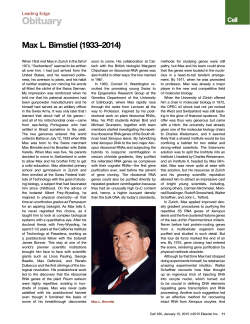


© Copyright 2026