
poster
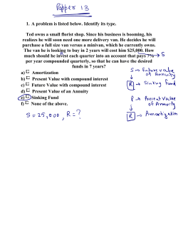
An#microbial studies of Marine Natural Product Mimics (MNPMs)
E. Mishchenko1, E. M. Igumnova2, H-M. Blencke1,E.G.A. Fredheim3, J. U. E. Sollid4,5, T. Haug1,5, M. B. Strøm2, K. Stensvåg1,5
1) Norwegian College of Fishery Science, Faculty of Biosciences, Fisheries and Economics (BFE), UiT The Arctic University of Norway, Breivika,
N-9037 Tromsø, Norway
Faculty of Health Science: 2) Department of Pharmacy, 3) Department of Clinical Medicine, 4) Department of Medical Biology
5) Centre for Research-based Innovation on marine bioactives and drug discovery (MabCent-CRI), BFE, UiT The Arctic University of Norway
INTRODUCTION
EFFECT ON BACTERIAL CELL MEMBRANE
Medical device-associated infections caused by staphylococcal biofilms
comprise a significant part of all cases of nosocomial infections. Staphylococcus
aureus and Staphylococcus epidermidis are among the leading causative agents
of bacteremia [1].
The intrinsic reduced antimicrobial susceptibility and rapid antibiotic resistance
development is a great treatment challenge of biofilm-associated infections [2].
Compound C affects membrane integrity of B. subtilis biosensor
Relative Luminecsence Units
Natural products (NP) from marine organisms have a unique structure and wide
spectrum of bioactivity [3]. Marine natural product mimics (MNPMs) with
improved characteristics compared to the original NPs could be promising antibiofilm agents effective against dormant and persister cells within a biofilm
matrix due to the specific mode of action.
120
2xMIC
1xMIC
100
0,5xMIC
80
Water
Chlorhexidine
60
40
20
0
Aim: To study the antimicrobial and anti-biofilm activity of the library of MNPMs
to reveal promising candidates for further development into novel therapeutics
0
50
100
Time, sec
S. epidermidis biofilms were
grown in chambered slides
and then incubated
with
compound C.
Treated biofilms were
examined using confocal
laser scanning microscope
(CLSM) [6] in combination
with fluorescent LIVE (SYTO
9) / DEAD (Propidium iodide)
staining.
Antimicrobial and anti-biofilm screening:
Panel of 6 test bacterial species
In vitro S. epidermidis biofilm models
Follow-up studies on selected compounds:
Mode of action
Anti-biofilm properties
/ ANTIMICROBIAL ACTIVITY OF SELECTED MNPMS
Minimal inhibitory concentration (MIC), µg/ml [4]
S. epidermidis RP62A
S. epidermidis 5179-R1
B. subtilis 168
E. coli ATCC 25922
P. aeruginosa PAO1
E
1,0
3,1
1,9
1,9
3,9
1,6
1,0
31,3 31,3 15,6
15,6 >125 62,5 15,6
7,8
7,8
7,8
3,9
6,3
3,9
3,9
Antibiotic control
0.5 (Vancomycin)
0.8 (Vancomycin)
0.6 (Erythromycin)
0.8 (Polymyxin B)
Alive biomass 0.4 (Polymyxin B)
Dead biomass and extracellular DNA MIC increase after serial passages through subMIC concentrations, µg/ml
S. epidermidis RP62A
Double
B. subtilis 168
No increase
A lot of dead biomass Bildetekst settes her…
Significant biomass eradica#on Compound C was selected for follow-up studies
Compound
C, 5 x MIC
Hemolytic activity (Human Red Blood Cells), EC50, µg/ml
61,4 176,0 >500 64,6 33,8
Untreated
control
Polysaccharide biofilm of RP62A
Selected test strains
Selected compounds
A
B
C
D
3,9 3,9 3,9
3,9
S. epidermidis biofilm killing and eradication by compound C Proteinateous biofilm of R1-5179
Library of > 130 MNPMs with molecular weights ≤ 500 Da
Compound C effectively penetrates through S. epidermidis biofilm.
Antibiotic disc
Compound C rapidly kills S. epidermidis in liquid culture
Biofilm
6
Compound C
5
4
3
2
1
0
control
1
2
Incubation time, hours
Compound C 1xMIC
3
Compound C 5xMIC
4
Vancomycin 5xMIC
S. epidermidis was treated with test compounds and aliquotes were plated for colony
forming units (CFU) counting over several time points.
Compound C causes membrane potential alterations in bacteria
E.coli S.epidermidis 6 10 5 Red/Green ra#o Red/Green ra#o 12 3 6 Bacteria were treated with
compound C and stained with
green fluorophore DiOC2(3)
which becomes red due to
cell membrane potential.
2 4 1 2 Nonetreated Compound C Compound C control 4 MIC 2 MIC Treatment 1.  Novel MNPMs with promising antimicrobial activity were identified
2.  The MNPMs were shown to be bactericidal and membranolytic.
3.  High in vitro efficiency against mature S. epidermidis biofilms was
revealed
References: 0 0 S. epidermidis biofilms were
grown on membrane filters
(pore size 0.2 µm).
Control strain (C. glutamicum)
was incubated on agar plates
with the test compounds
placed on top of biofilms.
Zones of growth inhibition
were compared to control
setups without biofilms [7].
4.  Further studies are needed to reveal the molecular targets of
selected MNPMs
4 8 Membrane
filter
/ CONCLUSION
EFFECT ON BACTERIAL CELL MEMBRANE
DiOC2 (3) Vancomycin
Log CFU/ml
200
/ ANTI-BIOFILM STUDIES
/ STRATEGY
0
150
B. subtilis luciferaseexpressing strain was
treated with compound C.
Light emission caused by
membrane disruption and
substrate penetration into
cells was measured in realtime [5].
Nonetreated Compound C Compound C control 2 MIC 1 MIC Treatment Changes in membrane potential lead to changes in red/green ratio.
Fluorescence intensities of bacterial populations were measured by flow cytometry.
1.  NORM/NORM-VET 2012. Usage of Antimicrobial Agents and Occurrence of Antimicrobial
Resistance in Norway. Tromsø / Oslo 2013. ISSN: 1890-9965.
2.  Hall-Stoodley, et al. Nat Rev Microbiol, 2004. 2(2): 95 - 108.
3.  Sperstad, S. V., et al. Biotech Adv, 2011. 29(4): 519-530.
4.  Haug, T., et al. Fish Shellfish Immun, 2002. 12(5): 371-385.
5.  Virta, M., et al. J Antimicrob Chemoth, 1995. 36(2): p. 303-315
6.  Christensen, B.B., et al. Molecular tools for study of biofilm physiology. In Methods
Enzymol. Academic Press, Inc. 1999. 20-42.
7.  Singh R et al. J. Antimicrob. Chemother. 2010. 65(9): 1955-1958


© Copyright 2026