
2014 NIA Standard Clinical Guidelines
2014 NIA Standard Clinical Guidelines Guidelines for Clinical Review Determination Preamble NIA is committed to the philosophy of supporting safe and effective treatment for patients. The medical necessity criteria that follow are guidelines for the provision of diagnostic imaging. These criteria are designed to guide both providers and reviewers to the most appropriate diagnostic tests based on a patient’s unique circumstances. In all cases, clinical judgment consistent with the standards of good medical practice will be used when applying the guidelines. Guideline determinations are made based on the information provided at the time of the request. It is expected that medical necessity decisions may change as new information is provided or based on unique aspects of the patient’s condition. The treating clinician has final authority and responsibility for treatment decisions regarding the care of the patient. Guideline Development Process These medical necessity criteria were developed by NIA for the purpose of making clinical review determinations for requests for diagnostic tests. The developers of the criteria sets included representatives from the disciplines of radiology, internal medicine, nursing, and cardiology. They were developed following a literature search pertaining to established clinical guidelines and accepted diagnostic imaging practices. All inquiries should be directed to: National Imaging Associates, Inc. 6950 Columbia Gateway Drive Columbia, MD 21046 Attn: NIA Associate Chief Medical Officer _______________________________________________________________ © 2000-2014 National Imaging Associates, Inc This document is the proprietary information of Magellan Health Services and its affiliates Page 2 of 451 TABLE OF CONTENTS TOC 22612/63030 – Lumbar Surgery __________________________________________________________ 6 62310-62311 – Spinal Epidural Injections _________________________________________________ 16 64490-64493 – Paravertebral Facet Joint Injections/Blocks ___________________________________ 22 64633-64635 – Cervical/Thoracic Facet Joint Neurolysis _____________________________________ 25 33225 – Cardiac Resynchronization Therapy (CRT)__________________________________________ 29 33249 – Implantable Cardioverter Defibrillator (ICD) ________________________________________ 37 33208 – Pacemaker __________________________________________________________________ 52 70336 – MRI Temporomandibular Joint (TMJ) _____________________________________________ 60 70450 – CT Head/Brain _______________________________________________________________ 62 70480 – CT Orbit (Includes Sella and Posterior Fossa) _______________________________________ 68 70480 – CT Internal Auditory Canal______________________________________________________ 70 70480 – CT Sella _____________________________________________________________________ 72 70486 – Face CT _____________________________________________________________________ 74 70486 – Maxillofacial/Sinus CT _________________________________________________________ 76 70490 – CT Soft Tissue Neck ___________________________________________________________ 80 70496 – CT Angiography, Head/Brain ____________________________________________________ 83 70498 – CT Angiography, Neck _________________________________________________________ 87 70540 – MRI Orbit ___________________________________________________________________ 89 70540 – MRI Face ___________________________________________________________________ 92 70540 – MRI Neck ___________________________________________________________________ 93 70544 – MR Angiography Head/Brain ____________________________________________________ 96 70547 – MR Angiography Neck _________________________________________________________ 99 70551 – MRI Brain (includes Internal Auditory Canal) ______________________________________ 102 70554 – Functional MRI Brain _________________________________________________________ 109 71250 – CT Chest (Thorax) ____________________________________________________________ 111 71275 – CT Angiography, Chest (non coronary) ___________________________________________ 116 71550 – MRI Chest (Thorax) __________________________________________________________ 119 71555 – MR Angiography Chest (excluding myocardium) ___________________________________ 122 72125 – CT Cervical Spine ____________________________________________________________ 126 72128 – CT Thoracic Spine ____________________________________________________________ 130 72131 – CT Lumbar Spine ____________________________________________________________ 134 72141 – MRI Cervical Spine ___________________________________________________________ 139 72146 – MRI Thoracic Spine __________________________________________________________ 144 72148 – MRI Lumbar Spine ___________________________________________________________ 148 72159 – MR Angiography Spinal Canal __________________________________________________ 153 72191 – CT Angiography, Pelvis________________________________________________________ 155 72192 – CT Pelvis ___________________________________________________________________ 158 72196 – MRI Pelvis _________________________________________________________________ 166 72198 – MR Angiography, Pelvis _______________________________________________________ 172 73200 – CT Upper Extremity (Hand, Wrist, Elbow, Long Bone or Shoulder) _____________________ 73206 – CT Angiography, Upper Extremity _______________________________________________ 73220 – MRI Upper Extremity _________________________________________________________ 73225 – MR Angiography Upper Extremity_______________________________________________ 73700 – CT Lower Extremity (Ankle, Foot, Hip or Knee) _____________________________________ 73706 – CT Angiography, Lower Extremity _______________________________________________ 73720 – MRI Lower Extremity (Ankle, Foot, Knee, Hip, Leg) _________________________________ 73725 – MR Angiography, Lower Extremity ______________________________________________ 74150 – CT Abdomen _______________________________________________________________ 74174 – CT Angiography, Abdomen and Pelvis ____________________________________________ 74175 – CT Angiography, Abdomen ____________________________________________________ 74176 – CT Abdomen and Pelvis Combo_________________________________________________ 74181 – MRI Abdomen ______________________________________________________________ 74185 – MR Angiography, Abdomen ___________________________________________________ 74261 – CT Colonoscopy Diagnostic (Virtual) _____________________________________________ 74263 - CT Colonoscopy Screening (Virtual) ______________________________________________ 75557 – MRI Heart__________________________________________________________________ 75571 – Electron Beam Tomography (EBCT)______________________________________________ 75572 – CT Heart & CT Heart Congenital ________________________________________________ 75574 – CTA Coronary Arteries (CCTA) __________________________________________________ 75635 – CT Angiography, Abdominal Arteries ____________________________________________ 76390 – MR Spectroscopy ____________________________________________________________ 76497 – Unlisted CT Procedure ________________________________________________________ 76498 – Unlisted MRI Procedure_______________________________________________________ 76805 – OB Ultrasound - Routine ______________________________________________________ 76811 – OB Ultrasound - Detailed ______________________________________________________ 76816 – OB Ultrasound - Monitoring ___________________________________________________ 76818 – Biophysical Profile ___________________________________________________________ 77058 – MRI Breast _________________________________________________________________ 77078 – CT Bone Density Studies ______________________________________________________ 77084 – MRI Bone Marrow ___________________________________________________________ 78205 – Liver SPECT_________________________________________________________________ 78320 – Bone and/or Joint SPECT ______________________________________________________ 78451 – Nuclear Cardiology/Myocardial Perfusion Imaging__________________________________ 78459 – PET Scan, Heart (Cardiac) _____________________________________________________ 78472 – MUGA Scan ________________________________________________________________ 78607 – Brain SPECT ________________________________________________________________ 78608 – PET Scan, Brain _____________________________________________________________ 78647 – Cerebrospinal Fluid Flow SPECT ________________________________________________ 78710 - Kidney SPECT _______________________________________________________________ 78813 – PET Scan ___________________________________________________________________ 93307 – Transthoracic Echocardiology (TTE) _____________________________________________ 93312 – Transesophageal Echocardiology (TEE) ___________________________________________ _______________________________________________________________ © 2000-2014 National Imaging Associates, Inc This document is the proprietary information of Magellan Health Services and its affiliates Page 4 of 451 176 183 186 192 194 200 203 209 212 221 225 229 239 245 249 251 252 262 263 272 284 286 288 289 290 292 293 303 308 313 316 318 320 321 334 342 345 347 351 352 354 360 371 93350 – Stress Echocardiography ______________________________________________________ 93452 – Heart Catheterization ________________________________________________________ 93925 – Lower Extremity Arterial Duplex Scan ____________________________________________ 93930 – Upper Extremity Arterial Duplex Scan ____________________________________________ 93970 – Extremity Venous Duplex Scan _________________________________________________ 93975 – Abdominal, Pelvis, Scrotal, Retroperitoneal Organ Duplex Scan _______________________ 93978 – Aorta, Inferior Vena Cava, Iliac Duplex Scan _______________________________________ 93980 – Penile Vessel Duplex Scan _____________________________________________________ 93990 – Hemodialysis Access Duplex Scan _______________________________________________ 94660 – Sleep Disorder Treatment Initiation and Management ______________________________ 95800 – Sleep Study – Unattended (Home Sleep Test) _____________________________________ 95811 – Sleep Study – Attended (Nocturnal Polysomnography) ______________________________ 0042T – Cerebral Perfusion CT ________________________________________________________ 0159T – CAD Breast MRI _____________________________________________________________ G0219 – PET Imaging whole body, melanoma - noncovered _________________________________ G0235 - PET imaging, any site, not otherwise specified _____________________________________ G0252 - PET imaging, initial diagnosis of breast cancer _____________________________________ S8037 – MR Cholangiopancreatography (MRCP) __________________________________________ S8042 – MRI Low Field_______________________________________________________________ _______________________________________________________________ © 2000-2014 National Imaging Associates, Inc This document is the proprietary information of Magellan Health Services and its affiliates Page 5 of 451 375 388 391 398 402 408 415 422 424 428 429 434 442 445 446 447 448 449 451 TOC 22612/63030 – Lumbar Surgery Last Review Date: August 2013 INTRODUCTION: This guideline outlines the key surgical treatments and indications for common lumbar spinal disorders and is a consensus document based upon the best available evidence. Spine surgery is a complex area of medicine and this document breaks out the treatment modalities for lumbar spine disorders into surgical categories: Lumbar Microdiscectomy, Lumbar Decompression, and Lumbar Fusion Surgery. See the additional information section for procedures considered not medically necessary. INTRODUCTION OF LUMBAR SURGERY A. Lumbar Microdiscectomy is a surgical procedure to remove part of the damaged spinal disc. The damaged spinal disc herniates into the spinal canal and irritates the nerve roots. Nerve root compression leads to symptoms like low back pain, radicular pain, numbness and tingling, muscular weakness, and paresthesia. Typical disc herniation pain is exacerbated with any movement that causes the disc to increase pressure on the nerve roots. B. Lumbar Decompression (Laminectomy, Facetectomy and Foraminotomy): Laminectomy is common decompression surgery. The American Association of Neurological Surgeons defines laminectomy as a surgery to remove the back part of vertebra, lamina, to create more space for the spinal cord and nerves. The most common indication for laminectomy is spinal stenosis. Spondylolisthesis and herniated disk are also frequent indications for laminectomy. Decompression surgery is usually performed as part of lumbar fusion surgery. C. Lumbar Fusion Surgery: Lumbar spinal fusion (arthrodesis) is a surgical procedure used to treat spinal conditions of the lumbar, e.g., degenerative disc disease, spinal stenosis, injuries/fractures of the spine, spinal instability, and spondylolisthesis. Spinal fusion is a “welding” process that permanently fuses or joins together two or more adjacent bones in the spine, immobilizing the vertebrae and restricting motion at a painful joint. It is usually performed after other surgical procedures of the spine, such as discectomy or laminectomy. The goal of fusion is to increase spinal stability, reduce irritation of the affected nerve roots, compression on the spinal cord, disability, and pain and/or numbness. Clinical criteria for single level fusion versus multiple level fusions are outlined under the indications section. INDICATIONS FOR LUMBAR & PRE-SACRAL SURGERY: This section of the clinical guidelines provides the clinical criteria each of the lumbar and pre-sacral spine surgery categories. A. Indications for Lumbar Microdiscectomy Surgical indications for inter-vertebral disc herniation*: Primary radicular symptoms noted upon clinical exam that hinders daily activities; AND _______________________________________________________________ © 2000-2014 National Imaging Associates, Inc This document is the proprietary information of Magellan Health Services and its affiliates Page 6 of 451 Failure to improve with at least six consecutive weeks of conservative treatment; AND Imaging studies showing evidence of inter-vertebral disc herniation *Other indications: Microdiscectomy may be used as the first line of treatment in the following clinical scenarios: Progressive nerve compression resulting in an acute neurologic deficit sensory or motor due to herniated disc; OR Cauda equina syndrome (loss of bowel or bladder control). NOTE: Percutaneous lumbar discectomy or radiofrequency disc decompression procedures are deemed investigational procedures and are not approved. B. Indications for Lumbar Decompression: Laminectomy, Facetectomy and Foraminotomy These procedures allow decompression by partial or total removal of various parts of vertebral bone and ligaments. Surgical Indications for spinal canal decompression due to lumbar spinal stenosis*: Low back pain, neurogenic claudication, and/or radicular leg pain that impairs daily activities for at least twelve (12) weeks; AND Failure to improve with at least 6 weeks of conservative therapy; AND Imaging findings consistent with clinical signs/symptoms; AND Imaging studies do not show evidence of spinal instability. *Other Indications: Lumbar decompression may be used as the first line of treatment in the following clinical scenarios: Progressive nerve compression resulting in an acute neurologic (sensory or motor) deficit. Cauda equina syndrome (loss of bowel or bladder control). Spinal stenosis due to tumor, infection, or trauma. C. Indications for Lumbar Spine Fusion: Single Level with or without decompression Because of variable outcomes with fusion surgery, patients should be actively involved in the decision-making process and provided appropriate decision-support materials when considering this intervention. The following indicators must be present*: Lumbar back pain, neurogenic claudication, and/or radicular leg pain without sensory or motor deficit that impairs daily activities for at least 6 months; AND Failure to improve with least 6-12 weeks of conservative, non-operative therapy; AND Imaging studies corresponding to the clinical findings; AND At least one of the following clinical conditions: a) Spondylolisthesis [Neural Arch Defect -Spondylolytic spondylolisthesis, degenerative spondylolisthesis, and congenital unilateral neural arch hypoplasia]; OR _______________________________________________________________ © 2000-2014 National Imaging Associates, Inc This document is the proprietary information of Magellan Health Services and its affiliates Page 7 of 451 b) Evidence of Segmental Instability -Excessive motion, as in degenerative spondylolisthesis, segmental instability, and surgically induced segmental instability; OR c) Revision surgery for failed previous operation(s) for pseudoarthorsis at the same level at least 6-12 months from prior surgery** if significant functional gains are anticipated; OR d) Revision surgery for failed previous operation(s) repeat disk herniations if significant functional gains are anticipated; OR e) Fusion for the treatment of spinal tumor, cancer, or infection; OR f) Chronic low back pain or degenerative disc disease must have failed at least 6 months of appropriate non-operative treatment (comprehensive rehabilitation) and must be evaluated on a case-by-case basis. *Other Indications: Lumbar spinal fusion may be used as the first line of treatment in the following clinical scenarios: Progressive nerve compression resulting in an acute neurologic deficit sensory or motor AND one of the aforementioned clinical conditions, except chronic low back pain or degenerative disc disease. Cauda equina syndrome (loss of bowel or bladder control) ** REPEAT LUMBAR SPINE FUSION OPERATIONS: Repeat lumbar fusion operations will be reviewed on a case-by-case basis upon submission of medical records and imaging studies that demonstrate remediable pathology. The below must also be documented and available for review of repeat fusion requests: Rationale as to why surgery is preferred over other non-invasive or less invasive treatment procedures. Signed documentation that the patient has participated in the decision-making process and understands the high rate of failure/complications. Instrumentation, bone formation or grafting materials, including biologics, should be used at the surgeon’s discretion; however, use should be limited to FDA approved devices or biologics and indications. NOTE: Pre-sacral, axial lumbar interbody fusion (AxiaLIF) is not an approved surgical approach due to insufficient evidence. Pre-Sacral Fusion Codes: 0195T, +0196T, 22586, 0309T. Artificial lumbar disc replacement or other lumbar implants are not an approved procedure due to insufficient evidence Lumbar Artificial Disc Replacement/Implant Codes: 22857, +0163T, 22862, +0164T, 22865, +0165T, 0221T, +0222T D. Indications for multi-level fusions with or without decompression All multi-level fusion surgeries will be reviewed on a case-by-case basis. Because of variable outcomes with fusion surgery, patients should be actively involved in the decision-making process and provided appropriate decision-support materials when considering this intervention. The following clinical indications must be present*: _______________________________________________________________ © 2000-2014 National Imaging Associates, Inc This document is the proprietary information of Magellan Health Services and its affiliates Page 8 of 451 Lumbar back pain, neurogenic claudication, and/or radicular leg pain without sensory or motor deficit that impairs daily activities for at least 6 months; AND Failure to improve with least 6-12 weeks of conservative, non-operative therapy; AND Imaging studies corresponding to the clinical findings; AND At least one of the following clinical conditions: a) Multiple Level Spondylolisthesis; OR b) Fusion for the treatment of spinal tumor, trauma, cancer, or infection affecting multiple levels; OR c) Intra-Operative Segmental Instability *Other Indications: Lumbar spinal fusion may be used as the first line of treatment in the following clinical scenarios: Progressive nerve compression resulting in an acute neurologic deficit (sensory or motor) AND one of the aforementioned clinical conditions. Instrumentation, bone formation or grafting materials, including biologics, should be used at the surgeon’s discretion; however, use should be limited to FDA approved devices or biologics and indications. This lumbar surgery guideline does not address spinal deformity surgeries or the clinical indications for spinal deformity surgery [CPT codes 22800-22812]. NOTE: Pre-sacral, axial lumbar interbody fusion (AxiaLIF) is not an approved surgical approach due to insufficient evidence. Pre-Sacral Fusion Codes: 0195T, +0196T, 22586, 0309T. Artificial lumbar disc replacement or other lumbar implants are not an approved procedure due to insufficient evidence Lumbar Artificial Disc Replacement/Implant Codes: 22857, +0163T, 22862, +0164T, 22865, +0165T, 0221T, +0222T. CONTRAINDICATIONS FOR SPINE SURGERY Medical contraindications to surgery, e.g., severe osteoporosis; infection of soft tissue adjacent to the spine, whether or not it has spread to the spine; severe cardiopulmonary disease; anemia; malnutrition and systemic infection Psychosocial risk factors. It is imperative to rule out non-physiologic modifiers of pain presentation or non-operative conditions mimicking radiculopathy or instability (e.g., peripheral neuropathy, piriformis syndrome, myofascial pain, sympathetically mediated pain syndromes, sacroiliac dysfunction, psychological conditions, etc.) prior to consideration of elective surgical intervention. Active Tobacco use prior to fusion surgery. It is recommended that the patient refrain from smoking for at least six weeks prior to surgery and during the period of fusion healing. Morbid Obesity. Contraindication to surgery in cases where there is significant risk and concern for improper post-operative healing, post-operative complications related to morbid obesity, and/or an inability to participate in post-operative rehabilitation. ADDITIONAL INFORMATION _______________________________________________________________ © 2000-2014 National Imaging Associates, Inc This document is the proprietary information of Magellan Health Services and its affiliates Page 9 of 451 Services Not Covered: The following procedures are considered are either still under investigation or are not recommended based upon the current evidence: Percutaneous lumbar discectomy; Laser discectomy; Percutaneous Radiofrequency Disc Decompression; intradiscal electrothermal annuloplasty (IDEA) or more commonly called IDET (Intradiscal Electrothermal therapy); Nucleus Pulpous Replacement; Pre-Sacral Fusion, or Lumbar Artificial Disc Replacement. PERCUTANEOUS DISCECTOMY is an invasive operative procedure to accomplish partial removal of the disc through a needle which allows aspiration of a portion of the disc trocar under imaging control. Percutaneous discectomy is rarely indicated. It is sometimes useful in suspected septic discitis or in order to obtain diagnostic tissue. Percutaneous discectomy is not recommended for contained disc herniations or bulges with associated radiculopathy, due to lack of evidence to support long-term improvement. This includes radiofrequency disc decompression. LASER DISCECTOMY is a procedure which involves the delivery of laser energy into the center of the nucleus pulposus using a fluoroscopically guided laser fiber under local anesthesia. The energy denatures protein in the nucleus, causing a structural change which is intended to reduce intradiscal pressure. Its effectiveness has not been fully established. INTRADISCAL ELECTROTHERMAL ANNULOPLASTY (IDEA) (more commonly called IDET, or Intradiscal Electrothermal therapy) is an outpatient non-operative procedure in which a wire is guided into the identified painful disc using fluoroscopy. The wire is then heated at the nuclearannular junction within the disc. Physicians performing this procedure must have been trained in the procedure and certified. Surgical Indications: Failure of conservative therapy including physical therapy, medication management, or therapeutic injections. Indications may include those with chronic low back pain, disc related back pain, or pain lasting for greater than 6 months. There is conflicting evidence regarding its effectiveness. NUCLEUS PULPOSUS REPLACEMENT Involves the introduction of a prosthetic implant into the intervertebral disc, replacing the nucleus pulposus while preserving the annulus fibrosus. INDICATIONS: Nucleus Pulposus Replacement is limited to investigational use in the United States at this time and is not recommended LUMBAR ARTIFICIAL DISC REPLACEMENT: Involves the insertion of a prosthetic device into an intervertebral space from which a degenerated disc has been removed, sparing only the peripheral annulus. The prosthetic device is designed to distribute the mechanical load of the vertebrae in a physiologic manner and maintain range of motion. Studies do not demonstrate a long-term advantage of measured function or pain over comparison groups undergoing fusion. _______________________________________________________________ © 2000-2014 National Imaging Associates, Inc This document is the proprietary information of Magellan Health Services and its affiliates Page 10 of 451 The longevity of this prosthetic device has not yet been determined. Lumbar Artificial Disc Replacement Codes: 22857, +0163T, 22862, +0164T, 22865, +0165T, 0221T, +0222T Conservative Therapy: (musculoskeletal) includes a combination of modalities, such as rest, ice, heat, modified activities, medical devices, (such as crutches, immobilizer, metal braces, orthotics, rigid stabilizer or splints, etc and not to include neoprene sleeves), medications, chiropractic treatments, physician supervised home exercise program. Part of this combination may include the physician instructing patient to rest the area or stay off the injured part. NOTE - conservative therapy can be expanded to require active therapy components (physical therapy and/or physician supervised home exercise) as noted in some elements of the guideline. Home Exercise Program - (HEP) – the following two elements are required to meet guidelines for completion of conservative therapy: o Information provided on exercise prescription/plan AND o Follow up with member with information provided regarding completion of HEP (after suitable 4-6 week period), or inability to complete HEP due to physical reason- i.e. increased pain, inability to physically perform exercises. (Patient inconvenience or noncompliance without explanation does not constitute “inability to complete” HEP). Claims Billing & Coding: NIA uses a combination of internally developed edits in addition to an enhanced set of industry standard editing. NIA’s Claims Edit Module is a group of system edits that run multiple times per day. Edits that are part of this module include industry standard edits that apply to spine surgery services and NIA custom edits developed specifically for spine surgery. The following describes each of the edits NIA applies: Outpatient Code Editor (OCE): This edit performs all functions that require specific reference to HCPCS codes, HCPCS modifiers, and ICD-9-CM diagnosis codes. The OCE only functions on a single claim and does not have any cross claim capabilities. NIA is consistent with CMS. National Correct Coding Initiative (NCCI) editing: The edit prevents improper payment when incorrect code combinations are reported. The NCCI contains two tables of edits. The Column One/Column Two Correct Coding Edits table and the Mutually Exclusive Edits table include code pairs that should not be reported together for a number of reasons explained in the Coding Policy Manual. NIA is consistent with CMS. − Incidental edits: This edit applies if a procedure being billed is a component of another procedure that occurred on the same date of service for the same provider and tax ID and claimant. _______________________________________________________________ © 2000-2014 National Imaging Associates, Inc This document is the proprietary information of Magellan Health Services and its affiliates Page 11 of 451 − Mutually exclusive editing: This edit applies if a procedure being billed is mutually exclusive with a procedure that occurred on the same date of service for the same provider tax ID and claimant. Multiple Procedure Discounts (MPD): This edit applies a reduction to the second and any other subsequent services by the same provider, in the same setting, for the same member. We typically apply a 50% reduction. NIA follows the CMS methodology that began in January 2011 which allows for application of MPD to codes within CMS’s two specific advanced imaging code families. However, NIA differs from CMS in that we apply MPD to all provider types unless health plan contracts prohibit this. Lumbar Fusion Fusions can be performed either anteriorly, laterally, or posteriorly, or via a combined approach; although simple posterolateral fusions are indicated in the great majority of cases requiring fusion. These are the surgical approaches: Intertransverse Fusion or Posterolateral Fusion Anterior Interbody Fusion (ALIF) Lateral or Transpsoas Interbody Fusion (XLIF) Posterior or Trans-foraminal Interbody Fusion (PLIF or TLIF) Anterior/posterior Fusion (360-degree) Pre-sacral, axial lumbar interbody fusion (AxiaLIF) is still being investigated and is not recommended. Use of bone grafts including autologous or allograft which might be combined with metal or biocompatible devices to produce a rigid, bony connection between two or more adjacent vertebrae are common. Bone formation or grafting materials including biologics should be used at the surgeon’s discretion; however, use of biologics should be limited to FDA approved indications in order to limit complications (especially BMP). All operative interventions must be based upon positive correlation of clinical findings, clinical course, and diagnostic tests. A comprehensive assimilation of these factors must lead to a specific diagnosis with positive identification of pathologic condition(s). It is imperative to rule out non-physiologic modifiers of pain presentation or non-operative conditions mimicking radiculopathy or instability (e.g., peripheral neuropathy, piriformis syndrome, myofascial pain, sympathetically mediated pain syndromes, sacroiliac dysfunction, psychological conditions, etc.) prior to consideration of elective surgical intervention. Operative treatment is indicated when the natural history of surgically treated lesions is better than the natural history for non-operatively treated lesions. _______________________________________________________________ © 2000-2014 National Imaging Associates, Inc This document is the proprietary information of Magellan Health Services and its affiliates Page 12 of 451 All patients being considered for surgical intervention should first undergo a comprehensive neuro-musculoskeletal examination to identify mechanical pain generators that may respond to non-surgical techniques or may be refractory to surgical intervention. While sufficient time allowances for non-operative treatment are required to determine the natural cause and response to non-operative treatment of low back pain disorders, timely decision making for operative intervention is critical to avoid de-conditioning and increased disability (exclusive of "emergent" or urgent pathology such as cauda equina syndrome or associated rapidly progressive neurologic loss). In general, if the program of non-operative treatment fails, operative treatment is indicated when: Improvement of the symptoms has plateaued or failed to occur and the residual symptoms of pain and functional disability are unacceptable at the end of 6 to 12 weeks of active treatment, or at the end of longer duration of non-operative programs for debilitated patients with complex problems; and/or Frequent recurrences of symptoms cause serious functional limitations even if a non-operative active treatment program provides satisfactory relief of symptoms, and restoration of function on each recurrence. Lumbar spinal stenosis and associated lumbar spondylolisthesis Spinal stenosis is narrowing of the spinal column or of the neural foramina where spinal nerves leave the spinal column, causing pressure on the spinal cord. The most common cause is degenerative changes in the lumbar spine. Neurogenic claudication is the most common symptom, referring to “leg symptoms encompassing the buttock, groin and anterior thigh, as well as radiation down the posterior part of the leg to the feet.”i In addition to pain, leg symptoms can include fatigue, heaviness, weakness and/or paresthesia. Some patients may also suffer from accompanying back pain. Symptoms are worse when standing or walking and are relieved by sitting. Lumbar spinal stenosis is often a disabling condition, and it is the most common reason for lumbar spinal surgery in adults over 65 years. Degenerative lumbar spondylolisthesis is the displacement of a vertebra in the lower part of the spine; one lumbar vertebra slips forward on another with an intact neural arch and begins to press on nerves. The slippage occurs at the L4-L5 level most commonly. The most common cause, in adults, is degenerative disease although it may also result from bone diseases and fractures. Spondylolisthesis seldom occurs before the age of 50 years and it disproportionately affects women, especially black women. Degenerative spondylolisthesis is not always symptomatic. Lumbar degenerative disease without stenosis or spondylolisthesis Spondylosis is an umbrella term describing age-related degeneration of the spine. Lumbar degenerative disease without stenosis or spondylolisthesis is characterized by disabling low back pain and spondylosis at L4-5, L5-S1, or both levels. _______________________________________________________________ © 2000-2014 National Imaging Associates, Inc This document is the proprietary information of Magellan Health Services and its affiliates Page 13 of 451 REFERENCES American Pain Society. (2009). American Pain Society’s Guideline for Interventional Procedures for Low Back Pain Published in Spine. Retrieved from http://www.ampainsoc.org/press/2009/downloads/20090513.pdf. Atlas, S.J., Keller, R.B., Wu, Y.A., Deyo, R.A., & Singer, D.E. (2005). Long-term outcomes of surgical and nonsurgical management of lumbar spinal stenosis: 8 to 10 year results from the Maine lumbar spine study. Spine, 30, 936-43. [PMID: 15834339] Retrieved from http://www.ncbi.nlm.nih.gov/pubmed/15834339 Bogduk, N., & Andersson, G. (2009). Is spinal surgery effective for back pain? F1000 Med Rep., 1, 60. doi: 10.3410/M1-60. Brox, I.J., Sorensen, R., Friis, A., Nyygaard, O., Indahl, A., Keller, A., … Reikeras, O. (2003). Randomized clinical trial of lumbar instrumented fusion and cognitive intervention and exercises in patients with chronic low back pain and disc degeneration. Spine, 28(17), 1913-1921. Retrieved from http://www.ncbi.nlm.nih.gov/pubmed/12973134 Carreon, L.Y., Glassman, S.D., & Howard, J. (2008). Fusion and nonsurgical treatment for symptomatic lumbar degenerative disease: A systematic review of Oswestry Disability Index and MOS Short Form36 outcomes. The Spine Journal, 8, 747-755. Retrieved from http://www.thespinejournalonline.com/article/S1529-9430(07)00269-0/abstract Chou, R., Baisden, J., Carragee, E.J., Resnick, D.K., Shaffer, W.O., & Loeser, J.D. (2009). Surgery for low back pain: A review of the evidence for an American Pain Society Clinical Practice Guideline. Spine, 34(10), 1094-109. doi: 10.1097/BRS.0b013e3181a105fc. Deyo, R.A., Mirza, S.K., Martin, B.I., Kreuter, W., Goodman, D.C., & Jarvik, J.G. (2010). Trends, major medical complications, and charges associated with surgery for lumbar spinal stenosis in older adults. JAMA, 303(13), 1259-1265. doi: 10.1001/jama.2010.338. Fardon, D.R., & Milette, P.C. (2001). Nomenclature and classification of lumbar disc pathology: Recommendations of the combined task forces of the North American Spine Society, American Society of Spine Radiology, and American Society of Neuroradiology. Spine, 26(5), E93-E113. Retrieved from http://www.ncbi.nlm.nih.gov/pubmed/?term=Fardon+DR%2C+Milette+PC.+Nomenclature+and+cla ssification+of+lumbar+disc+pathology%3A+recommendations+of+the+combined+task+forces+of+th e+North+Americvan+Spine+Society%2C+American+Society+of+Spine+Radiology%2C+and+American +Society+of+Neuroradiology.+Spine+2001%3B+26(5)%3AE93-E113 Fritzell, P., Wessberg, P., & Nordwall, A. (2001). Swedish Lumbar Spine Study Group: Lumbar fusion versus nonsurgical treatment for chronic low back pain – A multicenter randomized controlled trial from the Swedish Lumbar Spine Study Group. Spine, 26(23), 2521-32. Retrieved from http://www.ncbi.nlm.nih.gov/pubmed/11725230 _______________________________________________________________ © 2000-2014 National Imaging Associates, Inc This document is the proprietary information of Magellan Health Services and its affiliates Page 14 of 451 Genevay, S., & Atlas, S.J. (2010). Lumbar spinal stenosis. Best Pract Res Clin Rheumatol, 24(2), 253-265. doi: 10.1016/j.berh.2009.11.001. North American Spine Society (NASS). (2008). Clinical Guidelines for Multidisciplinary Spine Care: Diagnosis and Treatment of Degenerative Lumbar Spondylolisthesis. Retrieved from http://www.spine.org/Documents/Spondylolisthesis_Clinical_Guideline.pdf North American Spine Society (NASS). (2011). Clinical Guidelines for Multidisciplinary Spine Care: Diagnosis and Treatment of Degenerative Lumbar Spinal Stenosis. Retrieved from http://www.spine.org/Documents/NASSCG_stenosis.pdf Peul, W.C., van Houwelingen, H.C., van den Hout, W.B., Brand R., Eekhof, J.A., Tans, J.T., … Leiden-The Hague Spine Intervention Prognostic Study Group. (2007). Surgery versus prolonged conservative treatment for sciatica. N Engl J Med., 356, 2245-56. doi: 10.1056/NEJMoa064039. Resnick, D.K., Choudhri, T.F., Dailey, A.T., Groff, M.W., Khoo, L., Matz, P.G., … Hadley, M.N. (2005). Guidelines for the performance of fusion procedures for degenerative disease of the lumbar spine. Part 7: Intractable low-back pain without stenosis or spondylolisthesis. J Neurosurg: Spine, 2, 670672. Retrieved from http://thejns.org/doi/abs/10.3171/spi.2005.2.6.0670?url_ver=Z39.882003&rfr_id=ori:rid:crossref.org&rfr_dat=cr_pub%3dpubmed Tosteson, A.N.A., Tosteson,T.D., Lurie, J.D., Abdu, W., Herkowitz, H., Andersson, G., … Weinstein, J.N. (2011). Comparative effectiveness evidence from the spine patient outcomes research trial: surgical versus nonoperative care for spinal stenosis, degenerative spondylolisthesis, and intervertebral disc herniation. Spine, 36(24), 2061-2068. doi: 10.1097/BRS.0b013e318235457b. Tosteson, A.N.A., Lurie, J.D., Tosteson, T.D., Skinner, J.S., Hertowitz, H., Albert, T., … Weinstein, J.N. (2008). Surgical treatment of spinal stenosis with and without degenerative spondylolisthesis: Costeffectiveness after 2 years. Ann Intern Med, 149, 845-853. Retrieved from http://www.ncbi.nlm.nih.gov/pmc/articles/PMC2658642 Weinstein, J.N., Lurie, J.D., Tosteson, T.D., Hanscom, B., Tosteson, A.N.A., Blood E.A., … Hu, S.S. (2007). Surgical versus nonsurgical treatment for lumbar degenerative spondylolisthesis. N Engl J Med., 356, 2257-2270. doi:10.1056/NEJMoa070302. _______________________________________________________________ © 2000-2014 National Imaging Associates, Inc This document is the proprietary information of Magellan Health Services and its affiliates Page 15 of 451 TOC 62310-62311 – Spinal Epidural Injections Last Review Date: March 2013 INTRODUCTION: Therapeutic Spinal Epidural Injections or Select Nerve Root Blocks (Transforaminal) are types of interventional pain management procedures. The therapeutic use of epidural injections is for shortterm pain relief associated with acute back pain or exacerbation of chronic back pain. With therapeutic injections a corticosteroid is injected close to the target area with the goal of pain reduction. Epidural injections should be used in combination with other conservative treatment modalities and not as stand alone treatment for long-term back pain relief. There are different approaches used when administering spinal epidural injections: Interlaminar epidural injections, with steroids, access the epidural space between two vertebrae (Interlaminar) to treat cervical, lumbar or thoracic pain with radicular pain. These procedures should be performed using fluoroscopic guidance. Interlaminar epidural injections are the most common type of epidural injection. Transforaminal epidural injections (also called selective nerve root blocks) access the epidural space via the intervertebral foramen where the spinal nerves exit (cervical, lumbar or thoracic region). It is used both diagnostically and therapeutically. Some studies report lack of evidence and risks of transforaminal epidural injections. These procedures are always aided with fluoroscopic guidance. Caudal epidural injections, with steroids, are used to treat back and lower extremity pain, accessing the epidural space through the sacral hiatus, providing access to the lower nerve roots of the spine. These procedures should be performed using fluoroscopic guidance. Failed back surgery syndrome is the most common reason for the caudal approach. The rationale for the use of spinal epidural injections is that the sources of spinal pain, e.g., discs and joints, are accessible and amendable to neural blockade. Interventional pain management procedures must be performed by licensed interventional pain physicians only. Medical necessity management for epidural injections includes an initial evaluation including history and physical examination and a psychosocial and functional assessment. The following must be determined: nature of the suspected organic problem; non-responsiveness to conservative treatment; level of pain and functional disability; conditions which may be contraindications to epidural injections; and responsiveness to prior interventions. Interventional pain management specialists do not agree on how to diagnose and manage spinal pain; there is a lack of consensus with regards to the type and frequency of spinal interventional techniques for treatment of spinal pain. The American Society of Interventional Pain Physicians (ASIPP) guidelines and International Spine Intervention Society (ISIS) guidelines provide an algorithmic approach which provides a step-by-step procedure for managing chronic spinal pain based upon evidence-based guidelines. It is based on the structural basis of spinal pain and incorporates acceptable evidence of diagnostic and therapeutic interventional techniques available in managing chronic spinal pain. _______________________________________________________________ © 2000-2014 National Imaging Associates, Inc This document is the proprietary information of Magellan Health Services and its affiliates Page 16 of 451 The guidelines and algorithmic approach referred to above include the evaluation of evidence for diagnostic and therapeutic procedures in managing chronic spinal pain and recommendations for managing spinal pain. The Indications and Contraindications presented within this document are based on the guidelines and algorithmic approach. Prior to performing this procedure, shared decision-making between patient and physician must occur, and patient must understand the procedure and its potential risks and results (moderate short-term benefits, and lack of long-term benefits). INDICATIONS FOR EPIDURAL INJECTIONS OR SELECTIVE NERVE BLOCKS (caudal, interlaminar, and transforaminal) (Injection of local anesthetics with corticosteroids) o Back pain and/or extremity pain resulting from any of the following conditions and associated timeframes: o Disc herniation with radiculitis: after 2 weeks or more of acute back pain involving radiculopathy that has failed to respond or poorly responded to conservative management; o Failed back surgery syndrome or Epidural fibrosis typically not done immediately post-surgery : no sooner than 6 months post surgery patient must engage in some form of conservative treatment for a minimum of 6 weeks prior to epidural injections o Lumbar spinal stenosis; patient must engage in some form of conservative treatment for a minimum of 6 weeks prior to epidural injections OR o Discogenic low back pain/degenerative disc disease; patient must engage in some form of conservative treatment for a minimum of 6 weeks prior to epidural injections AND o Average pain levels of ≥ 6 on a scale of 0 to 10 or Intermittent or continuous pain causing functional disability. FREQUENCY OF REPEAT THERAPEUTIC INJECTIONS Epidural injections may be repeated only as medically necessary and with proof that: prior injection had a positive response by significantly decreasing pain; the patient continues to have ongoing pain or documented functional disability (≥ 6 on a scale of 0 to 10); AND The patient is actively engaged in other forms of conservative non-operative treatment (unless pain prevents the patient from participating in conservative therapy); AND Injections meet the following criteria: o There must be at least 14 days between injections; o No more than 3 procedures in a 12-week period of time per region; o Limited to a maximum total of 6 procedures per region per 12 months. Course of treatment, three epidural injections, regardless of approach must provide at least o > 50% pain relief obtained for a minimum of 6 weeks to be considered a positive and effective response. _______________________________________________________________ © 2000-2014 National Imaging Associates, Inc This document is the proprietary information of Magellan Health Services and its affiliates Page 17 of 451 If the neural blockade is applied for different regions (cervical and thoracic regions are considered as one region and lumbar and sacral are considered as one region), injections may be administered at intervals of no sooner than 14 days for most types of procedures. Injecting multiple regions or performing multiple procedures during the same visit may be deemed medically unnecessary unless documentation is provided outlining an unusual situation. CONTRAINDICATIONS FOR EPIDURAL INJECTIONS o Bleeding diathesis and full anticoagulation (risk of epidural hematoma); o Severe spinal stenosis resulting in intraspinal obstruction; o Local infection at injection site; o Predominantly psychogenic pain; o Sepsis; o Hypovolemia; o Pregnancy; o Uncontrolled diabetes; o Uncontrolled glaucoma; o High concentrations of local anesthetics in patients with multiple sclerosis; o For diagnosis or treatment of facet mediated pain; o Known or suspected allergic reaction to steroid medications; o Spinal infection; o Malignancy; OR o Acute fracture ADDITIONAL INFORMATION: Additional Terminology: Interlaminar Epidural; Selective Nerve Root Injection (transforaminal only); Transforaminal Injection; Injections of Spinal Canal Conservative Therapy: (musculoskeletal) includes a combination of modalities, such as rest, ice, heat, modified activities, medical devices, (such as crutches, immobilizer, metal braces, orthotics, rigid stabilizer or splints, etc and not to include neoprene sleeves), medications, diathermy, chiropractic treatments, or physician supervised home exercise program. Part of this combination may include the physician instructing patient to rest the area or stay off the injured part. NOTE - conservative therapy can be expanded to require active therapy components (physical therapy and/or physician supervised home exercise) as noted in some elements of the guideline. Home Exercise Program - (HEP) – the following two elements are required to meet guidelines for completion of conservative therapy: o Information provided on exercise prescription/plan AND _______________________________________________________________ © 2000-2014 National Imaging Associates, Inc This document is the proprietary information of Magellan Health Services and its affiliates Page 18 of 451 o Follow up with member with information provided regarding completion of HEP (after suitable 4-6 week period), or inability to complete HEP due to physical reason- i.e. increased pain, inability to physically perform exercises. (Patient inconvenience or noncompliance without explanation does not constitute “inability to complete” HEP). Hip-spine syndrome Hip-spine syndrome is a condition that includes both debilitating hip osteoarthritis and low back pain. Abnormal spinal sagittal alignment and difficulty in maintaining proper balance, as well as a wobbling gait, may be caused by severe osteoarthritis of the hip joint. Epidural injections are used to determine a primary pain generator in this condition. Spondylolisthesis and nerve root irritation Degenerative lumbar spondylolisthesis is the displacement of a vertebra in the lower part of the spine; one lumbar vertebra slips forward on another with an intact neural arch and begins to press on nerves. The most common cause, in adults, is degenerative disease although it may also result from bone diseases and fractures. Degenerative spondylolisthesis is not always symptomatic. Epidural injections may be used to determine a previously undocumented nerve root irritation as a result of spondylolisthesis. Lumbar spinal stenosis with radiculitis Spinal stenosis is narrowing of the spinal column or of the neural foramina where spinal nerves leave the spinal column, causing pressure on the spinal cord. The most common cause is degenerative changes in the lumbar spine. Neurogenic claudication is the most common symptom, referring to “leg symptoms encompassing the buttock, groin and anterior thigh, as well as radiation down the posterior part of the leg to the feet.” In addition to pain, leg symptoms can include fatigue, heaviness, weakness and/or paresthesia. Some patients may also suffer from accompanying back pain. Symptoms are worse when standing or walking and are relieved by sitting. Lumbar spinal stenosis is often a disabling condition, and it is the most common reason for lumbar spinal surgery in adults over 65 years. The most common levels of stenosis are L3 through L5, but it may occur at multilevels in some patients. Radiculitis is the inflammation of a spinal nerve root that causes pain to radiate along the nerve paths. Epidural injections help to ascertain the level of the pain generator in this condition. Postoperative epidural fibrosis Epidural fibrosis is a common cause of failed back surgery syndrome. With the removal of a disc, the mechanical reason for pain may be removed, but an inflammatory condition may continue after the surgery and may cause pain. Epidural corticosteroids, with their anti-inflammatory properties, are used to treat postoperative fibrosis and may be used along with oral Gabapentin to reduce pain. Lumbar herniated disc Epidural steroid injections have been proven to be effective at reducing symptoms of lumbar herniated discs. Evidence shows that they can be successful in 42% to 56% of patients who do not improve after 6 _______________________________________________________________ © 2000-2014 National Imaging Associates, Inc This document is the proprietary information of Magellan Health Services and its affiliates Page 19 of 451 weeks of conservative treatment. Observation and epidural steroid injection are effective nonsurgical treatments for this condition. Failed back surgery syndrome Failed back surgery syndrome (FBSS) is characterized by persistent or recurring low back pain, with or without sciatica, following lumbar surgery. The most common cause of FBSS is epidural fibrosis which can be triggered by a surgical procedure such as discectomy. The inflammation resulting from the surgical procedure may start the process of fibrosis and cause pain. Epidural steroid injections are administered to reduce pain. Discogenic pain Discogenic pain is predominant low back pain without disc herniation. 80% to 90% of low back pain is commonly believed to be of unknown etiology. The term, discogenic disc disease, may refer to degenerative disc disease or to internal disc disruption syndrome. Patients with the latter condition may have painful invertebral discs despite minimal degenerative changes. In the U.S., discogenic pain accounts for 25% of cases of chronic low back pain. Evidence has shown that epidural steroid injections are effective for short-term improvement of discogenic pain. REFERENCES Boswell MV, Trescot AM, Datta S, et al. Interventional techniques: evidence-based practice guidelines in the management of chronic spinal pain. Pain Physician 2007; 10:7-111. Chou R, Atlas SJ, Stanos SP. Nonsurgical interventional therapies for low back pain: a review of the evidence for an American Pain Society Clinical Practice Guideline. Spine 2009; 34(10): 1078-1093. Datta S, Everett CR, Trescot AM, et al. An updated systematic review of the diagnostic utility of selective nerve root blocks. Pain Physician 2007; 10:113-128. DePalma MJ, Slipman CW. Evidence-informed management of chronic low back pain with epidural steroid injections. The Spine Journal 2008:8:45-55. Genevay S, Atlas SJ. Lumbar spinal stenosis. Best Pract Res Clin Rheumatol 2010; 24(2): 253-265. Goodman BS, Posecion LWF, Mallempati S, et al. Complications and pitfalls of lumbar interlaminar and transforaminal epidural injections. Curr Rev Musculoskelet Med 2008; 1:212-222. Huston CW. Cervical epidural steroid injections in the management of cervical radiculitis: interlaminar versus transforaminal. A Review. Curr Rev Musculoskelet Med 2009; 2(1):30-42. Institute for Clinical Systems Improvement (ICSI). Adult Acute and Subacute Low Back Pain Fifteenth Edition/January 2012. www.icsi.org _______________________________________________________________ © 2000-2014 National Imaging Associates, Inc This document is the proprietary information of Magellan Health Services and its affiliates Page 20 of 451 Manchikanti L, Singh V, Cash KA, et al. Management of pain of post lumbar surgery syndrome: one-year results of a randomized, double-blind, active controlled trial of fluoroscopic caudal epidural injections. Pain Physician 2010; 13:509-521. Manchikanti L, Boswell MV, Singh V, et al. Comprehensive evidence-based guidelines for interventional techniques in the management of chronic spinal pain. Pain Physician 2009; 12:699-802. Mendoza-Lattes S, Weiss A, Found E, et al. Comparable effectiveness of caudal vs. transforaminal epidural steroid injections. Iowa Orthop J 2009; 29:91-96. North American Spine Society. Evidence-Based Clinical Guidelines for Multidisciplinary Spine Care: Diagnosis and Treatment of Degenerative Lumbar Spinal Stenosis; 2011 Revised. www.spine.org ISBN 1-929988-29-X Parr AT, Diwan S, Abdi S. Lumbar interlaminar epidural injections in managing chronic low back and lower extremity pain: a systematic review. Pain Physician 2009; 12:163-188. _______________________________________________________________ © 2000-2014 National Imaging Associates, Inc This document is the proprietary information of Magellan Health Services and its affiliates Page 21 of 451 TOC 64490-64493 – Paravertebral Facet Joint Injections/Blocks Last Review Date: March 2013 INTRODUCTION: Facet joints (also called zygapophysial joints or z-joints), posterior to the vertebral bodies in the spinal column and connecting the vertebral bodies to each other, are located at the junction of the inferior articular process of a more cephalad vertebra and the superior articular process of a more caudal vertebra. These joints provide stability and enable movement, allowing the spine to bend, twist, and extend in different directions. They also restrict hyperextension and hyperflexion. Facet joints are clinically important spinal pain generators in patients with chronic spinal pain. In patients with chronic low back pain, facet joints have been implicated as a cause of the pain in 15% to 45% of patients. Facet joints are considered as the cause of chronic spinal pain in 48% of patients with thoracic pain and 54% to 67% of patients with chronic neck pain. Facet joints may refer pain to adjacent structures, making the underlying diagnosis difficult as referred pain may assume a pseudoradicular pattern. Lumbar facet joints may refer pain to the back, buttocks, and lower extremities while cervical facet joints may refer pain to the head, neck and shoulders. Imaging findings are of little value in determining the source and location of ‘facet joint syndrome’, a term originally used by Ghormley and referring to back pain caused by pathology at the facet joints. Imaging studies may detect changes in facet joint architecture, but correlation between radiologic findings and symptoms is unreliable. Although clinical signs are also unsuitable for diagnosing facet joint-mediated pain, they may be of value in selecting patients for controlled local anesthetic blocks of either the medial branches or the facet joint itself. This is an established tool in diagnosing facet joint syndrome. The most common source of chronic pain is the spine and about two-thirds of the U.S. population suffers from spinal pain sometime during their life span. Facet joint interventions are used in the treatment of pain in certain patients with a confirmed diagnosis of facet joint pain. Interventions include intraarticular injections and medial branch nerve blocks in the lumbar, cervical and thoracic spine. Prior to performing this procedure, shared decision-making between patient and physician must occur, and patient must understand the procedure and its potential risks and results. Facet joint injections or medial branch nerve blocks require guidance imaging. INDICATIONS FOR FACET JOINT INJECTIONS OR MEDIAL BRANCH NERVE BLOCKS To confirm disabling non-radicular low back (lumbosacral) or neck (cervical) pain, suggestive of facet joint origin as documented in the medical record based upon all of the following: − (a) history, consisting of mainly axial or non-radicular pain, and − (b) physical examination, with positive provocative signs of facet disease (pain exacerbated by extension and rotation, or associated with lumbar rigidity). Lack of evidence, either for discogenic or sacroiliac joint pain; AND Lack of disc herniation or evidence of radiculitis; AND _______________________________________________________________ © 2000-2014 National Imaging Associates, Inc This document is the proprietary information of Magellan Health Services and its affiliates Page 22 of 451 Intermittent or continuous pain with average pain levels of ≥ 6 on a scale of 0 to 10 or functional disability; AND Duration of pain of at least 2 months; AND Failure to respond to conservative non-operative therapy management. All procedures must be performed using guidance (Fluro, CT, or Ultrasound). FREQUENCY OF FACET BLOCK There must be a minimum of 14 days between injections There must be a positive response of ≥ 50% pain relief and improved ability to perform previously painful movements Maximum of 3 procedures per region every 6 months. If the procedures are applied for different regions (cervical and thoracic regions are considered as one region and lumbar and sacral are considered as one region), they may be performed at intervals of no sooner than 2 weeks for most types of procedures. Maximum of 3 levels injected on same date of service. Radiofrequency Neurolysis procedures should be considered in patients with positive facet blocks (with at least 50% pain relief and ability to perform prior painful movements without any significant pain). CONTRAINDICATIONS FOR FACET JOINT INJECTIONS o History of allergy to contrast administration, local anesthetics, steroids, or other drugs potentially utilized; o Hypovolemia; o Infection over puncture site; o Bleeding disorders or coagulopathy; History of allergy to medications to be administered; o Inability to obtain percutaneous access to the target facet joint; o Progressive neurological disorder which may be masked by the procedure; o Pregnancy; o Spinal infection; OR o Acute Fracture ADDITIONAL INFORMATION: Additional Terminology: Facet Injections; Facet Joint Blocks; Paravertebral Facet Injections; Paravertebral Facet Joint Injections; Paravertebral Facet Joint Nerve Injections; Zygapophyseal injections; Lumbar Facet Blockade; Medial Branch blocks Conservative Therapy: (musculoskeletal) includes a combination of modalities, such as rest, ice, heat, modified activities, medical devices, (such as crutches, immobilizer, metal braces, orthotics, rigid stabilizer or splints, etc and not to include neoprene sleeves), medications, diathermy, chiropractic treatments, or physician supervised home exercise program. Part of this combination may include the physician instructing patient to rest the area or stay off the injured part. NOTE - conservative therapy can be expanded to require active therapy components (physical therapy and/or physician supervised home exercise) as noted in some elements of the guideline. _______________________________________________________________ © 2000-2014 National Imaging Associates, Inc This document is the proprietary information of Magellan Health Services and its affiliates Page 23 of 451 Home Exercise Program - (HEP) – the following two elements are required to meet guidelines for completion of conservative therapy: o Information provided on exercise prescription/plan AND o Follow up with member with information provided regarding completion of HEP (after suitable 4-6 week period), or inability to complete HEP due to physical reason- i.e. increased pain, inability to physically perform exercises. (Patient inconvenience or noncompliance without explanation does not constitute “inability to complete” HEP). REFERENCES Atluri S, Datta S, Falco FJE, et al. Systematic review of diagnostic utility and therapeutic effectiveness of thoracic facet joint interventions. Pain Physician 2008; 11:611-629. Binder DS, Nampiaparampil DE. The provocative lumbar facet joint. Curr Rev Musculoskelet Med 2009; 2:15-24. Bogduk N. A narrative review of intraarticular corticosteroid injections for low back pain. Pain Med 2005; 6:287-296. Datta S, Lee M, Falco FJ, et al. Systematic assessment of diagnostic accuracy and therapeutic utility of lumbar facet joint interventions. Pain Physician 2009; 437-460. Falco FJE, Erhart S, Wargo BW et al. Systematic review of diagnostic utility and therapeutic effectiveness of cervical facet joint interventions. Pain Physician 2009; 12:323-344. Manchikanti L, Singh V, Falco FJE, et al. Evaluation of lumbar facet joint nerve blocks in managing chronic low back pain: a randomized, double-blind, controlled trial with a 2-year follow-up. Int J Med Sci 2010; 7(3):124-135. Manchikanti L, Boswell MV, Singh V, et al. Prevalence of facet joint pain in chronic spinal pain of cervical, thoracic, and lumbar regions. BMC Musculoskeletal Disorders 2004; 5:15. Manchikanti L, Boswell MV, Singh V, et al. Comprehensive evidence-based guidelines for interventional techniques in the management of chronic spinal pain. Pain Physician 2009; 12:699-802. Manchikanti L, Pampati V, Singh V, et al. Explosive growth of facet joint interventions in the medicare population in the United states: a comparative evaluation of 1997, 2002, and 2006 data. BMC Health Serv Res 2010; 10:84. _______________________________________________________________ © 2000-2014 National Imaging Associates, Inc This document is the proprietary information of Magellan Health Services and its affiliates Page 24 of 451 TOC 64633-64635 – Cervical/Thoracic Facet Joint Neurolysis Last Review Date: March 2013 INTRODUCTION: Facet joints (also called zygapophysial joints or z-joints), posterior to the vertebral bodies in the spinal column and connecting the vertebral bodies to each other, are located at the junction of the inferior articular process of a more cephalad vertebra and the superior articular process of a more caudal vertebra. These joints provide stability and enable movement, allowing the spine to bend, twist, and extend in different directions. They also restrict hyperextension and hyperflexion. Facet joints are clinically important spinal pain generators in patients with chronic spinal pain. Pain mediated by the facet joints may be caused by repetitive stress and/or cumulative low-level trauma resulting in osteoarthritis and inflammation.ii In patients with chronic low back pain, facet joints have been implicated as a cause of the pain in 15% to 45% of patients. They are considered as the cause of chronic spinal pain in 48% of patients with thoracic pain and 54% to 67% of patients with chronic neck pain.iii Facet joints may refer pain to adjacent structures, making the underlying diagnosis difficult as referred pain may assume a pseudoradicular pattern. Lumbar facet joints may refer pain to the back, buttocks, and proximal lower extremities while cervical facet joints may refer pain to the head, neck and shoulders. Imaging findings are of little value in determining the source and location of ‘facet joint syndrome’, a term originally used by Ghormley and referring to back pain caused by pathology at the facet joints. Imaging studies may detect changes in facet joint architecture, but correlation between radiologic findings and symptoms is unreliable. Although clinical signs are also unsuitable for diagnosing facet joint-mediated pain, they may be of value in selecting patients for controlled local anesthetic blocks of either the medial branches or the facet joint itself. This is an established tool in diagnosing facet joint syndrome. Facet joints are known to be a source of pain with definitive innervations. Interventions used in the treatment of patients with a confirmed diagnosis of facet joint pain include: medial branch nerve blocks in the lumbar, cervical and thoracic spine; and radiofrequency neurolysis (see also additional terminology). The medial branch of the primary dorsal rami of the spinal nerves has been shown to be the primary innervations of facet joints. Substance P, a physiologically potent neuropeptide considered to play a role in the nociceptive transmission of nerve impulses, is found in the nerves within the facet joint. Radiofrequency neurolysis is a minimally invasive treatment for cervical, thoracic and lumbar facet joint pain. It involves using energy in the radiofrequency range to cause necrosis of specific nerves (medial branches of the dorsal rami), preventing the neural transmission of pain.iv The objective of radiofrequency neurolysis is to both provide relief of pain and reduce the likelihood of recurrence. Used most often for facet joint pain, radiofrequency neurolysis is recently emerging for sacroiliac joint pain. However, it has been shown to have limited evidence in treating sacroiliac joint pain and is considered investigational and not medically necessary. _______________________________________________________________ © 2000-2014 National Imaging Associates, Inc This document is the proprietary information of Magellan Health Services and its affiliates Page 25 of 451 Members of the American Society of Anesthesiologists (ASA) and the American Society of Regional Anesthesia and Pain Medicine (ASRA) have agreed that conventional or thermal radiofrequency ablation of the medial branch nerves to the facet joint should be performed for neck or low back pain. Radiofrequency neurolysis has been employed for over 30 years to treat facet joint pain. Prior to performing this procedure, shared decision-making between patient and physician must occur, and patient must understand the procedure and its potential risks and results. INDICATIONS FOR THERAPEUTIC FOR PARAVERTEBRAL FACET JOINT DENERVATION (RADIOFREQUENCY NEUROLYSIS) (local anesthetic block followed by the passage of radiofrequency current to generate heat and coagulate the target medial branch nerve) Positive response to controlled local anesthetic blocks of the facet joint, with at least 50% pain relief and ability to perform prior painful movements without significant pain, but with insufficient sustained relief (less than 2-3 months relief); OR Positive response to prior radiofrequency neurolysis procedures with at least 50% pain improvement for up to 6 months of relief in past 12 months; AND The presence of the following: o Lack of evidence that the primary source of pain being treated is from discogenic pain, sacroiliac joint pain, disc herniation or radiculitis; o Intermittent or continuous facet-mediated pain [average pain levels of ≥ 6 on a scale of 0 to 10] causing functional disability; o Duration of pain of at least 3 months; AND o Failure to respond to more conservative non-operative management FREQUENCY: Relief typically lasts between 6 and 12 months and sometimes provides relief for greater than 2 years. Repeat radiofrequency denervation is performed for sustained relief up to two and three times. Limit to 2 facet neurolysis procedures every 12 months, per region CONTRAINDICATIONS FOR PARAVERTEBRAL FACET JOINT DENERVATION (RADIOFREQUENCY NEUROLYSIS) o o o o o History of allergy to local anesthetics or other drugs potentially utilized; Lumbosacral radicular pain (dorsal root ganglion); Conditions/diagnosis for which procedure is used are other than those listed in Indications; Absence of positive diagnostic blocks; OR For any nerve other than the medial branch nerve. _______________________________________________________________ © 2000-2014 National Imaging Associates, Inc This document is the proprietary information of Magellan Health Services and its affiliates Page 26 of 451 ADDITIONAL INFORMATION: Additional Terminology: Paravertebral Facet Joint Denervation, Radiofrequency Neurolysis, Destruction Paravertebral Facet Joint Nerve, Facet Joint Rhizotomy, Facet Neurolysis, Medial Branch Radiofrequency Neurolysis, Medial Branch Radiofrequency Neurotomy or Radiofrequency Denervation Conservative Therapy: (musculoskeletal) includes a combination of modalities, such as rest, ice, heat, modified activities, medical devices, (such as crutches, immobilizer, metal braces, orthotics, rigid stabilizer or splints, etc and not to include neoprene sleeves), medications, , diathermy, chiropractic treatments, physician supervised home exercise program. Part of this combination may include the physician instructing patient to rest the area or stay off the injured part. NOTE - conservative therapy can be expanded to require active therapy components (physical therapy and/or physician supervised home exercise) as noted in some elements of the guideline. Home Exercise Program - (HEP) – the following two elements are required to meet guidelines for completion of conservative therapy: o Information provided on exercise prescription/plan AND o Follow up with member with information provided regarding completion of HEP (after suitable 4-6 week period), or inability to complete HEP due to physical reason- i.e. increased pain, inability to physically perform exercises. (Patient inconvenience or noncompliance without explanation does not constitute “inability to complete” HEP). REFERENCES American Society of Anesthesiologists Task Force on Chronic Pain Management, American Society of Regional Anesthesia and Pain Medicine. Practice guidelines for chronic pain management: an updated report by the American Society of Anesthesiologist Task Force on Chronic Pain Management and the American Society of Regional Anesthesia and Pain Medicine. Anesthesiology 2010; 112(4):810-33. http://www.asahq.org/Search.aspx?q=facet+radiofrequency&site=All. Binder DS, Nampiaparampil DE. The provocative lumbar facet joint. Curr Rev Musculoskelet Med 2009; 2:15-24. Boswell MV, Colson JD, Spillane WF. Therapeutic facet joint interventions in chronic spinal pain: a systematic review of effectiveness and complications. Pain Physician 2005; 8:101-114. Bogduk N. International spinal injection society guidelines for the performance of spinal injection procedures. Part 1: zygapophysial joint blocks. Clin J Pain 1997; 13(4):285-302. Chou R, Atlas SJ, Stanos SP, Rosenquist RW. Nonsurgical interventional therapies for low back pain: a review of the evidence for an American Pain Society clinical practice guideline. Spine (Phila Pa 1976). 2009; 34(10):1078-1093. Datta S, Lee M, Falco FJ, et al. Systematic assessment of diagnostic accuracy and therapeutic utility of lumbar facet joint intervention. Pain Physician 2009; 12:437-460. _______________________________________________________________ © 2000-2014 National Imaging Associates, Inc This document is the proprietary information of Magellan Health Services and its affiliates Page 27 of 451 Henschke N, Kuijpers T, Rubinstein S. Injection therapy and denervation procedures for chronic lowback pain: a systematic review. Eur Spine J 2010; 19:1425-1449. Muhlner SB. Review article: radiofrequency neurotomy for the treatment of sacroiliac joint syndrome. Curr Rev Musculoskelet Med 2009; 2:10-14. _______________________________________________________________ © 2000-2014 National Imaging Associates, Inc This document is the proprietary information of Magellan Health Services and its affiliates Page 28 of 451 TOC 33225 – Cardiac Resynchronization Therapy (CRT) Last Review Date: February 2013 INTRODUCTION: Pacemakers are implantable devices indicated for the treatment of slow heart rhythms (bradycardia) and, less commonly, for decreased heart muscle strength (cardiomyopathy). They are also very rarely used for the treatment of rapid heart rates (tachycardia) or hypertrophic cardiomyopathy. Dual chamber devices have been established to be beneficial for the vast majority of patients in terms of quality of life and incidence of congestive heart failure and atrial fibrillation, and they have become standard of care in most patients without permanent atrial fibrillation. The majority of the patients with dilated cardiomyopathy received implantable defibrillators with cardiac resynchronization therapy (CRT) capability, but pacemakers are sometimes chosen due to patient and physician preference. In order to identify if CRT is appropriate for a specific patient, CRT requires separate authorization. Approximately one third of patients who receive ICDs are also candidates for cardiac resynchronization therapy (CRT) because of congestive heart failure (CHF) and an abnormally wide QRS. CRT typically requires three leads, one each to pace the right and left ventricles, and a third to pace the atrium. This allows near-simultaneous stimulation (resynchronization) of both ventricles. CRT improves cardiac function and quality of life and decreases cardiac events and mortality among appropriately chosen patients. The improved survival in patients with CRT are greater than that provided by ICD insertion alone. Criteria for CRT are based on a 2012 focused update of the ACC/AHA/HRS 2008 ICD guideline. This guideline supports approval of ICD and CRT indications that are classed as IIb or higher. Relevant considerations are assigning designations I, IIa, and IIb are LVEF, QRS pattern and duration, and whether atrial fibrillation is present. INDICATIONS AND CONTRAINDICATIONS FOR PACEMAKERS BY CONDITION Cardiac Resynchronization Therapy (CRT): (Note: If CRT is indicated, use of an ICD with CRT should be considered). o LVEF <35%, sinus rhythm, LBBB with a QRS >119 ms, and NYHA class II, III, or ambulatory IV symptoms on GDMT (guideline-directed medical therapy). Also consider ICD with CRT. o LVEF <35%, sinus rhythm, a non-LBBB pattern with a QRS duration >120 ms, and NYHA class III/ambulatory class IV symptoms on GDMT. o Atrial fibrillation and LVEF <35% on GDMT if a) the patient requires ventricular pacing or otherwise meets CRT criteria and b) AV nodal ablation or pharmacologic rate control allows near 100% ventricular pacing with CRT. o LVEF <35%, on GDMT, with planned new or replacement device placement with anticipated requirement for (40%) ventricular pacing. o LVEF <30%, ischemic etiology of heart failure, sinus rhythm, LBBB with a QRS duration >150 ms, and NYHA class I symptoms on GDMT. o LVEF <35%, sinus rhythm, a non-LBBB pattern with a QRS duration >150 ms, and NYHA class II. _______________________________________________________________ © 2000-2014 National Imaging Associates, Inc This document is the proprietary information of Magellan Health Services and its affiliates Page 29 of 451 Contraindications for Cardiac Resynchronization Therapy (CRT): o NYHA class I or II symptoms and non-LBBB pattern with QRS duration <150 ms. o Comorbidities and/or frailty expected to limit survival to <1 year. Pacing for Sinus Node Dysfunction: o Symptomatic bradycardia, which includes syncope, near-syncope, dizziness, lethargy, congestive heart failure (CHF), fatigue, or dyspnea, whether spontaneous or as a result of clinically indicated medications or procedures (e.g. medical or catheter treatment for atrial fibrillation) that intentionally slow the heart rate, documented by EKG or telemetry. o Symptomatic heart beat pauses, documented by EKG or telemetry. o Chronotropic incompetence, documented by stress test or telemetry. o Heart rate less than 40 with symptoms consistent with bradycardia. o Syncope with electrophysiologic study (EPS) findings of abnormal sinus node function. Contraindications for Sinus Node Dysfunction: o Asymptomatic. o Symptoms in the absence of bradycardia. o Bradycardia resulting from nonessential drug therapy. Pacing for Acquired Third-Degree and Advanced Second-Degree Atrioventricular Block: o Persistent third-degree atrioventricular block, with or without symptoms o In atrial fibrillation and while awake, pauses in heartbeat ≥ 5 seconds with or without symptoms. o In sinus rhythm and while awake, pauses in heartbeat ≥ 3 seconds or heart rates less than 40 beats per minute or an escape rhythm below the AV node, with or without symptoms. o Following catheter ablation of the AV junction. o Following cardiac surgery, if expected to be permanent. o In neuromuscular diseases such as myotonic muscular dystrophy, Erb dystrophy (limb-girdle muscular dystrophy), Kearns-Sayre syndrome, and peroneal muscular atrophy. o Exercise-induced heart block without myocardial ischemia. Contraindications for Acquired Third-Degree and Advanced Second-Degree Atrioventricular Block: o AV block is expected to resolve and is unlikely to recur (e.g. drug toxicity, Lyme disease, or transient increases in vagal tone or during hypoxia in sleep apnea syndrome) and without symptoms. o AV block secondary to nonessential drug therapy. Pacing for Other Presentations of First- and Second-Degree AV Block: o Symptomatic second-degree AV block. o Type II second-degree AV block, with or without symptoms. o Second-degree AV block due to EP-documented intra- or infra-His levels. o First- or second-degree AV block with “pacemaker syndrome” symptoms or hemodynamic compromise (i.e. hypotension, syncope and pulmonary edema). _______________________________________________________________ © 2000-2014 National Imaging Associates, Inc This document is the proprietary information of Magellan Health Services and its affiliates Page 30 of 451 o In neuromuscular diseases such as myotonic muscular dystrophy, Erb dystrophy (limb-girdle muscular dystrophy), Kearns-Sayre syndrome, and peroneal muscular atrophy. o AV block due to drug use and/or drug toxicity AND block is expected to recur after drug withdrawal. o Exercise-induced second degree heart block without myocardial ischemia. Contraindications for Other Presentations of First- and Second-Degree AV Block: o AV block is expected to resolve and is unlikely to recur (e.g. drug toxicity, Lyme disease, or transient increases in vagal tone or during hypoxia in sleep apnea syndrome) and without symptoms. o AV Block secondary to nonessential drug therapy. Permanent Pacing for Chronic Bifascicular Block: o Type II second-degree AV block, advanced second-degree AV block (see definitions section) or intermittent third-degree AV block. o Alternating bundle-branch block. o Syncope and bifascicular block when other likely causes have been excluded, specifically ventricular tachycardia. o Electrophysiologic study (EPS) documentation of an H-V interval >100 milliseconds, even in asymptomatic patients. o Electrophysiologic study (EPS) documentation of non-physiological, pacing-induced infra-His block. o In neuromuscular diseases such as myotonic muscular dystrophy, Erb dystrophy (limb-girdle muscular dystrophy), and peroneal muscular atrophy with bifascicular block or any fascicular block. Contraindications for Permanent Pacing for Chronic Bifascicular Block: o Asymptomatic fascicular block without AV block. o Asymptomatic fascicular block with first-degree AV block. Permanent Pacing After the Acute Phase of Myocardial Infarction: o Persistent second- or third-degree AV block after STEMI. o Transient second- or third-degree AV block below the AV node after STEMI. If the site of block is uncertain, electrophysiologic study (EPS) may be necessary. Contraindications for Permanent Pacing After the Acute Phase of Myocardial Infarction: o Bradycardia secondary to nonessential drug therapy. o Transient AV block without intraventricular conduction defects. o Transient AV block with isolated left anterior fascicular block. o New bundle-branch block or fascicular block without AV block. o Asymptomatic first-degree AV block with bundle-branch or fascicular block. Permanent Pacing in Hypersensitive Carotid Sinus Syndrome and Neurocardiogenic Syncope: o Recurrent syncope due to spontaneously occurring carotid sinus stimulation AND carotid sinus pressure induces ventricular asystole ≥3 seconds. _______________________________________________________________ © 2000-2014 National Imaging Associates, Inc This document is the proprietary information of Magellan Health Services and its affiliates Page 31 of 451 o Syncope without clear, provocative events and with a hypersensitive cardioinhibitory response (asystole) of 3 seconds or longer. o Neurocardiogenic syncope associated with bradycardia occurring spontaneously or at the time of tilt-table testing. Contraindications for Permanent Pacing in Hypersensitive Carotid Sinus Syndrome and Neurocardiogenic Syncope: o Hypersensitive cardioinhibitory response to carotid sinus stimulation without symptoms or with vague symptoms. o Situational neurocardiogenic syncope in which avoidance behavior is effective and preferred. Pacing following Cardiac Transplantation: o Persistent inappropriate or symptomatic bradycardia not expected to resolve and for all other indications for permanent pacing. o Prolonged bradycardia limiting rehabilitation or discharge. o Syncope after transplantation even when bradyarrhythmia has not been documented. Contraindications for Pacing following Cardiac Transplantation: o Bradycardia secondary to nonessential drug therapy. Permanent Pacemakers That Automatically Detect and Pace to Terminate Tachycardia: o Symptomatic recurrent supraventricular tachycardia documented to be pacing terminated in the setting of failed catheter ablation and/or drug treatment or intolerance. Contraindications for Permanent Pacemakers That Automatically Detect and Pace to Terminate Tachycardia: o Presence of an accessory pathway with capacity for rapid anterograde conduction. Pacing to Prevent Tachycardia: o Sustained pause-dependent VT, with or without QT prolongation. o High-risk congenital long-QT syndrome. o Symptomatic, drug-refractory, recurrent atrial fibrillation in patients with coexisting Sinus Node Dysfunction (SND). Contraindications for Pacing to Prevent Tachycardia: o Ventricular ectopic without sustained VT in the absence of the long-QT syndrome. o Reversible, e.g., drug-related, Torsade de pointes VT. Pacing in Patients with Hypertrophic Cardiomyopathy: o Symptomatic hypertrophic cardiomyopathy and hemodynamically significant resting or provoked LV outflow tract obstruction AND refractory to medical therapy. Contraindications for Pacing in Patients with Hypertrophic Cardiomyopathy: o Asymptomatic OR symptoms controlled on medical therapy. o Without significant LV outflow tract obstruction. _______________________________________________________________ © 2000-2014 National Imaging Associates, Inc This document is the proprietary information of Magellan Health Services and its affiliates Page 32 of 451 Pacing in Children, Adolescents, and Patients with Congenital Heart Disease: o Second- or third-degree AV block with symptomatic bradycardia, ventricular dysfunction, or low cardiac output. o SND with symptoms and age-inappropriate bradycardia. The definition of bradycardia varies with the patient’s age and expected heart rate. For normal heart rates by age, please see the table at the end. o Postoperative advanced second- or third-degree AV block that is expected to be permanent or that persists >7 days after cardiac surgery. o Congenital third-degree AV block with a wide QRS escape rhythm, complex ventricular ectopy, or ventricular dysfunction. o Congenital third-degree AV block in the infant with a ventricular rate <55 bpm or with congenital heart disease and a ventricular rate <70 bpm. o Congenital heart disease and sinus bradycardia for the prevention of recurrent episodes of intraatrial reentrant tachycardia, either intrinsic or secondary to anti-arrhythmic treatment. o Congenital third-degree AV block after age 1 year with an average heart rate <50 bpm, abrupt pauses in ventricular rate that are 2 or 3 times the basic cycle length, or associated with symptoms due to chronotropic incompetence. o Sinus bradycardia with complex congenital heart disease AND a resting heart rate < 40 bpm OR pauses in ventricular rate >3 seconds. o Congenital heart disease and impaired hemodynamics due to sinus bradycardia or loss of AV synchrony. o Unexplained syncope after prior congenital heart surgery complicated by transient complete heart block, with residual fascicular block after a careful evaluation to exclude other causes of syncope. o Transient postoperative third-degree AV block that reverts to sinus rhythm with residual bifascicular block. o Permanent pacemaker implantation may be considered for congenital third-degree AV block in asymptomatic children or adolescents with an acceptable rate, a narrow QRS complex and normal ventricular function. o Asymptomatic sinus bradycardia following biventricular repair of congenital heart disease with a resting heart rate < 40 bpm or pauses in ventricular rate > 3 seconds. Contraindications for Pacing in Children, Adolescents, and Patients with Congenital Heart Disease: o Asymptomatic transient postoperative AV block with return of normal AV conduction. o Asymptomatic bifascicular block +/-first-degree AV block after surgery for congenital heart disease in the absence of prior transient complete AV block. o Asymptomatic type I second-degree AV block. o Asymptomatic sinus bradycardia with the longest RR interval < 3 seconds and a minimum heart rate > 40 bpm. o Bradycardia secondary to nonessential drug therapy. _______________________________________________________________ © 2000-2014 National Imaging Associates, Inc This document is the proprietary information of Magellan Health Services and its affiliates Page 33 of 451 ADDITIONAL INFORMATION: Appropriate use criteria have not been established for pacemaker insertion. Rather, clinicians rely upon ACC/AHA/HRS guidelines, which were updated for bradycardia indications in 2008. A focused guideline update was published in 2012, which considered Left ventricular ejection fraction (LVEF), QRS pattern, QRS duration, and consideration regarding the presence of atrial fibrillation in its differentiation between classes, I, IIa, and IIb indications. A pacemaker system is composed of a pulse generator and one or more leads. The pulse generator is implanted under the skin, usually below one of the collarbones. It contains a battery, a microprocessor that governs timing and function, and a radio antenna to allow for noninvasive reprogramming. The leads are insulated cables that conduct electricity from the pulse generator to the heart. Leads are most commonly inserted into a vein and then advanced under fluoroscopy (X-ray guidance) to within one or more heart chambers. The leads are fastened within the chambers to the heart muscle using either hooks or retractable/extendable screws, which are built into their tips. Timed electrical impulses are sent from the pulse generator down the leads to the heart, where stimulation results in heart muscle contraction. The most recent guidelines stress that asymptomatic bradycardia rarely qualifies as a class I indication for pacemaker insertion. However, there are some asymptomatic bradycardic rhythms for which pacemaker insertion is indicated because they present a risk of injury or death. In addition, there are also a small number of situations in which the electrocardiogram (EKG) or an invasive electrophysiologic study (EPS) can reveal evidence of specific disease in the cardiac conduction system that warrants pacemaker insertion in the absence of symptoms, for the same reason. Guidelines are fairly specific and technical in these instances. In the case dilated cardiomyopathy, near-simultaneous stimulation of both ventricles, referred to as cardiac resynchronization therapy (CRT) has been demonstrated to improve cardiac performance and quality of life and to decrease cardiac event rates and mortality among a subset of patients. Device implantation requires the insertion of leads that pace both the right and left ventricles, most commonly with a coronary sinus lead for the LV pacing. The majority of these patients received implantable defibrillators with CRT capability, but pacemakers are sometimes chosen due to patient and physician preference. A focused ACCF/AHA/HRS guideline update was published in 2012, which considered LVEF, QRS pattern, QRS duration, and consideration regarding the presence of atrial fibrillation in its differentiation between classes, I, IIa, and IIb indications. This document will provide criteria for approval of all CRT indications that are presently defined as IIb or stronger. Current guidelines group pacemaker indications together according to anatomic source and clinical syndromes, and this document follows this approach. Class I through IIb indications are condensed and included as approvable in this document. Generally speaking, for indications that are listed in this summary without reference to symptoms, the presence or absence of symptoms differentiate between class I and II indications. _______________________________________________________________ © 2000-2014 National Imaging Associates, Inc This document is the proprietary information of Magellan Health Services and its affiliates Page 34 of 451 NYHA Class Definitions: Class I: No limitation of functional activity or only at levels of exertion that would limit normal individuals. Class II: Slight limitation of activity. Dyspnea and fatigue with moderate exercise. Class III: Marked limitation of activity. Dyspnea with minimal activity. Class IV: Severe limitation of activity. Symptoms even at rest. Heart Block Definitions: First Degree: All atrial beats are conducted to the ventricles, but with a delay of > 200ms. Second Degree: Intermittent failure of conduction of single beats from atrium to ventricles. o Type I: Conducted beats have variable conduction times from atrium to ventricles. o Type II: Conducted beats have uniform conduction times from atrium to ventricles. o Advanced: Two or more consecutive non-conducted beats. Third Degree: No atrial beats are conducted from atrium to ventricle Abbreviations: AV = Atrioventricular CHF = congestive heart failure CRT = Cardiac resynchronization therapy EKG = Electrocardiogram EPS = Electrophysiologic Study GDMT = Guideline-Directed Medical Therapy HRS = Heart Rhythm Society HV = His-ventricle ICD = Implantable cardioverter-defibrillator LBBB = left bundle-branch block LV = Left ventricular/left ventricle LVEF = Left ventricular ejection fraction MI = myocardial infarction MS = milliseconds NYHA =New York Heart Association STEMI = ST-elevation Myocardial Infarction SND = Sinus node dysfunction VT = Ventricular tachycardia Normal Pediatric Heart Rates: From: www.pediatriccareonline.org/pco/ub/view/Pediatric-DrugLookup/153929/0/normal_pediatric_heart_rates Age Mean Heart Rate (beats/minute) Heart Rate Range (2nd – 98th percentile) <1 d 123 93-154 1-2 d 123 91-159 3-6 d 129 91-166 1-3 wk 148 107-182 _______________________________________________________________ © 2000-2014 National Imaging Associates, Inc This document is the proprietary information of Magellan Health Services and its affiliates Page 35 of 451 1-2 mo 149 121-179 3-5 mo 141 106-186 6-11 mo 134 109-169 1-2 y 119 89-151 3-4 y 108 73-137 5-7 y 100 65-133 8-11 y 91 62-130 12-15 y 85 60-119 Adapted from The Harriet Lane Handbook, 12th ed, Greene MG, ed, St Louis, MO: Mosby Yearbook, 1991 REFERENCES Antman, E.M., Anbe, D.T., Armstrong, P.W., Bates, E.R., Green, L.R., Hand, M. … Smith, S.C. (2004). ACC/AHA guide- lines for the management of patients with ST-elevation myocardial infarction: a report of the American College of Cardiology/American Heart Association Task Force on Practice Guidelines (Committee to Revise the 1999 Guidelines for the Management of patients with acute myocardial infarction). J Am Coll Cardiol. 44 e1–e211. Retrieved from http://www.med.umich.edu/AnesCriticalCare/Documents/Guidelines/Am%20Col%20Cardio%20Fou nd/ST%20elv%20MI.pdf Dolgin, M. (1994). The Criteria Committee of the New York Heart Association. Nomenclature and Criteria for Diagnosis of Diseases of the Heart and Great Vessels. 9th ed. (pp. 253-256). Boston, Mass: Little, Brown & Co. Epstein, A.E., DiMarco, J.P., Ellenbogen, K.A., Estes, M., Freedman, R.A., Gettes, L.S., … Sweeney, M.O. (2008). ACC/AHA/HRS 2008 Guidelines for Device-Based Therapy of Cardiac Rhythm Abnormalities: Executive Summary. Journal of the American College of Cardiology, 51(21), 2085-2105. doi:10.1016/j.jacc.2008.02.032 Greene, M.G. (1991). The Harriet Lane Handbook, 12th ed. St Louis, MO: Mosby Yearbook. Retrieved from www.pediatriccareonline.org/pco/ub/view/Pediatric-DrugLookup/153929/0/normal_pediatric_heart_rates Tracy, C.M., Epstein, A.E., Darbar, D., DiMarco, J.P., Dunbar, S.B., Estes, M. … Varosy, P.D. (2012). ACCF/AHA/HRS Focused Update of the 2008 Guidelines for Device-Based Therapy of Cardiac Rhythm Abnormalities. Journal of the American College of Cardiology, 60(14), 1297-1313. doi:10.1016/j.jacc.2012.07.009 _______________________________________________________________ © 2000-2014 National Imaging Associates, Inc This document is the proprietary information of Magellan Health Services and its affiliates Page 36 of 451 TOC 33249 – Implantable Cardioverter Defibrillator (ICD) Last Review Date: February 2013 INTRODUCTION: Implantable cardioverter defibrillators (ICDs) are indicated for the treatment of life-threatening ventricular tachycardia and ventricular fibrillation. An ICD system includes a pulse generator and one or more leads. ICDs are indicated both for patients who have survived life threatening rhythm disturbances (secondary prevention) and for those who are at risk for them (primary prevention). Most ICD implantations are for primary prevention in patients with ischemic cardiomyopathy. Studies published in the last decade have confirmed improved survival in patient with reduced left ventricular ejection fraction (LVEF) even when no cardiac arrhythmias have been noted. Approximately one third of patients who receive ICDs are also candidates for cardiac resynchronization therapy (CRT) because of congestive heart failure (CHF) and an abnormally wide QRS. CRT typically requires three leads, one each to pace the right and left ventricles, and a third to pace the atrium. This allows near-simultaneous stimulation (resynchronization) of both ventricles. CRT improves cardiac function and quality of life and decreases cardiac events and mortality among appropriately chosen patients. The improved survival in patients with CRT is greater than that provided by ICD insertion alone. Criteria for CRT are based on a 2012 focused update of the ACC/AHA/HRS 2008 ICD guideline. This guideline supports approval of ICD and CRT indications that are classed as IIb or higher. Relevant considerations are assigning designations I, IIa, and IIb are LVEF, QRS pattern and duration, and whether atrial fibrillation is present. INDICATIONS FOR ICD INSERTION: Cardiac arrest secondary to ventricular fibrillation (VF) or hemodynamically unstable sustained (at least 30 seconds) ventricular tachycardia (VT) after evaluation of etiology of event and exclusion of completely reversible causes. Spontaneous sustained VT in patients with structural heart disease, whether hemodynamically stable or unstable. Syncope of undetermined origin with hemodynamically significant sustained (30 seconds duration, causing hemodynamic collapse, or requiring cardioversion) VT or VF induced at electrophysiological study. LVEF <35% due to prior myocardial infarction (MI), New York Heart Association (NYHA) functional Class II or III and at least 40 days post-MI and 90 days post-revascularization. Non-ischemic dilated cardiomyopathy (DCM) with LVEF less than or equal to 35% and NYHA functional Class I, II, or III and at least 90 days after diagnosis of DCM. LVEF <30% due to prior MI and at least 40 days post-MI and 90 days post-revascularization. Non-sustained VT with prior MI and LVEF less than or equal to 40% and inducible VF or sustained VT at electrophysiological study. Unexplained syncope with significant LV dysfunction and nonischemic DCM. Sustained VT with normal or near-normal LV function. _______________________________________________________________ © 2000-2014 National Imaging Associates, Inc This document is the proprietary information of Magellan Health Services and its affiliates Page 37 of 451 Hypertrophic cardiomyopathy (HCM) who have one or more major risk factors for Sudden Cardiac Death (SCD). Risk factors include syncope, nonsustained VT, family history of sudden death, 30 mm septal thickness, or abnormal blood pressure response to exercise. Arrhythmogenic right ventricular dysplasia/cardiomyopathy (ARVD/C) and one or more risk factors for SCD, which include positive EP study, nonsustained VT, male gender, severe right ventricular (RV) dilatation, extensive RV involvement, LV involvement, unexplained syncope, or high-risk genotype. Long-QT syndrome with syncope and/or VT despite beta blocker therapy. Non-hospitalized patients awaiting cardiac transplantation. Brugada syndrome with syncope or documented VT. Catecholaminergic polymorphic VT with syncope and/or documented sustained VT while receiving beta blockers. Cardiac sarcoidosis or giant cell myocarditis or Chagas disease, accompanied by clinically relevant arrhythmia. Long-QT syndrome and risk factors for SCD, including syncope despite drug therapy, family history of sudden cardiac death, concern regarding medication compliance or intolerance, or high-risk genotype. Syncope and advanced structural heart disease (including congenital) in which thorough invasive and noninvasive investigations have failed to define a cause. Familial cardiomyopathy associated with SCD. LV noncompaction. CONTRAINDICATIONS FOR ICD IMPLANTATION: Patients with less than 1 year of expected survival, even if they otherwise meet ICD implantation criteria. Incessant VT or VF. Significant psychiatric illnesses that may be aggravated by device implantation or that may preclude systematic follow-up. NYHA Class IV symptoms with drug-refractory congestive heart failure and who are not eligible for cardiac transplantation, ventricular assist device, or CRT-D. Syncope of undetermined origin with no inducible ventricular tachyarrhythmias or structural heart disease. VF or VT amenable to surgical or catheter ablation (e.g., atrial arrhythmias associated with the Wolff-Parkinson-White syndrome, RV or LV outflow tract VT, idiopathic VT, or fascicular VT), in the absence of structural heart disease. Ventricular tachyarrhythmias due to a completely reversible disorder in the absence of structural heart disease (e.g., electrolyte imbalance, drugs, or trauma). INDICATIONS FOR CARDIAC RESYNCHRONIZATION THERAPY (CRT): LVEF < 35% and: o sinus rhythm with left bundle-branch block (LBBB) with a QRS duration >120 ms and NYHA class II, III, or ambulatory IV symptoms on Guideline-Directed Medical Therapy (GDMT). _______________________________________________________________ © 2000-2014 National Imaging Associates, Inc This document is the proprietary information of Magellan Health Services and its affiliates Page 38 of 451 o sinus rhythm with a non-LBBB pattern with a QRS duration >120 ms and NYHA class III, or ambulatory class IV symptoms on GDMT. o sinus rhythm with a non-LBBB pattern with a QRS duration >150 ms and NYHA class II o atrial fibrillation if: the patient requires ventricular pacing or otherwise meets CRT criteria and AV nodal ablation or pharmacologic rate control will allow near 100% ventricular pacing with CRT. o planned new or replacement device placement and anticipated requirement for significant (40%) ventricular pacing. LVEF <30% and ischemic heart failure with sinus rhythm and LBBB with a QRS duration >150 ms and NYHA class I symptoms on GDMT. CONTRAINDICATIONS FOR CARDIAC RESYNCHRONIZATION THERAPY (CRT): NYHA class I or II symptoms and non-LBBB pattern with QRS duration less than 150 ms. A projected survival of less than 1 year. ADDITIONAL INFORMATION: Implantable cardioverter defibrillators (ICDs) are indicated for the treatment of life-threatening ventricular tachycardia and ventricular fibrillation. An ICD system includes a pulse generator and one or more leads. ICDs are indicated both for patients who have survived life threatening rhythm disturbances (secondary prevention) and for those who are at risk for them (primary prevention). An ICD continually monitors heart rhythm. If a rapid rhythm is detected, the device delivers electrical therapy directly to the heart muscle in order to terminate the rapid rhythm and restore a normal heart rhythm. There are two types of therapy that can be delivered. o Rapid pacing, which is painless, is often effective in terminating ventricular tachycardia. o High-voltage shocks, which are painful to the patient, are necessary for ventricular fibrillation and also for instances where rapid pacing has failed to correct ventricular tachycardia. In addition, all ICDs have pacing capability, and they deliver pacing therapy for slow heart rhythms (bradycardia). The parameters defining limits for pacing therapy and for tachycardia therapy are programmable using noninvasive radio signals on all available ICDs. Waiting Period: An important issue in the timing of ICD insertion for primary prevention, which has garnered increasing attention recently, is the “waiting period” prior to ICD implantation for certain indications. This has resulted from guidelines and payment policies, predominantly on the part of CMS, which mirror the inclusion criteria of published primary and secondary prevention trials. For example, most primary prevention trials have excluded patients with recent coronary revascularization (under 90 days) or recent myocardial infarction (under 40 days). In addition, studies of patients who have received ICDs early after myocardial infarction have not demonstrated a mortality benefit. _______________________________________________________________ © 2000-2014 National Imaging Associates, Inc This document is the proprietary information of Magellan Health Services and its affiliates Page 39 of 451 o A recent study of a large Medicare database, which received a great deal of media attention, concluded that over 20% of ICD insertions in the United States are “inappropriate”, predominantly due to violations of these waiting periods. o Most thought leaders and practicing clinicians feel that the waiting periods are largely reasonable and appropriate, but there are certain clinical scenarios in which following them reduces the quality of care and increases patient risk without any benefit. For example, a patient with a longstanding cardiomyopathy, who is a candidate for an ICD, might have a small non-revascularized non- ST-elevation Myocardial Infarction (STEMI). This patient’s LVEF will certainly not improve over the next 40 days, and withholding an ICD makes little sense. o This scenario would be rendered even more problematic if the patient required a pacemaker, since waiting 40 days to upgrade a pacemaker to an ICD would subject the patient (and payer) to two procedures instead of one. Therefore, these guidelines will adhere to the current waiting periods but also provide an opportunity to request exemptions where patient benefit is clearly documented. NYHA Class Definitions: o Class I: No limitation of functional activity or only at levels of exertion that would limit normal individuals. o Class II: Slight limitation of activity. Dyspnea and fatigue with moderate exercise. o Class III: Marked limitation of activity. Dyspnea with minimal activity. o Class IV: Severe limitation of activity. Symptoms even at rest. ABBREVIATIONS ARVD/C =Arrhythmogenic right ventricular dysplasia/cardiomyopathy AV = Atrioventricular CHF = congestive heart failure CRT = Cardiac resynchronization therapy CRT-D = Cardiac resynchronization therapy ICD system DCM = Dilated cardiomyopathy EKG = Electrocardiogram EPS = Electrophysiologic Study GDMT = Guideline-Directed Medical Therapy HCM = Hypertrophic cardiomyopathy HRS = Heart Rhythm Society HV = His-ventricle ICD = Implantable cardioverter-defibrillator LBBB = left bundle-branch block LV = Left ventricular/left ventricle LVEF = Left ventricular ejection fraction MI = myocardial infarction MS = milliseconds NYHA = New York Heart Association RV = Right ventricular/right ventricle _______________________________________________________________ © 2000-2014 National Imaging Associates, Inc This document is the proprietary information of Magellan Health Services and its affiliates Page 40 of 451 STEMI = ST-elevation Myocardial Infarction SND = Sinus node dysfunction VT = Ventricular tachycardia VF = Ventricular fibrillation REFERENCES Al-Khatib, S.M., Hellkamp, A., Jeptha, C., Curtis, J., Mark, D., Peterson, E., … Hammill, S. (2011). Non– Evidence-Based ICD Implantations in the United States. JAMA, 305(1), 43-49. doi: 10.1001/jama.2010.1915 Alexander, M.E., Cecchin, F., Walsh, E.P., Triedman, J.K., Bevilacqua, L.M., & Berul, C.I. (2004). Implications of implantable cardioverter defibrillator therapy in congenital heart disease and pediatrics. J Cardiovasc Electrophysiol., 15, 72–76. doi: 10.1046/j.1540-8167.2004.03388.x Antman, E.M., Anbe, D.T., Armstrong, P.W., Bates, E.R., Green, L.R., Hand, M. … Smith, S.C. (2004). ACC/AHA guide- lines for the management of patients with ST-elevation myocardial infarction: a report of the American College of Cardiology/American Heart Association Task Force on Practice Guidelines (Committee to Revise the 1999 Guidelines for the Management of patients with acute myocardial infarction). J Am Coll Cardiol., 44, e1–e211. Retrieved from http://www.med.umich.edu/AnesCriticalCare/Documents/Guidelines/Am%20Col%20Cardio%20Fou nd/ST%20elv%20MI.pdf Bardy, G.H., Lee, K.L., Mark, D.B., Poole, J.e., Packer, D.L., Boineau, R., … Luceri, R.M. (2005). Amiodarone or an implantable cardioverter-defibrillator for congestive heart failure. (SCD-HeFT) N Engl J Med., 352, 225–237. doi: 10.1056/NEJMoa043399 Buxton, A.E., Lee, K.L., DiCarlo, L., Gold, M.R., Greer, G.S., Prystowsky, E.N., … Hafley, G. (2000). Electrophysiologic Testing to Identify Patients with Coronary Artery Disease Who Are at Risk for Sudden Death. Multicenter Unsustained Tachycardia Trial Investigators (MUSTT).N Engl J Med., 342, 1937-1945. doi: 10.1056/NEJM200006293422602 Connolly, S.J., Gent, M., Roberts, R.S., Dorian, P., Roy, D., Sheldon, R.S., … O’Brien, B. (2000). Canadian implantable defibrillator study (CIDS): a randomized trial of the implantable cardioverter defibrillator against amiodarone. Circulation, 101, 1297–1302. doi: 10.1161/01.CIR.101.11.1297 Connolly, S.J., Hallstrom, A.P., Cappato, R., Schron, E.B., Kuck, K.H., Zipes, D.P., … Roberts, R.S. (2000). Meta-analysis of the implantable cardioverter defibrillator secondary prevention trials. AVID, CASH and CIDS studies. Antiarrhythmics vs. Implantable Defibrillator study. Cardiac Arrest Study Hamburg. Canadian Implantable Defibrillator Study. Eur Heart J., 21, 2071–2078. doi: 10.1053/euhj.2000.2476 Choi, G.R., Porter, C.B., & Ackerman, M.J. (2004). Sudden cardiac death and channelopathies: a review of implantable defibrillator therapy. Pediatr Clin North Am., 51, 1289–1303. Retrieved from http://www.ncbi.nlm.nih.gov/pubmed Desai, A.S., Fang, J.C., Maisel, W.H., & Baughman, K.L. (2004). Implantable defibrillators for the _______________________________________________________________ © 2000-2014 National Imaging Associates, Inc This document is the proprietary information of Magellan Health Services and its affiliates Page 41 of 451 prevention of mortality in patients with non-ischemic cardiomyopathy: a meta-analysis of randomized controlled trials. JAMA, 292, 2874 –2879. doi:10.1001/jama.292.23.2874 Dolgin, M. (1994). The Criteria Committee of the New York Heart Association. Nomenclature and Criteria for Diagnosis of Diseases of the Heart and Great Vessels. 9th ed. (pp. 253-256). Boston, Mass: Little, Brown & Co. Dubin, A.M., Berul, C.I., Bevilacqua, L.M., Collins, K.K., Etheridge, S.P., Fenrich, A.L., … Kertesz, N.J. (2003). The use of implantable cardioverter-defibrillators in pediatric patients awaiting heart transplantation. J Card Fail., 9, 375–379. doi:10.1054/S1071-9164(03)00128-3 Epstein, A.E., DiMarco, J.P., Ellenbogen, K.A., Estes, M., Freedman, R.A., Gettes, L.S., … Sweeney, M.O. (2008). ACC/AHA/HRS 2008 Guidelines for Device-Based Therapy of Cardiac Rhythm Abnormalities: Executive Summary. Journal of the American College of Cardiology, 51(21), 2085-2105. doi:10.1016/j.jacc.2008.02.032 Goel, A.K., Berger, S., Pelech, A., & Dhala, A. (2004). Implantable cardioverter defibrillator therapy in children with long QT syndrome. Pediatr Cardiol., 25, 370–378. doi: 10.1007/s00246-003-0566-4 Hobbs, J.B., Peterson, D.R., Moss, A.J., McNitt, S., Zareba, W., Goldenberg, I., … Zhange, L. (2206). Risk of aborted cardiac arrest or sudden cardiac death during adolescence in the long-QT syndrome. JAMA, 296, 1249–1254. doi:10.1001/jama.296.10.1249. Hohnloser, S.H., Kuck, K.H., Dorian, P. Roberts, R.S., Hampton, J.R., Hatala, R., … Connolly, S.J. (2004). Prophylactic use of an implantable cardioverter-defibrillator after acute myocardial infarction. (DINAMIT). N Engl J Med., 351(24), 2481-2488. doi: 10.1056/NEJMoa041489 Josephson, M.E., Prystowsky, E.N., & Hafley, G. (1999). A randomized study of the prevention of sudden death in patients with coronary artery disease. Multicenter Unsustained Tachycardia Trial Investigators. N Engl J Med., 341, 1882–1890. doi: 10.1056/NEJM199912163412503 Kadish, A., Dyer, A., Daubert, J.P., Quigg, R., Estes, N.A., Anderson, K.P., … Levine, J.H. (2004). Prophylactic defibrillator implantation in patients with nonischemic dilated cardiomyopathy. N Engl J Med., 350, 2151–2158. doiI: 10.1056/NEJMoa033088 Kuck, K.H., Cappato, R., Siebels, J., & Ruppel, R. (2000). Randomized comparison of antiarrhythmic drug therapy with implantable defibrillators in patients resuscitated from cardiac arrest: the Cardiac Arrest Study Hamburg (CASH). Circulation, 102, 748–754. doi: 10.1161/01.CIR.102.7.748 Kammeraad, J.A., van Deurzen, C.H., Sreeram, N.,Bink-Boelkens, M.T., Ottenkamp, J., Helbing, W.A., … Balaji, S. (2004). Predictors of sudden cardiac death after Mustard or Senning repair for transposition of the great arteries. J Am Coll Cardiol., 44, 1095–1102. doi.org/10.1016/j.jacc.2004.05.073, Karamlou, T., Silber, I., Lao, R., McCrindle, B.W., Harris, L., Downar, E., … Williams, W.G. (2006). Outcomes after late reoperation in patients with repaired tetralogy of Fallot: the impact of arrhythmia and arrhythmia surgery. Ann Thorac Surg., 81, 1786–1793. _______________________________________________________________ © 2000-2014 National Imaging Associates, Inc This document is the proprietary information of Magellan Health Services and its affiliates Page 42 of 451 doi.org/10.1016/j.athoracsur.2005.12.039 Monnig, G., Kobe, J., Loher, A., Wasmer, K., Milberg, P., Zellerhoff, S., … Eckardt, L. (2012). Role of implantable cardioverter defibrillator therapy in patients with acquired long QT syndrome: a longterm follow-up. Europace,14(3), 396-401. doi: 10.1093/europace/eur316. Moss, A.J., Hall, W.J., Cannom, D.S., Daubert, J.P., Higgins, S.L., Klein, H., … Heo, M. (1996). Improved survival with an implanted defibrillator in patients with coronary disease at high risk for ventricular arrhythmia. Multicenter Automatic Defibrillator Implantation Trial Investigators (MADIT). N Engl J Med., 335, 1933–1940. doi: 10.1056/NEJM199612263352601 Moss, A.J., Zareba, W., Hall, W.J., Klein, H., Wilber, D.J., Cannom, D.S., … Multicenter Automatic Defibrillator Implantation Trial II Investigators. (2002). Prophylactic implantation of a defibrillator in patients with myocardial infarction and reduced ejection fraction (MADIT II). N Engl J Med., 346, 877–883. doi: 10.1056/NEJMoa013474 No author, (1997). Antiarrhythmics versus Implantable Defibrillators (AVID) Investigators. A comparison of antiarrhythmic-drug therapy with implantable defibrillators in patients resuscitated from nearfatal ventricular arrhythmias. N Engl J Med., 337, 1576–1583. DOI: 10.1056/NEJM199711273372202 Tracy, C.M., Epstein, A.E., Darbar, D., DiMarco, J.P., Dunbar, S.B., Estes, M. … Varosy, P.D. (2012). ACCF/AHA/HRS Focused Update of the 2008 Guidelines for Device-Based Therapy of Cardiac Rhythm Abnormalities. Journal of the American College of Cardiology, 60(14), 1297-1313. doi:10.1016/j.jacc.2012.07.009 Viskin, S. (2003). Implantable cardioverter defibrillator in high-risk long QT syndrome patients. J Cardiovasc Electrophysiol., 14,1130–1131. DOI: 10.1046/j.1540-8167.2003.03310.x Wever, E.F., Hauer, R.N., van Capelle, F.L., Tijssen, J.G., Crijns, H.J., Algra, A., … Robles de Medina, E.O. (1995). Randomized study of implantable defibrillator as first-choice therapy versus conventional strategy in post infarct sudden death survivors. Circulation, 91(8), 2195-2203. doi: 10.1161/ 01.CIR.91.8.2195 Zareba,W., Moss, A.J., Daubert, J.P., Hall, W.J., Robinson, J.L., & Andrews, M. (2003). Implantable cardioverter defibrillator in high-risk long QT syndrome patients. J Cardiovasc Electrophysiol., 14, 337– 341. doi: 10.1046/j.1540-8167.2003.02545.x _______________________________________________________________ © 2000-2014 National Imaging Associates, Inc This document is the proprietary information of Magellan Health Services and its affiliates Page 43 of 451 TOC 33208 - Pacemaker Last Review Date: February 2013 INTRODUCTION: Pacemakers are implantable devices used to treat bradycardia, certain tachycardias and occasionally certain cardiomyopathies. Dual chamber devices are helpful for many of patients in improving quality of life and congestive heart failure. Many patients with dilated cardiomyopathy receive implantable defibrillators with cardiac resynchronization therapy (CRT) capability. However, CRT requires separate authorization as CRT has specific criteria. Appropriate use criteria have not been established for pacemaker insertion. Clinicians rely upon ACC/AHA/HRS guidelines, which were updated for bradycardia indications in 2008. A focused guideline update was published in 2012, which considered left ventricular ejection fraction (LVEF), QRS pattern, QRS duration, and consideration regarding the presence of atrial fibrillation in its differentiation between classes, I, IIa, and IIb indications. INDICATIONS AND CONTRAINDICATIONS FOR PACEMAKERS BY CONDITION Pacing for Sinus Node Dysfunction: o Symptomatic bradycardia, which includes syncope, near-syncope, dizziness, lethargy, congestive heart failure (CHF), fatigue, or dyspnea, whether spontaneous or as a result of clinically indicated medications or procedures (e.g. medical or catheter treatment for atrial fibrillation) that intentionally slow the heart rate, documented by EKG or telemetry. o Symptomatic heart beat pauses, documented by EKG or telemetry. o Chronotropic incompetence, documented by stress test or telemetry. o Heart rate less than 40 with symptoms consistent with bradycardia. o Syncope with electrophysiologic study (EPS) findings of abnormal sinus node function. Contraindications for Sinus Node Dysfunction: Asymptomatic. Symptoms in the absence of bradycardia. Bradycardia resulting from nonessential drug therapy. Pacing for Acquired Third-Degree and Advanced Second-Degree Atrioventricular (AV) Block: o Persistent third-degree atrioventricular block, with or without symptoms o In atrial fibrillation and while awake, pauses in heartbeat ≥ 5 seconds with or without symptoms. o In sinus rhythm and while awake, pauses in heartbeat ≥ 3 seconds or heart rates less than 40 beats per minute or an escape rhythm below the AV node, with or without symptoms. o Following catheter ablation of the AV junction. o Following cardiac surgery, if expected to be permanent. o In neuromuscular diseases such as myotonic muscular dystrophy, Erb dystrophy (limb-girdle muscular dystrophy), Kearns-Sayre syndrome, and peroneal muscular atrophy. _______________________________________________________________ © 2000-2014 National Imaging Associates, Inc This document is the proprietary information of Magellan Health Services and its affiliates Page 44 of 451 o Exercise-induced heart block without myocardial ischemia. Contraindications for Acquired Third-Degree and Advanced Second-Degree Atrioventricular Block: o AV block is expected to resolve and is unlikely to recur (e.g. drug toxicity, Lyme disease, or transient increases in vagal tone or during hypoxia in sleep apnea syndrome) and without symptoms. o AV block secondary to nonessential drug therapy. Pacing for Other Presentations of First- and Second-Degree AV Block: o Symptomatic second-degree AV block. o Type II second-degree AV block, with or without symptoms. o Second-degree AV block due to EP-documented intra- or infra-His levels. o First- or second-degree AV block with “pacemaker syndrome” symptoms or hemodynamic compromise (i.e. hypotension, syncope and pulmonary edema). o In neuromuscular diseases such as myotonic muscular dystrophy, Erb dystrophy (limb-girdle muscular dystrophy), Kearns-Sayre syndrome, and peroneal muscular atrophy. o AV block due to drug use and/or drug toxicity AND block is expected to recur after drug withdrawal. o Exercise-induced second degree heart block without myocardial ischemia. Contraindications for Other Presentations of First- and Second-Degree AV Block: o AV block is expected to resolve and is unlikely to recur (e.g. drug toxicity, Lyme disease, or transient increases in vagal tone or during hypoxia in sleep apnea syndrome) and without symptoms. o AV Block secondary to nonessential drug therapy. Permanent Pacing for Chronic Bifascicular Block: o Type II second-degree AV block, advanced second-degree AV block (see definitions section) or intermittent third-degree AV block. o Alternating bundle-branch block. o Syncope and bifascicular block when other likely causes have been excluded, specifically ventricular tachycardia. o Electrophysiologic study (EPS) documentation of an H-V interval >100 milliseconds, even in asymptomatic patients. o Electrophysiologic study (EPS) documentation of non-physiological, pacing-induced infra-His block. o In neuromuscular diseases such as myotonic muscular dystrophy, Erb dystrophy (limb-girdle muscular dystrophy), and peroneal muscular atrophy with bifascicular block or any fascicular block. Contraindications for Permanent Pacing for Chronic Bifascicular Block: o Asymptomatic fascicular block without AV block. o Asymptomatic fascicular block with first-degree AV block. Permanent Pacing After the Acute Phase of Myocardial Infarction: _______________________________________________________________ © 2000-2014 National Imaging Associates, Inc This document is the proprietary information of Magellan Health Services and its affiliates Page 45 of 451 o Persistent second- or third-degree AV block after ST-elevation Myocardial Infarction (STEMI). o Transient second- or third-degree AV block below the AV node after STEMI. If the site of block is uncertain, electrophysiologic study (EPS) may be necessary. Contraindications for Permanent Pacing After the Acute Phase of Myocardial Infarction: o Bradycardia secondary to nonessential drug therapy. o Transient AV block without intraventricular conduction defects. o Transient AV block with isolated left anterior fascicular block. o New bundle-branch block or fascicular block without AV block. o Asymptomatic first-degree AV block with bundle-branch or fascicular block. Permanent Pacing in Hypersensitive Carotid Sinus Syndrome and Neurocardiogenic Syncope: o Recurrent syncope due to spontaneously occurring carotid sinus stimulation AND carotid sinus pressure induces ventricular asystole ≥3 seconds. o Syncope without clear, provocative events and with a hypersensitive cardioinhibitory response (asystole) of 3 seconds or longer. o Neurocardiogenic syncope associated with bradycardia occurring spontaneously or at the time of tilt-table testing. Contraindications for Permanent Pacing in Hypersensitive Carotid Sinus Syndrome and Neurocardiogenic Syncope: o Hypersensitive cardioinhibitory response to carotid sinus stimulation without symptoms or with vague symptoms. o Situational neurocardiogenic syncope in which avoidance behavior is effective and preferred. Pacing following Cardiac Transplantation: o Persistent inappropriate or symptomatic bradycardia not expected to resolve and for all other indications for permanent pacing. o Prolonged bradycardia limiting rehabilitation or discharge. o Syncope after transplantation even when bradyarrhythmia has not been documented. Contraindications for Pacing following Cardiac Transplantation: o Bradycardia secondary to nonessential drug therapy. Permanent Pacemakers That Automatically Detect and Pace to Terminate Tachycardia: o Symptomatic recurrent supraventricular tachycardia documented to be pacing terminated in the setting of failed catheter ablation and/or drug treatment or intolerance. Contraindications for Permanent Pacemakers That Automatically Detect and Pace to Terminate Tachycardia: o Presence of an accessory pathway with capacity for rapid anterograde conduction. Pacing to Prevent Tachycardia: o Sustained pause-dependent Ventricular tachycardia (VT), with or without QT prolongation. o High-risk congenital long-QT syndrome. _______________________________________________________________ © 2000-2014 National Imaging Associates, Inc This document is the proprietary information of Magellan Health Services and its affiliates Page 46 of 451 o Symptomatic, drug-refractory, recurrent atrial fibrillation in patients with coexisting Sinus Node Dysfunction (SND). Contraindications for Pacing to Prevent Tachycardia: o Ventricular ectopic without sustained VT in the absence of the long-QT syndrome. o Reversible, e.g., drug-related, Torsade de pointes VT. Pacing in Patients with Hypertrophic Cardiomyopathy: o Symptomatic hypertrophic cardiomyopathy and hemodynamically significant resting or provoked LV outflow tract obstruction AND refractory to medical therapy. Contraindications for Pacing in Patients with Hypertrophic Cardiomyopathy: o Asymptomatic OR symptoms controlled on medical therapy. o Without significant LV outflow tract obstruction. Pacing in Children, Adolescents, and Patients with Congenital Heart Disease: o Second- or third-degree AV block with symptomatic bradycardia, ventricular dysfunction, or low cardiac output. o SND with symptoms and age-inappropriate bradycardia. The definition of bradycardia varies with the patient’s age and expected heart rate. For normal heart rates by age, please see the table at the end. o Postoperative advanced second- or third-degree AV block that is expected to be permanent or that persists >7 days after cardiac surgery. o Congenital third-degree AV block with a wide QRS escape rhythm, complex ventricular ectopy, or ventricular dysfunction. o Congenital third-degree AV block in the infant with a ventricular rate <55 bpm or with congenital heart disease and a ventricular rate <70 bpm. o Congenital heart disease and sinus bradycardia for the prevention of recurrent episodes of intraatrial reentrant tachycardia, either intrinsic or secondary to anti-arrhythmic treatment. o Congenital third-degree AV block after age 1 year with an average heart rate <50 bpm, abrupt pauses in ventricular rate that are 2 or 3 times the basic cycle length, or associated with symptoms due to chronotropic incompetence. o Sinus bradycardia with complex congenital heart disease AND a resting heart rate < 40 bpm OR pauses in ventricular rate >3 seconds. o Congenital heart disease and impaired hemodynamics due to sinus bradycardia or loss of AV synchrony. o Unexplained syncope after prior congenital heart surgery complicated by transient complete heart block, with residual fascicular block after a careful evaluation to exclude other causes of syncope. o Transient postoperative third-degree AV block that reverts to sinus rhythm with residual bifascicular block. o Permanent pacemaker implantation may be considered for congenital third-degree AV block in asymptomatic children or adolescents with an acceptable rate, a narrow QRS complex and normal ventricular function. _______________________________________________________________ © 2000-2014 National Imaging Associates, Inc This document is the proprietary information of Magellan Health Services and its affiliates Page 47 of 451 o Asymptomatic sinus bradycardia following biventricular repair of congenital heart disease with a resting heart rate < 40 bpm or pauses in ventricular rate > 3 seconds. Contraindications for Pacing in Children, Adolescents, and Patients with Congenital Heart Disease: o Asymptomatic transient postoperative AV block with return of normal AV conduction. o Asymptomatic bifascicular block +/-first-degree AV block after surgery for congenital heart disease in the absence of prior transient complete AV block. o Asymptomatic type I second-degree AV block. o Asymptomatic sinus bradycardia with the longest RR interval < 3 seconds and a minimum heart rate > 40 bpm. o Bradycardia secondary to nonessential drug therapy. ADDITIONAL INFORMATION: For Cardiac Resynchronization Pacemaker Implementations, see separate CRT Pacemaker guideline. A pacemaker system is composed of a pulse generator and one or more leads. The pulse generator is implanted under the skin, usually below one of the collarbones. It contains a battery, a microprocessor that governs timing and function, and a radio antenna to allow for noninvasive reprogramming. The leads are insulated cables that conduct electricity from the pulse generator to the heart. Leads are most commonly inserted into a vein and then advanced under fluoroscopy (X-ray guidance) to within one or more heart chambers. The leads are fastened within the chambers to the heart muscle using either hooks or retractable/extendable screws, which are built into their tips. Timed electrical impulses are sent from the pulse generator down the leads to the heart, where stimulation results in heart muscle contraction. The most recent guidelines stress that asymptomatic bradycardia rarely qualifies as a class I indication for pacemaker insertion. However, there are some asymptomatic bradycardic rhythms for which pacemaker insertion is indicated because they present a risk of injury or death. In addition, there are also a small number of situations in which the electrocardiogram (EKG) or an invasive electrophysiologic study (EPS) can reveal evidence of specific disease in the cardiac conduction system that warrants pacemaker insertion in the absence of symptoms, for the same reason. Guidelines are fairly specific and technical in these instances. In the case dilated cardiomyopathy, near-simultaneous stimulation of both ventricles, referred to as cardiac resynchronization therapy (CRT) has been demonstrated to improve cardiac performance and quality of life and to decrease cardiac event rates and mortality among a subset of patients. Device implantation requires the insertion of leads that pace both the right and left ventricles, most commonly with a coronary sinus lead for the LV pacing. The majority of these patients received implantable defibrillators with CRT capability, but pacemakers are sometimes chosen due to patient and physician preference. A focused ACCF/AHA/HRS guideline update was published in 2012, which considered LVEF, QRS pattern, QRS duration, and consideration regarding the presence of atrial fibrillation in its differentiation between classes, I, IIa, and IIb indications. This document will provide criteria for approval of all CRT indications that are presently defined as IIb or stronger. _______________________________________________________________ © 2000-2014 National Imaging Associates, Inc This document is the proprietary information of Magellan Health Services and its affiliates Page 48 of 451 Current guidelines group pacemaker indications together according to anatomic source and clinical syndromes, and this document follows this approach. Class I through IIb indications are condensed and included as approvable in this document. Generally speaking, for indications that are listed in this summary without reference to symptoms, the presence or absence of symptoms differentiate between class I and II indications. NYHA Class Definitions: Class I: No limitation of functional activity or only at levels of exertion that would limit normal individuals. Class II: Slight limitation of activity. Dyspnea and fatigue with moderate exercise. Class III: Marked limitation of activity. Dyspnea with minimal activity. Class IV: Severe limitation of activity. Symptoms even at rest. Heart Block Definitions: First Degree: All atrial beats are conducted to the ventricles, but with a delay of > 200ms. Second Degree: Intermittent failure of conduction of single beats from atrium to ventricles. o Type I: Conducted beats have variable conduction times from atrium to ventricles. o Type II: Conducted beats have uniform conduction times from atrium to ventricles. o Advanced: Two or more consecutive non-conducted beats. Third Degree: No atrial beats are conducted from atrium to ventricle Abbreviations: AV = Atrioventricular CHF = congestive heart failure CRT = Cardiac resynchronization therapy EKG = Electrocardiogram EPS = Electrophysiologic Study GDMT = Guideline-Directed Medical Therapy HRS = Heart Rhythm Society HV = His-ventricle ICD = Implantable cardioverter-defibrillator LBBB = left bundle-branch block LV = Left ventricular/left ventricle LVEF = Left ventricular ejection fraction MI = myocardial infarction MS = milliseconds NYHA =New York Heart Association STEMI = ST-elevation Myocardial Infarction SND = Sinus node dysfunction VT = Ventricular tachycardia Normal Pediatric Heart Rates: From: www.pediatriccareonline.org/pco/ub/view/Pediatric-DrugLookup/153929/0/normal_pediatric_heart_rates _______________________________________________________________ © 2000-2014 National Imaging Associates, Inc This document is the proprietary information of Magellan Health Services and its affiliates Page 49 of 451 Age Mean Heart Rate (beats/minute) Heart Rate Range (2nd – 98th percentile) <1 d 123 93-154 1-2 d 123 91-159 3-6 d 129 91-166 1-3 wk 148 107-182 1-2 mo 149 121-179 3-5 mo 141 106-186 6-11 mo 134 109-169 1-2 y 119 89-151 3-4 y 108 73-137 5-7 y 100 65-133 8-11 y 91 62-130 12-15 y 85 60-119 Adapted from The Harriet Lane Handbook, 12th ed, Greene MG, ed, St Louis, MO: Mosby Yearbook, 1991 REFERENCES Antman, E.M., Anbe, D.T., Armstrong, P.W., Bates, E.R., Green, L.R., Hand, M. … Smith, S.C. (2004). ACC/AHA guide- lines for the management of patients with ST-elevation myocardial infarction: a report of the American College of Cardiology/American Heart Association Task Force on Practice Guidelines (Committee to Revise the 1999 Guidelines for the Management of patients with acute myocardial infarction). J Am Coll Cardiol. 44 e1–e211. Retrieved from http://www.med.umich.edu/AnesCriticalCare/Documents/Guidelines/Am%20Col%20Cardio%20Fou nd/ST%20elv%20MI.pdf Dolgin, M. (1994). The Criteria Committee of the New York Heart Association. Nomenclature and Criteria for Diagnosis of Diseases of the Heart and Great Vessels. 9th ed. (pp. 253-256). Boston, Mass: Little, Brown & Co. Epstein, A.E., DiMarco, J.P., Ellenbogen, K.A., Estes, M., Freedman, R.A., Gettes, L.S., … Sweeney, M.O. (2008). ACC/AHA/HRS 2008 Guidelines for Device-Based Therapy of Cardiac Rhythm Abnormalities: Executive Summary. Journal of the American College of Cardiology, 51(21), 2085-2105. doi:10.1016/j.jacc.2008.02.032 Greene, M.G. (1991). The Harriet Lane Handbook, 12th ed. St Louis, MO: Mosby Yearbook. Retrieved from www.pediatriccareonline.org/pco/ub/view/Pediatric-DrugLookup/153929/0/normal_pediatric_heart_rates Tracy, C.M., Epstein, A.E., Darbar, D., DiMarco, J.P., Dunbar, S.B., Estes, M. … Varosy, P.D. (2012). ACCF/AHA/HRS Focused Update of the 2008 Guidelines for Device-Based Therapy of Cardiac Rhythm _______________________________________________________________ © 2000-2014 National Imaging Associates, Inc This document is the proprietary information of Magellan Health Services and its affiliates Page 50 of 451 Abnormalities. Journal of the American College of Cardiology, 60(14), 1297-1313. doi:10.1016/j.jacc.2012.07.009 _______________________________________________________________ © 2000-2014 National Imaging Associates, Inc This document is the proprietary information of Magellan Health Services and its affiliates Page 51 of 451 TOC 33208 – Pacemaker Last Review Date: June 2013 INTRODUCTION Pacemakers are implantable devices used to treat bradycardia, certain tachycardias and occasionally certain cardiomyopathies. Dual chamber devices are helpful for many of patients in improving quality of life and congestive heart failure. Many patients with dilated cardiomyopathy receive implantable defibrillators with cardiac resynchronization therapy (CRT) capability. However, CRT requires separate authorization as CRT has specific criteria. Appropriate use criteria have not been established for pacemaker insertion. Clinicians rely upon ACC/AHA/HRS guidelines, which were updated for bradycardia indications in 2008. A focused guideline update was published in 2012, which considered left ventricular ejection fraction (LVEF), QRS pattern, QRS duration, and consideration regarding the presence of atrial fibrillation in its differentiation between classes, I, IIa, and IIb indications. INDICATIONS AND CONTRAINDICATIONS FOR PACEMAKERS BY CONDITION Pacing for Sinus Node Dysfunction: o Symptomatic bradycardia, which includes syncope, near-syncope, dizziness, lethargy, congestive heart failure (CHF), fatigue, or dyspnea, whether spontaneous or as a result of clinically indicated medications or procedures (e.g. medical or catheter treatment for atrial fibrillation) that intentionally slow the heart rate, documented by EKG or telemetry. o Symptomatic heart beat pauses, documented by EKG or telemetry. o Chronotropic incompetence, documented by stress test or telemetry. o Heart rate less than 40 with symptoms consistent with bradycardia. o Syncope with electrophysiologic study (EPS) findings of abnormal sinus node function. Contraindications for Sinus Node Dysfunction: Asymptomatic. Symptoms in the absence of bradycardia. Bradycardia resulting from nonessential drug therapy. Pacing for Acquired Third-Degree and Advanced Second-Degree Atrioventricular (AV) Block: o Persistent third-degree atrioventricular block, with or without symptoms o In atrial fibrillation and while awake, pauses in heartbeat ≥ 5 seconds with or without symptoms. o In sinus rhythm and while awake, pauses in heartbeat ≥ 3 seconds or heart rates less than 40 beats per minute or an escape rhythm below the AV node, with or without symptoms. o Following catheter ablation of the AV junction. o Following cardiac surgery, if expected to be permanent. o In neuromuscular diseases such as myotonic muscular dystrophy, Erb dystrophy (limb-girdle muscular dystrophy), Kearns-Sayre syndrome, and peroneal muscular atrophy. _______________________________________________________________ © 2000-2014 National Imaging Associates, Inc This document is the proprietary information of Magellan Health Services and its affiliates Page 52 of 451 o Exercise-induced heart block without myocardial ischemia. Contraindications for Acquired Third-Degree and Advanced Second-Degree Atrioventricular Block: o AV block is expected to resolve and is unlikely to recur (e.g. drug toxicity, Lyme disease, or transient increases in vagal tone or during hypoxia in sleep apnea syndrome) and without symptoms. o AV block secondary to nonessential drug therapy. Pacing for Other Presentations of First- and Second-Degree AV Block: o Symptomatic second-degree AV block. o Type II second-degree AV block, with or without symptoms. o Second-degree AV block due to EP-documented intra- or infra-His levels. o First- or second-degree AV block with “pacemaker syndrome” symptoms or hemodynamic compromise (i.e. hypotension, syncope and pulmonary edema). o In neuromuscular diseases such as myotonic muscular dystrophy, Erb dystrophy (limb-girdle muscular dystrophy), Kearns-Sayre syndrome, and peroneal muscular atrophy. o AV block due to drug use and/or drug toxicity AND block is expected to recur after drug withdrawal. o Exercise-induced second degree heart block without myocardial ischemia. Contraindications for Other Presentations of First- and Second-Degree AV Block: o AV block is expected to resolve and is unlikely to recur (e.g. drug toxicity, Lyme disease, or transient increases in vagal tone or during hypoxia in sleep apnea syndrome) and without symptoms. o AV Block secondary to nonessential drug therapy. Permanent Pacing for Chronic Bifascicular Block: o Type II second-degree AV block, advanced second-degree AV block (see definitions section) or intermittent third-degree AV block. o Alternating bundle-branch block. o Syncope and bifascicular block when other likely causes have been excluded, specifically ventricular tachycardia. o Electrophysiologic study (EPS) documentation of an H-V interval >100 milliseconds, even in asymptomatic patients. o Electrophysiologic study (EPS) documentation of non-physiological, pacing-induced infra-His block. o In neuromuscular diseases such as myotonic muscular dystrophy, Erb dystrophy (limb-girdle muscular dystrophy), and peroneal muscular atrophy with bifascicular block or any fascicular block. Contraindications for Permanent Pacing for Chronic Bifascicular Block: o Asymptomatic fascicular block without AV block. o Asymptomatic fascicular block with first-degree AV block. Permanent Pacing After the Acute Phase of Myocardial Infarction: _______________________________________________________________ © 2000-2014 National Imaging Associates, Inc This document is the proprietary information of Magellan Health Services and its affiliates Page 53 of 451 o Persistent second- or third-degree AV block after ST-elevation Myocardial Infarction (STEMI). o Transient second- or third-degree AV block below the AV node after STEMI. If the site of block is uncertain, electrophysiologic study (EPS) may be necessary. Contraindications for Permanent Pacing After the Acute Phase of Myocardial Infarction: o Bradycardia secondary to nonessential drug therapy. o Transient AV block without intraventricular conduction defects. o Transient AV block with isolated left anterior fascicular block. o New bundle-branch block or fascicular block without AV block. o Asymptomatic first-degree AV block with bundle-branch or fascicular block. Permanent Pacing in Hypersensitive Carotid Sinus Syndrome and Neurocardiogenic Syncope: o Recurrent syncope due to spontaneously occurring carotid sinus stimulation AND carotid sinus pressure induces ventricular asystole ≥3 seconds. o Syncope without clear, provocative events and with a hypersensitive cardioinhibitory response (asystole) of 3 seconds or longer. o Neurocardiogenic syncope associated with bradycardia occurring spontaneously or at the time of tilt-table testing. Contraindications for Permanent Pacing in Hypersensitive Carotid Sinus Syndrome and Neurocardiogenic Syncope: o Hypersensitive cardioinhibitory response to carotid sinus stimulation without symptoms or with vague symptoms. o Situational neurocardiogenic syncope in which avoidance behavior is effective and preferred. Pacing following Cardiac Transplantation: o Persistent inappropriate or symptomatic bradycardia not expected to resolve and for all other indications for permanent pacing. o Prolonged bradycardia limiting rehabilitation or discharge. o Syncope after transplantation even when bradyarrhythmia has not been documented. Contraindications for Pacing following Cardiac Transplantation: o Bradycardia secondary to nonessential drug therapy. Permanent Pacemakers That Automatically Detect and Pace to Terminate Tachycardia: o Symptomatic recurrent supraventricular tachycardia documented to be pacing terminated in the setting of failed catheter ablation and/or drug treatment or intolerance. Contraindications for Permanent Pacemakers That Automatically Detect and Pace to Terminate Tachycardia: o Presence of an accessory pathway with capacity for rapid anterograde conduction. Pacing to Prevent Tachycardia: o Sustained pause-dependent Ventricular tachycardia (VT), with or without QT prolongation. o High-risk congenital long-QT syndrome. _______________________________________________________________ © 2000-2014 National Imaging Associates, Inc This document is the proprietary information of Magellan Health Services and its affiliates Page 54 of 451 o Symptomatic, drug-refractory, recurrent atrial fibrillation in patients with coexisting Sinus Node Dysfunction (SND). Contraindications for Pacing to Prevent Tachycardia: o Ventricular ectopic without sustained VT in the absence of the long-QT syndrome. o Reversible, e.g., drug-related, Torsade de pointes VT. Pacing in Patients with Hypertrophic Cardiomyopathy: o Symptomatic hypertrophic cardiomyopathy and hemodynamically significant resting or provoked LV outflow tract obstruction AND refractory to medical therapy. Contraindications for Pacing in Patients with Hypertrophic Cardiomyopathy: o Asymptomatic OR symptoms controlled on medical therapy. o Without significant LV outflow tract obstruction. Pacing in Children, Adolescents, and Patients with Congenital Heart Disease: o Second- or third-degree AV block with symptomatic bradycardia, ventricular dysfunction, or low cardiac output. o SND with symptoms and age-inappropriate bradycardia. The definition of bradycardia varies with the patient’s age and expected heart rate. For normal heart rates by age, please see the table at the end. o Postoperative advanced second- or third-degree AV block that is expected to be permanent or that persists >7 days after cardiac surgery. o Congenital third-degree AV block with a wide QRS escape rhythm, complex ventricular ectopy, or ventricular dysfunction. o Congenital third-degree AV block in the infant with a ventricular rate <55 bpm or with congenital heart disease and a ventricular rate <70 bpm. o Congenital heart disease and sinus bradycardia for the prevention of recurrent episodes of intraatrial reentrant tachycardia, either intrinsic or secondary to anti-arrhythmic treatment. o Congenital third-degree AV block after age 1 year with an average heart rate <50 bpm, abrupt pauses in ventricular rate that are 2 or 3 times the basic cycle length, or associated with symptoms due to chronotropic incompetence. o Sinus bradycardia with complex congenital heart disease AND a resting heart rate < 40 bpm OR pauses in ventricular rate >3 seconds. o Congenital heart disease and impaired hemodynamics due to sinus bradycardia or loss of AV synchrony. o Unexplained syncope after prior congenital heart surgery complicated by transient complete heart block, with residual fascicular block after a careful evaluation to exclude other causes of syncope. o Transient postoperative third-degree AV block that reverts to sinus rhythm with residual bifascicular block. o Permanent pacemaker implantation may be considered for congenital third-degree AV block in asymptomatic children or adolescents with an acceptable rate, a narrow QRS complex and normal ventricular function. _______________________________________________________________ © 2000-2014 National Imaging Associates, Inc This document is the proprietary information of Magellan Health Services and its affiliates Page 55 of 451 o Asymptomatic sinus bradycardia following biventricular repair of congenital heart disease with a resting heart rate < 40 bpm or pauses in ventricular rate > 3 seconds. Contraindications for Pacing in Children, Adolescents, and Patients with Congenital Heart Disease: o Asymptomatic transient postoperative AV block with return of normal AV conduction. o Asymptomatic bifascicular block +/-first-degree AV block after surgery for congenital heart disease in the absence of prior transient complete AV block. o Asymptomatic type I second-degree AV block. o Asymptomatic sinus bradycardia with the longest RR interval < 3 seconds and a minimum heart rate > 40 bpm. o Bradycardia secondary to nonessential drug therapy. ADDITIONAL INFORMATION: For Cardiac Resynchronization Pacemaker Implementations, see separate CRT Pacemaker guideline. A pacemaker system is composed of a pulse generator and one or more leads. The pulse generator is implanted under the skin, usually below one of the collarbones. It contains a battery, a microprocessor that governs timing and function, and a radio antenna to allow for noninvasive reprogramming. The leads are insulated cables that conduct electricity from the pulse generator to the heart. Leads are most commonly inserted into a vein and then advanced under fluoroscopy (X-ray guidance) to within one or more heart chambers. The leads are fastened within the chambers to the heart muscle using either hooks or retractable/extendable screws, which are built into their tips. Timed electrical impulses are sent from the pulse generator down the leads to the heart, where stimulation results in heart muscle contraction. The most recent guidelines stress that asymptomatic bradycardia rarely qualifies as a class I indication for pacemaker insertion. However, there are some asymptomatic bradycardic rhythms for which pacemaker insertion is indicated because they present a risk of injury or death. In addition, there are also a small number of situations in which the electrocardiogram (EKG) or an invasive electrophysiologic study (EPS) can reveal evidence of specific disease in the cardiac conduction system that warrants pacemaker insertion in the absence of symptoms, for the same reason. Guidelines are fairly specific and technical in these instances. In the case dilated cardiomyopathy, near-simultaneous stimulation of both ventricles, referred to as cardiac resynchronization therapy (CRT) has been demonstrated to improve cardiac performance and quality of life and to decrease cardiac event rates and mortality among a subset of patients. Device implantation requires the insertion of leads that pace both the right and left ventricles, most commonly with a coronary sinus lead for the LV pacing. The majority of these patients received implantable defibrillators with CRT capability, but pacemakers are sometimes chosen due to patient and physician preference. A focused ACCF/AHA/HRS guideline update was published in 2012, which considered LVEF, QRS pattern, QRS duration, and consideration regarding the presence of atrial fibrillation in its differentiation between classes, I, IIa, and IIb indications. This document will provide criteria for approval of all CRT indications that are presently defined as IIb or stronger. _______________________________________________________________ © 2000-2014 National Imaging Associates, Inc This document is the proprietary information of Magellan Health Services and its affiliates Page 56 of 451 Current guidelines group pacemaker indications together according to anatomic source and clinical syndromes, and this document follows this approach. Class I through IIb indications are condensed and included as approvable in this document. Generally speaking, for indications that are listed in this summary without reference to symptoms, the presence or absence of symptoms differentiate between class I and II indications. NYHA Class Definitions: Class I: No limitation of functional activity or only at levels of exertion that would limit normal individuals. Class II: Slight limitation of activity. Dyspnea and fatigue with moderate exercise. Class III: Marked limitation of activity. Dyspnea with minimal activity. Class IV: Severe limitation of activity. Symptoms even at rest. Heart Block Definitions: First Degree: All atrial beats are conducted to the ventricles, but with a delay of > 200ms. Second Degree: Intermittent failure of conduction of single beats from atrium to ventricles. o Type I: Conducted beats have variable conduction times from atrium to ventricles. o Type II: Conducted beats have uniform conduction times from atrium to ventricles. o Advanced: Two or more consecutive non-conducted beats. Third Degree: No atrial beats are conducted from atrium to ventricle Abbreviations: AV = Atrioventricular CHF = congestive heart failure CRT = Cardiac resynchronization therapy EKG = Electrocardiogram EPS = Electrophysiologic Study GDMT = Guideline-Directed Medical Therapy HRS = Heart Rhythm Society HV = His-ventricle ICD = Implantable cardioverter-defibrillator LBBB = left bundle-branch block LV = Left ventricular/left ventricle LVEF = Left ventricular ejection fraction MI = myocardial infarction MS = milliseconds NYHA =New York Heart Association STEMI = ST-elevation Myocardial Infarction SND = Sinus node dysfunction VT = Ventricular tachycardia Normal Pediatric Heart Rates: From: www.pediatriccareonline.org/pco/ub/view/Pediatric-DrugLookup/153929/0/normal_pediatric_heart_rates _______________________________________________________________ © 2000-2014 National Imaging Associates, Inc This document is the proprietary information of Magellan Health Services and its affiliates Page 57 of 451 Age Mean Heart Rate (beats/minute) Heart Rate Range (2nd – 98th percentile) <1 d 123 93-154 1-2 d 123 91-159 3-6 d 129 91-166 1-3 wk 148 107-182 1-2 mo 149 121-179 3-5 mo 141 106-186 6-11 mo 134 109-169 1-2 y 119 89-151 3-4 y 108 73-137 5-7 y 100 65-133 8-11 y 91 62-130 12-15 y 85 60-119 Adapted from The Harriet Lane Handbook, 12th ed, Greene MG, ed, St Louis, MO: Mosby Yearbook, 1991 REFERENCES Antman, E.M., Anbe, D.T., Armstrong, P.W., Bates, E.R., Green, L.R., Hand, M., . . . Smith, S.C. (2004). ACC/AHA guide- lines for the management of patients with ST-elevation myocardial infarction: a report of the American College of Cardiology/American Heart Association Task Force on Practice Guidelines (Committee to Revise the 1999 Guidelines for the Management of patients with acute myocardial infarction). J Am Coll Cardiol. 44 e1–e211. Retrieved from http://www.med.umich.edu/AnesCriticalCare/Documents/Guidelines/Am%20Col%20Cardio%20Fou nd/ST%20elv%20MI.pdf Dolgin, M. (1994). The Criteria Committee of the New York Heart Association. Nomenclature and Criteria for Diagnosis of Diseases of the Heart and Great Vessels. 9th ed. (pp. 253-256). Boston, Mass: Little, Brown & Co. Epstein, A.E., DiMarco, J.P., Ellenbogen, K.A., Estes, M., Freedman, R.A., Gettes, L.S., . . . Sweeney, M.O. (2008). ACC/AHA/HRS 2008 Guidelines for Device-Based Therapy of Cardiac Rhythm Abnormalities: Executive Summary. Journal of the American College of Cardiology, 51(21), 2085-2105. doi:10.1016/j.jacc.2008.02.032 Greene, M.G. (1991). The Harriet Lane Handbook, 12th ed. St Louis, MO: Mosby Yearbook. Retrieved from www.pediatriccareonline.org/pco/ub/view/Pediatric-DrugLookup/153929/0/normal_pediatric_heart_rates Tracy, C.M., Epstein, A.E., Darbar, D., DiMarco, J.P., Dunbar, S.B., Estes, M., . . . Varosy, P.D. (2012). ACCF/AHA/HRS Focused Update of the 2008 Guidelines for Device-Based Therapy of Cardiac Rhythm _______________________________________________________________ © 2000-2014 National Imaging Associates, Inc This document is the proprietary information of Magellan Health Services and its affiliates Page 58 of 451 Abnormalities. Journal of the American College of Cardiology, 60(14), 1297-1313. doi:10.1016/j.jacc.2012.07.009 _______________________________________________________________ © 2000-2014 National Imaging Associates, Inc This document is the proprietary information of Magellan Health Services and its affiliates Page 59 of 451 TOC 70336 – MRI Temporomandibular Joint (TMJ) Last Review Date: July 2013 INTRODUCTION: Temporomandibular joint (TMJ) dysfunction causes pain and dysfunction in the jaw joint and muscles controlling jaw movement. Symptoms may include: jaw pain, jaw muscle stiffness, limited movement or locking of the jaw, clicking or popping in jaw joint when opening or closing the mouth, and a change in how the upper and lower teeth fit together. The cause of the condition is not always clear but may include trauma to the jaw or temporomandibular joint, e.g., grinding of teeth, clenching of jaw, or impact in an accident. Osteoarthritis or rheumatoid arthritic may also contribute to the condition. The modality of choice for the evaluation of temporomandibular joint dysfunction is magnetic resonance imaging (MRI) which provides tissue contrast for visualizing the soft tissue and periarticular structures of the TMJ. INDICATIONS FOR TEMPOROMANDIBULAR JOINT (TMJ) MRI: For evaluation of dysfunctional temporomandibular joint after unsuccessful conservative therapy for at least four (4) weeks with bite block or splint and anti-inflammatory medicine. For pre-operative evaluation of dysfunctional temporomandibular joint in candidates for orthognathic surgery. For evaluation of locked or frozen jaw. ADDITIONAL INFORMATION RELATED TO TEMPOROMANDIBULAR JOINT (TMJ) MRI: Request for a follow-up study - A follow-up study may be needed to help evaluate a patient’s progress after treatment, procedure, intervention or surgery. Documentation requires a medical reason that clearly indicates why additional imaging is needed for the type and area(s) requested. MRI imaging – Metal devices or foreign body fragments within the body, such as indwelling pacemakers and intracranial aneurysm surgical clips that are not compatible with the use of MRI, may be contraindicated. Other implanted metal devices in the patient as well as external devices such as portable O2 tanks may also be contraindicated. Intravascular administration of contrast material may be contraindicated in patients who have a documented allergy from prior contrast administration or a history of atopy. Intravascular contrast agents may be contraindicated in patients who have impaired renal function. MRI Imaging of Temporomandibular Joint – Imaging of the temporomandibular joint has been difficult as the mandibular condyle is small and located close to dense and complex anatomic structures. MRI produces cross-sectional multiplanar images that document both soft and osseous tissue abnormalities of the joint and the surrounding structures and may help in determining the pathology around the joint. REFERENCES _______________________________________________________________ © 2000-2014 National Imaging Associates, Inc This document is the proprietary information of Magellan Health Services and its affiliates Page 60 of 451 American Society of Temporomandibular Joint Surgeons. (2001). Guidelines for diagnosis and management of disorders involving the temporomandibular joint and related musculoskeletal structures. American Society of Temporomandibular Joint Surgeon, Retrieved from http://astmjs.org/final%20guidelines-04-27-2005.pdf. Arvidsson, L.Z., Smith, H.J., Flato, B., & Larheim, T.A. (2010, July). Temporomandibular joint findings in adults with long-standing juvenile idiopathic arthritis: CT and MR imaging assessment. Radiology, 256(1), 191-200. doi: 10.1148/radiol.10091810. Larheim, T.A. (2005). Role of magnetic resonance imaging in the clinical diagnosis of the temporomandibular joint. Cells, Tissues, Organs, 180(1), 6-21. doi: 10.1159/000086194 Shaefer, J.R., Riley, C.J., Caruso, P. & Keith, D. (2012). Analysis of Criteria for MRI Diagnosis of TMJ Disc Displacement and Arthralgia. Int J Dent. 283163. doi: 10.1155/2012/283163. Wadhwa, S., & Kapila, S. (2008). TMJ disorders: Future innovations in diagnostics and therapeutics. Journal of Dental Education, 72(8), 930-947. Retrieved from http://www.ncbi.nlm.nih.gov/pmc/articles/PMC2547984/pdf/nihms66136.pdf. _______________________________________________________________ © 2000-2014 National Imaging Associates, Inc This document is the proprietary information of Magellan Health Services and its affiliates Page 61 of 451 TOC 70450 – CT Head/Brain Last Review Date: August 2013 INTRODUCTION: Computed tomography (CT) is an imaging technique used to view the structures of the brain and is useful in evaluating pathologies in the brain. It provides more detailed information on head trauma, brain tumors, stroke, and other pathologies in the brain than regular radiographs. INDICATIONS FOR BRAIN CT: For evaluation of neurological symptoms or deficits: Acute, new or fluctuating neurologic symptoms or deficits such as tingling (paresthesia), numbness of one side, spastic weakness (hemiparesis) of one side, paralysis, loss of muscle control, inability to speak, lack of coordination or mental status changes. For evaluation of known or suspected trauma: Known or suspected trauma or injury to the head with documentation of one or more of the following acute, new or fluctuating: o Focal neurologic findings o Motor changes o Mental status changes o Amnesia o Vomiting o Seizures o Headache o Signs of increases intracranial pressure Skull fracture by physical exam and/positive x-ray For evaluation of headache: Chronic headache with a change in character/pattern (e.g. more frequent, increased severity or duration) and MRI is contraindicated or cannot be performed. New onset (< 48 hours) of “worst headache in my life” or “thunderclap” headache. Note: The duration of a thunderclap type headache lasts more than 5 minutes. Sudden onset new headache reaching maximum intensity within 2-3 minutes. New headache in occipitonuchal region in individual > 55 years old and MRI is contraindicated or cannot be performed. New temporal headache in person > 55, with Sedimentation Rate (ESR) > 55 and tenderness over the temporal artery and MRI is contraindicated or cannot be performed. Patient with history of cancer or HIV with new onset headache and MRI is contraindicated or cannot be performed. For evaluation of known or suspected brain tumor, mass, or metastasis: _______________________________________________________________ © 2000-2014 National Imaging Associates, Inc This document is the proprietary information of Magellan Health Services and its affiliates Page 62 of 451 For patient with history of cancer with suspected recurrence or metastasis [based on symptoms or examination findings (may include new or changing lymph nodes)]. Evaluation of patient with history of cancer that had a recent course of chemotherapy, radiation therapy (to the brain), or has been treated surgically within the last two (2) years. Evaluation for a bone tumor or abnormality of the skull Indication for combination studies for the initial pre-therapy staging of cancer, OR ongoing tumor/cancer surveillance OR evaluation of suspected metastases: < 5 studies to include CT or MRI of any of the following areas as appropriate depending on the cancer: Neck, Abdomen, Pelvis, Chest, Brain, Cervical Spine, Thoracic Spine or Lumbar Spine For evaluation of known or suspected stroke: To evaluate patient with history of a known stroke with new and sudden onset of severe headache. To evaluate patient with a suspected stroke or history of a known stroke with a family history (brother, sister, parent or child) of stroke or aneurysm. For evaluation of known or suspected aneurysm or arteriovenous malformation (AVM) and MRI is contraindicated or cannot be performed: With history of known aneurysm or AVM with new onset headache. With history or suspicion of aneurysm or AVM with family history (brother, sister, parent or child) of aneurysm or AVM. For evaluation of known or suspected inflammatory disease or infection, (e.g., meningitis, or abscesses) and MRI is contraindicated or cannot be performed: With positive lab findings. For evaluation of known or suspected congenital abnormalities and MRI is contraindicated or cannot be performed: To evaluate patient for suspected or known hydrocephalus or congenital abnormality. To evaluate patient for prior treatment OR treatment planned for congenital abnormality. Pre-operative evaluation for brain surgery: When ordered by surgeon/specialist or a primary care provider on behalf of the surgeon/specialist. Post-operative/procedural evaluation: When ordered by surgeon/specialist or a primary care provider on behalf of the surgeon/specialist. A follow-up study may be needed to help evaluate a patient’s progress after treatment, procedure, intervention or surgery. Documentation requires a medical reason that clearly indicates why additional imaging is needed for the type and area(s) requested. Other indications for a Brain CT: For the evaluation of a single study related to new onset of seizures or newly identified change in seizure activity/pattern AND cannot have a Brain MRI. Initial evaluation of a cholesteatoma ordered by ENT, Neurologist or Neurosurgeon or primary care provider on behalf of specialist who has seen the patient. _______________________________________________________________ © 2000-2014 National Imaging Associates, Inc This document is the proprietary information of Magellan Health Services and its affiliates Page 63 of 451 Follow up for known hemorrhage, hematoma or vascular abnormalities. Indication for Brain CT/Cervical CT combination studies: For evaluation of Arnold Chiari malformation ordered by neurosurgeon or neurologist. ADDITIONAL INFORMATION RELATED TO BRAIN CT: Intravascular administration of contrast material may be contraindicated in patients who have a documented allergy from prior contrast administration or a history of atopy. Intravascular contrast agents may be contraindicated in patients who have impaired renal function. CT scan for Head Trauma – Most types of head injury are minor injuries; clinical signs and symptoms help predict the need for brain CT following injury. A patient who presents with certain clinical risk factors may be more likely to benefit from CT imaging. Some of the clinical risk factors that may be used as a guide to predict the probability of abnormal CT following minor head injury are vomiting, skull fracture and age greater than 60 years. Patients with a Glasgow Coma Scale of 15 or less who also have vomiting or suspected skull fracture are likely to show abnormal results on CT scan. CT scan for Headache - Generally, magnetic resonance imaging is the preferred imaging technique for evaluating the brain parenchyma and CT is preferable for evaluating subarachnoid hemorrhage. CT is faster and more readily available than MRI and is often used in urgent clinical situations. Neurologic imaging is warranted in patients with headache disorders along with abnormal neurologic examination results or predisposing factors for brain pathology. CT scan for Head Trauma – CT has advantages in evaluating head injury due to its sensitivity for demonstrating mass effect, ventricular size and configuration, bone injuries and acute hemorrhage. CT has been used routinely as a screening tool to evaluate minor or mild head trauma in patients who are admitted to a hospital or for surgical intervention. CT is useful in detecting delayed hematoma, hypoxicischemic lesions or cerebral edema in the first 72 hours after head injury. CT scan for Stroke – Patients presenting with symptoms of acute stroke should receive prompt imaging to determine whether they are candidates for treatment with tissue plasminogen activator. Noncontrast CT can evaluate for hemorrhage that would exclude the patient from reperfusion therapy. Functional imaging can be used to select patients for thrombolytic therapy by measuring the mismatch between “infarct core” and “ischemic penumbra” which is a target for therapy. Contrast enhanced CT angiography (CTA) may follow the non-contrast CT imaging and may define ischemic areas of the brain with the potential to respond positively to reperfusion therapy. CT scan and Meningitis – In suspected bacterial meningitis, contrast CT may be performed before lumbar puncture to show beginning meningeal enhancement. It may rule out causes for swelling. CT may be used to define the pathology of the base of the skull and that may require therapeutic intervention and surgical consultation. Some causes of the infection include fractures of the paranasal sinus and inner ear infection. _______________________________________________________________ © 2000-2014 National Imaging Associates, Inc This document is the proprietary information of Magellan Health Services and its affiliates Page 64 of 451 REDUCING RADIATION EXPOSURE: Brain MRI is preferred to Brain CT in most circumstances where the patient can tolerate MRI and sufficient time is available to schedule the MRI examination. Assessment of subarachnoid hemorrhage, acute trauma or bone abnormalities of the calvarium (fracture, etc) may be better imaged with CT. REFERENCES American College of Radiology. (2012). ACR Appropriateness Criteria®: Ataxia. Retrieved from http://www.acr.org/Quality-Safety/Appropriateness-Criteria/Diagnostic/Neurologic-Imaging American College of Radiology. (2011). ACR Appropriateness Criteria®: Cerebrovascular Disease. Retrieved from http://www.acr.org/Quality-Safety/Appropriateness-Criteria/Diagnostic/NeurologicImaging American College of Radiology. (2012). ACR Appropriateness Criteria®: Cranial Neuropathy. Retrieved from http://www.acr.org/Quality-Safety/Appropriateness-Criteria/Diagnostic/Neurologic-Imaging American College of Radiology. (2012). ACR Appropriateness Criteria®: Focal Neurologic Deficit. Retrieved from http://www.acr.org/Quality-Safety/Appropriateness-Criteria/Diagnostic/NeurologicImaging American College of Radiology. (2012). ACR Appropriateness Criteria®: Head Trauma Retrieved from http://www.acr.org/Quality-Safety/Appropriateness-Criteria/Diagnostic/Neurologic-Imaging American College of Radiology. (2009). ACR Appropriateness Criteria®: Headache. Retrieved from http://www.acr.org/Quality-Safety/Appropriateness-Criteria/Diagnostic/Neurologic-Imaging American College of Radiology. (2012). ACR Appropriateness Criteria®: Orbits, Vision and Visual Loss. Retrieved from http://www.acr.org/Quality-Safety/Appropriateness-Criteria/Diagnostic/NeurologicImaging American College of Radiology. (2011). ACR Appropriateness Criteria®: Seizures and Epilepsy. Retrieved from http://www.acr.org/Quality-Safety/Appropriateness-Criteria/Diagnostic/Neurologic-Imaging American College of Radiology. (2008). ACR Appropriateness Criteria®: Vertigo and Hearing Loss. Retrieved from http://www.acr.org/Quality-Safety/Appropriateness-Criteria/Diagnostic/NeurologicImaging American College of Radiology. (2012). ACR Appropriateness Criteria®: Headache - Child. Retrieved from http://www.acr.org/Quality-Safety/Appropriateness-Criteria/Diagnostic/Pediatric-Imaging American College of Radiology. (2009). ACR Appropriateness Criteria®: Seizures - Child. Retrieved from http://www.acr.org/Quality-Safety/Appropriateness-Criteria/Diagnostic/Pediatric-Imaging _______________________________________________________________ © 2000-2014 National Imaging Associates, Inc This document is the proprietary information of Magellan Health Services and its affiliates Page 65 of 451 American College of Radiology. (2009). ACR Appropriateness Criteria®: Pre-Irradiation Evaluation and Management of Brain Metastasis. Retrieved from http://www.acr.org/QualitySafety/Appropriateness-Criteria/Diagnostic/Neurologic-Imaging Brown, C., Weng, J., Oh, D., Sallim, A., Kasotakis, G., Demetriades, D., . . . Rhee, P. (2004). Does routine serial computed tomography of the head influence management of traumatic brain injury? A Prospective Evaluation. Journal of Trauma-Injury Infection & Critical Care, 57(5), 939-943. Retrieved from http://journals.lww.com/jtrauma/pages/articleviewer.aspx?year=2004&issue=11000&article=00003 &type=abstract Chan, T. (2007). Computer aided detection of small acute intracranial hemorrhage on computer tomography of brain. Computerized Medical Imaging & Graphics, 31(4/5), 285-298. Retrieved from http://www.medicalimagingandgraphics.com/article/S0895-6111(07)00018-3/abstract DeFoer, B., Vercruysse, J.P., Pilet, B., Vertriest, R., Pourillon, M., Somers, T., . . . Offeciers, E. (2006). Single-shot, turbo spin-echo, diffusion-weighted imaging versus spin-echoplanar, diffusion-weighted imaging in the detection of acquired middle ear cholesteatoma. American Journal of Neuroradiology, 27, 1480-1482. http://www.ajnr.org/content/27/7/1480.long Frischberg, B., Rosenberg, J., Matchar, D., McCrory, D.C., Pietrazak, M.P., Rozen, T.D., & Silberstein, S.D. (2000) Evidence based guidelines in the primary care setting: Neuroimaging in patients with nonacute headache. National Headache Consortium. Retrieved from http://www.aan.com/professionals/practice/pdfs/gl0088.pdf Jang, C.H., & Wang, P., (2004). Preoperative evaluation of bone destruction using three dimensional CT in cholesteatoma. Journal of Laryngology & Otology, 118(10), 827-829. doi: http://dx.doi.org/10.1258/0022215042450779 Retrieved from http://journals.cambridge.org/action/displayAbstract?fromPage=online&aid=403545 Knopman, D.S., DeKosky, S.T., Cummings, J.L., Chui, H., & Corey-Bloom, J. (2001). Practice parameter: diagnosis of dementia (an evidence-based review). Neurology, 56, 1143-1153. Retrieved from http://www.aan.com/professionals/practice/pdfs/gl0071.pdf Labuguen, R.H. (2006). Initial evaluation of vertigo. American Family Physician, Retrieved from http://www.aafp.org/afp/20060115/244.html. Miller, J.C., Lev, M., Schwamm, L.H., Thrall, J.H., & Lee, S.I. (2008). Functional CT and MR imaging for evaluation of acute stroke. Journal of the American College of Radiology, 5(1), 67-70. Retrieved from http://www.ncbi.nlm.nih.gov/pubmed/18180014 Saboor, M., Ahmadi, J., & Farajzadegan, Z. (2007). Indications for Brain CT scan in patients with minor head injury. Clinical Neurology & Neurosurgery, 109(5), 399-405. Retrieved from http://www.clineujournal.com/article/S0303-8467(07)00027-3/abstract _______________________________________________________________ © 2000-2014 National Imaging Associates, Inc This document is the proprietary information of Magellan Health Services and its affiliates Page 66 of 451 Savitz, S., Levitan, E., Wears, R., & Edlow, J. (2009). Pooled analysis of patients with thunderclap headache evaluated by CT and LP: Is angiography necessary in patients with negative evaluations? Journal of the Neurological Sciences, 276(1/2), 123-125. Retrieved from http://www.ncbi.nlm.nih.gov/pmc/articles/PMC2626143/pdf/nihms-70024.pdf Schaefer, P.W., Miller, J.C., Signhal, A.B., Thrall, J.H., Lee, S.I. (2007). Headache: When is neurologic imaging indicated? Journal of the American College of Radiology, 4(8), 566-569. Retrieved from http://www.jacr.org/article/S1546-1440(06)00579-5/abstract Suleyman, T., Hasanbasoqiu, A., Gunduz, A., & Yandi, M. (2008). Clinical decision instruments for CT scan in minor head trauma. Journal of Emergency Medicine, 34(3), 253-259. Retrieved from http://www.jem-journal.com/article/S0736-4679(07)00611-7/abstract Tambasco, N., Scaroni, R., Corea, F., Silvestrelli, G., Rossi, A., Bocola, V., & Parnetti, L. (2006). Multimodal use of computed tomography in early acute stroke, Part 1. Clinical & Experimental Hypertension, 28(3/4), 421-426. Retrieved from http://www.ncbi.nlm.nih.gov/pubmed/16833055 Wintermark, M., Fischbein, N.J., Smith, W.S., Ko, N.U., Quist, M., & Dillon, W.P.. (2005). Accuracy of dynamic perfusion CT with deconvolution in detecting acute hemispheric stroke. Journal of the American College of Radiology, 26, 104-112. Retrieved from http://www.ajnr.org/content/26/1/104.full.pdf+html Wintermark, M., van Melle, G., Schnyder, P., et al. (2004). Admission perfusion CT: Prognostic value in patients with severe head trauma. Radiology, 232, 211-220. Retrieved from http://radiology.rsna.org/content/232/1/211.full.pdf+html _______________________________________________________________ © 2000-2014 National Imaging Associates, Inc This document is the proprietary information of Magellan Health Services and its affiliates Page 67 of 451 TOC 70480 – CT Orbit (Includes Sella and Posterior Fossa) Last Review Date: June 2013 INTRODUCTION: Computed tomography’s use of thin sections with multi-planar scanning, (e.g., axial, coronal and sagittal planes) along with its three-dimensional reconstruction permits thorough diagnosis and management of ocular and orbital disorders. Brain CT is often ordered along with CT of the orbit especially for head injury with orbital trauma. INDICATIONS FOR ORBIT CT: For assessment of proptosis (exophthalmos). For evaluation of progressive vision loss. For evaluation of decreased range of motion of the eyes. For screening and evaluation of ocular tumor, especially melanoma. For screening and assessment of suspected hyperthyroidism (such as Graves’ disease). For assessment of trauma. For screening and assessment of known or suspected optic neuritis if MRI is contraindicated or is unable to be performed. For evaluation of unilateral visual deficit. For screening and evaluation of suspected orbital Pseudotumor. ADDITIONAL INFORMATION RELATED TO ORBIT CT: Request for a follow-up study - A follow-up study may be needed to help evaluate a patient’s progress after treatment, procedure, intervention or surgery. Documentation requires a medical reason that clearly indicates why additional imaging is needed for the type and area(s) requested. Intravascular administration of contrast material may be contraindicated in patients who have a documented allergy from prior contrast administration or a history of atopy. Intravascular contrast agents may be contraindicated in patients who have impaired renal function. Proptosis or exophthalmos – Proptosis is a bulging of one or two of the eyes. Bulging of the eyes may be caused by hyperthyroidism (Graves’ disease) or it may be caused by orbital tumors, cancer, infection, inflammation and arteriovenous malformations. The extent of proptosis, the abnormal bulging of one or two eyes, can be assessed by using a mid-orbital axial scan. Orbital Pseudotumor – Pseudotumor may appear as a well-defined mass or it may mimic a malignancy. A sclerosing orbital Pseudotumor can mimic a lacrimal gland tumor. Grave’s Disease – Enlargement of extraocular muscles and exophthalmos are features of Grave’s disease. CT may show unilateral or bilateral involvement of single or multiple muscles. It will show fusiform muscle enlargement with smooth muscle borders, especially posteriorly and pre-septal edema _______________________________________________________________ © 2000-2014 National Imaging Associates, Inc This document is the proprietary information of Magellan Health Services and its affiliates Page 68 of 451 may be evident. Quantitative CT imaging of the orbit evaluates the size and density values of extraocular muscles and the globe position and helps in detecting opthalmopathy in Grave’s disease. Orbital Trauma – CT is helpful in assessing trauma to the eye because it provides excellent visualization of soft tissues, bony structures and foreign bodies. Ocular Tumor – In the early stages, a choroidal malignant melanoma appears as a localized thickening of sclero-uveal layer. It may be seen as a well defined mass if it is more than 3 mm thick. REFERENCES: American College of Radiology. (2012). ACR Appropriateness Criteria®: Neuroendocrine Imaging. Retrieved from http://www.acr.org/~/media/ACR/Documents/AppCriteria/Diagnostic/NeuroendocrineImaging.pdf. American College of Radiology. (2012). Appropriateness Criteria®. Orbits, Vision and Visual Loss. Retrieved from http://www.acr.org/~/media/ACR/Documents/AppCriteria/Diagnostic/OrbitsVisionAndVisualLoss.p df. Hickman, S.J., Dalton, C.M., Miller, D.H. & Plant, G.T. (2002). Management of acute optic neuritis. Lancet, 360(9349), 1953-1962. Retrieved from http://dx.doi.org/10.1016/S0140-6736(02)11919-2. Shields, J.A., & Shields, C.L. (2004). Orbital cysts of childhood--classification, clinical features, and management. Survey of Ophthalmology, 49(3), 281-299. doi:10.1016/j.survophthal.2004.02.001. Wu, A.Y., Jebodhsingh, K., Le, T., Tucker, N.A., DeAngelis, D.D., Oestreicher, J.H. & Harvey, J.T. (2011). Indications for orbital imaging by the oculoplastic surgeon. Ophthal Plast Reconstr Surg. 27(4). 2602. doi: 10.1097/IOP.0b13e31820b0365. _______________________________________________________________ © 2000-2014 National Imaging Associates, Inc This document is the proprietary information of Magellan Health Services and its affiliates Page 69 of 451 TOC 70480 – CT Internal Auditory Canal (Temporal Bone, Mastoid) Last Review Date: July 2013 INTRODUCTION: Temporal bone/mastoid computed tomography (CT) is a unique study performed for problems such as conductive hearing loss, chronic otitis media, mastoiditis, cholesteatoma, congenital hearing loss and cochlear implants. It is a modality of choice because it provides 3D positional information and offers contrast for different tissue types. INDICATIONS FOR TEMPORAL BONE, MASTOID CT: For evaluation of conductive hearing loss. For evaluation of chronic otitis media, ear infections or drainage. For evaluation of mastoiditis. For evaluation of cholesteatoma. For evaluation of congenital hearing loss or deformity. For evaluation of dehiscence of the jugular bulb or carotid canal. For evaluation of aberrant blood vessels or malformations. For evaluation of cochlear implants. ADDITIONAL INFORMATION RELATED TO TEMPORAL BONE, MASTOID CT: Request for a follow-up study - A follow-up study may be needed to help evaluate a patient’s progress after treatment, procedure, intervention or surgery. Documentation requires a medical reason that clearly indicates why additional imaging is needed for the type and area(s) requested. Intravascular administration of contrast material may be contraindicated in patients who have a documented allergy from prior contrast administration or a history of atopy. Intravascular contrast agents may be contraindicated in patients who have impaired renal function. Conductive Hearing Loss – Conductive hearing loss may be caused by fluid in the middle ear resulting from otitis media or from eustachian tube obstruction. CT scans may demonstrate underlying problems due to its aid in visualization of the middle ear space and the mastoid. Chronic Otitis – When the eustachian tube is blocked for long periods of time, the middle ear may become infected with bacteria. The infection sometimes spreads into the mastoid bone behind the ear. Chronic otitis may be due to chronic mucosal disease or cholesteatoma and it may cause permanent damage to the ear. CT scans of the mastoids may show spreading of the infection beyond the middle ear. Mastoiditis – CT is an effective diagnostic tool in determining the type of therapy for mastoiditis, a complication of acute otitis media leading to infection in the mastoid process. _______________________________________________________________ © 2000-2014 National Imaging Associates, Inc This document is the proprietary information of Magellan Health Services and its affiliates Page 70 of 451 Cholesteatoma – A cholesteatoma is a cyst-like mass occurring most commonly in the middle ear and mastoid region. CT scanning may help to determine the extent of the disease process. It can determine the extent of cholesteatoma by showing the combination of a soft tissue mass and bone erosion. Congenital Hearing Loss - Genetic factors and factors present either in utero or at time of birth may cause congenital hearing loss in children. High-resolution CT provides the examination of choice furnishing anatomic detail for planning a surgical approach Cochlear Implants – Cochlear implants provide an opportunity to restore partial hearing. The electronic device, surgically implanted, converts sound to an electrical signal. CT allows the visualization of cochlear anatomy and provides 3D positional information. CT also offers contrast for different tissue types and may be used even when the implant is in place. REFERENCES Jager, L., Bonell, H., Liebl, M., Srivastav, S., Arbusow, V., Hempel, M., & Reiser, M. (2005). CT of the normal temporal bone: Comparison of multi– and single–detector row CT. Radiology, 23, 133-141. doi: 10.1148/radiol.2351020895. Jain, R., & Mukherji, S.K. (2003). Cochlear implant failure: Imaging evaluation of the electrode course. Clinical Radiology, 8(4), 288-293. Retrieved from http://www.clinicalradiologyonline.net/article/S0009-9260(02)00523-8/abstract. Whiting, B.R., Holden, T.A., Brunsden, B.S., Finley, C.C., & Skinner, M.W. (2008). Use of computed tomography scans for cochlear implants. Journal of Digital Imaging 2008; 21(3):323-328. doi: 10.1007/s10278-007-9045-4. Yates, P.D., Flood, L.M., Banerjee, A., & Cliford, K. (2002). CT scanning of middle ear cholesteatoma: What does the surgeon want to know? British Journal of Radiology, 75, 847-852. Retrieved from http://bjr.birjournals.org/content/75/898/847.long. _______________________________________________________________ © 2000-2014 National Imaging Associates, Inc This document is the proprietary information of Magellan Health Services and its affiliates Page 71 of 451 TOC 70480 – CT Sella Last Review Date: June 2013 INTRODUCTION: The sella Turkic is a saddle-shaped depression in the sphenoid bone at the base of the human skull which holds the pituitary gland. Computed tomography (CT) is useful in the delineation of the osseous margins of the sella. It is particularly helpful in evaluating the bony changes related to pathologic processes. The most frequent finding is a change in the size of the sella Turkic such as an enlargement unaccompanied by bone erosion. The most common causes are the presence of interstellar adenomas and empty sella syndrome. The shape of the sella may also be affected by pathological conditions, such as Down syndrome, Williams’ syndrome, Sickle syndrome, and lumbosacral myelomeniogocele. INDICATIONS FOR SELLA CT: For assessment of proptosis (exophthalmos). For evaluation of progressive vision loss. For evaluation of decreased range of motion of the eyes. For screening and evaluation of ocular tumor, pituitary adenoma and parasellar bony structures for the evaluation of certain sellar tumors. For screening and assessment of known or suspected optic neuritis if MRI is contraindicated or is unable to be performed. For evaluation of unilateral visual deficit. For screening and evaluation of suspected orbital Pseudotumor. ADDITIONAL INFORMATION RELATED TO SELLA CT: Request for a follow-up study - A follow-up study may be needed to help evaluate a patient’s progress after treatment, procedure, intervention or surgery. Documentation requires a medical reason that clearly indicates why additional imaging is needed for the type and area(s) requested. Intravascular administration of contrast material may be contraindicated in patients who have a documented allergy from prior contrast administration or a history of atopy. Intravascular contrast agents may be contraindicated in patients who have impaired renal function. Proptosis or exophthalmos – Proptosis is a bulging of one or two of the eyes. Bulging of the eyes may be caused by hyperthyroidism (Graves’ disease) or it may be caused by orbital tumors, cancer, infection, inflammation and arteriovenous malformations. The extent of proptosis, the abnormal bulging of one or two eyes, can be assessed by using a mid-orbital axial scan. Orbital Pseudotumor – Pseudotumor may appear as a well-defined mass or it may mimic a malignancy. A sclerosing orbital Pseudotumor can mimic a lacrimal gland tumor. _______________________________________________________________ © 2000-2014 National Imaging Associates, Inc This document is the proprietary information of Magellan Health Services and its affiliates Page 72 of 451 Grave’s Disease – Enlargement of extraocular muscles and exophthalmos are features of Grave’s disease. CT may show unilateral or bilateral involvement of single or multiple muscles. It will show fusiform muscle enlargement with smooth muscle borders, especially posteriorly and pre-septal edema may be evident. Quantitative CT imaging of the orbit evaluates the size and density values of extraocular muscles and the globe position and helps in detecting opthalmopathy in Grave’s disease. Orbital Trauma – CT is helpful in assessing trauma to the eye because it provides excellent visualization of soft tissues, bony structures and foreign bodies. Ocular Tumor – In the early stages, a choroidal malignant melanoma appears as a localized thickening of sclero-uveal layer. It may be seen as a well defined mass if it is more than 3 mm thick. REFERENCES American College of Radiology. (2012). ACR Appropriateness Criteria®: Neuroendocrine Imaging. Retrieved from http://www.acr.org/Quality-Safety/Appropriateness-Criteria/Diagnostic/NeurologicImaging. American College of Radiology. (2012). Appropriateness Criteria®: Orbits, Vision and Visual Loss. Retrieved from http://www.acr.org/Quality-Safety/Appropriateness-Criteria/Diagnostic/NeurologicImaging. Hickman, S.J., Dalton, C.M., Miller, D.H. & Plant, G.T. (2002). Management of acute optic neuritis. Lancet, 360(9349), 1953-1962. doi: 10.1016/S0140-6736(02)11919-2. Shields, J.A., & Shields, C.L. (2004). Orbital cysts of childhood--classification, clinical features, and management. Survey of Ophthalmology, 49(3), 281-299. doi:10.1016/j.survophthal.2004.02.001. _______________________________________________________________ © 2000-2014 National Imaging Associates, Inc This document is the proprietary information of Magellan Health Services and its affiliates Page 73 of 451 TOC 70486 – Face CT Last Review Date: June 2013 INTRODUCTION: Computed tomography (CT) primarily provides information about bony structures, but may also be useful in evaluating some soft tissue masses. It helps document the extent of facial bone fractures secondary to facial abscesses and diagnosing parotid stones. Additionally, CT may be useful in identifying tumor invasion into surrounding bony structures of the face and may be used in the assessment of chronic osteomyelitis. INDICATIONS FOR FACE CT: For the evaluation of sinonasal or facial tumor. For the assessment of osteomyelitis. For the diagnosis of parotid stones. For the assessment of trauma, (e.g. suspected facial bone fractures). For the diagnosis of facial abscesses. ADDITIONAL INFORMTION RELATED TO FACE CT: Request for a follow-up study - A follow-up study may be needed to help evaluate a patient’s progress after treatment, procedure, intervention or surgery. Documentation requires a medical reason that clearly indicates why additional imaging is needed for the type and area(s) requested. Intravascular administration of contrast material may be contraindicated in patients who have a documented allergy from prior contrast administration or a history of atopy. Intravascular contrast agents may be contraindicated in patients who have impaired renal function. Facial Bone Fractures – Computed tomography (CT) of the facial bones following trauma provides high quality images of fracture sites and adjacent soft tissue injury. It is helpful in planning surgical intervention, if needed Sinonasal and facial tumors - Computed tomography (CT) of the face produces images depicting a patient’s paranasal sinus cavities, hollow and air-filled spaces located within the bones of the face and surrounding the nasal cavity. Face CT of this system of air channels connecting the nose with the back of the throat may be used to evaluate suspected nasopharyngeal tumors. Face CT may detect other tumors and usually provide information about the tumor invasion into surrounding bony structures. Chronic Osteomyelitis – CT may be used in patients with chronic osteomyelitis to evaluate bone involvement and to identify soft tissue involvement (cellulitis, abscess and sinus tracts). It is used to detect intramedullary and soft tissue gas, sequestra, sinus tracts, and foreign bodies but is not sufficient for the assessment of the activity of the process. _______________________________________________________________ © 2000-2014 National Imaging Associates, Inc This document is the proprietary information of Magellan Health Services and its affiliates Page 74 of 451 Parotid Stones – The sensitivity of CT to minimal amounts of calcific salts makes it well suited for the imaging of small, semicalcified parotid stones. Early diagnosis and intervention are important because patients with parotid stones eventually develop sialadenitis. With early intervention, it may be possible to avoid further gland degeneration and parotidectomy. The CT scan identifies the exact location of a parotid stone expediting intraoral surgical removal. REFERENCES: Beil, C.M., & Keberle, M. (2008). Oral and oropharyngeal tumors. European Journal of Radiology, 66(3), 448-459. doi: 10.1016/j.ejrad.2008.03.010 Khan, A.N., & MacDonald, S. (2011). Osteomyelitis, Chronic. Emedicine. Retrieved from http://emedicine.medscape.com/article/393345-overview. Mandel, L., & Hatzis, G. (2000). The role of computerized tomography in the diagnosis and therapy of parotid stones: A case report. Journal of the American Dental Association, 131(4), 479-482. Retrieved from http://jada.ada.org/content/131/4/479.abstract. Mandel, L. & Witek, E.L. (2001). Chronic parotitis: Diagnosis and treatment. Journal of the American Dental Association, 132, 1707-1711. Retrieved from http://jada.ada.org/content/132/12/1707.long. _______________________________________________________________ © 2000-2014 National Imaging Associates, Inc This document is the proprietary information of Magellan Health Services and its affiliates Page 75 of 451 TOC 70486 – Maxillofacial/Sinus CT Last Review Date: June 2013 INTRODUCTION: CT scans can provide much more detailed information about the anatomy and abnormalities of the paranasal sinuses than plain films. A CT scan provides greater definition of the sinuses and is more sensitive than plain radiography for detecting sinus pathology, especially within the sphenoid and ethmoid sinuses. CT scan findings can also be quite nonspecific, however, and should not be used routinely in the diagnosis of acute sinusitis. The primary role of CT scans is to aid in the diagnosis and management of recurrent and chronic sinusitis, or to define the anatomy of the sinuses prior to surgery. INDICATIONS FOR SINUS & MAXILLOFACIAL AREA CT: For evaluation of known or suspected infections or inflammatory disease: Unresolved sinusitis after four (4) consecutive weeks of medication, e.g., antibiotics, steroids or antihistamines. Immunocompromised patient (including but not limited to AIDS, cystic fibrosis, immotile cilia syndrome) predisposed to sinusitis. Osteomyelitis of facial bone where imaging study, (such as plain films, or brain MRI, etc.) demonstrates an abnormality or is indeterminate. For evaluation of known or suspected tumor: When ordered by specialist or primary care provider on behalf of the specialist who has seen the patient. For known or suspected tumor with bony abnormality or opaque sinuses seen on imaging or for mucocele (unusual benign tumor). For evaluation of trauma: Suspected fracture AND prior imaging was nondiagnostic or equivocal. For follow-up trauma with fracture or opaque sinuses visualized on x-ray. Pre-operative evaluation: Planned maxillo-facial surgery. For use as adjunct to image guided sinus exploration or surgery. Post-operative evaluation: Complications, e.g., suspected CSF leak, post-operative bleeding as evidenced by persistent opaqueness on imaging. Non-improvement two (2) or more weeks after surgery. Other indications for Sinus CT: For recurrent asthma associated with upper respiratory tract infections. _______________________________________________________________ © 2000-2014 National Imaging Associates, Inc This document is the proprietary information of Magellan Health Services and its affiliates Page 76 of 451 For presence of polyposis on imaging or direct visualization that may be causing significant airway obstruction. For deviated nasal septum or structural abnormality seen on imaging or direct visualization that may be causing significant airway obstruction. For new onset of anosmia (lack of sense of smell) or significant hyposmia (diminished sense of smell). Other conditions such as Granulomatosis with polyangiitis (Wegener’s) may present as rhinosinusitis. A follow-up study may be needed to help evaluate a patient’s progress after treatment, procedure, intervention or surgery. Documentation requires a medical reason that clearly indicates why additional imaging is needed for the type and area(s) requested. COMBINATION OF STUDIES WITH SINUS CT: Sinus CT/Chest CT - Asthma when ordered by a Pulmonologist. ADDITIONAL INFORMATION RELATED TO SINUS CT: Intravascular administration of contrast material may be contraindicated in patients who have a documented allergy from prior contrast administration or a history of atopy. Intravascular contrast agents may be contraindicated in patients who have impaired renal function. Sinusitis - In acute sinusitis, routine imaging is not recommended except for patients with suspected complications (especially in the brain and in the orbit). In addition to CT scanning, magnetic resonance (MR) imaging of the sinuses, orbits, and brain should be performed whenever extensive or multiple complications of sinusitis are suspected. In chronic sinusitis, CT scanning is the gold standard for the diagnosis and the management, because it also provides an anatomic road map, when surgery is required. Allergic rhinitis - Allergic rhinitis is rhinitis caused by allergens, which are substances that trigger an allergic response. Allergens involved in allergic rhinitis come from either outdoor or indoor substances. Outdoor allergens such as pollen or mold spores are usually the cause of seasonal allergic rhinitis (also called hay fever). Indoor allergens such as animal dander or dust mites are common causes of yearround allergic rhinitis. Multiple polyps - These are soft tissues that develop off stalk-like structures on the mucus membrane. They impede mucus drainage and restrict airflow. Polyps usually develop from sinus infections that cause overgrowth of the mucus membrane in the nose. They do not regress on their own and may multiply and cause considerable obstruction. Deviated Septum - A common structural abnormality of the nose that causes problems with air flow is a deviated septum. The septum is the inner wall of cartilage and bone that separates the two sides of the nose. When deviated, it is not straight but shifted to one side, usually the left. _______________________________________________________________ © 2000-2014 National Imaging Associates, Inc This document is the proprietary information of Magellan Health Services and its affiliates Page 77 of 451 A coronal CT image is the preferred initial procedure. Bone window views provide excellent resolution and a good definition of the complete osteomeatal complex and other anatomic details that play a role in sinusitis. The coronal view also correlates best with findings from sinus surgery. Approximately 30% of patients cannot lie in the needed position for coronal views and so axial views would be taken (and “reconstructed” afterwards). CT instead of MRI – MRI allows better differentiation of soft tissue structures within the sinuses. It is used occasionally in cases of suspected tumors or fungal sinusitis. Otherwise, MRI has no advantages over CT scanning in the evaluation of sinusitis. Disadvantages of MRI include high false-positive findings, poor bony imaging, and higher cost. MRI scans take considerably longer to accomplish than CT scans and may be difficult to obtain in patients who are claustrophobic. REFERENCES: American College of Radiology. (2012) ACR Appropriateness Criteria™: Sinusitis-Child: Retrieved from http://www.acr.org/Quality-Safety/Appropriateness-Criteria/Diagnostic/Pediatric-Imaging. Radiological Society of North America. (2006). CT of the sinuses. Retrieved from http://www.lraxray.com/information/(CT)-Sinuses.pdf Awaida, J.P., Woods, S.E., Doerzbacher, M., Gonzales, Y., & Miller, T.J. (2004). Four-cut sinus computed tomographic scanning in screening for sinus disease. Southern Medical Journal, 97(1), 18-20. Retrieved from http://www.unboundmedicine.com/medline/citation/14746416/Four_cut_sinus_computed_tomogr aphic_scanning_in_screening_for_sinus_disease. Cagici, C., Cakmak, O., Hurcan, C., & Tercan, F. (2005). Three-slice computerized tomography for the diagnosis and follow-up of rhinosinusitis. European Archives of Oto-Rhino-Laryngology, 262(9), 744750. doi: 10.1007/s00405-0896-8. Das, S., & Kirsch, C.F.E. (2005). Imaging of lumps and bumps in the nose: A review of sinonasal tumors. Cancer Imaging, 5(1), 167-177. doi: 10.1102/1470-7330.2005.0111. Deantonio, L., Beldì, D., Gambaro, G., Loi, G., Brambilla, M., Inglese, E. & Krengli. M. (2008). FDG-PET/CT imaging for staging and radiotherapy treatment planning of head and neck carcinoma. Radiation Oncology, 3, 1-6. Retrieved from doi: 10.1186/1748-717X-3-29. Dykewicz, M.S. (2003). Rhinitis and Sinusitis. Journal of Allergy and Clinical Immunology, 111(2), 520529. ISSN: 1080-0549. Jaswal, A., Jana, A., Sikder, B., Jana, U. & Nandi, T.K. (2007). Frontal sinus osteomyelitis with midline fistula. Indian Journal of Otolaryngology & Head & Neck Surgery, 59(3), 284-287. doi: 10.1007/S12070-007-0082-6. _______________________________________________________________ © 2000-2014 National Imaging Associates, Inc This document is the proprietary information of Magellan Health Services and its affiliates Page 78 of 451 Mehle, M.E., & Kremer, P.S. (2008). Sinus CT scan findings in "sinus headache" migraineurs. Headache, 48(1), 67-71. doi: 10.1111/j.1526-4610.2007.00811.x. Scadding, G., Durham, S., Mirakian, R., Jones, N.S., Drake-Lee, A.B., Ryan, D., . . . Nasser, S.M. (2008). BSACI guidelines for the management of rhinosinusitis and nasal polyposis. Clinical & Experimental Allergy 38(2), 260-275. doi: 10.1111/j.1365-2222.2007.02889.x. _______________________________________________________________ © 2000-2014 National Imaging Associates, Inc This document is the proprietary information of Magellan Health Services and its affiliates Page 79 of 451 TOC 70490 – CT Soft Tissue Neck Last Review Date: August 2013 INTRODUCTION: High resolution CT can visualize both normal and pathologic anatomy of the neck. It is used in the evaluation of neck soft tissue masses, abscesses, and lymphadenopathy. For neck tumors, it defines the extent of the primary tumor and identifies lymph node spread. CT provides details about the larynx and cervical trachea and its pathology. Additional information regarding airway pathology is provided by two and three-dimensional images generated by CT. It can also accurately depict and characterize tracheal stenoses. INDICATIONS FOR NECK CT: For evaluation of known tumor, cancer or mass: Evaluation of neck tumor, mass or history of cancer with suspected recurrence or metastasis [based on symptoms or examination findings (may include new or changing lymph nodes)]. Evaluation of skull base tumor, mass or cancer. Evaluation of tumors of the tongue, larynx, nasopharynx, pharynx, or salivary glands. Evaluation of parathyroid tumor when: o CA> normal and PTH > normal WITH Previous nondiagnostic ultrasound or nuclear medicine scan AND Surgery planned. Indication for combination studies for the initial pre-therapy staging of cancer, OR ongoing tumor/cancer surveillance OR evaluation of suspected metastases: < 5 studies to include CT or MRI of any of the following areas as appropriate depending on the cancer: Neck, Abdomen, Pelvis, Chest, Brain, Cervical Spine, Thoracic Spine or Lumbar Spine For evaluation of suspected tumor, cancer or mass: Evaluation of neck tumor, mass or cancer with suspected recurrence or metastasis [based on symptoms or examination findings (may include new or changing lymph nodes)]. Evaluation of palpable lesions in mouth or throat. Evaluation of non-thyroid masses in the neck when present greater than one month, noted to be >/= to 1 cm or associated with generalized lymphadenopathy For evaluation of known or suspected inflammatory disease or infections: For evaluation of abscesses of the pharynx and neck. Evaluation of lymphadenopathy in the neck when present greater than one month, noted to be >/= to 1 cm or associated with generalized lymphadenopathy. Pre-operative evaluation: When ordered by surgeon/specialist or a primary care provider on behalf of the surgeon/specialist. _______________________________________________________________ © 2000-2014 National Imaging Associates, Inc This document is the proprietary information of Magellan Health Services and its affiliates Page 80 of 451 Post-operative/procedural evaluation (e.g. post neck dissection): When ordered by surgeon/specialist or a primary care provider on behalf of the surgeon/specialist. A follow-up study may be needed to help evaluate a patient’s progress after treatment, procedure, intervention or surgery. Documentation requires a medical reason that clearly indicates why additional imaging is needed for the type and area(s) requested. Other indications for a Neck CT: For evaluation of vocal cord lesions or vocal cord paralysis. For evaluation of stones of the parotid and submandibular glands and ducts. For evaluation of tracheal stenosis. ADDITIONAL INFORMATION RELATED TO NECK CT: Intravascular administration of contrast material may be contraindicated in patients who have a documented allergy from prior contrast administration or a history of atopy. Intravascular contrast agents may be contraindicated in patients who have impaired renal function. CT and Tumors of the Neck (non-thyroid) –CT is a standard modality for imaging neck tumors. Pretreatment imaging is important in the management of neck cancer. CT assists in pre-treatment planning by defining the extent of the primary tumor; the peripheral borders of the neoplasm must be determined as accurately as possible. In neck cancer, the identification of lymphatic tumor spread is crucial. Multislice-spiral-CT improves the assessment of tumor spread and lymph node metastases and defines the critical relationship of tumor and lymph node metastasis. CT is also used in the follow-up after surgical, radiation or combined treatment for a neck neoplasm. CT and Tumoral and Non-Tumoral Trachea Stenoses – Bronchoscopy is the “gold standard” for detecting and diagnosing tracheobronchial pathology because it can directly visualize the airway lumen, but it may be contraindicated in patients with some conditions, e.g., hypoxemia, tachycardia. Spiral CT provides a non-invasive evaluation of the trachea and may be used in most patients to assess airway patency distal to stenoses. CT and Parotid and Submandibular Gland and Duct Stones – The sensitivity of CT to minimal amounts of calcific salts makes it well suited for the imaging of small, semi calcified parotid or submandibular gland stones. Early diagnosis and intervention are important because patients with salivary gland stones may eventually develop sialadenitis. With early intervention, it may be possible to avoid further gland degeneration requiring parotid or submandibular gland excision. The CT scan identifies the exact location of a ductal stone expediting intraoral surgical removal. REFERENCES: American College of Radiology. (2011). ACR Practice Guideline for the performance of computed tomography (CT) of the extracranial head and neck in adults and children. Retrieved from http://www.acr.org/~/media/ACR/Documents/PGTS/guidelines/CT_Head_Neck.pdf. _______________________________________________________________ © 2000-2014 National Imaging Associates, Inc This document is the proprietary information of Magellan Health Services and its affiliates Page 81 of 451 American College of Radiology. (2012). ACR Practice Guideline for the performance of computed tomography (CT) in Neurologic Imaging, Retrieved from http://www.acr.org/~/media/ACR/Documents/PGTS/guidelines/CT_Perfusion.pdf. American College of Radiology. (2012). ACR Appropriateness Criteria®: Soft Tissue Mass. Retrieved from http://www.acr.org/Quality-Safety/Appropriateness-Criteria/Diagnostic/Musculoskeletal-Imaging. Agarwal, V., Branstetter, B., & Johnson, J. (2008). Indications for PET/CT in the head and neck. Otolaryngologic Clinics of North America, 41(1), 23. Retrieved from http://www.metroatlantaotolaryngology.org/journal/sept08/PET%20scan%20indications.pdf. Harari, A., Zarnegar, R., Lee, J., Kazam, E., Inabnet, W., & Fahey, T. (2008). Computed tomography can guide focused exploration in select patients with primary hyperparathyroidism and negative sestamibi scanning. Surgery, 144(6), 970-976. doi: 10.1016/j.surg.2008.08.029. Lewis, C.M., Hessel, A.C., Roberts, D.B., Guo, Y.Z., Holsinger, F.C., Ginsberg, L.E., . . .Weber, R.S. (2010).Prereferral head and neck cancer treatment: Compliance with national comprehensive network treatment guidelines. Arch Otolaryngol Head Neck Surgery 136(12), 1205-11. doi: 10.1001/archoto.2010.206. Meyer, A., Kimbrough, T., Finkelstein, M., & Sidman, J.D. (2009). Symptom duration and CT findings in pediatric deep neck infection. Otolaryngology--Head and Neck Surgery: Official Journal of American Academy of Otolaryngology-Head and Neck Surgery, 140(2), 183-186. doi: 10.1016/j.otohns.2008.11.005. Pfister, D.G., Ang, K.K., Brizel, D.M., Burtness, B.A., Busse, P.M., Caudell, J.J., . . . Hughes, M. (2013). Head and Neck Cancers. J Natl Compr Canc Netw. 11(8), 917-923. Retrieved from http://www.jnccn.org/content/11/8/917.long. Rosenberg, T., Brown, J., & Jefferson, G. (2010). Evaluating the adult patient with a neck mass. The Medical Clinics of North America, 94(5), 1017-1029. doi.org/10.1016/j.mcna.2010.05.007. van Dalen, A., Smit, C., van Vroonhoven, T., Burger, H., & de Lange, E. (2001). Minimally invasive surgery for solitary parathyroid adenomas in patients with primary hyperparathyroidism: role of US with supplemental CT. Radiology, 220(3), 631-639. doi: 10.1148/radiol.2233011713. _______________________________________________________________ © 2000-2014 National Imaging Associates, Inc This document is the proprietary information of Magellan Health Services and its affiliates Page 82 of 451 TOC 70496 – CT Angiography, Head/Brain Last Review Date: August 2013 INTRODUCTION: Computed tomography angiography (CTA) is recognized as a valuable diagnostic tool for the management of patients with cerebrovascular disease. With its three-dimensional reconstructions, CTA can simultaneously demonstrate the bony skull base and its related vasculature. CTA use of ionizing radiation and an iodine-based intravascular contrast medium is a disadvantage when compared to magnetic resonance angiography (MRA) but it is quicker and requires less patient cooperation than MRA. CTA is much less invasive than catheter angiography which involves injecting contrast material into an artery. INDICATIONS FOR BRAIN CTA: For evaluation of known intracranial vascular disease: To evaluate known intracranial aneurysm or arteriovenous malformation (AVM). To evaluate known vertebral basilar insufficiency (VBI). To re-evaluate vascular abnormality visualized on previously brain MRI. For evaluation of known vasculitis. For evaluation for suspected intracranial vascular disease: To screen for suspected intracranial aneurysm in patient whose parent or sibling has history of intracranial aneurysm. Note: If there is a first degree familial history, repeat study is recommended every 5 years. Screening for aneurysm in polycystic kidney disease, Ehlers-Danlos syndrome, fibromuscular dysplasia, neurofibromatosis, or known aortic coarctation. To evaluate suspected vertebral basilar insufficiency (VBI). To evaluate suspected arteriovenous malformation (AVM). For evaluation of suspected venous thrombosis. For evaluation of pulsatile tinnitus for vascular etiology. For evaluation of suspected vasculitis with abnormal lab results suggesting acute inflammation or autoimmune antibodies. Pre-operative evaluation for brain surgery; When ordered by surgeon/specialist or a primary care provider on behalf of the surgeon/specialist. Post-operative/procedural evaluation: When ordered by surgeon/specialist or a primary care provider on behalf of the surgeon/specialist. A follow-up study may be needed to help evaluate a patient’s progress after treatment, procedure, intervention or surgery. Documentation requires a medical reason that clearly indicates why additional imaging is needed for the type and area(s) requested. Indications for Brain CTA/Neck CTA combination studies: _______________________________________________________________ © 2000-2014 National Imaging Associates, Inc This document is the proprietary information of Magellan Health Services and its affiliates Page 83 of 451 For evaluation of patients who have had a stroke or transient ischemic attack (TIA) within the past 2 weeks. For evaluation of patients with a sudden onset of one-sided weakness, inability to speak, vision defects or severe dizziness. For evaluation of head trauma in a patient with closed head injury for suspected carotid or vertebral artery dissection. ADDITIONAL INFORMATION RELATED TO BRAIN CTA: Intravascular administration of contrast material may be contraindicated in patients who have a documented allergy from prior contrast administration or a history of atopy. Intravascular contrast agents may be contraindicated in patients who have impaired renal function. CTA for Evaluation of Aneurysm – CTA is useful in the detection of cerebral aneurysms. The sensitivity of CTA to detect cerebral aneurysms < 5 mm is higher than that with digital subtraction angiography (DSA). Most aneurysms missed with CTA are < 3mm. Aneurysms in the region of the anterior clinoid process may extend into the subarachnoid space where they carry the threat of hemorrhage. CTA can help delineate the borders of the aneurysm in relation to the subarachnoid space and may help detect acute ruptured aneurysms. It may be used in the selection of patients for surgical or endovascular treatment of ruptured intracranial aneurysms. CTA for Screening of Patients whose Parent(s) or Sibling(s) have a history of aneurysm – Data has suggested that individuals with a parent or sibling harboring an intracranial aneurysm are at increased risk of aneurysms. It is likely that multiple genetic and environmental risk factors contribute to the increased risk. CTA for Evaluation of Vertebral Basilar Insufficiency (VBI) – Multidetector CT angiography (MDCTA) may be used in the evaluation of vertebral artery pathologies. The correlation between MDCTA and color Doppler sonography is moderate. CTA is used for minimally invasive follow-up after intracranial stenting for VBI. It enables visualization of the patency of the stent lumen and provides additional information about all brain arteries and the brain parenchyma. CTA for evaluation of Arteriovenous Malformation (AVM) – A good correlation has been found between catheter angiography and CTA in the detection of arteriovenous malformations. CTA allows calculation of the volume of an AVM nidus and identifies and quantifies embolic material within it. CTA may be used for characterization and stereotactic localization before surgical resection or radiosurgical treatment of arteriovenous malformations. REFERENCES American College of Radiology. (2012). ACR Appropriateness Criteria®: Ataxia. Retrieved from http://www.acr.org/Quality-Safety/Appropriateness-Criteria/Diagnostic/Neurologic-Imaging _______________________________________________________________ © 2000-2014 National Imaging Associates, Inc This document is the proprietary information of Magellan Health Services and its affiliates Page 84 of 451 American College of Radiology. (2011). ACR Appropriateness Criteria®: Cerebrovascular Disease. Retrieved from http://www.acr.org/Quality-Safety/Appropriateness-Criteria/Diagnostic/NeurologicImaging American College of Radiology. (2012). ACR Appropriateness Criteria®: Focal Neurologic Deficit. Retrieved from http://www.acr.org/Quality-Safety/Appropriateness-Criteria/Diagnostic/NeurologicImaging American College of Radiology. (2012). ACR Appropriateness Criteria®: Head Trauma Retrieved from http://www.acr.org/Quality-Safety/Appropriateness-Criteria/Diagnostic/Neurologic-Imaging American College of Radiology. (2009). ACR Appropriateness Criteria®: Headache. Retrieved from http://www.acr.org/Quality-Safety/Appropriateness-Criteria/Diagnostic/Neurologic-Imaging American College of Radiology. (2012). ACR Appropriateness Criteria®: Suspected Spinal Trauma. Retrieved from http://www.acr.org/Quality-Safety/Appropriateness-Criteria/Diagnostic/NeurologicImaging Buhk, J.H., Lingor, & P., Knauth, M. (2008). Angiographic CT with intravenous administration of contrast medium is a noninvasive option for follow-up after intracranial stenting. Neuroradiology, 50(4), 349354. doi: 10.1007/s00234-007-0342-x. Colen, T.W., Wang, L.C., Ghodke, B.V., Cohen, W., Hollingworth, W., & Anzai, Y. (2007). Effectiveness of MDCT angiography for the detection of intracranial aneurysms in patients with nontraumatic subarachnoid hemorrhage. American Journal of Roentgenology, 189, 898-903. doi: 10.2214/AJR.07.2491. Farsad, K., Mamourian, A., Eskey, C., & Friedman, J.A. (2009). Computed tomographic angiography as an adjunct to digital subtraction angiography for the pre-operative assessment of cerebral aneurysm. Open Neurology Journal, 3, 1-7. doi: 10.2174/1874205X00903010001. Ogilvy, C., Lustrin, E.S., & Brown, J.H. (2006). Computerized Tomographic Angiography (CTA) assists in the evaluation of patients with intracranial aneurysms. Neurovascular Surgery Brain Aneurysm & AVM Center, Massachusetts General Hospital. Retrieved from: http://neurosurgery.mgh.harvard.edu/Neurovascular/v-f-94-1.htm. Sanelli, P.C., Mifsud, M.J., & Steig, P.E. (2004). Role of CT Angiography in guiding management decisions of newly diagnosed and residual arteriovenous malformations. American Journal of Roentgenology, 183, 1123-1126. doi: 10.2214/ajr.183.4.1831123. Villablanca, J., Jahan, R., Hooshi, P., Lim, S., Duckwilwer, G., Patel, A.,. . . Vinuela, F. (2002). Detection and characterization of very small cerebral aneurysms by using 2D and 3D Helical CT Angiography American Journal of Neuroradiology, 23, 1187-1198. Retrieved from http://www.ajnr.org/content/23/7/1187.long. _______________________________________________________________ © 2000-2014 National Imaging Associates, Inc This document is the proprietary information of Magellan Health Services and its affiliates Page 85 of 451 Villablanca, J., Rodriguez, F.J., Stockman, T. Dahliwal, S., Omura, M., Hazany, S., & Sayre, J. (2007). MDCT Angiography for detection and quantification of small intracranial arteries: Comparison with conventional catheter angiography. American Journal of Roentgenology, 188, 593-602. doi:10.2214/AJR.05.2143. _______________________________________________________________ © 2000-2014 National Imaging Associates, Inc This document is the proprietary information of Magellan Health Services and its affiliates Page 86 of 451 TOC 70498 – CT Angiography, Neck Last Review Date: August 2013 INTRODUCTION: Neck computed tomography angiography (CTA) uses a computerized analysis of x-ray images enhanced by contrast material injected into a peripheral vein. Neck CTA may be performed after initial carotid duplex imaging that does not provide adequate information or shows abnormal results. Neck CTA may be used for the evaluation of carotid body tumors and for post-operative evaluation of carotid endarterectomy. INDICATIONS FOR NECK CTA: For evaluation of vascular disease: For evaluation of patients with an abnormal ultrasound of the neck or carotid duplex imaging. For evaluation of head trauma in a patient with closed head injury for suspected carotid or vertebral artery dissection. For evaluation of known or suspected tumor/mass: For evaluation of carotid body tumors, also called paragangliomas. For evaluation of pulsatile neck mass. Pre-operative evaluation: When ordered by surgeon/specialist or a primary care provider on behalf of the surgeon/specialist. Post-operative/procedural evaluation (e.g. carotid endarterectomy): When ordered by surgeon/specialist or a primary care provider on behalf of the surgeon/specialist. A follow-up study may be needed to help evaluate a patient’s progress after treatment, procedure, intervention or surgery. Documentation requires a medical reason that clearly indicates why additional imaging is needed for the type and area(s) requested. Indications for Neck CTA/Brain CTA combination studies: For evaluation of patients who have had a stroke or transient ischemic attack (TIA) within the past 2 weeks. For evaluation of patients with a sudden onset of one-sided weakness, inability to speak, vision defects or severe dizziness. For suspected vertebral basilar insufficiency with symptoms such as vision changes, vertigo, abnormal speech. For evaluation of head trauma in a patient with closed head injury for suspected carotid or vertebral artery dissection. ADDITIONAL INFORMATION RELATED TO NECK CTA: _______________________________________________________________ © 2000-2014 National Imaging Associates, Inc This document is the proprietary information of Magellan Health Services and its affiliates Page 87 of 451 Intravascular administration of contrast material may be contraindicated in patients who have a documented allergy from prior contrast administration or a history of atopy. Intravascular contrast agents may be contraindicated in patients who have impaired renal function. CTA and Carotid Body Tumor –Carotid body tumors are found in the upper neck at the branching of the carotid artery. Although most of them are benign they may be locally aggressive with a small malignant potential. Computed tomography angiography of carotid arteries may be performed using a multislice spiral CT scanner. The 3D volume-rendering reconstructions provide a selective visualization of the anatomic relationships among carotid body tumors, vessels, and surrounding osseous structures with good detail. Post-operative evaluation of carotid endarterectomy – Carotid endarterectomy is a vascular surgical procedure that removes plaque from the carotid artery. CTA, with multiprojection volume reconstruction, is a non-invasive imaging modality that is an alternative to postoperative angiography following carotid endarterectomy. It allows the surgeon to get informative and comparative data. REFERENCES: American College of Radiology. (2010). ACR-ASNR-SNIS-SPR Practice guidelines for the performance of pediatric and adult cervicocerebral Computed Tomography Angiography (CTA). Retrieved from http://www.guideline.gov/content.aspx?id=32519. American College of Radiology. (2011). ACR Appropriateness Criteria® Cerebrovascular Disease. Retrieved from http://www.acr.org/Quality-Safety/Appropriateness-Criteria/Diagnostic/NeurologicImaging. American College of Radiology. (2012). ACR Appropriateness Criteria® Head trauma. Retrieved from http://www.acr.org/Quality-Safety/Appropriateness-Criteria/Diagnostic/Neurologic-Imaging. American College of Radiology. (2012). ACR Appropriateness Criteria® Suspected Spine Trauma. Retrieved from http://www.acr.org/Quality-Safety/Appropriateness-Criteria/Diagnostic/NeurologicImaging. DeWeert, T.T., de Monye, C., Meijering, E., Booij, R., Niessen, W.J., Dippel, D.W.J., & van der Lugt, A. (2008). Assessment of atherosclerotic carotid plaque volume with multidetector computed tomography angiography. The International Journal of Cardiovascular Imaging, 24(7), 751-759. doi: 10.1007/s10554-008-9309-1. Iannaccone, R., Catalano, C., Laghi, A., Caratozzolo, M., Mangiapane, F., Danti, M., & Passariello, R. (2004). Bilateral carotid body tumor evaluated by three-dimensional multislice computed tomography angiography. Circulation, 109, 64. doi: 10.1161/01.CIR.0000108163.76108.2E Josephson S.A., Bryant S.O., Mak H.K., Johnston, S.C., Dillion, W.P., & Smith, W.S. (2004). Evaluation of carotid stenosis using CT angiography in the initial evaluation of stroke and TIA. Neurology, 63(3), 457-460. doi: 10.1212/01.WNL.0000135154.53953.2C _______________________________________________________________ © 2000-2014 National Imaging Associates, Inc This document is the proprietary information of Magellan Health Services and its affiliates Page 88 of 451 TOC 70540 – MRI Orbit Last Review Date: June 2013 INTRODUCTION: Magnetic resonance imaging (MRI) is a noninvasive and radiation free radiologic technique used in the diagnosis and management of ocular and orbital disorders. Common uses include the evaluation of suspected optic nerve involvement in patients suspected of having multiple sclerosis and assessment of tumor invasion of the orbit. MRI is used in the evaluation of hyperthyroid related exophthalmos as well as in identifying the structural causes of unilateral proptosis. It is a sensitive method for showing soft tissue abnormalities which makes it a useful technique in evaluating orbital disorders, e.g., orbital pseudotumor. INDICATIONS FOR ORBIT MRI: For assessment of proptosis (exophthalmos). For evaluation of progressive vision loss. For evaluation of decreased range of motion of the eyes. For screening and evaluation of ocular tumor, especially melanoma. For screening and assessment of suspected hyperthyroidism (such as Graves’ disease). For assessment of trauma. For screening and assessment of known or suspected optic neuritis. For evaluation of unilateral visual deficit. For screening and evaluation of suspected orbital Pseudotumor. COMBINATION OF STUDIES WITH ORBIT MRI: Brain MRI/Orbit MRI – authorize if ordered by a Neuro Ophthalmologist. ADDITIONAL INFORMATION RELATED TO ORBIT MRI: Request for a follow-up study - A follow-up study may be needed to help evaluate a patient’s progress after treatment, procedure, intervention or surgery. Documentation requires a medical reason that clearly indicates why additional imaging is needed for the type and area(s) requested. MRI imaging – Metal devices or foreign body fragments within the body, such as indwelling pacemakers and intracranial aneurysm surgical clips that are not compatible with the use of MRI, may be contraindicated. Other implanted metal devices in the patient as well as external devices such as portable O2 tanks may also be contraindicated. Intravascular administration of contrast material may be contraindicated in patients who have a documented allergy from prior contrast administration or a history of atopy. Intravascular contrast agents may be contraindicated in patients who have impaired renal function. _______________________________________________________________ © 2000-2014 National Imaging Associates, Inc This document is the proprietary information of Magellan Health Services and its affiliates Page 89 of 451 MRI and Optic Neuritis – MRI is useful in the evaluation of patients who have signs and symptoms of optic neuritis. These signs and symptoms may be the first indications of demyelinating disease, e.g., multiple sclerosis (MS). MRI findings showing the presence of three or more bright spots in brain white matter on T2-weighted images are indicative of MS and may be used as a criterion for initiating treatment. MRI and Exophthalmos (Proptosis) – Proptosis is characterized by a bulging of one or two eyes and may be caused by hyperthyroidism (Grave’s disease) or it may be caused by other conditions, e.g., orbital tumors, infection and inflammation. The degree of exophthalmos in thyroid-associated opthalmopathy is related to the orbital fatty tissue volume. MRI is able to define orbital soft tissues and measure the volumetric change in orbital fatty tissues. MRI and Orbit Tumors – The most common intraocular malignant tumor is choroidal melanoma. Most choroidal melanomas can be evaluated by ophthalmoscopy and ultransonography. MRI may be used to differentiate the types of mass lesions and to define their extent. 3.0 tesla MRI has higher signal-tonoise performance of higher magnetic field which improves image spatial and temporal resolution. It is valuable in evaluating the vascularity of lesions and the internal tumor characteristics. REFERENCES: American College of Radiology. (2012). Appropriateness Criteria®. Orbits, Vision and Visual Loss. Retrieved from http://www.acr.org/Quality-Safety/Appropriateness-Criteria/Diagnostic/NeurologicImaging. Buerk, B.M., Pulido, J.S., Chiong, I., Folberg, R., Edward, D.P., Duffy, M.T., & Thuborn, K.R. (2004).Vascular perfusion of choroidal melanoma by 3.0 tesla magnetic resonance imaging. Trans Am Ophthalmol Soc, 102, 209-218. Retrieved from http://www.ncbi.nlm.nih.gov/pmc/articles/PMC1280101/. Conneely, M.R., Hacein-Bey, L., & Jay, W.M. (2008). Magnetic resonance imaging of the orbit. Seminars in Ophthalmology, 23(3), 179-189. doi: 10.1080/08820530802028677. Georgouli, T., Chang, B., Nelson, M., James, T., Tanner, S., Shelley, D., . . . McGonagle, D. (2008). Use of High-Resolution microscopy coil MRI for depicting orbital anatomy. Orbit; 27(2), 107-114. doi: 10.1080/01676830701558166. Hickman, S.J., Miszkiel, K.A., Plant, G.T., & Miller, D.H. (2005). The optic nerve sheath on MRI in acute optic neuritis. Neuroradiology, 47(1), 51-55. doi: 10.1007/s00234-004-1308-x Kupersmith, M.J., Alban, T.H., Zeiffer, B., & Lefton, D. (2002). Contrast-enhanced MRI in acute opticneuritis: Relationship to visual performance. Brain, 125, 812-822. doi: 10.1093/brain/awf087. Mafee, M.F., Tran, B.H., & Chapa, A.R. (2006). Imaging of rhinosinusitis and its complications: plain film, CT, and MRI. Clinical Reviews in Allergy & Immunology, 30(3), 165-186. doi: 10.1385/CRIAI:30:3:165. _______________________________________________________________ © 2000-2014 National Imaging Associates, Inc This document is the proprietary information of Magellan Health Services and its affiliates Page 90 of 451 Park, W., White, W., Woog, J., Garrity, J.A., Kim Y.D., Lane, J., . . . Babovic-Vuksanovic, D. (2006). The role of high-resolution computed tomography and magnetic resonance imaging in the evaluation of isolated orbital neurofibromas. American Journal of Ophthalmology, 142(3), 456-463. doi:10.1016/j.ajo.2006.04.060. Wu, A.Y., Jebodhsingh, K., Le, T., Tucker, N.A., DeAngelis, D.D., Oestreicher, J.H. & Harvey, J.T. (2011). Indications for orbital imaging by the oculoplastic surgeon. Ophthal Plast Reconstr Surg. 27(4). 2602. doi: 10.1097/IOP.0b13e31820b0365. _______________________________________________________________ © 2000-2014 National Imaging Associates, Inc This document is the proprietary information of Magellan Health Services and its affiliates Page 91 of 451 TOC 70540 – MRI Face Last Review Date: June 2013 INTRODUCTION: Magnetic resonance imaging (MRI) is useful in the evaluation of the soft tissues of the face, facial tumors, and osteomyelitis. It is indicated for evaluating soft-tissue within the sinuses and is sensitive for differentiating between inflammatory disease and malignant tumors. INDICATIONS FOR FACE MRI: For evaluation of sinonasal and/or facial soft tissue masses or tumors. For evaluation of osteomyelitis. For evaluation of parotid tumors. ADDITIONAL INFORMATION RELATED TO FACE MRI: Request for a follow-up study - A follow-up study may be needed to help evaluate a patient’s progress after treatment, procedure, intervention or surgery. Documentation requires a medical reason that clearly indicates why additional imaging is needed for the type and area(s) requested. MRI imaging – Metal devices or foreign body fragments within the body, such as indwelling pacemakers and intracranial aneurysm surgical clips that are not compatible with the use of MRI, may be contraindicated. Other implanted metal devices in the patient as well as external devices such as portable O2 tanks may also be contraindicated. Intravascular administration of contrast material may be contraindicated in patients who have a documented allergy from prior contrast administration or a history of atopy. Intravascular contrast agents may be contraindicated in patients who have impaired renal function. MRI and Sinonasal Tumors – Sinus tumors are rare, but the prognosis is often poor due to advanced disease at diagnosis. MRI can distinguish between tumor and retained secretions or inflammatory sinus disease. Squamous cell carcinoma is the most common malignant tumor of the sinonasal cavity. On MRI these tumors are hypointense on T2W images and heterogeneous with solid enhancement, unlike the uniform appearance of secretions. MRI and Chronic Osteomyelitis – MRI may be used in patient with chronic osteomyelitis to identify soft tissue involvement. It may demonstrate edema in soft tissues beyond the usual sites of enhancement and the full extent of soft-tissue mass. REFERENCES Das, S., & Kirsch, C.F.E. (2005). Imaging of lumps and bumps in the nose: A review of sinonasal tumors. Cancer Imaging, 5(1), 167-177. Retrieved from doi: 10.1102/1470-7330.2005.0111. _______________________________________________________________ © 2000-2014 National Imaging Associates, Inc This document is the proprietary information of Magellan Health Services and its affiliates Page 92 of 451 TOC 70540 – MRI Neck Last Review Date: August 2013 INTRODUCTION: Magnetic resonance imaging (MRI) is used in the evaluation of head and neck region tumors. The softtissue contrast among normal and abnormal tissues provided by MRI permits the exact delineation of tumor margins in regions, e.g., the nasopharynx, oropharynx, and skull base regions. MRI is used for therapy planning and follow-up of head and neck neoplasms. It is also used for the evaluation of neck lymphadenopathy, tracheal stenosis, and vocal cord lesions. INDICATIONS FOR NECK MRI: For evaluation of known tumor, cancer or mass: For evaluation of neck tumor, mass or cancer for patient with history of cancer with suspected recurrence or metastasis [based on symptoms or examination findings (may include new or changing lymph nodes)]. Evaluation of skull base tumor, mass or cancer. Evaluation of tumors of the tongue, larynx, nasopharynx pharynx, or salivary glands. Evaluation of parathyroid tumor when: o CA> normal and PTH > normal WITH Previous nondiagnostic ultrasound or nuclear medicine scan AND Surgery planned. Indication for combination studies for the initial pre-therapy staging of cancer, OR ongoing tumor/cancer surveillance OR evaluation of suspected metastases: < 5 studies to include CT or MRI of any of the following areas as appropriate depending on the cancer: Neck, Abdomen, Pelvis, Chest, Brain, Cervical Spine, Thoracic Spine or Lumbar Spine. For evaluation of suspected tumor, cancer or mass: Evaluation of neck tumor, mass or with suspected recurrence or metastasis [based on symptoms or examination findings (may include new or changing lymph nodes)]. Evaluation of palpable lesions in mouth or throat. Evaluation of non-thyroid masses in the neck when persistent, greater than one month, and >/= to 1 cm. For evaluation of known or suspected inflammatory disease or infections: Evaluation of lymphadenopathy in the neck when greater than one month, and >/= to 1 cm or associated with generalized lymphadenopathy. Pre-operative evaluation: When ordered by surgeon/specialist or a primary care provider on behalf of the surgeon/specialist. Post-operative/procedural evaluation (e.g. post neck dissection/exploration): _______________________________________________________________ © 2000-2014 National Imaging Associates, Inc This document is the proprietary information of Magellan Health Services and its affiliates Page 93 of 451 When ordered by surgeon/specialist or a primary care provider on behalf of the surgeon/specialist. A follow-up study may be needed to help evaluate a patient’s progress after treatment, procedure, intervention or surgery. Documentation requires a medical reason that clearly indicates why additional imaging is needed for the type and area(s) requested. Other indications for a Neck MRI: For evaluation of vocal cord lesions or vocal cord paralysis. For evaluation of stones of the parotid and submandibular glands and ducts. Brachial plexus dysfunction (Brachial plexopathy/Thoracic Outlet Syndrome). ADDITIONAL INFORMATION RELATED TO NECK MRI: MRI imaging – Metal devices or foreign body fragments within the body, such as indwelling pacemakers and intracranial aneurysm surgical clips that are not compatible with the use of MRI, may be contraindicated. Other implanted metal devices in the patient as well as external devices such as portable O2 tanks may also be contraindicated. Intravascular administration of contrast material may be contraindicated in patients who have a documented allergy from prior contrast administration or a history of atopy. Intravascular contrast agents may be contraindicated in patients who have impaired renal function. MRI and Brachial Plexus - MRI is the only diagnostic tool that accurately provides high resolution imaging of the brachial plexus. The brachial plexus is formed by the cervical ventral rami of the lower cervical and upper thoracic nerves which arise from the cervical spinal cord, exit the bony confines of the cervical spine, and traverse along the soft tissues of the neck, upper chest, and course into the arms. MRI and Neck Tumors – MRI plays a positive role in the therapeutic management of neck tumors, both benign and malignant. It is the method of choice for therapy planning as well as follow-up of neck tumors. For skull base tumors, CT is preferred but MRI provides valuable information to support diagnosis of the disease. MRI and Vocal Cord Paralysis or Tumors –MRI helps in the discovery of tumors or in estimating the depth of invasion of a malignant process. It provides a visualization of pathological changes beneath the surface of the larynx. MRI scans may indicate the presence or absence of palsy and possible reasons for it. If one or both vocal cords show no movement during phonation, palsy may be assumed. MRI and Cervical Lymphadenopathy – MRI can show a conglomerate nodal mass that was thought to be a solitary node. It can also help to visualize central nodal necrosis and identify nodes containing metastatic disease. Imaging of the neck is not done just to evaluate lymphadenopathy, but is performed to evaluate a swollen lymph node and an unknown primary tumor site. Sometimes it is necessary to require a second imaging study using another imaging modality, e.g., a CT study to provide additional information. _______________________________________________________________ © 2000-2014 National Imaging Associates, Inc This document is the proprietary information of Magellan Health Services and its affiliates Page 94 of 451 MRI and Submandibular Stones – Early diagnosis and intervention are important because patients with submandibular stones may eventually develop sialadenitis. MRI provides excellent image contrast and resolution of the submandibular gland and duct and helps in the evaluation of stones. REFERENCES: American College of Radiology. (2011). ACR-ASNR-SNIS-SPR Practice guidelines for the performance of pediatric Magnetic Resonance Imaging (MRI). Retrieved from http://www.acr.org/SecondaryMainMenuCategories/quality_safety/guidelines/pediatric/mri_pediat ric.aspx. American College of Radiology. (2010). ACR-ASNR-SNIS-SPR Practice guidelines for the performance Magnetic Resonance Imaging (MRI) of the Head and Neck. Retrieved from http://www.acr.org/SecondaryMainMenuCategories/quality_safety/guidelines/dx/headneck/mri_head_neck.aspx. American College of Radiology. (2009). ACR Appropriateness Criteria® Neck mass/Adenopathy. Retrieved from http://www.acr.org/SecondaryMainMenuCategories/quality_safety/app_criteria/pdf/ExpertPanelon NeurologicImaging/NeckMassAdenopathy.aspx. Dammann, F., Horger, M., Mueller-Berg, M., Schlemmer, H., Claussen, C., Hoffman, J.,. . . Bares, R. (2005). Rational diagnosis of squamous cell carcinoma of the head and neck region: Comparative evaluation of CT, MRI, and 18FDG PET. American Journal of Roentgenology, 184, 1326-1331. Retrieved from http://www.ajronline.org/content/184/4/1326.full. Keogh, B.P. (2008). Recent advances in neuroendocrine imaging. Current Opinion in Endocrinology, Diabetes, and Obesity, 15, 371-375. Retrieved from http://www.ncbi.nlm.nih.gov/pubmed/18594279. Lewis, C.M., Hessel, A.C., Roberts, D.B., Guo, Y.Z., Holsinger, F.C., Ginsberg, L.E., . . . Weber, R.S. (2010). Prereferral head and neck cancer treatment: Compliance with national comprehensive network treatment guidelines. Arch Otolaryngol Head Neck Surgery 136(12), 1205-11. doi: 10.1001/archoto.2010.206. Pfister, D.G., Ang, K.K., Brizel, D.M., Burtness, B.A., Busse, P.M., Caudell, J.J., . . . Hughes, M. (2013). Head and Neck Cancers. J Natl Compr Canc Netw. 11(8), 917-923. Retrieved from http://www.jnccn.org/content/11/8/917.long. Schlamann, M., Lehnerdt, G., Maderwald, S., & Ladd, S. (2009). Dynamic MRI of the vocal cords using phased-array coils: A feasibility study. Indian Journal of Radiology Imaging, 19, 127-131. Retrieved from http://www.ncbi.nlm.nih.gov/pmc/articles/PMC2765177. _______________________________________________________________ © 2000-2014 National Imaging Associates, Inc This document is the proprietary information of Magellan Health Services and its affiliates Page 95 of 451 TOC 70544 – MR Angiography Head/Brain Last Review Date: August 2013 INTRODUCTION: Magnetic resonance angiography (MRA) or magnetic resonance venography (MRV) can be used as a first line investigation of intracranial vascular disease. It is an alternative to invasive intra-catheter angiography that was once the mainstay for the investigation of intracranial vascular disease. MRA may use a contrast agent, gadolinium, which is non-iodine-based, for better visualization. It can be used in patients who have history of contrast allergy and who are at high risk of kidney failure. Two different techniques of MRA are: time of flight (TOF) and phase contrast (PC) angiography. Three dimensional (3D) TOF-MRA is used for the examination of intracranial vessels. INDICATIONS FOR BRAIN (HEAD) MRA/MRV: For evaluation of known intracranial vascular disease: To evaluate known intracranial aneurysm or arteriovenous malformation (AVM). To evaluate known vertebral basilar insufficiency (VBI). To re-evaluate vascular abnormality visualized on previously brain MRI. For evaluation of known vasculitis. For evaluation for suspected intracranial vascular disease: To screen for suspected intracranial aneurysm in patient whose parent or sibling has history of intracranial aneurysm. Note: If there is a first degree familial history, repeat study is recommended every 5 years. Screening for aneurysm in polycystic kidney disease, Ehlers-Danlos syndrome, fibromuscular dysplasia, neurofibromatosis, or known aortic coarctation. To evaluate suspected vertebral basilar insufficiency (VBI). To evaluate suspected arteriovenous malformation (AVM). For evaluation of suspected venous thrombosis. For evaluation of pulsatile tinnitus for vascular etiology. For evaluation of suspected vasculitis with abnormal lab results suggesting acute inflammation or autoimmune antibodies. Pre-operative evaluation for brain surgery; When ordered by surgeon/specialist or a primary care provider on behalf of the surgeon/specialist. Post-operative/procedural evaluation: When ordered by surgeon/specialist or a primary care provider on behalf of the surgeon/specialist. A follow-up study may be needed to help evaluate a patient’s progress after treatment, procedure, intervention or surgery. Documentation requires a medical reason that clearly indicates why additional imaging is needed for the type and area(s) requested. _______________________________________________________________ © 2000-2014 National Imaging Associates, Inc This document is the proprietary information of Magellan Health Services and its affiliates Page 96 of 451 Indications for Brain MRA/Neck MRA combination studies: For evaluation of patients who have had a stroke or transient ischemic attack (TIA) within the past 2 weeks. For evaluation of patients with a sudden onset of one-sided weakness, inability to speak, vision defects or severe dizziness. For evaluation of head trauma in a patient with closed head injury for suspected carotid or vertebral artery dissection. ADDITIONAL INFORMATION RELATED TO BRAIN (HEAD) MRA MRI imaging – Metal devices or foreign body fragments within the body, such as indwelling pacemakers and intracranial aneurysm surgical clips that are not compatible with the use of MRI, may be contraindicated. Other implanted metal devices in the patient as well as external devices such as portable O2 tanks may also be contraindicated. Intravascular administration of contrast material may be contraindicated in patients who have a documented allergy from prior contrast administration or a history of atopy. Intravascular contrast agents may be contraindicated in patients who have impaired renal function. MRA and Cerebral Aneurysms – Studies that compared MRA with catheter angiography in detecting aneurysms found that MRA could find 77% - 94% of the aneurysms previously diagnosed by catheter angiography that were larger than 5 mm. For aneurysms smaller than 5 mm, MRI detected only 10% 60% of those detected with catheter angiography. On the other hand, aneurysms that were missed by catheter angiography in patients with acute subarachnoid hemorrhage were detected with MRA due to the much larger number of projections available with MRA. MRA and Cerebral Arteriovenous Malformations (AVM) – Brain arteriovenous malformation (AVM) may cause intracranial hemorrhage and is usually treated by surgery. 3D TOF-MRA is commonly used during the planning of radio-surgery to delineate the AVM nidus, but it is not highly specific for the detection of a small residual AVM after radio-surgery. REFERENCE American College of Radiology. (2012). ACR Appropriateness Criteria®: Ataxia. Retrieved from http://www.acr.org/Quality-Safety/Appropriateness-Criteria/Diagnostic/Neurologic-Imaging American College of Radiology. (2011). ACR Appropriateness Criteria®: Cerebrovascular Disease. Retrieved from http://www.acr.org/Quality-Safety/Appropriateness-Criteria/Diagnostic/NeurologicImaging American College of Radiology. (2012). ACR Appropriateness Criteria®: Focal Neurologic Deficit. Retrieved from http://www.acr.org/Quality-Safety/Appropriateness-Criteria/Diagnostic/NeurologicImaging _______________________________________________________________ © 2000-2014 National Imaging Associates, Inc This document is the proprietary information of Magellan Health Services and its affiliates Page 97 of 451 American College of Radiology. (2012). ACR Appropriateness Criteria®: Head Trauma Retrieved from http://www.acr.org/Quality-Safety/Appropriateness-Criteria/Diagnostic/Neurologic-Imaging American College of Radiology. (2009). ACR Appropriateness Criteria®: Headache. Retrieved from http://www.acr.org/Quality-Safety/Appropriateness-Criteria/Diagnostic/Neurologic-Imaging American College of Radiology. (2009). ACR Appropriateness Criteria®: Pre-Irradiation Evaluation and Management of Brain Metastasis. Retrieved from http://www.acr.org/QualitySafety/Appropriateness-Criteria/Diagnostic/Neurologic-Imaging Jager, H.R., & Grieve, J.P. (2000). Advances in non-invasive imaging of intracranial vascular disease. Annals of the Royal College of Surgeons of England, 82, 1-5. Retrieved from: http://www.ncbi.nlm.nih.gov/pmc/articles/PMC2503447/. Lee, K.E., Choi, C.G., Choi, J.W., Choi, B.S., Lee, D.H., Kim, S.J. & Kwon, D.H. (2008). Detection of residual brain arteriovenous malformations after radiosurgery: Diagnostic accuracy of contrast-enhanced three-dimensional time of flight MR Angiography at 3.0 tesla. Korean J Radiology, 10(4), 333-339. doi: 10.3348/kjr.2009.10.4.333. Takami, Y., & Masumoto, H. (2006). Brain magnetic resonance angiography-based strategy for stroke reduction in coronary artery bypass grafting. Interactive Cardiovascular and Thoracic Surgery, 5(4), 383-386. doi: 10.1510/icvts.2005.126995 _______________________________________________________________ © 2000-2014 National Imaging Associates, Inc This document is the proprietary information of Magellan Health Services and its affiliates Page 98 of 451 TOC 70547 – MR Angiography Neck Last Review Date: August 2013 INTRODUCTION: Magnetic resonance angiography (MRA) of the neck uses magnetic resonance imaging (MRI) technology and may be performed after abnormal results are found on carotid duplex imaging. MRA is used for the evaluation and imaging of vessels in the head and the neck. INDICATIONS FOR NECK MRA: For evaluation of vascular disease: For evaluation of patients with an abnormal ultrasound of the neck or carotid duplex imaging. For evaluation of head trauma in a patient with closed head injury for suspected carotid or vertebral artery dissection. For evaluation of known or suspected tumor/mass: For evaluation of carotid body tumors, also called paragangliomas. For evaluation of pulsatile neck mass. Pre-operative evaluation: When ordered by surgeon/specialist or a primary care provider on behalf of the surgeon/specialist. Post-operative/procedural evaluation (e.g. carotid endarterectomy): When ordered by surgeon/specialist or a primary care provider on behalf of the surgeon/specialist. A follow-up study may be needed to help evaluate a patient’s progress after treatment, procedure, intervention or surgery. Documentation requires a medical reason that clearly indicates why additional imaging is needed for the type and area(s) requested. Indications for Neck MRA/Brain MRA combination studies: For evaluation of patients who have had a stroke or transient ischemic attack (TIA) within the past 2 weeks. For evaluation of patients with a sudden onset of one-sided weakness, inability to speak, vision defects or severe dizziness. For suspected vertebral basilar insufficiency with symptoms such as vision changes, vertigo, abnormal speech. For evaluation of head trauma in a patient with closed head injury for suspected carotid or vertebral artery dissection. ADDITIONAL INFORMATION RELATED TO NECK MRA: MRI imaging – Metal devices or foreign body fragments within the body, such as indwelling pacemakers and intracranial aneurysm surgical clips that are not compatible with the use of MRI, may be _______________________________________________________________ © 2000-2014 National Imaging Associates, Inc This document is the proprietary information of Magellan Health Services and its affiliates Page 99 of 451 contraindicated. Other implanted metal devices in the patient as well as external devices such as portable O2 tanks may also be contraindicated. Intravascular administration of contrast material may be contraindicated in patients who have a documented allergy from prior contrast administration or a history of atopy. Intravascular contrast agents may be contraindicated in patients who have impaired renal function. MRA and Carotid Body Tumor – Carotid body tumors are found in the upper neck at the branching of the carotid artery. Although most of them are benign they may be locally aggressive with a small malignant potential. MRA may be used to identify a carotid body tumor due to its ability to define the extension of the tumor in relation to the carotid arteries, involvement of the base of the skull and bilateral tumors. Post-operative evaluation of carotid endarterectomy – Carotid endarterectomy is a vascular surgical procedure that removes plaque from the carotid artery. MRA with multiprojection volume reconstruction is a non-invasive imaging modality that is an alternative to postoperative angiography following carotid endarterectomy. It allows the surgeon to get informative and comparative data. REFERENCES American College of Radiology. (2010). ACR-ASNR-SNIS-SPR Practice guidelines for the performance of pediatric and adult cervicocerebral Magnetic Resonance Angiography (MRA). Retrieved from http://www.guideline.gov/content.aspx?id=32519. American College of Radiology. (2011). ACR Appropriateness Criteria® Cerebrovascular Disease. Retrieved from http://www.acr.org/Quality-Safety/Appropriateness-Criteria/Diagnostic/NeurologicImaging. American College of Radiology. (2012). ACR Appropriateness Criteria® Head trauma. Retrieved from http://www.acr.org/Quality-Safety/Appropriateness-Criteria/Diagnostic/Neurologic-Imaging. American College of Radiology. (2012). ACR Appropriateness Criteria® Suspected Spine Trauma. Retrieved from http://www.acr.org/Quality-Safety/Appropriateness-Criteria/Diagnostic/NeurologicImaging. Back, M. R., Wilson, J. S., Rushing, G., Stordahl, N., Linden, C., Johnson, B. L., & Bandyk, D. F. (2000). Magnetic resonance angiography is an accurate imaging adjunct to duplex ultrasound scan in patient selection for carotid endarterectomy. Journal of Vascular Surgery: Official Publication, The Society for Vascular Surgery [and] International Society for Cardiovascular Surgery, North American Chapter, 32(3), 429. doi: doi:10.1067/mva.2000.109330. Bernhardt, S. (2006). Sonography of the carotid body tumor: A literature review. Journal of Diagnostic Medical Sonography, (JDMS), 22(2), 85-89. doi: 10.1177/8756479306286496 DeMarco, J.K., Willinek, W.A., Finn, J.P., & Huston. J. (2012). Current state-of-the-art 1.5 T _______________________________________________________________ © 2000-2014 National Imaging Associates, Inc This document is the proprietary information of Magellan Health Services and its affiliates Page 100 of 451 and 3 T extracranial carotid contrast-enhanced magnetic resonance angiography. Neuroimaging Clin N Am.22(2), 235-57. doi: 10.1016/j.nic.2012.02.007. Jadhav, A.P. & Jovin, T.G. (2012). Vascular Imaging of the Head and Neck. Semin Neurol. 32(04), 401410. doi: 10.1055/s-0032-1331811 Kohler, R., Vargas, M.I., Masterson, K., Lovblad, K.O., Pereira, V.M., & Becker, M. (2011). CT and MR angiography features of traumatic vascular injuries of the neck. AJR Am J Roentgenol. 196(6), W800-9. doi: 10.2214/AJR.10.5735. Pantano, P., Toni, D., Caramia, F. Falcou, A., Fiorelli, M., Argentino, C., Fantozzi, L. M., & Bozzao, Luigi. (2001). Relationship between vascular enhancement, cerebral hemodynamics, and MR angiography in cases of acute stroke. AJNR. American Journal of Neuroradiology, 22(2), 255-260. Retrieved from http://www.ajnr.org/content/22/2/255.full?ck=nck Sailer, A.M.H., Grutters, J.P., Wildberger, J.E., Hofman, P.A., Wilmink, J.T., & van Zwam, W.H. (2013). Cost-effectiveness of CTA, MRA and DSA in patients with non-traumatic subarachnoid haemorrhage. Insights Imaging. 4(4), 499–507. doi: 10.1007/s13244-013-0264-6. _______________________________________________________________ © 2000-2014 National Imaging Associates, Inc This document is the proprietary information of Magellan Health Services and its affiliates Page 101 of 451 TOC 70551 – MRI Brain (includes Internal Auditory Canal) Last Review Date: July 2013 INTRODUCTION: Brain (head) MRI is the procedure of choice for most brain disorders. It provides clear images of the brainstem and posterior brain, which are difficult to view on a CT scan. It is also useful for the diagnosis of demyelinating disorders (disorders such as multiple sclerosis (MS) that cause destruction of the myelin sheath of the nerve). The evaluation of blood flow and the flow of cerebrospinal fluid (CSF) is possible with this non-invasive procedure. INDICATIONS FOR BRAIN MRI: For evaluation of suspected multiple sclerosis (MS): Stable condition with no prior imaging within the past ten (10) months. Exacerbation of symptoms or change in symptom characteristics such as frequency or type and demonstrated compliance with medical therapy. For repeat follow up and no prior imaging within the past ten (10) months (unless for exacerbation of symptoms) for patients taking Tysabri (Natalizumab). For evaluation of known or suspected seizure disorder: New onset of a seizure. Medically refractory epilepsy when ordered by neurosurgeon, neurologist or primary care provider on behalf of specialist. For evaluation of suspected Parkinson’s disease For evaluation of suspected Parkinson’s disease as a baseline study. For evaluation of known Parkinson’s disease For evaluation of new non-Parkinson symptoms complicating the evaluation of the current condition. For evaluation of neurological symptoms or deficits: Acute, new or fluctuating neurologic symptoms or deficits such as tingling (paresthesia), numbness of one side, spastic weakness (hemiparesis) of one side, paralysis, loss of muscle control, inability to speak, lack of coordination or mental status changes. For evaluation of known or suspected trauma: Known or suspected trauma or injury to the head with documentation of one or more of the following acute, new or fluctuating: Focal neurologic findings Motor changes Mental status changes Amnesia _______________________________________________________________ © 2000-2014 National Imaging Associates, Inc This document is the proprietary information of Magellan Health Services and its affiliates Page 102 of 451 Vomiting Seizures Signs of increases intracranial pressure Headache Skull fracture by physical exam and positive x-ray. For evaluation of headache: Chronic headache with a change in character/pattern (e.g. more frequent, increased severity or duration). Sudden onset (within the past 3 months) of a headache described by the patient as the worst headache of their life OR a “thunderclap” type headache. (Concerned with aneurysm). Note: The duration of a thunderclap type headache lasts more than 5 minutes. A headache that lasts less than 5 seconds in duration is not neurological. New severe unilateral headache with radiation to or from the neck. Associated with suspicion of carotid or vertebral artery dissection. Acute, sudden onset of headache with personal or family history (parent, sibling or child of patient) of stroke, brain aneurysm or AVM (arteriovenous malformation). Patient with history of cancer or HIV with new onset of headache. New onset headache in pregnancy. For evaluation of known or suspected brain tumor/metastasis: Known tumor and new onset of headache. Follow up for known tumor without any acute, new or fluctuating neurologic, motor or mental status changes. With any acute, new or fluctuating neurologic, motor or mental status changes. Known or suspected pituitary tumor with corroborating physical exam (galactorrhea), neurologic findings and/or lab abnormalities. Known lung cancer, or rule out metastasis and/or preoperative evaluation. Evaluation of metastatic melanoma (not all melanomas). Indication for combination studies for the initial pre-therapy staging of cancer, OR ongoing tumor/cancer surveillance OR evaluation of suspected metastases: < 5 studies to include CT or MRI of any of the following areas as appropriate depending on the cancer: Neck, Abdomen, Pelvis, Chest, Brain, Cervical Spine, Thoracic Spine or Lumbar Spine. For evaluation of known or suspected stroke: Symptoms of transient ischemic attack (TIA) (episodic neurologic symptoms) (may be tumor or Multiple Sclerosis [MS]). Known or rule out stroke with any acute, new or fluctuating neurologic, motor or mental status changes. For evaluation of known or suspected aneurysm or arteriovenous malformation (AVM): _______________________________________________________________ © 2000-2014 National Imaging Associates, Inc This document is the proprietary information of Magellan Health Services and its affiliates Page 103 of 451 Presents with new onset of headache or any acute, new or fluctuating neurologic, motor or mental status changes. For evaluation of known or suspected infection or inflammatory disease (i.e., meningitis, abscess): Intracranial abscess or brain infection with acute altered mental status OR positive lab findings (such as elevated WBC’s) OR follow up assessment during or after treatment completed. Inflammatory disease (i.e. vasculitis), sarcoid or infection for patient presenting with a fever, stiff neck and positive lab findings (such as elevated white blood cells or abnormal lumbar puncture fluid exam). Meningitis with positive physical findings (such as fever, stiff neck and positive lab findings (such as elevated white blood cells or abnormal lumbar puncture fluid exam.) Suspected encephalitis with a severe headache, altered mental status OR positive lab finding, (such as elevated WBC’s). Endocarditis with suspected septic emboli. For evaluation of known or suspected congenital abnormality (such as hydrocephalus, craniosynostosis): Treatment planned within four (4) weeks for congenital anormality (such as placement of shunt or problems with shunt; surgery). Known or rule out congenital anormality with any acute, new or fluctuating neurologic, motor or mental status changes. Evaluation of macrocephaly with child >6 months of age or microcephaly. Follow up shunt evaluation within six (6) months of placement or one (1) year follow up and/or with neurological symptoms. Suspected normal pressure hydrocephalus, (NPH) with symptoms. Pre-operative evaluation for brain surgery: When ordered by surgeon/specialist or a primary care provider on behalf of the surgeon/specialist. Post-operative/procedural evaluation: When ordered by surgeon/specialist or a primary care provider on behalf of the surgeon/specialist. A follow-up study may be needed to help evaluate a patient’s progress after treatment, procedure, intervention or surgery. Documentation requires a medical reason that clearly indicates why additional imaging is needed for the type and area(s) requested. Indications for a Brain MRI with Internal Auditory Canal (IAC): Tinnitus (constant ringing in one or both ears), hearing loss and an abnormal audiogram. Suspected acoustic neuroma (Schwannoma) or cerebellar pontine angle tumor with any of the following signs and symptoms: unilateral hearing loss by audiometry, headache, disturbed balance or gait, tinnitus, facial weakness, altered sense of taste, Suspected cholesteotoma ordered by ENT, Neurologist or Neurosurgeon or primary care provider on behalf of specialist who has seen the patient. Suspected glomus tumor. Acute onset or asymmetrical sensory neurological hearing loss. _______________________________________________________________ © 2000-2014 National Imaging Associates, Inc This document is the proprietary information of Magellan Health Services and its affiliates Page 104 of 451 Other indications for a Brain MRI: Evaluation of suspected acute Subarachnoid Hemorrhage (SAH). Initial imaging of a suspected or known Arnold Chiari Malformation ordered by Neurosurgeon or Neurologist or primary care provider on behalf of specialist who has seen the patient. Optic Neuritis ordered by Ophthalmologist or Neurologist. Initial brain evaluation for a known syrinx or syringomyelia. Vertigo associated with headache, blurred or double vision, or a change in sensation after full neurologic examination and initial work-up. Change in mental status; with a mental status score (MMSE of less than 25) AND a completed metabolic workup (including urinalysis, thyroid function testing, and complete blood count, etc). Abnormal eye findings on physical or neurologic examination (Papilledema, nystagmus, ocular nerve palsies, visual field deficit etc). Anosmia (loss of smell) (documented by objective testing). Follow up for known hemorrhage, hematoma or vascular abnormalities. For evaluation of known or suspected cerebrospinal fluid (CSF) leakage. Developmental delay. Indications for combination studies: Brain MRI/Neck MRA - confirmed carotid occlusion of >60%, surgery or angioplasty candidate (significant lesion can flip off emboli, looking for stroke). Brain MRI/Cervical MRI – o For evaluation of Arnold Chiari Malformation when ordered by Neurosurgeon or Neurologist or primary care provider on behalf of specialist who has seen the patient. o For follow-up of known Multiple Sclerosis (MS). Brain MRI/Orbit MRI – authorize if ordered by a Neuro Ophthalmologist (cannot approve for Neuro specialist alone unless they have ophthalmologic training, call MDO to verify specialty status) or when ordered by a neurologist or ophthalmologist in a child under 3 years of age who will need anesthesia for the procedure . ADDITIONAL INFORMATION RELATED TO BRAIN MRI: MRI imaging – Metal devices or foreign body fragments within the body, such as indwelling pacemakers and intracranial aneurysm surgical clips that are not compatible with the use of MRI, may be contraindicated. Other implanted metal devices in the patient as well as external devices such as portable O2 tanks may also be contraindicated. Intravascular administration of contrast material may be contraindicated in patients who have a documented allergy from prior contrast administration or a history of atopy. Intravascular contrast agents may be contraindicated in patients who have impaired renal function. Combination MRI/MRA of the Brain – This is one of the most misused combination studies and these examinations should be ordered in sequence, not together. Vascular abnormalities can be visualized on the brain MRI. _______________________________________________________________ © 2000-2014 National Imaging Associates, Inc This document is the proprietary information of Magellan Health Services and its affiliates Page 105 of 451 MRI for Headache - Generally, magnetic resonance imaging is the preferred imaging technique for evaluating the brain parenchyma and CT is preferable for evaluating subarachnoid hemorrhage. CT is faster and more readily available than MRI and is often used in urgent clinical situations. Neurologic imaging is warranted in patients with headache disorders along with abnormal neurologic examination results or predisposing factors for brain pathology. Contrast enhanced MRI is performed for evaluation of inflammatory, infectious, neoplastic and demyelinating conditions. MRI for Macrocephaly or Microcephaly - Consider ultrasound for child <6 months of age for Macrocephaly or Microcephaly. MRI and Positron Emission Tomography (PET) for Chronic Seizures – When MRI is performed in the evaluation of patients for epilepsy surgery, almost a third of those with electrographic evidence of temporal lobe epilepsy have normal MRI scans. Interictal positron emission tomography (PET) may be used to differentiate patients with MRI-negative temporal lobe epilepsy. MRI and Multiple Sclerosis – Current advances in MRI improve the ability to diagnose, monitor and understand the pathophysiology of MS. Different magnetic resonance methods are sensitive to different aspects of MS pathology and by the combining of these methods, an understanding of the mechanisms underlying MS may be increased. MRI and Vertigo – Magnetic resonance imaging is appropriate in the evaluation of patients with vertigo who have neurologic signs and symptoms, progressive unilateral hearing loss or risk factors for cerebrovascular disease. MRI is more appropriate than CT for diagnosing vertigo due to its superiority in visualizing the posterior portion of the brain, where most central nervous system disease that causes vertigo is found. MRI is helpful in diagnosing vascular causes of vertigo. REFERENCES American College of Radiology. (2012). ACR Appropriateness Criteria®: Ataxia. Retrieved from http://www.acr.org/Quality-Safety/Appropriateness-Criteria/Diagnostic/Neurologic-Imaging American College of Radiology. (2011). ACR Appropriateness Criteria®: Cerebrovascular Disease. Retrieved from http://www.acr.org/Quality-Safety/Appropriateness-Criteria/Diagnostic/NeurologicImaging American College of Radiology. (2012). ACR Appropriateness Criteria®: Cranial Neuropathy. Retrieved from http://www.acr.org/Quality-Safety/Appropriateness-Criteria/Diagnostic/Neurologic-Imaging American College of Radiology. (2010). ACR Appropriateness Criteria®: Dementia and Movement Disorders. Retrieved from http://www.acr.org/Quality-Safety/AppropriatenessCriteria/Diagnostic/Neurologic-Imaging American College of Radiology. (2012). ACR Appropriateness Criteria®: Focal Neurologic Deficit. Retrieved from http://www.acr.org/Quality-Safety/Appropriateness-Criteria/Diagnostic/NeurologicImaging _______________________________________________________________ © 2000-2014 National Imaging Associates, Inc This document is the proprietary information of Magellan Health Services and its affiliates Page 106 of 451 American College of Radiology. (2012). ACR Appropriateness Criteria®: Head Trauma Retrieved from http://www.acr.org/Quality-Safety/Appropriateness-Criteria/Diagnostic/Neurologic-Imaging American College of Radiology. (2009). ACR Appropriateness Criteria®: Headache. Retrieved from http://www.acr.org/Quality-Safety/Appropriateness-Criteria/Diagnostic/Neurologic-Imaging American College of Radiology. (2012). ACR Appropriateness Criteria®: Neuroendocrine Imaging. Retrieved from http://www.acr.org/Quality-Safety/Appropriateness-Criteria/Diagnostic/NeurologicImaging American College of Radiology. (2012). ACR Appropriateness Criteria®: Orbits, Vision and Visual Loss. Retrieved from http://www.acr.org/Quality-Safety/Appropriateness-Criteria/Diagnostic/NeurologicImaging American College of Radiology. (2011). ACR Appropriateness Criteria®: Seizures and Epilepsy. Retrieved from http://www.acr.org/Quality-Safety/Appropriateness-Criteria/Diagnostic/Neurologic-Imaging American College of Radiology. (2012). ACR Appropriateness Criteria®: Sinonasal Disease. Retrieved from http://www.acr.org/Quality-Safety/Appropriateness-Criteria/Diagnostic/Neurologic-Imaging American College of Radiology. (2008). ACR Appropriateness Criteria®: Vertigo and Hearing Loss. Retrieved from http://www.acr.org/Quality-Safety/Appropriateness-Criteria/Diagnostic/NeurologicImaging American College of Radiology. (2012). ACR Appropriateness Criteria®: Headache - Child. Retrieved from http://www.acr.org/Quality-Safety/Appropriateness-Criteria/Diagnostic/Pediatric-Imaging American College of Radiology. (2009). ACR Appropriateness Criteria®: Seizures - Child. Retrieved from http://www.acr.org/Quality-Safety/Appropriateness-Criteria/Diagnostic/Pediatric-Imaging American College of Radiology. (2009). ACR Appropriateness Criteria®: Pre-Irradiation Evaluation and Management of Brain Metastasis. Retrieved from http://www.acr.org/QualitySafety/Appropriateness-Criteria/Diagnostic/Neurologic-Imaging Agostoni, E., Aliprandi, A., & Logoni, M. (2009). Cerebral venous thrombosis. Expert Review of Neurotherapeutics, Apr; 9(4), 553-64. doi:10.1586/ern.09.3 Bakshi, R., Thompson, A.J., Rocca, M.A., Pelletier, D., Dousset, V., Barkhof, F., . . . Filippi, M. (2008). MRI in multiple sclerosis: Current status and future prospects. Lancet Neurology, 7(7), 615-625. Retrieved from http://www.ncbi.nlm.nih.gov/pmc/articles/PMC2586926/pdf/nihms-77305.pdf Edlow, J.A., Panagos, P.D., Godwin, S.A., Thomas, T.L., & Decker, W.W. (2009). Clinical policy: Critical issues in the evaluation and management of adult patients presenting to the emergency department _______________________________________________________________ © 2000-2014 National Imaging Associates, Inc This document is the proprietary information of Magellan Health Services and its affiliates Page 107 of 451 with acute headache. Journal of Emergency Nursing, 35(3), e43-71. Retrieved from http://www.jenonline.org/article/S0099-1767(08)00648-X/abstract Evans, R.W. (2001). Diagnostic testing for headache. Medical Clinic of North America, 85(4), 865-85. doi: 10.1016/S0025-7125%2805%2970348-5 Gondim, J.A., deAlmeida, J.P., deAlburquerque, L.A., Schops, M., Gomes, E., & Ferraz, T. (2009). Headache associated with pituitary tumors. Journal of Headache Pain, Feb; 10(1), 15-20. doi: 10.1007/s10194-008-0084-0 Gunner, K.B., & Smith, H.D. (2007). Practice guideline for diagnosis and management of migraine headaches in children and adolescents: Part One. Journal of Pediatric Health Care. October. Retrieved from http://www.jpedhc.org/article/S0891-5245(07)00218-0/abstract Kerjnick, D.P., Ahmed, F., Bahra, A., Dowson, A., Elrington, G., Fontebasso, M., . . . Goadsby, P.J. (2008). Imaging patients with suspected brain tumor: Guidance for primary care. British Journal of General Practice, 58(557), 880-5. Retrieved from http://www.ncbi.nlm.nih.gov/pmc/articles/PMC2593538/pdf/bjgp58-880.pdf Knopman, D.S., DeKosky, S.T., Cummings, J.L., Chui, H., & Corey-Bloom, J. (2001). Practice parameter: diagnosis of dementia (an evidence-based review). Neurology, 56, 1143-1153. Retrieved from http://www.aan.com/professionals/practice/pdfs/gl0071.pdf Labuguen, R.H. (2006). Initial evaluation of vertigo. American Family Physician, Retrieved from: http://www.aafp.org/afp/20060115/244.html. Schaefer, P.W., Miller, J.C., Signhal, A.B., Thrall, J.H., Lee, S.I. (2007). Headache: When is neurologic imaging indicated? Journal of the American College of Radiology, 4(8), 566-569. Retrieved from http://www.jacr.org/article/S1546-1440(06)00579-5/abstract Schwartz, T.H. (2005). MRI-negative temporal lobe epilepsy: Is there a role for PET? Epilepsy Current, 5(3), 118–119. Retrieved from http://www.ncbi.nlm.nih.gov/pmc/articles/PMC1198629/pdf/epc_05308.pdf Silberstein, S.D. (2000). Practice parameter: Evidence-based guidelines for migraine headache (an evidence-based review). American Academy of Neurology, 55. 754. Retrieved from http://www.neurology.org/content/55/6/754.long Wilbrink, L.A., Ferrari, M.D., Kruit, M.C., & Haan, J. (2009). Neuroimaging in trigeminal autonomic cephalgias: When, how, and of what? Current Opinion in Neurology. 22(3), 247-53. doi: 10.1097/WCO.0b013e32832b4bb3. _______________________________________________________________ © 2000-2014 National Imaging Associates, Inc This document is the proprietary information of Magellan Health Services and its affiliates Page 108 of 451 TOC 70554 – Functional MRI Brain Last Review Date: July 2013 INTRODUCTION: Functional MRI (fMRI) of the brain is a non-invasive imaging technique, using radio waves and a strong magnetic field, to image the brain activity of a patients undergoing brain surgery for tumors. It is based on the increase in blood flow to the local vasculature when parts of the brain are activated and helps to determine the location of vital areas of brain function. fMRI images capture blood oxygen levels in parts of the brain that are responsible for perception, cognition and movement, allowing neurosurgeons to operate with less possibility of harming areas that are critical to the patient’s quality of life. fMRI is also used to image and localize abnormal brain function in patients with seizures. INDICATIONS FOR FUNCTIONAL BRAIN MRI: Pre-operative Evaluation: With brain tumors where fMRI may have a significant role in mapping lesions. With seizures where fMRI may have a significant role in mapping lesions. ADDITIONAL INFORMATION RELATED TO BRAIN MRI: Request for a follow-up study - A follow-up study may be needed to help evaluate a patient’s progress after treatment, procedure, intervention or surgery. Documentation requires a medical reason that clearly indicates why additional imaging is needed for the type and area(s) requested. MRI imaging – Metal devices or foreign body fragments within the body, such as indwelling pacemakers and intracranial aneurysm surgical clips that are not compatible with the use of MRI, may be contraindicated. Other implanted metal devices in the patient as well as external devices such as portable O2 tanks may also be contraindicated. Intravascular administration of contrast material may be contraindicated in patients who have a documented allergy from prior contrast administration or a history of atopy. Intravascular contrast agents may be contraindicated in patients who have impaired renal function. fMRI and Brain Tumors – fMRI may significantly affect therapeutic planning in patients who have potentially resectable brain tumors. Due to its non-invasiveness, its relatively high spatial resolution and its pre-operative results, fMRI is used before surgery in the evaluation of patients with brain tumors. fMRI may have a significant role in mapping lesions that are located in close proximity to vital areas of brain function (language, sensory motor, and visual). It can determine the precise spatial relationship between the lesion and adjacent functionally essential parenchyma allowing removal of as much pathological tissue as possible during resection of brain tumors without compromising essential brain functions. fMRI provides an alternative to other invasive tests such as the Wada test and direct electrical stimulation. _______________________________________________________________ © 2000-2014 National Imaging Associates, Inc This document is the proprietary information of Magellan Health Services and its affiliates Page 109 of 451 fMRI and Seizures – Brain fMRI can influence the diagnostic and therapeutic decisions of the seizure team, thereby affecting the surgical approach and outcomes. Brain surgery is often the treatment for patients with epilepsy, especially patients with s single seizure focus. fMRI may have a significant role in mapping lesions that are located in close proximity to vital areas of brain function (language, sensory motor, and visual). fMRI can determine the location of the brain functions of areas bordering the lesion, resulting in better outcomes with less neurologic deficit. fMRI as an Alternative to the Invasive WADA test and Direct Electrical Stimulation – fMRI is considered an alternative to the Wada test and direct electrical stimulation as it is a non-invasive method for location of vital brain areas. The Wada test is used for the pre-operative evaluations of patients with brain tumors and seizures to determine which side of the brain is responsible for vital cognitive functions, e.g., speech and memory. It can assess the surgical risk of damaging the vital areas of the brain. The Wada test is invasive, involving an angiography procedure to guide a catheter to the internal carotid where a barbiturate is injected, putting one hemisphere of the brain to sleep. Direct electrical stimulation mapping is invasive requiring the placement of electrodes in the brain. The electrodes are used to stimulate multiple cortical sites in the planned area of resection to allow the surgeons to identify and mark which areas can be safely resected. REFERENCES Carmichael, D.W., Pinto, S., Limousin-Dowsey, P., Thobosis, S., Allen, P.J., Lemieux, L., . . . Thornton, J.S. (2007). Functional MRI with active, fully implanted, deep brain stimulation systems: safety and experimental confounds. Neuroimage, 37(2), 508-517. doi.org/10.1016/j.neuroimage.2007.04.058. Chakraborty, A., & McEvoy, A.W. (2008). Presurgical functional mapping with functional MRI. Current Opinion in Neurology, 21(4), 446-451. doi: 10.1097/WCO.0b013e32830866e2. Hall, W.A., Kim, P., & Truwit, C.L. (2009). Functional magnetic resonance imaging-guided brain tumor resection. Topics in Magnetic Resonance Imaging, 19(4), 205-212. doi: 10.1097/RMR.0b013e3181934a09. Owen, A.M., & Coleman, M.R. (2007). Functional MRI in disorders of consciousness: Advantages and limitations. Current Opinion in Neurology [serial online], 20(6), 632-637. doi: 10.1097/WCO.0b013e3282f15669. Petrella, J.R., Shah, L.M., Harris, K.M., Friedman, A.H., George, T.M., Sampson, J.H., . . . Voyvodic, J.T. (2006). Preoperative functional MR imaging localization of language and motor areas: Effect on therapeutic decision making in patients with potentially resectable brain tumors. Radiology, 240, 793-802. doi: 10.1148/radiol.2403051153. _______________________________________________________________ © 2000-2014 National Imaging Associates, Inc This document is the proprietary information of Magellan Health Services and its affiliates Page 110 of 451 TOC 71250 – CT Chest (Thorax) Last Review Date: July 2013 INTRODUCTION: Computed tomography (CT) scans provide greater clarity than regular x-rays and are used to further examine abnormalities found on chest x-rays. They may be used for detection and evaluation of various disease and conditions in the chest, e.g., tumor, inflammatory disease, vascular disease, congenital abnormalities, trauma and hemoptysis. INDICATIONS FOR CHEST CT: For evaluation of known tumor, cancer or mass: Initial evaluation of diagnosed cancer. Evaluation of known tumor or cancer for patient undergoing active treatment with most recent follow-up study > 2 months (documentation to include but not limited to type/timing/duration of recent treatment). Evaluation of known tumor or cancer or history of prior cancer presenting with new signs (i.e., physical, laboratory, or imaging findings) or new symptoms. Cancer surveillance excluding small cell lung cancer: Every 6 months for the first two years then annually thereafter. Cancer surveillance – small cell lung cancer: Up to every 3 months for the first two years then annually thereafter. Evaluation of suspicious mass/tumor (unconfirmed cancer diagnosis): Initial evaluation of suspicious mass/tumor found on an imaging study and needing clarification or found by physical exam and remains non-diagnostic after x-ray or ultrasound is completed. Known distant cancer with suspected chest/lung metastasis based on a sign, symptom, imaging study or abnormal lab value. For the follow-up evaluation of a nodule with a previous CT (follow-up intervals approximately 3, 6, 12 and 24 months). Known or suspected interstitial lung disease (e.g. idiopathic interstitial lung diseases, idiopathic pulmonary fibrosis, hypersensitivity pneumonitis, pneumoconiosis, sarcoidosis, silicosis and asbestosis) and initial x-ray has been performed: With abnormal physical, laboratory, and/or imaging findings requiring further evaluation. Known or suspected infection or inflammatory disease (i.e., complicated pneumonia not responding to treatment, abscess, T.B., empyema) and initial x-ray has been performed: With abnormal physical, laboratory, and/or imaging findings. Requiring further evaluation. For evaluation of known inflammatory disease: o Initial evaluation o During treatment _______________________________________________________________ © 2000-2014 National Imaging Associates, Inc This document is the proprietary information of Magellan Health Services and its affiliates Page 111 of 451 o With new signs and symptoms For evaluation of non-resolving pneumonia documented by at least two imaging studies: o Unimproved with 4 weeks of antibiotic treatment OR o Not resolved at 8 weeks For evaluation of lung abscess, cavitary lesion, or empyema, demonstrated or suggested on prior imaging. Suspected vascular disease, (e.g., aneurysm, dissection): For evaluation of widened mediastinum on x-ray For evaluation of known or suspected superior vena cava (SVC) syndrome Suspected thoracic/thoracoabdominal aneurysm or dissection (documentation of clinical history may include hypertension and reported “tearing or ripping type” chest pain). Known or suspected congenital abnormality: For evaluation of known or suspected congenital abnormality o Vascular - suggest Chest CTA or Chest MRA depending on age and radiation safety issues. o Nonvascular - abnormal imaging and/or physical examination finding. Hemoptysis: For evaluation of hemoptysis and prior x-ray performed. Post-operative/procedural evaluation: When ordered by surgeon/specialist or a primary care provider on behalf of the surgeon/specialist. A follow-up study may be needed to help evaluate a patient’s progress after treatment, procedure, intervention or surgery. Documentation requires a medical reason that clearly indicates why additional imaging is needed for the type and area(s) requested. Indication for combination studies for the initial pre-therapy staging of cancer, OR ongoing tumor/cancer surveillance OR evaluation of suspected metastases: < 5 studies to include CT or MRI of any of the following areas as appropriate depending on the cancer: Neck, Abdomen, Pelvis, Chest, Brain, Cervical Spine, Thoracic Spine or Lumbar Spine. Other indications for Chest CT: Pre-operative evaluation. For further evaluation after abnormal imaging within past 30 - 60 days and with no improvement on x-ray, (not indicated with known rib fractures). For evaluation of persistent unresolved cough with at least four weeks duration, unresponsive medical treatment and chest x-ray has been performed For evaluation of other chest or thorax adenopathy. Evaluation of pneumothorax. For evaluation of vocal cord paralysis. For suspected thymoma with myasthenia gravis Chest and Sinus CT combo for Wegener’s (granulomatosis with polyangiitis) disease. _______________________________________________________________ © 2000-2014 National Imaging Associates, Inc This document is the proprietary information of Magellan Health Services and its affiliates Page 112 of 451 COMBINATION OF STUDIES WITH CHEST CT: Sinus CT/Chest CT - Asthma when ordered by a Pulmonologist. ADDITIONAL INFORMATION RELATED TO CHEST CT: Intravascular administration of contrast material may be contraindicated in patients who have a documented allergy from prior contrast administration or a history of atopy. Intravascular contrast agents may be contraindicated in patients who have impaired renal function. CT for Management of Hemoptysis – High-resolution CT (HRCT) is useful for estimating the severity of hemoptysis, localizing the bleeding site and determining the cause of the bleeding. Its results can be related to the severity of bleeding. The volume of expectorated blood and the amount of blood that may be retained within the lungs without being coughed up are important. HRCT is a way to evaluate the amount of bleeding and its severity. It may also help in the localization of bleeding sites and help in detecting the cause of bleeding. CT and Solitary Pulmonary Nodules – Solitary Pulmonary nodules are abnormalities that are solid, semisolid and non solid; another term to describe a nodule is focal opacity. CT makes it possible to find smaller nodules and contrast-enhanced CT is used to differentiate benign from malignant pulmonary modules. When a nodule is increasing in size or has spiculated margins or mixed solid and ground-glass attenuation, malignancy should be expected. Patients who have pulmonary nodules and who are immunocompromised may be subject to inflammatory processes. CT and Empyema – Contrast-enhanced CT used in the evaluation of the chest wall may detect pleural effusion and differentiate a peripheral pulmonary abscess from a thoracic empyema. CT may also detect pleural space infections and help in the diagnosis and staging of thoracic empyema. CT and Superior Vena Cava (SVC) Syndrome – SVC is associated with cancer, e.g., lung, breast and mediastinal neoplasms. These malignant diseases cause invasion of the venous intima or an extrinsic mass effect. Adenocarcinoma of the lung is the most common cause of SVC. SVC is a clinical diagnosis with typical symptoms of shortness of breath along with facial and upper extremity edema. Computed tomography (CT), often the most readily available technology, may be used as confirmation and may provide information including possible causes. CT and Pulmonary Embolism (PE) – Spiral CT is sometimes used as a substitute for pulmonary angiography in the evaluation of pulmonary embolism. It may be used in the initial test for patients with suspected PE when they have an abnormal baseline chest x-ray. It can differentiate between acute and chronic pulmonary embolism but it can not rule out PE and must be combined with other diagnostic tests to arrive at a diagnosis. CT chest is NOT indicated if the patient has none of the risks/factors AND the D-Dimer is negative. (D-Dimer is a blood test that measures fibrin degradation products that are increased when increased clotting and clot degradation is going on in the body.) _______________________________________________________________ © 2000-2014 National Imaging Associates, Inc This document is the proprietary information of Magellan Health Services and its affiliates Page 113 of 451 REFERENCES American College of Radiology. (2012). ACR Appropriateness Criteria®: Thoracic Imaging Retrieved from http://www.acr.org/Quality-Safety/Appropriateness-Criteria/Diagnostic/Thoracic-Imaging Carman, T.L., & Deitcher, S.R. (2002). Advances in diagnosing and excluding pulmonary embolism: Spiral CT and D-dimer measurement. Cleveland Clinic Journal of Medicine, 69(9), 721-729. Retrieved from http://ccjm.org/content/69/9/721.full.pdf. Ceriani, E., Combescure, C., Le Gal, G., Nendaz, M., Perneger, T., Bounameaux, H., . . . Righini, M. (2010). Clinical prediction rules for pulmonary embolism: a systematic review and meta-analysis. Journal of Thrombosis and Haemostatis. 8(5), 957-70. doi: 10.1111/j.1538-7836.2010.03801.x Chiles C, & Carr JJ. (2005). Vascular Diseases of the Thorax: Evaluation with Multidetector CT. Radiol Clin N Am. 43, 543-569. doi:10.1016/j.rcl.2005.02.010. Cohen, R., Mena, D., Carbajal-Mendoza, R., Matos, N. & Karki, N. (2008). Superior vena CVA syndrome: a medical emergency? International Journal of Angiology, 17(1), 43-46. Retrieved from http://www.ncbi.nlm.nih.gov/pmc/articles/PMC2728369/pdf/ija17043.pdf. Kalemkerian, G.P., Akerley, W., Bogner, P., Borghaei, H., Chow, L.Q.M., Downey, R.J., . . . Williams, C.C. (February 2013). Small Cell Lung Cancer NCCN Clinical Practice Guidelines in Oncology. 1-48. Retrieved from NCCN.org http://www.nccn.org/professionals/physician_gls/pdf/sclc.pdf Khalil, A., Soussan, M., Mangiapan, G., Fautoukh, M., Parrot, A. & Carette, M.F. (2006). Utility of high-resolution chest CT scan in the emergency management of hemoptysis in the intensive care unit: severity, localization and etiology. British Journal of Radiology, 80, 21-25. doi: 10.1259/bjr/59233312 Koyama, T., Ueda, H., Togashi, K., Umeoka, S., Kataoka, M. & Nagai, S. (2004). Radiologic manifestations of sarcoidosis in various organs. RadioGraphics, 24, 87-104. doi: 10.1148/rg.241035076 Langan, C.J., & Weingart, S. (2006). New diagnostic and treatment modalities for pulmonary embolism: One path through the confusion. The Mount Sinai Journal of Medicine, New York 73, no. 2: 528-541. Retrieved from http://www.ncbi.nlm.nih.gov/sites/entrez?Db=pubmed&Cmd=Retrieve&list_uids=16568195&dopt= abstractplus. Lee, R., Matsutani, N., Polimenakos, A.C, Levers, L.C., Lee, M., & Johnson, R.G. (2007). Preoperative noncontrast chest computed tomography identifies potential aortic emboli. The Annals of Thoracic Surgery, 84(1), 38-42. doi:10.1016/j.athoracsur.2007.03.025. Libby, D.M, Smith, J.P, Altorki, N.K., Prasmantier, M.W., Yankelevitz, D. & Herschke, C.I. (2004). Managing the small pulmonary nodule discovered by CT. Chest, 125(4), 1522-1529. doi: 10.1378/chest.125.4.1522. _______________________________________________________________ © 2000-2014 National Imaging Associates, Inc This document is the proprietary information of Magellan Health Services and its affiliates Page 114 of 451 Macura, K.J., Corl, F.M., & Fishman, E.K., & Bluemke, D.A. (2003). Pathogenesis in Acute Aortic Syndromes: Aortic Aneurysm Leak and Rupture and Traumatic Aortic Transection. AJR 181, 303-307. doi: 10.2214/ajr.181.2.1810303. Morris, B.S, Maheshwari, M., & Chalwa, A. (2004). Chest wall tuberculosis: A review of CT appearances. British Journal of Radiology, 77, 449-457. doi: 0.1259/bjr/82634045 Wells, P.S., Anderson, D.R., Rodger, M., Stiell, I., Dreyer, J.R., Barnes, D., & Kovaca, M.J. (2001). Excluding pulmonary embolism at the bedside without diagnostic imaging: Management of patients with suspected pulmonary embolism presenting to the emergency department by using a simple clinical model and D-Dimer. Annals of Internal Medicine, 135(2), 98-107. doi:10.7326/0003-4819135-2-200107170-00010. Wood, D.E., Eapen, G.A., Ettinger, D.S., Hou, L., Jackman, D., Kazweooni, E. & Yang, S.Y. (2012). NCCN Clinical Practice Guidelines in Oncology (NCCN Guidelines™). National Compr. Cancer Network, 10:240-265. Retrieved from http://www.jnccn.org/content/10/2/240.full.pdf+html. Yoo, S., Lee, M.H., & White, C. (2010). MDCT Evaluation of Acute Aortic Syndrome. Radiologic Clinics of North America, 48(1),67-83. doi:10.1016/j.rcl.2009.09.006. _______________________________________________________________ © 2000-2014 National Imaging Associates, Inc This document is the proprietary information of Magellan Health Services and its affiliates Page 115 of 451 TOC 71275 – CT Angiography, Chest (non coronary) Last Review Date: May 2013 INTRODUCTION: Computed tomography angiography (CTA) is a non-invasive imaging modality that may be used in the evaluation of thoracic vascular problems. Chest CTA (non-coronary) may be used to evaluate vascular conditions, e.g., pulmonary embolism, thoracic aneurysm, thoracic aortic dissection, aortic coarctation. CTA depicts the vascular structures as well as the surrounding anatomical structures. INDICATIONS FOR CHEST CTA: For evaluation of suspected or known pulmonary embolism (excludes Low risk*) For evaluation of suspected or known vascular abnormalities: Thoracic aortic aneurysm or thoracic aortic dissection. Congenital thoracic vascular anomaly, (e.g., coarctation of the aorta or evaluation of a vascular ring suggested by GI study). Signs or symptoms of vascular insufficiency of the neck or arms (e.g., subclavian steal syndrome with abnormal ultrasound). Follow-up evaluation of progressive vascular disease when new signs or symptoms are present. Pulmonary hypertension. Preoperative evaluation Known vascular disease and patient has not had a catheter angiogram within the last month. Proposed ablation procedure for atrial fibrillation. Postoperative or post-procedural evaluation Known vascular disease with physical evidence of post-operative bleeding complication or restenosis. Request for a follow-up study - A follow-up study may be needed to help evaluate a patient’s progress after treatment, procedure, intervention or surgery. Documentation requires a medical reason that clearly indicates why additional imaging is needed for the type and area(s) requested. ADDITIONAL INFORMATION RELATED TO CHEST CTA: Intravascular administration of contrast material may be contraindicated in patients who have a documented allergy from prior contrast administration or a history of atopy. Intravascular contrast agents may be contraindicated in patients who have impaired renal function. CTA and Coarctation of the Aorta – Coarctation of the aorta is a common vascular anomaly characterized by a constriction of the lumen of the aorta distal to the origin of the left subclavian artery near the insertion of the ligamentum arteriosum. The clinical sign of coarctation of the aorta is a disparity in the pulsations and blood pressures in the legs and arms. Chest CTA may be used to evaluate _______________________________________________________________ © 2000-2014 National Imaging Associates, Inc This document is the proprietary information of Magellan Health Services and its affiliates Page 116 of 451 either suspected or known aortic coarctation and patients with significant coarctation should be treated surgically or interventionally. CTA and Pulmonary Embolism (PE) – Note: D-Dimer blood test in patients at low risk* for DVT is indicated to prior to CTA imaging. Negative D-Dimer suggests alternative diagnosis in these patients. *Low risk defined as NO to any of the following questions: 1) evidence of current or prior DVT; 2) HR > 100; 3) cancer diagnosis; 4) recent surgery or prolonged immobilization; 5) hemoptysis; 6) history of PE; 7) other diagnosis more likely. CTA has high sensitivity and specificity and is the primary imaging modality to evaluate patients suspected of having acute pulmonary embolism. When high suspicion of pulmonary embolism on clinical assessment is combined with a positive CTA, there is a strong indication of pulmonary embolism. Likewise, a low clinical suspicion and a negative CTA can be used to rule out pulmonary embolism. CTA and Thoracic Aortic Aneurysms – Computed tomographic angiography (CTA) allows the examination of the precise 3-D anatomy of the aneurysm from all angles and shows its relationship to branch vessels. This information is very important in determining the treatment: endovascular stent grafting or open surgical repair. REFERENCES: American College of Radiology. (2011). ACR Appropriateness Criteria™: Acute Chest Pain – Low Probability of Coronary Artery Disease. Retrieved from http://www.acr.org/QualitySafety/Appropriateness-Criteria/Diagnostic/Cardiac-Imaging American College of Radiology. (2011). ACR Appropriateness Criteria™: Acute Chest Pain – Suspected Aortic Dissection. Retrieved from http://www.acr.org/Quality-Safety/AppropriatenessCriteria/Diagnostic/Cardiac-Imaging American College of Radiology. (2011). ACR Appropriateness Criteria™: Acute Chest Pain – Suspected Pulmonary Embolism. Retrieved from http://www.acr.org/Quality-Safety/AppropriatenessCriteria/Diagnostic/Cardiac-Imaging American College of Radiology. (2012). ACR Appropriateness Criteria™: Chronic Chest Pain—Low to Intermediate Probability of Coronary Artery Disease. Retrieved from http://www.acr.org/QualitySafety/Appropriateness-Criteria/Diagnostic/Cardiac-Imaging Anderson, E.R., Kahn, S.R., Rodger, M.A., Kovacs, M.J., Morris, T., Hirsch, A., . . . Wells, P.S. (2007). Computed tomographic pulmonary angiography vs. ventilation-perfusion lung scanning in patients with suspected pulmonary embolism. JAMA, 298(23), 2743-2753. doi: 10.1001/jama.298.23.2743. Miller, J.C., Greenfield, A.J., Cambria, R.P., & Lee, S.I. (2008). Aortic aneurysms. Journal of the American College of Radiology, 5(5), 678-681. doi: 10.1016/j.jacr.2008.01.016. _______________________________________________________________ © 2000-2014 National Imaging Associates, Inc This document is the proprietary information of Magellan Health Services and its affiliates Page 117 of 451 Romano, M., Mainenti, P.P., Imbriaco, M., Amato, B., Markabaowi, K., Tamburrini, O., & Salvatore, M. (2004). Multidetector row CT angiography of the abdominal aorta and lower extremities in patients with peripheral arterial occlusive disease: Diagnostic accuracy and interobserver agreement. Radiology, 50(3), 303-308. doi: 10.1016/S0720-048X(03)00118-9. Stein, P.D., Fowler, S.E., Goodman, L.R., et al. (2006). Multidetector computed tomography for acute pulmonary embolism. The New England Journal of Medicine, 354(22), 2317-2327. doi: 10.1056/NEJMoa052367. _______________________________________________________________ © 2000-2014 National Imaging Associates, Inc This document is the proprietary information of Magellan Health Services and its affiliates Page 118 of 451 TOC 71550 – MRI Chest (Thorax) Last Review Date: May 2013 INTRODUCTION: Magnetic Resonance Imaging (MRI) is a noninvasive imaging technique for detection and evaluation of various disease and conditions in the chest, e.g., congenital anomalies and aneurysms. MRI may be used instead of computed tomography (CT) in patients with allergies to radiographic contrast or with impaired renal function. INDICATIONS FOR CHEST MRI: For evaluation of mediastinal or hilar mass of patient with renal failure or allergy to contrast material. For evaluation of myasthenia gravis with suspected thymoma. For evaluation of brachial plexus dysfunction (brachial plexopathy/thoracic outlet syndrome). For evaluation of an aneurysm or dissection of the thoracic aorta. For evaluation of congenital heart disease and malformations, [e.g., aortic arch anomalies and patent ductus arteriosus (PDA)]. For evaluating whether masses invade into specific thoracic structures (e.g. aorta, pulmonary artery, brachial plexus, subclavian vessels, thoracic spine). To determine the consistency of thoracic masses (cystic vs. solid vs. mixed). ADDITIONAL INFORMATION RELATED TO CHEST MRI: Request for a follow-up study - A follow-up study may be needed to help evaluate a patient’s progress after treatment, procedure, intervention or surgery. Documentation requires a medical reason that clearly indicates why additional imaging is needed for the type and area(s) requested. MRI imaging – Metal devices or foreign body fragments within the body, such as indwelling pacemakers and intracranial aneurysm surgical clips that are not compatible with the use of MRI, may be contraindicated. Other implanted metal devices in the patient as well as external devices such as portable O2 tanks may also be contraindicated. Intravascular administration of contrast material may be contraindicated in patients who have a documented allergy from prior contrast administration or a history of atopy. Intravascular contrast agents may be contraindicated in patients who have impaired renal function MRI and Myasthenia Gravis – Myasthenia Gravis is a chronic autoimmune disease characterized by weakness of the skeletal muscles causing fatigue and exhaustion that is aggravated by activity and relieved by rest. It most often affects the ocular and other cranial muscles and is thought to be caused by the presence of circulating antibodies. Symptoms include ptosis, diplopia, chewing difficulties, and dysphagia. Thymoma has a known association with myasthenia. Contrast-enhanced MRI may be used to _______________________________________________________________ © 2000-2014 National Imaging Associates, Inc This document is the proprietary information of Magellan Health Services and its affiliates Page 119 of 451 identify the presence of a mediastinal mass suggestive of myasthenia gravis in patients with renal failure or allergy to contrast material. MRI and Thoracic Outlet Syndrome – Thoracic outlet syndrome is a group of disorders involving compression at the superior thoracic outlet that affects the brachial plexus, the subclavian artery and veins. It refers to neurovascular complaints due to compression of the brachial plexus or the subclavian vessels. Magnetic resonance multi-plane imaging shows bilateral images of the thorax and brachial plexus and can demonstrate the compression of the brachial plexus and venous obstruction. MRI and Brachial Plexus - MRI is the only diagnostic tool that accurately provides high resolution imaging of the brachial plexus. The brachial plexus is formed by the cervical ventral rami of the lower cervical and upper thoracic nerves which arise from the cervical spinal cord, exit the bony confines of the cervical spine, and traverse along the soft tissues of the neck, upper chest, and course into the arms. MRI and Patent Ductus Arteriosus – Patent ductus arteriosus (PDA) is a congenital heart problem in which the ductus arteriosus does not close after birth. It remains patent allowing oxygen-rich blood from the aorta to mix with oxygen-poor blood from the pulmonary artery. MRI can depict the precise anatomy of a PDA to aid in clinical decisions. It allows imaging in multiple planes without a need for contrast administration. Patients are not exposed to ionizing radiation. MRI and Aortic Coarctation – Aortic coarctation is a congenital narrowing of the aorta. In the past, angiography was used to evaluate aortic coarctation. However, MRI, allowing excellent anatomic and functional evaluation of the aortic coarctation, may replace angiography as the first line modality for evaluating this condition. REFERENCES American College of Radiology. (2009). ACR Appropriateness Criteria®. Bone Tumors. Retrieved from http://www.acr.org/Quality-Safety/Appropriateness-Criteria/Diagnostic/Musculoskeletal-Imaging Amrami, K.K., & Port, J.D. (2005). Imaging the brachial plexus. Hand Clinics, 21(1), 25-37. Retrieved from http://dx.doi.org/10.1016/j.hcl.2004.09.005 Conti-Fine, B.M., Milani, M., & Kaminski, H.J. (2006). Myasthenia gravis: past, present, and future. The Journal of Clinical Investigation, 116(11), 2843-2854. doi: 10.1172/JCI29894. Dillman, J.R., Yarram, S.G., D’Amico, A.R., & Hernandez, R.J. (2008). Interrupted aortic arch: Spectrum of MRI findings. American Journal of Roentgenology, 190(6), 1467-1474. doi: 10.2214/AJR.07.3408. Erasmus, J.J., McAdams, H.P., Donnelly, L.F., & Spritzer, C.E. (2000). MR imaging of mediastinal masses. Magnetic Resonance Imaging Clinics of North America, 8(1), 59-89. PMID: 10730236. Goitein, O., Fuhrman, C., & Lacomis, J.M. (2005). Incidental finding of MDCT of patent ductus arteriosus: Use of CT and MRI to assess clinical importance. American Journal of Roentgenology, 184, 19241931. doi: 10.2214/ajr.184.6.01841924. _______________________________________________________________ © 2000-2014 National Imaging Associates, Inc This document is the proprietary information of Magellan Health Services and its affiliates Page 120 of 451 Gutierrez, F.R., Siegel, M.J., Fallah, J.H., & Poustchi-Amin, M. (2002). Magnetic resonance imaging of cyanotic and noncyanotic congenital heart disease. Magnetic Resonance Imaging Clinics of North America, 10(2), 209-235. PMID: 12424944. Haramati, L.B., & White, C.S. (2000). MR imaging of lung cancer. Magnetic Resonance Imaging Clinics of North America, 8(1), 43-57. PMID: 10730235 Konen, E., Merchant, N., Provost, Y., McLaughlin, R.R., Crossin, J. & Paul, N.S. (2004). Coarctation of the aorta before and after correction: The role of cardiovascular MRI. American Journal of Roentgenology, 182, 1333-1339. doi: 10.2214/ajr.182.5.1821333. Kurukumbi, M., Weir, R., Kalyanam, J., Nasim, M., & Jayam-Trouth, A. (2008). Rare association of thymoma, myasthenia gravis and sarcoidosis: A case report. Journal of Medical Case Reports, 2, 245248. doi: 10.1186/1752-1947-2-245. McMahon, C.L., Moniotte, S., Powell, A.J., del Nido, P.J., & Geva, T. (2007). Usefulness of magnetic resonance imaging evaluation of congenital left ventricular aneurysms. The American Journal of Cardiology, 100(2), 310-315. doi:10.1016/j.amjcard.2007.02.094. Medina, L.S., Yaylai, I., Zurakowski, D., Ruiz, J., Altman, N.R., &Grossman, J.A. (2006). Diagnostic performance of MRI and MR myelography in infants with a brachial plexus birth injury. Pediatric Radiology, 36(12), 1295-1299. doi: 10.1007/s00247-006-0321-0. Russo, V., Renzulli, M., LaPalombara, C., & Fattori, R. (2006). Congenital diseases of the thoracic aorta. Role of MRI and MRA. European Radiology, 16(3), 676-684. doi: 10.1007/s00330-005-0027-y. Wright, C.D., & Wain, J.C. Acute presentation of thymoma with infarction or hemorrhage. Annals of Thoracic Surgery, 82, 1901-1904. doi:10.1016/j.athoracsur.2006.02.082. _______________________________________________________________ © 2000-2014 National Imaging Associates, Inc This document is the proprietary information of Magellan Health Services and its affiliates Page 121 of 451 TOC 71555 – MR Angiography Chest (excluding myocardium) Last Review Date: May 2014 INTRODUCTION: Magnetic resonance angiography (MRA) is a noninvasive technique used to provide cross-sectional and projection images of the thoracic vasculature, including large and medium sized vessels, e.g., the thoracic aorta. It provides images of normal as well as diseased blood vessels and quantifies blood flow through these vessels. Successful vascular depiction relies on the proper imaging pulse sequences. MRA may use a contrast agent, gadolinium, which is non-iodine-based, for better visualization. It can be used in patients who have history of contrast allergy and who are at high risk of kidney failure. INDICATIONS FOR CHEST MRA: For evaluation of suspicious mass and CTA is contraindicated due to a history of contrast allergy or high risk for contrast induced renal failure. For evaluation of suspected or known pulmonary embolism (excludes Low risk*) For evaluation of suspected or known vascular abnormalities: Thoracic aortic aneurysm or thoracic aortic dissection. Congenital thoracic vascular anomaly, (e.g., coarctation of the aorta or evaluation of a vascular ring suggested by GI study). Signs or symptoms of vascular insufficiency of the neck or arms (e.g., subclavian steal syndrome with abnormal ultrasound). Follow-up evaluation of progressive vascular disease when new signs or symptoms are present. Pulmonary hypertension. Preoperative evaluation Known vascular disease and patient has not had a catheter angiogram within the last month. Proposed ablation procedure for atrial fibrillation. Postoperative or post-procedural evaluation Known vascular disease with physical evidence of post-operative bleeding complicationor restenosis. Request for a follow-up study - A follow-up study may be needed to help evaluate a patient’s progress after treatment, procedure, intervention or surgery. Documentation requires a medical reason that clearly indicates why additional imaging is needed for the type and area(s) requested. ADDITIONAL INFORMATION RELATED TO CHEST MRA: MRI imaging – Metal devices or foreign body fragments within the body, such as indwelling pacemakers and intracranial aneurysm surgical clips that are not compatible with the use of MRI, may be _______________________________________________________________ © 2000-2014 National Imaging Associates, Inc This document is the proprietary information of Magellan Health Services and its affiliates Page 122 of 451 contraindicated. Other implanted metal devices in the patient as well as external devices such as portable O2 tanks may also be contraindicated. Intravascular administration of contrast material may be contraindicated in patients who have a documented allergy from prior contrast administration or a history of atopy. Intravascular contrast agents may be contraindicated in patients who have impaired renal function. MRA and Coarctation of the Aorta – One of the most common congenital vascular anomalies is coarctation of the aorta which is characterized by obstruction of the juxtaductal aorta. Clinical symptoms, e.g., murmur, systemic hypertension, difference in blood pressure in upper and lower extremities, absent femoral or pedal pulses, may be present. Gadolinium enhanced 3D MRA may assist in preoperative planning as it provides angiographic viewing of the aorta, the arch vessels and collateral vessels. It may also assist in the identification of postoperative complications. MRA and Pulmonary Embolism (PE) – Note: D-Dimer blood test in patients at low risk* for DVT is indicated to prior to CTAimaging. Negative D-Dimer suggests alternative diagnosis in these patients. *Low risk defined as NO to any of the following questions: 1) evidence of current or prior DVT; 2) HR > 100; 3) cancer diagnosis; 4) recent surgery or prolonged immobilization; 5) hemoptysis; 6) history of PE; 7) other diagnosis more likely CTA has high sensitivity and specificity and is the primary imaging modality to evaluate patients suspected of having acute pulmonary embolism. When high suspicion of pulmonary embolism on clinical assessment is combined with a positive CTA, there is a strong indication of pulmonary embolism. Likewise, a low clinical suspicion and a negative CTA can be used to rule out pulmonary embolism. MRA and Thoracic Aortic Aneurysm – One of the most common indications for thoracic MRA is thoracic aortic aneurysm, most often caused by atherosclerosis. These aneurysms may also be due to aortic valvular disease. Aneurysms are defined by their enlargement and patients with rapidly expanding aortas, or with aortic diameters greater than five or six centimeters, are at high risk of rupture and may require surgery. MRA and Thoracic Aortic Dissection - The most common clinical symptom of aortic dissection is tearing chest pain and the most common risk factor is hypertension. An intimal tear is the hallmark for aortic dissection and intramural hematoma may also be detected. Unfortunately, patients with aortic dissection may be unstable and not good candidates for routine MR evaluation; MRA may be indicated as a secondary study. 3D MRA is also useful in postoperative evaluation of patients with repaired aortic dissections. MRA and Central Venous Thrombosis – MRA is useful in the identification of venous thrombi. Venous thrombosis can be evaluated by gadolinium enhanced 3D MRA as an alternative to CTA which may not be clinically feasible due to allergy to iodine contrast media or renal insufficiency. Other MRA Indications – MRA is useful in the assessment for postoperative complications of pulmonary venous stenosis. _______________________________________________________________ © 2000-2014 National Imaging Associates, Inc This document is the proprietary information of Magellan Health Services and its affiliates Page 123 of 451 REFERENCES: American College of Radiology. (2011). ACR Appropriateness Criteria™: Acute Chest Pain – Low Probability of Coronary Artery Disease. Retrieved from http://www.acr.org/QualitySafety/Appropriateness-Criteria/Diagnostic/Cardiac-Imaging American College of Radiology. (2011). ACR Appropriateness Criteria™: Acute Chest Pain – Suspected Aortic Dissection. Retrieved from http://www.acr.org/Quality-Safety/AppropriatenessCriteria/Diagnostic/Cardiac-Imaging American College of Radiology. (2011). ACR Appropriateness Criteria™: Acute Chest Pain – Suspected Pulmonary Embolism. Retrieved from http://www.acr.org/Quality-Safety/AppropriatenessCriteria/Diagnostic/Cardiac-Imaging American College of Radiology. (2012). ACR Appropriateness Criteria™: Chronic Chest Pain—Low to Intermediate Probability of Coronary Artery Disease. Retrieved from http://www.acr.org/QualitySafety/Appropriateness-Criteria/Diagnostic/Cardiac-Imaging American College of Radiology. (2011). ACR Appropriateness Criteria™: Suspected Congenital Heart Disease in Adults. Retrieved from http://www.acr.org/Quality-Safety/AppropriatenessCriteria/Diagnostic/Cardiac-Imaging Anderson, E.R., Kahn, S.R., Rodger, M.A., Kovacs, M.J., Morris, T., Hirsch, A., . . . Wells, P.S. (2007). Computed tomographic pulmonary angiography vs. ventilation-perfusion lung scanning in patients with suspected pulmonary embolism. JAMA, 298(23), 2743-2753. doi: 10.1001/jama.298.23.2743. Araoz, P.A., Reddy, G.P., Tarnoff, H., Roge, C.L., & Higgins, C.B. (2003). MR findings of collateral circulation are more accurate measures of hemodynamic significance than arm-leg blood pressure gradient after repair of coarctation of the aorta. Journal of Magnetic Resonance Imaging, 17(2), 177183. doi: 10.1002/jmri.10238. Ho., V.B., Corse, W.R., Hood, M.N., & Rowedder, A.M. (2003). MRA of the thoracic vessels. Seminars in Ultrasound, CT and MRI, 24(4), 192-216. Retrieved from PMID: 12954004 Kim, C.Y., & Merkle, E.M. (2008). Time-resolved MR angiography of the central veins of the chest. American Journal of Roentgenology, 191(5), 1581-1588. doi:10.2214/AJR.08.1027. Miller, J.C., Greenfield, A.J., Cambria, R.P., & Lee, S.I. (2008). Aortic aneurysms. Journal of the American College of Radiology, 5(5), 678-681. doi: 10.1016/j.jacr.2008.01.016. Russo, V., Renzulli, M., LaPalombara, C., & Fattori, R. (2006). Congenital diseases of the thoracic aorta. Role of MRI and MRA. European Radiology, 16(3), 676-684. doi: 10.1007/s00330-005-0027-y _______________________________________________________________ © 2000-2014 National Imaging Associates, Inc This document is the proprietary information of Magellan Health Services and its affiliates Page 124 of 451 Stein, P.D., Fowler, S.E., Goodman, L.R., et al. (2006). Multidetector computed tomography for acute pulmonary embolism. The New England Journal of Medicine, 354(22), 2317-2327. doi: 10.1056/NEJMoa052367. _______________________________________________________________ © 2000-2014 National Imaging Associates, Inc This document is the proprietary information of Magellan Health Services and its affiliates Page 125 of 451 TOC 72125 – CT Cervical Spine Last Review Date: May 2013 INTRODUCTION: Computed tomography (CT) is performed for the evaluation of the cervical spine. CT may be used as the primary imaging modality or it may complement other modalities. Primary indications for CT include conditions, e.g., traumatic, neoplastic, and infectious. CT is often used to study the cervical spine for conditions such as degenerative disc disease when MRI is contraindicated. CT provides excellent depiction of bone detail and is used in the evaluation of known fractures of the cervical spine and for evaluation of postoperative patients. INDICATIONS FOR CERVICAL SPINE CT: For evaluation of known fracture: To assess union of a fracture when physical examination or plain radiographs suggest delayed or non-healing. To determine the position of fracture fragments. For evaluation of neurologic deficits: With any of the following new neurological deficits: extremity weakness; abnormal reflexes; or new onset of abnormal sensory changes along a particular dermatome (nerve distribution) as documented on exam. For evaluation of chronic or degenerative changes, e.g., osteoarthritis, degenerative disc disease when Cervical Spine MRI is contraindicated: With an abnormal electromyography (EMG) or nerve conduction study. With exacerbation of chronic neck pain, muscle weakness, abnormal reflexes, new extremity numbness or tingling and unresponsive to trial of conservative treatment*, including physical therapy or physician supervised home exercise plan (HEP), for at least six (6) weeks. For evaluation of new onset of neck pain when Cervical Spine MRI is contraindicated: Failure of conservative treatment*, including physical therapy or physician supervised home exercise plan (HEP)**, for at least six (6) weeks.. With progression or worsening of symptoms during the course of conservative treatment*. With an abnormal electromyography (EMG) or nerve conduction study. For evaluation of trauma or acute injury within past 72 hour: Presents with radiculopathy (muscle weakness, abnormal reflexes, and/or sensory changes along a particular dermatome (nerve distribution). With progression or worsening of symptoms during the course of conservative treatment*. For evaluation of known tumor, cancer, or evidence of metastasis: Staging of known tumor. _______________________________________________________________ © 2000-2014 National Imaging Associates, Inc This document is the proprietary information of Magellan Health Services and its affiliates Page 126 of 451 For follow-up evaluation of patient undergoing active treatment. Presents with new signs (e.g., laboratory and/or imaging findings) of new tumor or change in tumor. Presents with radiculopathy (muscle weakness, abnormal reflexes, and/or sensory changes along a particular dermatome (nerve distribution). With an abnormal electromyography (EMG) or nerve conduction. With evidence of metastasis on bone scan or previous imaging study. With no imaging/restaging within the past ten (10) months. For evaluation of suspected tumor: Prior abnormal or indeterminate imaging that requires further clarification. Indication for combination studies for the initial pre-therapy staging of cancer, OR ongoing tumor/cancer surveillance OR evaluation of suspected metastases: < 5 studies to include CT or MRI of any of the following areas as appropriate depending on the cancer: Neck, Abdomen, Pelvis, Chest, Brain, Cervical Spine, Thoracic Spine or Lumbar Spine. For evaluation of known or suspected infection, abscess, or inflammatory disease when Cervical Spine MRI is contraindicated: As evidenced by signs/symptoms, laboratory or prior imaging findings. For evaluation of immune system suppression, e.g., HIV, chemotherapy, leukemia, lymphoma when Cervical Spine MRI is contraindicated: Presents with neck pain as a symptom of documented clinical findings of immune system suppression as evidenced by signs/symptoms, laboratory or prior imaging findings. For post-operative / procedural evaluation for surgery or fracture occurring within the past six (6) months: A follow-up study may be needed to help evaluate a patient’s progress after treatment, procedure, intervention or surgery. Documentation requires a medical reason that clearly indicates why additional imaging is needed for the type and area(s) requested. Changing neurologic status post-operatively With an abnormal electromyography (EMG) or nerve conduction study. Surgical infection as evidence by signs/symptoms, laboratory or prior imaging findings. Delayed or non-healing as evidence by signs/symptoms, laboratory or prior imaging findings. Continuing or recurring symptoms of any of the following neurological deficits: Lower extremity weakness, lower extremity asymmetric reflexes. Other indications for a Cervical Spine CT: For preoperative evaluation and Cervical Spine MRI is contraindicated CT myelogram or discogram. Suspected cord compression with any of the following neurologic deficits, e.g., extremity weakness, abnormal gait, asymmetric reflexes. Known Arnold-Chiari syndrome and Cervical Spine MRI is contraindicated. Syrinx or syringomyelia and Cervical Spine MRI is contraindicated. _______________________________________________________________ © 2000-2014 National Imaging Associates, Inc This document is the proprietary information of Magellan Health Services and its affiliates Page 127 of 451 FOR COMBINATION OF STUDIES WITH CERVICAL SPINE CT: Cervical/Thoracic/Lumbar CTs – CT myelogram or discogram Cervical/Thoracic/Lumbar CTs – any combination of these for spinal survey in patient with metastases. Cervical MRI/CT - unstable craniocervical junction. Brain CT/Cervical CT – for evaluation of Arnold Chiari malformation when ordered by Neurosurgeon or Neurologist. ADDITIONAL INFORMATION RELATED TO CERVICAL SPINE CT: Intravascular administration of contrast material may be contraindicated in patients who have a documented allergy from prior contrast administration or a history of atopy. Intravascular contrast agents may be contraindicated in patients who have impaired renal function. *Conservative Therapy: (musculoskeletal) includes a combination of modalities, such as rest, ice, heat, modified activities, medical devices, (such as crutches, immobilizer, metal braces, orthotics, rigid stabilizer or splints, etc and not to include neoprene sleeves), medications, injections (epidural, facet, bursal, and/or joint, not including trigger point, diathermy, chiropractic treatments, physician supervised home exercise program. Part of this combination may include the physician instructing patient to rest the area or stay off the injured part. NOTE - conservative therapy can be expanded to require active therapy components (physical therapy and/or physician supervised home exercise) as noted in some elements of the guideline. **Home Exercise Program - (HEP) – the following two elements are required to meet guidelines for completion of conservative therapy: Information provided on exercise prescription/plan AND Follow up with member with information provided regarding completion of HEP (after suitable 4-6 week period), or inability to complete HEP due to physical reason- i.e. increased pain, inability to physically perform exercises. (Patient inconvenience or noncompliance without explanation does not constitute “inability to complete” HEP). REFERENCES: American College of Radiology. ACR Appropriateness Criteria®. (2012) Retrieved from http://www.acr.org/Quality-Safety/Appropriateness-Criteria/Diagnostic/Neurologic-Imaging Bub, L., Blackmore, C.C., Mann, F.A., & Lomoschitz, F.M. (2005). Cervical spine fractures in patients 65 years and older: A clinical prediction rule for blunt trauma. Radiology, 234, 143-149. doi: 10.1148/radiol.2341031692. Hanson, J.A., Blackmore, C.C., Mann, F.A., & Wilson, A.J. (2000). Cervical spine injury. A clinical decision rule to identify high-risk patients for helical CT screening. American Journal of Radiology, 174, 713717. doi: 10.2214/ajr.174.3.1740713. _______________________________________________________________ © 2000-2014 National Imaging Associates, Inc This document is the proprietary information of Magellan Health Services and its affiliates Page 128 of 451 Holmes, J.F., Frederick, J., & Akkinepalli, R. (2005). Computed Tomography versus plain radiography to screen for cervical spine injury: A meta-analysis. Journal of Trauma-Injury Infection & Critical Care. 58(5), 902-905. Retrieved from http://journals.lww.com/jtrauma/pages/articleviewer.aspx?year=2005&issue=05000&article=00004 &type=abstract. Jaramillo, D., Poussaint, T.Y., & Grottkau, B.E. (2003). Scoliosis: Evidence-based diagnostic evaluation. Neuroimaging Clinic of North America, 13, 335-341. Retrieved from http://www.ncbi.nlm.nih.gov/pubmed/13677811. Keenan, H.T., Hollingshead, M.C., Chung, C.J., & Ziglar, M.K. (2001). Using CT of the cervical spine for early evaluation of pediatric patients with head trauma. American Journal of Radiology, 177, 14051409. Retrieved from http://www.ajronline.org/content/177/6/1405.full.pdf+html. Sekula, R.F., Daffner, R.H., Quigley, M.R., Roderiquez, A. Wilberger, J.E., Oh, M.Y., . . . Protetch, J. (2008). Exclusion of cervical spine instability in patients with blunt trauma with normal multidetector CT (MDCT) and radiography. British Journal of Neurosurgery, 22(5), 669-674. Retrieved from http://cranialdisorders.org/_pdfs/c-spine-multidetector-ct_2008.PDF. _______________________________________________________________ © 2000-2014 National Imaging Associates, Inc This document is the proprietary information of Magellan Health Services and its affiliates Page 129 of 451 TOC 72128 – CT Thoracic Spine Last Review Date: May 2013 INTRODUCTION: Computed tomography is used for the evaluation, assessment of severity and follow-up of diseases of the spine. Its use in the thoracic spine is limited, however, due to the lack of epidural fat in this part of the body. CT myelography improves the contrast severity of CT, but it is also invasive. CT may be used for conditions, e.g., degenerative changes, infection and immune suppression, when magnetic resonance imaging (MRI) is contraindicated. It may also be used in the evaluation of tumors, cancer or metastasis in the thoracic spine, and it may be used for preoperative and post-surgical evaluations. CT obtains images from different angles and uses computer processing to show a cross-section of body tissues and organs. CT is fast and is often performed in acute settings. It provides good visualization of cortical bone. INDICATIONS FOR THORACIC SPINE CT: For evaluation of known fracture: To assess union of a fracture when physical examination or plain radiographs suggest delayed or non-healing. To determine the position of fracture fragments. For evaluation of neurologic deficits: With any of the following new neurological deficits: lower extremity weakness; abnormal reflexes; or new onset of abnormal sensory changes along a particular dermatome (nerve distribution) as documented on exam. For evaluation of chronic or degenerative changes, e.g., osteoarthritis, degenerative disc disease when Thoracic MRI is contraindicated: With an abnormal electromyogram (EMG) or nerve conduction study With exacerbation of chronic back pain, muscle weakness, abnormal reflexes, new extremity numbness or tingling and unresponsive to trial of conservative treatment, including physical therapy or physician supervised home exercise plan (HEP), for at least six (6) weeks. For evaluation of new onset of back pain when Thoracic Spine MRI is contraindicated: Failure of conservative treatment*, including physical therapy or physician supervised home exercise plan (HEP)**, for at least six (6) weeks. With progression or worsening of symptoms during the course of conservative treatment*. With an abnormal electromyography (EMG) or nerve conduction study. For evaluation of trauma or acute injury within past 72 hours: Presents with radiculopathy (muscle weakness, abnormal reflexes, and/or sensory changes along a particular dermatome (nerve distribution). With progression or worsening of symptoms during the course of conservative treatment*. _______________________________________________________________ © 2000-2014 National Imaging Associates, Inc This document is the proprietary information of Magellan Health Services and its affiliates Page 130 of 451 For evaluation of known tumor, cancer or evidence of metastasis Staging of known tumor. For follow-up evaluation of patient undergoing active treatment. Presents with new signs (e.g., laboratory and/or imaging findings) of new tumor or change in tumor. Presents with radiculopathy, muscle weakness, abnormal reflexes, and/or sensory changes along a particular dermatome (nerve distribution). With an abnormal electromyogram (EMG) or nerve conduction study. With evidence of metastasis on bone scan or previous imaging study. With no imaging/restaging within the past ten (10) months. For evaluation of suspected tumor: Prior abnormal or indeterminate imaging that requires further clarification. Indication for combination studies for the initial pre-therapy staging of cancer, OR ongoing tumor/cancer surveillance OR evaluation of suspected metastases: < 5 studies to include CT or MRI of any of the following areas as appropriate depending on the cancer: Neck, Abdomen, Pelvis, Chest, Brain, Cervical Spine, Thoracic Spine or Lumbar Spine. For evaluation of known or suspected infection, abscess, or inflammatory disease when Thoracic MRI is contraindicated: As evidenced by signs/symptoms, laboratory or prior imaging findings. For evaluation of immune system suppression, e.g., HIV, chemotherapy, leukemia, lymphoma when Thoracic MRI is contraindicated: As evidenced by signs/symptoms, laboratory or prior imaging findings. For post-operative / procedural evaluation of surgery or fracture occurring within past six (6) months A follow-up study may be needed to help evaluate a patient’s progress after treatment, procedure, intervention or surgery. Documentation requires a medical reason that clearly indicates why additional imaging is needed for the type and area(s) requested. With an abnormal electromyogram (EMG) or nerve conduction study. Surgical infection as evidence by signs/symptoms, laboratory or prior imaging findings. Delayed or non-healing as evidence by signs/symptoms, laboratory or prior imaging findings. Continuing or recurring symptoms of any of the following neurological deficits: Lower extremity weakness, lower extremity asymmetric reflexes. Other indications for a Thoracic Spine CT: For pre-operative evaluation and Thoracic MRI is contraindicated CT myelogram or discogram. Suspected cord compression with any of the following neurologic deficits, e.g., extremity weakness, abnormal gait, asymmetric reflexes and Thoracic Spine MRI is contraindicated. Syrinx or syringomyelia and Thoracic Spine MRI is contraindicated. _______________________________________________________________ © 2000-2014 National Imaging Associates, Inc This document is the proprietary information of Magellan Health Services and its affiliates Page 131 of 451 COMBINATION OF STUDIES WITH THORACIC SPINE CT: Cervical/Thoracic/Lumbar CTs – CT myelogram or discogram Cervical/Thoracic/Lumbar CTs – spinal survey in patient with metastases. ADDITIONAL INFORMATION RELATED TO THORACIC SPINE CT: Intravascular administration of contrast material may be contraindicated in patients who have a documented allergy from prior contrast administration or a history of atopy. Intravascular contrast agents may be contraindicated in patients who have impaired renal function. *Conservative Therapy: (musculoskeletal) includes a combination of modalities, such as rest, ice, heat, modified activities, medical devices, (such as crutches, immobilizer, metal braces, orthotics, rigid stabilizer or splints, etc and not to include neoprene sleeves), medications, injections (epidural, facet, bursal, and/or joint, not including trigger point, diathermy, chiropractic treatments, physician supervised home exercise program. Part of this combination may include the physician instructing patient to rest the area or stay off the injured part. NOTE - conservative therapy can be expanded to require active therapy components (physical therapy and/or physician supervised home exercise) as noted in some elements of the guideline. **Home Exercise Program - (HEP) – the following two elements are required to meet guidelines for completion of conservative therapy: Information provided on exercise prescription/plan AND Follow up with member with information provided regarding completion of HEP (after suitable 4-6 week period), or inability to complete HEP due to physical reason- i.e. increased pain, inability to physically perform exercises. (Patient inconvenience or noncompliance without explanation does not constitute “inability to complete” HEP). CT and Infection of the spine - Infection of the spine is not easy to differentiate from other spinal disorders, e.g., degenerative disease, spinal neoplasms, and non-infective inflammatory lesions. Infections may affect different parts of the spine, e.g., vertebrae, intervertebral discs and paraspinal tissues. Imaging is important to obtain early diagnose and treatment to avoid permanent neurology deficits. When MRI is contraindicated, CT may be used to evaluate infections of the spine. MRI and Degenerative Disc Disease – Degenerative disc disease is very common and CT is indicated when chronic degenerative changes are accompanied by conditions, e.g., new neurological deficits; onset of joint tenderness of a localized area of the spine; new abnormal nerve conductions studies; exacerbation of chronic back pain unresponsive to conservative treatment; and unsuccessful physical therapy/home exercise program, and MRI is contraindicated. _______________________________________________________________ © 2000-2014 National Imaging Associates, Inc This document is the proprietary information of Magellan Health Services and its affiliates Page 132 of 451 REFERENCES: American College of Radiology. (2012). Practice guideline for the performance of magnetic resonance imaging (MRI) of the adult spine. Retrieved from http://www.acr.org/~/media/ACR/Documents/PGTS/guidelines/MRI_Adult_Spine.pdf American College of Radiology. ACR Appropriateness Criteria®. Suspected Spine Trauma (2012) Retrieved from http://www.acr.org/Quality-Safety/Appropriateness-Criteria/Diagnostic/NeurologicImaging. American College of Radiology. ACR Appropriateness Criteria®. Myelopathy (2011) Retrieved from http://www.acr.org/Quality-Safety/Appropriateness-Criteria/Diagnostic/Neurologic-Imaging. American College of Radiology. ACR Appropriateness Criteria®. Low Back Pain (2011) Retrieved from http://www.acr.org/Quality-Safety/Appropriateness-Criteria/Diagnostic/Neurologic-Imaging. Budoff, M.J., Khairallah, W., Li, D., Gao, Y.L., Ismaeel, H., Flores, F., . . . Mao, S.S. (2012). Trabecular bone mineral density measurement using thoracic and lumbar quantitative computed tomography. Aca Radiology, 19(2), 179-83. doi: 10.1016/j.acra.2011.10.006. Girard, C.H., Schweitzer, M.E., Morrison, W.B., Parellada, J.A., & Carrino, J.A. (2004). Thoracic spine discrelated abnormalities: Longitudinal MR imaging assessment. Skeletal Radiology, 33(4), 1432-2161. 10.1007/s00256-003-0736-8. Muller, D., Bauer, J.S., Zeile, M., Rummeny, E.J. & Link, T.M. (2008). Significance of sagittal reformations in routine thoracic and abdominal multislice CT studies for detecting osteoporotic fractures and other spine abnormalities. European Radiology, 18(8), 1696-1702. doi: 10.10007/s00330-008-09202. _______________________________________________________________ © 2000-2014 National Imaging Associates, Inc This document is the proprietary information of Magellan Health Services and its affiliates Page 133 of 451 TOC 72131 – CT Lumbar Spine Last Review Date: May 2013 INTRODUCTION: Computed tomographic scans provide bone detail and define the bony anatomy in one or two planes. It demonstrates the lumbar subarachnoid space and provides good visualization of the vertebral canal. Three-dimensional reconstructions using CT help to demonstrate the anatomy of the vertebral canal. INDICATIONS FOR LUMBAR SPINE CT: For evaluation of fracture: To assess union of a known fracture where physical or plain film findings suggest delayed or nonhealing. To determine position of known fracture fragments. For evaluation of neurologic deficits: With any of the following new neurological deficits: lower extremity weakness; abnormal reflexes; or new onset of abnormal sensory changes along a particular dermatome (nerve distribution) as documented on exam; evidence of Cauda Equina Syndrome; bowel or bladder dysfunction; new foot drop. For evaluation of chronic or degenerative changes, e.g., osteoarthritis, degenerative disc disease when Lumbar Spine MRI is contraindicated: With an abnormal electromyography (EMG) or nerve conduction study. With exacerbation of chronic back pain, muscle weakness, abnormal reflexes, new extremity numbness or tingling and unresponsive to trial of conservative treatment*, including physical therapy or physician supervised home exercise program (HEP)**, for at least six (6) weeks. For evaluation of new onset of back pain when Lumbar Spine MRI is contraindicated: Failure of conservative treatment*, including physical therapy or physician supervised home exercise plan (HEP)**, for at least six (6) weeks. With progression or worsening of symptoms during the course of conservative treatment*. With an abnormal electromyography (EMG) or nerve conduction study. For evaluation of trauma or acute injury within past 72 hours: Presents with radiculopathy (muscle weakness, abnormal reflexes, and/or sensory changes along a particular dermatome (nerve distribution). With progression or worsening of symptoms during the course of conservative treatment*. For evaluation of known tumor, cancer or evidence of metastasis: Staging of known tumor. For follow-up evaluation of patient undergoing active treatment. Presents with new signs (e.g., laboratory and/or imaging findings) of new tumor or change in tumor _______________________________________________________________ © 2000-2014 National Imaging Associates, Inc This document is the proprietary information of Magellan Health Services and its affiliates Page 134 of 451 Presents with radiculopathy (muscle weakness, abnormal reflexes, and/or sensory changes along a particular dermatome (nerve distribution). With an abnormal electromyography (EMG) or nerve conduction study. With evidence of metastasis on bone scan or previous imaging study. With no imaging/restaging within the past ten (10) months. For evaluation of suspected tumor: Prior abnormal or indeterminate imaging that requires further clarification Indication for combination studies for the initial pre-therapy staging of cancer, OR ongoing tumor/cancer surveillance OR evaluation of suspected metastases: < 5 studies to include CT or MRI of any of the following areas as appropriate depending on the cancer: Neck, Abdomen, Pelvis, Chest, Brain, Cervical Spine, Thoracic Spine or Lumbar Spine. For evaluation of known or suspected infection, abscess, or inflammatory disease when Lumbar Spine MRI is contraindicated: As evidenced by signs/symptoms, laboratory or prior imaging findings. For evaluation of immune system suppression, e.g., HIV, chemotherapy, leukemia, lymphoma and Lumbar Spine MRI is contraindicated: As evidenced by signs/symptoms, laboratory or prior imaging findings. For post-operative / procedural evaluation of surgery or fracture occurring within past six (6) months: A follow-up study may be needed to help evaluate a patient’s progress after treatment, procedure, intervention or surgery. Documentation requires a medical reason that clearly indicates why additional imaging is needed for the type and area(s) requested. Changing neurologic status post-operatively. With an abnormal electromyography (EMG) or nerve conduction study. Surgical infection as evidence by signs/symptoms, laboratory or prior imaging findings. Delayed or non-healing as evidence by signs/symptoms, laboratory or prior imaging findings. Continuing or recurring symptoms of any of the following neurological deficits: Lower extremity weakness, lower extremity asymmetric reflexes. Other indications for a Lumbar Spine CT: For preoperative evaluation and Lumbar Spine MRI is contraindicated CT myelogram or discogram. Suspected cord compression with any of the following neurologic deficits, e.g., extremity weakness, abnormal gait, asymmetric reflexes and Lumbar Spine MRI is contraindicated. Tethered cord, known or suspected spinal dysraphism and Lumbar Spine MRI is contraindicated. Ankylosing Spondylitis- For diagnosis when suspected as a cause of back or sacroiliac pain and completion of the following initial evaluation and Lumbar Spine MRI is contraindicated: o History of back pain associated with morning stiffness o Sedimentation rate and/or C-reactive protein o HLA B27 _______________________________________________________________ © 2000-2014 National Imaging Associates, Inc This document is the proprietary information of Magellan Health Services and its affiliates Page 135 of 451 o Non-diagnostic or indeterminate x-ray COMBINATION OF STUDIES WITH LUMBAR SPINE CT: Cervical/Thoracic/Lumbar CTs – any combination of these for CT myelogram or discogram or for spinal survey in patient with metastasis. ADDITIONAL INFORMATION RELATED TO LUMBAR SPINE CT: Intravascular administration of contrast material may be contraindicated in patients who have a documented allergy from prior contrast administration or a history of atopy. Intravascular contrast agents may be contraindicated in patients who have impaired renal function. *Conservative Therapy: (musculoskeletal) includes a combination of modalities, such as rest, ice, heat, modified activities, medical devices, (such as crutches, immobilizer, metal braces, orthotics, rigid stabilizer or splints, etc and not to include neoprene sleeves), medications, injections (epidural, facet, bursal, and/or joint, not including trigger point, diathermy, chiropractic treatments, physician supervised home exercise program. Part of this combination may include the physician instructing patient to rest the area or stay off the injured part. NOTE - conservative therapy can be expanded to require active therapy components (physical therapy and/or physician supervised home exercise) as noted in some elements of the guideline. **Home Exercise Program - (HEP) – the following two elements are required to meet guidelines for completion of conservative therapy: Information provided on exercise prescription/plan AND Follow up with member with information provided regarding completion of HEP (after suitable 4-6 week period), or inability to complete HEP due to physical reason- i.e. increased pain, inability to physically perform exercises. (Patient inconvenience or noncompliance without explanation does not constitute “inability to complete” HEP). CT and Fracture of the Lumbar Spine – CT scans of the lumbar spine generate high-resolution spinal images; their contrast definition and the absence of superimposed structures allow accurate diagnosis of lumbar fractures. CT and Radiculopathy –Lumbar radiculopathy is caused by compression of a dorsal nerve root and/or inflammation that has progressed enough to cause neurologic symptoms, e.g., numbness, tingling, and weakness in leg muscles. These are warning signs of a serious medical condition which need medical attention. Multidetector CT may be performed to rule out or localize lumbar disk herniation before surgical intervention. Radiation dose should be kept as low as possible in young individuals undergoing CT of the lumbar spine. CT and Degenerative Disease of the Lumbar Spine – Stenosis of the lumbar canal may result from degenerative changes of the discs, ligaments and facet joints surrounding the lumbar canal. Compression of the microvasculature of the bundle of nerve roots in the lumbosacral spine may lead to transient compression of the cauda equina. This is a surgical emergency and CT may be performed to _______________________________________________________________ © 2000-2014 National Imaging Associates, Inc This document is the proprietary information of Magellan Health Services and its affiliates Page 136 of 451 help assess the problem. CT scans provide visualization of the vertebral canal and may demonstrate encroachment of the canal by osteophytes, facets, pedicles or hypertrophied lamina. The anatomy of the vertebral canal is demonstrated by three-dimensional CT. CT and Low Back Pain – Low back pain by itself is a self-limited condition which does not warrant any imaging studies. One of the “red flags” signifying a more complicated status is focal neurologic deficit with progressive or disabling symptoms. When magnetic resonance imaging (MRI) is contraindicated, CT of the lumbar spine with or without contrast is indicated for low back pain accompanied by a “red flag” symptom. Myelography combined with post-myelography CT is accurate in diagnosing disc herniation and may be useful in surgical planning. Tethered spinal cord syndrome - a neurological disorder caused by tissue attachments that limit the movement of the spinal cord with the spinal column. Although this condition is rare, it can continue undiagnosed into adulthood. The primary cause is mylelomeningocele and lipomyelomeningocele; the following are other causes that vary in severity of symptoms and treatment. Dermal sinus tract (a rare congenital deformity) Diastemstomelia (split spinal cord) Lipoma Tumor Thickened/tight filum terminale (a delicatre filament near the tailbone) History of spine trauma/surgery Magnetic resonance imaging (MRI) can display the low level of the spinal cord and a thickened filum terminale, the thread-like extension of the spinal cord in the lower back. Treatment depends upon the underlying cause of the tethering. If the only abnormality is a thickened, shortened filum then limited surgical treatment may suffice. REFERENCES: American College of Radiology. ACR Appropriateness Criteria®. Ataxia (2012) Retrieved from http://www.acr.org/Quality-Safety/Appropriateness-Criteria/Diagnostic/Neurologic-Imaging. American College of Radiology. ACR Appropriateness Criteria®. Suspected Spine Trauma (2012) Retrieved from http://www.acr.org/Quality-Safety/Appropriateness-Criteria/Diagnostic/NeurologicImaging. American College of Radiology. ACR Appropriateness Criteria®. Myelopathy (2011) Retrieved from http://www.acr.org/Quality-Safety/Appropriateness-Criteria/Diagnostic/Neurologic-Imaging. American College of Radiology. ACR Appropriateness Criteria®. Low Back Pain (2011) Retrieved from http://www.acr.org/Quality-Safety/Appropriateness-Criteria/Diagnostic/Neurologic-Imaging. Bohy, P., Maertelaer, V., Roquigny, A.R., Keyzer, C., Tack, D., & Gevenois, P.A. (2007). Multidetector CT in patients suspected of having lumbar disk herniation: Comparison of standard-dose and simulated low-dose techniques. Radiology, 244, 524-531. doi: 10.1148/radiol.2442060606. _______________________________________________________________ © 2000-2014 National Imaging Associates, Inc This document is the proprietary information of Magellan Health Services and its affiliates Page 137 of 451 Brown, C.R., Antevil, J.L., Sise, M.J., & Sack D.I. (2005). Spiral computed tomography for the diagnosis of cervical, thoracic, and lumbar spine fractures: Its time has come. Journal of Trauma-Injury Infection & Critical Care, 58(5), 890-896. Retrieved from http://journals.lww.com/jtrauma/pages/articleviewer.aspx?year=2005&issue=05000&article=00002 &type=abstract Chou, R., Qaseem, A., Snow, V., Casey, D., Cross, J.T., Shekelle, P., & Owens, D.K. (2007). Diagnosis and treatment of low back pain: A Joint Clinical Practice Guideline from the American College of Physicians and the American Pain Society. Annals of Internal Medicine, 478-491. Retrieved from http://annals.org/article.aspx?volume=147&issue=7&page=478 Davis, P.C., Wippold, F.J., Brunberg, J.A., Cornelius, R. S., De La Paz, R.L., Dormont, P.D., . . . . Sloan, M.A. (2008). ACR appropriateness criteria on low back pain. Journal of American College of Radiology, 6, 401-407. doi: 10.1016/j.jacr.2009.02.008. Gilbert, F.J., Grant, A.M., Gillan, M.G., Vale, L.D., Campbell, M.K., Scott, N.W., . . . Wardlaw, D. (2004). Low Back Pain: Influence of early MR imaging or CT on treatment and outcome-multicenter randomized trial. Radiology, 231, 343-351. 10.1148/radiol.2312030886. Hazard, R.G. (2007). Low back and neck pain: Diagnosis and treatment. American Journal of Physical Medicine & Rehabilitation, 1-17. doi: 10.1097/PHM.0b013e31802ba50c. National Institute of Neurological Disorder and Stroke (NINDS) (2011). Tethered Spinal Cord Syndrome Information Page. Retrieved from http://www.ninds.nih.gov/disorders/tethered_cord/tethered_cord.htm. Tali, E.T. (2004). Spinal Infections. European Radiology, 50(2), 120-133. doi:10.1016/j.ejrad.2003.10.022. Willen, J., Wessberg, P.J., & Danielsson, B. (2008). Surgical results in hidden lumbar spinal stenosis detected by axial loaded computed tomography and magnetic resonance imaging: An outcome study. Spine, 33(4), E109-E115. doi: 10.1097/BRS.0b013e318163f9ab _______________________________________________________________ © 2000-2014 National Imaging Associates, Inc This document is the proprietary information of Magellan Health Services and its affiliates Page 138 of 451 TOC 72141 – MRI Cervical Spine Last Review Date: May 2013 INTRODUCTION: Magnetic resonance imaging (MRI) produces high quality multiplanar images of organs and structures within the body without radiation. It is the preferred modality for evaluating the internal structure of the spinal cord, providing assessment of conditions such as degenerative disc pathology, osteomyelitis and discitis. INDICATIONS FOR CERVICAL SPINE MRI: For evaluation of known or suspected multiple sclerosis (MS): Evidence of MS on recent baseline Brain MRI. Suspected MS with new or changing symptoms consistent with cervical spinal cord disease. Follow up to known Multiple Sclerosis. Follow up to the initiation or change in medication for patient with known Multiple Sclerosis. For evaluation of neurologic deficits: With any of the following new neurological deficits: extremity weakness; abnormal reflexes; or new onset of abnormal sensory changes along a particular dermatome (nerve distribution) as documented on exam. For evaluation of chronic or degenerative changes, e.g., osteoarthritis, degenerative disc disease: With an abnormal electromyography (EMG) or nerve conduction study. With exacerbation of chronic neck pain, new muscle weakness or abnormal reflexes; new extremity numbness or tingling and unresponsive to trial of conservative treatment*, including physical therapy or physician supervised home exercise plan (HEP)**, for at least six (6) weeks. For evaluation of new onset of neck pain: Failure of conservative treatment*, including physical therapy or physician supervised home exercise plan (HEP)**, for at least six (6) weeks. With progression or worsening of symptoms during the course of conservative treatment*. With an abnormal electromyography (EMG) or nerve conduction study. For evaluation of trauma or acute injury within past 72 hours: Presents with radiculopathy (muscle weakness, abnormal reflexes, and/or sensory changes along a particular dermatome (nerve distribution). With progression or worsening of symptoms during the course of conservative treatment*. For evaluation of known tumor, cancer, or evidence of metastasis: Staging of known tumor. For follow-up evaluation of patient undergoing active treatment. Presents with new signs (e.g., laboratory and/or imaging findings) of new tumor or change in tumor. _______________________________________________________________ © 2000-2014 National Imaging Associates, Inc This document is the proprietary information of Magellan Health Services and its affiliates Page 139 of 451 Presents with radiculopathy (muscle weakness, abnormal reflexes, and/or sensory changes along a particular dermatome (nerve distribution). With an abnormal electromyography (EMG) or nerve conduction. With evidence of metastasis on bone scan or previous imaging study. With no imaging/restaging within the past ten (10) months. For evaluation of suspected tumor: Prior abnormal or indeterminate imaging that requires further clarification. Indication for combination studies for the initial pre-therapy staging of cancer, OR ongoing tumor/cancer surveillance OR evaluation of suspected metastases: < 5 studies to include CT or MRI of any of the following areas as appropriate depending on the cancer: Neck, Abdomen, Pelvis, Chest, Brain, Cervical Spine, Thoracic Spine or Lumbar Spine. For evaluation of known or suspected infection, abscess, or inflammatory disease: As evidenced by signs/symptoms, laboratory or prior imaging findings. For evaluation of immune system suppression, e.g., HIV, chemotherapy, leukemia, lymphoma: As evidenced by signs/symptoms, laboratory or prior imaging findings. For post-operative / procedural evaluation: A follow-up study may be needed to help evaluate a patient’s progress after treatment, procedure, intervention or surgery. Documentation requires a medical reason that clearly indicates why additional imaging is needed for the type and area(s) requested. Changing neurologic status post-operatively. With an abnormal electromyography (EMG) or nerve conduction study. Surgical infection as evidence by signs/symptoms, laboratory or prior imaging findings. Delayed or non-healing as evidence by signs/symptoms, laboratory or prior imaging findings. Continuing or recurring symptoms of any of the following neurological deficits: Lower extremity weakness, lower extremity asymmetric reflexes. Other indications for a Cervical Spine MRI: For preoperative evaluation. Suspected cord compression with any of the following neurological deficits: extremity weakness; abnormal gait; asymmetric reflexes. Known Arnold-Chiari Syndrome. Syrinx or syringomyelia. COMBINATION OF STUDIES WITH CERVICAL SPINE MRI: Cervical/Thoracic/Lumbar MRIs – any combination of these for scoliosis survey in infant/child Cervical/Thoracic/Lumbar MRIs– any combination of these for spinal survey in patient with metastases Cervical MRI/CT – for unstable craniocervical junction. Brain MRI/Cervical MRI – _______________________________________________________________ © 2000-2014 National Imaging Associates, Inc This document is the proprietary information of Magellan Health Services and its affiliates Page 140 of 451 for evaluation of Arnold Chiari malformation ordered by neurosurgeon or neurologist or primary care provider on behalf of specialist who has seen the patient. For follow-up of known Multiple Sclerosis (MS). ADDITIONAL INFORMATION RELATED TO CERVICAL SPINE MRI: Intravascular administration of contrast material may be contraindicated in patients who have a documented allergy from prior contrast administration or a history of atopy. Intravascular contrast agents may be contraindicated in patients who have impaired renal function. *Conservative Therapy: (musculoskeletal) includes a combination of modalities, such as rest, ice, heat, modified activities, medical devices, (such as crutches, immobilizer, metal braces, orthotics, rigid stabilizer or splints, etc and not to include neoprene sleeves), medications, injections (epidural, facet, bursal, and/or joint, not including trigger point, diathermy, chiropractic treatments, physician supervised home exercise program. Part of this combination may include the physician instructing patient to rest the area or stay off the injured part. NOTE - conservative therapy can be expanded to require active therapy components (physical therapy and/or physician supervised home exercise) as noted in some elements of the guideline. **Home Exercise Program - (HEP) – the following two elements are required to meet guidelines for completion of conservative therapy: Information provided on exercise prescription/plan AND Follow up with member with information provided regarding completion of HEP (after suitable 4-6 week period), or inability to complete HEP due to physical reason- i.e. increased pain, inability to physically perform exercises. (Patient inconvenience or noncompliance without explanation does not constitute “inability to complete” HEP). MRI for Evaluation of Discitis – Discitis is a known complication of cervical discography. Postoperative discitis in the cervical spine does not occur frequently but result from accidental inoculation of bacteria into the disc space intra-operatively by a contaminated spinal needle being used as a radiological marker. There may be other causes for postoperative discitis, e.g., esophageal perforation, hematogenous spread, inoculation of bacteria during surgery. Patients with an alteration in the nature of their symptoms after cervical discectomy and fusion may have discitis. Symptoms may include complaints of mild paresthesia in extremities and neck pain. MRI may be performed to reveal feature of discitis with associated abscesses and may help to confirm the diagnosis and decide on the further management. MRI for Cervical Radiculopathy – MRI is a useful test to evaluate the spine because it can show abnormal areas of the soft tissues around the spine; it addition to the bones, it can also show pictures of the nerves and discs and is used to find tumors, herniated discs or other soft-tissue disorders. MRI has a role both in the pre-operative screening and post-operative assessment of radicular symptoms due to either disc or osteophyte. MRI and Multiple Sclerosis (MS) – MRI is a sensitive method of detecting the white matter lesions of MS. These plaques on MRI generally appear as multiple, well demarcated, homogenous, small ovoid _______________________________________________________________ © 2000-2014 National Imaging Associates, Inc This document is the proprietary information of Magellan Health Services and its affiliates Page 141 of 451 lesions which lack mass effect and are oriented perpendicular to the long axis of the lateral ventricles. Sometimes they present as large, space occupying lesions that may be misinterpreted as tumors, abscesses or infarcts. MRI and Neck Pain – Neck pain is common in the general population and usually relates to musculoskeletal causes but it may also be caused by spinal cord tumors. When neck pain is accompanied by extremity weakness, abnormal gait or asymmetric reflexes, spinal MRI may be performed to evaluate the cause of the pain. MRI may reveal areas of cystic expansion within the spinal cord. Enhancement with gadolinium contrast may suggest that the lesion is neoplastic. REFERENCES: Ahmed, T.S., Oliver, M., & Blackburn, N., (2007). Insidious onset neck pain – a symptom not to be dismissed. Annals of the Royal College of Surgeons of England, 89(6), 648. doi: 10.1308/147870807X227773. American College of Radiology. ACR Appropriateness Criteria®. (2012) Suspected Spine Trauma Retrieved from http://www.acr.org/Quality-Safety/Appropriateness-Criteria/Diagnostic/NeurologicImaging. American College of Radiology. (2010). ACR Appropriateness Criteria®: Chronic Neck Pain. Retrieved from http://www.acr.org/Quality-Safety/Appropriateness-Criteria/Diagnostic/MusculoskeletalImaging. Arnold, P.M. (2004). Patient Information Sheet on Tumors Involving the Cervical Spine. Cervical Spine Research Society. Retrieved from http://www.csrs.org/web/patientinfo/tumors.htm. Braga-Baiak, A., Shah, A., Pietrobon, R., Braga, L., Neto-Carvalho, A. & Cook, C. (2008). Intra- and interobserver reliability of MRI examination of intervertebral disc abnormalities in patients with cervical myelopathy. European Journal of Radiology, 65(1), 91-98. doi:10.1016/j.ejrad.2007.04.014. Carette, S., Phil, M., & Fehlings, M.G. (2005). Cervical Radiculopathy. The New England Journal of Medicine, 353(4), 392-399. doi: 10.1056/NEJMcp043887. Douglass, A.B., & Bope, E.T. (2004). Evaluation and treatment of posterior neck pain in family practice. Journal of American Board Family Practice, 17, S13-22. doi: 10.3122/jabfm.17.suppl_1.S13. Ge, Y. (2006). Multiple Sclerosis: The Role of MR Imaging. AJNR Am J Neuroradiol. 27. 1165–76. Retrieved from http://www.ajnr.org/content/27/6/1165.long. Koivilkko, M.P., & Koskinen, S.K. (2008). MRI of cervical spine injuries complicating ankylosing spondylitis. Skeletal Radiology, 37(9), 813-819. doi: 10.1007/s00256-008-0484-x. _______________________________________________________________ © 2000-2014 National Imaging Associates, Inc This document is the proprietary information of Magellan Health Services and its affiliates Page 142 of 451 Ryan, A.G., Morrissey, B.M., Newcombe, R.G., Halpin, S.F.S., & Hourihan, M.D. (2004). Are T1 weighted images helpful in MRI of cervical radiculopathy? British Journal of Radiology, 77, 189-196. 10.1259/bjr/97837637. Sarani, B., Waring, S., Sonnad, S., & Schwab, C.W. (2007). Magnetic resonance imaging is a useful adjunct in the evaluation of the cervical spine of injured patients. The Journal of Trauma, 63(3), 637640. doi: 10.1097/TA.0b013e31812eedb1. Strobel, K., Pfirrman, C.W., Schmid, M., Hadler, J., Boos, N. & Zanetti, M. (2004). Cervical nerve root blocks: Indications and role of MR imaging. Radiology, 233, 87-92. doi: 10.1148/radiol.2331030423. _______________________________________________________________ © 2000-2014 National Imaging Associates, Inc This document is the proprietary information of Magellan Health Services and its affiliates Page 143 of 451 TOC 72146 – MRI Thoracic Spine Last Review Date: May 2013 INTRODUCTION: Magnetic resonance imaging produces high quality multiplanar images of organs and structures within the body without using ionizing radiation. It is used for evaluation, assessment of severity and follow-up of diseases of the spine and is the preferred modality for imaging intervertebral disc degeneration. High contrast resolution (soft tissue contrast) and multiplanar imaging (sagittal as well as axial planes) are helpful in the evaluation of possible disc herniation and detecting nerve root compression. MRI is one of the most useful techniques to evaluate spine infection and is also used to evaluate tumors, cancer and immune system suppression. INDICATIONS FOR THORACIC SPINE MRI: For evaluation of neurologic deficits: With any of the following new neurological deficits: extremity weakness; abnormal reflexes; or new onset of abnormal sensory changes along a particular dermatome (nerve distribution) as documented on exam. For evaluation of chronic or degenerative changes, e.g., osteoarthritis, degenerative disc disease: With an abnormal electromyogram (EMG) or nerve conduction study. With exacerbation of chronic back pain, muscle weakness, abnormal reflexes, new extremity numbness or tingling and unresponsive to trial of conservative treatment*, including physical therapy or physician supervised home exercise program (HEP)**, for at least six (6) weeks. For evaluation of new onset of back pain: Failure of conservative treatment*, including physical therapy or physician supervised home exercise plan (HEP)**, for at least six (6) weeks. With progression or worsening of symptoms during the course of conservative treatment*. With an abnormal electromyography (EMG) or nerve conduction study. For evaluation of trauma or acute injury within past 72 hours: Presents with radiculopathy (muscle weakness, abnormal reflexes, and/or sensory changes along a particular dermatome (nerve distribution). With progression or worsening of symptoms during the course of conservative treatment*. For evaluation of known tumor, cancer or evidence of metastasis: Staging of known tumor. For follow-up evaluation of patient undergoing active treatment. Presents with new signs (e.g., laboratory and/or imaging findings) of new tumor or change in tumor Presents with radiculopathy muscle weakness, abnormal reflexes, and/or sensory changes along a particular dermatome (nerve distribution). With an abnormal electromyogram (EMG) or nerve conduction study. _______________________________________________________________ © 2000-2014 National Imaging Associates, Inc This document is the proprietary information of Magellan Health Services and its affiliates Page 144 of 451 With evidence of metastasis on bone scan or previous imaging study. With no imaging/restaging within the past ten (10) months. For evaluation of suspected tumor: Prior abnormal or indeterminate imaging that requires further clarification. Indication for combination studies for the initial pre-therapy staging of cancer, OR ongoing tumor/cancer surveillance OR evaluation of suspected metastases: < 5 studies to include CT or MRI of any of the following areas as appropriate depending on the cancer: Neck, Abdomen, Pelvis, Chest, Brain, Cervical Spine, Thoracic Spine or Lumbar Spine. For evaluation of known or suspected infection, abscess, or inflammatory disease: As evidenced by signs/symptoms, laboratory or prior imaging findings. For evaluation of immune system suppression, e.g., HIV, chemotherapy, leukemia, or lymphoma: As evidenced by signs/symptoms, laboratory or prior imaging findings. For post-operative / procedural evaluation of surgery or fracture occurring within past six (6) months: A follow-up study may be needed to help evaluate a patient’s progress after treatment, procedure, intervention or surgery. Documentation requires a medical reason that clearly indicates why additional imaging is needed for the type and area(s) requested. Changing neurologic status post-operatively With an abnormal electromyogram (EMG) or nerve conduction study. Surgical infection as evidence by signs/symptoms, laboratory or prior imaging findings. Delayed or non-healing as evidence by signs/symptoms, laboratory or prior imaging findings. Continuing or recurring symptoms of any of the following neurological deficits: Lower extremity weakness, lower extremity asymmetric reflexes. Other indications for a Thoracic Spine MRI: For preoperative evaluation Suspected cord compression with any of the following neurological deficits: extremity weakness; abnormal gait; asymmetric reflexes. Syrinx or syringomyelia. COMBINATION OF STUDIES WITH THORACIC SPINE MRI: Cervical/Thoracic/Lumbar MRIs – any combination of these for scoliosis survey in infant/child. Cervical/Thoracic/Lumbar MRIs – any combination of these for spinal survey in patient with metastases. ADDITIONAL INFORMATION RELATED TO THORACIC SPINE MRI MRI imaging – Metal devices or foreign body fragments within the body, such as indwelling pacemakers and intracranial aneurysm surgical clips that are not compatible with the use of MRI, may be _______________________________________________________________ © 2000-2014 National Imaging Associates, Inc This document is the proprietary information of Magellan Health Services and its affiliates Page 145 of 451 contraindicated. Other implanted metal devices in the patient as well as external devices such as portable O2 tanks may also be contraindicated. Intravascular administration of contrast material may be contraindicated in patients who have a documented allergy from prior contrast administration or a history of atopy. Intravascular contrast agents may be contraindicated in patients who have impaired renal function. *Conservative Therapy: (musculoskeletal) includes a combination of modalities, such as rest, ice, heat, modified activities, medical devices, (such as crutches, immobilizer, metal braces, orthotics, rigid stabilizer or splints, etc and not to include neoprene sleeves), medications, injections (epidural, facet, bursal, and/or joint, not including trigger point, diathermy, chiropractic treatments, physician supervised home exercise program. Part of this combination may include the physician instructing patient to rest the area or stay off the injured part. NOTE - conservative therapy can be expanded to require active therapy components (physical therapy and/or physician supervised home exercise) as noted in some elements of the guideline. **Home Exercise Program - (HEP) – the following two elements are required to meet guidelines for completion of conservative therapy: Information provided on exercise prescription/plan AND Follow up with member with information provided regarding completion of HEP (after suitable 4-6 week period), or inability to complete HEP due to physical reason- i.e. increased pain, inability to physically perform exercises. (Patient inconvenience or noncompliance without explanation does not constitute “inability to complete” HEP). MRI and Spinal Infections – Infection of the spine is not easy to differentiate from other spinal disorders, e.g., degenerative disease, spinal neoplasms, and noninfectious inflammatory lesions. Infections may affect different parts of the spine, e.g., vertebrae, intervertebral discs and paraspinal tissues. Imaging is important to obtain early diagnose and treatment to avoid permanent neurology deficits. MRI is the preferred imaging technique to evaluate infections of the spine. With its high contrast resolution and direct multiplanar imaging, it has the ability to detect and delineate infective lesions irrespective of their spinal location. MRI and Degenerative Disc Disease – Degenerative disc disease is very common and MRI is indicated when chronic degenerative changes are accompanied by conditions, e.g., new neurological deficits; onset of joint tenderness of a localized area of the spine; new abnormal nerve conductions studies; exacerbation of chronic back pain unresponsive to conservative treatment; and unsuccessful physical therapy/home exercise program. MRI and Multiple Sclerosis (MS) – MRI is a sensitive method of detecting the white matter lesions of MS. These plaques on MRI generally appear as multiple, well demarcated, homogenous, small ovoid lesions which lack mass effect and are oriented perpendicular to the long axis of the lateral ventricles. Sometimes they present as large, space occupying lesions that may be misinterpreted as tumors, abscesses or infarcts. _______________________________________________________________ © 2000-2014 National Imaging Associates, Inc This document is the proprietary information of Magellan Health Services and its affiliates Page 146 of 451 REFERENCES: American College of Radiology. (2012). Practice guideline for the performance of magnetic resonance imaging (MRI) of the adult spine. Retrieved from http://www.acr.org/~/media/ACR/Documents/PGTS/guidelines/MRI_Adult_Spine.pdf American College of Radiology. ACR Appropriateness Criteria®. Suspected Spine Trauma (2012) Retrieved from http://www.acr.org/Quality-Safety/Appropriateness-Criteria/Diagnostic/NeurologicImaging. American College of Radiology. ACR Appropriateness Criteria®. Myelopathy (2011) Retrieved from http://www.acr.org/Quality-Safety/Appropriateness-Criteria/Diagnostic/Neurologic-Imaging. American College of Radiology. ACR Appropriateness Criteria®. Low Back Pain (2011) Retrieved from http://www.acr.org/Quality-Safety/Appropriateness-Criteria/Diagnostic/Neurologic-Imaging. Ge, Y. (2006). Multiple Sclerosis: The Role of MR Imaging. AJNR Am J Neuroradiol. 27. 1165–76. Retrieved from http://www.ajnr.org/content/27/6/1165.long Girard, C.H., Schweitzer, M.E., Morrison, W.B., Parellada, J.A., & Carrino, J.A. (2004). Thoracic spine discrelated abnormalities: Longitudinal MR imaging assessment. Skeletal Radiology, 33(4), 14322161.Retrieved from http://rd.springer.com/article/10.1007/s00256-003-0736-8 Malik, T.H., Bruce, I.A., Kaushik, V., Willatt, D.J., Wright, N.B., & Rothera, M.P. (2006). The role of magnetic resonance imaging in the assessment of suspected extrinsic tracheobronchial compression due to vascular anomalies. Archives of Disease in Childhood, 91(1), 52-55. doi:10.1136/adc.2004.070250. Papanastassious, I.D., Gerochristou, M., Aghayev, K. & Vrionis, F.D. (2013). Defining the indications, types and biomaterials of corpectomy cages in the thoracolumbar spine. Expert Rev Med Devices 10(2), 269-79. doi: 10.1586/erd.12.79. Sharif, H.S. (1992). Role of MR imaging in the management of spinal infections. American Journal of Roentgenology, 158, 1333-1345. doi: 10.2214/ajr.158.6.1590137. _______________________________________________________________ © 2000-2014 National Imaging Associates, Inc This document is the proprietary information of Magellan Health Services and its affiliates Page 147 of 451 TOC 72148 – MRI Lumbar Spine Last Review Date: May 2013 INTRODUCTION: Magnetic resonance imaging (MRI) is used in the evaluation, diagnosis and management of spine related conditions, e.g., degenerative disc disease, cauda equine compression, radiculopathy, infections, or cancer in the lumbar spine. MRI provides high quality multiplanar images of organs and structures within the body without the use of x-rays or radiation. In the lumbar area where gonadal exposure may occur, MRI’s lack of radiation is an advantage. INDICATIONS FOR LUMBAR SPINE MRI: For evaluation of neurologic deficits: With any of the following new neurological deficits: lower extremity weakness; abnormal reflexes; or new onset of abnormal sensory changes along a particular dermatome (nerve distribution) as documented on exam; evidence of Cauda Equina Syndrome; bowel or bladder dysfunction; new foot drop. For evaluation of chronic or degenerative changes, e.g., osteoarthritis, degenerative disc disease: With an abnormal electromyography (EMG) or nerve conduction study. With exacerbation of chronic back pain, muscle weakness, abnormal reflexes, new extremity numbness or tingling and unresponsive to trial of conservative treatment*, including physical therapy or physician supervised home exercise program (HEP)**, for at least six (6) weeks. For evaluation of new onset of back pain: Failure of conservative treatment*, including physical therapy or physician supervised home exercise plan (HEP)**, for at least six (6) weeks. With progression or worsening of symptoms during the course of conservative treatment*. With an abnormal electromyography (EMG) or nerve conduction study. For evaluation of trauma or acute injury within past 72 hours: Presents with radiculopathy (muscle weakness, abnormal reflexes, and/or sensory changes along a particular dermatome (nerve distribution). With progression or worsening of symptoms during the course of conservative treatment*. For evaluation of known tumor, cancer or evidence of metastasis: For staging of known tumor. For follow-up evaluation of patient undergoing active treatment. Presents with new signs (e.g., laboratory and/or imaging findings) of new tumor or change in tumor. Presents with radiculopathy (muscle weakness, abnormal reflexes, and/or sensory changes along a particular dermatome (nerve distribution). With an abnormal electromyography (EMG) or nerve conduction study. _______________________________________________________________ © 2000-2014 National Imaging Associates, Inc This document is the proprietary information of Magellan Health Services and its affiliates Page 148 of 451 With evidence of metastasis on bone scan or previous imaging study. With no imaging/restaging within the past ten (10) months. For evaluation of suspected tumor: Prior abnormal or indeterminate imaging that requires further clarification. Indication for combination studies for the initial pre-therapy staging of cancer, OR ongoing tumor/cancer surveillance OR evaluation of suspected metastases: < 5 studies to include CT or MRI of any of the following areas as appropriate depending on the cancer: Neck, Abdomen, Pelvis, Chest, Brain, Cervical Spine, Thoracic Spine or Lumbar Spine. For evaluation of known or suspected infection, abscess, or inflammatory disease: As evidenced by signs/symptoms, laboratory or prior imaging findings. For evaluation of immune system suppression, e.g., HIV, chemotherapy, leukemia, or lymphoma: As evidenced by signs/symptoms, laboratory or prior imaging findings. For post-operative / procedural evaluation of surgery or fracture occurring within past six (6) months: A follow-up study may be needed to help evaluate a patient’s progress after treatment, procedure, intervention or surgery. Documentation requires a medical reason that clearly indicates why additional imaging is needed for the type and area(s) requested. Changing neurologic status post-operatively. With an abnormal electromyography (EMG) or nerve conduction study, Surgical infection as evidence by signs/symptoms, laboratory or prior imaging findings. Delayed or non-healing as evidence by signs/symptoms, laboratory or prior imaging findings. Continuing or recurring symptoms of any of the following neurological deficits: Lower extremity weakness, lower extremity asymmetric reflexes. Other indications for a Lumbar Spine MRI: For preoperative evaluation. Suspected cord compression with any of the following neurological deficits: extremity weakness; abnormal gait; asymmetric reflexes. Tethered cord or known/suspected spinal dysraphism. Ankylosing Spondylitis- For diagnosis when suspected as a cause of back or sacroiliac pain and completion of the following initial evaluation: o History of back pain associated with morning stiffness o Sedimentation rate and/or C-reactive protein o HLA B27 o Non-diagnostic or indeterminate x-ray COMBINATION OF STUDIES WITH LUMBAR SPINE MRI: Cervical/Thoracic/Lumbar MRIs – any combination of these for scoliosis survey in infant/child. Cervical/Thoracic/Lumbar MRIs – any combination of these for spinal survey in patient with metastasis. _______________________________________________________________ © 2000-2014 National Imaging Associates, Inc This document is the proprietary information of Magellan Health Services and its affiliates Page 149 of 451 ADDITIONAL INFORMATION RELATED TO LUMBAR SPINE MRI: MRI imaging – Metal devices or foreign body fragments within the body, such as indwelling pacemakers and intracranial aneurysm surgical clips that are not compatible with the use of MRI, may be contraindicated. Other implanted metal devices in the patient as well as external devices such as portable O2 tanks may also be contraindicated. Intravascular administration of contrast material may be contraindicated in patients who have a documented allergy from prior contrast administration or a history of atopy. Intravascular contrast agents may be contraindicated in patients who have impaired renal function. *Conservative Therapy: (musculoskeletal) includes a combination of modalities, such as rest, ice, heat, modified activities, medical devices, (such as crutches, immobilizer, metal braces, orthotics, rigid stabilizer or splints, etc and not to include neoprene sleeves), medications, injections (epidural, facet, bursal, and/or joint, not including trigger point, diathermy, chiropractic treatments, physician supervised home exercise program. Part of this combination may include the physician instructing patient to rest the area or stay off the injured part. NOTE - conservative therapy can be expanded to require active therapy components (physical therapy and/or physician supervised home exercise) as noted in some elements of the guideline. **Home Exercise Program - (HEP) – the following two elements are required to meet guidelines for completion of conservative therapy: Information provided on exercise prescription/plan AND Follow up with member with information provided regarding completion of HEP (after suitable 4-6 week period), or inability to complete HEP due to physical reason- i.e. increased pain, inability to physically perform exercises. (Patient inconvenience or noncompliance without explanation does not constitute “inability to complete” HEP). MRI and Back Pain – MRI is the initial imaging modality of choice in the evaluation of complicated low back pain. Contrast administration may be used to evaluate suspected inflammatory disorders, e.g., discitis, and it is useful in evaluating suspected malignancy. Radiculopathy, disease of the nerve roots, is the most common indication for MRI of patients with low back pain. The nerve roots become irritated and inflamed, due to direct pressure from degenerative changes in the lumbar spine, creating pain and numbness. Symptoms of radiculopathy also include muscle weakness. MRI is indicated for this condition if the symptoms do not improve after conservative treatment over six weeks. MRI is also preformed to evaluate Cauda equina syndrome, severe spinal compression. Tethered spinal cord syndrome - a neurological disorder caused by tissue attachments that limit the movement of the spinal cord with the spinal column. Although this condition is rare, it can continue undiagnosed into adulthood. The primary cause is mylelomeningocele and lipomyelomeningocele; the following are other causes that vary in severity of symptoms and treatment. Dermal sinus tract (a rare congenital deformity) Diastemstomelia (split spinal cord) Lipoma _______________________________________________________________ © 2000-2014 National Imaging Associates, Inc This document is the proprietary information of Magellan Health Services and its affiliates Page 150 of 451 Tumor Thickened/tight filum terminale (a delicatre filament near the tailbone) History of spine trauma/surgery Magnetic resonance imaging (MRI) can display the low level of the spinal cord and a thickened filum terminale, the thread-like extension of the spinal cord in the lower back. Treatment depends upon the underlying cause of the tethering. If the only abnormality is a thickened, shortened filum then limited surgical treatment may suffice. REFERENCES American College of Radiology. ACR Appropriateness Criteria®. Ataxia (2012) Retrieved from http://www.acr.org/Quality-Safety/Appropriateness-Criteria/Diagnostic/Neurologic-Imaging. American College of Radiology. ACR Appropriateness Criteria®. Suspected Spine Trauma (2012) Retrieved from http://www.acr.org/Quality-Safety/Appropriateness-Criteria/Diagnostic/NeurologicImaging. American College of Radiology. ACR Appropriateness Criteria®. Myelopathy (2011) Retrieved from http://www.acr.org/Quality-Safety/Appropriateness-Criteria/Diagnostic/Neurologic-Imaging. American College of Radiology. ACR Appropriateness Criteria®. Low Back Pain (2011) Retrieved from http://www.acr.org/Quality-Safety/Appropriateness-Criteria/Diagnostic/Neurologic-Imaging. Breslau, J, & Seidenwurm, D. (2000). Socioeconomic Aspects of Spinal Imaging: Impact of Radiological Diagnosis on Lumbar Spine-Related Disability. Topics in Magnetic Resonance Imaging: 11(4): 218223. Retrieved from http://www.ncbi.nlm.nih.gov/sites/entrez?cmd=Retrieve&db=pubmed&dopt=AbstractPlus&list_uid s=11133063&query_hl=1 Chow, R., Qaseem, A., Snow, V., Casey, D., Cross, J.T., Shekelle, P., & Owens, D.K. (2007). Diagnosis and Treatment of Low Back Pain: A Joint Clinical Practice Guideline from the American College of Physicians and the American Pain Society. Ann Intern Med. 478-491. doi: 10.7326/0003-4819-147-7200710020-00006. Davis, P.C., Wippold, F.J., Brunberg, J.A., Cornelius, R. S., De La Paz, R.L., Dormont, P.D., . . . Sloan, M.A. (2009). ACR Appropriateness criteria on low back pain. J Am Coll Radiol. 6, 401-407. doi: 10.1016/j.jacr.2009.02.008. de Vries, M., van Drumpt, A., van Royen, B., van Denderen, J., Manoliu, R., & van der Horst-Bruinsma, I. (2010). Discovertebral (Andersson) lesions in severe ankylosing spondylitis: a study using MRI and conventional radiography. Clinical Rheumatology, 29(12), 1433-1438. doi: 10.1007/s10067-0101480-9. Filler, A.G., Haynes, J, Jordan, S.E., Prager, J, Villablanca, J.P., Farahani, K,, . . . Johnson, J.P. (2005). Sciatica of nondisc origin and piriformis syndrome: Diagnosis by magnetic resonance neurography _______________________________________________________________ © 2000-2014 National Imaging Associates, Inc This document is the proprietary information of Magellan Health Services and its affiliates Page 151 of 451 and interventional magnetic resonance imaging with outcome study of resulting treatment. J Neurosurg Spine. 2(2), 99-115. Retrieved from http://thejns.org/doi/abs/10.3171/spi.2005.2.2.0099?url_ver=Z39.882003&rfr_id=ori:rid:crossref.org&rfr_dat=cr_pub%3dpubmed. Gray, L., Vandemark, R., & Hays, M. (2001). Thoracic and Lumbar Spine Trauma. Seminars in Ultrasound CT and MRI. 22(2):125-134. Retrieved from http://www.semultrasoundctmri.com/article/S08872171(01)90040-X/abstract Lee, C., Dorcil, J., & Radomisli, T.E. (2004). Nonunion of the Spine: A Review. Clin Orthop. 419: 71-73. Retrieved from http://www.ncbi.nlm.nih.gov/pubmed?term=Lee%2C%20C.%2C%20Dorcil%2C%20J.%2C%20%26%2 0Radomisli%2C%20T.E.%20(2004).%20Nonunion%20of%20the%20Spine%3A%20A%20Review.%20C lin%20Orthop.%20419%3A%2071-73 Machado, P., Landewé, R., Braun, J., Hermann, K., Baker, D., & van der Heijde, D. (2010). Both structural damage and inflammation of the spine contribute to impairment of spinal mobility in patients with ankylosing spondylitis. Annals of the Rheumatic Diseases, 69(8), 1465-1470. doi:10.1136/ard.2009.124206. Miller, J.C., Palmer, W.E., Mansfield, F., Thrall, J.H., & Lee, S.I. (2006).When is imaging helpful for patients with back pain? J Am Coll Radiol. 5(3), 189-192. doi:10.1016/j.jacr.2006.03.001. National Institute of Neurological Disorder and Stroke (NINDS) (2011). Tethered Spinal Cord Syndrome Information Page. Retrieved from http://www.ninds.nih.gov/disorders/tethered_cord/tethered_cord.htm Rossi, A., Biancheri, R., Cama, A., Piatelli, G., Ravegnani, M. & Tortori-Donati, P. (May 2004). Imaging in spine and spinal cord malformations. European Journal of Radiology. 50 (2), 177-200. doi: 10.1016/j.ejrad.2003.10.015. _______________________________________________________________ © 2000-2014 National Imaging Associates, Inc This document is the proprietary information of Magellan Health Services and its affiliates Page 152 of 451 TOC 72159 – MR Angiography Spinal Canal Last Review Date: August 2013 INTRODUCTION: Application of spinal magnetic resonance angiography (MRA) allows for more effective and noninvasive screening for vascular lesions than magnetic resonance imaging (MRI) alone. It may improve characterization of normal and abnormal intradural vessels while maintaining good spatial resolution. Spinal MRA is used for the evaluation of spinal arteriovenous malformations, cervical spine fractures and vertebral artery injuries. INDICATIONS FOR SPINAL CANAL MRA: For the evaluation of spinal arteriovenous malformation (AVM). For the evaluation of a cervical spine fracture. For the evaluation of known or suspected vertebral artery injury. ADDITIONAL INFORMATION RELATED TO SPINAL CANAL MRA: Request for a follow-up study - A follow-up study may be needed to help evaluate a patient’s progress after treatment, procedure, intervention or surgery. Documentation requires a medical reason that clearly indicates why additional imaging is needed for the type and area(s) requested. MRI imaging – Metal devices or foreign body fragments within the body, such as indwelling pacemakers and intracranial aneurysm surgical clips that are not compatible with the use of MRI, may be contraindicated. Other implanted metal devices in the patient as well as external devices such as portable O2 tanks may also be contraindicated. Intravascular administration of contrast material may be contraindicated in patients who have a documented allergy from prior contrast administration or a history of atopy. Intravascular contrast agents may be contraindicated in patients who have impaired renal function. Spinal Arteriovenous Malformations (AVMs) – Spinal cord arteriovenous malformations are comprised of snarled tangles of arteries and veins which affect the spinal cord. They are fed by spinal cord arteries and drained by spinal cord veins. Magnetic resonance angiography (MRA) can record the pattern and velocity of blood flow through vascular lesions as well as the flow of cerebrospinal fluid throughout the spinal cord. MRA defines the vascular malformation and may assist in determining treatment. Cervical Spine Fracture – The American College of Radiology (ACR) appropriateness criteria scale indicates that MRA of the neck is most appropriate for suspected acute cervical spine trauma and where clinical or imaging findings suggest arterial injury. Vertebral Artery Injury – Two-dimensional time-of-flight (2D TOF) magnetic resonance angiography (MRA) is used for detecting vertebral artery injury in cervical spine trauma patients. _______________________________________________________________ © 2000-2014 National Imaging Associates, Inc This document is the proprietary information of Magellan Health Services and its affiliates Page 153 of 451 REFERENCES Daffner, R.H., & Hackney, D.B. (2007). ACR appropriateness criteria on suspected spine trauma. JACR Journal of American College of Radiology, 11, 762-775. doi:10.1016/j.jacr.2007.08.006. National Institute of Neurological Disorders and Stroke, National Institutes of Health. Arteriovenous Malformations and other vascular lesions of the central nervous system: Fact sheet. NIH Publication No. 04-4854 2009. Bethesda Maryland. Pattany, P.M., Saraf-Laavi, E., & Bowen, B.C. (2003). MR angiography of the spine and spinal cord. Top Magnetic Imaging, 14(6), 444-460. PMID: 14872165. Rohany, M., Shaibani, A., Arafat, O., Walker, M.T., Russell, E.J., Batjer, H.H., & Getch, C.C. (2007). Spinal arteriovenous malformations associated with Klippel-Trenaunay-Weber syndrome: A literature search and report of two cases. American Journal of Neuroradiology, 28, 584-589. Retrieved from http://www.ajnr.org/content/28/3/584.long. Saraf-Lavi, E., Bowen, B.C., Quencer, R.M., Sklar, E.M., Holz, A., Latchaw, R.E., . . . Wakhloo, R. (2002). Detection of spinal dural arteriovenous fistulae with MR imaging and contrast-enhanced MR angiography: sensitivity, specificity, and prediction of vertebral level. American Journal of Neuroradiology, 23(5), 858-867. Retrieved from http://www.ajnr.org/content/23/5/858.long. _______________________________________________________________ © 2000-2014 National Imaging Associates, Inc This document is the proprietary information of Magellan Health Services and its affiliates Page 154 of 451 TOC 72191 – CT Angiography, Pelvis Last Review Date: August 2013 INTRODUCTION: Computed tomographic angiography (CTA) is used in the evaluation of many conditions affecting the veins and arteries of the pelvis or lower extremities. It is not appropriate as a screening tool for asymptomatic patients without a previous diagnosis. INDICATIONS FOR PELVIS CTA: For evaluation of known or suspected vascular disease: For known large vessel diseases (abdominal aorta, inferior vena cava, superior/inferior mesenteric, celiac, splenic, renal or iliac arteries/veins), e.g., aneurysm, dissection, arteriovenous malformations (AVMs), and fistulas, intramural hematoma, and vasculitis. Evidence of vascular abnormality seen on prior imaging studies. For suspected aortic dissection. Evaluation of suspected or known aortic aneurysm: o Suspected or known aneurysm < four (4) cm AND equivocal or indeterminate ultrasound results OR o Prior imaging demonstrated aneurysm ≥ four (4) cm in diameter OR o Suspected complications of known aneurysm as evidenced by signs/symptoms such as new onset of abdominal or pelvic pain. Suspected retroperitoneal hematoma or hemorrhage. Venous thrombosis if previous studies have not resulted in a clear diagnosis. Vascular invasion or displacement by tumor. Pelvic vein thrombosis or thrombophlebitis. For evaluation of suspected pelvic vascular disease when findings on ultrasound are indeterminate. Pre-operative evaluation: Evaluation of interventional vascular procedures for luminal patency versus restenosis due to conditions such as atherosclerosis, thromboembolism, and intimal hyperplasia. Post- operative or post-procedural evaluation: Evaluation of endovascular/interventional vascular procedures for luminal patency versus restenosis due to conditions such as atherosclerosis, thromboembolism, and intimal hyperplasia. Evaluation of post-operative complications, e.g. pseudoaneurysms, related to surgical bypass grafts, vascular stents and stent-grafts in peritoneal cavity. Follow-up for post-endovascular repair (EVAR) or open repair of abdominal aortic aneurysm (AAA). Routine, baseline study (post-op/intervention) is warranted within 1-3 months. Asymptomatic at six (6) month intervals, for two (2) years. Symptomatic/complications related to stent graft – more frequent imaging may be needed. _______________________________________________________________ © 2000-2014 National Imaging Associates, Inc This document is the proprietary information of Magellan Health Services and its affiliates Page 155 of 451 Follow-up study may be needed to help evaluate a patient’s progress after treatment, procedure, intervention or surgery. Documentation requires a medical reason that clearly indicates why additional imaging is needed for the type and area(s) requested. ADDITIONAL INFORMATION RELATED TO PELVIS CTA: Intravascular administration of contrast material may be contraindicated in patients who have a documented allergy from prior contrast administration or a history of atopy. Intravascular contrast agents may be contraindicated in patients who have impaired renal function. Abd/Pelvis CTA & Lower Extremity CTA Runoff Requests: Only one authorization request is required, using CPT Code 75635 Abdominal Arteries CTA. This study provides for imaging of the abdomen, pelvis and both legs. The CPT code description is CTA aorto-iliofemoral runoff; abdominal aorta and bilateral ilio-femoral lower extremity runoff. Bruits - blowing vascular sounds heard over partially occluded blood vessels. Abdominal bruits may indicate partial obstruction of the aorta or other major arteries such as the renal, iliac, or femoral arteries. Associated risks include but are not limited to; renal artery stenosis, aortic aneurysm, atherosclerosis, AVM, Coarctation of aorta. Peripheral Artery Disease (PAD) – Before the availability of computed tomography angiography (CTA), peripheral arterial disease was evaluated using CT and only a portion of the peripheral arterial tree could be imaged. Multi-detector row CT (MDCT) overcomes this limitation and provides an accurate alternative to CT and is a cost-effective diagnostic strategy in evaluating PAD. REFERENCES Chen, J.K., Johnson, P.T., & Fishman, E.K. (2007). Diagnosis of clinically unsuspected posttraumatic arteriovenous fistulas of the pelvis using CT angiography. American Journal of Roentgenology, 188(3), W269-273. Retrieved from http://www.ajronline.org/doi/abs/10.2214/AJR.05.1230?legid=ajronline%3B188%2F3%2FW269&cit ed-by=yes Kranokpiraksa, P., & Kaufman, J. (2008). Follow-up of endovascular aneurysm repair: plain radiography, ultrasound, CT/CT angiography, MR imaging/MR angiography, or what? Journal of Vascular and Interventional Radiology: JVIR, 19(6), S27-S36. doi:10.1016/j.jvir.2008.03.009 Lankisch, P. G., Gerzmann, M., Gerzmann, J.-F. & Lehnick, D. (2001), Unintentional weight loss: diagnosis and prognosis. The first prospective follow-up study from a secondary referral centre. Journal of Internal Medicine, 249: 41–46. doi: 10.1046/j.1365-2796.2001.00771.x Liu, P.S., & Platt, .J.F. (2010). CT angiography of the renal circulation. Radiol Clin North Am. 48(2), 347-65. doi: 10.1016/j.rcl.2010.02.005. _______________________________________________________________ © 2000-2014 National Imaging Associates, Inc This document is the proprietary information of Magellan Health Services and its affiliates Page 156 of 451 Maki, J.H., Wilson, G.J., Eubank, W.B., Glickerman, D.J., Millan, J.A., & Hoogeveen, R.M. (2007). Navigator-gated MR angiography of the renal arteries: A potential screening tool for renal artery stenosis. American Journal of Roentgenology, 188(6), W540-546. Retrieved from http://www.ajronline.org/content/188/6/W540.long Mohler, E.R., & Townsend, R.R. (2006). Advanced therapy in hypertension and vascular. Retrieved from http://books.google.com/books?hl=en&lr=&id=sCgURxhCJ8C&oi=fnd&pg=PA224&dq=abdominal+cta+and+hypertension&ots=cJxa6qcpRr&sig=ahv53M5fWFAt EmeLeNyfEFFErPo#PPA227,M1. Schwope, R.B., Alper, H.J., Talenfeld, A.D., Cohen, E.I., & Lookstein, R.A. (2007). MR angiography for patient surveillance after endovascular repair of abdominal aortic aneurysms. American Journal of Roentgenology, 188, W334-W340. Retrieved from http://www.ajronline.org/content/188/4/W334.full.pdf+html Seitz, M., Waggershauser, T., & Khoder, W, Congenital intrarenal arteriovenous malformation presenting with gross hematuria after endoscopic intervention: A case report. Journal of Medical Case Reports, 2, 326. Retrieved from doi: 10.1186/1752-1947-2-326 Shih, M.C., & Hagspiel, K.D. (2007). CTA and MRA in mesenteric ischemia: Part 1, role in diagnosis and differential diagnosis. American Journal of Roentgenology, 188, 452-461. Retrieved from http://www.ajronline.org/content/188/2/452.full.pdf+html Shih, M.P., Angle, J.F., Leung, D.A., Cherry, K.J., Harthun, N.L., Matsumoto, A.H., & Hagspiel, K.D. (2007). CTA and MRA in mesenteric ischemia: Part 2, normal findings and complications after surgical and endovascular treatment. American Journal of Roentgenology, 188, 462-471. Retrieved from http://www.ajronline.org/content/188/2/462.full.pdf+html Stavropoulos, S.W., Clark, T.W., Carpenter, J.P., Fairman, R.M., Litt, H., Velazquez, O.C. . . . Baum, R.A. (2005). Use of CT angiography to classify endoleaks after endovascular repair of abdominal aortic aneurysms. Official Journal of the Society of International Radiology, 16(5), 663-667. Retrieved from http://www.ncbi.nlm.nih.gov/pubmed/15872321 _______________________________________________________________ © 2000-2014 National Imaging Associates, Inc This document is the proprietary information of Magellan Health Services and its affiliates Page 157 of 451 TOC 72192 – CT Pelvis Last Review Date: August 2013 INTRODUCTION: CT provides direct visualization of anatomic structures in the abdomen and pelvis and is a fast imaging tool used to detect and characterize disease involving the abdomen and pelvis. Pelvic imaging begins at the iliac crests through pubic symphsis. It has an ability to demonstrate abnormal calcifications or fluid/gas patterns in the viscera or peritoneal space. In general, ionizing radiation from CT should be avoided during pregnancy. Ultrasound is clearly a safer imaging option and is the first imaging test of choice, although CT after equivocal ultrasound has been validated for diagnosis. Clinician should exercise increased caution with CT imaging in children, pregnant women and young adults. Screening for pregnancy as part of a work-up is suggested to minimize the number of unexpected radiation exposures for women of childbearing age. INDICATIONS FOR PELVIS CT: For known prostate cancer for recurrence workup: Prostatic cancer with: o PSA greater than twenty o Gleason score of seven or greater. Failure of PSA to fall to undetectable after radical prostatectomy or PSA detectable and rising on two or more subsequent determinations. Evaluation of suspicious known mass/tumors (unconfirmed diagnosis of cancer) for further evaluation of indeterminate or questionable findings: Initial evaluation of suspicious masses/tumors found only in the pelvis by physical exam or imaging study, such as Ultrasound (US). Surveillance: One follow-up exam to ensure no suspicious change has occurred in a tumor in the pelvis. No further surveillance CT unless tumor(s) are specified as highly suspicious, or change was found on last follow-up CT, new/changing sign/symptoms or abnormal lab values. Evaluation of known cancer for further evaluation of indeterminate or questionable findings, identified by physical examination or imaging exams such as Ultrasound (US): Initial staging of known cancer o All cancers, excluding the following: Excluding Basal Cell Carcinoma of the skin, Excluding Melanoma without symptoms or signs of metastasis. Three (3) month follow-up of known pelvic cancer undergoing active treatment within the past year. Six (6) month follow-up of known pelvic cancer undergoing active treatment within the past year. Follow-up of known cancer of patient undergoing active treatment within the past year. Known cancer with suspected pelvis metastasis based on a sign, symptom or an abnormal lab value. _______________________________________________________________ © 2000-2014 National Imaging Associates, Inc This document is the proprietary information of Magellan Health Services and its affiliates Page 158 of 451 Surveillance after known cancer: Once per year (last test must be over ten (10) months ago before new approval) for surveillance of known cancer. For evaluation of enlargement of organ: For the evaluation of an organ enlargement such as uterus or ovaries as evidenced by physical examination or confirmed on any previous imaging study. For evaluation of suspected infection or inflammatory disease: Suspected acute appendicitis (or severe acute diverticulitis) if pelvic pain and tenderness to palpation is present, with at LEAST one of the following: o WBC elevated o Fever o Anorexia or o Nausea and vomiting. Suspected complications of diverticulitis (known to be limited to the pelvis by prior imaging) with pelvic pain or severe tenderness, not responding to antibiotics treatment. Suspected infection in the pelvis ordered by Surgeon, Infectious Disease Specialist, Urologist, Nephrologist, Gynecologist, Gastroenterologist or primary care provider on behalf of identified specialist who has seen the patient. For evaluation of known infection or inflammatory disease follow up: Complications of diverticulitis with severe pelvic pain or severe tenderness, not responding to antibiotic treatment, (prior imaging study is not required for diverticulitis diagnosis). Known inflammatory bowel disease, (Crohn’s or Ulcerative colitis) with recurrence or worsening signs/symptoms requiring re-evaluation. Any known infection that is clinically suspected to have created an abscess in the pelvis. Any history of fistula limited to the pelvis that requires re-evaluation, or is suspected to have recurred. Abnormal fluid collection seen on prior imaging that needs follow-up evaluation. Known infection in the pelvis ordered by Surgeon, Infectious Disease Specialist, Urologist, Nephrologist, Gynecologist, Gastroenterologist or primary care provider on behalf of identified specialist who has seen the patient. For evaluation of known or suspected vascular disease (e.g., aneurysms, hematomas) Evidence of vascular abnormality identified on imaging studies. Evaluation of suspected or known aortic aneurysm limited to the pelvis o Suspected or known aneurysm < four (4) cm AND equivocal or indeterminate ultrasound results OR o Prior imaging demonstrated aneurysm ≥ four (4) cm in diameter OR o Suspected complications of known aneurysm as evidenced clinical findings such as new onset of pelvic pain. Scheduled follow-up evaluation of aorto/ilial endograft. o Asymptomatic at six (6) month intervals, for two (2) years o Symptomatic/complications related to stent graft – more frequent imaging may be needed. _______________________________________________________________ © 2000-2014 National Imaging Associates, Inc This document is the proprietary information of Magellan Health Services and its affiliates Page 159 of 451 Suspected retroperitoneal hematoma or hemorrhage. For evaluation of trauma: For evaluation of trauma with lab or physical findings of pelvic bleeding. For evaluation of physical or radiological evidence of pelvis fracture. Pre-operative evaluation: For pelvic surgery or procedure. For post-operative/procedural evaluation: Follow-up of known or suspected post-operative complication involving only the pelvis. A follow-up study to help evaluate a patient’s progress after treatment, procedure, intervention or surgery. Documentation requires a medical reason that clearly indicates why additional imaging is needed. Indication for combination studies for the initial pre-therapy staging of cancer, OR ongoing tumor/cancer surveillance OR evaluation of suspected metastases: < 5 studies to include CT or MRI of any of the following areas as appropriate depending on the cancer: Neck, Abdomen, Pelvis, Chest, Brain, Cervical Spine, Thoracic Spine or Lumbar Spine. Other indications for Pelvic CT: Persistent pelvic pain not explained by multiple previous imaging studies where at least two (2) of the following have been performed: plain film, laparoscopy, ultrasound, endoscopy including capsule endoscopy, colonoscopy, sigmoidoscopy or IVP. Unexplained pelvic pain in patients seventy-five (75) years or older. Hernia with suspected complications. Ischemic bowel. Known or suspected aseptic/avascular necrosis of hip(s) and MRI is contraindicated. If an Abdomen/Pelvis CT combo is indicated and the Abdomen CT has already been approved, then the Pelvis CT may be approved. ADDITIONAL INFORMATION RELATED TO PELVIS CT: Intravascular administration of contrast material may be contraindicated in patients who have a documented allergy from prior contrast administration or a history of atopy. Intravascular contrast agents may be contraindicated in patients who have impaired renal function. Ultrasound should precede any request for Pelvis CT for the following evaluations: o Possible gallstones or abnormal liver function tests with gall bladder present. o Evaluation of cholecystitis. o Repeat CT studies of renal or adrenal mass. o Repeat CT Hepatic mass follow-up. o Repeat CT for aortic aneurysm ordered by non-surgeon. _______________________________________________________________ © 2000-2014 National Imaging Associates, Inc This document is the proprietary information of Magellan Health Services and its affiliates Page 160 of 451 CT for organ enlargement - An abd/pelvis combo is most appropriate because it will demonstrate the kidneys and the ureters. Other organs may require an Abdomen CT or Pelvis CT only. CT for suspected renal stones - An initial CT study is done to identify the size of the stone and rule out obstruction. (7 mm is the key size- less than that size the expectation is that it will pass) After the initial CT study for kidney stone is done, the stone can be followed by x-ray or US (not CT). If a second exacerbation occurs/a new stone is suspected another CT would be indicated to access the size of stone and rule out obstruction. CT Imaging for Renal Colic and Hematuria – Multidetector computed tomography (CT) is the modality of choice for the evaluation of the urinary tract. It is fast and it has good spatial resolution. It is superior to plain-film for imaging the renal parenchyma. CT protocols include: “stone protocol” for detecting urinary tract calculi, “renal mass protocol” for characterizing known renal masses and CT urography for evaluating hematuria. Non-contrast CT can be used for detecting most ureteral and renal stones but sometimes an intravenous contrast agent is needed to determine the relationship of the calculus to the opacified ureter. CT is an effective imaging examination for diagnosing hematuria caused by urinary tract calculi, renal tumors and urothelia tumors. CT Imaging for Abdominal Aortic Aneurysms – Abdominal aortic aneurysms are usually asymptomatic and most are discovered during imaging studies ordered for other indications or on physical examination as a pulsatile abdominal mass. If a pulsatile abdominal mass is found, abdominal ultrasonography is an inexpensive and noninvasive technique for examination. For further examination, CT may be performed to better define the shape and extent of the aneurysm and the local anatomic relationships of the visceral and renal vessels. CT has high level of accuracy in sizing aneurysms. Combination request of Abdomen CT/Chest CT - A Chest CT will produce images to the level of L3. Documentation for combo is required. Hematuria and CT Imaging of Urinary Tract – Multidetector CT urography is a first line of investigation in patients with hematuria due to its ability to display the entire urinary tract, including renal parenchyma, pelvicaliceal systems, ureters and bladder with a single imaging test. To evaluate hematuria, the urinary tract is assessed for both calculi and neoplasms of the kidney and or urothelium. Prostate Cancer – For symptomatic patients and/or those with a life expectancy of greater than 5 years, a bone scan is appropriate for patients with T1 to T2 disease who also have a PSA greater than 20ng/mL or a Gleason score of 8 or higher. Patients with a T3 to T4 disease or symptomatic disease should also receive a bone scan. Pelvic computed tomography (CT) or magnetic resonance imaging (MRI) scanning is recommended if there is T3 or T4 disease, or T1 or T2 disease and a nomogram indicates that there is greater than 20% chance of lymph node involvement, although staging studies may not be cost effective until the chance of lymph node positively reaches 45%. Biopsy should be considered for further evaluation of suspicious nodal findings. For all other patients, no addition imaging is required for staging. _______________________________________________________________ © 2000-2014 National Imaging Associates, Inc This document is the proprietary information of Magellan Health Services and its affiliates Page 161 of 451 Men who suffer a biochemical recurrence following prostatectomy fall into two groups: (1) those whose PSA level fails to fall to undetectable levels after surgery, or (2) those who achieve an undetectable PSA after surgery with a subsequent detectable PSA level that increases on two or more laboratory determinations. Since PSA elevation alone does not necessary lead to clinical failure, the workup for both of these groups focuses on the assessment of distant metastasis. The specific test depends on the clinical history, but potentially includes a bone scan, biopsy, PSA doubling time assessment, CT/MRI or radioimmunologic scintigraphy. (e.g., ProstaScint scan). Bone scans are appropriate when patients develop symptoms or when the PSA level is increasing rapidly. In one study, the probability of a positive bone scan for a patient not on ADT after radical prostatectomy was less then 5% unless the PSA increased to 40 to 45 ng/mL Further work up is indicated in patients who are considered candidates for local therapy. These patients include those with original clinical stage T1-2, a life expectancy of greater than 10 years, and a current PSA of less than 10ng/mL. Work up includes a prostate biopsy, bone scan and additional tests as clinically indicated such as abdominal/pelvic CT, MRI or radioimmunologic scintigraphy, (i.e. ProstaScint scan). A negative biopsy following post-radiation biochemical recurrence poses clinical uncertainties. Observation, ADT, or enrolling in clinical trials is viable options. Alternatively, the patients may undergo more aggressive workup, such as repeat biopsy, MR spectroscopy, and or endorectal MRI. Helical CT of Prostate Cancer – Conventional CT is not useful in detecting prostate cancer as it does not allow direct visualization. Contrast-enhanced MRI is more useful in detecting prostate cancer. Helical CT of the prostate may be a useful alternative to MRI in patients with an increasing PSA level and negative findings on biopsy. Pelvic Trauma and CT Imaging – Helical CT is useful in the evaluation of low or high flow vascular injuries in patient with blunt pelvic trauma. It provides detailing of fractures and position of fracture fragments along with the extent of diastasis of the sacroiliac joints and pubic symphysis. CT helps determine whether pelvic bleeding is present and can identify the source of bleeding. With CT, high flow hemorrhage can be distinguished from low flow hemorrhage aiding the proper treatment. Bladder Cancer and CT Imaging – The diagnosis of upper tract transitional cell carcinoma is dependent on imaging. CT urography is increasingly being used in the imaging of the upper urinary tract in patients with bladder cancer. Multidetector CT scans are more accurate than the older ones and are used in the diagnosis, staging and surveillance of transitional cell carcinoma of the upper urinary tract. Urinary Calculi and Reduced Radiation Dose – Studies have been performed to retrospectively determine the effect of 50% and 75% radiation dose reductions on sensitivity and specificity of CT for the detection of urinary calculi. Ciaschini et al found no significant differences between the examinations at 100% radiation dose and those at the reduced dosage for the detection of calculi greater than 3 mm. _______________________________________________________________ © 2000-2014 National Imaging Associates, Inc This document is the proprietary information of Magellan Health Services and its affiliates Page 162 of 451 REFERENCES American College of Radiology. (2012). ACR Appropriateness Criteria™: Acute Abdominal Pain and Fever or Suspected Abdominal Abscess. Retrieved from http://www.acr.org/~/media/ACR/Documents/AppCriteria/Diagnostic/AcuteAbdominalPainFeverSu spectedAbdominalAbscess.pdf American College of Radiology. (2012). ACR Appropriateness Criteria™: Blunt Abdominal Trauma; Hematuria > 35 RBC/HPF (stable). Retrieved from http://www.acr.org/~/media/ACR/Documents/AppCriteria/Diagnostic/BluntAbdominalTrauma.pdf American College of Radiology. (2011). ACR Appropriateness Criteria™: Left Lower Quadrant Pain. Retrieved from http://www.acr.org/~/media/ACR/Documents/AppCriteria/Diagnostic/LeftLowerQuadrantPainSusp ectedDiverticulitis.pdf American College of Radiology. (2010). ACR Appropriateness Criteria™: Pretreatment Staging of Colorectal Cancer. Retrieved from http://www.acr.org/~/media/ACR/Documents/AppCriteria/Diagnostic/ColorectalCancerScreening.p df American College of Radiology. (2011). ACR Appropriateness Criteria™: Left Lower Quadrant Pain; Acute, Severe, with or without Fever. Retrieved from http://www.acr.org/~/media/ACR/Documents/AppCriteria/Diagnostic/RightLowerQuadrantPainSus pectedAppendicitis.pdf American College of Radiology. (2010). ACR Appropriateness Criteria™: Right Lower Quadrant Pain. Retrieved from http://www.acr.org/~/media/ACR/Documents/AppCriteria/Diagnostic/RightUpperQuadrantPain.pdf American College of Radiology. (2010). ACR Appropriateness Criteria™: Suspected Small Bowel Obstruction. Retrieved from http://www.acr.org/~/media/ACR/Documents/AppCriteria/Diagnostic/SuspectedSmallBowelObstru ction.pdf American Urological Association Education and Research, Inc. (2007). Prostate Cancer Guideline for the Management of Clinically Localized Prostate Cancer. Retrieved from http://xa.yimg.com/kq/groups/21789480/1752048018/name/2007+Guideline+for+the+treatment+ of+localized+prostate+cancer.pdf Grayson, D.E., Abbott, R.M., Levy, A.D., & Sherman, P.M. (2002). Emphysematous infections of the abdomen and pelvis: A pictorial review. RadioGraphics, 22, 543-561. Retrieved from http://radiographics.rsna.com/content/22/3/543.full.pdf+html. _______________________________________________________________ © 2000-2014 National Imaging Associates, Inc This document is the proprietary information of Magellan Health Services and its affiliates Page 163 of 451 Greene, K.L., Albertsen, P.C., Carter, H.B., Gann, P.H., Han, M., . . . Carroll, P. (2009). The Journal of Urology 182(5), 2232-2241, doi: 10.1016/j.juro.2009.07.093 Hirsch, A.T., Haskal, Z.J., Hertzer, N.R., Bakal, C.W., Creager, M.A., Halperin, J.L, . . . Roegel, B. (2006). ACC/AHA 2005 guidelines for the management of patients with peripheral arterial disease (lower extremity, renal, mesenteric, and abdominal aortic): executive summary a collaborative report from the American Association for Vascular Surgery/Society for Vascular Surgery, Society for Cardiovascular Angiography and Interventions, Society for Vascular Medicine and Biology, Society of Interventional Radiology, and the ACC/AHA Task Force on Practice Guidelines (Writing Committee to Develop Guidelines for the Management of Patients With Peripheral Arterial Disease) endorsed by the American Association of Cardiovascular and Pulmonary Rehabilitation; National Heart, Lung, and Blood Institute; Society for Vascular Nursing; TransAtlantic Inter-Society Consensus; and Vascular Disease Foundation. J Am Coll Cardiol. 47(6):1239-312. Retrieved from http://www.ncbi.nlm.nih.gov/pubmed/16545667. Israel G.M., Francis I.R., Roach M. III, Abdel-Wahab M, Casalino, D.D., Ciezki, J.P., . . . Sheth, S. (2009). Expert Panel on Urologic Imaging and Radiation Oncology-Prostate. ACR Appropriateness Criteria® pretreatment staging prostate cancer. American College of Radiology (ACR). 12. Retrieved from http://www.guidelines.gov/content.aspx?id=15768 Kranokpiraksa, P., & Kaufman, J. (2008). Follow-up of endovascular aneurysm repair: plain radiography, ultrasound, CT/CT angiography, MR imaging/MR angiography, or what? Journal of Vascular and Interventional Radiology: JVIR, 19(6 Suppl), S27-S36. Retrieved from http://www.jvir.org/article/S1051-0443(08)00282-0/abstract NCCN Practice guidelines in Oncology v.4.2013. Retrieved from http://www.nccn.org/professionals/physician_gls/pdf/prostate.pdf Ng, C., Doyle, T., Courtney, H., Campbell, G.A., Freeman, A.H., & Dixon, A.K. (2004). Extracolonic findings in patients undergoing abdomino-pelvic CT for colorectal carcinoma in the frail and disabled patient. Clinical Radiology, 59(5), 421-430. Retrieved from http://www.clinicalradiologyonline.net/article/S0009-9260(03)00342-8/abstract Oguzkurt, L., Tercan, F., Pourbagher, M.A., Osman, K., Turkoz, R., & Boyvat, F. (2005). Computed tomography findings in 10 cases of iliac vein compression (May–Thurner) syndrome. European Journal of Radiology, 55(3), 421-425. Retrieved from http://www.ejradiology.com/article/S0720048X(04)00360-2/abstract Pickhardt, P., Lawrence, E., Pooler, B., & Bruce, R. (2011). Diagnostic performance of multidetector computed tomography for suspected acute appendicitis. Annals of Internal Medicine, 154(12), 789. Retrieved from http://annals.org/article.aspx?volume=154&page=789 Romano, L., Pinto, A., De Lutio, D.I., Castelquidone, E., Scaglione, M., Giovine, S., Sacco, M. & Pinto, F. (2000). Spiral computed tomography in the assessment of vascular lesions of the pelvis due to blunt trauma. Radiology Medicine, 100(1-2), 29-32. Retrieved from http://www.ncbi.nlm.nih.gov/pubmed/11109448 _______________________________________________________________ © 2000-2014 National Imaging Associates, Inc This document is the proprietary information of Magellan Health Services and its affiliates Page 164 of 451 Stephens, N.J., Bharwani, N. & Heenan, S.D. (2008). Prostate cancer staging. Imaging, 20, 112-121. doi: 10.1259/imaging/68910043 Teichman, J. (2004). Acute renal colic from ureteral calculus. New England Journal of Medicine, 350(7), 684-693. Retrieved from https://secure.muhealth.org/~ed/students/rev_art/nejm_350_p684.pdf Vikram, R., Sandler, C.M., & Ng, C.S. (2009). Imaging and staging of transitional cell carcinoma: Part 1, upper urinary tract. American Journal of Roentgenology, 192(6), 1481-1487. Retrieved from http://www.ncbi.nlm.nih.gov/pubmed/19457808 Vikram, R., Sandler, C.M., & Ng, C.S. (2009). Imaging and staging of transitional cell carcinoma: Part 2, upper urinary tract. American Journal of Roentgenology, 192(6), 1488-1493. Retrieved from http://www.ncbi.nlm.nih.gov/pubmed/19457809 U.S. Preventive Services Task Force. (2005). Screening for Abdominal Aortic Aneurysm. AHRQ: Agency for Healthcare Research and Quality. http://www.uspreventiveservicestaskforce.org/uspstf/uspsaneu.htm. _______________________________________________________________ © 2000-2014 National Imaging Associates, Inc This document is the proprietary information of Magellan Health Services and its affiliates Page 165 of 451 TOC 72196 – MRI Pelvis Last Review Date: August 2013 INTRODUCTION: Magnetic resonance imaging of the pelvis is a noninvasive technique for the evaluation, assessment of severity, and follow-up of diseases of the male and female pelvic organs. MRI provides excellent contrast of soft tissues and provides multiplanar and 3D depiction of pathology and anatomy. Patients undergoing MRI do not have exposure to ionizing radiation or iodinated contrast materials. INDICATIONS FOR PELVIC MRI: For known or suspected prostate cancer for recurrence workup: Initial treatment by radical prostatectomy: o Failure of PSA to fall to undetectable levels or PSA detectable and rising on at least 2 subsequent determinations Initial treatment radiation therapy: Post-RT rising PSA or positive digital exam and is candidate for local therapy Evaluation of suspicious known mass/tumors (unconfirmed diagnosis of cancer) for further evaluation of indeterminate or questionable findings: Initial evaluation of suspicious pelvic masses/tumors found only in the pelvis by physical exam or imaging study, such as Ultrasound (US). Surveillance: One follow-up exam to ensure no suspicious change has occurred in a tumor in the pelvic. No further surveillance unless tumor(s) are specified as highly suspicious, or change was found on last follow-up new/changing sign/symptoms or abnormal lab values. Evaluation of known cancer for further evaluation of indeterminate or questionable findings, identified by physical examination or imaging exams such as Ultrasound (US) and CT: Initial staging of known cancer: o All cancers, excluding the following: Excluding Basal Cell Carcinoma of the skin, Excluding Melanoma without symptoms or signs of metastasis. Three (3) month follow-up of known pelvic cancer undergoing active treatment within the past year. Six (6) month follow-up of known pelvic cancer undergoing active treatment within the past year. Follow-up of known cancer of patient undergoing active treatment within the past year. Known cancer with suspected pelvic metastasis based on a sign, symptom or an abnormal lab value. Surveillance after known cancer: Once per year last test must be over ten (10) months ago before new approval for surveillance of known cancer. For evaluation of suspected infection or inflammatory disease: Suspected acute appendicitis (or severe acute diverticulitis) if pelvic pain and tenderness to palpation is present, with at LEAST one of the following: o WBC elevated _______________________________________________________________ © 2000-2014 National Imaging Associates, Inc This document is the proprietary information of Magellan Health Services and its affiliates Page 166 of 451 o Fever o Anorexia or o Nausea and vomiting. Suspected complications of diverticulitis (known to be limited to the pelvis by prior imaging) with pelvic pain or severe tenderness, not responding to antibiotics treatment. Suspected infection in the pelvis ordered by Surgeon, Infectious Disease Specialist, Urologist, Nephrologist, Gynecologist, Gastroenterologist or primary care provider on behalf of identified specialist who has seen the patient. For evaluation of known infection or inflammatory disease follow up: Complications of diverticulitis with severe abdominal pain or severe tenderness, not responding to antibiotic treatment, (prior imaging study is not required for diverticulitis diagnosis). Known inflammatory bowel disease, (Crohn’s or Ulcerative colitis) with recurrence or worsening signs/symptoms requiring re-evaluation.. Any known infection that is clinically suspected to have created an abscess in the pelvis. Any history of fistula limited to the pelvis that requires re-evaluation, or is suspected to have recurred. Abnormal fluid collection seen on prior imaging that needs follow-up evaluation. Known infection in the pelvis ordered by Surgeon, Infectious Disease Specialist, Urologist, Nephrologist, Gynecologist, Gastroenterologist or primary care provider on behalf of identified specialist who has seen the patient. For evaluation of known or suspected vascular disease (e.g., aneurysms, hematomas) Evidence of vascular abnormality identified on imaging studies. Evaluation of suspected or known aortic aneurysm limited to the pelvis o Suspected or known aneurysm < four (4) cm AND equivocal or indeterminate ultrasound results OR o Prior imaging demonstrated aneurysm ≥ four (4) cm in diameter OR o Suspected complications of known aneurysm as evidenced clinical findings such as new onset of pelvic pain. Scheduled follow-up evaluation of aorto/ilial endograft. o Asymptomatic at six (6) month intervals, for two (2) years o Symptomatic/complications related to stent graft – more frequent imaging may be needed. Suspected retroperitoneal hematoma or hemorrhage. Pre-operative evaluation: For pelvic surgery or procedure. For post-operative/procedural evaluation: Follow-up of known or suspected post-operative complication involving only the pelvis. A follow-up study to help evaluate a patient’s progress after treatment, procedure, intervention or surgery. Documentation requires a medical reason that clearly indicates why additional imaging is needed. Other Indications for a Pelvic MRI: _______________________________________________________________ © 2000-2014 National Imaging Associates, Inc This document is the proprietary information of Magellan Health Services and its affiliates Page 167 of 451 For location or evaluation of undescended testes in adults and in children, including determination of location of testes, where ultrasound has been done previously. To provide an alternative to follow-up of an indeterminate pelvic CT when previous CT/Ultrasound was equivocal and needed to clarify a finding a CT could not. For evaluation and characterization of uterine and adnexal masses, (e.g., fibroids, ovaries, tubes and uterine ligaments), where ultrasound has been done previously. For evaluation of uterus prior to embolization. For evaluation of endometriosis. Prior to uterine surgery if there is abnormality suspected on prior US ex: spinal bifida. For evaluation of known or suspected aseptic/avascular necrosis of hip(s). For evaluation of known or suspected abnormality of the fetus noted on prior imaging and no prior pelvis MRI. ADDITIONAL INFORMATION RELATED TO PELVIC MRI: MRI imaging – Metal devices or foreign body fragments within the body, such as indwelling pacemakers and intracranial aneurysm surgical clips that are not compatible with the use of MRI, may be contraindicated. Other implanted metal devices in the patient as well as external devices such as portable O2 tanks may also be contraindicated. Intravascular administration of contrast material may be contraindicated in patients who have a documented allergy from prior contrast administration or a history of atopy. Intravascular contrast agents may be contraindicated in patients who have impaired renal function MRI and Undescended Testes – The most common genital malformation in boys is undescended testis. The timely management of undescended testis is important to potentially minimize the risk of infertility and less the risk of malignancy. MRI is used as a diagnostic tool in the detection of undescended testes and can reveal information for both anatomic and tissue characterization. It is noninvasive, non-ionizing, and can obtain multiplanar images. MRI and Adnexal Masses – MRI is used in the evaluation of adnexal masses in pregnancy. It can identify and characterize different neoplastic and nonneoplastic abnormalities, e.g., exophytic leiomyoma, endometrioma, dermoid cyst, and ovarian edema. It is a useful adjunct when sonography is inconclusive in the evaluation of adnexal masses in pregnancy. MRI and Endometriosis – MRI manifestations of endometriosis vary including endometrioma, peritoneal endometrial implant, adhesion and other rare features. The data obtained from imaging must be combined with clinical data to perform preoperative assessment of endometriosis. MRI and Prostate Cancer – Although prostate cancer is the second leading cause of cancer in men, the majority of cases do not lead to a prostate cancer related death. Aggressive treatment of prostate cancer can have side effects such as incontinence, rectal injury and impotence. It is very important to do an evaluation which will assist in making decisions about therapy or treatment. MRI can non-invasively assess prostate tissue, functionally and morphologically. MRI evaluation may use a large array of _______________________________________________________________ © 2000-2014 National Imaging Associates, Inc This document is the proprietary information of Magellan Health Services and its affiliates Page 168 of 451 techniques, e.g., T1-weighted images, T2-weighted images, and dynamic contrast enhanced T1weighted images. Prostate Cancer – For symptomatic patients and/or those with a life expectancy of greater than 5 years, a bone scan is appropriate for patients with T1 to T2 disease who also have a PSA greater than 20ng/mL or a Gleason score of 8 or higher. Patients with a T3 to T4 disease or symptomatic disease should also receive a bone scan. Pelvic computed tomography (CT) or magnetic resonance imaging (MRI) scanning is recommended if there is T3 or T4 disease, or T1 or T2 disease and a nomogram indicates that there is greater than 20% chance of lymph node involvement, although staging studies may not be cost effective until the chance of lymph node positively reaches 45%. Biopsy should be considered for further evaluation of suspicious nodal findings. For all other patients, no addition imaging is required for staging. Men who suffer a biochemical recurrence following prostatectomy fall into two groups: (1) those whose PSA level fails to fall to undetectable levels after surgery, or (2) those who achieve an undetectable PSA after surgery with a subsequent detectable PSA level that increases on two or more laboratory determinations. Since PSA elevation alone does not necessary lead to clinical failure, the workup for both of these groups focuses on the assessment of distant metastasis. The specific tests depend on the clinical history, but potentially include a bone scan, biopsy, PSA doubling time assessment, CT/MRI or radioimmunologic scintigraphy. (i.e. ProstaScint scan). Bone scans are appropriate when patients develop symptoms or when the PSA level is increasing rapidly. In one study, the probability of a positive bone scan for a patient not on ADT after radical prostatectomy was less then 5% unless the PSA increased to 40 to 45 ng/mL Further work up is indicated in patients who are considered candidates for local therapy. These patients include those with original clinical stage T1-2, a life expectancy of greater than 10 years, and a current PSA of less than 10ng/mL. Work up includes a prostate biopsy, bone scan and additional tests as clinically indicated such as abdominal/pelvic CT, MRI or radioimmunologic scintigraphy. (i.e. ProstaScint scan). A negative biopsy following post-radiation biochemical recurrence poses clinical uncertainties. Observation, ADT, or enrolling in clinical trials is viable options. Alternatively, the patients may undergo more aggressive workup, such as repeat biopsy, MR spectroscopy, and or endorectal MRI. MRI and Rectal Cancer – MRI is used in the evaluation of rectal cancer to visualize not only the intestinal wall but also the surrounding pelvic anatomy. MRI is an excellent imaging technique due to its high softtissue contrast, powerful gradient system, and high resolution. It provides accurate evaluation of the topographic relationship between lateral tumor extent and the mesorectal fascia. REFERENCES: American College of Radiology. (2012). ACR Appropriateness Criteria™: Acute Abdominal Pain and Fever or Suspected Abdominal Abscess. Retrieved from http://www.acr.org/~/media/ACR/Documents/AppCriteria/Diagnostic/AcuteAbdominalPainFeverSu spectedAbdominalAbscess.pdf _______________________________________________________________ © 2000-2014 National Imaging Associates, Inc This document is the proprietary information of Magellan Health Services and its affiliates Page 169 of 451 American College of Radiology. (2012). ACR Appropriateness Criteria™: Blunt Abdominal Trauma; Hematuria > 35 RBC/HPF (stable). Retrieved from http://www.acr.org/~/media/ACR/Documents/AppCriteria/Diagnostic/BluntAbdominalTrauma.pdf American College of Radiology. (2011). ACR Appropriateness Criteria™: Left Lower Quadrant Pain. Retrieved from http://www.acr.org/~/media/ACR/Documents/AppCriteria/Diagnostic/LeftLowerQuadrantPainSusp ectedDiverticulitis.pdf American College of Radiology. (2010). ACR Appropriateness Criteria™: Pretreatment Staging of Colorectal Cancer. Retrieved from http://www.acr.org/~/media/ACR/Documents/AppCriteria/Diagnostic/ColorectalCancerScreening.p df American College of Radiology. (2011). ACR Appropriateness Criteria™: Left Lower Quadrant Pain; Acute, Severe, with or without Fever. Retrieved from http://www.acr.org/~/media/ACR/Documents/AppCriteria/Diagnostic/RightLowerQuadrantPainSus pectedAppendicitis.pdf American College of Radiology. (2010). ACR Appropriateness Criteria™: Right Lower Quadrant Pain. Retrieved from http://www.acr.org/~/media/ACR/Documents/AppCriteria/Diagnostic/RightUpperQuadrantPain.pdf American College of Radiology. (2010). ACR Appropriateness Criteria™: Suspected Small Bowel Obstruction. Retrieved from http://www.acr.org/~/media/ACR/Documents/AppCriteria/Diagnostic/SuspectedSmallBowelObstru ction.pdf American Urological Association Education and Research, Inc. (2007). Prostate Cancer Guideline for the Management of Clinically Localized Prostate Cancer. Retrieved from http://xa.yimg.com/kq/groups/21789480/1752048018/name/2007+Guideline+for+the+treatment+ of+localized+prostate+cancer.pdf Bloch, B.N., Lenkinski, R.E., & Rofskyk, N.M. (2008). The role of magnetic resonance imaging (MRI) in prostate cancer imaging and staging at 1.5 and 3 tesla: the Beth Israel Deaconess Medical Center (BIDMC) approach. Cancer Biomark, 4(4-5), 251-262. Retrieved from http://www.ncbi.nlm.nih.gov/pmc/articles/PMC2739836/pdf/nihms124629.pdf Fritzsche, P.J., Hricak, H., Kogan, B.A., Winkler, M.L., & Tanagho, E.A. (1987). Undescended testis: Value of MR imaging. Radiology, 164, 169-173. Retrieved from http://radiology.rsna.org/content/164/1/169.abstract Hirsch, A.T., Haskal, Z.J., Hertzer, N.R., Bakal, C.W., Creager, M.A., Halperin, J.L, … Roegel, B. (2006). ACC/AHA 2005 guidelines for the management of patients with peripheral arterial disease (lower _______________________________________________________________ © 2000-2014 National Imaging Associates, Inc This document is the proprietary information of Magellan Health Services and its affiliates Page 170 of 451 extremity, renal, mesenteric, and abdominal aortic): executive summary a collaborative report from the American Association for Vascular Surgery/Society for Vascular Surgery, Society for Cardiovascular Angiography and Interventions, Society for Vascular Medicine and Biology, Society of Interventional Radiology, and the ACC/AHA Task Force on Practice Guidelines (Writing Committee to Develop Guidelines for the Management of Patients With Peripheral Arterial Disease) endorsed by the American Association of Cardiovascular and Pulmonary Rehabilitation; National Heart, Lung, and Blood Institute; Society for Vascular Nursing; TransAtlantic Inter-Society Consensus; and Vascular Disease Foundation. J Am Coll Cardiol. 47(6):1239-312. Retrieved from http://www.ncbi.nlm.nih.gov/pubmed/16545667. Klessen, C., Rogalla, P., & Taupitz, M. (2007). Local staging of rectal cancer: The current role of MRI. European Radiology, 17, 379-389. Retrieved from http://www.ncbi.nlm.nih.gov/pmc/articles/PMC1779628/pdf/330_2006_Article_388.pdf NCCN Practice guidelines in Oncology v.4.2013. Retrieved from http://www.nccn.org/professionals/physician_gls/pdf/prostate.pdf U.S. Preventive Services Task Force. (2005). Screening for Abdominal Aortic Aneurysm. AHRQ: Agency for Healthcare Research and Quality. http://www.uspreventiveservicestaskforce.org/uspstf/uspsaneu.htm _______________________________________________________________ © 2000-2014 National Imaging Associates, Inc This document is the proprietary information of Magellan Health Services and its affiliates Page 171 of 451 TOC 72198 – MR Angiography, Pelvis Last Review Date: August 2013 INTRODUCTION: Magnetic resonance angiography (MRA) generates images of the arteries that can be evaluated for evidence of stenosis, occlusion or aneurysms. It is used to evaluate the arteries of the abdominal aorta and the renal arteries. Contrast enhanced MRA requires the injection of a contrast agent which results in very high quality images. It does not use ionizing radiation, allowing MRA to be used for follow-up evaluations. INDICATIONS FOR PELVIS MRA: For evaluation of known or suspected pelvic vascular disease: For known large vessel diseases (abdominal aorta, inferior vena cava, superior/inferior mesenteric, celiac, splenic, renal or iliac arteries/veins), e.g., aneurysm, dissection, arteriovenous malformations (AVMs), and fistulas, intramural hematoma, and vasculitis. Evidence of vascular abnormality seen on prior imaging studies. For suspected aortic dissection. Evaluation of suspected or known aortic aneurysm: o Suspected or known aneurysm < four (4) cm AND equivocal or indeterminate ultrasound results OR o Prior imaging demonstrated aneurysm ≥ four (4) cm in diameter OR o Suspected complications of known aneurysm as evidenced by signs/symptoms such as new onset of abdominal or pelvic pain. Suspected retroperitoneal hematoma or hemorrhage. For evaluation of suspected pelvic vascular disease when findings on ultrasound are indeterminate. Venous thrombosis if previous studies have not resulted in a clear diagnosis. Vascular invasion or displacement by tumor. Pelvic vein thrombosis or thrombophlebitis. Pre-operative evaluation: Evaluation of interventional vascular procedures for luminal patency versus restenosis due to conditions such as atherosclerosis, thromboembolism, and intimal hyperplasia. Post- operative or post-procedural evaluation: Evaluation of endovascular/ interventional vascular procedures for luminal patency versus restenosis due to conditions such as atherosclerosis, thromboembolism, and intimal hyperplasia. Evaluation of post-operative complications, e.g. pseudoaneurysms, related to surgical bypass grafts, vascular stents and stent-grafts in peritoneal cavity. Follow-up for post-endovascular repair (EVAR) or open repair of abdominal aortic aneurysm (AAA). Routine, baseline study (post-op/intervention) is warranted within 1-3 months. _______________________________________________________________ © 2000-2014 National Imaging Associates, Inc This document is the proprietary information of Magellan Health Services and its affiliates Page 172 of 451 Asymptomatic at six (6) month intervals, for two (2) years. Symptomatic/complications related to stent graft – more frequent imaging may be needed. Follow-up study may be needed to help evaluate a patient’s progress after treatment, procedure, intervention or surgery. Documentation requires a medical reason that clearly indicates why additional imaging is needed for the type and area(s) requested. ADDITIONAL INFORMATION RELATED TO PELVIS MRA: MRI imaging – Metal devices or foreign body fragments within the body, such as indwelling pacemakers and intracranial aneurysm surgical clips that are not compatible with the use of MRI, may be contraindicated. Other implanted metal devices in the patient as well as external devices such as portable O2 tanks may also be contraindicated. Intravascular administration of contrast material may be contraindicated in patients who have a documented allergy from prior contrast administration or a history of atopy. Intravascular contrast agents may be contraindicated in patients who have impaired renal function Abdomen/Pelvis MRA & Lower Extremity MRA Runoff Requests: Two auth requests are required, one Abd MRA, CPT code 74185 and one for Lower Extremity MRA, CPT code 73725. This will provide imaging of the abdomen, pelvis and both legs. Bruits: blowing vascular sounds heard over partially occluded blood vessels. Abdominal bruits may indicate partial obstruction of the aorta or other major arteries such as the renal, iliac, or femoral arteries. Associated risks include but are not limited to; renal artery stenosis, aortic aneurysm, atherosclerosis, AVM, Coarctation of aorta. MRA and Chronic Mesenteric Ischemia – Contrast-enhanced MRA is used for the evaluation of chronic mesenteric ischemia including treatment follow-up. Chronic mesenteric ischemia is usually caused by severe atherosclerotic disease of the mesenteric arteries, e.g., celiac axis, superior mesenteric artery, inferior mesenteric artery. At least two of the arteries are usually affected before the occurrence of symptoms such as abdominal pain after meals and weight loss. MRA is the technique of choice for the evaluation of chronic mesenteric ischemia in patients with impaired renal function. MRA and Abdominal Aortic Aneurysm Repair – MRA may be performed before endovascular repair of an abdominal aortic aneurysm. Endovascular repair of abdominal aortic aneurysm is a minimally invasive alternative to open surgical repair and its success depends on precise measurement of the dimensions of the aneurysm and vessels. This helps to determine selection of an appropriate stent-graft diameter and length to minimize complications such as endoleakage. MRA provides images of the aorta and branches in multiple 3D projections and may help to determine the dimensions needed for placement of an endovascular aortic stent graft. MRA is noninvasive and rapid and may be used in patients with renal impairment. _______________________________________________________________ © 2000-2014 National Imaging Associates, Inc This document is the proprietary information of Magellan Health Services and its affiliates Page 173 of 451 REFERENCES American College of Radiology. (2011). ACR Appropriateness Criteria™: Mesenteric Ischemia. Retrieved from http://www.acr.org/~/media/ACR/Documents/AppCriteria/Interventional/RadiologicManagement MesentericIschemia.pdf American College of Radiology. (2009). ACR Appropriateness Criteria™: Pulsatile Abdominal Mass. Retrieved from http://gm.acr.org/SecondaryMainMenuCategories/quality_safety/app_criteria/pdf/Vascular/Pulsatil eAbdominalMassDoc13.aspx Cohen, E.I., Weinreb, D.B., Siegelbaum, R.H., Honig, S., Marin, M., Weintraub, J.L., & Lookstein, R.A. (2008). Time-resolved MR angiography for the classification of endoleaks after endovascular aneurysm repair. Journal of Magnetic Resonance Imaging, 27(3), 500-503. doi: 10.1002/jmri.21257 Jain, R., & Sawhney, S. (2005). Contrast-enhanced MR angiography (CE-MRA) in the evaluation of vascular complications of renal transplantation. Clinical Radiology, 60(11), 1171-1181. http://dx.doi.org/10.1016/j.crad.2005.05.004, Jesinger, R.A., Thoreson, A.A., & Lamba, R. (2013). Abdominal and pelvic aneurysms and pseudoaneurysms: Imaging review with clinical, radiologic, and treatment correlation. Radiographics. 33(3), E71-96. doi: 10.1148/rg.333115036. Maki, J.H., Wilson, G.J., Eubank, W.B., Glickerman, D.J., Millan, J.A., & Hoogeveen, R.M. (2007). Navigator-gated MR angiography of the renal arteries: A potential screening tool for renal artery stenosis. American Journal of Roentgenology, 188(6), W540-546. Retrieved from http://www.ajronline.org/content/188/6/W540.long Michaely, H.J., Attenberger, U.I., Kramer, H., Nael, K., Reiser, M.F., & Schoenberg, S.O. (2007). Abdominal and pelvic MR angiography. Magn Reson Imaging Clin N Am. 15(3), 301-14. Retrieved from http://www.ncbi.nlm.nih.gov/pubmed/17893051 Patel, S.T., Mills, J.L. Sr, Tynan-Cuisinier, G., Goshima, K.R., Westerband, A., & Hughes, J.D. (2005). The limitations of magnetic resonance angiography in the diagnosis of renal artery stenosis: Comparative analysis with conventional arteriography. Journal of Vascular Surgery: Official Publication, The Society for Vascular Surgery and International Society for Cardiovascular Surgery, North American Chapter, 41(3), 462-468. Retrieved from http://www.researchgate.net/publication/223844650_The_limitations_of_magnetic_resonance_an giography_in_the_diagnosis_of_renal_artery_stenosis_Comparative_analysis_with_conventional_ar teriography Shih, M.C., & Hagspiel, K.D. (2007). CTA and MRA in mesenteric ischemia: Part 1, role in diagnosis and differential diagnosis. American Journal of Roentgenology, 188, 452-461. Retrieved from http://www.ajronline.org/content/188/2/452.full.pdf+html _______________________________________________________________ © 2000-2014 National Imaging Associates, Inc This document is the proprietary information of Magellan Health Services and its affiliates Page 174 of 451 Shih, M.P., Angle, J.F., Leung, D.A., Cherry, K.J., Harthun, N.L., Matsumoto, A.H., & Hagspiel, K.D. (2007). CTA and MRA in mesenteric ischemia: Part 2, normal findings and complications after surgical and endovascular treatment. American Journal of Roentgenology, 188, 462-471. Retrieved from http://www.ajronline.org/content/188/2/462.full.pdf+html Soulez, G., Pasowicz, M., Benea, G., Grazioli, L., Niedmann, J.P., Konopka, M., . . . Kirchin, M.A. (2008). Renal artery stenosis evaluation: diagnostic performance of gadobenate dimeglumine-enhanced MR angiography--comparison with DSA. Radiology, 247(1), 273-285. Retrieved from http://radiology.rsna.org/content/247/1/273.full.pdf+html Textor, S.C., & Lerman, L. (2010). Renovascular hypertension and ischemic nephropathy. Am J Hypertens. 23(11), 1159-69. Retrieved from http://www.ncbi.nlm.nih.gov/pmc/articles/PMC3078640/ _______________________________________________________________ © 2000-2014 National Imaging Associates, Inc This document is the proprietary information of Magellan Health Services and its affiliates Page 175 of 451 TOC 73200 – CT Upper Extremity (Hand, Wrist, Elbow, Long Bone or Shoulder) Last Review Date: August 2013 INTRODUCTION: Computed tomography (CT) may be used for the diagnosis, evaluation and management of conditions of the hand, wrist, elbow and shoulder. CT is not usually the initial imaging test, but is performed after standard radiographs. CT is used for preoperative evaluation, or to evaluate specific abnormalities of the bones, joints and soft tissues of the upper extremities. INDICATIONS FOR UPPER EXTREMITY CT (HAND, WRIST, ARM, ELBOW OR SHOULDER) (plain radiographs must precede CT evaluation): Evaluation of suspicious mass/tumor (unconfirmed cancer diagnosis): Initial evaluation of suspicious mass/tumor found on an imaging study and needing clarification or found by physical exam and remains non-diagnostic after x-ray or ultrasound is completed. Suspected tumor size increase or recurrence based on a sign, symptom, imaging study or abnormal lab value. Surveillance: One follow-up exam if initial evaluation is indeterminant and lesion remains suspicious for cancer. No further surveillance unless tumor is specified as highly suspicious, or change was found on last imaging. Evaluation of known cancer: Initial staging of known cancer in the upper extremity. Follow-up of known cancer of patient undergoing active treatment within the past year. Known cancer with suspected upper extremity metastasis based on a sign, symptom, imaging study or abnormal lab value. Prior cancer surveillance: Once per year (last test must be over 10 months ago before new approval) for surveillance of known cancer. For evaluation of known or suspected infection or inflammatory disease: (e.g. osteomyelitis) and MRI is contraindicated or cannot be performed: Further evaluation of an abnormality or non-diagnostic findings on prior imaging. With abnormal physical, laboratory, and/or imaging findings. Known or suspected (based upon initial workup including imaging) septic arthritis or osteomyelitis. For evaluation of suspected (AVN) avascular necrosis (e.g., aseptic necrosis, Legg-Calve-Perthes disease in children) and MRI is contraindicated or cannot be performed: Further evaluation of an abnormality or non-diagnostic findings on prior imaging. For evaluation of suspected or known Auto Immune Disease, (e.g. Rheumatoid arthritis) and MRI is contraindicated or cannot be performed: _______________________________________________________________ © 2000-2014 National Imaging Associates, Inc This document is the proprietary information of Magellan Health Services and its affiliates Page 176 of 451 Known or suspected auto immune disease and ordered by an orthopedist or rheumatologist and non-diagnostic findings on prior imaging. For evaluation of known or suspected fracture and/or injury: Further evaluation of an abnormality or non-diagnostic findings on prior imaging. Suspected fracture when imaging is negative or equivocal. Determine position of known fracture fragments/dislocation. For evaluation of persistent pain, initial imaging (e.g. x-ray) has been performed and MRI is contraindicated or cannot be performed: Chronic pain and/or persistent tendonitis unresponsive to conservative treatment, which include medical therapy (may include physical therapy or chiropractic treatments) and/or physician supervised home exercise of at least four (4) weeks. Pre-operative evaluation: When ordered by surgeon/specialist or a primary care provider on behalf of the surgeon/specialist. Post-operative/procedural evaluation: When ordered by surgeon/specialist or a primary care provider on behalf of the surgeon/specialist. When imaging, physical, or laboratory findings indicate joint infection, delayed or non-healing, or other surgical/procedural complications. A follow-up study may be needed to help evaluate a patient’s progress after treatment, procedure, intervention, or surgery. Documentation requires a medical reason that clearly indicates why additional imaging is needed for the type and area(s) requested. Other indications for an Upper Extremity (Hand, Wrist, Arm, Elbow, or Shoulder) CT: Abnormal bone scan and x-ray is non-diagnostic or requires further evaluation. CT arthrogram when ordered by orthopedic specialist, surgeon or primary care provider on behalf of specialist and MRI is contraindicated or cannot be performed. To assess status of osteochondral abnormalities including osteochondral fractures, osteochondritis dissecans, treated osteochondral defects where physical or imaging findings suggest its presence and MRI is contraindicated or cannot be performed. Additional indications for Shoulder CT: For any evaluation of patient with shoulder prosthesis or other implanted metallic hardware where prosthetic loosening or dysfunction is suspected on physical examination or imaging. Evaluation of recurrent dislocation and MRI is contraindicated or cannot be performed. For evaluation of brachial plexus dysfunction (brachial plexopathy/thoracic outlet syndrome) and MRI is contraindicated or cannot be performed. For evaluation of known or suspected impingement, rotator cuff tear, or labral tear (SLAP lesion, Bankart lesion) when ordered by orthopedic specialist and MRI is contraindicated or cannot be performed. _______________________________________________________________ © 2000-2014 National Imaging Associates, Inc This document is the proprietary information of Magellan Health Services and its affiliates Page 177 of 451 Known or suspected impingement or when impingement test is positive and is ordered by orthopedic surgeon and MRI is contraindicated or cannot be performed. Impingement or rotator cuff tear indicated by positive Neer’s sign, Hawkin’s sign or drop sign and MRI is contraindicated or cannot be performed. Status post prior rotator cuff repair with suspected re-tear and findings on prior imaging are indeterminate and MRI is contraindicated or cannot be performed. When additional indications for Wrist CT and MRI are contraindicated or cannot be performed: For evaluation of suspected ligament injury with evidence of wrist instability on examination or evidence of joint space widening on x-ray For suspected TFCC (triangular fibrocartilage complex) injury when ordered by orthopedic specialist or primary care physician on behalf of the specialist. ADDITIONAL INFORMATION RELATED TO UPPER EXTREMITY CT: Intravascular administration of contrast material may be contraindicated in patients who have a documented allergy from prior contrast administration or a history of atopy. Intravascular contrast agents may be contraindicated in patients who have impaired renal function. Conservative Therapy: (musculoskeletal) includes a combination of modalities, such as rest, ice, heat, modified activities, medical devices, (such as crutches, immobilizer, metal braces, orthotics, rigid stabilizer or splints, etc and not to include neoprene sleeves), medications, injections (bursal, and/or joint, not including trigger point), diathermy, chiropractic treatments, physician supervised home exercise program. Part of this combination may include the physician instructing patient to rest the area or stay off the injured part. NOTE - conservative therapy can be expanded to require active therapy components (physical therapy and/or physician supervised home exercise) as noted in some elements of the guideline. Home Exercise Program - (HEP) – the following two elements are required to meet guidelines for completion of conservative therapy: Information provided on exercise prescription/plan AND Follow up with member with information provided regarding completion of HEP (after suitable 4-6 week period), or inability to complete HEP due to physical reason- i.e. increased pain, inability to physically perform exercises. (Patient inconvenience or noncompliance without explanation does not constitute “inability to complete” HEP). CT to Evaluate Shoulder Pain – The initial work-up for chronic shoulder pain includes plain radiographs. When the diagnosis remains unclear, further testing including may include computed tomography. CT is the preferred imaging technique for evaluating bony disorders of the shoulders, e.g., arthritis, tumors, occult fractures, etc. CT may be useful in patients with suspected rotator cuff tears who cannot undergo magnetic resonance imaging (MRI). Shoulder Dislocation – Glenoid bone loss occurs in anterior shoulder dislocation. Severe degrees of glenoid bone loss are shown on axial radiography, but it can be quantified more definitively using CT. _______________________________________________________________ © 2000-2014 National Imaging Associates, Inc This document is the proprietary information of Magellan Health Services and its affiliates Page 178 of 451 This information is important as it helps to predict the likelihood of further dislocation and the need for bone augmentation surgery. The number of dislocations can not reliably predict the degree of glenoid bone loss; it is important to quantify glenoid bone loss, initially by arthroscopy and later by CT. In the CT examination, both glenoids can be examined simultaneously resulting in a comparison of the width of the glenoid in the dislocating shoulder and in the nondislocating shoulder. Shoulder fractures – CT may be used to characterize shoulder fractures when more information is need preoperatively. CT can show the complexity of the fracture, and the displacement and angulation. CT and Wrist Fractures – CT is indicated for wrist fractures where there is fracture comminution, displacement, or complex intraarticular extension. CT can provide a detailed evaluation of radiocarpal articular step-off and gap displacement which can predict the development of radiocarpal osteoarthritis. CT can be performed in several planes, providing soft-tissue and bone detail. CT is also useful in determining the position of known fracture fragments and in assessing the union or status of fracture healing. CT for Preoperative Evaluation – Where more information is needed preoperatively, CT is used to demonstrate fracture complexity, displacement and angulation. CT and Scaphoid Fractures – CT is accurate in depicting occult cortical scaphoid fractures. It may be used as a second choice diagnostic method when patients are clinically suspected of having a scaphoid fracture but radiographs are negative or equivocal. CT and Avascular Necrosis Complicating Chronic Scaphoid Nonunion – Preoperative CT of a scaphoid nonunion may be helpful in identifying avascular necrosis and predicting subsequent fracture union. If the results of CT suggest avascular necrosis, treatment options may include vascularized bone grafts or limited wrist arthrodesis. Occult Scaphoid Fractures – Usually the diagnosis of a scaphoid fracture of the wrist is based upon clinical presentation and conventional radiographs. However, a large percentage of patients with a high clinical probability of a scaphoid fracture have unremarkable radiographs. Computed tomography (CT) is another diagnostic tool for patients who have symptoms of a scaphoid fracture but have negative findings on conventional radiographs. Multidetector CT allows coverage of the whole wrist with excellent spatial resolution. It has been proved to be superior to MRI in the detection of cortical involvement of occult scaphoid fractures. CT and Posttraumatic Elbow Effusions– Multidetector computed tomography (MDCT) may help to detect occult fractures of the elbow when posttraumatic elbow effusions are shown on radiographs without any findings of fracture. Effusions may be visualized on radiographs as fat pads, which can be elevated by the presence of fluid in the joint caused by an acute fracture. MDCT may be useful when effusions are shown on radiographs without a visualized fracture, but there is a clinical suspicion of a lateral condylar or radial head fracture. CT and Avascular Necrosis – Sports such as racquetball and gymnastics may cause repeated microtrauma due to the compressive forces between the radial head and capitellum. Focal avascular _______________________________________________________________ © 2000-2014 National Imaging Associates, Inc This document is the proprietary information of Magellan Health Services and its affiliates Page 179 of 451 necrosis and osteochondritis dissecans of the capitellum may result. CT may show the extent of subchondral necrosis and chondral abnormalities. The images may also help detect intraarticular loose bodies. CT and Acute Osseous Trauma – Many elbow injuries result from repetitive microtrauma rather than acute trauma and the injuries are sometimes hard to diagnose. Non-displaced fractures are not always evident on plain radiographs. When fracture is suspected, CT may improve diagnostic specificity and accuracy. CT and Wrist Tumor – Osteoma does not often occur in the wrist. Symptoms may resemble atypical tenosynovitis. Pain may seem to be related to an injury. CT may be used to evaluate a suspected tumor and may visualize a round lucency surrounded by a rim of sclerosis. CT can give details about the location of the tumor, relative to joints. Upper Extremity Osteomyelitis and Septic Arthritis – CT helps to distinguish among the types of musculoskeletal infections. Its specific imaging features help identify the forms of infection in the bones and soft tissue. Osteomyelitis, a bone infection most commonly associated with an open fracture of direct trauma, is often not detected in the initial conventional radiographic evaluation because bone changes are not evident for 14-21 days after the onset of infection. CT is also used to help diagnose septic arthritis; CT features include joint effusion and bone erosions around the joint. REFERENCES American College of Radiology. (2011). ACR Appropriateness Criteria®: Chronic Elbow Pain. Retrieved from http://www.acr.org/Quality-Safety/Appropriateness-Criteria/Diagnostic/MusculoskeletalImaging. American College of Radiology. (2011). ACR Appropriateness Criteria®: Bone Tumors. Retrieved from http://www.acr.org/Quality-Safety/Appropriateness-Criteria/Diagnostic/Musculoskeletal-Imaging. American College of Radiology. (2012). ACR Appropriateness Criteria®: Soft Tissue Masses. Retrieved from http://www.acr.org/Quality-Safety/Appropriateness-Criteria/Diagnostic/MusculoskeletalImaging. American College of Radiology. (2008). ACR Appropriateness Criteria®: Acute Hand and Wrist Trauma. Retrieved from http://www.acr.org/Quality-Safety/AppropriatenessCriteria/Diagnostic/Musculoskeletal-Imaging. American College of Radiology. (2012 ). ACR Appropriateness Criteria®: Chronic Wrist Pain. Retrieved from http://www.acr.org/Quality-Safety/AppropriatenessCriteria/Diagnostic/Musculoskeletal-Imaging. American College of Radiology. (2010). ACR Appropriateness Criteria: Acute Shoulder Pain. Retrieved from http://www.acr.org/Quality-Safety/Appropriateness-Criteria/Diagnostic/MusculoskeletalImaging. _______________________________________________________________ © 2000-2014 National Imaging Associates, Inc This document is the proprietary information of Magellan Health Services and its affiliates Page 180 of 451 Aujla, R.S., Gulihar, A., & Taylor, G. (2008). Acromial stress fracture in a young wheelchair user with Charcot-Marie-Tooth disease: A case report. Cases Journals, 1(359), 1757-1624. doi:10.1186/17571626-1-359 Boileau, P., Bicknell, R.T., Mazzoleni, N., Walch, G., & Urien, J.P. (2008). CT Scan Method Accurately Assesses Humeral Head Retroversion. Clinical Orthopaedics and Related Research, 466(3) 661-669. Retrieved from http://www.clinorthop.org/journal/11999/466/3/89_10.1007_s11999-007-0089z/2007/CT Scan_Method_Accurately_Assesses_Humeral_Head_Re.html. Buckwalter, K.A, Rydberg, J., Kopecky, K.K., Crow, K. & Yang, E.L. (2001). Musculoskeletal imaging with multislice CT. American Journal of Roentgenology, 176, 979-986. doi: 10.2214/ajr.176.4.1760979. Burbank, K.M., Stevenson, J.H., Czarnecki, G.R., & Dorfman, J. (2008). Chronic shoulder pain: Part I. Evaluation and diagnosis. American Family Physician, 77(4), 453-460. Retrieved from http://www.aafp.o rg/afp/2008/0215/p453.html. Burbank, K.M., Stevenson, J.H., Czarnecki, G.R., & Dorfman, J. (2008). Chronic shoulder pain: Part II. Treatment. American Family Physician, 77(4), 493-497. Retrieved from http://www.aafp.org/afp/2008/0215/p493.html?printable=afp. Chapman, V., Brottkau, B., Albright, M., Elaini, A., Halpern, E., & Jaramillo, D. (2006). MDCT of the elbow in pediatric patients with posttraumatic elbow effusions. American Journal of Roentgenology, 187, 812-817. doi:10.2214/AJR.05.0606 Chuang, T.Y., Adams, C.R., & Burkhart, S.S. (2008). Use of preoperative three-dimensional computed tomography to quantify glenoid bone loss in shoulder. Instability Arthroscopy: The Journal of Arthroscopic and Related Surgery, 24(4), 376-382. doi:10.1016/j.arthro.2007.10.008. Fayad, L.M, Carrino, A., & Fishman, E.K. (2007). Musculoskeletal infection: Role of CT in the emergency department. Radiographics, 27, 1723-1735. doi:10.1148/rg.276075033. Griffith, J.F., Yung, P.S., Antonio, G.E., Tsang, P.H., Ahuja, A.T. & Chan, K.M. (2007). CT compared with arthroscopy in quantifying glenoid bone loss. American Journal of Roentgenology, 189, 1490-1493. doi:10.2214/AJR.07.2473. Kaewlai, R., Avery, L.L., Asrani, A.V., Abujudeh, J.H., Sacknoff, R. & Novelline, R.A. (2008). Multidetector CT of carpal injuries: Anatomy, fractures, and fracture-dislocations. RadioGraphics, 28, 1771-1784. doi: 10.1148/rg.286085511 Kralinger, F., Aigner, F., Longato, S., Rieger, M. & Wambacher, M. (2006). Is the bare spot a consistent landmark for shoulder arthroscopy? A study of 20 embalmed glenoids with 3-dimensional computed tomographic reconstruction. Arthroscopy: The Journal of Arthroscopic & Related Surgery: Official Publication of the Arthroscopy Association of North America and the International Arthroscopy Association, 22(4), 428-432. doi:10.1016/j.arthro.2005.12.006. _______________________________________________________________ © 2000-2014 National Imaging Associates, Inc This document is the proprietary information of Magellan Health Services and its affiliates Page 181 of 451 Laffosse, J.M., Tricoire, J.L., Cantagrel, A., Wagner, A. & Puget, J. (2006). Osteoid osteoma of the carpal bones. Two case reports. Joint Bone Spine, 73(5), 560-563. doi :10.1016/j.jbspin.2005.11.021. Lozano-Calderon, S., Blazer, P., Zurakowski, D., Lee, S.G. & Ring, D. (2006). Diagnosis of scaphoid fracture displacement with radiography and computed tomography. The Journal of Bone and Joint Surgery (American), 88, 2695-2703. doi: 10.2106/jbjs.E.01211. Major, N.M., & Crawford, S.T. (2002). Elbow effusions in trauma in adults and children: Is there an occult fracture? American Journal of Roentgenology, 178, 413-418. 10.2214/ajr.178.2.1780413. Smith, M.L., Bain, G.I., Chabrel, N., Turner, C.N., Carter, C., & Field, J. (2009). Using computed tomography to assist with diagnosis of avascular necrosis complicating chronic scaphoid nonunion. Journal of Hand Surgery (American), 34(6), 1037-43. doi:10.1016/j.jhsa.2009.02.016. Taylor, M.H., McFadden, J.A., Bolster, M.B., & Silver, R.M. (2002). Ulnar artery involvement in systemic sclerosis (scleroderma). Journal of Rheumatology, 29(1), 102-106. Retrieved from http://www.ncbi.nlm.nih.gov/pubmed/11824945. Welling, R.D., Jacobson, J.A., Jamadar, D.A., Chong, S., Caoili, E.M., & Jebson, P.J.L. (2008). MDCT and radiography of wrist fractures: Radiographic sensitivity and fracture patterns. American Journal of Roentgenology, 190, 10-16. doi:10.2214/AJR.07.2699 _______________________________________________________________ © 2000-2014 National Imaging Associates, Inc This document is the proprietary information of Magellan Health Services and its affiliates Page 182 of 451 TOC 73206 – CT Angiography, Upper Extremity Last Review Date: June 2013 INTRODUCTION: Computed tomography angiography (CTA) can visualize blood flow in arterial and venous structures throughout the upper extremity using a computerized analysis of x-ray images. It is enhanced by contrast material that is injected into a peripheral vein to promote visualization. CTA is much less invasive than catheter angiography which involves injecting contrast material into an artery. CTA is less expensive and carries lower risks than catheter angiography. INDICATIONS FOR UPPER EXTREMITY CTA: For assessment/evaluation of known or suspected vascular disease/condition: For evaluation of suspected vascular disease aneurysm, arteriovenous malformation, fistula, vasculitis, or intramural hematoma. For evaluation of Raynaud's syndrome. For evaluation of vascular invasion or displacement by tumor. For evaluation of complications of interventional vascular procedures, e.g., pseudoaneurysms related to surgical bypass grafts, vascular stents, or stent-grafts. Preoperative evaluations: For preoperative evaluation from known vascular disease/condition. Post-operative/ procedural evaluations: When ordered by surgeon/specialist or primary care provider on behalf of the surgeon/specialist. A follow-up study may be needed to help evaluate a patient’s progress after treatment, procedure, intervention or surgery. Documentation requires a medical reason that clearly indicates why additional imaging is needed for the type and area(s) requested. Other indications for Upper Extremity CTA: For evaluation of a dialysis graft. ADDITIONAL INFORMATION RELATED TO UPPER EXTREMITY CTA: Intravascular administration of contrast material may be contraindicated in patients who have a documented allergy from prior contrast administration or a history of atopy. Intravascular contrast agents may be contraindicated in patients who have impaired renal function. CTA and Raynaud’s Syndrome – Raynaud’s syndrome is evidenced by episodic waxy pallor or cyanosis of the fingers caused by vasoconstriction of small arteries or arterioles in the fingers. It usually occurs due to a response to cold or to emotional stimuli. CTA may be used in the evaluation of Raynaud’s syndrome. _______________________________________________________________ © 2000-2014 National Imaging Associates, Inc This document is the proprietary information of Magellan Health Services and its affiliates Page 183 of 451 CTA and Thoracic Aorta Endovascular Stent-Grafts – CTA is an effective alternative to conventional angiography for postoperative follow-up of aortic stent grafts. It is used to review complications after thoracic endovascular aortic repair. CTA can detect luminal and extraluminal changes to the thoracic aortic after stent-grafting and can be performed efficiently with fast scanning speed and high spatial and temporal resolution. CTA and Dialysis Graft – The management of the hemodialysis access is important for patients undergoing dialysis. With evaluation and interventions, the patency of hemodialysis fistulas may be prolonged. CTA is useful in the evaluation of hemodialysis graft dysfunction due to its speed and high resolution. Rapid data acquisition during the arterial phase, improved visualization of small vessels and lengthened anatomic coverage increase the usefulness of CTA. REFERENCES American College of Radiology. (2011). ACR Appropriateness Criteria®: Chronic Elbow Pain. Retrieved from http://www.acr.org/Quality-Safety/Appropriateness-Criteria/Diagnostic/MusculoskeletalImaging. American College of Radiology. (2011). ACR Appropriateness Criteria®: Suspected Upper Extremity Deep Vein Thrombosis. Retrieved from http://www.acr.org/Quality-Safety/AppropriatenessCriteria/Diagnostic/Vascular-Imaging . Hoang, J.K., & Hurwitz, L.M. (2009). MDCT angiography of thoracic aorta endovascular stent-grafts: Pearls and pitfalls. American Journal of Roentgenology, 192, 515-524. doi: 10.2214/AJR.08.1365. Hsu, C.S., Hellinger, J.C., Rubin, G.D., Chang, J. (2008). CT angiography in pediatric extremity trauma: preoperative evaluation prior to reconstructive surgery. Hand, 3(2), 139-145. doi: 10.1007/s11552007-9081-z. Levin, D.C., Rao, V.M., Parker, L., Frangos, A.J., & Sunshine, J.H. (2007). The effect of the introduction of MR and CT angiography n the utilization of catheter angiography for peripheral arterial disease. American Journal of the College of Radiology, 4, 457-460. doi:10.1016/j.jacr.2007.02.011. Neyman, E.G., Johnson, P.T., & Fishman, E.K. (2006). Hemodialysis fistula occlusion: Demonstration with 64 slice CT angiography. Journal of Computer Assisted Tomography, 30(1), 157-159. Retrieved from http://www.ncbi.nlm.nih.gov/pubmed/?term=Neyman%2C+E.G.%2C+Johnson%2C+P.T.%2C+%26+Fi shman%2C+E.K.+(2006).+Hemodialysis+fistula+occlusion%3A+Demonstration+with+64+slice+CT+an giography.+Journal+of+Computer+Assisted+Tomography%2C+30(1)%2C+157-159. Peng, P.D., Spain, D.A., Tataria, M., Hellinger, J.C., Rubin, G.D., & Brundage, S.I. (2008). CT angiography effectively evaluates extremity vascular trauma. The American Surgeon 74(2), 103-107. Retrieved from http://www.ncbi.nlm.nih.gov/pubmed/?term=Peng%2C+P.D.%2C+Spain%2C+D.A.%2C+Tataria%2C+ M.%2C+et+al.++(2008).+CT+angiography+effectively+evaluates+extremity+vascular+trauma.+The+A _______________________________________________________________ © 2000-2014 National Imaging Associates, Inc This document is the proprietary information of Magellan Health Services and its affiliates Page 184 of 451 merican+Surgeon+%5Bserial+on+the+Internet%5D.+%5Bcited+June+11%2C+2009%5D%2C+74(2)%2 C+103-107. _______________________________________________________________ © 2000-2014 National Imaging Associates, Inc This document is the proprietary information of Magellan Health Services and its affiliates Page 185 of 451 TOC 73220 – MRI Upper Extremity Last Review Date: February 2012 INTRODUCTION: Magnetic resonance imaging shows the soft tissues and bones. With its multiplanar capabilities, high contrast and high spatial resolution, it is an accurate diagnostic tool for conditions affecting the joint and adjacent structures. MRI has the ability to positively influence clinicians’ diagnoses and management plans for patients with conditions such as primary bone cancer, fractures, abnormalities in ligaments, tendons/cartilages, septic arthritis, and infection/inflammation. INDICATIONS FOR UPPER EXTREMITY MRI (HAND, WRIST, ARM, ELBOW or SHOULDER) (plain radiographs must precede MRI evaluation): Evaluation of suspicious mass/tumor (unconfirmed cancer diagnosis): Initial evaluation of suspicious mass/tumor found on an imaging study and needing clarification, or found by physical exam and remains non-diagnostic after x-ray or ultrasound is completed. Suspected tumor size increase or recurrence based on a sign, symptom, imaging study or abnormal lab value. Surveillance: One follow-up exam if initial evaluation is indeterminate and lesion remains suspicious for cancer. No further surveillance unless tumor is specified as highly suspicious, or change was found on last imaging. Evaluation of known cancer: Initial staging of known cancer in the upper extremity. Follow-up of known cancer of patient undergoing active treatment within the past year. Known cancer with suspected upper extremity metastasis based on a sign, symptom, imaging study or abnormal lab value. Prior cancer surveillance: Once per year (last test must be over 10 months ago before new approval) for surveillance of known cancer. For evaluation of known or suspected infection or inflammatory disease (e.g. osteomyelitis): Further evaluation of an abnormality or non-diagnostic findings on prior imaging. With abnormal physical, laboratory, and/or imaging findings. Known or suspected (based upon initial workup including x-ray) of septic arthritis or osteomyelitis. For evaluation of suspected (AVN) avascular necrosis (i.e. aseptic necrosis, Legg-Calve-Perthes disease in children): Further evaluation of an abnormality or non-diagnostic findings on prior imaging. For evaluation of suspected or known Auto Immune Disease, (e.g. Rheumatoid arthritis): Known or suspected auto immune disease and ordered by an orthopedist or rheumatologist and non-diagnostic findings on prior imaging. _______________________________________________________________ © 2000-2014 National Imaging Associates, Inc This document is the proprietary information of Magellan Health Services and its affiliates Page 186 of 451 For evaluation of known or suspected fracture and/or injury: Further evaluation of an abnormality or non-diagnostic findings on prior imaging. Suspected fracture when imaging is negative or equivocal. Determine position of known fracture fragments/dislocation. For evaluation of persistent pain and initial imaging (e.g. x-ray) has been performed: Chronic pain and/or persistent tendonitis unresponsive to conservative treatment, which include medical therapy (may include physical therapy or chiropractic treatments) and/or - physician supervised home exercise of at least four (4) weeks. Pre-operative evaluation: When ordered by surgeon/specialist or a primary care provider on behalf of the surgeon/specialist. Post-operative/procedural evaluation: When ordered by surgeon/specialist or a primary care provider on behalf of the surgeon/specialist. When imaging, physical or laboratory findings indicate joint infection, delayed or non-healing or other surgical/procedural complications. A follow-up study may be needed to help evaluate a patient’s progress after treatment, procedure, intervention or surgery. Documentation requires a medical reason that clearly indicates why additional imaging is needed for the type and area(s) requested. Other indications for an Upper Extremity (Hand, Wrist, Arm, Elbow, or Shoulder) MRI: Abnormal bone scan and x-ray is non-diagnostic or requires further evaluation. MR arthrogram when ordered by orthopedic specialist, surgeon or primary care provider on behalf of specialist. To assess status of osteochondral abnormalities including osteochondral fractures, osteochondritis dissecans, treated osteochondral defects where physical or imaging findings suggest its presence. Additional indications for Shoulder MRI: For evaluation of known or suspected impingement, rotator cuff tear, or labral tear (SLAP lesion, Bankart lesion) when ordered by orthopedic specialist. Known or suspected impingement or when impingement test is positive and MRI is ordered by orthopedic surgeon. Impingement or rotator cuff tear indicated by positive Neer’s sign, Hawkin’s sign or drop sign. Status post prior rotator cuff repair with suspected re-tear and findings on prior imaging are indeterminate. For evaluation of brachial plexus dysfunction (brachial plexopathy/thoracic outlet syndrome). For evaluation of recurrent dislocation. _______________________________________________________________ © 2000-2014 National Imaging Associates, Inc This document is the proprietary information of Magellan Health Services and its affiliates Page 187 of 451 Additional indications for Wrist MRI: For evaluation of suspected ligament injury with evidence of wrist instability on examination or evidence of joint space widening on x-ray For suspected TFCC (triangular fibrocartilage complex) injury when ordered by orthopedic specialist or primary care physician on behalf of the specialist. ADDITIONAL INFORMATION RELATED TO UPPER EXTREMITY MRI: MRI imaging – Metal devices or foreign body fragments within the body, such as indwelling pacemakers and intracranial aneurysm surgical clips that are not compatible with the use of MRI, may be contraindicated. Other implanted metal devices in the patient as well as external devices such as portable O2 tanks may also be contraindicated. Intravascular administration of contrast material may be contraindicated in patients who have a documented allergy from prior contrast administration or a history of atopy. Intravascular contrast agents may be contraindicated in patients who have impaired renal function. Conservative Therapy: (musculoskeletal) includes a combination of modalities, such as rest, ice, heat, modified activities, medical devices, (such as crutches, immobilizer, metal braces, orthotics, rigid stabilizer or splints, etc and not to include neoprene sleeves), medications, injections (epidural, facet, bursal, and/or joint, not including trigger point), diathermy, chiropractic treatments, physician supervised home exercise program. Part of this combination may include the physician instructing patient to rest the area or stay off the injured part. NOTE - conservative therapy can be expanded to require active therapy components (physical therapy and/or physician supervised home exercise) as noted in some elements of the guideline. Home Exercise Program - (HEP) – the following two elements are required to meet guidelines for completion of conservative therapy: Information provided on exercise prescription/plan AND Follow up with member with information provided regarding completion of HEP (after suitable 4-6 week period), or inability to complete HEP due to physical reason- i.e. increased pain, inability to physically perform exercises. (Patient inconvenience or noncompliance without explanation does not constitute “inability to complete” HEP). Rotator Cuff Tears – 3.0 Tesla MRI has been found valuable for the detection of partial thickness rotator cuff tendon tears and small rotator cuff tendon tears. It is especially useful in detecting the partial tears due to increased spatial resolution. Increased spatial resolution results in precise measurements of rotator cuff tendon tears in all 3 planes and it also reduces acquisition time which reduces motion artifacts. 3.0 Tesla makes it possible to adequately evaluate tendon edges and avoid under-estimation of tears. MRI is less invasive than MR arthrography and it is faster and less expensive. MRI may be useful in the selection of patients that may benefit from arthroscopic surgery. MRI and Occult Fractures – Magnetic resonance imaging may help to detect occult fractures of the elbow when posttraumatic elbow effusions are shown on radiographs without any findings of fracture. Effusions may be visualized on radiographs as fat pads, which can be elevated by the presence of fluid in _______________________________________________________________ © 2000-2014 National Imaging Associates, Inc This document is the proprietary information of Magellan Health Services and its affiliates Page 188 of 451 the joint caused by an acute fracture. MRI may be useful when effusions are shown on radiographs without a visualized fracture, but there is a clinical suspicion of a lateral condylar or radial head fracture. MRI and Avascular Necrosis – Sports such as racquetball and gymnastics may cause repeated microtrauma due to the compressive forces between the radial head and capitellum. Focal avascular necrosis and osteochondritis dissecans of the capitellum may result. MRI can be used to evaluate the extent of subchondral necrosis and chondral abnormalities. The images may also help detect intraarticular loose bodies. MRI and Acute Osseous Trauma – Many elbow injuries result from repetitive microtrauma rather than acute trauma and the injuries are sometimes hard to diagnose. Non-displaced fractures are not always evident on plain radiographs. When fracture is suspected, MRI may improve diagnostic specificity and accuracy. T1-weighted images can delineate morphologic features of the fracture. MRI and Brachial Plexus - MRI is the only diagnostic tool that accurately provides high resolution imaging of the brachial plexus. The brachial plexus is formed by the cervical ventral rami of the lower cervical and upper thoracic nerves which arise from the cervical spinal cord, exit the bony confines of the cervical spine, and traverse along the soft tissues of the neck, upper chest, and course into the arms. REFERENCES American College of Radiology. (2011). ACR Appropriateness Criteria TM: Chronic Elbow Pain. Retrieved from http://www.acr.org/SecondaryMainMenuCategories/quality_safety/app_criteria/pdf/ExpertPanelon MusculoskeletalImaging/ChronicElbowPainDoc6.aspx. American College of Radiology. (2009). ACR Appropriateness Criteria®, Bone Tumors. Retrieved from http://www.acr.org/SecondaryMainMenuCategories/quality_safety/app_criteria/pdf/ExpertPanelon MusculoskeletalImaging/BoneTumorsDoc4.aspx. American College of Radiology. (2009). ACR Appropriateness Criteria®, Soft Tissue Masses. Retrieved from http://www.acr.org/SecondaryMainMenuCategories/quality_safety/app_criteria/pdf/ExpertPanelon MusculoskeletalImaging/SoftTissueMassesDoc19.aspx. American College of Radiology. (2008). ACR Appropriateness Criteria®, Acute Hand and Wrist Trauma. Retrieved from http://www.acr.org/SecondaryMainMenuCategories/quality_safety/app_criteria/pdf/ExpertPanelon MusculoskeletalImaging/AcuteHandandWristTraumaDoc1.aspx. American College of Radiology. (2009). ACR Appropriateness Criteria®, Chronic Wrist Pain. Retrieved from http://www.acr.org/SecondaryMainMenuCategories/quality_safety/app_criteria/pdf/ExpertPanelon MusculoskeletalImaging/ChronicWristPainDoc10.aspx _______________________________________________________________ © 2000-2014 National Imaging Associates, Inc This document is the proprietary information of Magellan Health Services and its affiliates Page 189 of 451 American College of Radiology. (2010). ACR Appropriateness Criteria®, Acute Shoulder Pain. Retrieved from http://www.acr.org/SecondaryMainMenuCategories/quality_safety/app_criteria/pdf/ExpertPanelon MusculoskeletalImaging/AcuteShoulderPain.aspx. Ardic, F., Kahraman, Y., Kacar, M., Kahraman. M.C., Findikoglu, G., & Yourgancioglu, Z.R. (2006). Shoulder impingement syndrome: Relationships between clinical, functional, and radiologic findings. American Journal of Physical Medicine & Rehabilitation, 85, 53-60. Retrieved from http://journals.lww.com/ajpmr/Abstract/2006/01000/Shoulder_Impingement_Syndrome__Relation ships.8.aspx . Brunton, L.M., Anderson, M.W., Pannunzio, M.E., Khanna, A.J., & Chhabra, A.B. (2006). Magnetic resonance imaging of the elbow: Update on current techniques and indications. The Journal of Hand Surgery, 31(6), 1001-1011. Retrieved from http://www.jhandsurg.org/article/S0363-5023(06)005077/abstract. Buck, F.M., Jost, B., & Hodler, J. (2008). Shoulder arthroplasty. European Radiology, 18(12), 2937-2948. Retrieved from http://dx.doi.org/10.5167/uzh-11349. Burbank, K.M., Stevenson, J.H., Czarnecki, G.R., & Dorfman, J. (2008). Chronic shoulder pain: Part I. Evaluation and diagnosis. American Family Physician, 77(4), 453-460. Retrieved from http://www.aafp.org/afp/2008/0215/p453.html. Burbank, K.M., Stevenson, J.H., Czarnecki, G.R., & Dorfman, J. (2008). Chronic shoulder pain: Part II. Treatment. American Family Physician, 77(4), 493-497. Retrieved from http://www.aafp.org/afp/2008/0215/p493.html?printable=afp. Chapman, V., Brottkau, B., Albright, M., Elaini, A., Halpern, E., & Jaramillo, D. (2006). MDCT of the elbow in pediatric patients with posttraumatic elbow effusions. American Journal of Roentgenology, 187, 812-817. doi:10.2214/AJR.05.0606 Chen, A.L., Jospeh, T.N., & Zuckerman, J.D. (2003). Rheumatoid Arthritis of the shoulder. Journal of the American Academy of Orthopaedic Surgeons, 11, 12-24. Retrieved from: http://www.deepdyve.com/lp/american-academy-of-orthopaedic-surgeons/rheumatoid-arthritisof-the-shoulder-u0b1NwRCkK. Itamura, J., Roidis, N., Mirzayan, R., Vaishnav, S., Learch, T., & Shean, C. (2005). Radial head fractures: MRI evaluation of associated injuries. Journal of Shoulder and Elbow Surgery, 14(4), 421-424. Retrieved from http://www.shoulderdoc.co.uk/article.asp?article=567 Laffosse, J.M., Tricoire, J.L., Cantagrel, A., Wagner, A. & Puget, J. (2006). Osteoid osteoma of the carpal bones. Two case reports. Joint Bone Spine, 73(5), 560-563. doi :10.1016/j.jbspin.2005.11.021 _______________________________________________________________ © 2000-2014 National Imaging Associates, Inc This document is the proprietary information of Magellan Health Services and its affiliates Page 190 of 451 Lozano-Calderon, S., Blazer, P., Zurakowski, D., Lee, S.G. & Ring, D. (2006). Diagnosis of scaphoid fracture displacement with radiography and computed tomography. The Journal of Bone and Joint Surgery (American), 88, 2695-2703. Retrieved from http://www.jbjs.org/article.aspx?Volume=88&page=2695. Major, N.M., & Crawford, S.T. (2002). Elbow effusions in trauma in adults and children: Is there an occult fracture? American Journal of Roentgenology, 178, 413-418. Retrieved from http://www.ajronline.org/content/178/2/413.long. Melloni, P., & Valls, R. (2005). The use of MRI scanning for investigating soft-tissue abnormalities in the elbow. European Journal of Radiology, 54(2), 303-313. Retrieved from http://www.ejradiology.com/article/S0720-048X(04)00183-4/abstract Memarsadeghi, M., Breitenseher, M.J., Schaefer-Prokop, C., Weber, M., Aldrian, S., Gäbler, C., & Prokop, M. (2006). Occult scaphoid fractures: Comparison of multidetector CT and MR imaging-initial experience. Radiology, 240, 169-175. Retrieved from http://www.ncbi.nlm.nih.gov/pubmed/16793977. Ng, A.W., Chu, C.M., Lo, W.N., Lai, Y.M., & Kam, C.K. (2009). Assessment of capsular laxity in patients with recurrent anterior shoulder dislocation using MRI. American Journal of Roentgenology, 192(6), 1690-1695. doi:10.2214/AJR.08.1544 Smith, M.L., Bain, G.I., Chabrel, N., Turner, C.N., Carter, C., & Field, J. (2009). Using computed tomography to assist with diagnosis of avascular necrosis complicating chronic scaphoid nonunion. Journal of Hand Surgery (American), 34(6), 1037-43. Retrieved from http://www.jhandsurg.org/article/S0363-5023(09)00158-0/abstract. Taylor, M.H., McFadden, J.A., Bolster, M.B., & Silver, R.M. (2002). Ulnar artery involvement in systemic sclerosis (scleroderma). Journal of Rheumatology, 29(1), 102-106. Retrieved from http://www.ncbi.nlm.nih.gov/pubmed/11824945. Welling, R.D., Jacobson, J.A., Jamadar, D.A., Chong, S., Caoili, E.M., & Jebson, P.J.L. (2008). MDCT and radiography of wrist fractures: Radiographic sensitivity and fracture patterns. American Journal of Roentgenology, 190, 10-16. doi:10.2214/AJR.07.2699 _______________________________________________________________ © 2000-2014 National Imaging Associates, Inc This document is the proprietary information of Magellan Health Services and its affiliates Page 191 of 451 TOC 73225 – MR Angiography Upper Extremity Last Review Date: June 2013 INTRODUCTION: Magnetic resonance angiography (MRA) is a noninvasive alternative to catheter angiography for evaluation of vascular structures in the upper extremity. Magnetic resonance venography (MRV) is used to image veins instead of arteries. MRA and MRV are less invasive than conventional x-ray digital subtraction angiography. INDICATIONS FOR UPPER EXTREMITY MRA/MRV: For assessment/evaluation of known or suspected vascular disease/condition: For evaluation of suspected vascular disease aneurysm, arteriovenous malformation, fistula, vasculitis, or intramural hematoma. For evaluation of vascular invasion or displacement by tumor. For evaluation of complications of interventional vascular procedures, e.g., pseudoaneurysms related to surgical bypass grafts, vascular stents, or stent-grafts. For evaluation of suspected upper extremity embolism or venous thrombosis. Preoperative evaluations: For preoperative evaluation from known vascular disease/condition. Post-operative/ procedural evaluations: When ordered by surgeon/specialist or primary care provider on behalf of the surgeon/specialist. A follow-up study may be needed to help evaluate a patient’s progress after treatment, procedure, intervention or surgery. Documentation requires a medical reason that clearly indicates why additional imaging is needed for the type and area(s) requested. ADDITIONAL INFORMATION RELATED TO UPPER EXTREMITY MRA/MRV: MRI imaging – Metal devices or foreign body fragments within the body, such as indwelling pacemakers and intracranial aneurysm surgical clips that are not compatible with the use of MRI, may be contraindicated. Other implanted metal devices in the patient as well as external devices such as portable O2 tanks may also be contraindicated. Intravascular administration of contrast material may be contraindicated in patients who have a documented allergy from prior contrast administration or a history of atopy. Intravascular contrast agents may be contraindicated in patients who have impaired renal function. Bruits - blowing vascular sounds heard over partially occluded blood vessels. Abdominal bruits may indicate partial obstruction of the aorta or other major arteries such as the renal, iliac, or femoral arteries. Associated risks include but are not limited to; renal artery stenosis, aortic aneurysm, atherosclerosis, AVM, Coarctation of aorta. _______________________________________________________________ © 2000-2014 National Imaging Associates, Inc This document is the proprietary information of Magellan Health Services and its affiliates Page 192 of 451 MRA/MRV and Stenosis or Occlusion – MRA of the central veins of the chest is used for the detection of central venous stenoses and occlusions. High-spatial resolution MRA characterizes the general morphology and degree of stenosis. Enlarged and well-developed collateral veins in combination with the non-visualization of a central vein may be indicative of chronic occlusion, whereas less-developed or absent collateral veins are suggestive of acute occlusions. A hemodynamically significant stenosis may be indicated by the presence of luminal narrowing with local collaterals. REFERENCES American College of Radiology. (2011). ACR Appropriateness Criteria®: Chronic Elbow Pain. Retrieved from http://www.acr.org/Quality-Safety/Appropriateness-Criteria/Diagnostic/MusculoskeletalImaging. American College of Radiology. (2011). ACR Appropriateness Criteria®: Suspected Upper Extremity Deep Vein Thrombosis. Retrieved from http://www.acr.org/Quality-Safety/AppropriatenessCriteria/Diagnostic/Vascular-Imaging . American College of Radiology. (2011). ACR Appropriateness Criteria®: Acute Chest Pain-Suspected Pulmonary Embolism. Retrieved from: http://www.acr.org/Quality-Safety/AppropriatenessCriteria/Diagnostic/Cardiac-Imaging. Kim, C.Y., & Merkle, E.M. (2008). Time-resolved MR angiography of the central veins of the chest. American Journal of Roentgenology, 191, 1581-1588. doi: 10.2214/AJR.08.1027. _______________________________________________________________ © 2000-2014 National Imaging Associates, Inc This document is the proprietary information of Magellan Health Services and its affiliates Page 193 of 451 TOC 73700 – CT Lower Extremity (Ankle, Foot, Hip or Knee) Last Review Date: August 2013 INTRODUCTION: Plain radiographs are typically used as the first-line modality for assessment of lower extremity conditions. Computed tomography (CT) is used for evaluation of tumors, metastatic lesions, infection, fractures and other problems. Magnetic resonance imaging (MRI) is the first-line choice for imaging of many conditions, but CT may be used in these cases if MRI is contraindicated or unable to be performed. INDICATIONS FOR LOWER EXTREMITY CT (FOOT, ANKLE, KNEE, LEG or HIP): Evaluation of suspicious mass/tumor (unconfirmed cancer diagnosis): Initial evaluation of suspicious mass/tumor found on an imaging study and needing clarification or found by physical exam and remains non-diagnostic after x-ray or ultrasound is completed. Suspected tumor size increase or recurrence based on a sign, symptom, imaging study or abnormal lab value. Surveillance: One follow-up exam if initial evaluation is indeterminant and lesion remains suspicious for cancer. No further surveillance unless tumor is specified as highly suspicious, or change was found on last imaging. Evaluation of known cancer: Initial staging of known cancer in the lower extremity. Follow-up of known cancer of patient undergoing active treatment within the past year. Known cancer with suspected lower extremity metastasis based on a sign, symptom, imaging study or abnormal lab value. Prior cancer surveillance: Once per year (last test must be over 10 months ago before new approval) for surveillance of known cancer. For evaluation of known or suspected infection or inflammatory disease (e.g. osteomyelitis) and MRI is contraindicated or cannot be performed: Further evaluation of an abnormality or non-diagnostic findings on prior imaging. With abnormal physical, laboratory, and/or imaging findings. Known or suspected (based upon initial workup including imaging) septic arthritis or osteomyelitis. For evaluation of suspected (AVN) avascular necrosis (e.g., aseptic necrosis, Legg-Calve-Perthes disease in children) and MRI is contraindicated or cannot be performed: Further evaluation of an abnormality or non-diagnostic findings on prior imaging. For evaluation of suspected or known Auto Immune Disease, (e.g. Rheumatoid arthritis) and MRI is contraindicated or cannot be performed: _______________________________________________________________ © 2000-2014 National Imaging Associates, Inc This document is the proprietary information of Magellan Health Services and its affiliates Page 194 of 451 Known or suspected auto immune disease and ordered by an orthopedist or rheumatologist and non-diagnostic findings on prior imaging. For evaluation of known or suspected fracture and/or injury: Further evaluation of an abnormality or non-diagnostic findings on prior imaging. Suspected fracture when imaging is negative or equivocal. Determine position of known fracture fragments/dislocation. For evaluation of persistent pain, initial imaging (e.g. x-ray) has been performed and MRI is contraindicated or cannot be performed: Chronic pain and/or persistent tendonitis unresponsive to conservative treatment, which include medical therapy (may include physical therapy or chiropractic treatments) and/or - physician supervised home exercise of at least four (4) weeks. Pre-operative evaluation: When ordered by surgeon/specialist or a primary care provider on behalf of the surgeon/specialist. Post-operative/procedural evaluation: When ordered by surgeon/specialist or a primary care provider on behalf of the surgeon/specialist. When imaging, physical, or laboratory findings indicate joint infection, delayed or non-healing, or other surgical/procedural complications. A follow-up study may be needed to help evaluate a patient’s progress after treatment, procedure, intervention, or surgery. Documentation requires a medical reason that clearly indicates why additional imaging is needed for the type and area(s) requested. Other indications for Lower Extremity (Foot, Ankle, Knee, Leg, or Hip) CT: Abnormal bone scan and x-ray is non-diagnostic or requires further evaluation. For evaluation of leg length discrepancy when ordered by a specialist (orthopedic, sports medicine, physical medicine & rehabilitation) or a primary care provider on behalf of a specialist who has seen the patient when physical deformities of the lower extremities would prevent standard modalities such as x-rays or a Scanogram from being performed. (Scanogram (CPT code 77073); bone length study is available as an alternative to lower extremity CT evaluation for leg length discrepancy). CT arthrogram when ordered by orthopedic specialist, surgeon or primary care provider on behalf of specialist and MRI is contraindicated or cannot be performed. To assess status of osteochondral abnormalities including osteochondral fractures, osteochondritis dissecans, treated osteochondral defects where physical or imaging findings suggest its presence and MRI is contraindicated or cannot be performed. Additional indication specific for FOOT or ANKLE CT: Chronic pain in a child or an adolescent with painful rigid flat foot where imaging is unremarkable or equivocal or on clinician’s decision to evaluate for known or suspected tarsal coalition. Accompanied by physical findings of ligament damage such as an abnormal drawer test of the ankle or significant laxity on valgus or varus stress testing and/or joint space widening on x-ray, and MRI is contraindicated or cannot be performed. _______________________________________________________________ © 2000-2014 National Imaging Associates, Inc This document is the proprietary information of Magellan Health Services and its affiliates Page 195 of 451 Additional indications specific for KNEE CT and MRI is contraindicated or cannot be performed: Accompanied by blood in the joint (hemarthrosis) demonstrated by aspiration. Presence of a joint effusion. Accompanied by physical findings of a meniscal injury determined by physical examination tests (McMurray’s, Apley’s) or significant laxity on varus or valgus stress tests. Accompanied by physical findings of anterior cruciate ligament (ACL) or posterior cruciate ligament (PCL) ligamental injury determined by the drawer test or the Lachman test. Additional indications specific for HIP CT: For any evaluation of patient with hip prosthesis or other implanted metallic hardware where prosthetic loosening or dysfunction is suspected on physical examination or imaging. For evaluation of total hip arthroplasty patients with suspected loosening and/or wear or osteolysis or assessment of bone stock is needed. For evaluation of suspected slipped capital femoral epiphysis with non-diagnostic or equivocal imaging and MRI is contraindicated or cannot be performed. Suspected labral tear of the hip with signs of clicking and pain with hip motion especially with hip flexion, internal rotation and adduction which can also be associated with locking and giving way sensations of the hip on ambulation and MRI is contraindicated or cannot be performed. ADDITIONAL INFORMATION RELATED TO LOWER EXTREMITY CT: Intravascular administration of contrast material: may be contraindicated in patients who have a documented allergy from prior contrast administration or a history of atopy. Intravascular contrast agents may be contraindicated in patients who have impaired renal function. Conservative Therapy: (musculoskeletal) includes a combination of modalities, such as rest, ice, heat, modified activities, medical devices, (such as crutches, immobilizer, metal braces, orthotics, rigid stabilizer or splints, etc and not to include neoprene sleeves), medications, injections (bursal, and/or joint, not including trigger point), diathermy, chiropractic treatments, physician supervised home exercise program. Part of this combination may include the physician instructing patient to rest the area or stay off the injured part. NOTE - conservative therapy can be expanded to require active therapy components (physical therapy and/or physician supervised home exercise) as noted in some elements of the guideline. Home Exercise Program - (HEP) – the following two elements are required to meet guidelines for completion of conservative therapy: Information provided on exercise prescription/plan AND Follow up with member with information provided regarding completion of HEP (after suitable 4-6 week period), or inability to complete HEP due to physical reason- i.e. increased pain, inability to physically perform exercises. (Patient inconvenience or noncompliance without explanation does not constitute “inability to complete” HEP). _______________________________________________________________ © 2000-2014 National Imaging Associates, Inc This document is the proprietary information of Magellan Health Services and its affiliates Page 196 of 451 CT and Ankle Fractures – One of the most frequently injured areas of the skeleton is the ankle. These injuries may include ligament sprains as well as fractures. A suspected fracture is first imaged with conventional radiographs in anteroposterior, internal oblique and lateral projections. CT is used in patients with complex ankle and foot fractures after radiography. CT and Hip Trauma – Computed tomography is primarily used to evaluate acute trauma, e.g., acetabular fracture or hip dislocation. It can detect intraarticular fragments and associated articular surface fractures and it is useful in surgical planning. CT and Knee Fractures – CT is used after plain films to evaluate fractures to the tibial plateau. These fractures occur just below the knee joint, involving the cartilage surface of the knee. Soft tissue injuries are usually associated with the fractures. The meniscus is a stabilizer of the knee and it is very important to detect meniscal injury in patients with tibial plateau fractures. CT of the knee with two-dimensional reconstruction in the sagittal and coronal planes may be performed for evaluation of injuries with multiple fragments and comminuted fractures. Spiral CT has an advantage of rapid acquisition and reconstruction times and may improve the quality of images of bone. Soft tissue injuries are better demonstrated with MRI. CT and Knee Infections – CT is used to depict early infection which may be evidenced by increased intraosseous density or the appearance of fragments of necrotic bone separated from living bone by soft tissue or fluid density. Contrast-enhanced CT may help in the visualization of abscesses and necrotic tissue. CT and Knee Tumors – CT complements arthrography in diagnosing necrotic malignant soft-tissue tumors and other cysts and masses in the knee. Meniscal and ganglion cysts are palpable masses around the knee. CT is useful in evaluations of the vascular nature of lesions. CT and Legg-Calve-Perthes Disease (LPD) – This childhood condition is associated with an insufficient blood supply to the femoral head which is then at risk for osteonecrosis. Clinical signs of LPD include a limp with groin, thigh or knee pain. Flexion and adduction contractures may develop as the disease progresses and eventually movement may only occur in the flexion-extension plane. This condition is staged based on plain radiographic findings. CT scans are used in the evaluation of LPD and can demonstrate changes in the bone trabecular pattern. They also allow early diagnosis of bone collapse and sclerosis early in the disease where plain radiography is not as sensitive. CT and Osteolysis – Since computed tomography scans show both the extent and the location of lytic lesions, they are useful to guide treatment decisions as well as to assist in planning for surgical intervention, when needed, in patients with suspected osteolysis after Total Hip Arthroplasty (THA). CT and Tarsal Coalition – This is a congenital condition in which two or more bones in the mid-foot or hind-foot are joined. It usually presents during late childhood or late adolescence and is associated with repetitive ankle sprains. Mild pain, deep in the subtalar joint and limited range of motion is clinical symptoms. Tarsal coalition is detectable on oblique radiographs, but these are not routinely obtained at many institutions. Clinical diagnosis is not simple; it requires the expertise of skilled examiners. CT is valuable in diagnosing tarsal coalition because it allows differentiation of osseous from non-osseous _______________________________________________________________ © 2000-2014 National Imaging Associates, Inc This document is the proprietary information of Magellan Health Services and its affiliates Page 197 of 451 coalitions and also depicts the extent of joint involvement as well as degenerative changes. It may also detect the overgrowth of the medial aspect of the talus that may be associated with talocalcaneal coalitions. REFERENCES Aaron, R., Dyke, J., Ciombor, D., Ballon, D., Lee, J., Jung, E., & Tung, G. A. (2007). Perfusion abnormalities in subchondral bone associated with marrow edema, Osteoarthritis, and Avascular Necrosis. Annals of the New York Academy of Sciences, 1117, 124-137. Retrieved from http://www.ncbi.nlm.nih.gov/pubmed/18056039. American College of Radiology. (2012). ACR Appropriateness Criteria® Chronic Ankle Pain. Retrieved from http://www.acr.org/Quality-Safety/Appropriateness-Criteria/Diagnostic/MusculoskeletalImaging. American College of Radiology. (2009). ACR Appropriateness Criteria®, Bone Tumors.. Retrieved from http://www.acr.org/Quality-Safety/Appropriateness-Criteria/Diagnostic/Musculoskeletal-Imaging. American College of Radiology. (2012). ACR Appropriateness Criteria®, Soft Tissue Mass. Retrieved from http://www.acr.org/Quality-Safety/Appropriateness-Criteria/Diagnostic/Musculoskeletal-Imaging. American College of Radiology. (2009). ACR Appropriateness Criteria®, Avascular Necrosis (Osteonecrosis) of the Hip. Retrieved from http://www.acr.org/Quality-Safety/AppropriatenessCriteria/Diagnostic/Musculoskeletal-Imaging. American College of Radiology. (2011). ACR Appropriateness Criteria®, Chronic Hip pain. Retrieved from http://www.acr.org/SecondaryMainMenuCategories/quality_safety/app_criteria/pdf/ExpertPanelon MusculoskeletalImaging/ChronicHipPainDoc8.aspx. American College of Radiology. (2011). ACR Appropriateness Criteria®, Acute Trauma to the Knee. Retrieved from http://www.acr.org/Quality-Safety/AppropriatenessCriteria/Diagnostic/Musculoskeletal-Imaging. American College of Radiology. (2011). ACR Appropriateness Criteria®, Imaging After Total Knee Arthroplasty. Retrieved from http://www.acr.org/Quality-Safety/AppropriatenessCriteria/Diagnostic/Musculoskeletal-Imaging. American College of Radiology. (2010). ACR Appropriateness Criteria®, Acute Trauma to the Foot. Retrieved from http://www.acr.org/Quality-Safety/AppropriatenessCriteria/Diagnostic/Musculoskeletal-Imaging. American College of Radiology. (2012). ACR Appropriateness Criteria®, Imaging for Suspected Osteomyelitis of the Foot in Patient with Diabetes Mellitus. Retrieved from http://www.acr.org/Quality-Safety/Appropriateness-Criteria/Diagnostic/Musculoskeletal-Imaging. _______________________________________________________________ © 2000-2014 National Imaging Associates, Inc This document is the proprietary information of Magellan Health Services and its affiliates Page 198 of 451 ACR-SSR (2011). Practice guideline for the performance and interpretation of magnetic resonance imaging (MRI) of the hip and pelvis for musculoskeletal disorders. Retrieved from http://www.acr.org/SecondaryMainMenuCategories/quality_safety/guidelines/dx/musc/mri_hip_p elvis.aspx. Keidar, Z., Militianu, D., Melamed, E., Bar-Shalom, R., & Israel, O. (2005). PET/CT in diabetic foot infection. Journal of Nuclear Medicine, 46(3), 444-449. Retrieved from http://jnm.snmjournals.org/content/46/3/444.full.pdf+html. Khan, A.N., Seriki, D.M., Hutchinson, E., & MacDonald, S. (2011). Legg-Calve-Perthes Disease. Emedicine, Retrieved from http://emedicine.medscape.com/article/410482-overview. Mui, L.W., Engelsohn, E., & Umans, H. (2007). Comparison of CT and MRI in patients with tibial plateau fracture: Can CT findings predict ligament tear or meniscal injury? Skeletal Radiology, 36(2), 145151. doi: 10.1007/s00256-006-0216-z. Nalaboff, K.M., & Schweitzer, M.E. (2008). MRI of tarsal coalition: Frequency, distribution, and innovative signs. Bulletin NYU Hospital Joint Disease, 66(1), 14-21. Retrieved from http://www.nyuhjdbulletin.org/mod/bulletin/v66n1/docs/v66n1_3.pdf. National Guideline Clearinghouse (NGC). (2007). Guideline summary: Diagnostic imaging practice guidelines for musculoskeletal complaints in adults – an evidence-based approach. Part 1: lower extremity disorders. In: National Guideline Clearinghouse (NGC) website. Retrieved from http://www.guideline.gov/summary/summary.aspx?ss=15&doc_id=13007&nbr=6701 Palestro, C.J. (2011). 18F-FDG and Diabetic Foot Infections: The Verdict Is…Journal of Nuclear Medicine. 52(7), 1009-1011. doi: 10.2967/jnumed.111.087478. Sabharwal, S., Zhao, C., McKeon, J.J., McClemens, E., Edgar, M., & Behrens. F. (2006). Computed Radiographic Measurement of Limb-Length Discrepancy. The Journal of Bone and Joint Surgery, 88-A(10), 2243-2251. Retrieved from http://www.theuniversityhospital.com/limblength/pdf/JBJS_LLD.pdf. _______________________________________________________________ © 2000-2014 National Imaging Associates, Inc This document is the proprietary information of Magellan Health Services and its affiliates Page 199 of 451 TOC 73706 – CT Angiography, Lower Extremity Last Review Date: June 2013 INTRODUCTION: Lower extremity computed tomography angiography (CTA) is an effective, noninvasive and robust imaging modality that is used in the assessment of symptomatic lower extremity vascular disease. It has excellent spatial resolution and shows accurate details of peripheral vasculature. CTA is an effective alternative to catheter-based angiography and allows accurate planning of open surgical and endovascular interventions. INDICATIONS FOR LOWER EXTREMITY CTA: For assessment/evaluation of suspected or known vascular disease/condition: Significant ischemia in the presence of ulcers/gangrene. Large vessel diseases, e.g. aneurysm, dissection, arteriovenous malformations (AVMs), and fistulas, intramural hematoma, and vasculitis. Arterial entrapment syndrome, e.g. Peripheral artery disease (PAD). Venous thrombosis if previous studies have not resulted in a clear diagnosis. Vascular invasion or displacement by tumor. Pelvic vein thrombosis or thrombophlebitis Abnormal preliminary testing (Ankle/Brachial index, ultrasound/doppler arterial evaluation) associated with significant symptoms of claudication with exercise. Pre-operative evaluation: Evaluation of known aortoiliac occlusion or peripheral vascular disease of the leg and ultrasound indicates significant disease and an indeterminate conclusion about whether the condition would be amenable to surgery. Post- operative / procedural evaluation: Post-operative or interventional vascular procedure for luminal patency versus re-stenosis (due to atherosclerosis, thromboembolism, intimal hyperplasia and other causes) as well as complications such as pseudoaneurysms related to surgical bypass grafts and vascular stents and stent-grafts A follow-up study may be needed to help evaluate a patient’s progress after treatment, procedure, intervention or surgery. Documentation requires a medical reason that clearly indicates why additional imaging is needed for the type and area(s) requested. ADDITIONAL INFORMATION RELATED TO LOWER EXTREMITY CTA: Intravascular administration of contrast material may be contraindicated in patients who have a documented allergy from prior contrast administration or a history of atopy. Intravascular contrast agents may be contraindicated in patients who have impaired renal function. _______________________________________________________________ © 2000-2014 National Imaging Associates, Inc This document is the proprietary information of Magellan Health Services and its affiliates Page 200 of 451 Abd/Pelvis CTA & Lower Extremity CTA Runoff Requests: Abdominal Arteries CTA. This study provides for imaging of the abdomen, pelvis and both legs. The CPT code description is CTA aorto-iliofemoral runoff; abdominal aorta and bilateral ilio-femoral lower extremity runoff Peripheral Arterial Disease – Multi-detector CTA (MDCTA) is used in the evaluation of patients with peripheral arterial disease. It can be used to evaluate the patency after revascularization procedures. It is the modality of choice in patients with intermittent claudication. A drawback is its hampered vessel assessment caused by the depiction of arterial wall calcifications, resulting in a decreased accuracy in severely calcified arteries. Chronic Limb Threatening Ischemia - Assessment and promotion of blood flow through the calf arteries is very important in patients with chronic limb threatening ischemia. MDCTA allows for visualization of pedal vessels. Surgical or Percutaneous Revascularization – CTA is accurate in the detection of graft-related complications, including stenosis and aneurismal changes. It can reveal both vascular and extravascular complications. REFERENCES American College of Radiology. (2010). ACR Appropriateness Criteria®: Suspected Lower Extremity Deep Vein Thrombosis. Retrieved from http://www.acr.org/Quality-Safety/AppropriatenessCriteria/Diagnostic/Vascular-Imaging . Godshall, C.J. (2005). Computed tomographic angiography allows accurate planning of the setting and technique of open and percutaneous vascular interventions. The American Journal of Surgery, 190(2), 218-220. doi:10.1016/j.amjsurg.2005.05.015. Inaba, K., Potzman, J., Munera, F., McKenney, M., Munoz, R., Rivas, L., . . . Dubose, J. (2006). Multi-slice CT angiography for arterial evaluation in the injured lower extremity. The Journal of Trauma, 60(3), 502. doi: 10.1097/01.ta.0000204150.78156.a9 Kock, M.C., Dijkshoom, M.L., Pattynama, P.M.T., & Hunink, M.G.M. (2007). Multi-detector row computed tomography angiography of peripheral arterial disease. European Radiology, 17(12), 3208-3222. doi: 10.1007/s00330-007-0729-4. LeBus, G.F., & Collinge, C. (2008). Vascular abnormalities as assessed with CT angiography in highenergy tibial plafond fractures. Journal of Orthopedic Trauma, 22(1), 16-22. doi: 10.1097/BOT.0b013e31815cf6e9 Lopera, J.E., Trimmer, C.K., Josephs, S.G., et al. (2008). Multidetector CT angiography of infrainguinal arterial bypass. RadioGraphics: A Review Publication of the Radiological Society of North America, Inc., 28(2), 529. doi: 10.1148/rg.282075032. _______________________________________________________________ © 2000-2014 National Imaging Associates, Inc This document is the proprietary information of Magellan Health Services and its affiliates Page 201 of 451 Met, R., Bipat, S., Legemate, D.A., Reekers, J.A., & Koelemay, M.J.W. (2009). Diagnostic performance of computed tomography angiography in peripheral arterial disease: A systematic review and metaanalysis. JAMA: The Journal of the American Medical Association, 301(4), 415-424. doi:10.1001/jama.301.4.415. Toomay, S.M., & Dolmatch, B.L. (2006). CT angiography of lower extremity vascular bypass grafts. Techniques in Vascular and Interventional Radiology, 9(4), 172-179. doi:10.1053/j.tvir.2007.02.008. _______________________________________________________________ © 2000-2014 National Imaging Associates, Inc This document is the proprietary information of Magellan Health Services and its affiliates Page 202 of 451 TOC 73720 – MRI Lower Extremity (Ankle, Foot, Knee, Hip, Leg) (Joint and other than joint) Last Review Date: August 2013 INTRODUCTION: Magnetic resonance imaging shows the soft tissues and bones. With its multiplanar capabilities, high contrast and high spatial resolution, it is an accurate diagnostic tool for conditions affecting the joint and adjacent structures. MRI has the ability to positively influence clinicians’ diagnoses and management plans for patients with conditions such as primary bone cancer, fractures, and abnormalities in ligaments, tendons/cartilages, septic arthritis, and infection/inflammation. INDICATIONS FOR LOWER EXTREMITY MRI (FOOT, ANKLE, KNEE, LEG or HIP): Evaluation of suspicious mass/tumor (unconfirmed cancer diagnosis): Initial evaluation of suspicious mass/tumor found on an imaging study, and needing clarification, or found by physical exam and remains non-diagnostic after x-ray or ultrasound is completed. Suspected tumor size increase or recurrence based on a sign, symptom, imaging study or abnormal lab value. Surveillance: One follow-up exam if initial evaluation is indeterminant and lesion remains suspicious for cancer. No further surveillance unless tumor is specified as highly suspicious, or change was found on last imaging. Evaluation of known cancer: Initial staging of known cancer in the lower extremity. Follow-up of known cancer of patient undergoing active treatment within the past year. Known cancer with suspected lower extremity metastasis based on a sign, symptom, imaging study or abnormal lab value. Cancer surveillance: Once per year (last test must be over 10 months ago before new approval) for surveillance of known cancer. For evaluation of known or suspected infection or inflammatory disease (e.g. osteomyelitis): Further evaluation of an abnormality or non-diagnostic findings on prior imaging. With abnormal physical, laboratory, and/or imaging findings. Known or suspected (based upon initial workup including x-ray) of septic arthritis or osteomyelitis. For evaluation of suspected (AVN) avascular necrosis (i.e. aseptic necrosis, Legg-Calve-Perthes disease in children): Further evaluation of an abnormality or non-diagnostic findings on prior imaging. For evaluation of suspected or known Auto Immune Disease, (e.g. Rheumatoid arthritis): Known or suspected auto immune disease and ordered by an orthopedist or rheumatologist and non-diagnostic findings on prior imaging. _______________________________________________________________ © 2000-2014 National Imaging Associates, Inc This document is the proprietary information of Magellan Health Services and its affiliates Page 203 of 451 For evaluation of known or suspected fracture and/or injury: Further evaluation of an abnormality or non-diagnostic findings on prior imaging. Suspected fracture when imaging is negative or equivocal. Determine position of known fracture fragments/dislocation. For evaluation of persistent pain and initial imaging (e.g. x-ray) has been performed: Chronic pain and/or persistent tendonitis unresponsive to conservative treatment, which include medical therapy (may include physical therapy or chiropractic treatments) and/or - physician supervised home exercise of at least four (4) weeks. Pre-operative evaluation: When ordered by surgeon/specialist or a primary care provider on behalf of the surgeon/specialist. Post-operative/procedural evaluation: When ordered by surgeon/specialist or a primary care provider on behalf of the surgeon/specialist. When imaging, physical or laboratory findings indicate joint infection, delayed or non-healing or other surgical/procedural complications. A follow-up study may be needed to help evaluate a patient’s progress after treatment, procedure, intervention or surgery. Documentation requires a medical reason that clearly indicates why additional imaging is needed for the type and area(s) requested. Other indications for a Lower Extremity (Foot, Ankle, Knee, Leg or Hip) MRI: Abnormal bone scan and x-ray is non-diagnostic or requires further evaluation. MR arthrogram when ordered by orthopedic specialist, surgeon or primary care provider on behalf of specialist. To assess status of osteochondral abnormalities including osteochondral fractures, osteochondritis dissecans, treated osteochondral defects where physical or imaging findings suggest its presence. Additional indication specific for FOOT or ANKLE MRI Chronic pain in a child or adolescent with painful rigid flat foot where imaging is unremarkable or equivocal or on clinician’s decision to evaluate for known or suspected tarsal coalition. Accompanied by physical findings of ligament damage such as an abnormal drawer test of the ankle or significant laxity on valgus or varus stress testing and/or joint space widening on x-rays. Additional indications specific for KNEE MRI: Accompanied by blood in the joint (hemarthrosis) demonstrated by aspiration. Presence of a joint effusion. For evaluation of suspected Baker’s cyst or posterior knee swelling with ultrasound requiring further evaluation. Accompanied by physical findings of a meniscal injury determined by physical examination tests (McMurray’s, Apley’s) or significant laxity on varus or valgus stress tests. _______________________________________________________________ © 2000-2014 National Imaging Associates, Inc This document is the proprietary information of Magellan Health Services and its affiliates Page 204 of 451 Accompanied by physical findings of anterior cruciate ligament (ACL) or posterior cruciate ligament (PCL) ligamental injury determined by the drawer test or the Lachman test. Additional indications specific for HIP MRI: For evaluation of suspected slipped capital femoral epiphysis with non-diagnostic imaging. For any evaluation of patient with hip prosthesis or other implanted metallic hardware where prosthetic loosening or dysfunction is suspected on physical examination or imaging. Suspected labral tear of the hip with signs of clicking and pain with hip motion especially with hip flexion, internal rotation and adduction which can also be associated with locking and giving way sensations of the hip on ambulation. ADDITIONAL INFORMATION RELATED TO A LOWER EXTREMITY MRI: MRI imaging – Metal devices or foreign body fragments within the body, such as indwelling pacemakers and intracranial aneurysm surgical clips that are not compatible with the use of MRI, may be contraindicated. Other implanted metal devices in the patient as well as external devices such as portable O2 tanks may also be contraindicated. Intravascular administration of contrast material may be contraindicated in patients who have a documented allergy from prior contrast administration or a history of atopy. Intravascular contrast agents may be contraindicated in patients who have impaired renal function Conservative Therapy: (musculoskeletal) includes a combination of modalities, such as rest, ice, heat, modified activities, medical devices, (such as crutches, immobilizer, metal braces, orthotics, rigid stabilizer or splints, etc and not to include neoprene sleeves), medications, injections (bursal, and/or joint, not including trigger point, diathermy, chiropractic treatments, physician supervised home exercise program. Part of this combination may include the physician instructing patient to rest the area or stay off the injured part. NOTE - conservative therapy can be expanded to require active therapy components (physical therapy and/or physician supervised home exercise) as noted in some elements of the guideline. Home Exercise Program - (HEP) – the following two elements are required to meet guidelines for completion of conservative therapy: Information provided on exercise prescription/plan AND Follow up with member with information provided regarding completion of HEP (after suitable 4-6 week period), or inability to complete HEP due to physical reason- i.e. increased pain, inability to physically perform exercises. (Patient inconvenience or noncompliance without explanation does not constitute “inability to complete” HEP). MRI and Knee Trauma - MRI is an effective means of evaluating internal derangements of the knee with a very high accuracy for detection of meniscal injury. On MRI of the knee, meniscal injury may appear “free-floating”, corresponding to a meniscal avulsion or detachment from the tibial plateau. The floating meniscus seen on MRI is a result of significant trauma. It may also be associated with significant ligamentous injury. The results of the MRI are valuable to the surgeon as he plans to reattach the meniscus to the tibial plateau. _______________________________________________________________ © 2000-2014 National Imaging Associates, Inc This document is the proprietary information of Magellan Health Services and its affiliates Page 205 of 451 MRI and Osteonecrosis – Osteonecrosis is a complication of knee surgery which may be accompanied by new or persistent pain after meniscal surgery. It can be detected by MRI with subcortical low signal intensity of T1-weighted images with or without central high signal intensity on T2-weighted images. Osteonecrosis can result in collapse of the articular surface. MRI and Legg-Calve-Perthes Disease (LPD) –This childhood condition is associated with an insufficient blood supply to the femoral head which is then at risk for osteonecrosis. Clinical signs of LPD include a limp with groin, thigh or knee pain. Flexion and adduction contractures may develop as the disease progresses and eventually movement may only occur in the flexion-extension plane. This condition is staged based on plain radiographic findings. MRI is used in identifying the early stage of LPD when normal plain films are normal. It is also used in preoperative planning to diagnose “hinge abduction” (lateral side of the femoral head contacts the acetabular margin and femoral head does not slide as it should). However, MRI is not used as a standard diagnostic tool. MRI and Septic Arthritis – Young children and older adults are the most likely to develop septic arthritis in the hip joint. Early symptoms include pain in the hip, groin, or thigh along with a limping gait and fever. It is sometimes hard to differentiate this condition from transient synovitis, a less serious condition with no known long-term sequelae. MRI may help in the differential diagnosis of these two conditions. Coronal T1-weighted MRI, performed immediately after contrast administration, can evaluate blood perfusion at the femoral epiphysis. MRI and Slipped Capital Femoral Epiphysis – This condition, where the femoral head is displaced in relation to the femoral neck, is the most common hip disorder in adolescents and it is more common in obese children. Its symptoms include a limping gait, groin pain, thigh pain and knee pain. Most cases are stable and the prognosis is good with early diagnosis and treatment. Unstable slipped capital femoral epiphysis may lead to avascular necrosis. MRI is used for diagnosis of slipped capital femoral epiphysis. Its image can be oriented to a plane orthogonal to the plane of the physic to detect edema in the area of the physis. MRI and Tarsal Coalition – This is a congenital condition in which two or more bones in the midfoot or hindfoot are joined. It usually presents during late childhood or late adolescence and is associated with repetitive ankle sprains. Mild pain, deep in the subtalar joint and limited range of motion is clinical symptoms. Tarsal coalition is detectable on oblique radiographs, but these are not routinely obtained at many institutions. Clinical diagnosis is not simple; it requires the expertise of skilled examiners. MRI is valuable in diagnosing tarsal coalition because it allows differentiation of osseous from non-osseous coalitions and also depicts the extent of joint involvement as well as degenerative changes. It may also detect overgrowth of the medial aspect of the talus that may be associated with talocalcaneal coalitions. MRI and Ankle Fractures – One of the most frequently injured areas of the skeleton is the ankle. These injuries may include ligament sprains as well as fractures. A suspected fracture is first imaged with conventional radiographs in anteroposterior, internal oblique and lateral projections. MRI is normally not used in the initial imaging of suspected ankle fractures; MRI is more specific for ligamentous injuries. MRI may identify ankle ligament injuries associated with problematic subsets of ankle fracture. _______________________________________________________________ © 2000-2014 National Imaging Associates, Inc This document is the proprietary information of Magellan Health Services and its affiliates Page 206 of 451 REFERENCES Aaron, R., Dyke, J., Ciombor, D., Ballon, D., Lee, J., Jung, E., & Tung, G. A. (2007). Perfusion abnormalities in subchondral bone associated with marrow edema, Osteoarthritis, and Avascular Necrosis. Annals of the New York Academy of Sciences, 1117, 124-137. Retrieved from http://www.ncbi.nlm.nih.gov/pubmed/18056039. American College of Radiology. (2012). ACR Appropriateness Criteria® Chronic Ankle Pain. Retrieved from http://www.acr.org/Quality-Safety/Appropriateness-Criteria/Diagnostic/MusculoskeletalImaging. American College of Radiology. (2009). ACR Appropriateness Criteria®, Bone Tumors.. Retrieved from http://www.acr.org/Quality-Safety/Appropriateness-Criteria/Diagnostic/Musculoskeletal-Imaging. American College of Radiology. (2012). ACR Appropriateness Criteria®, Soft Tissue Mass. Retrieved from http://www.acr.org/Quality-Safety/Appropriateness-Criteria/Diagnostic/Musculoskeletal-Imaging. American College of Radiology. (2009). ACR Appropriateness Criteria®, Avascular Necrosis (Osteonecrosis) of the Hip. Retrieved from http://www.acr.org/Quality-Safety/AppropriatenessCriteria/Diagnostic/Musculoskeletal-Imaging. American College of Radiology. (2011). ACR Appropriateness Criteria®, Chronic Hip pain. Retrieved from http://www.acr.org/SecondaryMainMenuCategories/quality_safety/app_criteria/pdf/ExpertPanelon MusculoskeletalImaging/ChronicHipPainDoc8.aspx. American College of Radiology. (2011). ACR Appropriateness Criteria®, Acute Trauma to the Knee. Retrieved from http://www.acr.org/Quality-Safety/AppropriatenessCriteria/Diagnostic/Musculoskeletal-Imaging. American College of Radiology. (2011). ACR Appropriateness Criteria®, Imaging After Total Knee Arthroplasty. Retrieved from http://www.acr.org/Quality-Safety/AppropriatenessCriteria/Diagnostic/Musculoskeletal-Imaging. American College of Radiology. (2010). ACR Appropriateness Criteria®, Acute Trauma to the Foot. Retrieved from http://www.acr.org/Quality-Safety/AppropriatenessCriteria/Diagnostic/Musculoskeletal-Imaging. American College of Radiology. (2012). ACR Appropriateness Criteria®, Imaging for Suspected Osteomyelitis of the Foot in Patient with Diabetes Mellitus. Retrieved from http://www.acr.org/Quality-Safety/Appropriateness-Criteria/Diagnostic/Musculoskeletal-Imaging. Averill, L.W., Hernandez, A., Gonzalez, L., Pena, A. H., & Jaramillo, D. (2009). Diagnosis of osteomyelitis in children: Utility of fat-suppressed contrast-enhanced MRI. Am. J. Roentgenology, 192(5), 12321238. doi: 10.2214/AJR.07.3400 _______________________________________________________________ © 2000-2014 National Imaging Associates, Inc This document is the proprietary information of Magellan Health Services and its affiliates Page 207 of 451 Bikkina, R. S., Tujo, C. A., Schraner, A. B., & Major, N. M. (2005). The “floating” meniscus: MRI in knee trauma and implications for surgery. AJR, 184(1), 200-204. Retrieved from http://www.ajronline.org/content/184/1/200.full.pdf+html. De Filippo, M., Rovani, C., Sudberry, J. J., Rossi, F., Pogliacomi, F., & Zompatori, M. (2006) Magnetic resonance imaging comparison of intra-articular cavernous synovial hemangioma and cystic synovial hyperplasia of the knee. Acta Radiologica, 47(6), 581-584. doi: 10.1080/02841850600767724 Ejindu, V. C., Hine, A. L., Mashayekhi, M., Shorvon, P. J., & Misra, R. R. (2007). Musculoskeletal manifestations of sickle cell disease. RadioGraphics, 27(4), 1005-1021. doi: 10.1148/rg.274065142 McCauley, T. R. (2005). MR imaging evaluation of the postoperative knee. Radiology, 234(1), 53-61. Retrieved from http://radiology.rsna.org/content/234/1/53.full.pdf. Pape, D., Seil, R., Fritsch, E., Rupp, S., & Kohn, D. (2002). Prevalence of spontaneous osteonecrosis of the medial femoral condyle in elderly patients. Knee Surg Sports Traumatol Arthrosc, 10(4), 233-240. doi: 10.1007/s00167-002-0285-z Prasad, V. (2006). Derangement of knee: Role of radionuclide imaging in the diagnosis. Imaging Decisions MRI, 10(1), 8-13. doi: 10.1111/j.1617-0830.2006.00066.x _______________________________________________________________ © 2000-2014 National Imaging Associates, Inc This document is the proprietary information of Magellan Health Services and its affiliates Page 208 of 451 TOC 73725 – MR Angiography, Lower Extremity Last Review Date: June 2013 INTRODUCTION: MRA is used for imaging arterial obstructive disease in the lower extremity. It is noninvasive and has little risk. It can image tibia and pedal arteries and can evaluate symptoms that occur after angiography. INDICATIONS FOR LOWER EXTREMITY MRA/MRV: For assessment/evaluation of suspected or known vascular disease/condition: Significant ischemia in the presence of ulcers/gangrene. Large vessel diseases, e.g., aneurysm, dissection, arteriovenous malformations (AVMs), and fistulas, intramural hematoma, and vasculitis. Arterial entrapment syndrome e.g. Peripheral artery disease (PAD). Venous thrombosis if previous studies have not resulted in a clear diagnosis. Vascular invasion or displacement by tumor. Pelvic vein thrombosis or thrombophlebitis Abnormal preliminary testing (Ankle/Brachial index, ultrasound/doppler arterial evaluation) associated with significant symptoms of claudication with exercise. Pre-operative evaluation: Evaluation of known aortoiliac occlusion or peripheral vascular disease of the leg and ultrasound indicates significant disease and an indeterminate conclusion about whether the condition would be amenable to surgery. Post- operative / procedural evaluation: Post-operative or interventional vascular procedure for luminal patency versus re-stenosis (due to atherosclerosis, thromboembolism, intimal hyperplasia and other causes) as well as complications such as pseudoaneurysms related to surgical bypass grafts and vascular stents and stent-grafts A follow-up study may be needed to help evaluate a patient’s progress after treatment, procedure, intervention or surgery. Documentation requires a medical reason that clearly indicates why additional imaging is needed for the type and area(s) requested. ADDITIONAL INFORMATION RELATED TO LOWER EXTREMITY MRA/MRV: Request for a follow-up study - A follow-up study may be needed to help evaluate a patient’s progress after treatment, procedure, intervention or surgery. Documentation requires a medical reason that clearly indicates why additional imaging is needed for the type and area(s) requested. MRI imaging – Metal devices or foreign body fragments within the body, such as indwelling pacemakers and intracranial aneurysm surgical clips that are not compatible with the use of MRI, may be _______________________________________________________________ © 2000-2014 National Imaging Associates, Inc This document is the proprietary information of Magellan Health Services and its affiliates Page 209 of 451 contraindicated. Other implanted metal devices in the patient as well as external devices such as portable O2 tanks may also be contraindicated. Intravascular administration of contrast material may be contraindicated in patients who have a documented allergy from prior contrast administration or a history of atopy. Intravascular contrast agents may be contraindicated in patients who have impaired renal function. MRA of Foot – Fast contrast-enhanced time-resolved 3D MR angiography is used in evaluating the arterial supply of the foot. It does not require the use of ionizing radiation and iodinated contrast medium and it is minimally invasive, safe, fast and accurate. Dorsalis pedis bypass surgery is an option for preserving a foot in a patient with arterial occlusive disease and MRA may be used in the preoperative evaluation. It can discriminate arteries from veins and can provide other key information, e.g., patency of the pedal arch, presence of collateral pathways, and depiction of target vessel suitable for surgical bypass. Time-resolved gadolinium enhanced MRA can identify injured fat pads in the foot before they have become ulcerated. MRA and arterial obstructive disease –Catheter angiography is the standard of reference for assessing arterial disease but MRA with contrast enhanced media has gained acceptance and can image the entire vascular system. Contrast agents such as high dose gadolinium have been associated with the development of nephrogenic systemic fibrosis in patients with chronic renal insufficiency. Gadolinium dosage may be decreased without compromising image quality in high-spatial-resolution contrastenhanced MRA of the lower extremity. Bruits - blowing vascular sounds heard over partially occluded blood vessels. Abdominal bruits may indicate partial obstruction of the aorta or other major arteries such as the renal, iliac, or femoral arteries. Associated risks include but are not limited to; renal artery stenosis, aortic aneurysm, atherosclerosis, AVM, Coarctation of aorta. REFERENCES American College of Radiology. (2012). ACR Appropriateness Criteria™: Vascular Imaging Criteria Retrieved from http://www.acr.org/Quality-Safety/Appropriateness-Criteria/Diagnostic/VascularImaging. Ersoy, H., Zhang, H., & Prince, M.R. (2006). Peripheral MR Angiography. Journal of Cardiovascular Magnetic Resonance: Official Journal of the Society for Cardiovascular Magnetic Resonance, 8(3), 517-528. ISBN 10976647. Habibi, R., Krishnam, M.S., Lohan, D., Barkhordarian, F., Jalili, M., Saleh, R.S., . . . Finn, J.P. (2008). Highspatial-resolution lower extremity MR angiography at 3.0 T: Contrast agent dose comparison study. Radiology, 248, 680-692. doi: 10.1148/radiol.2482071505. Menke, J. & Larsen, J. (2010). Meta-analysis: Accuracy of contrast-enhanced magnetic resonance angiography for assessing steno-occlusions in peripheral arterial disease. Ann Intern Med. 153(5), 325-334. doi: 10.7326/0003-4819-153-5-201009070-00007. _______________________________________________________________ © 2000-2014 National Imaging Associates, Inc This document is the proprietary information of Magellan Health Services and its affiliates Page 210 of 451 Zhang, H.L., Khilnani, N.M., Prince, M.R., Winchester, P.A., Golia, P., Veit, P., . . . Wang, Y. (2005). Diagnostic accuracy of time-resolved 2D projection MR angiography for symptomatic infrapopliteal arterial occlusive disease. American Journal of Roentgenology, 184, 938-947. doi: 10.2214/ajr.184.3.01840938. _______________________________________________________________ © 2000-2014 National Imaging Associates, Inc This document is the proprietary information of Magellan Health Services and its affiliates Page 211 of 451 TOC 74150 – CT Abdomen Last Review Date: August 2013 INTRODUCTION: CT provides direct visualization of anatomic structures in the abdomen and pelvis and is a fast imaging tool used to detect and characterize disease involving the abdomen and pelvis. Abdominal imaging begins at the diaphragm and extends to the umbilicus or iliac crests. It has an ability to demonstrate abnormal calcifications or fluid/gas patterns in the viscera or peritoneal space. In general, ionizing radiation from CT should be avoided during pregnancy. Ultrasound is clearly a safer imaging option and is the first imaging test of choice, although CT after equivocal ultrasound has been validated for diagnosis. Clinician should exercise increased caution with CT imaging in children, pregnant women and young adults. Screening for pregnancy as part of a work-up is suggested to minimize the number of unexpected radiation exposures for women of childbearing age. INDICATIONS FOR ABDOMEN CT: Evaluation of suspicious known mass/tumors (unconfirmed diagnosis of cancer) for further evaluation of indeterminate or questionable findings: Initial evaluation of suspicious masses/tumors found only in the abdomen by physical exam or imaging study, such as Ultrasound (US). Surveillance: One follow-up exam to ensure no suspicious change has occurred in a tumor in the abdomen. No further surveillance CT unless tumor(s) are specified as highly suspicious, or change was found on last follow-up CT, new/changing sign/symptoms or abnormal lab values. Evaluation of known cancer for further evaluation of indeterminate or questionable findings, identified by physical examination or imaging exams such as Ultrasound (US): Initial staging of known cancer o All cancers, excluding the following: Excluding Basal Cell Carcinoma of the skin, Excluding Melanoma without symptoms or signs of metastasis. Three (3) month follow-up of known abdominal cancer undergoing active treatment within the past year. Six (6) month follow-up of known abdominal cancer undergoing active treatment within the past year. Follow-up of known cancer of patient undergoing active treatment within the past year. Known cancer with suspected abdominal metastasis based on a sign, symptom or an abnormal lab value. Surveillance after known cancer: Once per year [last test must be over ten (10) months ago before new approval] for surveillance of known cancer. _______________________________________________________________ © 2000-2014 National Imaging Associates, Inc This document is the proprietary information of Magellan Health Services and its affiliates Page 212 of 451 For evaluation of an organ enlargement: For the evaluation of an organ enlargement such as splenomegaly or hepatomegaly as evidenced by physical examination or confirmed on any previous imaging study. For evaluation of suspected infection or inflammatory disease: Suspected acute appendicitis (or severe acute diverticulitis) if abdominal pain and tenderness to palpation is present, with at LEAST one of the following: o WBC elevated o Fever o Anorexia or o Nausea and vomiting. Suspected peritonitis (from any cause) if abdominal pain and tenderness to palpation is present, and at LEAST one of the following: o Rebound, rigid abdomen, or o Severe tenderness to palpation present over entire abdomen. Suspected pancreatitis with abnormal elevation of amylase or lipase results. Suspected inflammatory bowel disease (Crohn’s or Ulcerative colitis) with abdominal pain, and persistent diarrhea, or bloody diarrhea. Follow up for peritonitis (from any cause) if abdominal pain and tenderness to palpation is present, and at LEAST one of the following: rebound, rigid abdomen, or severe tenderness to palpation present over entire abdomen. Suspected cholecystitis with recent equivocal ultrasound. Suspected infection in the abdomen ordered by Surgeon, Infectious Disease Specialist, Urologist, Nephrologist, Gynecologist, Gastroenterologist or primary care provider on behalf of identified specialist who has seen the patient. For evaluation of known infection or inflammatory disease follow up: Complications of diverticulitis with severe abdominal pain or severe tenderness, not responding to antibiotic treatment, (prior imaging study is not required for diverticulitis diagnosis). Pancreatitis by history, (including pancreatic pseudocyst) with abdominal pain suspicious for worsening, or re-exacerbation. Known inflammatory bowel disease, (Crohn’s or Ulcerative colitis) with recurrence or worsening signs/symptoms requiring re-evaluation. Any known infection that is clinically suspected to have created an abscess in the abdomen. Any history of fistula limited to the abdomen that requires re-evaluation, or is suspected to have recurred. Abnormal fluid collection seen on prior imaging that needs follow-up evaluation. Hepatitis C/hepatoma evaluation with elevated alpha-fetoprotein (AFP) and equivocal ultrasound results. Known infection in the abdomen ordered by Surgeon, Infectious Disease Specialist, Urologist, Nephrologist, Gynecologist, Gastroenterologist or primary care provider on behalf of identified specialist who has seen the patient. _______________________________________________________________ © 2000-2014 National Imaging Associates, Inc This document is the proprietary information of Magellan Health Services and its affiliates Page 213 of 451 For evaluation of known or suspected vascular disease (e.g., aneurysms or hematomas): Evidence of vascular abnormality seen on imaging studies. Evaluation of suspected or known aortic aneurysm limited to abdomen o Suspected or known aneurysm < four (4) cm AND equivocal or indeterminate ultrasound results OR o Prior imaging demonstrated aneurysm ≥ four (4) cm in diameter OR o Suspected complications of known aneurysm as evidenced clinical findings such as new onset of abdominal pain. Scheduled follow-up evaluation of aorto/ilial endograft. o Asymptomatic at six (6) month intervals, for two (2) years o Symptomatic/complications related to stent graft – more frequent imaging may be needed Suspected retroperitoneal hematoma or hemorrhage. For evaluation of trauma: For evaluation of trauma with lab or physical findings of intra-abdominal bleeding limited to the abdomen. Pre-operative evaluation: For abdominal surgery or procedure. Post-operative/procedural evaluation: Follow-up of known or suspected post-operative complication involving only the abdomen. A follow-up study to help evaluate a patient’s progress after treatment, procedure, intervention or surgery. Documentation requires a medical reason that clearly indicates why additional imaging is needed. Indication for combination studies for the initial pre-therapy staging of cancer, OR ongoing tumor/cancer surveillance OR evaluation of suspected metastases: < 5 studies to include CT or MRI of any of the following areas as appropriate depending on the cancer: Neck, Abdomen, Pelvis, Chest, Brain, Cervical Spine, Thoracic Spine or Lumbar Spine. Other Indications for an Abdomen CT: Persistent abdominal pain not explained by multiple imaging studies where at least two (2) of the following have been performed: plain film, ultrasound, endoscopy including capsule endoscopy, colonoscopy, sigmoidoscopy or IVP. Unexplained abdominal pain in patients seventy-five (75) years or older. Suspected complete or high-grade partial small bowel obstruction limited to the abdomen. Hernia with suspected complications. Ischemic bowel. Unexplained weight loss of 10% of body weight in two months (patient history is acceptable); with a second MD visit documenting some further decline in weight. _______________________________________________________________ © 2000-2014 National Imaging Associates, Inc This document is the proprietary information of Magellan Health Services and its affiliates Page 214 of 451 If an Abdomen/Pelvis CT combo is indicated and the Abdomen CT has already been approved, then the Pelvis CT may be approved. ADDITIONAL INFORMATION RELATED TO ABDOMEN CT: Combination studies for suspected appendicitis, peritonitis, diverticulitis, or inflammatory bowel disease (IBD): o Combined abdomen CT and pelvis CT is usually ordered o There are situations that a combo Abd/pelvis CT was not ordered such as pelvis CT previously approved and separate subsequent request for abdomen CT, etc. Intravascular administration of contrast material may be contraindicated in patients who have a documented allergy from prior contrast administration or a history of atopy. Intravascular contrast agents may be contraindicated in patients who have impaired renal function. Ultrasound should precede any request for Abdomen or Pelvis CT for the following evaluations: o Possible gallstones or abnormal liver function tests with gall bladder present. o Evaluation of cholecystitis. o Repeat CT studies of renal or adrenal mass. o Repeat CT Hepatic mass follow-up. o Repeat CT for aortic aneurysm ordered by non-surgeon. CT for organ enlargement - An abd/pelvis combo is most appropriate because it will demonstrate the kidneys and the ureters. Other organs may require an Abdomen CT or Pelvis CT only. CT for suspected renal stones - An initial CT study is done to identify the size of the stone and rule out obstruction. (7 mm is the key size- less than that size the expectation is that it will pass) After the initial CT study for kidney stone is done, the stone can be followed by x-ray or US (not CT). If a second exacerbation occurs/a new stone is suspected another CT would be indicated to access the size of stone and rule out obstruction. CT Imaging for Renal Colic and Hematuria – Multidetector computed tomography (CT) is the modality of choice for the evaluation of the urinary tract. It is fast and it has good spatial resolution. It is superior to plain-film for imaging the renal parenchyma. CT protocols include: “stone protocol” for detecting urinary tract calculi, “renal mass protocol” for characterizing known renal masses and CT urography for evaluating hematuria. Non-contrast CT can be used for detecting most ureteral and renal stones but sometimes an intravenous contrast agent is needed to determine the relationship of the calculus to the opacified ureter. CT is an effective imaging examination for diagnosing hematuria caused by urinary tract calculi, renal tumors and urothelia tumors. CT Imaging for Abdominal Aortic Aneurysms – Abdominal aortic aneurysms are usually asymptomatic and most are discovered during imaging studies ordered for other indications or on physical examination as a pulsatile abdominal mass. If a pulsatile abdominal mass is found, abdominal ultrasonography is an inexpensive and noninvasive technique for examination. For further examination, _______________________________________________________________ © 2000-2014 National Imaging Associates, Inc This document is the proprietary information of Magellan Health Services and its affiliates Page 215 of 451 CT may be performed to better define the shape and extent of the aneurysm and the local anatomic relationships of the visceral and renal vessels. CT has high level of accuracy in sizing aneurysms. Combination request of Abdomen CT/Chest CT - A Chest CT will produce images to the level of L3. Documentation for combo is required. REDUCING RADIATION EXPOSURE: CT urography - Utilization of appropriate imaging techniques can reduce radiation exposure in performance of CT urography. Some protocols may result in 15-35 mSv of exposure. In the article by Chow, et al. a technique involving administration of IV contrast in two boluses separated by a suitable time delay, allows nephrographic and excretory phases to be acquired in a single imaging pass. This allows for full non-contrast and contrast imaging to be obtained with two imaging passes. Evaluation for appendicitis following clinical and laboratory evaluation Sonography of the right upper quadrant and pelvis followed by graded compression and color Doppler sonography of the right lower quadrant was used by Gaitini and colleagues as the initial imaging study in 420 consecutive patients referred for emergency evaluation of acute appendicitis. This method correctly diagnosed acute appendicitis in 66 of 75 patients (88%) and excluded it correctly in 312 of 326 patients (96%). It was inconclusive in 19 patient (<5%). Sensitivity, specificity, positive predictive value, negative predictive value and accuracy were 74.2%, 97%, 88%, 93%, and 92%, respectively and comparable to CT. Appropriate and timely diagnosis of acute appendicitis is needed. Negative laparotomy rates can range from 16% to 47% when based on clinical and laboratory data alone, while perforation rate can reach 35% when surgery is delayed. Appropriate initial imaging can lower the negative laparotomy rate to 610%. Ultrasound has a higher non-diagnostic rate (4%) vs. 0.8% for MDCT. In a prospective study operator experience and patient BMI did not affect diagnostic accuracy. Consider the role of barium contrast studies - Effective doses for fluoroscopic SBFT (small bowel follow through) imaging ranged between 1.37-3.83 mSv for the right lower quadrant, central abdomen and pelvis, respectively. The findings by Jaffe, et al suggest a modified examination for Crohn’s disease indications would have lower effective doses than these. For MDCT the effective dose was 16.1 mSv. This indicates a 5 fold increase in the use of MDCT over SBFT. For patients with Crohn’s disease, efforts should be made to minimize the number of CT examinations, decrease the CT dose or consider MR Enterography. Limitations of SBFT include partial evaluation of extramucosal and extraluminal disease, impaired evaluation of small-bowel loops, especially those inaccessible in the deep pelvis. Consider the role of capsule endoscopy - Retrospective comparison of capsule endoscopy (CE) to CT in patients with no evidence of a small-bowel stricture at barium examination was the focus of the article by Hara, et al. Studies were done for bleeding of unknown origin after colonoscopy and/or Gastroenterologist, inflammatory bowel disease or chronic abdominal pain. CE was found to be more sensitive than CT examination in the 19 patients that underwent both. CE provides a complimentary and sensitive approach to the evaluation of the small bowel without radiation exposure. A negative examination does not completely rule out pathology. _______________________________________________________________ © 2000-2014 National Imaging Associates, Inc This document is the proprietary information of Magellan Health Services and its affiliates Page 216 of 451 Work up for distant metastasis in the initial evaluation of melanoma - Multiple studies, including the two authored by Miranda and Yancovitz below indicate that imaging studies, including Chest x-ray, Chest CT, Abdomen/Pelvis CT, Brain CT or Brain MRI in the absence of symptoms or findings of metastatic disease have extremely low yields (< 1%) in the survey evaluation of newly diagnosed melanoma, even in the presence of a positive sentinel node biopsy. The further work-up of the more common benign incidental finding (5-7%) on these studies lead to many more diagnostic tests, including surgery, which are seldom warranted. Initial evaluation of abdominal aortic aneurysm (AAA) - Initial evaluation of AAA is accurately made by ultrasound. Risk of rupture in 6 years for an AAA < 4 cm is 1%. For a 4-5 cm AAA the risk of rupture increases to 1-3% per year and becomes 6-11% per year for AAA 5-7 cm in cross sectional diameter. >7% the risk of rupture goes to 7% per year. Chronic contained ruptures should meet the following criteria- known abdominal aortic aneurysm, previous pain symptoms that may have resolved; stable hemodynamic status with a normal HCT, CT scans showing retroperitoneal hemorrhage, and pathologic confirmation of organized hematoma. Initial evaluation of adnexal masses - MRI is a sensitive and specific modality for evaluation of adnexal masses in comparison to CT. While improved diagnostic accuracy of MRI was not shown to be statistically significant in the study- there was a trend to more accurate results with MRI over multidetector (16-row) CT. Evaluation for recurrence of ovarian cancer metastases - MRI was noted to be superior to PET/CT (with non-contrast CT) in the detection of recurrence of ovarian cancer in a small study (36 patients). Pre-operative evaluation of primary rectal cancer - Abdomen CT may detect hepatic and extra-hepatic disease relevant to decision making and prognosis in rectal cancer- but complete imaging through the pelvis does not add useful information. The area of the pelvis in pre-operative evaluation of rectal cancer is better defined by Pelvis MRI. REFERENCES Adeyemo, D., & Hutchinson, R. (2009). Preoperative staging of rectal cancer: Pelvic MRI plus abdomen and pelvic CT. Does extrahepatic abdomen imaging matter: A case for routine thoracic CT. Colorectal Disease, 11(3), 259-263. Retrieved from http://web.ebscohost.com/ehost/pdfviewer/pdfviewer?vid=7&hid=15&sid=8030bc9d-c7f9-4a62981c-4baa83b2c027%40sessionmgr13 American College of Radiology. (2012). ACR Appropriateness Criteria™: Acute Abdominal Pain and Fever or Suspected Abdominal Abscess. Retrieved from http://www.acr.org/~/media/ACR/Documents/AppCriteria/Diagnostic/AcuteAbdominalPainFeverSu spectedAbdominalAbscess.pdf _______________________________________________________________ © 2000-2014 National Imaging Associates, Inc This document is the proprietary information of Magellan Health Services and its affiliates Page 217 of 451 American College of Radiology. (2012). ACR Appropriateness Criteria™: Blunt Abdominal Trauma; Hematuria > 35 RBC/HPF (stable). Retrieved from http://www.acr.org/~/media/ACR/Documents/AppCriteria/Diagnostic/BluntAbdominalTrauma.pdf American College of Radiology. (2011). ACR Appropriateness Criteria™: Left Lower Quadrant Pain. Retrieved from http://www.acr.org/~/media/ACR/Documents/AppCriteria/Diagnostic/LeftLowerQuadrantPainSusp ectedDiverticulitis.pdf American College of Radiology. (2010). ACR Appropriateness Criteria™: Pretreatment Staging of Colorectal Cancer. Retrieved from http://www.acr.org/~/media/ACR/Documents/AppCriteria/Diagnostic/ColorectalCancerScreening.p df American College of Radiology. (2010). ACR Appropriateness Criteria™: Right Lower Quadrant Pain. Retrieved from http://www.acr.org/~/media/ACR/Documents/AppCriteria/Diagnostic/RightUpperQuadrantPain.pdf American College of Radiology. (2010). ACR Appropriateness Criteria™: Suspected Small Bowel Obstruction. Retrieved from http://www.acr.org/~/media/ACR/Documents/AppCriteria/Diagnostic/SuspectedSmallBowelObstru ction.pdf Gaitini, D., Beck-Razi, N., Mor-Yosef, D., Fischer, D., Itzhak, O.B., . . . Engel, A.. (2008). Diagnosing acute appendicitis in adults: Accuracy of color doppler sonography and MDCT compared with surgery and clinical follow-up. American Journal of Roentgenology, 190(5), 1300-1306. Retrieved from http://www.ajronline.org/content/190/5/1300.full.pdf+html Grayson, D.E., Abbott, R.M., Levy, A.D., & Sherman, P.M. (2002). Emphysematous infections of the abdomen and pelvis: A pictorial review. RadioGraphics, 22, 543-561. Retrieved from http://radiographics.rsna.com/content/22/3/543.full.pdf+html Hara, A.K., Leighton, J.A., Sharma, V.K., & Flelscher, D.E. (2004). Small bowel: Preliminary comparison of capsule endoscopy with barium study and CT. Radiology, 230(1), 260-265. Retrieved from http://radiology.rsna.org/content/230/1/260.full.pdf+html Harder, J.N., Hany, T.F., von Schulthess, G.K., & Goerres, G.W. (2008). Pathologies of the lower abdomen and pelvis: PET/CT reduces interpretation due to urinary contamination. Clinical Imaging, 32(1), 16-21. Retrieved from http://www.ncbi.nlm.nih.gov/pubmed/18164389 Hirsch, A.T., Haskal, Z.J., Hertzer, N.R., Bakal, C.W., Creager, M.A., Halperin, J.L, . . . Roegel, B. (2006). ACC/AHA 2005 guidelines for the management of patients with peripheral arterial disease (lower extremity, renal, mesenteric, and abdominal aortic): executive summary a collaborative report from the American Association for Vascular Surgery/Society for Vascular Surgery, Society for Cardiovascular Angiography and Interventions, Society for Vascular Medicine and Biology, Society of _______________________________________________________________ © 2000-2014 National Imaging Associates, Inc This document is the proprietary information of Magellan Health Services and its affiliates Page 218 of 451 Interventional Radiology, and the ACC/AHA Task Force on Practice Guidelines (Writing Committee to Develop Guidelines for the Management of Patients With Peripheral Arterial Disease) endorsed by the American Association of Cardiovascular and Pulmonary Rehabilitation; National Heart, Lung, and Blood Institute; Society for Vascular Nursing; TransAtlantic Inter-Society Consensus; and Vascular Disease Foundation. J Am Coll Cardiol. 47(6):1239-312. Retrieved from http://www.ncbi.nlm.nih.gov/pubmed/16545667. Jaffe, T.A., Gaca, A.M., Delaney, S., Yoshizumi, T.T., Toncheva, G., Nguyen, G., & Frush, D.P. (2007). Radiation doses from small-bowel follow through and abdominopelvic MDCT in Crohn’s disease. American Journal of Roentgenology, 189(5), 1015-1022. Retrieved from http://www.ajronline.org/content/189/5/1015.full.pdf+html Jindal, G., & Ramchandani, P. (2007). Acute flank pain secondary to urolithiasis: Radiologic evaluation and alternate diagnoses. Radiology Clinics of North America, 45(3), 395-410. Retrieved from http://www.radiologic.theclinics.com/article/S0033-8389(07)00016-4/abstract Krajewski, S., Brown, J., Phang, P., Raval, M., & Brown, C. (2011). Impact of computed tomography of the abdomen on clinical outcomes in patients with acute right lower quadrant pain: a meta-analysis. Canadian Journal of Surgery. Journal Canadien De Chirurgie, 54(1), 43-53. Retrieved from http://www.ncbi.nlm.nih.gov/pmc/articles/PMC3038359/pdf/0540043.pdf Kranokpiraksa, P., & Kaufman, J. (2008). Follow-up of endovascular aneurysm repair: plain radiography, ultrasound, CT/CT angiography, MR imaging/MR angiography, or what? Journal of Vascular and Interventional Radiology: JVIR, 19(6 Suppl), S27-S36. Retrieved from http://www.jvir.org/article/S1051-0443(08)00282-0/abstract Miranda, E.P., Gertner, M., Wall, J., Grace, E., Kashani-Sabet, M., Allen, R., & Leong, S.P.I. (2004). Routine imaging of asymptomatic melanoma patients with metastasis to sentinel lymph nodes rarely identifies systemic disease. Arch Surgery, 139(8), 831-836. Retrieved from http://archsurg.jamanetwork.com/article.aspx?volume=139&issue=8&page=831 Neville, A.M. & Paulson, E.K. (2009). MDCT of acute appendicitis: Value of coronal reformations. Abdomen Imaging, 34(1), 42-48. doi: 10.1007/s00261-008-9415-5. Retrieved from http://www.ncbi.nlm.nih.gov/pubmed/18493813 Ng, C., Doyle, T., Courtney, H., Campbell, G.A., Freeman, A.H., & Dixon, A.K. (2004). Extracolonic findings in patients undergoing abdomino-pelvic CT for colorectal carcinoma in the frail and disabled patient. Clinical Radiology, 59(5), 421-430. doi:10.1016/S0009-9260(03)00342-8 Retrieved from http://www.clinicalradiologyonline.net/article/S0009-9260(03)00342-8/abstract Oguzkurt, L., Tercan, F., Pourbagher, M.A., Osman, K., Turkoz, R., & Boyvat, F. (2005). Computed tomography findings in 10 cases of iliac vein compression (May–Thurner) syndrome. European Journal of Radiology, 55(3), 421-425. Retrieved from http://www.ejradiology.com/article/S0720048X(04)00360-2/abstract _______________________________________________________________ © 2000-2014 National Imaging Associates, Inc This document is the proprietary information of Magellan Health Services and its affiliates Page 219 of 451 Pickhardt, P., Lawrence, E., Pooler, B., & Bruce, R. (2011). Diagnostic performance of multidetector computed tomography for suspected acute appendicitis. Annals of Internal Medicine, 154(12), 789. Retrieved from http://annals.org/article.aspx?volume=154&page=789 Romano, S., Romano, L., & Grassi, R. (2007). Multidetector row computed tomography findings from ischemia to infarction of the large bowel. European Journal of Radiology, 61(3), 433-441. Retrieved from http://www.ejradiology.com/article/S0720-048X(06)00442-6/abstract Schwartz, S.A., Taljanovic, M.S., Smyth, S., zzzo’Brien, M.J., & Rogers, L.F. (2007). CT findings of rupture, impending rupture, and contained rupture of abdominal aortic aneurysms. American Journal of Roentgenology, 188(1), W57-62. Retrieved from http://www.ajronline.org/content/188/1/W57.full.pdf+html U.S. Preventive Services Task Force. Screening for Abdominal Aortic Aneurysm. AHRQ: Agency for Healthcare Research and Quality. http://www.uspreventiveservicestaskforce.org/uspstf/uspsaneu.htm. _______________________________________________________________ © 2000-2014 National Imaging Associates, Inc This document is the proprietary information of Magellan Health Services and its affiliates Page 220 of 451 TOC 74174 – CT Angiography, Abdomen and Pelvis August 2013 INTRODUCTION: Computed tomographic angiography (CTA) is used in the evaluation of many conditions affecting the veins and arteries of the abdomen and pelvis or lower extremities. This study (Abdomen/Pelvis CTA) is useful for evaluation of the arteries/veins in the peritoneal cavity (abdominal aorta, iliac arteries) while the Abdominal Arteries CTA is more useful for the evaluation of the abdominal aorta and the vascular supply to the legs. It is not appropriate as a screening tool for asymptomatic patients without a previous diagnosis. INDICATIONS FOR ABDOMEN/PELVIS CTA: For evaluation of known or suspected abdominal vascular disease: For known large vessel diseases (abdominal aorta, inferior vena cava, superior/inferior mesenteric, celiac, splenic, renal or iliac arteries/veins), e.g., aneurysm, dissection, arteriovenous malformations (AVMs), and fistulas, intramural hematoma, and vasculitis. Evidence of vascular abnormality seen on prior imaging studies. For suspected aortic dissection. Evaluation of suspected or known aortic aneurysm: o Suspected or known aneurysm < four (4) cm AND equivocal or indeterminate ultrasound results OR o Prior imaging demonstrated aneurysm ≥ four (4) cm in diameter OR o Suspected complications of known aneurysm as evidenced by sign/symptoms such as new inset of abdominal or pelvic pain. Suspected retroperitoneal hematoma or hemorrhage. Venous thrombosis if previous studies have not resulted in a clear diagnosis. Vascular invasion or displacement by tumor. Pre-operative evaluation: Evaluation of interventional vascular procedures for luminal patency versus restenosis due to conditions such as atherosclerosis, thromboembolism, and intimal hyperplasia. Post- operative or post-procedural evaluation: Evaluation of endovascular/interventional abdominal vascular procedures for luminal patency versus restenosis due to conditions such as atherosclerosis, thromboembolism and intimal hyperplasia. Evaluation of post-operative complications, e.g. pseudoaneurysms, related to surgical bypass grafts, vascular stents and stent-grafts in the peritoneal cavity. Follow-up for post-endovascular repair (EVAR) or open repair of abdominal aortic aneurysm (AAA). Routine, baseline study (post-op/intervention) is warranted within 1-3 months. o Asymptomatic at six (6) month intervals, for two (2) years. o Symptomatic/complications related to stent graft – more frequent imaging may be needed. _______________________________________________________________ © 2000-2014 National Imaging Associates, Inc This document is the proprietary information of Magellan Health Services and its affiliates Page 221 of 451 Follow-up study may be needed to help evaluate a patient’s progress after treatment, procedure, intervention or surgery. Documentation requires a medical reason that clearly indicates why additional imaging is needed for the type and area(s) requested. ADDITIONAL INFORMATION RELATED TO ABDOMEN/PELVIS CTA: Intravascular administration of contrast material may be contraindicated in patients who have a documented allergy from prior contrast administration or a history of atopy. Intravascular contrast agents may be contraindicated in patients who have impaired renal function. Abd/Pelvis CTA & Lower Extremity CTA Runoff Requests: Only one authorization request is required, using CPT Code 75635 Abdominal Arteries CTA. This study provides for imaging of the abdomen, pelvis and both legs. The CPT code description is CTA aorto-iliofemoral runoff; abdominal aorta and bilateral ilio-femoral lower extremity runoff. Bruits - blowing vascular sounds heard over partially occluded blood vessels. Abdominal bruits may indicate partial obstruction of the aorta or other major arteries such as the renal, iliac, or femoral arteries. Associated risks include but are not limited to; renal artery stenosis, aortic aneurysm, atherosclerosis, AVM, Coarctation of aorta. Peripheral Artery Disease (PAD) – Before the availability of computed tomography angiography (CTA), peripheral arterial disease was evaluated using CT and only a portion of the peripheral arterial tree could be imaged. Multi-detector row CT (MDCT) overcomes this limitation and provides an accurate alternative to CT and is a cost-effective diagnostic strategy in evaluating PAD. Abdominal Arteries CTA (including runoff to the lower extremities) is the preferred study when evaluation of arterial sufficiency to the legs is part of the evaluation CTA and Abdominal Aortic Aneurysm – Endovascular repair is an alternative to open surgical repair of an abdominal aortic aneurysm. It has lower morbidity and mortality rates and is minimally invasive. In order to be successful, it depends on precise measurement of the aneurysm and involved vessels. CTA with 3D reconstruction is useful in obtaining exact morphologic information on abdominal aortic aneurysms. CTA is also used for the detection of postoperative complications of endovascular repair. CTA and Renal Artery Stenosis – Renal artery stenosis is the major cause of secondary hypertension. It may also cause renal insufficiency and end-stage renal disease. Abdomen CTA (limiting evaluation to the aorta above the bifurcation and including the abdominal arteries) is the preferred study. Atherosclerosis is one of the common causes of this condition, especially in older patients with multiple cardiovascular risk factors and worsening hypertension or deterioration of renal function. CTA is used to evaluate the renal arteries and detect renal artery stenosis. REFERENCES American College of Radiology. (2012). ACR Appropriateness Criteria™: Pulsatile Abdominal Mass. Retrieved from _______________________________________________________________ © 2000-2014 National Imaging Associates, Inc This document is the proprietary information of Magellan Health Services and its affiliates Page 222 of 451 http://www.acr.org/~/media/ACR/Documents/AppCriteria/Diagnostic/PulsatileAbdominalMassSusp ectedAAA.pdf American College of Radiology. (2012). ACR Appropriateness Criteria™: Abdominal Aortic Aneurysm: Interventional Planning and Follow-up. Retrieved from http://www.acr.org/~/media/ACR/Documents/AppCriteria/Diagnostic/AbdominalAorticAneurysmIn terventionalPlanningAndFollowUp.pdf Kranokpiraksa, P., & Kaufman, J. (2008). Follow-up of endovascular aneurysm repair: plain radiography, ultrasound, CT/CT angiography, MR imaging/MR angiography, or what? Journal of Vascular and Interventional Radiology: JVIR, 19(6), S27-S36. doi:10.1016/j.jvir.2008.03.009 Lankisch, P. G., Gerzmann, M., Gerzmann, J. F. & Lehnick, D. (2001), Unintentional weight loss: diagnosis and prognosis. The first prospective follow-up study from a secondary referral centre. Journal of Internal Medicine, 249, 41–46. doi: 10.1046/j.1365-2796.2001.00771.x Liu, P.S., & Platt, .J.F. (2010). CT angiography of the renal circulation. Radiol Clin North Am. 48(2), 347-65. doi: 10.1016/j.rcl.2010.02.005. Maki, J.H., Wilson, G.J., Eubank, W.B., Glickerman, D.J., Millan, J.A., & Hoogeveen, R.M. (2007). Navigator-gated MR angiography of the renal arteries: A potential screening tool for renal artery stenosis. American Journal of Roentgenology, 188(6), W540-546. Retrieved from http://www.ajronline.org/content/188/6/W540.long Mohler, E.R., & Townsend, R.R. (2006). Advanced therapy in hypertension and vascular. Retrieved from: http://books.google.com/books?hl=en&lr=&id=sCgURxhCJ8C&oi=fnd&pg=PA224&dq=abdominal+cta+and+hypertension&ots=cJxa6qcpRr&sig=ahv53M5fWFA tEmeLeNyfEFFErPo#PPA227,M1. Schwope, R.B., Alper, H.J., Talenfeld, A.D., Cohen, E.I., & Lookstein, R.A. (2007). MR angiography for patient surveillance after endovascular repair of abdominal aortic aneurysms. American Journal of Roentgenology, 188, W334-W340. Retrieved from http://www.ajronline.org/content/188/4/W334.full.pdf+html Seitz, M., Waggershauser, T., & Khoder, W, Congenital intrarenal arteriovenous malformation presenting with gross hematuria after endoscopic intervention: A case report. Journal of Medical Case Reports, 2, 326. Retrieved from doi: 10.1186/1752-1947-2-326 Shih, M.C., & Hagspiel, K.D. (2007). CTA and MRA in mesenteric ischemia: Part 1, role in diagnosis and differential diagnosis. American Journal of Roentgenology, 188, 452-461. Retrieved from http://www.ajronline.org/content/188/2/452.full.pdf+html Shih, M.P., Angle, J.F., Leung, D.A., Cherry, K.J., Harthun, N.L., Matsumoto, A.H., & Hagspiel, K.D. (2007). CTA and MRA in mesenteric ischemia: Part 2, normal findings and complications after surgical and endovascular treatment. American Journal of Roentgenology, 188, 462-471. Retrieved from http://www.ajronline.org/content/188/2/462.full.pdf+html _______________________________________________________________ © 2000-2014 National Imaging Associates, Inc This document is the proprietary information of Magellan Health Services and its affiliates Page 223 of 451 Stavropoulos, S.W., Clark, T.W., Carpenter, J.P., Fairman, R.M., Litt, H., Velazquez, O.C. . . . Baum, R.A. (2005). Use of CT angiography to classify endoleaks after endovascular repair of abdominal aortic aneurysms. Official Journal of the Society of International Radiology, 16(5), 663-667. Retrieved from http://www.ncbi.nlm.nih.gov/pubmed/15872321 _______________________________________________________________ © 2000-2014 National Imaging Associates, Inc This document is the proprietary information of Magellan Health Services and its affiliates Page 224 of 451 TOC 74175 – CT Angiography, Abdomen Last Review Date: August 2013 INTRODUCTION: Computed tomography angiography (CTA) generates images of the arteries that can be evaluated for evidence of stenosis, occlusion or aneurysms. It is used to evaluate the arteries of the abdominal aorta and the renal arteries. CTA uses ionizing radiation and requires the administration of iodinated contrast agent which is a potential hazard in patients with impaired renal function. Abdominal CTA is not used as a screening tool, e.g. evaluation of asymptomatic patients without a previous diagnosis. INDICATIONS FOR ABDOMEN CTA: For evaluation of known or suspected abdominal vascular disease: For known large vessel diseases (abdominal aorta, inferior vena cava, superior/inferior mesenteric, celiac, splenic, renal or iliac arteries/veins), e.g., aneurysm, dissection, arteriovenous malformations (AVMs), and fistulas, intramural hematoma, and vasculitis. Evidence of vascular abnormality seen on prior imaging studies. For suspected aortic dissection. Evaluation of suspected or known aortic aneurysm: o Suspected or known aneurysm < four (4) cm AND equivocal or indeterminate ultrasound results OR o Prior imaging demonstrated aneurysm ≥ four (4) cm in diameter OR o Suspected complications of known aneurysm as evidenced by signs/symptoms such as new onset of abdominal or pelvic pain. Suspected retroperitoneal hematoma or hemorrhage. Suspected renal vein thrombosis in patient with known renal mass. For evaluation of suspected chronic mesenteric ischemia. Venous thrombosis if studies have not resulted in a clear diagnosis. Vascular invasion or displacement by tumor. For evaluation of portal venous system (hepatic portal system). For evaluation of kidney failure or renal insufficiency if initial evaluation performed with Ultrasound is inconclusive. For evaluation of known or suspected renal artery stenosis or resistant hypertension demonstrated by any of the following: o Unsuccessful control after treatment with 3 or more anti-hypertensive medication at optimal dosing. o Acute elevation of creatinine after initiation of an ACE inhibitor or ARB. o Asymmetric kidney size noted on ultrasound. o Onset of hypertension in a person younger than age 30 without any other risk factors or family history of hypertension. o New onset of hypertension after age 55 (>160/100). o Acute rise in blood pressure in a person with previously stable blood pressures. _______________________________________________________________ © 2000-2014 National Imaging Associates, Inc This document is the proprietary information of Magellan Health Services and its affiliates Page 225 of 451 o Flash pulmonary edema without identifiable causes. o Malignant hypertension. Pre-operative evaluation: Evaluation of interventional vascular procedures for luminal patency versus restenosis due to conditions such as atherosclerosis, thromboembolism, and intimal hyperplasia. Post-operative or post-procedural evaluation: Evaluation of endovascular/interventional abdominal vascular procedures for luminal patency versus restenosis due to conditions such as atherosclerosis, thromboembolism, and intimal hyperplasia. Evaluation of post-operative complications, e.g. pseudoaneurysms, related to surgical bypass grafts, vascular stents and stent-grafts in the peritoneal cavity. Follow-up for post-endovascular repair (EVAR) or open repair of abdominal aortic aneurysm (AAA). Routine, baseline study (post-op/intervention) is warranted within 1-3 months. o Asymptomatic at six (6) month intervals, for two (2) years. o Symptomatic/complications related to stent graft – more frequent imaging may be needed. Follow-up study may be needed to help evaluate a patient’s progress after treatment, procedure, intervention or surgery. Documentation requires a medical reason that clearly indicates why additional imaging is needed for the type and area(s) requested. ADDITIONAL INFORMATION RELATED TO ABDOMEN CTA: Intravascular administration of contrast material may be contraindicated in patients who have a documented allergy from prior contrast administration or a history of atopy. Intravascular contrast agents may be contraindicated in patients who have impaired renal function. Abd/Pelvis CTA & Lower Extremity CTA Runoff Requests: Only one authorization request is required, using CPT Code 75635 Abdominal Arteries CTA. This study provides for imaging of the abdomen, pelvis and both legs. The CPT code description is CTA aorto-iliofemoral runoff; abdominal aorta and bilateral ilio-femoral lower extremity runoff. CTA and Abdominal Aortic Aneurysm – Endovascular repair is an alternative to open surgical repair of an abdominal aortic aneurysm. It has lower morbidity and mortality rates and is minimally invasive. In order to be successful, it depends on precise measurement of the aneurysm and involved vessels. CTA with 3D reconstruction is useful in obtaining exact morphologic information on abdominal aortic aneurysms. CTA is also used for the detection of postoperative complications of endovascular repair. CTA and Renal Artery Stenosis – Renal artery stenosis is the major cause of secondary hypertension. It may also cause renal insufficiency and end-stage renal disease. Atherosclerosis is one of the common causes of this condition, especially in older patients with multiple cardiovascular risk factors and worsening hypertension or deterioration of renal function. CTA is used to evaluate the renal arteries and detect renal artery stenosis. _______________________________________________________________ © 2000-2014 National Imaging Associates, Inc This document is the proprietary information of Magellan Health Services and its affiliates Page 226 of 451 REFERENCES American College of Radiology. (2012). ACR Appropriateness Criteria™: Pulsatile Abdominal Mass. Retrieved from http://www.acr.org/~/media/ACR/Documents/AppCriteria/Diagnostic/PulsatileAbdominalMassSusp ectedAAA.pdf American College of Radiology. (2012). ACR Appropriateness Criteria™: Abdominal Aortic Aneurysm: Interventional Planning and Follow-up. Retrieved from http://www.acr.org/~/media/ACR/Documents/AppCriteria/Diagnostic/AbdominalAorticAneurysmIn terventionalPlanningAndFollowUp.pdf Kranokpiraksa, P., & Kaufman, J. (2008). Follow-up of endovascular aneurysm repair: plain radiography, ultrasound, CT/CT angiography, MR imaging/MR angiography, or what? Journal of Vascular and Interventional Radiology: JVIR, 19(6), S27-S36. doi:10.1016/j.jvir.2008.03.009 Lankisch, P. G., Gerzmann, M., Gerzmann, J.-F. & Lehnick, D. (2001), Unintentional weight loss: diagnosis and prognosis. The first prospective follow-up study from a secondary referral centre. Journal of Internal Medicine, 249: 41–46. doi: 10.1046/j.1365-2796.2001.00771.x Liu, P.S., & Platt, .J.F. (2010). CT angiography of the renal circulation. Radiol Clin North Am. 48(2), 347-65. doi: 10.1016/j.rcl.2010.02.005. Maki, J.H., Wilson, G.J., Eubank, W.B., Glickerman, D.J., Millan, J.A., & Hoogeveen, R.M. (2007). Navigator-gated MR angiography of the renal arteries: A potential screening tool for renal artery stenosis. American Journal of Roentgenology, 188(6), W540-546. Retrieved from http://www.ajronline.org/content/188/6/W540.long Mohler, E.R., & Townsend, R.R. (2006). Advanced therapy in hypertension and vascular. Retrieved from: http://books.google.com/books?hl=en&lr=&id=sCgURxhCJ8C&oi=fnd&pg=PA224&dq=abdominal+cta+and+hypertension&ots=cJxa6qcpRr&sig=ahv53M5fWFA tEmeLeNyfEFFErPo#PPA227,M1. Schwope, R.B., Alper, H.J., Talenfeld, A.D., Cohen, E.I., & Lookstein, R.A. (2007). MR angiography for patient surveillance after endovascular repair of abdominal aortic aneurysms. American Journal of Roentgenology, 188, W334-W340. Retrieved from http://www.ajronline.org/content/188/4/W334.full.pdf+html Shih, M.C., & Hagspiel, K.D. (2007). CTA and MRA in mesenteric ischemia: Part 1, role in diagnosis and differential diagnosis. American Journal of Roentgenology, 188, 452-461. Retrieved from http://www.ajronline.org/content/188/2/452.full.pdf+html Shih, M.P., Angle, J.F., Leung, D.A., Cherry, K.J., Harthun, N.L., Matsumoto, A.H., & Hagspiel, K.D. (2007). CTA and MRA in mesenteric ischemia: Part 2, normal findings and complications after surgical and endovascular treatment. American Journal of Roentgenology, 188, 462-471. Retrieved from http://www.ajronline.org/content/188/2/462.full.pdf+html _______________________________________________________________ © 2000-2014 National Imaging Associates, Inc This document is the proprietary information of Magellan Health Services and its affiliates Page 227 of 451 Stavropoulos, S.W., Clark, T.W., Carpenter, J.P., Fairman, R.M., Litt, H., Velazquez, O.C. . . . Bau, R.A. (2005). Use of CT angiography to classify endoleaks after endovascular repair of abdominal aortic aneurysms. Official Journal of the Society of International Radiology, 16(5), 663-667. Retrieved from http://www.ncbi.nlm.nih.gov/pubmed/15872321 _______________________________________________________________ © 2000-2014 National Imaging Associates, Inc This document is the proprietary information of Magellan Health Services and its affiliates Page 228 of 451 TOC 74176 – CT Abdomen and Pelvis Combo Last Review Date: August 2013 INTRODUCTION: CT provides direct visualization of anatomic structures in the abdomen and pelvis and is a fast imaging tool used to detect and characterize disease involving the abdomen and pelvis. Abdomen/pelvis imaging begins at the diaphragmatic dome through pubic symphysis. It has an ability to demonstrate abnormal calcifications or fluid/gas patterns in the viscera or peritoneal space. In general, ionizing radiation from CT should be avoided during pregnancy. Ultrasound is clearly a safer imaging option and is the first imaging test of choice, although CT after equivocal ultrasound has been validated for diagnosis. Clinician should exercise increased caution with CT imaging in children, pregnant women and young adults. Screening for pregnancy as part of a work-up is suggested to minimize the number of unexpected radiation exposures for women of childbearing age. INDICATIONS FOR ABDOMEN/PELVIS CT: For evaluation of hematuria: Hematuria For evaluation of known or suspected kidney or ureteral stones: Delineation of known or suspected renal calculi or ureteral calculi with completion of initial work-up. Evaluation of suspicious known mass/tumors (unconfirmed diagnosis of cancer) for further evaluation of indeterminate or questionable findings: Initial evaluation of suspicious masses/tumors found by physical exam or imaging study, such as Ultrasound (US) and both the abdomen and pelvis are likely affected. Surveillance: One follow-up exam to ensure no suspicious change has occurred in a tumor in the abdomen and pelvis. No further surveillance CT unless tumor(s) are specified as highly suspicious or change was found on last follow-up CT, new/changing sign/symptoms or abnormal lab values. Evaluation of known cancer for further evaluation of indeterminate or questionable findings, identified by physical examination or imaging exams such as Ultrasound (US): Initial staging of known cancer o All cancers, excluding the following: Excluding Basal Cell Carcinoma of the skin, Excluding Melanoma without symptoms or signs of metastasis. Excluding Prostate cancer unless Gleason score seven plus (7+) or PSA over twenty (20) Three (3) month follow-up of known abdomen/pelvic cancer undergoing active treatment within the past year. Six (6) month follow-up of known abdomen/pelvic cancer undergoing active treatment within the past year. _______________________________________________________________ © 2000-2014 National Imaging Associates, Inc This document is the proprietary information of Magellan Health Services and its affiliates Page 229 of 451 Follow-up of known cancer of patient undergoing active treatment within the past year. Known cancer with suspected abdominal/pelvic metastasis based on a sign, symptom or an abnormal lab value. Surveillance after known cancer: Once per year (last test must be over ten (10) months ago before new approval) for surveillance of known cancer. For evaluation of an organ enlargement: For the evaluation of an organ enlargement such as splenomegaly, hepatomegaly, uterus or ovaries as evidenced by physical examination or confirmed on any previous imaging study. For evaluation of suspected infection or inflammatory disease: Suspected acute appendicitis (or severe acute diverticulitis) if abdominal pain and tenderness to palpation is present, with at LEAST one of the following: o WBC elevated o Fever o Anorexia or o Nausea and vomiting. Suspected peritonitis (from any cause) if abdominal pain and tenderness to palpation is present, and at LEAST one of the following: o Rebound, rigid abdomen, or o Severe tenderness to palpation present over entire abdomen. Suspected pancreatitis with abnormal elevation of amylase or lipase results. Suspected complications of diverticulitis (known to be limited to the abdomen/pelvis by prior imaging) with abdominal/pelvic pain or severe tenderness, not responding to antibiotics treatment. Suspected inflammatory bowel disease (Crohn’s or ulcerative colitis) with abdominal pain, and persistent diarrhea, or bloody diarrhea. Suspected cholecystitis with recent equivocal ultrasound. Suspected infection in abdomen/pelvis ordered by Surgeon, Infectious Disease Specialist, Urologist, Nephrologist, Gynecologist, Gastroenterologist or primary care provider on behalf of identified specialist who has seen the patient. For evaluation of known infection or inflammatory disease follow up: Complications of diverticulitis with severe abdominal/pelvic pain or severe tenderness, not responding to antibiotic treatment, (prior imaging study is not required for diverticulitis diagnosis). Pancreatitis by history, (including pancreatic pseudocyst) with abdominal pain suspicious for worsening, or re-exacerbation. Known inflammatory bowel disease, (Crohn’s or Ulcerative colitis) with recurrence or worsening signs/symptoms requiring re-evaluation. Any known infection that is clinically suspected to have created an abscess in the abdomen or pelvis. Any history of fistula that requires re-evaluation, or is suspected to have recurred in the abdomen or pelvis. Abnormal fluid collection seen on prior imaging that needs follow-up evaluation. _______________________________________________________________ © 2000-2014 National Imaging Associates, Inc This document is the proprietary information of Magellan Health Services and its affiliates Page 230 of 451 Follow up for peritonitis (from any cause) if abdominal/pelvic pain and tenderness to palpation is present, and at LEAST one of the following: rebound, rigid abdomen, or severe tenderness to palpation present over entire abdomen. Known infection in the abdomen/pelvis region ordered by Surgeon, Infectious Disease Specialist, Urologist, Nephrologist, Gynecologist, Gastroenterologist or primary care provider on behalf of identified specialist who has seen the patient. For evaluation of known or suspected vascular disease (e.g., aneurysms, hematomas) Evidence of vascular abnormality seen on imaging studies. Evaluation of suspected or known aortic aneurysm: o Suspected or known aneurysm < four (4) cm AND equivocal or indeterminate ultrasound results OR o Prior imaging demonstrated aneurysm ≥ four (4) cm in diameter OR o Suspected complications of known aneurysm as evidenced clinical findings such as new onset of abdominal or pelvic pain Scheduled follow-up evaluation of aorto/ilial endograft. o Asymptomatic at six (6) month intervals, for two (2) years o Symptomatic/complications related to stent graft – more frequent imaging may be needed. Suspected retroperitoneal hematoma or hemorrhage For evaluation of trauma: For evaluation of trauma with lab or physical findings of intra-abdominal/pelvic bleeding. Suspected retroperitoneal hematoma or hemorrhage. Pre-operative evaluation: For abdominal/pelvic surgery or procedure. Post-operative/procedural evaluation: Follow-up of known or suspected post-operative complication. A follow-up study to help evaluate a patient’s progress after treatment, procedure, intervention or surgery. Documentation requires a medical reason that clearly indicates why additional imaging is needed. Indication for combination studies for the initial pre-therapy staging of cancer, OR ongoing tumor/cancer surveillance OR evaluation of suspected metastases: < 5 studies to include CT or MRI of any of the following areas as appropriate depending on the cancer: Neck, Abdomen, Pelvis, Chest, Brain, Cervical Spine, Thoracic Spine or Lumbar Spine. Other indications for Abdomen/Pelvic CT Combo: Suspected adrenal mass or pheochromocytoma based on diagnostic testing/imaging results, and/or a suspicious clinical presentation. Persistent abdomen/pelvic pain not explained by multiple imaging studies where at least two (2) of the following have been performed: plain film, ultrasound, endoscopy including capsule endoscopy, colonoscopy, sigmoidoscopy or IVP. _______________________________________________________________ © 2000-2014 National Imaging Associates, Inc This document is the proprietary information of Magellan Health Services and its affiliates Page 231 of 451 Unexplained weight loss of 10% of body weight in two months (patient history is acceptable); with a second MD visit documenting some further decline in weight. Unexplained weight loss of 5% of body weight in six months confirmed by documentation to include the following o Related History and Abdominal exam. o Chest x-ray o Abdominal Ultrasound o Lab tests, must include TSH o Colonoscopy if patient fifty plus (50+) years old Unexplained abdominal pain in patients seventy-five (75) years or older. Suspected Spigelian hernia (ventral hernia) or incisional hernia (evidence by a surgical abdominal scar) when ordered as a pre-operative study by a surgeon OR when surgery scheduled within thirty (30) days. Hernia with suspected complications. Ischemic bowel. ADDITIONAL INFORMATION RELATED TO ABDOMEN/PELVIS CT: Intravascular administration of contrast material may be contraindicated in patients who have a documented allergy from prior contrast administration or a history of atopy. Intravascular contrast agents may be contraindicated in patients who have impaired renal function. Ultrasound should precede any request for Abdomen or Pelvis CT for the following evaluations: o Possible gallstones or abnormal liver function tests with gall bladder present. o Evaluation of cholecystitis. o Repeat CT studies of renal or adrenal mass. o Repeat CT Hepatic mass follow-up. o Repeat CT for aortic aneurysm ordered by non-surgeon. CT for organ enlargement - An abd/pelvis combo is most appropriate because it will demonstrate the kidneys and the ureters. Other organs may require an Abdomen CT or Pelvis CT only. CT for suspected renal stones - An initial CT study is done to identify the size of the stone and rule out obstruction. (7 mm is the key size- less than that size the expectation is that it will pass) After the initial CT study for kidney stone is done, the stone can be followed by x-ray or US (not CT). If a second exacerbation occurs/a new stone is suspected another CT would be indicated to access the size of stone and rule out obstruction. CT Imaging for Renal Colic and Hematuria – Multidetector computed tomography (CT) is the modality of choice for the evaluation of the urinary tract. It is fast and it has good spatial resolution. It is superior to plain-film for imaging the renal parenchyma. CT protocols include: “stone protocol” for detecting urinary tract calculi, “renal mass protocol” for characterizing known renal masses and CT urography for evaluating hematuria. Non-contrast CT can be used for detecting most ureteral and renal stones but sometimes an intravenous contrast agent is needed to determine the relationship of the calculus to the _______________________________________________________________ © 2000-2014 National Imaging Associates, Inc This document is the proprietary information of Magellan Health Services and its affiliates Page 232 of 451 opacified ureter. CT is an effective imaging examination for diagnosing hematuria caused by urinary tract calculi, renal tumors and urothelia tumors. CT Imaging for Abdominal Aortic Aneurysms – Abdominal aortic aneurysms are usually asymptomatic and most are discovered during imaging studies ordered for other indications or on physical examination as a pulsatile abdominal mass. If a pulsatile abdominal mass is found, abdominal ultrasonography is an inexpensive and noninvasive technique for examination. For further examination, CT may be performed to better define the shape and extent of the aneurysm and the local anatomic relationships of the visceral and renal vessels. CT has high level of accuracy in sizing aneurysms. Combination request of Abdomen CT/Chest CT - A Chest CT will produce images to the level of L3. Documentation for combo is required. REDUCING RADIATION EXPOSURE: CT urography - Utilization of appropriate imaging techniques can reduce radiation exposure in performance of CT urography. Some protocols may result in 15-35 mSv of exposure. In the article by Chow, et al. a technique involving administration of IV contrast in two boluses separated by a suitable time delay, allows nephrographic and excretory phases to be acquired in a single imaging pass. This allows for full non-contrast and contrast imaging to be obtained with two imaging passes. Evaluation for appendicitis following clinical and laboratory evaluation Sonography of the right upper quadrant and pelvis followed by graded compression and color Doppler sonography of the right lower quadrant was used by Gaitini and colleagues as the initial imaging study in 420 consecutive patients referred for emergency evaluation of acute appendicitis. This method correctly diagnosed acute appendicitis in 66 of 75 patients (88%) and excluded it correctly in 312 of 326 patients (96%). It was inconclusive in 19 patient (<5%). Sensitivity, specificity, positive predictive value, negative predictive value and accuracy were 74.2%, 97%, 88%, 93%, and 92%, respectively and comparable to CT. Appropriate and timely diagnosis of acute appendicitis is needed. Negative laparotomy rates can range from 16% to 47% when based on clinical and laboratory data alone, while perforation rate can reach 35% when surgery is delayed. Appropriate initial imaging can lower the negative laparotomy rate to 610%. Ultrasound has a higher non-diagnostic rate (4%) vs. 0.8% for MDCT. In a prospective study operator experience and patient BMI did not affect diagnostic accuracy. Consider the role of barium contrast studies - Effective doses for fluoroscopic SBFT (small bowel follow through) imaging ranged between 1.37-3.83 mSv for the right lower quadrant, central abdomen and pelvis, respectively. The findings by Jaffe, et al suggest a modified examination for Crohn’s disease indications would have lower effective doses than these. For MDCT the effective dose was 16.1 mSv. This indicates a 5 fold increase in the use of MDCT over SBFT For patients with Crohn’s disease, efforts should be made to minimize the number of CT examinations, decrease the CT dose or consider MR Enterography. Limitations of SBFT include partial evaluation of extramucosal and extraluminal disease, impaired evaluation of small-bowel loops, especially those inaccessible in the deep pelvis. _______________________________________________________________ © 2000-2014 National Imaging Associates, Inc This document is the proprietary information of Magellan Health Services and its affiliates Page 233 of 451 Consider the role of capsule endoscopy - Retrospective comparison of capsule endoscopy (CE) to CT in patients with no evidence of a small-bowel stricture at barium examination was the focus of the article by Hara, et al. Studies were done for bleeding of unknown origin after colonoscopy and/or Gastroenterologist, inflammatory bowel disease or chronic abdominal pain. CE was found to be more sensitive than CT examination in the 19 patients that underwent both. CE provides a complimentary and sensitive approach to the evaluation of the small bowel without radiation exposure. A negative examination does not completely rule out pathology. Work up for distant metastasis in the initial evaluation of melanoma - Multiple studies, including the two authored by Miranda and Yancovitz below indicate that imaging studies, including Chest x-ray, Chest CT, Abdomen/Pelvis CT, Brain CT or Brain MRI in the absence of symptoms or findings of metastatic disease have extremely low yields (< 1%) in the survey evaluation of newly diagnosed melanoma, even in the presence of a positive sentinel node biopsy. The further work-up of the more common benign incidental finding (5-7%) on these studies lead to many more diagnostic tests, including surgery, which are seldom warranted. Initial evaluation of abdominal aortic aneurysm (AAA) - Initial evaluation of AAA is accurately made by ultrasound. Risk of rupture in 6 years for an AAA < 4 cm is 1%. For a 4-5 cm AAA the risk of rupture increases to 1-3% per year and becomes 6-11% per year for AAA 5-7 cm in cross sectional diameter. >7% the risk of rupture goes to 7% per year. Chronic contained ruptures should meet the following criteria- known abdominal aortic aneurysm, previous pain symptoms that may have resolved; stable hemodynamic status with a normal HCT, CT scans showing retroperitoneal hemorrhage, and pathologic confirmation of organized hematoma. Initial evaluation of adnexal masses - MRI is a sensitive and specific modality for evaluation of adnexal masses in comparison to CT. While improved diagnostic accuracy of MRI was not shown to be statistically significant in the study- there was a trend to more accurate results with MRI over multidetector (16-row) CT. Evaluation for recurrence of ovarian cancer metastases - MRI was noted to be superior to PET/CT (with non-contrast CT) in the detection of recurrence of ovarian cancer in a small study (36 patients). Pre-operative evaluation of primary rectal cancer - Abdomen CT may detect hepatic and extra-hepatic disease relevant to decision making and prognosis in rectal cancer- but complete imaging through the pelvis does not add useful information. The area of the pelvis in pre-operative evaluation of rectal cancer is better defined by Pelvis MRI. REFERENCES Adeyemo, D., & Hutchinson, R. (2009). Preoperative staging of rectal cancer: Pelvic MRI plus abdomen and pelvic CT. Does extrahepatic abdomen imaging matter: A case for routine thoracic CT. Colorectal Disease, 11(3), 259-263. Retrieved from http://web.ebscohost.com/ehost/pdfviewer/pdfviewer?vid=7&hid=15&sid=8030bc9d-c7f9-4a62981c-4baa83b2c027%40sessionmgr13 _______________________________________________________________ © 2000-2014 National Imaging Associates, Inc This document is the proprietary information of Magellan Health Services and its affiliates Page 234 of 451 American College of Radiology. (2012). ACR Appropriateness Criteria™: Acute Abdominal Pain and Fever or Suspected Abdominal Abscess. Retrieved from http://www.acr.org/~/media/ACR/Documents/AppCriteria/Diagnostic/AcuteAbdominalPainFeverSu spectedAbdominalAbscess.pdf American College of Radiology. (2012). ACR Appropriateness Criteria™: Blunt Abdominal Trauma; Hematuria > 35 RBC/HPF (stable). Retrieved from http://www.acr.org/~/media/ACR/Documents/AppCriteria/Diagnostic/BluntAbdominalTrauma.pdf American College of Radiology. (2011). ACR Appropriateness Criteria™: Left Lower Quadrant Pain. Retrieved from http://www.acr.org/~/media/ACR/Documents/AppCriteria/Diagnostic/LeftLowerQuadrantPainSusp ectedDiverticulitis.pdf American College of Radiology. (2010). ACR Appropriateness Criteria™: Pretreatment Staging of Colorectal Cancer. Retrieved from http://www.acr.org/~/media/ACR/Documents/AppCriteria/Diagnostic/ColorectalCancerScreening.p df American College of Radiology. (2010). ACR Appropriateness Criteria™: Right Lower Quadrant Pain. Retrieved from http://www.acr.org/~/media/ACR/Documents/AppCriteria/Diagnostic/RightUpperQuadrantPain.pdf American College of Radiology. (2010). ACR Appropriateness Criteria™: Suspected Small Bowel Obstruction. Retrieved from http://www.acr.org/~/media/ACR/Documents/AppCriteria/Diagnostic/SuspectedSmallBowelObstru ction.pdf American Urological Association Education and Research, Inc. (2007). Prostate Cancer Guideline for the Management of Clinically Localized Prostate Cancer. Retrieved from http://xa.yimg.com/kq/groups/21789480/1752048018/name/2007+Guideline+for+the+treatment+ of+localized+prostate+cancer.pdf Gaitini, D., Beck-Razi, N., Mor-Yosef, D., Fischer, D., Itzhak, O.B., . . . Engel, A. (2008). Diagnosing acute appendicitis in adults: Accuracy of color doppler sonography and MDCT compared with surgery and clinical follow-up. American Journal of Roentgenology, 190(5), 1300-1306. Retrieved from http://www.ajronline.org/content/190/5/1300.full.pdf+html Grayson, D.E., Abbott, R.M., Levy, A.D., & Sherman, P.M. (2002). Emphysematous infections of the abdomen and pelvis: A pictorial review. RadioGraphics, 22, 543-561. Retrieved from http://radiographics.rsna.com/content/22/3/543.full.pdf+html Greene, K.L., Albertsen, P.C., Carter, H.B., Gann, P.H., Han, M., . . . Carroll, P. (2009). The Journal of Urology 182(5), 2232-2241, doi: 10.1016/j.juro.2009.07.093 _______________________________________________________________ © 2000-2014 National Imaging Associates, Inc This document is the proprietary information of Magellan Health Services and its affiliates Page 235 of 451 Hara, A.K., Leighton, J.A., Sharma, V.K., & Flelscher, D.E. (2004). Small bowel: Preliminary comparison of capsule endoscopy with barium study and CT. Radiology, 230(1), 260-265. Retrieved from http://radiology.rsna.org/content/230/1/260.full.pdf+html Harder, J.N., Hany, T.F., von Schulthess, G.K., & Goerres, G.W. (2008). Pathologies of the lower abdomen and pelvis: PET/CT reduces interpretation due to urinary contamination. Clinical Imaging, 32(1), 16-21. Retrieved from http://www.ncbi.nlm.nih.gov/pubmed/18164389 Hirsch, A.T., Haskal, Z.J., Hertzer, N.R., Bakal, C.W., Creager, M.A., Halperin, J.L, . . . Roegel, B. (2006). ACC/AHA 2005 guidelines for the management of patients with peripheral arterial disease (lower extremity, renal, mesenteric, and abdominal aortic): executive summary a collaborative report from the American Association for Vascular Surgery/Society for Vascular Surgery, Society for Cardiovascular Angiography and Interventions, Society for Vascular Medicine and Biology, Society of Interventional Radiology, and the ACC/AHA Task Force on Practice Guidelines (Writing Committee to Develop Guidelines for the Management of Patients With Peripheral Arterial Disease) endorsed by the American Association of Cardiovascular and Pulmonary Rehabilitation; National Heart, Lung, and Blood Institute; Society for Vascular Nursing; TransAtlantic Inter-Society Consensus; and Vascular Disease Foundation. J Am Coll Cardiol. 47(6):1239-312. Retrieved from http://www.ncbi.nlm.nih.gov/pubmed/16545667. Israel G.M., Francis I.R., Roach M. III, Abdel-Wahab M, Casalino, D.D., Ciezki, J.P., . . . Sheth, S. (2009). Expert Panel on Urologic Imaging and Radiation Oncology-Prostate. ACR Appropriateness Criteria® pretreatment staging prostate cancer. American College of Radiology (ACR). 12. Retrieved from http://www.guidelines.gov/content.aspx?id=15768 Jaffe, T.A., Gaca, A.M., Delaney, S., Yoshizumi, T.T., Toncheva, G., Nguyen, G., & Frush, D.P. (2007). Radiation doses from small-bowel follow through and abdominopelvic MDCT in Crohn’s disease. American Journal of Roentgenology, 189(5), 1015-1022. Retrieved from http://www.ajronline.org/content/189/5/1015.full.pdf+html Jindal, G., & Ramchandani, P. (2007). Acute flank pain secondary to urolithiasis: Radiologic evaluation and alternate diagnoses. Radiology Clinics of North America, 45(3), 395-410. Retrieved from http://www.radiologic.theclinics.com/article/S0033-8389(07)00016-4/abstract Krajewski, S., Brown, J., Phang, P., Raval, M., & Brown, C. (2011). Impact of computed tomography of the abdomen on clinical outcomes in patients with acute right lower quadrant pain: a meta-analysis. Canadian Journal of Surgery, 54(1), 43-53. Retrieved from http://www.ncbi.nlm.nih.gov/pmc/articles/PMC3038359/pdf/0540043.pdf Kranokpiraksa, P., & Kaufman, J. (2008). Follow-up of endovascular aneurysm repair: plain radiography, ultrasound, CT/CT angiography, MR imaging/MR angiography, or what? Journal of Vascular and Interventional Radiology: JVIR, 19(6 Suppl), S27-S36. Retrieved from http://www.jvir.org/article/S1051-0443(08)00282-0/abstract _______________________________________________________________ © 2000-2014 National Imaging Associates, Inc This document is the proprietary information of Magellan Health Services and its affiliates Page 236 of 451 Miranda, E.P., Gertner, M., Wall, J., Grace, E., Kashani-Sabet, M., Allen, R., & Leong, S.P.I. (2004). Routine imaging of asymptomatic melanoma patients with metastasis to sentinel lymph nodes rarely identifies systemic disease. Arch Surgery, 139(8), 831-836. Retrieved from http://archsurg.jamanetwork.com/article.aspx?volume=139&issue=8&page=831 NCCN Practice guidelines in Oncology v.4.2013. Retrieved from http://www.nccn.org/professionals/physician_gls/pdf/prostate.pdf Neville, A.M., & Paulson, E.K. (2009). MDCT of acute appendicitis: Value of coronal reformations. Abdomen Imaging, 34(1), 42-48. doi: 10.1007/s00261-008-9415-5 Retrieved from http://www.ncbi.nlm.nih.gov/pubmed/18493813 Ng, C., Doyle, T., Courtney, H., Campbell, G.A., Freeman, A.H., & Dixon, A.K. (2004). Extracolonic findings in patients undergoing abdomino-pelvic CT for colorectal carcinoma in the frail and disabled patient. Clinical Radiology, 59(5), 421-430. Retrieved from http://www.clinicalradiologyonline.net/article/S0009-9260(03)00342-8/abstract Oguzkurt, L., Tercan, F., Pourbagher, M.A., Osman, K., Turkoz, R., & Boyvat, F. (2005). Computed tomography findings in 10 cases of iliac vein compression (May–Thurner) syndrome. European Journal of Radiology, 55(3), 421-425. Retrieved from http://www.ejradiology.com/article/S0720048X(04)00360-2/abstract Pickhardt, P., Lawrence, E., Pooler, B., & Bruce, R. (2011). Diagnostic performance of multidetector computed tomography for suspected acute appendicitis. Annals of Internal Medicine, 154(12), 789. Retrieved from http://annals.org/article.aspx?volume=154&page=789 Romano, S., Romano, L., & Grassi, R. (2007). Multidetector row computed tomography findings from ischemia to infarction of the large bowel. European Journal of Radiology, 61(3), 433-441. Retrieved from http://www.ejradiology.com/article/S0720-048X(06)00442-6/abstract Schwartz, S.A., Taljanovic, M.S., Smyth, S., O’Brien, M.J., & Rogers, L.F. (2007). CT findings of rupture, impending rupture, and contained rupture of abdominal aortic aneurysms. American Journal of Roentgenology, 188(1), W57-62. Retrieved from http://www.ajronline.org/content/188/1/W57.full.pdf+html Stephens, N.J., Bharwani, N. & Heenan, S.D. (2008). Prostate cancer staging. Imaging, 20, 112-121. doi: 10.1259/imaging/68910043 Teichman, J. (2004). Acute renal colic from ureteral calculus. New England Journal of Medicine, 350(7), 684-693. Retrieved from https://secure.muhealth.org/~ed/students/rev_art/nejm_350_p684.pdf Vikram, R., Sandler, C.M., & Ng, C.S. (2009). Imaging and staging of transitional cell carcinoma: Part 1, upper urinary tract. American Journal of Roentgenology, 192(6), 1481-1487. Retrieved from http://www.ncbi.nlm.nih.gov/pubmed/19457808 _______________________________________________________________ © 2000-2014 National Imaging Associates, Inc This document is the proprietary information of Magellan Health Services and its affiliates Page 237 of 451 Vikram, R., Sandler, C.M., & Ng, C.S. (2009). Imaging and staging of transitional cell carcinoma: Part 2, upper urinary tract. American Journal of Roentgenology, 192(6), 1488-1493. Retrieved from http://www.ncbi.nlm.nih.gov/pubmed/19457809 U.S. Preventive Services Task Force. (2005). Screening for Abdominal Aortic Aneurysm. AHRQ: Agency for Healthcare Research and Quality. http://www.uspreventiveservicestaskforce.org/uspstf/uspsaneu.htm _______________________________________________________________ © 2000-2014 National Imaging Associates, Inc This document is the proprietary information of Magellan Health Services and its affiliates Page 238 of 451 TOC 74181 – MRI Abdomen Last Review Date: August 2013 INTRODUCTION: Abdominal magnetic resonance imaging (MRI) is a proven and useful tool for the diagnosis, evaluation, assessment of severity and follow-up of diseases of the abdomen. It is more expensive than computed tomography (CT) but it avoids exposing the patient to ionizing radiation. MRI may be the best imaging procedure for patients with allergy to radiographic contrast material or renal failure. It may also be the procedure of choice for suspected lesions that require a technique to detect subtle soft-tissue contrast and provide a three dimensional depiction of a lesion. Abdominal MRI studies are usually targeted for further evaluation of indeterminate or questionable findings, identified on more standard imaging exams such as Ultrasound (US) and CT. INDICATIONS FOR ABDOMEN MRI: Evaluation of suspicious known mass/tumors (unconfirmed diagnosis of cancer) for further evaluation of indeterminate or questionable findings: Initial evaluation of suspicious abdomen masses/tumors found only in the abdomen by physical exam or imaging study, such as Ultrasound (US). Surveillance: One follow-up exam to ensure no suspicious change has occurred in a tumor in the abdomen. No further surveillance unless tumor(s) are specified as highly suspicious, or change was found on last follow-up. Evaluation of known cancer for further evaluation of indeterminate or questionable findings, identified by physical examination or imaging exams such as Ultrasound (US) and CT: Initial staging of known cancer o All cancers, excluding the following: Excluding Basal Cell Carcinoma of the skin, Excluding Melanoma without symptoms or signs of metastasis. Three (3) month follow-up of known abdominal cancer undergoing active treatment within the past year. Six (6) month follow-up of known abdominal cancer undergoing active treatment within the past year. Follow-up of known cancer of patient undergoing active treatment within the past year. Known cancer with suspected abdominal metastasis based on a sign, symptom or an abnormal lab value. Surveillance after known cancer: Once per year [last test must be over ten (10) months ago before new approval] for surveillance of known cancer. For evaluation of suspected infection or inflammatory disease: Suspected acute appendicitis (or severe acute diverticulitis) if abdominal pain and tenderness to palpation is present, with at LEAST one of the following: o WBC elevated _______________________________________________________________ © 2000-2014 National Imaging Associates, Inc This document is the proprietary information of Magellan Health Services and its affiliates Page 239 of 451 o Fever o Anorexia or o Nausea and vomiting. Suspected peritonitis (from any cause) if abdominal pain and tenderness to palpation is present, and at LEAST one of the following: o Rebound, rigid abdomen, or o Severe tenderness to palpation present over entire abdomen. Suspected pancreatitis with abnormal elevation of amylase or lipase results. Suspected inflammatory bowel disease (Crohn’s or Ulcerative colitis) with abdominal pain, and persistent diarrhea, or bloody diarrhea. Suspected cholecystitis with recent equivocal ultrasound. Suspected infection in the abdomen ordered by Surgeon, Infectious Disease Specialist, Urologist, Nephrologist, Gynecologist, Gastroenterologist or primary care provider on behalf of identified specialist who has seen the patient. For evaluation of known infection or inflammatory disease follow up: Complications of diverticulitis with severe abdominal pain or severe tenderness, not responding to antibiotic treatment, (prior imaging study is not required for diverticulitis diagnosis). Pancreatitis by history, (including pancreatic pseudocyst) with abdominal pain suspicious for worsening, or re-exacerbation. Known inflammatory bowel disease, (Crohn’s or Ulcerative colitis) with recurrence or worsening signs/symptoms requiring re-evaluation. Any known infection that is clinically suspected to have created an abscess in the abdomen. Any history of fistula limited to the abdomen that requires re-evaluation, or is suspected to have recurred. Abnormal fluid collection seen on prior imaging that needs follow-up evaluation. Hepatitis C/hepatoma evaluation with elevated alpha-fetoprotein (AFP) and equivocal ultrasound results. Known infection ordered by Surgeon, Infectious Disease Specialist, Urologist, Nephrologist, Gynecologist, Gastroenterologist or primary care provider on behalf of identified specialist who has seen the patient. Evaluation of suspected or known vascular disease (e.g., aneurysms or hematomas): Evidence of vascular abnormality seen on imaging studies. Evaluation of suspected or known aortic aneurysm limited to abdomen o Suspected or known aneurysm < four (4) cm AND equivocal or indeterminate ultrasound results OR o Prior imaging demonstrated aneurysm ≥ four (4) cm in diameter OR o Suspected complications of known aneurysm as evidenced clinical findings such as new onset of abdominal pain. Scheduled follow-up evaluation of aorto/ilial endograft. o Asymptomatic at six (6) month intervals, for two (2) years o Symptomatic/complications related to stent graft – more frequent imaging may be needed Suspected retroperitoneal hematoma or hemorrhage. _______________________________________________________________ © 2000-2014 National Imaging Associates, Inc This document is the proprietary information of Magellan Health Services and its affiliates Page 240 of 451 Pre-operative evaluation: For abdominal surgery or procedure. Post-operative/procedural evaluation: Follow-up of suspected or known post-operative complication involving only the abdomen. A follow-up study to help evaluate a patient’s progress after treatment, procedure, intervention or surgery. Documentation requires a medical reason that clearly indicates why additional imaging is needed. Indication for combination studies for the initial pre-therapy staging of cancer, OR ongoing tumor/cancer surveillance OR evaluation of suspected metastases: < 5 studies to include CT or MRI of any of the following areas as appropriate depending on the cancer: Neck, Abdomen, Pelvis, Chest, Brain, Cervical Spine, Thoracic Spine or Lumbar Spine. Other Indications for an Abdominal MRI: For location or evaluation of undescended testes in adults and in children, including determination of location of testes, where ultrasound has been done previously. To provide an alternative to abdominal CT when CT would be limited due to allergy to radiographic contrast material. To provide an alternative to follow-up of an indeterminate abdomen CT when previous CT/Ultrasound was equivocal and needed to clarify a finding a CT could not. Suspected adrenal mass or pheochromocytoma based on diagnostic testing/imaging results, and/or a suspicious clinical presentation. ADDITIONAL INFORMATION RELATED TO ABDOMINAL MRI: MRI imaging – Metal devices or foreign body fragments within the body, such as indwelling pacemakers and intracranial aneurysm surgical clips that are not compatible with the use of MRI, may be contraindicated. Other implanted metal devices in the patient as well as external devices such as portable O2 tanks may also be contraindicated. Intravascular administration of contrast material may be contraindicated in patients who have a documented allergy from prior contrast administration or a history of atopy. Intravascular contrast agents may be contraindicated in patients who have impaired renal function. MRI of the liver – The liver is a common site of metastatic spread. Patients with a history of known or suspected malignancy, especially tumors from the colon, lung, pancreas and stomach, are at risk for developing hepatocellular carcinoma. Patients with chronic liver disease are also at risk for developing liver cancer and undergo periodic liver screening for focal liver lesion detection, usually with ultrasonography (US). Extra-cellular gadolinium chelate contrast-enhanced MRI is used for evaluating patients with an abnormal US. Patients with hepatic metastases being considered for metastasectomy undergo contrast-enhanced MRI using tissue-specific contrast agents. _______________________________________________________________ © 2000-2014 National Imaging Associates, Inc This document is the proprietary information of Magellan Health Services and its affiliates Page 241 of 451 MRI of the adrenal glands – The adrenal glands are susceptible for metastases from various tumors, especially of lung or breast. Adrenal lesions may also represent primary tumors of the adrenal cortex of medulla, both benign and malignant. MRI may be done to distinguish between benign and malignant lesions. Metastases are predominantly hypointense on T1-weighted images and hyperintense on T2weighted images. Benign lesions, which have high lipid content, exhibit clear suppression of the signals. MRI of the pancreas – The most common pancreatic endocrine tumors, accounting for up to 50% of all cases, are insulinomas, which are usually benign. The next most common is gastrinomas. Patients with gastrinomas generally present with recurrent, multiple or ‘ectopic’ peptic ulceration, the ZollingerEllison syndrome. After a diagnosis of gastrinomas has been confirmed, imaging should be done to localize and stage the disease. Other pancreatic endocrine tumors are rare and often associated with genetic disorders such as the multiple endocrine neoplasia type 1 (MEN 1). MRI is the preferred imaging for follow-up in patients with MEN 1 where repeated imaging may be required to assess the response to therapy. MRI of the kidney – MRI in renal imaging has been used to differentiate benign lesions versus malignant lesions in patients unable to undergo CT scanning with contrast media or in cases where the CT findings were questionable. Initial evaluation of renal lesions is often undertaken with ultrasound. MRI can have additional diagnostic value in the evaluation of lesions with minimal amounts of fat or with intracellular fat. MRI may have a higher accuracy than CT in the evaluation of early lymph node spread. Although MRI of the kidney has not yet found broad clinical application, it may have an increasing role in the management of patients with renal disease. MRI of the spleen – Among some radiologists, the spleen is considered a ‘forgotten organ’ although it is included and demonstrated on every abdominal CT and MRI. Malignant tumors of the spleen are rare; malignant lymphomas are the most common and are usually a manifestation of generalized lymphoma. Splenic metastases are predominantly hypointense on T1-weighted images and hyperintense on T2weighted images and MRI is used for the detection of necrotic or hemorrhagic metastases. MRI to diagnose abdominal aortic aneurysm- MRI can be useful in the diagnosis of aortic aneurysms in patients with chronic aortic disease. The advantages include: safety, noninvasive nature (except for intravenous contrast), wide field of view, multi-planar imaging and 3D relationship viewing. MRI, unlike CT, does not require large volumes of iodinated contrast. ECG-gated spin-echo MRI is the basis for many MRI imaging algorithms for diagnosing abdominal aortic disease. A rapid breath holds MRI, a more recent development, allows more comprehensive examination of the aorta and defines many types of aortic pathology. MRI for the evaluation of vascular abnormalities such as renal artery stenosis and celiac/superior mesenteric artery stenosis (in chronic mesenteric ischemia) - Doppler Ultrasound, MRA or CTA should be considered as the preferred imaging modalities. MRI to locate and evaluate undescended testes (UDT) in a child – When the testis is not located during the clinical examination (preferably by a physician with experience in small genital examination), tests such as US, CT or MRI imaging studies are considered to locate and evaluate the UDT. Ultrasound is the _______________________________________________________________ © 2000-2014 National Imaging Associates, Inc This document is the proprietary information of Magellan Health Services and its affiliates Page 242 of 451 method of choice as it does not use ionizing radiation and is cost effective, child-friendly and easily available. MRI is used to locate and evaluate UDT after the US has been done. REFERENCES Adeyemo, D., & Hutchinson, R. (2009). Preoperative staging of rectal cancer: Pelvic MRI plus abdomen and pelvic CT. Does extrahepatic abdomen imaging matter: A case for routine thoracic CT. Colorectal Disease, 11(3), 259-263. Retrieved from http://web.ebscohost.com/ehost/pdfviewer/pdfviewer?vid=7&hid=15&sid=8030bc9d-c7f9-4a62981c-4baa83b2c027%40sessionmgr13 American College of Radiology. (2012). ACR Appropriateness Criteria™: Acute Abdominal Pain and Fever or Suspected Abdominal Abscess. Retrieved from http://www.acr.org/~/media/ACR/Documents/AppCriteria/Diagnostic/AcuteAbdominalPainFeverSu spectedAbdominalAbscess.pdf American College of Radiology. (2012). ACR Appropriateness Criteria™: Blunt Abdominal Trauma; Hematuria > 35 RBC/HPF (stable). Retrieved from http://www.acr.org/~/media/ACR/Documents/AppCriteria/Diagnostic/BluntAbdominalTrauma.pdf American College of Radiology. (2011). ACR Appropriateness Criteria™: Left Lower Quadrant Pain. Retrieved from http://www.acr.org/~/media/ACR/Documents/AppCriteria/Diagnostic/LeftLowerQuadrantPainSusp ectedDiverticulitis.pdf American College of Radiology. (2010). ACR Appropriateness Criteria™: Pretreatment Staging of Colorectal Cancer. Retrieved from http://www.acr.org/~/media/ACR/Documents/AppCriteria/Diagnostic/ColorectalCancerScreening.p df American College of Radiology. (2010). ACR Appropriateness Criteria™: Right Lower Quadrant Pain. Retrieved from http://www.acr.org/~/media/ACR/Documents/AppCriteria/Diagnostic/RightUpperQuadrantPain.pdf American College of Radiology. (2010). ACR Appropriateness Criteria™: Suspected Small Bowel Obstruction. Retrieved from http://www.acr.org/~/media/ACR/Documents/AppCriteria/Diagnostic/SuspectedSmallBowelObstru ction.pdf Elsayes, K.M., Staveteig, P.T., Narra, V.R., Leyendecker, J.R., Lewis, J.S. & Brown, J.J. (2006). MRI of the peritoneum: Spectrum of abnormalities. American Journal of Roentgenology, 186(5), 1368-1379. Retrieved from http://www.ajronline.org/content/186/5/1368.long Giovagnoni, A., Giorgi, C., & Goteri, G. (2005). Tumors of the spleen. Cancer Imaging, 5(1), 73-77. Retrieved from http://www.pubmedcentral.nih.gov/articlerender.fcgi?artid=1665244. _______________________________________________________________ © 2000-2014 National Imaging Associates, Inc This document is the proprietary information of Magellan Health Services and its affiliates Page 243 of 451 Hecht, E.M., Israel, G.M., Krinsky, G.A., Hahn. W.Y., Kim, D.C., Belitskayea-Levy, I., & Lee, V.S. (2004). Renal masses: Quantitative analysis of enhancement with signal intensity measurements versus qualitative analysis of enhancement with image subtraction for diagnosing malignancy at MR imaging. Radiology, 232(2), 373-378. Retrieved from http://www.ncbi.nlm.nih.gov/pubmed/15215544. Hirsch, A.T., Haskal, Z.J., Hertzer, N.R., Bakal, C.W., Creager, M.A., Halperin, J.L, … Roegel, B. (2006). ACC/AHA 2005 guidelines for the management of patients with peripheral arterial disease (lower extremity, renal, mesenteric, and abdominal aortic): executive summary a collaborative report from the American Association for Vascular Surgery/Society for Vascular Surgery, Society for Cardiovascular Angiography and Interventions, Society for Vascular Medicine and Biology, Society of Interventional Radiology, and the ACC/AHA Task Force on Practice Guidelines (Writing Committee to Develop Guidelines for the Management of Patients With Peripheral Arterial Disease) endorsed by the American Association of Cardiovascular and Pulmonary Rehabilitation; National Heart, Lung, and Blood Institute; Society for Vascular Nursing; TransAtlantic Inter-Society Consensus; and Vascular Disease Foundation. J Am Coll Cardiol. 47(6):1239-312. Retrieved from http://www.ncbi.nlm.nih.gov/pubmed/16990459 Koh, D.M., & Collins, D.J. (2007). Diffusion-weighted MRI in the body: Applications and challenges in oncology. American Journal of Roentgenology, 188(6), 1622-1635. Retrieved from http://www.ajronline.org/content/188/6/1622.full.pdf+html Martin, D.R., Danrad, R., Herrmann, K., & Hussain, S.M. (2005). Magnetic resonance imaging of the gastrointestinal tract. Top Magnetic Resonance Imaging, 16(1), 77-98. Retrieved from http://journals.lww.com/topicsinmri/pages/articleviewer.aspx?year=2005&issue=02000&article=00 006&type=abstract Martin, D.R., Danrad, R., & Hussain, S.M. (2005). MR imaging of the liver. Radiologic Clinics of North America, 43(5), 861-886. Retrieved from http://www.radiologic.theclinics.com/article/S00338389(05)00089-8/abstract Oliva, M.R., & Saini, S. (2004). Liver cancer imaging: Role of CT, MRI, US and PET. Cancer Imaging, 4, S42S46. Retrieved from http://www.pubmedcentral.nih.gov/articlerender.fcgi?artid=1435346. Nikken, J.J., & Krestin, G.P. (2007). MRI of the kidney. European Radiology, 17(11), 2780-2793. Retrieved from http://www.pubmedcentral.nih.gov/articlerender.fcgi?artid=2039780. Reznek, R. (2006). CT/MRI of Neuroendocrine tumors. Cancer Imaging, 6, S163-177. Retrieved from http://www.pubmedcentral.nih.gov/articlerender.fcgi?artid=1805060. U.S. Preventive Services Task Force. (2005). Screening for Abdominal Aortic Aneurysm. AHRQ: Agency for Healthcare Research and Quality. Available at http://www.uspreventiveservicestaskforce.org/uspstf/uspsaneu.htm. _______________________________________________________________ © 2000-2014 National Imaging Associates, Inc This document is the proprietary information of Magellan Health Services and its affiliates Page 244 of 451 TOC 74185 – MR Angiography, Abdomen Last Review Date: August 2013 INTRODUCTION: Magnetic resonance angiography (MRA) generates images of the arteries that can be evaluated for evidence of stenosis, occlusion or aneurysms. It is used to evaluate the arteries of the abdominal aorta and the renal arteries. Contrast enhanced MRA requires the injection of a contrast agent which results in very high quality images. MRA does not use ionizing radiation, allowing MRA to be used for follow-up evaluations. MRA is not used as a screening tool, e.g. evaluation of asymptomatic patients without a previous diagnosis. INDICATIONS FOR ABDOMEN MRA: For evaluation of known or suspected abdominal vascular disease: For known large vessel diseases (abdominal aorta, inferior vena cava, superior/inferior mesenteric, celiac, splenic, renal or iliac arteries/veins), e.g., aneurysm, dissection, arteriovenous malformations (AVMs), and fistulas, intramural hematoma, and vasculitis. Evidence of vascular abnormality seen on prior imaging studies. Evaluation of suspected or known aortic aneurysm: o Suspected or known aneurysm < four (4) cm AND equivocal or indeterminate ultrasound results OR o Prior imaging demonstrated aneurysm ≥ four (4) cm in diameter OR o Suspected complications of known aneurysm as evidenced by signs/symptoms such as new onset of abdominal or pelvic pain. Suspected retroperitoneal hematoma or hemorrhage. Suspected renal vein thrombosis in patient with known renal mass. For evaluation of mesenteric ischemia/ischemic colitis. Venous thrombosis if previous studies have not resulted in a clear diagnosis. Vascular invasion or displacement by tumor. For evaluation of hepatic blood vessel abnormalities (aneurysm, hepatic vein thrombosis, stenosis post transplant). For evaluation of splenic artery aneurysm. Kidney failure or renal insufficiency if initial evaluation performed with Ultrasound is inconclusive. For evaluation of known or suspected renal artery stenosis or resistant hypertension demonstrated by any of the following: o Unsuccessful control after treatment with three (3) or more anti-hypertensive medications at optimal dosing. o Acute elevation of creatinine after initiation of an ACE inhibitor or ARB. o Asymmetric kidney size noted on ultrasound. o Onset of hypertension in a person younger than age 30 without any other risk factors or family history of hypertension. _______________________________________________________________ © 2000-2014 National Imaging Associates, Inc This document is the proprietary information of Magellan Health Services and its affiliates Page 245 of 451 o o o o New Onset of hypertension after age 55 (>160/100). Acute rise in blood pressure in a person with previously stable blood pressures. Flash pulmonary edema without identifiable causes. Malignant hypertension. Pre-operative evaluation: Evaluation of interventional vascular procedures for luminal patency versus restenosis due to conditions such as atherosclerosis, thromboembolism, and intimal hyperplasia. For pretransplant evaluation of either liver or kidney. Post- operative or post-procedural evaluation: Evaluation of endovascular/interventional vascular procedures for luminal patency versus restenosis due to conditions such as atherosclerosis, thromboembolism, and intimal hyperplasia. Evaluation of post-operative complications, e.g. pseudoaneurysms, related to surgical bypass grafts, vascular stents and stent-grafts in peritoneal cavity. Follow-up for post-endovascular repair (EVAR) or open repair of abdominal aortic aneurysm (AAA). Routine, baseline study (post-op/intervention) is warranted within 1-3 months. Asymptomatic at six (6) month intervals, for two (2) years. Symptomatic/complications related to stent graft – more frequent imaging may be needed. Follow-up study may be needed to help evaluate a patient’s progress after treatment, procedure, intervention or surgery. Documentation requires a medical reason that clearly indicates why additional imaging is needed for the type and area(s) requested. ADDITIONAL INFORMATION RELATED TO ABDOMEN MRA: MRI imaging – Metal devices or foreign body fragments within the body, such as indwelling pacemakers and intracranial aneurysm surgical clips that are not compatible with the use of MRI, may be contraindicated. Other implanted metal devices in the patient as well as external devices such as portable O2 tanks may also be contraindicated. Intravascular administration of contrast material may be contraindicated in patients who have a documented allergy from prior contrast administration or a history of atopy. Intravascular contrast agents may be contraindicated in patients who have impaired renal function. Abd/Pelvis MRA & Lower Extremity MRA Runoff Requests: Two (2) auth requests are required, one Abd MRA, CPT code 74185 and one for Lower Extremity MRA, CPT code 73725. This will provide imaging of the abdomen, pelvis and both legs. Bruits: blowing vascular sounds heard over partially occluded blood vessels. Abdominal bruits may indicate partial obstruction of the aorta or other major arteries such as the renal, iliac, or femoral arteries. Associated risks include but are not limited to; renal artery stenosis, aortic aneurysm, atherosclerosis, AVM, Coarctation of aorta. _______________________________________________________________ © 2000-2014 National Imaging Associates, Inc This document is the proprietary information of Magellan Health Services and its affiliates Page 246 of 451 MRA and Abdominal Aortic Aneurysm – Endovascular repair is an alternative to open surgical repair of an abdominal aortic aneurysm. It has lower morbidity and mortality rates and is minimally invasive. In order to be successful, it depends on precise measurement of the aneurysm and involved vessels. MRA with gadolinium allows visualization of the aorta and major branches and is effective and reliable for use in planning the placement of the endovascular aortic stent graft. MRA is also used for the detection of postoperative complications of endovascular repair. MRA and Renal Artery Stenosis – Renal artery stenosis is the major cause of secondary hypertension. It may also cause renal insufficiency and end-stage renal disease. Atherosclerosis is one of the common causes of this condition, especially in older patients with multiple cardiovascular risk factors and worsening hypertension or deterioration of renal function. Navigator-gated MR angiography is used to evaluate the renal arteries and detect renal artery stenosis. MRA and Renal Vein Thrombosis – Renal vein thrombosis is a common complication of nephritic syndrome and often occurs with membranous glomerulonephritis. Gadolinium-enhanced MRA can demonstrate both the venous anatomy and the arterial anatomy and find filling defects within renal veins. The test can be used for follow-up purposes as it does not use ionizing radiation Resistant Hypertension - Defined as failure to control blood pressure with 3 or more medications. Most often blood pressure is uncontrolled due to inadequate medications (a single blood pressure agent, for example) or inadequate dosing (medications given but not titrated to full blood pressure effect or limitation of further dosing due to side effects). Please document current medication list and any medications that are at maximum dose effective dose or have had maximum dose limited by side effects. REFERENCES American College of Radiology. (2011). ACR Appropriateness Criteria™: Mesenteric Ischemia. Retrieved from http://www.acr.org/~/media/ACR/Documents/AppCriteria/Interventional/RadiologicManagement MesentericIschemia.pdf American College of Radiology. (2009). ACR Appropriateness Criteria™: Pulsatile Abdominal Mass. Retrieved from http://gm.acr.org/SecondaryMainMenuCategories/quality_safety/app_criteria/pdf/Vascular/Pulsatil eAbdominalMassDoc13.aspx Jesinger, R.A., Thoreson, A.A., & Lamba, R. (2013). Abdominal and pelvic aneurysms and pseudoaneurysms: Imaging review with clinical, radiologic, and treatment correlation. Radiographics. 33(3), E71-96. doi: 10.1148/rg.333115036. Maki, J.H., Wilson, G.J., Eubank, W.B., Glickerman, D.J., Millan, J.A., & Hoogeveen, R.M. (2007). Navigator-gated MR angiography of the renal arteries: A potential screening tool for renal artery stenosis. American Journal of Roentgenology, 188(6), W540-546. Retrieved from http://www.ajronline.org/content/188/6/W540.long _______________________________________________________________ © 2000-2014 National Imaging Associates, Inc This document is the proprietary information of Magellan Health Services and its affiliates Page 247 of 451 Michaely, H.J., Attenberger, U.I., Kramer, H., Nael, K., Reiser, M.F., & Schoenberg, S.O. (2007). Abdominal and pelvic MR angiography. Magn Reson Imaging Clin N Am. 15(3), 301-14. Retrieved from http://www.ncbi.nlm.nih.gov/pubmed/17893051 Mohler, E.R., & Townsend, R.R. (2006). Advanced therapy in hypertension and vascular. Retrieved from: http://books.google.com/books?hl=en&lr=&id=sCgURxhCJ8C&oi=fnd&pg=PA224&dq=abdominal+cta+and+hypertension&ots=cJxa6qcpRr&sig=ahv53M5fWFA tEmeLeNyfEFFErPo#PPA227,M1. Nael, K., Saleh, R., Lee, M., Godinez, S.R., Laub, G., Finn, J.P. & Ruehm, S.G. (2006). High-spatialresolution contrast-enhanced MR angiography of abdominal arteries with parallel acquisition at 3.0 T: initial experience in 32 patients. American Journal of Roentgenology, 187, W77-85. Retrieved from http://www.ajronline.org/content/187/1/W77.full.pdf+html Schwope, R.B., Alper, H.J., Talenfeld, A.D., Cohen, E.I., & Lookstein, R.A. (2007). MR angiography for patient surveillance after endovascular repair of abdominal aortic aneurysms. American Journal of Roentgenology, 188, W334-W340. Retrieved from http://www.ajronline.org/content/188/4/W334.full.pdf+html Shih, M.C., & Hagspiel, K.D. (2007). CTA and MRA in mesenteric ischemia: Part 1, role in diagnosis and differential diagnosis. American Journal of Roentgenology, 188, 452-461. Retrieved from http://www.ajronline.org/content/188/2/452.full.pdf+html Shih, M.P., Angle, J.F., Leung, D.A., Cherry, K.J., Harthun, N.L., Matsumoto, A.H., & Hagspiel, K.D. (2007). CTA and MRA in mesenteric ischemia: Part 2, normal findings and complications after surgical and endovascular treatment. American Journal of Roentgenology, 188, 462-471. Retrieved from http://www.ajronline.org/content/188/2/462.full.pdf+html Soulez, G., Pasowicz, M., Benea, G., Grazioli, L., Niedmann, J.P., Konopka, M., . . . Kirchin, M.A. (2008). Renal artery stenosis evaluation: diagnostic performance of gadobenate dimeglumine-enhanced MR angiography--comparison with DSA. Radiology, 247(1), 273-285. Retrieved from http://radiology.rsna.org/content/247/1/273.full.pdf+html. Textor, S.C., & Lerman, L. (2010). Renovascular hypertension and ischemic nephropathy. Am J Hypertens. 23(11), 1159-69. Retrieved from http://www.ncbi.nlm.nih.gov/pmc/articles/PMC3078640/ _______________________________________________________________ © 2000-2014 National Imaging Associates, Inc This document is the proprietary information of Magellan Health Services and its affiliates Page 248 of 451 TOC 74261 – CT Colonoscopy Diagnostic (Virtual) Last Review Date: July 2013 INTRODUCTION: Computed tomographic (CT) colonography, also referred to virtual colonoscopy, is used to examine the colon and rectum to detect abnormalities such as polyps and cancer. Polyps may be adenomatous (which have the potential to become malignant) or completely benign. Colorectal cancer (CRC) is the third most common cancer and the second most common cause of cancer death in the United States. Symptoms include blood in the stool, change in bowel habit, abdominal pain and unexplained weight loss. In addition to its use as a diagnostic test in symptomatic patients, CT colonography may be used in asymptomatic patients with a high risk of developing colorectal cancer. Conventional colonoscopy and double-contrast barium enema are the main methods currently used for examining the colon. INDICATIONS FOR CT COLONOSCOPY (VIRTUAL COLONOSCOPY): For diagnostic evaluation when conventional colonoscopy is contraindicated: o Patient had failed colonoscopy due to conditions such as hypotension secondary to the sedation; adhesions from prior surgery; excessive colonic tortuosity. o Patient has obstructive colorectal cancer. o Patient is unable to undergo sedation or has medical conditions, e.g., recent myocardial infarction, recent colonic surgery, bleeding disorders, severe lung and/or heart disease. ADDITIONAL INFORMATION RELATED TO CT COLONOSCOPY (VIRTUAL COLONOSCOPY): Request for a follow-up study - A follow-up study may be needed to help evaluate a patient’s progress after treatment, procedure, intervention or surgery. Documentation requires a medical reason that clearly indicates why additional imaging is needed for the type and area(s) requested. Intravascular administration of contrast material may be contraindicated in patients who have a documented allergy from prior contrast administration or a history of atopy. Intravascular contrast agents may be contraindicated in patients who have impaired renal function. REFERENCES American Gastroenterological Association (AGA) Institute on Computed Tomographic Colonography. (2006). 131(5), 1627-1628. Retrieved from http://www.gastrojournal.org/article/PIIS0016508506022116/fulltext. American College of Radiology. (2010). ACR Appropriateness Criteria™: Colorectal Cancer Screening. Retrieved from http://www.acr.org/Quality-Safety/AppropriatenessCriteria/Diagnostic/Gastrointestinal-Imaging _______________________________________________________________ © 2000-2014 National Imaging Associates, Inc This document is the proprietary information of Magellan Health Services and its affiliates Page 249 of 451 El-Maraghi, R.H., Kielar, A.Z. (2009). CT colonography versus optical colonoscopy for screening asymptomatic patients for colorectal cancer: A patient, intervention, comparison, outcome (PICO) analysis. Academic Radiology, 16, 564-571. doi:10.1016/j.acra.2009.01.008. Friedman, A., & Lance, P. (2007). American Gastroenterology Association. (AGA) Position Statement of Computed Tomographic Colonography. Gastroenterology, 132(4), 1632-1633. doi:10.1053/j.gastro.2007.03.005. Levin, B., Lieberman, D.A., McFarland, B., Smith, R.A., Brooks, D., Andrews, K.S., . . . American College of Radiology Colon Cancer Committee. (2008). Screening and Surveillance for the Early Detection of Colorectal Cancer and Adenomatous Polyps: A Joint Guideline from the American Cancer Society, the US Multi-Society Task Force on Colorectal Cancer, and the American College of Radiology. CA Cancer Journal Clinics, 58(3), 130-160. doi: 10.3322/CA.2007.0018. Rex, D.K., Kahi, C.J., Levin, B., Smith, R.A., Bond, J.H., Brooks, D., . . . Winawer, S.J.(2006). Guidelines for Colonoscopy Surveillance after Cancer Resection: A consensus update by the American Cancer Society and US Multi-Society Task Force on Colorectal Cancer. CA Cancer Journal Clinics. 56(3), 160167. Retrieved from http://www.ncbi.nlm.nih.gov/pubmed/16737948. Roberts-Thomson, I.C., Tucker, G.R., Hewett, P.J., Cheung, P., Sebben, R.A., Khoo, E.E., . . . Clapton, W.K. (2008). Single-center study comparing computed tomography colonography with conventional colonoscopy. World Journal of Gastroenterology, 14(3), 469-473. doi: 10.3748/wjg.14.469. Sandeep, V., Hwang, I., Inadomi, J., Wong, R.K., Choi, J.R., Napierkowski, J., . . . Pickhardt, P.J. (2007). The cost-effectiveness of CT colonography in screening for colorectal neoplasia. American Journal of Gastroenterology, 102(2), 380-390. doi: 10.1111/j.1572-0241.2006.00970.x. Sheran, J., & Dachman, A.H. (2008). Quality of CT colonography-related web sites for consumers. Journal of the American College of Radiology, 5, 593-597. doi:10.1016/j.jacr.2007.11.009. Smith, R.A., Cokkinides, V., Brooks, D., Saslow, D., Shah, M., & Brawley, O.W. (2011). Cancer Screening in the United States, 2011 A Review of Current American Cancer Society Guidelines and Issues in Cancer Screening, CA: A Cancer Journal for Clinicians, 6(1) 8-30. doi: 10.3322/caac.20096. Whitlock, E.P., Lin, J.S., Liles, E., Beil, L.L., & Fu, R. (2008). Screening for Colorectal Cancer: A Targeted, Updated Systematic Review for the U.S. Preventive Services Task Force. Annals of Internal Medicine, doi: 10.7326/0003-4819-149-9-200811040-00245. _______________________________________________________________ © 2000-2014 National Imaging Associates, Inc This document is the proprietary information of Magellan Health Services and its affiliates Page 250 of 451 TOC 74263 - CT Colonoscopy Screening (Virtual) Last Review Date: July 2013 INTRODUCTION: CT colonography can be an effective screening test for colorectal neoplasia. However, it is more expensive and generally less effective than optical or conventional colonoscopy. The role of CTC is still being investigated as a screening modality for colorectal cancer. INDICATIONS FOR CT COLONOSCOPY (VIRTUAL COLONOSCOPY): No proven indications for CT colonography for use as a screening test in the detection of colorectal cancer. _______________________________________________________________ © 2000-2014 National Imaging Associates, Inc This document is the proprietary information of Magellan Health Services and its affiliates Page 251 of 451 TOC 75557 – MRI Heart Last Review Date: July 2013 INTRODUCTION: Cardiac magnetic resonance imaging (MRI) is an imaging modality utilized in the assessment and monitoring of cardiovascular disease. It has a role in the diagnosis and evaluation of both acquired and congenital cardiac disease. MRI is a noninvasive technique using no ionizing radiation resulting in high quality images of the body in any plane, unlimited anatomic visualization and potential for tissue characterization. ACCF/ASNC/ACR/AHA/ASE/SCCT/SCMR/SNM 2010 APPROPRIATE USE CRITERIA for Heart MRI: The crosswalk provides the relative appropriate use score between the two equivalent elements when there are other ACCF reviewed imaging modalities. Heart MRI (Appropriate ACCF et al. Criteria # with Use Score) A= Appropriate (7-9) U=Uncertain (4-6) INDICATIONS (*Refer to Additional Information section) Other imaging modality crosswalk Stress Echo (SE), Chest CTA, and CCTA (Appropriate ACCF et al. Criteria # with Use Score) 4 U(5) Detection of CAD: Symptomatic Evaluation of Chest Pain Syndrome (Use of Vasodilator Perfusion CMR or Dobutamine Stress Function CMR) • Intermediate pre-test probability of CAD* SE 116 A(7) • ECG interpretable AND able to exercise • Intermediate pre-test probability of CAD* SE 117 A(9) • ECG uninterpretable OR unable to exercise • High pre-test probability of CAD* SE 118 A(7) 8 A(8) Evaluation of Intra-Cardiac Structures (Use of MR Coronary Angiography) • Evaluation of suspected coronary anomalies CCTA 46 A(9) 2 U(4) 3 A(7) Acute Chest Pain (Use of Vasodilator Perfusion CMR or Dobutamine Stress Function CMR) 9 U(6) • • Intermediate pre-test probability of CAD No ECG changes and serial cardiac enzymes negative _______________________________________________________________ © 2000-2014 National Imaging Associates, Inc This document is the proprietary information of Magellan Health Services and its affiliates CCTA 6 A(7) Page 252 of 451 Heart MRI (Appropriate ACCF et al. Criteria # with Use Score) A= Appropriate (7-9) U=Uncertain (4-6) 12 U(6) 13 A(7) 15 U(6) 18 A(9) 19 U(6) 20 A(8) 21 A(8) 22 A(8) INDICATIONS (*Refer to Additional Information section) Other imaging modality crosswalk Stress Echo (SE), Chest CTA, and CCTA (Appropriate ACCF et al. Criteria # with Use Score) Risk Assessment With Prior Test Results (Use of Vasodilator Perfusion CMR or Dobutamine Stress Function CMR) • Intermediate CHD risk (Framingham) • Equivocal stress test (exercise, stress SE 153 A(8) SPECT, or stress echo) • Coronary angiography (catheterization or CT) SE 141 A(8) • Stenosis of unclear significance Risk Assessment: Preoperative Evaluation for Non-Cardiac Surgery – Intermediate or High Risk Surgery (Use of Vasodilator Perfusion CMR or Dobutamine Stress Function CMR) • Intermediate perioperative risk predictor Structure and Function Evaluation of Ventricular and Valvular Function Procedures may include LV/RV mass and volumes, MR angiography, quantification of valvular disease, and delayed contrast enhancement • Assessment of complex congenital heart disease including anomalies of coronary CCTA 47 A(8) circulation, great vessels, and cardiac chambers and valves • Procedures may include LV/RV mass and volumes, MR angiography, quantification of valvular disease, and contrast enhancement • Evaluation of LV function following myocardial infarction OR in heart failure patients • Evaluation of LV function following myocardial infarction OR in heart failure patients • Patients with technically limited images from echocardiogram • Quantification of LV function • Discordant information that is clinically significant from prior tests • Evaluation of specific cardiomyopathies (infiltrative [amyloid, sarcoid], HCM, or due to cardiotoxic therapies) _______________________________________________________________ © 2000-2014 National Imaging Associates, Inc This document is the proprietary information of Magellan Health Services and its affiliates Page 253 of 451 Heart MRI (Appropriate ACCF et al. Criteria # with Use Score) A= Appropriate (7-9) U=Uncertain (4-6) INDICATIONS (*Refer to Additional Information section) • • 23 A(8) • • 24 (A9) • • 25 (A8) • • 26 A(9) • • 27 A(8) 28 A(8) • Other imaging modality crosswalk Stress Echo (SE), Chest CTA, and CCTA (Appropriate ACCF et al. Criteria # with Use Score) Use of delayed enhancement Characterization of native and prosthetic cardiac valves—including planimetry of stenotic disease and quantification of regurgitant disease Patients with technically limited images from echocardiogram or TEE Evaluation for arrythmogenic right ventricular cardiomyopathy (ARVC) Patients presenting with syncope or ventricular arrhythmia Evaluation of myocarditis or myocardial infarction with normal coronary arteries Positive cardiac enzymes without obstructive atherosclerosis on angiography Evaluation of Intra- and Extra-Cardiac Structures Evaluation of cardiac mass (suspected tumor or thrombus) Use of contrast for perfusion and enhancement Evaluation of pericardial conditions (pericardial mass, constrictive pericarditis) Evaluation for aortic dissection • 29 A(8) 30 A(7) Evaluation of pulmonary veins prior to radiofrequency ablation for atrial Chest CTA 38 A(8) fibrillation • Left atrial and pulmonary venous anatomy including dimensions of veins for mapping purposes Detection of Myocardial Scar and Viability Evaluation of Myocardial Scar (Use of Late Gadolinium Enhancement) • To determine the location, and extent of myocardial necrosis including ‘no reflow’ regions • Post acute myocardial infarction _______________________________________________________________ © 2000-2014 National Imaging Associates, Inc This document is the proprietary information of Magellan Health Services and its affiliates Page 254 of 451 Heart MRI (Appropriate ACCF et al. Criteria # with Use Score) A= Appropriate (7-9) U=Uncertain (4-6) INDICATIONS (*Refer to Additional Information section) 31 U(4) • To detect post PCI myocardial necrosis 32 A(9) • To determine viability prior to revascularization Establish likelihood of recovery of function with revascularization (PCI or CABG) or medical therapy To determine viability prior to revascularization Viability assessment by SPECT or dobutamine echo has provided "equivocal or indeterminate" results • 33 A(9) • • Other imaging modality crosswalk Stress Echo (SE), Chest CTA, and CCTA (Appropriate ACCF et al. Criteria # with Use Score) INDICATIONS FOR HEART MRI: Where Stress Echocardiography (SE) is noted as an appropriate substitute for a Cardiac MRI indication (#’s 2, 3, 4, 12, and 13) then at least one of the following contraindications to SE must be demonstrated: o Stress echocardiography is not indicated; OR o Stress echocardiography has been performed however findings were inadequate, there were technical difficulties with interpretation, or results were discordant with previous clinical data; OR o Heart MRI is preferential to stress echocardiography including but not limited to following conditions: Ventricular paced rhythm Evidence of ventricular tachycardia Severe aortic valve dysfunction Severe Chronic Obstructive Pulmonary Disease, (COPD) as defined as FEV1 ‹ 30% predicted or FEV1 ‹ 50% predicted plus respiratory failure or clinical signs of right heart failure. (GOLD classification of COPD access http://www.pulmonaryreviews.com/jul01/pr_jul01_copd.html Congestive Heart Failure (CHF) with current Ejection Fraction (EF) , 40% Inability to get an echo window for imaging Prior thoracotomy, (CABG, other surgery) Obesity BMI>40 Poorly controlled hypertension [generally above 180 mm Hg systolic (both physical stress and dobutamine stress may exacerbate hypertension during stress echo)] Poorly controlled atrial fibrillation (Resting heart rate > 100 bpm on medication) Inability to exercise requiring pharmacological stress test _______________________________________________________________ © 2000-2014 National Imaging Associates, Inc This document is the proprietary information of Magellan Health Services and its affiliates Page 255 of 451 Segmental wall motion abnormalities at rest (e.g. due to cardiomyopathy, recent MI, or pulmonary hypertension) OR Arrhythmias with Stress Echocardiography ♦ - any patient on a type 1C anti- arrhythmic drug (i.e. Flecainide or Propafenone) or considered for treatment with a type 1C anti-arrhythmic drug. For all other requests, the patient must meet ACCF/ASNC Appropriateness criteria for indications (score 4-9) above. INDICATIONS IN ACC GUIDELINES WITH “INAPPROPRIATE” DESIGNATION: Patient meets ACCF/ASNC Appropriateness criteria for indications (score 1-3) noted below OR meets any one of the following: For any combination imaging study For same imaging tests less than six weeks part unless specific guideline criteria states otherwise. For different imaging tests, such as CTA and MRA, of same anatomical structure less than six weeks apart without high level review to evaluate for medical necessity. For re-imaging of repeat or poor quality study ACCF/ASNC/ACR/AHA/ASE/SCCT/SCMR/SNM 2006 APPROPRIATE USE CRITERIA for Heart MRI: Heart MRI (Appropriate ACCF et al. Criteria # with Use Score) 1 5 6 7 10 INDICATIONS APPROPRIATE USE SCORE (1-3); I= Inappropriate (*Refer to Additional Information section) Detection of CAD: Symptomatic Evaluation of Chest Pain Syndrome (Use of Vasodilator Perfusion CMR or Dobutamine Stress Function CMR) • Low pre-test probability of CAD I(2) • ECG interpretable AND able to exercise Evaluation of Chest Pain Syndrome (Use of MR Coronary Angiography) • Intermediate pre-test probability of CAD I(2) • ECG interpretable AND able to exercise • Intermediate pre-test probability of CAD I(2) • ECG uninterpretable OR unable to exercise • High pre-test probability of CAD I(1) Acute Chest Pain (Use of Vasodilator Perfusion CMR or Dobutamine Stress Function CMR) • • High pre-test probability of CAD ECG - ST segment elevation and/or positive cardiac enzymes _______________________________________________________________ © 2000-2014 National Imaging Associates, Inc This document is the proprietary information of Magellan Health Services and its affiliates I(1) Page 256 of 451 Heart MRI (Appropriate ACCF et al. Criteria # with Use Score) 11 14 16 17 INDICATIONS APPROPRIATE USE SCORE (1-3); I= Inappropriate (*Refer to Additional Information section) Risk Assessment With Prior Test Results (Use of Vasodilator Perfusion CMR or Dobutamine Stress Function CMR) • Normal prior stress test (exercise, nuclear, echo, I(2) MRI) • High CHD risk (Framingham) • Within 1 year of prior stress test Risk Assessment: Preoperative Evaluation for Non-Cardiac Surgery – Low Risk Surgery (Use of Vasodilator Perfusion CMR or Dobutamine Stress Function CMR) • Intermediate perioperative risk predictor I(2) Detection of CAD: Post-Revascularization (PCI or CABG) Evaluation of Chest Pain Syndrome (Use of MR Coronary Angiography) • Evaluation of bypass grafts I(2) • History of percutaneous revascularization with I(1) stents ADDITIONAL INFORMATION RELATED TO HEART MRI: Abbreviations ACS = acute coronary syndrome CABG = coronary artery bypass grafting surgery CAD = coronary artery disease CCTA = coronary CT angiography CHD = coronary heart disease CHF = congestive heart failure CT = computed tomography CTA = computed tomographic angiography ECG = electrocardiogram ERNA = equilibrium radionuclide angiography FP = First Pass HF = heart failure LBBB = left bundle-branch block LV = left ventricular MET = estimated metabolic equivalent of exercise MI = myocardial infarction MPI = myocardial perfusion imaging MRI = magnetic resonance imaging PCI = percutaneous coronary intervention PET = positron emission tomography RNA = radionuclide angiography _______________________________________________________________ © 2000-2014 National Imaging Associates, Inc This document is the proprietary information of Magellan Health Services and its affiliates Page 257 of 451 SE = stress echocardiography SPECT = single positron emission CT (see MPI) ECG–Uninterpretable Refers to ECGs with resting ST-segment depression (≥0.10 mV), complete LBBB, preexcitation (WolffParkinson-White Syndrome), or paced rhythm. *Pretest Probability of CAD for Symptomatic (Ischemic Equivalent) Patients: Typical Angina (Definite): Defined as 1) substernal chest pain or discomfort that is 2) provoked by exertion or emotional stress and 3) relieved by rest and/or nitroglycerin. Atypical Angina (Probable): Chest pain or discomfort that lacks 1 of the characteristics of definite or typical angina. Nonanginal Chest Pain: Chest pain or discomfort that meets 1 or none of the typical angina characteristics. Once the presence of symptoms (Typical Angina/Atypical Angina/Non angina chest pain/Asymptomatic) is determined, the probabilities of CAD can be calculated from the risk algorithms as follows: Age (Years) Gender <39 40–49 50–59 >60 o o o o Atypical / Probable Angina Pectoris Intermediate Very low Nonanginal Chest Pain Asymptomatic Men Women Typical / Definite Angina Pectoris Intermediate Intermediate Low Very low Very low Very low Men Women Men Women Men Women High Intermediate High Intermediate High High Intermediate Low Intermediate Intermediate Intermediate Intermediate Intermediate Very low Intermediate Low Intermediate Intermediate Low Very low Low Very low Low Low Very low: Less than 5% pretest probability of CAD Low: Less than 10% pretest probability of CAD Intermediate: Between 10% and 90% pretest probability of CAD High: Greater than 90% pretest probability of CAD **Coronary Heart Disease (CHD) Risk o CHD Risk—Low o Defined by the age-specific risk level that is below average. In general, low risk will correlate with a 10-year absolute CHD risk less than 10%. o CHD Risk—Moderate o Defined by the age-specific risk level that is average or above average. In general, moderate risk will correlate with a 10-year absolute CHD risk between 10% and 20%. _______________________________________________________________ © 2000-2014 National Imaging Associates, Inc This document is the proprietary information of Magellan Health Services and its affiliates Page 258 of 451 o CHD Risk—High o Defined as the presence of diabetes mellitus or the 10-year absolute CHD risk of greater than 20%. ***Perioperative Risk Predictors (As defined by the ACC/AHA Guideline Update for Perioperative Cardiovascular Evaluation of Non-Cardiac Surgery) o Major risk predictors Unstable coronary syndromes, decompensated heart failure (HF), significant arrhythmias, and severe valve disease. o Intermediate risk predictors Mild angina, prior myocardial infarction (MI), compensated or prior HF, diabetes, or renal insufficiency. o Minor risk predictors Advanced age, abnormal electrocardiogram (ECG), rhythm other than sinus, low functional capacity, history of cerebrovascular accident, and uncontrolled hypertension. Surgical Risk Categories (As defined by the ACC/AHA Guideline Update for Perioperative Cardiovascular Evaluation of Non-Cardiac Surgery) o High-Risk Surgery—cardiac death or MI greater than 5% Emergent major operations (particularly in the elderly), aortic and peripheral vascular surgery, prolonged surgical procedures associated with large fluid shifts and/or blood loss. o Intermediate-Risk Surgery—cardiac death or MI = 1% to 5% Carotid endarterectomy, head and neck surgery, surgery of the chest or abdomen, orthopedic surgery, prostate surgery. o Low-Risk Surgery—cardiac death or MI less than 1% Endoscopic procedures, superficial procedures, cataract surgery, breast surgery. Request for a follow-up study - A follow-up study may be needed to help evaluate a patient’s progress after treatment, procedure, intervention or surgery. Documentation requires a medical reason that clearly indicates why additional imaging is needed for the type and area(s) requested. Metal devices or foreign body fragments within the body, such as indwelling pacemakers and intracranial aneurysm surgical clips that are not compatible with the use of MRI, may be contraindicated. Other implanted metal devices in the patient as well as external devices such as portable O2 tanks may also be contraindicated Intravascular administration of contrast material may be contraindicated in patients who have a documented allergy from prior contrast administration or a history of atopy. Intravascular contrast agents may be contraindicated in patients who have impaired renal function. _______________________________________________________________ © 2000-2014 National Imaging Associates, Inc This document is the proprietary information of Magellan Health Services and its affiliates Page 259 of 451 Cardiomyopathy – Cardiac MRI is used to diagnose and differentiate cardiomyopathies in the same study. Very small morphological and functional changes in different types of cardiomyopathy may be detected and may be used to evaluate the chance of functional recovery after surgical revascularization. Cardiac Tumors – MRI is the modality of choice to evaluate cardiac tumors due to its high contrast resolution and multiplanar capability which allows for optimal evaluation of myocardial infiltration, pericardial involvement and extracardiac vascular structures within and beyond the thorax. It is also useful in the differentiation of benign and malignant cardiac tumors and in differentiating thrombi from cardiac tumors. Pericardial abnormalities –Complicated pericardial diseases may cause significant morbidity and mortality without therapeutic interventions. MRI imaging has an important role in the evaluation of pericardial abnormalities; the pericardium is well visualized on MRI due to its superb contrast resolution and multiplanar capability. REFERENCES ACCF/ACR/SCCT/SCMR/ASNC/NASCI/SCAI/SIR 2006 Appropriateness Criteria for Cardiac Computed Tomography and Cardiac Magnetic Resonance Imaging. A Report of the American College of Cardiology Foundation Quality Strategic Directions Committee Appropriateness Criteria Working Group, American College of Radiology, Society of Cardiovascular Computed Tomography, Society for Cardiovascular Magnetic Resonance, American Society of Nuclear Cardiology, North American Society for Cardiac Imaging, Society for Cardiovascular Angiography and Interventions, and Society of Interventional Radiology. J Am Coll Cardiol, 2006; 48:1475-1497, doi:10.1016/j.jacc.2006.07.003. Retrieved December 15, 2010 from: http://content.onlinejacc.org/cgi/content/full/48/7/1475 ACCF/ASE/AHA/ASNC/HFSA/HRS/SCAI/SCCM/SCCT/SCMR 2011 Appropriate Use Criteria for Echocardiography. A Report of the American College of Cardiology Foundation Appropriate Use Criteria Task Force, American Society of Echocardiography, American Heart Association, American Society of Nuclear Cardiology, Heart Failure Society of America, Heart Rhythm Society, Society for Cardiovascular Angiography and Interventions, Society of Critical Care Medicine, Society of Cardiovascular Computed Tomography, and Society for Cardiovascular Magnetic Resonance. Endorsed by the American College of Chest Physicians. J Am Coll Cardiol. doi:10.1016/j.jacc.2010.11.002. Alfayoumi, F., Gradman, A., Traub, D., & Biedermann, R. (2007). Evolving clinical application of cardiac MRI. Reviews in Cardiovascular Medicine, 8(3), 135-44. PMID: 17938613 Beerbaum, P., Parish, V., Bell, A., Gieseke, J., Körperich, H., & Sarikouch, S. (2008). Atypical atrial septal defects in children: noninvasive evaluation by cardiac MRI. Pediatric Radiology, 38(11), 1188-194. doi: 10.1007/s00247-008-0977-8. Benza, R., Biederman, R., Murali, S., & Gupta, H. (2008, November 18). Role of cardiac magnetic resonance imaging in the management of patients with pulmonary arterial hypertension. Journal of the American College of Cardiology, 52(21), 1683-1692. doi: 10.1016/j.jacc.2008.08.033. _______________________________________________________________ © 2000-2014 National Imaging Associates, Inc This document is the proprietary information of Magellan Health Services and its affiliates Page 260 of 451 Kafka, H., & Mohiaddin, R. (2009, January). Cardiac MRI and pulmonary MR angiography of sinus venous defect and partial anomalous pulmonary venous connection in cause of right undiagnosed ventricular enlargement. American Journal of Roentgenology, 192(1), 259-66. doi: 10.2214/AJR.07.3430. McGann, C. J., Kholmovski, E., Oakes, R. S, Blauer, J.J., Daccarett, M., Segerson, N. ...Marrouche, N.F. (2008, October 07). New magnetic resonance imaging-based method for defining the extent of left atrial wall injury after the ablation of atrial fibrillation. Journal of the American College of Cardiology, 52(15), 1263-1271. doi: 10.1016/j.jacc.2008.05.062. Nelson, K., Li., Ta, & Afonso, L. (2009, January). Diagnostic approach and role of MRI in the assessment of acute myocarditis. Cardiology in Review, 17(1), 24-30. doi: 10.1097/CRD.0b013e318184b383. Ordovás, K.G., Reddy, G.P., & Higgins, C.B. (2008, June). MRI in nonischemic acquired heart disease. Journal of Magnetic Resonance Imaging: JMRI, 27(6), 1195-1213. doi: 10.1002/jmri.21172. Shehata, M., Turkbey, E.B, Vogel-Claussen, J., & Bluemke, D.A. (2008, February). Role of cardiac magnetic resonance imaging in assessment of nonischemic cardiomyopathies. Topics in Magnetic Resonance Imaging: TMRI, 19(1), 43-57. doi: 10.1097/RMR.0b013e31816fcb22. Vogel-Claussen, J., Fishman, E.K., & Bluemke, D.A. (2007, July). Novel cardiovascular MRI and CT methods for evaluation of ischemic heart disease. Expert Review of Cardiovascular Therapy, 5(4), 791-802. (doi:10.1586/14779072.5.4.791. Weinreb, J.C., Larson, P.A., Woodard, P.K., Stanford, W., Rubin, G.D, Stillman, A.E., Bluemke, D.A., . . . Smith, G.G. (2005). ACR Clinical statement on noninvasive cardiac imaging. Journal of the American College Radiology, 2, 471-77. doi: 10.1016/j.jacr.2005.03.001. _______________________________________________________________ © 2000-2014 National Imaging Associates, Inc This document is the proprietary information of Magellan Health Services and its affiliates Page 261 of 451 TOC 75571 – Electron Beam Tomography (EBCT) Last Review Date: July 2013 INTRODUCTION: The use of Electron Beam CT/Coronary Artery Calcium Scoring (EBCT) for patients at risk for Coronary Artery Disease is considered unproven for the purpose of assessing cardiac risk stratification. Other modalities of risk assessment should be pursued, including but not limited to, standard stress testing, stress echocardiography, myocardial perfusion imaging/SPECT (MPI) or CCTA. INDICATIONS FOR EBCT: No proven indications for EBCT for use in documented coronary artery disease. REFERENCES ACCF/AHA 2007 Clinical Expert Consensus Document on Coronary Artery Calcium Scoring by Computed Tomography in Global Cardiovascular Risk Assessment and in Evaluation of Patients with Chest Pain. J Am Coll Cardiol, doi:10.1016/j.jacc.2006.10.001 Retrieved from http://circ.ahajournals.org/content/115/3/402.citation Dendukuri, N., Chiu, K., & Brophy JM. (2007). Validity of electron beam computed tomography for coronary artery disease: A systematic review and meta-analysis. BioMed Central, 5, 35-52. Retrieved from http://www.biomedcentral.com/content/pdf/1741-7015-5-35.pdf Leontiev, O., & Dubinsky, T.J. (2007). CT-based calcium scoring to screen for coronary artery disease: why aren’t we there yet? American Journal of Roentgenology, 189, 1061-1063. Retrieved from http://www.ajronline.org/content/189/5/1061.full.pdf+html Piers, L.H., Salachova, F., Slart, R.H., Vliegenthart, R., Dikkers, R., Hospers, F.A.P. … Tio, R.A. (2008). The role of coronary artery calcification score in clinical practice. BioMed Central Cardiovascular Disorders, 8, 38-48. Retrieved from http://www.biomedcentral.com/content/pdf/1471-2261-838.pdf Thomson, L.E., & Hachamovitch, R. (2002). Coronary artery calcium scoring using electron-beam computed tomography: Where does this test fit into a clinical practice? Reviews in Cardiovascular Medicine, 3(3), 121-128. Retrieved from http://www.ncbi.nlm.nih.gov/pubmed/12439436 _______________________________________________________________ © 2000-2014 National Imaging Associates, Inc This document is the proprietary information of Magellan Health Services and its affiliates Page 262 of 451 TOC 75572 – CT Heart & CT Heart Congenital Last Review Date: July 2013 INTRODUCTION: Cardiac computed tomography (Heart CT) can be used to image the cardiac chambers, valves, myocardium and pericardium to assess cardiac structure and function. Applications of Heart CT listed and discussed in this guideline include: characterization of congenital heart disease, characterization of cardiac masses, diagnosis of pericardial diseases, and pre-operative coronary vein mapping. The table below correlates and matches the clinical indications with the Appropriate Use Score based on a scale of 4 to 9, where the upper range (7 to 9) implies that the test is generally acceptable and is a reasonable approach. The mid-range (4 to 6) indicates uncertainty in the appropriateness of the test for the clinical scenario. In all cases, additional factors should be taken into account including but not limited to cost of test, impact of the image on clinical decision making when combined with clinical judgment and risks, such as radiation exposure and contrast adverse effects, should be considered. Where the Heart CT is the preferred test based upon the indication the Appropriate Use Score will be in the upper range such as noted with indication #29, assessment of right ventricular morphology or suspected arrhythmogenic right ventricular dysplasia. For indications in which there are one or more alternative tests appropriate use score rating (appropriate, uncertain) noted, for example indication #30 Assessment of myocardial viability, prior to myocardial revascularization for ischemic left ventricular systolic dysfunction and other imaging modalities are inadequate or contraindicated, additional factors should be considered when determining the preferred test (Stress Echocardiogram if there are no contra-indications). Where indicated as alternative tests, TTE (transthoracic echocardiography) and SE (Stress echocardiography) are a better choice, where possible, because of avoidance of radiation exposure. Heart MRI can be considered as an alternative, especially in young patients, where recurrent examinations may be necessary INDICATIONS FOR HEART CT: To qualify for cardiac computed tomography, the patient must meet ACCF/ASNC Appropriateness Use Score (Appropriate Use Score 7 – 9 or Uncertain Appropriate Use Score 4-6). ACCF/SCCT/ACR/AHA/ASE/ASNC/NASCI/SCAI/SCMR 2010 Appropriate Use Criteria for Cardiac (Heart) Computed Tomography: _______________________________________________________________ © 2000-2014 National Imaging Associates, Inc This document is the proprietary information of Magellan Health Services and its affiliates Page 263 of 451 ACCF et al. Criteria # Heart CT (Indication and Appropriate Use Score) A= Appropriate; U=Uncertain INDICATIONS (*Refer to Additional Information section) Other imaging modality crosswalk, TTE, Stress Echo (SE) and Heart MRI (ACCF et.al. Criteria # Indication with Appropriate Use Score Evaluation of Cardiac Structure and Function Adult Congenital Heart Disease • Assessment of anomalies of coronary arterial and other thoracic arteriovenous vessels♦ 25 A (9) 26 A (8) 27 A (7) 28 A (7) 29 A (7) 30 U (5) 31 A (8) (♦for “anomalies of coronary arterial vessels” CCTA preferred and for “other thoracic arteriovenous vessels” Heart CT preferred ) • Further assessment of complex adult congenital heart disease after confirmation by echocardiogram Footnote – reference ACCF Guideline for Stress Echocardiogram indications #92 and #94) Evaluation of Ventricular Morphology and Systolic Function • Evaluation of left ventricular function • Following acute MI or in HF patients • Inadequate images from other noninvasive methods • Quantitative evaluation of right ventricular function • Assessment of right ventricular morphology • Suspected arrhythmogenic right ventricular dysplasia • Assessment of myocardial viability • Prior to myocardial revascularization for ischemic left ventricular systolic dysfunction • Other imaging modalities are inadequate or contraindicated Evaluation of Intra- and Extracardiac Structures • Characterization of native cardiac valves • Suspected clinically significant valvular dysfunction • Inadequate images from other noninvasive methods _______________________________________________________________ © 2000-2014 National Imaging Associates, Inc This document is the proprietary information of Magellan Health Services and its affiliates TTE 15 A(9) SE 176 A(8) Heart MRI 23 A(8) Page 264 of 451 ACCF et al. Criteria # Heart CT (Indication and Appropriate Use Score) A= Appropriate; U=Uncertain INDICATIONS (*Refer to Additional Information section) • • 32 A (8) • • 33 A (8) • 34 A (8) • 35 A (8) • • 36 A (8) • • • • 37 A (8) Characterization of prosthetic cardiac valves Suspected clinically significant valvular dysfunction Inadequate images from other noninvasive methods Evaluation of cardiac mass (suspected tumor or thrombus) Inadequate images from other noninvasive methods Evaluation of pericardial anatomy Other imaging modality crosswalk, TTE, Stress Echo (SE) and Heart MRI (ACCF et.al. Criteria # Indication with Appropriate Use Score Heart MRI 23 A(8) Heart MRI 26 A(9) Evaluation of pulmonary vein anatomy Prior to radiofrequency ablation for atrial fibrillation Noninvasive coronary vein mapping Prior to placement of biventricular pacemaker Localization of coronary bypass grafts and other retrosternal anatomy♦ Prior to preoperative chest or cardiac surgery (♦for “localization of coronary bypass grafts” CCTA preferred and for “other retrosternal anatomy” Heart CT preferred ) INDICATIONS FOR HEART CT: Where Stress Echocardiography (SE) is noted as an appropriate substitute for a Heart CT indication #30 then at least one of the following contraindications to SE must be demonstrated: Stress echocardiography is not indicated; OR Stress echocardiography has been performed however findings were inadequate, there were technical difficulties with interpretation, or results were discordant with previous clinical data. OR Arrhythmias with Stress Echocardiography ♦ - any patient on a type 1C anti- arrhythmic drug (i.e. Flecainide or Propafenone) or considered for treatment with a type 1C anti-arrhythmic drug. _______________________________________________________________ © 2000-2014 National Imaging Associates, Inc This document is the proprietary information of Magellan Health Services and its affiliates Page 265 of 451 For all other requests, the patient must meet ACCF/ASNC Appropriateness criteria for indications (score 4-9) above. INDICATIONS IN ACC GUIDELINES WITH “INAPPROPRIATE” DESIGNATION: Patient meets ACCF/ASNC Appropriateness Use Score for inappropriate indications (median score 13) noted below OR one or more of the following: o For same imaging tests less than six weeks apart unless specific guideline criteria states otherwise. o For different imaging tests, such as CT and MRI, of same anatomical structure less than six weeks apart without high level review to evaluate for medical necessity. o For re-imaging of repeat or poor quality studies. o For imaging of pediatric patients twelve years old and younger under prospective authorizations. Contraindications - There is insufficient data to support the routine use of Heart CT for the following: o As the first test in evaluating symptomatic patients (e.g. chest pain) o To evaluate chest pain in an intermediate or high risk patient when a stress test (exercise treadmill, stress echo, MPI, cardiac MRI, cardiac PET) is clearly positive or negative. o Preoperative assessment for non-cardiac, nonvascular surgery o Preoperative imaging prior to robotic surgery (e.g. to visualize the entire aorta) o Evaluation of left ventricular function following myocardial infarction or in chronic heart failure. o Myocardial perfusion and viability studies. o Evaluation of patients with postoperative native or prosthetic cardiac valves who have technically limited echocardiograms, MRI or TEE. ADDITIONAL INFORMATION RELATED TO HEART CT: Abbreviations ACS = acute coronary syndrome ARVC = arrhythmogenic cardiomyopathy ARVD = arrhythmogenic right ventricular dysplasia CABG = coronary artery bypass grafting surgery CAD = coronary artery disease CCS = coronary calcium score CHD = coronary heart disease CT = computed tomography CTA = computed tomography angiography ECG = electrocardiogram HF = heart failure MET = estimated metabolic equivalent of exercise MI = myocardial infarction MPI = Myocardial Perfusion Imaging or Nuclear Cardiac Imaging PCI = percutaneous coronary intervention SE = Stress Echocardiogram _______________________________________________________________ © 2000-2014 National Imaging Associates, Inc This document is the proprietary information of Magellan Health Services and its affiliates Page 266 of 451 TTE = Transthoracic Echocardiography ECG–Uninterpretable Refers to ECGs with resting ST-segment depression (≥0.10 mV), complete LBBB, preexcitation (WolffParkinson-White Syndrome), or paced rhythm. Acute Coronary Syndrome (ACS): Patients with an ACS include those whose clinical presentations cover the following range of diagnoses: unstable angina, myocardial infarction without ST-segment elevation (NSTEMI), and myocardial infarction with ST-segment elevation (STEMI) *Pretest Probability of CAD for Symptomatic (Ischemic Equivalent) Patients: Typical Angina (Definite): Defined as 1) substernal chest pain or discomfort that is 2) provoked by exertion or emotional stress and 3) relieved by rest and/or nitroglycerin. Atypical Angina (Probable): Chest pain or discomfort that lacks 1 of the characteristics of definite or typical angina. Nonanginal Chest Pain: Chest pain or discomfort that meets 1 or none of the typical angina characteristics. Once the presence of symptoms (Typical Angina/Atypical Angina/Non angina chest pain/Asymptomatic) is determined, the pretest probabilities of CAD can be calculated from the risk algorithms as follows: Age (Years) <39 40–49 50–59 >60 o o o o Atypical/Probable Angina Pectoris Nonanginal Chest Pain Asymptomatic Gender Typical/Definite Angina Pectoris Men Women Men Women Men Women Men Women Intermediate Intermediate High Intermediate High Intermediate High High Intermediate Very low Intermediate Low Intermediate Intermediate Intermediate Intermediate Low Very low Intermediate Very low Intermediate Low Intermediate Intermediate Very low Very low Low Very low Low Very low Low Low Very low: Less than 5% pretest probability of CAD Low: Less than 10% pretest probability of CAD Intermediate: Between 10% and 90% pretest probability of CAD High: Greater than 90% pretest probability of CAD **Global CAD Risk: It is assumed that clinicians will use current standard methods of global risk assessment such as those presented in the National Heart, Lung, and Blood Institute report on Detection, Evaluation, and Treatment of High Blood Cholesterol in Adults (Adult Treatment Panel III [ATP III]) (18) or similar _______________________________________________________________ © 2000-2014 National Imaging Associates, Inc This document is the proprietary information of Magellan Health Services and its affiliates Page 267 of 451 national guidelines. CAD risk refers to 10-year risk for any hard cardiac event (e.g., myocardial infarction or CAD death). o Low global CAD risk Defined by the age-specific risk level that is below average. In general, low risk will correlate with a 10-year absolute CAD risk <10%. However, in women and younger men, low risk may correlate with 10-year absolute CAD risk <6%. o Intermediate global CAD risk Defined by the age-specific risk level that is average. In general, moderate risk will correlate with a 10-year absolute CAD risk range of 10% to 20%. Among women and younger age men, an expanded intermediate risk range of 6% to 20% may be appropriate. o High global CAD risk Defined by the age-specific risk level that is above average. In general, high risk will correlate with a 10-year absolute CAD risk of >20%. CAD equivalents (e.g., diabetes mellitus, peripheral arterial disease) can also define high risk. Perioperative Clinical Risk Predictors: o o o o o History of ischemic heart disease History of compensated or prior heart failure History if cerebrovascular disease Diabetes mellitus (requiring insulin) Renal insufficiency (creatinine >2.0) Surgical Risk Categories (As defined by the ACC/AHA Guideline Update for Perioperative Cardiovascular Evaluation of Non-Cardiac Surgery) o High-Risk Surgery—cardiac death or MI greater than 5% Emergent major operations (particularly in the elderly), aortic and peripheral vascular surgery, prolonged surgical procedures associated with large fluid shifts and/or blood loss. o Intermediate-Risk Surgery—cardiac death or MI = 1% to 5% Carotid endarterectomy, head and neck surgery, surgery of the chest or abdomen, orthopedic surgery, prostate surgery. o Low-Risk Surgery—cardiac death or MI less than 1% Endoscopic procedures, superficial procedures, cataract surgery, breast surgery. Request for a follow-up study - A follow-up study may be needed to help evaluate a patient’s progress after treatment, procedure, intervention or surgery. Documentation requires a medical reason that clearly indicates why additional imaging is needed for the type and area(s) requested. Intravascular administration of contrast material may be contraindicated in patients who have a documented allergy from prior contrast administration or a history of atopy. Intravascular contrast agents may be contraindicated in patients who have impaired renal function. _______________________________________________________________ © 2000-2014 National Imaging Associates, Inc This document is the proprietary information of Magellan Health Services and its affiliates Page 268 of 451 Echocardiography – This study remains the best test for initially examining children in the assessment of congenital heart disease. However, if findings are unclear or need confirmation, CT is useful and can often be performed with only mild sedation because of the short acquisition time. CT and Congenital Heart Disease (CHD) – Many more children with congenital heart disease (CHD) are surviving to adulthood, increasing the need for specialized care and sophisticated imaging. Currently more adults than children have CHD. CT provides 3D anatomic relationship of the blood vessels and chest wall, and depicts cardiovascular anatomic structures. It is used in the evaluation of congenital heart disease in adults, e.g., ventricular septal defect and anomalies of the aortic valve. CT is also used increasingly in the evaluation of patients with chest pain, resulting in detection of unsuspected congenital heart disease. CT is useful in the evaluation of children with CHD when findings from echocardiography are unclear or need confirmation. CT and Cardiac Masses – CT is used to evaluate cardiac masses, describing their size, density and spatial relationship to adjacent structures. Nearly all cardiac tumors are metastases. Primary tumors of the heart are rare and most are benign. Cardiac myxoma is the most common type of primary heart tumor in adults and usually develops in the left atrium. Characteristic features of myxomas that can be assessed accurately on CT include location in the left atrium, lobulated margin, inhomogeneous content, and a CT attenuation value lower that that of blood. Echocardiography is the method of choice for the diagnosis of cardiac myxoma; CT is used to evaluate a patient with suspected myxoma before surgery. Cardiac tumors generally vary in their morphology and CT assessment may be limited. MRI may be needed for further evaluation. CT and Pericardial Disease – CT is used in the evaluation of pericardial conditions. Echocardiography is most often used in the initial examination of pericardial disease, but has disadvantages when compared with CT which provides a larger field of view than echocardiography. CT also has superior soft-tissue contrast and provides anatomic delineations enabling localization of pericardial masses. Contrastenhanced CT is sensitive in differentiating restrictive cardiomyopathy from constrictive pericarditis which is caused most often by cardiac surgery and radiation therapy. CT can depict thickening and calcification of the pericardium, which along with symptoms of physiologic constriction or restriction, may indicate constrictive pericarditis. CT is also used in the evaluation of pericardial masses which are often detected initially with echocardiography. CT can accurately define the site and extent of masses, e.g., cysts, hematomas and neoplasms. CT and Radiofrequency Ablation for Atrial Fibrillation – Atrial fibrillation, an abnormal heart rhythm originating in the atria, is the most common supraventricular arrhythmia in the United States and can be a cause of morbidity. In patients with atrial fibrillation, radiofrequency ablation is used to electrically disconnect the pulmonary veins from the left atrium. Prior to this procedure, CT may be used to define the pulmonary venous anatomy which is commonly variable. Determination of how many pulmonary veins are present and their ostial locations is important to make sure that all the ostia are ablated. _______________________________________________________________ © 2000-2014 National Imaging Associates, Inc This document is the proprietary information of Magellan Health Services and its affiliates Page 269 of 451 REFERENCES ACCF/SCCT/ACR/AHA/ASE/ASNC/NASCI/SCAI/SCMR 2010 Appropriate Use Criteria for Cardiac Computed Tomography: A Report of the American College of Cardiology Foundation Appropriate Use Criteria Task Force, the Society of Cardiovascular Computed Tomography, the American College of Radiology, the American Heart Association, the American Society of Echocardiography, the American Society of Nuclear Cardiology, the North American Society for Cardiovascular Imaging, the Society for Cardiovascular Angiography and Interventions, and the Society for Cardiovascular Magnetic Resonance. J. Am. Coll. Cardiol. 56, 1864-1894 Retrieved from http://content.onlinejacc.org/cgi/content/short/56/22/1864 ACCF/ASE/AHA/ASNC/HFSA/HRS/SCAI/SCCM/SCCT/SCMR 2011 Appropriate Use Criteria for Echocardiography. A Report of the American College of Cardiology Foundation Appropriate Use Criteria Task Force, American Society of Echocardiography, American Heart Association, American Society of Nuclear Cardiology, Heart Failure Society of America, Heart Rhythm Society, Society for Cardiovascular Angiography and Interventions, Society of Critical Care Medicine, Society of Cardiovascular Computed Tomography, and Society for Cardiovascular Magnetic Resonance. Endorsed by the American College of Chest Physicians. J Am Coll Cardiol. Retrieved from http://www.asecho.org/files/EchoAUC.pdf American College of Radiology. ACR Appropriateness Criteria™: Suspected Congenital Heart Disease in the Adult. 2011. Retrieved from http://www.acr.org/Quality-Safety/Appropriateness-Criteria/Diagnostic/Cardiac-Imaging Cronin, P., Sneider, M. B., Kazerooni, E.A., Kelly, A. M., Scharf, C., Oral, H., & Morady, F. (2004, September). MDCT of the left atrium and pulmonary veins in planning radiofrequency ablation for atrial fibrillation: a how-to guide. Am J Roentgenol, 183(3), 767-78. Retrieved from http://www.ajronline.org/content/183/3/767.full Einstein, A. (2012). Effects of radiation exposure from cardiac imaging: how good are the data? Journal of the American College of Cardiology, 59(6), 553-565. Retrieved from http://content.onlinejacc.org/cgi/content/short/59/6/553 Frauenfelder, T., Appenzeller, P., Karlo, C., Scheffel, H., Desbiolles, L., Stolzmann, P., . . . Schertier, T. (2011). Triple rule-out CT in the emergency department: protocols and spectrum of imaging findings. European Radiology, 19(4), 789-99. Retrieved from http://www.ncbi.nlm.nih.gov/pmc/articles/PMC3062669/pdf/nihms-273286.pdf Jongbloed, M. R., Dirksen, M.S., Bax, J. J., Boersma, E., Geleijns, K., Lamb, H. J., . . . Schalij, M. J. (2005, March). Atrial fibrillation: Multi-detector row CT of pulmonary vein anatomy prior to radiofrequency catheter ablation--initial experience. Radiology, 234(3), 702-09. Retrieved from http://radiology.rsna.org/content/234/3/702.full.pdf+html _______________________________________________________________ © 2000-2014 National Imaging Associates, Inc This document is the proprietary information of Magellan Health Services and its affiliates Page 270 of 451 Napolitano, G., Pressacco, J., & Paquet, E. (2009, February). Imaging features of constrictive pericarditis: beyond pericardial thickening. Canadian Association of Radiologists Journal, 60(1), 40-46. Retrieved from http://www.carjonline.org/article/S0846-5371(09)00039-4/abstract Schoenhagen, P., Halliburton, S. S., Stillman, A. R., & White, R. D. (2005, February). CT of the heart: principles, advances, clinical uses. Cleveland Clinic Journal of Medicine, 72(2), 127-38. Retrieved from http://www.ccjm.org/content/72/2/127.full.pdf+html Scott-Moncrieff, A., Yang, J., Levine, D., Taylor, C., Tso, D., Johnson, M., ... Leipsic, J. (2011). Real-world estimated effective radiation doses from commonly used cardiac testing and procedural modalities. The Canadian Journal of Cardiology, 27(5), 613-618. Retrieved from http://www.unboundmedicine.com/medline/ebm/record/21652170/abstract/Real_world_estimate d_effective_radiation_doses_from_commonly_used_cardiac_testing_and_procedural_modalities_ Tatli, S., & Lipton, M. J. (2005, February). CT for intracardiac thrombi and tumors. International Journal of Cardiovascular Imaging, 21(1), 115-131. doi: 10.1007/s10554-004-5342-x. Techasith, T., & Cury, R. (2011). Stress myocardial CT perfusion: an update and future perspective. JACC. Cardiovascular Imaging, 4(8), 905-916. Retrieved from http://imaging.onlinejacc.org/cgi/content/short/4/8/905 Van de Veire, N. R., Schuijf, J. D., De Sutter, J., Devos, D., Bleeker, G. B., de Roos, A., … Bax, J. J. (2006, Nov). Non-invasive visualization of the cardiac venous system in coronary artery disease patients using 64-slice computed tomography. Journal of the American College of Cardiology, 48(9), 1832-38. Retreived from doi.org/10.1016/j.jacc.2006.07.042. Wang, Z. J., Reddy, G., Gotway, M. B., Yeh, B. M., Hetts, S. W., & Higgins, C. B. (2003, October). CT and MR imaging of pericardial disease. Radiographics, 23, S167-S180. Retrieved from http://radiographics.rsna.org/content/23/suppl_1/S167.short Wiant, A., Nyberg, E., Gilkeson, R. C. (2009, August). CT evaluation of congenital heart disease in adults. Am J Roentgenol, 193(2), 388-96. Retrieved from http://www.ajronline.org/doi/abs/10.2214/AJR.08.2192 _______________________________________________________________ © 2000-2014 National Imaging Associates, Inc This document is the proprietary information of Magellan Health Services and its affiliates Page 271 of 451 TOC 75574 – CTA Coronary Arteries (CCTA) Last Review Date: July 2013 INTRODUCTION: Coronary computed tomographic angiography (CCTA) is a noninvasive imaging study that uses intravenously administered contrast material and high-resolution, rapid imaging CT equipment to obtain detailed volumetric images of blood vessels. CTA can image blood vessels throughout the body. However, imaging of the coronary vasculature requires shorter image acquisition times to avoid blurring from the motion of the beating heart. The advanced spatial and temporal resolution features of these CT scanning systems offer a unique method for imaging the coronary arteries and the heart in motion, and for detecting arterial calcification that contributes to coronary artery disease. The table below correlates and matches the clinical indications with the Appropriate Use Score based on a scale of 4 to 9, where the upper range (7 to 9) implies that the test is generally acceptable and is a reasonable approach. The mid-range (4 to 6) indicates uncertainty in the appropriateness of the test for the clinical scenario. In all cases, additional factors should be taken into account including but not limited to cost of test, impact of the image on clinical decision making when combined with clinical judgment and risks, such as radiation exposure and contrast adverse effects, should be considered. Where the CCTA is the preferred test based upon the indication the Appropriate Use Score will be in the upper range such as noted with indication # 46, Assessment of anomalies of coronary arterial and other thoracic arteriovenous vessels. For indications in which there are one or more alternative tests that are equally appropriate use score rating (appropriate, uncertain) noted, for example indication #1 Intermediate pretest probability of CAD, ECG interpretable AND able to exercise, additional factors should be considered when determining the preferred test (Stress Echocardiogram if there are no contra-indications). ACCF/ASNC/ACR/AHA/ASE/SCCT/SCMR/SNM 2010 APPROPRIATE USE SCORE CRITERIA for CCTA: ACCF et al. Criteria # CCTA (Indication and Appropriate Use Score) 1 U(5) 1 A(7) INDICATIONS Other imaging modality crosswalk Stress Echo (SE) (*Refer to Additional Information section) (ACCF et al. Criteria # Indication with Appropriate Use Score) Detection of CAD in Symptomatic Patients Without Known Heart Disease Symptomatic Nonacute Symptoms Possibly Representing an Ischemic Equivalent • Low pretest probability of CAD* • ECG interpretable and able to exercise • • • Intermediate pretest probability of CAD* ECG interpretable AND Able to exercise _______________________________________________________________ © 2000-2014 National Imaging Associates, Inc This document is the proprietary information of Magellan Health Services and its affiliates SE 116 A(7) Page 272 of 451 ACCF et al. Criteria # CCTA (Indication and Appropriate Use Score) INDICATIONS (*Refer to Additional Information section) Other imaging modality crosswalk Stress Echo (SE) (ACCF et al. Criteria # Indication with Appropriate Use Score) 2 A(7) • • Low pretest probability of CAD* ECG uninterpretable or unable to exercise SE 115 A(7) 2 A(8) • • Intermediate pretest probability of CAD* ECG uninterpretable or unable to exercise SE 117 A(9) 2 U(4) • • High pretest probability of CAD* ECG uninterpretable or unable to exercise SE 118 A(7) • Acute Symptoms With Suspicion of ACS (Urgent Presentation) Persistent ECG ST-segment elevation following exclusion of MI 4 U(6) • 5 U(6) 6 Low/Int Risk* A(7) High Risk* U(4) 7 Low/Int Risk* A(7) High Risk* U(4) 8 Low/Int Risk* A(7) High Risk* U(4) 9 A(7) 10 Int Risk** A(7) High Risk** U(4) Acute chest pain of uncertain cause (differential diagnosis includes pulmonary embolism, aortic dissection, and ACS ["triple rule out"]) Pretest Probability of CAD • • • • Non-acute symptoms Possibly Representing an Ischemic Equivalent Normal ECG and cardiac biomarkers (troponin and CPK/CPK-MB) Non-acute symptoms Possibly Representing an Ischemic Equivalent ECG uninterpretable • Non-acute symptoms Possibly Representing an Ischemic Equivalent • Nondiagnostic ECG or equivocal cardiac biomarkers Detection of CAD/Risk Assessment in Asymptomatic Individuals Without Known CAD Noncontrast CT for CCS • Low global CHD risk estimate** • Family history of premature CHD • Risk assessment in Asymptomatic Patients • No known CAD Coronary CTA 11 High Risk** U(4) • • Asymptomatic No known CAD SE 127 U(5) Coronary CTA Following Heart Transplantation _______________________________________________________________ © 2000-2014 National Imaging Associates, Inc This document is the proprietary information of Magellan Health Services and its affiliates Page 273 of 451 ACCF et al. Criteria # CCTA (Indication and Appropriate Use Score) 12 U(6) 13 Low/Int Risk* A(7) High Risk* U(4) 14 Low/Int Risk* U(5) High Risk* U(4) INDICATIONS (*Refer to Additional Information section) • • • Other imaging modality crosswalk Stress Echo (SE) (ACCF et al. Criteria # Indication with Appropriate Use Score) Routine evaluation of coronary arteries Detection of CAD in Other Clinical Scenarios New-Onset or Newly Diagnosed Clinical HF and No Prior CAD Reduced left ventricular ejection fraction (<40% EF) Normal left ventricular ejection fraction SE 128 A(7) Preoperative Coronary Assessment Prior to Noncoronary Cardiac Surgery 15 Low Risk* U(6) Int Risk* A(7) • Coronary evaluation before noncoronary cardiac surgery Arrhythmias—Etiology Unclear After Initial Evaluation 17 U(6) • Nonsustained ventricular tachycardia SE 130 A(7) 18 U(4) • Syncope o Low global CAD risk**- initial evaluation SE 134 A(7) includes echocardiogram o Intermediate and High global CAD risk** initial evaluation includes echocardiogram Elevated Troponin of Uncertain Clinical Significance 19 U(6) • Elevated troponin without additional evidence of ACS or symptoms suggestive of CAD SE 135A(7) Use of CTA in the Setting of Prior Test Results Prior ECG Exercise Testing • • • • Normal ECG exercise test Continued symptoms Prior ECG exercise testing Intermediate risk*** Duke Treadmill Score— SE 149 A(7) Sequential Testing After Stress Imaging Procedures 22 A(8) • Discordant ECG exercise and imaging results 23 Equivocal A(8) • Prior stress imaging results: 20 A(7) 21 A(7) _______________________________________________________________ © 2000-2014 National Imaging Associates, Inc This document is the proprietary information of Magellan Health Services and its affiliates SE 153 A(8) Page 274 of 451 ACCF et al. Criteria # CCTA (Indication and Appropriate Use Score) Mild Ischemia U(6) INDICATIONS (*Refer to Additional Information section) Other imaging modality crosswalk Stress Echo (SE) (ACCF et al. Criteria # Indication with Appropriate Use Score) Prior CCS 24 U(4) • 26 U(6) • 26 A(8) 29 U(6) Zero Coronary Calcium Score >5 years ago Diagnostic impact of coronary calcium on the decision to perform contrast CTA in symptomatic patients • Coronary Calcium Score 401–>1000 • Diagnostic impact of coronary calcium on the decision to perform contrast CTA in symptomatic patients • Coronary Calcium Score <100-400 Evaluation of New or Worsening Symptoms in the Setting of Past Stress Imaging Study • Previous stress imaging study abnormal SE 151 A(7) 29 A(8) • Previous stress imaging study normal Risk Assessment Preoperative Evaluation of Noncardiac Surgery Without Active Cardiac Conditions Intermediate-Risk Surgery 33 U(5) • Functional capacity <4 METs with 1 or more clinical risk predictors Vascular Surgery 37 U(6) • Functional capacity <4 METs with 1 or more clinical risk predictors SE 161 A(7) Risk Assessment Post revascularization (PCI or CABG) SE 157 U(6) Symptomatic (Ischemic Equivalent) 39 A(8) • Evaluation of graft patency after CABG 41 U(6) • Prior coronary stent with stent diameter 3 mm Asymptomatic—CABG 42 U(5) • Prior coronary bypass surgery 5 y ago SE 172 U(6) Asymptomatic—Prior Coronary Stenting 43 A(7) • 45 U(4) • • Prior left main coronary stent with stent diameter 3 mm Stent diameter 3 mm Greater than or equal to 2 y after PCI _______________________________________________________________ © 2000-2014 National Imaging Associates, Inc This document is the proprietary information of Magellan Health Services and its affiliates Page 275 of 451 ACCF et al. Criteria # CCTA (Indication and Appropriate Use Score) INDICATIONS Other imaging modality crosswalk Stress Echo (SE) (*Refer to Additional Information section) (ACCF et al. Criteria # Indication with Appropriate Use Score) Evaluation of Cardiac Structure and Function Adult Congenital Heart Disease 46 A(9) • Assessment of anomalies of coronary arterial and other thoracic arteriovenous vessels♦ (♦for “anomalies of coronary arterial vessels” CCTA preferred and for “other thoracic arteriovenous vessels” Heart CT preferred ) Evaluation of Intra- and Extracardiac Structures 60 A(8) • • Localization of coronary bypass grafts and other retrosternal anatomy♦ Prior to preoperative chest or cardiac surgery (♦for “localization of coronary bypass grafts” CCTA preferred and for “other retrosternal anatomy” Heart CT preferred ) INDICATIONS FOR CORONARY CT ANGIOGRAPHY (CCTA): CCTA may be appropriately used when evaluating chest pain syndromes with low to intermediate risk CAD profiles such as in emergency room or observation unit situations. CCTA maybe an appropriate substitution exam for a left heart catheterization. Where Stress Echocardiography (SE) is noted as an appropriate substitute for a Coronary CT Angiography (CCTA) indication (#’s 1, 2, 11, 14, 17, 18, 19, 21, 23, 33, 37, and 42) then at least one of the following contraindications to SE must be demonstrated: Stress echocardiography is not indicated; OR Stress echocardiography has been performed however findings were inadequate, there were technical difficulties with interpretation, or results were discordant with previous clinical data; OR CCTA is preferential to stress echocardiography including but not limited to following conditions: o Ventricular paced rhythm o Evidence of ventricular tachycardia o Severe aortic valve dysfunction o Severe Chronic Obstructive Pulmonary Disease, (COPD) as defined as FEV1 ‹ 30% predicted or FEV1 ‹ 50% predicted plus respiratory failure or clinical signs of right heart failure. (GOLD classification of COPD access http://www.pulmonaryreviews.com/jul01/pr_jul01_copd.html o Congestive Heart Failure (CHF) with current Ejection Fraction (EF) , 40% o Inability to get an echo window for imaging o Prior thoracotomy, (CABG, other surgery) _______________________________________________________________ © 2000-2014 National Imaging Associates, Inc This document is the proprietary information of Magellan Health Services and its affiliates Page 276 of 451 o Obesity BMI>40 o Poorly controlled hypertension [generally above 180 mm Hg systolic (both physical stress and dobutamine stress may exacerbate hypertension during stress echo)] o Poorly controlled atrial fibrillation (Resting heart rate > 100 bpm on medication) o Inability to exercise requiring pharmacological stress test o Segmental wall motion abnormalities at rest (e.g. due to cardiomyopathy, recent MI, or pulmonary hypertension) OR Arrhythmias with Stress Echocardiography ♦ - any patient on a type 1C anti- arrhythmic drug (i.e. Flecainide or Propafenone) or considered for treatment with a type 1C anti-arrhythmic drug. For all other requests, the patient must meet ACCF/ASNC Appropriateness criteria for indications with Appropriate Use Scores 4-9, as noted above. INDICATIONS IN ACC GUIDELINES WITH “INAPPROPRIATE” DESIGNATION: The patient must meet ACCF/ASNC Appropriateness criteria for inappropriate indications (median score 1 – 3) below OR meets any one of the following: Contra-indications to beta blockers used to slow heart rate during procedure. Acute chest pain/angina (Patients with acute angina/chest pain may need to go directly to catheterization. Refer for MD Review). Pre-op request for non-cardiac surgery Significant premature ventricular contractions, significant frequent atrial fibrillation, or relative contra-indication to CCTA ACCF/ASNC/ACR/AHA/ASE/SCCT/SCMR/SNM 2010 APPROPRIATE USE SCORE CRITERIA: ACCF et al. Criteria # CCTA INDICATIONS APPROPRIATE USE SCORE (1-3); I= Inappropriate (*Refer to Additional Information section) Detection of CAD in Symptomatic Patients Without Known Heart Disease Symptomatic 1 Nonacute Symptoms Possibly Representing an Ischemic Equivalent • High pretest probability of CAD* • ECG interpretable and able to exercise 3 Acute Symptoms With Suspicion of ACS (Urgent Presentation) • Definite MI 10 I(3) I(1) Detection of CAD/Risk Assessment in Asymptomatic Individuals Without Known CAD Noncontrast CT for CCS • Low global CHD risk estimate** I(2) Coronary CTA _______________________________________________________________ © 2000-2014 National Imaging Associates, Inc This document is the proprietary information of Magellan Health Services and its affiliates Page 277 of 451 ACCF et al. Criteria # CCTA 11 15 16 INDICATIONS APPROPRIATE USE SCORE (1-3); I= Inappropriate (*Refer to Additional Information section) • Low or Intermediate global CHD risk I(2) estimate** Detection of CAD in Other Clinical Scenarios Preoperative Coronary Assessment Prior to Noncoronary Cardiac Surgery • • High pretest probability of CAD* Coronary evaluation before noncoronary cardiac surgery Arrhythmias—Etiology Unclear After Initial Evaluation I(3) • New-onset atrial fibrillation (atrial fibrillation I(2) is underlying rhythm during imaging Use of CTA in the Setting of Prior Test Results ECG Exercise Testing 21 21 23 25 27 27 28 28 30 • Prior ECG exercise testing • Duke Treadmill Score***—low risk findings • Prior ECG exercise testing • Duke Treadmill Score***—high risk findings Sequential Testing After Stress Imaging Procedures • Stress imaging results: moderate or severe ischemia Prior CCS • Positive Coronary Calcium Score >2 y ago I(2) I(3) I(2) I(2) Periodic Repeat Testing in Asymptomatic OR Stable Symptoms With Prior Stress Imaging or Coronary Angiography • No known CAD I(2) • Last study done <2 y ago • No known CAD I(3) • Last study done 2 y ago • Known CAD I(2) • Last study done <2 y ago • Known CAD I(3) • Last study done 2 y ago Risk Assessment Preoperative Evaluation of Noncardiac Surgery Without Active Cardiac Conditions Low-Risk Surgery • Preoperative evaluation for noncardiac surgery risk assessment, irrespective of functional capacity _______________________________________________________________ © 2000-2014 National Imaging Associates, Inc This document is the proprietary information of Magellan Health Services and its affiliates I(1) Page 278 of 451 ACCF et al. Criteria # CCTA INDICATIONS APPROPRIATE USE SCORE (1-3); I= Inappropriate (*Refer to Additional Information section) Intermediate-Risk Surgery 31 • No clinical risk predictors I(2) 32 • Functional capacity 4 METs I(2) 34 • Asymptomatic <1 y following a normal coronary angiogram, stress test, or a coronary revascularization procedure Vascular Surgery I(1) 35 • No clinical risk predictors I(2) 36 • Functional capacity 4 METs I(2) 38 • Asymptomatic <1 y following a normal I(2) coronary angiogram, stress test, or a coronary revascularization procedure Risk Assessment Post revascularization (PCI or CABG) Symptomatic (Ischemic Equivalent) 40 42 • Prior coronary stent with stent diameter <3 mm or not known Asymptomatic—CABG • Prior coronary bypass surgery <5 y ago I(3) I(2) Asymptomatic—Prior Coronary Stenting 44 • 45 • • Prior coronary stent with stent diameter <3 mm or not known Prior coronary stent with stent diameter 3 mm Less than 2 y after PCI Evaluation of Cardiac Structure and Function I(2) I(3) Evaluation of Ventricular Morphology and Systolic Function 48 55 • Initial evaluation of left ventricular function • Following acute MI or in HF patients Evaluation of Intra- and Extracardiac Structures • Initial evaluation of cardiac mass (suspected tumor or thrombus) I(2) I(3) ADDITIONAL INFORMATION RELATED TO CORONARY CT ANGIOGRAPHY: Abbreviations ACS = acute coronary syndrome _______________________________________________________________ © 2000-2014 National Imaging Associates, Inc This document is the proprietary information of Magellan Health Services and its affiliates Page 279 of 451 CABG = coronary artery bypass grafting surgery CAD = coronary artery disease CCS = coronary calcium score CHD = coronary heart disease CT = computed tomography CTA = computed tomography angiography ECG = electrocardiogram HF = heart failure MET = estimated metabolic equivalent of exercise MI = myocardial infarction MPI = Myocardial Perfusion Imaging PCI = percutaneous coronary intervention SE = Stress Echocardiogram TTE = Transthoracic Echocardiography Chest pain - Treat symptoms of angina, chest pressure or chest discomfort as chest pain under this guideline. Exercise Treadmill Testing - Exercise Treadmill Testing (ETT) is the appropriate first line test in most patients with suspected CAD. In appropriately selected patients the test provides adequate sensitivity and specificity with regard to diagnosis and prognostication. There are patients in whom the test is not the best choice, for example those with resting ECG abnormalities, inability to exercise and perhaps diabetes. Also of note from an operational standpoint the test does not require pre-authorization. ECG–Uninterpretable - Refers to ECGs with resting ST-segment depression (≥0.10 mV), complete LBBB, preexcitation (Wolff-Parkinson-White Syndrome), or paced rhythm. *Pretest Probability of CAD for Symptomatic (Ischemic Equivalent) Patients: o Typical Angina (Definite): Defined as 1) substernal chest pain or discomfort that is 2) provoked by exertion or emotional stress and 3) relieved by rest and/or nitroglycerin. o Atypical Angina (Probable): Chest pain or discomfort that lacks 1 of the characteristics of definite or typical angina. o Nonanginal Chest Pain: Chest pain or discomfort that meets 1 or none of the typical angina characteristics. Once the presence of symptoms (Typical Angina/Atypical Angina/Non angina chest pain/Asymptomatic) is determined, the pretest probabilities of CAD can be calculated from the risk algorithms as follows: Age (Years) <39 40–49 Gender Typical/Definite Angina Pectoris Men Women Men Intermediate Intermediate High Atypical/Probable Angina Pectoris Nonanginal Chest Pain Asymptomatic Intermediate Very low Intermediate Low Very low Intermediate Very low Very low Low _______________________________________________________________ © 2000-2014 National Imaging Associates, Inc This document is the proprietary information of Magellan Health Services and its affiliates Page 280 of 451 50–59 >60 o o o o Women Men Women Men Women Intermediate High Intermediate High High Low Intermediate Intermediate Intermediate Intermediate Very low Intermediate Low Intermediate Intermediate Very low Low Very low Low Low Very low: Less than 5% pretest probability of CAD Low: Less than 10% pretest probability of CAD Intermediate: Between 10% and 90% pretest probability of CAD High: Greater than 90% pretest probability of CAD **Global CAD Risk: It is assumed that clinicians will use current standard methods of global risk assessment such as those presented in the National Heart, Lung, and Blood Institute report on Detection, Evaluation, and Treatment of High Blood Cholesterol in Adults (Adult Treatment Panel III [ATP III]) (18) or similar national guidelines. CAD risk refers to 10-year risk for any hard cardiac event (e.g., myocardial infarction or CAD death). o Low global CAD risk Defined by the age-specific risk level that is below average. In general, low risk will correlate with a 10-year absolute CAD risk <10%. However, in women and younger men, low risk may correlate with 10-year absolute CAD risk <6%. o Intermediate global CAD risk Defined by the age-specific risk level that is average. In general, moderate risk will correlate with a 10-year absolute CAD risk range of 10% to 20%. Among women and younger age men, an expanded intermediate risk range of 6% to 20% may be appropriate. o High global CAD risk Defined by the age-specific risk level that is above average. In general, high risk will correlate with a 10-year absolute CAD risk of >20%. CAD equivalents (e.g., diabetes mellitus, peripheral arterial disease) can also define high risk. ***Duke Treadmill Score The equation for calculating the Duke treadmill score (DTS) is, DTS = exercise time - (5 * ST deviation) - (4 * exercise angina), with 0 = none, 1 = non limiting, and 2 = exercise-limiting. The score typically ranges from -25 to +15. These values correspond to low-risk (with a score of >/= +5), intermediate risk (with scores ranging from - 10 to + 4), and high-risk (with a score of </= -11) categories. REFERENCES ACCF/SCCT/ACR/AHA/ASE/ASNC/NASCI/SCAI/SCMR 2010 Appropriate Use Criteria for Cardiac Computed Tomography: A Report of the American College of Cardiology Foundation Appropriate Use Criteria Task Force, the Society of Cardiovascular Computed Tomography, the American College of Radiology, the American Heart Association, the American Society of Echocardiography, the _______________________________________________________________ © 2000-2014 National Imaging Associates, Inc This document is the proprietary information of Magellan Health Services and its affiliates Page 281 of 451 American Society of Nuclear Cardiology, the North American Society for Cardiovascular Imaging, the Society for Cardiovascular Angiography and Interventions, and the Society for Cardiovascular Magnetic Resonance. Journal of the American College of Cardiology, 56, 1864-1894; doi:10.1016/j.jacc.2010.07.005. Retrieved from http://content.onlinejacc.org/article.aspx?articleid=1143945 ACCF/ASE/AHA/ASNC/HFSA/HRS/SCAI/SCCM/SCCT/SCMR 2011 Appropriate Use Criteria for Echocardiography. A Report of the American College of Cardiology Foundation Appropriate Use Criteria Task Force, American Society of Echocardiography, American Heart Association, American Society of Nuclear Cardiology, Heart Failure Society of America, Heart Rhythm Society, Society for Cardiovascular Angiography and Interventions, Society of Critical Care Medicine, Society of Cardiovascular Computed Tomography, and Society for Cardiovascular Magnetic Resonance. Endorsed by the American College of Chest Physicians. J Am Coll Cardiol. doi:10.1016/j.jacc.2010.11.002. (Published online November 19, 2010) Retrieved from http://www.asecho.org/files/EchoAUC.pdf Datta, J., White, C.S., Gikleson, R.C., Meyer, C.A., Kansal, S., Jani, M.L., . . . Read K. (2005, June). Anomalous coronary arteries in adults: Depiction at multi-detector row CT angiography. Radiology, 235, 812-818. Retrieved from http://radiology.rsna.org/content/235/3/812.full.pdf+html Einstein, A. (2012). Effects of radiation exposure from cardiac imaging: how good are the data? Journal of the American College of Cardiology, 59(6), 553-565. Retrieved from http://content.onlinejacc.org/cgi/content/short/59/6/553 Hendel, RC, Patel, MR, Kramer, C.M., Poon, M., Carr, J.C., Gerstad, N.A., … Allen, J.M. (2006). ACCF/ACR/SCCT/SCMR/ASNC/NASCI/SCAI/SIR 2006 Appropriateness Criteria for Cardiac Computed Tomography and Cardiac Magnetic Resonance Imaging: A Report of the American College of Cardiology Foundation Quality Strategic Directions Committee Appropriateness Criteria Working Group, American College of Radiology, Society of Cardiovascular Computed Tomography, Society for Cardiovascular Magnetic Resonance, American Society of Nuclear Cardiology, North American Society for Cardiac Imaging, Society for Cardiovascular Angiography and Interventions, and Society of Interventional Radiology. Journal of the American College of Cardiology, 48, 1475-1497; doi:10.1016/j.jacc.2006.07.003. Retrieved from http://www.asnc.org/imageuploads/CCTCMRApprop080706.pdf Hoffmann, U., Truong, Q.A., Schoenfeld, D.A., Chou, E.T, Woodard, P.K., Nagurney, J.T., . . . Udelson, J.E. (2012, July). Coronary CT Angiography versus Standard Evaluation in Acute Chest Pain. N Engl J Med 367, 299-308. Retrieved from http://www.nejm.org/doi/pdf/10.1056/NEJMoa1201161 Nicol, E.D., Stirrup, J., Reyes, E., Roughton, M., Padley, S.P., Rubens, M.B., . . . Underwood, S.R. (2008, May). Sixty-four-slice computed tomography coronary angiography compared with myocardial perfusion scintigraphy for the diagnosis of functionally significant coronary stenoses in patients with a low to intermediate likelihood of coronary artery disease. Journal of Nuclear Cardiology, 15(3), 311-318. Retrieved from http://www.ncbi.nlm.nih.gov/pubmed/18513637 _______________________________________________________________ © 2000-2014 National Imaging Associates, Inc This document is the proprietary information of Magellan Health Services and its affiliates Page 282 of 451 Scott-Moncrieff, A., Yang, J., Levine, D., Taylor, C., Tso, D., Johnson, M., . . . Leipsic, J. (2011). Real-world estimated effective radiation doses from commonly used cardiac testing and procedural modalities. The Canadian Journal of Cardiology, 27(5), 613-618. Retrieved from http://www.unboundmedicine.com/medline/ebm/record/21652170/abstract/Real_world_estimate d_effective_radiation_doses_from_commonly_used_cardiac_testing_and_procedural_modalities_ Thilo, C., Auler, M., Zwerner, P., Costello, P., & Schoepf, U.J. (2007, Feb). Coronary CTA: Indications, patient selection, and clinical implications. Journal of Thoracic Imaging, 22(1), 33-39. Retrieved from http://www.ncbi.nlm.nih.gov/pubmed/17325574 _______________________________________________________________ © 2000-2014 National Imaging Associates, Inc This document is the proprietary information of Magellan Health Services and its affiliates Page 283 of 451 TOC 75635 – CT Angiography, Abdominal Arteries Last Review Date: August 2013 INTRODUCTION: Computed tomography angiography (CTA) provides a cost-effective and accurate imaging assessment in patients with suspected thoracic aortic aneurysms, aortic dissections or peripheral arterial disease. Early detection and treatment of a thoracic aortic aneurysm is important as it may rupture or dissect resulting in life-threatening bleeding. High resolution CTA may be used in the diagnosis and follow-up of patients with aortic dissection and lower extremity peripheral arterial disease (PAD). INDICATIONS FOR ABDOMINAL ARTERIES CTA: For evaluation of known or suspected abdominal vascular disease: For known or suspected peripheral arterial disease. Significant ischemia that could be related to the presence of an ulcer, gangrene or significant claudication. Pre-operative evaluation: Evaluation of interventional vascular procedures for luminal patency versus restenosis due to conditions such as atherosclerosis, thromboembolism, and intimal hyperplasia. Post- operative or post-procedural evaluation: Evaluation of post-operative complications, e.g. pseudoaneurysms, related to surgical bypass grafts, vascular stents and stent-grafts. Follow-up study may be needed to help evaluate a patient’s progress after treatment, procedure, intervention or surgery. Documentation requires a medical reason that clearly indicates why additional imaging is needed for the type and area(s) requested. ADDITIONAL INFORMATION RELATED TO ABDOMINAL ARTERIES CTA: Abd/Pelvis CTA & Lower Extremity CTA Runoff Requests: Only one authorization request is required, using CPT Code 75635 Abdominal Arteries CTA. This study provides for imaging of the abdomen, pelvis and both legs. The CPT code description is CTA aorto-iliofemoral runoff; abdominal aorta and bilateral ilio-femoral lower extremity runoff. Intravascular administration of contrast material may be contraindicated in patients who have a documented allergy from prior contrast administration or a history of atopy. Intravascular contrast agents may be contraindicated in patients who have impaired renal function. Thoracic Aortic Aneurysm – CTA is useful in diagnosing thoracic aortic aneurysms, determining their extent, and predicting best treatment. The Dual Source 64 slice CTA allows for removal of many artifacts on the images, thus improving image quality. Prior to initiating thoracic endovascular aneurysm repair for a ruptured aneurysm, CTA may assess the access route for device delivery. _______________________________________________________________ © 2000-2014 National Imaging Associates, Inc This document is the proprietary information of Magellan Health Services and its affiliates Page 284 of 451 Thoracic Aortic Dissection – Thoracic aortic dissection is difficult to diagnose as many other conditions share similar symptoms with dissection. It is the most common aortic life-threatening emergency and must be diagnosed and treated quickly. With a small amount of contrast medium, the 64-slice CT scanner can accurately locate aortic dissection and other vascular problems within a short period of time. Suspected Peripheral Arterial Disease –CTA is an excellent tool to diagnose lower extremity peripheral arterial disease (PAD). Benefits include the fast scanning time and accurate detection of occlusions and stenoses. REFERENCES American College of Cardiology and the American Heart Association Practice Guidelines. (2011). Management of peripheral arterial disease. Retrieved from http://www.cardiosource.org/~/media/Files/Science%20and%20Quality/Guidelines/Pocket%20Guid es/2011_PAD_PktGuide.ashx American College of Radiology. (2010). ACR Practice Guideline for the Performance of Magnetic Resonance Imaging (MRI) of the Abdomen (excluding the liver). The American College of Radiology. Retrieved from http://gm.acr.org/SecondaryMainMenuCategories/quality_safety/guidelines/dx/gastro/mri_abdom en.aspx Hodnett, P.A., Koktzoglou, I., Davarpanah, A.H., Scanlon, T.G., Collins, J.D., Sheehan, J.J., & Edelman, R.R. (2011). Evaluation of Peripheral Arterial Disease with Nonenhanced Quiescent-Interval Single-Shot MR Angiography. Radiology, 260, 282-293. doi: 10.1148/radiol.11101336 Lin, P.H. (2009). Assessment of aortic pathology and peripheral arterial disease using multidetector computed tomographic angiography. Vascular and Endovascular Surgery, 42(6), 583-598. doi: 10.1177/1538574408320029 Met, R., Bipat, M.R., Legemate, D.A., Reekers, J.A., & Koelemay, M.J.W. (2009). Diagnostic performance of computed tomography angiography in peripheral arterial disease: A systematic review and metaanalysis. JAMA, 301(4), 415-424. doi:10.1001/jama.301.4.415 Tseng, E. (2008). Thoracic aortic aneurysm. Emedicine. Retrieved from http://emedicine.medscape.com/article/424904-overview _______________________________________________________________ © 2000-2014 National Imaging Associates, Inc This document is the proprietary information of Magellan Health Services and its affiliates Page 285 of 451 TOC 76390 – MR Spectroscopy Last Review Date: July 2013 INTRODUCTION: Magnetic resonance spectroscopy (MRS) is a noninvasive imaging technique that determines the concentration of brain metabolites such as N-acetylaspartate, choline, creatine and lactate within the body tissue examined. Radiofrequency waves are translated into biochemical composition of the scanned tissue; the resulting metabolic profile is useful in identifying brain tumors, e.g., differentiating radiation necrosis from recurring brain tumor. INDICATIONS FOR BRAIN MRS: For the evaluation of a recurrent or residual brain tumor from post-treatment changes e.g., radiation necrosis. ADDITIONAL INFORMATION RELATED TO BRAIN MRS: Request for a follow-up study - A follow-up study may be needed to help evaluate a patient’s progress after treatment, procedure, intervention or surgery. Documentation requires a medical reason that clearly indicates why additional imaging is needed for the type and area(s) requested. MRI imaging – Metal devices or foreign body fragments within the body, such as indwelling pacemakers and intracranial aneurysm surgical clips that are not compatible with the use of MRI, may be contraindicated. Other implanted metal devices in the patient as well as external devices such as portable O2 tanks may also be contraindicated. Intravascular administration of contrast material may be contraindicated in patients who have a documented allergy from prior contrast administration or a history of atopy. Intravascular contrast agents may be contraindicated in patients who have impaired renal function. Tumor Recurrence vs. Radiation Necrosis – Differentiation between recurrent brain tumors and treatment related injury, e.g., radiation necrosis, is difficult using conventional MRI. The typical appearance of radiation necrosis is similar to that of recurrent brain tumors. MRS allows a new, quantitative approach, measuring various brain metabolic markers, to help in the differentiation of recurrent tumors and radiation necrosis. This differentiation is important as additional radiation can benefit recurrent disease but can be detrimental to radiation necrosis. It may help in determining treatment options and in preventing unnecessary surgery. In addition, a tumor recurrence diagnosed by MRS allows the surgeon to begin treatment early instead of having to wait for symptoms of recurrence or biopsy confirmation. Cystic lesions vs. cystic metastasis or cystic primary neoplasm – MRS may determine the concentration of certain brain metabolites whose ratios help in distinguishing abscesses from cystic necrotic tumors. For example, an increased choline signal or the ratio of certain brain metabolites may indicate the _______________________________________________________________ © 2000-2014 National Imaging Associates, Inc This document is the proprietary information of Magellan Health Services and its affiliates Page 286 of 451 presence of cancerous cells. MRS may be used to diagnose the disease and to determine appropriate treatment. REFERENCES American College of Radiology. (2013). ACR appropriateness criteriaTM Practice guideline for the performance and interpretation of magnetic resonance spectroscopy of the central nervous system. Retrieved from http://www.acr.org/~/media/ACR/Documents/PGTS/guidelines/MR_Spectroscopy.pdf Barajas, R.F., Chang, J.S., Sneed, P.K., Segal, M.R., McDermott, M.W. & Cha, S. (2009). Distinguishing recurrent intra-axial metastatic tumor from radiation necrosis following gamma knife radiosurgery using dynamic susceptibility-weighted contrast-enhanced perfusion MR imaging. American Journal of Neuroradiology, 30, 367-72. doi: 10.3174/ajnr.A1362. Debnam, J.M., Ketonen, L., Hamberg, L.M., & Hunter, G.J. (2007). Current techniques used for the radiologic assessment of intracranial neoplasms. Archives of Pathology & Laboratory Medicine. 131(2), 252-60. Online ISSN: 1543-2165. Lee, A.G., Brazis, P.W., Garrity, J.A., & White, M. (2004). Imaging for neuro-ophthalmic and orbital disease. American Journal of Ophthalmology, 138(5), 852-62. doi:10.1016/j.ajo.2004.06.069. Lin, A., Ross, B.D., Harris, K., Wong, W. (2005). Efficacy of proton magnetic resonance spectroscopy in neurological diagnosis and neurotherapeutic decision making. NeuroRx, 2(2), 197-214. Retrieved from http://www.ncbi.nlm.nih.gov/pmc/articles/PMC1064986 Smith, E.A., Carlos, R.C., Junck, L.R., Tsien, C.L., Elias, A., & Sundgren, P.C. (2009). Developing a clinical decision model: MR spectroscopy to differentiate between recurrent tumor and radiation change in patients with new contrast-enhancing lesions. American Journal of Roentgenology, 192(2), W45-52. doi: 10.2214/AJR.07.3934. Vezina, Louis-Gilbert. (2008). Imaging of central nervous system tumors in children: Advances and limitations. Journal of Child Neurology, 23, 1128-1135. doi: 10.1177/0883073808320753. _______________________________________________________________ © 2000-2014 National Imaging Associates, Inc This document is the proprietary information of Magellan Health Services and its affiliates Page 287 of 451 TOC 76497 – Unlisted CT Procedure Last Review Date: September 2013 IMPORTANT NOTE: The CPT code that has been selected is considered to be an “unlisted code”. CPT Code 76498, Unlisted MRI, can be used in the context of radiation treatment planning. For all other studies, another CPT code should be selected that describes the specific service being requested otherwise this procedure can not be approved. _______________________________________________________________ © 2000-2014 National Imaging Associates, Inc This document is the proprietary information of Magellan Health Services and its affiliates Page 288 of 451 TOC 76498 – Unlisted MRI Procedure Last Review Date: September 2013 IMPORTANT NOTE: The CPT code that has been selected is considered to be an “unlisted code”. CPT Code 76498, Unlisted MRI, can be used in the context of radiation treatment planning. For all other studies, another CPT code should be selected that describes the specific service being requested otherwise this procedure can not be approved. _______________________________________________________________ © 2000-2014 National Imaging Associates, Inc This document is the proprietary information of Magellan Health Services and its affiliates Page 289 of 451 TOC 76805 – OB Ultrasound - Routine Last Review Date: May 2013 INTRODUCTION: A limited number of ultrasounds are considered standard of care in early pregnancy management. These studies can be used to identify potential fetal abnormalities or other issues with the pregnancy that are more amenable to resolution early in the pregnancy. Ultrasounds required beyond the indications noted typically involve limited, follow-up or transvaginal ultrasounds to monitor medical conditions and complexities and are covered in Guideline for Obstetric Ultrasounds – Monitoring. INDICATIONS FOR ROUTINE ULTRASOUND: One ultrasound performed prior to fourteen (14) weeks gestation One nuchal translucency measurement per pregnancy performed between eleven (11) and fourteen (14) weeks gestation One complete screening obstetric ultrasound, typically performed between 18 – 22 weeks gestation In some circumstances, such as late pregnancy care, the complete ultrasound may be done after 22 weeks A second complete ultrasound may be approvable when the need is justified, such as when patient is referred to another provider or specialist ADDITIONAL INFORMATION RELATED TO OB US - ROUTINE: Three-dimensional (3D) and Four-dimensional (4D) Ultrasounds are considered experimental and investigational and are not indicated. REFERENCES American College of Obstetricians and Gynecologists. (2009). ACOG practice bulletin No. 101: Ultrasonography in pregnancy. Obstet Gynecol, 113, 451-461. doi: 10.1097/AOG.0b013e31819930b0. American College of Radiology (ACR). Revised 2007 (Res. 25). Practice Guideline for the Performance of Obstetrical Ultrasound. Retrieved from http://www.acr.org/~/media/ACR/Documents/PGTS/guidelines/US_Obstetrical.pdf American Institute of Ultrasound in Medicine. (2010). AIUM practice guideline for the performance of obstetric ultrasound examinations. J Ultrasound Med, 9(1), 157-166. Retrieved from http://www.jultrasoundmed.org/content/29/1/157.full.pdf+html. Chen, M., Lee, C.P., Lam, Y.H., Tang, R.Y., Chan, B.C., Wong, S.F., . . . Tang, M.H. (2008). Comparison of nuchal and detailed morphology ultrasound examinations in early pregnancy for fetal structural _______________________________________________________________ © 2000-2014 National Imaging Associates, Inc This document is the proprietary information of Magellan Health Services and its affiliates Page 290 of 451 abnormality screening: A randomized controlled trial. Ultrasound Obstet Gynecol, 31(2), 136-146. doi: 10.1002/uog.5232. Morin, L., Van den Hof, M.C. & Society of Obstetricians and Gynecologists of Canada. (June 2005). SOGC clinical practice guidelines. Ultrasound evaluation of first trimester pregnancy complications. Number 161, Int J Gynaecol Obstet, 93(1), 77-81. Retrieved from http://sogc.org/guidelines/ultrasound-evaluation-of-first-trimester-pregnancy-complications. Yagel, S., Cohen, S.M., Messing, B., & Valsky, D.V. (2009). Three-dimensional and four-dimensional ultrasound applications in fetal medicine. Curr Opin Obstet Gynecol, 21(2), 167-174. doi: 10.1097/GCO.0b013e328329243c. _______________________________________________________________ © 2000-2014 National Imaging Associates, Inc This document is the proprietary information of Magellan Health Services and its affiliates Page 291 of 451 TOC 76811 – OB Ultrasound - Detailed Last Review Date: May 2013 INTRODUCTION: A detailed obstetric ultrasound “is not intended to be the routine scan performed for all pregnancies. Rather, it is intended for a known or suspected fetal anatomic, genetic abnormality (i.e., previous anomalous fetus, abnormal scan this pregnancy, etc.) or increased risk for fetal abnormality (e.g. AMA, diabetic, fetus at risk due to teratogen or genetics, abnormal prenatal screen). Thus, the performance of CPT 76811 is expected to be rare outside of referral practices with special expertise in the identification of, and counseling about, fetal anomalies.” SMFM INDICATIONS FOR DETAILED ULTRASOUND: One detailed obstetric ultrasound per pregnancy is considered medically necessary for approved medical conditions as listed in the Appendix. ADDITIONAL INFORMATION RELATED TO OB US-DETAILED: Three-dimensional (3D) and Four-dimensional (4D) Ultrasounds are considered experimental and investigational and are not covered services. REFERENCES American College of Obstetricians and Gynecologists. (2009). ACOG practice bulletin No. 101: Ultrasonography in pregnancy. Obstet Gynecol, 113, 451-461. doi: 10.1097/AOG.0b013e31819930b0. American College of Radiology (ACR). Revised 2007 (Res. 25). Practice Guideline for the Performance of Obstetrical Ultrasound. Retrieved from http://www.acr.org/~/media/ACR/Documents/PGTS/guidelines/US_Obstetrical.pdf American Institute of Ultrasound in Medicine. (2010). AIUM practice guideline for the performance of obstetric ultrasound examinations. J Ultrasound Med, 9(1), 157-166. Retrieved from http://www.jultrasoundmed.org/content/29/1/157.full.pdf+html. Society for Maternal Fetal Medicine, Coding Committee. (Revised December 27, 2012). White Paper on Ultrasound Code 76811. Retrieved from https://www.smfm.org/attachedfiles/UltrasoundCode76811Revised-Dec272012.pdf Yagel, S., Cohen, S.M., Messing, B., & Valsky, D.V. (2009). Three-dimensional and four-dimensional ultrasound applications in fetal medicine. Curr Opin Obstet Gynecol, 21(2), 167-174. doi: 10.1097/GCO.0b013e328329243c. _______________________________________________________________ © 2000-2014 National Imaging Associates, Inc This document is the proprietary information of Magellan Health Services and its affiliates Page 292 of 451 TOC 76816 – OB Ultrasound - Monitoring Last Review Date: May 2013 INTRODUCTION: Prenatal ultrasounds may assist in the diagnosis and monitoring of complicating medical conditions and major fetal anomalies. Some high-risk, complicated pregnancies may require regular monitoring over time. INDICATIONS FOR ULTRASOUND EXAMINATIONS TO ASSESS AND MONITOR HIGH-RISK PREGNANCY: Limited, follow-up transabdominal and transvaginal obstetric ultrasounds will be approved for fetal, obstetrical or maternal complications when consistent with the indications and criteria below. 1. 2. Condition Advanced Maternal Age Amniotic fluid volume abnormalities: oligohydramnios polyhydramnios 3. 4. Antiphospholipid syndrome (APS) or other maternal autoimmune disease such as Systemic Lupus Erythematosis (SLE) Asthma Defined as or Evidenced by Frequency* Maternal age of 35 years or older for One ultrasound from 12 through a screening ultrasound from 12 27 weeks of gestation. through 27 weeks of gestation. Maternal age of thirty-eight (38) years or older for antepartum monitoring from 34 weeks. Ultrasounds (to accompany NonStress Tests when needed for amniotic fluid value checks) for antepartum testing weekly from 34 weeks. Decreased amniotic fluid volume relative to gestational age, characterized by an amniotic fluid index (AFI) less than 5 cm ACOG No.101 or single deepest pocket less than 1 cm by 2 cm. Nabhan, Magann et al Increased amniotic fluid volume relative to gestational age characterized by an AFI greater than or equal to 24 cm. ACOG No.101 Documented previous diagnosis of antiphospholipid syndrome (APS), or other maternal autoimmune disease, such as Systemic Lupus Erythematosis (SLE). Ultrasounds once per week (to accompany Non-Stress Tests when needed for amniotic fluid value checks) at diagnosis or as determined by clinical reviewer. Severe, documented asthma requiring daily medication such as Ultrasounds every 4 weeks from 24-32 weeks, weekly thereafter _______________________________________________________________ © 2000-2014 National Imaging Associates, Inc This document is the proprietary information of Magellan Health Services and its affiliates One ultrasound or as determined by clinical reviewer. Ultrasounds every 4 weeks from 24-32 weeks, weekly thereafter (to accompany Non-Stress Tests when needed for amniotic fluid value checks). Page 293 of 451 long-acting beta-agonist and/or inhaled or oral steroids. 5. Cardiac disease, maternal 6. Cholestasis of pregnancy 7. Decreased fetal movement Diabetes mellitusgestational 8. 9. Diabetes mellitus-Type I or Type II, pregestational 10. Drug/ ETOH abuse, or methadone use/abuse 11. Fetal anomaly, major 12. Fetal size/due date discrepancy (to accompany Non-Stress Tests when needed for amniotic fluid value checks). Severe, with documented history of Ultrasounds every 4 weeks from structural, valvular or ischemic heart 24-32 weeks, weekly thereafter disease. Dobbenga-Rhodes or as determined by clinical reviewer. Documented elevated serum bile Ultrasounds (to accompany Nonacid (upper limit of normal is Stress Tests when needed for between 10 and 14 µmol/L). Geenes amniotic fluid value checks) for antepartum testing weekly starting at diagnosis. Documented maternal perception of One ultrasound upon decreased fetal activity. Froen et al occurrence. Diabetes arising or first diagnosed during pregnancy. Kelly et al Ultrasounds at initiation of Medication (e.g. insulin, glyburide) is required to control. medications, every 4 weeks until 32 weeks, weekly thereafter (to accompany Non-Stress Tests when needed for amniotic fluid value checks). One ultrasound during third Controlled by diet, without trimester to screen for requiring medications. macrosomia. Diabetes diagnosed prior to Ultrasounds every 4 weeks from pregnancy requiring medication (e.g. 24-32 weeks, weekly thereafter insulin, glyburide) to control. (to accompany Non-Stress Tests when needed for amniotic fluid value checks). Active, documented in chart. Ultrasounds every 4 weeks from 24-32 weeks, weekly thereafter (to accompany Non-Stress Tests when needed for amniotic fluid value checks). Suspected or known major One ultrasound for screening of structural anomaly, including suspected anomaly. documented history of previous congenital anomaly. Follow-up ultrasounds for observation of identified fetal anomaly as determined by clinical reviewer. A significant discrepancy of 3 or One ultrasound or as determined more between fundal height by clinical reviewer. _______________________________________________________________ © 2000-2014 National Imaging Associates, Inc This document is the proprietary information of Magellan Health Services and its affiliates Page 294 of 451 13. Hypertension, chronic 14. Hyperthyroid disease, maternal 15. Hypothyroid disease, maternal 16. Human Immunodeficiency Virus (HIV) infection, maternal Incompetent cervix and no cerclage 17. (centimeters) to gestational age (weeks). Blood pressure ≥ 140 mm Hg systolic and/or 90 mm Hg diastolic, diagnosed before conception or before twenty (20) weeks gestation. Ultrasounds every 4 weeks from 24-32 weeks, weekly thereafter (to accompany Non-Stress Tests when needed for amniotic fluid Roberts et al value checks). Uncontrolled, defined by suppressed Ultrasounds every 4 weeks from TSH level with related maternal 24-32 weeks, weekly thereafter symptoms. (to accompany Non-Stress Tests when needed for amniotic fluid value checks) as long as treatment is ongoing, even if TSH has normalized. Uncontrolled, defined by elevated Ultrasounds every 4 weeks from thyroid stimulating hormone (TSH) 24-32 weeks, weekly thereafter and related maternal symptoms. (to accompany Non-Stress Tests when needed for amniotic fluid value checks) as long as symptoms persist, even if TSH has normalized during treatment. Confirmed HIV, documented in Ultrasounds every 4 weeks from Cejtin chart. 24-32 weeks. Premature opening of the cervix. Ultrasounds every two weeks during 16-24 weeks of gestation to determine need for intervention. Berghella Weekly ultrasounds from 32 weeks or from 2 weeks prior to the gestational age of prior IUFD (to accompany non-stress test when needed for AFV checks) or as determined by clinical reviewer. Weekly ultrasounds at diagnosis or as determined by clinical reviewer. 18. Intrauterine Fetal Death (IUFD), history Documented history of IUFD. 19. Intrauterine Growth Restriction (IUGR) 20. Malpresentation Estimated fetal weight less than the 10th percentile for gestational age Scifres or an estimated fetal weight between the 10th and 15th percentile for gestational age and an abdominal circumference less than the 5th percentile. Presentation other than vertex after One ultrasound at or beyond 36 _______________________________________________________________ © 2000-2014 National Imaging Associates, Inc This document is the proprietary information of Magellan Health Services and its affiliates Page 295 of 451 36 weeks. 21. MSAFP (Maternal serum alphafetoprotein) level, elevated Unexplained, elevated MSAFP, > 2.5 MoMs (quantitative unit of measure for MSAFP reported as multiples of the median). Bjorklund et al 22. Multiple gestations Two or more fetuses. 23. Obesity in pregnancy 24. PAPP-A (Pregnancyassociated plasma protein A), abnormal value Maternal body mass index (BMI) > 30 kg/m2 Davies et al at conception (usually determined during first obstetrical exam). Unexplained, <0.3 MoMs (multiples of the median). 25. Placenta previa 26. Placental abruption 27. Post term pregnancy Asymptomatic (without bleeding) with documented prior ultrasound report of placenta located near or over the internal cervical orifice. Vaginal bleeding with suspected placental abruption. Pregnancy that is at or beyond forty (40) weeks of gestation. ACOG Practice Bulletin No. 55 28. Pre-eclampsia 29. Premature rupture of membranes Pre-term delivery history 30. New onset of blood pressure elevation exceeding 140/90 mm Hg after twenty (20) weeks gestation plus new onset proteinuria (> 300 mg/d) Bellamy et al based on twentyfour (24) hour study OR urine dipstick of 1+ or greater. Confirmed and documented in chart. Patient has had a previous pregnancy that delivered between 20 and 37 weeks of gestation. _______________________________________________________________ © 2000-2014 National Imaging Associates, Inc This document is the proprietary information of Magellan Health Services and its affiliates weeks of gestation or as determined by clinical reviewer. Ultrasounds every 4 weeks from 24-32 weeks, weekly thereafter (to accompany Non-Stress Tests when needed for amniotic fluid value checks). Ultrasounds every 4 weeks from 24-32 weeks, weekly thereafter (to accompany Non-Stress Tests when needed for amniotic fluid value checks). One ultrasound between 30 and 34 weeks of gestation. Ultrasounds every 4 weeks from 24-32 weeks, weekly thereafter (to accompany Non-Stress Tests when needed for amniotic fluid value checks). One ultrasound between 30-34 weeks; possible follow-up at 36 – 38 weeks if condition continues. One ultrasound or as determined by physician reviewer. Ultrasounds two times per week post term (to accompany NonStress Tests when needed for amniotic fluid value checks). Upon occurrence, every 4 weeks until 32 weeks, weekly thereafter (to accompany Non-Stress Tests when needed for amniotic fluid value checks). One ultrasound or as determined by physician reviewer. Ultrasounds every two weeks during 16-24 weeks of gestation to determine need for intervention. Page 296 of 451 31. Pre-term labor 32. Renal disease, maternal 33. Sickle cell disease, maternal 34. Vaginal bleeding Active labor defined as regular painful contractions (≥4 in 20 minutes or ≥8 in one hour) and documented cervical change. Documented history of parenchymal renal disease prior to pregnancy. Documented maternal sickle cell disease (not just trait), normal Hb A is present in the blood of patient at a lower level than Hb S. Frenette One ultrasound upon occurrence. Ultrasounds every 4 weeks from 24-32 weeks, weekly thereafter (to accompany Non-Stress Tests when needed for amniotic fluid value checks). Ultrasounds every 4 weeks from 24-32 weeks, weekly thereafter (to accompany Non-Stress Tests when needed for amniotic fluid value checks). One ultrasound or as determined by physician reviewer. Suspected placental abruption, suspected placenta previa, suspected spontaneous abortion, etc. Situations beyond the medical conditions above: 1. Adjunct to procedures An ultrasound may be indicated for Upon occurrence when discussed amniocentesis, amnioinfusion, with a clinical reviewer. cervical cerclage, fetoscopy, shunt placement, etc. 2. Other high-risk medical Medical conditions that contribute Upon occurrence when discussed conditions to high risk that have not been with a clinical reviewer. listed above. Transvaginal Ultrasounds are generally used for the following scenarios: 1. Incompetent cervix and Premature opening of the cervix. Ultrasounds every two weeks no cerclage during 16-24 weeks of gestation to determine need for intervention.Berghella 2. Pre-term delivery Patient has had a previous Ultrasounds every two weeks history pregnancy that delivered between during 16-24 weeks of gestation 20 and 37 weeks of gestation. to determine need for intervention. 3. Placenta previa Asymptomatic (without bleeding) One ultrasound between 30-34 with documented prior ultrasound weeks; possible follow-up at 36 – report of placenta located near or 38 weeks if condition continues. over the internal cervical orifice. 4. Pre-term labor Active, regular painful contractions One ultrasound upon (≥4 in 20 minutes or ≥8 in one occurrence. hour) and documented cervical change. _______________________________________________________________ © 2000-2014 National Imaging Associates, Inc This document is the proprietary information of Magellan Health Services and its affiliates Page 297 of 451 *Typical frequency is provided as a guide for authorizations, though many patients may not need monitoring this frequently. More frequent monitoring will require physician review. ADDITIONAL INFORMATION RELATED TO OB US: Antepartum Fetal Testing is appropriate for monitoring patients at increased risk for adverse perinatal outcomes: Nageotte et al , Liston, et al o Testing may start after 24 weeks but usually starts at 32 weeks or beyond; o A reasonable first line antepartum fetal surveillance strategy includes a Non-Stress Test (NST) and, when indicated, Amniotic Fluid Volume (AFV) assessment, reserving the Biophysical Profile (BPP) for abnormal NST results. Haws, et al Universal screening of cervical length in singleton gestations without previous preterm birth is not recommended at this time. Due to the evolving nature of this issue, additional screening can be considered for specific patients upon evidence supporting such screening provided for physician review. A biophysical profile (BPP) consists of a NST plus 4 ultrasound components (fetal movement, fetal muscle tone, amniotic fluid volume and fetal breathing movement): o A BPP is an appropriate second line (back-up) testing strategy and is performed on the same day when the first line NST test is non-reactive or non-interpretable (non-reassuring). o See separate clinical guideline for Biophysical Profile. A positive quad screen for fetal Down Syndrome is not considered an indication for antepartum testing. Three-dimensional (3D) and Four-dimensional (4D) Ultrasounds are considered experimental and investigational as there is no evidence that they alter management over a two-dimensional (2D) ultrasound in a way that improves outcomes. _______________________________________________________________ © 2000-2014 National Imaging Associates, Inc This document is the proprietary information of Magellan Health Services and its affiliates Page 298 of 451 REFERENCES Akkerman, D., Cleland, L., Croft, G., Eskuchen, K., Heim, C., Levine, A., … Westby, E. (2012). Institute for Clinical Systems Improvement. Routine Prenatal Care. Retrieved from http://bit.ly.Prenatal0712. American College of Obstetricians and Gynecologists. (2001 Reaffirmed 2010). ACOG Practice Bulletin, Clinical Management for Obstetrician-Gynecologists. No. 30: Gestational Diabetes. Retrieved from http://www.acog.org/~/media/List%20of%20Titles/PBListOfTitles.pdf?dmc=1&ts=20120711T16063 86613 American College of Obstetricians and Gynecologists. (2000 Reaffirmed 2010). ACOG Practice Bulletin, Clinical Management for Obstetrician-Gynecologists. No. 22: Fetal Macrosomia. Retrieved from: http://www.acog.org/~/media/List%20of%20Titles/PBListOfTitles.pdf?dmc=1&ts=20120711T16063 86613 American College of Obstetricians and Gynecologists (ACOG), Committee on Obstetric Practice, Opinion: “Incidentally Detected Short Cervical Length”, Number 522, April, 2012. doi: 10.1097/AOG.0b013e3182538e64. American College of Obstetricians and Gynecologists. (1999). ACOG practice bulletin No. 9: Antepartum fetal surveillance. Int J Gynaecol Obstet. 68, 175-185. Retrieved from http://www.ncbi.nlm.nih.gov/pubmed/?term=American+College+of+Obstetricians+and+Gynecologi sts.+(1999).+ACOG+practice+bulletin+No.+9+Antepartum+fetal+surveillance.+Int+J+Gynaecol+Obste t.+68%2C+175-185 American College of Obstetricians and Gynecologists. (2004). ACOG practice bulletin No. 55: Management of post term pregnancy. Obstet Gynecol. 104(3), 639-646. Retrieved from http://www.ncbi.nlm.nih.gov/pubmed/15339790. American College of Obstetricians and Gynecologists. (2009). ACOG practice bulletin No. 101: Ultrasonography in pregnancy. Obstet Gynecol, 113, 451-461. doi: 10.1097/AOG.0b013e31819930b0. American College of Radiology (ACR). Revised 2007 (Res. 25). Practice Guideline for the Performance of Obstetrical Ultrasound. Retrieved from http://www.acr.org/~/media/ACR/Documents/PGTS/guidelines/US_Obstetrical.pdf American Institute of Ultrasound in Medicine. (2010). AIUM practice guideline for the performance of obstetric ultrasound examinations. J Ultrasound Med, 9(1), 157-166. Retrieved from http://www.jultrasoundmed.org/content/29/1/157.full.pdf+html. Bellamy, L., Casas, J.P., Hingorani, A.D., & Williams, D.J. (2007). Pre-eclampsia and risk of cardiovascular disease and cancer in later life: Systematic review and meta-analysis. British Medical Journal, 335(7627), 974. Retrieved from http://www.ncbi.nlm.nih.gov/pubmed/17975258. _______________________________________________________________ © 2000-2014 National Imaging Associates, Inc This document is the proprietary information of Magellan Health Services and its affiliates Page 299 of 451 Berghella, V. (2012). The Society for Maternal-Fetal Medicine: Publications Committee, Progesterone and Preterm Birth Prevention: Translating Clinical Trials Data into Clinical Practice, American Journal of Obstetrics and Gynecology. doi: 10.1016/j.ajog.2012.03.010. Bjorklund, N.K., Evans, J.A., Greenberg, C.R., Seargeant, C.R., Schneider, C.E., & Chodirker, B.N. (2004). The C677T methylenetetrahydrofolate reductase variant and third trimester obstetrical complications in women with unexplained elevations of maternal serum alpha-fetoprotein. Reprod Biol Endocrinol., 2, 65. doi: 10.1186/1477-7827-2-65. Caughey, A.B., Stotland, N.E., Washington, A.E., Escobar, G.J. (2007). Maternal complications of pregnancy increase beyond 40 weeks’ gestation. Am J Obstet Gynecol, 196(2), 155 el – 155e6. doi: 10.1016/j.ajog.2006.08.040. Centers for Medicare & Medicaid Services (CMS). Retrieved from http://www.cms.gov/MCD/viewncd.asp?ncd_id=220.5&ncd_version=3&basket=ncd%3A220.5%3A3 %3AUltrasound+Diagnostic+Procedures. Cejtin, H.E. (2008). Gynecologic issues in the HIV-infected woman. Infect Dis Clin North Am, 22(4), 709vii. doi: 10.1016/j.idc.2008.05.006 Chen, M., Lee, C.P., Lam, Y.H., Tang, R.Y., Chan, B.C., Wong, S.F., … Tang, M.H. (2008). Comparison of nuchal and detailed morphology ultrasound examinations in early pregnancy for fetal structural abnormality screening: A randomized controlled trial. Ultrasound Obstet Gynecol, 31(2), 136-146. doi: 10.1002/uog.5232. Clinical Practice Obstetrics Committee, Maternal Fetal Medicine Committee, Delaney, M., Roggensack, A., Leduc, D.C., Ballermann, C., … Wison, K. (2008). Guidelines for the management of pregnancy at 41+0 to 42+0 weeks. J Obstet Gynaecol Can, 30(9), 800-823. Retrieved from http://www.ncbi.nlm.nih.gov/pubmed/18845050?dopt=Abstract. Davies, G.A.L., Maxwell, C., McLeod, L., Gagnon, R., Basso, M., Bos, H., … Society of Obstetricians and Gynaecologists of Canada. (2010). SOGC clinical practice guideline: Obesity in pregnancy. J Obstet Gynaecol Can, 32, 165. Retrieved from http://www.ncbi.nlm.nih.gov. Dobbenga-Rhodes, Y.A. & Prive, A.M. (2006). Assessment and evaluation of the woman with cardiac disease during pregnancy. J Perinat Neonatal Nurs, 20(4), 295-302. Retreived from http://www.ncbi.nlm.nih.gov. Freeman, R.K. (2008). Antepartum testing in patients with hypertensive disorders in pregnancy. Semin Perinatol, 32(4), 271-273. doi: 10.1053/j.semperi.2008.04.009. Frenette, P.S., & Atweh, G.F. (2007). Sickle cell disease: old discoveries, new concepts, and future promise. J Clin Invest, 117(4), 850-858. doi: 10.1172/JCI30920 _______________________________________________________________ © 2000-2014 National Imaging Associates, Inc This document is the proprietary information of Magellan Health Services and its affiliates Page 300 of 451 Froen, J.F., Tveit, J.V.H., Saastad, E., Bordahl, P.E., Stray-Pedersen, B., Heazell, A.E., …Fretts, R.C. (2008). Management of decreased fetal movement. Semin Perinatol, 32(4), 307-311. Retrieved from http://www.ncbi.nlm.nih.gov/pubmed/18652933 Geenes, V., & Williamson, C. (2009). Intrahepatic cholestasis of pregnancy. World J Gastroenterol, 15(17), 2049-2066. doi: 10.3748/wjg.15.2049 Haws, R.A., Yakoob, M.Y., Soomro, T., Menezes, E.V., Darmstadt, G.L., … Bhutta, Z.A. (2009). Reducing stillbirths: screening and monitoring during pregnancy and labour. BMC Pregnancy Childbirth, 9 Suppl 1, S5. Retrieved from http://www.ncbi.nlm.nih.gov/pubmed/19426468 Kelly, L., Evans, L., & Messenger, D. (2005). Controversies around gestational diabetes. Can Fam Physician, 51(5), 688-695. Retrieved from http://www.ncbi.nlm.nih.gov/pmc/articles/PMC1472928/pdf/jCFP_v051_pg688.pdf. Kennelly, M.M., & Sturgiss, S.N. (2007). Management of small-for-gestational-age twins with absent/reversed end diastolic flow in the umbilical artery: Outcome of a policy of daily biophysical profile (BPP). Prenat Diagn, 27(1), 77-80. doi: 10.1002/pd.1630 Lalor, J.G., Fawole, B., Alfirevic, Z., & Devane, D. Biophysical profile for fetal assessment in high risk pregnancies. Cochrane Database of Systematic Reviews 2008, Issue 1. Art. No.: CD000038. doi: 10.1002/14651858.CD000038.pub2 Liston, R., Sawchuck, D., Young, D., Society of Obstetrics and Gynaecologists of Canada & British Columbia Perinatal Health Program. (2007). Fetal health surveillance: Antepartum and intrapartum consensus guideline. J Obstet Gynaecol Can, 29 (9 Suppl 4), 53-56. Retrieved from http://www.ncbi.nlm.nih.gov/pubmed/17845745. Morin, L., Van den Hof, M.C. & Society of Obstetricians and Gynecologists of Canada. (June 2005). SOGC clinical practice guidelines. Ultrasound evaluation of first trimester pregnancy complications. Number 161, Int J Gynaecol Obstet, 93(1), 77-81. Retrieved from http://sogc.org/guidelines/ultrasound-evaluation-of-first-trimester-pregnancy-complications. Roberts, C.L., Bell, J.C., Ford, J.B., Hadfield, R.M., Algert, C.S. & Morris, J.M. (2008). The accuracy of reporting of the hypertensive disorders of pregnancy in population health data. Hypertens Pregnancy, 27, 285-297. Retreived from doi: 10.1080/10641950701826695. Scifres, C.M., & Nelson, D.M. (2009). Intrauterine growth restriction, human placental development and trophoblast cell death. J Physiol, 587(pt 14), 3453-3458. doi: 10.1113/jphysiol.2009.173252. Sigmore, C., Freeman, R.K., & Spong, C.Y. (2009). Antenatal testing – a reevaluation: Executive summary of a Eunice Kennedy Shriver National Institute of Child Health and Human Development workshop. Obstet Gynecol, 113(3), 687-701. doi: 10.1097/AOG.0b013e318197bd8a. _______________________________________________________________ © 2000-2014 National Imaging Associates, Inc This document is the proprietary information of Magellan Health Services and its affiliates Page 301 of 451 Society for Maternal Fetal Medicine, Coding Committee. (Revised December 27, 2012). White Paper on Ultrasound Code 76811. Retrieved from https://www.smfm.org/attachedfiles/UltrasoundCode76811Revised-Dec272012.pdf Van den, Hof M., Crane, J. (2001). SOGC clinical practice guidelines: Ultrasound cervical assessment in predicting preterm birth. J Soc Obstet Gynaecol Can, 23(5), 418-421. Retrieved from http://sogc.org/wp-content/uploads/2013/01/102E-CPG-May2001.pdf Yagel, S., Cohen, S.M., Messing, B., & Valsky, D.V. (2009). Three-dimensional and four-dimensional ultrasound applications in fetal medicine. Curr Opin Obstet Gynecol, 21(2), 167-174. doi: 10.1097/GCO.0b013e328329243c. Yinon, Y., Nevo, O., Xu, J., Many, A., Rolf, A., Todros, T., …Canniggia, I. (2008). Severe intrauterine growth restriction pregnancies have increased placental endoglin levels. Am J Pathol, 172(1), 77-85. doi: 10.2353/ajpath.2008.070640. _______________________________________________________________ © 2000-2014 National Imaging Associates, Inc This document is the proprietary information of Magellan Health Services and its affiliates Page 302 of 451 TOC 76818 – Biophysical Profile Last Review Date: May 2013 INTRODUCTION: Antepartum fetal testing is commonly performed in pregnancies at increased risk for fetal compromise. The Non-Stress Test (NST) is the preferable first line antepartum fetal testing modality and may be supplemented with serial assessments of amniotic fluid volume for clinical scenarios with the potential for decreased amniotic fluid volume. The fetal biophysical profile is best reserved as a back-up testing methodology for those fetuses in which the NST is non-reassuring (non-reactive, non-interpretable). There is insufficient evidence at this time to support the use of the BPP as a first line antepartum fetal testing modality. L See Appendix for details. INDICATIONS FOR BIOPHYSICAL PROFILE: A biophysical profile BPP consists of a NST plus four (4) ultrasound components: fetal movement, fetal muscle tone, amniotic fluid volume and fetal breathing movement. Solt A BPP is an appropriate second line (back-up) testing strategy when the NST component of the BPP is non-reactive or noninterpretable (non-reassuring). Each BPP performed for follow-up of a high risk patient must include a NST performed the same day that is non-reassuring, unless the fetus has evidence or suspected congenital fetal heart block and the heart rate is uninterpretable or NSTs are unavailable in the geographic region. There is insufficient evidence at this time to support use of the biophysical profile (BPP) for the assessment of fetal well-being in high-risk pregnancies compared to a NST or NST and AFV. Compared with conventional fetal monitoring, which is based primarily on cardiotocography/NST, BPP appears to offer no improvement in pregnancy outcomes (Grade C evidence). When a patient meets the indications for antepartum fetal surveillance noted below, a NST would be done (when available), and when non-reactive, the 4 ultrasound components of the BPP would be completed. 1. 2. Condition Advanced Maternal Age Amniotic fluid volume abnormalities: oligohydramnios Defined as or Evidenced by Maternal age of thirty-eight (38) years or older. Decreased amniotic fluid volume relative to gestational age, characterized by an amniotic fluid index (AFI) less than 5 cm. ACOG No.101 or single deepest pocket is less than 1 cm by 2 cm. Nabhan, Magann et al polyhydramnios 3. Antiphospholipid syndrome (APS) or other maternal autoimmune disease such as Systemic Lupus Erythematosis (SLE) Increased amniotic fluid volume relative to gestational age characterized by an AFI greater than or equal to 24 cm. ACOG No.101 Documented previous diagnosis of antiphospholipid syndrome (APS), or other maternal autoimmune disease, such as Systemic Lupus Erythematosis (SLE). _______________________________________________________________ © 2000-2014 National Imaging Associates, Inc This document is the proprietary information of Magellan Health Services and its affiliates Page 303 of 451 4. Asthma 5. Cardiac disease, maternal 6. Cholestasis of pregnancy 7. Decreased fetal movement Severe, documented asthma requiring controller medication such as long-acting beta agonist and/or inhaled or oral steroids. Severe, with documented history of structural, valvular or ischemic heart disease. Dobbenga-Rhodes Documented elevated serum bile acid (upper limit of normal is between 10 and 14 µmol/L). Geenes Documented maternal perception of decreased fetal activity. Froen et al 8. Diabetes mellitus-gestational 9. 11. Diabetes mellitus-Type I or Type II, pre-gestational Drug/ ETOH abuse, or methadone use/abuse Fetal anomaly, major 12. Hypertension, chronic 13. Hyperthyroid disease, maternal Hypothyroid disease, maternal Intrauterine Fetal Death (IUFD), history Intrauterine growth restriction (IUGR) 10. 14. 15. 16. 17. MSAFP level, elevated Diabetes arising or first diagnosed during pregnancy Kelly et al requiring medication (e.g. insulin, glyburide) to control. Diabetes diagnosed prior to pregnancy requiring medication (e.g. insulin, glyburide) to control. Active, documented in chart. Suspected or known major structural anomaly, including documented history of previous congenital anomaly. Blood pressure ≥ 140 mm Hg systolic and/or 90 mm Hg diastolic, diagnosed before conception or before twenty (20) weeks gestation. Roberts et al Uncontrolled, defined by suppressed TSH level with related maternal symptoms. Uncontrolled, defined by elevated thyroid stimulating hormone (TSH) and related maternal symptoms. Documented history of IUFD. Estimated fetal weight less than the 10th percentile for gestational age, Scifres or an estimated fetal weight between the 10th and 15th percentile for gestational age and an abdominal circumference less than the 5th percentile. Unexplained, elevated MSAFP, > 2.5 MoMs (quantitative unit of measure for MSAFP reported as multiples of the median). Bjorklund et al 18. Multiple gestations Two or more fetuses. 19. 20. 21. PAPP-A, abnormal value Placental abruption Post term pregnancy Unexplained, <0.3 MoMs (multiples of the median). Vaginal bleeding with suspected placental abruption. Pregnancy that is at or beyond forty (40) weeks of gestation. ACOG Practice Bulletin No. 55 22. Pre-eclampsia 23. Premature rupture of membranes New onset of blood pressure elevation exceeding 140/90 mm Hg after twenty (20) weeks’ gestation plus new onset proteinuria (> 300 mg/d) Bellamy et al based on twenty-four (24) hour study OR urine dipstick of 1+ or greater. Confirmed and documented in chart. _______________________________________________________________ © 2000-2014 National Imaging Associates, Inc This document is the proprietary information of Magellan Health Services and its affiliates Page 304 of 451 24. Renal disease, maternal 25. Sickle cell disease, maternal Documented history of parenchymal renal disease prior to pregnancy. Documented maternal sickle cell disease (not just trait) -, normal Hb A is present in the blood of patient at a lower level than Hb S. Frenette 26. Other high-risk medical conditions Medical conditions that contribute to high risk that have not been listed above. REFERENCES American College of Obstetricians and Gynecologists. (1999). ACOG practice bulletin No. 9: Antepartum fetal surveillance. Int J Gynaecol Obstet. 68, 175-185. Retrieved from http://www.ncbi.nlm.nih.gov/pubmed/?term=American+College+of+Obstetricians+and+Gynecologi sts.+(1999).+ACOG+practice+bulletin+No.+9+Antepartum+fetal+surveillance.+Int+J+Gynaecol+Obste t.+68%2C+175-185. American College of Obstetricians and Gynecologists. (2004). ACOG practice bulletin No. 55: Management of post term pregnancy. Obstet Gynecol. 104(3), 639-646. Retrieved from http://www.ncbi.nlm.nih.gov/pubmed/15339790. American College of Obstetricians and Gynecologists. (2009). ACOG practice bulletin No. 101: Ultrasonography in pregnancy. Obstet Gynecol, 113, 451-461. doi: 10.1097/AOG.0b013e31819930b0. American College of Radiology (ACR). Revised 2007 (Res. 25). Practice Guideline for the Performance of Obstetrical Ultrasound. Retrieved from http://www.acr.org/~/media/ACR/Documents/PGTS/guidelines/US_Obstetrical.pdf American Institute of Ultrasound in Medicine. (2010). AIUM practice guideline for the performance of obstetric ultrasound examinations. J Ultrasound Med, 9(1), 157-166. Retrieved from http://www.jultrasoundmed.org/content/29/1/157.full.pdf+html. Bellamy, L., Casas, J.P., Hingorani, A.D., & Williams, D.J. (2007). Pre-eclampsia and risk of cardiovascular disease and cancer in later life: Systematic review and meta-analysis. British Medical Journal, 335(7627), 974. Retrieved from http://www.ncbi.nlm.nih.gov/pubmed/17975258. Bjorklund, N.K., Evans, J.A., Greenberg, C.R., Seargeant, C.R., Schneider, C.E., & Chodirker, B.N. (2004). The C677T methylenetetrahydrofolate reductase variant and third trimester obstetrical complications in women with unexplained elevations of maternal serum alpha-fetoprotein. Reprod Biol Endocrinol., 2, 65. doi: 10.1186/1477-7827-2-65. Caughey, A.B., Stotland, N.E., Washington, A.E., Escobar, G.J. (2007). Maternal complications of pregnancy increase beyond 40 weeks’ gestation. Am J Obstet Gynecol, 196(2), 155 el – 155e6. doi: 10.1016/j.ajog.2006.08.040. _______________________________________________________________ © 2000-2014 National Imaging Associates, Inc This document is the proprietary information of Magellan Health Services and its affiliates Page 305 of 451 Cejtin, H.E. (2008). Gynecologic issues in the HIV-infected woman. Infect Dis Clin North Am, 22(4), 709vii. doi: 10.1016/j.idc.2008.05.006 Clinical Practice Obstetrics Committee, Maternal Fetal Medicine Committee, Delaney, M., Roggensack, A., Leduc, D.C., Ballermann, C., … Wison, K. (2008). Guidelines for the management of pregnancy at 41+0 to 42+0 weeks. J Obstet Gynaecol Can, 30(9), 800-823. Retrieved from http://www.ncbi.nlm.nih.gov/pubmed/18845050?dopt=Abstract. Davies, G.A.L., Maxwell, C., McLeod, L., Gagnon, R., Basso, M., Bos, H., … Society of Obstetricians and Gynaecologists of Canada. (2010). SOGC clinical practice guideline: Obesity in pregnancy. J Obstet Gynaecol Can, 32, 165. Retrieved from http://www.ncbi.nlm.nih.gov. Dobbenga-Rhodes, Y.A. & Prive, A.M. (2006). Assessment and evaluation of the woman with cardiac disease during pregnancy. J Perinat Neonatal Nurs, 20(4), 295-302. Retreived from http://www.ncbi.nlm.nih.gov. Freeman, R.K. (2008). Antepartum testing in patients with hypertensive disorders in pregnancy. Semin Perinatol, 32(4), 271-273. doi: 10.1053/j.semperi.2008.04.009. Frenette, P.S., & Atweh, G.F. (2007). Sickle cell disease: old discoveries, new concepts, and future promise. J Clin Invest, 117(4), 850-858. doi: 10.1172/JCI30920 Froen, J.F., Tveit, J.V.H., Saastad, E., Bordahl, P.E., Stray-Pedersen, B., Heazell, A.E., …Fretts, R.C. (2008). Management of decreased fetal movement. Semin Perinatol, 32(4), 307-311. Retrieved from http://www.ncbi.nlm.nih.gov/pubmed/18652933 Gabbe Obstetrics, Fourth Edition (Eds Gabbe, Niebyl, Simpson) Chapter 12 Antepartum Fetal evaluation (Auth Druzin, Gabbe, Reed) Geenes, V., & Williamson, C. (2009). Intrahepatic cholestasis of pregnancy. World J Gastroenterol, 15(17), 2049-2066. doi: 10.3748/wjg.15.2049 Kelly, L., Evans, L., & Messenger, D. (2005). Controversies around gestational diabetes. Can Fam Physician, 51(5), 688-695. Retrieved from http://www.ncbi.nlm.nih.gov/pmc/articles/PMC1472928/pdf/jCFP_v051_pg688.pdf. Kennelly, M.M., & Sturgiss, S.N. (2007). Management of small-for-gestational-age twins with absent/reversed end diastolic flow in the umbilical artery: Outcome of a policy of daily biophysical profile (BPP). Prenat Diagn, 27(1), 77-80. doi: 10.1002/pd.1630 Lalor, J.G., Fawole, B., Alfirevic, Z., & Devane, D. Biophysical profile for fetal assessment in high risk pregnancies. Cochrane Database of Systematic Reviews 2008, Issue 1. Art. No.: CD000038. doi: 10.1002/14651858.CD000038.pub2 _______________________________________________________________ © 2000-2014 National Imaging Associates, Inc This document is the proprietary information of Magellan Health Services and its affiliates Page 306 of 451 Liston, R., Sawchuck, D., Young, D., Society of Obstetrics and Gynaecologists of Canada & British Columbia Perinatal Health Program. (2007). Fetal health surveillance: Antepartum and intrapartum consensus guideline. J Obstet Gynaecol Can, 29 (9 Suppl 4), 53-56. Retrieved from http://www.ncbi.nlm.nih.gov/pubmed/17845745. Management of High Risk Pregnancy, Eds Queenan, Spong, and Lockwood Fifth Edition Antepartum fetal monitoring (Shaffer,Parer) Manning, F.A. (1999) Fetal biophysical profile. Obstet Gynecol Clin North Am, 26(4), 557-577. Retrieved from http://www.uptodate.com/contents/the-fetal-biophysical-profile. Nageotte, M.P., Towers, C.V., Asrat, T., & Freeman, R.K. (1994). Perinatal outcome with the modified biophysical profile. Am J Obstet Gynecol, 170(6), 1672-1676. Retreived from http://www.ncbi.nlm.nih.gov/pubmed/8203424. Roberts, C.L., Bell, J.C., Ford, J.B., Hadfield, R.M., Algert, C.S. & Morris, J.M. (2008). The accuracy of reporting of the hypertensive disorders of pregnancy in population health data. Hypertens Pregnancy, 27, 285-297. Retreived from doi: 10.1080/10641950701826695. Scifres, C.M., & Nelson, D.M. (2009). Intrauterine growth restriction, human placental development and trophoblast cell death. J Physiol, 587(pt 14), 3453-3458. doi: 10.1113/jphysiol.2009.173252. Sigmore, C., Freeman, R.K., & Spong, C.Y. (2009). Antenatal testing – a reevaluation: Executive summary of a Eunice Kennedy Shriver National Institute of Child Health and Human Development workshop. Obstet Gynecol, 113(3), 687-701. doi: 10.1097/AOG.0b013e318197bd8a. Solt, I. & Divon, M.Y. (2005). Fetal Surveillance Tests. In S. Blazer MD, & E. Z. Zimmer MD (Eds.), The Embryo: Scientific Discovery and Medical Ethics. 291-308. Retrieved from http://content.karger.com/ProdukteDB/Katalogteile/isbn3_8055/_78/_02/embryo_3.pdf _______________________________________________________________ © 2000-2014 National Imaging Associates, Inc This document is the proprietary information of Magellan Health Services and its affiliates Page 307 of 451 TOC 77058 – MRI Breast Last Review Date: April 2013 INTRODUCTION: Magnetic resonance imaging (MRI) of the breast is a useful tool for the detection and characterization of breast disease, assessment of local extent of disease, evaluation of treatment response, and guidance for biopsy and localization. Breast MRI should be bilateral except for women with a history of mastectomy or when the MRI is being performed expressly to further evaluate or follow findings in one breast. MRI findings should be correlated with clinical history, physical examination results, and the results of mammography and any other prior breast imaging. INDICATIONS FOR BREAST MRI FOR WOMEN: Silicone Implants: Confirmation of silicone gel-filled breast implant ruptures, when this diagnosis cannot be confirmed by mammography or breast ultrasound. For postoperative evaluation of silicone breast implant complications. No History of Known Breast Cancer For screening examination to detect breast cancer in any of the following situations: A Breast Cancer Risk Assessment (by the Gail risk or other validated breast cancer risk assessment models) that identifies the patient as having a lifetime risk of 20% or greater of developing breast cancer (Approve annually). Two or more first degree relatives (parents, siblings, and children) have history of breast cancer. Women with histories of extensive chest irradiation (usually as treatment for Hodgkin’s or other lymphoma.) Approve annually starting at age 30. Patients with known BRCA mutation. Approve annually starting at age 30. Patients not yet tested for BRCA gene, but with known BRCA mutation in first degree relative. Approve annually starting at age 30. For evaluation of identified lesion, mass or abnormality in breast in any of the following situations: Two or more first degree relatives (parents, siblings, and children) have history of breast cancer. Evaluation of suspected breast cancer when other imaging examinations, such as ultrasound and mammography, and physical examination are inconclusive for the presence of breast cancer, and biopsy could not be performed (e.g. seen only in single view mammogram without ultrasound correlation). Previous positive breast biopsy within the previous four (4) months and no intervening previous breast MRI. Inconclusive mammogram due to breast characteristics limiting the sensitivity of mammography (e.g., extremely dense breasts, implants). _______________________________________________________________ © 2000-2014 National Imaging Associates, Inc This document is the proprietary information of Magellan Health Services and its affiliates Page 308 of 451 Evaluation of palpable lesion on physical examination and not visualized on ultrasound or mammogram and MRI guided biopsy considered. For evaluation of axillary node metastasis or adenocarcinoma with normal physical examination and normal breast mammogram. Patients diagnosed with biopsy-proven lobular neoplasia or ADH (atypical ductal hyperplasia). Personal history of or first-degree relative with Le-Fraumeni syndrome (TP53 mutation), Cowden syndrome (PTEN) or Bannayan-Riley-Ruvalcaba syndrome (BRRS). History of Known Breast Cancer For screening examination to detect breast cancer in any of the following situations: Patients with a known history of Breast Cancer: Approve Initial staging, with treatment [within three (3) months], and yearly surveillance for detection of recurrence or a new cancer. For evaluation of identified lesion, mass or abnormality in breast in any of the following situations: For evaluation of breast lesion, identifying whether single or multi-focal, in patient with diagnosed breast cancer. For evaluation of suspicious mass, lesion, distortion or abnormality of breast in patient with history of breast cancer. Pre-operative: For preoperative evaluation for known breast cancer when surgery planned within thirty (30) days. Evaluation of more than two (2) lesions to optimize surgical planning when requested by surgeon or primary care provider on behalf of surgeon who has seen the patient. ADDITIONAL INFORMATION RELATED TO BREAST MRI: Request for a follow-up study - A follow-up study may be needed to help evaluate a patient’s progress after treatment, procedure, intervention or surgery. Documentation requires a medical reason that clearly indicates why additional imaging is needed for the type and area(s) requested. MRI imaging – Metal devices or foreign body fragments within the body, such as indwelling pacemakers and intracranial aneurysm surgical clips that are not compatible with the use of MRI, may be contraindicated. Other implanted metal devices in the patient as well as external devices such as portable O2 tanks may also be contraindicated. Intravascular administration of contrast material may be contraindicated in patients who have a documented allergy from prior contrast administration or a history of atopy. Intravascular contrast agents may be contraindicated in patients who have impaired renal function. MRI as First-Line Screening Modality – Only recently has the use of MRI for screening been encouraged. It is now used for screening in women with increased risk for breast cancer due to certain factors, e.g., history of mediastinal irradiation for Hodgkin disease, mutation in a breast cancer susceptibility gene, and familial clustering of breast cancer. Certain mutations, including BRCA1 and BRCA2 genes confer significantly elevated risk of breast cancer. Even when a woman tests negative for BRCA mutations, she _______________________________________________________________ © 2000-2014 National Imaging Associates, Inc This document is the proprietary information of Magellan Health Services and its affiliates Page 309 of 451 may still be at risk for breast cancer if she has first degree relatives with a history of breast cancer or positive BRCA mutations. MRI in Women with Normal Physical Examination and Normal Mammogram but with Clinical Signs of Breast Cancer – Metastatic spread in the axillary lymph nodes suggest the breast as the site of the primary cancer even when the results of a mammogram are normal. MRI is useful in detecting primary breast malignancies in these cases. A negative MRI may also be used to prevent an unnecessary mastectomy. MRI during or after Neoadjuvant Chemotherapy – Dynamic contrast material-enhanced MRI may be used to monitor response of a tumor to neoadjuvant chemotherapy used to shrink the tumor before surgery. This is very important in clinical decision making as alternative therapies may be selected based upon the results obtained from the MRI. It may also be used to depict residual disease after neoadjuvant chemotherapy. MRI and Breast Implants – MRI may be used in patients with breast implants to evaluate breast implant integrity. It may also detect cancers arising behind an implant that may not be diagnosed with mammography. MRI and Invasive Lobular Carcinoma – Invasive lobular carcinoma (ILC) is not the most common type of breast carcinoma but it is second to invasive ductal carcinoma. MRI is used in the evaluation of ILC and can measure the extent of the disease with high reliability. REFERENCES American College of Radiology. ACR Appropriateness Criteria™ (2012). Practice Guideline for the Performance of Magnetic Resonance Imaging (MRI) of the Breast. Retrieved from http://www.acr.org/Quality-Safety/Appropriateness-Criteria/Diagnostic/Breast-Imaging Berg, W.A., Zhang, Z., Lehrer, D., Jong, R.A., Pisano, E.D., Barr, R.G., . . . ACRIN 6666 Investigators. (2012). Detection of breast cancer with addition of annual screening ultrasound or a single screening MRI to mammography in women with elevated breast cancer risk. JAMA. 307(13), 1394-404. doi: 10.1001/jama.2012.388. Blair, S., McElroy, M., Middleton, M.S., Comstock, C., Wolfson, T., Kamrava, M., . . . Mortimer, J. (2006). The efficacy of Breast MRI in predicting breast conservation therapy. Journal of Surgical Oncology, 94(3), 220-225. doi: 10.1002/jso.20561 Bruening, W., Uhl, S., Fontanarosa, J., Reston, J., Treadwell, J., & Schoelles, K. Noninvasive Diagnostic Tests for Breast Abnormalities: Update of a 2006 Review [Internet]. Rockville (MD): Agency for Healthcare Research and Quality (US); 2012 Feb. (Comparative Effectiveness Reviews, No. 47.) Retrieved from http://www.ncbi.nlm.nih.gov/books/NBK84530/ _______________________________________________________________ © 2000-2014 National Imaging Associates, Inc This document is the proprietary information of Magellan Health Services and its affiliates Page 310 of 451 Elsamaloty, H., Elzawawi, M.S., Mohammad, S., & Herial, N. (2009). Increasing accuracy of detection of breast cancer with 3-T MRI. American Journal of Roentgenology, 192, 1142-1148. doi: 10.2214/AJR.08.1226. Godinez, J., Gombos, E.C., Chikarmane, S.A., Griffin, G. K., & Birdwell, R.L. (2008). Breast MRI in the evaluation of eligibility for accelerated partial breast irradiation. American Journal of Roentgenology, 191(1), 272-277. doi: 10.2214/AJR.07.3465. Grobmyer, S.R., Mortellaro, V.E., Marshall, J., Higgs, G.M., Hochwald, S.N., Mendenhall, N.P., . . . Cance, W.G. (2008). Is there a role for routine use of MRI in selection of patients for breast-conserving cancer therapy? Journal of the American College of Surgeons, 206(5), 1045. doi: 10.1016/j.jamcollsurg.2007.12.039. Houssami, N., Ciattyo, S., Martinelli, F., Bondardi, R. & Duffy, S.W. (2009). "Early detection of second breast cancers improves prognosis in breast cancer survivors" Ann Oncol 20(9). 1505-1510. doi: 10.1093/annonc/mdp037. Khatcheressian, J.L., Hurley, P., Bantug, E., Esserman, L.J., Grunfeld, E., Halberg, F., . . . Davidson, N.E. (2013). Breast Cancer Follow-Up and Management after Primary Treatment: American Society of Clinical Oncology Clinical Practice Guideline Update. Journal of Clinical Oncology, 31(7), 961-965. doi: 10.1200/JCO.2012.45.9859. Lehman, C.D., DeMartini, W., Anderson, B.O., & Edge, S.B. (2009). Indications for breast MRI in the patient with newly diagnosed breast cancer. Journal of the National Comprehensive Cancer Network, 7(2), 193-201. Retrieved from http://www.ncbi.nlm.nih.gov/pubmed/19200417 Mainiero, M.B., Lourenco, A., Mahoney, M.C., Newell, M.S., Bailey, L., Barke, L.D., . . . Haffty, B.G. (2013). ACR Appropriateness Criteria Breast Cancer Screening. J Am Coll Radiol. 10(1), 11-14. doi: 10.1016/j.jacr.2012.09.036. Mann, R.M., Hoogeveen, Y.L., Blickman, J.G., & Boetes, C. (2008). MRI compared to conventional diagnostic work-up in the detection and evaluation of invasive lobular carcinoma of the breast: a review of existing literature. Breast Cancer Res Treat, 107, 1-14. Retrieved from http://www.ncbi.nlm.nih.gov/pubmed/18043894. Miller, J.C., Rafferty, E.A., Specht, M.C., Thrall, J.H., & Lee, S.I. (2008). When is breast magnetic resonance imaging recommended for cancer detection? Journal of American College of Radiology, 5(3), 224-226. doi: 10.1016/j.jacr.2007.07.017. National Comprehensive Cancer Network. NCCN Guidelines™ Version 3.2013 Breast Cancer Retrieved from www.nccn.org National Comprehensive Cancer Network. NCCN Guidelines ™ Version 1.2013 Breast cancer Screening and Diagnosis. Retrieved from www.nccn.org _______________________________________________________________ © 2000-2014 National Imaging Associates, Inc This document is the proprietary information of Magellan Health Services and its affiliates Page 311 of 451 Rockhill, B. Spiegelman, D., Byrne, C., Hunter, D.J., & Colditz, G.A. (2001). Validation of the Gail et al. Model of Breast Cancer Risk Prediction and Implications for Chemoprevention. Journal of the National Cancer Institute, 93(50), 358-366. doi: 20.2093/jnci/93.5.358. Saslow, D., Boetes, C., Burke, W., Harms, S., Leach, M.O., Lehman, C.D., . . . American Cancer Society Breast Advisory Group. (2007). American Cancer Society guidelines for breast screening with MRI as an adjunct to mammography. Cancer Journal for Clinicians, 57, 75-89. http://www.ncbi.nlm.nih.gov/pubmed Yu, J., Park, A., Morris, E., Liberman, L., Borgen P.I., & King, T.A. (2008). MRI screening in a clinic population with a family history of breast cancer. Annals of Surgical Oncology, 15(2), 452-461. doi: 10.1245/s10434-007-9622-2 _______________________________________________________________ © 2000-2014 National Imaging Associates, Inc This document is the proprietary information of Magellan Health Services and its affiliates Page 312 of 451 TOC 77078 – CT Bone Density Studies (77080 Bone Density Study) Last Review Date: July 2013 INTRODUCTION: Bone mineral density (BMD) measurement identifies patients with low bone density and increased fracture risk. Methods for measuring BMD are non-invasive, painless and available on an outpatient basis. Dual energy x-ray absorptiometry (DXA), previously referred to as DEXA, is the most commonly used method of evaluating BMD and is the only BMD technology for which World Health Organization (WHO) criteria for the diagnosis of osteoporosis can be used. Patients who have a BMD that is 2.5 standard deviations below that of a “young normal” adult (T-score at or below -2.5) are deemed to have osteoporosis. Quantitative computed tomography (QCT) has not been validated for WHO criteria but can identify patients with low BMD compared to the QCT reference database and it can be used to identify patients who are at risk of fracture. INDICATIONS FOR CT BONE DENSITY STUDY: For first time baseline screening in female patient with suspected osteoporosis or osteopenia: 65 years of age or older. 40 years of age or older AND at least ONE of the following risk factors: o Currently on medications associated with development of osteoporosis, e.g., steroids or glucocorticosteroids, anticonvulsants, heparin, lithium. o Currently a cigarette smoker and has a low body weight (<127 lbs.). o Caucasian with estrogen deficiency and low calcium intake or alcoholism. o Caucasian with adult history of fracture. o Evidence of osteoporosis or osteopenia from x-ray or ultrasound. o Patient’s parents or siblings have adult history of fracture. For first time baseline screening in male patient with suspected osteoporosis or osteopenia and meets one of the following risk factors below: Steroid therapy equivalent to 7.5 mg of Prednisone or greater per day for more than three (3) months. Initiation of selective estrogen receptor modulators (SERMs), calcitonin, or biphosphonates, e.g., Actonel, Etidronate, Calcimar, Didronel, Evista, Fosamax, Miacalcin within last six (6) months. Back pain associated with loss of vertebral body height per x-ray. Loss of body height. Multiple fractures including compression fractures of the spine. Malabsorption syndrome. Metabolic bone disease. Hyperparathyroidism. Hypogonadism. Thyroid hormone therapy or hyperthyroidism. Chemotherapy. _______________________________________________________________ © 2000-2014 National Imaging Associates, Inc This document is the proprietary information of Magellan Health Services and its affiliates Page 313 of 451 Long term Heparin therapy. Spinal deformities. Renal osteodystrophy. For screening of an individual with known osteoporosis or osteopenia: Has not had a bone mineral density study within the past 23 months. Had bone density within past 23 months AND meets any one of the following risk factor criteria: o Hormone replacement therapy (females only) o SERMs, calcitonin, or biphosphonates within the past 6 months (Actonel, Etidronate, Calcimar, Calcitonin, Didronel, Evista, Fosamax, Miacalcin) o Steroid therapy equivalent to 7.5 mg of Prednisone or greater per day for more than 3 months. o Back pain associated with loss of vertebral body height per x-ray. o Loss of body height. o Multiple fractures including compression fractures of the spine. o Malabsorption syndrome. o Metabolic bone disease. Metabolic bone disease, i.e. osteomalacia and vitamin D deficiency. o Hyperparathyroidism. o Hypogonadism (males only) o Thyroid hormone therapy or hyperthyroidism. o Chemotherapy o Long term Heparin therapy o Spinal deformities o Renal osteodystrophy In the following situations, follow-up imaging may be required in less than 23 months: o Glucocorticoid or anticonvulsant therapy greater than 3 months duration o Uncorrected hyperparathyroidism ADDITIONAL INFORMATION RELATED TO CT BONE DENSITOMETRY: DXA – Dual energy x-ray absorptiometry (DXA) is most often used to measure bone mineral density due to its low radiation exposure, low precision error, and capacity to measure multiple skeletal sites (spine, hip or total body). Axial DXA – This provides the “gold standard”. Axial DXA predicts fracture risk at the site being measured. Peripheral DXA – This device measures BMD at peripheral sites, generally at the heel or wrist. It is relatively cheap and portable and is an option when there is limited access to axial DXA. REFERENCES American College of Radiology. (2010). ACR Appropriateness Criteria®: Osteoporosis and Bone Mineral Density. Retrieved from _______________________________________________________________ © 2000-2014 National Imaging Associates, Inc This document is the proprietary information of Magellan Health Services and its affiliates Page 314 of 451 http://www.acr.org/~/media/ACR/Documents/AppCriteria/Diagnostic/OsteoporosisAndBoneMinera lDensity.pdf. Binkley, N.C., Schmeer, P., Wasnich, R.D., & Lenchik, L. (2002). What are the criteria by which a densitometric diagnosis of osteoporosis can be made in males and non-caucasians? Journal of Clinical Densitometry, 5(3), s19-s27. Retrieved from http://www.ammom.com.mx/S19-S272002.pdf. Ebeling, P.R. (2008). Osteoporosis in Men. New England Journal of Medicine, 358, 1474-1482. doi: 10.1056/NEJMcp0707217. Lane, N. (2006). Epidemiology, etiology, and diagnosis of osteoporosis. American Journal of Obstetrics and Gynecology, 194(2), S3-S11. Retrieved from http://dx.doi.org/10.1016/j.ajog.2005.08.047. Lewiecki, E.M., Watts, N.B., McClung, M.R., Petak, S.M., Bachrach, L.K., Shepherd, J.A., . . . the International Society for Clinical Densitemtry. (2004). Official Positions of the International Society for Clinical Densitometry. The Journal of Clinical Endocrinology & Metabolism, 89, 3651-3655. doi: 10.1210/jc.2004-0124. Mauck, K.F., & Clarke, B.L. (2006). Diagnosis, screening, prevention, and treatment of osteoporosis. Mayo Clinic Proceedings, 81(5), 662-672. Retrieved from http://dx.doi.org/10.4065/81.5.662. National Osteoporosis Foundation (NOF). (2010). Clinician’s guide to prevention and treatment of osteoporosis. Retrieved from http://www.nof.org/sites/default/files/pdfs/NOF_ClinicianGuide2009_v7.pdf. Olszynski, W.P., Davison, K.S., Adachi, J.D., Brown, J.P., Cumming, S.R., Hanley, D.A., . . . Yuen, C.K. (2004). Osteoporosis in men: Epidemiology, diagnosis, prevention, and treatment. Clinical Therapeutics, 26(1), 15-28. Retrieved from http://dx.doi.org/10.1016/S0149-2918(04)90002-1. Raisz, L.G. (2005). Screening for osteoporosis. New England Journal of Medicine, 353(2), 164-171. doi: 10.1056/NEJMcp042092. _______________________________________________________________ © 2000-2014 National Imaging Associates, Inc This document is the proprietary information of Magellan Health Services and its affiliates Page 315 of 451 TOC 77084 – MRI Bone Marrow Last Review Date: July 2013 INTRODUCTION: Magnetic Resonance Imaging (MRI) is currently used for the detection of metastatic disease in the bone marrow. Whole body MRI, using moving tables and special coils to survey the whole body, is used for screening to search for primary tumors and metastases. The unique soft-tissue contrast of MRI enables precise assessment of bone marrow infiltration and adjacent soft tissues allowing detection of alterations within the bone marrow earlier than with other imaging modalities. MRI results in a high detection rate for both focal and diffuse disease, mainly due to its high sensitivity in directly assessing the bone marrow components: fat and water bound protons. INDICATIONS FOR BONE MARROW MRI: For vertebral fractures with suspected bone metastasis. For the diagnosis, staging and follow-up of patients with multiple myeloma and related disorders. ADDITIONAL INFORMATION RELATED TO BONE MARROW MRI: Request for a follow-up study - A follow-up study may be needed to help evaluate a patient’s progress after treatment, procedure, intervention or surgery. Documentation requires a medical reason that clearly indicates why additional imaging is needed for the type and area(s) requested. MRI imaging – Metal devices or foreign body fragments within the body, such as indwelling pacemakers and intracranial aneurysm surgical clips that are not compatible with the use of MRI, may be contraindicated. Other implanted metal devices in the patient as well as external devices such as portable O2 tanks may also be contraindicated. Intravascular administration of contrast material may be contraindicated in patients who have a documented allergy from prior contrast administration or a history of atopy. Intravascular contrast agents may be contraindicated in patients who have impaired renal function. General Information - MRI allows bone marrow components to be visualized and is the most sensitive technique for the detection of bone marrow pathologies. The soft-tissue contrast of MRI enables detection of alterations within the bone marrow before osseous destruction becomes apparent in CT. Whole-body MRI has been applied for bone marrow screening of metastasis as well as for systemic primary bone malignancies such as multiple myeloma and it should be used as the first-line imaging method for detecting skeletal involvement in patients with multiple myeloma. Sensitive detection is mandatory in order to estimate prognosis and to determine adequate therapy. _______________________________________________________________ © 2000-2014 National Imaging Associates, Inc This document is the proprietary information of Magellan Health Services and its affiliates Page 316 of 451 REFERENCES Baur-Melnyk, A., Buhmann, S., Durr, H.R., & Reiser, M. (2005). Role of MRI for the diagnosis and prognosis of multiple myeloma. European Journal of Radiology, 55(1), 56-63. doi:10.1016/j.ejrad.2005.01.017. Baur-Melnyk, A., Buhmann, S., Becker, C., Schoenberg, S.O., Lang, N., Bartl, R. & Reiser, M. (2008). Whole-body MRI versus whole-body MDCT for staging of multiple myeloma. American Journal of Roentgenology, 190, 1097-1104. doi: 10.2214/AJR.07.2635. Schmidt, G.P., Reiser, M.F., & Baur-Melnyk, A. (2007). Whole-body imaging of the musculoskeletal system: the value of MR imaging. Skeletal Radiology, 36, 1109–1119. doi: 10.1007/s00256-0070323-5. Schmidt, G.P., Schoenberg, S.O., Reiser, M.F., & Baur-Melnyk, A. (2005). Whole-body MR imaging of bone marrow. European Journal of Radiology, 55(1), 33-40. doi: 10.1016/j.ejrad.2005.01.019. _______________________________________________________________ © 2000-2014 National Imaging Associates, Inc This document is the proprietary information of Magellan Health Services and its affiliates Page 317 of 451 TOC 78205 – Liver SPECT Last Review Date: April 2013 INDICATIONS FOR A LIVER SPECT SCAN: Evaluation of hepatic artery catheter placement AND the ordering physician is a surgeon, Hematologist or Gastrointestinal specialist. Detection of accessory splenic tissue or asplenia AND patient has not had a previous Nuclear Liver or Spleen scan. Evaluation of focal nodular hyperplasia AND the ordering physician is a surgeon, Hematologist or Gastrointestinal specialist. Evaluation of patients with suspected liver or spleen rupture or hematoma and an Abdominal CT or MRI is contraindicated AND patient has not had a previous Nuclear Liver or Spleen scan within the past three (3) months. Evaluation of size, shape, and position of liver and spleen and an Abdominal CT or MRI is contraindicated AND patient has not had a previous Nuclear Liver or Spleen scan within the past three (3) months. Detection of space-occupying lesions: abscesses, cysts, and primary tumors and an Abdominal CT or MRI is contraindicated AND patient has not had a previous Nuclear Liver or Spleen scan within the past three (3) months. Evaluation of hepatic metastasis (pre and post-therapy) AND patient has a contraindication to a PET scan or a PET scan is unavailable. ADDITIONAL INFORMATION RELATED TO A LIVER SPECT SCAN: Single photon emission computed tomography (SPECT) is a nuclear medicine tomographic imaging technique using gamma rays. It is very similar to conventional nuclear medicine planar imaging using a gamma camera to acquire multiple 2-D images (also called projections), from multiple angles. REFERENCES American College of Radiology. (2013). ACR Appropriateness Criteria®: Practice guideline for the performance of adult and pediatric hepatobiliary scintigraphy. Retrieved from http://www.acr.org/~/media/9a27c3c75bdd4cd98c7c91da22e33e9c.pdf. Hirsch, A.T., Haskal, Z.J., Hertzer, N.R., Bakal, C.W., Creager, M.A., Halperin, J.L, . . . Roegel, B. (2006). ACC/AHA 2005 guidelines for the management of patients with peripheral arterial disease (lower extremity, renal, mesenteric, and abdominal aortic): executive summary a collaborative report from the American Association for Vascular Surgery/Society for Vascular Surgery, Society for Cardiovascular Angiography and Interventions, Society for Vascular Medicine and Biology, Society of Interventional Radiology, and the ACC/AHA Task Force on Practice Guidelines (Writing Committee to Develop Guidelines for the Management of Patients With Peripheral Arterial Disease) endorsed by _______________________________________________________________ © 2000-2014 National Imaging Associates, Inc This document is the proprietary information of Magellan Health Services and its affiliates Page 318 of 451 the American Association of Cardiovascular and Pulmonary Rehabilitation; National Heart, Lung, and Blood Institute; Society for Vascular Nursing; TransAtlantic Inter-Society Consensus; and Vascular Disease Foundation. J Am Coll Cardiol. 47(6):1239-312. doi: 10.1016/j.jacc.2005.10.009. _______________________________________________________________ © 2000-2014 National Imaging Associates, Inc This document is the proprietary information of Magellan Health Services and its affiliates Page 319 of 451 TOC 78320 – Bone and/or Joint SPECT Last Review Date: April 2013 INDICATIONS FOR A BONE/JOINT SPECT SCAN: Evaluation of high risk patients with tumors that are known to metastasize frequently to bone and patient has any of the following tumors (such as breast, lung, prostate, thyroid or kidney) diagnosed by biopsy or other imaging study and patient has NOT had a previous nuclear bone scan within the past three (3) months. Detection of early osteomyelitis, ordered by an Orthopedist or an infectious disease specialist, with documented history of having a plain x-ray AND an MRI of the area performed. Detection of early avascular necrosis and patient has had a plain x-ray or a CT of the suspicious area. Detection of stress fractures and other occult skeletal trauma and patient has localized pain in the suspected area. (If history of recent MRI of suspected area, results should be positive or inconclusive.) Resolution of questionable abnormal skeletal radiographs. ADDITIONAL INFORMATION RELATED TO BONE/JOINT SPECT SCAN: Request for a follow-up study - A follow-up study may be needed to help evaluate a patient’s progress after treatment, procedure, intervention or surgery. Documentation requires a medical reason that clearly indicates why additional imaging is needed for the type and area(s) requested. Intravascular administration of contrast material may be contraindicated in patients who have a documented allergy from prior contrast administration or a history of atopy. Intravascular contrast agents may be contraindicated in patients who have impaired renal function. SPECT Scan - Single photon emission computed tomography (SPECT) is a nuclear medicine tomographic imaging technique using gamma rays. It is very similar to conventional nuclear medicine planar imaging using a gamma camera to acquire multiple 2-D images (also called projection), from multiple angles. REFERENCES ACR Practice Guideline for the Performance of Adult and Pediatric Skeletal Scintigraphy (Bone Scan). (2012). Retrieved from http://www.acr.org/Quality-Safety/AppropriatenessCriteria/Diagnostic/Musculoskeletal-Imaging _______________________________________________________________ © 2000-2014 National Imaging Associates, Inc This document is the proprietary information of Magellan Health Services and its affiliates Page 320 of 451 TOC 78451 – Nuclear Cardiology/Myocardial Perfusion Imaging Last Review Date: July 2013 INTRODUCTION: Stress tests are done to assess cardiac function in terms of the heart’s ability to respond to increased work. Stress testing can be done without imaging including Standard Exercise Treadmill Testing (ETT) or with imaging including Stress Echocardiography (SE) and nuclear myocardial perfusion imaging (MPI). Exercise Treadmill Testing (ETT) is often an appropriate first line test in many patients with suspected Coronary Artery Disease (CAD). However, there are patients in whom the test is not the best choice, for example those with resting ECG abnormalities, inability to exercise, and perimenopausal women. Stress Echocardiography is an initial imaging modality for the evaluation of coronary artery disease/ischemic heart disease when stress testing with imaging is indicated. It has similar sensitivity and superior specificity to MPI for evaluation of ischemic heart disease and avoids radiation. In addition to diagnostic capabilities stress echocardiography is useful in risk stratification and efficacy of therapy. Myocardial perfusion imaging is also often used as an initial test to evaluate the presence, and extent of coronary disease. Like stress echocardiography it is also used to stratify the risk for patients with and without significant disease. Similar to all stress testing MPI can be used for monitoring the efficacy of therapy and may have a more powerful role in the assessment of myocardial viability in patients who have had a myocardial infarction in whom interventions are contemplated. Perhaps it’s most important distinction lies in the tests ability to obtain useful information in patients who are unable to exercise. In such cases drugs such as, dipyridamole, dobutamine, or adenosine, are administered to mimic the physiological effects of exercise. The common approach for stress testing by American College of Cardiology and American Heart Association indicates the following: Treadmill test: sensitivity 68%, specificity 77% Stress Echocardiogram: sensitivity 76%, specificity 88% Nuclear test: sensitivity 88%, specificity 77% Stress echo and MPI have been evaluated by the American College of Cardiology (ACC) and found to be similar in rating across a number of indicators for cardiac stress testing. As part of NIA efforts to curb unneeded radiation exposure whenever possible, this guideline emphasizes the use of stress echocardiography for cardiac evaluation whenever the two modalities are found to be equivalent in “Acceptable” and “Uncertain” ranking status. Where the indicator shows a difference in ranking between MPI and Echocardiographic Stress testing, the MPI will be allowed as the preferential test. All pertinent indicators are marked with a large check mark in the table below. _______________________________________________________________ © 2000-2014 National Imaging Associates, Inc This document is the proprietary information of Magellan Health Services and its affiliates Page 321 of 451 ACCF/ASNC/ACR/AHA/ASE/SCCT/SCMR/SNM 2009 APPROPRIATE USE CRITERIA: ACCF et al. Criteria # MPI / Stress Echo INDICATIONS APPROPRIATE USE SCORE (*Refer to Additional Information section) (4-9); noted in section “Indications for a Nuclear Cardiac Imaging / Myocardial Perfusion Study”. Please see explanation in Introduction, paragraph “6” A= Appropriate; U=Uncertain (MPI / Stress Echo) Detection of CAD/Risk Assessment: Symptomatic Evaluation of Ischemic Equivalent (Non-Acute) 2 / 115 3 / 116 4 / 117 5 / 118 14 / 126 15 /127 • • Low pretest probability of CAD* ECG uninterpretable OR unable to exercise A(7) / A(7) • • Intermediate pretest probability of CAD* ECG interpretable AND able to exercise A(7) / A(7) • • • • Intermediate pretest probability of CAD* A(9) / A(9) ECG uninterpretable OR unable to exercise High pretest probability of CAD* A(8) / A(7) Regardless of ECG interpretability and ability to exercise Detection of CAD: Asymptomatic (Without Ischemic Equivalent) Asymptomatic • Intermediate CHD risk (ATP III risk criteria)*** U(5) / U(5) • ECG uninterpretable • High CHD risk (ATP III risk criteria)*** A(8) / U(5) New-Onset or Newly Diagnosed Heart Failure With LV Systolic Dysfunction Without Ischemic Equivalent 16 /128 17 / 132 18 / NA 19 / NA 21 / 134 • No prior CAD evaluation AND no planned coronary angiography New-Onset Atrial Fibrillation ♦ • Part of evaluation when etiology unclear Ventricular Tachycardia ♦ A(8) / A(7) U(6) / U(6) Low CHD risk (ATP III risk criteria)*** Intermediate or high CHD risk (ATP III risk criteria)*** A(7) / NA A(8) / NA Syncope • Intermediate or high CHD risk (ATP III risk criteria)*** Elevated Troponin A(7) / A(7) • • _______________________________________________________________ © 2000-2014 National Imaging Associates, Inc This document is the proprietary information of Magellan Health Services and its affiliates Page 322 of 451 ACCF et al. Criteria # MPI / Stress Echo INDICATIONS (*Refer to Additional Information section) (4-9); noted in section “Indications for a Nuclear Cardiac Imaging / Myocardial Perfusion Study”. Please see explanation in Introduction, paragraph “6” 22 / 135 APPROPRIATE USE SCORE • A= Appropriate; U=Uncertain (MPI / Stress Echo) A(7) / A(7) Troponin elevation without additional evidence of acute coronary syndrome (with ischemia present patient is not subject to Stress Echocardiogram contraindications) Risk Assessment With Prior Test Results and/or Known Chronic Stable CAD Asymptomatic OR Stable Symptoms Normal Prior Stress Imaging Study 26 / 145 28 / 147 29 / 153 30 / 151 31 / 152 32 / 141 Intermediate to high CHD risk (ATP III risk criteria)*** U(6) / U(4) Last stress imaging study done more than or equal to 2 years ago • If known CAD, not subject to Stress Echo contraindications Asymptomatic OR Stable Symptoms Abnormal Coronary Angiography OR Abnormal Prior Stress Imaging Study, No Prior Revascularization • • • Known CAD on coronary angiography OR prior abnormal stress imaging study • Last stress imaging study done more than or equal to 2 years ago Prior Noninvasive Evaluation • Equivocal, borderline, or discordant stress testing where obstructive CAD remains a concern New or Worsening Symptoms • Abnormal coronary angiography OR abnormal prior stress imaging study • Normal coronary angiography OR normal prior stress imaging study Coronary Angiography (Invasive or Noninvasive) • Coronary stenosis or anatomic abnormality of uncertain significance U(5) / U(5) A(8) / A(8) A(9) / A(7) U(6) / U(5) A(9) / A(8) Asymptomatic Prior Coronary Calcium Agatston Score 34 / 137 • • Low to intermediate CHD risk*** Agatston score between 100 and 400 U(5) / U(5) 35 / 138 • • High CHD risk*** Agatston score between 100 and 400 A(7) / U(6) _______________________________________________________________ © 2000-2014 National Imaging Associates, Inc This document is the proprietary information of Magellan Health Services and its affiliates Page 323 of 451 ACCF et al. Criteria # MPI / Stress Echo INDICATIONS (*Refer to Additional Information section) (4-9); noted in section “Indications for a Nuclear Cardiac Imaging / Myocardial Perfusion Study”. Please see explanation in Introduction, paragraph “6” 36 / 139 38 / 149 39 / 150 43 / 157 47 / 161 APPROPRIATE USE SCORE • Agatston score greater than 400 Duke Treadmill Score • Intermediate-risk Duke treadmill score**** • High-risk Duke treadmill score **** A= Appropriate; U=Uncertain (MPI / Stress Echo) A(7) / A(7) A(7) / A(7) A(8) / A(7) Risk Assessment: Preoperative Evaluation for Noncardiac Surgery Without Active Cardiac Conditions Intermediate-Risk Surgery • Greater than or equal to 1 clinical risk factor A(7) / U(6) • Poor or unknown functional capacity (less than 4 METs) Vascular Surgery • Greater than or equal to 1 clinical risk factor • Poor or unknown functional capacity (less than 4 METS) A(8) / A(7) Risk Assessment: Within 3 Months of an Acute Coronary Syndrome 50 / 164 52 / 166 55 / 169 56 / 170 STEMI • Hemodynamically stable, no recurrent chest pain A(8) / A(7) symptoms or no signs of HF • To evaluate for inducible ischemia • No prior coronary angiography UA/NSTEMI • Minor perioperative risk predictor A(9) / A(8) • Normal exercise tolerance (greater than or equal to 4 METS) • Hemodynamically stable, no recurrent chest pain symptoms or no signs of HF • To evaluate for inducible ischemia • No prior coronary angiography Risk Assessment: Postrevascularization (Percutaneous Coronary Intervention or Coronary Artery Bypass Graft) Symptomatic • Evaluation of ischemic equivalent A(8) / A(8) Asymptomatic • Incomplete revascularization A(7) / A(7) • Additional revascularization feasible _______________________________________________________________ © 2000-2014 National Imaging Associates, Inc This document is the proprietary information of Magellan Health Services and its affiliates Page 324 of 451 ACCF et al. Criteria # MPI / Stress Echo INDICATIONS (*Refer to Additional Information section) (4-9); noted in section “Indications for a Nuclear Cardiac Imaging / Myocardial Perfusion Study”. Please see explanation in Introduction, paragraph “6” 57 58 / 172 • • 60 /174 • 62 /176 APPROPRIATE USE SCORE Less than 5 years after CABG AND No MPI for 2 years or more unless most recent MPI showed reversible ischemia Greater than or equal to 5 years after CABG AND No MPI for 2 years or more unless most recent MPI showed reversible ischemia Greater than or equal to 2 years after PCI Assessment of Viability/Ischemia Ischemic Cardiomyopathy/Assessment of Viability • Known severe LV dysfunction • Patient eligible for revascularization A= Appropriate; U=Uncertain (MPI / Stress Echo) U(5) A(7) / U(6) U(6) / U(5) A(9) / A(8) Evaluation of Ventricular Function Evaluation of Left Ventricular Function 63 64 66 67 Assessment of LV function with radionuclide angiography (ERNA or FP RNA) In absence of recent reliable diagnostic information regarding ventricular function obtained with another imaging modality • Routine* use of rest/stress ECG-gating with SPECT or PET MPI *Performed under most clinical circumstances, except in cases with technical inability or clear-cut redundancy of information. • Selective use of stress FP RNA in conjunction with rest/stress gated SPECT MPI • Borderline, mild, or moderate stenoses in 3 vessels OR moderate or equivocal left main stenosis in left dominant system Use of Potentially Cardiotoxic Therapy (e.g., Doxorubicin) Serial assessment of LV function with radionuclide angiography (ERNA or FP RNA) Baseline and serial measures after key therapeutic milestones or evidence of toxicity _______________________________________________________________ © 2000-2014 National Imaging Associates, Inc This document is the proprietary information of Magellan Health Services and its affiliates A(8) A(9) U(6) A(9) Page 325 of 451 INDICATIONS FOR A NUCLEAR CARDIAC IMAGING/MYOCARDIAL PERFUSION STUDY: To qualify for SPECT MPI, the patient must meet ACCF/ASNC Appropriateness criteria for appropriate indications above and meets any one of the following conditions: Stress echocardiography is not indicated; OR Stress echocardiography has been performed however findings were inadequate, there were technical difficulties with interpretation, or results were discordant with previous clinical data; OR MPI is preferential to stress echocardiography including but not limited to following conditions: Ventricular paced rhythm Evidence of ventricular tachycardia Severe aortic valve dysfunction Severe Chronic Obstructive Pulmonary Disease, (COPD) as defined as FEV1 ‹ 30% predicted or FEV1 ‹ 50% predicted plus respiratory failure or clinical signs of right heart failure. (GOLD classification of COPD access http://www.pulmonaryreviews.com/jul01/pr_jul01_copd.html Congestive Heart Failure (CHF) with current Ejection Fraction (EF) , 40% Inability to get an echo window for imaging Prior thoracotomy, (CABG, other surgery) Obesity BMI>40 Poorly controlled hypertension [generally above 180 mm Hg systolic (both physical stress and dobutamine stress may exacerbate hypertension during stress echo)] Poorly controlled atrial fibrillation (Resting heart rate > 100 bpm on medication to control rate) Inability to exercise requiring pharmacological stress test Segmental wall motion abnormalities at rest (e.g. due to cardiomyopathy, recent MI, or pulmonary hypertension) OR Arrhythmias with Stress Echocardiography ♦ - any patient on a type 1C anti- arrhythmic drug (i.e. Flecainide or Propafenone) or considered for treatment with a type 1C anti-arrhythmic drug. For all other requests, the patient must meet ACCF/ASNC Appropriateness criteria for indications with Appropriate Use Scores 4-9, as noted above. INDICATIONS IN ACC GUIDELINES WITH “INAPPROPRIATE” DESIGNATION: Patients that meet ACCF/ASNC Inappropriate use score of (1-3) noted below OR meets any one of the following: Heart transplant recipients OR Follow-up to a previous Nuclear Cardiac Imaging (MPI) not meeting above indications _______________________________________________________________ © 2000-2014 National Imaging Associates, Inc This document is the proprietary information of Magellan Health Services and its affiliates Page 326 of 451 ACCF/ASNC/ACR/AHA/ASE/SCCT/SCMR/SNM 2009 APPROPRIATE USE CRITERIA: # INDICATIONS APPROPRIATE USE SCORE (1-3); (*Refer to Additional Information section) 1 I= Inappropriate; Detection of CAD/Risk Assessment: Symptomatic Evaluation of Ischemic Equivalent (Non-Acute) I (3) Low pretest probability of CAD* • ECG interpretable OR able to exercise 10 Acute Chest Pain • Definite ACS* 12 Acute Chest Pain (Rest Imaging only) Detection of CAD: Asymptomatic (Without Ischemic Equivalent) Asymptomatic • Low CHD risk (ATP III risk criteria)*** I (1) 13 20 I (1) • Intermediate CHD risk (ATP III risk criteria)*** I (3) • ECG interpretable Syncope • Low CHD risk (ATP III risk criteria)*** I (3) Risk Assessment With Prior Test Results and/or Known Chronic Stable CAD Asymptomatic OR Stable Symptoms Normal Prior Stress Imaging Study 23 • • Low CHD risk (ATP III risk criteria)*** Last stress imaging study done less than 2 years ago I (1) 24 • • Intermediate to high CHD risk (ATP III risk criteria)*** Last stress imaging study done less than 2 years ago I (3) 25 • • Low CHD risk (ATP III risk criteria)*** I (3) Last stress imaging study done more than or equal to 2 years ago Asymptomatic OR Stable Symptoms Abnormal Coronary Angiography OR Abnormal Prior Stress Imaging Study, No Prior Revascularization • 27 Known CAD on coronary angiography OR prior abnormal stress imaging study • Last stress imaging study done less than 2 years ago Asymptomatic Prior Coronary Calcium Agatston Score I (3) 33 • Agatston score less than 100 Duke Treadmill Score • Low-risk Duke treadmill score**** I (2) 37 _______________________________________________________________ © 2000-2014 National Imaging Associates, Inc This document is the proprietary information of Magellan Health Services and its affiliates I (2) Page 327 of 451 # INDICATIONS APPROPRIATE USE SCORE (1-3); (*Refer to Additional Information section) 40 41 I= Inappropriate; Risk Assessment: Preoperative Evaluation for Noncardiac Surgery Without Active Cardiac Conditions Low-Risk Surgery • Preoperative evaluation for noncardiac surgery risk I (1) assessment Intermediate-Risk Surgery • Moderate to good functional capacity (greater than or equal to 4 METs) I (3) 42 • No clinical risk factors I (2) 44 • Asymptomatic up to 1 year postnormal catherization, noninvasive test, or previous revascularization I (2) 45 Vascular Surgery • Moderate to good functional capacity (greater than or equal to 4 METs) I (3) 46 • No clinical risk factors I (2) 48 • Asymptomatic up to 1 year postnormal catherization, noninvasive test, or previous revascularization I (2) Risk Assessment: Within 3 Months of an Acute Coronary Syndrome 49 51 53 STEMI • Primary PCI with complete revascularization • No recurrent symptoms • Hemodynamically unstable, signs of cardiogenic shock, or mechanical complications ACS – Asymptomatic Postrevascularization (PCI or CABG) • Evaluation prior to hospital discharge I (2) I (1) I (1) 54 Cardiac Rehabilitation • Prior to initiation of cardiac rehabilitation (as a stand-alone indication) 59 Risk Assessment: Postrevascularization (Percutaneous Coronary Intervention or Coronary Artery Bypass Graft) Asymptomatic • Less than 2 years after PCI I (3) Cardiac Rehabilitation • Prior to initiation of cardiac rehabilitation (as a I (3) stand-alone indication) 61 _______________________________________________________________ © 2000-2014 National Imaging Associates, Inc This document is the proprietary information of Magellan Health Services and its affiliates I (3) Page 328 of 451 # INDICATIONS APPROPRIATE USE SCORE (1-3); (*Refer to Additional Information section) I= Inappropriate; Evaluation of Ventricular Function 65 Evaluation of Left Ventricular Function • Routine* use of stress FP RNA in conjunction with rest/stress gated SPECT MPI *Performed under most clinical circumstances, except in cases with technical inability or clear-cut redundancy of information. I (3) ADDITIONAL INFORMATION: Abbreviations ACS = acute coronary syndrome CABG = coronary artery bypass grafting surgery CAD = coronary artery disease CHD = coronary heart disease CT = computed tomography ECG = electrocardiogram ERNA = equilibrium radionuclide angiography FP = First Pass HF = heart failure LBBB = left bundle-branch block LV = left ventricular MET = estimated metabolic equivalent of exercise MI = myocardial infarction PCI = percutaneous coronary intervention PET = positron emission tomography RNA = radionuclide angiography PET = positron emission tomography RNA = radionuclide angiography Aortic valve dysfunction* Severe Aortic Stenosis (AS) is defined as o Jet velocity (m per second) - Greater than 4.0 o Mean gradient (mm Hg) - Greater than 40 o Valve area (cm2) - Less than 1.0 o Valve area index (cm2 per m2) - Less than 0.6 Severe Aortic Regurgitation (AR) is defined as o Qualitative Angiographic grade - 3–4 + Color Doppler jet width - Central jet, width greater than 65% LVOT _______________________________________________________________ © 2000-2014 National Imaging Associates, Inc This document is the proprietary information of Magellan Health Services and its affiliates Page 329 of 451 Doppler vena contracta width (cm) - Greater than 0.6 o Quantitative (cath or echo) Regurgitant volume (ml per beat) - Greater than or equal to 60 Regurgitant fraction (%) - Greater than or equal to 50 Regurgitant orifice area (cm2) - Greater than or equal to 0.30 Additional essential criteria o Left Ventricular size – Increased * Referred to ACC/AHA Practice guidelines for Classification of the Severity of Valve Disease in Adults. http://circ.ahajournals.org/cgi/reprint/114/5/e84 Electrocardiogram (ECG) –Uninterpretable Refers to ECGs with resting ST-segment depression (≥0.10 mV), complete LBBB, preexcitation WolffParkinson-White Syndrome (WPW), or paced rhythm. ♦ Use of class IC antiarrhythmic agents: Flecainide (Tambocor) and propafenone (Rythmol) are class IC anti arrhythmic agents. They are used to treat ventricular and supraventricular tachyarrhythmias. They are contraindicated in patients with structural heart disease due to the risk of precipitating life-threatening ventricular arrhythmias. These drugs can depress systolic function. They can suppress the sinus node in patients with sick sinus syndrome and impair AV and infra nodal conduction in patients with conduction disease. Propafenone has beta adrenergic receptor blocking effect. Acute Coronary Syndrome (ACS): Patients with an ACS include those whose clinical presentations cover the following range of diagnoses: unstable angina, myocardial infarction without ST-segment elevation (NSTEMI), and myocardial infarction with ST-segment elevation (STEMI) *Pretest Probability of CAD for Symptomatic (Ischemic Equivalent) Patients: Typical Angina (Definite): Defined as 1) substernal chest pain or discomfort that is 2) provoked by exertion or emotional stress and 3) relieved by rest and/or nitroglycerin. Atypical Angina (Probable): Chest pain or discomfort that lacks 1 of the characteristics of definite or typical angina. Nonanginal Chest Pain: Chest pain or discomfort that meets 1 or none of the typical angina characteristics. Once the presence of symptoms (Typical Angina/Atypical Angina/Non angina chest pain/Asymptomatic) is determined, the probabilities of CAD can be calculated from the risk algorithms as follows: Age (Years) <39 Gender Typical/Definite Angina Pectoris Men Women Intermediate Intermediate Atypical/Probable Angina Pectoris Nonanginal Chest Pain Asymptomatic Intermediate Very low Low Very low Very low Very low _______________________________________________________________ © 2000-2014 National Imaging Associates, Inc This document is the proprietary information of Magellan Health Services and its affiliates Page 330 of 451 40–49 50–59 >60 o o o o Men Women Men Women Men Women High Intermediate High Intermediate High High Intermediate Low Intermediate Intermediate Intermediate Intermediate Intermediate Very low Intermediate Low Intermediate Intermediate Low Very low Low Very low Low Low Very low: Less than 5% pretest probability of CAD Low: Less than 10% pretest probability of CAD Intermediate: Between 10% and 90% pretest probability of CAD High: Greater than 90% pretest probability of CAD **TIMI Risk Score: The TIMI risk score is determined by the sum of the presence of 7 variables at admission; 1 point is given for each of the following variables: age ≥65 years, at least 3 risk factors for CAD, prior coronary stenosis of ≥50%, ST-segment deviation on ECG presentation, at least 2 anginal events in prior 24 hours, use of aspirin in prior 7 days, and elevated serum cardiac biomarkers Low-Risk TIMI Score: TIMI score <2 High-Risk TIMI Score: TIMI score ≥2 ***Coronary Heart Disease (CHD) Risk (Based on the ACC/AHA Scientific Statement on Cardiovascular Risk Assessment): Absolute risk is defined as the probability of developing CHD, including myocardial infarction or CHD death over a given time period. The ATP III report specifies absolute risk for CHD over the next 10 years. CHD risk refers to 10-year risk for any hard cardiac event. • CHD Risk—Low Defined by the age-specific risk level that is below average. In general, low risk will correlate with a 10-year absolute CHD risk less than 10%. • CHD Risk—Moderate Defined by the age-specific risk level that is average or above average. In general, moderate risk will correlate with a 10-year absolute CHD risk between 10% and 20%. • CHD Risk—High Defined as the presence of diabetes mellitus in a patient 40 years of age or older, peripheral arterial disease or other coronary risk equivalents, or a 10-year absolute CHD risk of greater than 20%. **** Duke Treadmill Score The equation for calculating the Duke treadmill score (DTS) is, DTS = exercise time - (5 * ST deviation) - (4 * exercise angina), with 0 = none, 1 = non limiting, and 2 = exercise-limiting. The score typically ranges from -25 to +15. These values correspond to low-risk (with a score of >/= +5), intermediate risk (with scores ranging from - 10 to + 4), and high-risk (with a score of </= -11) categories. Perioperative Clinical Risk Factors: History of ischemic heart disease _______________________________________________________________ © 2000-2014 National Imaging Associates, Inc This document is the proprietary information of Magellan Health Services and its affiliates Page 331 of 451 History of compensated or prior heart failure History if cerebrovascular disease Diabetes mellitus (requiring insulin) Renal insufficiency (creatinine >2.0) REFERENCES ACCF/ASNC/ACR/AHA/ASE/SCCT/SCMR/SNM (2009) Appropriate Use Criteria for Cardiac Radionuclide Imaging. A Report of the American College of Cardiology Foundation Appropriate Use Criteria Task Force, the American Society of Nuclear Cardiology, the American College of Radiology, the American Heart Association, the American Society of Echocardiography, the Society of Cardiovascular Computed Tomography, the Society for Cardiovascular Magnetic Resonance, and the Society of Nuclear Medicine Endorsed by the American College of Emergency Physicians. J Am Coll Cardiol, 53, 2201-2229. doi:10.1016/j.jacc.2009.02.013 Retrieved from http://www.asnc.org/imageuploads/AUCCardiacRadionuclideImaging2009.pdf ACCF/ASE/AHA/ASNC/HFSA/HRS/SCAI/SCCM/SCCT/SCMR 2011 Appropriate Use Criteria for Echocardiography. A Report of the American College of Cardiology Foundation Appropriate Use Criteria Task Force, American Society of Echocardiography, American Heart Association, American Society of Nuclear Cardiology, Heart Failure Society of America, Heart Rhythm Society, Society for Cardiovascular Angiography and Interventions, Society of Critical Care Medicine, Society of Cardiovascular Computed Tomography, and Society for Cardiovascular Magnetic Resonance. Endorsed by the American College of Chest Physicians. J Am Coll Cardiol. doi:10.1016/j.jacc.2010.11.002. Retrieved from http://www.asecho.org/files/AUCEcho.pdf Berman, D.S., Hachamovitch, R., Shaw, L.J., Friedman, J.D., Hayes, S.W., Thomson, L.E.G., . . . Rozanski, A. (2006). Roles of nuclear cardiology, cardiac computed tomography, and cardiac magnetic resonance: Noninvasive risk stratification and a conceptual framework for the selection of noninvasive imaging tests in patients with known or suspected coronary artery disease. Nucl Med, 47(7), 1107-1118. Retrieved from http://jnm.snmjournals.org/content/47/7/1107.full.pdf+html Bourque, J., & Beller, G. (2011). Stress myocardial perfusion imaging for assessing prognosis: an update. JACC. Cardiovascular Imaging, 4(12), 1305-1319. Retrieved from http://www.sciencedirect.com/science/article/pii/S1936878X11007406 Dorbala, S., Hachamovitch, R., & DiCarli, M. (2006). Myocardial perfusion imaging and multidetector computed tomographic coronary angiography: Appropriate for all patients with suspected coronary artery disease? J Am Coll Cardiol, 48, 2515-2517. Retrieved from http://content.onlinejacc.org/cgi/reprint/48/12/2515.pdf Einstein, A. (2012). Effects of radiation exposure from cardiac imaging: how good are the data? Journal of The American College of Cardiology, 59(6), 553-565. Retrieved from http://content.onlinejacc.org/cgi/content/short/59/6/553 _______________________________________________________________ © 2000-2014 National Imaging Associates, Inc This document is the proprietary information of Magellan Health Services and its affiliates Page 332 of 451 Ferd-Esfahani, A., Assadi, M., Saghari, M., Mohagheghie, A., Fallahi, B., Eftekhari, M., . . . Ansari-Gilani, K. (2009). The role of myocardial perfusion imaging in the evaluation of patients undergoing percutaneous transluminal coronary angioplasty. Hellenic J Cardiol, 50, 396-401. Retrieved from http://www.hellenicjcardiol.com/archive/full_text/2009/5/2009_5_396.pdf Jeevanantham, V., Manne, K., Sengodhan, M., Haley, J.M., & Hsi, D.H. (2009). Predictors of coronary artery disease in patients with left bundle branch block who undergo myocardial perfusion imaging. Cardiology Journal, 16(4), 321-326. Retrieved from http://www.mendeley.com/research/predictorsof-coronary-artery-disease-in-patients-with-left-bundle-branch-block-who-undergo-myocardialperfusion-imaging/ Klocke, F.J., Baird, M.G., Lorell, B.H., Bateman, T.M., Messer, J.V., Berman, D.S., . . . Russell, R.O. (2003 September 16) ACC/AHA/ASNC Guidelines for the Clinical Use of Cardiac Radionuclide Imaging— Executive Summary: A Report of the American College of Cardiology/American Heart Association Task Force on Practice Guidelines (ACC/AHA/ASNC Committee to Revise the 1995 Guidelines for the Clinical Use of Cardiac Radionuclide Imaging. Circulation, 108(11), 1404-1418. Retrieved from http://circ.ahajournals.org/content/108/11/1404.full.pdf+html Scott-Moncrieff, A., Yang, J., Levine, D., Taylor, C., Tso, D., Johnson, M., . . . Leipsic, J. (2011). Real-world estimated effective radiation doses from commonly used cardiac testing and procedural modalities. The Canadian Journal of Cardiology, 27(5), 613-618. Retrieved from http://www.unboundmedicine.com/medline/ebm/record/21652170/abstract/Real_world_estimate d_effective_radiation_doses_from_commonly_used_cardiac_testing_and_procedural_modalities_ Techasith, T., & Cury, R. (2011). Stress myocardial CT perfusion: an update and future perspective. JACC. Cardiovascular Imaging, 4(8), 905-916. Retrieved from http://imaging.onlinejacc.org/cgi/content/short/4/8/905 _______________________________________________________________ © 2000-2014 National Imaging Associates, Inc This document is the proprietary information of Magellan Health Services and its affiliates Page 333 of 451 TOC 78459 – PET Scan, Heart (Cardiac) Last Review Date: July 2013 INTRODUCTION: Cardiac PET has two major clinical uses. First, it can characterize myocardial blood flow (perfusion scan). The FDA has approved both rubidium-82 (Rb-82) and nitrogen-13(N-13) radiotracers for this purpose. Second, PET can identify regions of myocardial viability that appear scarred (dead) on standard rest or stress SPECT/MPI imaging. The FDA has approved use of fluorine 18 (F-18) fluorodeoxyglucose for this purpose. INDICATIONS FOR CARDIAC PET SCAN WITH APPROVED FDA RADIOISOTOPES: Evaluation of myocardial viability prior to possible percutaneous or surgical revascularization if: o Previous SPECT/MPI imaging for viability is inadequate; AND o Patient has severe left ventricular dysfunction (LVEF ≤ 35%). Evaluation in patient with suspected or known Coronary Artery Disease. o To qualify for PET perfusion scan done either at rest or with pharmacologic stress, the patient must meet criteria◊ for indicated nuclear cardiac imaging/myocardial perfusion study AND is likely to experience attenuation artifact with SPECT imaging due to factors such as morbid obesity, large breasts, breast implants, previous mastectomy, chest wall deformity, pleural/pericardial effusion; OR o Patient had a previous inadequate SPECT/MPI imaging due to inadequate findings, technical difficulties with interpretation, or discordant results with previous clinical data. ◊ACCF/ASNC/ACR/AHA/ASE/SCCT/SCMR/SNM 2009 APPROPRIATE USE CRITERIA for Nuclear Cardiac Imaging / Myocardial Perfusion Study: ACCF et al. INDICATIONS APPROPRIATE USE SCORE Criteria # (4-9); MPI / (*Refer to Additional Information section) A= Appropriate; Stress U=Uncertain (MPI / Stress Echo noted in section “Indications for a Nuclear Cardiac Imaging / Echo) Myocardial Perfusion Study”. Please see explanation in Introduction, paragraph “6” Detection of CAD/Risk Assessment: Symptomatic Evaluation of Ischemic Equivalent (Non-Acute) • Low pretest probability of CAD* A(7) / A(7) 2 / 115 • ECG uninterpretable OR unable to exercise 3 / 116 • • Intermediate pretest probability of CAD* ECG interpretable AND able to exercise _______________________________________________________________ © 2000-2014 National Imaging Associates, Inc This document is the proprietary information of Magellan Health Services and its affiliates A(7) / A(7) Page 334 of 451 ACCF et al. INDICATIONS APPROPRIATE USE SCORE Criteria # (4-9); MPI / (*Refer to Additional Information section) A= Appropriate; Stress U=Uncertain (MPI / Stress Echo noted in section “Indications for a Nuclear Cardiac Imaging / Echo) Myocardial Perfusion Study”. Please see explanation in Introduction, paragraph “6” • Intermediate pretest probability of CAD* A(9) / A(9) 4 / 117 • ECG uninterpretable OR unable to exercise • High pretest probability of CAD* A(8) / A(7) 5 / 118 • Regardless of ECG interpretability and ability to exercise Detection of CAD: Asymptomatic (Without Ischemic Equivalent) Asymptomatic • Intermediate CHD risk (ATP III risk criteria)*** U(5) / U(5) 14 / 126 • ECG uninterpretable 15 /127 • High CHD risk (ATP III risk criteria)*** A(8) / U(5) New-Onset or Newly Diagnosed Heart Failure With LV Systolic Dysfunction Without Ischemic Equivalent 16 /128 17 / 132 18 / NA 19 / NA 21 / 134 22 / 135 • No prior CAD evaluation AND no planned coronary angiography New-Onset Atrial Fibrillation ♦ • Part of evaluation when etiology unclear Ventricular Tachycardia ♦ • • Low CHD risk (ATP III risk criteria)*** Intermediate or high CHD risk (ATP III risk criteria)*** A(8) / A(7) U(6) / U(6) A(7) / NA A(8) / NA Syncope • Intermediate or high CHD risk (ATP III risk criteria)*** A(7) / A(7) Elevated Troponin • Troponin elevation without additional evidence of acute A(7) / A(7) coronary syndrome (with ischemia is not subject to Stress Echocardiogram contraindications) Risk Assessment With Prior Test Results and/or Known Chronic Stable CAD Asymptomatic OR Stable Symptoms Normal Prior Stress Imaging Study 26 / 145 Intermediate to high CHD risk (ATP III risk criteria)*** U(6) / U(4) Last stress imaging study done more than or equal to 2 years ago • If known CAD, not subject to Stress Echo contraindications Asymptomatic OR Stable Symptoms Abnormal Coronary Angiography OR Abnormal Prior Stress Imaging Study, No Prior Revascularization • • _______________________________________________________________ © 2000-2014 National Imaging Associates, Inc This document is the proprietary information of Magellan Health Services and its affiliates Page 335 of 451 ACCF et al. INDICATIONS Criteria # MPI / (*Refer to Additional Information section) Stress Echo noted in section “Indications for a Nuclear Cardiac Imaging / Myocardial Perfusion Study”. Please see explanation in Introduction, paragraph “6” 28 / 147 • Known CAD on coronary angiography OR prior abnormal stress imaging study • Last stress imaging study done more than or equal to 2 years ago Prior Noninvasive Evaluation 29 / 153 30 / 151 31 / 152 32 / 141 • Equivocal, borderline, or discordant stress testing where obstructive CAD remains a concern New or Worsening Symptoms • Abnormal coronary angiography OR abnormal prior stress imaging study • Normal coronary angiography OR normal prior stress imaging study Coronary Angiography (Invasive or Noninvasive) • Coronary stenosis or anatomic abnormality of uncertain significance APPROPRIATE USE SCORE (4-9); A= Appropriate; U=Uncertain (MPI / Stress Echo) U(5) / U(5) A(8) / A(8) A(9) / A(7) U(6) / U(5) A(9) / A(8) Asymptomatic Prior Coronary Calcium Agatston Score 34 / 137 • • Low to intermediate CHD risk*** Agatston score between 100 and 400 U(5) / U(5) 35 / 138 • • High CHD risk*** Agatston score between 100 and 400 A(7) / U(6) 36 / 139 • Agatston score greater than 400 38 / 149 39 / 150 43 / 157 47 / 161 Duke Treadmill Score • Intermediate-risk Duke treadmill score**** A(7) / A(7) A(7) / A(7) • High-risk Duke treadmill score**** A(8) / A(7) Risk Assessment: Preoperative Evaluation for Noncardiac Surgery Without Active Cardiac Conditions Intermediate-Risk Surgery • Greater than or equal to 1 clinical risk factor A(7) / U(6) • Poor or unknown functional capacity (less than 4 METs) Vascular Surgery • Greater than or equal to 1 clinical risk factor • Poor or unknown functional capacity (less than 4 METS) A(8) / A(7) Risk Assessment: Within 3 Months of an Acute Coronary Syndrome _______________________________________________________________ © 2000-2014 National Imaging Associates, Inc This document is the proprietary information of Magellan Health Services and its affiliates Page 336 of 451 ACCF et al. INDICATIONS APPROPRIATE USE SCORE Criteria # (4-9); MPI / (*Refer to Additional Information section) A= Appropriate; Stress U=Uncertain (MPI / Stress Echo noted in section “Indications for a Nuclear Cardiac Imaging / Echo) Myocardial Perfusion Study”. Please see explanation in Introduction, paragraph “6” STEMI 50 / 164 • Hemodynamically stable, no recurrent chest pain A(8) / A(7) symptoms or no signs of HF • To evaluate for inducible ischemia • No prior coronary angiography UA/NSTEMI 52 / 166 • Minor perioperative risk predictor A(9) / A(8) • Normal exercise tolerance (greater than or equal to 4 METS) Hemodynamically stable, no recurrent chest pain symptoms or no signs of HF • To evaluate for inducible ischemia • No prior coronary angiography Risk Assessment: Postrevascularization (Percutaneous Coronary Intervention or Coronary Artery Bypass Graft) Symptomatic 55 / 169 56 / 170 • Evaluation of ischemic equivalent Asymptomatic • Incomplete revascularization • Additional revascularization feasible 57 • 58 / 172 60 /174 • • 62 /176 A(8) / A(8) A(7) / A(7) Less than 5 years after CABG U(5) Greater than or equal to 5 years after CABG Greater than or equal to 2 years after PCI Assessment of Viability/Ischemia Ischemic Cardiomyopathy/Assessment of Viability • • A(7) / U(6) U(6) / U(5) Known severe LV dysfunction Patient eligible for revascularization A(9) / A(8) ◊ INDICATIONS FOR A NUCLEAR CARDIAC IMAGING/MYOCARDIAL PERFUSION STUDY: To qualify for SPECT/MPI, the patient must meet ACCF/ASNC Appropriateness criteria for appropriate indications above and meets any one of the following conditions: o Stress echocardiography is not indicated; OR _______________________________________________________________ © 2000-2014 National Imaging Associates, Inc This document is the proprietary information of Magellan Health Services and its affiliates Page 337 of 451 o Stress echocardiography has been performed however findings were inadequate, there were technical difficulties with interpretation, or results were discordant with previous clinical data; OR o MPI is preferential to stress echocardiography including but not limited to following conditions: Ventricular paced rhythm Evidence of ventricular tachycardia Severe aortic valve dysfunction Severe Chronic Obstructive Pulmonary Disease, (COPD) as defined as FEV1 ‹ 30% predicted or FEV1 ‹ 50% predicted plus respiratory failure or clinical signs of right heart failure. (GOLD classification of COPD access http://www.pulmonaryreviews.com/jul01/pr_jul01_copd.html Congestive Heart Failure (CHF) with current Ejection Fraction (EF) , 40% Inability to get an echo window for imaging Prior thoracotomy, (CABG, other surgery) Obesity BMI>40 Poorly controlled hypertension [generally above 180 mm Hg systolic (both physical stress and dobutamine stress may exacerbate hypertension during stress echo)] Poorly controlled atrial fibrillation (Resting heart rate > 100 bpm on medication to control rate) Inability to exercise requiring pharmacological stress test Segmental wall motion abnormalities at rest (e.g. due to cardiomyopathy, recent MI, or pulmonary hypertension) OR o Arrhythmias with Stress Echocardiography ♦ - any patient on a type 1C anti- arrhythmic drug (i.e. Flecainide or Propafenone) or considered for treatment with a type 1C anti-arrhythmic drug. For all other requests, the patient must meet ACCF/ASNC Appropriateness criteria for indications with Appropriate Use Scores 4-9, as noted above. ADDITIONAL INFORMATION: The applications for Cardiac Viability Imaging with FDG PET are: The identification of patients with partial loss of heart muscle movement or hibernating myocardium is important in selecting candidates with compromised ventricular function to determine appropriateness for revascularization. Distinguish between dysfunctional but viable myocardial tissue and scar tissue in order to affect management decisions in patients with ischemic cardiomyopathy and left ventricular dysfunction. ♦ Use of class IC antiarrhythmic agents: Flecainide (Tambocor) and propafenone (Rythmol) are class IC anti arrhythmic agents. They are used to treat ventricular and supraventricular tachyarrhythmias. They are contraindicated in patients with structural heart disease due to the risk of precipitating life-threatening ventricular arrhythmias. These drugs can depress systolic function. They can suppress the sinus node in patients with sick sinus syndrome and impair AV and infra nodal conduction in patients with conduction disease. Propafenone has beta adrenergic receptor blocking effect. _______________________________________________________________ © 2000-2014 National Imaging Associates, Inc This document is the proprietary information of Magellan Health Services and its affiliates Page 338 of 451 *Pretest Probability of CAD for Symptomatic (Ischemic Equivalent) Patients: Typical Angina (Definite): Defined as 1) substernal chest pain or discomfort that is 2) provoked by exertion or emotional stress and 3) relieved by rest and/or nitroglycerin. Atypical Angina (Probable): Chest pain or discomfort that lacks 1 of the characteristics of definite or typical angina. Nonanginal Chest Pain: Chest pain or discomfort that meets 1 or none of the typical angina characteristics. Once the presence of symptoms (Typical Angina/Atypical Angina/Non angina chest pain/Asymptomatic) is determined, the probabilities of CAD can be calculated from the risk algorithms as follows: Age (Years) <39 40–49 50–59 >60 o o o o Gender Typical/Definite Angina Pectoris Men Women Men Women Men Women Men Women Intermediate Intermediate High Intermediate High Intermediate High High Atypical/Probable Angina Pectoris Nonanginal Chest Pain Asymptomatic Intermediate Very low Intermediate Low Intermediate Intermediate Intermediate Intermediate Low Very low Intermediate Very low Intermediate Low Intermediate Intermediate Very low Very low Low Very low Low Very low Low Low Very low: Less than 5% pretest probability of CAD Low: Less than 10% pretest probability of CAD Intermediate: Between 10% and 90% pretest probability of CAD High: Greater than 90% pretest probability of CAD REFERENCES ACCF/ASNC/ACR/AHA/ASE/SCCT/SCMR/SNM (2009) Appropriate Use Criteria for Cardiac Radionuclide Imaging. A Report of the American College of Cardiology Foundation Appropriate Use Criteria Task Force, the American Society of Nuclear Cardiology, the American College of Radiology, the American Heart Association, the American Society of Echocardiography, the Society of Cardiovascular Computed Tomography, the Society for Cardiovascular Magnetic Resonance, and the Society of Nuclear Medicine Endorsed by the American College of Emergency Physicians. J Am Coll Cardiol, 53, 2201-2229. doi:10.1016/j.jacc.2009.02.013 ACCF/ASE/AHA/ASNC/HFSA/HRS/SCAI/SCCM/SCCT/SCMR 2011 Appropriate Use Criteria for Echocardiography. A Report of the American College of Cardiology Foundation Appropriate Use Criteria Task Force, American Society of Echocardiography, American Heart Association, American Society of Nuclear Cardiology, Heart Failure Society of America, Heart Rhythm Society, Society for Cardiovascular Angiography and Interventions, Society of Critical Care Medicine, Society of Cardiovascular Computed Tomography, and Society for Cardiovascular Magnetic Resonance. _______________________________________________________________ © 2000-2014 National Imaging Associates, Inc This document is the proprietary information of Magellan Health Services and its affiliates Page 339 of 451 Endorsed by the American College of Chest Physicians. J Am Coll Cardiol. doi:10.1016/j.jacc.2010.11.002. (Published online November 19, 2010) Beanlands, R.S., Hendry, P.J., Masters, R.G., deKemp, R.A., Woodend, K., & Ruddy, T.D. (1998). Delay in revascularization is associated with increased mortality rate in patients with severe left ventricular dysfunction and viable myocardium on fluorine 18-FDG PET imaging. Circulation, 98(II), 51-56. PMID: 9852880. Beanlands, R.S., Nichol, G., Husztim, E., Humen, D., Racine, N., Freeman, M., . . . PARR-2 Investigator. (2007). F-18-fluorodeoxyglucose PET imaging-assisted management of patients with severe left ventricular dysfunction and suspected coronary disease: A randomized, controlled trial (PARR-2). Journal of the American College of Cardiology, 50, 2002-2012. Retrieved from http://dx.doi.org/10.1016/j.jacc.2007.09.006. Beanlands, R.S., Ruddy, T.D., deKemp R.A., Iwanochko, R.M., Coates, G., Freeman, M., . . . PARR-2 Investigator. (2002). PET and recovery following revascularization (PARR-1): The importance of scar and the development of a prediction rule for the degree of recovery of left ventricular function. Journal of the American College of Cardiology, 40, 1735-1743. Retrieved fromhttp://www.sciencedirect.com/science/article/pii/S0735109702024890 Bengel, F.M., Higuchi, T., Javadi, M.S., & Lautamaki, R. (2009). Cardiac PET. Journal of the American College of Cardiology, 54, 1-15. Retrieved from http://dx.doi.org/10.1016/j.jacc.2009.02.065. Centers for Medicare and Medicaid Services. Medicare National Coverage Determinations Manual. Retrieved from https://www.cms.gov/manuals/downloads/ncd103c1_Part4.pdf. Di Carli, M.F., & Hachamovitch, R. (2007). New technology for noninvasive evaluation of coronary artery disease. Circulation, 115, 1464-1480. doi: 10.1161/CIRCULATIONAHA.106.629808. Lertsburapa, K., Ahlberg, A.W., Batemanm T.M., Katten, D., Volker, L., Cullom, S.J., & Heller, G.V. (2008). Independent and incremental prognostic value of left ventricular ejection fraction determined by stress gated rubidium-82 PET imaging in patients with known or suspected coronary artery disease. Journal of Nuclear Cardiology, 15, 745-753. doi: 10.1007/BF03007355. Prakash, R., deKemp, R.A., Ruddy, T.D., Kitsikis, A., Hart, R., Beauchesne, L., . . . Beanlands, R.S. (2004). Potential utility of rubidium-82 PET quantification in patients with 3-vessel coronary artery disease. J ournal of Nuclear Cardiology, 11, 440-449. Retrieved from http://www.sciencedirect.com/science/article/pii/S1071358104001436 Schindler, T.H., Schelbert, H.R., Quercioli, A., & Dilsizian, V. (2010). Cardiac PET imaging for the detection and monitoring of coronary artery disease and microvascular health. Journal of the American College of Cardiology Imaging, 3(6), 623-640. doi: 10.1016/j.jcmg.2010.04.007. Society of Nuclear Medicine PET/CT Utilization Task Force. PET Professional Resources and Outreach Service – Cardiac PET and PET/CT Imaging Practice Guidelines. Retrieved from http://www.snm.org/docs/PET_PROS/CardiacPracticeGuidelinesSummary.pdf. _______________________________________________________________ © 2000-2014 National Imaging Associates, Inc This document is the proprietary information of Magellan Health Services and its affiliates Page 340 of 451 Tarakji, K.G., Brunken, R., McCarthy, P.M., Al-Chekakie, M.O., Abdel-Latif, A., Pothier, C.E., . . . Lauer, M.S. (2006). Myocardial viability testing and the effect of early intervention in patients with advanced left ventricular systolic dysfunction. Circulation, 113, 230-237. doi: 10.1161/ CIRCULATIONAHA.105.541664 Yoshinaga, K., Chow, B.J., Williams, K., Chen, L., deKemp, R.A., Garrard, L., . . . Beanlands, R.S.B. (2006). What is the prognostic value of myocardial perfusion imaging using rubidium-82 PET? Journal of the American College of Cardiology, 48, 1029-1039. Retrieved from http://www.sciencedirect.com/science/article/pii/S073510970601641X. _______________________________________________________________ © 2000-2014 National Imaging Associates, Inc This document is the proprietary information of Magellan Health Services and its affiliates Page 341 of 451 TOC 78472 – MUGA Scan Last Review Date: July 2013 INTRODUCTION: Multiple-gated acquisition (MUGA) scanning is a radionuclide ventriculography technique to evaluate the pumping function of the ventricles of the heart. During this noninvasive nuclear test, radioactive tracer is injected into a vein and a gamma camera detects the radiation released by the tracer, providing moving images of the heart. From these images, the health of the heart’s pumping chamber, the left ventricle, can be assessed. It is used to evaluate the left ventricular ejection fraction (LVEF), a measure of overall cardiac function. It may also detect areas of poor contractility following an ischemic episode and it is used to evaluate left ventricular hypertrophy. INDICATIONS FOR MULTIPLE-GATED ACQUISITION (MUGA) SCAN: To evaluate left ventricular (LV) function at baseline before chemotherapy or cardiotoxic therapy; may be repeated prior to subsequent chemotherapy cycles until a total cardiotoxic dose has been reached. To evaluate ejection fraction in a patient with congestive heart failure (CHF). To evaluate patient, who is obese or who has chronic obstructive pulmonary disease (COPD), for coronary artery disease (CAD). COMBINATION OF STUDIES WITH MUGA: Abdomen CT/Pelvis CT/Chest CT/Neck MRI/Neck CT with MUGA – known tumor/cancer for initial staging or evaluation before starting chemotherapy or radiation treatment. ADDITIONAL INFORMATION RELATED TO MUGA: Request for a follow-up study - A follow-up study may be needed to help evaluate a patient’s progress after treatment, procedure, intervention or surgery. Documentation requires a medical reason that clearly indicates why additional imaging is needed for the type and area(s) requested. Intravascular administration of contrast material may be contraindicated in patients who have a documented allergy from prior contrast administration or a history of atopy. Intravascular contrast agents may be contraindicated in patients who have impaired renal function. MUGA Scan Monitoring during Chemotherapy – Chemotherapeutic drugs that are used in cancer treatment may be toxic to the heart muscle. To minimize the risk of damaging the heart muscle with these drugs, the patient’s cardiac function may be monitored with the MUGA scan before and during administration of the drug. Before the first dose of the drug, a MUGA scan may be performed to establish a baseline left ventricle ejection fraction (LVEF). It may then be repeated after cumulative doses. If the LVEF begins to decrease, cardio toxicity risk must be considered if continuing the treatment. _______________________________________________________________ © 2000-2014 National Imaging Associates, Inc This document is the proprietary information of Magellan Health Services and its affiliates Page 342 of 451 REFERENCES Anagnostopoulos, C., Harbinson, M., Kelion, A., Kundley, K., Loong, C.Y., Notghi, A., . . . Underwood, S.R. (2004). Procedure guidelines for radionuclide myocardial perfusion imaging. Heart, 90, 1i010. Retrieved from http://www.ncbi.nlm.nih.gov/pmc/articles/PMC1876307/pdf/v090p000i1.pdf Berman, D.S., Kang, X, Hayes, S.W., Friedman, J.D., Cohen, I., Abidov, A., . . . Hachamovitch, R. (2003). Adenosine myocardial perfusion single-photon emission computed tomography in women compared with men: Impact of diabetes mellitus on incremental prognostic value and effect on patient management. Journal of the American College of Cardiology, 41, 1125-1133. Retrieved from http://content.onlinejacc.org/cgi/reprint/41/7/1125.pdf Fatima, N., Zaman, M.U., Hashmi, A., Kamal, S., & Hameed, A. (2011). Assessing adriamycin-induced early cardiotoxicity by estimating left ventricular ejection fraction using technetium-99m multiple-gated acquisition scan and echocardiography. Nucl Med Commun, 32(5), 381-385. Retrieved from http://www.ncbi.nlm.nih.gov/pubmed/21346663 Hachamovitch, R., Hayes, S.W., Friedman, J.D., Cohen, I., & Berman, D.S. (2004). Stress myocardial perfusion single-photon emission computed tomography is clinically effective and cost effective in risk stratification of patients with a high likelihood of coronary artery disease (CAD) but no known CAD. Journal of the American College of Cardiology, 43, 200-208. Retrieved from http://www.ncbi.nlm.nih.gov/pubmed/14736438 Hacker, M., Jakobs, T., Matthiesen, F., Vollmer, C., Nikolaou, K., Becker, C., & Tiling, R. (2005). Comparison of spiral multidetector CT angiography and myocardial perfusion imaging in the noninvasive detection of functionally relevant coronary artery lesions: First clinical experiences Journal of Nuclear Medicine, 46, 1294-1300. Retrieved from http://jnm.snmjournals.org/content/46/8/1294.full.pdf+html Hakeem, A., Bhatti, S., Dillie, K.S., Cook, J.R., Samad, Z., Roth-Cline, M.D., & Chang, S.M. (2008) Predictive value of myocardial perfusion single-photon emission computed tomography and the impact of renal function on cardiac death. Circulation, 118, 2540-2549. Retrieved from http://circ.ahajournals.org/content/118/24/2540.abstract Karkos, C.D., Thomson, G.J., Hughes, R., Hollis, S., Hill, J.C., & Mukhopadhyay, U.S. (2002). Prediction of cardiac risk before abdominal aortic reconstruction: Comparison of a revised Goldman Cardiac Risk Index and radioisotope ejection fraction. Journal of Vascular Surgery, 35(5), 943-949.Retrieved from http://www.jvascsurg.org/article/S0741-5214(02)23579-X/abstract Krahn, A.D., Hoch, J.S., Rockx, M.A., Leong-Sit, P., Gula, L.J., Yee, R., . . . Klein, G.C. (2008). Cost of preimplantation cardiac imaging in patients referred for a primary-prevention implantable cardioverter-defibrillator. American Journal of Cardiology, 102(5), 588-592. Retrieved from http://www.ncbi.nlm.nih.gov/pubmed/18721517 _______________________________________________________________ © 2000-2014 National Imaging Associates, Inc This document is the proprietary information of Magellan Health Services and its affiliates Page 343 of 451 Marcassa, C., Bax, J.J., Bengel, F., Hesse, B., Petersen, C.L., Reyes, E., & Underwood, R. (2008) Clinical value, cost-effectiveness, and safety of myocardial perfusion scintigraphy: a position statement. European Heart Journal, 29, 557-63. Retrieved from http://eurheartj.oxfordjournals.org/content/29/4/557.full.pdf+html Metz, L.D., Beattie, M., Hom, R., Redberg, R., Grady, D., & Fleischmann, K.E. (2007).The prognostic value of normal exercise myocardial perfusion imaging and exercise echocardiography: A metaanalysis. J ournal of the American College of Cardiology, 49, 227-237. Retrieved from http://content.onlinejacc.org/cgi/reprint/49/2/227.pdf Shureiqi, I., Cantor, S.B., Lippman, S.M., Brenner, D.E., Chernew, M.E., & Fendrick, A.M. (2002). Clinical and economic impact of multiple gated acquisition scan monitoring during anthracycline therapy. British Journal of Cancer, 86, 226-232. Retrieved from http://www.ncbi.nlm.nih.gov/pmc/articles/PMC2375190/pdf/86-6600037a.pdf _______________________________________________________________ © 2000-2014 National Imaging Associates, Inc This document is the proprietary information of Magellan Health Services and its affiliates Page 344 of 451 TOC 78607 – Brain SPECT Last Review Date: April 2013 INDICATIONS FOR A BRAIN SPECT: For the evaluation of suspected brain trauma for patient with recent neurological symptoms or deficits (such as one-sided weakness, speech impairments or vision defects) AND patient has had a recent Brain CT or Brain MRI. For the evaluation of suspected dementia, ordered by a neurologist, neurosurgeon or psychiatrist, for patient who has had a recent Brain CT or MRI AND all three (3) of the following were completed: o Thyroid study o B12 assay o Mini Mental State Exam (MMSE) For pre-surgical localization of epileptic foci, patient has had either a Brain CT or Brain MRI AND surgery is scheduled. For patient with history of cerebral vascular accident or stroke with recent Brain CT and/or MRI AND there are acute neurological changes or deficits not explained on the recent imaging study. ADDITIONAL INFORMATION RELATED TO A BRAIN SPECT: Literature for evaluation of brain trauma indicates that SPECT can help evaluate perfusion abnormalities not only in cases evaluating blunt brain trauma, but also in cases of post-concussive syndrome and whiplash. Evaluation of suspected dementia requires both specialty management and requires that several preliminary tests be performed. The majority of the literature indicates that SPECT can assist in the differential diagnosis of dementia disorders when used in conjunction with clinical examination and neuropsychological testing. However, there are several negative studies in the literature that suggest that the predictive value of SPECT is not high enough to be used on a routine clinical basis. In addition, there are other pathological processes that can produce patterns consistent with AD and FLD patterns, most notably brain injury that affects the prefrontal cortex pole and anterior temporal lobes (like FLD) or a brain injury that affects the temporal and parietal lobes. As with any test it is important that SPECT be used and interpreted within a clinical context. Pre operative evaluation for epilepsy seeks information as to whether an anatomic study (CT and/or MRI) has been performed and if the surgery has been scheduled. While a number of authors have evaluated the utility of brain SPECT and various structural techniques for the localization of seizure foci, at the time of writing the preferred examination under these circumstances (if available) is a functional MRI (fMRI) To put these advantages in perspective, functional images obtained by the earlier method of positron emission tomography, PET or SPECT, require injections of radioactive isotopes, multiple acquisitions, and, therefore, extended imaging times. Further, the expected resolution of PET images is much larger than the usual fMRI pixel size. Evaluation of cerebral vascular disease = Perfusion SPECT can provide valuable information in acute stroke with respect to complications, but anatomic studies such as CT and/or MRI must have also been performed. _______________________________________________________________ © 2000-2014 National Imaging Associates, Inc This document is the proprietary information of Magellan Health Services and its affiliates Page 345 of 451 REFERENCES American College of Radiology. (2010). ACR Appropriateness Criteria®: Dementia and Movement Disorders. Retrieved from http://www.acr.org/Quality-Safety/AppropriatenessCriteria/Diagnostic/Neurologic-Imaging American College of Radiology. (2011). ACR Appropriateness Criteria®: Cerebrovascular Disease. Retrieved from http://www.acr.org/Quality-Safety/Appropriateness-Criteria/Diagnostic/NeurologicImaging American College of Radiology. (2012). ACR Appropriateness Criteria®: Focal Neurologic Deficit. Retrieved from http://www.acr.org/Quality-Safety/Appropriateness-Criteria/Diagnostic/NeurologicImaging American College of Radiology. (2012). ACR Appropriateness Criteria®: Head Trauma Retrieved from http://www.acr.org/Quality-Safety/Appropriateness-Criteria/Diagnostic/Neurologic-Imaging American College of Radiology. (2011). ACR Appropriateness Criteria®: Seizures and Epilepsy. Retrieved from http://www.acr.org/Quality-Safety/Appropriateness-Criteria/Diagnostic/Neurologic-Imaging Frankle, W.G., Slifstein, M., Talbot, P.S., & Laruelle, M. (2005). Neuroreceptor Imaging in Psychiatry: Theory and Applications. International Review of Neurobiology, 67, 385-440. doi: 10.1016/S00747742(05)67011-0. Juni, J.E., Waxman, A.D., Devous, M.D., Tikofsky, R.S., Ichise, M., Van Heertum, R.L., . . . Chen, C.C. (2009). Procedure Guideline for Brain Perfusion SPECT Using 99mTc Radiopharmaceuticals 3.0*. Journal of Nuclear Medicine Technology, 37(3), 191-195. doi: 10.2967/jnmt.109.067850. _______________________________________________________________ © 2000-2014 National Imaging Associates, Inc This document is the proprietary information of Magellan Health Services and its affiliates Page 346 of 451 TOC 78608 – PET Scan, Brain Last Review Date: July 2013 INTRODUCTION: Positron Emission Tomography (PET) scanning is useful in brain tumor imaging and in the preoperative evaluation of refractory epilepsy. It is useful in the identification of epileptic foci in the brain as an adjunct to surgical planning and is useful for follow-up of brain tumor surgery or treatment. It helps in the evaluation of known brain tumor with new signs or symptoms indicative of a recurrence of cancer. INDICATIONS FOR BRAIN PET SCAN: For evaluation of known brain tumor or cancer: Known brain tumor or cancer with new signs or symptoms indicative of a reoccurrence of cancer. Brain tumor follow-up after surgery and/or after treatment recently completed. For pre-operative evaluation: Pre-surgical evaluation for refractory epilepsy. Post-operative/procedural evaluation: A follow-up study may be needed to help evaluate a patient’s progress after treatment, procedure, intervention or surgery. Documentation requires a medical reason that clearly indicates why additional imaging is needed for the type and area(s) of requested imaging. For patients with Dementia: A scan is reasonable and necessary in patients (who meet all 3 bullets below) with: 1. A recent diagnosis of dementia or fronto-temporal dementia (FTD) AND have documented cognitive decline of at least six months (request date of onset of symptoms). 2. Who have had more than one assessment done of patient’s mental status - documented by MMSE or other neuro-diagnostic testing, such as: o For MMSE, a score of 23 or lower is indicative of cognitive impairment o EEG and long-term EEG monitoring o Transcranial Dopplers o Evoked Potentials o Intraoperative Monitoring 3. Has had an appropriate baseline work-up for other treatable causes, including appropriate medication restriction or reduction to test for reversibility. (Refer to the Additional Information section of this document). ADDITIONAL INFORMATION RELATED TO BRAIN PET: Information applicable to Dementia/Alzheimer’s: Cognition is the act or process of thinking, perceiving, and learning. _______________________________________________________________ © 2000-2014 National Imaging Associates, Inc This document is the proprietary information of Magellan Health Services and its affiliates Page 347 of 451 Symptoms develop when the underlying condition affects areas of the brain involved with learning, memory, decision-making, and language. Memory impairment is often the first symptom to be noticed. Someone with dementia may be unable to remember ordinary information, such as their birth date and address, and may be unable to recognize friends and family members. There is progressive decline in these cognitive functions as well: o Decision making o Judgment o Orientation in time and space o Problem solving o Verbal communication Behavioral changes may include the following: o Eating, dressing, toileting (e.g., unable to dress without help; becomes incontinent) o Interests (e.g., abandons hobbies) o Routine activities (e.g., unable to perform household tasks) o Personality (e.g., inappropriate responses, lack of emotional control). Frontotemporal dementia diagnostic criteria: o Behavioral symptoms that should be recorded include apathy, aspontaneity, or, oppositely, disinhibition. o Executive function should also be assessed- patients would show impairment in ability to perform skills that require complex planning or sequencing (multi-step commands, drawing the face of a clock). o Primitive reflexes showing frontal release should be assessed including palmomental reflex, rooting reflex and palmar grasp. Alzheimer’s criteria: o Memory impairment (assessed as part of mini-mental status exam MMSE) o Cognitive disturbance (one or more) evidenced by o Aphasia (language disturbance) o Apraxia (impaired ability to carry out motor activities despite intact motor function) o Agnosia - failure to recognize or identify objects despite intact sensory (vision, touch, etc) function o Disturbance in executive function- patients would show impairment in ability to perform skills that require complex planning or sequencing (multi-step commands, drawing the face of a clock). Metabolic testing (in addition to neurologic examination, MMSE): o Urinalysis (to r/o urinary tract infection as a cause of dementia) o CBC (to r/o infection or anemia as a cause of impaired mental function) o Serum electrolytes, including magnesium o Serum chemistries, including liver function testing o Thyroid function tests (TSH or super sensitive (ss) TSH) o Vitamin B12 o Erythrocyte Sedimentation Rate (ESR, “Sed Rate”, etc) o Serologic test for syphilis (to r/o tertiary syphilis) o Possibly toxicology tests to r/o poisoning or overdose- salicylates, alcohol, other Medicines that may be causing cognitive impairment: _______________________________________________________________ © 2000-2014 National Imaging Associates, Inc This document is the proprietary information of Magellan Health Services and its affiliates Page 348 of 451 o o o o o o o o Anti-diarrheals Anti-epileptic medications Antihistamines, cold and flu medications Lithium Sleeping pills Tricylic antidepressants Opiates Salicylates PET in Seizure Disorders – Refractory epilepsy is defined as epilepsy that does not respond to medical treatment. These patients struggle with recurrent seizures even while undergoing treatment with antiepileptic drugs (AEDs). However, the definition is unclear as some of these patients will partially respond to treatment or will worsen when AEDs are discontinued. PET is helpful in locating the area of the brain causing seizures and is used in the preoperative evaluation of patients who have failed to respond to conventional medical treatment of epilepsy. PET and Known Brain Tumor/Cancer – Studies have shown that PET is useful in patients who have undergone surgery. PET, a biochemical and physiologic technology, provides precise information about brain tumors which helps to distinguish between brain tumors and other anatomic structures or surgical scars. It is useful in identifying tumors in the brain after surgery, radiation or chemotherapy. With the sensitivity and specificity of the radiotracer 18-F FDG, PET is able to evaluate recurrent tumor and treatment-induced changes. REFERENCES American College of Radiology (ACR), (2012). ACR appropriateness Criteria®, Seizures – Child. Retrieved from http://www.acr.org/Quality-Safety/Appropriateness-Criteria/Diagnostic/Pediatric-Imaging. American College of Radiology (ACR). (2011). ACR appropriateness Criteria®, Seizures and Epilepsy. Retrieved from http://www.acr.org/Quality-Safety/Appropriateness-Criteria/Diagnostic/NeurologicImaging. American College of Radiology (ACR). (2010). ACR appropriateness Criteria®, Dementia and Movement Disorders. Retrieved from http://www.acr.org/Quality-Safety/AppropriatenessCriteria/Diagnostic/Neurologic-Imaging. Chen, W. (2007). Clinical applications of PET in brain tumors. Journal of Nuclear Medicine, 48, 14681481. doi: 10.2967/jnumed.106.037689. Duerden, E.G., & Albanese, M.C. (2013). Localization of pain-related brain activation: a meta-analysis of neuroimaging data. Human Brain Mapping. 34(1), 109-49. doi: 10.1002/hbm.21416. French, J.A. (2006). Refractory epilepsy: one size does not fit all. Epilepsy Current, 6(6), 177-180. doi: 10.1111/j.1535-7511.2006.00137.x. _______________________________________________________________ © 2000-2014 National Imaging Associates, Inc This document is the proprietary information of Magellan Health Services and its affiliates Page 349 of 451 Kuzniecky, R.I. (2005). Neuroimaging of epilepsy: Therapeutic implications. NeuroRx, 2(2), 384-393. Retrieved from http://www.ncbi.nlm.nih.gov/pmc/articles/PMC1064999/pdf/neurorx002000384.pdf Jagust, W., Reed, B., Mungas, D., Ellis, W., & Decarli, C. (2007). What does fluorodeoxyglucose PET imaging add to a clinical diagnosis of dementia? Neurology, 69(9), 871-877. Retrieved from http://www.ncbi.nlm.nih.gov/pubmed/17724289 Johnson, K.A., Minoshima, S., Bohnen, N.I., Donohoe, K.J., Foster, N.I., Herscovitch, P., . . .Thies, W.H. (2013). Appropriate Use Criteria for Amyloid PET: A Report of the Amyloid Imaging Task Force (AIT), the Society of Nuclear Medicine and Molecular Imaging (SNMMI) and the Alzheimer Association (AA). Alzheimers Dement. 9(1), e–1-16. doi:10.1016/j.jalz.2013.01.002. Silverman, D.H., Small, G.W., Chang, C.Y., Lu, C.S., Kung, M.A., Chen, W., . . . Phelps, M.E. (2001). Positron emission tomography in evaluation of dementia. JAMA, 286(17), 2120-2127. doi:10.1001/jama.286.17.2120. Singhal, T. (2012). Positron emission tomography applications in clinical neurology. Semin Neurol. 32(4), 421-31. doi: 10.1055/s-0032-1331813. Sperling, R.A., Johnson, K.A., Reiman, E.M., Davis, M.D., Grundman, M., Sabbagh, M.N., Sadowsky, C.H., . . . Pontecorvo, M.J. (2012). Alzheimer's Plaques in PET Brain Scans Identify Future Cognitive Decline. Science Daily. Retrieved from http://www.sciencedaily.com/releases/2012/07/120711210100.htm. Widjaja, E., & Raybaud, C. (2008). Advances in neuroimaging in patients with epilepsy. Neurosurgical Focus, 25(3), E3. Retrieved from http://www.lucignani.it/download/Epi/Epi5.pdf. _______________________________________________________________ © 2000-2014 National Imaging Associates, Inc This document is the proprietary information of Magellan Health Services and its affiliates Page 350 of 451 TOC 78647 – Cerebrospinal Fluid Flow SPECT Last Review Date: April 2013 INDICATIONS FOR A CEREBROSPINAL FLUID FLOW (CSF) SPECT SCAN: Evaluation of hydrocephalus, ordered by a neurologist or neurosurgeon and the patient has NOT had a previous Nuclear CSF Scan with the past three (3) months. Detection of CSF leak, ordered by a neurologist or neurosurgeon and the patient has had a recent surgical procedure. Detection of CSF leak, ordered by a neurologist or neurosurgeon AND patient experienced recent trauma. Evaluation of the function of a CSF shunt and is ordered by a neurologist or neurosurgeon. ADDITIONAL INFORMATION RELATED TO CSF SPECT SCAN: Request for a follow-up study - A follow-up study may be needed to help evaluate a patient’s progress after treatment, procedure, intervention or surgery. Documentation requires a medical reason that clearly indicates why additional imaging is needed for the type and area(s) requested. Intravascular administration of contrast material may be contraindicated in patients who have a documented allergy from prior contrast administration or a history of atopy. Intravascular contrast agents may be contraindicated in patients who have impaired renal function. SPECT SCAN - Single photon emission computed tomography (SPECT) is a nuclear medicine tomographic imaging technique using gamma rays. It is very similar to conventional nuclear medicine planar imaging using a gamma camera to acquire multiple 2-D images (also called projections), from multiple angles. REFERENCES: Dumarey, N.E., Massager, N., Laureys, S., & Goldman, S. (2005). Voxel-based assessment of spinal tap test-induced regional cerebral blood flow changes in normal pressure hydrocephalus. Nucl Med Commun. 26(9), 757-63. Retrieved from http://www.ncbi.nlm.nih.gov/pubmed/16096578. Garg, A.K., Suri, A.M., Sharma, B.S., Shamim, S.A., & Bal, C.S. (2009) Changes in cerebral perfusion hormone profile and cerebrospinal fluid flow across the third ventriculostomy after endoscopic third ventriculostomy in patients with aqueductal stenosis; a prospective study. J Neurosurg Pediatrics 3, 29-36. doi: 10.3171/2008.10.PEDS08148. _______________________________________________________________ © 2000-2014 National Imaging Associates, Inc This document is the proprietary information of Magellan Health Services and its affiliates Page 351 of 451 TOC 78710 - Kidney SPECT Last Review Date: April 2013 INDICATIONS FOR A KIDNEY SPECT SCAN: Evaluation of renal perfusion and function, ordered by a surgeon or nephrologist and patient has NOT had a previous nuclear renal scan within the past three (3) months. Evaluation of renal trauma, ordered by a surgeon or nephrologist and patient has NOT had a previous nuclear renal scan within the past three (3) months. For diagnosis of reno-vascular hypertension, ordered by a surgeon or nephrologist and patient has NOT had a previous nuclear renal scan within the past three (3) months. Detection and evaluation of renal collecting system obstruction. Diagnosis of acute tubular necrosis, ordered by a nephrologist or an infectious disease specialist. ADDITIONAL INFORMATION RELATED TO KIDNEY SPECT SCAN: Request for a follow-up study - A follow-up study may be needed to help evaluate a patient’s progress after treatment, procedure, intervention or surgery. Documentation requires a medical reason that clearly indicates why additional imaging is needed for the type and area(s) requested. Intravascular administration of contrast material may be contraindicated in patients who have a documented allergy from prior contrast administration or a history of atopy. Intravascular contrast agents may be contraindicated in patients who have impaired renal function. SPECT Scan - Single photon emission computed tomography (SPECT) is a nuclear medicine tomographic imaging technique using gamma rays. It is very similar to conventional nuclear medicine planar imaging using a gamma camera to acquire multiple 2-D images (also called projections), from multiple angles. REFERENCES American College of Radiology. (2012). ACR Appropriateness Criteria®: Practice guideline for the performance of adult and pediatric renal scintigraphy. Retrieved from http://www.acr.org/QualitySafety/Appropriateness-Criteria/Diagnostic/Musculoskeletal-Imaging. Maki, J.H., Wilson, G.J., Eubank, W.B., Glickerman, D.J., Millan, J.A., & Hoogeveen, R.M. (2007). Navigator-gated MR angiography of the renal arteries: A potential screening tool for renal artery stenosis. American Journal of Roentgenology, 188(6), W540-546. Retrieved from http://www.ajronline.org/content/188/6/W540.long Patel, S.T., Mills, J.L. Sr, Tynan-Cuisinier, G., Goshima, K.R., Westerband, A., & Hughes, J.D. (2005). The limitations of magnetic resonance angiography in the diagnosis of renal artery stenosis: Comparative analysis with conventional arteriography. Journal of Vascular Surgery: Official Publication, The _______________________________________________________________ © 2000-2014 National Imaging Associates, Inc This document is the proprietary information of Magellan Health Services and its affiliates Page 352 of 451 Society for Vascular Surgery and International Society for Cardiovascular Surgery, North American Chapter, 41(3), 462-468. doi:10.1016/j.jvs.2004.12.045. _______________________________________________________________ © 2000-2014 National Imaging Associates, Inc This document is the proprietary information of Magellan Health Services and its affiliates Page 353 of 451 TOC 78813 – PET Scan Last Review Date: July 2013 INTRODUCTION: Positron emission tomography (PET) is a rapidly developing technology that is able to detect biochemical reactions, e.g., metabolism, within body tissues. A radioactive tracer, e.g., fluorine 18 fluorodeoxyglucose (FDG), is used during the procedure. Unlike other nuclear medicine examinations, PET measures metabolic activity of the cells of body tissues, providing information about the functionality and structure of the particular organ or tissue examined. PET may detect biochemical changes that help to evaluate malignant tumors and other lesions. The degree of uptake of FDG may indicate increased metabolism in the cells of body tissues. Cancer cells show increased metabolism of glucose and amino acids which can be monitored with FDG and llCL-methionine (MET) respectively. The most commonly used radionuclide is FDG for tumor cells. FDG uptake is higher in fast-growing tumors; PET is not useful or beneficial for slow growing tumors. FDG uptake may occur in various types of active inflammation and is not specific for cancer. Thus it is not used for the initial diagnosis of cancer, but is useful in monitoring cancer cell viability and for the diagnosis and detection of recurrence of cancer. PET is also useful for monitoring the response to treatment of various cancers. IMPORTANT NOTE: The following are noncovered for all other indications including (but not limited to): Breast Cancer – Initial Treatment Strategy (formerly diagnosis and initial staging) of axillary lymph nodes. Melanoma – Initial Treatment Strategy (formerly Evaluation) of regional lymph nodes. Prostate Cancer – Initial Treatment Strategy (formerly Diagnosis and initial staging.) Infection and/or Inflammation - PET for chronic osteomyelitis, infection of hip arthroplasty, and fever of unknown origin. _______________________________________________________________ © 2000-2014 National Imaging Associates, Inc This document is the proprietary information of Magellan Health Services and its affiliates Page 354 of 451 INDICATIONS FOR AN ONCOLOGICAL PET SCAN: Initial Treatment Strategy All solid tumors, including myeloma, with biopsy proven cancer or strongly suspected based on other diagnostic testing: Including CLL – chronic lymphocytic leukemia SPN – solitary pulmonary nodule to 8mm in size (may have non-suspicious nodules in the lung) Excluding ALL- acute lymphoblastic leukemia AML – acute myelogenous leukemia BCC – basal cell carcinoma (of the skin) Prostate cancer To determine the anatomic extent of tumor when the recommended anti-tumor treatment reasonably depends on the extent of the tumor, or To determine if patient is an appropriate candidate for an invasive diagnostic or therapeutic procedure, or To determine the optimal anatomic location for an invasive procedure. Subsequent Treatment Strategy Restaging or monitoring response to active treatment, and/or a single evaluation after completion/cessation of therapy not to be performed within 4 weeks of completion of therapy, and/or evaluation for suspicion of recurrence due to new or changing signs/symptoms. (Asymptomatic surveillance is not approvable.) Breast cancer (female and males) Cervical cancer Colorectal cancer (including colon, rectal, appendiceal or anal cancer) Esophageal cancer Head and neck cancer (not including Brain cancer/tumor; thyroid noted below) Lung cancer - Non-small cell Lymphoma Melanoma Myeloma Ovarian cancer Subsequent Treatment Strategy (Continued) _______________________________________________________________ © 2000-2014 National Imaging Associates, Inc This document is the proprietary information of Magellan Health Services and its affiliates Page 355 of 451 Subsequent PET Scans may be performed only if other imaging (US, CT, MRI) is inconclusive in determining a treatment plan or unable to be performed: Brain cancer: (with metastasis to non-head areas) o Refer to Brain PET Scan Guidelines to image the brain Lung cancer -Small cell Neuroendocrine cancer (e.g. carcinoid, pheochromocytoma, etc) Pancreatic cancer Soft tissue sarcoma Testicular cancer Tumors of unknown origin Prostate cancer: PET scan is not indicated for subsequent treatment strategy. Thyroid cancer: Subsequent treatment strategy for recurrence or distant metastasis for thyroid cancer of Papillary, Follicular, or Hurthle cell origin AND patient has the following: o a thyroidectomy and radioiodine ablation initially, and o current serum thyroglobulin > 10ng/mL, and o current whole body I-131 scan is negative. Medullary Thyroid cancer when calcitonin levels are elevated post-operatively. Surveillance/Remission Surveillance/remission PET scan testing to assess for possible changes in status with no signs or symptoms of active cancer changes and not on any active treatment. Unless otherwise specified above, PET scan is not indicated for surveillance/remission. ADDITIONAL INFORMATION RELATED TO PET SCANS: Initial Treatment Strategy - “Initial Anti-tumor Treatment Strategy” or “Initial Treatment Strategy” is replacing “diagnosis and initial staging”. Subsequent Treatment Strategy - “Subsequent Anti-tumor Treatment Strategy” or “Subsequent Treatment Strategy” is replacing “restaging and monitoring response to treatment”. PET with CT Attenuation – In contrast to the simple PET scan which requires a complex process of evaluation of body habitus to adjust for tissue density, newer scanners have the capacity to obtain a preliminary, general assessment of a patient’s habitus through the use of CT technology. Automatic adjustments (attenuation) are made. This is one study, not a combination study. _______________________________________________________________ © 2000-2014 National Imaging Associates, Inc This document is the proprietary information of Magellan Health Services and its affiliates Page 356 of 451 PET/CT – PET/CT fusion examination provides the sharp anatomical detail of a high performance CT with PET’s ability to measure tissue metabolic activity. The ability to view both the morphology and metabolic activity simultaneously helps to evaluate tumors with speed and clarity. PET and Breast Cancer - PET provides important qualitative and quantitative metabolic information that is important in the initial staging and re-staging of breast cancer. The combination of PET and computed tomography (PET/CT) has advantages over PET alone because areas of tracer uptake are better localized and the image acquisition time is reduced. PET and Cervical Cancer – Studies have shown that PET may be useful for the pre-treatment detection of retroperitoneal nodal metastasis in cervical cancer. PET and Colorectal Cancer – PET is useful in the detection of recurrent disease, the localization of recurrence in patients with a rise of carcinoembryonic antigen (CEA), the assessment of residual masses after treatment, and in staging patient before surgery. PET and Esophageal Cancer – The most common use of PET in esophageal cancer is to detect distant metastases and distant lymph node disease. It may also be used to assess therapy response and evaluate for esophageal tumor recurrence after treatment. PET findings do not specify each separate type of lesion. It is very helpful in detecting distant spread from invasive thymic carcinomas. PET and Head and Neck Cancer – PET is used to evaluate cancer/tumor in the head and neck region, e.g., face, orbit, temporal, neck and is useful to rule out head and/or neck cancer/tumor as the “primary” when there is evidence of tumor elsewhere in the body and clinical examination or conventional imaging has failed to localize the lesion. It is also used to distinguish a benign tumor from a malignant tumor. PET and Lung Cancer – The most common cause of death from cancer in western countries is lung cancer. PET is helpful in the evaluation of patients diagnosed with early-stage non small lung cancer. It is valuable in picking up hidden metastasis. PET identifies areas of hypermetabolic sites such as neoplasia or inflammation and reveals occult metastases. The detection of hidden or unsuspected metastasis prevents unnecessary surgery or treatments. PET and Lymphoma – FDG-PET is used in the early assessment of response to chemotherapy in Hodgkin lymphoma (HL) as well as in aggressive non-Hodgkin lymphoma (NHL). Soon after the initiation of therapy, changes in FDG uptake may occur and these changes precede changes in tumor volume. This information may be used to guide treatment for patients with HL and NHL. PET and Melanoma – FDG-PET is not used in the diagnosis of melanoma. It may be used in the evaluation of stage III melanoma for detection of distant metastases and to identify candidates for further treatment or surgery. PET and Pancreatic Cancer – In difficult cases, the presence of diffuse uptake of FDG by the pancreas or concomitant extrapancreatic uptake by the salivary glands on PET/CT can be used to aid in differentiation of autoimmune pancreatitis and pancreatic cancer. _______________________________________________________________ © 2000-2014 National Imaging Associates, Inc This document is the proprietary information of Magellan Health Services and its affiliates Page 357 of 451 PET and Solitary Pulmonary Nodule – FDG-PET may be used in the evaluation of patients with a single solitary nodule. It measures glucose metabolism which is different between benign and malignant nodules. FDG-PET is accurate in evaluation of the nodule. However, it may provide false positive results in patients who have inflammatory disease or active infections. PET and Thyroid Cancer – The differentiated thyroid carcinoma (DTC) represents the most common type of thyroid cancer. It can be cured with surgical treatment and adjunctive therapy, but tumor recurrence is associated with significant morbidity and mortality. FDG PET is used to evaluate DTC patients with negative radioiodine scans and elevated thyroglobulin (Tg) levels to detect recurrent or metastatic DTC. REFERENCES American College of Radiology, ACR appropriateness Criteria®, Staging of Cancer of the Cervix. (2012). Retrieved from http://www.acr.org/Quality-Safety/Appropriateness-Criteria/Diagnostic/WomensImaging. American College of Radiology, ACR Practice Guideline for Performing FDG-PET/CT in Oncology (2012). Retrieved from http://www.acr.org/~/media/ACR/Documents/PGTS/guidelines/FDG_PET_CT.pdf. American College of Radiology. ACR appropriateness criteria®, Staging and Follow Up for Ovarian Cancer. (2012). Retrieved from http://www.acr.org/Quality-Safety/AppropriatenessCriteria/Diagnostic/Womens-Imaging. American College of Radiology. ACR appropriateness criteria®, Primary Bone Tumors. (2009). Retrieved from http://www.acr.org/Quality-Safety/Appropriateness-Criteria/Diagnostic/MusculoskeletalImaging. American College of Radiology. ACR appropriateness criteria®, Follow-up Hodgkins Lymphoma. (2010). http://www.acr.org/~/media/ACR/Documents/AppCriteria/Oncology/FollowUpHodgkinsLymphoma. pdf . Connell, C.A., Corry, J., Milner, A.D., Hogg, A., Hicks, R.J., Rischin, D. & Peters, L.J. (2007). Clinical impact of and prognostic stratification by, F-18 FDG PET/CT in head and neck mucosal squamous cell carcinoma. Head & Neck, 29(11): 986-995. Retrieved from http://www.ncbi.nlm.nih.gov/pubmed/17563906. Hillner, B.E., Siegel, B.A., Liu, D., Shields, A.F., Gareen, I.F., Hanna, L., . . . Coleman, R.E. (2008). Impact of positron emission tomography/computed tomography and positron emission tomography (PET) alone on expected management of patients with cancer: initial results from the National Oncologic PET Registry. Journal of Clinical Oncology: Official Journal of the American Society of Clinical Oncology, 26(13), 2155-2161. doi: 10.1200/JCO.2007.14.5631. Khan, A. (2007). ACR appropriateness criteria® on solitary pulmonary nodule. Journal of the American College of Radiology, JACR, 4(3), 152-155. doi:10.1016/j.jacr.2006.12.003. _______________________________________________________________ © 2000-2014 National Imaging Associates, Inc This document is the proprietary information of Magellan Health Services and its affiliates Page 358 of 451 Kidd, E.A., Siegel, B.A., Dehdashti, F., Rader, J.S., Mutch, D.G., Powell, M.A., & Grigsby, P.W. (2010). Lymph Node Staging by Positron Emission Tomography in Cervical Cancer: Relationship to Prognosis. Journal of Clinical Oncology, 28(12), 2108-2113. doi: 10.1200/JCO.2009.25.4151. Lewis, D.A., Tann, M., Kesler, K., McCool, A. Foster, R.S., & Decker, P.A. (2006). Positron Emission Tomography Scans in postchemotherapy seminoma patients with residual masses: A Retrospective Review From Indiana University Hospital. J Clin Oncol, 24, e54-55. doi: 10.1200/JCO.2006.08.1737. Meyers, B.F., Downey, R.J., Decker, P.A., Keenan, R.J., Siegel, B.A., Cerfolio, R.J., . . . Putnam, J.B. (2007). The utility of positron emission tomography in staging of potentially operable carcinoma of the thoracic esophagus: Results of the American College of Surgeons Oncology Group Z0060 trial. J Thorac Cardiovascular Surg, 133(3), 738-45. doi:10.1016/j.jtcvs.2006.09.079. Mirallié, E., Guillan, T., Bridji, B., Resche, I., Rousseau, C., Ansquer, C., . . . Kraeber-Bodere, F. (2007). Therapeutic impact of 18FDG-PET/CT in the management of iodine-negative recurrence of differentiated thyroid carcinoma. Surgery, 142(6):952-58. doi:10.1016/j.surg.2007.09.015. Ospina, M.B., Horton, J., Seida, J., Vandermeer, B., & Liang, G. (2008). Positron emission tomography for nine cancers (bladder, brain, cervical, kidney, ovarian, pancreatic, prostate, small cell lung, testicular. Report to the Agency for Healthcare Research and Quality from the University of Alberta Evidence-based Practice Center. Retrieved from http://www.cms.gov/Medicare/Coverage/DeterminationProcess/downloads/id54TA.pdf. Pyo, J., Kim, K.W., Jacene, H.A., Sakellis, C.G., Brown, J.R., & Van den Abbeele, A.D. (2013). End-therapy positron emission tomography for treatment response assessment in follicular lymphoma: A systematic review and meta-analysis. Clin Cancer Res. Quint, L.E. (2006). PET; Other thoracic malignancies. Cancer Imaging, 6:S82-S88. doi: 10.1102/14707330.2006.9015. Siva, S., Herschtal, A., Thomas, J., Bernshaw, D., Gill, S., Hicks, R., & Narayan, K. (2011). Impact of posttherapy positron emission tomography on prognostic stratification and surveillance after chemoradiotherapy for cervical cancer. Cancer, 117(17), 3981-3988. doi: 10.1002/cncr.25991. Wei, C., Daniel, H.S., Silverman, S.D., Delaloye, S., Czernin, J., Kamdar, N., . . . Cloughesy, T. (2006). 18FFDOPA PET Imaging of Brain Tumors: Comparison Study with 18F-FDG PET and Evaluation of Diagnostic Accuracy. Journal of Nuclear Medicine, 47: 904-911. Retrieved from http://jnm.snmjournals.org/content/47/6/904.full. _______________________________________________________________ © 2000-2014 National Imaging Associates, Inc This document is the proprietary information of Magellan Health Services and its affiliates Page 359 of 451 TOC 93307 – Transthoracic Echocardiology (TTE) Last Review Date: July 2013 INTRODUCTION: Echocardiography also known as ‘cardiac ultrasound’ is a diagnostic test that uses ultrasound waves to create an image of the heart muscle. Ultrasound waves that rebound or echo off the heart can show the size, shape, and movement of the heart's valves and chambers as well as the flow of blood through the heart. Transthoracic Echocardiograms (TTE) are used to evaluate structural heart disease, ventricular function and valve function. Transesophageal echocardiogram (TEE) is an alternative way to perform an echocardiogram where the probe is passed into patient’s esophagus. INDICATIONS FOR A TRANSTHORACIC ECHOCARDIOGRAPHY (TTE): ACCF/ASE/AHA/ASNC/HFSA/HRS/SCAI/SCCM/SCCT/SCMR 2011 Appropriate Use Criteria for Transthoracic Echocardiography (TTE): ACCF et al. Criteria # TTE (Indication and Appropriate Use Score) 1 2 4 5 7 INDICATIONS APPROPRIATE USE SCORE (4-9); A= Appropriate; U=Uncertain General Evaluation of Cardiac Structure and Function Suspected Cardiac Etiology—General With TTE • Symptoms or conditions potentially related to A(9) suspected cardiac etiology including but not limited to chest pain, shortness of breath, palpitations, TIA, stroke, or peripheral embolic event • Prior testing that is concerning for heart disease A(9) or structural abnormality including but not limited to chest X-ray, baseline scout images for stress echocardiogram, ECG, or cardiac biomarkers Arrhythmias With TTE • Frequent VPCs or exercise-induced VPCs A(8) • Sustained or nonsustained atrial fibrillation, SVT, or VT Lightheadedness/Presyncope/Syncope With TTE • Clinical symptoms or signs consistent with a _______________________________________________________________ © 2000-2014 National Imaging Associates, Inc This document is the proprietary information of Magellan Health Services and its affiliates A(7) A(9) Page 360 of 451 ACCF et al. Criteria # TTE (Indication and Appropriate Use Score) 9 14 15 17 18 34 37 39 41 44 INDICATIONS APPROPRIATE USE SCORE (4-9); A= Appropriate; U=Uncertain • cardiac diagnosis known to cause lightheadedness / presyncope / syncope (including but not limited to aortic stenosis, hypertrophic cardiomyopathy, or HF) Syncope when there are no other symptoms or signs of cardiovascular disease Perioperative Evaluation With TTE • Routine perioperative evaluation of cardiac structure and function prior to noncardiac solid organ transplantation Pulmonary Hypertension With TTE • Evaluation of suspected pulmonary hypertension including evaluation of right ventricular function and estimated pulmonary artery pressure • Routine surveillance (≥1 y) of known pulmonary hypertension without change in clinical status or cardiac exam • Re-evaluation of known pulmonary hypertension if change in clinical status or cardiac exam or to guide therapy TTE for Evaluation of Valvular Function Murmur or Click With TTE • Initial evaluation when there is a reasonable suspicion of valvular or structural heart disease • Re-evaluation of known valvular heart disease with a change in clinical status or cardiac exam or to guide therapy Native Valvular Stenosis With TTE • Routine surveillance (≥3 y) of mild valvular stenosis without a change in clinical status or cardiac exam • Routine surveillance (≥1 y) of moderate or severe valvular stenosis without a change in clinical status or cardiac exam Native Valvular Regurgitation With TTE • Routine surveillance (≥3 y) of mild valvular regurgitation without a change in clinical status or cardiac exam _______________________________________________________________ © 2000-2014 National Imaging Associates, Inc This document is the proprietary information of Magellan Health Services and its affiliates A(7) U(6) A(9) A(7) A(9) A(9) A(9) A(7) A(8) U(4) Page 361 of 451 ACCF et al. Criteria # TTE (Indication and Appropriate Use Score) INDICATIONS A= Appropriate; U=Uncertain U(6) • 45 46 47 49 50 51 52 55 57 APPROPRIATE USE SCORE (4-9); Routine surveillance (<1 y) of moderate or severe valvular regurgitation without a change in clinical status or cardiac exam • Routine surveillance (≥1 y) of moderate or severe A(8) valvular regurgitation without change in clinical status or cardiac exam Prosthetic Valves With TTE • Initial postoperative evaluation of prosthetic A(9) valve for establishment of baseline • Routine surveillance (≥3 y after valve A(7) implantation) of prosthetic valve if no known or suspected valve dysfunction • Evaluation of prosthetic valve with suspected A(9) dysfunction or a change in clinical status or cardiac exam • Re-evaluation of known prosthetic valve A(9) dysfunction when it would change management or guide therapy Infective Endocarditis (Native or Prosthetic Valves) With TTE • Initial evaluation of suspected infective A(9) endocarditis with positive blood cultures or a new murmur • Re-evaluation of infective endocarditis at high A(9) risk for progression or complication or with a change in clinical status or cardiac exam TTE for Evaluation of Intracardiac and Extracardiac Structures and Chambers • Suspected cardiac mass A(9) • Suspected cardiovascular source of embolus A(9) • Suspected pericardial conditions A(9) • Re-evaluation of known pericardial effusion to guide management or therapy Guidance of percutaneous noncoronary cardiac procedures including but not limited to pericardiocentesis, septal ablation, or right ventricular biopsy A(8) 58 59 61 • 62 _______________________________________________________________ © 2000-2014 National Imaging Associates, Inc This document is the proprietary information of Magellan Health Services and its affiliates A(9) Page 362 of 451 ACCF et al. Criteria # TTE (Indication and Appropriate Use Score) 63 64 65 67 69 70 71 72 73 INDICATIONS APPROPRIATE USE SCORE (4-9); A= Appropriate; U=Uncertain TTE for Evaluation of Aortic Disease • Evaluation of the ascending aorta in the setting of A(9) a known or suspected connective tissue disease or genetic condition that predisposes to aortic aneurysm or dissection (e.g., Marfan syndrome) • Re-evaluation of known ascending aortic dilation A(9) or history of aortic dissection to establish a baseline rate of expansion or when the rate of expansion is excessive • Re-evaluation of known ascending aortic dilation A(9) or history of aortic dissection with a change in clinical status or cardiac exam or when findings may alter management or therapy TTE for Evaluation of Hypertension, HF, or Cardiomyopathy Hypertension With TTE • Initial evaluation of suspected hypertensive heart A(8) disease • Re-evaluation of known hypertensive heart U(4) disease without a change in clinical status or cardiac exam HF With TTE • Initial evaluation of known or suspected HF A(9) (systolic or diastolic) based on symptoms, signs, or abnormal test results • Re-evaluation of known HF (systolic or diastolic) A(8) with a change in clinical status or cardiac exam without a clear precipitating change in medication or diet • Re-evaluation of known HF (systolic or diastolic) U(4) with a change in clinical status or cardiac exam with a clear precipitating change in medication or diet • Re-evaluation of known HF (systolic or diastolic) A(9) to guide therapy • 75 Routine surveillance (≥1 y) of HF (systolic or U(6) diastolic) when there is no change in clinical status or cardiac exam Device Evaluation (Including Pacemaker, ICD, or CRT) With TTE _______________________________________________________________ © 2000-2014 National Imaging Associates, Inc This document is the proprietary information of Magellan Health Services and its affiliates Page 363 of 451 ACCF et al. Criteria # TTE (Indication and Appropriate Use Score) INDICATIONS • 76 77 78 81 82 83 84 85 86 87 89 90 91 92 93 APPROPRIATE USE SCORE (4-9); A= Appropriate; U=Uncertain A(9) Initial evaluation or re-evaluation after revascularization and/or optimal medical therapy to determine candidacy for device therapy and/or to determine optimal choice of device • Initial evaluation for CRT device optimization U(6) after implantation • Known implanted pacing device with symptoms A(8) possibly due to device complication or suboptimal pacing device settings Ventricular Assist Devices and Cardiac Transplantation With TTE • To determine candidacy for ventricular assist A(9) device • Optimization of ventricular assist device settings A(7) • Re-evaluation for signs/symptoms suggestive of ventricular assist device-related complications • Monitoring for rejection in a cardiac transplant recipient • Cardiac structure and function evaluation in a potential heart donor Cardiomyopathies With TTE • Initial evaluation of known or suspected cardiomyopathy (e.g., restrictive, infiltrative, dilated, hypertrophic, or genetic cardiomyopathy • Re-evaluation of known cardiomyopathy with a change in clinical status or cardiac exam or to guide therapy • Routine surveillance (≥1 y) of known cardiomyopathy without a change in clinical status or cardiac exam • Screening evaluation for structure and function in first-degree relatives of a patient with an inherited cardiomyopathy • Baseline and serial re-evaluations in a patient undergoing therapy with cardiotoxic agents TTE for Adult Congenital Heart Disease • Initial evaluation of known or suspected adult congenital heart disease • Known adult congenital heart disease with a change in clinical status or cardiac exam _______________________________________________________________ © 2000-2014 National Imaging Associates, Inc This document is the proprietary information of Magellan Health Services and its affiliates A(9) A(7) A(9) A(9) A(9) U(5) A(9) A(9) A(9) A(9) Page 364 of 451 ACCF et al. Criteria # TTE (Indication and Appropriate Use Score) 94 INDICATIONS • • 96 o o • 97 o o • 98 o o Re-evaluation to guide therapy in known adult congenital heart disease. Routine surveillance (≥2 y) of adult congenital heart disease following complete repair without residual structural or hemodynamic abnormality without a change in clinical status or cardiac exam Routine surveillance (<1 y) of adult congenital heart disease following incomplete or palliative repair with residual structural or hemodynamic abnormality without a change in clinical status or cardiac exam Routine surveillance (≥1 y) of adult congenital heart disease following incomplete or palliative repair with residual structural or hemodynamic abnormality without a change in clinical status or cardiac exam APPROPRIATE USE SCORE (4-9); A= Appropriate; U=Uncertain A(9) U(6) U(5) A(8) INDICATIONS IN ACC GUIDELINES WITH “INAPPROPRIATE” DESIGNATION: Patients that meet ACCF/ASNC Inappropriate use score of (1-3) noted above OR meets any one of the following: For same imaging test less than 52 weeks (1 year) apart unless specific guideline criteria states otherwise. For different imaging tests of same anatomical structure but different imaging type less than six (6) weeks (such as Heart MRI/CT) unless specific guideline criteria states otherwise (i.e. CT/MRI and now wants Echocardiogram) without high level review to evaluate for medical necessity. Additional images for same study (poor quality, etc). _______________________________________________________________ © 2000-2014 National Imaging Associates, Inc This document is the proprietary information of Magellan Health Services and its affiliates Page 365 of 451 ACCF/ASE/AHA/ASNC/HFSA/HRS/SCAI/SCCM/SCCT/SCMR 2011 Appropriate Use Criteria for Transthoracic Echocardiography (TTE): ACCF et al. Criteria # TTE (Indication and Appropriate Use Score) 3 6 8 10 11 12 13 16 35 36 INDICATIONS APPROPRIATE USE SCORE (1-3); I= Inappropriate General Evaluation of Cardiac Structure and Function Arrhythmias With TTE • Infrequent APCs or infrequent VPCs without other I(2) evidence of heart disease • Asymptomatic isolated sinus bradycardia I(2) Lightheadedness/Presyncope/Syncope With TTE • Lightheadedness/presyncope when there are no I(3) other symptoms or signs of cardiovascular disease Evaluation of Ventricular Function • Initial evaluation of ventricular function (e.g., I(2) screening) with no symptoms or signs of cardiovascular disease • Routine surveillance of ventricular function with I(3) known CAD and no change in clinical status or cardiac exam • Evaluation of LV function with prior ventricular I(1) function evaluation showing normal function (e.g., prior echocardiogram, left ventriculogram, CT, SPECT MPI,CMR) in patients in whom there has been no change in clinical status or cardiac exam Perioperative Evaluation With TTE • Routine perioperative evaluation of ventricular I(2) function with no symptoms or signs of cardiovascular disease transplantation Pulmonary Hypertension With TTE • Routine surveillance (<1 y) of known pulmonary I(3) hypertension without change in clinical status or cardiac exam TTE for Evaluation of Valvular Function Murmur or Click With TTE • Initial evaluation when there are no other I(2) symptoms or signs of valvular or structural heart disease • Re-evaluation in a patient without valvular I(1) disease on prior echocardiogram and no change _______________________________________________________________ © 2000-2014 National Imaging Associates, Inc This document is the proprietary information of Magellan Health Services and its affiliates Page 366 of 451 ACCF et al. Criteria # TTE (Indication and Appropriate Use Score) 38 40 42 43 48 53 54 56 60 66 INDICATIONS APPROPRIATE USE SCORE (1-3); I= Inappropriate in clinical status or cardiac exam Native Valvular Stenosis With TTE • Routine surveillance (≥3 y) of mild valvular I(3) stenosis without a change in clinical status or cardiac exam • Routine surveillance (≥1 y) of moderate or severe I(3) valvular stenosis without a change in clinical status or cardiac exam Native Valvular Regurgitation With TTE • Routine surveillance of trace valvular I(1) regurgitation • Routine surveillance (<3 y) of mild valvular I(2) regurgitation without a change in clinical status or cardiac exam Prosthetic Valves With TTE • Routine surveillance (<3 y after valve I(3) implantation) of prosthetic valve if no known or suspected valve dysfunction Infective Endocarditis (Native or Prosthetic Valves) With TTE • Transient fever without evidence of bacteremia I(2) or a new murmur • Transient bacteremia with a pathogen not I(3) typically associated with infective endocarditis and/or a documented nonendovascular source of infection • Routine surveillance of uncomplicated infective I(2) endocarditis when no change in management is contemplated TTE for Evaluation of Intracardiac and Extracardiac Structures and Chambers • Routine surveillance of known small pericardial I(2) effusion with no change in clinical status TTE for Evaluation of Aortic Disease • Routine re-evaluation for surveillance of known I(3) ascending aortic dilation or history of aortic dissection without a change in clinical status or cardiac exam when findings would not change management or therapy TTE for Evaluation of Hypertension, HF, or Cardiomyopathy Hypertension With TTE _______________________________________________________________ © 2000-2014 National Imaging Associates, Inc This document is the proprietary information of Magellan Health Services and its affiliates Page 367 of 451 ACCF et al. Criteria # TTE (Indication and Appropriate Use Score) INDICATIONS I= Inappropriate I(3) • 68 74 79 80 88 95 APPROPRIATE USE SCORE (1-3); Routine evaluation of systemic hypertension without symptoms or signs of hypertensive heart disease HF With TTE • Routine surveillance (<1 y) of HF (systolic or I(2) diastolic) when there is no change in clinical status or cardiac exam Device Evaluation (Including Pacemaker, ICD, or CRT) With TTE • Routine surveillance (<1 y) of implanted device I(1) without a change in clinical status or cardiac exam • Routine surveillance (≥1 y) of implanted device I(3) without a change in clinical status or cardiac exam Cardiomyopathies With TTE • Routine surveillance (<1 y) of known I(2) cardiomyopathy without a change in clinical status or cardiac exam TTE for Adult Congenital Heart Disease Routine surveillance (<2 y) of adult congenital heart disease following complete repair o without a residual structural or hemodynamic abnormality o without a change in clinical status or cardiac exam I(3) ADDITIONAL INFORMATION: Abbreviations ACS = acute coronary syndrome APC = atrial premature contraction CABG = coronary artery bypass grafting surgery CAD = coronary artery disease CMR = cardiovascular magnetic resonance CRT = cardiac resynchronization therapy CT = computed tomography ECG = electrocardiogram HF = heart failure ICD = implantable cardioverter-defibrillator LBBB = left bundle-branch block _______________________________________________________________ © 2000-2014 National Imaging Associates, Inc This document is the proprietary information of Magellan Health Services and its affiliates Page 368 of 451 LV = left ventricular MET = estimated metabolic equivalents of exercise MI = myocardial infarction PCI = percutaneous coronary intervention RNI = radionuclide imaging SPECT MPI = single-photon emission computed tomography myocardial perfusion imaging STEMI = ST-segment elevation myocardial infarction SVT = supraventricular tachycardia TEE = transesophageal echocardiogram TIA = transient ischemic attack TIMI = Thrombolysis In Myocardial Infarction TTE = transthoracic echocardiogram UA/NSTEMI = unstable angina/non–ST-segment elevation myocardial infarction VPC = ventricular premature contraction VT = ventricular tachycardia In general, transthoracic echocardiography (TTE) is adequate for diagnosing IE and for identifying vegetations in cases where cardiac structures-of-interest are well visualized. Contemporary TTE has improved the diagnostic accuracy of infective endocarditis by ameliorating image quality; it provides an accurate assessment of endocarditis and may reduce the need for TEE. However accuracy may be reduced because of technical difficulties like obesity, chronic obstructive pulmonary disease, chest-wall deformities etc. Specific situations where transesophageal echocardiography (TEE) is preferred over TTE and may be an appropriate initial study for evaluation of prosthetic device, suspected periannular complications, children with complex congenital cardiac lesions, selected patients with Staphylococcus aureus bacteremia, and certain pre-existing valvular abnormalities that make TTE interpretation problematic (e.g., calcific aortic stenosis). Transthoracic echocardiography is a valuable tool in the perioperative period. REFERENCES ACCF/ASE/AHA/ASNC/HFSA/HRS/SCAI/SCCM/SCCT/SCMR 2011 Appropriate Use Criteria for Echocardiography. J Am Coll Cardiol, doi:10.1016/j.jacc.2010.11.002. Retrieved from http://content.onlinejacc.org/cgi/reprint/j.jacc.2010.11.002v1.pdf Armstrong, W.F., & Zoghbi, W.A. (2005 June). Stress Echocardiography: Current methodology and clinical applications. J Am Coll Cardiol. 45(11), 1739-1747. Retrieved from http://www.sciencedirect.com/science/article/pii/S0735109705005346 Ballo, P., Bandini, F., Capecchi, I., Chiodi, L., Ferro, G., Fortini, A., & ... Zuppiroli, A. (2012). Application of 2011 American College of Cardiology Foundation/American Society of echocardiography appropriateness use criteria in hospitalized patients referred for transthoracic echocardiography in a community setting. Journal of The American Society of Echocardiography: Official Publication of The _______________________________________________________________ © 2000-2014 National Imaging Associates, Inc This document is the proprietary information of Magellan Health Services and its affiliates Page 369 of 451 American Society of Echocardiography, 25(6), 589-598. Retrieved from http://www.unboundmedicine.com/medline/ebm/record/22560735/abstract/Application_of_2011 _American_College_of_Cardiology_Foundation/American_Society_of_Echocardiography_Appropriat eness_Use_Criteria_in_Hospitalized_Patients_Referred_for_Transthoracic_Echocardiography_in_a_ Community_Setting_ Cowie, B.S. (2010 September). Focused transthoracic echocardiography in the perioperative period. Anaesth Intensive Care. 38(5), 823-36. Retrieved from http://web.ebscohost.com/ehost/pdfviewer/pdfviewer?vid=16&hid=19&sid=82ebaec3-bf12-4595b4fd-3945f7e612a8%40sessionmgr12 Kini, V., Logani, S., Ky, B., Chirinos, J. A., Ferrari, V. A., St. John Sutton, M.G., … Kirkpatrick, J.N. (2010, April). Transthoracic and transesophageal echocardiography for the indication of suspected infective endocarditis: Vegetations, blood cultures and imaging. J Am Soc Echocardiogaphyr, 23(4), 396-402. Retrieved from http://www.onlinejase.com/article/S0894-7317(09)01203-6/fulltext Parikh, P., Asheld, J., & Kort, S. (2012). Does the revised appropriate use criteria for echocardiography represent an improvement over the initial criteria? A comparison between the 2011 and the 2007 appropriateness use criteria for echocardiography. Journal of The American Society of Echocardiography: Official Publication of The American Society of Echocardiography, 25(2), 228-233. Retrieved from http://www.onlinejase.com/article/S0894-7317(11)00723-1/abstract Patil, H., Coggins, T., Kusnetzky, L., & Main, M. (2012). Evaluation of appropriate use of transthoracic echocardiography in 1,820 consecutive patients using the 2011 revised appropriate use criteria for echocardiography. The American Journal of Cardiology, 109(12), 1814-1817. Retrieved from http://www.ajconline.org/article/S0002-9149(12)00702-3/abstract Pellikka, P.A., Nagueh, S.F., Elhenda, A.A., Kuehl, C.A., & Sawada, S.G. (2007). American Society of Echocardiography recommendations for performance, interpretation, and application of stress echocardiography. Journal of the American Society of Echocardiography: Official Publication of the American Society of Echocardiography. 20(9), 1021-1041. Retrieved from http://www.suc.org.uy/emcc2008/Curso_Imag_2008_archivos/Bibliografia/Ecoestres/Guias%20STR ESS%20ASECHO_2007.pdf _______________________________________________________________ © 2000-2014 National Imaging Associates, Inc This document is the proprietary information of Magellan Health Services and its affiliates Page 370 of 451 TOC 93312 – Transesophageal Echocardiology (TEE) Last Review Date: July 2013 INTRODUCTION: Echocardiography also known as ‘cardiac ultrasound’ is a diagnostic test that uses ultrasound waves to create an image of the heart muscle. Ultrasound waves that rebound or echo off the heart can show the size, shape, and movement of the heart's valves and chambers as well as the flow of blood through the heart. Transesophageal Echocardiogram (TEE) is an alternative way to perform an echocardiogram where the probe is passed into patient’s esophagus and appropriately used as an adjunct or subsequent test to TTE when suboptimal TTE images preclude obtaining a diagnostic study. INDICATIONS FOR A TRANSESOPHAGEAL ECHOCARDIOGRAPHY (TEE): ACCF/ASE/AHA/ASNC/HFSA/HRS/SCAI/SCCM/SCCT/SCMR 2011 Appropriate Use Criteria for Transesophageal Echocardiography (TEE): ACCF et al. Criteria # TEE (Indication and Appropriate Use Score) A. INDICATIONS • 99 • 101 • 103 • 104 • 106 108 • • APPROPRIATE USE SCORE (4-9); A= Appropriate; U=Uncertain TEE as Initial or Supplemental Test—General Uses Use of TEE when there is a high likelihood of a A(8) nondiagnostic TTE due to patient characteristics or inadequate visualization of relevant structures Re-evaluation of prior TEE finding for interval change A(8) (e.g., resolution of thrombus after anticoagulation, resolution of vegetation after antibiotic therapy) when a change in therapy is anticipated Guidance during percutaneous noncoronary cardiac A(9) interventions including but not limited to closure device placement, radiofrequency ablation, and percutaneous valve procedures Suspected acute aortic pathology including but not A(9) limited to dissection/transsection TEE as Initial or Supplemental Test—Valvular Disease Evaluation of valvular structure and function to assess A(9) suitability for, and assist in planning of, an intervention To diagnose infective endocarditis with a moderate or _______________________________________________________________ © 2000-2014 National Imaging Associates, Inc This document is the proprietary information of Magellan Health Services and its affiliates A(9) Page 371 of 451 ACCF et al. Criteria # TEE (Indication and Appropriate Use Score) 109 110 INDICATIONS APPROPRIATE USE SCORE (4-9); A= Appropriate; U=Uncertain • • • 112 high pretest probability (e.g., staph bacteremia, fungemia, prosthetic heart valve, or intracardiac device) TEE as Initial or Supplemental Test—Embolic Event Evaluation for cardiovascular source of embolus with A(7) no identified noncardiac source Evaluation for cardiovascular source of embolus with a U(5) previously identified noncardiac source TEE as Initial Test—Atrial Fibrillation/Flutter Evaluation to facilitate clinical decision making with A(9) regards to anticoagulation, cardioversion, and/or radiofrequency ablation INDICATIONS IN ACC GUIDELINES WITH “INAPPROPRIATE” DESIGNATION: Patients that meet ACCF/ASNC Inappropriate use score of (1-3) noted below OR meets any one of the following: For same imaging test less than 52 weeks (1 year) apart unless specific guideline criteria states otherwise. For different imaging tests of same anatomical structure but different imaging type less than six (6) weeks (such as Heart MRI/CT) unless specific guideline criteria states otherwise (i.e. CT/MRI and now wants Echocardiogram) without high level review to evaluate for medical necessity. Additional images for same study (poor quality, etc). ACCF/ASE/AHA/ASNC/HFSA/HRS/SCAI/SCCM/SCCT/SCMR 2011 Appropriate Use Criteria for Transesophageal Echocardiography (TEE): ACCF et al. Criteria # TEE (Indication and Appropriate Use Score) INDICATIONS APPROPRIATE USE SCORE (1-3); I= Inappropriate 100 102 TEE as Initial or Supplemental Test—General Uses • Routine use of TEE when a diagnostic TTE is reasonably anticipated to resolve all diagnostic and management concerns • Surveillance of prior TEE finding for interval change (e.g., resolution of thrombus after anticoagulation, _______________________________________________________________ © 2000-2014 National Imaging Associates, Inc This document is the proprietary information of Magellan Health Services and its affiliates I(1) I(2) Page 372 of 451 ACCF et al. Criteria # TEE (Indication and Appropriate Use Score) INDICATIONS APPROPRIATE USE SCORE (1-3); I= Inappropriate • 105 • 107 111 113 • • resolution of vegetation after antibiotic therapy) when no change in therapy is anticipated Routine assessment of pulmonary veins in an asymptomatic patient status post pulmonary vein isolation TEE as Initial or Supplemental Test—Valvular Disease To diagnose infective endocarditis with a low pretest probability (e.g., transient fever, known alternative source of infection, or negative blood cultures/atypical pathogen for endocarditis) TEE as Initial or Supplemental Test—Embolic Event Evaluation for cardiovascular source of embolus with a known cardiac source in which a TEE would not change management TEE as Initial Test—Atrial Fibrillation/Flutter Evaluation when a decision has been made to anticoagulate and not to perform cardioversion I(3) I(3) I(1) I(2) ADDITIONAL INFORMATION: Abbreviations ACS = acute coronary syndrome APC = atrial premature contraction CABG = coronary artery bypass grafting surgery CAD = coronary artery disease CMR = cardiovascular magnetic resonance CRT = cardiac resynchronization therapy CT = computed tomography ECG = electrocardiogram HF = heart failure ICD = implantable cardioverter-defibrillator LBBB = left bundle-branch block LV = left ventricular MET = estimated metabolic equivalents of exercise MI = myocardial infarction RNI = radionuclide imaging SPECT MPI = single-photon emission computed tomography myocardial perfusion imaging STEMI = ST-segment elevation myocardial infarction SVT = supraventricular tachycardia TEE = transesophageal echocardiogram _______________________________________________________________ © 2000-2014 National Imaging Associates, Inc This document is the proprietary information of Magellan Health Services and its affiliates Page 373 of 451 TIA = transient ischemic attack TIMI = Thrombolysis in Myocardial Infarction TTE = transthoracic echocardiogram UA/NSTEMI = unstable angina/non–ST-segment elevation myocardial infarction VPC = ventricular premature contraction VT = ventricular tachycardia PCI = percutaneous coronary intervention REFERENCES ACCF/ASE/AHA/ASNC/HFSA/HRS/SCAI/SCCM/SCCT/SCMR 2011 Appropriate Use Criteria for Echocardiography. J Am Coll Cardiol, doi:10.1016/j.jacc.2010.11.002. Retrieved from http://content.onlinejacc.org/cgi/reprint/j.jacc.2010.11.002v1.pdf Armstrong, W.F., & Zoghbi, W.A. (2005 June). Stress Echocardiography: Current methodology and clinical applications. J Am Coll Cardiol. 45(11), 1739-1747. Retrieved from http://www.sciencedirect.com/science/article/pii/S0735109705005346 Ogbara, J., Logani, S., Ky, B., Chirinos, J. A., Silvestry, F. E., Eberman, K., & ... Kirkpatrick, J. N. (2011). The Utility of Prescreening Transesophageal Echocardiograms: A Prospective Study. Echocardiography, 28(7), 767-773. Retrieved from http://onlinelibrary.wiley.com/doi/10.1111/j.15408175.2011.01421.x/abstract Pellikka, P.A., Nagueh, S.F., Elhenda, A.A., Kuehl, C.A., & Sawada, S.G. (2007). American Society of Echocardiography recommendations for performance, interpretation, and application of stress echocardiography. Journal of the American Society of Echocardiography: Official Publication of the American Society of Echocardiography. 20(9), 1021-1041. Retrieved from http://www.suc.org.uy/emcc2008/Curso_Imag_2008_archivos/Bibliografia/Ecoestres/Guias%20STR ESS%20ASECHO_2007.pdf . _______________________________________________________________ © 2000-2014 National Imaging Associates, Inc This document is the proprietary information of Magellan Health Services and its affiliates Page 374 of 451 TOC 93350 – Stress Echocardiography Last Review Date: July 2013 INTRODUCTION: Stress tests are done to assess cardiac function in terms of heart’s ability to respond to increased work. Stress testing can be done without imaging including Standard Exercise Treadmill Testing (ETT) or with imaging including Stress Echocardiography and nuclear Myocardial Perfusion Imaging (MPI). Exercise Treadmill Testing (ETT) is the appropriate first line test in most patients with suspected CAD. However, there are patients in whom the test is not the best choice, for example those with resting electrocardiogram (ECG) abnormalities, inability to exercise, and perimenopausal women. Stress Echocardiography is an initial imaging modality for the evaluation of coronary artery disease/ischemic heart disease when stress testing with imaging is indicated. It has similar sensitivity and superior specificity to MPI for evaluation of ischemic heart disease and avoids radiation. In addition to diagnostic capabilities stress echocardiography is useful in risk stratification and efficacy of therapy. Myocardial perfusion imaging is also often used as an initial test to evaluate the presence, and extent of coronary disease. Like stress echocardiography it is also used to risk stratify patients with and without significant disease. Similar to all stress testing MPI can be used for monitoring the efficacy of therapy and may have a more powerful role in the assessment of myocardial viability in patients who have had a myocardial infarction in whom interventions are contemplated. Perhaps it’s most important distinction lies in the tests ability to obtain useful information in patients who are unable to exercise. In such cases drugs such as, dipyridamole, dobutamine, or adenosine, are administered to mimic the physiological effects of exercise. The common approach for stress testing by American College of Cardiology and American Heart Association indicates the following: o Treadmill test: sensitivity 68%, specificity 77% o Stress Echocardiogram: sensitivity 76%, specificity 88% o Nuclear test: sensitivity 88%, specificity 77% ACCF/ASE/AHA/ASNC/HFSA/HRS/SCAI/SCCM/SCCT/SCMR 2011 APPROPRIATENESS CRITERIA for Stress Echocardiogram: INDICATIONS ACCF et al. Criteria # MPI / Stress Echo APPROPRIATE USE SCORE (4-9); (*Refer to Additional Information section ) A= Appropriate; U=Uncertain Stress Echo Detection of CAD/Risk Assessment: Symptomatic or Ischemic Equivalent Evaluation of Ischemic Equivalent (Nonacute) With Stress Echocardiography _______________________________________________________________ © 2000-2014 National Imaging Associates, Inc This document is the proprietary information of Magellan Health Services and its affiliates Page 375 of 451 INDICATIONS ACCF et al. Criteria # MPI / Stress Echo 2/115 3/116 4/117 5/118 6/119 7/120 8/121 9/122 (*Refer to Additional Information section ) APPROPRIATE USE SCORE (4-9); A= Appropriate; U=Uncertain Stress Echo A(7) Low pretest probability of CAD* ECG uninterpretable or unable to exercise Intermediate pretest probability of CAD* A(7) ECG interpretable and able to exercise Intermediate pretest probability of CAD* A(9) ECG uninterpretable or unable to exercise High pretest probability of CAD* A(7) Regardless of ECG interpretability and ability to exercise Acute Chest Pain With Stress Echocardiography • Possible ACS A(7) • ECG: no ischemic changes or with LBBB or electronically paced ventricular rhythm • Low-risk TIMI score** • Negative Troponin levels • Possible ACS A(7) • ECG: no ischemic changes or with LBBB or electronically paced ventricular rhythm • Low-risk TIMI score** • Peak Troponin: borderline, equivocal, minimally elevated • Possible ACS A(7) • ECG: no ischemic changes or with LBBB or electronically paced ventricular rhythm • High-risk TIMI score** • Negative Troponin levels • Possible ACS A(7) • ECG: no ischemic changes or with LBBB or electronically paced ventricular rhythm • High-risk TIMI score** • Peak Troponin: borderline, equivocal, minimally elevated Detection of CAD/Risk Assessment: Asymptomatic (Without Ischemic Equivalent) • • • • • • • • General Patient Populations With Stress Echocardiography 14 / 126 15/127 • • • Intermediate global CAD risk*** ECG uninterpretable High global CAD risk*** _______________________________________________________________ © 2000-2014 National Imaging Associates, Inc This document is the proprietary information of Magellan Health Services and its affiliates U(5) U(5) Page 376 of 451 INDICATIONS ACCF et al. Criteria # MPI / Stress Echo 16/128 18 & 19/129 NA/130 17/132 21/134 22/135 34/137 35/138 36/139 NA/140 APPROPRIATE USE SCORE (4-9); (*Refer to Additional Information section ) A= Appropriate; U=Uncertain Stress Echo Detection of CAD/Risk Assessment: Asymptomatic (Without Ischemic Equivalent) in Patient Populations With Defined Comorbidities New-Onset or Newly Diagnosed HF or LV Systolic Dysfunction With Stress Echocardiography • No prior CAD evaluation and no planned A(7) coronary angiography Arrhythmias With Stress Echocardiography • • Sustained VT A(7) Frequent PVCs, exercise induced VT, or A(7) nonsustained VT • New-onset atrial fibrillation U(6) Syncope With Stress Echocardiography • Intermediate or high global CAD risk*** A(7) Elevated Troponin With Stress Echocardiography • Troponin elevation without symptoms or A(7) additional evidence of ACS Stress Echocardiography following prior test results Asymptomatic: Prior Evidence of Subclinical Disease With Stress Echocardiography • Low to intermediate global CAD risk*** U(5) • Coronary calcium Agatston score between 100 and 400 • High global CAD risk*** U6) • Coronary calcium Agatston score between 100 and 400 • Coronary calcium Agatston score >400 A(7) • Abnormal carotid intimal medial thickness (≥0.9 U(5) mm and/or the presence of plaque encroaching into the arterial lumen) Coronary Angiography (Invasive or Noninvasive) With Stress Echocardiography 32/141 • 26/145 Asymptomatic or Stable Symptoms With Stress Echocardiography Normal Prior Stress Imaging Study • Intermediate to high global CAD risk*** U(4) • Last stress imaging study ≥2 y ago Asymptomatic or Stable Symptoms With Stress Echocardiography; Abnormal Coronary Angiography or Abnormal Prior Stress Study; Coronary artery stenosis of unclear significance _______________________________________________________________ © 2000-2014 National Imaging Associates, Inc This document is the proprietary information of Magellan Health Services and its affiliates A(8) Page 377 of 451 INDICATIONS ACCF et al. Criteria # MPI / Stress Echo 28/147 38/149 39/150 30/151 31/152 29/153 43/157 47/161 50/164 52/166 (*Refer to Additional Information section ) APPROPRIATE USE SCORE (4-9); A= Appropriate; U=Uncertain Stress Echo No Prior Revascularization • Known CAD on coronary angiography or prior U(5) abnormal stress imaging study • Last stress imaging study ≥2 y ago Treadmill ECG Stress Test With Stress Echocardiography • Intermediate-risk treadmill score (e.g., Duke)**** A(7) **** • High-risk treadmill score (e.g., Duke) A(7) New or Worsening Symptoms With Stress Echocardiography • Abnormal coronary angiography or abnormal A(7) prior stress imaging study • Normal coronary angiography or normal prior U(6) stress imaging study Prior Noninvasive Evaluation With Stress Echocardiography • Equivocal, borderline, or discordant stress testing A(8) where obstructive CAD remains a concern Risk Assessment: Perioperative Evaluation for Noncardiac Surgery Without Active Cardiac Conditions Intermediate-Risk Surgery With Stress Echocardiography • ≥1 clinical risk factor U(6) • Poor or unknown functional capacity (<4 METs) Vascular Surgery With Stress Echocardiography • ≥1 clinical risk factor A(7) • Poor or unknown functional capacity (<4 METs) Risk Assessment: Within 3 Months of an ACS STEMI With Stress Echocardiography • Hemodynamically stable, no recurrent chest pain A(7) symptoms, or no signs of HF • To evaluate for inducible ischemia • No prior coronary angiography since the index event UA/NSTEMI With Stress Echocardiography • Hemodynamically stable, no recurrent chest pain A(8) symptoms, or no signs of HF • To evaluate for inducible ischemia • No prior coronary angiography since the index event Risk Assessment: Post revascularization (PCI or CABG) Symptomatic With Stress Echocardiography _______________________________________________________________ © 2000-2014 National Imaging Associates, Inc This document is the proprietary information of Magellan Health Services and its affiliates Page 378 of 451 INDICATIONS ACCF et al. Criteria # MPI / Stress Echo 55/169 56/170 58/172 60/174 62/176 NA/178 NA/179 NA/181 NA/182 NA/184 NA/185 NA/187 NA/188 NA/189 NA/190 NA/193 NA/194 NA/195 NA/198 (*Refer to Additional Information section ) APPROPRIATE USE SCORE (4-9); A= Appropriate; U=Uncertain Stress Echo A(8) • Ischemic equivalent Asymptomatic With Stress Echocardiography • Incomplete revascularization A(7) • Additional revascularization feasible • ≥5 y after CABG U(6) • ≥2 y after PCI U(5) Assessment of Viability/Ischemia Ischemic Cardiomyopathy/Assessment of Viability With Stress Echocardiography • • • Known moderate or severe LV dysfunction A(8) Patient eligible for revascularization Use of dobutamine stress only Hemodynamics (Includes Doppler During Stress) Chronic Valvular Disease—Asymptomatic With Stress Echocardiography • Moderate mitral stenosis U(5) • Severe mitral stenosis A(7) • Moderate aortic stenosis U(6) • Severe aortic stenosis U(5) • Moderate mitral regurgitation U(5) • Severe mitral regurgitation A(7) • LV size and function not meeting surgical criteria • Moderate aortic regurgitation U(5) • Severe aortic regurgitation A(7) • LV size and function not meeting surgical criteria Chronic Valvular Disease—Symptomatic With Stress Echocardiography • • • • Mild mitral stenosis Moderate mitral stenosis Evaluation of equivocal aortic stenosis Evidence of low cardiac output or LV systolic dysfunction (“low gradient aortic stenosis”) • Use of dobutamine only • Mild mitral regurgitation • Moderate mitral regurgitation Pulmonary Hypertension With Stress Echocardiography U(5) A(7) A(8) • • U(5) Suspected pulmonary artery hypertension Normal or borderline elevated estimated right ventricular systolic pressure on resting _______________________________________________________________ © 2000-2014 National Imaging Associates, Inc This document is the proprietary information of Magellan Health Services and its affiliates U(4) A(7) Page 379 of 451 INDICATIONS ACCF et al. Criteria # MPI / Stress Echo (*Refer to Additional Information section ) APPROPRIATE USE SCORE (4-9); A= Appropriate; U=Uncertain Stress Echo echocardiographic study • Re-evaluation of patient with exercise-induced U(5) pulmonary hypertension to evaluate response to therapy Contrast Use in Stress Echocardiography Ischemic Cardiomyopathy/Assessment of Viability With Stress Echocardiography NA/200 • • NA/201 Selective use of contrast ≥2 contiguous LV segments are not seen on noncontrast images A(8) INDICATIONS FOR STRESS ECHOCARDIOGRAPHY: To qualify for Stress Echo, the patient must meet ACCF/ASNC Appropriateness criteria for appropriate indications noted above. INDICATIONS IN ACC GUIDELINES WITH “INAPPROPRIATE” DESIGNATION: Patient meets ACCF/ASNC Appropriateness criteria for inappropriate indications score of (1-3) as noted below. INDICATIONS APPROPRIATE USE SCORE (1-3); (*Refer to Additional Information section ) I= Inappropriate Stress Echo Detection of CAD/Risk Assessment: Symptomatic or Ischemic Equivalent ACCF et al. Criteria # MPI / Stress Echo Evaluation of Ischemic Equivalent (Nonacute) With Stress Echocardiography 114 123 • Low pretest probability of CAD* • ECG interpretable and able to exercise Acute Chest Pain With Stress Echocardiography • Definite ACS I (3) I (1) Detection of CAD/Risk Assessment: Asymptomatic (Without Ischemic Equivalent) General Patient Populations With Stress Echocardiography 124 125 • • • Low global CAD risk*** Intermediate global CAD risk*** ECG interpretable _______________________________________________________________ © 2000-2014 National Imaging Associates, Inc This document is the proprietary information of Magellan Health Services and its affiliates I (1) I (2) Page 380 of 451 ACCF et al. Criteria # MPI / Stress Echo 131 133 136 INDICATIONS APPROPRIATE USE SCORE (1-3); (*Refer to Additional Information section ) I= Inappropriate Stress Echo Detection of CAD/Risk Assessment: Asymptomatic (Without Ischemic Equivalent) in Patient Populations With Defined Comorbidities Arrhythmias With Stress Echocardiography • Infrequent PVCs I (3) Syncope With Stress Echocardiography • Low global CAD risk*** I (3) Stress Echocardiography following prior test results Asymptomatic: Prior Evidence of Subclinical Disease With Stress Echocardiography • Coronary calcium Agatston score <100 I (2) 154 Asymptomatic or Stable Symptoms With Stress Echocardiography Normal Prior Stress Imaging Study • Low global CAD risk*** I (1) • Last stress imaging study <2 years ago • Low global CAD risk*** I (2) • Last stress imaging study ≥ 2 years ago • Intermediate to high global CAD risk*** I (2) • Last stress imaging study <2 years ago Asymptomatic or Stable Symptoms With Stress Echocardiography; Abnormal Coronary Angiography or Abnormal Prior Stress Study; No Prior Revascularization • Known CAD on coronary angiography or prior abnormal I (3) stress imaging study • Last stress imaging study <2 years ago Treadmill ECG Stress Test With Stress Echocardiography • Low-risk treadmill score (e.g., Duke)**** I (1) Risk Assessment: Perioperative Evaluation for Noncardiac Surgery Without Active Cardiac Conditions Low-Risk Surgery With Stress Echocardiography • Perioperative evaluation for risk assessment I (1) 155 Intermediate-Risk Surgery With Stress Echocardiography • Moderate to good functional capacity (≥4 METs) I (3) 156 • I (2) 142 143 144 146 148 158 No clinical risk factors • Asymptomatic < 1 year post normal catherization, noninvasive test, or previous revascularization Vascular Surgery With Stress Echocardiography _______________________________________________________________ © 2000-2014 National Imaging Associates, Inc This document is the proprietary information of Magellan Health Services and its affiliates I (1) Page 381 of 451 INDICATIONS ACCF et al. Criteria # MPI / Stress Echo (*Refer to Additional Information section ) APPROPRIATE USE SCORE (1-3); I= Inappropriate Stress Echo I (3) 159 • Moderate to good functional capacity (≥4 METs) 160 • No clinical risk factors I (2) • Asymptomatic < 1 year post normal catherization, noninvasive test, or previous revascularization Risk Assessment: Within 3 Months of an ACS I (2) 162 163 165 167 168 STEMI With Stress Echocardiography • Primary PCI with complete revascularization • No recurrent symptoms I (2) • I (1) Hemodynamically unstable, signs of cardiogenic shock, or mechanical complications ACS – Asymptomatic Postrevascularization (PCI or CABG) with Stress Echocardiography • Prior to hospital discharge in a patient who has been I (1) adequately revascularized Cardiac Rehabilitation with Stress Echocardiography • Prior to initiation of cardiac Rehabilitation (as a standI(3) alone indication) Risk Assessment: Post revascularization (PCI or CABG) 171 173 175 177 180 183 186 191 192 196 Asymptomatic With Stress Echocardiography • < 5y after CABG I (2) • <2 y after PCI I (2) Cardiac Rehabilitation with Stress Echocardiography • Prior to initiation of cardiac Rehabilitation (as a standI(3) alone indication) Hemodynamics (Includes Doppler During Stress) Chronic Valvular Disease—Asymptomatic With Stress Echocardiography • Mild mitral stenosis I (2) • Mild aortic stenosis • Mild mitral regurgitation • Mild aortic regurgitation Chronic Valvular Disease—Symptomatic With Stress Echocardiography I (3) I (2) I (2) • Severe mitral stenosis • Severe aortic stenosis • Severe mitral regurgitation • Severe LV enlargement or LV systolic dysfunction Acute Valvular Disease With Stress Echocardiography I (3) I (1) I (3) _______________________________________________________________ © 2000-2014 National Imaging Associates, Inc This document is the proprietary information of Magellan Health Services and its affiliates Page 382 of 451 INDICATIONS ACCF et al. Criteria # MPI / Stress Echo 197 (*Refer to Additional Information section ) • Acute moderate or severe mitral or aortic regurgitation APPROPRIATE USE SCORE (1-3); I= Inappropriate Stress Echo I (3) Pulmonary Hypertension With Stress Echocardiography 199 201 • • • Routine evaluation of patients with known resting pulmonary hypertension Routine use of contrast All LV segments visualized on noncontrast images I (3) I (1) ADDITIONAL INFORMATION: Abbreviations ACS = acute coronary syndrome CABG = coronary artery bypass grafting surgery CAD = coronary artery disease CHD = coronary heart disease CT = computed tomography ECG = electrocardiogram ERNA = equilibrium radionuclide angiography FP = First Pass HF = heart failure LBBB = left bundle-branch block LV = left ventricular MET = estimated metabolic equivalent of exercise MI = myocardial infarction PCI = percutaneous coronary intervention PET = positron emission tomography RNA = radionuclide angiography General Assumptions for Stress Echocardiography based on Appropriateness Criteria. To prevent any nuances of interpretation, all indications were considered with the following important assumptions: All indications are assumed to apply to adult patients (18 years of age or older). The test is performed and interpreted by qualified individuals in facilities that are proficient in the imaging technique. Electrocardiogram (ECG) –Uninterpretable: Refers to ECGs with resting ST-segment depression (≥0.10 mV), complete LBBB, preexcitation WolffParkinson-White Syndrome (WPW), or paced rhythm. Acute Coronary Syndrome (ACS): _______________________________________________________________ © 2000-2014 National Imaging Associates, Inc This document is the proprietary information of Magellan Health Services and its affiliates Page 383 of 451 Patients with an ACS include those whose clinical presentations cover the following range of diagnoses: unstable angina, myocardial infarction without ST-segment elevation (NSTEMI), and myocardial infarction with ST-segment elevation (STEMI) *Pretest Probability of CAD for Symptomatic (Ischemic Equivalent) Patients: Typical Angina (Definite): Defined as 1) substernal chest pain or discomfort that is 2) provoked by exertion or emotional stress and 3) relieved by rest and/or nitroglycerin. Atypical Angina (Probable): Chest pain or discomfort that lacks 1 of the characteristics of definite or typical angina. Nonanginal Chest Pain: Chest pain or discomfort that meets 1 or none of the typical angina characteristics. Once the presence of symptoms (Typical Angina/Atypical Angina/Non angina chest pain/Asymptomatic) is determined, the pretest probabilities of CAD can be calculated from the risk algorithms as follows: Age (Years) <39 40–49 50–59 >60 o o o o Typical/Definite Angina Pectoris Atypical/Probable Angina Pectoris Nonanginal Chest Pain Asymptomatic Gender Men Women Men Women Men Women Men Women Intermediate Intermediate High Intermediate High Intermediate High High Intermediate Very low Intermediate Low Intermediate Intermediate Intermediate Intermediate Low Very low Intermediate Very low Intermediate Low Intermediate Intermediate Very low Very low Low Very low Low Very low Low Low Very low: Less than 5% pretest probability of CAD Low: Less than 10% pretest probability of CAD Intermediate: Between 10% and 90% pretest probability of CAD High: Greater than 90% pretest probability of CAD **TIMI Risk Score: The TIMI risk score is determined by the sum of the presence of 7 variables at admission; 1 point is given for each of the following variables: age ≥65 years, at least 3 risk factors for CAD, prior coronary stenosis of ≥50%, ST-segment deviation on ECG presentation, at least 2 anginal events in prior 24 hours, use of aspirin in prior 7 days, and elevated serum cardiac biomarkers Low-Risk TIMI Score: TIMI score <2 High-Risk TIMI Score: TIMI score ≥2 ***Global CAD Risk: It is assumed that clinicians will use current standard methods of global risk assessment such as those presented in the National Heart, Lung, and Blood Institute report on Detection, Evaluation, and _______________________________________________________________ © 2000-2014 National Imaging Associates, Inc This document is the proprietary information of Magellan Health Services and its affiliates Page 384 of 451 Treatment of High Blood Cholesterol in Adults (Adult Treatment Panel III [ATP III]) (18) or similar national guidelines. CAD risk refers to 10year risk for any hard cardiac event (e.g., myocardial infarction or CAD death). o Low global CAD risk Defined by the age-specific risk level that is below average. In general, low risk will correlate with a 10-year absolute CAD risk <10%. However, in women and younger men, low risk may correlate with 10-year absolute CAD risk <6%. o Intermediate global CAD risk Defined by the age-specific risk level that is average. In general, moderate risk will correlate with a 10-year absolute CAD risk range of 10% to 20%. Among women and younger age men, an expanded intermediate risk range of 6% to 20% may be appropriate. o High global CAD risk Defined by the age-specific risk level that is above average. In general, high risk will correlate with a 10-year absolute CAD risk of >20%. CAD equivalents (e.g., diabetes mellitus, peripheral arterial disease) can also define high risk. **** Duke Treadmill Score The equation for calculating the Duke treadmill score (DTS) is, DTS = exercise time - (5 * ST deviation) - (4 * exercise angina), with 0 = none, 1 = non limiting, and 2 = exercise-limiting. The score typically ranges from -25 to +15. These values correspond to low-risk (with a score of >/= +5), intermediate risk (with scores ranging from - 10 to + 4), and high-risk (with a score of </= -11) categories. Perioperative Clinical Risk Factors: o o o o o History of ischemic heart disease History of compensated or prior heart failure History if cerebrovascular disease Diabetes mellitus (requiring insulin) Renal insufficiency (creatinine >2.0) Use of Contrast with Stress Echo – The routine use of contrast with stress echo is inappropriate. Contrast must be used selectively, and in instances when two or more contiguous segments are not seen on noncontrast images. REFERENCES ACCF/ASE/AHA/ASNC/HFSA/HRS/SCAI/SCCM/SCCT/SCMR 2011 Appropriate Use Criteria for Echocardiography. A Report of the American College of Cardiology Foundation Appropriate Use Criteria Task Force, American Society of Echocardiography, American Heart Association, American Society of Nuclear Cardiology, Heart Failure Society of America, Heart Rhythm Society, Society for Cardiovascular Angiography and Interventions, Society of Critical Care Medicine, Society of _______________________________________________________________ © 2000-2014 National Imaging Associates, Inc This document is the proprietary information of Magellan Health Services and its affiliates Page 385 of 451 Cardiovascular Computed Tomography, and Society for Cardiovascular Magnetic Resonance. Endorsed by the American College of Chest Physicians. J Am Coll Cardiol. doi:10.1016/j.jacc.2010.11.002. (Published online November 19, 2010) Retrieved from http://www.asecho.org/files/AUCEcho.pdf American College of Physicians, Inc. (2006). Estimating the pretest probability of Coronary Artery Disease. Retrieved from http://www.acponline.org/acp_press/essentials/cdim_ch01_wed01.pdf Armstrong, W.F., & Zoghbi, W.A. (2005). Stress Echocardiography: Current methodology and clinical applications. J Am Coll Cardiol, 45, 1739-1747. Retrieved from http://content.onlinejacc.org/cgi/reprint/45/11/1739.pdf Balady, G.J., Larson, M.G., Ramachandran, S.V., Vasan, R.S., Leip, E.P., O’Donnell, C.J., & Levy, D. (2004). Usefulness of exercise testing in the prediction of coronary disease risk among asymptomatic persons as a function of the Framingham Risk Score. Circulation, 110, 1920-1925. Retrieved from http://circ.ahajournals.org/content/110/14/1920.full.pdf+html Bouzas-Mosquera, A., Peteiro, J., Alvarez-Garcia, N., Broullón, F.J., García-Bueno, L., Ferro, L., … CastroBeiras, A. (2009). Prognostic value of exercise echocardiography in patients with left bundle branch block. J Am Coll Cardiol Img, 2, 251-259. Retrieved from http://imaging.onlinejacc.org/cgi/reprint/2/3/251 Kirkpatrick, J.N., Vannan, M.A., Narula, J.L., & Lang, R.M. (2007). Echocardiography in heart failure: Applications, utility, and new horizons. J Am Coll Cardiol, 5, 381-396. Retrieved from http://www.sciencedirect.com/science/article/pii/S0735109707014908 Marwick, T.H. (2000). Application of stress echocardiography to the evaluation of non-coronary heart disease. The Journal of the Working Group on Echocardiography of the European Society of Cardiology, 1(3), 171-179. doi:10.1016/j.jacc.2007.03.048 Retrieved from http://ehjcimaging.oxfordjournals.org/content/1/3/171.full.pdf+html Metz, L.D., Beattie, M., Hom, R., Redberg, R. F., Grady, D. & Fleischmann, K.E. (2007). The prognostic value of normal exercise myocardial perfusion imaging and exercise echocardiography: A MetaAnalysis. J Am Coll Cardiol, 49(2), 227-237. Retrieved from: http://www.sciencedirect.com/science/article/pii/S073510970602506X Pellikka, P.A., Nagueh, S.F., Elhendy, A.A., Kuchl, C.A. & Sawada, S.G. (2007). American Society of Echocardiography recommendations for performance, interpretation, and application of stress echocardiography. Journal of the American Society of Echocardiography: Official Publication of the American Society of Echocardiography, 20(9), 1021-1041.Retrieved from http://www.suc.org.uy/emcc2008/Curso_Imag_2008_archivos/Bibliografia/Ecoestres/Guias%20STR ESS%20ASECHO_2007.pdf Rudski, L.G., Lai, W.W., Afilalo, J., Hua, H., Handschumacher, M.D., Chandrasekaran, K. … Schiller, N.B. (2010). Guidelines for the Echocardiographic Assessment of the Right Heart in Adults: A Report from _______________________________________________________________ © 2000-2014 National Imaging Associates, Inc This document is the proprietary information of Magellan Health Services and its affiliates Page 386 of 451 the American Society of Echocardiograph: Endorsed by the European Association of Echocardiography, a registered branch of the European Society of Cardiology, and the Canadian Society of Echocardiography. J Am Echocardiogr, 23, 685-713. doi:10.1016/j.echo.2010.05.010. Retrieved from http://www.asecho.org/files/rhfinal.pdf Techasith, T., & Cury, R. (2011). Stress myocardial CT perfusion: an update and future perspective. JACC. Cardiovascular Imaging, 4(8), 905-916. Retrieved from http://imaging.onlinejacc.org/cgi/content/short/4/8/905 Yao, S.S., Qureshi, E., Sherrid, M.V., & Chaudhry, F.A. (2003). Practical applications in stress echocardiography: risk stratification and prognosis in patients with known or suspected ischemic heart disease. Journal of the American College of Cardiology, 42(6), 1084-1090. Retrieved from http://ac.els-cdn.com/S0735109703009239/1-s2.0-S0735109703009239main.pdf?_tid=66ac682f141f107273e0e553ae699f8c&acdnat=1340405421_0fc07af9bfa0c430a2ed0 9075413c352 Zoghbi, W.A., Chambers, J.B., Dumesnil, J.G., Foster, E., Gottdiener, J.S., … Zabalgoitia, M. (2009, September). Recommendations for Evaluation of Prosthetic Valves with Echocardiography and Doppler Ultrasound. (A Report From the American Society of Echocardiography’s Guidelines and Standards Committee and the Task Force on Prosthetic Valves, Developed in Conjunction with the American College of Cardiology Cardiovascular Imaging Committee, Cardiac Imaging Committee of the American Heart Association, the European Association of Echocardiography, a registered branch of the European Society of Cardiology, the Japanese Society of Echocardiography and the Canadian Society of Echocardiography, Endorsed by the American College of Cardiology Foundation, American Heart Association, European Association of Echocardiography, a registered branch of the European Society of Cardiology, the Japanese Society of Echocardiography, and Canadian Society of Echocardiography). Journal of the American Society of Echocardiography, Retrieved from http://www.ncbi.nlm.nih.gov/pubmed/19733789 _______________________________________________________________ © 2000-2014 National Imaging Associates, Inc This document is the proprietary information of Magellan Health Services and its affiliates Page 387 of 451 TOC 93452 – Heart Catheterization Last Review Date: October 2013 INTRODUCTION: Heart Catheterization is an invasive angiographic procedure used to evaluate the presence and extent of coronary artery disease (CAD) as well as ventricular and valvular function. It can be used to perform various tests, including angiography, intravascular ultrasonography, and measurement of cardiac output (CO), detection and quantification of shunts, endomyocardial biopsy, and measurements of myocardial metabolism. It should be primarily used in acute coronary syndromes and when an intervention is anticipated. These guidelines apply to patients with chronic stable conditions or new but stable conditions. In many but not all of these patients, exercise testing should be done prior to consideration of a left heart catheterization. However, a positive stress test should not automatically lead to cardiac catheterization since angioplasty/stenting may not be the best first-line therapy for stable coronary artery disease. This guideline may also apply to patients in the acute setting, e.g. patients with acute coronary syndrome or unstable angina, who should receive emergency medical care. INDICATIONS FOR LEFT HEART CATHETERIZATION: Acute coronary syndromes: o ST elevation or non-ST elevation myocardial infarction. o Acute chest pain suspicious for unstable angina with or without ECG changes. Identification of clinical syndromes in which revascularization may result in prolonged survival: o Left main coronary artery disease. o Three vessel coronary artery disease with left ventricular Ejection Fraction (EF) < 50%. o Strongly positive stress study, [abnormal hemodynamics, reduced exercise tolerance, strongly positive symptoms, (chest pain/ashen complexion)] and multiple wall motion defects on imaging. The clinical diagnosis of unstable angina, even in cases lacking additional supportive noninvasive cardiac testing. Evaluation of patients with: o results of noninvasive cardiac studies are equivocal or non-diagnostic, AND o symptoms are not responding adequately to optimized medical therapy. Evaluation of patients who: o are unresponsive to optimized medical therapy, AND o require invasive procedures for pain relief. Further evaluation of the presence and/or extent of coronary artery disease, identified by noninvasive imaging studies, for those cases in which the results of catheterization will have a material impact on the patient management. _______________________________________________________________ © 2000-2014 National Imaging Associates, Inc This document is the proprietary information of Magellan Health Services and its affiliates Page 388 of 451 Causal evaluation of left ventricular dysfunction (congestive heart failure) (EF<50%) in patients suspected of having coronary artery disease. Further evaluation of patients in whom non-invasive testing raised concerns for potential significant (>10%) jeopardized myocardium. Further evaluation in cases where recent noninvasive cardiac testing resulted in: o inability to delineate the clinical problem, or o indication for intervention or evaluation of the following conditions: Suspicion of cardiomyopathy, or myocarditis. progression of known CAD when symptoms are worsening. coronary grafts. previously placed coronary artery stents. structural disease. To rule out coronary artery disease prior to non-coronary cardiac or great vessel surgery (cardiac valve surgery, aortic dissection, aortic aneurysm, congenital disease repair such as atrial septal defect, or pericardial surgery). Significant ventricular arrhythmia such as Ventricular Tachycardia/Ventricular Fibrillation (VT/VF). Assessment of cardiac transplant for rejection. ADDITIONAL INFORMATION: Persistent symptoms indicative of CAD can include typical angina (e.g. exertional chest pain), atypical angina (e.g. arm or jaw pain, chest pressure or tightness), or angina equivalent (e.g. shortness of breath) Optimized Medical Therapy may include (where tolerated): antiplatelet agents, calcium channel antagonists, partial fatty acid oxidase inhibitors (e.g. ranolazine), statins, short-acting nitrates as needed, long-acting nitrates, beta blocker drugs (if no contraindication and patient can tolerate), angiotensin converting enzyme (ACE) inhibitors/angiotensin receptor blocking (ARB) agents (if no contraindication and patient can tolerate) REFERENCES 2012 American College of Cardiology Foundation/Society for Cardiovascular Angiography and Interventions Expert Consensus Document on Cardiac Catheterization Laboratory Standards Update. J. Am. Coll. Cardiology 59(23) 2221-2307. Retrieved from http://www.scai.org/Publications/Guidelines.aspx ACC/AHA 2004 Guidelines for the Management of Patients With ST-Elevation Myocardial Infarction -Executive Summary : A Report of the American College of Cardiology/American Heart Association Task Force on Practice Guidelines (Writing Committee to Revise the 1999 Guidelines for the Management of Patients With Acute Myocardial Infarction) Circulation. 110:588-636. Retrieved from http://circ.ahajournals.org/content/110/5/588.full.pdf+html _______________________________________________________________ © 2000-2014 National Imaging Associates, Inc This document is the proprietary information of Magellan Health Services and its affiliates Page 389 of 451 ACC/AHA 2008 guidelines for the management of patients with valvular heart disease: Focused Update on Infective Endocarditis: A report of the American College of Cardiology/American Heart Association Task Force on Practice Guidelines Endorsed by the Society of Cardiovascular Anesthesiologists, Society for Cardiovascular Angiography and Interventions, and Society of Thoracic Surgeons. Retrieved from http://content.onlinejacc.org/article.aspx?articleid=1139137 ACC/AHA/SCAI 2008 Guideline Update for Percutaneous Coronary Intervention A Report of the American College of Cardiology/American Heart Association Task Force on Practice Guidelines. Circulation. 117: 261. Retrieved from http://circ.ahajournals.org/content/117/2/261.full.pdf+html American College of Radiology. ACR Appropriateness Criteria™ Suspected Congenital Heart Disease in the Adult 2011. Retrieved from http://gm.acr.org/SecondaryMainMenuCategories/quality_safety/app_criteria/pdf/ExpertPanelonC ardiovascularImaging/SuspectedCongenitalHeartDiseaseintheAdultUpdateinProgressDoc18.aspx Horwich, T.B., Patel, J., MacLellan, W.R., & Fonarow, G.C. (2003, Aug 19). Cardiac Troponin I is associated with impaired hemodynamics, progressive left ventricular dysfunction, and increased mortality rates in advanced heart failure. Circulation, 108(7), 833-38. Retrieved from http://www.ncbi.nlm.nih.gov/pubmed/12912820 _______________________________________________________________ © 2000-2014 National Imaging Associates, Inc This document is the proprietary information of Magellan Health Services and its affiliates Page 390 of 451 TOC 93925 – Lower Extremity Arterial Duplex Scan Last Review Date: September 2013 INTRODUCTION: A Duplex scan is an ultrasonic scanning procedure used to characterize the pattern and direction of blood flow in arteries or veins with the production of real-time images. While duplex ultrasound is a relatively safe and widely available modality it does have its particular shortcomings and specific indications. Obtaining a high quality study requires the interplay of a number of factors. There are established criteria that are important to consider in order to ensure reliable, interpretable and meaningful results. A complete lower extremity arterial study is comprised of imaging of the common femoral, deep femoral (profunda), proximal mid and distal superficial femoral artery popliteal and trifurcation vessels (anterior, posterior tibial and peroneal arteries) in both legs. Duplex with spectral waveforms are included. Bypass grafts or interventional sites are investigated. The Ankle -Brachial index is included. A review of common clinical scenarios where cerebrovascular ultrasound is used follows. These scenarios are scored for appropriate use on a scale of 1-9. A median score of 7-9 indicates that this is an appropriate test for the specific indication. A median score of 4-6 indicates that there is unclear evidence as to the appropriateness of the test. A median score of 1-3 indicates that the test is not generally acceptable for the indication. ACCF/ACR/AIUM/ASE/ASN/ICAVL/SCAI/SCCT/SIR/SVM/SVS 2012 Appropriate Use Criteria ACCF et Indications Appropriate Use Score al. Criteria A _ appropriate; I _ inappropriate; U _ uncertain (1-9) # Lower Extremity Artery Testing Using Multilevel Physiological Testing Alone or Duplex Ultrasound With Single-Level ABI and PVR Evaluation for Lower Extremity Atherosclerotic Disease – Potential Signs and/or Symptoms 105. A (9) Lower Extremity claudication 106. Leg/foot/toe pain at rest A (9) 107. Foot or toe ulcer or gangrene A (9) 108. Infection of leg/foot without palpable pulses A (9) 109. Suspected acute limb ischemia (e.g., cold, painful limb with pallor, pulselessness, paresthesia) A (9) 110. Nocturnal leg cramps I (2) Normal pulses _______________________________________________________________ © 2000-2014 National Imaging Associates, Inc This document is the proprietary information of Magellan Health Services and its affiliates Page 391 of 451 111. I (2) Lack of hair growth on dorsum of foot or toes Normal pulses 112. Evidence of atheroemboli in the lower extremities A (8) 113. Lower Extremity Swelling I (2) Normal pulses 114. I (3) Diabetes with peripheral neuropathy Normal pulses 115. Surveillance of Known Lower Extremity PAD New or Worsening Symptoms Normal Baseline Study A (7) 116. Abnormal baseline ABI (i.e., ABI ≤ 0.90) A (8) No Change in Symptom Status (No revascularization) Asymptomatic or Stable Symptoms After Baseline At 3 to 5 At 6 to 8 Study, Surveillance Frequency During First Year months months 117. I (1) I (1) Normal baseline ABI (no stenosis) At 9 to 12 months I (1) 118. Mild or moderate disease (e.g., ABI >0.4) I (2) I (2) U (4) 119. Severe (e.g., ABI <0.4) I (3) U (5) U (5) Every 6 months I (1) Every 12 months I (1) Every 24 months or greater I (2) Symptomatic or Stable Symptoms After Baseline Study, Surveillance Frequency After First Year 120. Normal baseline ABI (no stenosis) 121. Mild or moderate disease (e.g., ABI >0.4) I (2) I (2) U (4) 122. Severe (e.g., ABI <0.4) U (4) U (4) I (3) 123. 124. Surveillance of Lower Extremity PAD After Revascularization (Duplex/ABI) Baseline surveillance (within 1 A (8) month) New or Worsening Symptoms After revascularization (angioplasty _______________________________________________________________ © 2000-2014 National Imaging Associates, Inc This document is the proprietary information of Magellan Health Services and its affiliates A (9) Page 392 of 451 ± stent or bypass) Asymptomatic or Stable Symptoms Asymptomatic or Stable Symptoms After Baseline At 3 to 5 At 6 to 8 Study, Surveillance Frequency During First Year months months 125. I (2) U (6) After angioplasty ± stent placement At 9 to 12 months U (6) 126. After vein bypass graft U (6) A (8) U (6) 127. After prosthetic bypass graft U (5) A (7) U (5) Every 6 months I (3) Every 12 months A (7) Every 24 months or greater U (5) Asymptomatic or Stable Symptoms After Baseline Study, Surveillance Frequency After First Year 128. After angioplasty ± stent placement 129. After vein bypass graft U (5) A (7) U (5) 130. After prosthetic bypass graft I (3) A (7) U (5) 131. 132. Lower Extremity Artery Testing With ABI Only Screening for Lower Extremity Atherosclerotic Disease - Potential Signs A (7) Diminished pulses Femoral Bruit A (7) Screening for Lower Extremity Atherosclerotic Disease – Asymptomatic With Comorbidities 133. A (7) Age >50 years With diabetes 134. Age <50 years U (5) With diabetes 135. Age <50 years A (7) Cigarette smoking (current or past) 136. Age >70years A (7) 137. Lower Extremity Artery Testing With Duplex Ultrasound Only Evaluation for Groin Complication After Femoral Access Pulsatile groin mass A (9) _______________________________________________________________ © 2000-2014 National Imaging Associates, Inc This document is the proprietary information of Magellan Health Services and its affiliates Page 393 of 451 138. Bruit or thrill over the groin A (8) 139. Ecchymosis U (4) 140. Significant hematoma A (7) 141. Severe pain within groin post procedure A (&) Duplex ultrasound of the lower extremities is INDICATED for the following: The diagnosis of the anatomic location of stenosis in peripheral vascular disease patients where the Ankle Brachial Index has been found to be .9 or less. Routine surveillance after femoral-popliteal or femoral-tibial-pedal bypass with a venous conduit. Minimal surveillance intervals are 3, 6 and 12 months then yearly. The evaluation of patients with acute lower extremity ischemia. Duplex Ultrasound MAY BE INDICATED for the following but generally other imaging studies will be performed, making the ultrasound redundant or unnecessary. To select patients as candidates for endovascular intervention To select patients as candidates for surgical bypass and to select sites foe anastomosis. Routine surveillance after femoral-popliteal bypass with a synthetic conduit ADDITIONAL INFORMATION: Definitions: Claudication: Reproducible muscle discomfort or fatigue occurring with exertion at the same workload and relieved with rest, typically due to arterial obstruction. Cold extremity: Reduced temperature from patient history or observed on physical examination by physician. Physiological testing: Evaluation of the peripheral circulation based on measurement of limb blood pressures with pulse volume recordings or Doppler waveforms, or other parameters without utilizing data from direct imaging of the blood vessels. Resistant hypertension: The failure to normalize blood pressure on 3 or more drug regimen with medications at maximum doses and at least 1 of the medications being a diuretic agent. Abbreviations: ABI - ankle-brachial index ACE - angiotensin-converting enzyme inhibitor ARB - angiotensin II receptor blocker CABG - coronary artery bypass graft CT - computed tomography GI - gastrointestinal ICA - internal carotid artery _______________________________________________________________ © 2000-2014 National Imaging Associates, Inc This document is the proprietary information of Magellan Health Services and its affiliates Page 394 of 451 ICAVL - Intersocietal Commission for the Accreditation of Vascular Laboratories IMT - intima-media thickness Scanning protocols may be developed by the vascular laboratory but are based upon technical recommendations from appropriate societies (Intersocietal Commission for the Accreditation of Vascular Laboratories, ICVL or American College of Radiology, ACR). Interpretation of studies are performed by a physician according to standard diagnostic criteria adapted from the Ultrasound literature and are validated internally for accuracy as part of an ongoing quality assurance program. Testing should be performed by a credentialed Technologist (RVT or RVS) and interpreted by a credentialed physician (RVPI, ACR or RVT). Documentation of the use of optimal angle correction techniques and appropriate sample volume placement are necessary. Literature Review: Duplex ultrasound of the lower extremities is used in the diagnosis of arterial occlusive disease. It is not a cost effective screening tool and should only be utilized in patients with significant clinical evidence of peripheral vascular disease as determined by physical exam findings such as abnormal Ankle-Brachial Index or non-invasive testing. Although duplex ultrasound produces images in either shades of black and white (2D or Greyscale) or color (Color Doppler), the majority of the important clinical information is gained through analysis of the velocity of blood flow. Quantitative criteria are used based on flow velocity (peak systolic velocity, peak systolic velocity ratios) before, within, and beyond a stenosis are compared The presence of turbulence, pulsatility and plaque morphology are more qualitative observations. Peak systolic velocity ratios are the most accurate method for diagnosing stenosis greater than 50%. A ratio of 2 is commonly used to diagnose a stenosis greater than 50%. Measurement of peak systolic velocity is operator dependent. The probe must be correctly oriented and the Doppler gate must be correctly aligned. Calcifications, stents and tortuous vessels can confound the measurement. The sensitivity and specificity for the diagnosis of a stenosis greater than 50% from the Iliac to the popliteal arteries is approximately 90-95%. Duplex ultrasound has been evaluated for use as a preintervention tool. It has been shown to be an accurate method to predict the suitability of a lesion for angioplasty, 84-94%. It has been used as a substitute for intraoperative angiography to select a distal bypass site in infrapopliteal (infragenicular) bypass operations. This has been shown to be inferior to angiography and has shown no differences in outcomes. Duplex ultrasound has been used for postrevascularization surveillance of graft patency with mixed results. Vein grafts fail either from the development of stenosis at the anastomoses, in the body of the graft or from proximal or distal disease progression. _______________________________________________________________ © 2000-2014 National Imaging Associates, Inc This document is the proprietary information of Magellan Health Services and its affiliates Page 395 of 451 These may occur and be detectable by ultrasound even if the patient is asymptomatic and the ABI is unchanged. It has been shown that revision of these threatened grafts results in better outcomes. Duplex surveillance of vein grafts is widely accepted and necessary. Duplex surveillance of synthetic grafts has not been as well defined. Several studies have failed to show an improved outcome where duplex guided the clinical decision making. Other studies have found some improvement in patency where duplex was used for graft surveillance The lack of consistency of these studies represents not only the marginal utility of duplex in the surveillance of synthetic grafts but also technical factors inherent when a synthetic conduit is used. Duplex surveillance of angioplasty procedures is of questionable value. Several studies have shown that increased velocities exist after a PTA procedure and that this does not influence patency. There are contradictory studies that suggest patency is influenced adversely by these increased velocities and predict early failure. Although it seems logical to assume that early detection of restenosis could improve outcomes this is unsupported by the literature at this point. REFERENCES Beckman, J.A., Jaff, M.R., & Creager, M.A. (2006). The United States Preventive Services Task Force recommendation statement on screening for peripheral arterial disease: more harm than benefit? Circulation 114, 861–866. doi: 10.1161/CIRCULATIONAHA.105.607846. Fitch, K., Bernstein, S.J., Aguilar, M.D., Burnand, B., Lacalle, J.R., Lazaro, P., . . . Kahan, J.P. (2001). RAND/UCLA Appropriateness Method User’s Manual. Arlington, VA: RAND. Retrieved from http://www.rand.org/content/dam/rand/pubs/monograph_reports/2011/MR1269.pdf. Hirsch, A.T., Haskal, Z.J., Hertzer, N.R., Bakal, C.W., Creager, M.A., Halperin, J.L., . . . White, R.A. (2006). ACC/AHA 2005 practice guidelines for the management of patients with peripheral arterial disease (lower extremity, renal, mesenteric, and abdominal aortic): a collaborative report from the American Association for Vascular Surgery/Society for Vascular Surgery, Society for Cardiovascular Angiography and Interventions, Society for Vascular Medicine and Biology, Society of Interventional Radiology, and the ACC/AHA Task Force on Practice Guidelines (Writing Committee to Develop Guidelines for the Management of Patients With Peripheral Arterial Disease). Circulation. 47, 1239– 1312. doi: 10.1161/CIRCULATIONAHA.106.174526. Intersocietal Commission for the Accreditation of Vascular Laboratories. (2010, April). The complete ICAVL standards for accreditation in noninvasive vascular testing. Parts I through VII. Retrieved from http://www.icavl.org/vascular/standards/IAC_Vascular_Testing_Standards.pdf. Mohler, E.R., Gornik, H.L., Gerhard-Herman, M., Misra, S., Olin, J.W., & Zierler, E. (2012). ACCF/ACR/AIUM/ASE/ASN/ICAVL/SCAI/SCCT/SIR/SVM/SVS Appropriate Use Criteria® for Peripheral Vascular Ultrasound and Physiological Testing Part I: Alexandrov AV. Ultrasound and angiography in the selection of patients for carotid endarterectomy. Curr Cardiol Rep. 5, 141–147. doi: 10.1016/j.jacc.2012.02.009. _______________________________________________________________ © 2000-2014 National Imaging Associates, Inc This document is the proprietary information of Magellan Health Services and its affiliates Page 396 of 451 Patel, M.R., Spertus, J.A., Brindis, R.G., Hendel, R.C., Douglas, P.S., Peterson, E.D., . . . Raskin, I.E. (2005). ACCF proposed method for evaluating the appropriateness of cardiovascular imaging. J Am Coll Cardiol 46(8), 1606–1613. J Am Coll Cardiol. doi: 10.1016/j.jacc.2005.08.030 _______________________________________________________________ © 2000-2014 National Imaging Associates, Inc This document is the proprietary information of Magellan Health Services and its affiliates Page 397 of 451 TOC 93930 – Upper Extremity Arterial Duplex Scan Last Review Date: September 2013 INTRODUCTION: A Duplex scan is an ultrasonic scanning procedure used to characterize the pattern and direction of blood flow in arteries or veins with the production of real-time images. While duplex ultrasound is a relatively safe and widely available modality it does have its particular shortcomings and specific indications. Obtaining a high quality study requires the interplay of a number of factors. There are established criteria that are important to consider in order to ensure reliable, interpretable and meaningful results. A complete upper extremity arterial study is comprised of imaging of the subclavian, axillary, brachial, ulnar and radial arteries. Duplex with spectral waveforms are included. Bypass grafts or interventional sites are investigated. The Ankle -Brachial index is usually not included. A review of common clinical scenarios where cerebrovascular ultrasound is used follows. These scenarios are scored for appropriate use on a scale of 1-9. A median score of 7-9 indicates that this is an appropriate test for the specific indication. A median score of 4-6 indicates that there is unclear evidence as to the appropriateness of the test. A median score of 1-3 indicates that the test is not generally acceptable for the indication. ACCF/ACR/AIUM/ASE/ASN/ICAVL/SCAI/SCCT/SIR/SVM/SVS 2012 Appropriate Use Criteria ACCF et Indications Appropriate Use Score al. Criteria A _ appropriate; I _ inappropriate; U _ uncertain (1-9) # Upper Extremity Arterial Testing – Physiological Testing or Duplex Ultrasound Study Evaluation for Upper Extremity PAD – Potential Signs and/or Symptoms 142. A (8) Arm or hand claudication 143. Finger discoloration or ulcer A (8) 144. Unilateral cold painful hand A (8) 145. Raynaud’s phenomenon U (5) 146. Suspected positional arterial obstruction (e.g., thoracic outlet syndrome). A (7) 147. Upper extremity trauma with suspicion of vascular injury A (8) 148. Discrepancy in arm pulses or blood pressure discrepancy of >20mm Hg between arms. U (6) 149. Periclavicular bruit U (5) _______________________________________________________________ © 2000-2014 National Imaging Associates, Inc This document is the proprietary information of Magellan Health Services and its affiliates Page 398 of 451 150. Pre-op radial artery harvest (e.g., for CABG) A (7) 151. Presence of pulsatile mass or hand ischemia after upper extremity vascular access. A (8) 152. Presence of bruit after upper extremity access for intervention. A (8) 153. Surveillance of Upper Extremity PAD After Revascularization Baseline (within 1 month) A (8) 154 New or Worsening Symptoms After revascularization (stent or bypass) A (8) 155. Post trauma A (8) Asymptomatic or Stable Symptoms After Baseline Study, Surveillance Frequency During First Year 156. After vein bypass graft 157. After prosthetic bypass graft Asymptomatic or Stable Symptoms After Baseline Study, Surveillance Frequency After First Year 158. After vein bypass graft 159. After prosthetic bypass graft At 3 to 5 months U (6) At 6 to 8 months A (7) At 9 to 12 months I (3) U (6) U (4) Every 6 months U (4) Every 12 months A (7) Every 23 months or greater U (5) U (4) A (7) U (4) U (5) ADDITIONAL CONSIDERATIONS: The Appropriate indications for upper extremity arterial testing included claudication, ulcer, unilateral cold painful hand, suspected positional arterial obstruction, and trauma with suspicion of vascular injury. The presence of Raynaud’s phenomenon was an Uncertain indication. A preoperative evaluation for a procedure such as radial artery harvest or suspected complication after an upper extremity arterial intervention was also Appropriate indications for testing. Similar to the lower extremity, a baseline study after revascularization and new or worsening symptoms are Appropriate indications for upper extremity arterial testing. The most Appropriate initial surveillance time interval after upper extremity revascularization with either vein or prosthetic bypass graft was at 12 months. A surveillance period of every 6 months after initial postoperative evaluation was most Inappropriate for asymptomatic patients. _______________________________________________________________ © 2000-2014 National Imaging Associates, Inc This document is the proprietary information of Magellan Health Services and its affiliates Page 399 of 451 ADDITIONAL INFORMATION: Definitions: Claudication: Reproducible muscle discomfort or fatigue occurring with exertion at the same workload and relieved with rest, typically due to arterial obstruction. Cold extremity: Reduced temperature from patient history or observed on physical examination by physician. Physiological testing: Evaluation of the peripheral circulation based on measurement of limb blood pressures with pulse volume recordings or Doppler waveforms, or other parameters without utilizing data from direct imaging of the blood vessels. Resistant hypertension: The failure to normalize blood pressure on 3 or more drug regimen with medications at maximum doses and at least 1 of the medications being a diuretic agent. Abbreviations: ABI = ankle-brachial index ACE = angiotensin-converting enzyme inhibitor ARB = angiotensin II receptor blocker CABG = coronary artery bypass graft CT = computed tomography GI = gastrointestinal ICA = internal carotid artery ICAVL = Intersocietal Commission for the Accreditation of Vascular Laboratories IMT = intima-media thickness PAD = peripheral artery disease PVR = pulse volume recording REFERENCES AbuRahma, A.F., Saiedy, S., & Robinson, P.A. (1997) Role of venous duplex imaging of the lower extremities in patients with fever of unknown origin. Surgery. 121, 366-371. doi: 10.S00396060(97)90305-6. Kazmers, A., Groehn, H., & Meeker, C. (2000). Do patients with acute deep vein thrombosis have fever? Am Surg. 66, 598-601. PMID: 10888140 Mohler, E.R., Gornik, H.L., Gerhard-Herman, M., Misra, S., Olin, J.W., & Zierler, E. (2012). ACCF/ACR/AIUM/ASE/ASN/ICAVL/SCAI/SCCT/SIR/SVM/SVS Appropriate Use Criteria® for Peripheral Vascular Ultrasound and Physiological Testing Part I: Alexandrov AV. Ultrasound and angiography in _______________________________________________________________ © 2000-2014 National Imaging Associates, Inc This document is the proprietary information of Magellan Health Services and its affiliates Page 400 of 451 the selection of patients for carotid endarterectomy. Curr Cardiol Rep. 5, 141–7. doi: 10.1016/j.jacc.2012.02.009. Mourad, O., Palda, V., & Detsky, A.S. (2003). A comprehensive evidence-based approach to fever of unknown origin. Arch Intern Med. 163, 545-551. doi: 10.1001/archinte.163.5.545. _______________________________________________________________ © 2000-2014 National Imaging Associates, Inc This document is the proprietary information of Magellan Health Services and its affiliates Page 401 of 451 TOC 93970 – Extremity Venous Duplex Scan Last Review Date: September 2013 INTRODUCTION: A Duplex scan is an ultrasonic scanning procedure used to characterize the pattern and direction of blood flow in arteries or veins with the production of real-time images. While duplex ultrasound is a relatively safe and widely available modality it does have its particular shortcomings and specific indications. Obtaining a high quality study requires the interplay of a number of factors. There are established criteria that are important to consider in order to ensure reliable, interpretable and meaningful results. Interpretation of venous duplex examinations must use validated criteria to assess the presence and extent of venous thrombosis, vessel patency, valvular competence, and /or calf muscle pump function. Duplex ultrasonography for venous evaluation includes transverse gray scale imaging with transducer compressions and long axis spectral Doppler evaluation, with or without color imaging. The interpretation and report must state the presence or absence of abnormalities in the vessels that were investigated. Disease if present, must be characterized according to its location, extent, severity, and in the case of venous thrombosis, age when possible. A review of common clinical scenarios where cerebrovascular ultrasound is used follows. These scenarios are scored for appropriate use on a scale of 1-9. A median score of 7-9 indicates that this is an appropriate test for the specific indication. A median score of 4-6 indicates that there is unclear evidence as to the appropriateness of the test. A median score of 1-3 indicates that the test is not generally acceptable for the indication. ACCF/ACR/AIUM/ASE/IAC/SCAI/SCVS/SIR/SVM/SVS/SVU 2013 Appropriate Use Criteria for Peripheral Vascular Ultrasound and Physiological Testing Part II ACCF et al. Criteria # Indications A = appropriate; M = maybe appropriate; R = rarely appropriate Appropriate Use Score (1-9) 1. Venous Duplex of the Upper extremities for Patency and Thrombosis Limb Swelling A (9) Unilateral – Acute 2. Unilateral – chronic, persistent A (7) 3. Bilateral – acute Suspected central venous obstruction Bilateral—chronic, persistent No alternative diagnosis identified (e.g., no CHF or anasarca A (8) 4. _______________________________________________________________ © 2000-2014 National Imaging Associates, Inc This document is the proprietary information of Magellan Health Services and its affiliates A (7) Page 402 of 451 5. from hypoalbuminemia) Suspected central venous obstruction Limb Pain (without swelling) Nonarticular pain in the upper extremity (no indwelling upper extremity venous catheter) M (5) 6. Nonarticular pain in the upper extremity with indwelling upper extremity venous catheter A (7) 7. Tender, palpable cord in the upper extremity A (8) 8 Shortness of Breath Suspected pulmonary embolus (no indwelling upper extremity venous catheter) M (4) 9. Suspected pulmonary embolus with indwelling upper extremity venous catheter M (6) 10. Diagnosed pulmonary embolus (no indwelling upper extremity venous catheter) M (4) 11. Diagnosed pulmonary embolus with indwelling upper extremity venous catheter M (6) 12. Fever Fever of unknown origin (no indwelling upper extremity venous catheter) R (2) 13. Fever with indwelling upper extremity venous catheter R (4) 14. 15. 16. 17. 18. 19. Known Upper Extremity Venous Thrombosis New upper extremity pain or swelling while on anticoagulation. New upper extremity pain or swelling not on anticoagulation (i.e., contraindication to anticoagulation) Before anticipated discontinuation of anticoagulation treatment Shortness of breath in a patient with known upper extremity DVT Surveillance after diagnosis of upper extremity superficial phlebitis. Not on anticoagulation, phlebitis location ≤ 5 cm from deep vein junction. Surveillance after diagnosis of upper extremity superficial _______________________________________________________________ © 2000-2014 National Imaging Associates, Inc This document is the proprietary information of Magellan Health Services and its affiliates A (7) A (7) M (5) R (3) M (6) M (4) Page 403 of 451 phlebitis. Not on anticoagulation, phlebitis location ≥5 cm from deep vein junction. Vein Mapping Prior to ByPass Surgery (Coronary or Peripheral) 20. A (8) In the absence of adequate leg vein for harvest 21. M (4) In the presence of adequate leg vein for harvest. Screening Examination for Upper Extremity DVT (Screening examination performed in the absence of upper extremity pain or swelling. 22. Prior to pacemaker or implantable cardiac defibrillator R (3) placement 23. Prolonged ICU stay (e.g., >4 days) R (2) No indwelling upper extremity venous catheter 24. Prolonged ICU stay (e.g., >4 days) with indwelling upper R (3) extremity venous catheter 25. Monitoring indwelling upper extremity venous catheter that R (2) is functional 26. In those with high risk: acquired, inherited, or R (2) hypercoagulable state. 27. R (1) Positive D-dimer test in a hospital inpatient Venous Duplex of the Upper extremities for Patency and Thrombosis Limb Swelling 28. A (9) Unilateral – Acute 29. Unilateral – chronic, persistent A (7) 30. Bilateral – acute A (8) 31. M (6) 32. Bilateral—chronic, persistent No alternative diagnosis identified (e.g., no CHF or anasarca from hypoalbuminemia) Limb Pain (without swelling) Nonarticular pain in the lower extremity (e.g., calf or thigh) 33. Knee pain M (4) 34. Tender, palpable cord in the lower extremity A (8) 35. Shortness of Breath Suspected pulmonary embolus A (8) 36. Diagnosed pulmonary embolus A (7) 37. Fever Fever of unknown origin (no indwelling lower extremity M (5) _______________________________________________________________ © 2000-2014 National Imaging Associates, Inc This document is the proprietary information of Magellan Health Services and its affiliates A (7) Page 404 of 451 venous catheter) 38. 39. 40. Fever with indwelling lower extremity venous catheter Known Lower Extremity Venous Thrombosis Surveillance of calf vein thrombosis for proximal propagation in patient with contraindication to anticoagulation (within 2 weeks of diagnosis) New lower extremity pain or swelling M (5) A (7) A (7) Duplex Evaluation for Venous Incompetency Venous Insufficiency (Venous Duplex with Provocative Maneuvers for Incompetency) 56. A (9) Active venous ulcer 57. Healed venous ulcer A (7) 58. Spider veins (telangiectasias) R (3) 59. Varicose veins, entirely asymptomatic M (5) 60. Varicose veins with lower extremity pain or heaviness A (7) 61. Visible varicose veins with chronic lower extremity swelling or skin changes of chronic venous insufficiency (e.g., hyperpigmentation, lipodermatosclerosis) A (7) 62. Skin changes of chronic venous insufficiency without visible varicose veins (e.g., hyperpigmentation, lipodermatosclerosis) A (7) 63. Lower extremity pain or heaviness without signs of venous disease M (5) 64. Mapping prior to venous ablation procedure A (8) 65. Prior endovenous (great or small) saphenous ablation procedure with new or worsening varicose veins in the ipsilateral limb A (8) 66. Prior endovenous (great or small) saphenous ablation procedure with no residual symptoms R (3) _______________________________________________________________ © 2000-2014 National Imaging Associates, Inc This document is the proprietary information of Magellan Health Services and its affiliates Page 405 of 451 ADDITIONAL CONSIDERATIONS: Lower extremity venous duplex ultrasound is Appropriate in the setting of limb swelling, non articular lower extremity pain with or without a palpable cord, pulmonary embolism, or when new pain or swelling occurs in the presence of known lower extremity DVT. Testing with duplex ultrasound is also Appropriate in certain surveillance situations, such as calf vein thrombosis where anticoagulation is contraindicated and for early follow up of venous ablation surgery (first 10 days). Duplex ultrasound is Appropriate for surveillance of patients with superficial venous thrombosis where the thrombus is adjacent to its deep junction. Duplex ultrasound is Appropriate study when evidence of venous obstruction exist from venous physiologic testing (plethysmography). In these situations CPT code 93971 should be used where only the symptomatic limb is scanned. Duplex ultrasound is felt to be Appropriate in the evaluation of suspected paradoxical embolism in a patient with an atrial septal defect or patent foramen ovale. Lower extremity venous mapping prior to coronary or peripheral bypass surgery is Appropriate, but generally constitutes a limited study, (CPT code 93971). Screening for DVT with duplex ultrasound in an asymptomatic patient is so rarely productive as to make it Inappropriate. These scenarios include, patients with prolonged ICU stay, positive D-Dimer, following orthopedic surgery, and those with a hypercoagulable state. Evaluation of patients with fever of unknown origin may possibly be appropriate but there is little evidence to support this Duplex ultrasound evaluation for venous valvular insufficiency or venous reflux, with provocative maneuvers such as distal limb augmentation and/or Valsalva is Appropriate in the setting of significant clinical signs and symptoms of venous disease. These are active or healed ulcers, varicosities with lower extremity discomfort, swelling or chronic skin changes. Duplex ultrasound May Be Appropriate for evaluation of the patient with significant though asymptomatic varicose veins or for the patient with lower extremity pain and swelling. Duplex ultrasound is Inappropriate in the evaluation of patients with spider veins (telangiectasia) without other stigmata of venous disease. Duplex ultrasound is also Inappropriate for the patient with prior vein ablation and no residual symptoms (follow up duplex is indicated within 10 days of the procedure). ADDITIONAL INFORMATION: Definitions: Physiological testing: Evaluation of the peripheral venous circulation based on measurement of limb blood flow using plethysmographic sensors (e.g., air, strain gauge, or photoplethysmography) with physiological maneuvers (e.g., limb positioning, limb exercise, tourniquet application), or other parameters, without utilizing data from direct imaging of the blood vessels. _______________________________________________________________ © 2000-2014 National Imaging Associates, Inc This document is the proprietary information of Magellan Health Services and its affiliates Page 406 of 451 Screening examination: Testing conducted to determine the presence or absence of disease in an asymptomatic patient. Surveillance examination: Testing conducted to monitor disease progression based solely on the passage of time since initial diagnosis or revascularization (e.g., calf vein thrombosis with contraindication to anticoagulation). It is assumed that baseline testing has already been conducted Abbreviations: ACR = American College of Radiology AVF = autogenous arteriovenous fistula (including venous transpositions) AVG = prosthetic arteriovenous graft CHF = congestive heart failure DVT = deep vein thrombosis IAC = Intersocietal Accreditation Commission ICU = intensive care unit IVC = inferior vena cava RPVI = registered physician in vascular interpretation RVT = registered vascular technologist RVS = registered vascular sonographer TIPS = transjugular intrahepatic portosystemic shunt REFERENCES AbuRahma A.F., Saiedy S., & Robinson P.A. (1997). Role of venous duplex imaging of the lower extremities in patients with fever of unknown origin. Surgery. 1997;121:366-371. doi: 10.S00396060(97)90305-6. Gornik, H.L., Gerhard-Herman, M., Misra, S., Mohler, E.R., Zierler, E., . . . Apropriate Use Criteria Task Force. (2013). ACCF/ACR/AIUM/ASE/IAC/SCAI/SCVS/SIR/SVM/SVS/SVU 2013 Appropriate Use Criteria for Peripheral Vascular Ultrasound and Physiological Testing Part II. Testing for Venous Disease and Evaluation of Hemodialysis Access. J Am Coll Cardiol. 62(7), 649-665. doi: 10.1016/j.jacc.2013.05.001. Kazmers, A., Groehn, H., & Meeker, C. (2000). Do patients with acute deep vein thrombosis have fever? Am Surg. 66, 598-601. PMID: 10888140 Mourad, O., Palda, V., & Detsky, A.S. (2003). A comprehensive evidence-based approach to fever of unknown origin. Arch Intern Med. 163, 545-551. doi: 10.1001/archinte.163.5.545. _______________________________________________________________ © 2000-2014 National Imaging Associates, Inc This document is the proprietary information of Magellan Health Services and its affiliates Page 407 of 451 TOC 93975 – Abdominal, Pelvis, Scrotal, Retroperitoneal Organ Duplex Scan Last Review Date: September 2013 INTRODUCTION: A Duplex scan is an ultrasonic scanning procedure used to characterize the pattern and direction of blood flow in arteries or veins with the production of real-time images. While duplex ultrasound is a relatively safe and widely available modality it does have its particular shortcomings and specific indications. Obtaining a high quality study requires the interplay of a number of factors. There are established criteria that are important to consider in order to ensure reliable, interpretable and meaningful results. Renal Artery imaging involves the use of color Doppler to access flow disturbance and the presence of plaque and spectral Doppler to measure flow velocities from the renal artery ostium to the hilum. Doppler spectral waveforms are obtained from the segmental arteries of the renal parenchyma. Kidney length is noted. Multiple renal arteries are noted. Patency of the renal veins and any other abnormalities such as masses or cysts are documented. A review of common clinical scenarios where cerebrovascular ultrasound is used follows. These scenarios are scored for appropriate use on a scale of 1-9. A median score of 7-9 indicates that this is an appropriate test for the specific indication. A median score of 4-6 indicates that there is unclear evidence as to the appropriateness of the test. A median score of 1-3 indicates that the test is not generally acceptable for the indication. ACCF/ACR/AIUM/ASE/ASN/ICAVL/SCAI/SCCT/SIR/SVM/SVS 2012 Appropriate Use Criteria ACCF et al. Criteria # 34. Indications Appropriate Use A _ appropriate; I _ inappropriate; U _ uncertain Score (1-9) Renal and Mesenteric Artery Duplex Evaluation of Renal Artery Stenosis – Potential Signs and/or Symptoms Creatinine Evaluation and/or Hypertension A (8) Malignant Hypertension (see Assumptions) 35. Resistant Hypertension (see Assumptions) A (8) 36. Worsening blood pressure control in long standing hypertensive patient. A (8) 37. Hypertension in younger patient (age <35 years) A (8) 38. Unexplained size discrepancy between kidneys (>1.5 cm; in longest dimension) A (7) _______________________________________________________________ © 2000-2014 National Imaging Associates, Inc This document is the proprietary information of Magellan Health Services and its affiliates Page 408 of 451 39. Unknown cause of azotemia (e.g., unexplained increase in creatinine) A (7) 40. Increased creatine (>50% baseline or above normal levels) after the administration of ACE/ARBs. A (8) 41. Acute renal failure with aortic dissection A (8) 42. Epigastric bruit A (7) Heart Failure of Unknown Origin 43. Refractory CHF A (7) 44. “Flash” pulmonary edema A (8) 45. Screening for Renal Artery Stenosis - Asymptomatic Atherosclerotic vascular disease in other beds (e.g., peripheral artery disease) and well-controlled hypertension I (3) 46. 47. Unexplained size discrepancy between kidneys (>1.5 cm; in longest dimension) as discovered by CT or ultrasound U (4) Evaluation for Mesenteric Artery Stenosis – Potential Signs and/or Symptoms Symptomatic I (3) Evaluation for acute abdominal pain “out of proportion to exam" Leukocytosis, “thumbprinting” pneumatosis or hemoconcentration, and acidosis with or without elevated amylase, alkaline phosphatase, or CPK 48. Postprandial pain or weight loss not otherwise explained A (8) GI evaluation previously completed 49. Postprandial pain or discomfort U (5) GI evaluation not yet undertaken 50. Chronic constipation or diarrhea I (3) GI evaluation not yet undertaken 51. Unexplained or unintended weight loss U (5) 52. Abdominal or epigastric bruit U (4) _______________________________________________________________ © 2000-2014 National Imaging Associates, Inc This document is the proprietary information of Magellan Health Services and its affiliates Page 409 of 451 53. Follow-Up Testing for Renal Artery Stenosis - Asymptomatic Prior imaging indicates renal artery stenosis A (7) Determine hemodynamic significance 54. Surveillance of known renal artery stenosis U (6) 55. Surveillance After Renal or Mesenteric Artery Revascularization Asymptomatic Baseline surveillance (within 1 month) after revascularization A (8) 56. New or Worsening Symptoms After Baseline After renal or mesenteric artery revascularization A (8) Asymptomatic or Stable Symptoms After Baseline Study, Surveillance Frequency During First Year 57. During first 12 months after endovascular revascularization At 3 to 5 months I (3) At 6 to 8 months U (6) At 9 to 12 months U (6) Asymptomatic or Stable Symptoms After Baseline Study, Surveillance Frequency After First Year Every 6 months Every 12 months I (3) A (7) Every 23 months or greater U (5) 58. After first 12 months after endovascular revascularization _______________________________________________________________ © 2000-2014 National Imaging Associates, Inc This document is the proprietary information of Magellan Health Services and its affiliates Page 410 of 451 ACCF/ACR/AIUM/ASE/IAC/SCAI/SCVS/SIR/SVM/SVS/SVU 2013 Appropriate Use Criteria ACCF et al. Criteria # Indications Appropriate Use A _ appropriate; M _ maybe inappropriate; Score (1-9) R _ rarely appropriate Duplex of the Hepatoportal System (Portal Vein, Hepatic Veins, Splenic Vein, Superior Mesenteric Vein, Inferior Cava) for Patency, Thrombosis, and Flow Direction Evaluation of Hepatic Dysfunction or Portal Hypertension 86. M (6) Abnormal liver function tests. No alternative diagnosis identified (e.g., medication related or infectious hepatitis) 87. Cirrhosis with or without ascites A (7) 88. Jaundice R (3) As an initial diagnostic test 89. Jaundice M (6) No alternative diagnosis identified after initial evaluation (e.g., no biliary obstruction) 90. Hepatomegaly and/or splenomegaly A (7) 91. Portal hypertension A (7) 92. Surveillance Following Portal Decompression Procedure Follow-up of a TIPS A (8) 93. 94. 95. 96. Evaluation of other Symptoms or Signs of Abdominal Vascular Disease Abdominal pain Fever of unknown origin Evaluation of Other Symptoms or Signs of Abdominal Vascular Disease Pulmonary symptoms (suspected pulmonary embolus) Cor Pulmonale _______________________________________________________________ © 2000-2014 National Imaging Associates, Inc This document is the proprietary information of Magellan Health Services and its affiliates M (4) R (3) R (3) R (3 Page 411 of 451 ADDITIONAL CONSIDERATIONS: Renal artery Duplex ultrasound is Appropriate in the evaluation of hypertension, increasing or elevated serum creatinine, and heart failure as described in the table s below. It is Not Appropriate for screening in an asymptomatic patient. Duplex ultrasound is also Inappropriate in the surveillance of known stenotic lesions in the absence of changing symptoms or laboratory findings. Mesenteric/Celiac artery The only Appropriate indication for evaluation of the mesenteric and celiac arteries for stenosis is postprandial pain and weight loss in patients who have undergone a gastrointestinal evaluation. Surveillance after Renal, Mesenteric or Celiac artery revascularization Surveillance after renal, mesenteric or celiac revascularization (Surgical or endovascular) is Appropriate at 1 month following the procedure to establish a baseline and any time there are new signs or symptoms. Surveillance is Appropriate after 12 months from the procedure. Routine surveillance is Not Appropriate in the absence of recurrent or worsening symptoms. Duplex evaluation of the Hepatoportal System Duplex ultrasound evaluation is Appropriate for the evaluation of cirrhosis without ascites, hepatomegaly and/or splenomegaly, and portal hypertension. Duplex scanning is Appropriate in the surveillance after a transjugular intrahepatic portosystemic shunt (TIPS) procedure. Duplex ultrasound is Not Appropriate in the initial evaluation of jaundice, but May Be Appropriate in cases where there are elevated liver enzymes and jaundice without a diagnosis identified after other evaluations. Hepatoportal duplex scanning is Inappropriate in the initial evaluation of abdominal pain, fever of unknown origin, cor pulmonale or pulmonary symptoms. Duplex Ultrasound evaluation of the renal venous system Isolated Renal Vein pathology is uncommon as a cause of genitourinary symptoms or signs. There are clinical indications rated as Appropriate for assessment of the native renal veins with duplex ultrasound. For indications of acute renal failure, acute flank pain and other symptoms compatible with renal vein thrombosis, renal venous duplex scanning may be Appropriate. Renal venous duplex is Inappropriate for the evaluation of microscopic hematuria, fever of unknown origin and pulmonary symptoms. Renal venous duplex is Inappropriate for evaluation of abdominal bruits and hypertension where an arterial study would be more appropriate. _______________________________________________________________ © 2000-2014 National Imaging Associates, Inc This document is the proprietary information of Magellan Health Services and its affiliates Page 412 of 451 ADDITIONAL INFORMATION: Definitions: Claudication: Reproducible muscle discomfort or fatigue occurring with exertion at the same workload and relieved with rest, typically due to arterial obstruction. Cold extremity: Reduced temperature from patient history or observed on physical examination by physician. Physiological testing: Evaluation of the peripheral circulation based on measurement of limb blood pressures with pulse volume recordings or Doppler waveforms, or other parameters without utilizing data from direct imaging of the blood vessels. Resistant hypertension: The failure to normalize blood pressure on 3 or more drug regimen with medications at maximum doses and at least 1 of the medications being a diuretic agent. Abbreviations: ABI = ankle-brachial index ACE = angiotensin-converting enzyme inhibitor ACR = American College of Radiology ARB = angiotensin II receptor blocker AVF = autogenous arteriovenous fistula (including venous transpositions) AVG = prosthetic arteriovenous graft CABG = coronary artery bypass graft CHF = congestive heart failure CT = computed tomography DVT = deep vein thrombosis GI =gastrointestinal ICA = internal carotid artery ICAVL = Intersocietal Commission for the Accreditation of Vascular Laboratories IMT = intima-media thickness IVC = inferior vena cava PAD = peripheral artery disease PVR = pulse volume recording RPVI = registered physician in vascular interpretation RVT = registered vascular technologist RVS = registered vascular sonographer TIPS = transjugular intrahepatic portosystemic shunt _______________________________________________________________ © 2000-2014 National Imaging Associates, Inc This document is the proprietary information of Magellan Health Services and its affiliates Page 413 of 451 REFERENCES Beckman, J.A., Jaff, M.R., & Creager, M.A. (2006). The United States Preventive Services Task Force recommendation statement on screening for peripheral arterial disease: more harm than benefit? Circulation 114, 861–866. doi: 10.1161/CIRCULATIONAHA.105.607846. Fitch, K., Bernstein, S.J., Aguilar, M.D., Burnand, B., Lacalle, J.R., Lazaro, P., . . . Kahan, J.P. (2001). RAND/UCLA Appropriateness Method User’s Manual. Arlington, VA: RAND. Retrieved from http://www.rand.org/content/dam/rand/pubs/monograph_reports/2011/MR1269.pdf. Gornik, H.L., Gerhard-Herman, M., Misra, S., Mohler, E.R., Zierler, E., . . . Apropriate Use Criteria Task Force. (2013). ACCF/ACR/AIUM/ASE/IAC/SCAI/SCVS/SIR/SVM/SVS/SVU 2013 Appropriate Use Criteria for Peripheral Vascular Ultrasound and Physiological Testing Part II. Testing for Venous Disease and Evaluation of Hemodialysis Access. J Am Coll Cardiol. 62(7), 649-665. doi: 10.1016/j.jacc.2013.05.001. Hirsch, A.T., Haskal, Z.J., Hertzer, N.R., Bakal, C.W., Creager, M.A., Halperin, J.L., . . . White, R.A. (2006). ACC/AHA 2005 practice guidelines for the management of patients with peripheral arterial disease (lower extremity, renal, mesenteric, and abdominal aortic): a collaborative report from the American Association for Vascular Surgery/Society for Vascular Surgery, Society for Cardiovascular Angiography and Interventions, Society for Vascular Medicine and Biology, Society of Interventional Radiology, and the ACC/AHA Task Force on Practice Guidelines (Writing Committee to Develop Guidelines for the Management of Patients With Peripheral Arterial Disease). Circulation. 47, 1239– 1312. doi: 10.1161/CIRCULATIONAHA.106.174526. Intersocietal Commission for the Accreditation of Vascular Laboratories. (2010, April). The complete ICAVL standards for accreditation in noninvasive vascular testing. Parts I through VII. Retrieved from http://www.icavl.org/vascular/standards/IAC_Vascular_Testing_Standards.pdf. Mohler, E.R., Gornik, H.L., Gerhard-Herman, M., Misra, S., Olin, J.W., & Zierler, E. (2012). ACCF/ACR/AIUM/ASE/ASN/ICAVL/SCAI/SCCT/SIR/SVM/SVS Appropriate Use Criteria® for Peripheral Vascular Ultrasound and Physiological Testing Part I: Alexandrov AV. Ultrasound and angiography in the selection of patients for carotid endarterectomy. Curr Cardiol Rep. 5, 141–147. doi: 10.1016/j.jacc.2012.02.009. Patel, M.R., Spertus, J.A., Brindis, R.G., Hendel, R.C., Douglas, P.S., Peterson, E.D., . . . Raskin, I.E. (2005). ACCF proposed method for evaluating the appropriateness of cardiovascular imaging. J Am Coll Cardiol 46(8), 1606–1613. J Am Coll Cardiol. doi: 10.1016/j.jacc.2005.08.030 _______________________________________________________________ © 2000-2014 National Imaging Associates, Inc This document is the proprietary information of Magellan Health Services and its affiliates Page 414 of 451 TOC 93978 – Aorta, Inferior Vena Cava, Iliac Duplex Scan Last Review Date: September 2013 INTRODUCTION: A Duplex scan is an ultrasonic scanning procedure used to characterize the pattern and direction of blood flow in arteries or veins with the production of real-time images. While duplex ultrasound is a relatively safe and widely available modality it does have its particular shortcomings and specific indications. Obtaining a high quality study requires the interplay of a number of factors. There are established criteria that are important to consider in order to ensure reliable, interpretable and meaningful results. An abdominal Aortoiliac duplex examination should examine the native aorta with 2D sonography from the diaphragm to the groins bilaterally. Diameter measurements are made of the suprarenal, juxtarenal and infrarenal segments of the aorta and common and external iliac arteries. The internal iliac arteries are identified if possible. Measurements are made at the point of maximal diameter. Color duplex is used to determine patency. The presence of thrombus, residual lumen, dissection, flaps, pseudoaneurysms, wall defects stenoses and occlusions are documented. Stenosis is confirmed by spectral Doppler waveform analysis. Evaluation of endovascular stent grafts is somewhat more complex. Using gray scale or B-mode imaging the diameter of the residual aortic aneurysm is measured, the fixation sites are accessed and the residual sac is observed for areas of echolucency or motion/pulsation. Doppler is used to demonstrate patency of renal and mesenteric arteries, graft limbs, and runoff vessels. Color Doppler is used to detect any endoleak. Pulse wave spectral Doppler is used to detect any flow restrictions or turbulence that may indicate a technical problem. Examination of the mesenteric and splanchnic arteries requires obtaining spectral waveforms from the celiac axis, splenic and hepatic arteries, and the superior and inferior mesenteric arteries. As a screening examination this is by definition a limited study. A standard screening exam images the native aorta with 2D ultrasound beginning at the diaphragm and documents the maximal transverse and AP diameter. Color may be used to access patency and define the lumen. A gray scale image of the aorta should be recorded. A review of common clinical scenarios where cerebrovascular ultrasound is used follows. These scenarios are scored for appropriate use on a scale of 1-9. A median score of 7-9 indicates that this is an appropriate test for the specific indication. A median score of 4-6 indicates that there is unclear evidence as to the appropriateness of the test. A median score of 1-3 indicates that the test is not generally acceptable for the indication. ACCF/ACR/AIUM/ASE/ASN/ICAVL/SCAI/SCCT/SIR/SVM/SVS 2012 Appropriate Use Criteria ACCF et al. Criteria # Indications A _ appropriate; I _ inappropriate; U _ uncertain _______________________________________________________________ © 2000-2014 National Imaging Associates, Inc This document is the proprietary information of Magellan Health Services and its affiliates Appropriate Use Score (1-9) Page 415 of 451 59. Aortic and Aortoiliac Duplex Abdominal Aortic Disease - Signs and/or Symptoms Lower extremity claudication A (7) 60. Nonspecific lower extremity discomfort I (3) 61. New onset abdominal or back pain U (6) 62. Aneurysmal femoral or popliteal pulse A (8) 63. Pulsatile abdominal mas A (9) 64. Decreased or absent femoral pulse A (7) 65. Abdominal or femoral bruit A (7) 66. Fever of unknown origin I (3) 67. Lower extremity swelling I (2) 68. Evidence of atheroemboli in the lower extremities, including ischemic toes A (8) 69. Erectile dysfunction U (4) 70. Abnormal physiologic physiologic testing indicating aortooiliac occlusive disease A (8) 71. Hypertension I (3) 72. Abnormal abdominal x-ray suggestive of aneurysm A (8) 73. Presence of a lower extremity arterial aneurysm (e.g., femoral or popliteal) A (8) 74. Presence of a thoracic aortic aneurysm A (8) 82 New or Worsening Symptoms Known abdominal aortic aneurysm (any size) A (9) Asymptomatic or Stable Symptoms After Baseline Study, Surveillance Frequency During First Year 83. Men, aneurysm 3.0 to 3.9 cm in diameter 84. Women, aneurysm 3.0 to 3.9 cm in At 3 to 5 months I (1) At 6 to 8 months U (4) At 9 to 12 months I (1) U (4) A (7) _______________________________________________________________ © 2000-2014 National Imaging Associates, Inc This document is the proprietary information of Magellan Health Services and its affiliates A (7) Page 416 of 451 diameter 85. Aneurysm 4.0 to 5.4 cm in diameter U (4) A (7) A (7) 86. Aneurysm ≥ 5.5 cm in diameter A (7) A (7) U (6) Every 6 months Every 12 months Every 23 months or greater I (2) A (7) A (7) Asymptomatic or Stable Symptoms, No or Slow Progression During First Year, Surveillance Frequency After First Year 87. Men, aneurysm 3.0 to 3.9 cm in diameter 88. Women, aneurysm 3.0 to 3.9 cm in diameter I (2) A (7) A (7) 89. Aneurysm 4.0 to 5.4 cm in diameter U (5) A (7) U (6) 90. Aneurysm ≥ 5.5 cm in diameter A (8) A (7) U (5) Every 6 months Every 12 months Every 23 months or greater A (7) A (7) U (4) Asymptomatic or Stable Symptoms, Rapid Progression During First Year, Surveillance Frequency After First Year 91. Men, aneurysm 3.0 to 3.9 cm in diameter 92. Women, aneurysm 3.0 to 3.9 cm in diameter A (8) A (7) U (4) 93. Aneurysm 4.0 to 5.4 cm in diameter A 8) A (7) U (4) 94. Aneurysm ≥ 5.5 cm in diameter A (9) U (5) I (3) 95. Surveillance After Aortic Endograft or Aortoiliac Stenting Baseline (Within 1 Month After the Intervention) Aortic or iliac endograft A (8) 96. Aortic and iliac artery stents A (7) 97. New or Worsening Lower Extremity Symptoms After Baseline Exam Aortic or iliac endograft A (8) 98. Aortic and iliac artery stents A (8) Asymptomatic or Stable Symptom After Baseline Study, Surveillance Frequency During First Year. At 3 to 5 months _______________________________________________________________ © 2000-2014 National Imaging Associates, Inc This document is the proprietary information of Magellan Health Services and its affiliates At 6 to 8 months At 9 to 12 months Page 417 of 451 99. Aortic endograft without endoleak stable and/or decreasing residual aneurysm sac size I (3) U (5) U (6) 100 Aortic enodgraft with enodleak and/or increasing residual aneurysm sac size U (6) A (8) A (7) 101. Aortic or iliac artery stents I (2) U (5) U (6) Every 6 months Every 12 months Every 24 months or greater Asymptomatic or Stable Symptom After Baseline Study, Surveillance Frequency After the First Year. 102. Aortic endograft without endoleak stable and/or decreasing residual aneurysm sac size I (3) A (7) U (5) 103. Aortic endodgraft with endoleak and/or increasing residual aneurysm sac size A (8) A (7) U (5) 104. Aortic or iliac artery stents I (2) U (5) U (5) ACCF/ACR/AIUM/ASE/IAC/SCAI/SCVS/SIR/SVM/SVS/SVU 2013 Appropriate Use Criteria ACCF et al. Criteria # 75. Indications Appropriate Use Score A _ appropriate; M _ maybe appropriate; (1-9) R _ rarely appropriate Duplex of the IVC and Iliac Veins for Patency and Thrombosis Prior to IVC Filter Placement M (6) Prior to IVC filter placement For procedural access planning 76. Evaluation for Suspected Deep Vein Thrombosis Lower extremity swelling – unilateral or bilateral-as a “stand-alone test” without venous duplex of the lower extremities R (3) 77. Lower extremity swelling – unilateral or bilateral-combined routinely with a venous duplex of the lower extremities M (4) 78. Lower extremity swelling – unilateral or bilateralperformed selectively – when the lower extremity venous M (6) _______________________________________________________________ © 2000-2014 National Imaging Associates, Inc This document is the proprietary information of Magellan Health Services and its affiliates Page 418 of 451 duplex is normal 79. Lower extremity swelling – unilateral or bilateralperformed selectively – when the lower extremity venous duplex is positive for acute proximal DVT A (7) 80. Selectively – when the flow pattern in 1 or both common femoral veins is abnormal A (8) 81. 82. 83. Evaluation for Suspected Pulmonary Embolus Pulmonary symptoms (suspected pulmonary embolus) as a “stand-alone test” without a venous duplex of the lower extremities Pulmonary symptoms (suspected pulmonary embolus) – combined routinely with a venous duplex of the lower extremities R (2) M (4) Evaluation of Other Symptoms or Signs of Abdominal Vascular Disease R (3) Abdominal pain 84. Abdominal bruit R (3) 85. Fever of unknown origin R (3) ADDITIONAL CONSIDERATIONS: Duplex ultrasound is used for assessment of the Iliac Veins and Inferior Vena Cava most often in conjunction with an abnormal Lower extremity venous duplex. Scanning of the iliac veins is Appropriate when there is acute proximal femoral thrombus thought to extend superior to the inguinal ligament. An obstructive flow pattern, which is associated with lack of augmentation of femoral venous flow with expiration, suggests proximal obstruction. In patients with this finding during a lower extremity venous duplex study a scan of the iliac veins and IVC is warranted. Most often these are limited and/or unilateral studies as generally it is not necessary to fully evaluate the arterial system or scan the unaffected side. Duplex evaluation of the iliac veins and IVC is Not Appropriate as a stand alone test for shortness of breath, limb swelling, or abdominal pain. It has some utility in the preprocedual planning in patients being considered for placement of a Vena Caval filter. _______________________________________________________________ © 2000-2014 National Imaging Associates, Inc This document is the proprietary information of Magellan Health Services and its affiliates Page 419 of 451 ADDITIONAL INFORMATION: Definitions: Claudication: Reproducible muscle discomfort or fatigue occurring with exertion at the same workload and relieved with rest, typically due to arterial obstruction. Cold extremity: Reduced temperature from patient history or physical examination by physician. Physiological testing: Evaluation of the peripheral circulation based on measurement of limb blood pressures with pulse volume recordings or Doppler waveforms, or other parameters without utilizing data from direct imaging of the blood vessels. Abbreviations: ABI = ankle-brachial index ACE = angiotensin-converting enzyme inhibitor ACR = American College of Radiology ARB = angiotensin II receptor blocker AVF = autogenous arteriovenous fistula (including venous transpositions) AVG = prosthetic arteriovenous graft CABG = coronary artery bypass graft CHF = congestive heart failure CT = computed tomography DVT = deep vein thrombosis GI =gastrointestinal ICA = internal carotid artery ICAVL = Intersocietal Commission for the Accreditation of Vascular Laboratories IMT = intima-media thickness IVC = inferior vena cava PAD = peripheral artery disease PVR = pulse volume recording RPVI = registered physician in vascular interpretation RVT = registered vascular technologist RVS = registered vascular sonographer TIPS = transjugular intrahepatic portosystemic shunt REFERENCES AbuRahma, A.F., Saiedy, S., & Robinson, P.A. (1997) Role of venous duplex imaging of the lower extremities in patients with fever of unknown origin. Surgery. 121, 366-371. doi: 10.S00396060(97)90305-6. Gornik, H.L., Gerhard-Herman, M., Misra, S., Mohler, E.R., Zierler, E., . . . Apropriate Use Criteria Task Force. (2013). ACCF/ACR/AIUM/ASE/IAC/SCAI/SCVS/SIR/SVM/SVS/SVU 2013 Appropriate Use _______________________________________________________________ © 2000-2014 National Imaging Associates, Inc This document is the proprietary information of Magellan Health Services and its affiliates Page 420 of 451 Criteria for Peripheral Vascular Ultrasound and Physiological Testing Part II. Testing for Venous Disease and Evaluation of Hemodialysis Access. J Am Coll Cardiol. 62(7), 649-665. doi: 10.1016/j.jacc.2013.05.001. Kazmers, A., Groehn, H., & Meeker, C. (2000). Do patients with acute deep vein thrombosis have fever? Am Surg. 66, 598-601. PMID: 10888140 Mohler, E.R., Gornik, H.L., Gerhard-Herman, M., Misra, S., Olin, J.W., & Zierler, E. (2012). ACCF/ACR/AIUM/ASE/ASN/ICAVL/SCAI/SCCT/SIR/SVM/SVS Appropriate Use Criteria® for Peripheral Vascular Ultrasound and Physiological Testing Part I: Alexandrov AV. Ultrasound and angiography in the selection of patients for carotid endarterectomy. Curr Cardiol Rep. 5, 141–7. doi: 10.1016/j.jacc.2012.02.009. Mourad, O., Palda, V., & Detsky, A.S. (2003). A comprehensive evidence-based approach to fever of unknown origin. Arch Intern Med. 163, 545-551. doi: 10.1001/archinte.163.5.545. _______________________________________________________________ © 2000-2014 National Imaging Associates, Inc This document is the proprietary information of Magellan Health Services and its affiliates Page 421 of 451 TOC 93980 – Penile Vessel Duplex Scan Last Review Date: September 2013 INTRODUCTION: A Duplex scan is an ultrasonic scanning procedure used to characterize the pattern and direction of blood flow in arteries or veins with the production of real-time images. While duplex ultrasound is a relatively safe and widely available modality it does have its particular shortcomings and specific indications. Obtaining a high quality study requires the interplay of a number of factors. There are established criteria that are important to consider in order to ensure reliable, interpretable and meaningful results. INDICATIONS FOR VENOUS DUPLEX ULTRASONOGRAPHY: Evaluation of erectile dysfunction, impaired erection or complete impotence. INDICATIONS FOR PENILE COLOR CODED DUPLEX SONOGRAPHY (CCDS)* or DYNAMIC PENILE COLOR DUPLEX ULTRASOUND (D-PCDU): Evaluation of patients with erectile dysfunction unresponsive to oral medications. * Penile color coded duplex sonography (CCDS) combined with the pharmaco-erection test represents an acceptable method of evaluating penile arterial and veno-occlusive function. Peak systolic velocity and a change in cavernous artery diameter are indicators of arterial inflow, while the pathologic end diastolic velocity and resistance index point out veno-occlusive dysfunction. REFERENCES Altinkilic, B., Hauck, E.W., & Weidner, W. (2004) Evaluation of penile perfusion by color-coded duplex sonography in the management of erectile dysfunction. World J Urol. 22(5), 361-4. doi: 10.1007/s00345-004-0423-y. Aversa, A., & Sarteschi, L.M. (2007). The role of penile color-duplex ultrasound for the evaluation of erectile dysfunction. J Sex Med. 4(5), 1437-47. doi: 10.1111/j.1743-6109.2007.00546.x. Culha, M., Alici, B., Acar, O., Mutlu, N., & Gokalp., A. (1998) The relationship between diabetes mellitus, impotence and veno-occlusive dysfunction in Peyronie's disease patients. Urol Int. 60(2), 101-4. doi: 10.1159/000030220. Hafez, E. S., & Hafez, S. D. (2005). Erectile dysfunction: anatomical parameters, etiology, diagnosis, and therapy. Arch Androl. 51(1), 15-31. doi: 10.1080/1485010490475147. Levine, L. A., & Coogan, C. L., (1996) P Penile vascular assessment using color duplex sonography in men with Peyronie's disease. J Urol. 155(4), 1270-3. Retrieved from http://www.ncbi.nlm.nih.gov/pubmed/8632549 _______________________________________________________________ © 2000-2014 National Imaging Associates, Inc This document is the proprietary information of Magellan Health Services and its affiliates Page 422 of 451 Lue, T.F. (2000) Erectile Dysfunction. N Engl J Med. 342(24), 1802-13. doi: 10.1056/NEJM200006153422407. _______________________________________________________________ © 2000-2014 National Imaging Associates, Inc This document is the proprietary information of Magellan Health Services and its affiliates Page 423 of 451 TOC 93990 – Hemodialysis Access Duplex Scan Last Review Date: September 2013 INTRODUCTION: A Duplex scan is an ultrasonic scanning procedure used to characterize the pattern and direction of blood flow in arteries or veins with the production of real-time images. While duplex ultrasound is a relatively safe and widely available modality it does have its particular shortcomings and specific indications. Obtaining a high quality study requires the interplay of a number of factors. There are established criteria that are important to consider in order to ensure reliable, interpretable and meaningful results. Ultrasound assessment prior to creation of dialysis access (AVF or AVG) typically includes a combined arterial and venous duplex examination that is performed to determine the adequacy of superficial veins (patency, size, and length of conduit), patency of central venous outflow, and adequacy of adequate arterial inflow. Determination of central venous patency is particularly important for patients with a history of prior central venous catheter(s) in a mature AVF or AVG that is being accessed for hemodialysis. A review of common clinical scenarios where cerebrovascular ultrasound is used follows. These scenarios are scored for appropriate use on a scale of 1-9. A median score of 7-9 indicates that this is an appropriate test for the specific indication. A median score of 4-6 indicates that there is unclear evidence as to the appropriateness of the test. A median score of 1-3 indicates that the test is not generally acceptable for the indication. ACCF/ACR/AIUM/ASE/IAC/SCAI/SCVS/SIR/SVM/SVS/SVU 2013 Appropriate Use Criteria ACCF et al. Indications Appropriate Use Criteria # A _ appropriate; R _ rarely appropriate; M _maybe appropriate Score (1-9) Pre-Operative Planning and Post-Operative Assessment of a Vascular Access Site Assessment Prior to Access Site Placement 105. R (3) Pre-operative mapping study (upper extremity arterial and venous duplex) ≥ 3 months prior to access placement 106. Pre-operative mapping study (upper extremity arterial and venous duplex) < 3 months prior to access placement A (8) Failure to Mature 107. “Failure to mature” on basis of physical examination 0-6 weeks after placement M (6) 108. “Failure to mature” on basis of physical examination >6 weeks after placement A (8) Symptoms and Signs of Disease _______________________________________________________________ © 2000-2014 National Imaging Associates, Inc This document is the proprietary information of Magellan Health Services and its affiliates Page 424 of 451 109. Signs of access site malfunction during dialysis (e.g., low blood flows, kt/V, recirculation times, or increased venous pressure) A (8) 110. Mass associated with an AVF/AVG A (8) 111. Loss of palpable thrill of AVF/AVG A (8) 112. Arm swelling A (8) 113. Hand pain, pallor, and/or digital ulceration (i.e., evaluation for suspected arterial steal syndrome) A (8) 114. Cool extremity R (3) Without pain, pallor, or ulceration 115. Difficult cannulation by multiple personnel on multiple attempts A (8) 116. Asymptomatic Routine surveillance of a functioning AVF or AVG R (3) ADDITIONAL CONSIDERATIONS: Duplex ultrasound is Appropriate for vascular assessment of hemodialysis access when performed within three months of the access placement. It is Inappropriate to perform scans earlier than 3 months prior to access placement due to the potential for interval development of vascular lesions such as venous thrombosis. Following access placement the need for scans are largely dictated by clinical findings and performance of the access during dialysis. Determination of failure to mature is Appropriate 6 months following access placement. Evaluation of signs of access malfunction in mature, previously functional access sites is Appropriate as is evaluation of a mass, loss of thrill, and arm swelling. Hand pain, pallor and ulceration are signs and symptoms of arterial steal which results from reversal of flow in the palmer arteries. It is Appropriate to use duplex ultrasound in the evaluation of that scenario. It is Inappropriate to use duplex ultrasound for surveillance of normal functioning access. ADDITIONAL INFORMATION: Definitions: Claudication: Reproducible muscle discomfort or fatigue occurring with exertion at the same workload and relieved with rest, typically due to arterial obstruction. _______________________________________________________________ © 2000-2014 National Imaging Associates, Inc This document is the proprietary information of Magellan Health Services and its affiliates Page 425 of 451 Cold extremity: Reduced temperature from patient history or observedon physical examination by physician. KT/V = Kt/V is another test that tells you how well dialysis is cleaning your blood. Kt/V is considered more accurate than URR because it takes into account your size, treatment time, blood flow rate, how much urea your body makes during dialysis and the extra urea and fluid removed in your dialysis session Physiological testing: Evaluation of the peripheral circulation based on measurement of limb blood pressures with pulse volume recordings or Doppler waveforms, or other parameters without utilizing data from direct imaging of the blood vessels. Resistant hypertension: The failure to normalize blood pressure on 3 or more drug regimen with medications at maximum doses and at least 1 of the medications being a diuretic agent. Abbreviations: ACR = American College of Radiology AVF = autogenous arteriovenous fistula (including venous transpositions) AVG = prosthetic arteriovenous graft CHF = congestive heart failure DVT = deep vein thrombosis IVC = inferior vena cava RPVI = registered physician in vascular interpretation RVT = registered vascular technologist RVS = registered vascular sonographer TIPS = transjugular intrahepatic portosystemic shunt REFERENCES Beckman, J.A., Jaff, M.R., & Creager, M.A. (2006). The United States Preventive Services Task Force recommendation statement on screening for peripheral arterial disease: more harm than benefit? Circulation 114, 861–6. doi: 10.1161/CIRCULATIONAHA.105.607846. Fitch, K., Bernstein, S.J., Aguilar, M.D., Burnand, B., Lacalle, J.R., Lazaro, P., . . . Kahan, J.P. (2001). RAND/UCLA Appropriateness Method User’s Manual. Arlington, VA: RAND. Retrieved from http://www.rand.org/content/dam/rand/pubs/monograph_reports/2011/MR1269.pdf. Gornik, H.L., Gerhard-Herman, M., Misra, S., Mohler, E.R., Zierler, E., . . . Apropriate Use Criteria Task Force. (2013). ACCF/ACR/AIUM/ASE/IAC/SCAI/SCVS/SIR/SVM/SVS/SVU 2013 Appropriate Use Criteria for Peripheral Vascular Ultrasound and Physiological Testing Part II. Testing for Venous Disease and Evaluation of Hemodialysis Access. J Am Coll Cardiol. 62(7), 649-665. doi: 10.1016/j.jacc.2013.05.001. _______________________________________________________________ © 2000-2014 National Imaging Associates, Inc This document is the proprietary information of Magellan Health Services and its affiliates Page 426 of 451 Intersocietal Commission for the Accreditation of Vascular Laboratories. (2010, April). The complete ICAVL standards for accreditation in noninvasive vascular testing. Parts I through VII. Retrieved from http://www.icavl.org/vascular/standards/IAC_Vascular_Testing_Standards.pdf. Patel, M.R., Spertus, J.A., Brindis, R.G., Hendel, R.C., Douglas, P.S., Peterson, E.D., . . . Raskin, I.E. (2005). ACCF proposed method for evaluating the appropriateness of cardiovascular imaging. J Am Coll Cardiol 46(8), 1606–1613. J Am Coll Cardiol. doi: 10.1016/j.jacc.2005.08.030 _______________________________________________________________ © 2000-2014 National Imaging Associates, Inc This document is the proprietary information of Magellan Health Services and its affiliates Page 427 of 451 TOC 94660 – Sleep Disorder Treatment Initiation and Management Last Review Date: September 2013 INTRODUCTION: Treatment of sleep disorders is often managed during standard evaluation and management services. The “Sleep Disorder Treatment Initiation and Management” service can be used when the only purpose for the office visit is for the implementation of, or issue resolution related to, a Positive Airway Pressure device. Devices include Continuous Positive Airway Pressure (CPAP), Bi-Positive Airway Pressure (BiPAP), Auto-Adjusting Positive Airway Pressure (APAP) and Variable Positive Airway Pressure (VPAP). INDICATIONS FOR SLEEP DISORDER TREATMENT INITIATION AND MANAGEMENT: The patient has been previously diagnosed by a physician with a sleep disorder that would benefit from treatment using a Positive Airway Pressure device, AND the chief purpose of the office visit with the physician is to initiate PAP device treatment or address issues related to the PAP device, AND The patient requires education or problem solution related to the PAP device, AND The visit does not include discussion of other health issues beyond initiation and management of a PAP device. ADDITIONAL INFORMATION RELATED TO SLEEP DISORDER TREATMENT INITIATION AND MANAGEMENT: This service should not occur for the same patient on the same date as an evaluation and management service. REFERENCES Changes in Medicare Sleep Reimbursement. (2010, December). American Thoracic Society: Coding & Billing Quarterly. Retrieved from http://www.thoracic.org/clinical/coding-andbilling/resources/2010/december-2010.pdf _______________________________________________________________ © 2000-2014 National Imaging Associates, Inc This document is the proprietary information of Magellan Health Services and its affiliates Page 428 of 451 TOC 95800 – Sleep Study – Unattended (Home Sleep Test) Last Review Date: September 2013 INTRODUCTION: Obstructive sleep apnea is a common disorder and is associated with significant morbidity and mortality. Epidemiologic studies have demonstrated that 4% of middle aged males and 2 % of middle aged females had OSA.1 OSA is caused by recurrent complete or partial upper airway obstruction during sleep resulting in loud snoring or apnea frequently reported by a bed partner, episodes of gasping or choking, and associated frequent awakenings from sleep. The diagnosis of OSA is made by clinical evaluation and confirmed by sleep testing. Unattended home sleep studies are indicated to confirm the diagnosis of sleep apnea as part of a comprehensive sleep evaluation. This guideline outlines the indications and contra indications for unattended home sleep studies in adults with suspected OSA. INDICATIONS FOR HOME SLEEP STUDY, UNATTENDED- ADULTS Indications for the evaluation of suspected Obstructive Sleep Apnea in adults >18 years old: Home sleep studies are indicated if the patient meets the following criteria and there are no contraindications to a home sleep study2 (see “Contraindications to Home Sleep Study” below). Witnessed apnea during sleep OR any two of the following: Habitual loud snoring punctuated by choking, gasping or grunting episodes Epworth Sleepiness Scale score >10 (See “Additional Information”) Headache upon awakening Decreased concentration, memory or daytime alertness Sleep fragmentation or sleep maintenance insomnia (difficulty maintaining sleep) Obesity (BMI > 35kg/m2); Large neck circumference (> 17 inches in men, >16 inches in women) Craniofacial or upper airway soft tissue abnormalities, including: 1) Adenotonsillar enlargement 2) Modified Mallampati score of 3 or 4 (See “Additional Information”) 3) Retrognathia 4) Lateral peritonsillar narrowing 5) Elongated/enlarged uvula 6) High arched/narrow hard palate 7) Nasal abnormalities( polyps, deviation, valve abnormalities, turbinate hypertrophy) 1 Young T, Palta M, Dempsey J, Skatrud J, Weber S, Badr S. The occurrence of sleep disordered breathing among middle aged adults. N Engl J Med 1993; 328:1230-1235. 2 Collop NA, Anderson WM, Boehlecke B, et al. Clinical guidelines for the use of unattended portable monitors in the diagnosis of obstructive sleep apnea in adult patients. J Clin Sleep Med. 2007; 3(7):737-747. _______________________________________________________________ © 2000-2014 National Imaging Associates, Inc This document is the proprietary information of Magellan Health Services and its affiliates Page 429 of 451 Hypertension Stroke, remote (6 months ago or more) Congestive Heart Failure: NYHA class I or II (see ”Additional Information”) CONTRAINDICATIONS FOR HOME SLEEP STUDY, UNATTENDED- ADULTS Comorbid Medical Conditions Moderate to severe COPD-FEV1/FVC 0.7 and FEV1 less than 80% predicted; Neuromuscular disease( e.g. Parkinsons disease, ALS) Congestive Heart Failure: NYHA class III or IV (see “ Additional Information”) Stroke, recent (less than 6 months ago) Comorbid Sleep Disorders suspected Periodic Limb Movement disorder Parasomnia REM behavior disorder Nocturnal seizures Narcolepsy or idiopathic hypersomnia Circadian Rhythm Disorder Central sleep apnea or complex sleep apnea Technical Contraindications Inability to follow instructions or lack of mobility or dexterity to use portable equipment and the absence of a competent care giver Previous negative or technically inadequate home sleep study INDICATIONS FOR REPEAT HOME SLEEP STUDY IN PATIENTS WITH DIAGNOSED OSA A repeat home sleep study is indicated in patients with diagnosed OSA if there is no contraindication listed in Table 3 and a re-evaluation is required for: Appropriateness of PAP pressure settings because of persistent symptoms of disturbed sleep or daytime sleepiness despite AHI < 5 on initial titration AND documented compliance with PAP > 4 hrs/night for > 5 nights/week. Response to upper airway surgical procedures Response after initial treatment with oral appliances Appropriateness of PAP after either gain or loss of > 10% of body weight ADDITIONAL INFORMATION RELATED TO SLEEP STUDIES Types/Levels: Sleep studies refer to the continuous and simultaneous recording of various physiological parameters of sleep and breathing. Sleep studies have been classified based on the number and type of physiologic variables recorded, and whether or not the study is attended by a technologist, or performed using portable equipment in the home or some other unattended setting. _______________________________________________________________ © 2000-2014 National Imaging Associates, Inc This document is the proprietary information of Magellan Health Services and its affiliates Page 430 of 451 The types of sleep studies are as follows: Type(Level) I II III IV Description Standard polysomnography (PSG) with a minimum of 7 parameters measured, including EEG, EOG, chin EMG, and ECG, as well as monitors for airflow, respiratory effort, and oxygen saturation. A sleep technician is in constant attendance. Comprehensive portable PSG studies that measure the same channels as type I testing, except that a heart rate monitor can replace the ECG and a sleep technician is not necessarily in attendance. Monitor and record a minimum of 4 channels and must record ventilation (at least two channels of respiratory movement, or respiratory movement and airflow), heart rate or ECG, and oxygen saturation. A sleep technician is not necessarily in constant attendance but is needed for preparation. Three or more channels, one of which is airflow. Other measurements include oximetry and at least 2 other parameters (e.g. body position, EOG, peripheral arterial tonometry (PAT) snoring, actigraphy, airflow). A sleep technician is not necessarily in attendance but is needed for preparation. Type II, Type III and Type IV devices are used for unattended home sleep studies. When Type III and Type IV devices, which do not include sleep EEG recording channels, AHI is calculated by dividing the total number of apneas + hypopneas by the total recording time. CPAP Titration: A cardiorespiratory sleep study without EEG recording is not recommended for CPAP titration. CPAP titration should include sleep staging and the ability to identify arousals to appropriately titrate CPAP with a goal of the elimination or near elimination of apneas, hypopneas and respiratory related arousals in REM and NREM sleep, including REM sleep with the patient in the supine position.3 Epworth Sleepiness Scale: The Epworth Sleepiness Scale can be found at http://www.narcolepsynetwork.org/wp-content/uploads/2010/05/ESS_Form-052210.pdf Mallampati Score: The Mallampati score was first described by an anesthesiologist and colleagues (Mallampati, et al) to describe difficulty of intubation of patients going to surgery. The score actually describes the degree of patency of the posterior pharynx. The more patent the posterior airway, the easier the intubation. The original Mallampati score had only 3 gradations, but the more commonly used system has four classifications The Modified Mallampati scores are described as follows: Class I: Soft palate, uvula, fauces, pillars visible. Class II: Soft palate, uvula, fauces visible. Class III: Soft palate, base of uvula visible. 3 Epstein, LJ, Kristo D, Strollo PJ, Friedman N, Malhortra A, Patil SP, Ramar K Rogers R, Schwab RJ, Weaver EM, Weinstein MD. Clinical guideline for the evaluation, management and long-term care of obstructive sleep apnea in adults. J Clin Sleep Med 2009; 5 (3):263-276. _______________________________________________________________ © 2000-2014 National Imaging Associates, Inc This document is the proprietary information of Magellan Health Services and its affiliates Page 431 of 451 Class IV: Only hard palate visible The scoring is done with the patient opening his mouth wide and sticking out his tongue. Proceeding from Class I to Class IV, there is less and less of an opening observed at the back of the mouth/throat, implying a narrower airway due to excessive tissue or anatomic variation. New York Heart Association (NYHA) Functional Classes Class Patient Symptoms Class I (Mild) Cardiac disease, but no symptoms and no limitation in ordinary physical activity, e.g. shortness of breath when walking, climbing stairs etc. Class II (Mild) Mild symptoms (mild shortness of breath and/or angina) and slight limitation during ordinary activity. Class III (Moderate) Marked limitation in activity due to symptoms, even during less-than-ordinary activity, e.g. walking short distances (20–100 m). Comfortable only at rest. Class IV (Severe) Severe limitations. Experiences symptoms even while at rest. Mostly bedbound patients. Unattended Sleep Study - Home Sleep Test (HST) vs. Attended Sleep Study: When a Sleep Study, Unattended (i.e. Home Sleep Test, or HST) is a covered benefit, the health plan may require use of the unattended study unless the patient has contraindications or co-morbidities that would require an attended sleep study. AHI/RDI: After physician review and interpretation of the data recorded in sleep studies, the total number, type and rate of occurrence of apneas (cessation of breathing for at least 10 seconds) and hypopneas (reduction, but not cessation of airflow with an associated fall in oxygen saturation of 3 to 4% or an arousal) are reported and the number of events per hour , the Apnea/Hypopnea Index( AHI) or respiratory disturbance index (RDI) is calculated to classify the severity of OSA: Severity of OSA in adults > 18 years old AHI= 5-15/hr AHI= 15-30/hr AHI= >30/hr Mild OSA Moderate OSA Severe OSA An AHI of 15 or more /hr of sleep even in the absence of sleep related symptoms is sufficient for the diagnosis of sleep apnea and warrants treatment due to a greater association of this level of sleep disordered breathing with consequences such as increased cardiovascular risk. 4 Treatment of OSA: Treatment of OSA requires the use of positive airway pressure devices to provide a pneumatic splint to maintain upper airway patency during sleep. PAP devices can deliver continuous positive airway pressure (CPAP), bilevel positive airway pressure (BPAP), where there is a difference in inspiratory and expiratory positive pressure, or automatically titrating positive pressure 4 Epstein LJ et al. Clinical guideline for the evaluation, management and long term care of obstructive sleep apnea in adults. J Clin Sleep med 2009;5(3):263-276 _______________________________________________________________ © 2000-2014 National Imaging Associates, Inc Page 432 of 451 This document is the proprietary information of Magellan Health Services and its affiliates (APAP). Prior to the initiation of PAP therapy with CPAP or BPAP, pressure levels must be titrated in an attended setting, either at the time of a diagnostic attended sleep study ( so called Split night testing) or on a separate PAP titration study. Consequences of OSA: The most significant consequences of sleep apnea include neurocognitive and cardiovascular effects. Excessive daytime sleepiness, difficulties with concentration and memory, decreased libido and irritability result from OSA and sleep fragmentation. Motor vehicle accidents are more common among patients with sleep apnea compared with normal controls, and the degree of driving impairment is similar to what is seen in drivers who are impaired by alcohol consumption.5 Patients with OSA are at increased risk for cardiovascular consequences including hypertension, coronary artery disease and heart failure, nocturnal cardiac arrhythmias, stroke, and death6. REFERENCES Collop NA, Anderson WM, Boehlecke B, et al. Clinical guidelines for the use of unattended portable monitors in the diagnosis of obstructive sleep apnea in adult patients. J Clin Sleep Med. 2007; 3(7):737-747. Epstein LJ et al. Clinical guideline for the evaluation, management and long term care of obstructive sleep apnea in adults. J Clin Sleep med 2009; 5(3):263-276 Mallampati SR, Gatt SP, Gugino LD, Desai SP, Waraksa B, Freiberger D, Liu PL: A clinical sign to predict difficult tracheal intubation: A prospective study. Can Anaesth Soc J 1985; 32:429–34 Shahar E, Whitney CW, Redline S, Lee ET, Newman AB, Nieto FJ, O’Connor GT, BolandLL, Schwartz JE, Samet JM. Sleep-disordered breathing and cardiovascular disease: cross-sectional results of the Sleep Heart Health Study. Am J Respir Crit Care Med 2001:163:12-25. The Criteria Committee of the New York Heart Association. Nomenclature and Criteria for Diagnosis of Diseases of the Heart and Great Vessels. 9th ed. Boston, Mass: Little, Brown & Co; 1994:253-256 Tragear S, Reston J, Schoelles K, Phillips B. Obstructive sleep apnea and risk of motor vehicle crash: systematic review and meta-analysis. J Clin Sleep Med 2009;5:573-581. Young T, Palta M, Dempsey J, Skatrud J, Weber S, Badr S. The occurrence of sleep disordered breathing among middle aged adults. N Engl J Med 1993; 328:1230-1235. 5 Tragear S, Reston J, Schoelles K, Phillips B. Obstructive sleep apnea and risk of motor vehicle crash: systematic review and meta-analysis. J Clin Sleep Med 2009;5:573-581. 6 Shahar E, Whitney CW, Redline S, Lee ET, Newman AB, Nieto FJ, O’Connor GT, BolandLL, Schwartz JE, Samet JM. Sleep-disordered breathing and cardiovascular disease: cross-sectional results of the Sleep Heart Health Study. Am J Respir Crit Care Med 2001:163:12-25. _______________________________________________________________ © 2000-2014 National Imaging Associates, Inc Page 433 of 451 This document is the proprietary information of Magellan Health Services and its affiliates TOC 95811 – Sleep Study – Attended (Nocturnal Polysomnography) Last Review Date: September 2013 INTRODUCTION: Attended Sleep Tests, or Nocturnal Polysomnography (NPSG), are used to assess sleep related breathing disorders. This guideline provides criteria for attended sleep studies for initial and repeat diagnosis as well as follow-up of therapeutic interventions for these condition


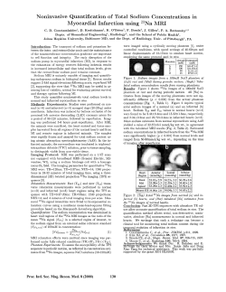


© Copyright 2026