
6 High Relaxivity Contrast Agents for MRI and Molecular Imaging
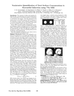
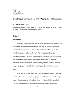
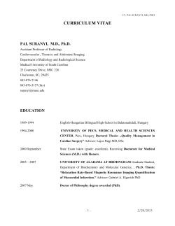
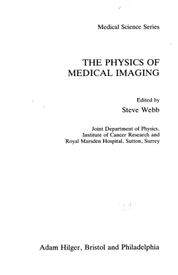
6 High Relaxivity Contrast Agents
for MRI and Molecular Imaging
S. Aime, A. Barge, E. Gianolio, R. Pagliarin, L. Silengo, L. Tei
6.1 Introduction . . . . . . . . . . . . . . . . . . . . . . . .
6.2 Determinants of Relaxivity . . . . . . . . . . . . . .
6.3 How to Improve Relaxivity . . . . . . . . . . . . . .
6.3.1 Interactions of Gd(III) Complexes with Proteins .
6.3.2 Effect of the Water Exchange Rate on Relaxivity
6.4 Targeting Cells with Gd(III) Chelates . . . . . . . .
6.5 Concluding Remarks . . . . . . . . . . . . . . . . . .
References . . . . . . . . . . . . . . . . . . . . . . . . . . . . .
.
.
.
.
.
.
.
.
.
.
.
.
.
.
.
.
.
.
.
.
.
.
.
.
.
.
.
.
.
.
.
.
.
.
.
.
.
.
.
.
.
.
.
.
.
.
.
.
.
.
.
.
.
.
.
.
.
.
.
.
.
.
.
.
.
.
.
.
.
.
.
.
99
103
105
105
107
111
118
119
6.1 Introduction
The superb spatial resolution and the outstanding capacity of differentiating soft tissues have determined the widespread success of
magnetic resonance imaging (MRI) in clinical diagnosis (Young
2000; Rinck 2003). The main determinants of the contrast in an MR
image are the proton relaxation times T1 and T2. When there is a
poor contrast between healthy and pathological regions due to a toosmall variation in relaxation times, the use of a contrast agent can
be highly beneficial. Contrast agents are chemicals able to alter
markedly the relaxation times of water protons in the tissues where
they distribute. Their use has led to remarkable improvements in
medical diagnosis in terms of higher specificity, better tissue characterization, reduction of image artifacts, and functional information.
Depending on whether the dominant effect occurs mainly on T1 or
100
S. Aime et al.
T2, MRI contrast agents can be classified as positive or negative
agents, respectively. The most representative class of T1-positive
agents is represented by paramagnetic Gd(III) chelates, whereas
iron-oxide particles represent the class of T2-negative agents (Merbach and Toth 2001).
Currently, about one-third of the MRI scans recorded in clinical
settings make use of contrast agents, mainly Gd(III) complexes
(Caravan et al. 1999). The effectiveness of a Gd(III) complex to act
as MRI contrast agent is first assessed by measuring its relaxivity,
i.e., the relaxation enhancement of water protons observed for a
millimolar solution of the contrast agent. In the past 15 years, a
number of papers addressing the relationship between structure/dynamics and relaxivity of Gd(III) complexes have been published.
This has led to a substantial advancement of our understanding of
the structural, dynamic, and electronic parameters determining the
relaxivity of paramagnetic chelates.
In addition to acting as a catalyst for the relaxation processes of
tissutal water protons, a potential MRI-CA has to fulfill several requirements related to tolerance, safety, toxicity, stability, osmolality,
viscosity, biodistribution, elimination, and metabolism. The currently
used Gd(III) chelates are based on polyaminocarboxylate ligands
(Scheme 1), either linear or macrocyclic molecules. All these ligands
form very stable complexes (Table 1) so that the risk of dissociation
is so low that the danger of acute toxic effects occurring after injection of gadolinium chelates is practically nonexistent with all the
products currently in use.
The study of the central nervous system (CNS) is the primary
clinical indication for the use of extracellular Gd(III) agents. The
majority of these pathologies are brain tumors, and three quarters of
them are represented by metastases occurring in patients undergoing
treatment for systemic cancer (Fig. 1). Other brain diseases, such as
multiple sclerosis and cerebral injuries can be also investigated by
contrast-enhanced MRI.
There are several other indications for the use of CAs outside
SNC. For instance, in the diagnosis of breast cancer, MRI with contrast agents is becoming an alternative diagnostic procedure to mammography. Particularly interesting is the dynamic use of contrast
agents effect. The breast is imaged repeatedly over the first few min-
High Relaxivity Contrast Agents for MRI and Molecular Imaging
101
Scheme 1. Polyaminocarboxylate ligands, either linear or macrocyclic, of
currently used Gd(III) chelates as commercial MRI contrast agents
utes following contrast agent administration, and a graph reporting
the increase of signal for a selected region of interest is plotted as a
function of time. The kinetics of the distribution of the contrast
agents in the extravascular space is related to the vascular permeability. Neo-formed vessels functional to the tumor growth display a permeability much higher than normal capillaries and the corresponding
102
S. Aime et al.
Table 1. Clinically accepted Gd(III)-based contrast agents
Complex
Brand name
Company
Log KGdL (8)
Gd-DTPA
Gd-DOTA
Gd-HPDO3A
Gd-DO3A-butrol
Gd-DTPA-BMA
Magnevist
Dotarem
ProHance
Gadovist
Omniscan
22.5
24.7
23.8
20.8
16.8
Gd-DTPA-BMEA
Gd-BOPTA
Gd-EOB-DTPA
OptiMARK
MultiHance
Eovist
Schering
Guerbet
Bracco
Schering
NycomedAmersham
Mallinckrodt
Bracco
Schering
16.8
22.6
23.5
Fig. 1. Metastases in the brain of a patient with brain tumor. The metastases
are detected upon intravenously administration of GdDTPA (right). Blood
vessels (capillaries) are typically quite leaky to small molecules like
GdDTPA, allowing them to enter the extracellular space. In the brain, however, the cells forming the walls of capillaries have very tight junctions and
prevent small molecules leaving the intravascular space, thereby forming a
blood-brain barrier (BBB). Tumors and other pathologies of CNS cause impairment of the BBB, thus allowing the contrast agent to leak from capillaries into extracellular fluid
High Relaxivity Contrast Agents for MRI and Molecular Imaging
103
areas are therefore characterized by high signal intensity (Padhani
2002). Of course, dynamic enhanced MRI is applied to demonstrate
the aggressive nature of tumors in several other areas.
The new landscape of molecular imaging applications requires the
development of a novel class of CAs characterized by higher contrasting ability and improved targeting capabilities. In this survey,
we intend to tackle some basic issues that are of fundamental importance for the use of Gd(III)-based systems in molecular imaging applications, namely: (1) actual understanding of the determinants of
T1-relaxivity of Gd(III) complexes, and how to proceed to attain
very high relaxivities; (2) how one may envisage efficient routes for
the delivery of a high number of Gd(III) complexes at the site of interest; and (3) the most practical ways to pursue the cell-internalization of a high number of Gd(III) complexes.
6.2 Determinants of Relaxivity
First of all, any MRI CA should be endowed with high thermodynamic and kinetic stability, and have at least one water molecule coordinated to the metal ion in fast exchange with the bulk water. This
would allow strong influence over the relaxation process of all protons present in the solvent in which the CA is dissolved. The Gd(III)
chelate efficiency is commonly estimated in vitro through the measure of its relaxivity (r1); that for commercial CAs as Magnevist,
Dotarem, Prohance, and Omniscan is around 3.4±3.5 mM±1 s±1 (at
20 MHz and 39 8C). The observed longitudinal relaxation rate (Robs
1 )
of the water protons in an aqueous solution containing the paramagnetic complex is the sum of three contributions (Banci et al. 1991):
(a) the diamagnetic one, whose value corresponds to proton relaxation rate measured in the presence of a diamagnetic (La, Lu, Y)
complex of the same ligand; (b) the paramagnetic one, relative to
the exchange of water molecules from the inner coordination sphere
of the metal ion with bulk water (Ris1p); and (c) the paramagnetic one
relative to the contribution of water molecules that diffuse in the external coordination sphere of the paramagnetic center (Ros
1p). Sometimes also a fourth paramagnetic contribution is taken in account,
that is due to the presence of mobile protons or water molecules
104
S. Aime et al.
(normally bound to the chelate through hydrogen bonds) in the second coordination sphere of the metal (Botta 2000). The inner sphere
contribution is directly proportional to the molar concentration of
the paramagnetic complex, to the number of water molecules coordinated to the paramagnetic center, q, and inversely proportional to the
sum of the mean residence lifetime, sM, of the coordinated water
protons and their relaxation time, T1M. This latter parameter is directly correlated to the sixth power of the distance between the metal center and the coordinated water protons and depends on the molecular reorientational time, sR, of the chelate, on the electronic relaxation times, TiE (i = 1, 2), of the unpaired electrons of the metal
(which depend on the applied magnetic field strength) and on the
applied magnetic field strength itself (Fig. 2). The outer sphere contribution depends on TiE, on the distance of maximum approach between the solvent and the paramagnetic solute, on the relative diffusion coefficients and again on the magnetic field strength (Aime et
al. 1998). The dependence of Ris1p and Ros
1p on magnetic field is very
Fig. 2. Schematic representation of relaxation mechanism in an aqueous solution containing a Gd(III)-complex
High Relaxivity Contrast Agents for MRI and Molecular Imaging
105
important because from the analysis of the magnetic field dependence allows the determination of the principal parameters characterizing the relaxivity of a Gd(III) chelate. This information can be obtained through an NMR instrument in which the magnetic field is
changed (Field-Cycling Relaxometer) to obtain the measure of r1 on
a wide range of frequencies (typically 0.01±50 MHz). At the frequencies most commonly used in commercial tomographs (20±
63 MHz), r1 is generally determined by the sR of the chelate so that
high molecular weight systems display a higher relaxivity. A quantitative analysis of r1 dependence on the different structural and dynamic parameters shows that, for systems with long sR (e.g., protein
bound complex), the maximum attainable r1 values can be achieved
through the optimization of sM and Tie (Banci et al. 1991).
6.3 How to Improve Relaxivity?
6.3.1 Interactions of Gd(III) Complexes with Proteins
Upon interacting with a macromolecule, the relaxation induced by
Gd(III) chelates usually displays remarkable changes, primarily related to the increase of the molecular reorientational time sR on
going from the free to the bound form, which results in a marked increase of the inner sphere Ris1 term. From the measurement of the relaxivity enhancement, it is possible to assess the affinity (and the
number of binding sites) between the interacting partners (Dwek
1973). Quantitatively, in the presence of a reversible interaction between the paramagnetic species and the macromolecule, the observed enhancement depends on both the molar fraction of the
macromolecular adduct vb and the relaxivity of the paramagnetic
species bound to the macromolecule in the all-bound limit (rb1).
Human serum albumin (HSA) has been by far the most investigated protein for binding of Gd(III) chelates. Besides the attainment
of high relaxivities, a high binding affinity to HSA enables the
Gd(III) chelate with a long intravascular retention time, which is the
property required for a good blood-pool agent for MR angiography.
In blood, HSA has a concentration of about 0.6 mM and its main
physiological role deals with the transport of a huge number of sub-
106
S. Aime et al.
Table 2. Affinity constant and relaxivity of the adducts of some Gd(III)
complexes wiht HSA, calculated at 0.47 T and 298 K. The coordinated
water exchange lifetime of free complexes is also reported
Metal complex
n ´ KA
(M±1)
rb1
sM
(mM±1s±1) (ns)
[Gd-DOTA(BOM)(H2O)]±
Cis-[Gd-DOTA(BOM)2(H2O)]±
Trans-[Gd-DOTA(BOM)2(H2O)]±
[Gd-DOTA(BOM)3(H2O)]MS-325
[Gd-BOPTA (H2O)]2±
< 1 ´ 102
6.4 ´ 102
7.2 ´ 02
3.4 ´ 103
3.0 ´ 104
4.0 ´ 102
35.2
44.2
53.2
35.0
33.0
175
130
80
250
280
[Gd-DTPA(BOM)2(H2O)]2±
3.6 ´ 103 28.2
260
[Gd-DTPA(BOM)3(H2O)]2±
[Gd-DOTA-IOP(H2O)]±
[Gd-DOTA-IOPsp(H2O)]±
[Gd-DOTA-TIBsp(H2O)]±
[Gd-DTPA-IOP(H2O)]2±
[Gd-DTPA-IOPsp(H2O)]2±
4.0 ´ 104
6.2 ´ 102
2.9 ´ 103
5.3 ´ 102
3.8 ´ 102
4.8 ´ 103
180
730
550
630
860
630
44.0
24.1
20.8
23.2
16.1
19.9
Reference
Aime
Aime
Aime
Aime
Aime
Aime
Aime
Aime
Aime
Aime
Aime
Aime
Aime
Aime
Aime
1996 a
1996 a
1996 a
1996 a
1999 c
2001;
1999 b
2001;
1999 b
1999 c
2001
2001
2001
2001
2001
strates (Carter and Ho 1994). For many of them, the binding region
has been identified on the basis of extensive competitive assays.
Now, the availability of solid state X-ray crystal structure of HSA allows more insight into the structural details of the binding interaction. The information gained from the studies of the interaction of
the various substrates to HSA has been very important in addressing
the design of Gd(III)-based blood-pool agents.
In Table 2 a list of Gd(III) complexes and the relevant parameters
of their binding interaction to HSA are reported.
Although the theory of the paramagnetic relaxation foresees relaxivities up to 100±120 mM±1 s±1 (at 20 MHz) for complexes bound to
macromolecular systems, the data listed in Table 1 show r1b values
significantly lower than the predicted ones. The primary reason for
the quenching of the relaxation enhancement is often associated to
the occurrence of a long exchange lifetime, sM, of the coordinated
water. This may be easily checked by measuring the relaxivity as a
function of temperature. The detection of a higher rb1 value as the
High Relaxivity Contrast Agents for MRI and Molecular Imaging
107
temperature is increased is an unambiguous indication of the occurrence of a too-long sM value.
6.3.2 Effect of the Water Exchange Rate on Relaxivity
Slow rates of the coordinated water appear to be primarily characteristic of the complex rather than a consequence of the binding to the
protein. Thus, for the attainment of high relaxivities, one has to
avoid Gd(III) chelates displaying slow exchange rates. The rational
which accounts for the water exchange rates in linear and cyclic
polyaminocarboxylate Gd(III) complexes has been elucidated in a
number of systems (Aime et al. 2001; Powell et al. 1996). In short,
the water-exchange process for the enneacoordinate Gd(III) complexes follows an idealized dissociative pathway. This may be represented by a simple diagram involving the transition state where the
metal ion has reduced its coordination number from 9 to 8 (Fig. 3).
Fig. 3. Energy diagram for the idealized dissociative water exchange mechanism in enneacoordinate Gd(III) complexes
108
S. Aime et al.
Fig. 4. Structure of the Square Antiprismatic (SA) and Twisted Square Antiprismatic (TSA) isomers in lanthanide DOTA-like complexes
It follows that fast exchange rates can be pursued by decreasing
the activation energy and this goal can be addressed either by destabilizing the ground state or stabilizing the intermediate state. Moreover, it has been shown that for DOTA-like systems there is a subtle
structural relationship between the exchange rate of the coordinated
water and the actual isomeric form. In fact, this class of complexes
exists in solution as a mixture of two structural isomers, namely the
square antiprismatic (AP) and the twisted square antiprismatic (TAP)
isomer (Fig. 4; Aime et al. 1992).
The polyhedron of the AP isomer corresponds closely to the regular square antiprismatic geometry, whereas a twisted square antiprismatic coordination cage with a smaller tilt angle between the two
square planes is assigned to the TAP isomer. The latter isomer displays a water exchange rate that is approximately 50 times faster
than the corresponding rate in the AP isomer (Aime et al. 1999 a).
Thus, given the above considerations, the functionalization of a ligand for protein targeting requires the choice of a Gd(III) chelate
that displays a fast exchange rate of the coordinated water.
In order to get more insight into the problem of why the relaxivity values obtained up to now are significantly lower than those predicted by the theory of paramagnetic relaxation, we have recently
High Relaxivity Contrast Agents for MRI and Molecular Imaging
109
Fig. 5. Structure of two DOTMA derivatives
undertaken a detailed relaxometric study of the interaction of GdDOTMA derivatives with HSA (Aime et al., to be published). GdDOTMA displays a relatively fast exchange of the coordinated molecule, likely because it possesses a TAP-like structure as shown by
1
H-NMR spectra of the Eu(III) and Yb(III) analogs (Aime et al.
1996 b; Di Bari et al. 2000). Two new ligands based on DOTMA
structure have been synthesized (Fig. 5). 1H-NMR spectra of Eu(III)
complexes showed that the TAP geometry found for the parent
DOTMA complexes is maintained. Complex A is soluble enough to
allow the determination of the water exchange rate by 17O-T2 measurements at variable temperature. The obtained value (s298
M = 65 ns)
indicates that the replacement of the methyl with the bulkier bisPhenyl group does not affect the coordination cage, leaving unchanged the water exchange rate. The solubility of complex B was
too low to pursue such measurement, but we may safely assume that
its s298
M is similar to that determined for A.
110
S. Aime et al.
Both A and B binds strongly to HSA, yielding Ka values of
2.7 ´ 103 M±1 and 9.5 ´ 104 M±1 respectively. rb1 of the A/HSA adduct, at 298 K and 20 MHz, is equal to 35 mM±1 s±1. 17O-measurements showed no difference between solutions containing the paramagnetic adduct and HSA alone. Clearly, the expected relaxation enhancement has been ªquenchedº by a direct interference of the donor groups on the surface of the protein with the inner hydration
sphere of the Gd(III) ion. We could not determine whether it results
in a replacement of the inner sphere water or simply in a dramatic
elongation of its exchange lifetime. Thus, the bis-phenyl moiety
does not appear long enough to protrude the chelate moiety outside
the interference of the residues on the surface of the protein in
proximity of the binding site. However, it is worthwhile to note that
the observed relaxation enhancement is due to water molecules/mobile protons on the surface of the protein.
B contains a binding synthon made of three phenyl groups and resulted long enough to avoid such interference. Unfortunately, the
low solubility of the adduct prevented the acquisition of 17O-NMR
VT spectra, but we got an indirect assessment of the occurrence of
the fast exchange of the coordinated water by measuring the water
proton relaxation rates as a function of temperature. As shown in
Fig. 6, the observed relaxation rate displays an exponential decrease
as the temperature increases, i.e., the typical behavior expected for
systems not ªquenchedº by long exchange lifetimes of the coordinated water. However, the observed rb1 for B/HSA adduct is only
43.5 mM±1 s±1, whereas the theory predicts much higher values
(Fig. 6) for a system tumbling with the sR of HSA (30 ns) and the
sM value of Gd-DOTMA.
We surmise that the limited relaxation enhancement has to be associated to a molecular reorientational correlation time, for the coordinated water, that is significantly shorter than that one of the macromolecule. This should not depend on an internal motion of the
chelate as the binding through the tris-phenyl substituent is tight enough. Rather, the observed behavior should be related to an internal
rotation of the coordinated water along its coordination axis, which
overlaps with the overall motion of macromolecular adduct. If this
suggestion is correct, a challenging question is posed to chemists:
how to design a Gd-chelate whose coordinated water is in fast ex-
High Relaxivity Contrast Agents for MRI and Molecular Imaging
111
Fig. 6. Longitudinal proton relaxivity as a function of temperature of free complex B (white boxes) and of the adduct complex B/HSA (black boxes). The
dotted line represents a simulation of the expected profile with sR = 30 ns
and sM = 65 ns
change with the bulk (short sM) and ªrigidº inside the coordination
cage?
6.4 Targeting Cells with Gd(III) Chelates
Molecular imaging (Weissleder and Mahmood 2001) deals with in
vivo characterization and measurement of biological processes at the
cellular and molecular level. With molecular imaging, early diagnosis of disease will become possible as the detection of altered biochemical processes largely anticipates the anatomical changes that
are at the basis of current diagnostic modalities.
Several modalities have contributed to the early stages of this innovative approach, namely PET, SPECT, MRI, and optical imaging.
MRI-Gd(III)-based agents are much less sensitive than radionuclear
and optical imaging probes. Therefore, molecular imaging based on
MRI invariantly involves the need of accumulating a high number of
contrast-enhancing units at the site of interest. The basis for the design of a Gd-containing imaging probes is first dictated from the
112
S. Aime et al.
Fig. 7. Targeting of the endothelial integrin avb3 as a specific angiogenesis
marker. The receptor is recognized by a biotinylated antibody that is then
bound by an avidin moiety bearing a Gd(III)-loaded liposome
concentration and localization (vascular, extracellular matrix, on the
cellular membrane, intracellular) of the target molecule (Aime et al.
2002 a). Of course, the most accessible targets are those present on
the surface of endothelial vessels. In principle, they can be visualized by a number of macromolecular conjugates containing many
Gd(III) complexes endowed with the proper vector recognizing the
given target. A nice example of targeting an endothelial site has
been reported by Sipkins et al. (1998) in the targeting of a specific
angiogenesis marker, the endothelial integrin avb3, whose presence
has been shown to correlate with tumor grade. The imaging probe
used in this work is a Gd-containing polymerized liposome. The target is first bound by a biotinylated antibody against avb3, which is
successfully recognized by an avidin moiety on the surface of the liposome. Each liposome has a mean diameter of 300±350 nm, which
appears suitable to avoid the uptake by the reticuloendothelial system (Fig. 7). This approach provided enhanced and detailed detection of rabbit carcinoma through the imaging of the angiogenetic
vasculature.
Recently, the same avb3 target has been addressed with lipidic nanoparticles containing a huge number of Gd-chelated units [94,400 Gd/
High Relaxivity Contrast Agents for MRI and Molecular Imaging
113
particle characterized by r1 = 19.1 s±1mM±1 (per Gd), r1 = 1,800,000 per
particle]. One of the lipidic components is covalently bound to the
avb3-integrin peptidomimetic antagonist (Winter et al. 2003).
The large molecular size of these constructs limits their delivery
to targets on the endothelial walls. To target receptors in solid tissues, other routes have to be followed. Bhujwalla and coworkers
(2003) have recently developed and applied a two-component Gdbased avidin-biotin system for the visualization of HER-2/scan receptors. The latter is a member of the epidermal growth factor family and it is amplified in multiple cancers. Their approach consisted
of addressing the extracellular domain of the receptors by means of
a biotinylated mAb. After clearance of the unbound mAb, Gd-labeled avidin is administered and binds, with high affinity, to the biotinylated mAb. The expression level of the receptor was estimated at
7 ´ 105 receptors/cell and the average number of Gd-DTPA units per
avidin molecule was 12.5. The method has been successfully applied
in an experimental mouse model of breast carcinoma.
An interesting route to MR signal amplification has been developed by Weissleder and coworkers (2002). Their approach is based
on enzyme-mediated polymerization of paramagnetic substrates into
oligomers of higher relaxivity. As substrates they used Gd-chelates
functionalized with phenolic substituents which undergo rapid condensation in the presence of H2O2 and peroxidase. The increased
molecular size of the oligomeric structures causes an increase of the
molecular reorientational time which, in turn, results in an increase
of the observed relaxivity. This approach has been applied to the
imaging of E-selectin-peroxidase conjugate.
As far as the cell internalization of Gd-chelates is concerned, several routes have been explored:
1. Via Pinocytosis. Pinocytosis is the cell-internalized portion of the
surrounding fluid by means of the invagination of its membrane and
formation of small vesicles (£ 150 nm diameter) called endosomes.
Therefore, incubation of cells, for a sufficiently long time, in a medium containing the imaging probe at relatively high concentration
leads to its internalization at amounts that may be sufficient for MRI
visualization. Among a number of systems we have considered, the
neutral, highly hydrophilic GdHPDO3A is a good candidate for la-
114
S. Aime et al.
beling stem cells by the pinocytotic route (Aime et al., in press).
The in vivo MR visualization of labeled stem cells will allow their
monitoring after transplantation. In a typical experiment of uptake
via pinocytosis, a few million stem cells are incubated in a culture
medium containing GdHPDO3A in mM concentration range (10±
50 mM) for few hours. Upon incubation no saturation effect is observed and the amount of taken up Gd is linearly proportional to the
concentration of the paramagnetic agent in the incubation medium.
Once cell internalized, the GdHPDO3A molecules end up entrapped
in endosomic vesicles as can be seen by observing the cells incubated with EuHPDO3A at the confocal microscope. In fact, Gd and
Eu chelates with the same ligands display the same chemical/biological behavior, and the fluorescent response of EuHPDO3A acts as a
histological reporter of the localization of GdHPDO3A in the cell.
We have proved the potential of this approach by observing a mouse
model of angiogenesis, on which blood-derived endothelial progenitor cells (EPCs) have been implanted subcutaneously, within a matrigel plug. After few days, the histologic examination showed large
capillary structure transposing the gel plugs. MR images parallel histologic findings as hyperintense spots corresponding to the labeled
cells were clearly detected. In Figure 8, we report a MR image
taken 14 days after implantation. As control, matrigel-embedding
unlabelled cells implanted in the same conditions are always negative for MRI signal.
The cell-labeling procedure described above appears to have
general applicability. We have tested it on several tumor cell lines,
obtaining invariantly a very efficient uptake with no apparent cytotoxicity. Likely, the entrapment of GdHPDO3A into the endosomic
vesicles prevents any impact of the paramagnetic agent on relevant
cellular process, meanwhile maintaining the full accessibility to cytoplasmatic water molecules.
2. Via Phagocytosis. Phagocytosis is the process of internalization of
particles by cells endowed with phagocytic activity. In such a case,
this route appears highly efficient for a single-step internalization of
a large amount of imaging probes. However, to be effective on MR
images, Gd-chelates must be water soluble. Therefore, the particles
must be biodegradable in order to release soluble Gd chelates once
High Relaxivity Contrast Agents for MRI and Molecular Imaging
115
Fig. 8. In vivo T1-weighted spin echo MR image (7.05 T) of EPCs labeled
with GdHPDO3A (left). The cells are dispersed into a subcutaneous matrigel
plug 7 days after the implantation. On the right is the control image of the
same in the absence of the paramagnetic label
internalized into phagocytic cells. One may envisage several ways
for the release of the Gd chelates. For instance, one may think of
gel nano-particles of chitosan loaded with negatively charged Gdchelates. Such particles (200±400 nm diameter) are easily phagocytosed and slowly degraded once internalized into the cells (S. Aime
et al., unpublished results). Another approach to biodegradable Gdcontaining particles has been pursued by designing particles whose
116
S. Aime et al.
Fig. 9. The insoluble system is represented by a Gd-DTPA-like complex
functionalized with a long aliphatic chain which is bound to the chelate
moiety through an ester bond. The solubilization is obtained through the enzymatic cleavage of the ester bonds yielding to a soluble Gd(III) complex
insolubility is a property of the Gd-chelates themselves (Aime et al.
2002 b). This goal is easily reached by introducing long aliphatic
chains on the surface of the ligand. However, one can control the
fate of these systems by means of the functionality used to link the
insolubilizing moiety to the Gd-chelate. In fact, by using ester or
peptidic functionalities, the insolubilizing synthon can be displaced
from the Gd chelates by the activity of the proper enzyme (Fig. 9).
3. Via Receptors. Cell-internalization via receptors is the route of
choice in a number of nuclear medicine assays. For MRI, the design
of the imaging probe requires the attachment of one or more
Gd(III)-chelates to the ligand molecule. Such structural modification
may drastically affect the internalization process with respect to the
mechanism occurring for the native ligand.
In order to deal with a system whose structural characteristics
were unaltered by the loading with Gd(III) chelates, we choose Apoferritin because it allows the imaging probes to be entrapped inside
High Relaxivity Contrast Agents for MRI and Molecular Imaging
117
its inner cavity (Aime et al. 2002 b). The exterior of such Gd(III)loaded Apoferritin is exactly the same as in the parent Ferritin and
then, once administered intravenously, it is quickly cleared-up by the
proper receptors on hepatocytes (Osterloch et al. 1996). The process
of loading Apoferritin with GdHPDO3A first entails the dissociation
of the protein into subunits at pH 2 followed by its reforming at pH
7, thereby trapping the solution components (e.g., GdHPDO3A)
within its interior. In such a system, water can freely diffuse through
the channels formed at the intersection of the protein subunits (10
channels), but the larger GdHPDO3A molecules cannot. The relaxivity shown by each GdHPDO3A entrapped in the Apoferritin cavity
is very high (approximately 80 s±1 mM Gd±1 at 20 MHz and 25 8C).
It has been possible to assess that the Gd-loaded Apoferritin maintains its integrity upon the cell internalization process as the relaxivity observed for the cytoplasmatic extract corresponds to that of the
intact system. Finally, the amount of cell-internalized Gd-loaded
Apoferritin is similar to that reported for the native Ferritin
(6.5 ´ 106 molecules per cell in 6 h).
4. Via Receptor-Mediated Endocytosis. This is probably the most
common route for the internalization of Gd(III) chelates as it is expected to occur any time a given ligand is modified by attaching one
or more Gd-chelating units. Thus, the internalization process is no
longer the one followed by the native ligand but the binding to the
receptor stimulates an endocytotic process which starts with the introflection of a portion of cellular membrane and ends up with the
welding of its extremities. Through this process, a number of substances remain entrapped in endosomic vesicles, primarily the molecules bound to the membrane or in close proximity of the portion of
membrane involved in the endosome formation.
Weiner et al. (1997) showed that the uptake of a folate-conjugate
dendrimer into tumor cells overexpressing high affinity folate receptor (hFR) occurs through this type of endocytotic pathway.
Another example of receptor-mediated endocytosis of a large dendrimer has been recently reported (Kobayashi and Brechbiel 2003).
The macromolecular construct comprised of Avidin and a biotinylated dendrimer bearing 254 GdDTPA chelates (AV-G6Gd) has
shown to accumulate in vitro into SHIN3 cells (a cell line obtained
118
S. Aime et al.
originally from human ovarian cancer) 50-fold greater than
GdDTPA. The internalization process is driven by avidin molecule, a
glycoprotein that binds to b-d-galactose receptors which are present
on these tumor cells.
Although not yet proven, it is likely that receptor-mediated endocytosis occurs with a number of systems in which the imaging Gdprobes are linked either to peptides as targeting vectors that bind to
specific receptors or to nutrient or pseudo-nutrient moieties that interact with the proper transporter upregulated in tumor cells.
5. Via Transmembrane Carrier Peptides. Another route to enter cells
with Gd(III) chelates is based on the use of membrane translocation
peptides which have been proven useful for the internalization of a
number of substrates like proteins, oligonucleotides, and plasmid
DNA. For instance, Bhorade et al. (2000) showed that GdDTPA
bound to 13-merHIV-tat peptide is efficiently internalized.
Another example has been recently reported by Allen and Meade
(2003), who showed that GdDTPA conjugated to polyarginine (8±16
monomer units) is able to permeate all membranes.
Finally, along this line, an interesting development has been recently reported by Heckl et al. (2003). They synthesized an imaging
probe consisting of a Gd-complex, a PNA (Peptide Nucleic Acid) sequence, and a transmembrane carrier peptide. Although the systems
enters any type of cell, it accumulates only in tumor cells because
of the specific binding of the PNA moiety for the c-myc mRNA
whose production is upregulated in those cells.
6.5 Concluding Remarks
Gd(III) chelates have played an important role in the development of
clinical applications of MRI technique by adding relevant physiological information to the superb anatomical resolution attainable with
this imaging modality.
More is still expected with the currently available contrast agents,
especially in the field of dynamic contrast enhancement protocols reporting on changes of the vascular permeability associated with the
staging and therapeutic follow-up of important pathologies. How-
High Relaxivity Contrast Agents for MRI and Molecular Imaging
119
ever, the major challenges are in the emerging field of molecular
imaging where the competition with other imaging modalities can
be very tight. Targeting of thrombi and atherosclerotic plaques by
peptides functionalized with Gd(III) chelates appears to be the next
goal for industrial research. The possibility of identifying and characterizing vulnerable plaques will certainly represent an important
task. Clearly, there is a need for new ideas for enhancing the attainable relaxivity at higher fields as the 3-T indication for clinical
imagers seems to be quite established. Moreover, it will be necessary to improve the efficiency of the available delivery systems and,
possibly, to exploit suitable amplification procedures in order to
reach the sensitivity required for the visualization of target molecules present at low concentrations.
The results herein surveyed show that there are several routes for
cell entrapment of paramagnetic Gd-agents at concentrations sufficient for MRI visualization. The huge work carried out in a number
of laboratories in the last two decades for the development of Gdbased MRI contrast agents provides an excellent platform for designing a new generation of probes for molecular imaging applications.
Though one should not underestimate the difficulties that will arise
when going from in vitro experiments to in vivo animal studies, we
think that the available results suggest that Gd-chelates will have an
important role in the armory of imaging probes for cellular and molecular imaging applications.
References
Aime S, Botta M, Ermondi G (1992) Nmr-study of solution structures and dynamics of lanthanide(III) complexes of dota. Inorg Chem 31:4291±4299
Aime S, Botta M, Fasano M, Geninatti Crich S, Terreno E (1996 a) Gd(III)
complexes as contrast agents for magnetic resonance imaging: a proton
relaxation enhancement study of the interaction with human serum albumin. J Biol Inorg Chem 1:312±319
Aime S, Botta M, Fasano M, Terreno E, Kinchesh P, Calabi L, Paleari L
(1996b) A new ytterbium chelate as contrast agent in chemical shift
imaging and temperature sensitive probe for MR spectroscopy. Magn Res
Med 35:648±651
Aime S, Botta M, Fasano M, Terreno E (1998) Lanthanide(III) chelates for
NMR biomedical applications. Chem Soc Rev 27:19±29
120
S. Aime et al.
Aime S, Barge A, Bruce J, Botta M, Howard JAK, Moloney JM, Parker D, de
Sousa AS, Woods M (1999 a) NMR, relaxometric, and structural studies of
the hydration and exchange dynamics of cationic lanthanide complexes of
macrocyclic tetraamide ligands. J Am Chem Soc 121:5762±5771
Aime S, Botta M, Fasano M, Terreno E (1999 b) Prototropic and waterexchange processes in aqueous solutions of Gd(III) chelates. Acc Chem
Res 32:941±949
Aime S, Chiaussa M, Digilio G, Gianolio E, Terreno E (1999 c) Contrast
agents for magnetic resonance angiographic applications: H-1 and O-17
NMR relaxometric investigations on two gadolinium(III) DTPA-like chelates endowed with high binding affinity to human serum albumin. J Biol
Inorg Chem 4:766±774
Aime S, Fasano M, Terreno E, Botta M (2001) In: Merbach AE, TÕth E
(eds) The chemistry of contrast agents in medical magnetic resonance
imaging. Wiley, Chichester, pp 193±241
Aime S, Cabella C, Colombatto S, Crich SG, Gianolio E, Maggioni F (2002
a) Insights into the use of paramagnetic Gd(III) complexes in MR-molecular imaging investigations. J Magn Reson Imaging 16:394±406
Aime S, Frullano L, Geninatti Crich S (2002 b) Compartmentalization of a
gadolinium complex in the apoferritin cavity: A route to obtain high relaxivity contrast agents for magnetic resonance imaging. Angew Chemie
Int Ed 41:1017±1019
Allen MJ, Meade TJ (2003) Synthesis and visualization of a membranepermeable MRI contrast agent. J Biol Inorg Chem 8:746±750
Banci L, Bertini I, Luchinat C (1991) Nuclear and electronic relaxation.
VCH, Weinheim, pp 91±122
Bhorade R, Weissleder R, Nakakoshi T, Moore A, Tung CH (2000) Macrocyclic chelators with paramagnetic cations are internalized into mammalian cells via a HIV-tat derived membrane translocation peptide. Bioconjugate Chem 11:301±305
Bhujwalla ZM, Artemov D, Mori N, Ravi R (2003) Magnetic resonance molecular imaging of the HER-2/neu receptor. Cancer Res 63:2723±2727
Botta M (2000) Second coordination sphere water molecules and relaxivity
of gadolinium(III) complexes: implications for MRI contrast agents. Eur
J Inorg Chem 3:399±407
Caravan P, Ellison JJ, McMurry TJ, Lauffer RB (1999) Gadolinium(III)
chelates as MRI contrast agents: structure, dynamics, and applications.
Chem Rev 99:2293±2352
Carter D, Ho JX (1994) Structure of serum-albumin. Adv Prot Chem 45:
153±203
Di Bari L, Pintacuda G, Salvadori P (2000) Solution equilibria in YbDOTMA, a chiral analogue of one of the most successful contrast agents for
MRI, GdDOTA. Eur J Inorg Chem 75±82
Dwek RA (1973) Nuclear magnetic resonance in biochemistry, applications
to enzyme systems, Clarendon Press, Oxford, pp 174±283
High Relaxivity Contrast Agents for MRI and Molecular Imaging
121
Heckl S, Pipkorn R, Waldeck W, Spring H, Jenne J, von der Lieth CW, CorbanWilhelm H, Debus J, Braun K (2003) Intracellular visualization of prostate
cancer using magnetic resonance imaging. Cancer Res 63:4766±4772
Kobayashi H, Brechbiel MW (2003) Dendrimer-based macromolecular MRI
contrast agents: characteristics and application. Mol Imaging 2:1±10
Merbach AE, TÕth E (2001) The chemistry of contrast agents in medical
magnetic resonance imaging. Wiley, Chichester
Osterloh K, Aisen P (1989) Pathways in the binding and uptake of ferritin
by hepatocytes. Biochem Biophys Acta 1011:40±45
Padhani AR (2002) Dynamic contrast-enhanced MRI in clinical oncology:
current status and future directions. J Magn Res 16:407±422
Powell DH, Ni Dhubhghaill OM, Pubanz D, Helm L, Lebedev HS, Schlaepfer W, Merbach AE (1996) Structural and dynamic parameters obtained
from O-17 NMR, EPR, and NMRD studies of monomeric and dimeric
Gd3+ complexes of interest in magnetic resonance imaging: an integrated
and theoretically self consistent approach. J Am Chem Soc 118:9333±
9346
Rinck PA (2003) Magnetic resonance in medicine. ABW Wissenschaftsverlag, Berlin
Sipkins DA, Cheresh DA, Kazemi MR, Nevin LM, Bednarski MD, Li KCP
(1998) Detection of tumor angiogenesis in vivo by alpha(v)beta(3)-targeted magnetic resonance imaging. Nat Med 4:623±626
Weissleder R, Mahmood U (2001) Molecular imaging. Radiology 219:316±
333
Weissleder R, Bogdanov A, Matuszewski L, Bremer C, Petrovski A (2002)
Oligomerization of paramagnetic substrates result in signal amplification
and can be used for MR imaging of molecular targets. Mol Imag 1:16±23
Wiener EC, Konda S, Shadron A, Brechbiel M, Gansow O (1997) Targeting
dendrimer-chelates to tumors and tumor cells expressing the high-affinity
folate receptor. Invest Radiol 32:748±754
Winter PM, Caruthers SD, Kassner A, Harris TD, Chinen LK, Allen JS,
Lacy EK, Zhang HY, Robertson JD, Wickline SA, Lanza GM (2003)
Molecular imaging of angiogenesis in nascent vx-2 rabbit tumors using a
novel alpha(v)beta(3)-targeted nanoparticle and 1.5 tesla magnetic resonance imaging. Cancer Res 63:5838±5843
Young IR (2000) Methods in biomedical magnetic resonance imaging and
spectroscopy. Wiley, Chichester


© Copyright 2026