
Diencephalic amnesia and apraxia after left thalamic infarction
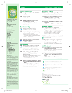
Downloaded from jnnp.bmj.com on 9 October 2008
Diencephalic amnesia and apraxia after left thalamic
infarction
JD WARREN, PD THOMPSON and PD THOMPSON
J. Neurol. Neurosurg. Psychiatry 2000;68;248-249
doi:10.1136/jnnp.68.2.248
Updated information and services can be found at:
http://jnnp.bmj.com/cgi/content/full/68/2/248
These include:
References
2 online articles that cite this article can be accessed at:
http://jnnp.bmj.com/cgi/content/full/68/2/248#otherarticles
Rapid responses
You can respond to this article at:
http://jnnp.bmj.com/cgi/eletter-submit/68/2/248
Email alerting
service
Receive free email alerts when new articles cite this article - sign up in the box at the
top right corner of the article
Notes
To order reprints of this article go to:
http://journals.bmj.com/cgi/reprintform
To subscribe to Journal of Neurology, Neurosurgery, and Psychiatry go to:
http://journals.bmj.com/subscriptions/
Downloaded from jnnp.bmj.com on 9 October 2008
J Neurol Neurosurg Psychiatry 2000;68:246–256
246
Clinical features of patients with optic neuritis
(ON)
LETTERS TO
THE EDITOR
Acute Optic Neuritis in Australia: a 13
year prospective study
The frequency with which multiple sclerosis
develops after an attack of acute optic neuritis varies widely in diVerent series and has
been reported to range from 11.5% to 85%.1
The variability in the findings may relate to
diVerent methods of patient selection, diagnostic criteria, geographical factors, duration
of follow up, and study design.
A cohort of 82 patients (59 females, 23
males) with uncomplicated ON aged 10 to 50
years (mean 29.2) who were examined
neurologically and had visual evoked responses (VERs) performed in our department during the period 1973–83 were
re-examined in 1983–85. Twenty six of the
patients (32%) had progressed to probable or
clinically definite multiple sclerosis during
the follow up period of 7–114 (mean 57)
months. Female sex, young adult age, and the
presence of HLA-B7 or DR2 seemed to
increase the risk of developing the disease.2
Seventy one of the 82 (87%) (52 females,
19 males) were reviewed in 1991–2; 11
patients could not be traced. Neurological
examination was performed on 49; two
patients had died with multiple sclerosis and a
telephone questionnaire was completed on
the remainder. Thirty three (46%, or 40% of
the original 82) had developed probable or
clinically definite multiple sclerosis after a
mean duration of 13.25 years (range 8–29.6
years). Eight cases had developed multiple
sclerosis since the previous review. KaplanMeier and actuarial methods of assessment,3
predicted that 52% would develop the disease
after 15 years (figure). There was a significantly greater risk of developing multiple sclerosis for patients in the 21–30 year age group
than those outside this range but there was no
significant diVerence in the rate of progression
to the disease for males and females. There
was no significant diVerence in the probability
of developing multiple sclerosis in patients
with single or recurrent attacks of optic neuritis or bilateral optic neuritis, nor in those who
were DR-2 positive (table 1).
The finding in the Australian cohort that
52% of patients with optic neuritis were at risk
of developing probable or clinically definite
1.00
Probability
0.75
0.50
0.25
0.00
0
50
100
150
200
Months
Probability of patients with a first attack of optic
neuritis not developing multiple sclerosis in 15
years.
ON
ON-multiple
sclerosis
Age (y):
<20
21–30
31–40
41–50
10
8
11
9
4
17
9
3
Sex:
Males
Females
13
25
6
27
ON Type:
Single
Recurrent
Bilateral
23
11
4
17
13
3
HLA Type*:
DR2+
DR2−
15
19
22
11
÷2 = 8.7, 3df,
p=0.07
÷2 = 1.57, 1df,
p>0.10
÷2 = 0.82, 2df,
p>0.5
÷2 = 2.63, 1df,
p>0.1
*Four subjects not typed.
multiple sclerosis in 15 years is comparable
with that of 57% in 11.6 years in the United
Kingdom,4 49% in 20 years in the United
States,1 and 45% in 15 years in Sweden.5
JANE A FRITH
JAMES G MCLEOD
Institute of Clinical Neurosciences, University of
Sydney and Royal Prince Alfred Hospital, Sydney,
Australia
MARIESE HELY
Department of Neurology, Westmead Hospital, Sydney,
Australia
Correspondence to: Professor J G McLeod, Department of Medicine, University of Sydney, NSW
2006, Australia email [email protected]
1 Rodriguez M, Siva A, Cross SA, et al. Optic neuritis: a population-based study in Olmsted
County, Minnesota. Neurology 1995;45:244–50.
2 Hely MA, McManis PJ, Doran TJ, et al. Acute
optic neuritis: a prospective study of risk
factors for multiple sclerosis. J Neurol Neurosurg Psychiatry 1986;49:1125–30.
3 Armitage P, Berry G. Statistical methods in medical research, 3rd ed. Oxford: Blackwell, 1994.
4 Francis DA, Compston DAS, Batchelor JR, et
al. A re-assessment of the risk of multiple sclerosis developing in patients with optic neuritis
after extended follow-up. J Neurol Neurosurg
Psychiatry 1987;50:758–65.
5 Sandberg-Wollheim M, Bynke H, Cronqvist S,
et al. A long-term prospective study of optic
neuritis: evaluation of risk factors. Ann Neurol
1990;27:386–93.
Post-traumatic hydrocephalus: influence
of craniectomy on the CSF circulation
Post-traumatic ventricular dilatation may have
a wide range of aetiological factors: starting
from neuronal loss due to head trauma and
possible secondary ischaemic insults, to obstruction of CSF circulation resulting in
hydrocephalus. It is important to diVerentiate
between post-traumatic hydrocephalus and
brain atrophy before considering placement of
a shunt. Making this decision can be facilitated
by measurement of the resistance to CSF
outflow.1 However, the pattern of the CSF circulation may change dramatically after a
cranioplasty resulting from a previous decompressive craniectomy for refractory intracranial
hypertension after head injury. The eVect of
the skull and dura on CSF hydrodynamics has
been explored experimentally: the resistance to
CSF outflow after craniectomy decreases twofold and brain compliance (expressed using
the pressure-volume index, PVI) increases.2
This problem is important clinically as the following case illustrates:
A 44 year old man fell downstairs and was
admitted with a Glasgow coma score (GCS)
of 4. Brain CT disclosed an intracerebral
haematoma, which required a right frontal
lobectomy and decompressive craniectomy to
control raised intracranial pressure. Five
months later he remained severely disabled
with deteriorating GCS and increasing spasticity. Brain CT showed a progressive ventricular dilatation with widening of the cortical sulci. Cranioplasty had been delayed
because of persistent problems with infections.
The first lumbar computerised infusion
test3 was performed 5 months after injury to
study the patient’s CSF circulation. The
opening pressure was low (5 mm Hg) with a
very low pulse amplitude. An infusion of normal saline at a rate of 1.5 ml/min increased
the intracranial pressure (ICP) to a plateau of
12.2 mm Hg within 22 minutes. The
calculated resistance to CSF outflow was
normal (5 mm Hg/ml/min) and the pressurevolume index was increased to 28 ml (figure).
It is important to mention here that the
normal range for the pressure-volume index,
calculated from the constant rate infusion (as
an inverse of elastance coeYcient3) is diVerent from the values obtained by the bolus
injection.1 Values below 13 ml indicate a tight
brain, from 13 ml to 23 ml normal compliance, and above 23 ml hypercompliant brain.
A slow constant rate infusion tests global
compliance of the craniospinal axis whereas a
fast bolus volume load probably tests compartmental compliance of the container into
which the extra volume is added.
This pattern of CSF circulation with low or
normal resistance to CSF outflow, increased
brain compliance, and very few vasogenic
waves is characteristic of cerebral atrophy.3
Cranioplasty was carried out as his deterioration was attributed to the “syndrome of
trephined” where the brain sinks in, particularly with erect posture and dehydration producing deterioration in conscious level and
focal signs. However, 1 month later, there had
been no progress in the patient’s condition
and repeat CT again suggested progressive
ventriculomegaly. The infusion study was
repeated. The opening ICP was not dramatically diVerent (10 mm Hg) to the previous
study. However, the pulse amplitude (1.5 mm
Hg) was increased, and the calculated resistance to CSF outflow was greatly increased to
20 mm Hg/ml/min, with a normal pressurevolume index of 15 ml. Such a pattern is specific for hydrocephalus. After this test the
patient was shunted with a Codman Medos
programmable valve (setting 120 mm H2O)
ventriculoperitoneal shunt with remarkable
clinical improvement, the GCS rose to 14, he
began to talk and his spasticity in his arms
decreased dramatically. It is obvious why the
pressure-volume compensatory reserve (PVI)
decreases after cranioplasty2, but the
interpretation of an increase in the resistance
to CSF is not immediately apparent. Two
explanations are possible:
The patient had developed an acute hydrocephalus, possibly as a result of traumatic
subarachnoid haemorrhage. Craniectomy
was a factor allowing compensation of CSF
circulation in the early stages. It is diYcult to
explain what is the nature of such compensation. Shapiro et al2 attempted to oVer an
interesting but conceptually diYcult hypothesis that the time constant (resistance to CSF
outflow×compliance of cerebrospinal space)
of cerebrospinal system hydrodynamics has a
tendency to remain constant. Therefore, a
Downloaded from jnnp.bmj.com on 9 October 2008
J Neurol Neurosurg Psychiatry 2000;68:246–256
mechanistic increase in compliance after
craniectomy tends to be followed by a
decrease in the resistance to CSF outflow.
This process may be reversed after
cranioplasty—that is, a decrease in PVI may
be followed by an increase in the resistance to
CSF outflow.
The second possible scenario is more
important for clinical management. A large
craniectomy may facilitate irreversible ventricular enlargement over weeks or months.4
Thus, after cranioplasty, the expanded
247
ventricles may, via the cerebral mantle,
obstruct the lumen of the cortical subarachnoid space and increase the resistance to
CSF outflow.
This case demonstrates that when the
CSF circulation is studied in patients with a
large craniectomy the CSF outflow resistance cannot be taken reliably as a guide
for shunting. Overnight ICP monitoring or
CSF infusion study should be performed
after cranioplasty, when CSF circulatory
reserve decreases dramatically. Moreover, a
prolonged period without a bone flap may
encourage ventricular dilatation.
MAREK CZOSNYKA
JO COPEMAN
ZOFIA CZOSNYKA
ROY S MCCONNELL
CATHERINE DICKINSON
JOHN D PICKARD
Academic Neurosurgical Unit, Addenbrooke’s
Hospital, Cambridge CB2 2QQ, UK
A
15
F
Before cranioplasty
B
AMP
(mm Hg)
0
50
C
ICP
(mm Hg)
Infusion
0
Time
5 min
15
D
After cranioplasty
G
AMP
(mm Hg)
0
50
Infusion
E
ICP
(mm Hg)
0
Time
5 min
CT and infusion studies. (A) Scan performed on admission. (B) After right frontal lobectomy and bone flap removal. (C) Four months after injury, before
cranioplasty. (F) Infusion test demonstrated low resistance to CSF outflow and increased brain compliance. ICP=mean intracranial pressure; AMP= pulse
amplitude of ICP waveform. Constant infusion rate of 1.5 ml/min is indicated by a thick horizontal line. X axis=time. (D) Five months after injury, after
cranioplasty. (G) Infusion test demonstrated grossly increased resistance to CSF outflow and normal brain compliance. (E) One month after shunting:
normalisation of ventricles. Bicauduate index decreased from 33% to 21% with a decrease in the 3rd ventricle diameter ( from 13 mm to 8 mm).
Downloaded from jnnp.bmj.com on 9 October 2008
J Neurol Neurosurg Psychiatry 2000;68:246–256
248
Correspondence to: Dr Marek Czosnyka, Academic Neurosurgical Unit, Box 167, Addenbrooke’s
Hospital, Cambridge CB2 2QQ, UK email
[email protected]
1 Marmarou A, Foda MA, Bandoh K, et al. Posttraumatic ventriculomegaly: hydrocephalus or
atrophy? A new approach for diagnosis using
CSF dynamics. J Neurosurg 1996;85:1026–35.
2 Shapiro K, Fried A, Takei F, et al. EVect of the
skull and dura on neural axis pressure-volume
relationships and CSF hydrodynamics. J Neurosurg 1985;63:76–81.
3 Czosnyka M, Whitehouse H, Smielewski P, et al.
Testing of cerebrospinal compensatory reserve
in shunted and non-shunted patients: a guide
to interpretation based on observational study.
J Neurol Neurosurg Psychiatry 1996;60:549–58.
4 Hochwald GM, Epstein F, Malhan CrashoV J.
The role of skull and dura in experimental
feline hydrocephalus. Dev Med Child Neurol
1972;14(suppl 27):65–9.
Diencephalic amnesia and apraxia after
left thalamic infarction
Amnesia and apraxia are unusual manifestations of unilateral thalamic lesions. A patient
in whom severe amnesia and apraxia were the
presenting features of a left thalamic infarct is
presented. The findings support the concept
that memory and praxis both utilise circuits
which include the dominant thalamus.
A 78 year old right handed Hungarian
woman presented with memory loss and
disorientation. She had been well and conversed normally with her daughter on the
evening before presentation. The next morning, her daughter was alarmed to find her
mother’s house in a state of disarray. Dishes
were unwashed, lights left on, and doors
open. The patient seemed baZed by eating
utensils, attempting to scoop food with her
knife. Later that morning, she failed to recognise longstanding Hungarian friends. She was
unable to recall her address, the name of the
city in which she lived, or the names of her
grandchildren. She subsequently failed to
recognise her family doctor of 7 years.
History included non-insulin dependent
diabetes, hypertension, hyperlipidaemia, and
atrial fibrillation. Medications were digoxin,
glibenclamide, and metoprolol. Captopril
had been prescribed 4 weeks previously but
was ceased 2 days before presentation due to
presyncopal symptoms. The patient consumed no alcohol. There was no history of
cerebrovascular events.
Cognitive functions were examined at the
bedside with the assistance of an interpreter, as
the patient spoke no English, although she
conversed freely in her native Hungarian. She
had no recollection of events since emigrating
to Australia 50 years previously, gave her
correct maiden name, and could not recognise
or name her grandchildren, although she
recognised her daughter. She acknowledged
she was in a hospital, but maintained it was in
Budapest and the year was 1947. Although her
recollections regarding her early life and
wartime Hungary seemed accurate, she confabulated when asked for details of recent
events. Short term recall of verbal material and
people was poor. The patient was able to name
objects such as a pencil and a watch, and obey
two and three stage commands. She wrote her
name and copied simple designs correctly, and
could imitate gestures such as waving goodbye
or blowing a kiss. However, she was unable to
use eating utensils or a toothbrush, either in
pantomime or when provided with the object
itself. Movements of the face and limbs were
normal, and there were no sensory abnormalities. Knee and ankle jerks were absent bilaterally and both plantar responses were extensor.
General examination revealed atrial fibrillation
and mild cardiomegaly.
The patient continued to display severe
impairment of anterograde memory. She was
reluctant to leave her bed, and quickly
became lost unless supervised. She did not
recognise familiar staV members and was
unable to use ward landmarks to reorientate
herself. She required assistance to feed
herself, brush her teeth, and shower. When
reviewed 3 months later, her memory disturbance and apraxia for simple activities of
daily life (such as brushing her teeth)
persisted, necessitating care in a supervised
environment.
Brain MRI (figure) showed a left anterior
thalamic lesion consistent with lacunar infarction and periventricular white matter disease.
Deficits of anterograde and retrograde
memory after thalamic lesions are well
recognised.1 The syndrome of diencephalic
amnesia after bilateral medial thalamic lesions typically involves striking disorientation
for time, loss of autobiographical information
(often extending back for many years),
confabulation, and severe anterograde amnesia for verbal and visual material, including
recognition of familiar faces.1 These features
were well illustrated by our patient, who
became “marooned” in an earlier place and
time. Amnesia after unilateral thalamic lesions is rare.2 There is increasing evidence
that thalamic lesions interrupt the multiple
brain networks which form the anatomical
substrate of memory,1–4 encompassing the
hippocampus, medial thalamus, frontal and
cingulate cortices, and overlapping with the
language areas of the left hemisphere.3 The
thalamus is activated in retrieval of episodic
(autobiographical) and semantic (encyclopaedic) information from long term storage4
and execution of learned motor tasks,5 which
may reflect its widespread connections with
other subcortical and cortical structures.
The patient’s ability to name or identify
objects was not tested systematically. However,
on the evidence available, it seems likely that her
diYculty in utilising common objects was a
manifestation of apraxia for daily tasks rather
than, for example, agnosia for the objects
involved. Apraxia is a rare manifestation of isolated thalamic lesions.5 The ability to access
stored motor representations is thought to be
crucial for normal execution of learned actions.5
These motor representations or “engrams” are
analogous to motor memories. Although praxis
is generally regarded as a function of distributed
cortical regions in the left hemisphere, apraxia
in association with thalamic amnesia has not
been emphasised in previous reviews of this
syndrome.1–2 Involvement of deep hemispheric
white matter in association with basal ganglia
pathology is thought to be critical for the
development of apraxia after lesions of subcortical structures.5 The conjunction of diencephalic amnesia and apraxia after thalamic
infarction in the present case may be interpreted as further evidence that retrievial of
episodic, semantic, and motor memories is
mediated by overlapping functional networks
in the dominant hemisphere.1 3 4
JD WARREN
PD THOMPSON
Department of Neurology, Royal Adelaide Hospital,
Adelaide, South Australia
PD THOMPSON
University Department of Medicine
Correspondence to: Professor PD Thompson, University Department of Medicine, Royal Adelaide
Hospital, Adelaide, South Australia 5000, Australia
T2 weighted axial MRI brain slice showing ischaemic infarction of the left anterior thalamus and
periventricular white matter ischaemic changes.
1 Hodges JR, McCarthy RA. Autobiographical
amnesia resulting from bilateral paramedian
thalamic infarction: a case study in cognitive
neurobiology. Brain 1993;116:921–40.
2 Clarke S, Assal E, Bogousslavsky J, et al. Pure
amnesia after unilateral left polar thalamic
infarct: topographic and sequential neuropsychological metabolic (PET) correlations. J
Neurol Neurosurg Psychiatry 1994;57:27–34.
3 Perani D, Bressi S, Cappa SF, et al. Evidence of
multiple memory systems in the human brain:
a [18F]FDG PET metabolic study. Brain 1990;
116:903–19.
4 Fletcher PC, Frith CD, Grasby PM, et al. Brain
systems for encoding and retrieval of auditoryverbal memory: an in vivo study in humans.
Brain 1995;118:401–16.
5 Pramstaller PP, Marsden CD. The basal ganglia
and apraxia. Brain 1996;119:319–40.
Downloaded from jnnp.bmj.com on 9 October 2008
J Neurol Neurosurg Psychiatry 2000;68:246–256
Transverse myelopathy in the antiphospholipid antibody syndrome: pinworm
infestation as a trigger?
The antiphospholipid antibody syndrome is a
disorder characterised by the production of
autoantibodies directed against negatively
charged cell membrane phosholipids. Antiphospholipid antibodies have been described in various neurological disorders.
It has been generally accepted that viral,
bacterial, and parasitic infections can serve as
a trigger factor for autoimmune reactions.
Despite the growing knowledge that has
accumulated, the relation between parasites
and autoimmunity has not been clarified.
Enterobius vermicularis (pinworm) is a
nematode rarely found outside the gastrointestinal tract, but allergic reaction due to
enterobiasis has been reported.1 We describe
the case of transverse myelopathy preceded by
intestinal pinworm infestation in the primary
antiphospholipid antibody syndrome. To our
knowledge, such an association has not been
reported previously. Pinworm therapy was
complicated by the Jarisch-Herxheimer reaction manifested by temporary exacerbation of
pre-existing neurological symptoms.
In March 1998, a 40 year old woman who
complained of perianal itching noticed the
presence of worms migrating from the anus.
Three days later itching and numbness
involved both legs, and the patient had weakness in the legs. These symptoms progressed
over the next 3 days to severe paraparesis and
urinary urgence. Her medical history was relevant for three unexplained miscarriages
which all occurred in midpregnancies. In the
local hospital, she underwent brain and
lumbar-sacral spine MRI and findings were
normal. Cerebrospinal fluid examination disclosed slightly raised proteins of 670 mg/l, 2
lymphocytes/mm3;
oligoclonal
immunoglobulin (Ig) G bands were absent. She was
treated with methylprednisolone (1 g/day)
intravenously for 5 days with subsequent
gradual tapering oV, which was associated
with substantial improvement of motor,
sensory, and sphincter disturbances. At the
end of April 1998, the patient was transferred
to our hospital for further investigation.
Neurological examination showed mild
spastic paraparesis, bilateral Babinski’s sign,
and a Th-12 sensory level.
Erythrocyte sedimentation rate was 34.
Complete blood count, tests for hepatic and
renal function, angiotensin converting enzyme, concentrations of IgG, IgM, IgA, IgE,
and immune complexes, screening for antinuclear (HEp-2 cells), anti-ds DNA, antineutrophil cytoplasmic, antimitochondrial, and
antiparietal cell antibodies, rheumatoid factor, and the search for antineurotropic virus
and antiBorrelia antibodies were normal or
negative. A venereal disease research laboratory flocculation test was negative. A medium
positive concentration of IgM anticardiolipin
antibody was detected, and lupus anticoagulant was negative. Raised titres of serum IgG
and IgM anti-GM1 (1:1600 and 1:3200,
respectively) and antisulfatide antibodies
(1:6400, for both classes) were also demonstrated. Class II human leucocyte antigen
(HLA) typing showed the presence of HLADR3, DR4, DR52, DR53, DQ2, and DQ3.
Cervical and thoracic spine MRI was normal.
Electromyoneurography was normal.
Because the patient complained of reappearence of worms and perianal itching, a
cellulose adhesive tape test was performed
and diagnosis of enterobiasis was established.
249
Mebendazole was given in a single dose of
100 mg and the next day the JarishHerxheimer reaction occurred, with deterioration of leg spasticity, inability to walk, and
development of urinary retention. At that
time, low positive IgG ACA was detected.
The dose of prednisone was raised to 60
mg/day and slowly tapered oV within the next
2 months. Sphincter disturbances resolved in
1 day and motor dysfunction gradually
improved with only mild spasticity left.
Diagnosis of antiphospholipid antibody
syndrome in our patient was based on the
presence of reccurrent fetal loss, transverse
myelopathy, and raised ACA. The ACA titre
was probably lowered by previously administered corticosteroid therapy. There are several reports of transverse myelopathy as a
manifestation of antiphospholipid antibody
syndrome in the past decade.2 3 A potential
pathogenic role of antiphospholipid antibodies in transverse myelopathy might be based
either on vasculopathy or on interaction with
spinal cord phospholipids.
Infection by helminths is universally associated with activation of T helper 2 (Th2)type cells. Regardless of the mechanisms and
protective value of antihelminthic Th2 responses, such responses may also be detrimental to the host. The presence of ACA,
anti-GM1, and antisulfatide antibodies in our
patient suggests a systemic response to E vermicularis, as it has been shown that nematodes contain cardiolipin, ganglioside GM1,
and sulfatides within their complex lipid
composition.4 When parasites share epitopes
with host tissue, such molecular mimicry may
exploit host immune tolerance against a self
determinant. Autoimmunity may occur if
immune tolerance is overriden in genetically
susceptible hosts. It has been proposed that
the presence of pathogenic cross reactive
autoantibodies could be the basis for the
relation between nematodes and autoimmunity. It may be also postulated that E vermicularis stimulated Th2 response which enhanced polyclonal autoantibody production
resulting in the presence of ACA, anti-GM1,
and antisulfatide antibodies. The association
of transverse myelopathy, ACA, and enterobiasis might be purely coincidental, which we
assume to be highly unlikely. The finding of
diVerent autoantibodies, as well as the isotype
switch of ACA, strongly suggests that pinworm infestation in our patient was the “triggering event” that increased the production
of autoantibodies against cardiolipin and led
to the development of transverse myelopathy.
The appearance of the spinal cord damage
caused by ACA in our patient might have
been facilitated by the simultaneous eVect of
anti-GM1 and antisulfatide antibodies. A
significant subset of the human anti-GM1
antibodies that reacted with the Gal(b1–
3)GalNAc determinant also bound to
oligodendrocyte-myelin glycoprotein which
is a constituent of the myelin of the CNS. As
for antisulfatide antibodies, their presence
has been already shown in some diseases
aVecting the CNS.
It is clear that parasitic infections can serve
as a trigger factor of autoimmune reactivity,
but the presence of autoantibodies or self
reactive T cells is rarely associated with clinical manifestations. They develop only in
patients with adequate immunogenetic and
hormonal background for autoimmune diseases. In several studies, increased frequencies of HLA-DR4, DR7, DR53, and DQ7
were found in patients with antiphospholipid
antibody syndrome,5 and in our patient
HLA-DR4 and DR53 were present. Additional studies are necessary to further elucidate the complex mechanisms of involvement
of intestinal helminths in the processes of
autoimmune activity.
JELENA DRULOVIC
u
IRENA DUJMOVIC
u
NEBOJŠA STOJSAVLJEVIC
u
IVANA TRIPKOVIC
u
SLOBODAN APOSTOLSKI
ZVONIMIR LEVIC
u
Institute of Neurology, CCS, School of Medicine,
University of Belgrade, Dr Subotiæa 6, 11000
Belgrade, Yugoslavia
MARIJA MOSTARICA STOJKOVIC
u
Institute of Microbiology and Immunology
BRANKA BONAC
yI
SLADJANA ANDREJEVIC
u
Institute of Allergology
Correspondence to: Dr J Drulović, Institute of
Neurology, CCS, School of Medicine, University of
Belgrade, Dr Subotića 6, 11000 Belgrade, Yugoslavia
1 Montag A, Ulrich R. Chronic urticaria in
enterobiasis vermicularis (oxyuriasis). Hautarzt
1992;43:652–3.
2 Schantz V, Oestergaard LL, Junker P. Shrinking
spinal cord following transverse myelopathy in
a patient with systemic lupus erythematosus
and the phospholipid antibody syndrome. J
Rheumatol 1998;25:1425–8.
3 Ruiz-Arguelles GJ, Guzman-Ramos J, FloresFlores J, et al. Refractory hiccough heralding
transverse myelitis in the primary antiphospholipid syndrome. Lupus 1998;7:49–50.
4 Maloney MD, Semprevivo LH. Thin-layer and
liquid column chromatographic analyses of the
lipids of adult Onchocerca gibsoni. Parasitol Res
1991;77:294–300.
5 Wilson WA. Histocompatibility genes in antiphospholipid antibody syndrome. Lupus
1996;5:259–262.
Radiologically selective visual pathway
involvement in adult onset cerebral
adrenoleukodystrophy
A case of adult onset cerebral adrenoleukodystrophy is presented with serial MRI showing selective involvement of the visual system
with spread of disease along the fibre tracts of
this system.
Adult onset cerebral adrenoleukodystrophy is the rarest presentation of
adrenoleukodystrophy.1 2 It may present with
various symptoms often including visual
impairment.3 Brain MRI may show multiple
areas of symmetric high signal intensity
within cerebral white matter, usually aVecting
the occipital lobes.4–6 We present a case of
adrenoleukodystrophy, in whom serial MRI
demonstrated selective progression of demyelination through the visual pathways.
A thirty year old man presented in May
1996 with a 7 month history of deteriorating
vision, slurred speech, incoordination, poor
balance, generalised weakness, sleep disturbance, and headaches. His symptoms were
worse on the right. He had no symptoms of
postural hypotension.
His mother had been shown to be a carrier
of X linked adrenoleukodystrophy (XLALD). His two elder brothers had died of
XL-ALD at the ages of 6 and 7 years. In 1993
our patient had been shown to have abnormal
serum concentrations of very long chain fatty
acids (VLCFAs) and to be a carrier of the
XL-ALD gene. At that time he was asymptomatic and had no abnormal neurological
signs. Crohn’s disease had been diagnosed in
1987 after an ileal resection although this had
remained in remission.
On examination, visual acuities were 6/12
(right), 6/9 (left). Fields were full to confron-
Downloaded from jnnp.bmj.com on 9 October 2008
J Neurol Neurosurg Psychiatry 2000;68:246–256
250
tation using a finger but there was a left
homonymous field defect to a red pin and he
had a left aVerent pupillary defect. Fundoscopy showed bilateral optic atrophy. The
remainder of the cranial nerve examination
was normal. In the arms tone and power were
normal, but coordination was mildly impaired on the right. The reflexes were
exaggerated and HoVman’s sign was present
bilaterally. A palmomental reflex was present
on the right. In the legs power was normal,
but tone was increased and there were several
beats of ankle clonus; reflexes were exaggerated and plantar responses were upgoing.
Coordination was impaired in both legs, his
gait was ataxic, and Romberg’s test was positive. He had a minor reduction in vibration
sensation at the right ankle; otherwise sensa-
tion was normal. He appeared moderately
tanned, but there was no other hyperpigmentation. Supine blood pressure was 114/78,
falling to 108/80 on standing. The remainder
of the examination was normal.
Routine biochemistry was normal. A
morning cortisol was 469 nmol/l (normal
>160 nmol/l), but a short synacthen test
showed an abnormally flat response (serum
Brain MRI at four diVerent times spanning 2 years. (A) May 1996: T2 weighted axial. (B) May 1996: contrast enhanced T1 weighted axial. (C)
October 1996: T2 weighted axial. (D) October 1996: T1 weighted contrast enhanced axial. (E) October 1996: T1 weighted contrast-enhanced axial. (F)
August 1997: T2 weighted axial. (G) August 1997: T1 weighted contrast enhanced coronal. (H) May 1998: T2 weighted axial. (I) May 1998: T1
weighted contrast enhanced coronal.
Downloaded from jnnp.bmj.com on 9 October 2008
J Neurol Neurosurg Psychiatry 2000;68:246–256
cortisol rise from 338 to 449 nmol/l over 1
hour). His plasma VLCFA profile was abnormal consistent with XL-ALD. Humphrey
visual field testing demonstrated a left
homonymous field defect. Brain MRI was
abnormal (figure A and B). He was placed on
a very low fat diet with supplements of
glycerol trioleate oil.
By October 1996 his headaches had settled
but his eyesight, memory, coordination, and
walking were worse. Visual acuity was below
6/60 in both eyes. Brain MRI was repeated
(figure C-E). By August 1997 there had been
no new clinical developments (MRI figure
F-G). In May 1998 he complained of navigational diYculties in familiar surroundings,
further memory loss, and cognitive decline
(MRI figure H-I).
In May 1996 (figure A) T2 weighted axial
imaging showed high signal intensity areas in
the region of the right lateral geniculate
nucleus and left optic tract. The occipital
white matter was normal. T1 weighted
images with gadolinium contrast enhancement (figure B) showed bilateral enhancement of the intracerebral optic tracts.
By October 1996 (figure C) T2 weighted
axial imaging showed spread of the areas of
high signal intensity continuously from the
lateral geniculate nuclei posteriorly along the
optic radiations into the white matter of both
occipital lobes, more prominent on the right.
T1 weighted images showed contrast enhancement in the optic chiasm and optic
tracts (figure D), lateral geniculate nuclei,
origins of the optic radiations, and right
occipital white matter (figure E).
By August 1997 (figure F) there had been
further progression in the white matter
changes in both occipital lobes, with spread
to the splenium of the corpus callosum. Contrast enhancement (figure G) was seen in the
optic radiations and right occipital white
matter. The cerebellar white matter was of
low signal intensity, with a small area of contrast enhancement above the fourth ventricle
to the right of the midline.
In May 1998 (figure H and I) the changes
in white matter were yet more extensive with
the appearance of ring enhancement.
Our case illustrates MRI appearances typical of adrenoleukodystrophy and demonstrates in particular the evolution of these
changes with time. The tendency of this condition to aVect the visual pathways selectively
is well illustrated as is the spread of disease
along the fibre tracts of that system. This
allowed visualisation of parts of the visual
system, the anatomy of which is usually
hidden—for example, the intracerebral portion of the optic tracts. The characteristic
MRI appearances are thought to result from
an advancing front of active demyelination,
followed by an area of inflammatory cellular
response demonstrating contrast enhancement, surrounded by areas of established
damage, gliosis, and neuronal loss.7
251
3 Wilson WB. The visual system manifestations of
adrenoleukodystrophy.
Neuroophthalmology
1981;1:175–83.
4 Kumar AJ, Rosenbaum AE, Naidu S, et al.
Adrenoleukodystrophy: correlating MR imaging with CT. Radiology 1987;165:497–504.
5 Loes DJ, Hite S, Moser H, et al.
Adrenoleukodystrophy: a scoring method for
brain MR observations. Am J Neuroradiol
1994;15:1761–6.
6 Kumar AJ, Köhler W, Kruse B, et al. MR
findings in adult-onset adrenoleukodystrophy.
Am J Neuroradiol 1995;16:1227–37.
7 Schaumberg HH, Powers JM, Raine CS, et al.
Adrenoleukodystrophy: a clinical and pathological study of 17 cases. Arch Neurol 1975;32:
577–91.
Golf ball epilepsy
Blunt head injuries may cause epilepsy. We
present the cases of four young people whose
heads were all subject to contact with golf
balls travelling at speed. Each had posttraumatic seizures, three early and one late,
despite the apparent absence of posttraumatic amnesia. Although many patients
who develop epilepsy recall some type of head
injury preceding their first seizure, posttraumatic epilepsy probably accounts for less
than 5% of all the epilepsies.1 There is good
D S FARRELL
M C LAWDEN
Department of Neurology, Leicester Royal Infirmary,
Leicester, UK
N MESSIOS
Department of Radiology
Correspondence to: Dr M C Lawden, Leicester
Royal Infirmary, Leicester, LE1 5WW, UK
1 Moser HW. Adrenoleukodystrophy: phenotype,
genetics, pathogenesis and therapy. Brain 1997;
120:1485–508.
2 van Geel BM, Assies J, Wanders RJA, et al. X
linked adrenoleukodystrophy: clinical presentation, diagnosis, and therapy. J Neurol Neurosurg Psychiatry 1997;63:4–14.
(A) CT showing acute extradural haematoma in
patient 1. (B) CT 5 days after A showing
persisting cortical abnormality after evacuation of
extradural haematoma. (C) CT demonstrating
minor depressed skull fracture at site of impact.
(D) CT showing late cortical changes at
presumed site of impact 4 years after injury.
evidence that the risk of post-traumatic
epilepsy increases with the severity of the
injury. Thus, Jennett2 identified the presence
of intracranial haemorrhage, dural laceration,
and early post-traumatic seizures as the chief
risk factors for late post-traumatic epilepsy.
Annegers et al also emphasised that in the
absence of a post-traumatic amnesia of 30
minutes or greater, there was no significant
increased risk for the development of posttraumatic epilepsy.3
From a practical point of view and for
medicolegal purposes, it is necessary to
decide if a seizure is post-traumatic. As a
general rule it may be stated that if the person
concerned does not give a history of a
post-traumatic amnesia lasting for a significant period of time (an hour or more), and
there is no history of a compound or
depressed fracture with dural tear, it is
reasonable to exclude the possibility that the
epilepsy is post-traumatic. However, it is
worth noting that this view is based on
Jennett’s work and precedes CT. There is no
good evidence from a large series to indicate
whether findings on acute imaging add
anything to the prediction of post-traumatic
epilepsy.
Downloaded from jnnp.bmj.com on 9 October 2008
J Neurol Neurosurg Psychiatry 2000;68:246–256
252
Four examples of acute symptomatic
seizures and epilepsy developing after head
injuries with golf balls are described, which
seem to be an exception to these clinical rules.
An 11 year old boy was struck on the right
temple by a golf ball resulting in right frontal
scalp contusion. His consciousness was not
impaired until about 3 hours later when he
became drowsy and had two focal motor
seizures aVecting the left arm. He was intubated and ventilated. A head CT showed a
right frontal extradural haematoma with no
skull fracture (figure A). The haematoma was
evacuated (figure B). He was woken and extubated the next day and was discharged without
neurological impairment two days later on
phenytoin. No further follow up is yet available.
A 16 year old boy, who was a keen golfer
with a single figure handicap, was struck on
the head by a golf ball which rebounded several yards after striking him on the forehead.
He experienced local pain, bruising, and
swelling. Although he was never unconscious, some 4–5 hours later he developed
repetitive jerking of the right face and arm.
He was taken to his local casualty department where a diagnosis of serial focal motor
seizures was made. His consciousness was
then somewhat obtunded. A brain CT was
performed which showed a small, discrete,
spherical intracerebral haematoma in the left
frontal lobe immediately beneath the skull at
the point where he had been struck (similar
in shape, but more hyperintense than the
appearance in the figure B). The haematoma
gave the distinct impression of a golf ball
embedded in the surface of the cerebral
hemisphere!
He was treated with parenteral antiepileptic drugs and subsequently with thiopentone requiring ventilation for 48 hours
while he was loaded with phenytoin.
He was maintained on phenytoin for 12
months but subsequently this was withdrawn
and he has remained seizure free.
A 5 year old girl was struck on the forehead
above the right eye by a golf ball struck 10
metres away. On arrival in the accident and
emergency department she was fully alert,
orientated, and neurologically intact. A laceration was present but there had been no
apparent impairment of consciousness or
vomiting. However, 90 minutes after the
injury she had a generalised tonic clonic
seizure lasting 25 minutes. She was intubated
and ventilated. A CT scan showed a very
small depressed fracture with minimal haemorrhagic contusion in the cortex of the right
frontal lobe (figure C). She was woken and
extubated later that day. She has had no further seizures
A 12 year old boy was practising golf with a
friend. He was struck on the front of the head
by a golf ball which rebounded a considerable
distance after striking him. He did not lose
consciousness and had no more than localised pain, tenderness, and bruising at the site
of impact. He did not seek any medical advice
about the injury. Over the next 4 years he had
three well documented tonic-clonic seizures
that started during sleep.
A CT scan 3 years after the original injury
showed a small, wedge shaped area of low
density aVecting the cortex close to the point
at which he recalls being struck (figure D).
The heads of these four young people were
all subject to contact with golf balls which at
club level travel at speeds of up to 130 miles/
hour. Each had post-traumatic seizures, three
early and one late, despite the apparent
absence of post-traumatic amnesia.
Patients 1 and 2 would indicate that this
kind of injury is capable of transferring energy
across the skull, independent of a skull
fracture, to cause an acute extradural or cortical haematoma.4 In patient 4 the lesion identified at a later date by CT is consistent with the
late consequences of a localised intracranial
haematoma. It therefore seems reasonable to
assume that the late epilepsy in patient 4 was
also related to the initial golf ball injury.
In the third patient, a minor depressed
fracture and contusion were again associated
with an early seizure without evidence of
intervening impairment of consciousness.
It does therefore seem that golf ball injuries
are capable of giving rise to both acute symptomatic seizures and late epilepsy without
causing post-traumatic amnesia, skull fracture or dural tear. CT evidence, however,
would predict the possibility of seizures in
these examples in whom the development of
post-traumatic epilepsy probably results from
the physical properties of golf balls and their
ability to transmit considerable mechanical
energy at a small site of impact. The problem
is one of which spectators on golf courses
(and their doctors) should be aware.
DAVID CHADWICK
DANIELA B CLEAR
PAUL ELDRIDGE
CONNOR MALUCCI
The Walton Centre for Neurology and Neurosurgery,
Lower Lane, Liverpool L9 7LJ, UK
Correspondence to: Professor D Chadwick, University Department of Neurological Science, The
Walton Centre for Neurology and Neurosurgery,
Lower Lane, Liverpool L9 7LJ, UK
1 Hauser WA, HesdorVer DC. Epilepsy: frequency,
causes and consequences. New York: Demos
Publications, 1990.
2 Jennett WB. Epilepsy after non-missile head
injuries. London: Heinemann Medical Books,
1995.
3 Annegers JF, Grabow JD, Groover RV, et al. Seizures after head trauma: a population study.
Neurology 1980;30:683–9.
4 Teasdale GM, Murray G, Anderson E, et al.
Risks of intracranial, haematoma in children
and adults: implications for managing head
injuries. BMJ 1990;300:363–7.
Sensory predominant neuropathy with
GM1 antibodies, conduction blocks, and
orbital pseudotumour
Two male patients developed multifocal sensory neuropathy with high titre IgM antiGM1 antibodies (up to 1: 64 000) and
infiltrative orbitopathy. Nerve conduction
studies showed multiple motor conduction
blocks and evidence of a similar phenomenon
in sensory nerves. Both patients deteriorated
after corticosteroid administration but benefited substantially from intravenous immunoglobulin therapy. Our findings suggest the
existence of a predominantly sensory subtype
of multifocal motor neuropathy (MMN) and
challenge the postulated motor specificity of
anti-GM1 antibodies.
Anti-GM1 antibodies have been implicated
in the aetiology of multifocal motor neuropathy (MMN) and are assumed to be specific for this disease when occurring at high
titres.1 2 We report on two patients with high
titre IgM anti-GM1 antibodies and electrophysiological features typical of MMN presenting with severe sensory neuropathy.
Patient 1 was a man who developed
asymmetric numbness of limbs and diYculty
in performing fine motor movements around
the age of 55. Sensory deficits showed a multifocal pattern (multiple mononeuropathy) and
involved proximal limb regions, trunk, and
face. The course of illness was steadily
deteriorating with some episodes of prominent
disease progression usually preceded by minor
infections. After 10 years he was unable to
write, needed assistance for dressing and walking, and complained of diplopia. Neurological
examination showed profound loss of all
sensory modalities in the arms and legs and
pseudoathetosis of the fingers and wrist. Deep
tendon reflexes were preserved and muscle
strength was normal. The patient showed
marked protrusion and downwards and outwards deviation of the left eye with a complex
impairment of all eye movements.
Patient 2, a 68 year old man, reported an
insidious onset and gradual worsening of
asymmetric sensory loss and painful patchy
dysaesthesia. At a neurological examination
15 years after disease onset, pinprick sensation, vibration, and proprioception were
prominently aVected in his arms and legs.
There was severe ataxia of gait. Postural
maintenance and voluntary movements
strongly depended on visual guidance. Muscle
strength was normal except for slight bilateral
paresis of the tibialis anterior and intrinsic
hand muscles (4+/5 MRC scale). There was
no evidence of autonomic nerve or pyramidal
tract dysfunction. Tendon reflexes were absent. The patient had an incomplete left side
third nerve paresis and ipsilateral visual loss.
General physical examination was normal
in both patients, as were repeated screenings
for neoplastic and connective tissue diseases.
Enzyme linked immunosorbent assays
(ELISAs)3 showed IgM antibody activity
against GM1, asialo-GM1, and GD1b, presumably recognising the Gal(â1–3)GalNAc
group (table). Clonality of ganglioside antibodies was not investigated. Serum immunoelectrophoresis did not show monoclonal
gammopathy. All the following laboratory
indices were normal or negative: creatine
phosphokinase, erythrocyte sedimentation
rate, renal and liver function, antinuclear
antibodies, thyroxin, vitamins B1, B6, B12, folic
acid, urine porphobilinogen, and serum
cryoglobulins. Cerebrospinal fluid was acellular with 52 mg/dl and 90 mg/dl protein,
respectively (normal <50 mg/dl).
Nerve conduction studies showed multifocal slowing of motor nerve conduction
velocities and F wave latencies (table). Despite near normal muscle strength we found
motor conduction blocks at sites not prone to
compression. Sensory nerve potentials could
not be elicited in the median, ulnar, radial, or
sural nerves. Electromyography showed fibrillation activity, generalised fasciculations, and
features of chronic neurogenic damage. Magnetic resonance imaging of the brain, spinal
cord, and dorsal nerve roots did not show relevant abnormalities. Both patients had nonprogressive orbital infiltration with slight
gadolinium enhancement suggestive of ectopic lymphoproliferative tissue. Lesion extension was most pronounced at the apex
orbitae and fissura orbitalis superior and
caused compression of the left optic and oculomotor nerves in one patient and mechanical
interference with eye movement in the other.
Analogous orbital infiltration has been described in patients with paraproteinemic neuropathies and antibody mediated autoimmune diseases such as myasthenia.
Administration of methylprednisolone at
dosages of 40 to 60 mg/day was followed by
marked deterioration of sensory ataxia in
both patients. By contrast, substantial and
rapid benefit was achieved by means of intravenous immunoglobulin (IVIg) therapy (0.4
Downloaded from jnnp.bmj.com on 9 October 2008
J Neurol Neurosurg Psychiatry 2000;68:246–256
253
Ganglioside antibody patterns and electrophysiological characteristics
Variable
Patient 1
Patient 2
Antibody titres:
IgM GM1
IgG GM1
IgM asialo-GM1
IgG asialo-GM1
IgM GD1a
IgG GD1a
IgM GD1b
IgG GD1b
IgM GT1b
IgG GT1b
IgM GQ1b
IgG GQ1b
IgM sulfatide
IgG sulfatide
1:64000
—
1:32000
—
—
—
1:500
—
—
—
—
—
—
—
1:4000
—
1:8000
—
—
—
1:32000
—
1:2000
—
—
—
—
—
4.5/48/56
33.0
+
Absent
4.6/32/65
43.6
+
Absent
2.6/56/50
32.5
+
Absent
3.0/35/45
43.9
−
Absent
6.6/42/39
86.9
+
Absent
5.2/33/31
62.5
+
Absent
Nerve conduction studies:
Right median nerve:
DL (ms)/NCV1/NCV2 (m/s)
F wave latency (ms)
Motor conduction block
SNAP
Right ulnar nerve:
DL (ms)/NCV1/NCV2 (m/s)
F wave latency (ms)
Motor conduction block
SNAP
Right peroneal nerve:
DL (ms)/NCV1/NCV2 (m/s)
F wave latency (ms)
Motor conduction block
SNAP
Antibody titres presented are those before treatment (baseline).
SNAP=sensory nerve action potential; DL=distal latency; NCV=motor nerve conduction velocity in the following segments: median nerve: wrist-elbow (NCV1, normal >50m/s), elbow-axilla (NCV2, normal >56m/s);
ulnar nerve: wrist-elbow (NCV1, normal >50m/s), elbow-axilla (NCV2, normal >55m/s); peroneal nerve:
ankle-fibula neck (NCV1, normal >42m/s), fibula neck-popliteal fossa (NCV2, normal >41m/s).
g/kg body weight/day for five consecutive
days). Gait normalised, fine motor movements and ophthalmoplegia markedly improved, and motor conduction blocks partially resolved. In addition, low amplitude
sensory nerve action potentials reoccurred.
In the literature, there is circumstantial evidence of a pathogenic role of high titre IgM
anti-GM1 antibodies in the mediation of
motor conduction blocks in MMN.1 2 Often,
the antibody attack is directed at the Gal(â1–
3)GalNAc epitope of GM1. This carbohydrate
group, however, is also an obligatory constituent of GD1b (cross reactivity), which was
implicated in the aetiology of sensory neuropathies. GD1b distributes on human dorsal
root ganglion cells and paranodal myelin and
shows comparable immunostaining with antiGM1 antibodies with Gal(â1–3)GalNAc specificity and antibodies to disialosyl residues of
GD1b.4 This and various other findings
challenge a motor specificity of anti-GM1
antibodies and, instead, suggest a vulnerability of part of the sensory system. Actually,
nerve conduction studies in previous series of
patients with MMN showed numerous sensory abnormalities, as did pathological evaluations of sensory nerve biopsy specimens.5 6 7
We report on a predominantly sensory variant
of MMN found in two male patients. Both
had IgM anti-GM1 and antiasialo-GM1 antibodies at titres high enough to be assumed
specific for MMN.1 2 Despite near normal
muscle strength both of our patients had multiple motor conduction blocks. Rapid clinical
improvement after IVIg therapy despite a
more than 10 year history of illness tempts us
to speculate on a similar phenomenon in sensory nerves (sensory conduction block).
Good response to therapy argues against a
pathogenetic significance of high titre GD1b
antibodies in patient 2, because the rare GD1b
associated sensory axonal neuropathy (sensory ganglionopathy) is mostly irreversible. In
addition, this patient showed multiple con-
duction blocks and slowing of nerve conduction velocities rather than predominantly
axonal damage and had no monoclonal gammopathy, which is usually present in cases
with the GD1b associated neuropathy.
We propose that multifocal neuropathies
with conduction blocks and high titre antiGM1 antibodies have a clinical range from
predominantly sensory to predominantly
motor variants and suggest that all these variants be subsumed under the term “multifocal
motor-sensory neuropathy (MMSN)”. This
concept is of clinical relevance in that all phenotypes share the same therapeutic peculiarities including good response to IVIg and ineffectiveness (most cases) or even unfavourable
eVects of corticosteroids, which are first line
drugs in the treatment of other immune neuropathies. Our report aims to increase awareness for sometimes prominent sensory involvement in MM(S)N and to facilitate early
and accurate diagnosis in such patients.
STEFAN KIECHL
JOHANN WILLEIT
FLORIAN DEISENHAMMER
WERNER POEWE
Department of Neurology, Innsbruck University Clinic,
Austria
Correspondence to: Dr S Kiechl, Department of
Neurology, Innsbruck University Clinic, Anichstr
35,
A-6020
Innsbruck,
Austria.
email:
[email protected]
1 Taylor BV, Gross L, Windebank AJ. The
sensitivity and specificity of anti-GM1 antibody
testing. Neurology 1996;47:951–5.
2 van Schaik IN, Bossuyt PM, Brand A, Vermeulen M. Diagnostic value of GM1 antibodies in
motor neuron disorders and neuropathies: a
meta-analysis. Neurology 1995;45:1570–7.
3 Deisenhammer F, Keir G, Pfausler B, et al.
AYnity of anti-GM1 antibodies in GuillainBarre syndrome patients. J Neuroimmunol
1996;66:85–93.
4 Kusunoki S, Mashiko H, Mochizuki N, et al.
Binding of antibodies against GM1 and GD1b
in human peripheral nerve. Muscle Nerve 1997;
20:840–5.
5 Lewis RA, Sumner AJ, Brown MJ, et al.
Multifocal demyelinating neuropathy with persistent conduction block. Neurology 1982;32:
958–64.
6 Valls-Solé J, Cruz Martinez A, Graus F, et al.
Abnormal sensory nerve conduction in multifocal demyelinating neuropathy with persistent
conduction block. Neurology 1995;45:2024–8.
7 Corse AM, Chaudhry V, Crawford TO, et al.
Sensory nerve pathology in multifocal motor
neuropathy. Ann Neurol 1996;39:319–25.
CORRESPONDENCE
Alterations of muscarinic acetylcholine
receptor subypes in diVuse Lewy body
disease: relation to Alzheimer’s disease
The article by Shiozaki et al1 demonstrating
significantly less muscarinic acetylcholine
receptor binding sites in the temporal cortex
in dementia with Lewy bodies than in
Alzheimer’s disease and diVerent upregulation of the m1 and m2 receptor subtypes
suggests diVerences in the manner of degeneration of the cholinergicsystem between
both dementing disorders that may be of
basic and practical therapeutic relevance.
The more severe reduction of ChAT activity
in the neocortex in dementia with Lewy bodies than in Alzheimer’s disease, the higher
upregulation of the postsynaptic m1 receptor
in dementia with Lewy bodies, and the higher
level of the presynaptic m 2 receptor subtype
in Alzheimer’s disease suggest a severe depletion of presynaptic projection neurons in
dementia with Lewy bodies but their relative
preservation or upregulation in Alzheimer’s
disease.1 These data are in line with
previous—including personal— findings on
cell loss and shrinkage in the cholinergic
magnocellular posterior part of the nucleus
basalis of Meynert in dementing neurodegenerative disorders2: In Parkinson’s disease
(brain stem type of dementia with Lewy bodies), cell depletion in the nucleus basalis of
Meynert averages 30% to 40% without
correlation with age or duration of the illness.
It is much higher in demented patients with
Parkinson’s disease (similar to Alzheimer’s
disease range 50% to 70%) than in nondemented patients (0% to 40%), who show
neuronal loss similar to or only slightly higher
than age matched controls. Cell loss in the
nucleus basalis of Meynert in non-demented
patients with Parkinson’s disease is usually
associated with little or no cortical
Alzheimer’s disease pathology, whereas in
severely demented patients with Parkinson’s
disease, heavy cell depletion in the nucleus
basalis of Meynert is often, but inconsistently,
accompanied by severe cortical neuritic
Alzheimer’s disease pathology suggesting
threshold levels of cholinergic forebrain
impairment and deficit for the development
of dementia.2 Even more severe depletion of
the nucleus basalis of Meynert with 75% to
80% loss of large cholinergic neurons is
found in dementia with Lewy bodies (figure).
There were no major diVerences in cell loss in
the nucleus basalis of Meynert between
dementia with Lewy bodies with “plaque
only” Alzheimer’s disease (two cases) and
with “true” Alzheimer’s disease (eight cases
with Braak stages V or VI). Lewy bodies and
neurofibrillary tangles in the nucleus basalis
of Meynert neurons were seen in eight brains
of patients with Lewy body disease.3
Downloaded from jnnp.bmj.com on 9 October 2008
J Neurol Neurosurg Psychiatry 2000;68:246–256
254
1700
100
1600
Mean
SEM
1500
Mean
SEM
90
1400
80
1300
70
Neuronal density/mm2
Number of neurons
1200
1100
1000
900
800
700
600
500
60
50
40
30
400
20
300
200
10
100
0
Controls
PD
PD
DLB
AD
n = 29
(76.2(3))
Nondementia
n = 18
(72.9(2))
Dementia
n = 32
(80.4(2))
n = 10
(69.3(2))
n = 43
(80.1(2))
0
Controls
PD
PD
Nondementia
Dementia
DLB
AD
(A) Total number and (B) mean density of neurons in the magnocellular part (Ch4) of the nucleus basalis of Meynert in Parkinson’s disease (PD) with
and without dementia, Alzheimer’s disease (AD), Lewy body variant of Alzheimer’s disease (DLB), and age matched controls. Numbers in parentheses
are mean age (SD).
These changes are associated with a
decrease in cholinergic innervation of the
cortex and hippocampus that may or may not
correlate with the severity of cell loss in the
nucleus basalis of Meynert and mental status.
Neocortical cholinergic activity (choline
acetyl transferase) is far more severely
depleted in dementia with Lewy bodies than
in Alzheimer’s disease and Parkinson’s disease, and correlates well with dementia and
nucleus basalis of Meynert pathology (neuron loss, tangles, and Lewy bodies), but not
with local cortical pathology.4 The heterogeneity of degeneration of cholinergic neurons in
the basal forebrain and its relative independence from cortical pathology suggests primary involvement of the basal forebrain in
Parkinson’s disease, by contrast with probable retrograde damage in Alzheimer’s disease and dementia with Lewy bodies confirmed by defective retrograde transport of
nerve growth factor to the nucleus basalis of
Meynert in Alzheimer’s disease.5
These morphological diVerences in the
degeneration of the cholinergic forebrain system between various dementing neurodegenerating disorders are, at least in part,
supported by the data presented by Shiozaki
et al1 indicating diVerences between
Alzheimer’s disease and dementia with Lewy
bodies as are also to be suggested between
Alzheimer’s disease and Parkinson’s disease.
These and other genetic, morphological, and
biochemical diVerences between the three
disorders may strengthen the hypothesis that
they represent diVerent nosological entities.
This, however, needs further confirmation.
KURT A JELLINGER
Ludwig Boltzmann Institute of Clinical Neurobiology,
Vienna, Austria
Correspondence to:
Dr Kurt A Jellinger, Ludwig Boltzmann Institute
of Clinical Neurobiology, PKH/B-Building,
Baumgartner Hoehe 1, A-1140 Vienna, Austria
Correspondence to: Kurt A Jellinger
email [email protected]
1 Shiozaki K, Iseki E, Uchiyama H, et al.
Alterations of muscarinic acetylcholine receptor subypes in diVuse Lewy body disease: relation to Alzheimer’s disease. J Neurol Neurosurg
Psychiatry 1999;67:209–13.
2 Jellinger KA. Morphological substrates of dementia in parkinsonism. A critical update. J
Neural Transm 1997; suppl 51:57–82.
3 Jellinger KA, Bancher C. Dementia with Lewy
bodies: relationship to Parkinson’s and
Alzheimer’s diseases. In: Perry RH, McKeith
IG, Perry EK, eds. Dementia with Lewy bodies.
Clinical, pathological, and treatment issues.
Cambridge: Cambridge University Press, 1996.
4 Lippa CF, Smith TW, Perry E. Dementia with
Lewy bodies: choline acetyltransferase parallels
nucleus basalis pathology. J Neurol Transm
1999;1 06:525–35.
5 Mufson EJ, Conner JM, Kordower JH. Nerve
growth factor in Alzheimer’s disease. Defective
retrograde transport to nucleus basalis. Neuroreport 1995;6:1063–66.
Mortality from Parkinson’s disease
The publication of the 10 year mortality data
from the Sydney multicentre study of Parkinson’s disease serves as a timely reminder that
patients with this condition still die at a rate
in excess of their peers despite advances in
therapeutics and surgery.1
This fact has been lost on many of our colleagues working in this area, both on the
clinical and the research fronts. On many
occasions at local and national meetings, I
have been forced to remind people that levodopa has not normalised mortality rates in
this condition.
Figure A indicates, as Hely et al1 point out,
the fall in standardised mortality rates (SMRs)
in the early years of levodopa use but a return
to mean SMRs of between 1.5 and 2.0 over the
past decade. All of the studies over the past 10
years show a statistically significant diVerence
as the 95% confidence intervals (95% CIs) do
not embrace 1. In some, the upper 95% CI
overlaps the original Hoehn and Yahr study in
the prelevodopa era. These results mirror
national mortality statistics for England and
Wales (figure B). The fall in death rates in the
late 1970s and early 1980s has now returned
to a steady rise, thought to be due to the aging
of the population.2
These data must act as a spur to attempts
to develop neuroprotective or restorative
therapies which substantially reduce mortality from Parkinson’s disease. Large pragmatic studies in the future which examine
novel treatments or approaches in early
Parkinson’s disease must consider not only
quality of life and health economics issues,
but also mortality in the hope of establishing
reduced death rates.
C E CLARKE
Department of Neurology, City Hospital NHS Trust,
Dudley Road, Birmingham B18 7QH, UK
1 Hely MA, Morris JGL, Traficante R, et al. The
Sydney multicentre study of Parkinson’s disease: progression and mortality at 10 years. J
Neurol Neurosurg Psychiatry 1999;67:300–7.
2 Clarke CE. Mortality from Parkinson’s disease
in England and Wales 1921–89. J Neurol Neurosurg Psychiatry 1993;56:690–3.
3 Hoehn MM, Yahr MD. Parkinsonism: onset,
progression, and mortality. Neurology 1967;17:
427–42.
4 Sweet RD, McDowell FH. Five years’ treatment
of Parkinson’s disease with levodopa. Annals of
Internal Medicine 1975;83:456–63.
5 Yahr MD. Evaluation of long-term therapy in
Parkinson’s disease: mortality and therapeutic
eYcacy. In: Birkmayer W, Hornykiewicz O, eds.
Advances in parkinsonism. Basel: Editiones
Roche, 1976: 435–43.
6 Diamond SG, Markham CH. Long-term
experience with L-dopa: eYcacy, progression,
and mortality. In: Birkmayer W, Hornykiewicz
O, eds. Advances in parkinsonism. Basel: Editiones Roche, 1976: 444–55.
7 Diamond SG, Markham CH. Present mortality
in Parkinson’s disease: the ratio of observed to
expected deaths with a method to calculate
expected deaths. J Neural Transm 1976;38:
259–69.
8 Zumstein H, Siegfried J. Mortality among
Parkinson patients treated with L-Dopa combined with a decarboxylase inhibitor. Eur Neurol 1976;14:321–7.
9 Barbeau A. Six years of high-level levodopa
therapy in severely akinetic parkinsonian patients. Arch Neurol 1976;33:333–8.
10 Martilla RJ, Rinne UK, Siirtola T, et al. Mortality
of patients with Parkinson’s disease treated with
levodopa. J Neurol 1977;216:147–53.
Downloaded from jnnp.bmj.com on 9 October 2008
J Neurol Neurosurg Psychiatry 2000;68:246–256
10
255
Anaphylactoid
reaction
to
methylprenisolone. Is it surprising when
pharmacological and immune eVects of
a drug diVer?
A
8
6
4
SMR (95%CI)
2
1
0.8
0.6
0.4
0.2
0.1
99 1
ly 2
7
He e 9 20 9
k
1
96 4
ar
Cl ett o 9
nn om
Be hl 18
S 0
9
nBe er
ei 17
m 92
Eb da 16
0
ro d 9
Ku on 15
7
am d 8
Di on 4
1
am 6
Di n 8
3
eh 1
Ho s 84
rti 12
Cu 80 11
9
aw d 7
Sh on 10
am 77
Di lla 9
ti
6
ar u 7 8
M
ea 6
rb n 7 7
Ba tei b
6
s
m d7 6
Zu on a
6
am d 7
Di on
am 5
Di 76
hr 54
Ya t 7
ee 73
Sw n 6
eh
Ho
1
Crude mortality/100 000 population
6
B
5
4
3
2
1
0
19
97
93
91
89
87
85
83
81
95
19
19
19
19
19
19
19
19
77
75
73
71
69
67
79
19
19
19
19
19
19
19
Year
(A) Standardised mortality ratios (SMR) for previous cohort and case-control series (first author only
given) examining mortality in Parkinson’s disease plotted in chronological order of year of publication.
Ratios greater than 1 with 95% confidence intervals (95% CIs) that do not embrace 1 indicate
increased mortality in parkinsonian patients. (B) Crude mortality rate for Parkinson’s disease in
England and Wales over the same period as in (A). Corrected 1984–93 for changes in method of
coding death certificates. (Source: OYce of Populations, Censuses and Surveys mortality statistics.)
11 Diamond SG, Markham CH. Mortality of parkinson patients treated with Sinemet. In:
Poirier LJ, Sourkes TL, Bedard PJ, eds.
Advances in neurology. New York: Raven Press,
1979:489–97.
12 Shaw KM, Lees AJ, Stern GM. The impact of
treatment with levodopa on Parkinson’s disease. Q J Med 1980;49:283–93.
13 Curtis L, Lees AJ, Stern GM, et al. EVect of
L-dopa on the course of Parkinson’s disease.
Lancet 1984;ii:211–12.
14 Hoehn MM. Parkinson’s disease: progression
and mortality. Adv Neurol 1986;45:457–61.
15 Diamond SG, Markham CH, Hoehn MM, et al.
Multi-center study of Parkinson mortality with
early versus later dopa treatment. Ann Neurol
1987;22:8–12.
16 Diamond SG, Markham CH. Longitudinal
study of eVects of early levodopa treatment on
disability and mortality in Parkinson’s disease.
In: Streifler MB, Korczyn AD, Melamed E, et
al, eds. Advances in neurology. New York: Raven
Press, 1990:399–403.
17 Kuroda K, Tatara K, Takatorige T, et al. EVects
of physical exercise on mortality in patients
with Parkinson’s disease. Acta Neurol Scand
1992;86:55–9.
18 Ebmeier K, Calder SA, Crawford JR, et al.
Parkinson’s disease in Aberdeen: survival after
3.5 years. Acta Neurol Scand 1990;81:
294–9.
19 Ben-Shlomo Y, Marmot MG. Survival and
cause of death in Parkinson’s disease: possible
clues to aetiology. New Trends in Clinical
Neuropharmacology 1994;8:143.
20 Bennett DA, Beckett LA, Murray AM,
et al. Prevalence of parkinsonian signs and
associated mortality in a community population of older people. N Engl J Med 1996;334:
71–6.
21 Clarke C, Pavelin A, Carter P, et al. Survival in
286 cases of pathologically confirmed cases of
idiopathic Parkinson’s disease. Mov Disord
1997;12(suppl 1):22.
Clear reports a case of anaphylactoid reaction
to methylprednisolone which developed after
starting treatment with interferon â-1b. She
states that “allergic reaction to steroids is rare
and anaphylactoid reaction to methylprednisolone rarer still with only three reports in
the literature.”1 Her report surprised us as on
the week of publication of her case we had a
patient with multiple sclerosis who developed
an urticarial rash within 15 minutes of commencing treatment with intravenous methylprednisolone. Although we thought this to be
an unusual response to methylprednisolone,
we were not overly perplexed by the drug’s
capacity to induce a presumably IgE mediated immune response. Surely for almost all
drugs the pharmacological and immune
properties are quite distinct.
I undertook a brief literature search. The
database was interrogated using Medline
Pubmed and the words “anaphylaxis” and
“methylprednisolone”. At least 29 cases of
anaphylactoid reaction to methylprednislone
are documented in this simple search. Kamm
and Hagmeyer systematically review allergic
reactions to corticosteroids in the April 1999
publication of Annals of Pharmacotherapy.2
Their primary data source is a Medline
search from January 1966 to December
1997. They report 56 allergic-type reactions
to intravenous corticosteroids, including
death in 12 patients suspected to be related to
corticosteroid anaphylaxis. Methylprednisolone and hydrocortisone were the most
commonly implicated corticosteroids. Is it
surprising that the frequency of reporting of
anaphylactoid responses to corticosteroids is
low? I can see no inherent paradox between
the ability of methylprednisolone to bind IgE
and its pharmacological anti-inflammatory
action. Clear’s speculation about mechanisms by which interferon â may predispose
to anaphylaxis may be interesting. However,
it is unreasonable to ascribe the anaphylactoid response to methylprenisolone to
therapy with interferon â.
M F YOUNG
J P MCCONVILLE
Department of Neurology, Royal Victoria Hospital,
Grosvenor Road, Belfast, Northern Ireland, UK
Correspondence to: Dr J P McConville,
10 Shorte Close, Oxford OX3 7FG
1 Clear D. Anaphyactoid reaction to methylprednisolone developing after starting treatment
with interferon â-1b. J Neurol Neurosurg
Psychiatry 1999;66:690.
2 Kamm GL, Hagmeyer KO. Allergic-type reactions to corticosteroids. Ann Pharmacother
1999;33:451–60.
Clear replies:
Mea culpa! The disparity in yield of our
literature searches reflects diVerent search
strategies. These are often problems in
electronic search systems.
It is still reasonable to state, though, as Van
den Berg and Van Eikema Hommes do in
their report,1 that anaphylactoid reaction to
methylprednisolone is rare. Few clinicians
have come across it.
I agree that it is unreasonable to ascribe the
anaphylactoid response to methylprednisolone therapy with interferon â.
Nevertheless, it remains the case that a man
who had had numerous courses of methylprednisolone without adverse eVect had
Downloaded from jnnp.bmj.com on 9 October 2008
J Neurol Neurosurg Psychiatry 2000;68:246–256
256
anaphylactoid reactions to the drug soon
after the introduction of interferon â, and that
such an unusual event should alert us to the
possibility that interferon â may have paradoxical eVects. If we see only what we expect
to see we run the risk of missing the truth.
DANIELA CLEAR
University Department of Neurological Science,
Liverpool, UK
1 Van den Berg, Van Eikema Hommes. Anaphylactoid reaction to intravenous methylprednisolone in a patient with multiple sclerosis. J
Neurol Neurosurg Psychiatry 1997;63:813–14.
BOOK REVIEWS
Advances in Research on Neurodegeneration. Volume 6. Edited by W
POEWE and G RANSMAYR. (Pp147, DM168).
Published by Springer, Wien, 1999. ISBN 3
211 83262 9.
This slim volume is the result of a small
meeting held towards the end of 1997 by a
largely European group of scientists designed
to explore recent advances in the treatment of
some neurodegenerative conditions. The
result is a highly eclectic collection of short
chapters ranging from the general to highly
specific, which overall makes the book hard to
follow and thus recommend. For example the
second chapter discusses a whole variety of
diVerent types of myelin mutant mice,
whereas three chapters later we are treated to
a discussion on the inhibitory eVects of apomorphine on the proliferative potential of a
Chinese hamster ovary cell line.
In addition the book tends to leap from
clinical to scientific topics with no obvious
linking sections; thus we move from a discussion on the newer dopamine agonists to others on animal models of multiple system
atrophy and their treatment by neural transplantation. Indeed, the book, by presenting
short, often unrelated topics, suVers from
being misleading to the uninitiated reader.
For example it begins with a chapter on neural precursor cells isolated from the rat spinal
cord and their diVerentiation potential. This
is an area of great current interest given the
potential of these cells for repairing the damaged and diseased CNS. However this chapter, while, giving an insight to the field is
bereft of companion chapters, and so it is not
obvious to the newcomer how this chapter
relates to embryonic stem (ES) cells, neural
precursor cells from other mammalian species as well as those isolated from the adult
CNS. Furthermore, it is not clear how the
conclusions of the studies presented in this
chapter relate to other strategies being
adopted with neural precursor cells in animal
models of Parkinson’s disease, for example.
Indeed many chapters can mislead the reader
as a result of their failure to be put their topic
fully into context—for example, the use of
riluzole and gabapentin in amyotrophic
lateral sclerosis as discussed by Ludolph et al
in their chapter. However, other chapters are
more successful by virtue of being more balanced and as a result are more appealing. For
example, the chapters by Karl Kiebutz on
emerging drug therapies in Huntington’s disease and Steve Dunnett on striatal grafts are
particularly good examples of this.
Overall, although the book presents a series
of short unrelated articles that often contain
biases and no overall context for interpretation, it is of use to people familiar with the
field of restorative neuroscience, but even
then it is often only helpful in summarising
small islands of work. However, to those not
familiar to the field, this book will be
misleading and hard to follow, and as result it
is unlikely to appeal to many neurologists or
neuroscientists.
ROGER BARKER
Atlas of psychiatric pharmacotherapy.
By RONI SHILOH, DAVID NUTT, and ABRAHAM
WEIZMAN. (Pp 235, £49.95). Published by
Martin Dunitz, London, 1999. ISBN 1–
85317–630–3.
What I liked most reading through the
Shiloh, Nutt, and Weizman’s Atlas of Psychiatric Pharmacotherapy is its completeness. It is
indeed a miniencyclopaedia of basic and
clinical psychopharmacology, divided into
four main sections: basic principles of psychiatric pharmacotherapy, abused substances, drug
interactions, and treatment strategies.
As a basic science researcher, I like the fact
that these authors succeed, in the first section,
in the very diYcult task of translating complex
biochemical mechanisms into concise pictures and legends. I particularly like those on
second messengers/signal transduction pathways, as these are rare to find and diYcult to
understand in other books. Switching to more
specific psychopharmacology topics, the tables explaining the mechanisms of action of
the various drugs are also very well made and
updated. For example, the tables illustrating
the mechanisms of action of antidepressant
drugs go beyond the “catecholamine hypothesis” into explaining the eVects, at the
genomic level, on the synthesis of growth factors. There is also a great deal of information
on the side eVects of psychotropic medications, including the pharmacological mechanisms involved. In this regard, the tables
describing sexual dysfunctions are particularly useful, as they describe the physiology
and the pharmacology of sexual functions in
both males and females.
The second section deals with well established as well as novel findings in the field of
substance abuse. For each substance, the
book explains the receptor mediated eVects,
the acute psychiatric and non-psychiatric
eVects, and the biological mechanisms responsible for dependence, adverse eVects,
and treatments. The book also gives up to
date information on drugs for which biological pathways are less well known, such as
phencyclidine and LSD.
The third section is on drug interactions.
For each class of medication—and, if relevant, for each single drug—the book lists
diVerent drugs, the serum concentrations of
which are decreased or increased by the index
one, interact with the index one at the receptor site, or potentiate its side-eVects. This
section is very useful in a clinical setting, and
also gives an interesting theoretical framework with which to understand drug interactions at a pharmacodynamic as well as at a
pharmacokinetic level.
The final section contains algorithms for
treatment of 35 (!) diVerent psychiatric
conditions. These include well described
entities such as treatment resistant depression, acute manic episode, and acute exacerbation of schizophrenia. However, what I find
most useful are the treatment strategies for
conditions that are frequent in clinical
practice but have received less attention as far
as treatment protocols are concerned—for
example, seasonal and atypical depression,
delusional disorder, generalised anxiety disorder, post-traumatic stress disorder, anorexia nervosa, and bulimia nervosa. There are
also strategies for treatment of rarer disorders
and/or disorders with few evidence based
recommendations, such as borderline personality disorder, obsessive-compulsive personality disorder, schizoid personality disorder, and attention deficit hyperactivity
disorder adult type. Of notice is that the
authors do not restrict the guidelines to
biological treatments, but recommend, where
appropriate, specific psychotherapies. Also,
the authors indicate whether the advised
treatment have clear cut, partial, or only anedoctal support from scientific literature.
So far, so good. So, what is the problem?
The authors say in the Introduction that “this
book is written, first and foremost, for the clinician who is required to... decide eYciently
about options for biological treatments”. A
second target is “students in other fields—for
example, pharmacology, psychology, and
neuroscience”. Unfortunately, one possible
problem of this book is that the tables,
schemes, figures, and algorithms may be too
complex for it to be used as a “quick”
reference by the busy clinician or by somebody without prior knowledge of the topic. It
is possible, therefore, that the book could not
reach these two stated target audiences.
However, it is well suited to be used in “various academic spheres” (the third stated
target). In an academic setting, this book will
be used as a teaching tool or as a consultation
book to find important details that are not
ready available from other sources. The book
also has two other minor shortcomings: the
absence of an index and some misspellings.
In summary, the Atlas of Psychiatric Pharmacotherapy is clearly the result of a detailed
and updated revision of the literature in all
fields of psychopharmacology, from basic science to treatment of rare psychiatric conditions. It may be too complex to be used as the
main or only source of knowledge by a
student or by a clinician involved with everyday clinical practice, but is definitively a must
for those academics involved in psychopharmacology teaching or research. Also, departmental or medical school libraries should buy
this book, because it will be used by those
doctors and students who are looking for an
answer to specific or diYcult psychopharmacology questions.
CARMINE M PARIANTE
CORRECTION
Sheppard DM, Bradshaw JL, Mattingley JB, Lee P. EVects of stimulant
medication on the lateralisation of line
bisection judgements of children with
attention deficit hyperactivity disorder. J Neurol Neurosurg Psychiatry
1999;66:57-63.In this paper figs 2 and
3 were wrongly ascribed the legends
for figs 4 and 5 and figs 4 and 5 were
given the legends for figs 2 and 3.


© Copyright 2026