ABC
docz
Explore
Log in
Create new account
Download
Report
No category
Neonatal Drug Withdrawal: AAP Clinical Guidance
Management of Neonatal Opioid Withdrawal
AAP Guidelines: Levels of Neonatal Care
G M I N
Long-term Neurodevelopmental Outcomes of Interdisciplinary Institute: Premature and High-risk Infants:
3/27/2013 Treating Women for Opioid Dependence during Pregnancy and the Postpartum Period:
Noncystic white matter injury – ...
Neonatal Hypoglycemia
William Odita Tarnow-Mordi, Dominic Wilkinson, Amit Trivedi and Jesper Brok
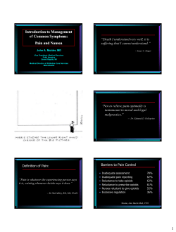
Introduction to Management of Common Symptoms: Pain and Nausea
Document 11282
LISINOPRIL Class Indications
Neonatal Intensive Care Unit Council Annual Report
© Copyright 2026
About abcdocz
DMCA / GDPR
Report