
A
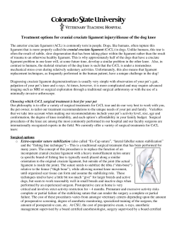
Prolotherapy
Dextrose Prolotherapy and Pain of Chronic TMJ Dysfunction
Many of the subjective symptoms of pain, stiffness, and crunching sensation in
patients with TMJ dysfunction were reduced greater than 50% in 92% of the prolotherapy patients in this study.
In the last issue, Dr. Hauser et al reported on a retrospective study on successful dextrose prolotherapy for patients with
chronic neck pain. In this study, the authors report on fourteen patients who suffered from TMJ pain for an average of 5.4 years
and had seen, on average, four medical doctors—including half who were told that no other treatment options were available.
Overall, substantial improvements were reported in range of motion, pain medicine utilization, disability, depression/anxiety,
quality of life, and patient satisfaction. These improvements persisted through follow up at eighteen months after the conclusion
of prolotherapy treatments. As Dr. Hauser at al report, these positive outcomes resulted despite the inability to individualize treatment protocols since they were done at a charity clinic having limited resources.
—Donna Alderman, DO
Prolotherapy Department Head
By Ross A. Hauser, MD; Marion A. Hauser, MS, RD; and Krista A. Blakemore, BA
ccording to the American Dental
Association, more than 15% of
American adults suffer from
chronic facial pain.1 One of the most
common causes is Tempomandibular
Ross A. Hauser, MD
Joint Disease (TMD), a collective term
used to describe a group of medical disorders causing temporomandibular joint
(TMJ) pain and dysfunction, and is estimated by The National Institute of Dental and Craniofacial Research of the National Institutes of Health to affect 10.8
million people in the United States at any
given time.2 It occurs predominantly in
Marion A. Hauser, MS, RD
women, with the female to male ratio
ranging from 2:1 to 6:1, with 90% of
those seeking treatment being women in
their childbearing years.3,4
The TMJ is often predisposed to similar degenerative changes and pathologies seen in other synovial joints as a consequence of the frequent and repetitive
stresses that the TMJ undergoes.5 SympKrista A. Blakemore, BA
toms commonly associated with TMD include pain at the TMJ, generalized orofacial pain, chronic
headaches and ear aches, jaw dysfunction including hyper- and
hypo-mobility and limited movement or locking of the jaw,
painful clicking or popping sounds with opening or closing of
the mouth, and difficulty chewing or speaking.6 While pain is
the most common symptom, some people report no pain, but
still have problems using their jaws. Sometimes the bite just feels
“off.” Additional symptoms may include ringing in the ears, ear
pain, decreased hearing, dizziness, and vision problems.7
The first-line approach to managing TMD typically includes
resting the jaw, relaxing the jaw muscles, and doing jaw exercises as recommended by a physical therapist.8 Recommendations
A
may also include eating a soft diet that minimizes hard repetitive chewing of crunchy or chewy foods, such as bagels and steak.
All gum chewing must be stopped, talking minimized, and teeth
clenching discouraged. Relaxation exercises that emphasize
gentle range of motion of the joint are recommended. Application of warm compresses to the affected area twice daily, for 10
minutes, to decrease pain and increase joint movement are
done. If this fails, then typically a short course of an anti-inflammatory medication such as ibuprofen is prescribed and often a
dental consultation is given. The dentist then evaluates the patient for malocclusion and bruxism. Many times, a mouth splint
used at night can completely resolve or control the problem.
When pain, clicking, and locking symptoms persist, TMD sufferers commonly seek out the advice of a myriad of TMJ dental
and surgical specialists. Because the causes of TMD are varied
and run the gamut from mechanical issues—such as disc degeneration and dislocation or erosion of the fibrocartilagenous surfaces of the condyle, fossa and articular eminence—to hormonal as well as psychological causes,9,10,11 the treatment approaches for the chronic TMJ case are also quite varied. As surgery is
considered a last resort for TMD, it is common for sufferers to
seek out alternatives and one of the treatments they may consider is prolotherapy.
This article presents a retrospective analysis of patients who
received dextrose prolotherapy to their tempomandibular
joints, and was conducted on a patient population from a charity clinic in rural Illinois. Patients were called by an independent data collector and asked numerous questions concerning
their response to the dextrose prolotherapy they received. The
data was analyzed in all TMJ pain patients, as well as a subset
whose medical doctors told them there were no other treatment
options for their TMJ dysfunction and pain.
Prolotherapy Modality
Prolotherapy, as defined by Webster’s Third New International Dictionary, is “the rehabilitation of an incompetent structure, such
Practical PAIN MANAGEMENT, November/December 2007
©2007 PPM Communications, Inc. Reprinted with permission.
49
Prolotherapy
as a ligament or tendon, by the induced
proliferatin of cells.” “Prolo” comes from
the world proliferate. Prolotherapy injections proliferate or stimulate the growth
of new, normal ligament and tendon tissue.12 In human studies on prolotherapy,
biopsies performed after the completion
of treatment showed statistically significant increases in collagen fiber and ligament diameter of up to 60%.13
Prolotherapy is based on the concept
that the cause of most chronic musculoskeletal pain is ligament and/or tendon
weakness (or laxity). Prolotherapy has
been shown in one double-blinded animal
study over a six-week period to increase
ligament mass by 44%, ligament thickness
by 27%, and the ligament-bone junction
strength by 28%.14 Another animal study
confirmed that prolotherapy induced the
normal healing reaction that occurs when
an injured tissue is healing itself. In this
study, the prolotherapy caused the circumference of tendons to increase by approximately 25% after six weeks time.15
Prolotherapists have a long history
treating TMD since the time of Louis W.
Schultz, MD, DDS in the 1930’s. Dr.
Schultz was unique in that he was both a
dentist and a medical doctor. He was an
Associate Professor in the Department of
Surgery at the University of Illinois and
Rush College of Medicine. He published
several papers on the treatment of subluxation of the temporomandibular joint, including one in 1937 in the Journal of the
American Medical Association.16 In this
paper he described just how common TMJ
syndrome was and that the traditional
treatments of rest, appliances in the
mouth, physical therapy, and surgery were
only partially successful. He described a
simple method of shortening and
strengthening the TMJ capsule by injection (later termed prolotherapy). He tested various solutions in animals until he
found one that caused a strengthening of
the ligaments that support the TMJ but
caused no injury to other structures.17 In
regard to prolotherapy into the TMJ he
found that:
• There was no alteration of the normal joint cavity; the proliferation
occurred in the ligaments.
• There were no gross changes in the ligaments other than their thickening.
• Lymphocytes infiltrate the area
injected within 30 minutes.
• Proliferation of tissue can be seen in
four to six days.
50
He found that a series of three to five
injections were required to often permanently stop the clicking, pain, and hypermobility of the TMJ joint. Dr. Schultz
noted that over the course of his twenty
years of doing prolotherapy for TMD, not
only was it effective, but the treatment
lacked significant side effects.
Dr. Schultz taught the technique of
TMJ prolotherapy to Gustav S. Hemwall,
MD. The primary author has worked with
Dr. Hemwall and eventually assumed his
practice upon his retirement from medicine in 1996. After acquiring Dr.
Hemwall’s practice, Dr. Schultz’s son
came to the clinic for a prolotherapy evaluation. He commented that in his father’s
many years of practice as a dentist, medical doctor, and surgeon, the procedure
that gave him the most amount of satisfaction in treating a TMJ case was prolotherapy.
While practitioners of prolotherapy
since the time of Dr. Schultz have commonly used prolotherapy for all sorts of
TMD, even in cases not involving subluxation, no other studies have been done
since that time. This retrospective observational study was undertaken to evaluate
the effectiveness of Hemwall-Hackett dextrose prolotherapy—not just for TMJ
pain—but also for quality of life measures.
Patients and Methods
Framework and Setting. In October
1994, the authors started a Christian charity medical clinic called Beulah Land Natural Medicine Clinic in an impoverished
area in southern Illinois. The primary
modality of treatment offered was
Hemwall-Hackett dextrose prolotherapy
for pain control. Dextrose was selected as
the main ingredient in the solution because it is the most common proliferant
used in prolotherapy, is readily available,
TABLE 1. PATIENT CHARACTERISTICS PRIOR
TO PROLOTHERAPY
TMJ patients
14
Number of Male Patients
3
11
Number of Female Patients
Average age of TMJ patients
50
Average number of MDs seen (Q3)
4
5.4
Average years of pain (Q2)
Surgery only option to chronic pain 14%
No other treatment options available 50%
Taking 1 pharmaceutical drug for pain 31%
Taking 2 or more pharm drugs for pain 39%
is inexpensive when compared to other
proliferants, and has a high safety profile.
The clinic met every three months until it
ended in July 2005. All treatments were
given free of charge.
Patient Criteria. General inclusion criteria included being at least 18 years old,
having TMD for more than six months,
and a willingness to undergo at least four
prolotherapy sessions (unless the pain remitted with fewer sessions).
Interventions. Each patient received
four to six injections of a 15% dextrose,
0.2% lidocaine solution with a total of two
to four cc’s of solution used per temporomandibular joint. Typically, one cc of solution was injected into the joint and the
remaining solution was injected onto the
TMJ ligament and capsular attachments
on the zygomatic arch and mandibular
condyle and neck (See Figures 1 and 2).
The patients were asked to hold their
mouths half open while the injections were
given. No other therapies were used. The
patients were asked to reduce or stop other
pain medications and therapies they were
using as much as the pain would allow.
Data Collection. Patients who were
seen in the clinic in the years from 20012005 and met the inclusion criteria were
called by telephone and interviewed by an
independent data collector who had no
prior knowledge of prolotherapy and was
the sole data collector gathering the patient information during the telephone
interviews. The patients were asked a series of detailed questions about their pain
and previous treatments before starting
prolotherapy. Their response to prolotherapy treatments was also documented in detail with an emphasis on the effect the treatments had on their need for
subsequent pain treatments, as well as
their quality of life. Specifically, patients
were asked questions concerning years of
pain, pain intensity, overall disability,
number of physicians seen, medications
taken, stiffness, crunching sensations in
the joint, quality of life concerns, and psychological factors. Also noted was whether
the post-treatment benefits continued
substantially after the sessions concluded.
Statistical Analysis. For the analysis,
the results of the patient responses were
calculated by another independent data
collector who had no prior knowledge of
prolotherapy. Pre-prolotherapy treatment responses were compared with the
patients’ responses to the same questions
after prolotherapy treatment.
Practical PAIN MANAGEMENT, November/December 2007
©2007 PPM Communications, Inc. Reprinted with permission.
Prolotherapy
F IGURE 2. Ligamentous structures of the TMJ
typically treated with Hemwall-Hackett dextrose
prolotherapy.
6
Number of Patients
Patients received an average of 4.6 prolotherapy treatments. The average time of follow-up from their
last prolotherapy session was eighteen months.
Pain, Crunching Sensation, and Stiffness. Patients were
asked to rate their pain and stiffness on a scale of 1 to 10, with
1 being no pain/stiffness and 10 being severe, crippling
pain/stiffness. The 14 patients had an average starting pain level
of 5.9, crunching sensation in the TMJ of 5.5, and stiffness of
5.4. Their average ending pain, crunching, and stiffness levels
were 2.5, 2.7, and 2.4 respectively (See Figure 3). Over 71% percent said that they had retained at least 75% of the improvements and 91% noted that they retained at least 50% of their
improvements in their pain, crunching, and stiffness levels since
the last treatment session. Ninety-three percent of patients reported that pain relief was at least 50% while 57% reported
greater than 75% pain relief. Only one person noted that the
long term pain relief was only somewhat successful in having
only 25-49% of the pain relieved.
Range of Motion. Patients were asked to rate their range of
motion on a scale of 1 to 7, with 1 being no motion, 2 through
5 were fractions of normal motion, 6 was normal motion, and 7
was excessive motion. The average starting range of motion was
4.3 and ending range of motion was 5.1. Before prolotherapy,
29% had very limited motion (49% or less of normal motion).
This decreased to only 7% after treatments were concluded.
Pain Medication Utilization. Seventy-one percent discontinued pain medications altogether after prolotherapy. In all, 90%
of patients on medications at the start of prolotherapy were able
to decrease them by 75% or more. None of the patients had to
increase pain medication usage after stopping prolotherapy.
Fifty-seven percent of patients needed no additional pain management care after prolotherapy. After prolotherapy, 93% of patients were able to decrease additional pain management care
by 50% or more.
Disability. In regard to quality of life issues prior to receiving
treatment, 50% exhibited overall disability of at least 50% in that
they could only do about half of the jaw motions without pain.
This decreased to 7% after prolotherapy. In regard to overall
TMJ disability, only 14% noted almost no disability (25% or less)
prior to prolotherapy, but this increased to 72% after treatment
Before Prolo
5
After Prolo
4
3
2
1
0
1
2
3
4
5
7
6
8
9
10
Pain Level
Number of Patients
Treatment Outcomes
FIGURE 1. Typical injection sites for
Hemwall-Hackett dextrose prolotherapy of the TMJ.
Before Prolo
5
After Prolo
4
3
2
1
0
1
2
3
4
5
6
7
8
9
10
Crunching Level
Number of Patients
Patient Characteristics. Complete
data was obtained on 14 patients who
met the inclusion criteria. Of the 14
study participants, 63% were female
and 37% were male. The average age
was 50. Patients reported an average of
5.4 years in pain. Fifty-one percent had
pain greater than six years. The average patient saw four medical doctors
before receiving prolotherapy. Fifty
percent of the patients were told by
their physicians that no other treatment options existed for their pain
problem and 14% were told that surgery was their only option. Thirty-one
percent were taking one pharmaceutical drug, while 39% were taking two or
more drugs for pain (See Table 1).
8
7
6
5
4
3
2
Before Prolo
After Prolo
B
1
0
1
2
3
4
5
6
7
8
9
10
Stiffness Level
FIGURE 3. Starting and ending pain, crunching, and stiffness levels
before and after receiving Hemwall-Hackett dextrose prolotherapy in
14 patients with unresolved TMJ pain.
Practical PAIN MANAGEMENT, November/December 2007
©2007 PPM Communications, Inc. Reprinted with permission.
51
Prolotherapy
Starting
Disability
StartingOverall
Overall Disability
Ending
Overall
Disability
Ending
Overall
Disability
I was not disabled
7%
1 - 25%
14%
50-74% - could do
100% - couldn't do
anything
14%
N/A because I was
not disabled
before
Prolotherapy.
7%
75-99%
14%
25 - 49%
29%
50 - 74% - could do
a little less than
half of what tasks
I wanted to do
22%
a little less than
half of tasks I
wanted to do
7%
25 - 49%
14%
1 - 25%
72%
75 - 99%
0%
100% - couldn't do
anything
0%
FIGURE 4. Starting and ending overall disability before and after receiving Hemwall-Hackett dextrose prolotherapy in 14 patients with
unresolved TMJ pain.
Starting
Depression
Starting
Depression Level
Level
Not depressed
44%
Extremely
depressed and on
medication
21%
Extremely
depressed but not
on medication
7%
Somewhat
depressed
21%
EndingDepression
Depression
Level
Ending
Level
Very depressed
7%
Somewhat
depressed
21%
Not depressed
72%
Very depressed
7%
Extremely
Extremely
depressed and on depressed but not
medication
on medication
0%
0%
FIGURE 5. Starting and ending depression level before and after receiving Hemwall-Hackett dextrose prolotherapy in 14 patients with
unresolved TMJ pain.
Starting
AnxietyLevel
Level
Starting Anxiety
Not anxious
36%
Extremely anxious
and taking
medication
14%
Extremely anxious
but not taking
medication
14%
Very anxious
0%
Ending
Anxiety
Level
Ending Anxiety
Level
Somewhat
anxious
36%
Somewhat
anxious
36%
Not anxious
64%
Extremely anxious
Extremely anxious
and taking
but not taking
medication
Very anxious medication
0%
0%
0%
FIGURE 6. Starting and ending anxiety level before and after receiving Hemwall-Hackett dextrose prolotherapy in 14 patients with
unresolved TMJ pain.
(See Figure 4).
Depression & Anxiety. Prior to prolotherapy, 56% of patients reported feelings of depression and 64% reported feelings of anxiety. After treatments, only 28%
reported depressed feelings and 36% re-
52
ported feelings of anxiety (See Figures 5
and 6). Patients reported that on average
86% of the improvements in depression
and anxiety have at least somewhat continued. Seventy-eight percent of these patients reported 75% continuing improve-
ment at the time of follow-up.
Sleep. Sixty-four percent of patients reported their pain interrupted their sleep
prior to prolotherapy treatments and 55% of
them subsequently showing improvements
in their sleeping ability after treatments.
Practical PAIN MANAGEMENT, November/December 2007
©2007 PPM Communications, Inc. Reprinted with permission.
Prolotherapy
Number of Patients
2.5
Before Prolo
2
After Prolo
1.5
1
0.5
0
1
2
3
4
5
6
7
8
9
10
7
8
9
10
Pain Level
3.5
Before Prolo
Number of Patients
3
2.5
After Prolo
2
1.5
1
0.5
0
1
2
3
4
5
6
Crunching Level
2.5
Number of Patients
Before Prolo
2
After Prolo
1.5
1
0.5
0
1
2
3
4
5
7
6
8
9
10
Stiffness Level
FIGURE 7. Starting and ending pain, crunching, and stiffness levels before and after eceiving Hemwall-Hackett dextrose prolotherapy
in 7 patients with unresolved TMJ pain who were told that there
were no other treatment options.
Quality of Life. To a simple yes or no question, “Has prolotherapy changed your life for the better?” 100% of patients
treated answered “yes.” In quantifying the response,
• Eighty-six percent felt their life was at least very much
better from prolotherapy
• Sixty-nine percent stated that the results from prolotherapy have very much continued to this day (75% or
greater).
• One hundred percent felt that they still have some benefits (at least 25%) from the prolotherapy they received.
Patients who experienced regression of some of their symptoms were asked, “Are there reasons beside the prolotherapy effect wearing off that are causing some return of your pain/disability?” 79% answered “yes.” The patients noted the reasons for
some of their returning pain were the following:
• stopped prolotherapy treatments too soon, (before pain
was completely gone):37%
• re-injury: 14%
• increased life stressors: 21%
• new area of pain: 7%
Of the patients whose pain recurred after prolotherapy was
stopped, 58% were planning on receiving additional prolotherapy treatments.
Patient Satisfaction. Eighty-six percent of patients knew
someone who had benefited from prolotherapy. In fact, 44%
came to receive their first prolotherapy session at the recommendation of a friend who had already received prolotherapy.
Ninety-three percent of patients treated considered the prolotherapy treatment to be very successful (greater than 50% pain
relief). Fifty-seven percent noted the prolotherapy was very successful (greater than 75% pain relief). None indicated that the
prolotherapy treatment made them worse. One hundred percent had subsequently recommended prolotherapy to someone.
“No Other Treatment Options” Subgroup Analysis. Fifty
percent (n=7) of the patients had been told by their doctors that
there were no other treatment options for their pain prior to
presenting for prolotherapy. This group had average starting
pain, stiffness, and crunching levels of 7.1, 5.9, and 5.7, respectively, before prolotherapy. Their ending levels were 3.1, 3.1 and
3.3 for pain, stiffness and crunching levels after treatment (See
Figure 7). Three of the patients noted less than 25% normal TMJ
motion before prolotherapy, but after prolotherapy every patient said they had improved to greater than 25% of normal motion. Before prolotherapy all seven patients were taking at least
one pain medication while, after treatment, only two were taking medications. Five of the patients (71%) had 75% or greater
pain relief, with the other two patients achieving 50-74% pain
relief. Before prolotherapy, 100% had depressed feelings, with
three of the seven (43%) being on medications. All three on medications were able to get off medications after prolotherapy and
four of the seven (57%) no longer had depressed feelings (See
Figure 8). In this group of seven patients, six felt they still had
at least 75% of the benefit they received after the prolotherapy
treatments stopped.
Principal Findings. The results of this retrospective, uncontrolled, observational study, demonstrated that prolotherapy
helps decrease pain and improve the quality of life of patients
with chronic temporomandibular joint symptoms. Decreases in
pain, stiffness, and crunching levels of the TMJ were seen, even
in patients who were told by their physicians that no other treatment options were available. Fifty-seven percent of the patients
achieved greater than 75% pain relief with prolotherapy and
93% of patients stated prolotherapy relieved them of at least
50% of their pain. In regard to quality of life issues prior to receiving treatment, 50% had an overall disability of at least 50%
(jaw motions restricted by about half). This decreased to 7% after
prolotherapy. Prolotherapy also caused clinically relevant improvements in patients’ TMJ range of motion, sleep, depressive
and anxious feelings. Ninety percent of patients on medications
at the start of prolotherapy were able to decrease them by 75%
or more. One hundred percent of patients said that dextrose
prolotherapy changed their life for the better. Eighteen months,
on average, after their last prolotherapy treatment, one hundred percent of patients said they had retained the majority of
their benefits from the treatment.
Study Strengths and Weaknesses. Our study does not compare to clinical trial in which an intervention is investigated
under controlled conditions. Instead, its aim was to document
Practical PAIN MANAGEMENT, November/December 2007
©2007 PPM Communications, Inc. Reprinted with permission.
53
Prolotherapy
S
Starting Depression Level
Ending Depression Level
Very depressed
14%
Extremely
depressed and on
medication
43%
Somewhat
depressed
43%
Not depressed
57%
Extremely
depressed but not
on medication
0%
Very depressed
14%
Not depressed
0%
Somewhat
depressed
29%
Extremely
Extremely
depressed and on depressed but not
medication
on medication
0%
0%
FIGURE 8. Starting and ending depression levels before and after receiving Hemwall-Hackett dextrose prolotherapy in 7 patients with
unresolved TMJ pain who were told that there were no other treatment options.
the response of patients with chronic temporomandibular joint dysfunction to the
Hemwall-Hackett technique of dextrose
prolotherapy. Strengths of the study were
that numerous quality of life parameters
affecting TMJ sufferers were studied.
Quality of life issues such as stiffness,
range of motion, overall disability, sleep,
anxiety, and depression—in addition to
pain level—are important factors affecting an individual with chronic TMJ syndrome. Decreases in medication usage
and additional pain management care
were objective measures that were also
documented.
Though the sample in this study was
small (n=14), the quality of the cases
treated is notable. The average person in
this study had unresolved TMJ pain/dysfunction for 5.4 years and had been seen,
on average, by four medical doctors prior
to receiving prolotherapy. Fifty percent of
the cases were told that no other treatment options existed and 14% were told
surgery was their only option. A follow-up
time of eighteen months, on average,
since their last treatment session provided a measure of the long-lasting effect of
this modality.
Because this was a charity medical clinic with limited resources and personnel,
the only therapy offered was prolotherapy treatments given every three months.
In private practice, the Hemwall-Hackett
technique of dextrose prolotherapy is typically given every four to six weeks. If a
client is not improving or has poor healing ability, the prolotherapy solutions may
be changed or strengthened or the client
is advised about additional measures to
improve their overall health. This can include advice on diet, supplements, exer-
54
cise, changes in medications, additional
blood tests, physiotherapy, and/or other
medical care. Often clients are weaned immediately off any anti-inflammatory and
narcotic medications that inhibit the inflammatory response that is needed to
achieve a healing effect from prolotherapy. Since none of these were done, the results of this study are expected to represent the least optimum level of success
achievable with Hemwall-Hackett dextrose prolotherapy.
Another shortcoming of this study was
the subjective nature of some of the evaluated parameters, including pain, anxiety, depression, and disability levels since
the results relied on answers to questions
by the patients. Further, any additional
pain management care that the patients
may have been receiving was not controlled. Lack of x-ray and MRI correlation for diagnosis and response to treatment, as well as a lack of physical examination documentation in the patients’
charts made categorization of the patients into various diagnostic parameters
impossible.
Discussion. While the exact cause of
chronic temporomandibular dysfunction
is still debated, this study did demonstrate
that the Hemwall-Hackett technique of
dextrose prolotherapy improves not only
the pain level for those having chronic
TMD, but also a host of other quality of
life measures. The Hemwall-Hackett technique of dextrose prolotherapy to the
temporomandibular joint involves injections into the joint, as well as the fibro-osseous junction of the ligament and capsular attachments on the zygomatic arch, as
well as the mandibular neck and condyle.
Clearly the structural goal of Hemwall-
Hackett dextrose prolotherapy is to improve the stability of the TMJ by enhancing capsular and ligament strength. Congenital disorders that are characterized by
overstretched ligaments, such as EhlersDanlos Syndrome, are typically predisposed to TMJ problems.18 Weakening of
the TMJ capsule and ligament would explain a lot of the varied pathology involving TMD including joint subluxations,
disc displacements, as well as muscle
spasms and myofascial pain patterns.
The most common cause of TMJ pain
is myofascial pain dysfunction syndrome
and primarily involves the muscles of
mastication.19 While massage, physiotherapy, pain medications, splints, surgeries,
and other treatment modalities offer temporary help, they rarely cure the condition.20,21,22 A known cause of persistent
muscle spasms and myofascial pain dysfunction is underlying ligament laxity.23
By stimulating ligament and capsular repair for such cases, prolotherapy would
represent a more permanent solution.
The most common presentation of the
TMJ is disc displacement.24 In essence,
this is when the articular disc, attached
anteriorly to the superior head of the lateral pterygoid muscle and posteriorly to
the retrodiscal tissue, moves out from between the condyle and the fossa, so that
the mandible and temporal bone contact
something other than the articular disc.
In most instances of the disorder, the disc
is displaced anteriorly upon translation.
On opening, a “pop” or “click” can sometimes be heard—and usually felt—indicating the condyle is moving back onto
the disc.25
The TMJ is divided into an upper and
lower joint cavity by a fibrocartilaginous
Practical PAIN MANAGEMENT, November/December 2007
©2007 PPM Communications, Inc. Reprinted with permission.
Prolotherapy
articulating disc.26 It is thicker posteriorly, thus making posterior dislocations
more unlikely. Anteriorly, the disc is fused
with the thin, loose, and fibrous joint capsule. The ligaments which contribute to
the formation of the fibrous joint capsule
and unite the articular bones are the temporomandibular (a.k.a. lateral), sphenomandibular, and stylomandibular. The
temporomandibular ligament restrains
the movement of the mandible and prevents compression of tissues behind the
condyle.27 Some authors note that this
collateral ligament is simply a thickening
of the joint capsule.28 The joint capsule
itself attaches to the articular eminence,
the articular disc, and the neck of the
mandibular condyle. Basically, the articular disc is a fibrous extension of the capsule between the two bones of the joint.29
The sphenomandibular and stylomandibular ligaments keep the condyle,
disc, and temporal bone firmly opposed
and the multiple ligamentous attachments provide disc stability. Laterally, the
disc is continuous with ligament tissue attaching it to the neck of the condyle.30
While the cause of disc displacement is
still under debate, an argument could be
made that for many, it is injury to the joint
capsule and TMJ ligament complex that
is the issue. Anteriorly, the TMJ disc depends on the support of the joint capsule
and TMJ ligament complex. If, for some
reason, these became weakened,
stretched, or torn, anterior disc dislocation would result. Only treatments designed to specifically strengthen and repair the injured joint capsule and ligament structures—such as prolotherapy—
would have a lasting effect.
Conclusions
In this observational study, the HemwallHackett technique of dextrose prolotherapy used on patients who presented with
over five years of unresolved TMJ pain
and dysfunction were shown to improve
their quality of life even eighteen months
subsequent to their last prolotherapy session. All patients reported significantly reduced levels of pain, stiffness, crunching
sensation, disability, depression, anxiety,
medication, and other pain therapy. They
also reported improved range of motion
and sleep. The results confirm that prolotherapy is a treatment that should be
highly considered for people suffering
with unresolved temporomandibular
joint pain and dysfunction.n
Ross A. Hauser, MD is the Medical Director of Caring Medical and Rehabilitation Services in Oak Park, IL and is a renowned prolotherapist and Natural Medicine Specialist
with a national referral base seeing patients
from all over the USA, and abroad. Dr. Hauser
and his wife, Marion, authored the national
best seller “Prolo Your Pain Away! Curing
Chronic Pain with Prolotherapy,”, now in its
third edition, along with a four-book topical
mini series of prolotherapy books. He also spearheaded the writing of a 900-page epic sports
book that discusses the use of prolotherapy for
sports injuries, “Prolo Your Sports Injuries
Away! Curing Sports Injuries and Enhancing
Athletic Performance with Prolotherapy.”
Marion A. Hauser, MS, RD is the CEO of
Caring Medical and Rehabilitation Services, a
comprehensive Natural Medicine Clinic in Oak
Park, IL and owner of Beulah Land Nutritionals. As a registered dietitian, Marion is also a
well-known speaker and writer on a variety of
topics related to natural medicine and nutrition
providing information for weekly e-newsletters
and TV shows on a variety of health topics.
Marion has recently released “The Hauser Diet:
A Fresh Look at Healthy Living.” Along with
her husband, Dr. Ross Hauser, Marion co-authored the national best seller entitled “Prolo
Your Pain Away! Curing Chronic Pain with
Prolotherapy” along with a four-book topical
mini series of prolotherapy books, as well as a
comprehensive sports book discussing the use of
prolotherapy for sports injuries.
Krista A. Blakemore, BA graduated with
honors from Eastern Washington University in
Cheney, Washington in 2002, and is research
assistant to Ross and Marion Hauser of Caring Medical and Rehabilitation Services in
Oak Park, Illinois.
References
1. American Dental Association. Available at
http://www.ada.org/public/topics/tmd_tmj.asp Accessed 11/9/07.
2. National Institute of Dental and Craniofacial Research. Available at: http://www.nidcr.nih.gov. Accessed
11/9/07.
3. Van Korff M, Dworkin, SF, Le Resche L, and Kruger
A. An epidemiologic comparison of pain complaints.
Pain. 1988. 32: 173-183.
4. Ta LE and Dionne RA. Treatment of painful temporomandibular joints with a cyclooxygenase-2 inhibitor: a
randomized comparison of celecoxib to naprosyn.
Pain. 2004. 111: 13-21.
5. Helland MM. Anatomy and function of the temporomandibular joint. JOSPT. 1980. 1(3): 145-52.
6. Mayo Clinic. Available at:
http://www.mayoclinic.com/health/tmj-disorders/
DS00355/DSECTION=6 Accessed 11/9/07.
7. The TMJ Association. Available at:
http://www.tmj.org/basics.asp Accessed 11/9/07.
8. Eriksson PO and Zafar H. Musculoskeletal disorders
in the jaw, face and neck. In Rakel RE, Bope ET, eds.,
Conn’s Current Therapy in TMJ. WB Saunders. Philadel-
phia, PA. 2005. pp 1128-1133.
9. Malone TP, McPoil T, and Nitz AJ. Orthopaedic and
Sports Physiotherapy. Third Edition. Mosby. Philadelphia, PA. 1997.
10. Tabassum N, Trang D, and Gihan H. Relaxin’s Induction of Metalloproteinases is Associated with the
Loss of Collagen and Glycosaminoglycans in Synovial
Joint Fibrocartilaginous Explants. Arthritis Res Ther.
2005. 7(1): R1-R11.
11. Meldolesi G and Picardi A. Personality and psychopathology in patients with temporomandibular joint
dysfunction syndrome. A controlled investigation. Psychother Psychosom. 2000. 69: 322-328.
12. Dorman T. Treatment for spinal pain arising in ligaments using Prolotherapy: A retrospective study. Journal of Orthopaedic Medicine. 1991. 13(1): 13-19.
13. Klein R. Proliferant injections for low back pain: histologic changes of injected ligaments and objective
measures of lumbar spine mobility before and after
treatment. Journal of Neurology, Orthopedic Medicine
and Surgery. 1989. 10: 141-144.
14. Liu Y. An in situ study of the influence of a sclerosing solution in rabbit medical collateral ligaments and
its junction strength. Connective Tissue Research.
1983. 2: 95-102.
15. Maynard J. Morphological and biomechanical effects of sodium morrhuate on tendons. Journal of Orthopaedic Research. 1985. 3: 236-248.
16. Schultz L. A treatment of subluxation of the temporomandibular joint. JAMA. September 25, 1937.
17. Schultz L. Twenty years’ experience in treating hypermobility of the temporomandibular joints. American
Journal of Surgery. Vol. 92. December 1956.
18. Hagberg C, Korpe L, and Berglund B. Temporomandibular joint problems and self-registration of
mandibular opening capacity among adults with
Ehlers-Danlos Syndrome. A questionnaire study. Orthod Carniofac Res. 2004. 7(1): 40-6.
19. Darnell M. A proposed chronology of events for forward head posture. The Journal of Craniomandibular
Practice. 1983. 1: 62-66.
20. Ernest E. Three disorders that frequently cause
temporomandibular joint pain: internal derangement,
temporal tendonitis, and Ernest syndrome. Journal of
Neurological Orthopedic Surgery. 1986. 7: 189-191.
21. Headache Relief Newsletter, Edition 13. Philadelphia, PA: The Pain Center, 1995.
22. Al-Ani MZ, et al. Stabilisation splint therapy for temporomandibular pain dysfunction syndrome. Cochrane
Rev Abstract. 2007. Available at: http://www.medscape.com/viewarticle/486213_print Accessed 11/9/07.
23. Hauser R and Hauser M. Prolo Your Sports Injuries
Away! Beulah Land Press. Oak Park, IL. 2001.
24. Epker, J. A model for predicting TMD: Practical application in clinical settings. Journal of the American
Dental Association. 1999. 130: 1470-1475.
25. Tecco S, Festa F, and Salini V. Treatment of joint
pain and joint noises associated with a recent TMJ internal derangement: a comparison of an anterior repositioning splint, a full-arch maxillary stabilization splint,
and an untreated control group. Cranio. 2004. 22(3):
209-219.
26. Rao V, Ferule A, and Karasick D. Temporomandibular joint dysfunction: Correlation of MR imaging,
arthrography and arthroscopy. Radiology. 1990. 174:
663-667.
27. Hall L. Physiotherapy treatment results for 178 patients with temporomandibular joint syndrome. Am J
Otol. Jan 1984. 5(3): 183-96.
28. Laskin D. Diagnosis of pathology of the temporomandibular joint: Clinical and imaging perspectives.
Radiol Clin North Am. 1993. 31: 135-147.
29. Temporomandibular Joint. Available at
http://en.wikipedia.org/wiki/Temporomandibular_joint
Accessed 11/9/07.
30. Roth C, Ward R, and Tsai S. MR Imaging of the
TMJ: A Pictorial Essay. Appl Radiol. 2005. 34(5): 9-16.
Practical PAIN MANAGEMENT, November/December 2007
©2007 PPM Communications, Inc. Reprinted with permission.
55


© Copyright 2026