
Salivary Gland Dysfunction: Etiology, Epidemiology, Clinical Manifestations, Diagnosis, and Treatment
Salivary Gland Dysfunction: Etiology,
Epidemiology, Clinical Manifestations,
Diagnosis, and Treatment
Vidya Sankar, DMD, MHS; Géza T. Terézhalmy, DDS, MA
Continuing Education Units: 2 hours
This continuing education course presents the etiology, epidemiology, clinical manifestations, diagnosis, and
treatment of salivary gland dysfunction.
Conflict of Interest Disclosure Statement
• Dr. Sankar reports no conflicts of interest associated with this work.
• Dr. Terézhalmy has done consulting work for P&G.
ADA CERP
The Procter & Gamble Company is an ADA CERP Recognized Provider.
ADA CERP is a service of the American Dental Association to assist dental professionals in identifying
quality providers of continuing dental education. ADA CERP does not approve or endorse individual courses
or instructors, nor does it imply acceptance of credit hours by boards of dentistry.
Concerns or complaints about a CE provider may be directed
to the provider or to ADA CERP at:
http://www.ada.org/prof/ed/ce/cerp/index.asp
Overview
Salivary gland dysfunction may be characterized by either hyposalivation or hypersalivation. To provide
competent care to patients with salivary gland dysfunction, clinicians must understand its many causes and
associated complications, and develop preventive and therapeutic strategies accordingly.
1
®
Crest® Oral-B at dentalcare.com Continuing Education Course, March 12, 2012
Learning Objectives
Upon completion of this course, the dental professional should be able to: • Discuss the etiology and epidemiology of salivary gland dysfunction.
• Recognize the clinical manifestations of salivary gland dysfunction.
• Diagnose salivary gland dysfunction.
• Develop strategies for the prevention and treatment of hyposalivation and hypersalivation.
Course Contents
salivary glands, located in labial, buccal, and
palatal oral mucosa, account for the balance.4
• Introduction
• Hyposalivation
Etiology and Epidemiology
Non-infectious Sialoadenitis
Infectious Sialoadenitis
Benign and Malignant Salivary Gland
Tumors
Sialoadenosis
Sjögren’s Syndrome
Diabetes Mellitus (DM)
Polypharmacy
Head and Neck Radiotherapy
Clinical Manifestations
Diagnosis
Therapeutic strategies
Palliative and Supportive Care
Sialagogues
Caries Prevention
Candidiasis
• Hypersalivation
Etiology and Epidemiology
Clinical Manifestations
Therapeutic strategies
• Conclusion
• Course Test
• References
• About the Authors
A healthy person secretes up to 1.5L of saliva
per day.5 Whole saliva is composed mostly of
water and contains many organic and inorganic
substances.5,6,7,8,9 Two defining constituents are
amylase10,11 and mucin12. The parotid glands
secrete predominantly amylase-rich serous saliva;
the sublingual and minor salivary glands secrete
predominantly mucin-rich mucous saliva; and the
submandibular glands secrete mixed seromucous
saliva. The parotid glands contribute most of the
stimulated saliva, while the submandibular glands
contribute most of the unstimulated saliva. Minor
salivary glands produce up to 70% of the mucin.
Saliva provides a physical barrier against local
irritants and promotes mucosal repair (epidermal
growth factor); lubricates and cleans oral
tissues (mucin); maintains a stable intraoral
pH with its bicarbonate buffering system;
and maintains electrolyte balance.13 Saliva
prevents demineralization and contributes to the
remineralization (proline-rich proteins) of dental
hard tissues.14 Saliva contains antibacterial
(lysozymes), antiviral (secretory leukocyte
protease inhibitors), and antifungal (histatins)
components, which help to maintain a favorable
ecosystem for the normal oral flora.15 Finally,
saliva facilitates mastication, the formation of a
bolus (mucin and water), swallowing, initiates
food processing (amylase), and has a salutary
effect on the sense of taste.4
Introduction
Salivary gland function is under the control of
the autonomic nervous system.1,2,3 In response
to smell, taste, and mastication, afferent fibers of
the salivary reflex arch activate the salivary nuclei
located in the medulla oblongata.4 Sympathetic
and parasympathetic fibers innervating the
salivary glands, with acetylcholine as the
neurotransmitter, comprise the efferent part of
the secretory reflex arch.3 Following a circadian
rhythm, the major salivary glands (parotid,
sublingual, and submandibular) are responsible
for the secretion of about 90% of all saliva; minor
Salivary gland dysfunction may be characterized
by either hyposalivation or hypersalivation. To
provide competent care to patients with salivary
dysfunction, clinicians must understand its many
causes and associated complications, and
develop preventive and therapeutic strategies
accordingly.
2
®
Crest® Oral-B at dentalcare.com Continuing Education Course, March 12, 2012
Hyposalivation
duct is severed by trauma, saliva leaks into the
surrounding connective tissue, which results in
a non-epithelium-lined fluid-filled cavity (Figure
1). Ranulas present as either circumscribed
lesions (subsequent to ductal obstruction and
cystic dilatation) or plunging lesions (following
extravasation of saliva into tissues of the floor of
A patient is considered to have reduced salivary
flow if the unstimulated salivary flow rate
measured for 5 to 15 minutes is ≤0.1 mL/min; or
if the stimulated salivary flow rate measured for 5
minutes is ≤0.5 mL/min.5
Etiology and Epidemiology
Hyposalivation may be related to rare congenital
absence or aplasia of one or more major salivary
glands or ducts; non-infectious and infectious
sialoadenitis; benign and malignant salivary
gland tumors; and systemic conditions that
directly affect salivary gland function, or more
commonly secondarily, as side effects associated
with pharmacotherapeutic agents prescribed
for the treatment of a primary disease.16,17
Hyposalivation is also a major complication
of cancer chemotherapy and head and neck
radiotherapy.18,19
Figure 1. Mucocele
There is a paucity of data on the true prevalence
of hyposalivation in the general population;
reports vary from 0.9% to 64.8%.20 In a 15-year
longitudinal study of xerostomia, it was found that
hyposalivation increased from 6% at age 50 to
15% at age 65.21 Other investigators estimated
that among the elderly ≥65 years old, the
incidence of xerostomia may be as high as 30%.22
However, there is no strong evidence that saliva
production by the major glands is significantly
decreased with age.22,23 It is more likely that
the increased incidence of systemic disease
in the elderly and associated polypharmacy is
the primary contributor to clinically significant
salivary gland hypofunction.24,25,26 A 5-year
longitudinal study in a cohort of older persons
found that the severity of hyposalivation was
higher among women and related to the use of
xerogenic medications.27 Clearly, the incidence
of hyposalivation increases with exposure to
polypharmacy and over 400 drugs have been
identified as xerogenic.28 The prevalence
of hyposalivation for patients with Sjögren’s
syndrome and those who have received head
and neck radiotherapy is nearly 100%.22
Figure 2. Ranula
Figure 3. Sialolith
Non-infectious Sialoadenitis
Non-infectious sialoadenitis is most commonly
due to obstruction of a salivary gland duct. Mucoceles are common reactive minor
salivary gland lesions of the lower lip. As a
Figure 4. Sialolith Radiograph
3
®
Crest® Oral-B at dentalcare.com Continuing Education Course, March 12, 2012
the mouth) in association with a submandibular or
sublingual gland (Figure 2). Sialoliths develop
primarily in the submandibular ductal system
and are caused by calcification of mucous
plugs and cellular debris (Figures 3 and 4). Sialoliths tend to occur much less frequently in
association with the parotids and are considered
to be rare in sublingual and minor salivary
glands. Mucous cysts of minor salivary glands
are caused by ductal obstruction by a mucous
plug, other cellular debris, or calcification and
may be characterized as mucous extravasation
surrounded by granulation tissue or as true cysts
lined with ductal epithelium.29
swelling of the parotid and/or other salivary
glands.33,34 Characteristic findings include an
outward lifting of the ear lobe, obscuration of the
angle of the mandible, and erythema affecting
Stensen’s ducts. The parotitis progresses over
2-3 days and persists for about a week. While
the hallmark of mumps is parotitis (92%), the
sublingual and submandibular glands are also
affected in about 7% of cases.35
Approximately 5-10% of HIV infected individuals
and those with AIDS experience salivary
gland disease.36 Parotid gland enlargement
usually occurs bilaterally and is associated with
cervical lymphadenopathy. Diffuse infiltrative
lymphocytosis syndrome (DILS) results from CD8
lymphocytic infiltration and is often followed by
lymphoepithelial cyst formation. This will affect
either the parotid or submandibular glands with
a predilection for the parotids. Salivary gland
disease is considered a Group 2 oral condition,
i.e., a condition less commonly associated with
HIV infection.37
Infectious Sialoadenitis
Bacterial
Bacterial infections, typically affecting the parotid
glands, are more common in the elderly who are
dehydrated or experience salivary hypofunction
secondary to medications, head and neck
radiation, and systemic diseases. Organisms
implicated include Staphylococcus aureus,
Streptococcus viridians, group B streptococci,
Haemophilus influenza, Escherichia coli, and
Pseudomonas aeruginosa. Cases of tuberculous
parotitis have also been documented.30,31,32
Benign and Malignant Salivary Gland Tumors
Benign and malignant salivary gland tumors
primarily affect the palate followed by the upper
lip. Signs and symptoms of a malignant salivary
gland tumor include a swelling with facial nerve
paralysis, pain, or facial paresis. The majority
of tumors that arise within the salivary glands
originate from epithelial tissues; however
some are derived from adjacent tissues or
structures (i.e., adipose, nerves, blood vessels,
lymph nodes, lymphatics). Most salivary gland
neoplasms are benign. Pleomorphic adenoma
and papillary cystadenoma lymphomatosum
represent 75% and 5% of all salivary gland
tumors, respectively.38 Adenoid cystic carcinoma
(Figure 5) and mucoepidermoid carcinoma (Figure
6) are the most common malignant salivary
Viral
Viral infections can occur in persons of all ages
and preferentially affect the parotid glands.
Organisms commonly implicated in children and
adolescents include the mumps virus; in adults
the Cytomegalovirus, Coxsackievirus, EpsteinBarr virus, hepatitis C virus, and the human
immunodeficiency virus (HIV).29
Mumps, an acute viral infection caused by an
RNA virus of the Paramyxoviridae family, is
characterized by unilateral or bilateral tender
Figure 6. Mucoepidermoid carcinoma
Figure 5. Adenoid cystic carcinoma
4
®
Crest® Oral-B at dentalcare.com Continuing Education Course, March 12, 2012
gland tumors, representing 10% (5% and 5%)
of all salivary gland tumors. Less common
malignant tumors include acinic cell carcinoma,
adenocarcinoma, squamous cell carcinoma, and
carcinoma arising in a pleomorphic adenoma.39
Alzheimer disease, Bell’s palsy], and others
[fibromyalgia, chronic fatigue syndrome]).40,41 In
addition, hyposalivation is a predictable sequel of
polypharmacy, cancer chemotherapy and head
and neck radiotherapy.15,16,40
Sialoadenosis
Sialoadenosis is seen in association with a
number of systemic conditions (e.g., chronic
inflammatory/autoimmune diseases [Sjögren’s
syndrome, systemic lupus erythematosus,
scleroderma, graft-versus-host-disease
(GVHD)], endocrine/metabolic disorders
[diabetes mellitus, thyroid dysfunction, adrenal
dysfunction], nutritional abnormalities and eating
disorders [anorexia nervosa, bulimia, alcohol
abuse, and neuropathic diseases [depression,
Sjögren’s Syndrome
Sjögren’s syndrome is a relatively common
condition primarily affecting women (it may be
as prevalent as 1 out of every 2,500 females)
with a typical onset during the fourth or fifth
decade of life.42,43,44 Primary Sjögren’s syndrome
is characterized by dry mouth and dry eyes,
which are the result of a progressive loss of
salivary and lacrimal gland function.42 As the
disease progresses, lymphocytic infiltrates of
salivary glands ultimately produce acinar gland
5
®
Crest® Oral-B at dentalcare.com Continuing Education Course, March 12, 2012
degeneration, necrosis, atrophy, and complete
salivary gland destruction.45 Patients frequently
present with intermittent enlargement of the
parotid and/or submandibular glands. The
diagnosis of primary Sjögren’s syndrome is
predicated on meeting at least 4 of the 6 criteria
in (Table 1).46 In its secondary form, Sjögren’s
syndrome is associated with co-morbidities
such as rheumatoid arthritis, systemic sclerosis,
systemic lupus erythematosus, or scleroderma.47,48
forms of DM.54 In addition, a significant reduction
in resting salivary flow rates was reported in
patients with type 1 DM taking parasympatholytic
drugs in association with elevated fasting blood
sugar levels.52 A similar association was reported
between parasympatholytic drugs and autonomic
neuropathy in patients with type 2 DM.54
Polypharmacy
Xerostomia is a common complication of
both prescription and non-prescription drug
therapy and over 400 active medications have
been implicated (Figures 7 and 8).28 Reduced
salivary flow is generally related to a drug’s
parasympatholytic or antimuscarinic effect. The
site of action of these drugs may be in the central
nervous system, at parasympathetic and some
sympathetic ganglia, and at parasympathetic and
some sympathetic effector junctions.
Diabetes Mellitus (DM)
In well-controlled diabetic patients, salivary
gland function does not seem to be significantly
affected.49 However, several studies have
reported subjective complaint of dry mouth
among diabetic patients in general.50,51,52 In one
study, investigators reported an association
between lower resting and stimulated salivary
flow and elevated HbA1c levels.50 In another
study, investigators have demonstrated
reduced resting and stimulated salivary flow
rates in type 1 diabetic patients with elevated
fasting blood glucose (FBG) concentrations
and diabetic neuropathy.52 Abnormalities in
parotid gland basement membranes have
also been demonstrated in subjects with DM.53
Sialoadenosis, associated with excessive
accumulation of secretory granules and
enlargement of the acinar cells characterized by
a diffuse, noninflammatory, bilateral enlargement
of the parotid glands was reported with both
Other drug-induced salivary gland problems
include fluid and electrolyte imbalance, glandular
vasoconstriction, and altered fluid movement
from plasma through salivary acinar cells to the
ductal system, and ultimately into the oral cavity.
The major classes of drugs causing xerostomia
are included in (Table 2).28,40 Salivary gland
dysfunction associated with cytotoxic drugs
appears to be temporal, during and immediately
after treatment, and salivary function returns to
pre-chemotherapy levels in most patients after
completion of therapy.19,55
Figure 7. Antihistamine-induced dry mouth
Figure 8. Diuretic-induced dry mouth
6
®
Crest® Oral-B at dentalcare.com Continuing Education Course, March 12, 2012
Head and Neck Radiotherapy
Xerostomia is the most frequently reported
late side effect of radiotherapy to the head and
neck (Figures 9 and 10).56-62 For reasons not
clearly understood, the well-differentiated and
slow replicating salivary gland cells are highly
radiosensitive.61,63,64 The effects are profound and
depend on the volume of the glands irradiated,
the total dose delivered, and the functional state
of the glands prior to radiotherapy.61,63,65
Radiotherapy causes acute degeneration and
necrosis of acinar cells, followed by attempted
regeneration. If the radiation dose to the salivary
glands is sufficient, regeneration fails leading
to fibrosis and atrophy of glandular tissue.64,66,67
While a mean dose of 60 Gy is the accepted
threshold for producing irreversible damage, in
some cases mean doses of as little as 10 Gy
have been implicated.60,63,65 Radioactive iodine
(I-131) used in the treatment for cancers of
the thyroid gland may cause parotid but not
submandibular dysfunction in a dose-dependent
fashion.68
Figures 9 & 10. Radiation-induced dry
mouth and caries
It is generally accepted that the serous acini are
more radiosensitive and less likely to regenerate
compared to the mucous acini, although a recent
study suggests that serous and mucous acini are
equally radiosensitive. Acinar recovery, if any, is
dose dependent and may occur approximately 12
to 18 months following completion of therapy. As
a general rule, however, patients will experience
significant reduction in salivary flow for the
remainder of their life. When the major glands
are affected, salivary output may be reduced
7
®
Crest® Oral-B at dentalcare.com Continuing Education Course, March 12, 2012
by as much as 80% within the first 2 weeks of
conventional radiotherapy.69
As a consequence, a distinctive form of caries
begin on plaque forming surfaces and areas
of exposed dentin resulting in circumferential
lesions at the cementoenamel junction, and
smooth surface caries on cusp tips and incisal
edges where attrition had previously occurred. Tooth loss, a predictable sequel of advanced
carious lesions, presents further difficulties for
both the patient and clinician. Alteration of the
normal flora may also increase the potential for
aspiration pneumonia and colonization of the lungs
with gram-negative anaerobes from the gingival
sulcus.73,74
Clinical Manifestations
Qualitative and quantitative changes in saliva
lead to reduced lubrication. The dryness affects
taste and speech. The oral mucosal tissues
appear dry and atrophic, the tongue is often
fissured. Loss of mucosal integrity may lead to
mucositis. Reduced lavage and cleansing of oral
tissues promotes plaque accumulation, gingivitis,
periodontitis, and dental caries.16
Mucositis (i.e., loss of mucosal integrity) may
result in reduced lysozyme, secretory leukocyte
protease inhibitor, and histatin activity, which
predisposes to bacterial, viral, and fungal
infections.70 Secondary infections and mucositis
may compromise the wearing of tissue-borne
prostheses and lead to impaired chewing and
swallowing. Impaired chewing and swallowing
and / or the presence of dysgeusia, hypogeusia,
or ageusia may cause the patient to alter their
diet and fluid intake, increasing the risk of
malnutrition and weight loss. Hyposalivation
alone may decrease the retention of even well
fitting prostheses, which in turn may lead to the
development of traumatic ulcers. In irradiated
patients, such ulcers take on added significance
because they provide a portal to oral bacteria,
with a potential for the development of markedly
morbid osteoradionecrosis.18,71
Reduced antibacterial and antifungal activity
associated with hyposalivation also contributes
to an increased risk of both acute and chronic
infection by the Candida spp, which under normal
conditions is inhibited by a normal bacterial flora
and salivary gland function.75 The lesions often
appear as white, raised, or cottage cheese-like
growths that can be scraped off, leaving a red,
hemorrhagic base. Candidiasis may also appear
as an erythematous lesion under prostheses or
as angular cheilosis. A comprehensive review
of oropharyngeal candidiasis is presented
elsewhere.76
Diagnosis
The diagnosis of salivary gland dysfunction
is predicated on a comprehensive physical
evaluation. It typically begins by assessing the
patient’s chief complaint in an attempt to identify
conditions or predisposing factors.77 Subjective
assessment of salivation may include simple
questionnaire requiring a “yes” or “no” response
to four questions (Box 1).78 A visual analogue or
an ordinal scale based on ranked categories, i.e.,
“I have no – slight – severe – annoying feeling of
dry mouth,” may also provide subjective data.79
Objective evaluation requires a measurement of
the patient’s salivary flow rates.80
Decrease in salivary immunoglobulin levels
leads to a highly cariogenic oral microflora,
(Streptococcus mutans, lactobacillus and
Actinomyces). In addition, the lack of saliva
promotes the availability of sugars and other
substrates essential for microbial survival and
the loss of salivary buffering capacity and loss of
the insoluble pellicle formed by saliva on teeth
interfere with normal remineralization of teeth.72
8
®
Crest® Oral-B at dentalcare.com Continuing Education Course, March 12, 2012
Serologic testing for antinuclear antibodies (e.g.,
rheumatoid factor, anti-Ro/anti-SS-A, anti-La/antiSS-B), minor salivary gland biopsy (lymphocytic
infiltrates), and a variety of imaging techniques
(e.g., sialograms performed with radio-opaque
iodine and extraoral radiographs and radioactive
isotype scintiscans (T99 pertechnetate) can
provide additional diagnostic data. MRI and CT
scans can help rule out salivary gland tumors and
other pathoses associated with the craniofacial
region that may adversely affect salivation.
The diagnosis of salivary gland neoplasia
requires incisional or excisional biopsies and
histopathological evaluation.
examinations to identify sore spots and to enhance
adhesion with soft- and hard-tissue relines.
Patients with residual salivary gland function
may benefit from using simple dietary measures
such as eating carrots or celery, or by chewing
sugarless or xylitol-containing gums. A
variety over-the-counter saliva substitutes and
moisturizers are also available. The viscosity
and electrolyte concentrations of these agents
are adjusted to approximate whole saliva and
pleasant-tasting flavoring is added to most
preparations. Patient acceptance is variable and
depends on effect, duration, lubrication, taste,
delivery system, and cost.71
Therapeutic strategies
Sialagogues
Pilocarpine hydrochloride (Salagen®, Pharmacia,
Saint Paul, MN) a cholinergic agonist, stimulates
the production of saliva from functioning salivary
glands.81 It enhances oral comfort, improves the
ability to speak, and offers prolonged symptomatic
relief. The recommended initial dosing regimen is
5 mg three times daily. If necessary, up to 30 mg
per day may be given in divided doses not exceed
2 tablets per dose. The lowest effective dose
should be used to maintain optimal salivary flow. The continuing effect of pilocarpine hydrochloride
depends on regular use. Maximum benefit is
typically noted after 90 days of use. Pilocarpine
hydrochloride is contraindicated in patients with
uncontrolled asthma or narrow angle glaucoma,
and should be used with caution in patients with
significant cardiovascular disease. Common
dose-dependent side effects include sweating,
nausea, rhinitis, flushing, and increased urinary
frequency.82
Once a diagnosis of salivary gland dysfunction is
established, medical management is predicated
on its etiology and to a great extent falls in the
purview of the medical profession. Irrespective
of the therapeutic strategy, however, the role of
oral healthcare providers is critical to minimize
the many potential oral complications of salivary
gland hypofunction.
Palliative and Supportive Care
Good oral hygiene and adequate hydration are
essential elements of palliative and supportive
care. Desiccated oral tissues are friable and
prone to irritation and ulceration. Patients should
be instructed to avoid products irritating to oral
soft tissues (such as alcohol and tobacco; hot,
spicy, and coarse foods; fruits and beverages
with a high acid content); to refrain from wearing
removable prostheses; to eat a dental soft diet;
and to frequently rinse with a sodium bicarbonate
solution. Xerostomia-related mucosal lesions
are susceptible to secondary infections by
microorganisms of the normal flora that have
become pathogenic and by transient organisms. Prompt treatment of such infections is mandatory.
Another sialagogue, cevimeline hydrochloride
(Evoxac®, Daiichi Sankyo Inc., Tokyo, Japan)
has been approved by the Food and Drug
Administration (FDA) for the treatment of dry
mouth in Sjögren’s syndrome in a dosage of 30
mg three times daily.71,81,83 Like pilocarpine, it is a
muscarinic agonist that increases production of
saliva. Pilocarpine is a non-selective muscarinic
agonist, whereas cevimeline reportedly has a
higher affinity for M1 and M3 muscarinic receptor
subtypes. Since M2 and M4 receptors are
located on cardiac and lung tissues, cevimeline
can enhance salivary secretions while minimizing
adverse effects on pulmonary and cardiac
Retention of removable prostheses is frequently
impaired and painful in the presence of
desiccated oral mucosa tissues and the lack of
adequate salivary output. Daily oral hygiene of
dentures and prosthesis-bearing mucosal tissues
is important, as is regular observation for candidal
infections. Careful chewing and swallowing
is advised with the addition of frequent sips of
liquids to avoid choking and aspiration. Denture
problems can be diminished with frequent dental
9
®
Crest® Oral-B at dentalcare.com Continuing Education Course, March 12, 2012
Hypersalivation
function. However, patients with uncontrolled
asthma, significant cardiac disease, and narrow
angle glaucoma should not take cevimeline.
A patient is considered to have increased
salivary flow if the unstimulated salivary flow rate
measured for 5 to 15 minutes is ≥1 mL/min; or if
the stimulated salivary flow rate measured for 5
minutes is ≥3.5 mL/min.5
Caries Prevention
It must be emphasized that saliva substitutes and
sialagogues do not constitute a total chemical
solution to the problem of rampant caries. Daily
topical fluoride use and anti-microbial mouth
rinses help prevent caries in patients with
reduced salivary flow. To reduce increased
caries risk, the daily use of a fluoridated dentifrice
(0.05% sodium fluoride) and the daily use of
a prescription fluoride gel (1% sodium fluoride
or 0.4% stannous fluoride) is recommended. In addition, the application of a 0.5% sodium
fluoride varnish and regular (every 2 to 6 months
based on risk factors) preventive care should be
implemented.71
Etiology and Epidemiology
A less common disorder than hyposalivation,
hypersalivation usually is not reflective of a
true increase in salivary flow, but of an inability
to coordinate control mechanisms of orofacial
musculature leading to drooling (Table 3).84 While
the condition is considered normal in infants up
to the age of 18 months, it is a rather uncommon
condition in adults, and may lead to social,
psychological, and clinical consequences. True
hypersalivation may be due to inflammation or
dental infections, exposure to toxins (mercury
vapor), or medication side effects. In association
with neuromuscular dysfunction (i.e., Parkinson’s
disease, cerebral palsy, etc), it may be an
apparent rather than true hypersalivation. Muscle
incoordination interferes with swallowing and
the transition of saliva from the mouth to the
oropharynx. Anatomical abnormalities such as
macroglossia, malocclusion and lip incompetence
can lead to an inability to manage saliva leading to
the perception of hypersalivation.84,85
Candidiasis
Hyposalivation increases the risk of
superinfection by Candida spp., which under
normal conditions is inhibited by normal flora
and salivary gland function. Topical antifungal
agents such as the various nystatin formulations
or clotrimazole troches may be prescribed. However, these agents contain sugar and may
add to the caries risk. Systemic antifungal agents
such as fluconazole may be better alternatives. A comprehensive discussion of the treatment
of oropharyngeal candidiasis is presented
elsewhere.76
Clinical Manifestations
Drooling or excessive salivation manifests as
saliva beyond the margin of the lip. Physical
10
®
Crest® Oral-B at dentalcare.com Continuing Education Course, March 12, 2012
11
®
Crest® Oral-B at dentalcare.com Continuing Education Course, March 12, 2012
signs include perioral chapping, maceration,
secondary fungal or bacterial infection, malodor,
aspiration related respiratory and pulmonary
complications. Drooling can lead to functional,
social, psychological and clinical consequences
for the patient, their family and caregivers.
Surgical therapy includes gland excision, duct
re-routing, duct ligation and transtympanic
neurectomy. The results of these surgical as
well as the pharmaceutical treatments have been
variable. Drooling continues to be a condition
which is difficult to manage.
Therapeutic strategies
The management of these patients often requires
a multidisciplinary approach, including personnel
from specialties such as ENT, neurology,
dentistry, speech, occupational and physical
therapy. Improving posture, stabilization of body
positioning, lip closure exercises and other oral
motor training may be of use.
Conclusion
Once a diagnosis of salivary gland dysfunction is
established, medical management is predicated
on its etiology and to a great extent falls in the
purview of the medical profession. Irrespective
of the therapeutic strategy, however, the role of
oral healthcare providers is critical to minimize
the many potential oral complications of salivary
gland hypofunction. Symptomatic and supportive
care of the xerostomic patient should include
good oral hygiene procedures, proper dietary
control, the use of saliva substitutes, and/or a
sialogogue. The substitutes should preferably
have a pleasant taste, contain electrolytes in
concentrations normally found in saliva, and have
an appropriate viscosity. The use of supplemental
fluoride agents to promote remineralization of
the enamel is recommended. Fluoride delivery
systems that provide optimal protection are now
available.
Pharmacological intervention may include
medications that reduce cholinergic activity such
as atropine or ipratropium, or medications that
increase adrenergic activity such as clonidine.
Botulinum toxin is an emerging therapy in the
treatment of drooling; however side effects
include weakness of the adjacent muscles
resulting in difficulty swallowing and increased
episodes of aspiration. Irradiation of the salivary
glands has produced variable success.
12
®
Crest® Oral-B at dentalcare.com Continuing Education Course, March 12, 2012
Course Test Preview
To receive Continuing Education credit for this course, you must complete the online test. Please go to
www.dentalcare.com and find this course in the Continuing Education section.
1.
The major salivary glands are responsible for the secretion of about ______ of all saliva.
a. 10%
b. 90%
c. 50%
d. 30%
2.
Most of the stimulated saliva is contributed by the ____________ glands.
a. minor salivary
b. submandibular
c.parotid
d. sublingual
3.
Saliva provides a physical barrier against local irritants; promotes mucosal repair;
lubricates and cleans oral tissues; maintains a stable oral pH and electrolyte balance; and
_______________.
a. prevents demineralization and contributes to remineralization
b. contains antibacterial, antiviral, and antifungal components to help maintain a favorable
ecosystem for the normal flora
c. facilitates mastication, the formation of a bolus, initiates food processing, and has a salutary
effect on the sense of taste
d. All of the above.
4.
Hyposalivation may be related to all of the following conditions except which one?
a. Rare congenital absence or aplasia of one or more minor salivary glands or ducts.
b. Non-infectious and infectious sialadenitis.
c. Benign and malignant salivary glands.
d. Systemic conditions that directly affect salivary gland function, pharmacotherapeutic agents, and
head and neck radiotherapy.
5.
All of the following statements are true relative to hyposalivation and the elderly except
which one?
a. It has been found that hyposalivation increased from 6% at age 50 to 15% at age 65.
b. Among elderly ≥65 years old, the incidence of xerostomia may be as high as 30%.
c. There is strong evidence that saliva production by the major salivary glands is significantly
decrease with age.
d. The increased incidence of systemic disease in the elderly and associated polypharmacy is the
primary contributor to clinically significant salivary gland hypofunction.
6.
Non-infectious sialoadenitis that presents as either a circumscribed lesion or plunging
lesions of the floor of the mouth gland is most likely a ____________.
a.mucocele
b. ranula
c.sialolith
d. mucous cyst
13
®
Crest® Oral-B at dentalcare.com Continuing Education Course, March 12, 2012
7.
Infectious sialoadenitis (bacterial or viral) is most likely to affect the ____________ glands.
a. minor salivary
b. sublingual
c.parotid
d. submandibular
8.
All of the following statements are correct with respect to benign and malignant salivary
gland tumors except which one?
a. Benign and malignant salivary gland tumors primarily affect the palate followed by the upper lip.
b. Most salivary gland tumors are malignant.
c. Pleomorphic adenoma and papillary cystadenoma lymphomatosum representing 75% and 5%
of all salivary gland tumors, respectively.
d. Mucoepidermoid carcinoma and adenoid cystic carcinoma (cylindroma) are the most common
malignant salivary gland tumors.
9.
Primary Sjögren’s syndrome _______________.
a. primarily affect women during the fourth and fifth decade of life
b. is characterized by dry mouth and dry eyes
c. manifests as intermittent enlargement of the parotid and/or submandibular glands
d. All of the above.
10. Which of the following statements is correct relative to diabetes mellitus?
a. Investigators reported an association between lower resting and stimulated salivary flow and
elevated HbA1c levels.
b. Investigators reported an association between lower resting and stimulated salivary flow in type
1 diabetic patients with elevated fasting blood glucose concentrations and diabetic neuropathy.
c. In well controlled diabetic patients, salivary gland function does not seem to be significantly
affected.
d. All of the above.
11. All of the following statements a correct with respect to polypharmacy and xerostomia
except which one?
a. Xerostomia is a common complication of both prescription and non-prescription drug therapy
and over 400 ingredients in various formulations have been implicated.
b. Reduced salivary flow is generally related to a drug’s muscarinic or parasympathomimetic
effect.
c. Drug-induced salivary gland problems include fluid and electrolyte imbalance, glandular
vasoconstriction, and altered fluid movement from plasma through salivary acinar cells to the
ductal system, and ultimately into the oral cavity.
d. Salivary gland dysfunction associated with cytotoxic drugs appears to be temporal, during and
immediately after treatment, and salivary function returns to pre-chemotherapy levels in most
patients after completion of therapy.
12. Which of the following determine the effects of head and neck radiotherapy on salivary
glands?
a. The volume of the gland irradiated.
b. The total dose delivered to the gland.
c. The functional state of the gland prior to radiotherapy.
d. All of the above.
14
®
Crest® Oral-B at dentalcare.com Continuing Education Course, March 12, 2012
13. Patients with residual salivary gland function experience increased salivary flow following
the initiation of all of the therapeutic strategies except which one?
a. Simple dietary measures such as eating carrots and celery, or by chewing sugarless or xylitolcontaining gums.
b. Saliva substitutes
c. Pilocarpine (Salagen®)
d. Cevimeline (Evoxac®)
14. Which of the therapeutic strategies should be implemented to reduce caries activity in
association with hyposalivation?
a. The daily use of a dentifrice containing 0.05% sodium fluoride.
b. The daily use of a prescription fluoride gel (1% NaF or 0.4% SnF).
c. Regular preventive care and the application of a 0.5% NaF varnish.
d. All of the above.
15. Hypersalivation usually is not reflective of a true increase in salivary flow, but of an inability
to coordinate control mechanisms of orofacial musculature leading to drooling.
a.True
b. False
15
®
Crest® Oral-B at dentalcare.com Continuing Education Course, March 12, 2012
References
1. Baum BJ. Evaluation of stimulated parotid saliva flow rate in different age groups. J Dent Res. 1981
Jul;60(7):1292-6.
2. Heft MW, Baum BJ. Unstimulated and stimulated parotid salivary flow rate in individuals of different
ages. J Dent Res. 1984 Oct;63(10):1182-5.
3. Mese H, Matsuo R. Salivary secretion, taste and hyposalivation. J Oral Rehabil. 2007 Oct;34(10):
711-23.
4. Pedersen AM, Bardow A, Jensen SB, Nauntofte B. Saliva and gastrointestinal functions of taste,
mastication, swallowing and digestion. Oral Dis. 2002 May;8(3):117-29.
5. Chiappin S, Antonelli G, Gatti R, De Palo EF. Saliva specimen: a new laboratory tool for diagnostic
and basic investigation. Clin Chim Acta. 2007 Aug;383(1-2):30-40.
6. Dawes C. Salivary flow patterns and the health of hard and soft oral tissues. J Am Dent Assoc. 2008
May;139 Suppl:18S-24S.
7. Gardner MS, Rowland MD, Siu AY, Bundy JL, et al. Comprehensive defensin assay for saliva. Anal
Chem. 2009 Jan 15;81(2):557-66.
8. Eliasson L, Birkhed D, Osterberg T, Carlén A. Minor salivary gland secretion rates and immunoglobulin
A in adults and the elderly. Eur J Oral Sci. 2006 Dec;114(6):494-9.
9. Van Nieuw Amerongen A, Bolscher JG, Veerman EC. Salivary proteins: protective and diagnostic
value in cariology? Caries Res. 2004 May-Jun;38(3):247-53.
10. Hensten-Pettersen A. Some chemical characteristics of human minor salivary gland secretions. Acta
Odontol Scand. 1976;34(1):13-22.
11. Ben-Aryeh H, Shalev A, Szargel R, Laor A, et al. The salivary flow rate and composition of whole and
parotid resting and stimulated saliva in young and old healthy subjects. Biochem Med Metab Biol.
1986 Oct;36(2):260-5.
12. Veerman EC, Valentijn-Benz M, van den Keybus PA, Rathman WM, et al. Immunochemical analysis
of high molecular-weight human salivary mucins (MG1) using monoclonal antibodies. Arch Oral Biol.
1991;36(12):923-32.
13. Brosky ME. The role of saliva in oral health: strategies for prevention and management of xerostomia.
J Support Oncol. 2007 May;5(5):215-25.
14. Dowd FJ. Saliva and dental caries. Dent Clin North Am. 1999 Oct;43(4):579-97.
15. Abiko Y, Nishimura M, Kaku T. Defensins in saliva and the salivary glands. Med Electron Microsc.
2003 Dec;36(4):247-52.
16. Scully C, Felix DH. Oral medicine -- update for the dental practitioner: dry mouth and disorders of
salivation. Br Dent J. 2005 Oct 8;199(7):423-7.
17. Napeñas JJ, Brennan MT, Fox PC. Diagnosis and treatment of xerostomia (dry mouth). Odontology.
2009 Jul;97(2):76-83.
18. Huber MA, Terezhalmy GT. The head and neck radiation oncology patient. Quintessence Int. 2003
Oct;34(9):693-717.
19. Huber MA, Terezhalmy GT. The medical oncology patient. Quintessence Int. 2005 May;36(5):383-402.
20. Orellana MF, Lagravère MO, Boychuk DG, Major PW, Flores-Mir C. Prevalence of xerostomia in
population-based samples: a systematic review. J Public Health Dent. 2006 Spring;66(2):152-8.
21. Johansson AK, Johansson A, Unell L, Ekbäck G, et al. A 15-yr longitudinal study of xerostomia in a
Swedish population of 50-yr-old subjects. Eur J Oral Sci. 2009 Feb;117(1):13-9.
22. Ship JA, Pillemer SR, Baum BJ. Xerostomia and the geriatric patient. J Am Geriatr Soc. 2002
Mar;50(3):535-43.
23. Astor FC, Hanft KL, Ciocon JO. Xerostomia: a prevalent condition in the elderly. Ear Nose Throat J.
1999 Jul;78(7):476-9.
24. Navazesh M, Brightman VJ, Pogoda JM. Relationship of medical status, medications, and salivary
flow rates in adults of different ages. Oral Surg Oral Med Oral Pathol Oral Radiol Endod. 1996
Feb;81(2):172-6.
25. Wu AJ, Ship JA. A characterization of major salivary gland flow rates in the presence of medications
and systemic diseases. Oral Surg Oral Med Oral Pathol. 1993 Sep;76(3):301-6.
16
®
Crest® Oral-B at dentalcare.com Continuing Education Course, March 12, 2012
26. Thomson WM, Chalmers JM, John Spencer A, Slade GD, Carter KD. A longitudinal study of
medication exposure and xerostomia among older people. Gerodontology. 2006 Dec;23(4):205-13.
27. Thomson WM, Chalmers JM, Spencer AJ, Slade GD. Medication and dry mouth: findings from a
cohort study of older people. J Public Health Dent. 2000 Winter;60(1):12-20.
28. Sreebny LM, Schwartz SS. A reference guide to drugs and dry mouth--2nd edition. Gerodontology.
1997 Jul;14(1):33-47.
29. Neville B, Damm DD, Allen CM, Bouquot J, Neville BW. Oral and maxillofacial pathology. Third
Edition. St. Louis: Saunders Elsevier, 2009.
30. Suleiman AM. Tuberculous parotitis: report of 3 cases. Br J Oral Maxillofac Surg. 2001
Aug;39(4):320-3.
31. Holmes S, Gleeson MJ, Cawson RA. Mycobacterial disease of the parotid gland. Oral Surg Oral
Med Oral Pathol Oral Radiol Endod. 2000 Sep;90(3):292-8.
32. Rowe-Jones JM, Vowles R, Leighton SE, Freedman AR. Diffuse tuberculous parotitis. J Laryngol
Otol. 1992 Dec;106(12):1094-5.
33. Centers for Disease Control and Prevention. Case definitions for infectious conditions under public
health surveillance. MMWR Recomm Rep. 1997 May 2;46(RR-10):1-55.
34. Hviid A, Rubin S, Mühlemann K. Mumps. Lancet. 2008 Mar 15;371(9616):932-44.
35. Dayan GH, Quinlisk MP, Parker AA, Barskey AE, et al. Recent resurgence of mumps in the United
States. N Engl J Med. 2008 Apr 10;358(15):1580-9.
36. Shugars DC, Slade GD, Patton LL, Fiscus SA. Oral and systemic factors associated with increased
levels of human immunodeficiency virus type 1 RNA in saliva. Oral Surg Oral Med Oral Pathol Oral
Radiol Endod. 2000 Apr;89(4):432-40.
37. Classification and diagnostic criteria for oral lesions in HIV infection. EC-Clearinghouse on Oral
Problems Related to HIV Infection and WHO Collaborating Centre on Oral Manifestations of the
Immunodeficiency Virus. J Oral Pathol Med. 1993 Aug;22(7):289-91.
38. Ellis GL, Auclair PL. Tumors of the Salivary Glands; Atlas of Tumor Pathology, 3rd Series.
Washington, DC : Armed Forces Institute of Pathology, 1996.
39. Lopes MA, Kowalski LP, da Cunha Santos G, Paes de Almeida O. A clinicopathologic study of 196
intraoral minor salivary gland tumours. J Oral Pathol Med. 1999 Jul;28(6):264-7.
40. Tschoppe P, Wolgin M, Pischon N, Kielbassa AM. Etiologic factors of hyposalivation and
consequences for oral health. Quintessence Int. 2010 Apr;41(4):321-33.
41. Pape SA, MacLeod RI, McLean NR, Soames JV. Sialadenosis of the salivary glands. Br J Plast
Surg. 1995 Sep;48(6):419-22.
42. Fox RI, Stern M, Michelson P. Update in Sjögren syndrome. Curr Opin Rheumatol. 2000
Sep;12(5):391-8.
43. Pillemer SR, Matteson EL, Jacobsson LT, Martens PB, et al. Incidence of physician-diagnosed
primary Sjögren syndrome in residents of Olmsted County, Minnesota. Mayo Clin Proc. 2001 Jun;
76(6):593-9.
44. Thomas E, Hay EM, Hajeer A, Silman AJ. Sjögren’s syndrome: a community-based study of
prevalence and impact. Br J Rheumatol. 1998 Oct;37(10):1069-76.
45. Daniels TE, Fox PC. Salivary and oral components of Sjögren’s syndrome. Rheum Dis Clin North
Am. 1992 Aug;18(3):571-89.
46. Vitali C, Bombardieri S, Jonsson R, Moutsopoulos HM, et al. Classification criteria for Sjögren’s
syndrome: a revised version of the European criteria proposed by the American-European
Consensus Group. Ann Rheum Dis. 2002 Jun;61(6):554-8.
47. Russell SL, Reisine S. Investigation of xerostomia in patients with rheumatoid arthritis. J Am Dent
Assoc. 1998 Jun;129(6):733-9.
48. Arneberg P, Bjertness E, Storhaug K, Glennås A, Bjerkhoel F. Remaining teeth, oral dryness and
dental health habits in middle-aged Norwegian rheumatoid arthritis patients. Community Dent Oral
Epidemiol. 1992 Oct;20(5):292-6.
49. Cherry-Peppers G, Sorkin J, Andres R, Baum BJ, Ship JA. Salivary gland function and glucose
metabolic status. J Gerontol. 1992 Jul;47(4):M130-4.
17
®
Crest® Oral-B at dentalcare.com Continuing Education Course, March 12, 2012
50. Sreebny LM, Yu A, Green A, Valdini A. Xerostomia in diabetes mellitus. Diabetes Care. 1992 Jul;
15(7):900-4.
51. Ben-Aryeh H, Serouya R, Kanter Y, Szargel R, Laufer D. Oral health and salivary composition in
diabetic patients. J Diabetes Complications. 1993 Jan-Mar;7(1):57-62.
52. Moore PA, Guggenheimer J, Etzel KR, Weyant RJ, Orchard T. Type 1 diabetes mellitus, xerostomia,
and salivary flow rates. Oral Surg Oral Med Oral Pathol Oral Radiol Endod. 2001 Sep;92(3):281-91.
53. Murrah VA, Crosson JT, Sauk JJ. Parotid gland basement membrane variation in diabetes mellitus.
J Oral Pathol. 1985 Mar;14(3):236-46.
54. Russotto SB. Asymptomatic parotid gland enlargement in diabetes mellitus. Oral Surg Oral Med Oral
Pathol. 1981 Dec;52(6):594-8.
55. Meurman JH, Laine P, Lindqvist C, Teerenhovi L, Pyrhönen S. Five-year follow-up study of
saliva, mutans streptococci, lactobacilli and yeast counts in lymphoma patients. Oral Oncol. 1997
Nov;33(6):439-43.
56. Epstein JB, Emerton S, Kolbinson DA, Le ND, et al. Quality of life and oral function following
radiotherapy for head and neck cancer. Head Neck. 1999 Jan;21(1):1-11.
57. Valdez IH, Atkinson JC, Ship JA, Fox PC. Major salivary gland function in patients with radiationinduced xerostomia: flow rates and sialochemistry. Int J Radiat Oncol Biol Phys. 1993 Jan;25(1):41-7.
58. Silverman S Jr. Oral cancer: complications of therapy. Oral Surg Oral Med Oral Pathol Oral Radiol
Endod. 1999 Aug;88(2):122-6.
59. Henson BS, Inglehart MR, Eisbruch A, Ship JA. Preserved salivary output and xerostomia-related
quality of life in head and neck cancer patients receiving parotid-sparing radiotherapy. Oral Oncol.
2001 Jan;37(1):84-93.
60. Eisbruch A, Ship JA, Kim HM, Ten Haken RK. Partial irradiation of the parotid gland. Semin Radiat
Oncol. 2001 Jul;11(3):234-9.
61. Regelink G, Vissink A, Reintsema H, Nauta JM. Efficacy of a synthetic polymer saliva substitute in
reducing oral complaints of patients suffering from irradiation-induced xerostomia. Quintessence Int.
1998 Jun;29(6):383-8.
62. Johnson JT, Ferretti GA, Nethery WJ, Valdez IH, et al. Oral pilocarpine for post-irradiation xerostomia
in patients with head and neck cancer. N Engl J Med. 1993 Aug 5;329(6):390-5.
63. Mandel ID. Sialochemistry in diseases and clinical situations affecting salivary glands. Crit Rev Clin
Lab Sci. 1980;12(4):321-66.
64. Fox PC. Management of dry mouth. Dent Clin North Am. 1997 Oct;41(4):863-75.
65. Eisbruch A, Dawson LA, Kim HM, Bradford CR, et al. Conformal and intensity modulated irradiation
of head and neck cancer: the potential for improved target irradiation, salivary gland function, and
quality of life. Acta Otorhinolaryngol Belg. 1999;53(3):271-5.
66. Rhodus NL, Bereuter J. Clinical evaluation of a commercially available oral moisturizer in relieving
signs and symptoms of xerostomia in postirradiation head and neck cancer patients and patients with
Sjögren’s syndrome. J Otolaryngol. 2000 Feb;29(1):28-34.
67. Warde P, Kroll B, O’Sullivan B, Aslanidis J, et al. A phase II study of Biotene in the treatment
of postradiation xerostomia in patients with head and neck cancer. Support Care Cancer. 2000
May;8(3):203-8.
68. Allweiss P, Braunstein GD, Katz A, Waxman A. Sialadenitis following I-131 therapy for thyroid
carcinoma: concise communication. J Nucl Med. 1984 Jul;25(7):755-8.
69. Parsons JT. The effect of radiation on normal tissues of the head and neck. In: Million RR,
Cassisi NJ, editors. Management of Head and Neck Cancer. A Multidisciplinary Approach. 2nd ed.
Philadelphia, PA: J. B. Lippincott Co.; 1994. p. 245-89.
70. Herbert AA, Berg JH. Oral mucous membrane diseases of childhood: I. Mucositis and xerostomia. II.
Recurrent aphthous stomatitis. III. Herpetic stomatitis. Semin Dermatol. 1992 Mar;11(1):80-7.
71. Turner M, Jahangiri L, Ship JA. Hyposalivation, xerostomia and the complete denture: a systematic
review. J Am Dent Assoc. 2008 Feb;139(2):146-50.
72. Mandel ID, Baurmash H. Salivary immunoglobulins in diseases affecting salivary glands. Adv Exp
Med Biol. 1978;107:839-47.
18
®
Crest® Oral-B at dentalcare.com Continuing Education Course, March 12, 2012
73. Loesche WJ, Schork A, Terpenning MS, Chen YM, Stoll J. Factors which influence levels of selected
organisms in saliva of older individuals. J Clin Microbiol. 1995 Oct;33(10):2550-7.
74. Gibson G, Barrett E. The role of salivary function on oropharyngeal colonization. Spec Care Dentist.
1992 Jul-Aug;12(4):153-6.
75. Navazesh M; ADA Council on Scientific Affairs and Division of Science. How can oral health care
providers determine if patients have dry mouth? J Am Dent Assoc. 2003 May;134(5):613-20.
76. Terezhalmy GT, Huber MA. Oropharyngeal Candidiasis: Etiology, Epidemiology, Clinical
Manifestations, Diagnosis, and Treatment. CE course, dentalcare.com.
77. Navazesh M, Denny P, Sobel S. Saliva: a fountain of opportunity. J Calif Dent Assoc. 2002
Oct;30(10):783-8.
78. Fox PC, Busch KA, Baum BJ. Subjective reports of xerostomia and objective measures of salivary
gland performance. J Am Dent Assoc. 1987 Oct;115(4):581-4.
79. Pai S, Ghezzi EM, Ship JA. Development of a Visual Analogue Scale questionnaire for subjective
assessment of salivary dysfunction. Oral Surg Oral Med Oral Pathol Oral Radiol Endod. 2001
Mar;91(3):311-6.
80. Navazesh M, Kumar SK; University of Southern California School of Dentistry. Measuring salivary
flow: challenges and opportunities. J Am Dent Assoc. 2008 May;139 Suppl:35S-40S.
81. von Bültzingslöwen I, Sollecito TP, Fox PC, Daniels T, et al. Salivary dysfunction associated with
systemic diseases: systematic review and clinical management recommendations. Oral Surg Oral
Med Oral Pathol Oral Radiol Endod. 2007 Mar;103 Suppl:S57.e1-15.
82. Davies AN, Shorthose K. Parasympathomimetic drugs for the treatment of salivary gland dysfunction
due to radiotherapy. Cochrane Database Syst Rev. 2007 Jul 18;(3):CD003782.
83. Petrone D, Condemi JJ, Fife R, Gluck O, Cohen S, Dalgin P. A double-blind, randomized,
placebo-controlled study of cevimeline in Sjögren’s syndrome patients with xerostomia and
keratoconjunctivitis sicca. Arthritis Rheum. 2002 Mar;46(3):748-54.
84. Scully C, Limeres J, Gleeson M, Tomás I, Diz P. Drooling. J Oral Pathol Med. 2009 Apr;38(4):321-7.
85. Hockstein NG, Samadi DS, Gendron K, Handler SD. Sialorrhea: a management challenge. Am Fam
Physician. 2004 Jun 1;69(11):2628-34.
About the Authors
Vidya Sankar, DMD, MHS
Dr. Sankar received her DMD from the University of Medicine and Dentistry of New
Jersey in 1994. She did a one year General Practice Residency from 1994-1995
and stayed on an additional year as Staff Dentist in the Infectious Disease Clinic at
UMDNJ. After a few years of private practice, Dr. Sankar earned her Certificate in
Oral Medicine from the National Institutes of Health (NIH)/ National Institute of Dental
and Craniofacial Research (NIDCR), Bethesda, Maryland in 2001. She is certified
by the American Board of Oral Medicine. During her 6 years at NIDCR, she was a
recipient of the Sjogren’s Syndrome Foundation Fellowship, was involved with several clinical trials and
investigations at the Sjogren’s Syndrome Clinic. She received her Masters in Health Science degree
from Duke University in 2001. Dr. Sankar is currently the Director of the Tertiary Care Oral Medicine
Clinic here at the Dental school and has had professional affiliations with the American Academy of Oral
Medicine, the American Board of Oral Medicine, the American Dental Association, the Organization for
Safety and Asepsis Procedures and the American Academy of Dental Research.
Division of Oral Medicine
Department of Dental Diagnostic Science
The University of Texas Health Science Center at San Antonio, Dental School
Email: [email protected]
19
®
Crest® Oral-B at dentalcare.com Continuing Education Course, March 12, 2012
Géza T. Terézhalmy, DDS, MA
Professor and Dean Emeritus
School of Dental Medicine
Case Western Reserve University
Dr. Terézhalmy is Professor and Dean Emeritus, School of Dental Medicine, Case
Western Reserve University. In addition, he is a Consultant, Naval Postgraduate
Dental School, National Naval Medical Center; and Civilian National Consultant for
Dental Pharmacotherapeutics, Department of the Air Force.
Dr. Terézhalmy earned a B.S. degree from John Carroll University; a D.D.S. degree from Case Western
Reserve University; an M.A. in Higher Education and Human Development from The George Washington
University; and a Certificate in Oral Medicine from the National Naval Dental Center. Dr. Terézhalmy
is certified by the American Board of Oral Medicine and the American Board of Oral and Maxillofacial
Radiology (Life).
Dr. Terézhalmy has many professional affiliations and over the past 40 years, has held more than
30 positions in professional societies. He has served as editor or contributing editor for several
publications, co-authored or contributed chapters for several books and has had over 200 papers
and abstracts published. Dr. Terézhalmy has accepted invitations to lecture before many local, state,
national, and international professional societies.
Email: [email protected]
20
®
Crest® Oral-B at dentalcare.com Continuing Education Course, March 12, 2012

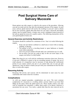


© Copyright 2026