
2 Vaginal Bleeding in the First Trimester of Pregnancy Bastiaan Jager
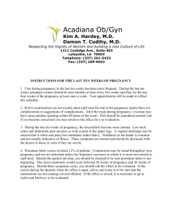
2
Vaginal Bleeding in the First Trimester of Pregnancy
Bastiaan Jager
INTRODUCTION
ectopic pregnancy, a miscarriage with severe vaginal bleeding and infected abortion with signs of
septic shock. Your first step should be to rule out
these conditions. The flow chart at the end of the
chapter should help you with this. If the patient
does not suffer from these serious conditions, one
should explore other causes of first-trimester vaginal
bleeding.
The differential diagnosis of first-trimester
vaginal bleeding is best sub-divided according to
anatomical location; therefore your main focus
should be to locate the anatomical origin of the
bleeding (Table 1).
A health-care provider in the field of gynecology
will certainly encounter numerous patients with
vaginal bleeding who are unaware of their early
pregnancy. All female patients of reproductive age
presenting with vaginal bleeding should therefore
be assessed for possible pregnancy.
Fortunately, the variety of causes of vaginal
bleeding in early pregnancy is limited. However,
differentiating between ectopic pregnancy and
miscarriage, both common causes of bleeding in
early pregnancy, can sometimes be challenging.
Diagnostic problems can especially arise in lowresource settings. This chapter specifically helps
you prioritize your diagnosis and treatment. It also
helps you identify the various causes of vaginal
bleeding in the first trimester of pregnancy.
Guidelines for proper history taking and physical examination will be given. After that, signs and
symptoms and treatment options will be presented
per cause. Finally, we will present a flow chart
which should guide you through the most important issues to be considered when encountering a
pregnant patient with vaginal bleeding in early
pregnancy. The key points that are occasionally
presented at the end of a section indicate the minimal standard of knowledge you should have after
reading the specific section.
Table 1 Differential diagnosis of first-trimester vaginal
bleeding
Originating from uterus, tubes, amniotic sac with its
contents or placenta:
• Ectopic pregnancy (see Chapter 12)
• Miscarriage (see Chapter 13)
• Miscarriage with infection (see Chapter 13)
• Molar pregnancy (see Chapter 27)
• Subchorionic hemorrhage
• Idiopathic bleeding in a viable pregnancy
Originating from cervix or vagina:
• Infection (Chlamydia, etc.) (see Chapter 17)
• Trauma (e.g. after intercourse, medical treatment)
• Malignancies, especially cervix cancer (see Chapter 26)
• Cervical abnormalities (e.g. excessive friability or
polyps) (see Chapter 9)
Differential diagnosis of bleeding in early pregnancy
Originating from anus, bladder or vulva:
• Hemorrhoids
• Lacerations of skin due to trauma, malignancy (rare) or
infection
• UTI, schistosomiasis
It is not very difficult in itself to diagnose vaginal
bleeding in a pregnant patient; the key point is to
recognize life-threatening situations which need
urgent treatment. Life-threatening conditions are:
21
GYNECOLOGY FOR LESS-RESOURCED LOCATIONS
Signs and symptoms of vaginal bleeding in the
first trimester
Box 1 Naegele’s rule
An estimated EDD from the first day of the
woman’s LMP can be found by adding 1 year,
subtracting 3 months and adding 7 days to that
date. The result is approximately 280 days (40
weeks) from the LMP.
If a woman of reproductive age presents herself at
your clinic with vaginal bleeding or any other
gynecological problem, always consider the possibility that she might be pregnant. It could be that
she is not aware of that herself. If she is aware of her
pregnancy, she might have forgotten the date of her
last menstrual period. Only after assessing for a
possible pregnancy can you start exploring the
patient’s complaint: vaginal bleeding.
Pregnancy is more likely if the woman has unspecific signs such as: nausea, fatigue, darkened
color of nipples and of old scars, swollen and tender
breasts and weight gain. If pregnancy is likely, you
may confirm this by taking a urine pregnancy test
(UPT) or, even better, an ultrasound if available at
your facility. Only perform a UPT if you are in
doubt of possible pregnancy. This could save costs
and more importantly, UPTs might not be readily
available in some clinics.
After confirmation of her pregnancy, try to assess
the duration (or gestational age) of the pregnancy.
This can be obtained by verifying the first day of
her last menstrual period. This is called the LMP.
With the LMP and Naegele’s rule one can estimate
the expected date of delivery (EDD) and deduce
the gestational age of the pregnancy (Box 1). If the
woman does not know the exact LMP, try to make
a reasonable estimate of the gestational age in
weeks. Naegele’s rule assumes an average cycle
length of 28 days, which is not true for everyone.
There are also pregnancy wheels which help in calculating the EDD and gestational age. Nowadays
there are several online calculators as well, e.g.
http://www.medcalc.com.
After assessing the gestational age of the patient’s
pregnancy, you should explore her complaint.
Suggested questions for assessment of the vaginal
bleeding are:
• Assess the severity When did the bleeding start? Is
there fresh blood (red) or old (darker, brown)
blood? Is bleeding daily present? Did it start
acutely or gradually? Was it already present before pregnancy? Also try to estimate the amount
of blood lost. (Rule of thumb is that when
bleeding is less than a woman perceives as a normal menstruation, it probably needs no urgent
treatment. Be aware though, as an ectopic pregnancy can present with little loss of blood.)
Example:
LMP = 8 May 2009
+1 year = 8 May 2010
–3 months = 8 February 2010
+7 days = 15 February 2010
Note: the calculation method does not always result in
280 days because not all calendar months are the same
length and it does not account for leap years. If you
want to be correct, you have to use a calendar. Exact
280 days past LMP is found by checking the day of
the week of the LMP and adjusting the calculated date
to land on the same day of the week.
Using the example above, 8 May 2009 is a
Tuesday. The calculated date (15 February) is a
Friday; adjusting to the closest Tuesday produces 12
February, which is exactly 280 days past 8 May1,2.
All these questions can give insight in the severity
of the case. A woman who suffered from bleeding
which started before pregnancy might have cervical
or vaginal lacerations due to multiple reasons. A
woman with acute bleeding, who has to change
her underwear frequently and suffers from accompanying (cramping) abdominal pain, might have a
threatening miscarriage or an ectopic pregnancy.
Do not forget to check for other accompanying
symptoms like nausea and fainting (a sign of hypotension due to ‘invisible’ intra-abdominal bleeding).
• Provoked bleeding Is the bleeding spontaneous or
after intercourse or defecation? This could indicate a cervical origin of the problem, e.g. infections like chlamydia and malignancies or even
hemorrhoids.
Do ask for other accompanying symptoms:
• Abdominal cramping pain: acute, continuous,
localized or general.
• Nausea and fainting might indicate shock due to
heavy (intra-abdominal) bleeding in ectopic
pregnancy.
• Did she lose any tissue vaginally? This might
point towards an incomplete abortion.
22
Vaginal Bleeding in the First Trimester of Pregnancy
• Fever can be a result of recent aseptic procedures or of miscarriage which has been infected.
It could also be a symptom of an infection which
in itself is correlated with miscarriage, e.g.
malaria.
• Dysuria? Sometimes a urinary tract infection
(UTI) presents itself with fresh blood in the
toilet or stains in her underwear. Painless macrohematuria is a sign for urinary schistosomiasis.
• Vaginal discharge? This could point towards
sexually transmitted infection (STI) such as
gonorrhea. Chlamydia classically presents with
painless bleeding or bleeding after intercourse.
• Was there trauma or involuntary sexual intercourse? This could present with lacerations and
STI.
• Concurrent infections like malaria can induce a
miscarriage and schistosomiasis can cause cervical bleeding, ectopic pregnancy and miscarriage.
Physical examination
You should be aware that the patient might be
concerned about losing her pregnancy and therefore she could be emotional. Pay special attention
to this when taking her history or performing
physical examination.
Assess her recent medical history:
• Every (gynecological) physical examination
should include an assessment of the: blood pressure, temperature and pulse rate.
• After that, examine her abdomen:
N Scars from previous laparotomies present? An
ectopic pregnancy can reoccur.
N Fundal height to assess the gestational age.
N Pain located on the uterus? More likely in
septic abortion or miscarriage or STI.
N Pain located laterally or even generalized
peritoneal pain? Rule out ectopic pregnancy.
Then consider STI, pelvic abscesses or nongynecological causes of peritonitis (see
Chapter 17 on STI).
N Palpable masses? Ectopic pregnancy, pelvic
abscesses, tubo-ovarian masses or nongynecological tumors (see Chapter 12 on
ectopic pregnancy and Chapter 17 on STI).
• Perform a speculum examination (see Chapter 1
on how to do that) to assess whether the blood
originates from the cervix, vagina or the uterine
cavity. Sometimes the cervix is very friable in
pregnancy. Even when gently touched with a
speculum, it starts to bleed.
• Perform a digital vaginal examination (see
Chapter 1). Be sure to perform the vaginal
examination carefully in cases where you suspect
an ectopic pregnancy. It can rupture if you
manipulate it too much.
N Assess the ostium of the cervix. If this is open
in pregnancy, it is very likely that the woman
has either an incomplete miscarriage or an
inevitable miscarriage. It should be noted
that cervical evaluation is not reliable in distinguishing between complete and incomplete miscarriage3. Sometimes products of
• Prior medical interventions Has she been seeking
medical attention before and what has been
done? She might have had an earlier treatment which has not been aseptic or has been
incomplete.
• Risk of criminal abortion Did the woman try to
conceive? Sometimes women do not dare to
admit that they underwent a non-professional
(and most of the times septic) abortion. It also
gives you a good clue towards the emotional
situation of the patient.
Assess her past medical history:
• Obstetric history Is this her first pregnancy? There
could be a history of miscarriage or bleeding in
the first trimester.
• Did she use any drugs? Some drugs are known
to increase the risk of miscarriage, e.g. diuretics,
anti-epileptic drugs, non-steroidal antiinflammatory drugs (NSAIDs), misoprostol,
retinoids, cytostatic drugs. You might want to
check your pharmaceutical reference guides for
possible association between drugs and miscarriage. Toxins such as lead, mercury, formaldehyde, ethylene oxide, benzene and large doses
of radiation also increase the risk of miscarriage2.
• Did she have any operations in the past? An
ectopic pregnancy due to pelvic inflammatory
disease (PID) can reoccur.
• Did she receive treatment for STIs? PID is a risk
factor for ectopic pregnancy.
• Are there known diseases such as diabetes,
clotting disorders or HIV? All are risk factors for
miscarriage. HIV infection is also a risk factor for
developing cervical carcinoma and these patients
may have had other STIs.
23
GYNECOLOGY FOR LESS-RESOURCED LOCATIONS
conception are still present in the ostium of
the cervix and can be removed during digital
vaginal examination. If the ostium is closed,
this could indicate a threatening miscarriage,
missed abortion or ectopic pregnancy.
N Assess for cervical tenderness. This could
indicate an infection or ectopic pregnancy.
N Assess for foreign bodies.
N Assess for cervical masses, malignancies or
(rarely) schistosomiasis or tuberculosis (TB).
If no blood is found on digital and visual
vaginal examination, perform an inspection
of the entire genital area. It might be that
blood is originating from the urethra, labia,
anus or skin. Sometimes patients have difficulties assessing the origin of the blood loss
themselves, especially in rural areas where no
proper sanitary facilities are present.
leukocyte count are both slightly elevated in
pregnancy.
Urine:
• UPT: urine pregnancy test to confirm pregnancy if in doubt.
• Urine screen for leukocytes, blood. This could
indicate UTI. Make sure you explain to the
patient how to produce a midstream urine
sample (see Chapter 1) to decrease contamination with vaginal blood.
Direct light microscopy:
• Blood slide if signs of malaria.
• Gram stain of urine if you suspect a UTI which
is not responding to standard treatment. Schistosoma in the urine may indicate (concomitant)
genital tract schistosomiasis but can be false
negative (see Chapter 9).
• Wet mount or Gram stain of vaginal discharge if
suspicious of STIs that are not responding to
trial of treatment (see Chapter 17).
Patients without proper diagnosis and on-going
complaints after treatment should be referred to a
facility where ultrasonic or laboratory investigation
is available. If your facility has an ultrasound
machine, every patient with first-trimester bleeding
should receive an ultrasound investigation to establish a viable intrauterine pregnancy. Rule of thumb
for referral is: if a patient (who has not been labelled
as having a serious, possibly life-threatening, condition which calls for imminent referral) presents
herself three times at a health facility with the same
complaint, she should be referred.
Other investigations
Ultrasound
Chapter 1 on gynecological examination presents
detailed information on how to perform an
ultrasound.
In trained hands, vaginal or abdominal ultrasonography can be crucial in identifying the location of the pregnancy. However, the diagnosis
depends on the quality of the ultrasound equipment, the experience of the sonographer, the specific signs and symptoms of the patient and presence
of physical factors such as fibroids. The first two
factors are often absent in resource-poor settings.
So, beware, a pregnant patient with peritoneal pain
and signs of hypovolemic shock should have an urgent explorative laparotomy and ultrasound might
only delay treatment.
If no intrauterine pregnancy is visible on ultrasound, check for adnexal masses or clear ectopic
gestational sacs to confirm the diagnosis. An empty
uterus on ultrasound in a patient with a positive
UPT should raise the suspicion of ectopic pregnancy at all times. In case of a ruptured ectopic
pregnancy, free fluid (blood) will be seen in
Douglas’ pouch in ultrasound (see Chapter 12 on
ectopic pregnancy for more information on diagnostic tools).
Laboratory investigations
It should be noted that most of the causes of vaginal
bleeding in the first trimester can be identified after
thorough history taking and physical examination.
Additional laboratory investigations hardly contribute to the diagnosis.
Possible laboratory investigations
Blood:
• Hemoglobin and cross-matching in case of
severe bleeding or suspected ectopic pregnancy.
It helps you to estimate the amount of blood
lost and helps to anticipate a possible blood
transfusion.
• Erythrocyte sedimentation rate (ESR) or leukocytes supposing an infection might be present.
(Fever measured by rectal thermometer is more
accurate/reliable.) Remember that ESR and
24
Vaginal Bleeding in the First Trimester of Pregnancy
Culdocentesis
Box 2 Danger signs indicating a possible ectopic
pregnancy
You can perform a culdocentesis to assess the presence of blood or pus in the pouch of Douglas [see
Chapter 12 (culdocentesis) and 18 (culdotomy)]. If
you are seriously considering ectopic pregnancy
and an ultrasound is not feasible in your setting and
culdocentesis does not produce clear results or has
failed, you might want to consider a diagnostic
(mini) laparotomy as the negative predictive value
of culdocentesis is poor4.
Diagnostic problems could evolve if a pregnant woman (confirmed by a positive UPT)
shows no intrauterine pregnancy on vaginal ultrasonography, has no fluid in Douglas’ pouch and
has no signs of ectopic masses or gestational sacs.
In this case she could have the following
conditions:
• Vaginal bleeding and continuous abdominal
pain with a closed cervix on digital vaginal
examination might be labelled as threatened
miscarriage, but could in fact be an ectopic
pregnancy. Pain in miscarriage is usually
cramping. Pain in ectopic pregnancy is usually continuous and/or accompanied with
localized peritonism. If in doubt perform a
culdocentesis. If an ultrasound machine is
present, assess for the presence of intrauterine
pregnancy products and or blood in the
pouch of Douglas. If still in doubt perform an
explorative (mini) laparotomy.
• Little tissue is obtained on performing an
manual vacuum aspiration (MVA). In this
case you might have mistaken an ectopic
pregnancy for an incomplete abortion or you
might have left the gestational sac and its
contents behind. One should always obtain
tissue when performing an MVA.
• Tachycardia and low blood pressure accompanied by scarce vaginal bleeding. This means
that the patient has signs of hypovolemic
shock which cannot be explained by the
amount of blood lost vaginally alone. The
first sign of hypovolemic shock is tachycardia
(heartbeat >100 bpm). Only after 2 liters of
blood, or more, have been lost, a pregnant
woman shows signs of drops in blood pressure. Urgent laparotomy is then necessary.
• A woman treated for threatened ‘incomplete
abortion’ who does not respond to misoprostol treatment might have an ectopic
pregnancy instead.
• she could have had a complete miscarriage,
• she could have a pregnancy too early to be seen
on ultrasound,
• she could have a spontaneously resolving intrauterine or tubal pregnancy,
• or she could still have an ectopic pregnancy.
In this case it may be appropriate to admit the
patient and reassess her regularly in order not to
miss the ectopic pregnancy about to rupture. Do
monitor the patient’s vital signs on a daily basis.
Only, if the UPT has become negative (this can
take some time, depending on the gestational age)
is it safe to discharge her.
Key points
• Always rule out ectopic pregnancy when encountering a patient with vaginal bleeding early
in pregnancy.
• Ectopic pregnancy can be easily mistaken for
threatened miscarriage.
• If available, an ultrasound is very useful to differentiate between viable pregnancy, miscarriage
and ectopic pregnancy.
• Use culdocentesis to assess for intra-abdominal
bleeding.
• Always take proper patient history and perform
sufficient physical examination.
• Gynecological physical examination always includes vital signs, assessment of the abdomen,
speculum examination and a digital vaginal
examination.
MISCARRIAGES
Miscarriage (also known as abortion) is the main
reason for vaginal bleeding early in pregnancy. You
will find more detailed information about this
condition in Chapter 13.
Ten to twenty per cent of clinically recognized
pregnancies result in spontaneous miscarriage5. The
occurrence of ectopic pregnancies is not as high as
miscarriage, but is definitely more prevalent in
areas where PID is highly endemic. Miscarriage can
be a life-threatening condition when it results in
massive bleeding (even more in areas where anemia
25
GYNECOLOGY FOR LESS-RESOURCED LOCATIONS
is prevalent) or septicemia as the result of unsafe,
induced abortion. Abortions are unsafe when performed by persons without the proper skills and
materials or outside a medically safe environment.
Gynaecologists (RCOG); a miscarriage refers to
the loss of pregnancy below 24 weeks of gestational age. Moreover, the RCOG recommends the
medical term for pregnancy loss to be miscarriage
instead of abortion, as abortion also refers to the
elective termination of pregnancy which is illegal
in most low-resourced countries. The term miscarriage acknowledges the emotional aspects of losing
a pregnancy and prevents stigmatization of the
pregnant woman and her medical caretaker8,9.
The most important subdefinitions for miscarriage and abortion are presented in Table 2.
As mentioned above, miscarriage can be an
emotional event for the woman and you should
consider this while doing your consultation. In
some cultures, stigmatization of the mother and/or
partner does occur, but pregnant women and their
partners are in fact not to be blamed.
Definitions
By definition, miscarriage refers to the loss of a
pregnancy before the fetus has reached a viable gestational age. Unfortunately, the cut-off point for a
viable gestational age varies internationally and in
time. As a result of progressive medical knowledge
neonates can nowadays survive pre-term birth from
23–24 weeks of gestational age onwards. This of
course heavily depends on the setting in which
neonatal care can be provided and most likely does
not apply to your local setting. In short, the gestational cut-off point for a loss of pregnancy to be
labelled as miscarriage varies between 16 and 24
weeks6,7. Fortunately, this lack of conformity in
definition does not influence the general approach
to diagnosis and treatment. In general, one can state
that the earlier a miscarriage occurs in pregnancy,
the less risk of complications.
In this chapter we will make use of definitions
made by the Royal College of Obstetricians and
Table 2
Etiology
The cause of spontaneous miscarriage is in most
cases related to the embryo itself. Random morphological abnormalities or absence of the embryo
itself are present in approximately 70% of cases
of miscarriage before 10 weeks of gestation.
Miscarriage: definitions of subcategories
Spontaneous miscarriage
Pregnancy loss within the first 24 weeks of gestation without deliberate interference
Incomplete miscarriage
Pregnancy loss within the first 24 weeks of gestation in which not all products of
conception have been expelled at presentation
Recurrent or habitual
miscarriage
Three or more consecutive spontaneous abortions/miscarriages
Threatened miscarriage
Uterine bleeding within the first 24 weeks of gestation without any cervical dilation. It is
characterized by vaginal bleeding, lower back discomfort or midline pelvic cramping and a
risk factor for miscarriage. This condition can be difficult to distinguish from ectopic
pregnancy when no ultrasound is present
Missed miscarriage
Retention in the uterus of a dead embryo or fetus which has died within the first 16 weeks
of gestation
Induced abortion
The intentional removal of a fetus from the uterus by any of a number of techniques
Criminal abortion
Illegal termination of pregnancy
Legal abortion
Termination of pregnancy under conditions allowed under local or national laws
Therapeutic abortion or
induced abortion for
medical reasons
Abortion induced to save the life or health of a pregnant woman
Miscarriage with infection
or infected abortion
Any type of miscarriage, induced or spontaneous, that is associated with infection of the
uterus and its appendages. It is characterized by fever, uterine tenderness, and foul discharge
26
Vaginal Bleeding in the First Trimester of Pregnancy
Spontaneous miscarriage
Morphological abnormalities are most likely the
result of random chromosomal abnormalities (50–
60%). Most commonly identified: Down’s syndrome, also called trisomy 21, polyploidy (more
than one pair of chromosomes in the nucleus),
Turner’s syndrome or monosomy X9. The frequency of spontaneous miscarriage increases with
maternal age. At the age of 35 years the incidence
of spontaneous miscarriage reaches 20% and at the
age of 40 it reaches 40–50%10. Besides age, several
maternal conditions can also lead to spontaneous
miscarriage:
Signs are cramping, lower abdominal pain accompanied by clear, red vaginal bleeding. Sometimes
tissue and blood clots can be seen as well. Tissue is
usually a dark purple color. To differentiate between blood clots and (abortion) tissue you can put
the clots/tissue in a glass container with normal tap
water and shake: blood clots will resolve, tissue will
stay intact. Up to 12 weeks the whole amniotic sac
can be evacuated. Physical examination might
show uterine enlargement. Digital vaginal examination reveals an open ostium of the cervix, with
blood on the finger. In general, the bleeding diminishes dramatically after complete evacuation of
the products of pregnancy. An ultrasound is generally not necessary, but if performed, it will show a
uterus with double lining endometrium if the miscarriage is complete. Some clots might be present
in the fundus (top) of the uterus.
• Diabetes, thyroid disease and infections, malaria,
syphilis, gonorrhea, cytomegalovirus (CMV),
toxoplasmosis, listeria, HIV, parvo-B19, chlamydia, etc.
• Maternal antibodies which predispose to thrombosis such as anticardiolipin and lupus coagulant.
• Genetic thrombophilias (clotting disorders) such
as factor V Leiden.
• Acquired or congenital anatomical variations of
the uterus: septa, abnormalities of the uterine
cavity, myoma, adhesions of the uterine cavity,
e.g. Asherman’s syndrome after infection.
• Environmental exposure: smoking, alcohol and
toxins (lead, heavy metals, organic solvents).
• Medication, e.g. diuretics, anti-epileptic drugs,
NSAIDs, misoprostol, retinoids, cytostatic drugs2.
Treatment If the amount of blood lost is less than
generally perceived during normal menstruation or
is dramatically diminishing, there is no need for
specific treatment.
Incomplete miscarriage
This means that a miscarriage has occurred but
some products of conception are still present in the
uterine cavity. The complaints are similar to those
of spontaneous abortion, but in this case the vaginal
bleeding has not stopped. Bleeding is usually more
than perceived during her normal menstrual period.
On digital vaginal examination the ostium of the
cervix is still open. On ultrasound one might see
clots, parts of the amniotic sac or tissue in the uterine cavity (Figure 1)13. Tissue appears as clear white
translucencies on the ultrasound, blood clots are
less dense in nature, fluid or blood is dark.
Epidemiology
Spontaneous miscarriage is something which actually occurs quite often. Estimates are that one in
every eight pregnancies results in a spontaneous
miscarriage. Two out of ten women suffer from
vaginal bleeding in the first trimester of pregnancy.
In 50% of these cases the pregnancy is viable despite the fact that bleeding continues. The other
50% will miscarry sooner or later11,12. It is believed
that many (more than 75%) pregnancies are lost before the woman recognizes she is actually pregnant.
The clinical signs of the miscarriage are in those
cases mistaken for delayed menstruation. As the
causes for miscarriage are so various, it is difficult to
say who will carry on and who not.
Treatment This patient may need treatment with
MVA or alternatively she can be treated with misoprostol (see Chapter 13). If, during ultrasound examination, no embryo or fetus is seen and the tissue
remaining in the uterine cavity has a diameter of
less than 30 mm, expectant management can be
considered. But only if the patient is willing and
able to come back when bleeding increases or
when signs of infection develop. Another precondition for expectant management is that she should
have a reasonable Hb.
Signs and symptoms
As mentioned the signs and symptoms depend on
the specific sort of miscarriage (see Table 2).
27
GYNECOLOGY FOR LESS-RESOURCED LOCATIONS
INFECTED ABORTION
(a)
An infected abortion (also called miscarriage with
infection) can either be due to prior incomplete
spontaneous miscarriage which has not evacuated
completely, or due to an aseptic procedure, either
criminal or medical. If an MVA is performed under
aseptic conditions the uterus or remaining placental
tissue could become infected. In countries where
there are restrictive laws towards abortion, women
might attend traditional healers or try to induce
miscarriage themselves when pregnancy is unwanted. Sticks, roots or other septic instruments
could be used for the induction of an abortion.
Infected abortion is still a leading cause of death
in developing countries. Approximately 100,000
women die annually of infection related to childbirth and unsafe abortion. They account for 15%
and 13% of total maternal deaths, respectively.
Ninety-nine per cent of maternal deaths occur in
developing countries and most of that in sub-Saharan
Africa and South-East Asia. On top of this, 20 times
more women suffer injury or disease because of infections related to childbirth14,15. As a medical professional one should definitely not miss this diagnosis.
(b)
Figure 1 Incomplete miscarriage with placental tissue
present in (a) and a diminished amniotic sac in (b).13
Source: http://www.fetalultrasound.com/online/
text/4–006.HTM
Signs and symptoms
The signs are of a miscarriage accompanied by
fever and foul smelling discharge. The patient may
develop septic shock. Septic shock has a mortality
rate of approximately 25–50%, even with proper
treatment in high-care facilities16. Signs of disseminated sepsis are: high fever and prostration, tachycardia, tachypnea or respiratory distress and low
blood pressure17. In a low-resource setting however, one should already be alerted if a patient
presents with fever and tachycardia. If she reports
with fever and low blood pressure, septic shock
may already be present.
Infecting organisms are usually vaginal or bowel
bacteria. Check for signs of abdominal guarding
and rebound tenderness to find out if the peritoneal
cavity is infected (see Chapter 3 on abdominal pain
in the first trimester). This can be due to uterine
perforation or infection, tubo-ovarian abscess or
bowel perforation.
Threatened miscarriage
If the pregnancy is viable, the amount of blood lost
can be significant. If the Hb level is adequate you
may admit the patient and re-assess regularly for
increased bleeding. Fifty per cent of these pregnancies continue to be viable. If bleeding is only
minimal and the patient has easy access to treatment she may go home and return if bleeding
increases.
Missed miscarriage
There are no complaints, but on ultrasound there is
an embryo without a heartbeat. Spontaneous miscarriage may follow in days or weeks.
Treatment There are three options and according
to your setting you can choose between them: (1)
expected management (wait for the pregnancy to
terminate itself); (2) start misoprostol and admit
patient to ward; (3) perform an MVA.
Treatment
Start imminently on a combination of antibiotics:
penicillin, metronidazole and gentamicin (Box 3).
28
Vaginal Bleeding in the First Trimester of Pregnancy
• Removal of aseptic products and a combination
of antibiotics is mandatory treatment.
• Start fluid resuscitation, antibiotics and adrenalin
(optional) if clear signs of septic shock.
• If signs of peritoneal guarding or rebound tenderness are present, consider that she may also have
a perforated uterus or even a bowel perforation.
• Take cultures if possible at your facility.
If no signs of septic shock are present, it is probably
wiser to perform the MVA after 12–24 h of intravenous antibiotic treatment. In case of septic shock,
start with fluid resuscitation and admit to a highcare ward or refer if possible. When performing an
MVA look carefully for signs of perforation and for
foreign bodies in the vagina or uterus.
Be aware that tetanus could also be the cause of
septicemia in settings where there is a low standard
rate of vaccination. In case of proper immunization
(i.e. five injections in the past 10 years and a booster in pregnancy) one should only give another
booster injection of tetanus toxoid 0.5 ml intramuscularly (IM). If the patient has not been immunized before, give anti-tetanus serum 1500 units
IM and booster of tetanus toxoid 0.5 ml IM after 4
weeks. Higher doses of anti-tetanus serum are
needed in clear cases of tetanus. Check your local
guidelines18.
If possible at your facility, take vaginal swabs for
culture and blood cultures for antibiotic sensitivity
testing19. Be careful, an infected uterus is easily perforated on evacuation.
When signs of abdominal guarding and rebound
tenderness are present, a perforation of the uterus
might already be present and a culdocentesis or
laparotomy should be considered.
MOLAR PREGNANCY (HYDATIDIFORM
MOLE, GESTATIONAL TROPHOBLASTIC
DISEASE)
A molar pregnancy is an abnormal form of pregnancy where a non-viable, fertilized egg becomes a
pathological pregnancy. The chorionic villi around
the fetus degenerate and form clusters of fluid-filled
sacs, hence the name molar pregnancy (Figure 2).
Detailed information on diagnosis and treatment of
a molar pregnancy can be found in Chapter 27.
Clinically, the patient will show signs and symptoms of normal pregnancy. These signs and symptoms could even be more prominent due to
abnormally high levels of human chorionic gonadotropin (hCG) (produced by the trophoblast cells):
nausea and vomiting leading to hyperemesis gravidarum, enhanced pigmentation of nipples, scars
and moles, painful breasts and signs of early preeclampsia. Painless vaginal bleeding is the rule. Loss
of vesicles is characteristic for molar pregnancy. A
dough-like uterus which is usually too large for
gestational age can be palpated. hCG levels can
Box 3 Antibiotics for infected abortion
Benzylpenicillin intravenously (IV) 5 million
IU q.d.s. or ampicillin 2 g IV q.d.s. (2nd choice
amoxicillin 500 mg tablets t.d.s.)
plus
Metronidazole 500 mg IV t.d.s. or 500 mg
tablets (orally or rectally)
plus
Gentamicin 5 mg/kg o.d. IM
If fever has resided for 24 h one can start 1 week
of oral treatment with amoxicillin and metronidazole.
Key points
• Infected abortion is one of the leading causes of
maternal death worldwide.
• Professionals should be aware that septic shock is
present when patients present with fever accompanied by tachycardia and/or low blood pressure.
Figure 2 Typical ultrasound picture of a hydatidiform
mole
29
GYNECOLOGY FOR LESS-RESOURCED LOCATIONS
vary and range from excessively high in early
pregnancy (>100,000 IU/ml) to normal and slowly
rising in case of a partial molar pregnancy.
Conclusive diagnosis can only be made by histopathology, which is scarcely available in lowresource settings. Histopathological diagnosis
reveals hydropic swelling of the chorionic villi and
hyperplasia of the trophoblastic cells.
is usually no cervical tenderness. Depending on the
stage of the malignancy, the whole vaginal wall and
rectum can be included in the lesion as well. The
lesions can become necrotic and infected. As a
result cervical malignancies are very often quite
smelly. In Chapter 26 cervical carcinoma is discussed in depth.
INFECTIONS THAT CAUSE (VAGINAL)
BLEEDING
Treatment
Evacuation of the uterus as described in Chapter
27. Be aware that massive hemorrhage can occur
during MVA. Do have blood compounds available!
The infections that can cause vaginal bleeding are
presented below. For a thorough outline of STIs,
please see Chapter 17.
Key points
Chlamydia
• Hydatidiform mole is difficult to diagnose, but
can lead to serious complications.
This usually goes unnoticed in female patients.
Sometimes it causes painless vaginal bleeding. In
fact this is a classical symptom. Every woman presenting with painless vaginal bleeding should be
checked for chlamydia if no instant cause has been
found. When untreated it has a high prevalence of
resulting in PID. This is not very likely to happen
during pregnancy however. When inspecting the
cervix with a speculum a reddish, infected cervix
can be seen, sometimes accompanied by yellow
purulent discharge; it bleeds easily on touch.
CERVICAL MALIGNANCIES
In Western settings, screening programs have significantly reduced the prevalence and mortality of
cervical carcinoma. In developing countries however, cervical carcinoma is the most common
malignancy in women. The malignancy usually
presents itself at 30–50 years of age20,21. HIV-infected
persons are more susceptible to cervical carcinoma.
As a result, it is not unusual to encounter a cervical
carcinoma in pregnancy in low-resourced settings.
Unfortunately, medical professionals are usually not
aware that a cervical malignancy might be a possibility when encountering (recurrent, minimal) vaginal
bleeding in pregnancy. The key to a proper diagnosis is speculum examination and digital vaginal
examination. Needless to say that vaginal examination is not contraindicated in pregnancy, only when
rupture of membranes is confirmed.
Treatment
Use your national guidelines for STI treatment. In
pregnancy doxycycline is contraindicated, but
erythromycin 500 mg q.d.s. for 2 weeks is adequate
as well. Do not forget to treat the partner as well.
Gonorrhea
Fifty per cent of the patients with gonorrhea are
asymptomatic. In symptomatic patients with
gonorrhea, dysuria, vaginal discharge and bleeding
may be present. On examination cervicitis with
purulent discharge can be seen. For treatment see
Chapter 17.
Signs and symptoms
The signs are vaginal bleeding due to cervical
erosions. Cervical erosions develop as a result of
malignancies or infections. Bleeding can occur
spontaneously, but classically, due to direct contact
of the cervix, i.e. vaginal bleeding after intercourse,
inserting tampons or passage of hard stools. Bleeding due to cervical erosions is usually self-limiting
and not severe (as in miscarriage or ectopic pregnancy). On digital vaginal examination a solid,
painless mass or irregularity is palpated. Bleeding
can be triggered due to digital examination. There
Urogenital schistosomiasis
This is caused by a parasite called Schistosoma haematobium or S. japonicum. It is endemic in some areas
in the tropics. Its main symptom is painless hematuria, which can be mistaken for vaginal bleeding.
In addition, schistosomiasis can mimic cervical cancer with bleeding from an ulcerated cervix or it can
30
Vaginal Bleeding in the First Trimester of Pregnancy
mimic cervicitis. Genital schistosomiasis can cause
ectopic pregnancy through specific granulomata in
the tubal wall as well as miscarriage through an
endometritis (see Chapter 9 for diagnostics).
An upper urinary tract infection (or pyelonephritis) is a more serious condition which calls for
intravenous antibiotics.
Treatment
Treatment
Nitrofurantoin 50 mg q.d.s. for 3–5 days. Alternative regimes are: amoxicillin/clavulanic acid 625 mg
t.d.s. for 5 days or (only in the first trimester) cotrimoxazole 960 mg b.d.
Praziquantel 40 mg/kg body weight as a single dose
is recommended outside pregnancy. However, as
praziquantel is contraindicated in pregnancy, treatment has to be postponed until after delivery.
OTHER CONDITIONS CAUSING VAGINAL
BLEEDING
Urogenital tuberculosis
Due to increasing co-infection with HIV, genital
TB is more frequently seen; previously it was quite
uncommon. Many TB patients have difficulties getting pregnant at all because of subfertility due to the
disease. TB can lead to bleeding due to endometritis
and miscarriage or ectopic pregnancy through
chronic TB salpingitis. Additionally, genitourinary
TB can mimic signs and symptoms of a UTI.
When you suspect a patient of urogenital TB,
check for concurrent symptoms such as cough, enlarged lymph nodes, ascites and sterile pyuria or
hematuria. Also check for other TB cases in the
direct family and perform a chest X-ray. When
urogenital TB is present, counsel the patient for
HIV testing.
This section contains incidental causes of vaginal
bleeding in early pregnancy. They usually do not
urge for a treatment. Diagnosis can only be made
on direct sight or when conditions like miscarriage,
ectopic and molar pregnancy, infections and malignancies have been excluded.
Cervical polyps
These do sometimes present in pregnancy. In
general they bleed after intercourse, but also spontaneously. Most polyps do not bleed profusely.
Diagnosis is made on speculum examination.
A typical polyp consists of mucosa similar to the
vaginal–cervical mucosa; it is soft and painless. The
stem of the polyp is usually avascular.
If services are available at your center, you may
send a specimen for histological confirmation. The
majority (99%) of cervical polyps are benign.
Treatment
Tuberculostatics according to local protocols.
Treatment is mostly given by directly observed
treatment (DOTS).
Treatment
Urinary tract infection
A cervical polyp can easily be removed by clamping it with a sponge-holding forceps: grasp the
polyp and gently turn clockwise until it loosens.
This procedure is usually painless. There is only
minimal bleeding. Alternatively, when the stalk of
a polyp is broad you may consider ligating it
and cutting it off. It is recommended that this is
done several weeks after the delivery.
A lower urinary tract infection (or cystitis) can
cause hematuria and can easily be mistaken for
vaginal bleeding. A lower urinary tract infection is
a fairly common condition in pregnancy due to
increased urinary stasis. In pregnancy it can present
itself with mild symptoms, but dysuria, lower abdominal pain, hematuria, frequent micturation and
incontinence are all possible symptoms. The infection is usually caused by the bacterium Escherichia
coli or enterococci (bacteria originating from the
gut). E. coli is becoming more resistant to penicillin
nowadays.
Diagnosis can be confirmed by a dipstick of a
midstream urine specimen. In some persistent cases
a culture is mandatory before repeating treatment.
Friable cervix
In some women the cervix is extremely friable in
pregnancy. In pregnancy the endocervical epithelium (that is usually inside the ostium) everts and
becomes the exterior portion of the cervix. This
ectropion will bleed easily on contact.
31
GYNECOLOGY FOR LESS-RESOURCED LOCATIONS
perigenital skin as well, such as lymphogranuloma
venereum.
Subchorionic or idiopathic bleeding
This can only be diagnosed if other (more serious)
causes have been excluded. Idiopathic means that
no specific cause can be found. In some cases the
blood originates from the subchorionic area. In
theory, due to growth of the uterus and its components in pregnancy, small lacerations occur below
the chorionic layer and the uterine wall which
presents as fresh vaginal bleeding. On direct inspection using a speculum, one can clearly see blood
coming straight out of the ostium of the cervix.
Treatment
Subchorionic or idiopathic bleeding needs reassurance, but no specific treatment. Sometimes subchorionic hematomas may lead to bothersome uterine
contractions or even miscarriage. In 10% of cases
the bleeding re-occurs. The woman should be informed about that.
Hemorrhoids
Hemorrhoids are also easily diagnosed. Sometimes
anal bleeding is mistaken for vaginal bleeding. Usually it is sufficient to ask the patient and confirm by
physical examination. They usually disappear after
pregnancy and conservative treatment is your first
option; painkiller (local) and laxatives may be added
if hard stools are present as well.
Figure 3 Examples of ectropion
On physical examination with a speculum, there
is no clear discharge or generalized reddish cervix
to be seen as in an infected cervix. The ectropion is
a very specific shallow, vascular, red area (Figure 3).
Key points
• The incidental causes of bleeding in the first
trimester should only be diagnosed if more
serious conditions of miscarriage, ectopic pregnancy, infections and malignancies have been
ruled out first.
Treatment
Besides reassurance a friable cervix needs no specific treatment. In some cases if bleeding is persistent, and VIA (see Chapter 26) is negative, one
could cryocoagulate the bleeding part of the ectropion. Only do this several weeks after delivery.
A flow chart for the diagnosis and management
of first-trimester vaginal bleeding is shown in the
Appendix.
REFERENCES
Vaginal or perigenital lacerations
1. Cunningham FG, McDonalds PC, Gant NF, et al. Williams Obstetrics, 20th edn. Stamford, Connecticut:
Appleton & Lange, 1997
2. Duff P, Edwards RK, Davis JD, et al. Obstetrics &
Gynaecology: Just the Facts. New York: McGraw-Hill,
2004
These are easily diagnosed if a proper physical
examination is performed. They can be a result of
trauma or a skin infection like fungal infection
or eczema. Be careful, some STIs involve the
32
Vaginal Bleeding in the First Trimester of Pregnancy
13. Suchet IB. Incomplete abortion In: The Ultrasound
of Life. http://www.fetalultrasound.com/online/text/
4–006.HTM
14. World Health Organization. Why do so many women
still die in pregnancy or childbirth? http://www.who.
int/features/qa/12/en/sindex.html
15. Grimes DA, Benson J, Singh S, et al. Unsafe abortion:
the preventable pandemic. Lancet 2006;368:1908–19
16. Kumar V, Abbas AK, Fausto N, et al. Robbins Basic
Pathology, 8th edn. Philadelphia: Saunders, 2007;102–3.
17. Stubblefield PG, Averbach SH, Grimes DA. Septic
abortion: prevention and management. Glob. libr.
women’s med. (ISSN: 1756–2228), 2008 (updated
2012)
18. Eddleston M, Pierine S. Oxford Handbook of Tropical
Medicine. Oxford: Oxford University Press (ISBN
0192627724), 2002
19. Department of Reproductive Health and Research.
Managing Complications in Pregnancy and Childbirth: a
guide for midwives and doctors. Department of Reproductive Health and Research, WHO library, 2005. http://
whqlibdoc.who.int/hq/2000/WHO_RHR_00.7.pdf
20. Onwudiegwu U, Bako A, Oyewumi A. Cervical cancer
– a neglected health tragedy. J Obstet Gynaecol 1999;19:
61–4
21. Cancer Research UK. Cervical cancer – UK incidence
statistics. http://info.cancerresearchuk.org/cancerstats/
types/cervix/incidence
3. Wieringade-Waard M, Bonsel GJ, Ankum WM, et al.
Threatened miscarriage in general practice: diagnostic
value of history taking and physical examination. Br J
Gen Pract 2002;52:825–9
4. Royal College of Gynaecology. The Management of Tubal
Pregnancy. RCOG Green-top guideline 21. http://
www.rcog.org.uk/files/rcog-corp/GTG21_230611.pdf
5. Alberman E. Spontaneous abortion: epidemiology. In:
Stabile S, Grudzinkas G, Chard T, eds. Spontaneous
Abortion: Diagnosis and Treatment. London: SpringerVerlag, 1992;9–20
6. Regan L, Rai R. Epidemiology and the medical causes
of miscarriage. Bailières Clin Obstet Gynaecol 2000;14:83954
7. Heineman MJ, Bleker OP, Evers JLH, et al. Obstetrie en
Gynaecologie; de voortplanting van de mens. Maarssen: Elsevier/Bunge, 1999
8. Royal College of Gynaecology. Management of Early
Pregnancy. RCOG Green-top guideline 25. http://
www.rcog.org.uk/womens-health/clinical-guidance/
management-early-pregnancy-loss-green-top-25
9. Griebel CP, Halvorsen J, Golemon TB, et al. Management of spontaneous abortion. Am Fam Phys 2005;72:
1243–50.
10. Nybo-Andersen AM, Wohlfahrt J, Christens P, et al.
Maternal age and fetal loss: population based register
linkage study. BMJ 2000;320:1708–12
11. Everett C. Incidence and outcome of bleeding before
the 20th week of pregnancy: prospective study from
general practice. BMJ 1997;315:32–4
12. Wilcox AJ, Weinberg CR, O’Connor JF et al. Incidence of early loss of pregnancy. N Engl J Med 1988;319:
189–94
33
GYNECOLOGY FOR LESS-RESOURCED LOCATIONS
APPENDIX – Flowchart for management of first-trimester vaginal bleeding
34


© Copyright 2026