
Coccidiosis in Rabbits
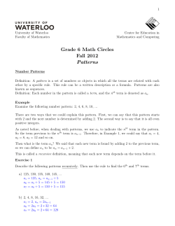
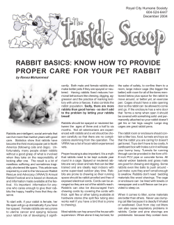
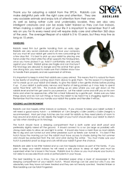
Coccidiosis
in
Rabbits
Amy Halls, M.Sc.
Monogastric Nutritionist
November 2005
Coccidiosis is a highly contagious sporozoal infection in rabbits. It is caused by Eimeria
species, protozoa parasites that are microscopic, one-celled organisms. Twelve species of the
family Eimeria have been reported to affect rabbits, however, only a few actually create disease.
Eimeria spp. invade the epithelial cells of the intestinal wall and cells lining certain ducts within
the rabbit. Each species is highly host, organ and tissue specific, and the species that affect
rabbits are rarely a zoonotic danger to humans. There are two types of rabbit coccidiosis –
intestinal and hepatic. Eimeria perforans, E. magna, E. media, and E. irresidua are the four
main species that cause intestinal coccidiosis in rabbits. E. steidae causes hepatic coccidiosis in
the liver. Coccidiosis is mainly seen in intensively managed animals, especially young rabbits,
although it can occur in small rabbitries and pet rabbits.
Figure 1. Oocysts of various Eimeria species in the domestic rabbit (Coudert P.,
D. Licois, F. Drouet-Viard, F. Provôt. 2000, in: Enfermedades del conejo. Tomo
II. 219-234.).
Life Cycle of Eimeria Species
The life cycle of a typical Eimeria species (Figure 2) can take 4 to 14 days to complete. The
cycle begins with the rabbit ingesting feces containing Eimeria oocysts or eggs (Figure 2, a).
This can happen when the rabbit is cleaning contaminated fur or consuming contaminated feed.
Mechanical (muscular contractions of the upper intestinal tract) and enzymatic digestive
(pancreatic and biliary enzymes) processes of rabbit’s upper digestive tract weaken or digest part
of the oocyst walls, allowing the sporozoites to be released (Figure 2, b). The released
sporozoites penetrate the epithelial cells lining the intestinal wall (Figure 2, c) and initiate
merogony (asexual multiple division). During merogony (Figure 2, c-e), sporozoites divide
asexually to form as many as 100,000 merozoites per sporozoite, depending on the Eimeria
species. When the merozoites are mature, the host cell ruptures and releases the merozoites
(Figure 2, f), which in turn seek a new cell to infect to repeat merogony again (Figure 2, g-i).
The number of merogony generations is species specific and can vary from two to four
generations.
Figure 2. Life cycle of an Eimeria species.
(http://biology.unm.edu/biology/coccidia/eimeriabiol.html).
The last generation of merozoites to enter host cells develops into gametes (Figure 2, j) during
gamogony, the sexual reproductive stage. The majority develops into macrogametes (Figure 2,
k-n), the “female” cell involved in sexual reproduction. The remaining gametes divide into
thousands of mobile, flagellated microgametes (Figure 2, 0-r), the “male” cell involved in sexual
reproduction. When the microgametes are mature, they leave their host cell (Figure 2, s) and
penetrate a host cell containing a macrogamete (Figure 2, n) and fertilization occurs. As the
newly formed zygote begins to mature it develops its resistant oocyst wall. When the wall is
fully formed, the oocyst bursts from its host cell and is leaves the host via its feces (Figure 2, t).
Once outside the host, the oocyst must undergo sporogony (Figure 2, u-x) in order to become
fully mature. The oocyst divides twice to form four sporocysts (Figure 2, x) and is ready to
infect a new host.
Symptoms
Clinical symptoms of intestinal and hepatic coccidiosis are very similar and hard to distinguish
between the two. Often healthy rabbits may be carriers of the protozoa, but show no symptoms
of the disease. The severity of the disease depends on the number of oocysts ingested by the
rabbit. The entrance of Eimeria spores into the intestinal and liver cells cause cells to
malfunction and expand in size. Erosion and ulceration occurs in the epithelial lining of the
intestine, which results in poor absorption of nutrients, electrolyte imbalance, anemia and
dehydration of the cells. Common clinical signs include a reduced appetite, depression,
abdominal pain, retarted growth, diarrhea and pale mucous membranes. Feces may contain
blood or mucous. Any or all of these signs may be absent in older rabbits.
Intestinal Coccidiosis
Intestinal coccidiosis mainly affects young weaned rabbits six weeks to five months of age. This
is attributed to stress, noise, transportation or immunosupression. Symptoms appear within four
to six days post infection and include a rough coat, dullness, decreased appetite, dehydration and
weight loss. Rabbits may also develop intussusception, a blockage of the intestine caused by a
telescoping of the bowel on itself. When weight loss is greater than 20%, convulsions or
paralysis is seen, followed by death within 24 hours. The majority of deaths are a result of
dehydration and secondary bacterial infections.
Hepatic Coccidiosis
Rabbits infected with E. steidae, the protozoan responsible for hepatic (liver) coccidiosis, may
have mild to severe infections. Mild infections show no symptoms while moderately infected
rabbits will have growth retardation. Severe infections result in loss of appetite, weakness,
diarrhea and possibly constipation in the later stages of the disease. E steidae inhabits cells of
the bile ducts and liver, causing blockage and severe liver damage. X-rays show enlarged livers
and fluid in the abdomen, which contribute to the water or pot belly symptom. This disease will
either linger for several weeks or cause death within 10 days, preceded by a coma. Necropsy
reveals white spots or nodules on the surface of the liver.
Diagnosis and Treatment
The diagnosis of coccidiosis in live rabbits is very difficult to do. Examination of feces for the
presence of oocysts is one method, however, coccidiosis oocysts are often difficult to distinguish
from the rabbit-specific yeast, Cyniclomyces guttulatus. Each Eimeria species is tissue specific
(Figure 3). Necropsies will distinguish the species causing the disease by locating the area of
infection in the intestinal tract, whether it is in the small intestine, cecum, large intestine or the
liver.
Figure 3. Tissue specificity of Eimeria species. Shaded areas indicate the area
of coccidial infection (Coudert P., d. Licois, f. Drouet-Viard, F. Provôt. 2000, in:
Enfermedades del conejo. Tomo II. 219-234.).
Treatment involves the administration of antibiotics prescribed by a veterinarian. Anti-coccidial
treatment is usually only successful for rabbits that have been infected for less than five or six
days. If treatment is successful, diarrhea and mortality may still be seen for a few days after the
initiation of treatment. Relapse is regularly observed after one or two weeks. The role of the
antibiotic is to control the protozoa until the rabbit’s immunity develops. Life long immunity
usually occurs from mild infections. However, immunity is species specific and therefore,
exposure to one species does not protect against other species.
Prevention
Prevention is the best method in the control of rabbit coccidiosis. The elimination of several
factors can reduce the chance of coccidiosis occurring in a rabbitry. Sanitation and husbandry
are the most important aspects in controlling infection. The following points should be taken
into consideration to reduce the chance of coccidiosis developing in your rabbitry.
1. Overcrowding causes unnecessary stress and difficulty in maintaining hygiene in the
rabbitry. Avoid mixing litters at weaning and do not breed if you do not have the hutch
space available.
2. Wean youngsters as late as possible, unless overcrowding becomes an issue. Kits can
stay with the doe up to ten weeks of age. Weaning as late as possible will minimize the
stress of early weaning and will help establish a healthy gut flora in kits. Always remove
the doe from the litter to reduce the stress and incidence of disease.
3. Proper nutrition is necessary to produce healthy rabbits. Feed anticoccidial medicated
rations to weaned and non-lactating/pregnant does to prevent the development of
coccidiosis. Young rabbits (1-3 months) have a low immunity to coccidiosis and thus are
more susceptible to the disease and pose a threat to the rabbit industry. Rabbits will
eventually develop immunity to coccidial parasites as they continue to be exposed to
them in their environment. Shur-Gain’s 16% Medicated Rabbit ration contains Robenz,
the only approved anti-coccidial medication for rabbits in Canada. It will help protect
young rabbits from coccidiosis while they develop their immune system. Robenz should
not be fed to lactating or pregnant does, they can be fed Shur-Gain’s non-medicated 16%
Rabbit ration. Avoid mixing different types/sources of feeds as it can lead to an
unbalanced ration, resulting in the dilution of the medications and rendering them
ineffective in the prevention of disease. All Shur-Gain rabbit rations are nutritionally
balanced to provide the necessary nutrients for proper growth and maintenance.
4. Hay in the diet is also essential – rabbits of all ages should have access to unlimited, good
quality hay. Lack of fibre is the primary cause of many digestive upsets, and can easily
be avoided if rabbits are fed a complete ration in addition to unlimited hay and fresh
water.
5. Quarantine all new rabbits for a minimum of 30 days before introducing them to other
rabbits. Isolation of all new rabbits reduces the chance of disease spreading to your
established rabbitry.
6. Cleaning and disinfecting schedules should be established and followed to reduce the
incidence of disease in rabbitries. Several rabbit coccidial diseases produce tough
oocysts that are very resistant to disinfectants. Routine cleaning two or three times a
week will decrease coccidial loads and is recommended for rabbitries experiencing
coccidiosis problems. If possible, rotate hutches to allow them to remain unoccupied
every other breeding period to reduce the potential disease challenge. Feed hoppers
should be disinfected regularly and remain free of rabbit feces. Frequently clean cages,
hutches, transport carriers and litter pans with the appropriate cleaner and disinfectant.
See Appendix A for a list of cleaners and disinfectants available from your Shur-Gain
Dealer.
Appendix A. A list of detergents and disinfectants available from your Shur-Gain Dealer.
DETERGENTS
1. Biofoam
• Alkaline cleaner
• Suitable for the removal of organic and inorganic residues, particularly the buildup of scale
• Excellent cleaning and degreasing properties
2. Biosolve
• Alkaline cleaner
• Ideal for the removal of grease, fats, faecal matter and soiling
3. BioSentry Acid-a-Foam
• Foaming acidic cleaner
• Suitable for general purpose manual cleaning and wetting agent for house and washdown
• Removes mineral deposits and “biofilms”
4. BioSentry EZ-Kleen
• Alkaline cleaner and deodorizer
• Suitable for routine soaking and cleaning in preparation for disinfection
• Emulsifies, suspends and removes grime, grease, protein and fatty soils
5. Proquat
• Alkaline cleaner
• Quaternary ammonium activity suitable for routine soaking and washing
• Removes proteins and fatty soils
DISINFECTANTS:
1. Hyperox
•
•
•
2. Profilm
•
•
•
Active Ingredients: 5% Peracetic Acid; 25% Hydrogen Peroxide
Broad spectrum activity – efficacy against bacteria, viruses and fungi
Environmentally friendly – completely biodegradable
Active Ingredients: 19.2% 2-(Hydroxymethyl)-2-Nitro-1,3-Propanediol
(HNP); 2.02% Formaldehyde; 2.29% Dimethyl benzyl ammonium chloride
Formaldehyde Fumigant concentrate
Broad spectrum activity – efficacy against bacteria, viruses and fungi
3. Virkon
•
•
•
4. Virocid
•
•
Active Ingredients: 21.4% Potassium monopersulphate
Full spectrum activity - efficacy against bacteria, viruses and fungi
Non corrosive germicide
Active Ingredients: 14.6% Alkyldimethylbenzylammoniumchloride; 10.7%
didecyldimethylbenzlammoniumchloride; 7.8% glutaraldehyde; Full spectrum
activity - efficacy against bacteria, viruses and fungi
Non corrosive disinfectant; 90% biodegradable


© Copyright 2026