
Korean Scleritis Study: Clinical Features, Risk Factors & Treatment Outcomes
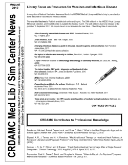
Korean J Ophthalmol 2010;24(6):331-335
DOI: 10.3341/kjo.2010.24.6.331
pISSN: 1011-8942 eISSN: 2092-9382
Original Article
Clinical Features, Predisposing Factors, and Treatment
Outcomes of Scleritis in the Korean Population
1,2
1,2
1,2
1,2
1,2
Seong Joon Ahn , Joo Youn Oh , Mee Kum Kim , Jin Hak Lee , Won Ryang Wee
1
2
Department of Ophthalmology, Seoul National University College of Medicine, Seoul, Korea
Seoul Artificial Eye Center, Seoul National University Hospital Clinical Research Institute, Seoul, Korea
Purpose: To evaluate the clinical features, associated factors, and treatment outcomes of scleritis in the Korean
population.
Methods: Medical records were retrospectively reviewed for 94 eyes of 76 patients with scleritis. Clinical features of
scleritis, including systemic disease, presence of microorganisms, serologic markers, history of previous ocular
surgery, and use of immunosuppressants were investigated and compared amongst the subtypes of scleritis.
Treatment outcomes were evaluated using best corrected visual acuity (BCVA) and time to scleritis remission.
Results: Nodular scleritis was the most common form observed, followed by necrotizing scleritis with inflammation,
diffuse scleritis, and necrotizing scleritis without inflammation, respectively. A total of 16 of 76 patients (21.1%)
had connective tissue diseases. Eleven cases (14.5%) had infectious scleritis, of which bacteria (54.5%) and fungi (45.5%) were the causative microorganisms. Thirty-three patients (43.4%) had previous ocular surgery, mostly
pterygium excision. Notably, a history of pterygium excision was significantly associated with development of necrotizing and infectious scleritis (odds ratio [OR], 399 and 10.1; p < 0.001 and 0.002, respectively). In addition,
patients with necrotizing scleritis were more likely to have infectious scleritis (OR, 11.7; p = 0.001). BCVA after
treatment and time to remission also showed significant differences among the different scleritis subtypes.
Systemic immunosuppression was required in addition to steroids for treating diffuse and necrotizing scleritis.
Conclusions: Careful taking of patient history including previous pterygium excision should be performed, especially
in patients with necrotizing and infectious scleritis. In addition, evaluation of microbiological infection can be crucial for patients with necrotizing scleritis and history of pterygium excision.
Key Words: Clinical feature, Pterygium excision, Scleritis, Treatment outcome
Scleritis is a severe ocular inflammatory disorder that is
commonly associated with systemic collagen vascular disease.
Previous reports have demonstrated that approximately 50%
of patients with scleritis have systemic connective tissue diseases [1-6]. However, scleritis can also be caused by ocular
surgery, such as pterygium, cataract extraction, or strabismus
surgeries. Although scleritis is a serious postsurgical complication [7-9], clinical characteristics of surgically-induced
scleritis or the type of surgery associated have not been extensively investigated.
Received: June 22, 2010
Accepted: July 13, 2010
Reprint requests to Won Ryang Wee. Department of Ophthalmology,
Seoul National University College of Medicine, #28 Yeongeon-dong,
Jongno-gu, Seoul 110-744, Korea. Tel: 82-2-2072-2435, Fax: 82-2-7413187, E-mail: [email protected]
Infectious and necrotizing scleritis are two subtypes of
scleritis with particularly poor prognoses; there are several
cases reporting the requirement for either evisceration or
enucleation in order to control the infection and inflammation
in infectious and necrotizing scleritis [10]. Systemic immunosuppression in necrotizing scleritis and early antibiotic
treatment in infectious scleritis are essential for preserving
vision and preventing the spreading of infection into adjacent
ocular structures [11]. However, predisposing factors for early diagnosis of these two forms of scleritis have not been
thoroughly studied. Here, we investigated the clinical characteristics and predisposing factors of scleritis with respect to
treatment course and outcome in the Korean population.
Materials and Methods
Medical records of patients who were diagnosed with
ⓒ 2010 The Korean Ophthalmological Society
This is an Open Access article distributed under the terms of the Creative Commons Attribution Non-Commercial License (http://creativecommons.org/licenses
/by-nc/3.0/) which permits unrestricted non-commercial use, distribution, and reproduction in any medium, provided the original work is properly cited.
331
Korean J Ophthalmol Vol.24, No.6, 2010
scleritis at Seoul National University Hospital between
January 2005 and December 2008 were reviewed. The diagnosis of scleritis was made based on the presence of inflammation of scleral and episcleral vessels that did not
blanch with topical phenylephrine, the presence of scleral
thinning, edema, or nodules. Patients who did not have a follow-up visit were excluded from this study. Scleritis was
classified into the following subtypes; diffuse, nodular, and
necrotizing scleritis with or without inflammation. Diffuse
scleritis was characterized by the presence of scleral edema
and redness with a tortuous vascular network. Nodular scleritis presented as a nodule that was firm and tender to
palpation. Necrotizing scleritis with inflammation was defined as having an avascular area of sclera with adjacent
scleral edema and congestion while necrotizing scleritis
without inflammation was classified for cases presenting
with a thin and avascular area of sclera with no active neighboring inflammation. Infectious scleritis was diagnosed following isolation of the causative microorganism from the
lesion according to a microbiological workup. For first-line
treatment for non-infectious nodular or diffuse scleritis without any underlying systemic disease, topical and oral corticosteroids or systemic non-steroidal anti-inflammatory drugs
were used. In severe cases refractory to oral corticosteroid
therapy, systemic immunosuppressive agents such as azathioprine, cyclosporine, and mycophenolate mofetil were
used. For treatment of infectious scleritis, topical and oral antibiotics or anti-fungal agents were used after a causative microorganism was isolated by culture from scleral scrapings in
the base of active lesion [9,10].
In total, 94 eyes of 76 patients met the inclusion criteria.
The study protocol was approved by the Institutional Review
Board, and informed consent was obtained from patients.
Patient medical records were retrospectively reviewed with
respect to clinical manifestations, subtypes of scleritis, presence
of systemic or ocular co-morbidities, and a history of previous
ocular surgery. In addition, serologic markers and presence
of causative microorganisms isolated from culture from cases
of infectious scleritis were investigated. The markers included
veneral disease research laboratory, fluorescent anti-nuclear
antibody (FANA), anti-neutrophil cytoplasmic antibody
(ANCA), rheumatoid factor, human leukocyte antigen-B27,
anti-streptolysin O, and ribonucleoprotein antibody. To estimate the significance of the risk factors for the development
of infectious and necrotizing scleritis, an odds ratio was calculated for each risk factor.
To compare visual outcomes, best corrected visual acuities
(BCVA) were evaluated based on the logarithmic value of the
minimal angle of resolution scale at the time of diagnosis and
after clinical remission. Additionally, the time elapsed from
diagnosis to remission of scleritis was analyzed. Clinical remission was defined as when 1) the patient complained of no
symptoms and when 2) any sign of scleral inflammation disappeared on slit-lamp examination. Both BCVA and time to
remission were compared between seropositive and seronegative patients as well as infectious and noninfectious using Student’s t-test, one-way analysis of variance (ANOVA),
and survival analysis.
In order to determine which factors required the use of additional systemic immunosuppression for treating scleritis,
patients were divided into two groups consisting of those
who were treated with oral corticosteroids only and those
who needed additional systemic immunosuppressants for
resolution of the disease. The subtypes of scleritis, seropositivity, and systemic comorbidities were then compared between the two groups using Fisher’s exact test, likelihood ratio test for categorical data, and Student’s t-test.
All statistical analyses were performed using SPSS ver.
Table 1. Demographics, clinical features, previous ocular surgery, and seropositive cases of scleritis
Parameters
Age (yr)
Gender (male : female)
Follow-up period (mon)
Laterality (unilateral : bilateral)
Prevalence of subtype (nodular : diffuse:necrotizing)
Previous ocular surgery
Pterygium excision
Cataract extraction
Strabismus surgery
Seropositivity
Fluorescent anti-nuclear antibody
Anti-neutrophil cytoplasmic antibody
Rheumatoid factor
Human leukocyte antigen-B27
Anti-streptolysin O
Ribonucleoprotein antibody
Cryoglobulin
Values are presented as mean ± SD or number (%).
332
Values
59.5 ± 13.8 (range, 11-81)
23 : 53
19.05 ± 34.17 (range, 1-240)
58 (76.3) : 18 (23.7)
31 (40.8) : 18 (23.7) : 27 (35.5)
33 (43.4)
29 (38.2)
3 (3.95)
1 (1.32)
16 (21.1)
9 (11.8)
4 (5.26)
3 (3.95)
1 (1.32)
1 (1.32)
1 (1.32)
1 (1.32)
SJ Ahn, et al. Scleritis in the Korean Population
Table 2. Incidence of pterygium excision in patients with scleritis and combined connective tissue disease (CTD) at
presentation according to scleritis subtype
Nodular
No. of patients with previous
†
pterygium excision (%)
No. of patients with CTD (%)†
Connective tissue disease (no. of
patients)
Diffuse
2 (6.5)
1 (5.6)
9 (29.0)
Rheumatoid arthritis (2)
Wegeners granulomatosis (2)
Juvenile rheumatoid arthritis (1)
Behcet’s disease (1)
Systemic lupus erythematosus (1)
Ankylosing spondylitis (1)
Takayasu arteritis (1)
5 (27.8)
Rheumatoid arthritis (2)
Wegeners granulomatosis (2)
Systemic lupus erythematosus (1)
Necrotizing
26 (96.3)
p-value*
< 0.001
2 (7.4)
Rheumatoid
arthritis (2)
< 0.001
N/A
N/A = not applicable.
*
p-value was obtained by Fisher exact test; †The percentage was calculated among the patients with one subtype of scleritis.
15.0 (SPSS Inc., Chicago, IL, USA). A p-value < 0.05 was
considered statistically significant.
Results
Demographic data are summarized in Table 1. Scleritis
was more than twice as times prevalent in females than males
(male:female = 23:53), and 76.3% of the cases were
unilateral. The most common subtype of scleritis was nodular scleritis (40.8%), followed by necrotizing scleritis with
inflammation (27.6%), diffuse scleritis (23.7%), and necrotizing scleritis without inflammation (7.89%). Associated
clinical factors are shown in Table 1. Sixteen patients
(21.1%) were seropositive, most commonly FANA-positive
(n = 9). Notably, 33 out of 76 patients (43.4%) had previously ocular surgeries, of which 29 cases were pterygium
excision. Of note was a high association between a history of
pterygium surgery and the development of necrotizing or infectious scleritis. Specifically, most of the patients with necrotizing scleritis (96.3%) had a previous history of pterygium surgery; however, only 3 of 49 patients (6.1%) with
non-necrotizing scleritis had a prior pterygium excision.
Additionally, 89.3% of scleritis cases with a history of pterygium excision were necrotizing scleritis. The odds ratio for a
history of pterygium excision was 399 in patients with necrotizing scleritis compared to those with non-necrotizing
scleritis (95% confidence interval [CI], 39.4-4,030; p <
0.001). In addition, 9 out of 11 patients (81.8%) with infectious scleritis had a history of prior pterygium surgery.
The odds ratio for a history of pterygium excision was 10.1 in
infectious scleritis compared to noninfectious scleritis (95%
CI, 2.00-51.2; p = 0.002).
Moreover, a significant association between necrotizing
and infectious scleritis was observed. Specifically, of the 11
patients with infectious scleritis, 9 (88.8%) were diagnosed
with necrotizing scleritis, which resulted in an odds ratio for
infectious scleritis of 11.8 for patients with necrotizing compared to non-necrotizing scleritis (95% CI, 2.31-59.7; p =
0.001). Thus, necrotizing scleritis was significantly asso-
Fig. 1. Visual acuities before and after treatment. Patients with infectious necrotizing scleritis had the poorest best corrected visual
acuities (BCVA) before and after treatment. The data are presented
as the mean ± standard deviation. logMAR= logarithmic value of
the minimal angle of resolution.
ciated with infectious scleritis and a history of pterygium
surgery.
Sixteen patients (21.1%) had connective tissue diseases.
The percentage of associated connective tissue diseases was
different among the subtypes of scleritis (Table 2). In addition, diffuse and nodular scleritis was associated with comorbidity of connective tissue disease, although statistical
significance was borderline (p = 0.07). Rheumatoid arthritis
was the most common connective tissue disease associated
with scleritis.
Eleven eyes (14.5%) were diagnosed as having infectious
scleritis. Bacterial infection comprised 54.5% (n = 6) of these
cases, followed by fungi (n = 5, 45.5%) and viruses (n = 1,
9.09%). Pseudomonas was the most common pathogen (3
cases), followed by Fusarium and Stenotrophophilia (2 cases).
Haemophilus, Acromobacter, Aspergillus, Beauveria, Candida,
333
Korean J Ophthalmol Vol.24, No.6, 2010
Table 3. Comparison of clinical factors between patients treated with oral steroids only and those treated with steroids
and systemic immunosuppressants for controlling inflammation
Steroid only
Steroid with
(n=15)
immunosuppressant (n=31)
63.8 ± 12.7
Age (yr)
57.0 ± 13.3
Gender (male : female)
3:12
12:19
Laterality (unilateral : bilateral)
12:3
25:6
Prevalence of subtype (nodular : diffuse:necrotizing)
9:2:4
7:10:14
Seropositivity
3
9
Connective tissue disease
2
6
*
p-value was obtained by Student’s t-test; †Fisher’s exact test; ‡Likelihood ratio test, respectively.
Parameters
p-value
0.099*
0.176†
0.624†
0.043‡
0.392†
0.478†
Fig. 2. Survival curves of remission. A significant difference in remission was observed (p = 0.028) among the different scleritis subtypes. No
significant difference was seen between infectious versus noninfectious, and seropositive versus seronegative cases. The y-axis indicates the
proportion of cases with persistent scleritis as calculated by the following equation: (1 – the proportion of remission).
and herpes zoster were also isolated. There were two cases of
coinfections; one was coinfection of Pseudomonas and Candida,
and the other was coinfection of Stenotrophophilia and
Acromobacter. Two eyes with fungal scleritis were eviscerated because of uncontrolled infection that eventually progressed into endophthalmitis.
To evaluate visual outcome according to subtypes of scleritis, BCVA was investigated at the time of diagnosis and after
remission (Fig. 1). BCVA at the time of diagnosis was worse
in patients with infectious and necrotizing scleritis than patients
with nodular and diffuse scleritis. Patients with infectious
scleritis had the worst BCVA after remission (1.19 ± 1.11),
followed by non-infectious necrotizing (0.538 ± 0.457), nodular (0.367 ± 0.637), and diffuse scleritis (0.282 ± 0.506).
These differences in visual acuities among subtypes of scleritis were statistically significant (p = 0.013 at the time of diagnosis and 0.008 after remission; one-way ANOVA).
The median time to resolution was significantly longer in
necrotizing scleritis (10 months, p = 0.028; log rank test),
compared to diffuse (7 months) and nodular subtypes (7
months) (Fig. 2). The median time to resolution of infectious
scleritis was longer than non-infectious type (10 months vs. 8
months); however, this difference was not statistically significant (p = 0.166). There was no difference in the median
time to resolution between seropositive and seronegative
scleritis (p = 0.732).
334
In order to determine the factors associated with the use of
additional systemic immunosuppression, we analyzed the
clinical characteristics between the patients who were treated
with conventional oral corticosteroids and the patients who
needed additional systemic immunosuppressants because of
inflammation refractory to systemic steroids (Table 3). We
found that patients with diffuse and necrotizing scleritis were
more likely to require systemic immunosuppression (p =
0.043, likelihood ratio test).
Discussion
A noticeable finding of this study was that patients with necrotizing scleritis were more likely to have had previous
pterygium excision and infectious scleritis. This finding is
consistent with the report by O'Donoghue [8], who demonstrated a high association between necrotizing and surgically-induced scleritis. The association between a history of
pterygium excision and two destructive subtypes of scleritis
(necrotizing and infectious) might suggest a role for pterygium excision as either a causative or inducing factor of the
two types of scleritis. This hypothesis can be explained by
the previously suggested proposal that surgical techniques or
intra- or postoperative medications for pterygium excision
might predispose inflammation and necrosis of the sclera and
make ocular tissue vulnerable to infection [12]. In addition, it
SJ Ahn, et al. Scleritis in the Korean Population
is possible that the use of mitomycin C during pterygium surgery may induce necrotizing and infectious scleritis.
For successful treatment of scleritis patients, early discrimination between noninfectious autoimmune scleritis and
infectious scleritis is important since the treatment regimens
for infectious scleritis, antibiotics, is contrary to the treatment for non-infectious necrotizing scleritis, which consists
of systemic immunosuppressants. However, the differential
diagnosis between necrotizing scleritis with and without infection is often difficult. Indeed, two patients in our study
whose eyes were eviscerated because of uncontrolled fungal
scleritis were initially misdiagnosed as having an immune-mediated necrotizing scleritis and were initially treated with topical and systemic immunosuppressants. Given
that pterygium excision was a significant factor associated
with both necrotizing and infectious scleritis, careful examination for signs of infection and microbiological work-up
should be performed, especially in patients who present with
scleral necrosis.
Another remarkable finding of this study was that fungal
infection comprised of almost half cases of infectious scleritis in our study. Indeed, the proportion of fungal scleritis in
the present study was substantially higher than previously reported [9,10,13]. This may be because of a geographic difference of an infectious pathogen in scleritis as reported in previous studies [9,10,13]. The prognosis of fungal scleritis in
this study was poor, as previously reported [14-18].
As shown in the present study, visual outcome depended
on the subtypes of scleritis. Nodular or diffuse scleritis
showed better visual acuity both at the time of diagnosis and
after remission. Necrotizing scleritis, especially when associated with infectious scleritis, demonstrated poor visual
acuity both at the time of diagnosis and after remission. In addition, patients with necrotizing scleritis had a prolonged duration of disease. Furthermore, patients with non-infectious
necrotizing scleritis were more likely to require systemic immunosuppressants in addition to conventional oral steroids
for treatment, compared to those with nonnecrotizing
scleritis. An association between the use of additional immunosuppressants and other clinical factors such as ANCA,
which was reported in previous studies [19], was not found in
this study.
In conclusion, the results of this study suggest that a history of pterygium excision in scleritis is a significant factor
associated with poor prognosis due to the strong association
with necrotizing and infectious scleritis. Hence, when patients with a history of pterygium surgery present with necrotizing scleritis, immunosuppressants should be cautiously
started after excluding the possibility of an infection by a
thorough microbiological work-up.
Conflict of Interest
No potential conflict of interest relevant to this article was
reported.
References
1. Akpek EK, Thorne JE, Qazi FA, et al. Evaluation of patients
with scleritis for systemic disease. Ophthalmology 2004;111:
501-6.
2. Sainz de la Maza M, Jabbur NS, Foster CS. Severity of scleritis and episcleritis. Ophthalmology 1994;101:389-96.
3. Tuft SJ, Watson PG. Progression of scleral disease. Ophthalmology 1991;98:467-71.
4. Watson PG, Hayreh SS, Awdry PN. Episcleritis and scleritis. I.
Br J Ophthalmol 1968;52:278-9.
5. Sainz de la Maza M, Foster CS, Jabbur NS. Scleritis associated
with rheumatoid arthritis and with other systemic immune-mediated diseases. Ophthalmology 1994;101:1281-6.
6. Erkanli L, Akova YA, Guney-Tefekli E, Tugal-Tutkun I.
Clinical features, prognosis, and treatment results of patients
with scleritis from 2 tertiary eye care centers in Turkey. Cornea
2010;29:26-33.
7. Lee JE, Oum BS, Choi HY, Lee JS. Methicillin-resistant
Staphylococcus aureus sclerokeratitis after pterygium excision.
Cornea 2007;26:744-6.
8. O'Donoghue E, Lightman S, Tuft S, Watson P. Surgically induced necrotising sclerokeratitis (SINS): precipitating factors
and response to treatment. Br J Ophthalmol 1992;76:17-21.
9. Su CY, Tsai JJ, Chang YC, Lin CP. Immunologic and clinical
manifestations of infectious scleritis after pterygium excision.
Cornea 2006;25:663-6.
10. Huang FC, Huang SP, Tseng SH. Management of infectious
scleritis after pterygium excision. Cornea 2000;19:34-9.
11. Jabs DA, Mudun A, Dunn JP, Marsh MJ. Episcleritis and scleritis: clinical features and treatment results. Am J Ophthalmol
2000;130:469-76.
12. Singh RP, McCluskey P. Scedosporium prolificans sclerokeratitis 10 years after pterygium excision with adjunctive mitomycin C. Clin Experiment Ophthalmol 2005;33:433-4.
13. Jain V, Garg P, Sharma S. Microbial scleritis-experience from
a developing country. Eye (Lond) 2009;23:255-61.
14. Hsiao CH, Chen JJ, Huang SC, et al. Intrascleral dissemination
of infectious scleritis following pterygium excision. Br J
Ophthalmol 1998;82:29-34.
15. Lincoff HA, McLean JM, Nano H. Scleral abscess. I. A complication of retinal detachment. Buckling procedures. Arch
Ophthalmol 1965;74:641-8.
16. Margo CE, Polack FM, Hood CI. Aspergillus panophthalmitis
complicating treatment of pterygium. Cornea 1988;7:285-9.
17. Milauskas AT, Duke JR. Mycotic scleral abscess. Report of a
case following a scleral buckling operation for retinal
detachment. Am J Ophthalmol 1967;63:951-4.
18. Lin CP, Shih MH, Tsai MC. Clinical experiences of infectious
scleral ulceration: a complication of pterygium operation. Br J
Ophthalmol 1997;81:980-3.
19. Hoang LT, Lim LL, Vaillant B, et al. Antineutrophil cytoplasmic antibody-associated active scleritis. Arch Ophthalmol
2008;126:651-5.
335


© Copyright 2026