
Integrated MR Imaging & Proton Nuclear Magnetic Resonance
ryt
lil
jl
IntegratedMR lmagingand Proton
NuclearMaqneticResonance
in a FamilYwith an
SpectroscoFy
X-LinkedSpasticParaParesis
syndromewere
Seven membersof a family with an X-linkedspastic paraparesas
DavidM. Youseml
DavidH. Gutmann2 analyzed by MR imaging and stimulatedecho, solvent'suppressedproton nuclear
symptomaticmalesand two
BartonN. Milestonel magneticresonancespectroscopy.The MRscansof thtee
whitematler.
femalesdemonstratedabnormalsignalin the supratentorial
asymptomatic
RobertE. Lenkinskil
patients
protonspectroseopicexaminationol a2x 2 x 2 cm voxel
Eachol these
had a
localizedto the abnormalwnite matterof the centrumsemiovale.The spectrademonand crea'
ir-acetylaspartate/choline,
strateddepressionol N-acetylaspartate/creatine,
tine/cholineratioscomparedwith normalcontrolsubjects.Additionally,these patients
hadabnormalelevationsof aminoacid resonancesin the 2.1-3.0ppmrange'ln a patient
an asymmetricapectroscopic
with symmetricwhitemattersignalintensityabnormalities,
study correlatedwith asymmetricaymptoms.Oneasymptomaticlamily memberwith a
She was referredfor
normalMR study had abnormalmetaboliteratio measurements,
furtherevaluation,sincethe protonspectrumsuggestedshe may possessthe affected
9ene.
lf the lindingsin this studyare duplicatedin othercasesof hereditarydysmyelinating
syndromes,we believethe integratedMR/protonnuclearmagneticresonancespectroscopy examinationwill be of benelit in evaluatingand counselingtamilieswith familia!
disorders.
dysmyelinating
AJNR12:785-789,July/August1991
Received
October10, 1990;revisionrequested
January17, 1991;revisionreceivedFebruary6,
20, 1991.
1991i acceptedFebruary
1Department
Hospitalof the Uniof Fadiology,
3400SpruceSt., Philadelversityof Pennsylvania,
phia,PA 19104.Addressreprintrequests
to D. M.
Yousem"
'?Department
of lleurology,Hospitalof the thiPA 19104.
Philadelphia,
versityof Pennsylvania,
0195-6108/91/1204-0785
O AmericanSocietyot Neuroradiology
The clinicalapplicationsand technlcalaspects of MR spectroscopyhave recently
been reviewed in the literature [1, 2]. Over the past severalyears there has been
increasing interest in using proton nuclear magnetic resonance spectroscopy
(PMRS) to study the metabolism of the brain [3-8]. Instead of a conventional
anatomicimageof the brain, proton spectroscopyprovidesbiochemicalinformation
concerninghydrogen-bearingnon-water metabolitesin the CNS, includingN-acetyl
aspartate, the creatine-phosphocreatinepool, and choline-containingcompounds.
The integrationof MR imaging with spectroscopy otfers the potential for a more
comprehensiveevaluationof pathologicconditionsof the brain.
Patients with diffuse dysmyelinating disorders are ideal subjects for PMRS
studies becausethe area of abnormalityis easily sampledwithin the constraintsof
relatively large voxel sizes. Voxels that include white matter without significant
gray matter or ventricularCSF contaminationcan be sampled with a high degree
of accuracy and assurance.In order to determinethe value of the integrated MR/
PMRS examinationin patientswith hereditarywhite matter disorders,we examined
seven members of a pedigree with a complicated X{inked hereditary spastic
paraparesis(HSP)syndrome [9].
Subjects and Methods
form
undescribed
affectedwitha previously
A familywithfivemalesin a singlegeneration
has recentlybeenreported[9]. Thispedigree
hereditary
spasticparaparesis
of complicated
786
YOUSEMET AL.
AJNR:12, Juty/Augustj 991
Fig. l.-Pedigree of family with complicaled
hereditary spastic paraparosi:, Males are represented by squares and temales by circles.
Open squares and circles reptegent aiymptomatic indiyiduals while closed squares and circles represent aftected individuals. The agos of
the indiyiduals are displayed beeide the circles
or Squares.
II
18 III
IV
is illustratedin Figure'l . The diseaseis characterized
by speech eight-stepphaseryclingschemewas usedfor each90" orthogonal
ditficulties,
lowerlimbspasticity
andhypereflexia,
mentalretardation, pulsein the STEAMsequence
and256 scanswereaveraged.
Scan
cerebellar
ataxia,and{remor.The symptomsbeginin the first two
timefor the PMRSdatacollection
was4 min36 sec.Attemptswere
decadesol lifeandprogressfor g to 6 yearsbeforestabilizing.
Three
madeto placethevoxelwithinareasof abnormal
whitematterwithout
of the four livingmalesaffectedby the disease0lt-2,lll-5, lll-7),as
overlapping
soft tissue,skull,ventricle,
or graymatter.Two regions
well as the asymptomatic
mother(ll-2),sister(lll-3),maternaluncle
of interestweresampledin threeof the patientsin identicalareasof
(ll-3),andniece(lV-1),wereevatuated
by MR and PMRS.A second the centrumsemiovale
on oppositesidesof the brainwithoutvoxel
asymptomatic
sister(lll-4)andthefather(ll-1)wereevaluated
by MR
overlap.Onlyone regionof interestwas obtainablein four patients
alone.
owingto patienttimeconstraints.
Thus,10 spectrafromthe family
StandardMRexaminations
inctuded
sagittalshortTRfIE (600/20/ were availablefor analysis.Both sets of data in the threepatients.
1) andaxiallongTR sequences
(9000/95,90/1)
performed
on a 1.S_ withtwo voxelswereusedin dataanalysis.
T GESignascanner.Gradientmomentnulling(a flow-compensation
technique),
inferiorpresaturation
pulses,and a variablebandwidth
technique
(4 KHzand,16 KHzto increasesignal-to-noise
ratio)were
Data Analysis
employedon the longjTR images.In caselll-5,gadopentetate
dimeglmine (0.1mmol/kg)wasadministered
andpostcontrast
shortTR/
Spectralanalysiswas performedon an AT&Tpersonatcomputer
TE sequences
with firstordergradientmomentnullingtechniques systemusingan analysispackagedeveloped
in house,Thespectra
wereperformed.
wereevaluated
withoutfittingroutinesapplied.Nobaseline
conection
MR imageswerereadby two neuroradiologists.
Clinicalhistories programswereused,andonlyfirst-andsecond-order
phaseconec_
werenot withheld.At the timeof the MR interpretation,
the PMRS
tions were used. In order to preventbias in the analysisof the
clatawerenot available
to the reviewers.
spectra,28 spectrawere submittedfor analysisto three blinded
Thecoordinates
for sampling
a2 x 2 x 2-cmvolume-olinterest
in
examiners.
Thesespectraincludedmembersof the familywith the
the affectedwhitematierof thecentrumsemiovale
weredetermined white matterdisorder,normalcontrolsubjects,and patientswith
fromthe MRimages.Voxellocalization
wasconfirmedby a shortTR
otherCNSdiseases.In five casesthe datafrom the samespectra
(300/19)imageof the 2 x 2 x 2-cmvoxelin the axialplane(with
weresubmittedtwicein orderto determine
the reproducibility
of the
suppressionof all signaloutsidethe voxel and with no solvent examiners'results(threeof thesefive
were membersof the HSp
suppression)
[6, 10, 11].Thevoxel'sX, y, andZ coordinates
were
family).
verifiedon thisscanandthe localization
of the sampledvolumewas
The spectrawere blindlyanclindependently
processedby two
furtherconfirmedby addingthe imageof the voxelto the spin-echo investigators,
andthespectrawereinterpreted
bythreeinvestigators.
rmageusing an algorithmdevelopedin houseon the GE Signa /V-acetylaspartate(NAA)to creatine/phosphocreatine
(CR)ratios,
scanner.
NAAto choline{ontaining
compound(CH)ratios,and CR/CHratios
pMRS
after giving
werecomputedfor eachpatientby eachreviewerand the average
. _Sevenfamilymembersunderwentlocalized
informed
consent.Gradient
shimming
(X,y, Z) wasaccomplishecl
on
valuesrecorded(Table1).To determinemetaboliteratios,the peak
the 2 x 2 x 2 cm voxeland linewidthsrangedfrom 3.Sto 6.0 Hz
heightsweremeasuredfrombaselineratherthanfromareasunder
(<0.1 ppmvariation).rspectroscopic
acquisitions
weremadeby use
curves,andthenonfittedspectrawereused.Additionally,
thereviewof a stimulated
echotechnique
(STEAM)precededby chemicat
shift
erswereaskedto evaluatethe spectrafor the presenceof elevated
selectivesolventsuppression
pulsescenteredon watertollowedby
aminoacidpeaksbetweenthe NAA(2.01ppm)and CR (3.02ppm)
spoilergradients('l gauss/cmfor 4-g msec)performedin all three
peaKs.
axesto furtherreducecontributions
fromthe waterprotons[g, 12,
To provideestimatesof normalNAA/CR,NAAICH,and CR/CH
131.TR was2000msec,TE was 19 msec,andTM imiddleinterval ratios,
sevennormalsubjectsalso had pMRS, and the imaging,
betweensecondand thirdslice-selective
pulses)was g.6 msec.An
localization,
and analysistechniqueswere identicalto those used
AINR:12,July/August1991
787
MR AND PMRS
SPASTICPARAPARESIS:
TABLE 1: Resultsof PMRSStudies in Seven Family Members and Seven Controls
lll-2, lll-5, llFT/Symptomatic,abnormalWM
lF2, lll-3/Asymptomatic,abnormalWM
lV-1/Asymptomatic,normalwM
ll-3/Asymptomatic,normalWM
Controls(n = 7)
SD
Elevated
Amino Acids
NM/CH
Patient/Findings
1.0
1.1
't
.1
'l.4
la
.16
1.3
1.0
1.0
1.8
2.1
.17
1.3
0.9
0.9
l2
1.7
2.3
Yes
Yes
No
NO
No
CH = choline-containingcompounds,WM = white matter, SD = standarddeviation.
Note.-NAA = N-acetylaspartate,CR = creatine/phosphocreatine,
MRexaminations
hadnormal
Thenormalvolunteers
withthepatients.
were
Thevoxelsanalyzed
symptoms.
andwerewithoutneurologic
to thosetakenin
comparable
at locations
semiovale
in thecentrum
forthepresence
werealsoanalyzed
These
volunteers
theHSPfamily.
in an
aminoacidpeaksandtheirratioswerecomputed
of elevated
(Table
1).
fashion
bythesamethreeevaluators
blinded
identical
Results
MR
intensityon the longTR sequenceswas
Abnormalsignal
patients,
observedinthewhitematterof allthreesymptomatic
theirmother,and two sisters.The white matterabnormality
diffuse
in thethreeatfectedpatientsconsistedof a prominent
distributed
confluentincreasein signalintensitysymmetrically
white matter structures(Fig. 2). The
in all supratentorial
abnormalwhite matterdid not enhancein the one patient
The motherand
dimeglumine.
who receivedgadopentetate
on thelongTR images
sistershadwhitematterhyperintensity
althoughlimitedto the centrumsemiovale.The unaffected
myelinon all pulseseniece(lV-1)had normal-appearing
quences.Theuncle(ll-3)hadfocalareasof highsignalintensity in the peripheralwhite matter,which was thoughtto
representsmallvesselischemicchangesin this 56-year-old
Hiswhitematterappearance
hypertension.
withlong-standing
was markedlydifferentfrom thoseof the atfectedpatients'
MR
Thefatherof the atfectedmaleshada normal-appearing
exceptfor minorsmallvesselischemicchanges.
examination
He refusedspectroscopy.
PMRS
patients
normaland abnormalMR studies.In asymptomatic
with abnormalMR studies,the aminoacid peakswere elematterby MR
vated.Thus,all patientswith abnormalwhite
had elevatedaminoacid peaks.In patientswith normalMR
studies,the aminoacids were normalin magnitude,even
ratioswereoftenlow.ln the
thoughthe measuredmetabolite
niece(lV-1)thewhitematter,aminoacidpeaks,
asymptomatic
were normal,but the PMRS
and neurologicexamination
uncle
Thenon-gene-bearing
spectralratiosweredepressed.
(ll-3)had a normalNM/CR ratio and minimallydepressed
NAA/CHand CRICHratios, probablydue to a prominent
cholinepeak.
The averagedifferencesbetweenNM/CR, NAA/CH'and
CR/CHratiosin controlsubjectsversusfamilymemberswith
:
: 15o/o,
(2'1-1.1)12.1
were(1.3-1.1y1.3
thegenepresent
: 41"/",respectively.
48o/",
and('1.7-1.0111.7
In one patientthe MR resultsdemonstratedsymmetric
white matterhigh signalon the long TR images,but the
PMRSdatashowedthat, whileboth sidesof the braindemonstrateddepressedNM/CH and CR/CHpeaks,the right
sideof the brainhad lowervaluesthan the left (Fig.2). The
elevationsof aminoacid peakswere also morestrikingon
wellwith
findingscorrelated
the rightside.Thespectroscopic
increasedleft-sidedspasticityand hyperreflexianoted on
physicalexamination.
In all five patientswith diffuseabnormalsignalintensityof
the white matter,the threeevaluatorsagreedthat elevated
aminoacidpeaksin the 2.0-2.5 ppmrangewerepresent.All
three evaluatorsagreedthat the amino acid peaks were
normalin heightin sixof sevencontrolsubjects.Inonecontrol
subject,one of the three evaluatorsreportedan elevated
aminoacidpeak:the othertwo evaluatorsreportedno such
elevation.
Theresultsof the PMRSspectraaregivenin Table1. The
membersof the familywith abnormalwhitemattersignal(lllNAA/CHand CR/CH Discussion
2, lll-3,lll-5,lll-7,ll-2)had depressed
levelsandslightlydepressedNAA/CRratios.Becauseof the
pulsesand
By employingthreeorthogonalslice-selective
of statisticallydifsmallnumberof subjects,an assessment
ferentratiovaluesbetweenthe familymembersand normal spoilingall signalarisingoutsidethe intersectionof these
pulses,one can obtaina well{ocdizedsamplingvolumefor
volunteersis not possible.However,a trendis seenin that
of thewater
suppression
presumably
PMRSspectra[6, 10,11]. Chemical
abnormalwhite
the
who
bore
thefamilymembers
proton contributioncan result in a greaterthan 500{old
mattergenehadmeanvaluesof NM/CH, NM/CR, andCR/
reductionin water-protonsignal.Voxelsas smallas 1'5 x
CH that werebeyonda standarddeviationfrom normalvolthemain
at ourinstitution;
1.5x 1.5cm havebeenemployed
notablein the NM/CH and
unteervalues.Thisis particularly
in
signalis
the
reduction
volume
small
this
in
sampling
cost
(Table
1).
CR/CHratios
In all threeaffectedsymptomaticmembersof the family, to-noiseratio(SNR).ThisSNRreductioncanbecompensated
Whilethis
thenumberof samplingaverages.
for by increasing
elevatedamino acid peaks were detected,whereasthe
pathologic
andvery
to
specimens
applicable
is
readily
solution
familymemberswere dividedinto thosewith
asymptomatic
1991
AJNR:12,
July/August
YOUSEMET AL.
788
c
6.rt!.1
a
Sttt
I
6il1..1
bt0
$lft
b.l
6..tc.l
D
E
2
$rlt
bt.l
F
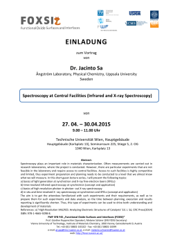
Fig. 2.-MR and PilRS images ot male (lll'2) wlth spastic paraParesis'
highsignal
in poeteriorlimbof intemalcap3ulesbilaterally(attow)'
signalin
demonstrates high
(30fl!/90) through
through intemit
intemat capsule
oapsule demonsirates
image (30fl!/90.-)
l,-lxia
A,
Axial Un
ilR image
g, Un image(SdOOieO)
(iop]ett1dimonstratesabnormalintensitybilatlrallyin whiie matterof centrumsemiovale.STEAMscan(300/19)(top ttghtl ot
on axialscan(bottomreft)to ensutecortectvoxellocaliza$on.
the 2.x 2 x 2 cm voxelselectedtor specttoscopymaythenbe superimposed
ot voxel in left cenirumeimiovaleie iupedmposedon axial(30d)/90)image.The voxelin B cottelateswith the sPectrumin
C, STEAMscan(O0O/19)
D, andthe voxelin C producedthe sPectrumin E
depressionot NAA/CHan<lCR/CHratios.The valuesfor the dght voxel_wore1.0for NAA/CH
D-F, protonspectrumot rijht voiet (D) demonstrates
and t.i tor CR/iH, whereaso-nthe left iEi the NAA/CHwas 1.4and the.CR/CHwas 1.4as well,Notealsothe elevatedaminoacid peaks(anorvs)most
striking on the right (D) side. compare uiittr pMRSspectrum(F) of noima'lvolunteer. All the spectra were acquircd with 10(XlHz sweep width' 1024
compl;xpoints,2--si rirpetitiontimes,and 256averages(32tijnis throughan eiEht-stepphase-iyclingprocedure).The spectrawereprocessedwith 2'
Hz line-bioadening,
zero-tillingto 2048poinE,Fouriel|tanstom, zero-and tirst-orderphasecorection.
TEs increasesthe sensitivityof the STEAMsequenceto
cooperative
volunteers,the drawbackin potentialmotionas
scanningtimesare extendedbecomesprohibitive.Because coupledspins (i.e., from protonsof the -CH2- groups of
glutamine,gammaamino butyrate,valine,etc.) presentin
of this,we haveselectedan 8-cm3volumethat allowsgood
aminoacidsin the2-3 ppmrange.lt maybe usefulto analyze
time.
5
min
of
scan
in
under
256
SNRwith
samplingaverages
theseaminoacidsin certainCNS disorderssuch as amino
This volumeis well-suitedfor evaluatingpatientswith dysmyopamaplesyrupurinedisease,or mitochondrial
acidurias,
diseases,sincethe volumeof whitematterin the
myelinating
noise
is
increased
TEs
using
shorter
in
drawback
thies.
The
without
skull,
centrumsemiovaleallowsadequatesampling
and increasededdy currentdistortion.Baselinecorrection
ventricle,or graymattercontamination.
andsmoothingprograms
curvefittingtechniques,
Theuseof shortTE valuesin our STEAMsequenceallows algorithms,
but
smallaminoacid peaks.
critical
to
the
all tend obscure
to the
increased
SNRandlesssensitivityto T2 contributions
the spectrain a more
to
analyze
we
tend
reason,
For
this
reduction
of
scan
averages
in SNRallows
signal.Theincrease
(orsamplesize),therebyreducingscantimesandsubsequent "raw"form.Thespectraarenot asattractive,andthebaseline
fluctuates.but essentialdatais not "smoothed"out.
patientmotion.In addition,we believethat the use of short
AJNR:12, July/August1991
SPASTICPARAPARESIS:MR AND PMRS
Becausethe processing
of PMRSspectrais an operatordependent
functionin mostinstitutions,
we believethat the
unbiased
analysisof scandata,as in thisstudy(withblinded,
independent,
and multipleevaluators)
is essentialto determinethevalidityof PMRSstudies.Thisis especially
true of
clinicalstudieswith a smallsamplesize,suchas haveappearedin thespectroscopy
literatureto date.
Theresultsof the PMRSstudyof this familyraiseseveral
issues.The presenceof elevatedaminoacid peaks in the
2.1-2.5 ppm rangecorresponds
to resonancesusuallyascribedto glutamine
(2.14, 2.45),keto-glutarate
s (2.44,2.23),
(2.39),valine(2.27),GABA(2.25),andmethionine
succinate
(2.16,2.14)
of thesepeakshasalsobeenseen
[5].Elevation
in patientswith activeplaquesof multipleselerosis[12]. In
previouswork done in this institutionwe have found that
plaquesof multiplesclerosis
thatenhanceon MRoftenshow
elevation
of protonspectrapeaksin the 2J-23 ppm range
(Grossman
et al.Paperpresented
1989).
at RSNA,November
Theseplaquesarebelieved
to represent
areasof activeblood
brainbanierbreakdownandactivemyelinolysis.
The PMRS
findingssuggestthat one may be samplingby-productsof
(Grossman
activemyelinbreakdown
et al. RSNA1989)[12].
Suchpeakswerepresentin eachfamilymemberwho had
abnormal
whitematterdetectedon the imagingstudies(even
in the women"carriers"who were asymptomatic).
These
peakswerenot presentin patientsor controlsubjectswho
hadnormal-appearing
whitematter.
Analysisof the NAA/CR,NM/CH, and CR/CH ratios is
madedifficultby residualwatercontamination,
which may
causefluctuations
in the baselinedue to its overwhelming
dominance
of the MR signal.Althoughthe smallsamplesize
inherentin analysis
familygroupdoesnot
of a seven-member
permita rigorousstatisticalanalysis,we believethere are
differences
betweenthefamilyandthenormalvolunteers.We
noteda trendthattheNM/CH ratioin patientswithabnormal
whitematterwas almosthalfthat of the controls,and that
theCR/CHratioalsoappeared
to be slightlydepressed.Less
definitive
depression
of the NM/CR ratiowas also present.
Only throughPMRSevaluationof a larger populationof
patientswith dysmyelinating
disordersand normalcontrol
subjectscan we conclusively
state whetherthese findings
aretypicalof whitematterdisorders.
Thatthe NAAvaluesmaybe depressedis consistentwith
the beliefthat NAAarisesin neurons,is essentialfor normal
with brain
brainfunctioning,
anddepression
of NAAcorrelates
tissueloss[12, 13].Similarreductions
in the absoluteconcentrations
of cholineandcreatinewouldbe expectedin the
diffuseneuroaxonal
loss associatedwith white matter diseases.
Theasymmetric
exaggeration
of PMRSabnormalityin the
patientwho had symmetricwhitematterhighsignalabnormalityon MR imagessuggeststhat PMRSmay add informationto conventional
MR for the assessmentof myelin
abnormalities.
Thismaybe translatedinto morepreciseand
meaningful
clinicalcorrelation.
This patient'sPMRS abnorii
$
li
i*
it
.i_
iI
t
fil
M
&
malitymorecloselyreflected
hisclinical
symptomatology
than
did his MRstudy.
Anotherdilemmawas raisedwith regardto the asymptomaticniece(lV-1)whohada normalMRstudy,normalamino
acid peaks,but depressionof NM/CH and CR/CHratios.
The possibilitythat this womancarriesthe atfectedX gene
signalintensityabnormality
was sugbut hasnot manifested
gestedto theclinician
dealingwiththiskindred.lf thiscontributionof PMRSis verifiedin futurecasesand by chromoit will heralda moreactiverolefor PMRSin
somalanalysis,
geneticcounseling.
Patientswith acquiredor inheriteddisordersof myelin
providethe neuroradiologist
with an excellentopportunityto
examinethe full capabilityof combinedMR and PMRStechseenin the hereditary
niques.The largeareaof abnormality
metachrodisorders(Pelizaeus-Merzbacher,
dysmyelinating
Canavandisadrenoleukodystrophy,
matic leukodystrophy,
ease,Alexanderdisease,etc.) readilylendsitselfto PMRS.
MR/PMRSstudymaybe helpfulin theevaluaAn integrated
tion and subsequentgeneticcounselingof familiesaffected
by thesedisorders.
REFERENCES
1. WeinerMW. The promiseof magneticresonancespectroscopyfor medical
diagnosis. /nvest Radiol 1988;23:253-261
2. Bottomley PA. Human in vivo NMR spectroscopyin diagnosticmedicine:
cfinicaftoof or research$obe? Radiolqy 1989;170:1-15
3. Tanaka C, NaruseS, HorikawaY, HirakawaK, YoshizakiK, NishikawaH.
Proton nuclear magnetic resonance spectra of brain tumors. Magn Reson
/maging 1986;4:503-508
4. Bamey M, Langer BG, Glick RP, VenkatasubramanianPN, Wilbur AC,
Spigos DG. ln vivo H-1 spectroscopy in humans at 1.5T. Radiology
1988;167:839-8tt4
5. Frahm J, Bruhn H, Gyngell ML, Merboldt KD, Hanicke W, Sauter R.
Localized high-resolutionproton NMR spectroscopy using stimulated
echoes: initial applications to human brain in vivo. Magn Reson Med
1989;9:79-93
6. Bruhn H, Frahm J, Gyngell ML, Merboldt KD, Hanicke W, Sauter R.
Cerebral metabolismin man after acute stroke: new observationsusing
localizedproton NMR spectroscopy.Magn ResonMed 1989;9:126-131
7. Frahm J, Bruhn H, Gyngell ML, Merboldt KD, Hanicke W, Sauter R.
Localized proton NMB spectroscopy in different regions of the human
brain in vivo. Relaxationtimes and concentrationsol cerebralmetabolites.
Magn ResonMed 1989;11 :47-63
8. Bruhn H, FrahmJ, GyngellML, et al. Noninvasiveditferentiationof tumors
with use of localized H-1 MR spectroscopy in vivo: initial experience in
patientswith cerebraltumors. Racliology 1989i172: 541-548
9. Gutmann D, Fischbeck KH, Kamholz J. Complicatedhereditary spastic
paraparesiswith cerebralwhite matter lesions.Am J Med Genetics 199O
(in press)
10. Frahm J, Merboldt KO, HanickeW. Localizedproton spectroscopyusing
stimufatedechoes.J Magt Reson 1987;72:502-5QB
11. OrdidgeRJ, ConnellyA, LohmanJAB. lmage-selectedin vivo spectroscopy
(lSfS):a new techniquefor spatiallyselectiveNMR spectroscopy.J Magn
Reson 1986;66:283-294
12. Horrocks LA. Compositionof myelinfrom p€ripheraland central nervous
system of the sguinel monkey.L/pd Res 1967;8:569-575
13. Nadler JV, Cooper JB. N-acetyl-L-asparticacid content of human neural
tumors and bovine peripheral nervous tissues. J Neurochem 1972;19:
313-319


© Copyright 2026