
Carrot
6 Carrot Figures 6.1 to 6.26 Bacterial diseases 6.1 Bacterial leaf blight 6.2 Bacterial soft rot 6.3 Crown gall 6.4 Scab Fungal diseases 6.5 Alternaria leaf blight 6.6 Black root rot (black mold) 6.7 Black rot 6.8 Cavity spot 6.9 Cercospora leaf blight 6.10 Crater rot 6.11 Crown rot (rhizoctonia canker) 6.12 Fusarium dry rot 6.13 Pythium root dieback 6.14 Rubbery brown rot 6.15 Sclerotinia rot (white mold) 6.16 Violet root rot Viral-like diseases 6.17 Aster yellows Non-infectious diseases 6.18 Growth cracks 6.19 Heat canker Nematode pests 6.20 Northern root-knot nematode 6.21 Root-lesion nematode Insect pests 6.22 Aster leafhopper 6.23 Carrot rust fly 6.24 Carrot weevil 6.25 Cutworms 6.26 Other insect pests White grubs Wireworms Additional references BACTERIAL DISEASES 6.1 Bacterial leaf blight Figs. 6.1a,b Xanthomonas campestris pv. carotae (Kendrick) Dye Bacterial leaf blight occurs occasionally throughout Canada but has caused relatively little loss in processing and table- stock carrots. In the United States, leaf blight has sometimes caused serious damage to carrot seed crops. The cultivated carrot is the only host of X. campestris pv. carotae. Symptoms Lesions on the leaves are irregular, dark brown, necrotic in the center, and soon become dry and brittle with an irregular yellow halo (6.1b). In contrast, lesions on the petioles, peduncles and pedicels appear water-soaked and are linear. Infection often takes place along the leaf margin, inducing lateral curling of the leaflet. If the flower umbel is attacked when young, the entire umbel can be killed or blighted. However, if infection takes place after umbel formation, only part of it may be affected. A gummy bacterial exudate sometimes collects and flows down from the infected plant parts (6. 1a). When the disease is severe, most of the leaflets turn yellow and many lower leaves are killed. Early symptoms can be mistaken for cercospora and alternaria blights, but the flow of bacterial exudate from infected plant parts is characteristic of bacterial leaf blight. Infected plant parts can be cut, placed in water, and examined microscopically to see if bacterial streaming is evident. Causal agent The cells of Xanthomonas campestris pv. carotae are cylindrical rods, rounded at the ends, which occur singly or in pairs. The single cells are 1.4 to 2.8 µm long, 0.4 to 0.9 µm wide, and motile by one polar flagellum, 7 µm in length, attached to one end. The bacteria are aerobic, do not form spores, have no pronounced capsulation, and are Gram- negative. The bacteria grow well on potato-dextrose agar, on which the colonies are straw yellow. The optimum pH for growth in culture is 7 and the optimum temperature is 25 to 30°C. Growth is very sparse below 13°C. The thermal death point is 49°C. Disease cycle The pathogen survives in or on carrot seed originating from diseased plants and may persist for one winter season in infested crop residues in the soil. The bacteria are dispersed by splashing water and by insects. The presence of liquid water or plant exudates is required for infection. The optimum temperature for multiplication of the pathogen is 25 to 30°C. Symptoms appear 10 to 12 days after inoculation, and epidemics can develop rapidly under warm, wet conditions. Management Cultural practices — Growers should plant disease-free seed, follow a two- to three-year crop rotation and plow or disc infested crop residues to reduce inoculum levels. Seed can be disinfested with hot water at 52°C for 25 minutes. Resistant cultivars — Some tolerance to bacterial leaf blight occurs in the cultivars Waltham Hi-Color and Danvers. Selected references Kendrick, J.B. 1934. Bacterial blight of carrot. J. Agric. Res. 49:493-510. Pfleger, F.L., G.E. Harman and G.A. Marx. 1974. Bacterial blight of carrots: Interaction of temperature, light and inoculation procedures on disease development of various carrot cultivars. Phytopathology 64:746-749. Saad, S.M., and E.K. Wade. 1972. Bacterial blight of carrot in Wisconsin. Plant Dis. Rep. 56:744-746. (Original by A.C. Kushalappa) 6.2 Bacterial soft rot Fig. 6.2 Erwinia carotovora subsp. carotovora (Jones) Bergey et al. Bacterial soft rot is a common disease of carrot. It can cause major losses in storage, where it occurs as a secondary invader of previously diseased or damaged roots. The pathogen can also cause major losses in the field, although this occurs only sporadically. Field infections are usually associated with extremely wet conditions. Bacterial soft rot also occurs on other vegetable crops, especially those with succulent heads, fruits or tubers. Symptoms The first symptoms to appear on carrot roots are small water-soaked lesions, which quickly enlarge and coalesce. The affected areas soon become watery and mushy, the surface becomes depressed and the lesions may darken (6.2). The outer surface often remains intact over the liquified interior, but is easily broken if the carrots are moved. The lesions soon become opaque and slimy. Cracks often form on the root surface and the macerated tissue can ooze out. When this tissue is exposed to the air, it turns tan or gray. There is no foul odor associated with bacterial soft rot until the tissues collapse and secondary organisms invade. Sclerotinia sclerotiorum can also cause a soft rot of carrot (see sclerotinia rot, 6.15). This disease can be distinguished from bacterial soft rot by the presence of white mycelium on the affected roots. Because infection by Sclerotinia and Rhizoctonia fungi can predispose carrots to attack by soft rot bacteria, it is not unusual for two or more of these pathogens to occur together, thus making diagnosis more difficult. In the field, soft rot can create pits in the carrot root, with a distinct margin between diseased and healthy tissue. Under severe conditions, for instance where the soil has become waterlogged, rotted portions of the root may remain in the ground when the plant is pulled. If drier soil conditions return, the root will continue to grow, but will be forked or stunted and hence unmarketable. Causal agent (see Potato, bacterial soft rot, 16.2) Disease cycle (see Potato, bacterial soft rot, 16.2) Management Cultural practices — It is advisable to grow carrots in well-drained soil. Crop rotation with corn, small grains, grasses, alfalfa, clover, beet and bean may help reduce populations of soft rot bacteria in soil. When harvesting, carrot roots should be handled carefully to minimize cuts and bruises. Wherever possible, crops should be graded before storage to remove any damaged or diseased roots. Producers should store carrots close to 0°C and 90 to 95% relative humidity. Care should be taken not to let moisture condense on the roots. As well, ventilation equipment should be set to allow for regular exchanges with fresh outside air. Chemical control — If carrot roots are washed before storage, transit or packaging, rinsing them in a solution of sodium hypochlorite in clean water may suppress bacteria and inhibit rot. Growers and packers should consult the Health Protection Branch, Health and Welfare Canada, for guidelines on the use of chlorinated water on vegetables. When soft rot becomes a recurring problem, storage rooms, pallet boxes, and washing and handling equipment should be thoroughly disinfested before reuse. Selected references Jones, L.R. 1901. A soft rot of carrot and other vegetables caused by Bacillus carotovorus, Jones. Vermont Agric. Exp. Stn. Anna. Rep. (18991900) 13:299-332. Lauritzen, J.I. 1932. Development of certain storage and transit diseases of carrot. J. Agric. Res. 44:861-912. Schaad, N.W., ed. 1988. Laboratory Guide for the Identification of Plant Pathogenic Bacteria. APS Press, St. Paul, Minnesota. 164 pp. Segall, R.H., and A.T. Dow. 1973. Effects of bacterial contamination and refrigerated storage on bacterial soft rots of carrots. Plant Dis. Rep. 57:896-899. (Original by M.R. McDonald) 6.3 Crown gall Fig. 6.3 Agrobacterium tumefaciens (E.F. Smith & Townsend) Conn Crown gall has been reported only rarely on carrot and, when present, disease incidence has been low. Small galls on roots are usually removed during washing, so slightly affected roots may be marketable. Badly affected roots are unsaleable. The crown gall bacterium is distributed worldwide and occurs across Canada. It affects woody and herbaceous plants belonging to 140 genera in more than 60 families. It is most often found on pome and stone fruits, brambles (Rubus spp.), grape and roses. Symptoms Crown gall in carrot appears as tubular to irregular, yellow to tan galls on the stem near the crown or on the roots (6.3). The galls usually develop where lateral roots join the tap root. However, galls can develop wherever the plant has been injured. One or more galls of various sizes may appear on a single plant by about midsummer and continue to increase in number and size until harvest. Causal agent The ability of Agrobacterium tumefaciens to produce galls is resident in a tumor-inducing (Ti) plasmid. Related species, such as A. rhizogenes (Riker et al.) Conn and A. rubi (Hildebrand) Starr & Weiss, can also cause disease if they contain the Ti plasmid, but with different symptoms. Bacteria in the genus Agrobacterium are Gram-negative, single-celled rods that do not form spores. They are motile, have reduced peritrichous flagella and are oxidative and oxidase positive. Agrobacterium tumefaciens can be isolated by excising small pieces from the white, actively-growing portion of the gall. The pieces should be macerated in about 0.1 mL of sterile water and let stand for 30 minutes. The liquid should be streaked onto a non-selective medium, such as Difco nutrient agar containing 0.01% yeast extract. Selective media have also been developed. It is not unusual for gall tissue to be devoid of Agrobacterium. Not all strains of Agrobacterium are pathogenic. Pathogenicity can be confirmed by inoculating the isolated bacteria onto a host plant and reproducing the symptoms. Disease cycle The pathogen can live as a saprotroph in soil for several years and can also overwinter in galls. Any means that moves infested soil can spread the pathogen. The bacteria enter the plant through fresh wounds where they move intercellularly. In a short time, usually about three days, part of the 77-plasmid is transferred from the bacteria to the plant cell. The infected plant cells are induced to produce hormones, which result in uncontrolled growth and division, and to produce specific chemicals known as opines, which can be utilized only by the crown gall bacteria. Once plasmid transfer has taken place, the bacteria are no longer necessary for the production of a gall. Small galls can be seen 10 to 14 days after inoculation. Management Cultural practices — Some reduction in soil populations may be achieved with long crop rotations with onion, corn, oat, grasses and other immune crops. Biological control — Successful biological control of crown gall on stone fruits and roses has been achieved by inoculating these hosts with a non-pathogenic strain of Agrobacterium radiobacter (Beij. & Van Delden) Conn. However, this technique is not yet practical or economical for carrot. Selected references Hayward, A.C., and J.M. Waterston. 1965. Agrobacterium tumefaciens. CMI Descriptions of Pathogenic Fungi and Bacteria, No. 42. Commonw. Mycol. Inst., Kew, Surrey, England. 2 pp. Kerr, A., and P.G. Brisbane. 1983. Agrobacterium. Pages 27-43 in P.G. Fahy and G.J. Persley, eds., Plant Bacteria! Diseases, A Diagnostic Guide. Academic Press, Sydney. 393 pp. Lippincott, J.A., and B.B. Lippincott. 1975. The genus Agrobacterium and plant tumorigenesis. Annu. Rev. Microbiol. 29:377-405. (Original by M.R. McDonald and R. Crête) 6.4 Scab Fig. 6.4 ? Streptomyces scabies (Thaxt.) Waksman & Henrici (syn. Actinomyces scabies (Thaxt.) Giissow) This disease occurs throughout the world. In Canada, it has been reported on commercial carrot only rarely. The pathogen can attack several other species of root crop vegetables, including beet, potato, turnip, radish and parsnip. Symptoms Some forms of S. scabies cause damping-off. Surviving plants show typical scab symptoms on the roots (6.4). Scab lesions are formed by the abnormal growth of the host cells, resulting in corky tissue that is usually darker than healthy tissue. Lesions are sometimes sunken below or raised above the surface of the healthy skin. Many single lesions may join to form continuous scabby areas. Causal agent (see Potato, common scab, 16.5) The identification of Streptomyces scabies as the cause of carrot scab is provisional. The causal agent, first isolated from scabby carrots in Michigan, was reported to be characteristic of 5. scabies, except that the spore surfaces were echinulate rather than smooth. One potato strain of S. scabies produced scab symptoms on 40% of the carrots grown in soil infested with this organism. Scab symptoms on carrot have also been attributed to physiological causes; for example, excessive soil moisture before harvest may induce proliferation of lenticels. Disease cycle (see Potato, common scab, 16.5) The disease tends to be more severe in dry, alkaline soils. Management Little is known about scab on carrot, so management strategies are based on those recommended for potato. Cultural practices — Scab is rarely severe enough on carrot to warrant specific control measures. However, because alkaline soils are known to favor the development of scab in other crops, such as potato, some reduction of disease may be achieved by avoiding carrot production on alkaline soils or by applying acid-tending fertilizers, such as ammonium sulfate or sulfur, to lower the soil pH. Growing carrot in soils with good moisture-holding capacity or irrigating to maintain an even water supply may reduce scab. Growers should avoid planting carrot in fields used for potato production. Long rotations with small grains, grasses or corn may also help to reduce scab severity. Selected references Grogan, R.F., L.W. Zink and K.A. Kimble. 1961. Pathological anatomy of carrot root scab and some factors affecting its incidence and severity. Hilgardia 31:53-68. Hanson, L.E., and M.L. Lacy. 1990. Carrot scab caused by Streptomyces spp. in Michigan. Plant Dis. 74:1037. Janse, J.D. 1988. A Streptomyces species identified as the cause of carrot scab. Neth. J. Plant Pathol. 94:303-306. (Original by R. Crête and M.R. McDonald) FUNGAL DISEASES 6.5 Alternaria leaf blight Figs. 6.5a,b Alternaria dauci (Kühn) Groves & Skolko Alternaria blight is the most common foliar disease of carrot. It can lower yields by reducing the leaf area available for photosynthesis and by destroying the carrot tops, which are necessary for mechanical harvesting. Alternaria dauci can also moderately affect parsley. Symptoms Early infection of seedlings can cause damping-off. Foliar lesions on mature plants resemble those caused by Cercospora carotae but are more irregularly shaped. The lesions generally first appear along the leaflet margins and are dark brown to black with a yellow border (6.5a). When numerous, the spots grow together and the leaflets shrivel and die, giving a blighted appearance to the plant. Under cool, humid conditions, a velvety surface layer of mycelial growth and conidia on the leaves is visible to the naked eye. Merging of the lesions can girdle the petiole and the entire leaf may collapse and die. Blighted carrot tops may break off when gripped by mechanical harvesters, leaving the roots in the ground. Fleshy roots are not attacked by Alternaria dauci. In commercial fields, alternaria blight often appears later than cercospora blight because older leaves are more susceptible than younger leaves to Alternaria. In the past, damage by A. dauci may have been overestimated because of the similarity of symptoms with those caused by A. alternata, which is a weak secondary pathogen that produces numerous conidia on blighted leaves. Causal agent Alternaria dauci conidiophores arise singly or in small groups and are straight, flexuous or sometimes geniculate. They are olivaceous brown or brown, about 80 pm long and 6 to 10 pm thick, and bear conidia successively during growth. The conidia are usually solitary, brownish, straight or curved, obclavate and rostrate, with the beak up to three times the length of the body of the spore (6.5b). They are 100 to 450 µm long, including the beak, and 16 to 25 µm thick at the broadest part. There are 7 to 11 transverse and one to several longitudinal or oblique septa per conidium. The beaks are often branched and flexuous. To isolate A. dauci, diseased carrot leaves should be collected early in the morning and examined microscopically at 60 x for conidia, which can be picked up by lightly touching them with a sterile inoculating needle. The conidia should be streaked onto potato-dextrose or V-8 agar and the plates incubated at 24°C for 16 hours under cool, white, fluorescent lamps at 50 to 150 µE/sec/m2. New conidia should form within three days and can be identified by their size and morphology. For long-term storage of cultures, V-8 and carrot-leaf agar have proven effective. Alternaria alternata is often isolated from the blight lesions, sometimes abundantly and without A. dauci. Alternaria alternata can be distinguished from A. dauci by its beak, which is shorter than the length of the conidium body and has a swollen tip, and by its habit of forming conidia in long chains. Disease cycle The pathogen survives in or on seed and can be introduced to the field in this way. It overwinters on diseased crop residues in the soil, on carrot tops discarded in spring after storage, and on weed hosts. Alternaria dauci produces conidia at temperatures ranging from 8 to 28°C, with abundant production at 20 to 30°C when there is high humidity. The conidia are spread by wind, running and splashing water, farm machinery and field workers. Most are dispersed during the morning hours when humidity decreases as temperature and wind speed increase. Moisture from dew or rain is essential for germination and penetration. Symptoms appear in 8 to 16 days, depending on weather conditions. The optimum temperature for fungal growth and infection is 28°C, with some infection occurring as low as 14°C and as high as 35°C. Cool, humid weather favors alternaria blight. This disease normally occurs late in the crop’s growth, in contrast to cercospora blight. This may be related to availability of initial inoculum, the growth stage when the plant is most susceptible, and environmental conditions late in the growing season. Management Monitoring — The methods recommended for cercospora leaf blight may also be used for alternaria leaf blight. Cultural practices — Growers should use carrot seed produced in areas where Alternaria dauci is absent. A two- to threeyear crop rotation and turning under infested crop residues in the fall will reduce the carry-over of inoculum. Resistant cultivars — Waltham Hi-Color, Orlando Gold and Hi-Color 9 carrrots are tolerant to alternaria blight. Chemical control — Carrot seed should be treated with a recommended fungicide before planting. Producers should consider initiating fungicide sprays when the disease threshold reaches 25% of plants with the middle leaf diseased and when accompanied by a rain forecast or a minimum temperature greater than 16°C for the next night. When these conditions persist, subsequent sprays should be made at 7- to 10-day intervals. Urea can be applied as a foliar spray toward the end of the season to stimulate the growth of new foliage, which will assist mechanical harvesting. Selected references David, J.C. 1988. Alternaria dauci. CMI Descriptions of Pathogenic Fungi and Bacteria, No. 952. Commonw. Mycol. Inst., Kew, Surrey, England.2 pp. Gillespie, T.J., and J.C. Sutton. 1979. A predictive scheme for timing fungicide applications to control alternaria leaf blight in carrots. Can. J. Plant Pathol. 1:95-99. Soteros, J.J. 1979. Pathogenicity and control of Alternaria radicina and A. dauci in carrots. N.Z. J. Agric. Res. 22:191-196. Strandberg, J.O. 1987. Isolation, storage and inoculum production methods for Alternaria dauci. Phytopathology 77:1008-12. (Original by A.C. Kushalappa) 6.6 Black root rot (black mold) Figs. 6.6a-c Chalara elegans Nag Raj & Kendrick (syn. Trichocladium basicola (Berk. & Broome) J.W. Carmichael) (synanamorph Thielaviopsis basicola (Berk. & Broome) Ferraris) Black root rot has been a severe post-harvest disease on fresh market carrots grown in muck soils in the Fraser Valley of British Columbia. There have also been sporadic occurrences on muck-grown carrots in Ontario and on carrots grown in mineral soils in Alberta. Chalara elegans has a worldwide distribution and a wide host range, attacking crop plants in the legume, potato and cucurbit families, as well as numerous ornamental and some woody species. Symptoms Superficial, black, irregular, randomly scattered lesions, varying from 3 to 20 mm, develop on roots after carrots are washed, graded and stored in polyethylene bags at temperatures above 25°C and at high relative humidities (6.6a). The root discoloration affects only the epidermis and is due to extensive sporulation of the fungus (6.6b). In some cases, the blemishes are less than 1 mm deep. Chalara elegans can cause a serious root rot of tobacco, bean, pea and many other crops, but field symptoms have not been noted on carrot. Likewise, carrots grown in C. elegans-infested soil in the greenhouse show no visible symptoms of root infection. However, if carrots are wounded, the fungus can rapidly invade the root and the damaged area becomes covered with chlamydospores. Causal agent The fungus may be variable in morphology when grown on V-8 or potato-dextrose agar. Variants may be zoned with dark gray mycelium, albino, brown, aconidial (absence of endoconidia), or miscellaneous mycelial types. Some variants differ not only in cultural characteristics, such as production and shape of chlamydospores, but also in pathogenicity. In general, colonies are slow growing, effuse, gray, olivaceous, brown or black, and often velvety. The mycelium is partly superficial and partly immersed. Two types of spores are produced (6.6c). Chlamydospores are dark brown, thick-walled, subrectangular, and about 6.5 to 14 by 9 to 13 pm. They are produced laterally or terminally from hyaline basal cells in short chains of about five to seven cells that resemble large multiseptate conidia. Only the apical chlamydospore in each chain is rounded at the end; the others are truncate at both ends. The chain of chlamydospores breaks apart at maturity. Endoconidia are subhyaline, produced in long chains from subhyaline phialides that are up to 100 pm long, thicker at the base, and tapering towards the apex. The endoconidia are cylindrical with truncated ends, 7.5 to 19 by 3 to 5 pm, and liberated through the apex of the phialide in succession. Chalara elegans can be selectively isolated from soil or plant tissues using fresh carrot disks. Semi-selective media have also been developed. Disease cycle Chalara elegans occurs widely as a soil inhabitant and persists as chlamydospores in cultivated and non-cultivated areas. It can survive for long periods in organic matter in the soil; however, survival is greatly reduced by a high moisture content. Chlamydospore germination is optimum at 25°C and is stimulated by carrot residues and alternating dry and moist conditions. Disease development is associated with freshly harvested carrot roots stored at high temperature and high relative humidity. It is rarely a serious problem if carrots are stored under optimum conditions (0 to 1°C and 98 to 100% relative humidity). Lesions always occur at the sites of wounds incurred during harvesting, grading and sorting. Black root rot has not been reported in late crops harvested under cooler conditions or in carrots dug by hand. Wounding appears to be a prerequisite for infection. Management Cultural practices — Growers should avoid bruising or otherwise damaging carrot roots during and after harvest. Efforts should be made to remove as much of the soil adhering to roots as possible before grading. It is advisable to cool freshly harvested carrots as soon as possible and to avoid storing them at high temperatures and high relative humidities. Prompt hydrocooling will rapidly reduce core temperature in the largest carrots below 7°C. Storage temperature should be maintained so that core temperature does not exceed 7°C. This requires a room temperature of 5°C or less. Chemical control — Washed carrots should be rinsed in chlorinated water before packaging in plastic bags. Growers and packers should consult the Health Protection Branch, Health Canada, for guidelines on the use of chlorinated water on vegetables. Selected references Friedman, B.A., W.R. Barger and W.A. Radspinner. 1954. Thielaviopsis basicola on carrot roots from California. Plant Dis. Rep. 38:855. Mcllveen, W.D., and L.V. Edgington. 1972. Isolation of Thielaviopsis basicola from soil with umbelliferous root tissue as baits. Can. J. Bot. 50:1363-1366. Punja, Z.K. 1990. Development of black root rot (Thielaviopsis basicola) as a post-harvest disease on fresh market carrots and strategies for disease control. Phytopathology 80:1027. Subramanian, C.V. 1968. Thielaviopsis basicola. CMI Descriptions of Pathogenic Fungi and Bacteria, No. 170. Commonw. Mycol. Inst., Kew, Surrey, England. 2 pp. Yarwood, C.E. 1981. The occurrence of Chalara elegans. Mycologia 73:524-529. (Original by R. Cerkauskas and Z.K. Punja) 6.7 Black rot Fig. 6.7 Alternaria radicina Meier, Drechs. & E.D. Eddy (syn. Stemphylium radicinum (Meier, Drechs. & E.D. Eddy) Neergaard) Black rot has been reported from most provinces where carrot is grown. Losses may be high in the field, although the disease was originally reported causing a serious storage rot. The black rot pathogen can also attack celery, parsley, parsnip and dill. Symptoms Symptoms occur on seeds, umbels, foliage, petioles and roots. On seeds, Alternaria radicina produces a diffuse black weft of mycelium that may envelop seeds and within which black conidia are found. Among the various symptoms that may develop on seedlings are seed decay, damping-off, blackened hypocotyls and deformed roots. The latter symptom occurs when the lower end of the tap root is killed. Pre- and post-emergence damping-off may result from seed-borne infection or planting in infested soil. Affected seedlings may have a continuous, tan-brown to black lesion constricting the stem. This lesion can extend from soil level upward and sometimes reaches the cotyledons. The greatest damage occurs on the roots. Initially, infection may occur at the petiole base, where shallow, shiny black lesions develop and later spread into the crown and down the sides of the root. When the fungus attacks the crown, the invasion is usually deep and extends along the core. Lesions on the side of the root are generally circular, shallow and slightly depressed. The decayed tissue is greenish to black, with the surface bearing conidiophores and conidia (6.7). The secondary lesions that develop below ground are often coincident with cracks and splits that result from nutritional or other stresses. Extensive black rot often develops in roots of carrot seed crops. A dry, mealy rot without specific odor may develop when carrot roots are held in storage. At high humidity, the root decay is soft and watery with a dark advancing margin. Under field conditions, foliar symptoms are less severe than those caused by Alternaria dauci and generally occur on older outer leaves. Occasionally, irregular black lesions develop on the edges of the leaflets. However, inoculated plants initially show small brown lesions on the leaves and petioles, which later turn black. Lesions on affected petioles extend to the vascular tissue, resulting in foliar chlorosis, wilting and finally death of the leaves. In seed crops, umbels, seedheads and seed stalks are also affected and darken as they approach maturity. Causal agent Colonies of Alternaria radicina are blackish-brown to black and develop rapidly and abundantly on potatodextrose agar. The hyphae are simple, straight with some branching, brown and septate. The conidiophores are usually simple and unbranched, straight or flexuous, septate, pale to mid-brown or olivaceous brown, smooth, up to 200 by 3 to 9 pm, with one or more conidial scars. The conidia are solitary or in chains of two or rarely three. They are variable in shape, often elliptical to pear- shaped, with the broadest end attached to the conidiophore. There are usually three to seven transverse and one to several longitudinal or oblique septa. Conidia vary from 27 to 57 pm in length and from 9 to 27 pm in width in the broadest part, with mean values of 38 and 19 pm, respectively. Conidia of A. radicina are differentiated from those of A. dauci by the absence of a long appendage at the distal end and the smaller size of the conidia, and from A. alternata by the absence of long chains of spores. The fungus is readily isolated from diseased tissue hy surface sterilizing the material and placing it on potato-dextrose agar. Incubation of surface-disinfested seeds in the light in a sterile chamber containing moist filter paper enhances recovery of the fungus from infected seed. Disease cycle The fungus can infect carrot tissue at all stages of growth. Primary infections may occur on foliage, although this is not essential since root penetration from soil-borne inoculum is also possible through rootlets, wounds or unwounded tissue. Contamination of healthy roots may also occur from infected foliage during harvest, leading to storage decay. Alternaria radicina is seed- and soil-borne, survives on carrot residues on the soil surface, and persists for at least eight years in mineral soil. However, viability is lost more quickly when the residue is buried than when left on the soil surface. The fungus tolerates temperatures ranging from –0.5 to 34°C with optimal in vitro growth at 28°C, although seed emergence in muck soil infested with A. radicina is greatly reduced below 18°C. In storage, the fungus requires a relative humidity of above 92% for rapid root rot development. Extensive leaf and root infections usually reduce the storage life of carrots. Management Cultural practices — Where possible, growers should practice a minimum eight-year crop rotation with crops other than carrot, dill, parsley, parsnip and celery, using only seed that has been treated with hot water or a fungicide. For hot-water treatment, carrot seed is placed in a cheesecloth bag, which should be no more than half full with seed, and soaked for 25 minutes in water at 50°C, with continuous stirring to obtain rapid and uniform distribution of heat. Infested crop residues should be turned under to avoid leaving them on the soil surface where the fungus can form conidia and spread. All diseased or damaged roots should be discarded before storage. In storage, the temperature should be maintained near 0°C and the relative humidity at about 92% to keep storage decay to a minimum. This relative humidity is lower than that required for optimum storage, therefore it is advisable not to store infested crops for more than three or four months. Storages and containers should be cleaned and disinfested before use. Resistant cultivars — Improved Half Long Chantenay, K-2043 and several other cultivars are relatively resistant to black rot. Chemical control — Fungicides can be used to control the foliar phase of the disease. They may also reduce the incidence of storage decay. Selected references Benedict, W.G. 1977. Effect of soil temperature on the pathology of Alternaria radicina on carrots. Can. J. Bot. 55:1410-1418. Ellis, M.B., and P. Holliday. 1972. Alternaria radicina. CMI Descriptions of Pathogenic Fungi and Bacteria, No. 346. Commonw. Mycol. Inst., Kew, Surrey, England. 2 pp. Grogan, R.G., and W.C. Snyder. 1952. The occurrence and pathological effects of Stemphylium radicinum on carrots in California.Phytopathology 42:215-218. Maude, R.B. 1966. Studies on the etiology of black rot, Stemphylium radicinum (Meier, Drechsl. & Eddy) Neerg., and leaf blight, Alternaria dauci (Kuehn) Groves & Skolko, on carrot crops; and on fungicide control of their seed-borne infection phases. Ann. Appl. Biol. 57:83-93. Meier, F.C., C. Drechsler and E.D. Eddy. 1922. Black rot of carrots caused by Alternaria radicina n.sp. Phytopathology 12:157-166. (Original by R.F. Cerkauskas) 6.8 Cavity spot Fig. 6.8 Pythium intermedium de Bary Pythium irregulare Buisman Pythium sulcatum Pratt & Mitchell Pythium sylvaticum W.A. Campbell & J.W. Hendrix Pythium ultimum Trow Pythium violae Chesters & C.J. Hickman Cavity spot of carrot is widely distributed and has been reported in North America, Europe and parts of Asia. It affects carrot in all regions of Canada and also has been observed on parsnip. While the disease rarely reduces tonnage, carrot roots with cavities are not acceptable for the fresh market or for processing, and marketable yield can be severely reduced. In extreme cases, fields of carrot with severe cavity spot have been abandoned. This disease occurs on carrot grown in both organic and mineral soils. In some early accounts, cavity spot was referred to as “horizontal lesions.” The Pythum species causing cavity spot can also attack a wide range of other vegetable crops. Symptoms Cavity spot is most easily seen on freshly washed carrots. The cavities appear as elliptical lesions that are sunken a few millimetres below the surface of the root. The lesions are elongated horizontally, arranged randomly, and darken with age (6.8). Vertical cracks are sometimes associated with the cavities. The first symptoms appear under intact periderm as sunken areas that are either gray or not discolored. As the lesions develop, the periderm ruptures and darkens. Lesions vary in size, and secondary organisms may infect the carrot, causing rapid rotting. The cavities increase in size as the roots grow. There have also been reports that they can increase in size while the carrots are in storage, but it is not clear whether this is due to the initial infection or to secondary invasion. There are no foliar symptoms of the disease. To determine the severity of cavity spot, carrots must be pulled and the roots washed. Causal agent Since its description in 1961, cavity spot has been attributed to numerous physiological causes, including calcium deficiency, soil ammonification and anaerobic growing conditions. Biological agents that have been implicated in cavity spot formation include anaerobic pectolytic bacteria (Clostridium spp.), fungus gnat larvae (Bradysia spp.), and slow-growing Pythium species. The issue is further complicated by a disease of carrot roots caused by Rhizoctonia solani, which has also been called “cavity spot.” In Canada, Pythium species appear to be the main cause of cavity spot. The mycelium of Pythium spp. is colorless and the hyphae are aseptate, except for old hyphae. Septa are found at the base of reproductive structures. Cytoplasmic streaming can be seen in young hyphae. Most species do not produce aerial mycelium on cornmeal or potato-carrot agar, but many form cottony aerial mycelium on oatmeal agar. Pythium sylvaticum and P. ultimum produce thick cottony mycelium on cornmeal agar. Identification of Pythium spp. is based on the morphology of the sporangia, zoospores, oogonia and antheridia. The presence, size, shape and number of these structures varies considerably, depending on culture media, age of the culture, temperature and other environmental conditions. Standard cultural methods are very important for accurate identification. Pythium violae and P. ultimum are similar, but P. violae has larger oogonia (mean diameter 29.5 versus 21.5 µm) and it has more numerous, sometimes stalked, monoclinous antheridia that originate a short distance from the oogonia. The daily growth rate of P. violae and P. ultimum on potato-carrot agar at 25°C is 15 and 30 µm per day, respectively. Both species have aplerotic, globose oospores, sac-like antheridia, globose hyphal swellings that can be terminal or intercalary, and no sporangia. Most oogonia are terminal, but P. violae may have intercalary oogonia as well. Pythium sulcatum has filamentous sporangia and zoospores that are produced at 20°C. The oogonia are terminal or intercalary and smooth with oospores that are aplerotic and average 14.5 µm in diameter. The mycelial growth rate on potatocarrot agar is 13 to 14 mm per day. Antheridia are the major distinguishing feature of P. sulcatum. There are one to three per oogonium and they are monoclinous and diclinous, but with stalks that are often branched and with antheridial cells that are large, folded or furrowed and often encircle the oogonium. The cardinal growth temperatures are: minimum 2 to 3°C, optimum 20 to 28°C, and maximum 36 to 37°C. Pythium sylvaticum sometimes produces oogonia in single cultures but is primarily heterothallic and produces oogonia along the line of contact of two compatible cultures. The oogonia are smooth, terminal or intercalary with an average diameter of 19.3 µm. Oospores are aplerotic. The antheridia are two to four per oogonium, diclinous with branched stalks and soon disappear after fertilization. Sporangia are not formed. Daily growth rate on potato-carrot agar is 30 mm or more. Pythium irregulare is characterized hy its ornamented oogonia, which are irregular in shape and also vary in size. Sporangia are seldom formed. Antheridia are one to two per oogonium and usually monoclinous, originating some distance from the oogonia. Oogonia are usually intercalary, sometimes terminal, 15 to 25 (mean 18.5) µm in diameter, and most oospores are aplerotic. Daily growth rate on cornmeal agar is 25 mm. The cardinal temperatures are: minimum 1°C, optimum 30°C, and maximum 35°C. Pythium intermedium does not form sporangia. The hyphal swellings are abundant and form regular, dense chains. This species is heterothallic and oogonia which form in dual cultures are globose, mean diameter 21.5 µm, and smooth with a thin wall. The oospores are aplerotic and occasionally there are two per oogonium. Antheridia are diclinous with one to seven antheridial cells. The stalks are often branched. Daily growth rate on potato-carrot agar is about 30 mm. To isolate Pythium spp. from cavities, carrots should be thoroughly washed in running water, but not surface sterilized. Pieces of tissue should be excised from the edge of the cavities and placed on Mircetich medium (see Selected references, Mircetich 1971). This medium should be freshly prepared and kept in the dark. Inoculated plates may be incubated at room temperature in the dark. Pythium colonies will begin to grow within 24 to 48 hours. Slow-growing species are more difficult to isolate and may require longer incubation. Transfers can be made to water agar. Placing the mycelial plug on the bottom of the petri dish under a flap of water agar helps to reduce bacterial contamination. Pythium spp. will grow upward through the water agar. Hyphal tips can be transferred to rolled-oats agar to stimulate the production of reproductive structures and to allow for identification. Disease cycle Typical cavity spot symptoms are normally seen on carrots that have been growing for at least 12 weeks and are nearing marketable size and maturity. In Ontario, the first symptoms can be seen on carrots in early to mid-August, and cavity spot severity increases during September and October. Pythium spp. can be isolated from cavities at any time after formation; however, reports from elsewhere in Canada and from Britain indicate that Pythium spp. can also be isolated from carrot seedlings. Infection of the carrot root probably occurs at an early stage of carrot growth. Increases in cavity spot severity have been associated with the application of high rates of chemical fertilizers, and also with increases in soil moisture early in the season or at the time of maturation, while reductions are observed in soils with a pH greater than 8. Pythium violae and P. sulcatum cannot readily be isolated from field soils using standard dilution-plate methods, possibly because faster-growing species overgrow them. Therefore, it is difficult to determine the effects of environmental factors on populations of the fungus in the soil or what effect these populations may have on the incidence or severity of cavity spot. Severe cavity spot may develop on carrot grown in newly cleared land or cultivated fields where umbelliferous crops have never been grown. Conversely, fields where carrot has been cultivated repeatedly may have no history of cavity spot. Fields known to produce carrots infected with cavity spot may not show disease from one year to the next depending on environmental conditions. Management Cultural practices — It is advisable to avoid using fields with a history of cavity spot and to grow carrot on raised beds to reduce the likelihood of excessive soil moisture levels. Crop rotation is not recommended because there is no relationship between cropping history and cavity spot severity nor any evidence that rotation will reduce cavity spot. Carrot should not be planted in soils with a high clay content. While no direct relationship between soil nutrients and cavity spot has been shown, decreasing the level of chemical fertilizers applied to a field has been observed to reduce the severity of cavity spot. Resistant cultivars — There is a wide range of susceptibility to cavity spot among commercial carrot cultivars. Six Pak, Six Pak II, 24 Karat, Spartan Premium, Dagger 78 and Orlando Gold are relatively resistant. Growers should consult provincial recommendations for a more complete list. Selected references Guba, E.F., R.E. Young and T. Ui. 1961. Cavity spot disease of carrot and parsnip roots. Plant Dis. Rep. 45:102-105. Hafidh, F.T., and W.C. Kelly. 1982. Cavity spot of carrot caused by feeding of fungus gnat larvae. J. Am. Soc. Hortic. Sei. 107:1177-1181. Mircetich, S.M. 1971. The role of Pythium in feeder roots of diseased and symptomless peach trees and in orchard soils in peach tree decline. Phytopathology 61:357-360. Perry, D.A., and J.G. Harrison. 1979. Cavity spot of carrots. I.Symptomology and calcium involvement. Ann. Appl. Biol. 93:101-108. Perry, D.A., and J.G. Harrison. 1979. Cavity spot of carrots. II. The effect of soil conditions and the role of pectolytic anaerobic bacteria. Ann. Appl. Biol. 93:109-115. White, J.G. 1988. Studies on the biology and control of cavity spot of carrots. Ann. Appl. Biol. 113:259-268. (Original by M.R. McDonald) 6.9 Cercospora leaf blight Figs. 6.9a-d Cercospora carotae (Pass.) Solheim Cercospora blight is frequently serious on carrot. In Quebec, it is more severe than alternaria blight. Generally, cercospora blight occurs earlier than alternaria blight. With both diseases, crop losses are due mainly to the carrots that are left behind by mechanical harvesters. Cercospora carotae is reported to attack only carrot. Symptoms Primary lesions appear on leaflet margins and cause lateral curling. These lesions are elongate, while those that are not along the margin tend to be roughly circular. On the leaf, the lesions first appear as small chlorotic specks that soon enlarge into small, tan, brown or almost black spots with a necrotic center surrounded by a yellowish area having no clear border (6.9a). As the lesions increase in number and size, they grow together and the entire leaflet withers and dies (6.9c). On the petioles and stems, lesions are elliptical and brownish with a paler center (6.9b), while in humid weather the spots are darker and the lower surface of the lesion appears light gray or silvery because of the mass of hyaline conidia, which is characteristic. Lesions may merge and girdle the stem, eventually causing collapse and death of the entire leaf. When mechanical harvesters grip the blighted carrot, the tops break easily, leaving the roots in the ground. When floral parts on carrot grown for seed are infected early, they shrivel before the seed is produced; however, when the infections occur later, the pathogen may enter the seed and serve as seedborne inoculum. Cercospora carotae does not attack the fleshy roots. Causal agent Host specificity has been used in identifying species of Cercospora. Although all cross-inoculations with other genus or species groups have not been made, C. carotae infects species of the genus Daucus. Colonies of Cercospora carotae are gray with small, scattered tufts of conidiophores. On leaf tissue, the conidiophores arise in groups from a pseudostroma in the substomatal cavity, usually emerging through stomata or rupturing the stomatal opening. They are unbranched, straight or flexuous, sometimes geniculate with two to three scars near the tip, olivaceous brown, usually 2 to 3 µm thick, and not enlarged at the base. The conidia are borne successively at the tip as the conidiophore grows. They are filiform, cylindrical, truncate at the base, hyaline to slightly dark, one- to six-septate, and 40 to 110 µm long by 2.2 to 2.5 µm wide (6.9d). The fungus grows and sporulates best in carrot-leaf-infusion agar at a pH from 5 to 6.5 and temperatures from 19 to 28°C; no growth is observed below 7°C and above 37°C. Most of the conidia are produced in 6 to 12 days. Disease cycle Cercospora carotae overwinters on and in seed, in diseased host debris and on wild carrot and other host plants. Conidia are dispersed by wind, splashing rain, farm machinery and workers. Conidia germinate and penetrate through stomata over a wide range of temperature and leaf wetness duration. A significant amount of disease occurs at temperatures from 20 to 30°C (optimum 28°C) and after a leaf wetness period longer than 12 hours. The lesions appear in about 10 days depending on the incubation temperature and the cultivar. In contrast to alternaria blight, the younger leaves are more susceptible to cercospora blight. This may explain why cercospora blight is more severe than alternaria blight in the early stages of plant growth. The initial increase of cercospora blight varies with the seeding date. Epidemics usually develop more rapidly in late-sown carrot because of the influx of inoculum from neighboring fields seeded earlier in the season. Management Monitoring — Disease incidence should be determined at biweekly intervals after the five-leaf stage by randomly sampling 50 plants while walking diagonally across the field. Sequential sampling methods can be adopted to reduce the sample size to below 30 plants per field, depending on the disease incidence. Disease incidence levels are used in determining the need for fungicide application. Cultural practices — Producers should use carrot seed produced in areas where the pathogen is absent. Fall plowing of infected crop residues coupled with a two- to three- year rotation should reduce pathogen populations in the field. Resistant cultivars — The Spartan cultivars Delite, Delux, Fancy, Bonus, Classic, Winner and Premium are tolerant to cercospora leaf blight. Chemical control — Carrot seed should be treated with a fungicide before sowing. Growers should consider initiating foliar fungicide sprays 1) after the eight-leaf stage; 2) when degree-days reach 550 above a base of 7°C; 3) 48 days after sowing; or, preferably, 4) when disease incidence in the crop reaches 50% of plants with middle leaves diseased. After the first spray, subsequent applications should be made at 7- to 10-day intervals, provided the temperature is above 16°C and wet periods extend longer than 12 hours. Urea can be mixed with the spray at the end of the season to stimulate foliage production and thereby aid mechanical harvesting. Selected references Angell, F.F., and W.H. Gabelman. 1968. Inheritance of resistance in carrot, Daucus carota var. sativa, to the leafspot fungus, Cercospora carotae. J. Am. Soc. Hortic. Sei. 93:434-437. Boivin, G., A.C. Kushalappa and L. Brodeur. 1990. Spatial dispersion and binomial sequential sampling plan for Cercospora carotae on carrots. Can. J. Plant Pathol. 12:209-212. Carisse, O., and A.C. Kushalappa. 1989. Effect of media, pH and temperature on spore production and of inoculum concentration on number of lesions produced by Cercospora carotae. Phytoprotection 70:119-124. Carisse, O., and A.C. Kushalappa. 1990. Development of an infection model for Cercospora carotae on carrot based on temperature and leaf wetness duration. Phytopathology 80:1233-1238. Kushalappa, A.C. 1989. Forecasting incidence thresholds of cercospora blight in carrots to initiate fungicide application. Plant Dis. 73:979-983. (Original by A.C. Kushalappa) 6.10 Crater rot Fig. 6.10 Rhizoctonia carotae Rader (teleomorph Athelia arachnoidea (Berk.) Jülich) Crater rot occurs sporadically as a storage disease of carrot in North America. In Denmark, this disease has caused losses of 50 to 70% in stored carrots, and severe outbreaks have been reported in the United States. Carrot is the only natural host for R. carotae. Symptoms The characteristic symptoms of crater rot are dry, sunken root lesions (6.10) lined with white cottony mycelium. Symptoms of the disease are not evident when the carrots are harvested, and in storage symptoms usually take two to three months to develop. The first signs of infection are small, white hyphal knots on the root surface. Small craters develop under the knots and then enlarge rapidly. At advanced stages of disease development, all the carrots in a pallet box may be covered with a weft of white mycelium; at that stage the disease may resemble Sclerotinia rot. Microscopic examination of the mycelium may be necessary to confirm the pathogen involved. Causal agent The vegetative hyphae of Rhizoctonia carotae are 3.5 to 6.0 pm in diameter, hyaline, and have the characteristic branching pattern of Rhizoctonia spp. Side branches emerge at acute or right angles to the main hyphae, a septum is present in each side branch near the point of origin, and the hyphae constrict at the point where each side branch originates. Clamp connections are present and hyphal anastomosis is common. Dolipore septa are present and hyphal tips have an average of four to five nuclei per cell. The pathogen can be isolated from sections of carrot root removed from lesion margins, surface sterilized and plated onto acidified potato-dextrose agar. Colonies form after 10 to 15 days of incubation at 20 to 24°C with a 12-hour photoperiod. Colonies measure 1.5 to 2 cm in diameter after 10 days under these conditions. The optimum temperature range for growth is 16 to 20°C. Colonies are slow-growing, white and interspersed with small hyphal aggregates. The morphological characteristics of different isolates may vary considerably. In older cultures, crystals of calcium oxalate are formed along the hyphae. Rhizoctonia carotae produces oxalic acid in V-8, malt, potato-dextrose and carrot agar, and in liquid salts media with glucose or pectin as a carbon source and ammonium phosphate or asparagine as the nitrogen source. Disease cycle Crater rot is primarily a disease of stored carrots. The pathogen is a soil-inhabitant and indications are that it can survive indefinitely in soil. Infections that develop in storage probably occur in the field, although symptoms are rarely seen at harvest. Infested pallet boxes may also be a source of inoculum. When carrots are inoculated with R. carotae, the hyphae grow over the root within a few days and appear to penetrate it without forming appressoria or other specialized infection structures. Cells are killed in advance of hyphal penetration. The fungus can grow at temperatures as low as -1°C. Disease development and spread are accelerated by high relative humidity and especially by the presence of moisture on the root surface. Once the first symptoms become apparent on carrots in storage, the disease can develop rapidly. A carrot can be rendered unmarketable in less than three weeks. Management Cultural practices — Clean cultivation, weed control and wide spacing of plants help to reduce the level of field infection by allowing the soil surface to dry more quickly. Delaying harvest to late autumn may accentuate disease development in storage. Carrot roots should be carefully harvested to avoid cuts and bruises. If the disease has developed in storage, containers and handling equipment should be disinfested before reuse. Proper storage management is the most important means of controlling this disease. Roots should be cooled quickly and stored close to 0°C. The relative humidity should be kept below 95% and moisture should not be allowed to condense on the carrots. It is advisable to regularly circulate outside air through the storage area. Selected references Adams, G., B. Kropp and R.G. Grogan. 1984. Athelia arachnoidea (Berk.) Julich, The sexual state of Rhizoctonia carotae Rader. Phytopathology 74:1135. Mordue, J.E.M. 1974. Rhizoctonia carotae. CMI Descriptions of Pathogenic Fungi and Bacteria, No. 408. Commonw. Mycol. Inst., Kew, Surrey, England. 2 pp. Punja, Z.K. 1987. Mycelial growth and pathogenesis by Rhizoctonia carotae on carrot. Can. J. Plant Pathol. 9:24-31. Rader, W.E. 1948. Rhizoctonia carotae n.sp. and Gliocladium aureum n.sp., two new root pathogens of carrots in cold storage. Phytopathology 38:440-452. Ricker, M.D., and Z.K. Punja. 1991. Influence of fungicide and chemical salt dip treatments on crater rot caused by Rhizoctonia carotae in longterm storage. Plant Dis. 75:470-474. (Original by M.R. McDonald) 6.11 Crown rot (rhizoctonia canker) Figs. 6.11a-c Rhizoctonia solani Kühn (teleomorph Thanatephorus cucumeris (A.B. Frank) Donk) Crown rot occurs sporadically and occasionally causes economic losses. It is sometimes referred to as “cavity spot,” which creates confusion with the disease caused by Pythium species. Crown rot occurs most frequently on organic soils, especially those that have been cropped to carrot for many years. Rhizoctonia solani also causes damping-off of carrot seedlings, which can considerably reduce yield. The fungus has a very wide host range that includes many vegetable crops. Symptoms Although the pathogen can cause damping- off of seedlings, it is usually more damaging on roots of larger carrots. Mid-season infections may continue to cause rot during storage. The earliest symptoms of crown rot are horizontal dark brown lesions that develop at the points where lateral roots emerge from the tap root (6.11a). These lesions may penetrate several millimetres into the tap root. This distinguishes them from the cavity spot lesions caused by Pythium spp., which are much shallower and develop over the entire root. The crown rot lesions are more numerous on the upper portion of the root. The disease may also appear as a band of dark brown, dry rot around the crown (6.11b). The outer leaves of diseased roots wilt and die, leaving the plant with only a few enlarged inner leaves that remain upright (6.11c). When infected roots are pulled, they have masses of soil and mycelium adhering to them. Causal agent (see Bean, rhizoctonia root rot, 15B.7) Studies in the United States have shown that most isolates capable of causing damping- off and crown rot of muck-grown carrots belong to anastomosis group AG-2. The pathogen can be isolated from carrots using the techniques described for Rhizoctonia carotae (see crater rot, 6.10). Disease cycle The pathogen is found in many types of soil and can survive for many years (see Bean, rhizoctonia root rot, 15B.7). Crown rot and damping-off are most severe at 20 to 28°C, with little infection or disease development below 16°C. Soil moisture levels above field capacity (near -0.1 bar) are optimal for disease development. Carrot plants of all ages are uniformly susceptible to R. solani, although crown rot is more severe on older plants. Management Cultural practices — There are few control measures for this disease. If practical, roots with crown rot symptoms should be culled before storage. The effectiveness of long crop rotations is doubtful. Chemical control — Carrot seed should be treated with a recommended fungicide to reduce the incidence of damping-off. Selected references Grisham, M.P, and N.A. Anderson. 1983. Pathogenicity and host specificity of Rhizoctonia solani isolated from carrots. Phytopathology 73:15641569. Mildenhall, J.P., and P.H. Williams. 1970. Rhizoctonia crown rot and cavity spot of muck-grown carrots. Phytopathology 60:887-890. Mildenhall, J.P., and P.H. Williams. 1973. Effect of soil temperature and host maturity on infection of carrots by Rhizoctonia solani. Phytopathology 63:276-280. Shelvin, E., and J. Katan. 1975. Rhizoctonia disease of carrot seedlings and its control. Plant Dis. Rep. 59:29-32. (Original by M.R. McDonald) 6.12 Fusarium dry rot Fig. 6.12 Fusarium acuminatum Ellis & Everh. Fusarium avenaceum (Fr.:Fr.) Sacc. Fusarium equiseti (Corda) Sacc. Fusarium oxysporum Schlechtend.:Fr. Fusarium redolens Wollenweb. (syn. Fusarium oxysporum var. redolens W.L. Gordon) Fusarium solani (Mart.) Sacc. Fusarium dry rot is a storage disease of carrots. It has been an infrequent problem in most carrot-producing areas of Canada. The Fusarium species that cause dry rot have a wide host range. Symptoms Root symptoms incited by the various species of Fusarium can be differentiated. In general, they all produce a crown rot and cankers of varying size on the sides of roots (6.72). The affected tissues are slightly discolored, dry and crumble easily. Later, after drying, the side cankers become hard and mummified. Often, however, crown decay progresses to affect the entire root. In other cases, sunken lesions varying from a few millimetres to several centimetres in diameter have been observed. These lesions are lined with white to yellow-tan or reddish mycelium, usually with a somewhat granular appearance due to spore production. Causal agent Various criteria are used in identifying species of Fusarium. These include the presence or absence of microconidia and chlamydospores, the shape of the macro- and microconidia, method of conidial production, and cultural characteristics. Taxonomic keys (see Selected references, Booth, Nelson et al., and Toussoun and Nelson) should be consulted for identification to species level. The fungi are easily isolated from root tissue by surface sterilizing the material and placing it on potato-dextrose agar or other suitable agar media. Spore formation is stimulated by use of natural substrates or various culture media. Macroconidial formation is stimulated and increased in many Fusarium cultures by growth under fluorescent lights. Production of disease symptoms may be encouraged by maintaining carrot roots in a humid environment at 16 to 20°C for two to three weeks. Other organisms are frequently associated with fusarium dry rot. These include Gliocladium spp., Penicillium spp., Botrytis cinerea and Mucor spp. The characteristic dry decay of fusarium dry rot may be masked by these secondary organisms. Cankers caused by Fusarium spp. that have dried out are nearly indistinguishable from those caused by Gliocladium spp. The presence of Rhizoctonia hyphae serves to distinguish the crater rot and crown rot diseases from fusarium dry rot. Disease cycle Fusarium spp. occur widely in the soil on below-ground and aerial plant parts, crop residues and other organic matter. They may persist as resistant or dormant hyphae in plant residues colonized parasitically or saprophytically, or as chlamydospores and resistant conidia. Fusarium avenaceum and four other species of Fusarium have been found on carrot seed in Canada. In storage, fusarium dry rot may become severe between 15 and 20°C, although it has been observed on carrots stored between 6 and 35°C. Infection occurs from 7 to 20°C with the optimum between 16 and 20°C. In cold storage, fusarium dry rot seldom appears before three months have elapsed. The disease can spread from diseased to healthy roots in storage by mycelial contact or air-borne spores. Management Cultural practices — Storage temperatures should be no higher than 1°C. To reduce losses from this disease, the roots should be kept dry and the relative humidity below 95% but above 90% (to prevent shrinkage) by the use of proper ventilation. Pre-storage washing and grading of carrots can markedly reduce losses from storage decay. Selected references Booth, C. 1971. The Genus Fusarium. Commonw. Mycol. Inst., Kew, Surrey, England. 237 pp. Gordon, W.L. 1959. The occurrence of Fusarium species in Canada. VI. Taxonomy and geographic distribution of Fusarium species on plants, insects, and fungi. Can. J. Bot. 37: 257-290. Lockhart, C.L., and R.W. Delbridge. 1972. Control of storage diseases of carrots by washing, grading, and postharvest fungicide treatments. Can. Plant Dis. Surv. 52:140-142. Nelson, P.E., T.A. Toussoun and R.J. Cook. 1981. Fusarium: Diseases, Biology and Taxonomy. The Pennsylvania State Univ., University Park, Pennsylvania. 457 pp. Rader, W.E. 1948. Rhizoctonia carotae n.sp. and Gliocladium aureum n.sp., two new root pathogens of carrots in cold storage. Phytopathology 38:440-452. Rader, W.E. 1952. Diseases of stored carrots in New York State. Cornell Univ. Agric. Exp. Stn. Bull. 889. 64 pp. Toussoun, T.A., and P.E. Nelson. 1968. A Pictorial Guide to the Identification of Fusarium Species according to the Taxonomic System of Snyder and Hansen. The Pennsylvania State Univ., University Park, Pennsylvania. 51 pp. (Original by R.F. Cerkauskas) 6.13 Pythium root dieback Figs. 6.13a,b Pythium coloratum Vaartaja Pythium irregulare Buisman Pythium sulcatum Pratt & Mitchell Pythium sylvaticum W.A. Campbell & J.W. Hendrix Pythium ultimum Trow Pythium root dieback is a disease complex that is incited by one or more pathogenic Pythium species. The disease has also been referred to as rusty root, lateral root dieback and forked root. However, pythium root dieback is considered a more descriptive and appropriate name for the disease. During the 1960s and 1970s, substantial damage to carrots grown on organic soils in North America was attributed to this disease. More recent reports have noted reductions in marketable yield of up to 80% in carrots grown on mineral soils in California. In Canada, the incidence of pythium root dieback in carrot grown on organic soils has been low in recent years. This may result from the wide availability of resistant cultivars and the adoption of precision-seeding methods. Pythium root dieback, or disorders with similar symptoms, have been reported in Canada, the United States, Norway and the Netherlands. The Pythium species that cause root dieback on carrot are also capable of infecting a number of other vegetable crops. Pythium irregulare, P. sulcatum and P. sylvaticum also cause root dieback of celery, lettuce, parsnip and radish, and stunting of the tops in beet, celery, parsnip, potato, radish and tomato. Oogonia, sporangia and other structures of Pythium have been found in corn, cucumber, potato and lettuce. Pythium has also been isolated from onion. Root dieback symptoms occur on the roots of lamb's-quarters (Chenopodium album L.), pineapple weed (Matricaria matricarioides (Less.) Porter) and purslane (Portulaca oleraceae L.), and Pythium has been observed in the roots of pineapple weed and smartweed (Polygonum spp.). Symptoms Carrots affected by pythium root dieback have numerous, rusty-brown lateral roots. Sometimes, the tap root is stunted and surrounded by many long lateral roots (6.13a). In other instances, it is larger, but stunted or forked (6.13b). The foliage usually looks healthy but occasionally may appear stunted or wilted. Severely affected seedlings may wilt and die. Older plants may recover by forming an abundance of lateral roots, but such plants usually produce poor quality tap roots. Causal agent The hyphae of Pythium species are hyaline and aseptate, except for old hyphae. Septa are found at the base of reproductive structures. Cytoplasmic streaming can be seen in young hyphae. Most species do not produce aerial mycelium on cornmeal or potato-carrot agar, but many form cottony aerial mycelium on oatmeal agar. Pythium ultimum produces thick, cottony mycelium on commeal agar. Identification of Pythium spp. is based on the morphology of the sporangia, conidia, oogonia and antheridia. The presence, size, shape and number of these structures varies considerably, depending on culture media, age of the culture, temperature and other environmental conditions. Standardization of the methods used to culture these fungi is very important for accurate identification (see cavity spot, 6.8, for a detailed description of the Pythium species described above; see also Selected references, Van der Plaats-Niterink, 1981). To isolate Pythium spp. from diseased carrot roots, wash the roots in running tap water and plate onto PVPP agar (17 g of Difco cornmeal agar, 5 mg of pimaricin, 250 mg of vancomycin, 50 mg penicillin and 100 mg pentachloronitrobenzene per litre of water) and incubate in the dark at 20 to 26°C for 24 to 28 hours. Transfers from the colony edge can then be made to other media, such as cornmeal agar, for identification. The method outlined for isolating Pythium spp. from cavity spot lesions can also be used. Stunting, forking and proliferation of lateral roots in carrot can also be caused by other factors, including root-knot nematodes, soil compaction and saturation, root-feeding insects, and mechanical injury. Olpidium and tobacco necrosis virus, as well as Alternaria, Cylindrocarpon, Gliocladium and Fusarium species isolated from carrot roots, have been investigated for their possible role in causing root dieback, but pathogenicity tests demonstrate that none is the primary cause of root dieback. Attempts to correlate root dieback with use of the herbicide linuron and with high soluble salt concentrations in the soil also have had negative results. Disease cycle In the field, the primary root of the carrot is infected within the first weeks of growth and root tip necrosis can be observed after the two-leaf stage. In controlled- environment studies using naturally or artificially infested soils, root necrosis is evident 21 days after seeding. The symptomatic root branching and lateral root proliferation occur when injury to the primary root destroys apical dominance. The lateral roots of infected plants are often rusty-brown or have rusty-brown lesions, indicating that infection may take place throughout the growing season. The Pythium species that cause root dieback are common soil inhabitants in North America and may persist indefinitely in fields. In organic soils, the severity of root dieback has been positively correlated with soil moisture levels and total Pythium populations. In studies with mineral soils, there has been no correlation between the severity of root dieback and population densities of P. ultimum and P. irregulare. Many Pythium species are active at matric potentials below field capacity (-0.3 bars), making it unlikely that moisture levels in organic soils limit their growth. The optimum temperature for disease development varies. Carrot plants grown in sand infested with P. ultimum and maintained at a soil moisture potential of -2.5 kPa have significantly more forked roots at 23°C than at 27°C. Pythium ultimum, P. aphanidermatum and P. irregulare are known to kill more carrot seedlings at 35°C than at 25°C. Management Cultural practices — Carrot should not be planted in fields that are poorly drained or prone to flooding, and care should be taken not to over-irrigate young crops. Growing carrot on raised beds reduces the incidence of root forking and improves the percentage of marketable carrots. Also, precision seeding has been shown to reduce the incidence of root dieback. Crop rotations with cabbage, corn, mint, onion and potato may reduce the incidence of pythium root dieback in subsequent carrot crops. Resistant cultivars — Several commercial cultivars are available that have a high degree of tolerance to pythium root dieback. These include Waltham Hi-Color, Hi-Color 9, Paramount, Spartan Fancy, Canada Super X, Gold Pak 28C and Orlando Gold. Growers should consult provincial recommendations for a more complete list. Selected references Fushtey, S.G., and C.C. Filman. 1968. An early wilt and rusty root problem in carrots at the Bradford Marsh. Can. Plant Dis. Surv. 48:150. Howard, R.J., R.G. Pratt and P.H. Williams. 1978. Pathogenicity to carrots of Pythium species from organic soils in North America. Phytopathology 68:1293-1296. Liddell, C.M., R.M. Davis and J.J. Nunex. 1989. Association of Pythium spp. with carrot root dieback in the San Joaquin Valley of California. Plant. Dis. 73:246-249. McElroy, F.D., H.S. Pepin and DJ. Ormrod. 1971. Dieback of carrots caused by Pythium debaryanum. Phytopathology 61:586-587. Mildenhall, J.P., R.G. Pratt, P.H. Williams and J.E. Mitchell. 1971. Pythium brown root and forking of muck-grown carrots. Plant Dis. Rep. 55:536-540. Sutton, J.C. 1975. Pythium spp. produce rusty root of carrots in Ontario. Can.J. Plant Sci. 55:139-143 Van der Plaats-Niterink, A.J. 1981. Monograph of the Genus Pythium. Stud. Mycol. 21. Centraalbureau v. Schimmelcultures, Baarn, The Netherlands. 242 pp. (Original by M.R. McDonald) 6.14 Rubbery brown rot Fig. 6.14 Phytophthora porri Foister Rubbery brown rot is a disease that affects carrot in storage and transit. Losses of up to 20% in stored carrots have been reported. The disease has been observed sporadically in Alberta and British Columbia. Phytophthora porri infections of carrot have also been reported in Tasmania and New York State. Phytophthora porri is known to cause white-tip of leeks. It is also capable of attacking gladiolus, onion, scallion, tulip, stored cabbage and a number of ornamental flowers. Other vegetable species are susceptible when inoculated with this fungus, but beet, parsnip and celery are not susceptible. Symptoms There are no visible symptoms of rubbery brown rot in the field or when the carrots are first harvested. The disease becomes apparent after the roots have been in storage for some time. Infected carrots develop dark brown, firm, water-soaked areas that may appear anywhere on the root, but most often are found near the middle or crown area. The rot is sometimes present in wide bands. A dense growth of white surface mycelium may also be present (6.14). As the decay worsens, the roots turn darker and have a moist glistening surface. The roots tend to collapse easily and the interior portions are brown and rubbery but not wet. Secondary infections are often involved in the later stages of the disease. Causal agent Phytophthora porri has branched, aseptate mycelium when young, which becomes empty and septate in old colonies. On V-8 agar, the colonies form dense aerial mycelium. The margins are smooth to slightly irregular and growth is slow, about 3.5 mm per day at 25°C. The cardinal temperatures for growth in culture are: minimum 0°C, optimum 15 to 20°C, and maximum 31 to 32°C. The young hyphae are straight and smooth but soon become irregular, knobby and looped. Hyphal swellings are common and may form singly or in chains. The sporangia are terminal or intercalary, average 50 by 41 pm and are non-caducous. The apex is broad and slightly papillate with a shallow thickening. Many chlamydospores are produced singly or in chains. The oogonia are spherical, colorless and 28 to 37 pm in diameter. The antheridia are predominantly amphigynous, but some are paragynous. Oospores are aplerotic. The pathogen can be isolated by breaking apart the edges of discolored areas on infected roots and aseptically transferring the underlying tissue to an antibiotic-containing medium such as PVPP agar (see pythium root dieback, 6.13). The fungus can be maintained on cornmeal agar. The production of oospores can be induced by long-term storage on mature barley, wheat or wild oat straw. Disease cycle The disease has been observed on carrot grown under irrigation and from fields that have received prolonged heavy rainfall during the growing season. Symptoms develop on inoculated carrots from 0 to 20°C. They develop within one week at 20°C, whereas at 0°C, some darkening is observed after seven weeks of incubation and typical symptoms are not seen after 13 weeks. The pathogen can spread by direct contact from diseased to healthy carrots in storage or in transit. Management Cultural practices — It is advisable to grow carrot on well-drained soils and to avoid over-irrigation. Carrots should be stored at 0°C and below 95% relative humidity. Infected roots should be culled, if practical. Pallets and storages should be cleaned and disinfested between crops. Selected references Ho, H.H. 1983. Phytophthora porri from stored carrots in Alberta. Mycologia 75:747-751. Stelfox, D., and A.W. Henry. 1978. Occurrence of rubbery brown rot of stored carrots in Alberta. Can. Plant Dis. Surv. 58:87-91. Waterhouse, G.M., F.J. Newhook and D.J. Stamps. 1983. Present criteria for classification of Phytophthora. Pages 139-147 in D.C. Erwin, S. Bartniki- Garcia and P.H. Tsao, eds., Phytophthora: Its Biology, Taxonomy, Ecology, and Pathology. APS Press, St. Paul, Minnesota. 392 pp. 6.15 Sclerotinia rot (white mold) Figs. 6.15a,b Sclerotinia sclerotiorum (Lib.) de Bary (syn. Whetzelinia sclerotiorum (Lib.) Korf & Dumont) Sclerotinia rot is the most destructive disease of stored carrot and can also affect crops in the field. It can cause damping-off and infection of the petioles that may later spread to the leaves and crown. Foliar infections can reduce yields by weakening the tops so that the carrots cannot be mechanically harvested. Sclerotinia sclerotiorum affects many vegetables, including lettuce, celery, bean and cruciferous crops, as well as numerous weeds; the disease is often known as white mold. Symptoms In the field, foliar infection occurs at the base of the petioles and the fungus spreads rapidly, killing the leaves. Infected foliage is dark brown and often covered with the white, cottony mycelium that is characteristic of Sclerotinia. Some time after infection and death of the leaf tissue, black sclerotia may also appear amid the cottony mycelium. Infection of the leaves and petioles is usually accompanied by infection of the crown. Infected roots are often symptomless at harvest, but disease develops in storage. Sclerotinia causes a soft, watery rot on stored carrots. Infected tissue darkens, turns grayish and is soon covered with white cottony mycelium (6.15a). The formation of black sclerotia, 1 to 2 cm long, amid the mycelium (6.15b) distinguishes lesions caused by Sclerotinia from those caused by Rhizoctonia or Fusarium spp. Bacterial soft rot also occurs on carrots in storage but is slimy and lacks the surface mycelium. Causal agent (see Bean, white mold, 15B.9) Disease cycle (see Bean, white mold, 15B.9) Primary infection probably results from colonization of leaf and stem tissues by mycelium produced from sclerotia in the soil. Root infection may take place after the foliage and crown become infected. Direct infection of roots by mycelium in the field has been postulated but appears unlikely. Ascospores may infect senescent or damaged tissue under conditions of sustained high humidity. Disease development in storage usually starts from infections that have occurred in the field or at harvest. However, infection originating from inoculum on used or dirty pallet boxes has also been reported. The optimum temperature for disease development is 13 to 18°C, but disease will develop provided temperatures are above 0°C. Free moisture and a relative humidity greater than 92% also contribute to disease development. Once infection is established, the infected tissue usually provides enough moisture for further development. Mycelium from a single infected carrot can spread to adjacent carrots, producing radiating pockets of infection (nesting) on roots stored in pallet boxes, plastic bags or in bulk storage. Secondary bacterial soft rot may follow sclerotinia rot. Management Cultural practices — Growers should rotate carrot with non-host crops such as onion, beet, spinach, cereals and corn for three to five years to reduce the level of soil- borne inoculum. This practice must be accompanied by good weed control. A heavy infestation of weeds can contribute to disease development by increasing the relative humidity and duration of leaf wetness within the canopy. Growing carrot on ridges or raised beds may reduce the incidence of foliar infection by allowing increased air circulation and thereby decreasing the duration of leaf-wetness periods. Flooding of fields between crops can also reduce the numbers of viable sclerotia in the soil. Rapid cooling of harvested carrots and storage at a constant 0°C are critical factors in reducing disease development in storage. Resistant cultivars — Six Pak II is more susceptible to storage decay than Paramount and Dess Dan. Selected references Finlayson, J.E., M.K. Pritchard and S.R. Rimmer. 1989. Electrolyte leakage and storage decay of five carrot cultivars in response to infection by Sclerotinia sclerotiorum. Can. J. Plant Pathol. 11:313-316. Finlayson, J.E., S.R. Rimmer and M.K. Pritchard. 1989. Infection of carrots by Sclerotinia sclerotiorum. Can. J. Plant Pathol. 11:242-246. Mordue, J.E.M., and P. Flolliday. 1976. Sclerotinia sclerotiorum. CM1 Descriptions of Pathogenic Fungi and Bacteria, No. 513. Commonw. Mycol. Inst., Kew, Surrey, England. 2 pp. Mukula, J. 1957. On the decay of stored carrots in Finland. Acta Agric. Scand., Suppl. 2. 132 pp. Rader, W.E. 1952. Diseases of stored carrots in New York State. Cornell Univ. Agric. Exp. Stn. Bull. 889. 64 pp. (Original by M.R. McDonald) 6.16 Violet root rot Fig. 6.16 Rhizoctonia crocorum (Pers.iFr.) DC. (syn. Rhizoctonia violaceae Tui. & C. Tui.) (teleomorph Helicobasidium brebis soni i (Desmaz.) Donk) Violet root rot is endemic but economic damage occurs in only a few production areas. The fungus is widely distributed throughout northern Europe and the United States. In Canada, its distribution is not limited to a particular soil type. The disease often occurs in one or two areas of a field. Affected carrots are unmarketable. The fungus is known to infect other vegetable crops, including asparagus, bean, beet, cabbage, parsley, parsnip, potato, rhubarb, sea kale, sweet potato and turnip. It has also been isolated from alfalfa, clover and rapeseed, and weeds such as yarrow (Achillea millefolium L.), quackgrass (.Agropyron repens L.), sweet vernal grass (Anthoxanthum odoratum L.), thistles (Cirsium spp.), silverweed (Potentilla anserina L.), creeping buttercup (Ranunculus repens L.), sheep’s-sorrel (Rumex acetosella L.), dock (Rumex spp.), and dandelion (Taraxacum officinale Weber). Symptoms Foliar symptoms of the disease may be detected in the field in mid-summer to early fall. The leaves of affected plants become chlorotic, wilt and eventually die. Roots become covered with an external mat of mycelium and spores which initially is pale buff to violet but which gradually turns red-violet and finally purple-brown (6.16). The mycelial mat contains numerous papillae that are slightly darker than the rest of the mycelium and resemble sclerotia. Lesions enlarge and grow together as the disease progresses to cause an overall decay. At this stage, the affected areas have a firm, leathery covering, but the underlying tissues are soft and rotted. The disease may develop to this extent in the field. Shallow lesions, which may be present at harvest, enlarge during storage. When infected carrots are pulled from the ground they usually have a mass of soil clinging to them. Rhizoctonia crocorum can also grow from plant to plant as a thick brown mycelial mat on the soil surface. These mats have been reported to be up to 30 cm long and 15 cm wide. Causal agent Rhizoctonia crocorum mycelium is branched, septate and spreads evenly over the surface of the host. The hyphae branch at right angles with a septum not more than 10 pm from each junction. The mycelium aggregates into papillae that vary in size from a few millimetres to several centimetres. The papillae are rounded, flattened, appear to be covered with a thick velvety felt, and function as infection cushions where the fungus penetrates the host tissue. The basidial stage Helicobasidium brebissonii is found only in the spring. Curved basidia are formed directly on the mycelium, which forms a purplish hyménium. The basidia are hyaline, septate, and produce two or three sterigmata, 10 to 35 µm long, which carry hyaline basidiospores. The basidiospores vary in shape from oval to reniform, and measure 10 to 12 by 6 to 7 µm. The pathogen can be isolated from carrot roots by following the technique recommended for Rhizoctonia carotae (see crater rot, 6.10). Disease cycle The pathogen is soil-borne. It spreads very slowly as the mycelium grows through the soil from plant to plant. The major means of spread within and between fields is by infested soil on farm implements and by infected plants. Infection and disease development occur slowly. In culture, R. crocorum grows between 9 and 39°C, with an optimum of 26°C. Infection of carrot takes place between 5 and 30°C, with an optimum of 20°C. Carrot plants are usually infected in the spring and it may take several months for foliar symptoms to appear. Experiments in Britain indicate that the number of infected carrots increases the longer they are left in the ground. High soil moisture levels and low pH increase the severity of violet root rot. Management Cultural practices — The main control is to avoid planting carrot in infested fields. Crop rotation with grasses and cereals, combined with good weed control, may reduce the inoculum level in the soil. Good soil drainage, proper fertilization, and liming to increase the pH may also help to reduce the level of root infection. The crop should be harvested as early as possible if the disease is detected in the field. Care must be taken to prevent the spread of infested soil on farm machinery to uninfested fields. Growers should not return diseased plant material to the soil; it should be disposed of away from agricultural land. Selected references Garrett, S.D. 1949. A study of violet root rot. Trans. Br. Mycol. Soc. 29:114-127. Whitney, N.J. 1954. Investigations of Rhizoctonia crocorum (Pers.) DC. in relation to violet root rot of carrot. Can. J. Bot. 32:679-704. (Original by M.R. McDonald) VIRAL-LIKE DISEASES 6.17 Aster yellows Figs. 6.17a-c Aster yellows mycoplasma-like organism Aster yellows is a common disease of carrot, but in most carrot-producing areas it is of minor concern. In Ontario and Quebec, the prevalence of aster yellows varies from year to year, but it generally affects less than 2% of the carrot hectarage. It occasionally causes economic losses, but the cost of controlling the leafhopper vector may be greater than the value of the crop lost. The pathogen has a wide host range than includes many vegetable crops. Symptoms The first field symptom is leaf yellowing, with some vein clearing of the younger leaves at the center of the crown (6.17a). Later, a mass of sickly new shoots grows from the crown, giving a witches’-broom appearance to the top. Older leaves are whitish at first, then become bronze, reddened or both. The reddened leaves are distinctive and are easily recognizable in the field (6.17b). The petioles are twisted and eventually break off leaving a short bunchy top unsuited for mechanical harvesting or for bunching the carrots for the fresh market. Many malformed fibrous rootlets usually appear in rows along the vertical axis of the main root (6.17c). The color, texture and flavor of roots may be altered. The crowns of diseased plants are subject to bacterial soft rot in wet weather. The disease can continue to develop in storage. On seed plants, various degrees of stunting occur, as well as malformation, chlorosis and sterility of the flowering umbels. The severity of aster yellows and the injury to the carrot crop depend on the age of the crop when infection occurs and the length of time the disease has to develop before harvest. The disease is most severe on late-harvested crops. Causal agent (see Lettuce, aster yellows, 11.15) Disease cycle (see Lettuce, aster yellows, 11.15) Management (see also aster leafhopper, 6.22, 11.23) Monitoring — In Quebec, symptoms of aster yellows on carrot are rated on a scale of 0 (no symptoms), 1 (symptoms rarely seen), 2 (symptoms every 10 paces), 3 (symptoms every 5 paces), or 4 (symptoms every pace). At a level of 4, it is usually best to obtain a more accurate measure of the incidence of the disease to predict the potential level of crop losses. Cultural practices — It is important to control weeds on which the aster yellows organism can survive, and to avoid planting carrot near fields of lettuce or other susceptible crops. All residues from susceptible crops must be destroyed immediately after harvest. Selected references Crête, R. 1980. Diseases of Carrots in Canada. Agric. Can. Publ 1615/E. 26 pp. (Original by R. Crête and G. Boivin) NON-INFECTIOUS DISEASES 6.18 Growth cracks Fig. 6.18 Growth cracking is the result of fluctuating soil moisture levels throughout the growing season. The growth and expansion of the carrot root are retarded when the soil is dry. If a dry period is followed by a heavy rainfall, growth resumes and the carrot root may expand so rapidly that it splits. Growth cracks occur occasionally on carrot grown under conditions of regular rainfall and moderate soil moisture levels, suggesting that other factors may be involved that have not yet been identified. Growth cracks occur sporadically in carrot, but rarely is the incidence high enough to affect the marketable yield. In a normal harvest, a 20% grade-out due to crooked, broken or misshappened carrots is not unusual, so a 5% incidence of growth cracks would not be a concern. Occasionally, however, the incidence can be 30 to 50%. Symptoms Growth cracks on carrot roots appear as vertical cracks, which may vary in length from less than a centimetre to a split down the entire length of the root (6.18). Usually there is no apparent lesion, rot or insect damage associated with the crack and the tissues around the crack appear healthy. Some short vertical cracks may be associated with cavity spot lesions, but these are not considered growth cracks. Growth cracks have been observed on all sizes of carrots but usually are more common on larger roots. Growth cracks that have been present on the root for some time may have a layer of rough, suberized tissue over the interior of the crack. The cracks may provide an entry site for soil-borne pathogens such as Sclerotinia, Rhizoctonia and Fusarium spp., and soft rot bacteria. Management Cultural practices — To reduce growth cracks, carrot should be grown in soil that is well drained but has a good moisture holding capacity. If carrot is grown under irrigation, regular watering helps to prevent moisture stress and growth cracks. (Original by M.R. McDonald) 6.19 Heat canker Figs. 6.19a-c Heat canker results when the carrot root tissue at or near the soil surface is injured and killed by high temperatures. On sunny days, surface temperatures can reach 50 to 65 °C. Heat canker can cause considerable losses in carrot seedlings and may also cause injury at later stages of growth. It can occur on carrots growing on both mineral and organic soils, but dark-colored soils are the most prone to surface heating. Symptoms In seedling carrot, heat will cause the tissue at or near the soil surface to collapse and die. The top of the plant often falls over or breaks off and the seedling dies (6.19a). If the plant is larger when affected, only the cells of the cortex are killed. These shrivel, discolor and form a constriction near the top of the root (6.19b). The vascular tissue may remain alive, and consequently the tops continue to grow. Depending on the severity of the injury, the top may eventually break off, or the root may die if nutrient flow in the phloem is interrupted. Other carrot plants may survive, but the injury usually renders them unmarketable (6.19c). Management Cultural practices — Control of heat canker depends on avoiding or preventing excessive heating of the soil surface. Seeding carrots early in the spring when the soil is moist and cool often avoids the problem. Using overhead irrigation to cool the soil surface and provide moisture to the seedlings has been tried with variable success. Increasing plant density so that the seedlings help to shade the soil can be effective. Broadcast seeding of a cover crop such as barley or spinach to shade the soil and reduce wind erosion has been successful. Leaving weeds to provide shade is another option. Timely removal of the cover crop or weeds with a selective herbicide is essential if either of these approaches is used. Selected references Crête, R. 1980. Diseases of Carrots in Canada. Agric. Can. Publ. 1615E. 26 pp. (Original by M.R. McDonald and R. Crête) NEMATODE PESTS 6.20 Northern root-knot nematode o Figs. 6.20; see text Meloidogyne hapla Chitwood This nematode attacks almost all types of vegetable crops commonly grown in gardens, fields and greenhouses in Canada. It survives and develops at lower temperatures than do Meloidogyne incognita (Kofoid & White) Chitwood, M. javanica (Treub) Chitwood, and M. arenaria (Neal) Chitwood, the major root-knot nematode pests found at southern latitudes. When introduced, these latter three species can infect and persist in greenhouse crops in Canada. Diseased transplants, infested soil, and culled roots and tubers are sources of inoculum. Many greenhouses and other areas have been infested with root-knot nematodes by planting infected tomato, celery or pepper transplants. Symptoms Because of the occurrence of the northern root-knot nematode on almost all types of vegetables grown in Canada, the symptoms on a wide range of crops are presented here. Root and tuber vegetables (carrot, ginseng, parsnip and potato) — When the density of nematodes in soil is high, there may be areas within fields with missing or stunted plants. Leaves usually appear healthy, although they may be smaller and lighter colored than normal. A reddish tinge may appear on the back of leaves while they are still green. Older leaves often turn yellow and dry prematurely. Infected plants usually senesce early in the season. A few weeks after planting, small swellings and branches may be visible on the lateral roots, even before the tap roots start to size. Tap root development is delayed and mature roots are deformed, short and branched or knobby. Secondary roots are often abnormally branched and hairy (6.20). There may be numerous root swellings, from which small rootlets originate. Marketable yields are reduced considerably because of the poor appearance of tap roots or tubers, rather than by a direct weight loss. In potato (16.35), root-knot nematodes penetrate the root and tuber lenticels. Scab-like lesions on the skin of tubers may render them unmarketable. Leaf vegetables (celery, lettuce, rhubarb and spinach) — Symptoms vary depending on the density of nematodes in the soil at planting time. With heavy infestations, affected plants wilt, turn light green and progressively yellow. Roots show numerous small swellings from which adventitious rootlets grow, producing increased branching that which can result in a bushy appearance. The swellings resemble the root nodules formed by root nodulation bacteria Rhizobium spp. on legumes, except that nematode galls are spherical and never elongate or colored. The increased branching makes the root systems of diseased plants look more developed than those of healthy plants. In celery and spinach, growth reduction is expressed as a yellowing and stunting of stalks and leaves; in head lettuce, growth reduction is expressed as a delay in maturation or lack of head formation. Seed vegetables (broad bean, green bean, snap bean, and pea) — Symptoms on the foliage and roots are similar to those on leaf vegetables. In addition, there is often poor flower set, resulting in fewer and smaller fruits and seeds. Fruit vegetables (cucurbits, eggplant, pepper and tomato) — These vegetables are highly susceptible to the northern rootknot nematode (18.30). Infected plants are stunted and show conspicuous symptoms of foliar chlorosis and early senescence. Flowers and sets are affected and fruits are usually fewer and smaller than those of healthy plants. These crops also are grown in greenhouses, where damage can be substantial. In contrast to Meloidogyne hapla, the southern root-knot nematode M. incognita causes root galls that are usually compound, large and conspicuous (22.30a-d, 25.26); tomato plants infested with M. incognita sometimes also show purpling of the undersides of leaves, resembling phosphorus deficiency. Cruciferous crops (broccoli, Brussels sprouts, cabbage, cauliflower, kale, turnip and rutabaga) and Swiss chard — These crops are tolerant or resistant to northern root- knot nematodes and sustain relatively less damage than most other vegetables. Crops with resistance have very small galls that may be hard to recognize. With heavy root-knot nematode infestations, there is a loss of yield and a delay in maturity. Bulb vegetables (onion, garlic, leek and shallot) — Foliar and root symptoms are similar to those on leaf vegetables. The nematodes infect the roots but not the bulbs. Bulb vegetables are generally quite sensitive to root-knot nematode infestation. Onion is sometimes planted in rotation with carrot because it sustains less damage than carrot from relatively low populations of this nematode. Identification Meloidogyne hapla (order Tylenchida, family Heteroderidae) has a delicate cephalic framework and stylet in both the motile second-stage juvenile and adult female. There is marked sexual dimorphism. Males are migratory, long and robust, with a short round tail. Females are sedentary, globose and stay in the roots. Annulations of the cuticle around the genital opening (vulva) and anus of the mature female form a pattern that is useful in identification. Life history Nematodes are attracted by root secretions and migrate toward roots soon after seed germination and root elongation. Second-stage juveniles penetrate the root tips. They position themselves with their head in the vascular tissue and induce the formation of giant cells upon which they feed. The juveniles enlarge considerably, undergoing three molts. Migration of these parasites through the cortex and the establishment of feeding sites in the vascular tissue cause changes in root morphology. The root tissue increases in size through hypertrophy and enlargement (hyperplasia) of vascular parenchyma cells, resulting in small swellings, knots or galls. At each gall, and especially at the root tips, nematode development causes the roots to branch, giving them a matted, bushy appearance. Females become so large that they often protrude from the gall. At soil temperatures around 20°C, several hundred eggs are produced by each female within a few weeks. The eggs are laid at the surface of the gall in dark brown, gelatinous egg masses the size of a small pin head, which can be seen with the naked eye. Infective second-stage juveniles develop in approximately two weeks. They can reinfect newly formed roots and form additional galls. Management Monitoring — Meloidogyne hapla reproduces quickly and, by mid-season, medium to high densities of juveniles in soil or eggs on roots (500 to several thousand per 100 mL of soil or per gram of roots) usually develop. Low to medium densities of M. hapla before planting generally mean that susceptible vegetable crops will suffer some damage. The damage threshold for carrot and parsnip is one or very few juveniles per 100 mL of soil, which approaches the limit of the detection level. Cultural practices — Rotation with non-hosts such as cereals help to reduce populations of root-knot nematodes in soil. In small plantings and gardens, interplanting with marigolds (Tagetes patula L. and T. erecta L.), solarization and fumigation also are effective. See also Management of nematode pests, 3.12. Selected references Bélair, G. 1987. A note on the influence of cultivai-, sowing date and density on damage to carrot caused by Meloidogyne hapla in organic soil. Phytoprotection 68:71-74. Kimpinski, J. 1975. Nematodes associated with vegetables in Prince Edward Island, Canada. Plant Dis. Rep. 59:37-39. Olthof, T.H.A., and J.W. Potter. 1972. Relationship between population densities of Meloidogyne hapla and crop losses in summer maturing vegetables in Ontario. Phytopathology 62:981-986. Olthof, T.H.A., and J.W. Potter. 1977. Effect of population densities of Meloidogyne hapla on growth and yield of tomato. J. Nematol. 9:296300. Potter, J.W., and T.H.A. Olthof. 1974. Yield losses in fall maturing vegetables relative to population densities of Pratylenchus penetrans and Meloidogyne hapla. Phytopathology 64:1072-1075. Vrain, T.C., and L.R. Baker. 1980. Reaction of hybrid carrot cultivars to Meloidogyne hapla. Can. J. Plant Pathol. 2:163-168. Vrain, T.C. 1982. Relationship between Meloidogyne hapla density and damage to carrots in organic soils. J. Nematol. 14:50-57. (Original by T.C. Vrain) 6.21 Root-lesion nematode Fig. 16.38T1 Pratylenchus penetrans (Cobb) Filip. & Stek. Symptoms on carrot include wilting and stunting in patches in heavy infestations; leaves become yellow. Tap root may be small and branched and slow to mature. Secondary roots become necrotic, with dried areas. For a complete description, see Potato, 16.38; see also Management of nematode pests, 3.12. INSECT PESTS 6.22 Aster leafhopper Figs. 11.23a,b Macrosteles quadrilineatus (Forbes) (syn. Macrosteles fascifrons of authors, not Stål) Aster leafhopper (see Lettuce, 11.23, for identification and life history) populations begin to develop on carrot when the migrant adults move away from winter cereals and early- seeded vegetables, such as lettuce. Further build-up occurs when the succeeding generation migrates from spring grains. The leafhopper feeds on carrot crops throughout the summer, declining in numbers only in the autumn. Damage On carrot, the aster leafhopper feeds on the leaves but does not cause economical damage. However, the feeding adult can transmit the aster yellows mycoplasma-like organism (see aster yellows, 6.17). Management In some years, leafhopper populations or the proportion of leafhoppers infected with aster yellows can increase, necessitating management to minimize economic damage from aster yellows. Monitoring — Ideally, the need to apply an insecticide should be based on leafhopper numbers and the proportion of adult leafhoppers carrying the aster yellows pathogen. However, a practical method is not available for rapid evaluation of both leafhopper numbers and the proportion of leafhoppers actually carrying the pathogen. For this reason, there is no threshold for carrot crops. Monitoring, which is done with yellow sticky traps similar to those used for the carrot rust fly (3.2T1), helps growers to synchronize insecticidal treatments with increases in population density of the leafhopper. In Quebec, leafhoppers are monitored along with other pests of carrot and are noted on a scale from 0 (no leafhoppers) to 2 (large numbers of leafhoppers). In Ontario, monitoring traps reveal sudden increases in leafhopper numbers, which may indicate a need for control. Growers should monitor early and continue monitoring throughout the season to detect population build-ups. Chemical control — The adult aster leafhopper on carrot can be controlled by an insecticide. Growers are advised to apply an insecticide if the density of leafhopper adults increases rapidly because of migration from recently harvested lettuce, alfalfa or hay fields, or if symptoms of aster yellows appear. No resistance to insecticides used on carrot has been reported for this leafhopper in Canada. Selected references Chapman, R.K. 1973. Integrated control of aster yellows. Proc. North Central Branch Entomol. Soc. Am. 28:71-92. Chaput, J., and M.K. Sears. 1991. The aster leafhopper and aster yellows. Ontario Ministry Agric. Food Factsheet 91-003. 3 pp. Miller, L.A., and A.J. DeLyzer. 1960. A progress report on studies of biology and ecology of the six-spotted leafhopper, Macrosteles fascifrons (Stal), in southwestern Ontario. Proc. Entomol. Soc. Ontario 90:7-13. (Original by G. Boivin) 6.23 Carrot rust fly Figs. 6.23a-e; 3.2T1 Psila rosae (Fabricius) The carrot rust fly is a major pest in the principal carrotgrowing areas of eastern Newfoundland, Quebec, Ontario and British Columbia, and recently it has been identified in Alberta. It was introduced into Canada in 1885 but did not become a major pest until the 1940s. In Newfoundland, it was first observed at St. John’s in the late 1930s, then spread to communities throughout the Conception Bay area and, by the late 1950s, to carrot-producing areas in the Bonavista Bay area. In Quebec, carrot rust fly was mentioned as early as 1908, but it only became important at the beginning of the 1980s, when it was present at low population levels in all carrot-growing areas. In Ontario, where this insect is a major pest of carrot, infested celery plants may serve as a reservoir for infestation of carrot later in the season. In British Columbia, carrot rust fly is the main insect pest of carrot in southern coastal areas, and a sporadic pest in the southern Okanagan and Kootenay areas. The carrot rust fly attacks many umbelliferous plants. Carrot is the most important cultivated crop host, but celery, parsley and parsnip also are subject to attack. In British Columbia, carrot rust fly damage on plants other than carrot is of no economic importance. In Ontario, infestations on celery seldom justify treatment (see Celery, carrot rust fly, 7.19). Damage Damage by the carrot rust fly is caused by the larvae (6.23a). They are attracted by carbon dioxide emitted by the carrot plant, and feed on the root radicles. Young carrot plants may die from damage to the radicles. Roots of older carrot plants may become forked, stunted, or fibrous because of these early attacks. Older larvae enter the main root and tunnel in the lower third, root portion (6.23b,d). In Quebec and Ontario, the first summer-generation matures before it can damage early carrots. Most damage is caused by the second summer-generation and, in British Columbia, also by the first and third summer-generations. Areas near shelter-plants are more likely to show damage, whereas carrot crops in open areas generally are not affected by this insect. The adult carrot rust fly does not transmit pathogens. However, bacteria and fungi can invade the carrot root through tunnels made by the larvae, and late-maturing larvae can cause important post-harvest damage to carrots in storage. In eastern Newfoundland, the carrot rust fly is becoming more damaging on small farms and in garden plots. In Quebec, carrot rust fly populations and damage are increasing. In Ontario, this fly is already a major pest of carrot in the Bradford Marsh and Holland Marsh areas. The unreliability of results obtained with chemical insecticides and the lack of a commercially available method of biological control increase the importance of this insect as a pest. However, implementation of a monitoring program, as is being done in British Columbia, can reduce the amount of insecticide used against the carrot rust fly. Identification Carrot rust fly (family Psilidae) adults (6.23e) are black, about 6 mm in length with a small, reddish head and long yellow legs. The larva (6.23a) is legless and cream-white with dark mouthhooks. The pupa (puparium) is cylindrical, about 4.5 mm in length, and red-brown (6.23c). Life history The carrot rust fly overwinters as a pupa (puparium) in the soil to depths of 10 cm. Adults emerge in late April or early May in British Columbia, mid-May in Ontario, late May or early June in Quebec, and late June and early July in Newfoundland. These adults leave carrot fields and seek shelter-plants where they feed and mate. In the evening, females leave the shelter-plants to oviposit in carrot fields. The eggs are deposited on the ground. Young larvae feed on carrot root-radicles. Older larvae enter the main root, tunneling generally in the lower third of the root. At maturity, the larvae leave the carrot and pupate in the soil. In Newfoundland, there is only one generation per year, pupae (puparia) of which overwinter. In Quebec and Ontario, adults emerge from mid-August to mid-September; in British Columbia, from late July to mid-August. In Quebec, pupae of the second summer-generation overwinter. In some years in Ontario, a (partial) third summer-generation comprises all or most of the overwintering pupae (puparia). In British Columbia, the second summer-generation matures and breeds in mid-October, and the third summer-generation overwinters (in the pupal stage). Management Monitoring — Carrot rust fly adults are monitored with yellow sticky traps in Quebec, Ontario and British Columbia. These traps are clipped vertically to upright stakes, 5 to 10 cm above the carrot canopy and 1 to 2 m inside the field (3.2T1). These traps may be placed around the field at 100-m intervals, as is done in Quebec, or at a density of one to two traps per hectare, as in British Columbia. Areas that are protected from wind must be monitored carefully. Traps are serviced twice a week and carrot rust fly adults are counted. The traps are replaced when dirty, or every 7 to 10 days. Monitoring is done from mid-April to harvest in British Columbia, and from mid- August to the end of September in Quebec. In Quebec and Ontario, the economic threshold is 0.2 and 0.1 flies per trap per day, respectively. When captures exceed that threshold, the probability of damage is high. In British Columbia, the threshold is 0.25 flies per trap per day or captures between 0.1 and 0.25 flies per trap per day for more than a week. Cultural practices — In the Holland Marsh and Bradford Marsh areas of Ontario, damage by first-generation larvae can be avoided by delaying the seeding of carrot until after mid-May. In British Columbia, growers are sometimes advised to mow the carrot tops and leave only enough stem to proceed with mechanical harvesting. This ensures better spray coverage on mature crops, improves ventilation, and also slows the growth and prevents oversizing of the carrots. In general, damage is confined to the edges of fields and near shelters, so field borders should be harvested earlier to remove the most vulnerable carrots before the larvae begin to enter the roots. In Quebec and Ontario, carrots harvested before early October escape most damage. Growers should avoid planting carrot crops in high risk areas, such as near sheltered or humid areas, in the vicinity of wild or volunteer carrot and parsley, or near fields where there was significant damage the previous year. Biological control — Two parasites of the carrot rust fly, Dacnusa gracilis (Nees) and Loxotropa tritoma (Thoms), have been imported into Canada. These were released in Ontario and British Columbia in the 1950s, and D. gracilis alone was released in 1986 in Quebec. Neither species has become established. The impact of parasites and naturally occurring microorganisms on populations of carrot rust fly is unknown. No biocontrol agent is commercially available. Chemical control — A granular insecticide in the seed furrow is no longer used because the insecticide was subject to breakdown by soil microorganisms. Sprays of adulticides are recommended if fly numbers on traps exceed threshold values. In the absence of a monitoring program, treatments must be applied at 7- to 10-day intervals. Such treatments are of limited value because adults are present in carrot fields for only a short time; they migrate from nearby host reservoirs; there is resistance to insecticides; and, in British Columbia, cool temperatures in late summer and autumn reduce the effectiveness of certain insecticides. For best results, sprays should be applied in early evening when flies are present and active in the field. The carrot rust fly developed resistance to organochlorine insecticides in the early 1960s. Presently, one of the recommended organophosphates, diazinon, seems to be less effective in Ontario, but resistance has not been demonstrated definitively. Selected references Boivin, G. 1987. Seasonal occurrence and geographical distribution of the carrot rust fly (Diptera: Psylidae) in Quebec. Environ. Entomol. 16:503-506. Ellis, P.R., J.A. Hardman and P.L. Saw. 1992. Host plants of the carrot fly, Psila rosae (F.) (Dipt., Psilidae). Entomologist's Mon. Mag. 128:1-9. Judd, G.J.R., R.S. Vernon and J.H. Borden. 1985. Commercial implementation of a monitoring program for Psila rosae (F.) (Diptera: Psylidae) in southwestern British Columbia. J. Econ. Entomol. 78:477-481. Stevenson, A.B. 1983. Seasonal occurrence of carrot rust fly (Diptera: Psylidae) adults in Ontario and its relation to cumulative degree-days. Environ. Entomol. 12:1020-1025. (Original by G. Boivin) 6.24 Carrot weevil Figs. 6.24a-d; 3.2T2 Listronotus oregonensis (LeConte) The carrot weevil is indigenous to North America and occurs in Manitoba, Ontario, Quebec, and Nova Scotia. Like the carrot rust fly, carrot weevil has been one of the major pests of carrot crops grown in organic soil in Quebec and Ontario since the beginning of the 1970s. The carrot weevil attacks umbelliferous plants. In addition to carrot, celery, dill, parsley and parsnip are subject to attack. There are numerous wild umbelliferous hosts, such as wild carrot, wild parsnip and water parsnip (Sium suave Walt.); Polygonaceae, such as broad-leaved dock (Rumex obtusifolius L.), and curled dock (R. crispus L.); and Plantaginaceae, such as broad-leaved plantain (Plantago major L.), and narrow-leaved plantain (P. lanceolata L.). Damage On carrot, the larvae of the carrot weevil cause economic damage by tunneling into the petiole, heart, and root of the plant. The tunnels of young larvae are small. Tunnels of late-instar larvae may be as much as 5 to 8 mm wide. The feeding larva leaves a thin layer of cells, which eventually collapses during the season, leaving visible scars on the roots. Generally, larval tunnels are present in the upper third of the root (6.24a). Young carrot plants may wilt or die as a result of attack by carrot weevil larvae, and bacteria and fungi may invade carrot roots through the tunnels made by the larvae (6.24b). Damage to poorly treated, commercial fields may reach 12%. In untreated fields, however, the carrot weevil can damage up to 70% of a carrot crop. Identification Carrot weevil (family Curculionidae) adults are elongate and dark brown to black. A striped pattern on the thorax and forewings (elytra) results from the presence of rows of dark scales (6.24c). Adults average 7 mm in length and 2.5 mm in width, males generally being smaller than females. Eggs measure 0.8 by 0.5 mm, are pale yellow when laid, darken with age, and turn black just prior to hatching. There are four larval instars. Larvae are legless and creamy white with an amber-colored head (6.24d). Pupae are similar in size and color to the fourth-instar larva. Life history Carrot weevil adults overwinter in and around carrot fields. They emerge early in the spring and feed on the foliage of young carrot plants. The females oviposit on carrot petioles when the plants reach the four- leaf stage, and on early celery transplants. In Quebec, the oviposition period lasts until the accumulation of 600 degree-days above 7°C. The larvae tunnel into the main root and, after completing their development, leave the root and pupate in the soil. New adults emerge in late August and September. They feed on carrot leaves but cause no economic damage, and search for winter quarters. At that time of year, the conditions of temperature and daylength (photoperiod) are such that the new adults are in a state of reproductive arrest (diapause) and rarely produce a fall brood of eggs. The carrot weevil usually has only one generation per year on cultivated carrot in Quebec and Ontario. However, there may be a partial second generation if oviposition occurs on other hosts early in the spring, and if the new generation matures in July when conditions are still suitable for reproductive activity and oviposition. In the spring, carrots left unharvested after a rainy fall provide ready oviposition sites for adults when they emerge from their winter quarters. Management Monitoring — In Quebec, carrot weevil adults are monitored by traps made of wooden plates spaced 3 mm apart. A carrot as bait is positioned in a depression at the base of the trap (3.2T2). Two sets of three traps each are used for every field of four hectares or less. Each set is located near possible infestation sites at 3 to 5 m inside the field. The traps are spaced 2 m apart, pressed slightly into the soil, and held in place with a metal rod. The traps should be visited twice a week from early May until the carrot plants reach the five-leaf stage. If traps are not available, mature carrots are placed on the soil around carrot fields. Ten carrots at 1-m intervals are placed about 3 m inside the field and renewed twice a week, starting at seeding time. On each carrot, the total number of feeding and oviposition punctures is recorded and the total number of punctures for each seven-day period is calculated. When wooden-plate traps are used as described, the following thresholds are applicable in Quebec. If the cumulative number of weevil captures from the beginning of the monitoring period is below nine adults per six traps, no treatment is warranted; from 9 to 30 adults per six traps, a treatment notice is given to the grower at the two-leaf stage and treatment should be applied before the four-leaf stage. The traps are then renewed to verify the effectiveness of the treatment. If there are more than 30 adults per six traps, a treatment notice is given at the two-leaf stage and the traps are replaced. In that case, another treatment may be needed at the four-leaf stage to reduce the weevil population below the economic threshold. When carrot roots are used as bait, the threshold in Quebec is 20 feeding or oviposition punctures per 10 root-pieces over a seven-day period. In Ontario, carrot weevil adults are monitored either by the method used in Quebec or by placing 5- to 10-cm lengths of mature carrots vertically in the soil of carrot fields between the rows. From 5 to 10 groups of five root-pieces each are distributed at both ends of a field. The presence of carrot weevil adults is determined by the oviposition punctures made in the root-pieces. The number of cavities per root- piece per day, the proportion of root-pieces attacked, and the maximum number of attacks observed indicate the likelihood of weevil injury in a field. The threshold is 0.3 oviposition punctures per root-piece per day, or over 25% of the root- pieces with oviposition punctures. Cultural practices — Crop rotation is often recommended but it is almost impossible to isolate carrot fields from a source of carrot weevil in areas of intensive carrot cultivation. Late sowing can be used to reduce carrot weevil damage because carrot sown after an accumulation of 400 to 450 degree-days above 7°C becomes suitable only after most weevils have laid their eggs. Growers should remove left-over carrot root-pieces which otherwise may serve as overwintering and early oviposition sites, resulting in a second generation. They also compete with the traps and baits in monitored fields. Weeds, if left as windbreaks early in the season, should be removed because later they may act as a barrier between insecticides and the target adult-stage of the weevil. In rotations, non-umbelliferous plants should be used whenever possible. Biological control — Numerous ground beetles (family Carabidae) attack eggs, larvae and adults of the carrot weevil and, in Quebec, the wasp Anaphes sordidatus (Girault) parasitizes over 50% of carrot weevil eggs in untreated plots. Also, the Listronotus strain of the entomophagous nematode Steinernema carpocapsae (Weiser) (syn. Neoaplectana car- pocapsae Weiser and Steinernema feltiae (Filipjev) in earlier literature), the fungi Beauveria bassiana (Bals.) Vuill. and Metarhizium anisopliae (Metsch.) Sorok., and the bacterium Bacillus thuringiensis Berliner have potential as biocontrol agents against an insect like the carrot weevil, which spends much of its life in the soil. Despite the natural occurrence of these organisms and a number of studies dealing with them as biocontrol agents of the carrot weevil, biological control is not commercially developed. Chemical control — The use of a granular insecticide at seeding to control carrot weevil larvae before they enter the root was abandoned in the early 1980s. There are no reports or evidence of resistance to the organophosphate insecticides that are presently in use. At present, adults of the carrot weevil are controlled by applying one or two foliar treatments at 10- to 14-day intervals. Treatments must be applied after most adults have left their overwintering sites but before they have started egg laying. Recommendations are for treatments to coincide with the three-leaf stage of carrot in Quebec, and the one-leaf stage in Ontario. If damage in the preceding year was higher than 2%, an insecticidal treatment is suggested. When no weeds are present, the insecticide must be applied in 250 to 400 L of water per hectare. If weeds are present, more water should be used for better canopy penetration. Selected references Boivin, G. 1988. Effects of carrot developmental stages on feeding and oviposition of carrot weevil, Listronotus oregonensis (LeConte) (Coleoptera: Curculionidae). Environ. Entomol. 17:330-336. Boivin, G. 1988. Laboratory rearing of Anaphes sordidatus (Hymenoptera: Mymaridae) on carrot weevil eggs (Col.: Curculionidae). Entomophaga 33:245-248. Boivin, G., and G. Bélair. 1989. Infectivity of two strains of Steinernema feltiae (Rhabditida: Steinernematidae) in relation to temperature, age and sex of carrot weevil (Coleoptera: Curculionidae) adults. J. Econ. Entomol. 82:762-765. Le Blanc, J.P.R., and G. Boivin. 1993. A note on the detection of the carrot weevil in Nova Scotia. Phytoprotection 74:113-115. Stevenson, A.B. 1985. Early warning system for the carrot weevil (Coleoptera: Curculionidae) and its evaluation in commercial carrots in Ontario. J. Econ. Entomol. 78:704-708. (Original by G. Boivin) 6.25 Cutworms Figs. 6.25a-c; 11.26; 18.35a-g For species, see Table 18.35 Cutworms are occasional pests on carrot in all regions of Canada. The larvae are active mainly at night. In daylight they usually are found in the surface soil near the plants and assume a tight curl when disturbed. Many species attack young carrot plants. Coloration and markings differ from one species to another (6.25a-c). For more information on cutworms, see Tomato (18.35; 18.35a-g). Damage Cutworm larvae (6.25a-c) on carrot feed on the petioles, cutting them near the ground. A single larva can destroy numerous plants in the course of a night and the resulting damage is often concentrated in large, circular areas of the carrot field. Identification (see Tomato, 18.35) Life history (see Tomato, 18.35) Management The only management strategy presently available for cutworms is to monitor and use insecticides if necessary. Monitoring — Growers should check for cutworm damage while monitoring for other carrot pests, particularly in the spring, by watching for cut petioles and then searching for larvae in the nearby soil. Cultural practices — Because many cutworm moths lay eggs in weedy fields and headlands the previous fall, keeping these areas clean by cultivation may reduce crop damage. Chemical control — If damage is seen early, the larvae can be controlled with a foliar insecticide applied in the evening. Treatment is best confined to that part of the field where damage is present. No threshold is available but application of an insecticide is advised when cutworm damage is evident in a field. No resistance to insecticides has been reported in cutworms. Selected references Rings, R.W. 1977. Pictorial field key to armyworms and cutworms attacking vegetables in north central states. Ohio Agric. Res. Dev. Center Res. Circ. 231.36 pp. (Original by G. Boivin) 6.26 Other insect pests Figs. 6.26; 12.21a,b White grubs Wireworms White grubs (6.26) may be a problem on recently broken land but control measures are rarely necessary. For more information on white grubs, see Potato, 16.49. Wireworms (12.21a,b) may be just as important as cutworms or white grubs in some areas. For more information on wireworms, see Maize, 12.21; Potato, 16.50. (Original by G. Boivin) ADDITIONAL REFERENCES Crête, R. 1980. Diseases of Carrots in Canada. Agric. Can. Publ. 1615/E. 26 pp. Strandberg, J.O., and J.M. White. 1989. Response of carrot seeds to heat treatments. J. Am. Soc. Hortic. Sci. 114:766-769. Walker, G.E. 1991. Chemical, physical and biological control of carrot seedling diseases. Plant Soil 136:31-39.


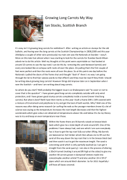


© Copyright 2026