
Blunted Prefrontal Cortical Fluorodeoxyglucose Positron Emission Tomography Response to Meta-Chlorophenylpiperazine in Impulsive Aggression
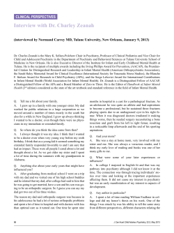
ORIGINAL ARTICLE Blunted Prefrontal Cortical 18Fluorodeoxyglucose Positron Emission Tomography Response to Meta-Chlorophenylpiperazine in Impulsive Aggression Antonia S. New, MD; Erin A. Hazlett, PhD; Monte S. Buchsbaum, MD; Marianne Goodman, MD; Diedre Reynolds, MD; Vivian Mitropoulou, MA; Larry Sprung, BA; Robert B. Shaw, Jr, BS; Harold Koenigsberg, MD; Jimcy Platholi, MA; Jeremy Silverman, PhD; Larry J. Siever, MD Background: Impulsive aggression is a prevalent problem and yet little is known about its neurobiology. Preclinical and human studies suggest that the orbital frontal cortex and anterior cingulate cortex play an inhibitory role in the regulation of aggression. Methods: Using positron emission tomography, re- gional metabolic activity in response to a serotonergic stimulus, meta-chlorophenylpiperazine (m-CPP), was examined in 13 subjects with impulsive aggression and 13 normal controls. The anterior cingulate and medial orbitofrontal regions were hypothesized to respond differentially to m-CPP in patients and controls. In the frontal cortex, regional metabolic glucose response to m-CPP was entered into a group (impulsive aggressive, control) ⫻ slice (dorsal, middle, orbital) ⫻ position (medial, lateral) ⫻ location (anterior, posterior) ⫻ hemisphere (right, left) mixed-factorial analysis of variance design. A separate group (impulsive aggressive, con- From the Psychiatry Service, Bronx Veterans Affairs Medical Center, Bronx, NY (Drs New, Goodman, Reynolds, Koenigsberg, Silverman, and Siever, Ms Mitropoulou, and Mr Sprung); the Department of Psychiatry (Drs New, Hazlett, Buchsbaum, Goodman, Reynolds, Koenigsberg, Silverman, and Siever, Ms Mitropoulou, and Messrs Sprung and Shaw), and the Neuroscience PET Laboratory (Drs Hazlett and Buchsbaum, Mr Shaw, and Ms Platholi), Mount Sinai School of Medicine, New York, NY. A trols) ⫻ anteroposterior location (Brodmann areas 25, 24, 31, 29) ⫻ hemisphere (right, left) analysis of variance was used to examine regional glucose metabolism in the cingulate gyrus. Results: Unlike normal subjects, patients with impulsive aggression did not show activation specifically in the left anteromedial orbital cortex in response to m-CPP. The anterior cingulate, normally activated by m-CPP, was deactivated in patients; in contrast, the posterior cingulate gyrus was activated in patients and deactivated in controls. Conclusions: The decreased activation of inhibitory regions in patients with impulsive aggression in response to a serotonergic stimulus may contribute to their difficulty in modulating aggressive impulses. Arch Gen Psychiatry. 2002;59:621-629 LTHOUGH VIOLENT crime has decreased in the past decade, violent incidents involving impulsive aggression rather than planned violence are increasing.1 These include juvenile violence, domestic violence, and workplace acts of aggression.2,3 Violence and homicide are significantly associated with mental illness, especially antisocial and borderline personality disorder.4 Considering the serious consequences of impulsive-aggressive behavior, its neurobiology has received little scrutiny. Evidence from metabolite and neuroendocrine studies has linked abnormalities in central serotonin activity to impulsive aggression.5-8 The association of lesions in the orbitofrontal cortex (OFC) and anterior cingulate gyrus (ACG) with disinhibited aggression suggests that faulty regulation of negative emotion, through (REPRINTED) ARCH GEN PSYCHIATRY/ VOL 59, JULY 2002 621 a reduced serotonin-mediated activation of the prefrontal cortex, may predispose an individual to impulsive aggression.9 BRAIN REGIONS AND AGGRESSION Studies of brain lesions suggest regional control of aggression, with the ACG and OFC playing central roles.10-15 The critical influence of the OFC and the ACG in human aggression is exemplified by the case of Phineas Gage, who, after a penetrating brain injury, became hostile and verbally aggressive. Computerized reconstruction of Gage’s skull demonstrated the location of his brain lesion in the anteromedial cortex, the OFC and the ACG, with more marked damage in the left hemisphere.16 Most lesions in the medial OFC also include damage to the ACG. In the human brain, the ACG has 2 main subdivi- WWW.ARCHGENPSYCHIATRY.COM ©2002 American Medical Association. All rights reserved. Downloaded From: https://jamanetwork.com/ on 06/11/2014 SUBJECTS AND METHODS SUBJECTS Thirteen patients with impulsive aggression (8 men, 5 women; mean [SD] age, 31.7 [8.5] years; range, 20-43 years; 9 right-handed, 3 left-handed, 1 mixed) who met DSM-IV criteria for 1 or more personality disorders were included. Patients with a history of schizophrenia, psychotic disorder, or bipolar type I affective disorder were excluded. Patients with current major depressive disorder were also excluded since this has been associated with impaired brain regional response to fenfluramine.38 All patients had been medication-free for 6 weeks or more (9 of 13 had never taken medication). An age- and sex-matched group of 13 normal subjects was also studied (8 men,5 women; mean [SD] age, 31.6 [8.1] years; range, 21-43 years; 11 right-handed, 1 left-handed, 1 mixed). Subjects were screened for severe medical or neurological illness, head injury, history of alcohol/drug dependence, and substance abuse in the past 6 months. All subjects had negative urine toxicology screen results for drugs of abuse, and women had negative pregnancy tests on each positron emission tomography (PET) scan day. Participants provided written informed consent in accordance with the guidelines of our institutional review board. Patients were recruited for the study through advertisement in local newspapers (90%) and through referrals from outpatient psychiatric clinics at the Bronx Veterans Affairs Medical Center (Bronx, NY) and Mount Sinai School of Medicine (New York, NY) (10%). Of 85 subjects screened, 13 subjects were successfully recruited into the patient group. Patients were excluded, in order of frequency, because of current substance abuse, a chronic medical problem such as diabetes or heart disease, pregnancy, the presence of current major depression, and in one case, the presence of current psychotic symptoms. In addition, one subject declined participation because of fear of the radioactive isotope. In the control group, approximately 90 candidates responded to our advertisement. Many subjects were excluded because of the presence of an Axis I or Axis II diagnosis in themselves (detected at screening) or a firstdegree relative. Axis I and personality disorder diagnoses were made through interviews with a psychologist using the Structured Clinical Interview for DSM-IV Axis I disorders39 and the Structured Interview for DSM-IV Personality Disorders (SIDP-IV),40 respectively. Trait aggression was assessed using the Module for Intermittent Explosive Disorder–Revised (IED-R)41 and depression with the Hamilton Depression Rating Scale (HDRS).42 All subjects completed the Buss-Durkee Hostility Inventory (BDHI)43; both total (BDHItotal) and composite Irritability-Assaultiveness subscale (BDHIIRR-ASS) scores have been associated with biological markers of aggression7 (Table 1). All patients met the following criteria: (1) significant physical and/or verbal aggression meeting criteria for IED-R ( = 0.92); (2) impulsivity as assessed by the SIDP-IV sions: the dorsal cognitive division (including the dorsal part of Brodmann areas [BAs] BA24 and BA32) and the rostral-ventral affective division (including the rostral part of BA24, BA32, and BA25).17,18 The affective sub(REPRINTED) ARCH GEN PSYCHIATRY/ VOL 59, JULY 2002 622 “impulsiveness” criterion for borderline personality disorder (=0.78), including behavior such as reckless driving or impulsive sexual behavior; and/or (3) self-damaging acts (predominantly self-mutilatory cutting of the skin) as assessed by the SIDP-IV “self-damaging” borderline criterion (A5) (interrater reliability, =0.90). Controls met none of the 3 above-defined criteria and had no personal or firstdegree family history of psychiatric illness. Prolactin and cortisol levels, obtained from all subjects except for 2 controls (technical difficulties with the intravenous line precluded blood sampling), were measured as described previously, and the peak minus baseline was calculated (⌬ prolactin, ⌬ cortisol).44 PROCEDURE On 2 separate occasions, each participant received m-CPP or placebo (counterbalanced to control for order effects). At 8 AM, after an overnight fast, 1 intravenous line was inserted into each forearm (1 used for blood sampling, the other for injection of m-CPP/placebo and 18fluorodeoxyglucose). An 0.08-mg/kg solution of m-CPP/placebo in an identical syringe of 20 mL of saline was given by slow push over 90 seconds. Immediately following, 5 mCi (185 MBq) of 18fluorodeoxyglucose was administered into the venous set rubber diaphragm behind the subject’s back as a 4560-second slow push. The subject remained in a resting state in a soundattenuated, dimly lit room for the 35-minute tracer-uptake period, after which the subject was escorted to an adjacent bathroom to void. The subject was then positioned in the PET scanner using a previously prepared thermosetting plastic mask. The imaging data-acquisition period lasted about 40 minutes. Scans were separated by at least 1 week to allow for drug elimination (3-4 days) and to coincide with a weekly scan schedule. All subjects and staff were blind to the dosing/placebo regimen. On each scan day, patients were evaluated with the HDRS. IMAGING Positron emission tomography scans were carried out as described elsewhere33,45 (General Electric Medical Systems scanner model 2048, General Electric, Milwaukee, Wis; [resolution 4.5 mm in plane, 5.0 mm axially]). Fifteen slices at 6.5-mm intervals were obtained in 2 sets to cover the entire brain. Slice counts of 1.53 million counts are typical. Scans were reconstructed with a blank and a transmission scan using the Hanning filter (width, 3.15 mm). The same individually molded thermoplastic face mask was used for each scan to keep the head stationary during image acquisition and to assist in PET/magnetic resonance imaging (MRI) image coregistration. Positron emission tomography images were obtained in nCi/pixel and standardized as relative metabolic rate (rGMR) by dividing each pixel by the mean value for the entire brain (defined by brain edge from coregistered MRI). While this limits interpretations of single-structure absolute activity, this method is widely used when evaluating hypotheses related to patterns of metabolic rate across brain areas and division receives wide input from regions, including the hippocampus, amygdala, medial OFC, and dorsal raphe, and projects to the basal ganglia, subthalamic nuclei, and lateral hippocampus.18,19 In an animal model, WWW.ARCHGENPSYCHIATRY.COM ©2002 American Medical Association. All rights reserved. Downloaded From: https://jamanetwork.com/ on 06/11/2014 was used in 4 earlier imaging studies of serotonin activation.33,34,37,38 Within 1 week of their PET scans, participants underwent MRI examination as described previously46 (General Electric Signa 5X, acquisition parameters: repetition time, 24 milliseconds; echo time, 5 milliseconds; flip angle, 40°; slice thickness, 1.2 mm; matrix, 256⫻256; field of view, 23 cm). Magnetic resonance images were resectioned to standard Talairach-Tournoux47 position. Positron emission tomography–MRI coregistration used the algorithm of Woods et al.48 Brain edges were visually traced on all MRI axial slices. Intertracer reliability on 27 individuals is 0.99 for area. On the basis of earlier studies,33,34,37 the cingulate, orbitofrontal, and medial frontal regions were hypothesized to respond differentially to serotonin agonists in patients and controls. These areas were located in advance of any analysis in the Talairach-Tournoux atlas and their coordinates recorded (Table 2). For the cingulate, we used x-coordinates 5 mm from midline; for BAs, we chose the position of numerals or halfway between duplicate numerals, 5 mm from the cortical edge. The square region of interest (ROI) (5⫻5 pixels) was applied centered on that coordinate and at the proportion as the brain-bounding box in the Talairach-Tournoux atlas. An adjustment was made so that ROIs were moved closer to the centroid of the slice if the box fell partly outside the coregistered brain outline, as could happen in brains that were especially narrow in the y direction for boxes placed at 45° and 135°. This method, the reverse Talairach hypothesis–driven strategy, was used for 3 reasons: (1) to minimize type I statistical errors in evaluating large numbers of ROIs in both hemispheres through the use of multiway repeatedmeasures analysis of variance (ANOVA) and a single F ratio test indicating the hypothesized diagnostic group ⫻ condition ⫻ region interaction; (2) to minimize type II errors resulting from assessing small individual, potentially noisy ROIs and failing to observe orbitofrontal system–wide response by combining ROIs; and (3) to provide standard and known brain atlas locations for replication. We also controlled type I error by not discussing main effects or interactions that are not interpretable (eg, main effect of slice level across structures measured at multiple axial slice levels) or peripheral to our interest (main effect of hemisphere across the normal controls and patients). Our analysis is limited in power by the sample size (n=13 in each of the 2 groups). STATISTICAL DESIGN A 2 ⫻ 3 ⫻ 2 ⫻ 2 ⫻ 2 mixed-factorial ANOVA design was applied to rGMR data obtained from frontal ROIs. Dependent variables were expressed as difference scores (m-CPP − placebo) for rGMR within each ROI. The first variable consisted of the 2 participant groups (impulsiveaggressive and controls), and the remaining variables were all repeated measures, consisting of 3 slice levels (dorsal, middle, and orbital; corresponding to TalairachTournoux levels: +12, +4, and −4, respectively), 2 medial/ lateral positions (medial and lateral prefrontal cortex), 2 anteroposterior locations (anterior and posterior), and electrical stimulation of the ACG in the cat brain ACG resulted in an increased latency in attack behavior.20 While the ACG is implicated in affective-cognitive activity,18 the posterior cingulate gyrus is implicated in (REPRINTED) ARCH GEN PSYCHIATRY/ VOL 59, JULY 2002 623 2 hemispheres (right and left). At the dorsal slice level (+12 slice), the medial regions included BA10 (anterior region) and BA32 (posterior region), and the lateral regions included BA46 (anterior region) and BA45 (posterior region). At the middle and orbital slice levels, the ROIs were the same as at the dorsal level except that the lateral regions were BA10 (anterior region) and BA47 (posterior region). A separate ANOVA was performed on 4 BAs within the cingulate gyrus. This 2⫻4⫻2 mixed-factorial design was employed to examine the drug-placebo rGMR difference values within the BAs for the 2 cohorts. The first variable consisted of the 2 groups, the second variable consisted of 4 BAs in the anteroposterior position (BA25, BA24, BA31, and BA29), and the third variable consisted of 2 hemispheres. Brodmann area 25 was located on the +4 slice level and the 3 remaining cingulate regions were located on the +12 slice level. All statistical analyses involving repeated measures with more than 2 levels used GreenhouseGeisser ⑀ corrections to adjust probabilities for repeatedmeasures F values. Uncorrected degrees of freedom are reported. To detect the source of significant interactions between group and hypothesized BA, we carried out an ANOVA on each BA separately. For interactions involving slice level, replicated ROIs adjacent in position or hemisphere were not followed up because they were not part of our hypothesis or were neuroanatomically not important. In addition, we report results of the Mauchley sphericity test, the Levine homogeneity of variance test, and the multivariate Rao R. To explore relationships between the prefrontal cortex and cingulate gyrus rGMR and clinical measures of the degree of impulsivity, measured by BDHItotal and BDHIIRR-ASS scores, Spearman correlations were computed only for regions entered into the ANOVAs above. EXPLORATORY SIGNIFICANCE PROBABILITY MAPPING To provide a survey of the entire brain slice, we carried out voxel-by-voxel t tests on the same brain slices assessed by the stereotaxic ROI method. The significance probability mapping technique is similar to other approaches but uses MRI-based region alignment.49 Continuous edges were manually drawn around the brain. Nine midline points equally spaced in the z direction were identified. Slices were then adjusted by the number of rows and columns so that every slice contained an equal number of pixels, with every edge pixel aligned and midline pixels positioned in a vertical strip at the edge center. Positron emission tomography images for the placebo and drug scans were coregistered to the same MRI similarly standardized, and unpaired t tests were carried out for the drug minus placebo difference scores. To confirm our original report of blunted response to fenfluramine and to provide validation of the reverse-Talairach ROI approach, we present these images with 1-tailed probability maps. To examine other already published studies and provide exploratory results for future investigators, we present 2-tailed probability maps. sensory processing and perhaps in processing fearinducing stimuli.21-25 The posterior cingulate has reciprocal pathways to the hippocampus, ACG, parahippocampal gyrus, and temporal areas.26,27 WWW.ARCHGENPSYCHIATRY.COM ©2002 American Medical Association. All rights reserved. Downloaded From: https://jamanetwork.com/ on 06/11/2014 Table 1. Impulsive-Aggressive Patients With Personality Disorders* Patient No./ Sex/Age, y Handedness HAMD Suicidal BDHI IRR-ASS Self-Injury Axis I Disorders Axis II Disorders 1/M/38 2/M/23 3/F/22 4/F/20 5/M/24 6/M/38 7/F/36 8/M/29 9/M/43 10/F/26 11/F/42 12/M/43 L R R R R R M R L R L R 14 15 11 14 10 2 1 11 14 15 9 14 0 0 1 0 0 0 0 0 0 0 1 1 38 47 22 41 53 14 40 31 51 46 49 46 12 14 10 13 17 2 13 10 17 14 18 18 0 0 0 0 0 0 0 0 0 1 0 0 Bipolar II, HX ETOH, IED-R MDD (past), HX ETOH, IED-R MDD (past), DYSTH, IED-R MDD (past), GAD, SOCPHOB, IED-R HX ETOH, SOCPHOB, GAD, IED-R HX ETOH, IED-R BDD, IED-R MDD (past), HX ETOH, IED-R MDD (past), SOCPHOB, IED-R MDD (past), DYSTH, IED-R MDD (past), DYSTH, IED-R MDD (past), HX POLYSUB, IED-R PPD, SPD, ASPD, NPD NPD, BPD AVPD, BPD OCPD, BPD PPD, SPD, OCPD, ASPD PPD, SPD, AVPD, BPD NPD, AVPD AVPD, BPD, ASPD PPD, OCPD, NPD, BPD PPD, OCPD, AVPD, BPD PPD, NPD, BPD NPD, PPD, ASPD *HAMD, Hamilton Depression Rating Scale; BDHI, Buss-Durkee Hostility Inventory; IRR-ASS, Buss-Durkee Hostility Inventory Composite Subscale of Irritability and Assaultiveness; self-injury, self-mutilatory cutting; HX ETOH, history of alcohol abuse; IED-R, intermittent explosive disorder-revised; PPD, paranoid personality disorder; SPD, schizotypal personality disorder; ASPD, antisocial personality disorder; NPD, narcissistic personality disorder; MDD (past), history of major depressive disorder; BPD, borderline personality disorder; DYSTH, dysthymia; AVPD, avoidant personality disorder; GAD, generalized anxiety disorder; SOCPHOB, social phobia; OCPD, obsessive-compulsive personality disorder; BDD, body dysmorphic disorder; and POLYSUB, history of polysubstance abuse. Table 2. Talairach Coordinates for Locatization Brodmann Area (BA) X Y Z Talairach Coordinates for Cingulate Boxes (Right Hemisphere) Anterior BA25 8 32 4 Middle BA24 8 33 12 Middle BA31 8 −57 12 Posterior BA29 8 −65 12 Talairach Coordinates for Frontal Boxes (Right Hemisphere) BA10 medial anterior 5 60 12 BA32 medial posterior 5 45 12 BA46 lateral anterior 48 25 12 BA45 lateral posterior 45 40 12 BA10 medial anterior 5 60 4 BA32 medial posterior 5 45 4 BA46 lateral anterior 45 45 4 BA45 lateral posterior 48 30 4 BA11 medial anterior 5 60 −4 BA11 medial posterior 5 45 −4 BA11 lateral anterior 42 50 −4 BA47 lateral posterior 48 30 −4 POSITRON EMISSION TOMOGRAPHY STUDIES AND AGGRESSION Positron emission tomography (PET) studies of relative glucose metabolic rate (rGMR) have related abnormalities in the ACG and prefrontal cortex to impulsive aggression.28-30 In an anger-induction model, normal men showed increased rGMR in the left OFC and right ACG,31 perhaps reflecting the normal activation of inhibitory regions in response to anger stimulation. A variety of serotonergic agents can modulate rGMR in the prefrontal cortex and in the ACG. d-Fenfluramine has been found to increase rGMR in the left ACG and in the prefrontal cortex in normal subjects.32 In our previous study, normal subjects showed increased metabolism in the ACG and OFC following fenfluramine administration, while patients did not.33 These findings were replicated in a study of borderline personality disorder.34 Meta-chlorophenylpiperazine (m-CPP), a non(REPRINTED) ARCH GEN PSYCHIATRY/ VOL 59, JULY 2002 624 specific 5-HT agonist,35-36 increased rGMR in the right OFC, middle frontal gyrus, posterior cingulate, and thalamus in normal subjects.37 Our study assesses rGMR in a larger sample of patients with impulsive aggression and normal controls after administration of m-CPP. We hypothesized that (1) patients would show decreased rGMR in the OFC and ACG after m-CPP relative to controls; (2) the posterior cingulate would not show blunting in patients vs controls; (3) in patients, medial regions of the OFC would show a more blunted response to m-CPP than would lateral regions, suggesting that the ACG with the adjacent OFC, which normally modulates aggression through a serotonergic mechanism, is underactive in impulsive aggression. RESULTS NEUROENDOCRINE MEASURES Analysis of mean responses to m-CPP showed no significant between-group differences for either ⌬ prolactin levels (controls: mean [SD], 20.22 [21.34] ng/mL; median, 18.1 ng/mL; patients: 23.55 [18.78] ng/mL; median, 16.3 ng/mL; t22 = 0.41, P = .69; Mann-Whitney U=63.5, P=.64) or ⌬ cortisol levels (controls: 11.45 [7.60] µg/dL; median, 12.5 µg/dL; patients: 12.97 [6.08] µg/dL; median, 13.3 µg/dL; t22 = −0.54, P = .59; Mann-Whitney U=58, P=.46). CLINICAL MEASURES The mean (SD) HDRS score of patients on the day of m-CPP administration was 10.5 (4.8), a typical score for patients with personality disorders who experience some dysphoria even when not clinically depressed. As expected, BDHI scores showed significant between-group differences (BDHItotal, controls: mean [SD], 20.23 [7.84]; range, 6-32; median, 22.0; patients: 40.54 [11.74]; range, 14-49; median, 46.0; t24 =5.18, P⬍.001; Mann-Whitney WWW.ARCHGENPSYCHIATRY.COM ©2002 American Medical Association. All rights reserved. Downloaded From: https://jamanetwork.com/ on 06/11/2014 0.15 Right Hemisphere 0.12 Medial Frontal 0.10 0.04 rGMR (MCPP–Placebo) rGMR (MCPP–Placebo) 0.08 0.00 –0.04 Impulsive-Aggressive Normal Controls Lateral Frontal Impulsive-Aggressive Normal Controls 0.05 0.00 ∗ ∗ –0.05 –0.08 Left Hemisphere rGMR (MCPP–Placebo) 0.12 –0.10 0.07 –0.15 Anterior BA24 Middle BA24 Posterior BA31 Posterior BA29 Anteroposterior Position 0.02 Figure 2. Cingulate gyrus regions (meta-chlorophenylpiperazine [m-CPP] effect). Mean relative glucose metabolic rate (rGMR) difference values (m-CPP − placebo) in the cingulate gyrus are shown for normal controls and patients with impulsive aggression (group ⫻ anteroposterior cingulate region interaction; F3,72 = 7.12, P⬍.001). Asterisks indicate significant group differences, P⬍.05. –0.03 –0.08 Slice +12 +4 –4 Slice +12 +4 –4 Figure 1. Frontal cortex regions. Mean relative glucose metabolic rate (rGMR) difference values (meta-chlorophenylpiperazine [m-CPP]− placebo) in prefrontal cortex regions of interest are shown for normal controls and patients with impulsive aggression (group ⫻ slice ⫻ medial/lateral ⫻ hemisphere interaction; F2,48 =5.20, P=.009). At the most dorsal slice level (+12), the medial regions include BA10 and BA32 and the lateral regions include BA46 and BA45. For the middle (+4) and orbital (−4) slice levels, the medial regions include BA10 and BA32 and the lateral regions include BA10 and BA47. (group ⫻ medial/lateral; F2,48 =0.08, P=.91; group ⫻ right/ left; F1,24 =0.40, P=.53) failed to reach significance. Despite the fact that none of the post-hoc tests were significant, this interaction reflects a significant rGMR pattern that differs between the 2 groups. U=17.5, P⬍.001; BDHIIRR-ASS, controls: mean, 5.93 [3.86]; range, 1-15; median, 6.0; patients: 13.54 [4.52]; range, 2-18; median, 14.0; t24 = 4.62, P⬍.001; Mann-Whitney U=17.5, P⬍.001). Baseline. To determine whether the groups differed in baseline rGMR, we conducted a 2 (group) ⫻ 3 (slice) ⫻ 2 (medial/lateral cortex) ⫻ 2 (anterior and posterior) ⫻ 2 (hemisphere) ANOVA on the placebo scan data. There was neither a main effect of group nor an interaction effect, indicating that patients did not differ from controls in baseline rGMR in the frontal lobe ROIs examined. POSITRON EMISSION TOMOGRAPHY Cingulate Gyrus Prefrontal Cortex Effects of m-CPP. Figure 2 shows mean rGMR difference scores (m-CPP − placebo) in the cingulate gyrus for patients and controls. A 2 (group) ⫻ 4 (anteroposterior BA) ⫻ 2 (hemisphere) ANOVA revealed a significant group ⫻ anteroposterior region interaction (univariate F3,72 =7.12, P⬍.001; multivariate Rao R3,22 =4.63, P=.01), indicating that in the ACG (BA25), m-CPP response was blunted in patients compared with controls. The Levene test for homogeneity of variances (ANOVA on absolute within-cell deviation scores, degrees of freedom for all F values 1,24) shows none of the 8 variables to be significant (P range, .2-.97) (Rao R3,22 =7.11, P=.002 [Wilks ⌳, 0.507]; Mauchley sphericity test Wilks ⌳=0.32; 25 =25.8, P⬍.001). When the order of drug and placebo administration was added as a fourth independent group dimension, neither the main effect of order (F1,21 =0.73, P=.40) nor the group ⫻ order ⫻ region interactions (F3,63 =0.39, P=.76) were statistically significant. In the posterior cingulate (BA31 and BA29), the effect was reversed, with patients showing a greater m-CPP response than controls (Figure Effects of m-CPP. Figure 1 shows mean rGMR difference scores (m-CPP−placebo) in frontal lobe ROIs for patients and controls. A 2 (group) ⫻ 3 (slice) ⫻ 2 (medial/ lateral cortex) ⫻ 2 (anterior, posterior location) ⫻ 2 (hemisphere) ANOVA of rGMR difference scores revealed a significant group ⫻ slice ⫻ medial/lateral ⫻ hemisphere interaction (univariate: F2,48 = 5.20, P= .009; multivariate: Rao R 2,23 =4.54, P=.02). In the right hemisphere, patients showed a blunted m-CPP response at the orbital slice level in the lateral but not medial frontal regions compared with controls. In the left hemisphere, this effect was reversed, with patients, unlike controls, showing a blunted response at the orbital slice level in medial but not lateral frontal regions. Although the interaction effect was statistically significant, simple-effects tests for each of the regions within the ANOVA failed to reach significance. The main effect of group (F1,24 = 0.04, P=.83) and all other interpretable interaction effects with group (REPRINTED) ARCH GEN PSYCHIATRY/ VOL 59, JULY 2002 625 WWW.ARCHGENPSYCHIATRY.COM ©2002 American Medical Association. All rights reserved. Downloaded From: https://jamanetwork.com/ on 06/11/2014 P < .05, 1-Tailed Confirmation P < .05, 2-Tailed Exploration z = –4 z=4 z = 12 Figure 3. Statistical probability map. Relative metabolic rate differences (drug − placebo). Blue indicates that patient response to meta-chlorophenylpiperazine (m-CPP) was less than that of normal controls; red, patient response to m-CPP was greater than that of normal controls (1- and 2-tailed t tests, P⬍.05). Background is mean coregistered and shape-standardized magnetic resonance imaging. 2). An orthogonal set of individual planned comparisons confirmed that patients, compared with controls, showed a significantly weaker m-CPP response in the ACG (BA25) (F1,24 =6.13, P=.02) but a significantly greater m-CPP response in the posterior cingulate (BA29) (F1,24 = 7.92, P=.001). There were no significant group effects for BA31 (F1,24 =3.45, P=.08) or for BA24 (F1,24 =.13, P=.71). Statistical probability mapping (Figure 3) of drug minus placebo scores confirm the blunted response in the ACG in patients (blue), especially slices z=4 and z=−4, and the greater response in the posterior cingulate (red, z=12). which revealed a significant group ⫻ anteroposterior region interaction (univariate F3,72 =5.63, P=.008; multivariate Rao R3,22 =7.12, P=.001). Compared with controls, patients had lower rGMR in the posterior cingulate but not in the anterior (BA25) and middle cingulate (BA24) regions. Individual planned comparisons confirmed that patients had significantly lower rGMR than controls in BA31 and BA29 (F1,24 = 4.52, P = .04 and F1,24 = 9.88, P = .004, respectively). There were no group differences for BA25 (F1,24 =3.10, P=.09) and BA24 (F1, 24 =1.24, P=.27). rGMR AND CLINICAL RATINGS Baseline. Figure 4 shows mean rGMR in the cingulate gyrus on the placebo scan day. To determine whether patients and controls differed in baseline rGMR in the cingulate gyrus, we conducted a 2 (group) ⫻ 4 (anteroposterior) ⫻ 2 (hemisphere) ANOVA on the placebo scan data, (REPRINTED) ARCH GEN PSYCHIATRY/ VOL 59, JULY 2002 626 Prefrontal Cortex Baseline. In controls during the placebo condition, increased rGMR was associated with higher trait aggression WWW.ARCHGENPSYCHIATRY.COM ©2002 American Medical Association. All rights reserved. Downloaded From: https://jamanetwork.com/ on 06/11/2014 Effects of m-CPP. In controls, decreased m-CPP response in BA47 bilaterally was associated with higher BDHItotal score at the middle slice level (rs = −0.61, P=.02; rs =−0.62, P = .02). In addition, lower BDHIIRR-ASS subscales were associated with increased rGMR in the left BA47 (rs =−0.55, P = .05). An inverse correlation between rGMR and aggression scores was also observed in BA45 bilaterally (rs = −0.61, P = .03; and rs = −0.66, P = .02). In patients, a direct correlation was seen between m-CPP response in the right BA45 at the dorsal slice level (BDHItotal, rs = .56, P = .04; BDHIIRR-ASS, rs =0.58, P =.03) and in the right BA10 at the middle slice level (rs =0.57, P=.04). Cingulate Gyrus Baseline. In the baseline (placebo) condition, increased rGMR in right and left middle cingulate gyrus (BA24) in controls was associated with increased BDHIIRR-ASS scores (rs =0.59, P=.03 and rs =0.12, P=.69, respectively). In contrast, increased rGMR in the left posterior cingulate (BA29) was associated with increased BDHIIRR-ASS scores in patients (rs =0.52, P = .06). Effects of m-CPP. There were no significant Spearman correlations in either patients or controls between rGMR for BA25, BA24, BA31, and BA29 and measures of aggression. COMMENT Patients with impulsive aggression react aggressively in response to interpersonal emotional cues, such as conflict or perceived disrespect. We hypothesized that limbic structures (ie, the hippocampus and amygdala) may be activated by an interpersonal trigger. Then, through a mechanism facilitated by serotonin, inhibitory regions (ie, the ACG and OFC) are activated. In our current experiment, m-CPP provided a serotonergic activation that is expected to activate inhibitory areas in normal subjects. Our data show that in response to a serotonergic stimulus, rGMR in the left medial posterior OFC is lower in patients with impulsive aggression compared with controls. Alternative regions connected to the medial OFC, including the lateral orbital cortex and areas of the fron(REPRINTED) ARCH GEN PSYCHIATRY/ VOL 59, JULY 2002 627 1.8 Impulsive-Aggressive Normal Controls 1.6 ∗ 1.4 rGMR scores (BDHItotal) in the right BA46 at the dorsal (rs =0.61, P=.027) and middle (rs =0.69, P=.009) slice levels. In addition, higher-measure subscale BDHIIRR-ASS scores were associated with increased rGMR in BA46 bilaterally in the middle slice level (right: rs =0.581, P=.04; left: rs =0.61, P=.03 in the right and left, respectively), and BA46 at the ventral slice level on the right (rs = 0.64, P=.02), as well as in BA10 bilaterally at the middle slice level (right: rs = 0.49, P = .08; left: rs = 0.56, P = .05). In patients, increased rGMR was associated with higher scores of aggression (BDHItotal) in the left BA46 at the middle- and ventral slice levels (rs = 0.587, P = .03; rs = 0.59, P = .02, respectively). Similarly, higher scores of aggression were associated with increased rGMR in the right BA10 at the ventral slice level (rs = 0.639, P = .02). ∗ 1.2 1.0 0.8 0.6 Anterior BA24 Middle BA24 Posterior BA31 Posterior BA29 Anteroposterior Position Figure 4. Cingulate gyrus regions baseline (placebo). Mean relative glucose metabolic rate (rGMR) in the cingulate gyrus on the placebo scan day in normal controls and in patients with impulsive aggression. Group ⫻ anteroposterior cingulate region interaction; F3,72 = 5.63, P = .008. Asterisks indicate significant group differences, P⬍.05. tal cortex, are activated in patients. No group differences emerged in the baseline condition, suggesting that differences between patients and controls can only be observed under a serotonergic challenge. Although post hoc comparisons of the m-CPP response between groups in individual frontal ROIs were not significant, the model comparing drug activation between groups in medial vs lateral and orbital vs dorsal areas was significant. This supports our a priori hypothesis, that relative m-CPP rGMR in specific frontal areas (medial vs lateral; orbital vs dorsal) would be diminished in patients with impulsive aggression. In the cingulate cortex, there were important differences in responses to m-CPP. The ACG (BA25) was activated in response to m-CPP in controls, whereas in patients, it was deactivated. In contrast, the posterior cingulate was deactivated in controls in response to m-CPP and was activated in patients (Figure 2). The overall model entered into the ANOVA and the post-hoc comparisons of responses to m-CPP in the ACG and posterior cingulate were significant. This suggests that in patients with impulsive aggression, activation of the posterior cingulate rather than the ACG is the gateway to the inhibitory medial OFC. Activation of the posterior cingulate is not accompanied by activation of the OFC and thus is less effective in modulating aggression in patients than in normal subjects. LATERALITY The diminished m-CPP response in the ACG and the adjacent medial OFC in patients was especially marked in the left hemisphere. Previous studies of emotional processing and frontal lobe laterality have suggested that the left hemisphere may be involved with “approach” and the right with “withdrawal,”50 Left frontal regions have been described as the center for self-regulation and planWWW.ARCHGENPSYCHIATRY.COM ©2002 American Medical Association. All rights reserved. Downloaded From: https://jamanetwork.com/ on 06/11/2014 ning51 whereas right frontal regions may be involved with negative affects, such as fear and disgust.50 Traumatic brain injury in the left dorsofrontal region gives rise to anger and hostility, whereas lesions of the right OFC result in anxiety and depression.14 Phineas Gage’s lesion was predominantly left-sided.16 The reported predominance of the left hemisphere in the control of emotion was borne out in our study, which demonstrated a blunted metabolic response to m-CPP in the left medial OFC in patients relative to controls. The opposite effect was observed in the right OFC, where controls showed lower rGMR after m-CPP than did patients. Findings of significant aggression-related laterality have not been reported for the ACG and were not seen in our analysis. MEASURES OF IMPULSIVE AGGRESSION AND rGMR Clinical correlations between aggression and rGMR in the regions entered into the ANOVAs were performed, although the groups were not comparable because the scores of aggression fell into a much higher range in patients than in controls. Controls demonstrated a direct correlation between the degree of aggression and rGMR in BA46 bilaterally in the baseline condition. Patients showed a similar effect but it was limited to the left hemisphere. In response to m-CPP, however, controls with higher aggression scores exhibited increased m-CPP activation in BA47 and BA45. In contrast, patients with higher aggression scores showed lower m-CPP response in BA45 and no relationship in BA47. This gives further evidence that patients and controls may use frontal brain regions differently in regulating aggression. In the cingulate region, there were no associations between m-CPP–stimulated rGMR and the degree of aggression in controls or patients. Thus, the m-CPP probe was sensitive enough to distinguish between groups that differ substantially in impulsive aggression (ie, patients vs controls) but not to pick up differences in the narrower range of aggressive behavior seen within groups. The absence of patient-control differences in neuroendocrine responses to m-CPP may be the result of relatively small numbers of subjects in each cell, particularly when results are examined separately by sex. The use of a serotonin stimulus in conjunction with 18fluorodeoxyglucose-PET to examine specific activation of brain regions may be a more sensitive probe for serotonergic dysfunction in impulsive aggression than the challenge paradigm. This study used a serotonergic probe to activate ACG and OFC. Future studies examining rGMR in response to aggression induction would provide even more powerful evidence of the relationship between the activation of specific brain regions and the control of aggression. Our study implicates the ACG and the medial posterior orbital cortex in the control of aggressive behavior, and suggests that serotonin may facilitate this control. m-CPP is known to act as a partial agonist at 5-HT2A and 5-HT2C receptors, but may also have a presynaptic site of action.52 As specific ligands become available, more specific pharmacologic targets underlying the serotonergically mediated activation of the OFC and the ACG observed with m-CPP can be identified. (REPRINTED) ARCH GEN PSYCHIATRY/ VOL 59, JULY 2002 628 Submitted for publication December 12, 2000; final revision received August 6, 2001; accepted October 1, 2001. This research was supported by grant 5-RO1MH566606 from the National Institute of Mental Health, Bethesda, Md (Dr Siever), and by the Veterans Affairs Medical Research Program Career Development Award, Washington, DC (Dr New), and was supported in part by grant 5-M01 RR00071 from the National Center for Research Resources, the National Institutes of Health, Bethesda (for the Mount Sinai General Clinical Research Center). This research was presented at the annual meeting of the Society of Biological Psychiatry, New Orleans, La, May 5, 2001. Invaluable editorial assistance was provided by Sherry Buchsbaum. Excellent technical assistance was provided by Nina Roberto and Elizabeth Iskander. Corresponding author: Antonia S. New, MD, Psychiatry Service-116A, Bronx VA Medical Center, 130 W Kingsbridge Rd, Bronx, NY 10468 (e-mail: [email protected]). REFERENCES 1. Federal Bureau of Investigation. Crime in the United States. Washington, DC: Federal Government Press; 1999. 2. Coker AL, Derrick C, Lumpkin JL, Aldrich TE, Oldendick R. Help-seeking for intimate partner violence and forced sex in South Carolina. Am J Prev Med. 2000; 19:316-320. 3. Peek-Asa C, Schaffer KB, Kraus JF, Howard J. Surveillance of non-fatal workplace injuries, using police and employer’s reports. J Occup Environ Med. 1998; 40:707-713. 4. Coid JW. Axis II disorders and motivation for serious criminal behavior. In: Skodol AE, ed. Psychopathology and Violent Crime: Review of Psychiatry Series. Washington, DC: American Psychiatric Press; 1998:53-97. 5. Coccaro EF, Siever LJ, Klar HM, Mauer G, Cochrane K, Cooper TB, Mohs RC, Davis KL. Serotonin studies in patients with affective and personality disorders. Arch Gen Psychiatry. 1989;46:587-599. 6. Coccaro EF, Kavoussi RJ, Hauger RL. Physiological responses to d-fenfluramine and ipsapirone challenge correlate with indices of aggression in males with personality disorder. Int Clin Psychopharmacol. 1995;10:177-179. 7. Coccaro EF, Kavoussi RJ, Trestman RL, Gabriel SM, Cooper TB, Siever LJ. Hormonal responses to meta-chlorophenylpiperazine (m-CPP) undiminished by acute m-CPP pretreatment. Psychiatry Res. 1996;62:139-145. 8. O’Keane V, Maloney E, O’Neil H, O’Connor A, Smith C, Dinan TG. Blunted prolactin response to d-fenfluramine in sociopathy: evidence for subsensitivity of central serotonergic function. Br J Psychiatry. 1992;160:643-646. 9. Davidson RJ, Putnam KM, Larson CL. Dysfunction in the neural circuitry of emotion regulation: a possible prelude to violence. Science. 2000;289:591-594. 10. Butter CM, Snyder DR, McDonald JA. Effects of orbital frontal lesions on aversive and aggressive behavior in rhesus monkeys. J Comp Physiol Psychol. 1970; 72:132-144. 11. Raleigh MJ, Steklis HD, Ervin FR, Kling AS, McGuire MT. The effects of orbital frontal lesions on the aggressive behavior of vervet monkeys (Cercopitheous aethiops sabeaus ). Exp Neurol. 1979;66:158-168. 12. WeigerWA, Bear DM. An approach to the neurology of aggression. J Psychiatr Res. 1988;22:85-98. 13. Heinrichs RW. Frontal cerebral lesions and violent incidents in chronic neuropsychiatric patients. Biol Psychiatry. 1989;25:174-178. 14. Grafman J, Schwab K, Warden D, Pridgen A, Brown HR, Salazar AM. Frontal lobe injuries, violence and aggression: a report from the Vietnam Head Injury Study. Neurology. 1996;46:1231-1238. 15. Anderson SW, Bechara A, Damasio H, Tranel D, Damasio AR. Impairment of social and moral behavior related to early damage in human prefrontal cortex. Nat Neurosci. 1992;2:1032-1037. 16. Damasio H, Grabowski T, Frank R, Galaburda AM, Damasio AR. The return of Phineas Gage: clues about the brain from the skull of a famous patient. Science. 1994;264:1102-1105. 17. Bush G, Luu P, Posner MI. Cognitive and emotional influences in anterior cingulate cortex. Trends Cogn Sci. 2000;4:215-222. 18. Devinsky O, Morrell MJ, Vogt BA. Contributions of anterior cingulated cortex to behavior. Brain. 1995;118:279-306. WWW.ARCHGENPSYCHIATRY.COM ©2002 American Medical Association. All rights reserved. Downloaded From: https://jamanetwork.com/ on 06/11/2014 19. Mega MS, Cummings JL. Frontal-subcortical circuits and neuropsychiatric disorders. J Neuropsychiatry Clin Neurosci. 1994;6:358-370. 20. Siegel A, Edinger HM. Role of the limbic system in hypothalamically elicited attack behavior. Neurosci Biobehav Rev. 1983;7:395-407. 21. Vogt BA, Finch DM, Olson CR. Functional heterogeneity in cingulate cortex: the anterior executive and posterior evaluative regions. Cereb Cortex. 1992;2:435-443. 22. Maddock RJ, Buonocore MH. Activation of left posterior cingulate gyrus by the auditory presentation of threat related words: an fMRI study. Psychiatry Res. 1997; 75:1-14. 23. Wik G, Fredrikson M, Ericson K, Erikson L, Stone-Elander S, Greitz T. Regional cerebral blood flow during experimental phobic fear. Psychophysiology. 1993; 30:126-130. 24. Fredrikson M, Wik G, Annas P, Ericson K, Stone-Elander S. Functional neuroanatomy of visually elicited simple phobic fear: additional data and theoretical analysis. Psychophysiology. 1995;32:43-48. 25. McGuire PK, Bench CJ, Frith CD, Marks IM, Frackowiak RS, Dolan RJ. Functional anatomy of obsessive-compulsive phenomena. Br J Psychiatry. 1994;164: 459-468. 26. Goldman-Rakic PS, Selemon LD, Schwartz ML. Dual pathways connecting the dorsolateral prefrontal cortex with the hippocampus and parahippocampal cortex in rhesus monkey. Neuroscience. 1984;12:719-743. 27. Suzuki WA, Amaral DG. Cortical inputs to the CA1 field of the monkey hippocampus originate from the perirhinal and parahippocampal cortex but not from area TE. Neurosci Lett. 1990;115:43-48. 28. Raine A, Buchsbaum M, LaCasse L. Brain abnormalities in murderers indicated by positron emission tomography. Biol Psychiatry. 1997;42:495-508. 29. Goyer PF, Andreasen PJ, Semple WE, Clayton AH, Compton-Toth BA, Schulz SC, Cohen RM. Positron-emission-tomography and personality disorders. Neuropsychopharmacology. 1994;10:21-28. 30. Volkow ND, Tancredi L. Neural substrates of violent behavior: a preliminary study with positron emission tomography. Br J Psychiatry. 1987;151:668-673. 31. Dougherty DD, Shin LM, Alpert NM, Pitman RK, Orr SP, Lasko M, Macklin ML, Fischman AJ, Rauch SL. Anger in healthy men: a PET study using script-driven imagery. Biol Psychiatry. 1999;46:466-472. 32. Mann JJ, Malone KM, Diehl DJ, Perel J, Nichols TE, Mintun MA. Positron emission tomographic imaging of serotonin activation effects on prefrontal cortex in healthy volunteers. J Cereb Blood Flow Metab. 1996;16:418-426. 33. Siever LJ, Buchsbaum M, New A, Spiegel-Cohen J, Wei T, Hazlett E, Sevin E, Nunn M, Mitropoulou V. d,l-Fenfluramine response in impulsive personality disorder assessed with 18F-deoxyglucose positron emission tomography. Neuropsychopharmacology. 1999;20:413-423. 34. Soloff PH, Meltzer CC, Greer PJ, Constantine D, Kelly TM. A fenfluramineactivated FDG-PET study of borderline personality disorder. Biol Psychiatry. 2000; 47:540-547. 35. Fiorella D, Hesley S, Rabin R, Winter J. 5HT2c receptor-mediated phosphoinositide turnover and the stimulus effects of m-chlorophelpiperazine. Psychopharmacology (Berl). 1995;122:237-243. (REPRINTED) ARCH GEN PSYCHIATRY/ VOL 59, JULY 2002 629 36. Sugimoto Y, Yamada J, Yoshikawa T, Horisaka K. Effects of the 5HT2c/2b receptor agonist 1-(3-chlorophenyl) piperazine on plasma glucose levels in rats. Eur J Pharmacol. 1996;307:75-80. 37. Hommer D, Andreason P, Rio D, Williams W, Ruttimann U, Monenan R, Zamethrin A, Rawlings R, Linnoila M. Effects of m-chlorophenylpiperazine on regional brain glucose utilization: a positron emission tomography comparison of alcoholic and control subjects. J Neurosci. 1997;17:2796-2806. 38. Mann JJ, Malone KM, Diehl DJ, Perel J, Cooper TB, Mintun MA. Demonstration in vivo of reduced serotonin responsivity in the brain of untreated depressed patients. Am J Psychiatry. 1996;153:174-182. 39. First M, Spitzer R, Gibbon M, Williams J. Structured Clinical Interview for Axis I Disorders-Patient Edition. New York, NY: Biometrics Research, New York State Psychiatric Institute; 1996. 40. Pfohl B, Blum N, Zimmerman M. Structured Clinical Interview for DSM-IV. Washington, DC: American Psychiatric Press; 1996. 41. Coccaro EF, Kavoussi RJ, Berman ME, Lish JD. Intermittent explosive disorderrevised: development, reliability, and validity of research criteria. Compr Psychiatry. 1998;39:368-376. 42. Hamilton M. Development of a rating scale for primary depressive illness. Br J Soc Clin Psychol. 1967;6:278-296. 43. Buss AH, Durkee A. An inventory for assessing different kinds of hostility. J Consult Psychol. 1957;21:343-348. 44. New AS, Trestman RL, Mitropoulou V, Benishay DS, Coccaro EF, Silverman J, Siever LJ. Serotonergic function and self-injurious behavior in personality disorder patients. Psychiatry Res. 1997;69:17-26. 45. Haznedar MM, Buchsbaum MS, Metzger M, Solimando A, Spiegel-Cohen J, Hollander E. Anterior cingulate gyrus volume and glucose metabolism in autistic disorder. Am J Psychiatry. 1997;154:1047-1050. 46. Hazlett EA, Buchsbaum MS, Mohs RC, Speigel-Cohen J, Wei TC, Azueta R, Haznedar MM, Singer MB, Shihabuddin L, Luu-Hsia C, Harvey PD. Age-related shift in brain region activity during successful memory performance. Neurobiol Aging. 1998;19:437-445. 47. Talairach J, Tournoux P. Co-Planar Stereotactic Atlas of the Human Brain: ThreeDimensional Proportional System: An Approach to Medical Cerebral Imaging. Rayport M, trans. New York, NY: Thieme Medical Publishers; 1988. 48. Woods RP, Mazziotta JC, Cherry SR. MRI-PET registration with automated algorithm. J Comput Assist Tomogr. 1993;17:536-546. 49. Buchsbaum MS, Hollander E, Haznedar MM, Tang C, Spiegel-Cohen J, Wei TC, Solimando A, Buchsbaum BR, Robins D, Bienstock C, Cartwright C, Mosovich S. Effect of fluoxetine on regional cerebral metabolism in autistic spectrum disorders: a pilot study. Int J Neuropsychopharmacol. 2001;4:119-125. 50. Davidson RJ. Anterior cerebral asymmetry and the nature of emotion. Brain Cogn. 1992;20:125-151. 51. Luria AR. The Working Brain. New York, NY: Basic Books; 1973. 52. Baumann MH, Mash DC, Staley JK. The serotonin agonist m-chlorophenylpiperazine (m-CPP) binds serotonin transporter sites in human brain. Neuroreport. 1995;6:2150-2152. WWW.ARCHGENPSYCHIATRY.COM ©2002 American Medical Association. All rights reserved. Downloaded From: https://jamanetwork.com/ on 06/11/2014


© Copyright 2026