
Positive regulation of meiotic DNA double
Downloaded from http://rsob.royalsocietypublishing.org/ on July 7, 2015
rsob.royalsocietypublishing.org
Research
Cite this article: Gray S, Allison RM, Garcia V,
Goldman ASH, Neale MJ. 2013 Positive
regulation of meiotic DNA double-strand break
formation by activation of the DNA
damage checkpoint kinase Mec1(ATR). Open
Biol 3: 130019.
http://dx.doi.org/10.1098/rsob.130019
Received: 1 February 2013
Accepted: 5 July 2013
Subject Area:
genetics/molecular biology
Keywords:
meiosis, checkpoint, recombination, Mec1 ATR,
Tel1 ATM, Spo11
Author for correspondence:
Matthew J. Neale
e-mail: [email protected]
Positive regulation of meiotic
DNA double-strand break
formation by activation of the
DNA damage checkpoint
kinase Mec1(ATR)
Stephen Gray1, Rachal M. Allison1, Valerie Garcia1,
Alastair S. H. Goldman2 and Matthew J. Neale1
1
MRC Genome Damage and Stability Centre, School of Life Sciences, University of Sussex,
Brighton BN1 9RQ, UK
2
Department of Molecular Biology and Biotechnology, University of Sheffield,
Sheffield S10 2TN, UK
1. Summary
During meiosis, formation and repair of programmed DNA double-strand
breaks (DSBs) create genetic exchange between homologous chromosomes—a
process that is critical for reductional meiotic chromosome segregation and
the production of genetically diverse sexually reproducing populations. Meiotic
DSB formation is a complex process, requiring numerous proteins, of which
Spo11 is the evolutionarily conserved catalytic subunit. Precisely how Spo11
and its accessory proteins function or are regulated is unclear. Here, we use
Saccharomyces cerevisiae to reveal that meiotic DSB formation is modulated by
the Mec1(ATR) branch of the DNA damage signalling cascade, promoting
DSB formation when Spo11-mediated catalysis is compromised. Activation of
the positive feedback pathway correlates with the formation of single-stranded
DNA (ssDNA) recombination intermediates and activation of the downstream
kinase, Mek1. We show that the requirement for checkpoint activation can be
rescued by prolonging meiotic prophase by deleting the NDT80 transcription
factor, and that even transient prophase arrest caused by Ndt80 depletion is
sufficient to restore meiotic spore viability in checkpoint mutants. Our observations are unexpected given recent reports that the complementary kinase
pathway Tel1(ATM) acts to inhibit DSB formation. We propose that such antagonistic regulation of DSB formation by Mec1 and Tel1 creates a regulatory
mechanism, where the absolute frequency of DSBs is maintained at a level
optimal for genetic exchange and efficient chromosome segregation.
2. Introduction
Electronic supplementary material is available
at http://dx.doi.org/10.1098/rsob.130019.
To prevent the chromosome copy number doubling during each reproductive
cycle, sexually reproducing organisms create gametes containing half the chromosome copy number of their parents. Halving the chromosome copy number
occurs during meiosis—a specialized pair of sequential nuclear divisions that
first segregates homologous chromosomes, then sister chromatids. In many organisms, accurate segregation of homologous chromosomes during meiosis I requires
& 2013 The Authors. Published by the Royal Society under the terms of the Creative Commons Attribution
License http://creativecommons.org/licenses/by/3.0/, which permits unrestricted use, provided the original
author and source are credited.
Downloaded from http://rsob.royalsocietypublishing.org/ on July 7, 2015
likelihood that more than one DSB arises per chromatid quartet at a given chromosomal locus [47]. Here, we reveal a novel
role for the Mec1 pathway in promoting DSB formation when
the catalytic activity of Spo11 is compromised. Our results
suggest that both positive and negative feedback loops influence the rate of DSB formation, creating a regulated system
with optimal levels of recombination initiated.
3. Results
In each budding yeast meiotic cell, accurate reductional segregation of homologous chromosomes at the first nuclear
division involves the formation and repair of approximately
160 Spo11–DSBs [48,49]. The first step in the repair of
Spo11–DSBs is their nucleolytic processing by Mre11 and
Sae2, creating covalent Spo11-oligonucleotide complexes
(Spo11-oligos; figure 1a [11,18,48]). Spo11-oligos are detected
by immunopurifying Spo11 and radiolabelling the ends of
the covalently attached DNA [50]. The absolute abundance
of Spo11-oligo complexes can be used to estimate total DSB
formation. Spo11–DSBs trigger transient activation of the
DNA damage checkpoint response machinery (DDR [24])—a
state that persists in recombination-defective mutants such as
dmc1D [22]. During our studies, we were intrigued to discover
that while the abundance of Spo11-oligo complexes was
increased in dmc1D strains (where DSB repair is abolished,
and DSBs accumulate [22]), such an increase was absent
when the DDR was inactivated (rad24D; figure 1b,c).
Our observations suggest a link between checkpoint
activity and Spo11-oligo abundance. However, one limitation
of our method is that it makes use of a SPO11-HA3-His6 allele
(henceforth referred to as spo11-HA) to enrich for Spo11-oligo
complexes. This C-terminal epitope tag is reported to moderately reduce the catalytic activity of Spo11 (DSB formation
drops by 11 –50% compared with the untagged wild-type
[51]). Because the reduction in Spo11-oligo abundance in
rad24D dmc1D could be due to fewer total DSBs or to their
faster turnover—perhaps due to changes in meiotic cell
cycle progression caused by rad24D—we were interested to
determine the abundance of DSBs in our panel of strains.
We began by assessing DSB formation at the wellcharacterized HIS4::LEU2 reporter locus on chromosome III
[52] in synchronized meiotic cultures, where Spo11 was either
HA-tagged or untagged. At HIS4::LEU2, there are two prominent
DSB sites flanking the LEU2 insertion (figure 2a). In the untagged
strain (SPO11þ), DSB signals peaked at approximately 6% of the
total lane signal (figure 2c), with more of the DSBs arising at site 1
(figure 2d). Contrary to our expectation, total DSB signals were
not reduced by the Spo11-HA tag (figure 2c). However, the distribution was altered such that now a similar proportion of
DSBs was detected at site 1 and site 2 (figure 2d; see also [52]).
Accurately measuring DSB abundance from a culture of
wild-type cells is complicated by overlapping kinetics of DSB
formation and repair. To work around this issue, we remeasured the DSB frequency in strains similar to those we had
used for the Spo11-oligo analysis, where the Dmc1 repair
protein was mutated, causing DSBs to accumulate (figure 2e).
Open Biol 3: 130019
3.1. Loss of Rad24/Mec1(ATR) checkpoint activity causes
a synergistic reduction in double-strand break
signals in hypomorphic spo11-HA strains
2
rsob.royalsocietypublishing.org
the formation and repair of numerous DNA double-strand
breaks (DSBs) distributed across the genome. DSB repair uses
homologous recombination to drive interaction and genetic
exchange between homologous chromosomes, which facilitates
accurate chromosome disjunction at anaphase I. Much of our
detailed understanding of the mechanisms of meiotic recombination comes from studies performed in the budding yeast
Saccharomyces cerevisiae [1].
In S. cerevisiae, meiotic DSB formation requires the function of 10 proteins, of which Spo11 is the catalytic subunit.
The precise function of the nine accessory proteins is unclear,
as are the detailed mechanisms that govern the frequency and
distribution of Spo11– DSB formation across the genome.
Spo11 shares similarity to a type-II topoisomerase (Top6A)
from archaea and initiates DSB formation via a transesterification reaction that coordinately severs the DNA backbone
on both strands, leaving Spo11 monomers covalently
attached to the 50 ends of the DSB (reviewed in [2]).
Spo11–DSBs are processed nucleolytically by the Mre11,
Sae2 and Exo1 proteins, creating DSBs with extended 30 ending single-stranded termini [3–18], which are rapidly
bound by Replication Protein A [19]. In the absence of
Mre11 nuclease activity or of the accessory factor, Sae2,
Spo11 remains covalently bound to DSB ends [8,11 –
14,18,20,21]. In these circumstances, repair of the DSBs is
impossible and cells undergo a catastrophic meiotic nuclear
division that generates fragmented chromosomes [4–6,18].
In wild-type cells, the ssDNA ends are used in a homology search by the eukaryotic RecA orthologues, Rad51
and Dmc1 [22,23], creating intermolecular interactions
between homologous chromosome pairs. In the absence of
Dmc1, DSBs accumulate with extended ssDNA ends due to
failed repair [22]. The accumulation of ssDNA elicits a
strong checkpoint response, causing cells to arrest in late prophase I [22,24], at least in part due to inactivation of the
meiotic transcription factor, Ndt80 [25]. In mammals, similar
recombination failure leads to programmed cell death [26,27].
The checkpoint response depends on activation of orthologues of the mammalian phosphoinositide-3-kinase-related
kinase (PIKK) ATR, the RAD9-RAD1-HUS1 (9-1-1) checkpoint clamp complex and the RAD17 clamp loader [28–31].
In budding yeast, these proteins are, respectively, named
Mec1, Rad17, Mec3, Ddc1 and Rad24 [32,33]. Such activation
leads to persistent phosphorylation of Mek1 [34,35], a meiosis-specific paralogue of the Rad53 kinase (CHK2 in
mammalian cells [36 –39]).
Removing the function of checkpoint proteins causes
entry into the meiotic divisions of mutants that accumulate
unrepaired DSBs, leading to cell death [24]. Interestingly,
budding yeast cells, which possess mutations in the checkpoint proteins, actually undergo a delayed DSB-dependent
meiosis I division, suggesting that checkpoint proteins are
directly involved in the DNA repair and recombination process itself [40]. Indeed, checkpoint mutants have increased
rates of ectopic (also called, non-allelic) recombination [41],
whereas mutation of Mek1 abolishes repair partner choice
entirely, with most or all DSBs now competent to repair
using the sister chromatid (rather than the homologous
chromosome) as repair template [42 –44].
Recently, it has been shown that mutation of another
PIKK kinase, ATM, results in increased DSB formation in
mammals and in flies [45,46], and both Mec1(ATR) and
Tel1(ATM) are necessary in budding yeast to reduce the
Downloaded from http://rsob.royalsocietypublishing.org/ on July 7, 2015
(a)
(c)
3
40
(arb. units)
20
10
Rad51 + Dmc1
0
2
6
8
spo11-HA
dmc11D spo11-HA
repair
rad24D dmc1D spo11-HA
(b)
*
hours
0
3
4
5
6
spo11-HA
7
8
0
3
4
5
6
7
dmc1D spo11-HA
8
0
3
4
5
6
7
8
rad24D dmc1D spo11-HA
Figure 1. Levels of Spo11-oligonucleotides are increased in the recombination-defective dmc1D mutant but decreased when combined with the rad24D checkpoint
mutant. (a) Meiotic recombination is initiated by DSBs created by Spo11 (orange ellipses), which are nucleolytically processed by Mre11, Sae2 and Exo1 generating
transient Spo11-oligonucleotide complexes and ssDNA flanking the DSB site. DSB repair uses Dmc1 and Rad51-dependent recombination between homologous
chromosomes, which facilitates their pairing and segregation at anaphase I. (b) Spo11-oligonucleotide complexes were immunoprecipitated from whole cell extracts
of synchronous cultures of the indicated strains and timepoints after meiotic induction, 30 -labelled with a-32P dCTP using TdT and resolved by SDS-PAGE. Spo11oligonucleotides are marked by an arrowhead. Asterisk indicates non-specific TdT labelling. (c) Abundance of Spo11-oligonucleotide complexes was quantified using
a phosphorimager and IMAGEGAUGE software.
In both the tagged and untagged Spo11 strains, DSBs accumulated to a similar extent, reaching approximately 15%
and approximately 18% of total lane signal, respectively
(figure 2f ). As before, the distribution of events was altered
by the HA-tag, such that the fractions of DSBs forming at site
1 and site 2 were now similar (figure 2d). A second strong hotspot region, ARE1, showed the same trend of relatively
unchanged DSB frequency when the spo11-HA allele was
present (see electronic supplementary material, figure S1a,b).
Next, we assessed DSB formation in the checkpoint-defective rad24D strain (figure 2b–f). Reduced MEC1 checkpoint
activity (as caused by mutations such as rad24D) results in
rapid exonucleolytic hyper-resection of the DSB ends creating
extensive 30 -ending ssDNA tails that cause DSB molecules to
migrate much more rapidly and heterogeneously when
separated by agarose electrophoresis [40]. At HIS4::LEU2,
despite the signal arising in rad24D being more diffuse, DSBs
remained readily detectable above lane background signals,
peaking at 9% (figure 2c), slightly greater than wild-type
cells, possibly due to a slight delay in DSB repair causing
DSB signals to transiently accumulate to higher steady-state
levels. In rad24D dmc1D cells, despite extensive hyper-resection, DSBs accumulated and persisted similar to RAD24
dmc1D, reaching a maximum level of approximately 16–20%
of the total DNA (figure 2e,f). Notably, in the rad24D and
rad24D dmc1D backgrounds, barely any signal was observed
at DSB site 2, resulting in a significantly altered ratio compared
with RAD24þ cells (figure 2d). In addition, we observed
increases in the frequency of ectopic recombination products
in rad24D strains (see figure 2b,e; electronic supplementary
material, figure S2a–c), as has been reported by others [41].
In contrast to the situation in RAD24þ cells, the combination
of spo11-HA and rad24D resulted in a substantial reduction in
DSB signal (figure 2b–f). DSBs were reduced to 50% of the
rad24D level in rad24D spo11-HA cells (figure 2b,c), and to less
than 25% of the rad24D dmc1D level in the rad24D dmc1D spo11HA strain, where DSB signals were barely detected despite
being expected to accumulate (figure 2e,f). These reductions in
DSB abundance correlated with reduced abundance of ectopic
recombination products (see electronic supplementary material,
figure S2). Similar effects were observed at the ARE1 locus (see
electronic supplementary material, figure S1b). In rad24D
dmc1D cells, despite extensive hyper-resection, DSBs accumulated to levels similar to both the dmc1D and dmc1D spo11-HA
controls (see electronic supplementary material, figure S1b). By
contrast, DSBs were undetectable in the dmc1D rad24D spo11HA strain (see electronic supplementary material, figure S1b).
This surprising reduction in DSB abundance was reproduced
in two independently generated isolates of the same rad24D
dmc1D spo11-HA genotype, indicating that the phenotype was
Open Biol 3: 130019
4
hours
rsob.royalsocietypublishing.org
30
Mre11 + Sae2
Exo1
Downloaded from http://rsob.royalsocietypublishing.org/ on July 7, 2015
DSB II
(a)
DSB I
PstI
6.4 kb
LEU2
2.4 kb
(g)
PstI
3.8 kb
(c)
E
DSB 2
10
8
6
4
2
DSB 1
0
0 2 3 4 6 78
wild-type
spo11-HA
0 2 3 4 6 78
rad24D
2
0 2 3 4 6 78
rad24D
spo11-HA
4
6
hours
wild-type
spo11-HA
rad24D
rad24D spo11-HA
8
(d)
DSB 1
chromosome VIII
fraction of total DSB signal
0.8
DSB 2
0.6
0.4
wild-type dmc1D
rad24D
rad24D spo11-HA dmc1D rad24D
dmc1D
spo11-HA spo11-HA
(e)
sae2D
sae2D
sae2D
sae2D
rad24D spo11-HA rad24D
spo11-HA
(f)
P
20
DSBs (% of lane signal)
E
DSB 2
DSB 1
hours 0 2 3 4 6 7 8 0 2 3 4 6 7 8 0 2 3 4 6 7 8 0 2 3 4 6 7 8
dmc1D
dmc1D
spo11-HA
rad24D
dmc1D
rad24D
dmc1D
spo11-HA
5
2
4
hours
6
8
dmc1D
dmc1D spo11-HA
dmc1D rad24D
dmc1D rad24D spo11-HA
582
485
388
340
291
243
194
145
97
48
582
485
388
340
291
243
194
145
97
48
III V VIII XI
0.8
0.6
0.4
0.2
0
SPO11 HA SPO11 HA SPO11 HA SPO11 HA
rad24D
rad24D
P
60
40
20
80
wild-type
60
spo11-HA
40
20
hours
20
15
10
5
0
DSB 1
4
3
2
1
0
viable spores per tetrad
sp
wi
ld
DSB 2
rad24D
rad24D
spo11-HA
0
0
DSBs (% of lane signal)
percentage of tetrads
80
sae2D
dmc1D
(k)
( j)
100
100
-ty
pe
o1
1H
A
ra
ra
d2
d
24
4D
D
sp
o1
1H
A
spore viability (% of spores)
(i)
243
194
145
97
48
1.0
10
0
582
485
388
340
291
(h)
15
frequency of DSBs
relative to dmc1D
0
chromosome XI
0.2
sae2D
rad24D
rad24D
SPO11 HA SPO11 HA SPO11 HA SPO11 HA
hr: 0 6 8 0 6 8 0 6 8 0 6 8 0 810 0 810 0 810 0 810
kb:
340
291
243
194
145
97
48
0 6 8 10 0 6 8 10 0 6 8 10 0 6 8 10
sae2D
sae2D
sae2D
sae2D
spo11-HA rad24D rad24D
spo11-HA
2
4
6
8 10
hours
sae2D
sae2D spo11-HA
sae2D rad24D
sae2D rad24D spo11-HA
Figure 2. Synergistic reduction in meiotic DSBs in rad24D spo11-HA strains. (a) Physical map of the HIS4::LEU2 region, including PstI restriction sites, DSB sites and
location of MXR2 probe. (b,e,j ) Genomic DNA was isolated at the indicated time points from synchronous cultures of the indicated strains, digested with PstI,
fractionated on a 0.7% agarose gel, transferred to nylon membrane and hybridized with the MXR2 probe. Arrowheads indicate DSB signals; P, parental band;
E, ectopic recombinant (see electronic supplementary material, figure S2). (c,f,k) Quantification of the total DSB signal (DSB 1þDSB 2) shown in (b,e,j ) plotted
as a percentage of total lane signal. (d ) Proportion of DSB signal present in DSB 1 and DSB 2 for the indicated strains. (g) Intact chromosomal DNA was isolated at
the indicated time points from synchronously sporulating meiotic cultures, separated by PFGE, transferred to a nylon membrane and sequentially hybridized using a
radiolabelled fragment of a gene close to the left telomere of each chromosome (chr III ¼ CHA1; chr V ¼ RMD6; chr VIII ¼ CBP2; chr XI ¼ JEN1). Intact, fulllength chromosomes migrate near the top of the gel. DSB signals appear as the shorter, faster-migrating bands/molecules present in the 6 – 10 h timepoints. (h)
Quantification of total DSB signals in each lane expressed as a fraction of total lane signal. Measured signals were adjusted to account for the likelihood of multiple
DSBs occurring on the same molecule (see material and methods). Because the absolute frequency of DSB formation varies with chromosome, calculated average
frequencies (+s.d.) are plotted relative to dmc1D to aid easier strain-to-strain comparison. (i) Spore viability displayed as total viability (left) or separated according
to number of viable spores per tetrad (right). Error bars (left) are 95% confidence limits.
not due to an underlying defect in the genotype of the strain nor
a culture problem (data not shown).
To determine whether the synergistic reduction in DSB
abundance observed in rad24D spo11-HA strains was a general
4
Open Biol 3: 130019
hours 0 2 3 4 6 7 8
chromosome III
DSBs (% of lane signal)
P
chromosome V
(b)
dmc1D
rsob.royalsocietypublishing.org
his4X
Chr III
MXR2 probe
phenomenon affecting global DSB levels, we assessed total
DSB signals in the dmc1D background across four chromosomes
using pulsed-field gel electrophoresis (PFGE; figure 2g,h;
electronic supplementary material, figure S3). We detected an
Downloaded from http://rsob.royalsocietypublishing.org/ on July 7, 2015
Mutation of the Rad24-dependent DDR was not expected to
affect meiotic DSB formation. The spo11-HA allele, on the
other hand, is reported to be a DSB formation hypomorph,
variably reducing DSBs by 11– 50% [51]. Contrary to these
expectations, we have made two intriguing observations:
(i) in the DMC1 and dmc1D backgrounds, DSB frequency at
two recombination hotspots (HIS4::LEU2 and ARE1), and
across three other chromosomes, is reduced less than we
expected by spo11-HA; and (ii) DSB frequency, however, is
more severely reduced when Rad24 checkpoint activity
is abolished in spo11-HA; and spo11-HA dmc1D strains.
Previous analysis of DSB abundance in spo11-HA made use
of the rad50S mutant background [51]. In rad50S (as in the
similar mutations, sae2D and mre11-‘nuclease-dead’), DSB signals accumulate due to a failure to remove Spo11 from the DSB
end [11,20,52]. We remeasured DSB abundance in the sae2D
background at HIS4::LEU2 (figure 2j,k), ARE1 (see electronic
supplementary material, figure S1c) and across the length of
four chromosomes (figure 2g,h). In contrast to our previous
results in SAE2þ cells (see figure 2b–h; electronic supplementary material, figure S1b) and now more consistent with the
published work [51], we observed DSB signals to be reduced
by 50% in the spo11-HA sae2D strain compared with sae2D
(see figure 2g,h,j,k; electronic supplementary material,
figure S1c). Moreover, removal of checkpoint activity
(rad24D) in sae2D strains had little impact on DSB formation
in either SPO11þ or spo11-HA (see figure 2g,h,j,k; electronic
supplementary material, figure S1c).
Taken together, these observations suggest that while spo11HA is deficient in DSB formation (as observed in sae2D), such a
defect is substantially corrected in wild-type and dmc1D cells
by a process that is dependent on the activity of the Rad24
(Mec1/ATR) checkpoint. We propose that these observations
reveal the presence of a DDR-dependent positive feedback
3.3. The requirement for positive stimulation of doublestrand break formation by Rad24/Mec1 is not
unique to the spo11-HA hypomorph
The precise reason for the hypomorphic behaviour of the
Spo11-HA protein is unknown. To determine whether the
positive feedback mechanism we have uncovered is a general
feature of meiotic recombination regulation (rather than
uniquely required to rescue the spo11-HA allele), we investigated whether other Spo11 hypomorphs also benefit from
Rad24/Mec1 checkpoint activation. We selected a weak
hypomorphic allele of SPO11, spo11-D290A, which contains
a point mutation in the putative Toprim domain thought to
aid coordination of a metal ion necessary for DSB catalysis
[53]. Under normal conditions, this allele is reported to
have extremely mild defects in recombination, but suffer a
severe defect at lower temperatures, or when HA-tagged
[51,53]. For these reasons, we considered the spo11-D290A
(untagged) allele to be a good candidate for testing the
requirement for checkpoint activity.
We first measured DSB formation at the HIS4::LEU2 locus
(figure 4a–d). In the dmc1D background, the spo11-D290A
allele resulted in a 30% reduction in DSB formation, and a dramatic redistribution of events away from DSB 1 and towards
DSB 2 (figure 4a,b,d). These effects were slightly more pronounced than the defects caused by spo11-HA (figure 2b–f),
but nevertheless, in the DMC1þ background, spo11-D290A supported almost wild-type levels of spore viability (figure 4e). In
the sae2D background, spo11-D290A caused a more severe 50%
5
Open Biol 3: 130019
3.2. Stimulation of double-strand break formation
by a Rad24/Mec1(ATR)-dependent positive
feedback loop
mechanism that is activated by ssDNA and promotes DSB
formation under conditions of suboptimal catalysis (i.e. the
spo11-HA hypomorph). Consistent with this idea, we note
that the final frequency of DSBs that accumulates in rad24D
dmc1D spo11-HA cells (as assessed by PFGE) is very similar to
that observed in sae2D spo11-HA (figure 2 g,h), which, because
of the absence of ssDNA intermediates, we expect will also lack
efficient Rad24/Mec1 activation [34].
To determine whether the proposed feedback mechanism
was a general feature of the DDR, we repeated experiments in
strains mutated for components of either the checkpoint clamp
(Rad17) or the Mec1(ATR) kinase itself (figure 3). Owing to
problems with synchronizing mec1 cultures, we used an allele
of MEC1 where expression is specifically repressed during meiosis ( pCLB2-MEC1; electronic supplementary material, figure S4).
DSBs accumulated to high levels in both rad17D dmc1D
and pCLB2-MEC1 dmc1D (approx. 16–20%; figure 3a–d). By
contrast, in spo11-HA derivatives, DSB levels were reduced to
approximately 30% of the SPO11þ levels, as were ectopic
recombination products (see electronic supplementary material,
figure S2d,e), largely mimicking the synergistic DSB defects we
observed with rad24D (figure 2). Furthermore, similar to the
synergistic reduction in spore viability observed in rad24D
spo11-HA strains (figure 2i), loss of the MEC1 or RAD17 activity
in the spo11-HA background also caused a defect in spore viability (figure 3e,f). Overall, the effects conferred by the loss of
Rad17 were less severe than Rad24 or Mec1, suggesting that
there may be previously uncharacterized differences between
the roles of these DDR components. Nevertheless, we tentatively
conclude that the proposed positive feedback mechanism uses
all major components of the canonical ssDNA-dependent
DDR pathway.
rsob.royalsocietypublishing.org
approximately 25–30% reduction in DSBs in the dmc1D
spo11-HA strain relative to dmc1D (figure 2g,h). In rad24D
dmc1D, DSBs were reduced by 10–20% relative to dmc1D
(figure 2g,h). Finally, in the dmc1D rad24D spo11-HA strain,
DSB frequency fell to just a third of the dmc1D level (figure
2g,h). Representative comparisons of lane profile plots are presented in the electronic supplementary material (figure S3).
Collectively, these results indicate that the reduced abundance
of Spo11-oligo complexes we had observed in rad24D dmc1D
spo11-HA strains (figure 1b,c) is correlated with a reduced
ability to detect DSBs by southern blotting.
In S. cerevisiae, as in mammals, meiotic recombination is
necessary for accurate chromosome segregation—with
errors in this process causing aneuploidy. To determine
whether the molecular defects in DSBs and Spo11-oligo
abundance correlate with defects in meiotic chromosome segregation, we measured the viability of the haploid spores
formed upon completion of meiosis. By itself, the Spo11HA tag is reported to have very little effect on spore viability
[51,53]. We confirmed that spo11-HA alone causes no major
reduction in spore viability (figure 2i). By contrast, in the
rad24D strain, the spo11-HA allele caused spore viability to
drop from 30% down to less than 5% (figure 2i).
Downloaded from http://rsob.royalsocietypublishing.org/ on July 7, 2015
(a)
(b)
6
20
E
DSB 2
15
10
5
DSB 1
0
2
DSBs (% of lane signal)
DSB 1
20
15
10
5
0
*
hours 0 2 3 4 6 7 8 0 2 3 4 6 7 8 0 2 3 4 6 7 8 0 2 3 4 6 7 8
4
hours
6
8
( f ) 100
wild-type
rad17D
20
1
0
20
0
pC
LB
2
-M
pC
wi
ld
-ty
pe
rad17D spo11-HA
spore viability
2
40
A
M
EC
EC
1
1
sp
o1
1H
A
ra
ra
d1
7D d17
D
sp
o1
1H
A
40
pCLB2-MEC1
spo11-HA
3
60
LB
2
pCLB2-MEC1
4
H
60
80
o1
1-
spo11-HA
sp
80
0
2
rad17D
rad17D spo11-HA
rad17D dmc1D
rad17D dmc1D spo11-HA
rad17D
dmc1D
spo11-HA
% of tetrads
% of viable spores
(e) 100
8
Figure 3. DSB levels are synergistically reduced in spo11-HA pCLB2-MEC1 and rad17D mutants. (a,c) Genomic DNA was isolated at the indicated time points from
synchronous cultures of the indicated strains, digested with PstI, fractionated on a 0.7% agarose gel, transferred to nylon membrane and hybridized with the MXR2
probe. Arrowheads indicate DSB signals; asterisk marks non-specific band; P, parental band; E, ectopic band (see electronic supplementary material, figure S2).
(b,d ) Quantification of the total DSB signal (DSB 1þDSB 2) shown in (a,c) plotted as a percentage of total lane signal. (e,f ) Spore viability for the indicated
strains displayed as (e) total viability or (f ) separated according to number of viable spores per tetrad. Error bars in (e) are 95% confidence limits.
reduction in DSB signal at HIS4::LEU2 compared with SPO11
sae2D (figure 4a,c), and the same redistribution of events towards
DSB 2 (figure 4d).
We next estimated DSB formation globally using PFGE and
three chromosome probes (figure 4f,g). We detected no
reduction in DSB formation in the dmc1D spo11-D290A
background compared with dmc1D (on average it was 10%
greater; figure 4g). By contrast, the spo11-D290A allele reduced
DSB formation in the sae2D background by approximately 40%
(figure 4f,g), similar to what we had observed for spo11-HA
(figure 2h). Loss of rad24D checkpoint activity caused DSB
levels at HIS4::LEU2 to drop below our detection limit in the
Open Biol 3: 130019
DSB 2
rad17D
dmc1D
6
(d)
E
rad17D
spo11-HA
4
hours
pCLB2-MEC1
pCLB2-MEC1 spo11-HA
pCLB2-MEC1 dmc1D
pCLB2-MEC1 dmc1D spo11-HA
hours 0 2 3 4 6 7 8 0 2 3 4 6 7 8 0 2 3 4 6 7 8 0 2 3 4 6 7 8
pCLB2pCLB2pCLB2pCLB2-MEC1
MEC1
MEC1
MEC1
dmc1D
spo11-HA
dmc1D
spo11-HA
(c)
P
rad17D
rsob.royalsocietypublishing.org
DSBs (% of lane signal)
P
Downloaded from http://rsob.royalsocietypublishing.org/ on July 7, 2015
(f)
7
*
hours
dmc1D dmc1D dmc1D dmc1D sae2D sae2D
D290A rad24D rad24D
D290A
D290A
(b)
679
630
582
534
485
436
388
340
291
243
194
145
97
48
dmc1D
20
dmc1D
spo11-D290A
rad24D dmc1D
15
10
5
0
chromosome VIII
(c)
582
534
485
436
388
340
291
243
194
145
97
48
chromosome XI
DSBs
(% of lane signal)
25
rad24D dmc1D
spo11-D290A
2
4
hours
6
8
20
sae2D
10
sae2D
spo11-D290A
5
0
2
4
hours
6
8
DSB 1
fraction of total
DSB signal
(d)
(g)
0.2
SPO11
HA
RAD24
rad24D
rad24D
D290A
no.1 no.2 no.3
hr: 8 8 0 8 10 0 8 10 0 8 10 0 8 10 8 10 8 10 10 10 0 810 0 810 8 10
1.4
V VIII XI
1.2
dmc1D dmc1D dmc1D
spo11-HA D290A
sae2D
sae2D sae2D
spo11-HA D290A
100
80
% of spores
sae2D
0.4
0
(e)
DSB 2
0.6
wild-type
60
spo11-D290A
40
rad24D
20
rad24D
spo11-D290A
0
frequency of DSBs
relative to dmc1D
DSBs
(% of lane signal)
25
15
kb:
582
534
485
436
388
340
291
243
194
145
97
48
no.3
Open Biol 3: 130019
0 6 8 0 6 8 0 6 8 0 6 8 0 6 8 0 6 8
chromosome V
DSB 1
RAD24
RAD24
rad24D
no.1
DSB 2
1.0
0.8
0.6
0.4
0.2
0
dmc1D
dmc1D
rad24D
dmc1D
D290A
dmc1D
rad24D
D290A
sae2D
sae2D
D290A
spore viability
Figure 4. Synergistic reduction in meiotic DSBs in rad24D spo11-D290A strains. (a) Genomic DNA was isolated at the indicated time points from synchronous
cultures of the indicated strains, digested with PstI, fractionated on a 0.7% agarose gel, transferred to nylon membrane and hybridized with the MXR2 probe.
Arrowheads indicate DSB signals; asterisk indicates non-specific band; P, parental band; E, ectopic recombinant. (b,c) Quantification of the total DSB signal
(DSB 1 þ DSB 2) shown in (a) plotted as a percentage of total lane signal. (d ) Proportion of DSB signal present in DSB 1 and DSB 2 for the indicated strains.
(e) Spore viability displayed as total viability for the indicated strains. Error bars are 95% confidence limits. ( f ) Intact chromosomal DNA was isolated at the
indicated time points from synchronously sporulating meiotic cultures, separated by PFGE, transferred to a nylon membrane and sequentially hybridized using
a radiolabelled fragment of a gene close to the left telomere of each chromosome (chr V ¼ RMD6; chr VIII ¼ CBP2; chr XI ¼ JEN1). Presented gel shows experiment in triplicate. Intact, full-length chromosomes migrate near the top of the gel. DSBs signals appear as the shorter, faster-migrating bands/molecules present in
the 8 – 10 h time points. (g) Quantification of total DSB signals in each lane expressed as a fraction of total lane signal. Measured signals were adjusted to account
for the likelihood of multiple DSBs occurring on the same molecule (see material and methods). Because the absolute frequency of DSB formation varies with
chromosome, calculated average frequencies (+s.d.) are plotted relative to dmc1D to aid easier strain-to-strain comparison.
spo11-D290A dmc1D background (figure 4a,b), and a 60–70%
reduction in DSBs relative to the dmc1D control, as measured
by PFGE (figure 4f,g), down to the same frequency of DSBs
observed in sae2D spo11-D290A. Finally, we asked whether
spore survival in the spo11-D290A strain required Rad24/Mec1
rsob.royalsocietypublishing.org
E
dmc1D
spo11-D290A
no.2
rad24D
P
RAD24
(a)
activity (figure 4e). Consistent with our observations made
with the spo11-HA allele, spore viability in rad24D spo11-D290A
fell to just 2.7%.
Collectively, these results support the view that activation
of the Rad24/Mec1 checkpoint by ssDNA intermediates acts
Downloaded from http://rsob.royalsocietypublishing.org/ on July 7, 2015
(a)
(i)
8
*
0 3 4 5 6 8
wild-type
spo11-HA
(ii)
rsob.royalsocietypublishing.org
0 3 4 5 6 8
Mek1-P
Mek1
* Mek1-P
* Mek1
dmc1D
(b)
rad24D
spo11-HA
6
(i)
*
*
Mek1-P
*
*
Mek1-P
Mek1
(ii)
0 3 4 6 8
0 3 4 6 8
0 3 4 6 8
0 3 4 6 8
dmc1D
dmc1D
spo11-HA
rad24D
dmc1D
rad24D
dmc1D
spo11-HA
Mek1
Figure 5. Mek1 phosphorylation is synergistically reduced in a checkpoint-defective spo11-HA background. (a,b) Whole cell lysates from the indicated time points
and strains were resolved by SDS-PAGE containing 10 mM phos-tag reagent and probed with anti-Mek1 antibody. Mek1 is indicated with an arrowhead;
phosphorylated Mek1 by an asterisk. (b) The blot in (ii) is a longer exposure of the blot in (i).
as a general mechanism to promote DSB formation when the
efficiency of Spo11– DSB formation is compromised.
3.4. Involvement of Mek1 kinase in the double-strand
break feedback mechanism
Activation of the DDR kinases (Mec1 and Tel1 in budding
yeast) causes phosphorylation of Hop1 and subsequent activation of Mek1 [34], a meiotic kinase with roles related to
Rad53/CHK2 [36 –39]. Mek1 activation is associated with
phosphorylation on a number of residues [35]. To investigate
the relationship between Mek1 activity and the proposed
feedback process, we assessed the abundance of phosphorylated forms of Mek1 using phos-tag SDS-PAGE and western
blotting (figure 5a,b). In wild-type and spo11-HA cultures,
slower migrating phosphorylated forms of Mek1 appear coincidently with the time of DSB recombination intermediates
(3– 6 h; figure 5a(i)). Phosphorylated Mek1 was slightly
delayed in rad24D cells, suggesting a partial requirement for
Rad24 to achieve Mek1 activation (figure 5a(ii)). Strikingly,
phosphorylation of Mek1 was completely abolished in the
rad24D spo11-HA strain (figure 5a(ii)).
Because Mek1 phosphorylation signals are transient in
DMC1þ cells, we repeated the analysis in the dmc1D background, where DSB recombination intermediates and Mek1
signals would usually be expected to persist (figure 5b). In
dmc1D and dmc1D spo11-HA, there was a steady accumulation
in the abundance of multiple-shifted species of Mek1, indicating varying levels of phosphorylation (figure 5b). In the rad24D
dmc1D strain, much of this hyper-phosphorylation was lost,
confirming that hyper-activation of Mek1 is Rad24-dependent
(figure 5b). In the rad24D spo11-HA dmc1D strain, all residual
Mek1 phosphorylation was abolished (figure 5b), and not visible even when overexposing the film (figure 5b(ii)). Similar
loss of Mek1 phosphorylation was observed in pCLB2-MEC1
dmc1D spo11-HA strains (data not shown). Thus, activated
Mek1 correlates strongly with the ability to accumulate
appreciable levels of meiotic DSBs.
3.5. Reduction of double-strand breaks in spo11-HA
rad24D cells is not due to altered repair
partner choice
During meiosis, DSB repair reactions take place preferentially
between homologous chromosomes rather than between
sister chromatids [54,55]. The Mec1/Tel1 DDR checkpoint
machinery is important for enforcing such ‘repair partner
choice’ via activation of the Hop1 adapter protein and the
Mek1 kinase [34,44]. It seemed plausible that the synergistic
loss of DSB and Spo11-oligo signals, and drop in spore viability
caused by the combination of spo11-HA and rad24D, could be
explained by the observed failure to fully activate Mek1, and
thus to appropriately control repair partner choice, resulting
in increased occurrence of Rad51-dependent inter-sister recombination. To test this idea, we repeated our DSB analyses in
rad51D dmc1D strains, thereby preventing all recombinationmediated pathways of DSB repair (figure 6). As expected, in
the rad24D rad51D dmc1D strain, DSB signals at HIS4::LEU2
accumulated, reaching a plateau of approximately 15% of
total lane signal (figure 6a,b). In the rad24D rad51D dmc1D
spo11-HA strain, despite having removed the potential for
both Rad51 and Dmc1-dependent repair, accumulated
DSB signals were barely increased relative to rad24D dmc1D
Open Biol 3: 130019
rad24D
0 3 4 5 6 8 6
mek1D
0 3 4 5 6 8
Downloaded from http://rsob.royalsocietypublishing.org/ on July 7, 2015
(c)
no. 2
chromosome III
E
DSB 2
DSB 1
0 4 6 7 8 0 4 6 7 8
rad24D
dmc1D rad51D
spo11-HA
rad24D
dmc1D rad51D
spo11-HA
ndt80D
chromosome V
rad24D
dmc1D rad51D
15
rad24D dmc1D spo11-HA
10
rad24D dmc1D rad51D
5
rad24D dmc1D rad51D
spo11-HA
rad24D dmc1D rad51D
spo11-HA ndt80D
0
2
4
6
chromosome VIII
DSBs (% of lane signal)
(b)
8
hours
(e)
(ii)
100
chromosome XI
MI+MII (% of cells)
(i)
80
60
40
20
rad51D
SPO11 HA SPO11 HA
hr: 0 6 8 0 6 8 0 6 8 0 6 8
kb:
340
291
243
194
145
97
48
582
485
388
340
291
243
194
145
97
48
582
485
388
340
291
243
194
145
97
48
582
485
388
340
291
243
194
145
97
48
0
(iv)
(d)
1.2
ndt80D
SPO11 HA
HA
hr: 0 6 8 0 6 8 0 6 8
kb:
340
291
243
194
145
97
48
582
534
485
436
388
340
291
243
194
145
97
48
*
582
534
485
436
388
340
291
243
194
145
97
48
*
679
630
582
534
485
436
388
340
291
243
194
145
97
48
*
III V VIII XI
1.0
80
frequency of DSBs
relative to dmc1D
MI+MII (% of cells)
(iii) 100
rad24D dmc1D rad51D
60
40
20
0.8
0.6
0.4
0.2
0
0
2
3
4 5 6
hours
7
8
0
2
3
4 5
hours
6
wild-type
rad24D
spo11-HA
rad24D spo11-HA
7
8
0
rad24D
dmc1D
rad24D
dmc1D
spo11-HA
rad24D
dmc1D
rad51D
rad24D
dmc1D
rad51D
spo11-HA
rad24D
dmc1D
rad51D
spo11-HA
ndt80D
Figure 6. Ndt80-dependent meiotic progression and not inter-sister repair is responsible for the decreased DSB signal in rad24D spo11-HA. (a) Genomic DNA was isolated at the
indicated time points from synchronous cultures of the indicated strains, digested with PstI, fractionated on a 0.7% agarose gel, transferred to nylon membrane and hybridized with
the MXR2 probe. Arrowheads indicate DSB signals, asterisk marks non-specific band; P, parental band, E, ectopic band. (b) Quantification of the total DSB signal (DSB 1 þ DSB 2)
shown in (a) plotted as a percentage of total lane signal. rad24D dmc1D spo11-HA data from figure 2f are overlaid to aid visual comparison. (c,f) Intact chromosomal DNA was
isolated at the indicated time points from synchronously sporulating meiotic cultures, separated by PFGE, transferred to a nylon membrane and sequentially hybridized using a
radiolabelled fragment of a gene close to the left telomere of each chromosome (chr III ¼ CHA1; chr V ¼ RMD6; chr VIII ¼ CBP2; chr XI ¼ JEN1). Intact, full-length chromosomes migrate near the top of the gel. DSBs signals appear as the shorter, faster-migrating bands/molecules present in the 6–8 h time points. Asterisk marks residual parental
signal from the chromosome III hybridization. (d) Quantification of total DSB signals in each lane expressed as a fraction of total lane signal. Measured signals were adjusted to
account for the likelihood of multiple DSBs occurring on the same molecule (see material and methods). Because the absolute frequency of DSB formation varies with chromosome,
calculated average frequencies (+s.d.) are plotted relative to dmc1D to aid easier strain-to-strain comparison. (e) Samples were stained with DAPI and the average fraction of cells
having undergone the first and/or second meiotic division was plotted +s.d. (n . 200). (i) Shown without error bars for clarity. (ii–iv) Individual comparisons.
spo11-HA, remaining approximately threefold lower than
the SPO11þ (figure 6a,b) control. Analysis of DSB formation
across four chromosomes revealed a similar relationship
(figure 6c,d). Specifically, we detected no increase in DSB abundance in rad24D dmc1D rad51D spo11-HA relative to rad24D
dmc1D spo11-HA (see figure 6c,d; electronic supplementary
Open Biol 3: 130019
*
0 2 3 4 5 6 7 8 0 2 3 4 5 6 7 8
9
rsob.royalsocietypublishing.org
P
hours
(f)
rad24D dmc1D
chromosome III
no.1
chromosome V
(ii)
chromosome VIII
(i)
chromosome XI
(a)
Downloaded from http://rsob.royalsocietypublishing.org/ on July 7, 2015
3.6. Reduction of double-strand break formation in
spo11-HA rad24D cells is due to precocious
Ndt80 activity
3.7. Transient ndt80-induced prophase arrest
improves spore viability in hypomorphic spo11
rad24D strains
Our results suggest that the synergistic reduction in DSB
signal observed in rad24D spo11-HA and rad24D spo11-D290A
strains is due to precocious Ndt80 expression and/or activation causing more rapid passage through meiotic
prophase, which we suppose prevents efficient DSB formation
by the hypomorphic Spo11-HA and Spo11-D290A proteins.
If this idea is correct, we reasoned that transiently extending meiotic prophase might be sufficient to restore DSB
formation (and the subsequent crossover formation necessary
for accurate chromosome segregation) to checkpoint-defective
Spo11 hypomorphs. To test this idea, we placed the NDT80
transcription factor under the control of the GAL1-10 promoter, where expression can be induced by addition of
oestradiol to the growth media [58]. As expected, without
addition of oestradiol, cells failed to sporulate, arresting permanently in meiotic prophase, similar to what occurs upon
3.8. Cdc5 depletion does not permit double-strand
break formation in rad24D dmc1D
spo11-HA strains
To determine whether the DSB defects in rad24D strains is
directly due to Ndt80 expression or instead to the onset of
anaphase I, which ensues as a result of the Ndt80-dependent
transcriptional cascade, we asked whether arresting cells at
the metaphase–anaphase transition is sufficient to restore
DSB formation in NDT80þ rad24D dmc1D spo11-HA cells.
The budding yeast Polo-like kinase, Cdc5, regulates late prophase events including Holliday junction resolution and the
dissociation of Rec8 cohesin from meiotic chromosomes to
enable their segregation [59,60]. Because CDC5 is an essential
gene, we took advantage of a pCLB2-CDC5 allele developed
previously, which, similar to our pCLB2-MEC1 allele, is not
expressed during meiosis [59,60].
Depletion of Cdc5 caused cells to arrest in prophase (data
not shown), yet had little effect on measured DSB frequency
at HIS4::LEU2 (see electronic supplementary material, figure
S5). In rad24D dmc1D pCLB2-CDC5 cells, DSB frequencies
rose to 25% of total DNA at late time points (see electronic
supplementary material, figure S5), a modest increase relative
to CDC5þ that may be due to prolonged prophase I arrest.
Cdc5 depletion also had almost no effect on DSB accumulation in the rad24D dmc1D spo11-HA background, with
measured signals barely rising above general lane background (see electronic supplementary material, figure S5).
Our observation that DSB formation is restored by NDT80
deletion (figure 6), but not by Cdc5 depletion, points to a
direct inhibitory role of Ndt80 itself, or of a protein other
than Cdc5 that is upregulated by Ndt80 expression, rather
than an indirect consequence of prophase arrest.
4. Discussion
4.1. A model for regulation of double-strand
break formation
Meiotic DSB formation is regulated at numerous levels by relatively poorly understood mechanisms. Although Spo11 is the
evolutionarily conserved catalytic component, its activity
depends on interactions made between itself and the accessory
10
Open Biol 3: 130019
DDR mutants initiate the first meiotic nuclear division about
1 h later than wild-type cells [40] (figure 6e). This delay is
thought to be caused by errors in recombination. We noted
that the spo11-HA allele alleviated the meiotic delay observed
in the rad24D strain (figure 6e), consistent with the observed
synergistic reduction in DSB signal alleviating recombination
problems. However, it was possible that the reduced DSB
signal in rad24D spo11-HA and rad24D dmc1D spo11-HA cells
compared with rad24D SPO11þ cells was itself due to the
more rapid cell cycle progression beyond prophase I that
arises in the rad24D spo11-HA double mutant compared
with rad24D SPO11þ (figure 6e).
Exit from prophase I into anaphase I is controlled by
expression of the Ndt80 transcription factor [56,57]. To distinguish between these two possibilities, DSB abundance
was assessed in the ndt80D rad24D rad51D dmc1D spo11-HA
background, where all DSB signals should accumulate, and
where the cells will be forced to arrest at late prophase I in
a checkpoint-independent manner owing to deletion of the
NDT80 gene. To our surprise, the ndt80D-enforced prophase
arrest was sufficient to restore DSB accumulation at
HIS4::LEU2 to the otherwise DSB-deficient rad24D rad51D
dmc1D spo11-HA strain (figure 6a(ii)).
To confirm that the restoration of DSB formation by ndt80Denforced arrest was not unique to HIS4::LEU2, we measured
DSB formation across four chromosomes using PFGE (figure
6d,f). Consistent with our results at HIS4::LEU2, the ndt80D
arrest caused DSB signals to return to levels indistinguishable
from the rad24D dmc1D rad51D control (figure 6d,f). Taken
together, our results suggest that the apparent positive feedback
on meiotic DSB formation is mediated by Mec1-dependent
inhibition of Ndt80.
deletion of the NDT80 gene (data not shown). By contrast,
samples induced with oestradiol after 8 h in meiosis sporulated efficiently and permitted us to assess the viability of
the haploid spores (figure 7). Dramatically, we observed
spore viability in both the rad24D spo11-HA and rad24D
spo11-D290A strains to reach 73% and 68%, respectively—
increases of approximately 20-fold—as a consequence of
simply extending the period in meiotic prophase (figure 7).
Intriguingly, the transient ndt80-arrest improved spore viability significantly above that of the rad24D strain (figure 7).
This suggests that one of the reasons for low spore viability
in rad24D cells is due to untimely onset of the meiotic divisions—perhaps before all DSB repair has finished.
Collectively, these results provide compelling evidence for
the activation of the Rad24/Mec1 checkpoint pathway being
critical for the orchestration of meiotic recombination events
with the meiotic nuclear divisions.
rsob.royalsocietypublishing.org
material, figure S3). We conclude that the loss of DSB signal
observed in rad24D spo11-HA strains is not primarily due to
changes in repair partner choice.
Downloaded from http://rsob.royalsocietypublishing.org/ on July 7, 2015
(a)
11
rsob.royalsocietypublishing.org
spore viability (% of spores)
100
80
60
NDT80
pGAL-NDT80 (8 h)
40
20
24
D
ra
d2
4D
D
24
ra
d
29
-D
11
sp
o
sp
o
11
sp
o
-H
A
0A
29
-D
11
ra
d
A
e
-H
11
ty
p
sp
o
ild
-
Open Biol 3: 130019
w
0A
0
NDT80
(b) 100
pGAL-NDT80 (induced at 8 h)
% of tetrads
80
4
3 viable
2 spores
1 per tetrad
0
60
40
20
d2
4D
4D
29
0A
ra
A
H
sp
o1
1-
D
1o1
sp
ra
d2
d2
4D
0A
D
o1
1-
ra
29
H
1-
sp
o1
sp
D
1-
o1
sp
A
pe
29
w
ild
-ty
4D
d2
4D
0A
ra
A
H
1-
o1
sp
ra
d2
d2
ra
29
D
4D
0A
A
H
1sp
o1
1-
o1
sp
w
ild
-ty
pe
0
Figure 7. Transient prophase arrest rescues rad24D spore viability defects. (a,b) Spore viability displayed as (a) total viability or (b) separated according to number
of viable spores per tetrad for the indicated strains. Error bars are 95% confidence limits. NDT80 indicates spore viability of normally sporulated strains. pGAL-NDT80
derivatives were transiently arrested in prophase until 8 h, then induced to sporulate by addition of beta-oestradiol to 2 mM.
DSB complex proteins, and with the chromosomal substrate.
Spo11 activity is modulated temporally by the local timing
of DNA replication [61] and by cell cycle-dependent phosphorylation events, such as phosphorylation of Mer2 by
cyclin-dependent kinase (CDK) and Dbf4-dependent kinase
(DDK) [62–64], and spatially by histone occupancy [48] and
their post-translational modification [65,66], and by meiosisspecific chromosome structure components [48,67].
Here, by using an allele of Spo11, which is hypomorphic
for DSB catalysis, we additionally reveal that DSB formation
is modulated in a positive manner by activation of the DNA
damage checkpoint machinery Mec1(ATR). Recent work in
mice, flies and yeast [45–47], and additional observations
from our own laboratory, suggest that the complementary
Tel1(ATM) kinase pathway acts to inhibit DSB formation
(S.G., R.A., V.G. & M.J.N. 2012, unpublished data). Collectively, these results point to the interaction of two regulatory
loops that appear to work antagonistically to modulate DSB
frequency, thereby creating a moderate level of recombination
that is neither too high nor too low (figure 8a).
Our model contains a number of features (figure 8a,b): the
rate of Spo11–DSB formation is initially increased concomitantly with CDK/DDK-dependent phosphorylation of Mer2
[62–64], but reaches a plateau that we suggest is at least in
part due to the activation of Tel1 and Ndt80 (see below). We
propose that in the hypomorphic spo11-HA and spo11-D290A
strains, DSB accumulation is initially slower, but that activation
of the Rad24/Mec1(ATR) kinase by ssDNA formation at emergent DSBs causes transient inhibition of Ndt80, which enables
the rate of DSB formation to increase until it reaches a plateau
similar to that observed in wild-type cells. Therefore, activation
of this positive feedback results in the similar efficiency of DSB
formation that we observe in SPO11þ relative to spo11-HA or
spo11-D290A strains. By contrast, in the absence of the Mec1
checkpoint pathway, precocious Ndt80 activity results in a
residual low rate of DSB formation in the hypomorphic spo11
strains. Our model is consistent with the increased Ndt80 transcription observed in the dmc1D rad17D checkpoint mutant
relative to dmc1D [68]. We suggest that a comparable effect is
observed in sae2D spo11-HA and sae2D spo11-D290A strains,
which, owing to an inability to produce ssDNA, may also exhibit precocious Ndt80 activation.
Overall, our results are surprising given the proposed
negative role of ATM in DSB formation in mouse and flies
[45,46], and the negative role in DSB formation reported at
the HIS4::LEU2 hotspot for Tel1 in S. cerevisiae [47]. We
assume that over time Tel1 becomes hyper-activated by the
presence of multiple ongoing recombination intermediates,
and that this then downregulates DSB formation. As such,
mutation of TEL1 increases the rate and/or prolongs the
activity of the Spo11–DSB machinery (S.G., R.A., V.G. &
M.J.N. 2012, unpublished data).
It is worth noting that we have revealed the requirement of
the Mec1 checkpoint response in situations where Spo11
activity is downregulated due to defined genetic modification.
Although we are unable to exclude that the mechanism we
describe reflects a unique consequence of this mutant situation,
we interpret our results to indicate that the transient prophase
Downloaded from http://rsob.royalsocietypublishing.org/ on July 7, 2015
(a)
positive
Spo11
Sae2
Tel1
Mec1
Ndt80
Dmc1
Rad51
meiosis I nuclear division
rate of DSB formation
(b)
tel1D
SPO11+
spo11-HA
spo11-HA rad24D
time
CDK Mec1
DDK
Tel1 Ndt80
Figure 8. Model for checkpoint-dependent regulation of DSB formation.
(a) DSB formation triggers Mec1/Tel1-dependent positive and negative feedback
loops, which modulate the rate of DSB formation. The Rad24/Mec1-dependent
positive stimulation of DSB formation is activated by ssDNA formation, leading
to transient Ndt80 inactivation, and is required to overcome defects in DSB formation (i.e. spo11-HA and spo11-D290A backgrounds). Negative regulation of
DSB formation is mediated by both Tel1 and Ndt80. (b) Cartoon to describe
the relative changes in rate of DSB formation over time in meiotic prophase
for various mutants (see text for details).
delay caused by Mec1 activation will also promote meiotic DSB
formation in other suboptimal circumstances. For example,
mutation in a gene other than Spo11, defects in chromosome
pairing and/or synapsis or perhaps slower Spo11–DSB
formation for an environmental reason (see below).
4.2. Possible mechanisms of regulation
Mec1 and Tel1 are members of the PIKK-family, and preferentially phosphorylate substrate serine and threonine residues
that precede a glutamine (SQ/TQ sites [69]). One of the
many intriguing points of our study is how the two kinases
are able to elicit both positive and negative effects. This
could be explained if the rate of phosphorylating different substrates changes with kinase activity. For example, an initial
low-level phosphorylation of an abundant (and easily phosphorylatable) target might promote DSB formation, but upon
hyper-activation of Mec1/Tel1, additional substrates become
phosphorylated that begin to negatively impact on DSB formation. Candidate targets are many, and could be histones,
chromosomal structural proteins or members of the DSB-forming complex itself. The chromosomal protein, Hop1—a known
4.3. Differential effects on double-strand break
formation across the HIS4::LEU2 locus
The HIS4::LEU2 DSB site 1 hotspot arose fortuitously via the
ectopic insertion of a short bacterial DNA sequence during
integration of the LEU2 marker downstream of the HIS4
gene [74]. DSB site 1 remains one of the strongest recombination sites characterized in the budding yeast genome [48]. The
precise reason for becoming such a strong DSB site is not
known, but if we assume DSB site 1 conforms to the general
mechanisms regulating DSB formation at natural sites, it is
likely to be due to the creation of a higher-order chromatin
structure that makes the local DNA sequence particularly
receptive to Spo11–DSB activity [48]. Given this idea, it is intriguing that the two Spo11 hypomorphs, spo11-HA and spo11D290A, have such a severe effect on DSB site 1, but not at
site 2 nor at DSB formation generally across the genome.
DSB site 1 is particularly narrow relative to its heat
(see electronic supplementary material, figure S6). When compared to all other mapped DSB hotspots [48], DSB site 1 is
narrower than 60% of all hotspots (see electronic supplementary material, figure S6a), yet is in the top 3% of sites ranked
according to the frequency of mapped Spo11-oligonucleotides
(see electronic supplementary material, figure S6b)—with
Spo11-oligo hits about 10 times greater than at other similarly
narrow sites. Together, this makes DSB site 1 have the second
highest density of mapped Spo11-oligonucleotides per bp
(see electronic supplementary material, figure S6c), and a
significant outlier relative to hotspots of similar width (see
electronic supplementary material, figure S6a). We speculate
Open Biol 3: 130019
repair
12
rsob.royalsocietypublishing.org
negative
target of Mec1 and Tel1 [34]—is involved in both DSB formation and DSB repair, and more recently Rec114 (a
member of the Spo11–DSB complex) has been identified as
a target of Mec1/Tel1 [70]. Our results also implicate the involvement of the downstream kinase, Mek1—whose substrates
currently remain unclear—and of Ndt80, the transcription
factor required to promote exit from meiotic prophase. Defining the relevant targets of Ndt80 and these three kinases, and
of their mechanism of action, are important future goals if we
are to fully understand how DSB formation is regulated.
The spatial and temporal distributions of meiotic DSB formation may also be modulated by meiotic chromosome
morphogenesis. For example, chromosome axis-associated proteins involved in Spo11–DSB formation are excluded from
synapsed chromosomes [71,72], suggesting that synapsis itself
may promote their eviction. Consistent with this idea, in
mouse spermatocytes, regions of chromosomes that fail to
synapse display increased density of DSB markers [73]. Therefore, it remains possible that under conditions of suboptimal
Spo11–DSB formation, concomitant reductions in chromosome
pairing and synapsis will cause an increase in DSB formation at
chromosomal regions that have yet to become paired or
synapsed [70,73]. Such a homeostatic mechanism, which
could arise via a reduction of cis- and/or trans-inhibition, is
similar to that discussed by Zhang et al. [47]. We wish to
emphasize that the transient prophase arrest activated by the
Mec1(ATR)- or Tel1(ATM)-dependent damage response (as
we observe in this work) could ensure that progression into
late stages of meiotic prophase does not occur before sufficient
numbers of recombination interactions have been established
to ensure accurate meiotic chromosome segregation.
Downloaded from http://rsob.royalsocietypublishing.org/ on July 7, 2015
During meiosis, only a portion of DSBs repair as interhomologue crossover events—the prerequisites for chiasma
formation and the accurate segregation of homologous
chromosomes. In budding yeast, about half of total DSB
events resolve as a crossover [49,75], whereas in mammals
the fraction is less than one-tenth [76]. An excess of precursor
events (DSBs) may provide a large buffering pool from which
to generate the essential, but smaller, number of crossovers.
Such a system has the potential to better tolerate cell-to-cell
variation in DSB number. The name used to describe this
phenomenon is crossover homeostasis, and it was revealed
via the analysis of a hypomorphic series of Spo11 alleles
that systematically reduce DSB frequency [51]. Martini et al.
uncovered a nonlinear relationship between gene conversion
and crossing over at a modified ARG4 locus, supporting the
notion of crossover homeostasis [51]. However, it was not
reported whether the hypomorphic SPO11 series affected
DSB frequency and/or DSB distribution at this locus (such
as we have witnessed at HIS4::LEU2). Changes in either parameter can alter measured gene conversion frequencies, and
chromosomal estimates of DSB formation were made in a
rad50S background, where, like sae2D, Spo11–DSBs accumulate without ssDNA resection [52].
Our work here suggests that the measured frequency of
DSB formation obtained in such hypomorphic Spo11 strains
may be substantially different in rad50S/sae2D when compared
with resection-proficient wild-type or dmc1D strains due to the
activation of Mec1 substantially improving DSB formation.
Assuming our observations are an accurate reflection of DSB
formation in repair-proficient (DMC1þ) meiosis, then the
strength of crossover homeostasis may need to be reviewed.
Alternatively, it is possible that the substantial increases in
DSB formation we report for dmc1D spo11-HA and dmc1D
spo11-D290A compared with sae2D spo11-HA and sae2D spo11D290A are themselves overestimates due to checkpoint
hyper-activation stimulating DSB formation in a manner that
would not occur in repair-proficient cells. Developing accurate
and quantitative measures of genome-wide recombination is
needed to resolve such conundrums.
The initiation of meiotic recombination by DSB formation is a
potentially catastrophic event for the maintenance of genome
stability: any unrepaired DSB can result in the loss of genetic
information upon nuclear division. In this context, it makes
sense for the DSB programme to be tightly temporally regulated: DSBs should form only in meiosis, and only after local
DNA replication has occurred. DSB formation and repair
must also occur efficiently across the genome prior to the
onset of anaphase. We suggest that the positive and negative
action of the meiotic checkpoint helps to make this system efficient and robust under what may often be suboptimal
conditions. At this point, we note that budding yeast did not
evolve to enter meiosis under the controlled laboratory conditions in which most experiments are performed. In the wild,
variation in genotype and in the local environment may cause
substantial deficiencies in these processes. Indeed, effects on
recombination caused by polymorphism, temperature and
nutritional deficiencies have all been reported [49,77–79]. In
this context, activation and utilization of the DNA damage
response may be just one of many redundant mechanisms
that help to modulate appropriate recombination outcome.
5. Material and methods
5.1. Yeast strains and culture methods
Meiotic cultures were prepared as follows: YPD cultures (1%
yeast extract/2% peptone/2% glucose) were diluted 100-fold
into YPA (1% yeast extract/2% peptone/1% K-acetate) and
grown vigorously for 14 h at 308C. Cells were collected by centrifugation, washed once in water, resuspended in an equal
volume of prewarmed 2% K-acetate containing diluted amino
acid supplements and shaken vigorously at 308C. Saccharomyces
cerevisiae strains (see electronic supplementary material, table S1)
are isogenic to the SK1 subtype and were generated using standard genetic techniques. The base strain genotype is ho::LYS2,
lys2, ura3, arg4-nsp, leu2::hisG, his4X::LEU2, nuc1::LEU2. Spo11
protein is tagged with the HA3-His6::kanMX4 epitope [53].
The spo11-D290A::kanMX4 allele is a single copy point mutation
integrated without epitope tag at the natural Spo11 locus [53].
dmc1D::LEU2, dmc1D::hphMX, sae2D::kanMX6, mek1D::URA3,
rad51D::hisG-URA3-hisG, rad17D::natMX, rad24D::hphMX and
ndt80D::hphMX are all full replacements of the ORF with the
selection marker. In the pCLB2-MEC1 and pCLB2-CDC5 alleles,
the natural MEC1 or CDC5 promoter is replaced with the CLB2
promoter, whose activity is downregulated upon meiotic entry
(see electronic supplementary material, figure S4 [59,60,
80,81]). The pGAL-NDT80::TRP1 strains replace the NDT80 promoter with the GAL1-10 promoter sequence and include the
GAL4::ER chimeric transactivator for oestradiol-induced
expression [58].
5.2. Double-strand break analysis
DSB signals were detected using standard techniques by indirect
end-labelling of specific genomic loci after fractionation and
transfer to nylon membranes [82]. For DSBs at HIS4:LEU2, genomic DNA was digested with PstI, fractionated on 0.7% agarose in
1 tris-acetate-EDTA (TAE) for approximately 18 h at room
13
Open Biol 3: 130019
4.4. Double-strand break homeostasis versus crossover
homeostasis
4.5. Why did checkpoint-dependent regulation of
double-strand break formation arise?
rsob.royalsocietypublishing.org
that these unique characteristics render DSB site 1 particularly
sensitive to reductions in Spo11’s catalytic activity. By contrast,
DSB site 2 maps to the promoter of the inserted LEU2 gene, and
probably behaves in a way more similar to canonical DSB sites
and provides a buffer for defects in DSB formation at site 1.
DSB site preference was also modified by loss of Rad24
activity. However, in this case, rad24D caused an increase in
the frequency of DSBs at site 1 (figure 2). Although we do
not know the precise reason for this, one explanation is that
while Rad24/Mec1 acts generally to promote DSB formation
via inactivation of Ndt80, Rad24/Mec1 may also act locally
to inhibit DSB formation at the equivalent locus on the sister
chromatid or homologous chromosome [47]. The fact that
such effects are observed in dmc1D recombination mutants—
which are thought to lack DNA strand exchange interactions
between homologous chromosomes—suggests that such
Rad24-dependent trans-inhibition may predominantly take
place between sister chromatids.
Downloaded from http://rsob.royalsocietypublishing.org/ on July 7, 2015
Cells were fixed in 100% methanol and aliquots mixed with
1 mg ml21 DAPI. Cells were scored for the presence of one,
two or four nuclei and percentage at each time point calculated.
5.5. Spo11-oligonucleotide assay
Spo11-oligonucleotide complexes were detected by immunoprecipitation and end-labelling following established methods
[11,18,50]. Briefly, cells were broken in 10% ice-cold TCA using
zirconium beads and a BioSpec 24. Precipitated material was dissolved in SDS buffer, diluted with Triton X100, and Spo11 was
immunoprecipitated from total soluble protein using anti-HA
antibody (F-7; Santa Cruz Biotechnology) and protein-G-agarose (Roche). Oligonucleotide complexes were 30 -labelled with
a-32P dCTP using Terminal deoxynucleotidyl transferase (Fermentas) and fractionated on a 7.5% SDS-PAGE gel. Following
transfer to polyvinylidene difluoride (PVDF) membrane, radioactive signals were collected on phosphor screens, scanned with
a Fuji FLA5100 and quantified using IMAGEGAUGE software.
5.6. Western blotting
Mek1 was detected from TCA-denatured whole cell lysates
dissolved in 2% SDS/0.5 M Tris– HCl pH 8.1/0.005% bromophenol blue. Total soluble protein was fractionated on 7%
SDS-PAGE containing 10 mM phos-tag (Wako) and 20 mM
MnCl2 for 3 h at 100 V. Gels were soaked in 1 CAPS transfer
buffer (10 mM CAPS/10% methanol) containing 1 mM
EDTA for 10 min and then transferred to PVDF membrane
in 1 CAPS for 1 h at constant 0.6 A. Membrane was
probed with anti-Mek1 (1 : 2500; kindly provided by Pedro
San-Segundo [35]), followed by anti-Rabbit-HRP (1 : 3000).
Acknowledgements. We thank R. Cha, E. Hoffmann, S. Keeney, M. Lichten
and H. Tsubouchi for yeast strains, and P. San-Segundo for antisera.
We thank members of the Neale, Hoffmann, Downs and Hochegger
groups for useful discussion. S.G. and M.J.N. designed the experiments and wrote the paper. R.M.A., V.G. and A.S.H.G. made useful
experimental suggestions and contributed to writing the paper. S.G.
performed all experiments except for PFGE and westerns. S.G. and
M.J.N. performed data analysis and spore viability measurements.
M.J.N. performed Mek1 westerns. A.S.H.G. generated the pCLB2MEC1 allele and performed Mec1 western. R.M.A. performed PFGE.
Funding statement. S.G. is supported by an MRC PhD studentship and
5.4. Spore viability
Fresh diploid colonies were incubated in 2% K-acetate liquid
for 48 h at 308C, and then incubated with zymolyase 100 T at
MRC Centenary Award. V.G. was supported by an MRC New Investigator Grant to M.J.N. M.J.N. is supported by a University Research
Fellowship from the Royal Society, a Career Development Award
from the Human Frontiers Science Program Organisation and a Starting Grant from the European Research Council.
References
1.
Neale MJ, Keeney S. 2006 Clarifying the mechanics
of DNA strand exchange in meiotic recombination.
Nature 442, 153 –158. (doi:10.1038/nature04885)
2.
Keeney S. 2001 Mechanism and control of meiotic
recombination initiation. Curr. Top. Dev. Biol. 52,
1 –53. (doi:10.1016/S0070-2153(01)52008-6)
3.
Sun H, Treco D, Szostak JW. 1991 Extensive 30 overhanging, single-stranded DNA associated with
the meiosis-specific double-strand breaks at the
14
Open Biol 3: 130019
5.3. Meiotic progression
a final concentration of 10 mg ml21 in a 150 mM sodium
phosphate buffer at 378C for 10 min. Dissected spores were
incubated for 2 days at 308C on YPD and scored for percentage viability per strain and viable spores per tetrad. For
the pGAL-NDT80 experiments, synchronized cultures were
split after 8 h in 2% K-acetate, and one fraction induced to
sporulate by addition of beta-oestradiol to a final concentration of 2 mM. Cultures were then incubated for a further
40 h at 308C prior to dissection. Where shown, error bars
are 95% confidence limits.
rsob.royalsocietypublishing.org
temperature, transferred to nylon membrane under denaturing
conditions then hybridized with a probe that recognizes the
MXR2 locus. For DSBs at ARE1, genomic DNA was digested
with BglII, fractionated and transferred as above, and then hybridized with a probe that recognizes the RSC6 locus. To measure
the total DSB formation along whole chromosomes, chromosomal-length DNA was prepared after first immobilizing cells in
agarose plugs as described [82]. Chromosomes were fractionated
using a CHEF-DRIII PFGE system (BioRad) using the following
conditions: 1.3% agarose in 0.5 TBE; 148C; 6 V cm – 1; switch
angle 1208; figures 2g and 6c, ramped switch time of 15–22 s
over 25 h, then constant switch time of 45 s for 4 h; figures 4f
and 6f, ramped switch time of 20–50 s over 28 h. After transfer
to nylon membrane under denaturing conditions, genomic
DNA was sequentially hybridized with DNA probes that localize close to the left telomere of four chromosomes. Probes
used were CHA1 (Chr III), RMD6 (Chr V), CBP2 (Chr VIII)
and JEN1 (Chr XI). Between each hybridization, radiolabelled
probes were removed by sequential washes in 0.4 M NaOH
(2 5 min), 2 SSC (2 5 min) and six rinses in distilled
water, and then air-dried on Whatman paper for 1 h and
stored at 48C. Radioactive signals were collected on phosphor
screens, scanned with a Fuji FLA5100 and quantified using
IMAGEGAUGE software (FujiFilm). DSBs at HIS4::LEU2 are
reported as a percentage of the total lane signal after background
subtraction. For analysis of PFGE experiments, DSBs occurring
far from the probed chromosome end will be underrepresented
whenever they arise on a chromosome with additional DSBs
closer to the probe. To correct for this, the measured frequency
of DSBs (fraction of total lane signal) was estimated using the following standard formula: corrected DSB fraction ¼ –ln(1 –
measured DSB fraction). This correction assumes that DSBs at
different loci on the same chromosome occur independently of
one another.
Values plotted with standard deviation bars are the mean
of at least two independent time-course experiments. For
PFGE experiments, all data are the average of at least three
independent time courses of each strain except for rad24D
dmc1D (two experiments). Where the exact same sample has
been analysed on different gels, the average measure for
that sample is used before calculating the average and standard deviation of the experimental repeats. For PFGE data,
DSB frequencies for all strains are expressed relative to the
average frequency of DSBs forming in dmc1D strain.
Downloaded from http://rsob.royalsocietypublishing.org/ on July 7, 2015
5.
6.
8.
9.
10.
11.
12.
13.
14.
15.
16.
30.
31.
32.
33.
34.
35.
36.
37.
38.
39.
40.
41.
42.
43.
complexes. Proc. Natl Acad. Sci. USA 100, 13 827–
13 832. (doi:10.1073/pnas.2336100100)
Zou L, Elledge SJ. 2003 Sensing DNA damage
through ATRIP recognition of RPA-ssDNA complexes.
Science 300, 1542– 1548. (doi:10.1126/science.
1083430)
Zou L. 2007 Single- and double-stranded DNA:
building a trigger of ATR-mediated DNA damage
response. Genes Dev. 21, 879–985. (doi:10.1101/
gad.1550307)
Majka J, Burgers PM. 2003 Yeast Rad17/Mec3/Ddc1:
a sliding clamp for the DNA damage checkpoint.
Proc. Natl Acad. Sci. USA 100, 2249–2254. (doi:10.
1073/pnas.0437148100)
Majka J, Niedziela-Majka A, Burgers PM. 2006 The
checkpoint clamp activates Mec1 kinase during
initiation of the DNA damage checkpoint. Mol. Cell
24, 891 –901. (doi:10.1016/j.molcel.2006.11.027)
Carballo JA, Johnson AL, Sedgwick SG, Cha RS. 2008
Phosphorylation of the axial element protein Hop1
by Mec1/Tel1 ensures meiotic interhomolog
recombination. Cell 132, 758–770. (doi:10.1016/j.
cell.2008.01.035)
Refolio E, Cavero S, Marcon E, Freire R, San-Segundo
PA. 2011 The Ddc2/ATRIP checkpoint protein
monitors meiotic recombination intermediates.
J. Cell Sci. 124, 2488 –2500. (doi:10.1242/jcs.
081711)
Sanchez Y, Bachant J, Wang H, Hu F, Liu D, Tetzlaff
M, Elledge SJ. 1999 Control of the DNA damage
checkpoint by Chk1 and Rad53 protein kinases
through distinct mechanisms. Science 286,
1166– 1171. (doi:10.1126/science.286.5442.1166)
Roeder GS, Bailis JM. 2000 The pachytene
checkpoint. Trends Genet. 16, 395– 403. (doi:10.
1016/S0168-9525(00)02080-1)
Usui T, Ogawa H, Petrini JH. 2001 A DNA damage
response pathway controlled by Tel1 and the Mre11
complex. Mol. Cell 7, 1255 –1266. (doi:10.1016/
S1097-2765(01)00270-2)
Wan L, de los Santos T, Zhang C, Shokat K,
Hollingsworth NM. 2004 Mek1 kinase activity
functions downstream of RED1 in the regulation of
meiotic double strand break repair in budding yeast.
Mol. Biol. Cell 15, 11 –23. (doi:10.1091/mbc.E0307-0499)
Shinohara M, Sakai K, Ogawa T, Shinohara A. 2003
The mitotic DNA damage checkpoint proteins Rad17
and Rad24 are required for repair of double-strand
breaks during meiosis in yeast. Genetics 164,
855–865.
Grushcow JM, Holzen TM, Park KJ, Weinert T, Lichten M,
Bishop DK. 1999 Saccharomyces cerevisiae checkpoint
genes MEC1, RAD17 and RAD24 are required for normal
meiotic recombination partner choice. Genetics 153,
607–620.
Xu L, Weiner BM, Kleckner N. 1997 Meiotic cells
monitor the status of the interhomolog
recombination complex. Genes Dev. 11, 106 –118.
(doi:10.1101/gad.11.1.106)
Niu H, Li X, Job E, Park C, Moazed D, Gygi SP,
Hollingsworth NM. 2007 Mek1 kinase is regulated to
suppress double-strand break repair between sister
15
Open Biol 3: 130019
7.
17. Hodgson A, Terentyev Y, Johnson RA, Bishop-Bailey
A, Angevin T, Croucher A, Goldman AS. 2010 Mre11
and Exo1 contribute to the initiation and
processivity of resection at meiotic double-strand
breaks made independently of Spo11. DNA Repair
(Amst) 10, 138 –148. (doi:10.1016/j.dnarep.2010.
11.008)
18. Garcia V, Phelps SEL, Gray S, Neale MJ. 2011
Bidirectional resection of DNA double-strand breaks
by Mre11 and Exo1. Nature 479, 241 –244. (doi:10.
1038/nature10515)
19. Wold MS. 1997 Replication protein A: a
heterotrimeric, single-stranded DNA-binding
protein required for eukaryotic DNA metabolism.
Annu. Rev. Biochem. 66, 61 –92. (doi:10.1146/
annurev.biochem.66.1.61)
20. Keeney S, Kleckner N. 1995 Covalent protein-DNA
complexes at the 50 strand termini of meiosisspecific double-strand breaks in yeast. Proc. Natl
Acad. Sci. USA 92, 11 274–11 278. (doi:10.1073/
pnas.92.24.11274)
21. Liu J, Wu TC, Lichten M. 1995 The location and
structure of double-strand DNA breaks induced
during yeast meiosis: evidence for a covalently
linked DNA-protein intermediate. EMBO J. 14,
4599 –4608.
22. Bishop DK, Park D, Xu L, Kleckner N. 1992 DMC1: a
meiosis-specific yeast homolog of E. coli recA
required for recombination, synaptonemal complex
formation, and cell cycle progression. Cell 69,
439 –456. (doi:10.1016/0092-8674(92)90446-J)
23. Shinohara A, Ogawa H, Ogawa T. 1992 Rad51
protein involved in repair and recombination in S.
cerevisiae is a RecA-like protein. Cell 69, 457– 470.
(doi:10.1016/0092-8674(92)90447-K)
24. Lydall D, Nikolsky Y, Bishop DK, Weinert T. 1996 A
meiotic recombination checkpoint controlled by
mitotic checkpoint genes. Nature 383, 840 –843.
(doi:10.1038/383840a0)
25. Tung KS, Hong EJ, Roeder GS. 2000 The pachytene
checkpoint prevents accumulation and
phosphorylation of the meiosis-specific transcription
factor Ndt80. Proc. Natl Acad. Sci. USA 97,
12 187– 12 192. (doi:10.1073/pnas.220464597)
26. Yoshida K, Kondoh G, Matsuda Y, Habu T,
Nishimune Y, Morita T. 1998 The mouse RecA-like
gene Dmc1 is required for homologous chromosome
synapsis during meiosis. Mol. Cell 1, 707– 718.
(doi:10.1016/S1097-2765(00)80070-2)
27. Pittman DL, Cobb J, Schimenti KJ, Wilson LA,
Cooper DM, Brignull E, Handel MA, Schimenti JC.
1998 Meiotic prophase arrest with failure of
chromosome synapsis in mice deficient for Dmc1,
a germline-specific RecA homolog. Mol. Cell 1,
697 –705. (doi:10.1016/S1097-2765(00)80069-6)
28. Bermudez VP, Lindsey-Boltz LA, Cesare AJ, Maniwa
Y, Griffith JD, Hurwitz J, Sancar A. 2003 Loading of
the human 9-1-1 checkpoint complex onto DNA by
the checkpoint clamp loader hRad17-replication
factor C complex in vitro. Proc. Natl Acad. Sci. USA
100, 1633–1638. (doi:10.1073/pnas.0437927100)
29. Zou L, Liu D, Elledge SJ. 2003 Replication protein
A-mediated recruitment and activation of Rad17
rsob.royalsocietypublishing.org
4.
ARG4 recombination initiation site. Cell 64,
1155–1161. (doi:10.1016/0092-8674(91)90270-9)
Nairz K, Klein F. 1997 mre11S—a yeast mutation
that blocks double-strand-break processing and
permits nonhomologous synapsis in meiosis. Genes
Dev. 11, 2272–2290. (doi:10.1101/gad.11.17.2272)
Prinz S, Amon A, Klein F. 1997 Isolation of COM1, a
new gene required to complete meiotic doublestrand break-induced recombination in
Saccharomyces cerevisiae. Genetics 146, 781–795.
McKee AH, Kleckner N. 1997 A general method for
identifying recessive diploid-specific mutations in
Saccharomyces cerevisiae, its application to the
isolation of mutants blocked at intermediate stages
of meiotic prophase and characterization of a new
gene SAE2. Genetics 146, 797–816.
Furuse M, Nagase Y, Tsubouchi H, MurakamiMurofushi K, Shibata T, Ohta K. 1998 Distinct roles
of two separable in vitro activities of yeast Mre11 in
mitotic and meiotic recombination. EMBO J. 17,
6412–6425. (doi:10.1093/emboj/17.21.6412)
Tsubouchi H, Ogawa H. 1998 A novel mre11
mutation impairs processing of double-strand
breaks of DNA during both mitosis and meiosis.
Mol. Cell Biol. 18, 260–268.
Moreau S, Ferguson JR, Symington LS. 1999 The
nuclease activity of Mre11 is required for meiosis
but not for mating type switching, end joining, or
telomere maintenance. Mol. Cell Biol. 19, 556–566.
Tsubouchi H, Ogawa H. 2000 Exo1 roles for repair of
DNA double-strand breaks and meiotic crossing over
in Saccharomyces cerevisiae. Mol. Biol. Cell 11,
2221–2233. (doi:10.1091/mbc.11.7.2221)
Neale MJ, Pan J, Keeney S. 2005 Endonucleolytic
processing of covalent protein-linked DNA doublestrand breaks. Nature 436, 1053 –1057. (doi:10.
1038/nature03872)
Hartsuiker E, Mizuno K, Molnar M, Kohli J, Ohta K,
Carr AM. 2009 Ctp1CtIP and Rad32Mre11 nuclease
activity are required for Rec12Spo11 removal, but
Rec12Spo11 removal is dispensable for other MRNdependent meiotic functions. Mol. Cell Biol. 29,
1671–1681. (doi:10.1128/MCB.01182-08)
Milman N, Higuchi E, Smith GR. 2009 Meiotic DNA
double-strand break repair requires two nucleases,
MRN and Ctp1, to produce a single size class of
Rec12 (Spo11)-oligonucleotide complexes. Mol. Cell
Biol. 29, 5998 –6005. (doi:10.1128/MCB.01127-09)
Rothenberg M, Kohli J, Ludin K. 2009 Ctp1 and the
MRN-complex are required for endonucleolytic
Rec12 removal with release of a single class of
oligonucleotides in fission yeast. PLoS Genet. 5,
e1000722. (doi:10.1371/journal.pgen.1000722)
Zakharyevich K, Ma Y, Tang S, Hwang PY, Boiteux S,
Hunter N. 2010 Temporally and biochemically
distinct activities of Exo1 during meiosis: doublestrand break resection and resolution of double
holliday junctions. Mol. Cell 40, 1001 –1015.
(doi:10.1016/j.molcel.2010.11.032)
Keelagher RE, Cotton VE, Goldman AS, Borts RH.
2010 Separable roles for Exonuclease I in meiotic
DNA double-strand break repair. DNA Repair (Amst)
10, 126–173. (doi:10.1016/j.dnarep.2010.09.024)
Downloaded from http://rsob.royalsocietypublishing.org/ on July 7, 2015
45.
47.
48.
49.
50.
51.
52.
53.
54.
55.
56.
58.
59.
60.
61.
62.
63.
64.
65.
66.
67.
68.
69.
70. Carballo JA, Lichten M, Panizza S, Serrentino ME,
Johnson AL, Geymonat M, Borde V, Klein F, Cha RS.
2013 Budding yeast ATM/ATR control meiotic
double-strand break (DSB) levels by downregulating Rec114, an essential component of the
DSB-machinery. PLoS Genet. 9, e1003545. (doi:10.
1371/journal.pgen.1003545)
71. Li J, Hooker GW, Roeder GS. 2006 Saccharomyces
cerevisiae Mer2, Mei4 and Rec114 form a complex
required for meiotic double-strand break formation.
Genetics 173, 1969 –1981. (doi:10.1534/genetics.
106.058768)
72. Maleki S, Neale MJ, Arora C, Henderson KA, Keeney
S. 2007 Interactions between Mei4, Rec114, and
other proteins required for meiotic DNA doublestrand break formation in Saccharomyces cerevisiae.
Chromosoma 116, 471–486. (doi:10.1007/s00412007-0111-y)
73. Kauppi L, Barchi M, Lange J, Baudat F, Jasin M,
Keeney S. 2013 Numerical constraints and feedback
control of double-strand breaks in mouse meiosis.
Genes Dev. 27, 873–886. (doi:10.1101/gad.
213652.113)
74. Xu L, Kleckner N. 1995 Sequence non-specific
double-strand breaks and interhomolog interactions
prior to double-strand break formation at a meiotic
recombination hot spot in yeast. EMBO J. 14,
5115– 5128.
75. Mancera E, Bourgon R, Brozzi A, Huber W,
Steinmetz LM. 2008 High-resolution mapping of
meiotic crossovers and non-crossovers in yeast.
Nature 454, 479– 485. (doi:10.1038/nature07135)
76. Cole F, Kauppi L, Lange J, Roig I, Wang R, Keeney S,
Jasin M. 2012 Homeostatic control of recombination
is implemented progressively in mouse meiosis.
Nat. Cell Biol. 14, 424–430. (doi:10.1038/ncb2451)
77. Abdullah MF, Borts RH. 2001 Meiotic recombination
frequencies are affected by nutritional states in
Saccharomyces cerevisiae. Proc. Natl Acad. Sci. USA
98, 14 524 –14 529. (doi:10.1073/pnas.201529598)
78. Borner GV, Kleckner N, Hunter N. 2004 Crossover/
noncrossover differentiation, synaptonemal complex
formation, and regulatory surveillance at the
leptotene/zygotene transition of meiosis. Cell 117,
29– 45. (doi:10.1016/S0092-8674(04)00292-2)
79. Chan AC, Borts RH, Hoffmann E. 2009 Temperaturedependent modulation of chromosome segregation
in msh4 mutants of budding yeast. PLoS ONE 4,
e7284. (doi:10.1371/journal.pone.0007284)
80. Grandin N, Reed SI. 1993 Differential function and
expression of Saccharomyces cerevisiae B-type cyclins in
mitosis and meiosis. Mol. Cell Biol. 13, 2113–2125.
81. Jessop L, Rockmill B, Roeder GS, Lichten M. 2006
Meiotic chromosome synapsis-promoting proteins
antagonize the anti-crossover activity of Sgs1. PLoS
Genet. 2, e155. (doi:10.1371/journal.pgen.0020155)
82. Murakami H, Borde V, Nicolas A, Keeney S. 2009 Gel
electrophoresis assays for analyzing DNA double-strand
breaks in Saccharomyces cerevisiae at various spatial
resolutions. Methods Mol. Biol. 557, 117–142. (doi:10.
1007/978-1-59745-527-5_9)
16
Open Biol 3: 130019
46.
57.
exit from pachytene in Saccharomyces cerevisiae.
Mol. Cell Biol. 15, 6572–6581.
Chu S, Herskowitz I. 1998 Gametogenesis in yeast is
regulated by a transcriptional cascade dependent on
Ndt80. Mol. Cell 1, 685–696. (doi:10.1016/S10972765(00)80068-4)
Benjamin KR, Zhang C, Shokat KM, Herskowitz I.
2003 Control of landmark events in meiosis by the
CDK Cdc28 and the meiosis-specific kinase Ime2.
Genes Dev. 17, 1524 –1539. (doi:10.1101/gad.
1101503)
Clyne RK, Katis VL, Jessop L, Benjamin KR,
Herskowitz I, Lichten M, Nasmyth K. 2003 Polo-like
kinase Cdc5 promotes chiasmata formation and
cosegregation of sister centromeres at meiosis I.
Nat. Cell Biol. 5, 480–485. (doi:10.1038/ncb977)
Lee BH, Amon A. 2003 Role of Polo-like kinase CDC5
in programming meiosis I chromosome segregation.
Science 300, 482 –486. (doi:10.1126/science.
1081846)
Borde V, Goldman AS, Lichten M. 2000 Direct
coupling between meiotic DNA replication and
recombination initiation. Science 290, 806–869.
(doi:10.1126/science.290.5492.806)
Henderson KA, Kee K, Maleki S, Santini PA, Keeney
S. 2006 Cyclin-dependent kinase directly regulates
initiation of meiotic recombination. Cell 125,
1321 –1332. (doi:10.1016/j.cell.2006.04.039)
Wan L, Niu H, Futcher B, Zhang C, Shokat KM,
Boulton SJ, Hollingsworth NM. 2008 Cdc28-Clb5
(CDK-S) and Cdc7-Dbf4 (DDK) collaborate to initiate
meiotic recombination in yeast. Genes Dev. 22,
386 –397. (doi:10.1101/gad.1626408)
Sasanuma H, Hirota K, Fukuda T, Kakusho N, Kugou
K, Kawasaki Y, Shibata T, Masai H, Ohta K. 2008
Cdc7-dependent phosphorylation of Mer2 facilitates
initiation of yeast meiotic recombination. Genes Dev.
22, 398– 410. (doi:10.1101/gad.1626608)
Borde V, Robine N, Lin W, Bonfils S, Geli V, Nicolas
A. 2009 Histone H3 lysine 4 trimethylation marks
meiotic recombination initiation sites. EMBO J. 28,
99 –111. (doi:10.1038/emboj.2008.257)
Neale MJ. 2010 PRDM9 points the zinc finger at
meiotic recombination hotspots. Genome Biol. 11,
104. (doi:10.1186/gb-2010-11-2-104)
Panizza S, Mendoza MA, Berlinger M, Huang L,
Nicolas A, Shirahige K, Klein F. 2011 Spo11accessory proteins link double-strand break sites to
the chromosome axis in early meiotic
recombination. Cell 146, 372 –383. (doi:10.1016/j.
cell.2011.07.003)
Hepworth SR, Friesen H, Segall J. 1998 NDT80 and
the meiotic recombination checkpoint regulate
expression of middle sporulation-specific genes
in Saccharomyces cerevisiae. Mol. Cell Biol. 18,
5750 –5761.
Kim ST, Lim DS, Canman CE, Kastan MB. 1999
Substrate specificities and identification of putative
substrates of ATM kinase family members. J. Biol.
Chem. 274, 37 538 –37 543. (doi:10.1074/jbc.274.
53.37538)
rsob.royalsocietypublishing.org
44.
chromatids during budding yeast meiosis. Mol. Cell Biol.
27, 5456–5467. (doi:10.1128/MCB.00416-07)
Terentyev Y, Johnson R, Neale MJ, Khisroon M,
Bishop-Bailey A, Goldman AS. 2010 Evidence that
MEK1 positively promotes interhomologue doublestrand break repair. Nucleic Acids Res. 38,
4349–4360. (doi:10.1093/nar/gkq137)
Lange J, Pan J, Cole F, Thelen MP, Jasin M, Keeney
S. 2011 ATM controls meiotic double-strand-break
formation. Nature 479, 237–240. (doi:10.1038/
nature10508)
Joyce EF, Pedersen M, Tiong S, White-Brown SK,
Paul A, Campbell SD, McKim KS. 2011 Drosophila
ATM and ATR have distinct activities in the
regulation of meiotic DNA damage and repair. J. Cell
Biol. 195, 359–367. (doi:10.1083/jcb.201104121)
Zhang L, Kleckner NE, Storlazzi A, Kim KP. 2011
Meiotic double-strand breaks occur once per pair of
(sister) chromatids and, via Mec1/ATR and Tel1/
ATM, once per quartet of chromatids. Proc. Natl
Acad. Sci. USA 108, 20 036 –20 041. (doi:10.1073/
pnas.1117937108)
Pan J et al. 2011 A hierarchical combination of
factors shapes the genome-wide topography of
yeast meiotic recombination initiation. Cell 144,
719–731. (doi:10.1016/j.cell.2011.02.009)
Martini E, Borde V, Legendre M, Audic S, Regnault
B, Soubigou G, Dujon B, Llorente B, Lichten M. 2011
Genome-wide analysis of heteroduplex DNA in
mismatch repair-deficient yeast cells reveals novel
properties of meiotic recombination pathways. PLoS
Genet. 7, e1002305. (doi:10.1371/journal.pgen.
1002305)
Neale MJ, Keeney S. 2009 End-labeling and analysis
of Spo11-oligonucleotide complexes in
Saccharomyces cerevisiae. Methods Mol. Biol. 557,
183–195. (doi:10.1007/978-1-59745-527-5_12)
Martini E, Diaz RL, Hunter N, Keeney S. 2006
Crossover homeostasis in yeast meiosis. Cell 126,
285–295. (doi:10.1016/j.cell.2006.05.044)
Cao L, Alani E, Kleckner N. 1990 A pathway for
generation and processing of double-strand breaks
during meiotic recombination in S. cerevisiae. Cell
61, 1089 –1101. (doi:10.1016/0092-8674(90)
90072-M)
Diaz RL, Alcid AD, Berger JM, Keeney S. 2002
Identification of residues in yeast Spo11p critical for
meiotic DNA double-strand break formation. Mol.
Cell Biol. 22, 1106 –1115. (doi:10.1128/MCB.22.4.
1106-1115.2002)
Schwacha A, Kleckner N. 1994 Identification of joint
molecules that form frequently between homologs
but rarely between sister chromatids during yeast
meiosis. Cell 76, 51 –63. (doi:10.1016/00928674(94)90172-4)
Collins I, Newlon CS. 1994 Meiosis-specific
formation of joint DNA molecules containing
sequences from homologous chromosomes. Cell 76,
65 –75. (doi:10.1016/0092-8674(94)90173-2)
Xu L, Ajimura M, Padmore R, Klein C, Kleckner N.
1995 NDT80, a meiosis-specific gene required for

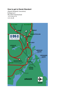


© Copyright 2026