
RESULTS AFTER SURGICAL TREATMENT OF ANTERIOR CRUCIATE LIGAMENT INJURIES Jon Olav Drogset
Jon Olav Drogset
RESULTS AFTER SURGICAL
TREATMENT OF ANTERIOR
CRUCIATE LIGAMENT INJURIES
A clinical study
Thesis for the degree of doctor medicinae
Trondheim, October 2006
Norwegian University of
Science and Technology
Faculty of Medicine
Institute of Neuroscience
NTNU
Norwegian University of Science and Technology
Thesis for the degree of doctor medicinae
Faculty of Medicine
Institute of Neuroscience
©Jon Olav Drogset
ISBN 82-471-8111-8 (printed ver.)
ISBN 82-471-8109-6 (electronic ver.)
ISSN 1503-8181
Doctoral Theses at NTNU, 2006: 170
Printed by Tapir Uttrykk
RESULTS AFTER SURGICAL TREATMENT OF ANTERIOR CRUCIATE
LIGAMENT INJURIES
A clinical study
Jon Olav Drogset, M.D.
An expert is a person who has made all the mistakes that can be made in a very narrow field.
- Niels Bohr
Publication from the Norwegian University of Science and Technology
Faculty of Medicine
Department of Orthopedic Surgery and Rheumatology
Trondheim University Hospital
N-7006 Trondheim, Norway
© Jon Olav Drogset 2006
CONTENTS
Page
LIST OF PAPERS
4
ACKNOWLEDGEMENTS
5
INTRODUCTION
6
ANATOMY
7
FUNCTION
9
MECHANISMS OF INJURY
10
NATURAL HISTORY OF THE ACL-INJURED KNEE
12
ASSOCIATED INJURIES
12
TREATMENT
13
GRAFTS
15
GRAFT FIXATION
16
BONE PLUG GRAFT FIXATION
17
AIMS OF THE STUDY
19
REVIEW OF PAPERS
20
GENERAL DISCUSSION
35
EVALUATION OF MATERIALS
35
EVALUATION OF METHODS
38
STATISTICAL METHODS
41
DISCUSSION
42
GENERAL CONCLUSIONS
50
REFERENCES
51
ABBREVIATIONS
64
PAPER I-V
LIST OF PAPERS
This thesis is based on the following papers, which are referred to in the text by their Roman
numerals:
I.
Drogset, JO, Grøntvedt, T, Robak, OR, Mølster, A, Viset, AT, Engebretsen, L. A
Sixteen-Year Follow-up of Three Operative Techniques for the Treatment of Acute
Ruptures of the Anterior Cruciate Ligament. J Bone Joint Surg Am. 2006 May; 88
(5): 944-52.
II.
Drogset, JO, Grøntvedt, T. Anterior cruciate ligament reconstruction with and without
a ligament augmentation device: results at 8-Year follow-up. Am J Sports Med. 2002
Nov-Dec; 30 (6):851-6.
III.
Drogset, JO, Grøntvedt, T, Tegnander, A. Endoscopic reconstruction of the anterior
cruciate ligament using bone-patellar tendon-bone grafts fixed with bioabsorbable or
metal interference screws: a prospective randomized study of the clinical outcome.
Am J Sports Med. 2005 Aug; 33(8):1160-5.
IV.
Drogset, JO, Grøntvedt, T, Myhr, G. Magnetic Resonance Imaging Analysis of
Bioabsorbable Interference Screws Used for Fixation of Bone-Patellar Tendon-Bone
Autografts in Endoscopic Reconstruction of the Anterior Cruciate Ligament. Am J
Sports Med. 2006 Feb 21; [Epub ahead of print]
V.
Drogset, JO, Grøntvedt, T, Jessen, V, Tegnander, A, Mollnes, TE, Bergh, K.
Comparison of in vitro and in vivo complement activation by metal and bioabsorbable
screws used in anterior cruciate ligament reconstruction.
Arthroscopy. 2006 May; 22(5):489-96.
4
ACKNOWLEDGEMENTS
This thesis is based on studies carried out at the Department of Orthopedic Surgery,
Trondheim University Hospital.
I wish to express my sincere gratitude to the following persons:
Torbjørn Grøntvedt, a good friend and mentor, for inspiring me to do this work and for
valuable advice, constructive criticism and many stimulating discussions.
Professor Pål Benum, Henrik Sandbu and Eirik Rødevand, for believing in my ideas and
giving me the opportunity to carry them through.
Vigleik Jessen, Grete Sofie Hortemo and Arild Aamodt for active participation in
randomizing and operating some of the patients.
Ivar Rossvoll, for friendship and help with statistics.
Øystein Haave and Annja T. Viset for their great help in evaluating the radiographs.
Gunnar Myhr for his enthusiasm and support in MRI examinations and evaluation of images.
Professor Lars Engebretsen, Ullevål University Hospital and Ole Rasmus Robak, Aker
University Hospital Oslo, Professor Anders Mølster, Haukeland Hospital, Torbjørn Strand,
Diakonissehjemmet Hospital, Asbjørn Bøe, Hagavik Hospital, University of Bergen, Gisle
Uppheim and Bjørn Ødegaard, Lovisenberg Diakonale Hospital, Oslo, Professor Kåre Bergh
and Agnar Tegnander Trondheim University Hospital, Professor Tom Eirik Mollnes,
Rikshospitalet University Hospital; Oslo for their support and practical work in our
prospective randomized studies.
Liv Randi Sand 1, Randi Sand2 and other nurses and technical assistants at the operating
theatre2 and outpatient clinic1 at the Department of Orthopedics, Trondheim, for their help
and patience in keeping the clinical studies going.
Professor Vilh. Finsen for help with the manuscripts.
Berit Wenaas, secretary at the Institute of Neuroscience for support and practical help with
the collection of data.
Professor Are Holen and Professor Helge Rønningen at the Institute of Neuroscience for
letting me keep the office during these years.
To Toril, Nikolay and Camilla for all their support and patience during some busy years, and
for letting me work on my laptop at any hour of the day.
To Mona, Emilie and Aleksander.
This study was supported by grants from the Norwegian Association of Sports Medicine,
Zimmer Norway and Smith & Nephew Norway.
5
INTRODUCTION
The motion characteristics of the articular surfaces of the tibia relative to the femur
are very complex and are guided by the anterior cruciate ligament (ACL) and the other
primary ligaments that span the knee. Studies performed using cadaveric models have
revealed that the ACL is the primary restraint to anterior displacement of the tibia relative to
the femur (Butler 1980; Fukubayashi 1982), and acts as a restraint to internal-external
rotation (Markolf 1990; Fleming 2001; Kanamori 2002), varus-valgus angulation (Marder
1991) and combinations thereof (Markolf 1995; Kanamori 2002). In addition to this the ACL
is an important component in the proprioceptive feedback mechanism of the joint.
The management of ACL injuries is one of the most challenging issues in the field of
sports traumatology. Sporting activities in modern society are increasing and have also
increased the incidence of ACL injuries. Danish studies have shown an annual incidence of
3.1 ACL injuries per 10.000 inhabitants between 0 and 50 years of age (Nielsen 1989). The
reported incidence of ACL tears for female athletes ranges between 4 and 6 times greater than
that of male athletes competing in similar activities. In women’s professional soccer Giza et
al. (Giza 2005) found an overall injury incidence rate of 1.93 injuries per 1000 player hours.
The incidence of injury during practice and games was 1.17 and 12.63 per 1000 player hours,
respectively. Sports activities are responsible for approximately 95% of all ACL ruptures
seen in our hospital (Grontvedt 1996b). Bjordal et al (Bjordal 1997) studied 176 patients who
had participated in organized soccer. Women had an incidence rate of 0.10 injuries per 1000
game hours, significantly higher than that for men (0.057). The Norwegian National ACL
Registry was initiated in 2004. The numbers of ACL reconstructions recorded in Norway in
2005 was 1543 (Strand, Hafjell 2006). Hamstrings autografts were used in 57% of the cases
and bone-patella-tendon-bone graft in 43%.
The material properties of the ACL vary along its length and cross section, and
therefore, it may be important to match the structural properties of the graft with those of the
native ACL to fully restore normal kinematics and knee function (Butler 1992). Woo et al
(Woo 1991) reported that the structural properties of the ACL decrease with age and that in
younger subjects the failure load and stiffness values of the ACL were 2160 ± 157 N and 242
± 28 N/mm, respectively. These values are similar to a 10-mm-wide patellar tendon graft (the
failure strength and stiffness properties are 1784 [± 580] N and 210 N/mm) and a 4-bundled
hamstring tendon graft (the failure strength and stiffness values are 2422 [± 538] N and 238
N/mm) (Wilson 1999). Therefore, theoretically both graft types should provide adequate
strength and normal laxity to the knee immediately after surgery.
Biological substitutes for the ACL can be prepared as either bone-tendon-bone grafts
or tendon grafts. Depending on the graft material that is selected for ACL reconstruction, the
bony or soft tissue portions of these constructs can be fixed either within bone tunnels or
externally to cortical bone. From a biomechanical perspective, fixation represents the weakest
link during the early stages of healing. The long-term goal is to obtain biological
incorporation of the graft at the anatomical attachment sites of the ACL and to restore the
transition from soft tissue to fibrocartilage, to calcified fibrocartilage, and to bone.
Some of these grafts and fixation methods are evaluated in the studies included in this thesis.
6
ANATOMY
The knee joint develops from a cleft between the mesenchymal rudiments of the
femur and tibia during the 8th week of the human embryo (GRAY 1950; Gardner 1968). As
the mesenchyme in the region of the future knee joint condenses to form the precartilage and
the capsule of the joint, vascular mesenchyme becomes isolated within the joint. This tissue
is the precursor to the cruciate ligaments and the menisci.
At 9 weeks, the cruciate ligaments are composed of numerous immature fibroblasts,
the long axes of which are parallel to the course of the ligaments. At 10 weeks, the ACL and
posterior cruciate ligament (PCL) are separated from each other, and over the next 4 weeks
the cruciate ligaments continue to differentiate from the adjacent tissues and the insertion
sites becomes more defined. By 18 weeks, the cruciate ligaments stand almost alone, and a
few vascular elements are found within their substance. During the next weeks there is a
continuous growth and increase in vascularity, until the ACL resembles that found in the
adult. The remaining development consists of marked growth but little change in form.
The mature ACL is a band of regularly oriented, dense connective tissue which connects the
femur and tibia. It is surrounded by a fold of synovium that originates from the posterior
intercondylar area of the knee, and completely envelopes both the anterior and posterior
cruciate ligaments. Thus, the cruciate ligaments are intraarticular and extrasynovial (Girgis
1975; Arnoczky 1983).
The ACL is attached to a fossa on the posterior aspect of the medial surface of the
lateral femoral condyle. The attachment has a semilunate form, with the anterior border
straight and the posterior border convex. The long axis of the femoral attachment is tilted
slightly forward from the vertical, and is about 23 mm long (Kennedy 1974; Arnoczky 1983).
On the tibia, the ACL is attached to a fossa in front of and lateral to the anterior tibial
spine. The anterior part of the tibial attachment lies beneath the transverse meniscal ligament,
and some fascicles of the ACL may blend with the anterior attachment of the lateral
meniscus. The tibial attachment of the ACL is somewhat broader than the femoral. Its length
is about 30 mm. Thus, because the tibial attachment is somewhat larger than the femoral, the
ACL is fan-shaped.
The ACL courses anteriorly, medially and distally across the joint as it passes from
the femur to the tibia. It also has a slight outward (lateral) spiral form, as a result of its bony
attachments.
The average thickness of the ACL is 11 mm, but the cross-sectional area varies along
its length, being larger at its insertion sites than in the mid-region. The average length varies
from 31 mm (Girgis 1975) to 38 mm (Odensten 1985a).
The geometry of the ACL is complex and not duplicated with current reconstruction
techniques. The fibers on the anterior border of the ACL are longest, whereas those on the
posterior edge are the shortest. The ACL has a distinct crimp pattern that straightens as the
ligament is strained. Although the ACL does not have bundles that are distinct from an
anatomical perspective, it has been subdivided into at least 2 functional bundles, the
anteromedial (AMB) and posterolateral bundles (PLB), which work synergistically with each
other to optimize its restraining function over the range of knee motion (Xerogeanes
7
1995).The orientation of the femoral attachment of the ACL in flexion and extension is
responsible for maintaining tension in the ligament throughout the range of motion. The ACL
is attached to the femur and tibia as a collection of individual fascicles that fan out over a
broad, flattened area. When the knee is extended, the entire ligament is taut, with most of the
load on the PLB. In flexion, the femoral attachment of the ACL assumes a more horizontal
orientation, causing the AMB to tighten and the PLB to slacken (Arnoczky 1983).
An intermediate bundle is also described (Norwood 1979). This is felt to be
responsible for the straight anterior stability of the knee. However, these macroscopic
bundles lack a corresponding microstructure in the substance of the ACL. The functional
significance of the fascicles of the ACL is that groups of fascicles work together throughout
the range of joint motion (Odensten 1985a; Clark 1990)
The cells of the ACL are fibroblasts surrounded by an extracellular matrix formed by
a solid, highly ordered arrangement of macromolecules, primarily type I collagen, and water.
The mechanical properties of the ligament depend on the composition of the matrix, the
organization of the macromolecules, and the interaction between these and the water. Small
changes in this composition may lead to changes in mechanical properties, as seen with
increasing age. Microscopically, the ACL is composed of 30 to 175 nm wide collagen fibrils,
which are arranged in parallel (Clark 1990). These fibrils are in turn grouped into fibers that
vary from 1 to 20 micrometers in diameter. They run almost parallel to the long axis of the
ligament. These fiber bundles coalesce to form subfascicular units, which vary from 100 to
250 micrometers in diameter. Three to 20 subfasciculi form a collagen fasciculus which can
be several millimeters in diameter. Clark and Sidles (Clark 1990) could not demonstrate an
anatomical delineation of bands within the ligament. In this respect the ACL is different from
other ligaments around the knee.
The ACL has a microstructure of collagen bundles of multiple types (mostly type I)
and a matrix made of a network of proteins, glycoproteins, elastic systems, and
glycosaminoglycans with multiple functional interactions. The complex ultrastructural
organization and abundant elastic system of the ACL allow it to withstand multiaxial stresses
and varying tensile strains (Duthon 2005).
The blood supply to the ACL arises from the ligamentous branches of the middle
genicular artery as well as from some terminal branches of the medial and lateral inferior
genicular arteries (Arnoczky 1985). These vessels reach the synovial fold that covers the
ACL. In this synovium they arborize to form a web-like network of periligamentous vessels
that give rise to smaller connecting branches that penetrate the ligament transversely and
anastomose with a network of endoligamentous vessels. These, together with their connective
tissue, are orientated in a longitudinal direction and lie parallel to the collagen bundles within
the ligament.
The role of the vascular tissues in ACL repair and reconstruction has been
documented both in experimental (Arnoczky 1979) and clinical (O'Donogue 1963) studies.
Bray et al (Bray 2003) found the superior capacity of the medial collateral ligament (MCL)
to increase its blood supply through angiogenesis and increased flow to be essential for
ligament healing to occur, and may be the major difference in healing potential between the
ACL and MCL.
Both the synovium and the fat pad serve as potential sources for revascularization of
repaired ligaments. When no bone tunnels are used, revascularization in the dog begins at 6
weeks and is complete by 20 weeks following repair. Howell et al (Howell 1995) used MRI
and an intravenous contrast agent (gadolinium diethylenetriamine pentacetic acid) to evaluate
8
the blood supply of hamstring autografts and periligamentous tissues during the first 2 years
after reconstruction. They found that the grafts acquired no discernable blood supply during
the first 2 years, and the graft retained the same hypovascular appearance as the normal PCL.
In contrast, the periligamentous soft tissues were richly vascularized and covered the graft by
1 month postoperatively. They concluded that the viability of the grafts may depend more on
synovial diffusion than on revascularization.
The ACL receives nerve fibers from branches of the tibial nerve which penetrate the
joint posteriorly. Nerve fibers and sensory receptors follow the vessels in the form of
neurovascular bundles, entering the ligament from the synovium. Some nerve fibers and a
variety of sensory end organs are also located in the ligament, isolated from the vessels
(Kennedy 1982; Zimny 1988; Halata 1989). The nerve fibers are either unmyelinated or
myelinated, and four morphologically different types of sensory endings have been described.
These are two types of Ruffini end organs, Pacinian corpuscles, and free nerve endings
(Schutte 1987). It has been hypothesized that these nerve endings have an important
proprioceptive function in the knee (Johansson 1991; Pitman 1992). Adachi et al. (Adachi
2002) found a positive correlation between the number of mechanoreceptors and accuracy of
the joint position sense, suggesting that proprioceptive function of the ACL is related to the
number of mechanoreceptors.
FUNCTION
Studies performed using cadaveric models have revealed that the ACL is the primary
restraint to anterior displacement of the tibia relative to the femur (Butler 1980; Fukubayashi
1982) and acts as a restraint to internal-external rotation (Markolf 1990; Fleming 2001;
Kanamori 2002) varus-valgus angulation (Marder 1991), and combinations thereof (Markolf
1995; Kanamori 2002). The anteromedial part is most important in flexion, whereas the
posterolateral part contributes most to stability in extension (Furman 1976; Amis 1991). In
addition, the ACL resists hyperextension, and is an important factor in preventing
anterolateral rotatory instability of the tibia (Girgis 1975). In extension it aids medial and
lateral structures in maintaining the varus-valgus stability of the knee.
The estimated loading of the ACL during daily activities vary in different studies.
Morrison (Morrison 1970) estimated in vivo forces from force plate analysis, and found a
peak load of 169 N in level walking, 67 N in ascending a ramp, and about 500 N in
descending a ramp. Chen and Black (Chen 1980) estimated the tensile forces in the ACL to
be 67 N in ascending stairs and 630 N in jogging. Downhill running seems to create the
highest ACL strain (strain = change in length / initial length of the structure) among the
activities tested (Henning 1985). Both cadaveric (Grood 1984) and clinical (Henning 1985;
Beynnon 1995) studies have demonstrated a considerable rise in ligament stress (stress =
force / area) from 30 degrees of flexion to full extension, especially if done actively against
resistance placed distally on the leg.
The muscles spanning the knee apply forces and moments to the joint. As the position
of the knee joint is changed, the moment arms of the different muscles and tibiofemoral joint
contact position also change, which, in turn, affect ACL biomechanics. It is well documented
9
that the ACL is strained when the quadriceps are contracted with the knee near extension
(between full extension and 50° of flexion) (Markolf 1990; Fleming 2001). In contrast,
contraction of the hamstring muscles produces a posterior-directed force that acts on the tibia
throughout the range of knee flexion. These muscles thus protect the ACL or ACL graft
(O'Connor 1993). The gastrocnemius muscle also spans the knee joint, yet its effect on the
ACL has only been recently established. O’Connor (O'Connor 1993) hypothesized that
contraction of the gastrocnemius could displace the tibia in an anterior direction, which, in
turn, would increase the strain on the ACL. Direct measurements of ACL strain during
gastrocnemius contractions have verified that the gastrocnemius is an ACL antagonist when
the knee is near extension and that combined contraction of the gastrocnemius and quadriceps
produces greater ACL strain values than isolated contraction of these muscles (Fleming
2001). Compressive loading of the tibiofemoral joint has been shown to increase joint
stiffness and decrease anterior displacement of the tibia relative to the femur (Markolf 1981;
Torzilli 1994; Beynnon 2002).Therefore, a compressive joint load, such as that produced by
body weight, has been considered by some to protect a healing ACL graft. However, recent
research has demonstrated that ACL strain values increase as the knee undergoes a transition
from nonweightbearing to weight bearing conditions (Fleming 2001).
The ultimate strength of the ACL in a young cadaver specimen is about 1750 N. This,
however, depends on several factors, of which one is the direction of the load applied. If
loaded in the anatomical axis, the ligament fails at about 2200 N, and if the load is applied in
the tibial axis, the failure occurs at about 1500 N. These values are similar to a 10-mm-wide
patellar tendon graft (the failure strength and stiffness properties are 1784 [± 580] N and 210
N/mm, respectively) and a 4-bundled hamstring tendon graft (Woo 1991). The ultimate
strength is also highly dependent on the strain rates (Noyes 1974; Kennedy 1976). At low
strain rates, the failure will occur early, and at the ligament-bone junction. As the rate of load
application is increased, the bony junction becomes stronger and stiffer than the ligament,
due to its viscoelastic properties. In the experiment performed by Kennedy et al. (Kennedy
1976) with a fourfold increase in strain rate, the maximum load tolerated by the ACL
increased by 32%.
By the use of a scanning electron microscope, Yahia et al. (Yahia 1990) found that
some of the collagen fibers in the ACL start to fail between 10 - 15% of ultimate strain, and
at 20% it showed ruptures of thick collagen fibers, thus indicating microscopic partial
ruptures before macroscopic or total rupture occurs.
MECHANISMS OF INJURY
Rupture of the ACL was first described by J. Stark in the Edinburgh Medical and
Surgical Journal in 1850. Since then, and especially during the last three decades, this injury
has been the topic of many publications.
Four types of injury mechanisms have been described. The classic "unhappy triade":
ACL rupture, damage to the medial collateral ligament and rupture of the medial meniscus, is
caused by a forceful valgus-external rotation of the knee (Fetto 1980). This injury is often
seen in skiing, when the tip of the ski is caught on an obstacle, the knee is bent into valgus,
and the tibia goes into external rotation. Olsen et al. (Olsen 2004) found that the injury
10
mechanism for anterior cruciate ligament injuries in female team handball appeared to be a
forceful valgus collapse with the knee close to full extension combined with external or
internal rotation of the tibia. Sell et al. (Sell 2006) concluded that the lateral jumps are the
most dangerous of the stop-jumps.
A second mechanism of injury is varus-internal rotation, as seen in team handball and
basketball players. Recent cadaveric research has shown that simulated contraction of the
quadriceps with the knee near extension produces substantial anterior tibial translations that
can create ACL injury and may explain, at least in part, one mechanism responsible for
noncontact ACL injury (DeMorat 2004). In contrast, contraction of the hamstring muscles
serves a protective function for the ACL or ACL graft (O'Connor 1993). Special shoes on
indoor floors with high frictional surfaces are usually involved. Injury factors such as
friction, angular velocity and rate of directional change seem to be important. Today’s elite
team handball is played at high speed. With rapid directional changes, the foot may be
"locked" on the surface; the knee bent in varus and the tibia go into internal rotation. This
may either cause an isolated ACL rupture, or a rupture combined with damage to the lateral
meniscus, lateral capsular structures, and the lateral collateral ligament.
A third mechanism of injury is the forceful internal rotation of the tibia in knee
extension, which might cause impingement of the ACL on the anterior aspect of the medial
femoral condyle, and rupture of the ligament (Kennedy 1974; Fetto 1980). In support of this
theory, studies have shown that a narrow intercondylar notch leads to a higher risk of ACL
ruptures in athletes (Souryal 1993; Lund-Hanssen 1994). In another study however
(Lombardo 2005), anterior cruciate ligament injury could not be predicted by the absolute
measurement of the femoral inter-condylar notch.
In a study by Ebstrup et al (Ebstrup 2000) three video recorded incidents of knee
injuries inflicted during indoor ball games were reported. Injuries and especially anterior
cruciate ligament ruptures seemed to be triggered in varus loaded knees by femoral external
rotation or in valgus loaded knees by femoral internal rotation with the pivot shifted to the
lateral femurotibial compartment.
Finally, the fourth mechanism of injury is typically seen in the down-hill skier who is
at the point of falling backwards. To prevent this, he or she powerfully contracts the
quadriceps, thus pulling the tibia anteriorly. It may, however, be even more important that in
this situation, the posterior part of the ski boot pushes the tibia forward. This may lead to an
isolated tear of the ACL (Johnson 1982; McConkey 1986). MRI examinations of acute ACL
tears have shown consistent and recurring subchondral bone lesions in the femoral and tibial
condyles, with locations typical for this specific injury mechanism (Spindler 1993). These
findings have led to the conclusion that the ski acts as a lever arm, producing a sudden and
violent rotation of the tibia on the femur (Speer 1995). This, in combination with the
pull/push mechanism, is suggested to cause the ACL rupture.
Anterior cruciate ligament injury occurs with a 4- to 6-fold greater incidence in
female athletes compared with male athletes playing the same landing and cutting sports
(Arendt 1995). Uhorchak et al (Uhorchak 2003) performed a comprehensive study on
military academy cadets and reported that several risk factors predisposed them to noncontact
ACL injury. Significant risk factors included a small femoral notch width, generalized joint
laxity, and, in women, higher than normal body mass index and KT-2000 arthrometer
measurements of A-P knee laxity that were 1 standard deviation or more above the mean. The
mechanism underlying gender disparity in ACL injury risk is likely multifactorial in nature.
Several theories have been proposed to explain those observations. These theories include
11
related extrinsic (physical and visual differences, bracing, and shoe-surface interaction) and
intrinsic (anatomical, hormonal, neuromuscular, and biomechanical differences between
genders) variables. Potential factors that have not been discussed may play a role (e.g.,
genetics). In addition, injury data from many fields demonstrate that numerous physical and
psychological parameters affect injury rates. Although there are multiple factors underlying
the differences in ACL injury rates in male and female athletes, neuromuscular control may
be important to injury risk and the most modifiable factor. Neuromuscular training in female
athletes has been shown to increase active knee stabilization in the laboratory and to decrease
the incidence of ACL injury (Hewett 1996, 1999; Myklebust 2003a).
NATURAL HISTORY OF THE ACL-INJURED KNEE
Over time, ACL-deficient patients may experience giving-way episodes and are more
likely to develop further intra-articular damage, such as meniscal tears, (Noyes 1983a-b;
Hawkins 1986b; Bray 1989; Buss 1995; Grontvedt 1999) and osteoarthritis of the knee
(Johnson 1974; Kannus 1987). Subjects with ACL injury and posttraumatic osteoarthritis are,
on average, 15 to 20 years younger than patients with primary osteoarthritis when they seek
medical advice for their symptoms and when their joints show radiographic evidence of
osteoarthritis (Roos 1995a).
ASSOCIATED INJURIES
Injuries of the ACL rarely occur in isolation. The effects of other injuries, including
other ligament sprains, meniscal tears, articular cartilage injuries, and bone bruises,
complicate the treatment and final outcome of ACL ruptures. There can be no doubt that the
addition of one or more of these associated injuries adversely affects the outcomes of
treatment, but it is very difficult to quantify or predict exactly how they will alter the results.
Many surgeons now advocate nonsurgical management, even when grade III tears of the
MCL exist in association with ACL disruptions (Noyes 1995).
Disruption of the posterolateral and lateral structures of the knee is not commonly
associated with rupture of the ACL, but when they are present and not recognized, an ACL
reconstruction will often fail (Jaureguito 1996; Getelman 1999; Harner 2001; Noyes 2001;
Allen 2003).
Meniscal damage frequently occurs at the time of an ACL injury, and the incidence
increases in patients who do not undergo reconstruction. Reports of meniscal injury
associated with acute ACL disruption range from 15% to 40% and become much higher with
chronic ACL deficiency (Levy 2003).The effects of chondral lesions observed at the time of
ACL reconstruction are difficult to ascertain from the current literature. With the advent of
MRI, it became apparent that occult osteochondral lesions (bone bruises) are commonly
found in association with ACL injuries. Several investigators reported that 80% or more of
ACL injuries were associated with bone bruises in the lateral compartment (Rosen 1991;
Spindler 1993; Johnson 1998). Johnson et al (Johnson 1998) performed histological analysis
of bone bruises in humans. They found large areas of degeneration of chondrocytes in the
overlying articular cartilage and necrotic osteocytes in subchondral bone, which led them to
suggest that MRI evidence of bone bruising indicates significant damage to articular cartilage
12
homeostasis.
TREATMENT
The treatment options for ACL ruptures are either conservative or surgical. The
rationale for surgical treatment includes the assumption that the ACL is vital for knee
function, that ACL-deficient knees frequently degenerate, and that surgical reconstructions
can succeed in restoring normal function. The rationale for nonsurgical treatment assumes
that the ACL-deficient knee may function reasonably well under certain circumstances and
that reconstructions do not necessarily prevent the untoward sequela of osteoarthritis. Several
studies reported during the last decade, however, show that conservative treatment often
leads to increasing instability of the joint, meniscal tears, cartilage injuries, pain and
eventually osteoarthritic changes in patients who subject their knee to rotational loads
(McDaniel Jr 1983; Noyes 1983a; Kannus 1987; Engebretsen 1990; Tandogan 2004).
Patients who may be satisfactorily treated without surgical intervention, despite a totally
disrupted ACL, include those who have little exposure to high-risk activities such as sports
and heavy work, those who are willing to avoid high-risk activities, those who are more than
40 years of age, those who are successful in long-term coping with or adapting to an ACL
insufficiency, those who have advanced arthrosis of the involved joint, and those who are
unable or unwilling to comply with postreconstruction rehabilitation. Several case series
reveal that a nonoperative approach to the torn ACL can be effective for the majority of
patients willing to avoid high-risk activities (Bonamo 1990; Ciccotti 1994; Buss 1995;
Segawa 2001). Other case series have indicated that even with return to high-risk activities,
many patients do well (Roos 1995b; Casteleyn 1996). In three Swedish randomized clinical
trials (RCT), superior results were achieved in patients with ACL repairs with augmentation
using the iliotibial band, compared to nonoperative treatment (Odensten 1984b; Andersson
1989b, 1991).
Interest in repair techniques was low until Palmer (1937) reported success in suturing
10 cases of torn ACLs. Short term follow-up showed good results (O'DONOGHUE 1963;
Liljedahl 1965). There was considerable controversy concerning the importance of the ACL
until the 1970s. Thus, simply excising the disrupted ACL was common before that time. In
the 1970s a trend toward primary repair of the ACL was started by surgeons who thought the
ACL performed an important function (Odensten 1985b; Sandberg 1987b; Andersson 1989b,
1991). Several long term follow-up studies during recent years conclude, however, that the
results of this operation deteriorate with time (Odensten 1984b; Sommerlath 1991b;
Fruensgaard 1992b; Grontvedt 1996a; Strand 2005).
A nearly universally accepted indication for an ACL reconstruction is a high-risk
lifestyle requiring heavy work, sports, or recreational activities (Daniel 1994). Likewise,
repeated episodes of giving way (pivot shift) in spite of rehabilitation are considered a strong
indication for ACL reconstruction.
In order to preserve as much as possible of the ruptured ACL with its nerve endings
and possible proprioceptive function (Johansson 1991; Pitman 1992) several surgical
procedures with synthetic or biologic augmentation of the repaired ACL have been
published. Some of these have shown promising short term results (Clancy Jr 1988b; Schabus
1988; Sgaglione 1990). However, Grøntvedt et al (Grontvedt 1995a, 1996a) showed an
13
unacceptable high incidence of reruptures in repairs augmented with a synthetic graft.
In recent years, arthroscopically assisted ACL reconstruction has become the
procedure of choice (Arciero 1996). Initially, arthroscopic techniques required 2 incisions for
outside-in drilling of bone tunnels, but there has been a trend toward using a single-incision
arthroscopic approach with inside-out drilling of the femoral tunnel. This technique has also
been called the 1-incision, endoscopic, or all-inside arthroscopic reconstruction of the ACL.
Karlsson et al (Karlsson 1999a) found a significantly higher Lysholm score in their 1-incision
group. Delay et al (Delay 2001) published a large survey among members of the American
Orthopaedic Society for Sports Medicine (AOSSM) that found that 85% of surgeons use an
endoscopic single-incision technique, 12% use an arthroscopic 2-incision technique, and 3%
use mini-open techniques. In reviewing the additional literature concerning the outcomes of
the 1-incision versus 2-incision procedures for ACL reconstruction, there is no clear evidence
that one method is superior to the other.
The complex anatomy and the biomechanical function of the ACL have been well
described by numerous authors. Several investigators, using human cadaveric knees, have
reported improved ability to restore ACL function after ACL reconstructions in which 2
femoral attachment sites are used to more nearly replicate the normal ACL anatomy (Amis
1991; Radford 1994; Muneta 1999; Mae 2001; Yagi 2002). In spite of these findings, several
authors conclude that 2 femoral attachment sites are not superior to a single site (Radford
1994; Hamada 2001).
In chronic ACL deficient knees the primary cause of pain and discomfort is the
anterolateral rotatory instability. To prevent this, several extraarticular surgical procedures
have been proposed. Benum reported a method using the lateral one third of the patellar
tendon with a bone block from the patella, for stabilization. This method has shown good
short term results (Benum 1982). However, the recurrence rate of symptomatic instability and
the deterioration of subjective knee function are too high (Grontvedt 1995b).
There are many methods of tenodesing the lateral aspect of the tibia to the femur in
order to prevent anterolateral rotation of the tibia and minimize anterior translation of the
tibia relative to the femur, either as an isolated procedure or as a backup for intra-articular
reconstruction. Several groups have not been satisfied with the results of these procedures
and do not advise their continued routine use (Dahlstedt 1988; Larsen 1991; Vail 1992).
Despite the uncertainties of outcomes after ACL reconstructions reported in the
literature, many investigators conclude that patients who desire to return to high-risk
activities or who have other significant knee injuries (meniscal tears, disruptions of other
ligaments, articular cartilage damage, as well as marked anterior laxity) are probably best
served by an ACL reconstruction. Conversely, patients with low-risk activity levels, isolated
ACL injuries, and mild pathologic laxity may be successfully treated without surgery.
GRAFTS
In the last decade, intraarticular reconstructions using autografts or allografts have
become popular. Prosthetic devices have also been in use.
Many studies have compared bone–patellar tendon–bone with hamstrings tendon
grafts (used as 2-strand or 4-strand constructs). Corry et al (Corry 1999) compared the
outcome of anterior cruciate ligament reconstruction using hamstring tendon autograft with
14
patellar tendon autograft. Kneeling pain after reconstruction with the hamstring tendon
autograft was significantly less common than with the patellar tendon autograft. These
findings are supported in a Norwegian study (Aune 2001). Eriksson et al (Eriksson 2001)
concluded that patellar tendon and quadruple semitendinosus tendon grafts had similar
outcomes in the medium term. Associated meniscal pathology significantly affected the final
outcome and early reconstruction seemed to be beneficial. Aglietti et al (Aglietti 2004)
concluded that both grafts were an equivalent option for anterior cruciate ligament
reconstruction. In contrast to findings in the RCTs that compared ACL reconstruction with
the 2-strand hamstrings graft to the BPTB- graft, ACL reconstruction with a 4-strand
hamstrings graft appears to result in similar clinical and functional outcomes as
reconstruction with a BPTB- graft during a 2-year follow-up period. Freedman et al.
(Freedman 2003) found in a Meta analysis that patellar tendon autografts had a significantly
lower rate of graft failure and resulted in better static knee stability and increased patient
satisfaction compared with hamstring tendon autografts. However, patellar tendon autograft
reconstructions resulted in an increased rate of anterior knee pain.
Allografts have been advocated as an option for ACL reconstruction and have been
used a lot for many years. Decreased morbidity, preservation of the extensor or flexor
mechanisms, provision of a source of graft material when the patient has none, decreased
operative time, availability of larger graft sources, lower incidence of arthrofibrosis, and
improved cosmetic appearance are all advantages that have been attributed to the use of
allografts (Harner 1996; Victor 1997; Kleipool 1998). The possible disadvantages have been
the risk of infection, slow and incomplete incorporation and remodeling of the graft, higher
costs, availability, and tunnel enlargement, alteration of graft structural properties by
sterilization and storage procedures, and immunologic response (Pinkowski 1989; Marks
1993; Linn 1993; Jackson 1996; Zijl 2000).
Prosthetic replacement of the ACL with synthetic material, whether for total,
permanent replacement (prosthesis), a scaffold for ingrowth of host tissue, or a ligament
augmentation device to protect an allograft or autograft as it heals, has not been proven to be
a satisfactory solution to the ACL-deficient knee (Noyes 1992; Moyen 1994; Denti 1995;
Grontvedt 1996b; Kumar 1999; Marumo 2000; Muren 2003). Although claims of acceptable
results with short-term follow-ups have been published, at the present time, we are unaware
of any investigation that indicates that these devices have any place in the treatment of ACL
injuries (Guidoin 2000).
The position of an ACL graft is the most critical surgical variable because it has a
direct effect on knee biomechanics and, ultimately, on clinical outcome. In the well-designed
study by Good et al (Good 1994) subjects with an ACL graft insertion greater than 2 mm
anterior to the center of the anatomical femoral insertion had significantly greater anteriorposterior (A-P) knee laxity values than subjects with centrally or posteriorly positioned
femoral tunnels. Howell and Clark (Howell 1992) performed a retrospective study with a 6month follow-up period of BPTB grafts and reported that knee stability and extension were
significantly better when the center of the tibial tunnel was 2 to 3 mm posterior to the center
of the normal ACL insertion on the tibia.
Most surgeons would agree that the initial tension applied to an ACL graft at the time
of fixation has a direct effect on outcome. An under-tensioned graft may result in abnormal
knee laxity and an unstable knee, and an over-tensioned graft may lead to graft failure,
fixation failure, or restricted range of motion. The effect of preloading (application of a 39-N
tensile load for 10 minutes before graft implantation) to no preloading before graft
15
implantation showed no differences between the treatment groups with regard to activity
level, clinical outcome (IKDC grade), and A-P knee laxity after two years (Ejerhed 2001).
Graft tensioning at the time of fixation involves consideration of the knee’s position
during the tensioning procedure (i.e., the flexion angle and internal-external rotational
position of the tibia relative to the femur) and, of course, the magnitude of tension applied to
the graft at the time of its fixation to bone. Both variables interact and have a direct effect on
knee biomechanics. If a BPTB graft is used and the tensioning procedure is performed with
the knee in extension, application of high tension (90 N) appears to produce more normal A-P
knee laxity values compared to application of low tension (45 N). Similarly, if a 4-strand
hamstring graft is used, application of high tension (80 N) appears to produce A-P knee laxity
values similar to normal, whereas low tension (40 N and less) results in increased anterior
knee laxity (Yoshiya 2002; Nicholas 2004).
Most of the studies of graft material have considered AP knee laxity as an important
primary outcome measure. It is, however, essential to point out that it may be equally
important to restore the rotational laxity of the reconstructed knee to within the limits of the
normal knee. To accomplish this, new techniques must be developed to measure internalexternal rotational laxity of the knee, or at least document displacements of the medial and
lateral compartments of the tibiofemoral joint.
GRAFT FIXATION
The bony or soft tissue portions of grafts can be fixed either within bone tunnels or
externally to cortical bone. The long-term goal is to obtain biological incorporation of the
graft at the anatomical attachment site and to induce the transition from soft tissue to
fibrocartilage, to calcified fibrocartilage, and to bone. Current rehabilitation protocols after
knee ligament surgery stress immediate full range of motion, return of neuromuscular
function, proprioception, and early weight bearing. Fixation methods must be rigid and stiff to
allow aggressive rehabilitation principles. Current fixation techniques involve soft tissue or
bone within a bone tunnel or periosteal fixation away from joint surfaces.
Stiffness is the slope of the linear region of the load-elongation curve and is usually
reported in units such as Newtons per millimeter (N/mm). Present graft fixation alternatives
are less stiff than the native ACL. Mechanically, the majority of tendon fixation constructs
are less stiff than the interference screw against a bone plug, which has been considered the
standard for fixation. Many tendon fixation devices are "indirect." They rely on linkage
material to connect the tendon to the fixation device.
In the native cruciate ligament, the point of fixation is at the joint surface. However,
most tendon fixation constructs are placed at a distance from the joint surface with a staple,
screw and suture, or soft tissue washer. When interference fixation is placed closer to the
joint surface, there is increased knee stability and also improved graft isometry.
It has been suggested that bone plug incorporation occurs before tendon incorporation
in a bone tunnel, (Safran 1995) but basic science on this matter is not definite.
16
BONE PLUG GRAFT FIXATION
Interference Fixation
The fixation strength required for activities of daily living and an aggressive
rehabilitation program appears to be met by the strength and stiffness of interference fixation.
It has for this reason become the standard of graft fixation (Steiner 1994). Interference
fixation was first described by Lambert (Lambert 1983) in a study using a 6.5-mm cancellous
screw. In 1987, Kurosaka et al. (Kurosaka 1987) demonstrated superior strength with a larger
screw diameter. Currently, a screw that is 9 mm in diameter and at least 20 mm in length is
the standard used for fixation. Despite the clinical success of interference fixation,
complications, usually preventable, have been reported. Screw laceration, of either the bone
plug suture or the graft itself is clinical concerns. It may require another graft.
Biodegradable Interference Screws
Several different biodegradable interference screws consisting of different polymeric
raw materials are currently available. Biodegradable implants consist mainly of the polyalpha-hydroxy acids, polylactide and polyglycolide, including their copolymers, poly-D,Llactide-co-glycolide and polyglycolide-co-trimethylencarbonate. Stereopolymers, such as
poly-L-lactide, poly-L-co-D, L-lactide and poly-D, L-lactide are also used.
One polymer consists of slow degrading and highly crystalline poly-L-lactide and
poly-L-co-D, L-lactide stereocopolymers with a low D, L amount. These materials are
considered to have high mechanical properties compared to other poly-alpha-hydroxy acids,
but their degradation can take up to several years and can be incomplete because of an
accumulation of insoluble crystalline implant remnants (Claes 1992; Pistner 1993; Bergsma
1995). Another polymer is represented by amorphous poly-L-co-D, L-lactide
stereocopolymers with high D, L amount and the porous poly-D, L-lactide. These materials
degrade completely within 1 to 2 years, but their mechanical properties are lower (Stahelin
1997). The third polymer consists of fast-degrading copolymers such as poly-D, L-lactide-coglycolide or polyglycolide-co-trimethylencarbonate, whose strength retention lasts for only
some weeks.
For many years, biodegradable implants have been thought to offer advantages over
metal. Metal implants can distort magnetic resonance imaging and release metal ions into the
surrounding tissue (Shellock 1992). Further disadvantages include the need for a second
surgical procedure for implant removal and revision surgeries may be complicated by the
presence of a metal implant. In cruciate ligament surgery, the major advantage of
biodegradable interference screws is an uncompromised revision surgery. This is especially
important because the number of revisions has risen dramatically during the last few years
(Noyes 1996; Uribe 1996). In case of revision after using biodegradable interference screws,
surgery may be performed like a primary procedure if the material has degraded and osseous
replacement has taken place with an appropriate amount of newly formed bone at the former
implant site. The major disadvantage of biodegradable screws is screw breakage or drive
failure during insertion.
There are still concerns about an appropriate biocompatibility of biodegradable
materials because of reports on severe foreign-body reactions associated with the use of selfreinforced and highly crystalline polyglycolide implants (Bostman 1992; Hoffmann 1992;
17
Caborn 1997b). Today, other materials such as polylactide and its copolymers and
stereocopolymers are considered to have better biocompatibility, (Bucholz 1994; Bostman
1995; Hunter 1996)found that a large proportion of patients who underwent ACL
reconstruction within 3 weeks of the injury had additional surgical intervention for
complications related to loss of motion, whereas none of the patients who underwent ACL
reconstruction more than 3 weeks after injury required repeat operations for revision surgery
or motion problems. After reviewing the literature on this subject (Cosgarea 1995; Karlsson
1999b; Mayr 2004), it appears that the time interval from ACL injury to reconstruction is not
as important as the condition of the knee at the time of surgery. The knee should have a full
range of motion with minimal effusion; the patient should have minimal pain and be mentally
prepared for the reconstruction and rehabilitation after surgery.
18
AIMS OF THE STUDY
The study was designed to answer the following questions:
1. Which of the following three treatment options for acute anterior cruciate ligament
tears gives the best long term results: simple repair, synthetic augmented repair
(LAD), or biologic augmented repair (BPTB)? Are any of the procedures good
enough to be recommended for use today?
2. What is the long term incidence of osteoarthritis of these three repair techniques?
3. Does the Kennedy Ligament Augmentation Device (LAD) have any effect on the
functional and clinical outcome when using BPTB graft followed by an aggressive
rehabilitation program to reconstruct chronic anterior cruciate ligament ruptures?
4. What is the medium term incidence of osteoarthritis in these two reconstruction
techniques?
5. Does fixation with the poly-L-lactic acid (PLLA) interference screw improve clinical
outcome after ACL reconstructions with BPTB graft compared to the metal screws?
6. Is it possible to quantify the amount of resorption of PLLA interference screws by
MRI examinations two years after ACL reconstruction with BPTB graft fixed with
these screws, and what is the clinical relevance of these findings?
7. How good is the short term bony integration of screws and bone blocks as measured
by MRI after ACL reconstruction with BPTB grafts fixed with PLLA interference
screws?
8. Is the biocompatibility of the PLLA interference screw any different from that of the
metal screw?
19
REVIEW OF PAPERS
Paper I:
A 16-year Follow-up of Three Operative Techniques for the Treatment of Acute Ruptures of
the Anterior Cruciate Ligament.
This study compares three surgical procedures used in the past to treat ruptures of the
anterior cruciate ligament: acute primary repair, acute repair augmented with a synthetic
ligament-augmentation device (LAD), and acute repair augmented with autologous bonepatellar tendon-bone graft (BPTB).
This is the third report on a group of patients who were randomized to the three
different procedures between 1986 and 1988. There were 50 patients in each group. The
patients were evaluated prospectively at one, two, five, and 16-years with use of the Tegner
activity score and the Lysholm functional score. Stability of the knee was assessed with
clinical examination and with use of the KT-1000 arthrometer.
A total of 129 patients (88%) completed the study. Eleven patients (24%) in the
primary repair group, four patients (10%) in the LAD-group and one patient (2%) in the
BPTB-group underwent anterior cruciate ligament revisions between the primary operation
and the 16 years follow-up. The incidence of revisions was ten times higher in the primary
repair group compared to the BPTB-group (p=0.003)
20
Figure 1:
1,0
,9
,8
Method
,7
BPTB group
,6
BPTB group-censored
Cum Survival
,5
LAD group
,4
LAD group-censored
,3
Primary repair group
,2
Primary repair group
,1
-censored
Total Population
0,0
0
4
2
8
6
12
10
16
14
20
18
Time from primary surgery to revision
In the remaining patients, the BPTB group had significantly more stable knees than the LADaugmented group as measured by the Lachman test.
21
TABLE 1 LAXITY:
Laxity
Primary repair group
Pivot Shift
Preop.
0
1+
2+
3+
Total
3
17
24
6
50
Five years postop.
0
11
1+
9
2+
9
3+
12
Total
41
LAD-group
BPTB-group
(6)
(34)
(48)
(12)
(100)
1
14
29
3
47
(2)
(30)
(62)
(6)
(100)
0
11
35
4
50
(0)
(22)
(70)
(8)
(100)
(27)
(22)
(22)
(29)
(100)
20
7
8
7
42
(48)
(17)
(19)
(17)
(100)
36
8
4
0
48
(75)
(17)
(8)
(0)
(100)
16 years postop.
0
1+
2+
3+
Total
16
7
2
3
28
(57)
(25)
(7)
(11)
(100)
14
10
5
0
29
(48)
(35)
(17)
(0)
(100)
24
9
2
0
35
(68)
(26)
(6)
(0)
(100)
Lachman test
Preop.
0
1+
2+
3+
Total
1
1
40
8
50
(2)
(2)
(80)
(16)
(100)
0
1
37
9
47
(0)
(2)
(79)
(19)
(100)
0
0
39
11
50
(0)
(0)
(78)
(22)
(100)
Five years postop.
0
11
1+
12
2+
15
3+
3
Total
41
(27)
(29)
(37)
(7)
(100)
16
14
11
1
42
(38)
(33)
(26)
(2)
(100)
27
16
4
1
48
(56)
(33)
(8)
(2)
(100)
16 years postop.
0
11
1+
12
2+
4
3+
1
Total
28
(39)
(43)
(14)
(4)
(100)
6
13
9
1
29
(21)
(45)
(31)
(3)
(100)
15
16
4
0
35
(43)
(46)
(11)
(0)
(100)
22
KT 1000 measurements
Five years postop.
<3 mm
14
3-5 mm
17
>5 mm
10
Total
41
16 years postop.
<3 mm
14
3-5 mm
11
>5 mm
2
Total
27
(34)
(41)
(24)
(100)
22
15
5
42
(52)
(36)
(12)
(100)
38
10
0
48
(79)
(21)
(0)
(100)
(52)
(41)
(7)
(100)
18
8
3
29
(62)
(28)
(10)
(100)
23
9
1
33
(70)
(27)
(3)
(100)
The values are given as the number of patients, with the percentage in parentheses.
Pivot shift: A grade of 0 is negative, 1+ is trace-positive, 2+ is moderate shift, and 3+ is subluxation.
The preoperative Lachman test was performed with the patient under anesthesia. A grade of 0 is negative, 1+ is slight (less
than five Millimeters), 2+ is moderate (five to ten millimeters), and 3+ is severe (more than ten millimeters), compared with
the normal knee.
The side-to-side difference in anterior laxity of the knee was measured with the KT-1000 arthrometer, with a load level of
eighty-nine Newtons (twenty pounds) applied at 20 degrees of flexion.
23
Of the patients 11% had any grade of osteoarthrosis in the primarily reconstructed knee and
4% had osteoarthrosis in the other knee at 16 years (p=0.001) with no difference noted
between the groups.
Figure 2:
28
28
Ahlback grade 0
grade 1
25
Number of patients
20
grade 2
grade 3
20
15
10
5
1
2
Primary repair
3
1
1
LAD
1
BPTB
The Lysholm activity score was 88 in the primary repair group, 85 in the LAD group and 90
in the BPTB-group at 16 years (p=0.286).
At long-term follow-up, the incidence of revision anterior cruciate ligament surgery is
much higher following primary repair as compared to primary repair augmented by a bonepatellar tendon bone graft. It can be expected that approximately 11% of patients undergoing
anterior cruciate ligament reconstruction acutely will develop radiologic signs of
osteoarthritis in the reconstructed knee.
Paper II:
ACL-reconstruction with and without LAD-augmentation. Results after 8 years follow-up.
It has been suggested that load sharing between the Kennedy ligament augmentation
device (LAD) and the biological graft would protect the latter during the early period of
degeneration and weakening and that that this would allow the graft to regain mechanical
24
strength with less risk of elongation and rupture. It was postulated that the load would
gradually be transferred from the synthetic device to the autogenous graft as the biological
graft remodeled and became stronger. Because of the strength of the device, a more
aggressive rehabilitation would be possible.
Between 1991 and 1993 100 patients were randomized in a prospective study to ACLreconstruction with bone-patella tendon-bone grafts with and without a LAD. A total of 51
patients were operated without augmentation (control group), and 49 patients with LADaugmentation.
A total of 94 patients were examined in an average of eight years after surgery. Of
these 15 patients were excluded because of ACL-reconstruction in the other knee in the
observation period and 11 patients excluded because of reruptures in the same knee.
In the remaining 68 patients, the mean Lysholm function score was 84 in the LAD-group and
87 in the control group. There was a statistically significant relationship between
peroperative detected cartilage injury and osteoarthritis after eight years (p<0.005). The
patients assessed their clinical situation after eight years as better than before surgery. This is
despite the fact that almost half of the patients had developed osteoarthritis shown on X-rayexaminations. The presence of a cartilage injury is an important predisposition for the
development of osteoarthritis.
Table 2:
OA in the two groups after eight years:
Grade
No OA
1
2
3
Total
Augmented
13
9
10
1
32
Controls
21
9
5
1
36
Total
34
18
15
2
68
Table 3:
The relationship between cartilage injury detected at the time of surgery and OA after eight
years (p<0,005)
OA
No
Yes
Total
Cartilage injury
No
Yes Total
24
10
34
12
22
34
36
32
68
We observed no significant difference between the two groups concerning reruptures,
Lysholm score, Lachman`s test or KT-1000 measurements. We found no positive long-term
effects supporting the use of LAD-augmentations in ACL-reconstructions.
25
Paper III:
Endoscopic reconstruction of the anterior cruciate ligament using bone-patellar tendon-bone
grafts fixed with bioabsorbable or metal interference screws.
A prospective randomized study of the clinical outcome.
Forty-one patients were randomized for the use of either metal interference screws
(20 patients) or biologic absorbable PLLA-screws (21 patients). The patients were followed
with clinical examinations at 6, 12 and 24 weeks, 1 and 2 years postoperatively.
The subjective knee function was better in the patients in the metal screw group.
Table 4: Subjective knee function as evaluated by the patients.
Subjective knee function
Metal screws
Bioscrews
p value
_________________________________________________________________
Preoperatively
Excellent
0
0
Good
2
6
Fair
11
9
Poor
4
5
0.47
Six weeks postoperatively
Excellent
Good
Fair
Poor
0
15
4
0
0
13
4
0
0.86
12 weeks postoperatively
Excellent
Good
Fair
Poor
2
11
2
0
2
10
3
0
0.74
Six months postoperatively
Excellent
Good
Fair
Poor
5
9
0
0
6
12
0
0
0.89
One year postoperatively
Excellent
Good
Fair
Poor
10
7
0
0
2
16
0
0
0.03
26
Two years postoperatively
Excellent
Good
Fair
Poor
14
5
0
0
7
11
0
0
0.03
They had less pain at rest and a higher Tegner score.
Figure 3: Mean Tegner Activity score in the two groups.
Metalscrew
Bioscrew
Pr
Be
fo
re
eo inj
pe ury
ra
ti v
el
y
6
w
ee
k
3
m s
on
th
6
m s
on
th
s
1
ye
ar
2
ye
ar
s
10
9
8
7
6
5
4
3
2
1
0
The patients in the metal screw group also had a higher Lysholm score compared to
the bioscrew group. However, there was no difference in stability between the groups.
Because of the inferior results in the bioscrew group in our study, and until larger studies
show other, we do not find the potential advantages of using bioscrews compared to metal
screws sufficient to warrant the routine use of PLLA-screws in ACL-reconstructions.
Paper IV:
MRI analysis of bioabsorable interference screws used for fixation of bone-patellar tendonbone autografts in endoscopic reconstruction of the anterior cruciate ligament.
The purpose of this study was to evaluate the resorption of poly-L-lactic acid (PLLA)
interference screws and bony integration of the screws and bone blocks two years after
endoscopic ACL reconstruction. It is useful to know whether an absorbable screw really is
absorbed. The absorption of the screws is also of interest for the surgeon planning a revision.
Postoperative MRI-evaluation is easier without metal artifacts.
The goal of the study was to quantify the amount of resorption of PLLA interference
screws and integration of the femoral and tibial bone blocks two years after endoscopic ACL
reconstruction. The hypothesis was that the screws are resorbed after two years.
The study group consisted of 19 patients with isolated ACL ruptures reconstructed
27
with bone-patellar tendon-bone autografts fixed with PLLA interference screws using an
endoscopic technique.
Figure 4:
Measurement of the screw volume preoperatively. (V1= пr² x h)
d= 2r
r
h
MRI was used to evaluate resorption of the screws, bony integration of the screws and
integration of the bone blocks after two years.
Figure 5:
Measurement of the screw volume postoperatively. (V2=п (½(r1+r2))² x h)
d1
d2
28
At two years the mean reduction in the volume of the femoral screws was 64% and 63% for
the tibial screws.
Figure 6:
The mean volume of the femur and tibia screws preoperatively and at the follow up
examination.
Volum e of the screws in mm³
1250
1000
750
500
250
Femur pr eopr .
Femur postopr.
Tibia pr eopr .
Tibia postopr.
Bony integration of the femoral bone block was considered good in 17 patients and fair in
two patients. Integration of the tibial bone block was considered good in 16 patients and fair
in one patient who demonstrated widening of the tibial tunnel.
29
Figure 7:
Intermediately weighted coronal oblique image (TR/TE 2840/16) along the femur screw.
Good integration of the bone block in the femur with no visible border between the bone
block and the bone.
30
Figure 8:
Widening of the tibia tunnel.
Osteolysis around the screws was seen in three patients in the femur, and none in the tibia.
Figure 9:
Osteolysis with visible fluid around the screw in the femur on T2-weighted image.
In conclusion the mean reduction in volume of the PLLA screws as measured by MRI
after two years was approximately 2/3. The integration of the bone blocks was good in 90%
31
of the patients. Osteolysis around the screws was visible in 16% (3/19) of the patients. Two
years after ACL reconstruction using PLLA interference screws, the surgeon can thus expect
to find approximately 1/3 of the volume of the screw remaining in the bone tunnels.
Paper V:
Comparison of in vitro and in vivo complement activation by metal and bioabsorbable screws
used in ACL reconstruction.
The purpose of the study was to evaluate the biocompatibility of bioabsorbable
screws (PLLA) in comparison to standard metal screws for fixation of the patellar tendon
graft in human anterior cruciate ligament (ACL) reconstruction.
In this prospective randomized study we compared the biocompatibility of bioabsorbable
screws with metal screws using synovial fluid complement activation (C5a and TCC),
cytokine release (IL-1β, Il-8 and IL-10) and C-reactive protein (CRP) as read-outs.
Furthermore, we investigated whether in vitro incubation of the two types of screws in serum
was associated with a difference in complement activating potential.
41 patients (22 women and 19 men) were prospectively randomized for the use of
either metal interference screws (20 patients) or biologically resorbable PLLA-screws from
Linvatec® (21 patients). Average age at the time of surgery was 26 years (15-51 years).
Synovial fluid and plasma were collected preoperatively and after six weeks in both groups.
Plasma was analyzed for C5a and synovial fluid in addition for TCC and IL-8. One year after
surgery serum was incubated with metal, PLLA and no screws, and analyzed for C5a after
one and six hours of incubation. The inflammatory mediators were measured with ELISA.
Four patients in the bioscrew group samples had high C5a concentration in synovial fluid
after 6 weeks, but there was no statistically significant difference between the two groups
(p=0.11).
32
Figure 10: The increased C5a-concentration (ng/ml) in four patients in the bioscrew group in
synovial fluid in the two groups after six weeks is a trend toward increased complement
activation in the bioscrew group.
60
50
C5a in synovial fluid at six weeks
40
30
20
10
0
Metal screw
Bioscrew
One patient in the bioscrew group had a high TCC value after 6 weeks, but there was
no statistically significant difference between the two groups (p=0.20).
33
Figure 11:
The increased TCC-concentration (AU/ml) in two patients in the bioscrew group in synovial
fluid in the two groups after six weeks is a trend toward increased complement activation in
the bioscrew group.
TCC AU/ml in synovial fluid at six weeks
30
20
10
0
Metal screws
Bioscrews
In the in vitro study, no increased C5a generation was observed in sera incubated with
a bioscrew or a metal screw compared to control.
In conclusion we did not find a statistically significant difference between the
bioscrew and metal screw groups concerning C5a, TCC and IL-8 formation. However, some
of the patients in the bioscrew group showed elevated values. We do not find the potential
advantages of using bioscrews compared to metal screws sufficient to warrant the routine use
of PLLA-screws in ACL-reconstructions.
34
GENERAL DISCUSSION
EVALUATION OF MATERIALS
Paper I, II, III and V are prospective, randomized studies. Paper IV is a prospective
case series. The patients in paper IV were recruited from an arm of paper III comparing
bioscrews with metal interference screws. In addition to this, the material in paper I was
obtained from a continuation of a long-term follow-up multicenter study.
This thesis mainly deals with the surgical treatment of ACL injuries. As new surgical
methods have developed over the years, we have tried to compare some of these procedures
in an attempt to find the best graft and the best fixation of these grafts.
Paper I, II, III and V are designed according to the "golden standard" of clinical
studies, as they are all prospective and randomized. The patients were included
consecutively, and randomized to the special surgical procedure after the ACL rupture was
verified clinically and/or arthroscopically. There were no statistically significant differences
between the groups in these studies concerning preinjury activity level, concomitant ligament
or meniscal injuries, sex or age.
In each of the studies in paper I, II, III and V, the patients participated in a
standardized rehabilitation program designed for each study. This was managed by a team of
physical therapists, who followed the patients until they were considered to be fully
rehabilitated.
Paper I compares three surgical procedures we used in the past to treat ruptures of the
anterior cruciate ligament: acute primary repair, acute repair augmented with a synthetic
ligament-augmentation device (LAD), and acute repair augmented with autologous bonepatellar tendon-bone graft (BPTB). The material presented in paper I consists of 150 patients
participating in a multicenter study, with three participating hospitals. In the different
hospitals there was a possibility of dissimilar performance of the surgical procedures.
However, in each hospital an experienced orthopedic surgeon was in charge of the surgery of
these patients. Furthermore, prior to initiation of the study, surgery on cadaver knees were
performed by these three surgeons to standardize the procedure. The follow-up protocol was
similarly rehearsed with particular emphasis on the Tegner and Lysholm registrations, the
Lachman and pivot shift tests, and the KT-1000 arthrometer tests. At the 16 year follow-up,
all the patients were examined by one surgeon, to ensure similar evaluation of the outcome.
There were 50 patients in each group. It is difficult to interpret the outcome of repair
procedures for the treatment of ACL ruptures because most published studies are
retrospective, nonrandomized, and compare different surgical techniques, rehabilitation and
follow-up protocols. Sandberg et al. (Sandberg 1987a) performed a prospective study
comparing nonoperative cast treatment with direct repair of the ACL. They found that
although there was no significant difference between the functional outcomes of the two
treatments, there was a higher percentage of patients who had a positive pivot shift, in the
group treated nonoperatively. Fruensgaard et al. (Fruensgaard 1992a) and Sommerlath et al.
(Sommerlath 1991a) reported, in long-term retrospective follow-up studies that conservative
treatment and simple suture of acutely torn ACL`s produced similar poor outcomes.
35
Our study was designed to see if there was a difference in the revision rate between
the three procedures. Patients who had concomitant meniscal and/or medial collateral
ligament injuries were included, whereas those who had posterior cruciate or lateral collateral
ligament ruptures were excluded. Three patients were lost to follow-up during the first
postoperative year and were excluded from the study. The two and five years results have
previously been published (Grontvedt 1996a; Engebretsen 1997).
The clinical results after 16 years are presented in this paper. At this follow-up four
patients were deceased, eight patients had emigrated and nine patients were lost to follow-up
for other reasons. 129 patients (88%) were followed up at 16 years; 14 of these patients were
interviewed by telephone only. Therefore, 78% (115 patients) had complete follow up data
including an examination at 16 years. Only patients who had no ACL revisions in the
operated knee and no ACL reconstructions in the contra lateral knee were included in the
comparisons of the groups at the 16 year follow-up (103 patients). The reason for that is the
difficulty in comparing stability without having a normal knee for comparison. Although no
power analysis was performed in this study, this number of patients was considered sufficient
for statistical evaluation of the results.
In paper II, which is a consequence of the results in paper I, 100 patients were
randomized in a prospective study to ACL-reconstruction with bone-patella-bone grafts with
and without a Kennedy ligament augmentation device (LAD, polypropylen).
Although no power analysis was performed in our study, 100 patients were
considered sufficient for statistical analyses, even if the differences between the two groups
should turn out to be minor. There were no statistically significant differences between the
two groups concerning time from injury to surgery, preoperative meniscal injuries, preinjury
activity level, preoperative functional level, age or sex. A total of 94 patients were examined
an average of eight years after surgery. The patients had previously been evaluated at six
months, one and two years after surgery. The evaluation after eight years was performed by
one independent observer (The author, JOD). A total of 16 patients were excluded because of
ACL-reconstruction in the other knee in the observation period and 10 patients, four in the
control group and six in the LAD- augmented group, were excluded because of reruptures in
the same knee. Of the 68 patients left, 36 were in the control group and 32 in the LADaugmented group.
In paper III, forty one patients (22 women and 19 men) were prospectively
randomized for reconstruction with either metal interference screws (20 patients) or biologic
absorbable PLLA-screws (21 patients). Several other studies show no statistical difference in
pullout tests using bioscrews or a metal screw (Johnson 1996; Caborn 1997a; Abate 1998;
Rupp 1999; Kousa 2001). Other studies (Marti 1997; Benedetto 2000; Hackl 2000; Barber
2000; Kotani 2001; Lee 2003) show no statistical difference between the clinical results
using either the bioscrew or the metal screw.
Patients with isolated ACL-ruptures or ACL-ruptures with additional minor meniscal
and cartilage lesions (Outerbridge grade I and II) were included.
There were no statistically significant differences between the two groups in any
measured parameter preoperatively. We wanted to observe a KT1000 max. man. difference
<2mm. From previous studies the standard deviation was calculated to 1.33. From this
information we calculated that we needed 10 patients in each group to be able to observe this
difference. The number of patients was doubled to be able to observe differences also in other
36
variables.
One patient in the metal screw group suffered a rupture of the ACL in the contralateral knee
six months postoperatively, and was excluded from the study. Three patients in the bioscrew
group were lost to follow up at two years. The patients were followed up at 6, 12 and 24
weeks, 1 and 2 (24-35 months) years.
The study presented in paper IV was designed to quantify the amount of resorption of
PLLA interference screws and integration of the femoral and tibial bone blocks two years
after endoscopic ACL reconstruction on MRI. A total of 19 consecutive patients (12 women
and seven men) with ACL- ruptures were included in the study. Average age at the time of
surgery was 28 years (17-51 years). Mean time from surgery to MRI was 26.5 months (24-35
months). Patients with isolated ACL-ruptures or ACL-ruptures with additional minor
meniscal lesions and minor cartilage lesions (Outerbridge grade I and II) were included. The
patients were recruited from an arm of Paper 3.
The patients were reconstructed with a BPTB-procedure fixed with biologic
resorbable poly L-lactic acid (PLLA) (Linvatec, Largo, FL, USA) screws using the
endoscopic technique. It is useful to know how fast an absorbable screw is absorbed. The
absorption of the screws is also of interest for the surgeon planning a revision. Postoperative
MRI-evaluation is also easier without metal artifacts. Very little is found in the literature
concerning quantification of the absorption and integration of the screws in humans. The
absence of metal implants for fixation gives an opportunity to examine the adjacent tissue in
detail and to form a time line of the tissue response in this type of surgery. As a consequence
of this we wanted to quantify the integration of the femoral and tibial bone blocks and the
PLLA interference screws two years after endoscopic ACL reconstruction.
In paper V, the same forty one patients as in paper III were evaluated. The aim of this
study was to compare the biocompatibility of bioabsorbable screws with metal screws using
synovial fluid complement activation (C5a and TCC), cytokine release (IL-1β, Il-8 and IL10) and C-reactive protein (CRP) as read-outs. Furthermore, to investigate whether in vitro
incubation of the two types of screws in serum was associated with a difference in
complement activating potential. The use of bioabsorbable material for interference screws
has potential benefits if the problems of inflammation are minimal and the fixation properties
are sufficient. Many of these patients are young and prefer having an implant that will
disappear over time. Other potential advantages for use of bioabsorbable screw fixation are a
diminished need for hardware removal and that revision surgery is simplified. Postoperative
MRI-diagnostics also are made easier without artifacts from metal (Macarini 2004). Blood
sampling and synovial fluid aspiration were carried out prior to surgery. Serum samples were
collected one year after surgery for the biocompatibility studies.
Complement analyses are being used to evaluate the biocompatibility of biomaterials
(Mollnes 1991). We are not aware of previous publications studying complement activation
after using biodegradable interference screws in ACL-reconstruction. Therefore we did not
have a standard deviation of the results, and no sample size estimation could be performed
beforehand. The weaknesses of this study were the relatively small number of patients and
the fact that it was difficult to perform sample size estimation beforehand.
There is general agreement in the literature on the necessity of at least a 4 year
37
follow-up in studies of ACL repair or reconstructions. The results in paper I and II fulfill this
criterion, with a follow-up between 8 and 16 years. In paper III, IV and V, the follow-up was
only 2 years. This is too short to make definitive statements about the surgical procedures
used per se. Paper IV and V were, however, designed to study the biocompatibility, the
integration and the resorption of the bioabsorbable interference screws and the integration of
the bone blocks in the first two years postoperatively.
EVALUATION OF METHODS
All patients in the studies were examined by the author. This should ensure a
standardized evaluation of the patients. However, due to the fact that in paper III, IV and V
the author had performed the surgery on about half of the patients, there is a theoretical risk
of bias. The use of an experienced, blinded independent observer would have provided the
best chance of avoiding detection bias. Due to the fact that I did not perform any of the
surgery in paper I and II, I should be considered an independent observer in these papers.
Many different methods of evaluating disability after knee ligament injury exist.
Some are based on a combined evaluation of symptoms, objective knee instability, level of
activity and a performance test (Noyes 1987). A comparison between a symptom-related
score and a score of more complex design showed that the symptom-related score gave a
more differentiated picture of the disability (Tegner 1985). As a consequence, the Lysholm
score of the patients` own opinion of function was used in the clinical part of this thesis
(Lysholm 1982). Risberg at al (Risberg 1999) found that the Lysholm score was not
sensitive to changes over time. The Cincinnati knee score (Noyes 1984b), however, was
highly sensitive to changes over time and showed significantly improved outcome between
each follow-up in that study. This score has previously been found to be a sensitive outcome
measurement both early and during long-term follow-up after ACL reconstruction (Risberg
1995). The Cincinnati knee score was therefore used in paper III.
The weakness of a functional score such as Lysholm is the possibility that limitations
in knee function may be masked by lowering of the activity level. The functional score
therefore needs to be combined with a score measuring the activity level in daily living and
during recreational and competitive sports. Although terms such as "return to sports" are
frequently used in similar evaluations, they do not take into consideration the vast difference
in various sports in the amount of stress to the knee. Consequently, we decided to use the
Tegner activity level score (Tegner 1985) which grades different activities in a standardized
way on a numeric scale. The preinjury and present level of activity is readily defined with
this scale. However, using these scores, we were still not able to determine whether the
patient reached the same skill level in their sport. Even though the patient returned to the
previous sport activity, he or she might be playing at a lower level. Furthermore, delay of
surgery in chronic unstable knees may contribute significantly to a reduced level of sports
activity. Thus, the Tegner score is of limited value in studies of such cases, except where
various treatment methods are compared.
Grading of knee stability at clinical examination is difficult, obviously subjective, and
38
dependent upon the skill and experience of the investigator. In the clinical studies of this
thesis, the Lachman test, pivot shift tests and the KT-1000 test in paper I, II and III were all
carried out by me. The Lachman test was carried out according to Torg et al (Torg 1976),
and was graded as negative, slight (1+; < 5mm), moderate (2+; 5-10mm) or severe (3+; >
10mm), compared to the normal knee. The pivot shift sign (Galway 1980) is a clinical finding
that confirms ACL insufficiency. In the present studies, the pivot shift sign was evaluated
according to the McIntosh, Slocum and flexion-rotation-drawer (FRD) tests (Slocum 1976).
The results were graded as negative, trace positive (1+), moderate (2+) or severe (3+), as
compared to the normal knee. A trace positive anterolateral rotatory instability is a subtle, but
definitely abnormal catching of the lateral tibial condyle felt by palpation. A 3+ anterolateral
rotatory instability is a classic pivot shift phenomenon, felt like a subluxation of the lateral
tibial condyle. The McIntosh, Slocum and FRD tests were used and the highest degree of
instability obtained was recorded.
Instrumented testing has been introduced in an attempt to increase the precision of
knee stability measurements. Several different types of arthrometers have been developed.
The one used in our studies, the KT-1000 arthrometer, was designed by Daniel and
coworkers (Daniel 1985). It measures anterior laxity of the tibia at constant loads of 67 and
89 N, and at maximal manual force, which is dependent on the investigator, with the knee in
approximately 25 degrees of flexion. The uninjured knee serves as a control, and a side-toside difference of more than 3 mm has been found to be indicative of ACL injury (Daniel
1985). However, it is important to consider the limitations of these types of measurements. A
normal KT-1000 result does not indicate normal biomechanics of the knee. It merely reflects
the position of the tibia compared to the femur and the amount of anterior-posterior
translation at a certain flexion angle of the knee. The results depend firstly on finding the
"neutral" position of the tibia on the femur each time the measurements are repeated, and
secondly on the reproducibility of the placement of the test apparatus on the tibia. In a study
by Sernert et al (Sernert 2004) the KT-1000 arthrometer testing revealed a significant
increase in laxity measurements in right knees compared with left knees. This difference was
found both preoperatively and postoperatively in patients undergoing ACL reconstruction.
However, the KT-1000 test, done correctly, is a convenient way to check the Lachman results
of the investigator (Konig 1998). In our studies the results of the two tests correlated well.
A combination with a functional knee performance test would probably have given a
better indication of the rehabilitation status. One of the weaknesses of the present study is the
lack of a functional test in the postoperative evaluations.
A radiographic evaluation of the knee was carried out in paper I, II and IV.
In paper I radiographs of both knees standing at 45 degrees of flexion and with a 30 degree
angulation of the beam were obtained at the 16 year follow-up, and the degree of
osteoarthritis was graded by an independent experienced radiologist (A.T. Viset) using the
Ahlback-classification (Ahlback 1968). Also in this study the radiologist, in more than half
of the patients, could tell which knee had undergone surgery.
The knees in paper II were examined with antero-posterior radiographs of both knees
while standing with equal weight on both legs. The film-focus distance was standardized. The
amount of osteoarthritic changes was graded according to the method reported by Ahlback
(Ahlback 1968). Grade 0 indicates no degenerative changes, grade 1 minor change, grade 2
major changes, and grade 3 severe osteoarthritic changes. The radiographs were evaluated by
an experienced radiologist (Ø. Haave), not involved in any other part of these studies, and
39
blinded with respect to the other test results. However, due to the fact that the involved knee
had implants, the radiologist knew which knee had undergone surgery.
Paper IV was a MRI evaluation of the knees. The MRI examinations were performed
on a Marconi Edge 1.5T MR-scanner with sagittal FSE 4000/16/128, coronal fat saturated
1500/15, and coronal SE 500/10 with 4 mm slices thickness and image matrix 256x384. Two
oblique coronal FSE 2840/16 imaging sequences with slice thickness 3 mm and image matrix
256x384 were added in all patients. Number of excitation was one and field of view was 16.
The oblique sequences were oriented along the biological screws.
In paper IV the measurement of the screw volume preoperatively (V1) was calculated from
the formula V1= пr² x h
where
r = radius of the screw
h = length of the screw
The measurement of the screw volume postoperatively (V2) was calculated from the formula
V2=п (½(r1+r2))² x h
where
r1 = largest radius of the screw
r2 = smallest radius of the screw
The resorption of the screw ΔV was
ΔV= V1-V2
The resorption of the screw in percent was
% = 100% x ΔV/V1
The integration of the bone block was considered good if no visible border between
the bone block and the bone was visible on MRI; fair with <1mm visible border and poor
with >1mm visible border between the bone block and the bone. Osteolysis around the
screws was defined as visible fluid around the screws. To choose the appropriate imaging
sequences and avoid distortions, MRI measurements were compared to an in vitro MRI
measurement of a sample screw.
The volume of the screws in this study is calculated as the volume of a cylinder. The
fact that the screws are conical at one end, and therefore do not have the shape of a cylinder,
is a systematic error and should not influence the results. The surface of the screw is
threaded. The MRI measurements were done at the outer part of the thread. The screws are
also resorbed from the inside through the screw hole, and this factor is not measured in our
study. The MRI evaluation was performed by a radiologist (G. Myhr), experienced in
musculoskeletal MRI.
In paper V blood sampling and synovial fluid aspiration were carried out prior to
surgery. Plasma samples were analyzed for CRP and C5a. The preoperative synovial fluid
and the synovial fluid after six weeks were analyzed with measurements of C5a, TCC, IL-1β,
IL-8- and IL-10. Serum samples were collected one year after surgery for the
biocompatibility studies. C5a was quantified by a sandwich ELISA technique using
neoepitope-specific monoclonal antibodies as described elsewhere by Bergh et al (Bergh
1992). TCC (soluble SC5b-9), the final activation product generated when complement is
activated was detected by ELISA principally as described for C5a. The assay is based on a
monoclonal antibody (aE11) specific for a neoepitope exposed in C9 when incorporated into
the TCC complex, as previously described by Mollnes et al (Mollnes 1985). For the
40
quantification of cytokines the following commercial ELISA kits were used and the
procedures performed according to the instructions of the manufacturer (CLB, Amsterdam,
The Netherlands): IL-1β, IL-8 and IL-10.
We are not aware of previous publications studying complement activation after using
biodegradable interference screws in ACL-reconstruction. Complement analyses are used to
evaluate the biocompatibility of biomaterials (Mollnes 1991). In this study we measured C5a,
biologically the most potent of the anaphylatoxins generated during complement activation,
and the terminal SC5b-9 complex (TCC) as a sensitive indicator of complement activation
and bioincompatibility (Bergh 1992; Mollnes 1995, 1997). These studies have demonstrated
widely varying complement activation properties of different materials. Reference levels of
C5a in normal or inflamed synovial fluid have not been established. However, it is important
to bear in mind that the true levels of C5a generated are not directly reflected by
measurements of C5a in the fluid phase as C5a binds rapidly and strongly to cellular C5a
receptors, particularly on leucocytes. The true extent of complement activation when
measured in the fluid phase may thus be underestimated (Bergh 1991).
The weaknesses of this study were the relatively small number of patients and the fact
that it was difficult to perform a power analysis beforehand.
STATISTICAL METHODS
All data in the clinical studies were processed with the Statistical Package for Social
Studies (SPSS Inc, Chicago, Illinois).
In paper I the Statistical Package for the Social Sciences (SPSS) 11.0 was used for the
statistical analyses. A nonparametric analysis of variance (Kruskal-Wallis test) was used to
determine the significance of the overall difference in the outcome measures between the
three repair groups at a particular follow-up time point. Paired comparisons between the three
groups at each evaluation time point were made using the Mann-Whitney test. The Wilcoxon
test for paired data was used to test for the significance of the changes in the outcome
measures for a particular repair group between any two follow-up time points. Comparisons
were considered significant at a p level of less than 0.05.
In paper II the Mann-Whitney, Pearson chi-squared and the unpaired t-tests were used
for comparisons of the two groups. A p-value of <0.05 was considered as significant.
In paper III the Mann-Whitney, Pearson chi-squared and the independent samples t-tests
were used for statistical analysis. The significance level was set to p<0.05. A power of 0.9
was selected for sample size estimation. We wanted to observe a KT1000 max. man.
difference <2mm. From previous studies the standard deviation was calculated to 1.33. From
this information we calculated that we needed 10 patients in each group to be able to observe
this difference. The number of patients was doubled to be able to observe differences also in
other variables (Karlsson 2003).
In paper IV the Fisher Exact Test was used to calculate the correlation between
resorption and osteolysis of the screws and the KT 1000 measurements.
In paper V the level of significance was also set at p < 0.05. The Mann-Whitney, Wilcoxon
Rank Test and the Pearson chi-squared were used. For the correlation analysis the Spearman
Test was used. For the comparison of the three groups in the in vitro test, the Kruskal Wallis
41
Test was used.
DISCUSSION
Paper I:
A 16-year Follow-up of Three Operative Techniques for the Treatment of Acute Ruptures of
the Anterior Cruciate Ligament.
Several follow-up studies have reported that results of the acute repair of anterior
cruciate ligament (ACL) ruptures deteriorate with time. (Feagin Jr 1976; Odensten 1984a;
Sommerlath 1991a; Fruensgaard 1992a). Short-term results of ACL repair plus augmentation
were encouraging (Clancy Jr 1988a; Schabus 1988; Andersson 1989a). However, there are no
long-term studies.
Eleven patients (24%) in the primary repair group, four patients (10%) in the LADgroup and one patient (2%) in the BPTB-group underwent ACL-revision surgery between the
primary operation and the 16 years follow-up. The incidence of revisions was thus ten times
higher in the primary repair group than in the BPTB-group (p=0.003). With the numbers
available the incidence of revisions was not statistically significant different between the
primary repair group and the LAD-group (p= 0.066) nor between the BPTB-group and the
LAD-group (p=0.167). We found that the patients who had a primary repair of a ruptured
anterior cruciate ligament had an unacceptably high revision rate.
At 16 years there was no significant difference between the groups as measured with
the Tegners activity level score. Concerning the Lysholm functional score, 30% of the
patients in the primary repair group had functional scores within the poor and fair categories
(0 to 83 points). A total of 35.5% of the LAD-group and 14% of the BPTB-group had
functional scores within the poor and fair categories.
The subjective knee function as graded by the patients was fair/poor in five patients in
the primary repair group, six patients in the LAD-group and one patient in the BPTB-group.
We did not find any difference between the groups concerning subjective knee function.
However, the patients who had the most lax knees and the greatest functional problems in
each group had been revised and excluded from further comparison.
In the present study, the patients in the BPTB-group had a higher activity and functional level
at 16 years than the two other repair groups. The Tegner activity score was higher in the
BPTB-group at all time intervals. In this group five patients underwent ACL-reconstruction
in the contralateral knee as compared to only one patient in the primary repair group. This
may indicate that the patients who had stable knees performed more risky activities and
thereby more often injured their opposite knee and.
At 16 years one patient in the primary repair group, one in the LAD-group and none
in the BPTB-group had an extension deficit greater than 10 degrees. In general, the patients
in all three repair groups showed an improvement in their ability to attain full flexion over the
one to 16 year follow-up period. However, one patient in the primary repair group and one in
the BPTB-group still demonstrated a flexion deficit of more than 30 degrees at 16 years. At
that time, there was no significant difference in range of motion among the three groups.
Arthrofibrosis following patellar tendon reconstructions, when ACL surgery is done acutely,
has been reported previously (Paulos 1987; Mohtadi 1991; Shelbourne 1991). The early
limited range of motion problems in the BPTB-group in the present study is probably related
to the acute, open surgical procedure and the conservative rehabilitation protocol that was
42
used.
The knees in the BPTB-group were more stable than those in the LAD-group at 16
years (p=0.026). There was no difference in anterior laxity as measured with KT 1000
between the knees in the primary repair group and those in the LAD group (p=0.55) nor those
in the BPTB group (p=0.50) at 16 years. There was also no difference between the knees in
the LAD group or the BPTB group (p=0.13). The percentage of patients with a side-to-side
difference in anterior laxity of three millimeters or more were 48% (13 of 27) in the primary
repair group, 38% (11 of 29) in the LAD- group and 30% (10 of 33) in the BPTB group. At
the five-year follow-up, the rate of instability failures in the LAD group had increased
significantly, probably due to ruptures of the LAD itself. However, the stability outcome of
the LAD group was still significantly better than that of the primary repair group. In paper II,
no positive long-term effects supporting the use of LAD augmentation in anterior cruciate
ligament reconstruction were found. In the present study 21% of the BPTB-patients at five
years and 30% at 16 years had side-to-side differences of three millimeters or more as
measured by the KT-1000 at follow-up (p=0.114). This indicates that the BPTB-graft did not
stretch during the 16 years of this study.
Nine out of 85 patients (11%) had osteoarthrosis in the operated knee compared to 4%
(3 of 85) in the other knee 16 years after surgery (p=0.001). A total of 11% osteoarthrosis is
surprisingly low, especially when the long postoperative immobilization and slow
rehabilitation are taken into account. Four patients in the primary repair group, one patient in
the LAD-group and four patients in the BPTB-group had osteoarthrosis of the knee at 16
years. In paper II, half developed osteoarthrosis at eight years as verified by radiographic
examination using the Ahlback classification. The only differences between these two studies
were that this study of 100 patients involved an aggressive rehabilitation protocol and
arthroscopic surgery and that the patients were allowed to return to sports early. Myklebust et
al. (Myklebust 2003b) found in a study of handball players with a reconstructed ACL that
42% of the group had developed gonarthrosis after eight years. The high percentage of
osteoarthrosis in their study may be explained by a high sports activity level. We found a
surprisingly low incidence of osteoarthrosis in our patients after 16 years, but patients in our
study had a lower activity level, compared to the handball players in Myklebust`s study.
In summary we obtained the best results when the ACL repair was augmented with a
BPTB-graft. In all outcome measures other than for early limited range of motion, the BPTBaugmentation technique was superior to primary repair and repair augmented with the LAD.
Based on these data, we conclude that open primary repair has an unacceptably high revision
rate and should not be performed. It can be expected that approximately 10% of patients
undergoing anterior cruciate ligament reconstruction acutely will develop radiological
evidenced osteoarthritis in the reconstructed knee. We no longer perform any of these open
surgical techniques.
Paper II:
Anterior Cruciate Ligament Reconstruction with and without a Ligament Augmentation
Device. Results at 8-Year Follow-up.
The treatment of chronic ACL deficient knees has been a problem for several
43
decades. ACL-reconstruction with the use of the bone-patellar tendon-bone autologous graft
has been one of the most frequently used methods with overall good results (Noyes 1984a).
However, it has been well documented in animal studies that these grafts undergo an initial
necrosis, followed by revascularization and remodeling (Clancy Jr 1981; Butler 1989).
During this period, the mechanical resistance of the graft is reduced, and theoretically it can
stretch or rupture if submitted to strong forces before it has regained normal strength. The
length of this reduced strength period is not known in humans, but the majority of surgeons
recommend not returning to twisting sports until 6 to 12 months after surgery. Since Kennedy
and his colleagues (Kennedy 1980) in 1980 introduced the LAD made of a polypropylene
braid to protect the autologous transplant during the initial repair phase, several reports have
been published on the short term effects of the use of the augmentation of various biological
grafts both in animals and in humans. They claimed that the load-shearing between the LAD
and the biological graft would protect the graft during the period of degeneration and
weakening sufficiently to allow the transplant to regain mechanical strength without risk of
elongation or rupture.
Grøntvedt et al (Grontvedt 1996b) compared 100 patients randomized to ACLreconstruction with and without augmentation of the bone-patellar tendon-bone graft with the
Kennedy ligament augmentation device, and observed no significant functional or clinical
difference between the two groups after two years.
The aim of our study was to observe any differences concerning these parameters in
the same patients after eight years. A total of 15 patients were excluded because of
reconstruction of the anterior cruciate ligament in the other knee in the observation period.
Eleven patients suffered reruptures of the reconstructed anterior cruciate ligament in the same
knee and were excluded from the study; four in the non-augmented and six in the augmented
group. There was no statistically significant difference in the Tegner activity level between
the two groups at either the preinjury or follow-up examinations. For both groups together,
the Tegner score decreased from a mean preinjury level of 7.5 to 5.3 after eight years. This
decrease is highly significant (p<0,001) and can at least partly be explained by the fact that
these patients have become older, and for that reason have decreased their level of activity.
There was no statistically significant difference in the Lysholm score between the two
groups at either the preinjury or follow-up examinations. The Lysholm score was statistically
highly significantly improved after 8 years compared to the preinjury level (p<0,001).
There was no statistically significant difference in the range of motion between the two
groups at either the preinjury or follow-up examinations.
At eight years three patients had a Lachman 2+ in the augmented group and six in the
non-augmented group. One patient in each group had a Lachman 3+. In addition four patients
(7.8% of all the patients in the non-augmented group) and six patients in the augmented
group (12.3%) were revised.
There was no statistically significant difference in the Lachman test between the two
groups at either the preinjury or follow-up examinations. Eight years of follow-up should
have shown the putative benefit of the LAD in the remodeling process. Several other studies
also show no significant benefit with respect to residual laxity after augmentation (Noyes
1984a, 1992; Roth 1985; Jonsson 1992; Moyen 1992).
There was a significant relationship between peroperative detected cartilage injury
and osteoarthritis after eight years (p<0,005). There was no significant relationship between
meniscal injuries and osteoarthritis after eight years. This indicates that concomitant cartilage
injuries may play an important role in the development of osteoarthritis in ACL- injured
44
patients. There was a significant correlation between decrease in subjective function and the
development of osteoarthritis (p=0,001). 50% of the patients had developed radiological
osteoarthritis after eight years. There was no significant difference between the groups
concerning osteoarthritis (p=0,145). Several other studies also show a similar incidence of
osteoarthritis (Sommerlath 1992; Roos 1995a; Otto 2002; Myklebust 2003b).
In summary, our results show no benefit of the LAD, as we found a complete
equivalence both in functional and clinical tests between the augmented and the nonaugmented groups. Half of the patients had developed osteoarthritis. The presence of a
cartilage injury is an important predisposition for the development of osteoarthritis.
Paper III:
Endoscopic reconstruction of the anterior cruciate ligament using bone-patellar
tendon-bone grafts fixed with bioabsorbable or metal interference screws. A prospective
randomized study of the clinical outcome.
The great majority of patients with non operated ACL injuries experience that giving
way is a problem (Hawkins 1986a). ACL reconstruction using bone-patella-tendon-bonegrafts (BPTB) has been the gold standard. Metal screws have been used for graft fixation.
Because these screws will remain in the femur and tibia, and may cause problems if revision
is required, different bioabsorbable screws have been developed.
Several studies show no statistical difference in pullout tests using bioscrews or metal screws
(Johnson 1996; Caborn 1997a; Abate 1998; Rupp 1999; Kousa 2001). The
bioabsorbable screws in the present study are made from poly L-lactic acid (PLLA) and are
fully resorbed. Many of these patients are young and prefer having an implant that disappears
over time. The aim of this study was to compare the clinical outcome using bioabsorbable
interference screws or metal screws for ACL-reconstruction.
At the one and two year follow-up the patients’ assessments of the subjective knee
function showed that there was a statistically significant difference between the two groups in
favor of the metal screw group (p<0.05). At the one year control mean pain score at rest on
the modified Cincinnati score from 0 (no pain) to 10 (unbearable pain) was 0.27 in the metal
screw group and 0.84 in the bioscrew group (p<0.05). At the two year control the mean
values were 0.04 in the metal screw group and 0.81 in the bioscrewgroup (p<0.005). There
was a statistically significant difference between the two groups
concerning Tegner activity score in favor of the metal screws at the 2-year control (p<0.005).
The knee function at two years compared to before injury was evaluated on a scale from 0
(same function as before the injury) to 10 (very bad knee function compared to before
injury). The mean values were 0.93 in the metal screw group and 2.19 in the bioscrew group
(p<0.05). This may be due to dissolving of the screws and activation of the complement
system with an inflammatory reaction and pain (Paper IV). The mean Lysholm score was 84
in the metal screw group and 78 in the bioscrew group at six weeks (p<0.05) and respectively
97 and 94 at two years (p<0.05). Six patients in the bioscrew group had an extension deficit
between 5-10° after three months to only one patient in the metal screw group (p<0.05).
Several other studies (Marti 1997; Benedetto 2000; Hackl 2000; Barber 2000; Kotani 2001;
Lee 2003) show no statistical difference between the clinical results when using either the
bioscrew or the metal screw.
45
We found no statistically significant difference between the groups in stability
measured by pivot shift, the Lachman test or the KT-1000 at any time. In our study the
measurements of increased pain and decreased subjective knee function may be explained by
an increase in inflammatory parameters in some patients in the bioscrew group (Paper V),
although we could not detect any difference in intraarticular swelling between the groups.
The differences in the parameters found in this study are numerically small but statistically
significantly different. The clinical significance of these findings is uncertain.
Because of the inferior results in the bioscrew group in our study, and until larger
studies show other, we do not find the potential advantages of using bioscrews compared to
metal screws sufficient to warrant the routine use of PLLA-screws in ACL-reconstructions.
Paper IV:
MRI analysis of bioabsorable interference screws used for fixation of bone-patellar tendonbone autografts in endoscopic reconstruction of the anterior cruciate ligament.
Metal screws have been used for many years for interference fixation of ACL
reconstructions performed using bone-patellar tendon bone grafts with good clinical results
reported (Clancy Jr 1988a; Grontvedt 1996a; Shelbourne 1997; Drogset 2002). Metal screws
usually will be left in place both on the femoral and tibial side with difficulties in revising a
reconstruction. The screws interfere with MRI imaging following surgery. Many of these
patients are young and prefer having an implant that disappears over time. For the above
reasons, bioabsorbable screws have been developed. It is interesting to know whether an
absorbable screw really is absorbed. The absorption of the screws is also of interest for the
surgeon planning a revision. Very little is found in the literature concerning quantification of
the absorption and integration of the screws in humans. The use of MRI (Lajtai 1999)
provides observations not available by other imaging methods. The absence of metal implants
for fixation gives an opportunity to examine the adjacent tissue in detail and to form a time
line of the tissue response in this type of surgery.
The bioabsorbable screws used in this study were made from poly L-lactic acid
(PLLA). The mean reduction in volume was 64% (26.0- 86.1) of the femur screw and 63%
(24.0-76.7) of the tibia screw. Pistner et al (Pistner 1993) used three poly L-lactides with
different molecular weights and implanted them as small rods into the dorsal muscle of rats.
The samples degraded very fast, reaching the same molecular weight level after 20 weeks,
and then degraded simultaneously. Morgan et al (Morgan 2002) reported 75% decrease in the
molecular weight of the PLLA-screws after 30 months in vivo after ACL-reconstruction in
one patient. In another study Fink (Fink 2000) included 20 patients who had the femoral bone
block fixed with a polyglyconate bioabsorbable interference screw. Computed tomography
scans showed complete degradation of the bioabsorbable screws at 12 months. The reason for
the big differences in the results between these studies is probably because of the different
polymers used in the different implants.
The integration of the bone blocks in the femur in our study was good in 17 patients
(90%) and fair in two patients (10%). The integration of the bone blocks in the tibia was good
in 16 patients (94%) and fair in one patient (6%) who had widening of the tibia tunnel. None
of the patients had a poor integration of the bone blocks in the femur or tibia. In Warden’s
study (Warden 1999) two patients developed an abnormal signal in the tibial tunnel; one with
46
fluid anterior to the graft and the other with fluid/edema within the graft. The abnormal signal
resolved with time in both patients. Martinek (Martinek 1999) reported a case of an osteolytic
tibial tunnel enlargement in association with a pretibial cyst formation 8 months after
successful ACL reconstruction with autologous BPTB graft and tibial graft fixation with a
bioabsorbable interference screw.
In our study osteolysis around the screws in the femur was visible in 3 patients (16%).
In a retrospective study Marti (Marti 1997) compared the clinical outcome and the
radiographic incorporation of the bone blocks between two groups of patients undergoing
ACL reconstruction using either metallic or bioabsorbable interference screws for fixation of
the BPTB autograft. All patients showed osseous incorporation of the bone block autografts
within the femoral and tibial bone tunnels with no osteolytic changes. In a prospective,
randomized study comparing the safety and efficacy of the PLLA screw with that of the
metal cannulated interference screw for ACL- reconstruction, McGuire (McGuire 1999)
found no radiographic evidence of osteolytic change or bone resorption around the
bioscrews. In our study there was no statistically significant correlation between the patients
having osteolysis around the screws in the femur and the patients having poor subjective knee
function (p=1.00), low Lysholm score (p=0.34) or intraarticular effusions (p=1.00) at two
years. None of the patients had osteolysis with fluid around the screw in the tibia.
There was no significant correlation between the absorption of the screws in the
femur and the KT 1000 measurements (p=1.000) or the screws in the tibia and the KT 1000
measurements (p=1.000). There was no significant correlation between the osteolysis around
the screws in the femur and the KT 1000 measurements (p=0.817). Macarini et al (Macarini
2004) found that in only 4 out of 35 cases was the process of osteointegration of the PDLLA
bioabsorbable screw and bone plug complete 3 years after the operation, with consequent
restoration of bone morphology. In another study by Barber (Barber 1995), six of 85
bioscrews (7%) broke on insertion. No lytic bone changes or tunnel widening were found in
any patient where bioscrews were used. In our study none of the screws broke during
insertion. Overall the integration of the bioabsorbable screws is satisfactory in most studies,
and the rate of osteolysis is low.
Paper V:
Comparison of in vitro and in vivo complement activation by metal- and bioabsorbable
screws used in ACL reconstruction.
Metal screws have been used for graft fixation of ACL reconstructions with bonepatella-tendo-bone-grafts (BPTB) for several years. Due to the fact that the screws will be
left in the bone, and difficulties in revising a reconstruction with metal interference screws, a
bioabsorbable screw has been developed. The use of this material for interference screws has
potential benefits if the problems of inflammation are minimal and the fixation properties are
sufficient. Many of these patients are young and prefer having an implant that will disappear
over time. Other potential advantages for use of bioabsorbable screw fixation are a
diminished need for hardware removal and that revision surgery is simplified. Postoperative
MRI-diagnostics also are made easier without artifacts from metal (Macarini 2004). Friden
and Rydholm (Friden 1992) reported a case of severe synovial reaction to biodegradable rods
used for fixation of osteochondritis dissecans of the knee, and Smith et al reported fractures
of the screws on insertion (Smith 2003). Tegnander et al (Tegnander 1994) showed that the
C5a-concentration in plasma incubated in the presence of polylactic acid was found to be
47
higher than in plasma incubated in the absence of PLLA. Activation of the alternative
pathway of the complement system may occur when blood or other bodily fluids are exposed
to foreign substances. It is highly important that biodegradable materials are biocompatible.
In this study there were no statistically significant differences between the groups in
any measured parameter preoperatively. The C5a concentration in synovial fluid after 6
weeks was 5.0 ng/mL (3-9) in the metal screw group and 10.3 ng/mL (3-51) in the bioscrew
group (p=0.11). Four out of 21 patients in the bioscrew group, but no patients in the metal
screw group had a substantially higher C5a value in the 6-week sample and correspondingly
a greater increase during the 6 weeks after surgery. The clinical results in these patients
(Paper III) showed that the subjective knee function was better in the patients with metal
screws than those with bioscrews; the metal screw patients had less pain at rest, a higher
Tegner score, a higher Lysholm score and a better subjective knee function at two years
compared to the bioscrew group. However, there was no difference in stability between the
groups.
After six weeks the mean TCC concentration in synovial fluid was 2.3 AU/mL
(range: 0.7-9.6) in the metal screw group and 4.1 AU/mL (1.0-25.1) in the bioscrew group
(p=0.20).
Selected samples were initially tested for the cytokines IL1-β, IL-8 and IL-10. Only
IL-8 was detectable in the synovial fluid and thus the whole material was measured only for
IL-8. The IL-8 concentration in synovial fluid after 6 weeks was 187.4 pg/mL (13.3-472.0) in
the metal screw group and 180.4 pg/mL (13.7-480.0) in the bioscrew group (p=0.89). The
increase in IL-8 concentration in synovial fluid from preoperatively to six weeks was
statistically significant in both the metal screw group (p=<0.0001) and in the bioscrew group
(p=<0.0001). There was a statistically significant correlation between the increase in C5a
concentration and the increase in TCC concentration (r=0.52; p=0.001), between the increase
in C5a concentration and the increase in IL-8 concentration (r=0.49; p=0.005) and between
the increase in TCC concentration and the increase in IL-8 concentration (r=0.44; P=0.011)
in synovial fluid from preoperatively to six weeks after operation.
No overall difference was observed when sera were incubated for 1 h with a metal
screw or a bioscrew (p=0.22). Similar results were obtained after six hours incubation.
In our study no patients had clinically detectable synovial reactions, however, some
patients in the bioscrew group showed a trend toward increased complement activation. We
are not aware of previous publications studying complement activation after using
biodegradable interference screws in ACL-reconstruction. Complement analyses are used to
evaluate the biocompatibility of biomaterials (Mollnes 1991). In this study we measured C5a,
biologically the most potent of the anaphylatoxins generated during complement activation,
and the terminal SC5b-9 complex (TCC) as a sensitive indicator of complement activation
and bioincompatibility (Bergh 1992; Mollnes 1995, 1997). The results from those studies
have demonstrated widely varying complement activation properties of different materials.
We found significant increases of C5a, TCC and IL-8 in synovial fluid from
preoperative values compared to six weeks postoperatively. At 6 weeks postoperatively a
trend towards higher levels in the bioscrew group compared to the metal screw group was
observed regarding C5a. The same trend is reflected by the finding that 3 of 21 patients
receiving a bioscrew but none receiving a metal screw, responded with a more pronounced
increase of C5a (i.e.> 10 ng/mL) from baseline to the 6 weeks control. Concerning TCC, this
trend was somewhat less pronounced.
However, it is important to bear in mind that the true levels of C5a generated are not
48
directly reflected by measurements of C5a in the fluid phase as C5a binds rapidly and
strongly to cellular C5a receptors, particularly on leucocytes. The true extent of complement
activation when measured in the fluid phase may thus be underestimated (Bergh 1991).
The weaknesses of this study were the relatively small number of patients and the fact
that it was difficult to perform sample size estimation beforehand.
In summary no statistically significant difference was observed between the bioscrew
and metal screw groups concerning C5a, TCC and IL-8 formation. We have therefore in this
study not been able to demonstrate a general bioincompatibility of the materials used.
However, some of the patients in the bioscrew group showed elevated values of C5a.
49
GENERAL CONCLUSIONS
1. In paper I we obtain the best results when the ACL repair is augmented with a BPTBgraft. In all outcome measures other than for early limited range of motion, the
BPTB-augmentation technique is superior to primary repair and repair augmented
with the LAD. Based on these data, we conclude that open primary repair has an
unacceptable high revision rate and should not be performed.
2. It can be expected that approximately 11% of patients undergoing acute anterior
cruciate ligament reconstruction with these three repair techniques will develop
osteoarthritis in the reconstructed knee.
3. In paper II the patients assess their clinical situation after eight years as better than
before surgery. We observe no significant difference between the two groups
concerning reruptures, Lysholm score, Lachman`s test or KT-1000 measurements. We
find no positive short or long-term effects supporting the use of LAD-augmentations
in ACL-reconstructions.
4. Almost half of the patients in the two groups in paper II have developed osteoarthritis.
The presence of a cartilage injury is an important predisposition for the development
of osteoarthritis.
5. In paper III the subjective knee function is better in the patients in the metal screw
group; they have less pain at rest, a higher Tegner score, a higher Lysholm score and
a better subjective knee function. However, there is no difference in stability between
the groups. Because of the inferior results in the bioscrew group in our study, and
until larger studies show other, we do not find the potential advantages of using
bioscrews compared to metal screws sufficient to warrant the routine use of PLLAscrews in ACL-reconstructions.
6. In paper IV we find that the mean reduction in volume of the PLLA screws after two
years is approximately 2/3. The clinical relevance is that the surgeon can expect to
find approximately 1/3 of the volume of the screw remaining in the bone tunnels at
two years.
7. When using PLLA interference screws for graft fixation, MRI analyses shows that the
integration of the bone blocks is good in 90% of the patients. Osteolysis around the
screws is visible in 16% of the patients. None of the patients had a poor integration of
the bone blocks in the femur or tibia.
8. In paper V no statistically significant difference was observed between the bioscrew
and metal screw groups concerning C5a, TCC and IL-8 formation. We have therefore
in this study not been able to demonstrate a general bioincompatibility of the
materials used. However, some of the patients in the bioscrew group showed elevated
values.
50
Papers are not included due to copyright.
REFERENCES
Abate JA. Initial fixation strength of polylactic acid interference screws in anterior cruciate
ligament reconstruction. Arthroscopy 1998;14(3):278-84.
Adachi N, Ochi M, Uchio Y, Iwasa J, Ryoke K, Kuriwaka M. Mechanoreceptors in the
anterior cruciate ligament contribute to the joint position sense. Acta Orthop Scand
2002;73(3):330-4.
Aglietti P, Giron F, Buzzi R, Biddau F, Sasso F. Anterior cruciate ligament reconstruction:
bone-patellar tendon-bone compared with double semitendinosus and gracilis tendon
grafts. A prospective, randomized clinical trial. J Bone Joint Surg Am 2004;86A(10):2143-55.
Ahlback S. Osteoarthrosis of the knee. A radiographic investigation. Acta Radiol Diagn
(Stockh) 1968;:Suppl 277:7-72.
Allen CR, Giffin JR, Harner CD. Revision anterior cruciate ligament reconstruction. Orthop
Clin North Am 2003;34(1):79-98.
Amis AA, Dawkins GP. Functional anatomy of the anterior cruciate ligament. Fibre bundle
actions related to ligament replacements and injuries. J Bone Joint Surg Br
1991;73(2):260-7.
Andersson C. Surgical or non-surgical treatment of acute rupture of the anterior cruciate
ligament. A randomized study with long-term follow-up. J Bone Joint Surg Am
1989a;71(7):965-74.
Andersson C, Odensten M, Gillquist J. Knee function after surgical or nonsurgical treatment
of acute rupture of the anterior cruciate ligament: a randomized study with a longterm follow-up period. Clin Orthop Relat Res 1991;264:255-63.
Andersson C, Odensten M, Good L, Gillquist J. Surgical or non-surgical treatment of acute
rupture of the anterior cruciate ligament. A randomized study with long-term followup. J Bone Joint Surg Am 1989b;71(7):965-74.
Arciero RA, Scoville CR, Snyder RJ, Uhorchak JM, Taylor DC, Huggard DJ. Single versus
two-incision arthroscopic anterior cruciate ligament reconstruction. Arthroscopy
1996;12(4):462-9.
Arendt E, Dick R. Knee injury patterns among men and women in collegiate basketball and
soccer. NCAA data and review of literature. Am J Sports Med 1995;23(6):694-701.
Arnoczky SP. Anatomy of the anterior cruciate ligament. Clin Orthop Relat Res
1983;172:19-25.
Arnoczky SP. Blood supply to the anterior cruciate ligament and supporting structures.
Orthop Clin North Am 1985;16(1):15-28.
Arnoczky SP, Rubin RM, Marshall JL. Microvasculature of the cruciate ligaments and its
response to injury. An experimental study in dogs. J Bone Joint Surg Am
1979;61(8):1221-9.
Aune AK, Holm I, Risberg MA, Jensen HK, Steen H. Four-strand hamstring tendon autograft
compared with patellar tendon-bone autograft for anterior cruciate ligament
reconstruction. A randomized study with two-year follow-up. Am J Sports Med
2001;29(6):722-8.
51
Barber FA. Preliminary results of an absorbable interference screw. Arthroscopy
1995;11(5):537-48.
Barber FA. Bioscrew fixation of patellar tendon autografts. Biomaterials 2000;21(24):26239.
Benedetto KP. A new bioabsorbable interference screw: preliminary results of a prospective,
multicenter, randomized clinical trial. Arthroscopy 2000;16(1):41-8.
Benum P. Anterolateral rotary instability of the knee joint. Results after stabilization by
extraarticular transposition of the lateral part of the patellar ligament. A preliminary
report. Acta Orthop Scand 1982;53(4):613-7.
Bergh K. Production of monoclonal antibodies against the human anaphylatoxin C5a des Arg
and their application in the neoepitope-specific sandwich-ELISA for the
quantification of C5a des Arg in plasma. J Immunol Methods 1992;152(1):79-87.
Bergh K, Olsen PO, Halgunset J, Iversen OJ. Complement activation and pulmonary
dysfunction in experimental Escherichia coli septicaemia. Acta Anaesthesiol Scand
1991;35(3):267-74.
Bergsma JE, de Bruijn WC, Rozema FR, Bos RR, Boering G. Late degradation tissue
response to poly(L-lactide) bone plates and screws. Biomaterials 1995;16(1):25-31.
Beynnon BD, Fleming BC, Johnson RJ, Nichols CE, Renstrom PA, Pope MH. Anterior
cruciate ligament strain behavior during rehabilitation exercises in vivo. Am J Sports
Med 1995;23(1):24-34.
Beynnon BD, Johnson RJ, Fleming BC, Kannus P, Kaplan M, Samani J et al. Anterior
cruciate ligament replacement: comparison of bone-patellar tendon-bone grafts with
two-strand hamstring grafts. A prospective, randomized study. J Bone Joint Surg Am
2002;84-A(9):1503-13.
Bjordal JM, Arnly F, Hannestad B, Strand T. Epidemiology of anterior cruciate ligament
injuries in soccer. Am J Sports Med 1997;25(3):341-5.
Bonamo JJ, Fay C, Firestone T. The conservative treatment of the anterior cruciate deficient
knee. Am J Sports Med 1990;18(6):618-23.
Bostman OM. Intense granulomatous inflammatory lesions associated with absorbable
internal fixation devices made of polyglycolide in ankle fractures. Clin Orthop Relat
Res 1992;278:193-9.
Bostman OM, Pihlajamaki HK, Partio EK, Rokkanen PU. Clinical biocompatibility and
degradation of polylevolactide screws in the ankle. Clin Orthop Relat Res
1995;320:101-9.
Bray RC, Dandy DJ. Meniscal lesions and chronic anterior cruciate ligament deficiency.
Meniscal tears occurring before and after reconstruction. J Bone Joint Surg Br
1989;71(1):128-30.
Bray RC, Leonard CA, Salo PT. Correlation of healing capacity with vascular response in the
anterior cruciate and medial collateral ligaments of the rabbit. J Orthop Res
2003;21(6):1118-23.
Bucholz RW, Henry S, Henley MB. Fixation with bioabsorbable screws for the treatment of
fractures of the ankle. J Bone Joint Surg Am 1994;76(3):319-24.
Buss DD, Min R, Skyhar M, Galinat B, Warren RF, Wickiewicz TL. Nonoperative treatment
of acute anterior cruciate ligament injuries in a selected group of patients. Am J Sports
Med 1995;23(2):160-5.
Butler DL, Grood ES, Noyes FR, Olmstead ML, Hohn RB, Arnoczky SP et al. Mechanical
properties of primate vascularized vs. nonvascularized patellar tendon grafts; changes
52
over time. J Orthop Res 1989;7(1):68-79.
Butler DL, Guan Y, Kay MD, Cummings JF, Feder SM, Levy MS. Location-dependent
variations in the material properties of the anterior cruciate ligament. J Biomech
1992;25(5):511-8.
Butler DL, Noyes FR, Grood ES. Ligamentous restraints to anterior-posterior drawer in the
human knee. A biomechanical study. J Bone Joint Surg Am 1980;62(2):259-70.
Caborn DN. Biomechanical comparison between BioScrew and titanium alloy interference
screws for bone-patellar tendon-bone graft fixation in anterior cruciate ligament
reconstruction. Arthroscopy 1997a;13(2):229-32.
Caborn DN, Urban Jr WP, Johnson DL, Nyland J, Pienkowski D. Biomechanical comparison
between BioScrew and titanium alloy interference screws for bone-patellar tendonbone graft fixation in anterior cruciate ligament reconstruction. Arthroscopy
1997b;13(2):229-32.
Casteleyn PP, Handelberg F. Non-operative management of anterior cruciate ligament
injuries in the general population. J Bone Joint Surg Br 1996;78(3):446-51.
Chen EH, Black J. Materials design analysis of the prosthetic anterior cruciate ligament. J
Biomed Mater Res 1980;14(5):567-86.
Ciccotti MG, Lombardo SJ, Nonweiler B, Pink M. Non-operative treatment of ruptures of the
anterior cruciate ligament in middle-aged patients. Results after long-term follow-up.
J Bone Joint Surg Am 1994;76(9):1315-21.
Claes LE. Mechanical characterization of biodegradable implants. Clin Mater 1992;10(12):41-6.
Clancy Jr WG. Acute tears of the anterior cruciate ligament. Surgical versus conservative
treatment. J Bone Joint Surg Am 1988a;70(10):1483-8.
Clancy Jr WG, Narechania RG, Rosenberg TD, Gmeiner JG, Wisnefske DD, Lange TA.
Anterior and posterior cruciate ligament reconstruction in rhesus monkeys. J Bone
Joint Surg Am 1981;63(8):1270-84.
Clancy Jr WG, Ray JM, Zoltan DJ. Acute tears of the anterior cruciate ligament. Surgical
versus conservative treatment. J Bone Joint Surg Am 1988b;70(10):1483-8.
Clark JM, Sidles JA. The interrelation of fiber bundles in the anterior cruciate ligament. J
Orthop Res 1990;8(2):180-8.
Corry IS, Webb JM, Clingeleffer AJ, Pinczewski LA. Arthroscopic reconstruction of the
anterior cruciate ligament. A comparison of patellar tendon autograft and four-strand
hamstring tendon autograft. Am J Sports Med 1999;27(4):444-54.
Cosgarea AJ, Sebastianelli WJ, DeHaven KE. Prevention of arthrofibrosis after anterior
cruciate ligament reconstruction using the central third patellar tendon autograft. Am J
Sports Med 1995;23(1):87-92.
Dahlstedt LJ, Dalen N, Jonsson U. Extraarticular repair of the unstable knee. Disappointing
6-year results of the Slocum and Ellison operations. Acta Orthop Scand
1988;59(6):687-91.
Daniel DM, Malcom LL, Losse G, Stone ML, Sachs R, Burks R. Instrumented measurement
of anterior laxity of the knee. J Bone Joint Surg Am 1985;67(5):720-6.
Daniel DM, Stone ML, Dobson BE, Fithian DC, Rossman DJ, Kaufman KR. Fate of the
ACL-injured patient. A prospective outcome study. Am J Sports Med 1994;22(5):63244.
Delay BS, Smolinski RJ, Wind WM, Bowman DS. Current practices and opinions in ACL
reconstruction and rehabilitation: results of a survey of the American Orthopaedic
53
Society for Sports Medicine. Am J Knee Surg 2001;14(2):85-91.
DeMorat G, Weinhold P, Blackburn T, Chudik S, Garrett W. Aggressive quadriceps loading
can induce noncontact anterior cruciate ligament injury. Am J Sports Med
2004;32(2):477-83.
Denti M, Bigoni M, Dodaro G, Monteleone M, Arosio A. Long-term results of the LeedsKeio anterior cruciate ligament reconstruction. Knee Surg Sports Traumatol Arthrosc
1995;3(2):75-7.
Drogset JO, Grontvedt T. Anterior cruciate ligament reconstruction with and without a
ligament augmentation device : results at 8-Year follow-up. Am J Sports Med
2002;30(6):851-6.
Duthon VB, Barea C, Abrassart S, Fasel JH, Fritschy D, Menetrey J. Anatomy of the anterior
cruciate ligament. Knee Surg Sports Traumatol Arthrosc 2005;In press.
Ebstrup JF, Bojsen-Moller F. Anterior cruciate ligament injury in indoor ball games. Scand J
Med Sci Sports 2000;10(2):114-6.
Ejerhed L, Kartus J, Kohler K, Sernert N, Brandsson S, Karlsson J. Preconditioning patellar
tendon autografts in arthroscopic anterior cruciate ligament reconstruction: a
prospective randomized study. Knee Surg Sports Traumatol Arthrosc 2001;9(1):6-11.
Engebretsen L, Benum P, Fasting O, Molster A, Strand T. A prospective, randomized study
of three surgical techniques for treatment of acute ruptures of the anterior cruciate
ligament. Am J Sports Med 1997;5(3):585-90.
Engebretsen L, Tegnander A. Short-term results of the nonoperated isolated anterior cruciate
ligament tear. J Orthop Trauma 1990;4(4):406-10.
Eriksson K, Anderberg P, Hamberg P, Lofgren AC, Bredenberg M, Westman I et al. A
comparison of quadruple semitendinosus and patellar tendon grafts in reconstruction
of the anterior cruciate ligament. J Bone Joint Surg Br 2001;83(3):348-54.
Feagin Jr JA. Isolated tear of the anterior cruciate ligament: 5-year follow-up study. Am J
Sports Med 1976;4(3):95-100.
Fetto JF, Marshall JL. The natural history and diagnosis of anterior cruciate ligament
insufficiency. Clin Orthop Relat Res 1980;147:29-38.
Fink C. Bioabsorbable polyglyconate interference screw fixation in anterior cruciate ligament
reconstruction: a prospective computed tomography-controlled study. Arthroscopy
2000;16(5):491-8.
Fleming BC, Renstrom PA, Beynnon BD, Engstrom B, Peura GD, Badger GJ et al. The effect
of weightbearing and external loading on anterior cruciate ligament strain. J Biomech
2001;34(2):163-70.
Freedman KB, D'Amato MJ, Nedeff DD, Kaz A, Bach Jr BR. Arthroscopic anterior cruciate
ligament reconstruction: a metaanalysis comparing patellar tendon and hamstring
tendon autografts. Am J Sports Med 2003;31(1):2-11.
Friden T. Severe aseptic synovitis of the knee after biodegradable internal fixation. A case
report. Acta Orthop Scand 1992;63(1):94-7.
Fruensgaard S. Suture of the torn anterior cruciate ligament. 5-year follow-up of 60 cases
using an instrumental stability test. Acta Orthop Scand 1992a;63(3):323-5.
Fruensgaard S, Kroner K, Riis J. Suture of the torn anterior cruciate ligament. 5-year followup of 60 cases using an instrumental stability test. Acta Orthop Scand
1992b;63(3):323-5.
Fukubayashi T, Torzilli PA, Sherman MF, Warren RF. An in vitro biomechanical evaluation
of anterior-posterior motion of the knee. Tibial displacement, rotation, and torque. J
54
Bone Joint Surg Am 1982;64(2):258-64.
Furman W, Marshall JL, Girgis FG. The anterior cruciate ligament. A functional analysis
based on postmortem studies. J Bone Joint Surg Am 1976;58(2):179-85.
Galway HR, MacIntosh DL. The lateral pivot shift: a symptom and sign of anterior cruciate
ligament insufficiency. Clin Orthop Relat Res 1980;147:45-50.
Gardner E, O'Rahilly R. The early development of the knee joint in staged human embryos. J
Anat 1968;102(PT 2):289-99.
Getelman MH, Friedman MJ. Revision anterior cruciate ligament reconstruction surgery. J
Am Acad Orthop Surg 1999;7(3):189-98.
Girgis FG, Marshall JL, Monajem A. The cruciate ligaments of the knee joint. Anatomical,
functional and experimental analysis. Clin Orthop Relat Res 1975;106:216-31.
Giza E, Mithofer K, Farrell L, Zarins B, Gill T. Injuries in women's professional soccer. Br J
Sports Med 2005;39(4):212-6 DISCUSSION 212.
Good L, Odensten M, Gillquist J. Sagittal knee stability after anterior cruciate ligament
reconstruction with a patellar tendon strip. A two-year follow-up study. Am J Sports
Med 1994;22(4):518-23.
GRAY DJ, GARDNER E. Prenatal development of the human knee and superior tibiofibular
joints. Am J Anat 1950;86(2):235-87.
Grontvedt T, Engebretsen L. Comparison between two techniques for surgical repair of the
acutely torn anterior cruciate ligament. A prospective, randomized follow-up study of
48 patients. Scand J Med Sci Sports 1995a;5(6):358-63.
Grontvedt T, Engebretsen L, Benum P, Anda S. Extraarticular transposition of the patellar
tendon for anterolateral instability of the knee. Poor results in 52 patients after 5-14year follow-up. Acta Orthop Scand 1995b;66(4):321-4.
Grontvedt T, Engebretsen L, Benum P, Fasting O, Molster A, Strand T. A prospective,
randomized study of three operations for acute rupture of the anterior cruciate
ligament. Five-year follow-up of one hundred and thirty-one patients. J Bone Joint
Surg Am 1996a;78(2):159-68.
Grontvedt T, Engebretsen L, Bredland T. Arthroscopic reconstruction of the anterior cruciate
ligament using bone-patellar tendon-bone grafts with and without augmentation. A
prospective randomised study. J Bone Joint Surg Br 1996b;78(5):817-22.
Grontvedt T, Heir S, Rossvoll I, Engebretsen L. Five-year outcome of 13 patients with an
initially undiagnosed anterior cruciate ligament rupture. Scand J Med Sci Sports
1999;9(1):62-4.
Grood ES, Suntay WJ, Noyes FR, Butler DL. Biomechanics of the knee-extension exercise.
Effect of cutting the anterior cruciate ligament. J Bone Joint Surg Am 1984;66(5):72534.
Guidoin MF, Marois Y, Bejui J, Poddevin N, King MW, Guidoin R. Analysis of retrieved
polymer fiber based replacements for the ACL. Biomaterials 2000;21(23):2461-74.
Hackl W. Transplantatfixation bei der vorderen Kreuzbandrekonstruktion. Metall- vs.
bioresorbierbare Polyglykonatinterferenzschraube--Eine prospektive randomisierte
Studie von 40 Patienten. Unfallchirurg 2000;103(6):468-74.
Halata Z, Haus J. The ultrastructure of sensory nerve endings in human anterior cruciate
ligament. Anat Embryol (Berl) 1989;179(5):415-21.
Hamada M, Shino K, Horibe S, Mitsuoka T, Miyama T, Shiozaki Y et al. Single- versus bisocket anterior cruciate ligament reconstruction using autogenous multiple-stranded
hamstring tendons with endoButton femoral fixation: A prospective study.
55
Arthroscopy 2001;17(8):801-7.
Harner CD, Giffin JR, Dunteman RC, Annunziata CC, Friedman MJ. Evaluation and
treatment of recurrent instability after anterior cruciate ligament reconstruction. Instr
Course Lect 2001;50:463-74.
Harner CD, Olson E, Irrgang JJ, Silverstein S, Fu FH, Silbey M. Allograft versus autograft
anterior cruciate ligament reconstruction: 3-to 5-year outcome. Clin Orthop Relat Res
1996;324:134-44.
Hawkins RJ. Followup of the acute nonoperated isolated anterior cruciate ligament tear. Am J
Sports Med 1986a;14(3):205-10.
Hawkins RJ, Misamore GW, Merritt TR. Followup of the acute nonoperated isolated anterior
cruciate ligament tear. Am J Sports Med 1986b;14(3):205-10.
Henning CE, Lynch MA, Glick Jr KR. An in vivo strain gage study of elongation of the
anterior cruciate ligament. Am J Sports Med 1985;13(1):22-6.
Hewett TE, Lindenfeld TN, Riccobene JV, Noyes FR. The effect of neuromuscular training
on the incidence of knee injury in female athletes. A prospective study. Am J Sports
Med 1999;27(6):699-706.
Hewett TE, Stroupe AL, Nance TA, Noyes FR. Plyometric training in female athletes.
Decreased impact forces and increased hamstring torques. Am J Sports Med
1996;24(6):765-73.
Hoffmann R, Krettek C, Hetkamper A, Haas N, Tscherne H. Osteosynthese distaler
Radiusfrakturen mit biodegradablen Frakturstiften. Zweijahresergebnisse.
Unfallchirurg 1992;95(2):99-105.
Howell SM, Clark JA. Tibial tunnel placement in anterior cruciate ligament reconstructions
and graft impingement. Clin Orthop Relat Res 1992;283:187-95.
Howell SM, Knox KE, Farley TE, Taylor MA. Revascularization of a human anterior
cruciate ligament graft during the first two years of implantation. Am J Sports Med
1995;23(1):42-9.
Hunter RE, Mastrangelo J, Freeman JR, Purnell ML, Jones RH. The impact of surgical
timing on postoperative motion and stability following anterior cruciate ligament
reconstruction. Arthroscopy 1996;12(6):667-74.
Jackson DW, Corsetti J, Simon TM. Biologic incorporation of allograft anterior cruciate
ligament replacements. Clin Orthop Relat Res 1996;324:126-33.
Jaureguito JW, Paulos LE. Why grafts fail. Clin Orthop Relat Res 1996;325:25-41.
Johansson H, Sjolander P, Sojka P. A sensory role for the cruciate ligaments. Clin Orthop
Relat Res 1991;268:161-78.
Johnson DL, Urban Jr WP, Caborn DN, Vanarthos WJ, Carlson CS. Articular cartilage
changes seen with magnetic resonance imaging-detected bone bruises associated with
acute anterior cruciate ligament rupture. Am J Sports Med 1998;26(3):409-14.
Johnson LL. Metal and biodegradable interference screws: comparison of failure strength.
Arthroscopy 1996;12(4):452-6.
Johnson RJ, Ettlinger CF. Alpine ski injuries: changes through the years. Clin Sports Med
1982;1(2):181-97.
Johnson RJ, Kettelkamp DB, Clark W, Leaverton P. Factors effecting late results after
meniscectomy. J Bone Joint Surg Am 1974;56(4):719-29.
Jonsson H, Elmqvist LG, Karrholm J, Fugl-Meyer A. Lengthening of anterior cruciate
ligament graft. Roentgen stereophotogrammetry of 32 cases 2 years after repair. Acta
Orthop Scand 1992;63(6):587-92.
56
Kanamori A, Zeminski J, Rudy TW, Li G, Fu FH, Woo SL. The effect of axial tibial torque
on the function of the anterior cruciate ligament: a biomechanical study of a simulated
pivot shift test. Arthroscopy 2002;18(4):394-8.
Kannus P, Jarvinen M. Conservatively treated tears of the anterior cruciate ligament. Longterm results. J Bone Joint Surg Am 1987;69(7):1007-12.
Karlsson J, Engebretsen L, Dainty K. Considerations on sample size and power calculations
in randomized clinical trials. Arthroscopy 2003;19(9):997-9.
Karlsson J, Kartus J, Brandsson S, Magnusson L, Lundin O, Eriksson BI. Comparison of
arthroscopic one-incision and two-incision techniques for reconstruction of the
anterior cruciate ligament. Scand J Med Sci Sports 1999a;9(4):233-8.
Karlsson J, Kartus J, Magnusson L, Larsson J, Brandsson S, Eriksson BI. Subacute versus
delayed reconstruction of the anterior cruciate ligament in the competitive athlete.
Knee Surg Sports Traumatol Arthrosc 1999b;7(3):146-51.
Kennedy JC, Alexander IJ, Hayes KC. Nerve supply of the human knee and its functional
importance. Am J Sports Med 1982;10(6):329-35.
Kennedy JC, Hawkins RJ, Willis RB, Danylchuck KD. Tension studies of human knee
ligaments. Yield point, ultimate failure, and disruption of the cruciate and tibial
collateral ligaments. J Bone Joint Surg Am 1976;58(3):350-5.
Kennedy JC, Roth JH, Mendenhall HV, Sanford JB. Presidential address. Intraarticular
replacement in the anterior cruciate ligament-deficient knee. Am J Sports Med
1980;8(1):1-8.
Kennedy JC, Weinberg HW, Wilson AS. The anatomy and function of the anterior cruciate
ligament. As determined by clinical and morphological studies. J Bone Joint Surg Am
1974;56(2):223-35.
Kleipool AE, Zijl JA, Willems WJ. Arthroscopic anterior cruciate ligament reconstruction
with bone-patellar tendon-bone allograft or autograft. A prospective study with an
average follow up of 4 years. Knee Surg Sports Traumatol Arthrosc 1998;6(4):22430.
Konig DP, Rutt J, Kumm D, Breidenbach E. Diagnostik der vorderen Knieinstabilitat.
Vergleich zwischen dem Lachman-Test, KT-1000-Arthrometer und sonographischem
Lachman-Test. Unfallchirurg 1998;101(3):209-13.
Kotani A. Reconstruction of the anterior cruciate ligament using poly-L-lactide interference
screws or titanium screws: a comparative study. The Knee 2001;8(4):311-5.
Kousa P. Initial fixation strength of bioabsorbable and titanium interference screws in
anterior cruciate ligament reconstruction. Biomechanical evaluation by single cycle
and cyclic loading. Am J Sports Med 2001;29(4):420-5.
Kumar K, Maffulli N. The ligament augmentation device: an historical perspective.
Arthroscopy 1999;15(4):422-32.
Kurosaka M, Yoshiya S, Andrish JT. A biomechanical comparison of different surgical
techniques of graft fixation in anterior cruciate ligament reconstruction. Am J Sports
Med 1987;15(3):225-9.
Lajtai G. Ergebnisse nach Rekonstruktion vorderer Kreuzbandrupturen. Verwendung
resorbierbarer Interferenzschrauben (minimum Follow-up 2.5 Jahre). Unfallchirurg
1999;102(1):35-42.
Lambert KL. Vascularized patellar tendon graft with rigid internal fixation for anterior
cruciate ligament insufficiency. Clin Orthop Relat Res 1983;172:85-9.
Larsen E, Blyme P, Hede A. Pes anserinus and iliotibial band transfer for anterior cruciate
57
insufficiency. Am J Sports Med 1991;19(6):601-4.
Lee MC. Analysis of initial fixation strength of press-fit fixation technique in anterior
cruciate ligament reconstruction. A comparative study with titanium and
bioabsorbable interference screw using porcine lower limb. Knee Surg Sports
Traumatol Arthrosc 2003;11(2):91-8.
Levy AS, Meier SW. Approach to cartilage injury in the anterior cruciate ligament-deficient
knee. Orthop Clin North Am 2003;34(1):149-67.
Liljedahl SO, Lindvall N, Wetterfors J. Early diagnosis and treatment of acute ruptures of the
anterior cruciate ligament; a clinical and arthrographic study of forty-eight cases. J
Bone Joint Surg Am 1965;47(8):1503-13.
Linn RM, Fischer DA, Smith JP, Burstein DB, Quick DC. Achilles tendon allograft
reconstruction of the anterior cruciate ligament-deficient knee. Am J Sports Med
1993;21(6):825-31.
Lombardo S, Sethi PM, Starkey C. Intercondylar notch stenosis is not a risk factor for
anterior cruciate ligament tears in professional male basketball players: an 11-year
prospective study. Am J Sports Med 2005;33(1):29-34.
Lund-Hanssen H, Gannon J, Engebretsen L, Holen KJ, Anda S, Vatten L. Intercondylar notch
width and the risk for anterior cruciate ligament rupture. A case-control study in 46
female handball players. Acta Orthop Scand 1994;65(5):529-32.
Lysholm J, Gillquist J. Evaluation of knee ligament surgery results with special emphasis on
use of a scoring scale. Am J Sports Med 1982;10(3):150-4.
Macarini L, Murrone M, Marini S, Mocci A, Ettorre GC. [MRI in ACL reconstructive
surgery with PDLLA bioabsorbable interference screws: evaluation of degradation
and osteointegration processes of bioabsorbable screws]. Radiol Med (Torino)
2004;107(1-2):47-57.
Mae T, Shino K, Miyama T, Shinjo H, Ochi T, Yoshikawa H et al. Single- versus twofemoral socket anterior cruciate ligament reconstruction technique: Biomechanical
analysis using a robotic simulator. Arthroscopy 2001;17(7):708-16.
Marder RA, Raskind JR, Carroll M. Prospective evaluation of arthroscopically assisted
anterior cruciate ligament reconstruction. Patellar tendon versus semitendinosus and
gracilis tendons. Am J Sports Med 1991;19(5):478-84.
Markolf KL, Bargar WL, Shoemaker SC, Amstutz HC. The role of joint load in knee
stability. J Bone Joint Surg Am 1981;63(4):570-85.
Markolf KL, Burchfield DM, Shapiro MM, Shepard MF, Finerman GA, Slauterbeck JL.
Combined knee loading states that generate high anterior cruciate ligament forces. J
Orthop Res 1995;13(6):930-5.
Markolf KL, Gorek JF, Kabo JM, Shapiro MS. Direct measurement of resultant forces in the
anterior cruciate ligament. An in vitro study performed with a new experimental
technique. J Bone Joint Surg Am 1990;72(4):557-67.
Marks PH, Cameron M, Fu FH. Die Rekonstruktion der Kreuzbander mit allogenen
Transplantaten. Techniken, Ergebnisse und Perspektiven. Orthopade 1993;22(6):38691.
Marti C. Metallic versus bioabsorbable interference screw for fixation of bone-patellar
tendon-bone autograft in arthroscopic anterior cruciate ligament reconstruction. A
preliminary report. Knee Surg Sports Traumatol Arthrosc 1997;5(4):217-21.
Martinek V. Tibial and pretibial cyst formation after anterior cruciate ligament reconstruction
with bioabsorbable interference screw fixation. Arthroscopy 1999;15(3):317-20.
58
Marumo K, Kumagae Y, Tanaka T, Fujii K. Long-term results of anterior cruciate ligament
reconstruction using semitendinosus and gracilis tendons with Kennedy ligament
augmentation device compared with patellar tendon autografts. J Long Term Eff Med
Implants 2000;10(4):251-65.
Mayr HO, Weig TG, Plitz W. Arthrofibrosis following ACL reconstruction--reasons and
outcome. Arch Orthop Trauma Surg 2004;124(8):518-22.
McConkey JP. Anterior cruciate ligament rupture in skiing. A new mechanism of injury. Am
J Sports Med 1986;14(2):160-4.
McDaniel Jr WJ, Dameron Jr TB. The untreated anterior cruciate ligament rupture. Clin
Orthop Relat Res 1983;172:158-63.
McGuire DA. Bioabsorbable interference screws for graft fixation in anterior cruciate
ligament reconstruction. Arthroscopy 1999;15(5):463-73.
Mohtadi NG. Limitation of motion following anterior cruciate ligament reconstruction. A
case-control study. Am J Sports Med 1991;19(6):620-4.
Mollnes TE. Quantification of the terminal complement complex in human plasma by an
enzyme-linked immunosorbent assay based on monoclonal antibodies against a
neoantigen of the complex. Scand J Immunol 1985;22(2):197-202.
Mollnes TE. A new model for evaluation of biocompatibility: combined determination of
neoepitopes in blood and on artificial surfaces demonstrates reduced complement
activation by immobilization of heparin. Artif Organs 1995;19(9):909-17.
Mollnes TE. Biocompatibility: complement as mediator of tissue damage and as indicator of
incompatibility. Exp Clin Immunogenet 1997;14(1):24-9.
Mollnes TE, Videm V, Riesenfeld J, Garred P, Svennevig JL, Fosse E et al. Complement
activation and bioincompatibility. The terminal complement complex for evaluation
and surface modification with heparin for improvement of biomaterials. Clin Exp
Immunol 1991;86 Suppl 1:21-6.
Morgan CD, Gehrmann RM, Jayo MJ, Johnson CS. Histologic findings with a bioabsorbable
anterior cruciate ligament interference screw explant after 2.5 years in vivo.
Arthroscopy 2002;394:E47.
Morrison JB. The mechanics of the knee joint in relation to normal walking. J Biomech
1970;3(1):51-61.
Moyen B, Lerat JL. Artificial ligaments for anterior cruciate replacement. A new generation
of problems. J Bone Joint Surg Br 1994;76(2):173-5.
Moyen BJ, Jenny JY, Mandrino AH, Lerat JL. Comparison of reconstruction of the anterior
cruciate ligament with and without a Kennedy ligament-augmentation device. A
randomized, prospective study. J Bone Joint Surg Am 1992;74(9):1313-9.
Muneta T, Sekiya I, Yagishita K, Ogiuchi T, Yamamoto H, Shinomiya K. Two-bundle
reconstruction of the anterior cruciate ligament using semitendinosus tendon with
endobuttons: operative technique and preliminary results. Arthroscopy
1999;15(6):618-24.
Muren O, Dahlstedt L, Dalen N. Reconstruction of acute anterior cruciate ligament injuries: a
prospective, randomised study of 40 patients with 7-year follow-up. No advantage of
synthetic augmentation compared to a traditional patellar tendon graft. Arch Orthop
Trauma Surg 2003;123(4):144-7.
Myklebust G, Engebretsen L, Braekken IH, Skjolberg A, Olsen OE, Bahr R. Prevention of
anterior cruciate ligament injuries in female team handball players: a prospective
intervention study over three seasons. Clin J Sport Med 2003a;13(2):71-8.
59
Myklebust G, Holm I, Maehlum S, Engebretsen L, Bahr R. Clinical, functional, and
radiologic outcome in team handball players 6 to 11 years after anterior cruciate
ligament injury: a follow-up study. Am J Sports Med 2003b;31(6):981-9.
Nicholas SJ, D'Amato MJ, Mullaney MJ, Tyler TF, Kolstad K, McHugh MP. A prospectively
randomized double-blind study on the effect of initial graft tension on knee stability
after anterior cruciate ligament reconstruction. Am J Sports Med 2004;32(8):1881-6.
Nielsen AB, Yde J. Epidemiology and traumatology of injuries in soccer. Am J Sports Med
1989;17(6):803-7.
Norwood LA, Cross MJ. Anterior cruciate ligament: functional anatomy of its bundles in
rotatory instabilities. Am J Sports Med 1979;7(1):23-6.
Noyes FR, Barber SD. The effect of a ligament-augmentation device on allograft
reconstructions for chronic ruptures of the anterior cruciate ligament. J Bone Joint
Surg Am 1992;74(7):960-73.
Noyes FR, Barber-Westin SD. The treatment of acute combined ruptures of the anterior
cruciate and medial ligaments of the knee. Am J Sports Med 1995;23(4):380-9.
Noyes FR, Barber-Westin SD. Revision anterior cruciate ligament surgery: experience from
Cincinnati. Clin Orthop Relat Res 1996;325:116-29.
Noyes FR, Barber-Westin SD. Revision anterior cruciate ligament reconstruction: report of
11-year experience and results in 114 consecutive patients. Instr Course Lect
2001;50:451-61.
Noyes FR, Butler DL, Grood ES, Zernicke RF, Hefzy MS. Biomechanical analysis of human
ligament grafts used in knee-ligament repairs and reconstructions. J Bone Joint Surg
Am 1984a;66(3):344-52.
Noyes FR, DeLucas JL, Torvik PJ. Biomechanics of anterior cruciate ligament failure: an
analysis of strain-rate sensitivity and mechanisms of failure in primates. J Bone Joint
Surg Am 1974;56(2):236-53.
Noyes FR, Mangine RE, Barber S. Early knee motion after open and arthroscopic anterior
cruciate ligament reconstruction. Am J Sports Med 1987;15(2):149-60.
Noyes FR, Matthews DS, Mooar PA, Grood ES. The symptomatic anterior cruciate-deficient
knee. Part II: the results of rehabilitation, activity modification, and counseling on
functional disability. J Bone Joint Surg Am 1983a;65(2):163-74.
Noyes FR, McGinniss GH, Mooar LA. Functional disability in the anterior cruciate
insufficient knee syndrome. Review of knee rating systems and projected risk factors
in determining treatment. Sports Med 1984b;1(4):278-302.
Noyes FR, Mooar PA, Matthews DS, Butler DL. The symptomatic anterior cruciate-deficient
knee. Part I: the long-term functional disability in athletically active individuals. J
Bone Joint Surg Am 1983b;65(2):154-62.
O'Connor JJ. Can muscle co-contraction protect knee ligaments after injury or repair? J Bone
Joint Surg Br 1993;75(1):41-8.
O'DONOGHUE DH. A method for replacement of the anterior cruciate ligament of the knee.
J Bone Joint Surg Am 1963;45:905-24.
Odensten M. Suture of fresh ruptures of the anterior cruciate ligament. A 5-year follow-up.
Acta Orthop Scand 1984a;55(3):270-2.
Odensten M, Gillquist J. Functional anatomy of the anterior cruciate ligament and a rationale
for reconstruction. J Bone Joint Surg Am 1985a;67(2):257-62.
Odensten M, Hamberg P, Nordin M, Lysholm J, Gillquist J. Surgical or conservative
treatment of the acutely torn anterior cruciate ligament. A randomized study with
60
short-term follow-up observations. Clin Orthop Relat Res 1985b;198:87-93.
Odensten M, Lysholm J, Gillquist J. Suture of fresh ruptures of the anterior cruciate ligament.
A 5-year follow-up. Acta Orthop Scand 1984b;55(3):270-2.
Olsen OE, Myklebust G, Engebretsen L, Bahr R. Injury mechanisms for anterior cruciate
ligament injuries in team handball: a systematic video analysis. Am J Sports Med
2004;32(4):1002-12.
Otto D, Pinczewski LA, Clingeleffer A, Odell R. Five-year results of single-incision
arthroscopic anterior cruciate ligament reconstruction with patellar tendon autograft.
Am J Sports Med 2002;30(3):181-8.
Paulos LE. Infrapatellar contracture syndrome. An unrecognized cause of knee stiffness with
patella entrapment and patella infera. Am J Sports Med 1987;15(4):331-41.
Pinkowski JL, Reiman PR, Chen SL. Human lymphocyte reaction to freeze-dried allograft
and xenograft ligamentous tissue. Am J Sports Med 1989;17(5):595-600.
Pistner H, Bendix DR, Muhling J, Reuther JF. Poly(L-lactide): a long-term degradation study
in vivo. Part III. Analytical characterization. Biomaterials 1993;14(4):291-8.
Pitman MI, Nainzadeh N, Menche D, Gasalberti R, Song EK. The intraoperative evaluation
of the neurosensory function of the anterior cruciate ligament in humans using
somatosensory evoked potentials. Arthroscopy 1992;8(4):442-7.
Radford WJ, Amis AA, Kempson SA, Stead AC, Camburn M. A comparative study of
single- and double-bundle ACL reconstructions in sheep. Knee Surg Sports
Traumatol Arthrosc 1994;2(2):94-9.
Risberg MA, Holm I, Ekeland A. Reliability of functional knee tests in normal athletes.
Scand J Med Sci Sports 1995;5(1):24-8.
Risberg MA, Holm I, Steen H, Beynnon BD. Sensitivity to changes over time for the IKDC
form, the Lysholm score, and the Cincinnati knee score. A prospective study of 120
ACL reconstructed patients with a 2-year follow-up. Knee Surg Sports Traumatol
Arthrosc 1999;7(3):152-9.
Roos H, Adalberth T, Dahlberg L, Lohmander LS. Osteoarthritis of the knee after injury to
the anterior cruciate ligament or meniscus: the influence of time and age.
Osteoarthritis Cartilage 1995a;3(4):261-7.
Roos H, Ornell M, Gardsell P, Lohmander LS, Lindstrand A. Soccer after anterior cruciate
ligament injury--an incompatible combination? A national survey of incidence and
risk factors and a 7-year follow-up of 310 players. Acta Orthop Scand
1995b;66(2):107-12.
Rosen MA, Jackson DW, Berger PE. Occult osseous lesions documented by magnetic
resonance imaging associated with anterior cruciate ligament ruptures. Arthroscopy
1991;7(1):45-51.
Roth JH, Kennedy JC, Lockstadt H, McCallum CL, Cunning LA. Polypropylene braid
augmented and nonaugmented intraarticular anterior cruciate ligament reconstruction.
Am J Sports Med 1985;13(5):321-36.
Rupp S. Ligament graft initial fixation strength using biodegradable interference screws. J
Biomed Mater Res 1999;48(1):70-4.
Safran MR. Graft selection in knee surgery. Current concepts. Am J Knee Surg
1995;8(4):168-80.
Sandberg R, Balkfors B. Partial rupture of the anterior cruciate ligament. Natural course. Clin
Orthop 1987a;220:176-8.
Sandberg R, Balkfors B, Nilsson B, Westlin N. Operative versus non-operative treatment of
61
recent injuries to the ligaments of the knee. A prospective randomized study. J Bone
Joint Surg Am 1987b;69(8):1120-6.
Schabus R. Fixationen eines Kreuzbandersatzes. Experimentelle mechanische
Stabilitatsprufung am Leichenknie. Unfallchirurg 1988;91(5):224-8.
Schutte MJ, Dabezies EJ, Zimny ML, Happel LT. Neural anatomy of the human anterior
cruciate ligament. J Bone Joint Surg Am 1987;69(2):243-7.
Segawa H, Omori G, Koga Y. Long-term results of non-operative treatment of anterior
cruciate ligament injury. The Knee 2001;8(1):5-11.
Sell TC, Ferris CM, Abt JP, Tsai YS, Myers JB, Fu FH et al. The effect of direction and
reaction on the neuromuscular and biomechanical characteristics of the knee during
tasks that simulate the noncontact anterior cruciate ligament injury mechanism. Am J
Sports Med 2006;34(1):43-54.
Sernert N, Kartus Jr JT, Ejerhed L, Karlsson J. Right and left knee laxity measurements: a
prospective study of patients with anterior cruciate ligament injuries and normal
control subjects. Arthroscopy 2004;20(6):564-71.
Sgaglione NA, Warren RF, Wickiewicz TL, Gold DA, Panariello RA. Primary repair with
semitendinosus tendon augmentation of acute anterior cruciate ligament injuries. Am J
Sports Med 1990;18(1):64-73.
Shelbourne KD. Arthrofibrosis in acute anterior cruciate ligament reconstruction. The effect
of timing of reconstruction and rehabilitation. Am J Sports Med 1991;19(4):332-6.
Shelbourne KD. Anterior cruciate ligament reconstruction with autogenous patellar tendon
graft followed by accelerated rehabilitation. A two- to nine-year followup. Am J
Sports Med 1997;25(6):786-95.
Shellock FG, Mink JH, Curtin S, Friedman MJ. MR imaging and metallic implants for
anterior cruciate ligament reconstruction: assessment of ferromagnetism and artifact.
J Magn Reson Imaging 1992;2(2):225-8.
Slocum DB, James SL, Larson RL, Singer KM. Clinical test for anterolateral rotary
instability of the knee. Clin Orthop Relat Res 1976;118:63-9.
Smith CA, Tennent TD, Pearson SE, Beach WR. Fracture of Bilok interference screws on
insertion during anterior cruciate ligament reconstruction. Arthroscopy
2003;19(9):E4-6.
Sommerlath K. The long-term course after treatment of acute anterior cruciate ligament
ruptures. A 9 to 16 year followup. Am J Sports Med 1991a;19(2):156-62.
Sommerlath K, Lysholm J, Gillquist J. The long-term course after treatment of acute anterior
cruciate ligament ruptures. A 9 to 16 year followup. Am J Sports Med
1991b;19(2):156-62.
Sommerlath K, Odensten M, Lysholm J. The late course of acute partial anterior cruciate
ligament tears. A nine to 15-year follow-up evaluation. Clin Orthop Relat Res
1992;281:152-8.
Souryal TO, Freeman TR. Intercondylar notch size and anterior cruciate ligament injuries in
athletes. A prospective study. Am J Sports Med 1993;21(4):535-9.
Speer KP, Warren RF, Wickiewicz TL, Horowitz L, Henderson L. Observations on the injury
mechanism of anterior cruciate ligament tears in skiers. Am J Sports Med
1995;23(1):77-81.
Spindler KP, Schils JP, Bergfeld JA, Andrish JT, Weiker GG, Anderson TE et al. Prospective
study of osseous, articular, and meniscal lesions in recent anterior cruciate ligament
tears by magnetic resonance imaging and arthroscopy. Am J Sports Med
62
1993;21(4):551-7.
Stahelin AC, Weiler A, Rufenacht H, Hoffmann R, Geissmann A, Feinstein R. Clinical
degradation and biocompatibility of different bioabsorbable interference screws: a
report of six cases. Arthroscopy 1997;13(2):238-44.
Steiner ME, Hecker AT, Brown Jr CH, Hayes WC. Anterior cruciate ligament graft fixation.
Comparison of hamstring and patellar tendon grafts. Am J Sports Med
1994;22(2):240-6 DISCUSSION 246.
Strand T, Molster A, Hordvik M, Krukhaug Y. Long-term follow-up after primary repair of
the anterior cruciate ligament: clinical and radiological evaluation 15-23 years
postoperatively. Arch Orthop Trauma Surg 2005;125(4):217-21.
Tandogan RN, Taser O, Kayaalp A, Taskiran E, Pinar H, Alparslan B et al. Analysis of
meniscal and chondral lesions accompanying anterior cruciate ligament tears:
relationship with age, time from injury, and level of sport. Knee Surg Sports
Traumatol Arthrosc 2004;12(4):262-70.
Tegnander A. Activation of the complement system and adverse effects of biodegradable pins
of polylactic acid (Biofix) in osteochondritis dissecans. Acta Orthop Scand
1994;65(4):472-5.
Tegner Y, Lysholm J. Rating systems in the evaluation of knee ligament injuries. Clin Orthop
Relat Res 1985;198:43-9.
Torg JS, Conrad W, Kalen V. Clinical diagnosis of anterior cruciate ligament instability in
the athlete. Am J Sports Med 1976;4(2):84-93.
Torzilli PA, Deng X, Warren RF. The effect of joint-compressive load and quadriceps muscle
force on knee motion in the intact and anterior cruciate ligament-sectioned knee. Am J
Sports Med 1994;22(1):105-12.
Uhorchak JM, Scoville CR, Williams GN, Arciero RA, St Pierre P, Taylor DC. Risk factors
associated with noncontact injury of the anterior cruciate ligament: a prospective
four-year evaluation of 859 West Point cadets. Am J Sports Med 2003;31(6):831-42.
Uribe JW, Hechtman KS, Zvijac JE, Tjin-A-Tsoi EW. Revision anterior cruciate ligament
surgery: experience from Miami. Clin Orthop Relat Res 1996;325:91-9.
Vail TP, Malone TR, Bassett FH 3. Long-term functional results in patients with anterolateral
rotatory instability treated by iliotibial band transfer. Am J Sports Med
1992;20(3):274-82.
Victor J, Bellemans J, Witvrouw E, Govaers K, Fabry G. Graft selection in anterior cruciate
ligament reconstruction--prospective analysis of patellar tendon autografts compared
with allografts. Int Orthop 1997;21(2):93-7.
Warden WH. Magnetic resonance imaging of bioabsorbale polylactic acid interference
screws during the first 2 years after anterior cruciate ligament reconstruction.
Arthroscopy 1999;15(5):474-80.
Wilson TW, Zafuta MP, Zobitz M. A biomechanical analysis of matched bone-patellar
tendon-bone and double-looped semitendinosus and gracilis tendon grafts. Am J
Sports Med 1999;27(2):202-7.
Woo SL, Hollis JM, Adams DJ, Lyon RM, Takai S. Tensile properties of the human femuranterior cruciate ligament-tibia complex. The effects of specimen age and orientation.
Am J Sports Med 1991;19(3):217-25.
Xerogeanes JW, Takeda Y, Livesay GA, Ishibashi Y, Kim HS, Fu FH et al. Effect of knee
flexion on the in situ force distribution in the human anterior cruciate ligament. Knee
Surg Sports Traumatol Arthrosc 1995;3(1):9-13.
63
Yagi M, Wong EK, Kanamori A, Debski RE, Fu FH, Woo SL. Biomechanical analysis of an
anatomic anterior cruciate ligament reconstruction. Am J Sports Med 2002;30(5):6606.
Yahia L, Brunet J, Labelle S, Rivard CH. A scanning electron microscopic study of rabbit
ligaments under strain. Matrix 1990;10(1):58-64.
Yoshiya S, Kurosaka M, Ouchi K, Kuroda R, Mizuno K. Graft tension and knee stability
after anterior cruciate ligament reconstruction. Clin Orthop Relat Res 2002;394:15460.
Zijl JA, Kleipool AE, Willems WJ. Comparison of tibial tunnel enlargement after anterior
cruciate ligament reconstruction using patellar tendon autograft or allograft. Am J
Sports Med 2000;28(4):547-51.
Zimny ML. Mechanoreceptors in articular tissues. Am J Anat 1988;182(1):16-32.
Abbreviations:
ACL
AMB
BPTB graft
C5a
CRP
ELISA
IL
KT 1000 max. man.
MCL
MRI
N
N/mm
OA
LAD
PCL
PLLA
PLB
RCT
SPSS
TCC
anterior cruciate ligament
anteromedial bundle
bone patella tendon bone graft
complement 5a
C-reactive protein
enzyme-linked immunosorbent assays
interleukin
KT 1000 maximum manual force
medial collateral ligament
magnetic resonance imaging
Newton
Newtons per millimeter
osteoarthrosis
Kennedy ligament augmentation device
posterior cruciate ligament
poly-L-lactic acid
posterolateral bundle
randomized clinical trials
Statistical Package for Social Studies
terminal complement complex
64
Dissertations at the Faculty of Medicine, NTNU
1977
1. Knut Joachim Berg: EFFECT OF ACETYLSALICYLIC ACID ON RENAL FUNCTION
2. Karl Erik Viken and Arne Ødegaard: STUDIES ON HUMAN MONOCYTES CULTURED IN
VITRO
1978
3. Karel Bjørn Cyvin: CONGENITAL DISLOCATION OF THE HIP JOINT.
4. Alf O. Brubakk: METHODS FOR STUDYING FLOW DYNAMICS IN THE LEFT
VENTRICLE AND THE AORTA IN MAN.
1979
5. Geirmund Unsgaard: CYTOSTATIC AND IMMUNOREGULATORY ABILITIES OF HUMAN
BLOOD MONOCYTES CULTURED IN VITRO
1980
6. Størker Jørstad: URAEMIC TOXINS
7. Arne Olav Jenssen: SOME RHEOLOGICAL, CHEMICAL AND STRUCTURAL PROPERTIES
OF MUCOID SPUTUM FROM PATIENTS WITH CHRONIC OBSTRUCTIVE BRONCHITIS
1981
8. Jens Hammerstrøm: CYTOSTATIC AND CYTOLYTIC ACTIVITY OF HUMAN
MONOCYTES AND EFFUSION MACROPHAGES AGAINST TUMOR CELLS IN VITRO
1983
9. Tore Syversen: EFFECTS OF METHYLMERCURY ON RAT BRAIN PROTEIN.
10. Torbjørn Iversen: SQUAMOUS CELL CARCINOMA OF THE VULVA.
1984
11. Tor-Erik Widerøe: ASPECTS OF CONTINUOUS AMBULATORY PERITONEAL DIALYSIS.
12. Anton Hole: ALTERATIONS OF MONOCYTE AND LYMPHOCYTE FUNCTIONS IN
REALTION TO SURGERY UNDER EPIDURAL OR GENERAL ANAESTHESIA.
13. Terje Terjesen: FRACTURE HEALING AN STRESS-PROTECTION AFTER METAL PLATE
FIXATION AND EXTERNAL FIXATION.
14. Carsten Saunte: CLUSTER HEADACHE SYNDROME.
15. Inggard Lereim: TRAFFIC ACCIDENTS AND THEIR CONSEQUENCES.
16. Bjørn Magne Eggen: STUDIES IN CYTOTOXICITY IN HUMAN ADHERENT
MONONUCLEAR BLOOD CELLS.
17. Trond Haug: FACTORS REGULATING BEHAVIORAL EFFECTS OG DRUGS.
1985
18. Sven Erik Gisvold: RESUSCITATION AFTER COMPLETE GLOBAL BRAIN ISCHEMIA.
19. Terje Espevik: THE CYTOSKELETON OF HUMAN MONOCYTES.
20. Lars Bevanger: STUDIES OF THE Ibc (c) PROTEIN ANTIGENS OF GROUP B
STREPTOCOCCI.
21. Ole-Jan Iversen: RETROVIRUS-LIKE PARTICLES IN THE PATHOGENESIS OF PSORIASIS.
22. Lasse Eriksen: EVALUATION AND TREATMENT OF ALCOHOL DEPENDENT
BEHAVIOUR.
23. Per I. Lundmo: ANDROGEN METABOLISM IN THE PROSTATE.
1986
24. Dagfinn Berntzen: ANALYSIS AND MANAGEMENT OF EXPERIMENTAL AND CLINICAL
PAIN.
25. Odd Arnold Kildahl-Andersen: PRODUCTION AND CHARACTERIZATION OF
MONOCYTE-DERIVED CYTOTOXIN AND ITS ROLE IN MONOCYTE-MEDIATED
CYTOTOXICITY.
26. Ola Dale: VOLATILE ANAESTHETICS.
1987
27. Per Martin Kleveland: STUDIES ON GASTRIN.
28. Audun N. Øksendal: THE CALCIUM PARADOX AND THE HEART.
29. Vilhjalmur R. Finsen: HIP FRACTURES
1988
30. Rigmor Austgulen: TUMOR NECROSIS FACTOR: A MONOCYTE-DERIVED REGULATOR
OF CELLULAR GROWTH.
31. Tom-Harald Edna: HEAD INJURIES ADMITTED TO HOSPITAL.
32. Joseph D. Borsi: NEW ASPECTS OF THE CLINICAL PHARMACOKINETICS OF
METHOTREXATE.
33. Olav F. M. Sellevold: GLUCOCORTICOIDS IN MYOCARDIAL PROTECTION.
34. Terje Skjærpe: NONINVASIVE QUANTITATION OF GLOBAL PARAMETERS ON LEFT
VENTRICULAR FUNCTION: THE SYSTOLIC PULMONARY ARTERY PRESSURE AND
CARDIAC OUTPUT.
35. Eyvind Rødahl: STUDIES OF IMMUNE COMPLEXES AND RETROVIRUS-LIKE ANTIGENS
IN PATIENTS WITH ANKYLOSING SPONDYLITIS.
36. Ketil Thorstensen: STUDIES ON THE MECHANISMS OF CELLULAR UPTAKE OF IRON
FROM TRANSFERRIN.
37. Anna Midelfart: STUDIES OF THE MECHANISMS OF ION AND FLUID TRANSPORT IN
THE BOVINE CORNEA.
38. Eirik Helseth: GROWTH AND PLASMINOGEN ACTIVATOR ACTIVITY OF HUMAN
GLIOMAS AND BRAIN METASTASES - WITH SPECIAL REFERENCE TO
TRANSFORMING GROWTH FACTOR BETA AND THE EPIDERMAL GROWTH FACTOR
RECEPTOR.
39. Petter C. Borchgrevink: MAGNESIUM AND THE ISCHEMIC HEART.
40. Kjell-Arne Rein: THE EFFECT OF EXTRACORPOREAL CIRCULATION ON
SUBCUTANEOUS TRANSCAPILLARY FLUID BALANCE.
41. Arne Kristian Sandvik: RAT GASTRIC HISTAMINE.
42. Carl Bredo Dahl: ANIMAL MODELS IN PSYCHIATRY.
1989
43. Torbjørn A. Fredriksen: CERVICOGENIC HEADACHE.
44. Rolf A. Walstad: CEFTAZIDIME.
45. Rolf Salvesen: THE PUPIL IN CLUSTER HEADACHE.
46. Nils Petter Jørgensen: DRUG EXPOSURE IN EARLY PREGNANCY.
47. Johan C. Ræder: PREMEDICATION AND GENERAL ANAESTHESIA IN OUTPATIENT
GYNECOLOGICAL SURGERY.
48. M. R. Shalaby: IMMUNOREGULATORY PROPERTIES OF TNF-α AND THE RELATED
CYTOKINES.
49. Anders Waage: THE COMPLEX PATTERN OF CYTOKINES IN SEPTIC SHOCK.
50. Bjarne Christian Eriksen: ELECTROSTIMULATION OF THE PELVIC FLOOR IN FEMALE
URINARY INCONTINENCE.
51. Tore B. Halvorsen: PROGNOSTIC FACTORS IN COLORECTAL CANCER.
1990
52. Asbjørn Nordby: CELLULAR TOXICITY OF ROENTGEN CONTRAST MEDIA.
53. Kåre E. Tvedt: X-RAY MICROANALYSIS OF BIOLOGICAL MATERIAL.
54. Tore C. Stiles: COGNITIVE VULNERABILITY FACTORS IN THE DEVELOPMENT AND
MAINTENANCE OF DEPRESSION.
55. Eva Hofsli: TUMOR NECROSIS FACTOR AND MULTIDRUG RESISTANCE.
56. Helge S. Haarstad: TROPHIC EFFECTS OF CHOLECYSTOKININ AND SECRETIN ON THE
RAT PANCREAS.
57. Lars Engebretsen: TREATMENT OF ACUTE ANTERIOR CRUCIATE LIGAMENT INJURIES.
58. Tarjei Rygnestad: DELIBERATE SELF-POISONING IN TRONDHEIM.
59. Arne Z. Henriksen: STUDIES ON CONSERVED ANTIGENIC DOMAINS ON MAJOR OUTER
MEMBRANE PROTEINS FROM ENTEROBACTERIA.
60. Steinar Westin: UNEMPLOYMENT AND HEALTH: Medical and social consequences of a
factory closure in a ten-year controlled follow-up study.
61. Ylva Sahlin: INJURY REGISTRATION, a tool for accident preventive work.
62. Helge Bjørnstad Pettersen: BIOSYNTHESIS OF COMPLEMENT BY HUMAN ALVEOLAR
MACROPHAGES WITH SPECIAL REFERENCE TO SARCOIDOSIS.
63. Berit Schei: TRAPPED IN PAINFUL LOVE.
64. Lars J. Vatten: PROSPECTIVE STUDIES OF THE RISK OF BREAST CANCER IN A
COHORT OF NORWEGIAN WOMAN.
1991
65. Kåre Bergh: APPLICATIONS OF ANTI-C5a SPECIFIC MONOCLONAL ANTIBODIES FOR
THE ASSESSMENT OF COMPLEMENT ACTIVATION.
66. Svein Svenningsen: THE CLINICAL SIGNIFICANCE OF INCREASED FEMORAL
ANTEVERSION.
67. Olbjørn Klepp: NONSEMINOMATOUS GERM CELL TESTIS CANCER: THERAPEUTIC
OUTCOME AND PROGNOSTIC FACTORS.
68. Trond Sand: THE EFFECTS OF CLICK POLARITY ON BRAINSTEM AUDITORY EVOKED
POTENTIALS AMPLITUDE, DISPERSION, AND LATENCY VARIABLES.
69. Kjetil B. Åsbakk: STUDIES OF A PROTEIN FROM PSORIATIC SCALE, PSO P27, WITH
RESPECT TO ITS POTENTIAL ROLE IN IMMUNE REACTIONS IN PSORIASIS.
70. Arnulf Hestnes: STUDIES ON DOWN´S SYNDROME.
71. Randi Nygaard: LONG-TERM SURVIVAL IN CHILDHOOD LEUKEMIA.
72. Bjørn Hagen: THIO-TEPA.
73. Svein Anda: EVALUATION OF THE HIP JOINT BY COMPUTED TOMOGRAMPHY AND
ULTRASONOGRAPHY.
1992
74. Martin Svartberg: AN INVESTIGATION OF PROCESS AND OUTCOME OF SHORT-TERM
PSYCHODYNAMIC PSYCHOTHERAPY.
75. Stig Arild Slørdahl: AORTIC REGURGITATION.
76. Harold C Sexton: STUDIES RELATING TO THE TREATMENT OF SYMPTOMATIC NONPSYCHOTIC PATIENTS.
77. Maurice B. Vincent: VASOACTIVE PEPTIDES IN THE OCULAR/FOREHEAD AREA.
78. Terje Johannessen: CONTROLLED TRIALS IN SINGLE SUBJECTS.
79. Turid Nilsen: PYROPHOSPHATE IN HEPATOCYTE IRON METABOLISM.
80. Olav Haraldseth: NMR SPECTROSCOPY OF CEREBRAL ISCHEMIA AND REPERFUSION
IN RAT.
81. Eiliv Brenna: REGULATION OF FUNCTION AND GROWTH OF THE OXYNTIC MUCOSA.
1993
82. Gunnar Bovim: CERVICOGENIC HEADACHE.
83. Jarl Arne Kahn: ASSISTED PROCREATION.
84. Bjørn Naume: IMMUNOREGULATORY EFFECTS OF CYTOKINES ON NK CELLS.
85. Rune Wiseth: AORTIC VALVE REPLACEMENT.
86. Jie Ming Shen: BLOOD FLOW VELOCITY AND RESPIRATORY STUDIES.
87. Piotr Kruszewski: SUNCT SYNDROME WITH SPECIAL REFERENCE TO THE
AUTONOMIC NERVOUS SYSTEM.
88. Mette Haase Moen: ENDOMETRIOSIS.
89. Anne Vik: VASCULAR GAS EMBOLISM DURING AIR INFUSION AND AFTER
DECOMPRESSION IN PIGS.
90. Lars Jacob Stovner: THE CHIARI TYPE I MALFORMATION.
91. Kjell Å. Salvesen: ROUTINE ULTRASONOGRAPHY IN UTERO AND DEVELOPMENT IN
CHILDHOOD.
1994
92. Nina-Beate Liabakk: DEVELOPMENT OF IMMUNOASSAYS FOR TNF AND ITS SOLUBLE
RECEPTORS.
93. Sverre Helge Torp: erbB ONCOGENES IN HUMAN GLIOMAS AND MENINGIOMAS.
94. Olav M. Linaker: MENTAL RETARDATION AND PSYCHIATRY. Past and present.
95. Per Oscar Feet: INCREASED ANTIDEPRESSANT AND ANTIPANIC EFFECT IN
COMBINED TREATMENT WITH DIXYRAZINE AND TRICYCLIC ANTIDEPRESSANTS.
96. Stein Olav Samstad: CROSS SECTIONAL FLOW VELOCITY PROFILES FROM TWODIMENSIONAL DOPPLER ULTRASOUND: Studies on early mitral blood flow.
97. Bjørn Backe: STUDIES IN ANTENATAL CARE.
98. Gerd Inger Ringdal: QUALITY OF LIFE IN CANCER PATIENTS.
99. Torvid Kiserud: THE DUCTUS VENOSUS IN THE HUMAN FETUS.
100. Hans E. Fjøsne: HORMONAL REGULATION OF PROSTATIC METABOLISM.
101. Eylert Brodtkorb: CLINICAL ASPECTS OF EPILEPSY IN THE MENTALLY RETARDED.
102. Roar Juul: PEPTIDERGIC MECHANISMS IN HUMAN SUBARACHNOID HEMORRHAGE.
103. Unni Syversen: CHROMOGRANIN A. Phsysiological and Clinical Role.
1995
104. Odd Gunnar Brakstad: THERMOSTABLE NUCLEASE AND THE nuc GENE IN THE
DIAGNOSIS OF Staphylococcus aureus INFECTIONS.
105. Terje Engan: NUCLEAR MAGNETIC RESONANCE (NMR) SPECTROSCOPY OF PLASMA
IN MALIGNANT DISEASE.
106. Kirsten Rasmussen: VIOLENCE IN THE MENTALLY DISORDERED.
107. Finn Egil Skjeldestad: INDUCED ABORTION: Timetrends and Determinants.
108. Roar Stenseth: THORACIC EPIDURAL ANALGESIA IN AORTOCORONARY BYPASS
SURGERY.
109. Arild Faxvaag: STUDIES OF IMMUNE CELL FUNCTION in mice infected with MURINE
RETROVIRUS.
1996
110. Svend Aakhus: NONINVASIVE COMPUTERIZED ASSESSMENT OF LEFT VENTRICULAR
FUNCTION AND SYSTEMIC ARTERIAL PROPERTIES. Methodology and some clinical
applications.
111. Klaus-Dieter Bolz: INTRAVASCULAR ULTRASONOGRAPHY.
112. Petter Aadahl: CARDIOVASCULAR EFFECTS OF THORACIC AORTIC CROSSCLAMPING.
113. Sigurd Steinshamn: CYTOKINE MEDIATORS DURING GRANULOCYTOPENIC
INFECTIONS.
114. Hans Stifoss-Hanssen: SEEKING MEANING OR HAPPINESS?
115. Anne Kvikstad: LIFE CHANGE EVENTS AND MARITAL STATUS IN RELATION TO RISK
AND PROGNOSIS OF CANSER.
116. Torbjørn Grøntvedt: TREATMENT OF ACUTE AND CHRONIC ANTERIOR CRUCIATE
LIGAMENT INJURIES. A clinical and biomechanical study.
117. Sigrid Hørven Wigers: CLINICAL STUDIES OF FIBROMYALGIA WITH FOCUS ON
ETIOLOGY, TREATMENT AND OUTCOME.
118. Jan Schjøtt: MYOCARDIAL PROTECTION: Functional and Metabolic Characteristics of Two
Endogenous Protective Principles.
119. Marit Martinussen: STUDIES OF INTESTINAL BLOOD FLOW AND ITS RELATION TO
TRANSITIONAL CIRCULATORY ADAPATION IN NEWBORN INFANTS.
120. Tomm B. Müller: MAGNETIC RESONANCE IMAGING IN FOCAL CEREBRAL ISCHEMIA.
121. Rune Haaverstad: OEDEMA FORMATION OF THE LOWER EXTREMITIES.
122. Magne Børset: THE ROLE OF CYTOKINES IN MULTIPLE MYELOMA, WITH SPECIAL
REFERENCE TO HEPATOCYTE GROWTH FACTOR.
123. Geir Smedslund: A THEORETICAL AND EMPIRICAL INVESTIGATION OF SMOKING,
STRESS AND DISEASE: RESULTS FROM A POPULATION SURVEY.
1997
124. Torstein Vik: GROWTH, MORBIDITY, AND PSYCHOMOTOR DEVELOPMENT IN
INFANTS WHO WERE GROWTH RETARDED IN UTERO.
125. Siri Forsmo: ASPECTS AND CONSEQUENCES OF OPPORTUNISTIC SCREENING FOR
CERVICAL CANCER. Results based on data from three Norwegian counties.
126. Jon S. Skranes: CEREBRAL MRI AND NEURODEVELOPMENTAL OUTCOME IN VERY
LOW BIRTH WEIGHT (VLBW) CHILDREN. A follow-up study of a geographically based year
cohort of VLBW children at ages one and six years.
127. Knut Bjørnstad: COMPUTERIZED ECHOCARDIOGRAPHY FOR EVALUTION OF
CORONARY ARTERY DISEASE.
128. Grethe Elisabeth Borchgrevink: DIAGNOSIS AND TREATMENT OF WHIPLASH/NECK
SPRAIN INJURIES CAUSED BY CAR ACCIDENTS.
129. Tor Elsås: NEUROPEPTIDES AND NITRIC OXIDE SYNTHASE IN OCULAR AUTONOMIC
AND SENSORY NERVES.
130. Rolf W. Gråwe: EPIDEMIOLOGICAL AND NEUROPSYCHOLOGICAL PERSPECTIVES ON
SCHIZOPHRENIA.
131. Tonje Strømholm: CEREBRAL HAEMODYNAMICS DURING THORACIC AORTIC
CROSSCLAMPING. An experimental study in pigs.
1998
132. Martinus Bråten: STUDIES ON SOME PROBLEMS REALTED TO INTRAMEDULLARY
NAILING OF FEMORAL FRACTURES.
133. Ståle Nordgård: PROLIFERATIVE ACTIVITY AND DNA CONTENT AS PROGNOSTIC
INDICATORS IN ADENOID CYSTIC CARCINOMA OF THE HEAD AND NECK.
134. Egil Lien: SOLUBLE RECEPTORS FOR TNF AND LPS: RELEASE PATTERN AND
POSSIBLE SIGNIFICANCE IN DISEASE.
135. Marit Bjørgaas: HYPOGLYCAEMIA IN CHILDREN WITH DIABETES MELLITUS
136. Frank Skorpen: GENETIC AND FUNCTIONAL ANALYSES OF DNA REPAIR IN HUMAN
CELLS.
137. Juan A. Pareja: SUNCT SYNDROME. ON THE CLINICAL PICTURE. ITS DISTINCTION
FROM OTHER, SIMILAR HEADACHES.
138. Anders Angelsen: NEUROENDOCRINE CELLS IN HUMAN PROSTATIC CARCINOMAS
AND THE PROSTATIC COMPLEX OF RAT, GUINEA PIG, CAT AND DOG.
139. Fabio Antonaci: CHRONIC PAROXYSMAL HEMICRANIA AND HEMICRANIA
CONTINUA: TWO DIFFERENT ENTITIES?
140. Sven M. Carlsen: ENDOCRINE AND METABOLIC EFFECTS OF METFORMIN WITH
SPECIAL EMPHASIS ON CARDIOVASCULAR RISK FACTORES.
1999
141. Terje A. Murberg: DEPRESSIVE SYMPTOMS AND COPING AMONG PATIENTS WITH
CONGESTIVE HEART FAILURE.
142. Harm-Gerd Karl Blaas: THE EMBRYONIC EXAMINATION. Ultrasound studies on the
development of the human embryo.
143. Noèmi Becser Andersen:THE CEPHALIC SENSORY NERVES IN UNILATERAL
HEADACHES. Anatomical background and neurophysiological evaluation.
144. Eli-Janne Fiskerstrand: LASER TREATMENT OF PORT WINE STAINS. A study of the efficacy
and limitations of the pulsed dye laser. Clinical and morfological analyses aimed at improving the
therapeutic outcome.
145. Bård Kulseng: A STUDY OF ALGINATE CAPSULE PROPERTIES AND CYTOKINES IN
RELATION TO INSULIN DEPENDENT DIABETES MELLITUS.
146. Terje Haug: STRUCTURE AND REGULATION OF THE HUMAN UNG GENE ENCODING
URACIL-DNA GLYCOSYLASE.
147. Heidi Brurok: MANGANESE AND THE HEART. A Magic Metal with Diagnostic and
Therapeutic Possibilites.
148. Agnes Kathrine Lie: DIAGNOSIS AND PREVALENCE OF HUMAN PAPILLOMAVIRUS
INFECTION IN CERVICAL INTRAEPITELIAL NEOPLASIA. Relationship to Cell Cycle
Regulatory Proteins and HLA DQBI Genes.
149. Ronald Mårvik: PHARMACOLOGICAL, PHYSIOLOGICAL AND PATHOPHYSIOLOGICAL
STUDIES ON ISOLATED STOMACS.
150. Ketil Jarl Holen: THE ROLE OF ULTRASONOGRAPHY IN THE DIAGNOSIS AND
TREATMENT OF HIP DYSPLASIA IN NEWBORNS.
151. Irene Hetlevik: THE ROLE OF CLINICAL GUIDELINES IN CARDIOVASCULAR RISK
INTERVENTION IN GENERAL PRACTICE.
152. Katarina Tunòn: ULTRASOUND AND PREDICTION OF GESTATIONAL AGE.
153. Johannes Soma: INTERACTION BETWEEN THE LEFT VENTRICLE AND THE SYSTEMIC
ARTERIES.
154. Arild Aamodt: DEVELOPMENT AND PRE-CLINICAL EVALUATION OF A CUSTOMMADE FEMORAL STEM.
155. Agnar Tegnander: DIAGNOSIS AND FOLLOW-UP OF CHILDREN WITH SUSPECTED OR
KNOWN HIP DYSPLASIA.
156. Bent Indredavik: STROKE UNIT TREATMENT: SHORT AND LONG-TERM EFFECTS
157. Jolanta Vanagaite Vingen: PHOTOPHOBIA AND PHONOPHOBIA IN PRIMARY
HEADACHES
2000
158. Ola Dalsegg Sæther: PATHOPHYSIOLOGY DURING PROXIMAL AORTIC CROSSCLAMPING CLINICAL AND EXPERIMENTAL STUDIES
159. xxxxxxxxx (blind number)
160. Christina Vogt Isaksen: PRENATAL ULTRASOUND AND POSTMORTEM FINDINGS – A
TEN YEAR CORRELATIVE STUDY OF FETUSES AND INFANTS WITH
DEVELOPMENTAL ANOMALIES.
161. Holger Seidel: HIGH-DOSE METHOTREXATE THERAPY IN CHILDREN WITH ACUTE
LYMPHOCYTIC LEUKEMIA: DOSE, CONCENTRATION, AND EFFECT
CONSIDERATIONS.
162. Stein Hallan: IMPLEMENTATION OF MODERN MEDICAL DECISION ANALYSIS INTO
CLINICAL DIAGNOSIS AND TREATMENT.
163. Malcolm Sue-Chu: INVASIVE AND NON-INVASIVE STUDIES IN CROSS-COUNTRY
SKIERS WITH ASTHMA-LIKE SYMPTOMS.
164. Ole-Lars Brekke: EFFECTS OF ANTIOXIDANTS AND FATTY ACIDS ON TUMOR
NECROSIS FACTOR-INDUCED CYTOTOXICITY.
165. Jan Lundbom: AORTOCORONARY BYPASS SURGERY: CLINICAL ASPECTS, COST
CONSIDERATIONS AND WORKING ABILITY.
166. John-Anker Zwart: LUMBAR NERVE ROOT COMPRESSION, BIOCHEMICAL AND
NEUROPHYSIOLOGICAL ASPECTS.
167. Geir Falck: HYPEROSMOLALITY AND THE HEART.
168. Eirik Skogvoll: CARDIAC ARREST Incidence, Intervention and Outcome.
169. Dalius Bansevicius: SHOULDER-NECK REGION IN CERTAIN HEADACHES AND
CHRONIC PAIN SYNDROMES.
170. Bettina Kinge: REFRACTIVE ERRORS AND BIOMETRIC CHANGES AMONG
UNIVERSITY STUDENTS IN NORWAY.
171. Gunnar Qvigstad: CONSEQUENCES OF HYPERGASTRINEMIA IN MAN
172. Hanne Ellekjær: EPIDEMIOLOGICAL STUDIES OF STROKE IN A NORWEGIAN
POPULATION. INCIDENCE, RISK FACTORS AND PROGNOSIS
173. Hilde Grimstad: VIOLENCE AGAINST WOMEN AND PREGNANCY OUTCOME.
174. Astrid Hjelde: SURFACE TENSION AND COMPLEMENT ACTIVATION: Factors influencing
bubble formation and bubble effects after decompression.
175. Kjell A. Kvistad: MR IN BREAST CANCER – A CLINICAL STUDY.
176. Ivar Rossvoll: ELECTIVE ORTHOPAEDIC SURGERY IN A DEFINED POPULATION. Studies
on demand, waiting time for treatment and incapacity for work.
177. Carina Seidel: PROGNOSTIC VALUE AND BIOLOGICAL EFFECTS OF HEPATOCYTE
GROWTH FACTOR AND SYNDECAN-1 IN MULTIPLE MYELOMA.
2001
178. Alexander Wahba: THE INFLUENCE OF CARDIOPULMONARY BYPASS ON PLATELET
FUNCTION AND BLOOD COAGULATION – DETERMINANTS AND CLINICAL
CONSEQUENSES
179. Marcus Schmitt-Egenolf: THE RELEVANCE OF THE MAJOR hISTOCOMPATIBILITY
COMPLEX FOR THE GENETICS OF PSORIASIS
180. Odrun Arna Gederaas: BIOLOGICAL MECHANISMS INVOLVED IN 5-AMINOLEVULINIC
ACID BASED PHOTODYNAMIC THERAPY
181. Pål Richard Romundstad: CANCER INCIDENCE AMONG NORWEGIAN ALUMINIUM
WORKERS
182. Henrik Hjorth-Hansen: NOVEL CYTOKINES IN GROWTH CONTROL AND BONE DISEASE
OF MULTIPLE MYELOMA
183. Gunnar Morken: SEASONAL VARIATION OF HUMAN MOOD AND BEHAVIOUR
184. Bjørn Olav Haugen: MEASUREMENT OF CARDIAC OUTPUT AND STUDIES OF
VELOCITY PROFILES IN AORTIC AND MITRAL FLOW USING TWO- AND THREEDIMENSIONAL COLOUR FLOW IMAGING
185. Geir Bråthen: THE CLASSIFICATION AND CLINICAL DIAGNOSIS OF ALCOHOLRELATED SEIZURES
186. Knut Ivar Aasarød: RENAL INVOLVEMENT IN INFLAMMATORY RHEUMATIC DISEASE.
A Study of Renal Disease in Wegener’s Granulomatosis and in Primary Sjögren’s Syndrome
187. Trude Helen Flo: RESEPTORS INVOLVED IN CELL ACTIVATION BY DEFINED URONIC
ACID POLYMERS AND BACTERIAL COMPONENTS
188. Bodil Kavli: HUMAN URACIL-DNA GLYCOSYLASES FROM THE UNG GENE:
STRUCTRUAL BASIS FOR SUBSTRATE SPECIFICITY AND REPAIR
189. Liv Thommesen: MOLECULAR MECHANISMS INVOLVED IN TNF- AND GASTRINMEDIATED GENE REGULATION
190. Turid Lingaas Holmen: SMOKING AND HEALTH IN ADOLESCENCE; THE NORDTRØNDELAG HEALTH STUDY, 1995-97
191. Øyvind Hjertner: MULTIPLE MYELOMA: INTERACTIONS BETWEEN MALIGNANT
PLASMA CELLS AND THE BONE MICROENVIRONMENT
192. Asbjørn Støylen: STRAIN RATE IMAGING OF THE LEFT VENTRICLE BY ULTRASOUND.
FEASIBILITY, CLINICAL VALIDATION AND PHYSIOLOGICAL ASPECTS
193. Kristian Midthjell: DIABETES IN ADULTS IN NORD-TRØNDELAG. PUBLIC HEALTH
ASPECTS OF DIABETES MELLITUS IN A LARGE, NON-SELECTED NORWEGIAN
POPULATION.
194. Guanglin Cui: FUNCTIONAL ASPECTS OF THE ECL CELL IN RODENTS
195. Ulrik Wisløff: CARDIAC EFFECTS OF AEROBIC ENDURANCE TRAINING:
HYPERTROPHY, CONTRACTILITY AND CALCUIM HANDLING IN NORMAL AND
FAILING HEART
196. Øyvind Halaas: MECHANISMS OF IMMUNOMODULATION AND CELL-MEDIATED
CYTOTOXICITY INDUCED BY BACTERIAL PRODUCTS
197. Tore Amundsen: PERFUSION MR IMAGING IN THE DIAGNOSIS OF PULMONARY
EMBOLISM
198. Nanna Kurtze: THE SIGNIFICANCE OF ANXIETY AND DEPRESSION IN FATIQUE AND
PATTERNS OF PAIN AMONG INDIVIDUALS DIAGNOSED WITH FIBROMYALGIA:
RELATIONS WITH QUALITY OF LIFE, FUNCTIONAL DISABILITY, LIFESTYLE,
EMPLOYMENT STATUS, CO-MORBIDITY AND GENDER
199. Tom Ivar Lund Nilsen: PROSPECTIVE STUDIES OF CANCER RISK IN NORDTRØNDELAG: THE HUNT STUDY. Associations with anthropometric, socioeconomic, and
lifestyle risk factors
200. Asta Kristine Håberg: A NEW APPROACH TO THE STUDY OF MIDDLE CEREBRAL
ARTERY OCCLUSION IN THE RAT USING MAGNETIC RESONANCE TECHNIQUES
2002
201. Knut Jørgen Arntzen: PREGNANCY AND CYTOKINES
202. Henrik Døllner: INFLAMMATORY MEDIATORS IN PERINATAL INFECTIONS
203. Asta Bye: LOW FAT, LOW LACTOSE DIET USED AS PROPHYLACTIC TREATMENT OF
ACUTE INTESTINAL REACTIONS DURING PELVIC RADIOTHERAPY. A PROSPECTIVE
RANDOMISED STUDY.
204. Sylvester Moyo: STUDIES ON STREPTOCOCCUS AGALACTIAE (GROUP B
STREPTOCOCCUS) SURFACE-ANCHORED MARKERS WITH EMPHASIS ON STRAINS
AND HUMAN SERA FROM ZIMBABWE.
205. Knut Hagen: HEAD-HUNT: THE EPIDEMIOLOGY OF HEADACHE IN NORD-TRØNDELAG
206. Li Lixin: ON THE REGULATION AND ROLE OF UNCOUPLING PROTEIN-2 IN INSULIN
PRODUCING ß-CELLS
207. Anne Hildur Henriksen: SYMPTOMS OF ALLERGY AND ASTHMA VERSUS MARKERS OF
LOWER AIRWAY INFLAMMATION AMONG ADOLESCENTS
208. Egil Andreas Fors: NON-MALIGNANT PAIN IN RELATION TO PSYCHOLOGICAL AND
ENVIRONTENTAL FACTORS. EXPERIENTAL AND CLINICAL STUDES OF PAIN WITH
FOCUS ON FIBROMYALGIA
209. Pål Klepstad: MORPHINE FOR CANCER PAIN
210. Ingunn Bakke: MECHANISMS AND CONSEQUENCES OF PEROXISOME PROLIFERATORINDUCED HYPERFUNCTION OF THE RAT GASTRIN PRODUCING CELL
211. Ingrid Susann Gribbestad: MAGNETIC RESONANCE IMAGING AND SPECTROSCOPY OF
BREAST CANCER
212. Rønnaug Astri Ødegård: PREECLAMPSIA – MATERNAL RISK FACTORS AND FETAL
GROWTH
213. Johan Haux: STUDIES ON CYTOTOXICITY INDUCED BY HUMAN NATURAL KILLER
CELLS AND DIGITOXIN
214. Turid Suzanne Berg-Nielsen: PARENTING PRACTICES AND MENTALLY DISORDERED
ADOLESCENTS
215. Astrid Rydning: BLOOD FLOW AS A PROTECTIVE FACTOR FOR THE STOMACH
MUCOSA. AN EXPERIMENTAL STUDY ON THE ROLE OF MAST CELLS AND SENSORY
AFFERENT NEURONS
2003
216. Jan Pål Loennechen: HEART FAILURE AFTER MYOCARDIAL INFARCTION. Regional
Differences, Myocyte Function, Gene Expression, and Response to Cariporide, Losartan, and
Exercise Training.
217. Elisabeth Qvigstad: EFFECTS OF FATTY ACIDS AND OVER-STIMULATION ON INSULIN
SECRETION IN MAN
218. Arne Åsberg: EPIDEMIOLOGICAL STUDIES IN HEREDITARY HEMOCHROMATOSIS:
PREVALENCE, MORBIDITY AND BENEFIT OF SCREENING.
219. Johan Fredrik Skomsvoll: REPRODUCTIVE OUTCOME IN WOMEN WITH RHEUMATIC
DISEASE. A population registry based study of the effects of inflammatory rheumatic disease and
connective tissue disease on reproductive outcome in Norwegian women in 1967-1995.
220. Siv Mørkved: URINARY INCONTINENCE DURING PREGNANCY AND AFTER
DELIVERY: EFFECT OF PELVIC FLOOR MUSCLE TRAINING IN PREVENTION AND
TREATMENT
221. Marit S. Jordhøy: THE IMPACT OF COMPREHENSIVE PALLIATIVE CARE
222. Tom Christian Martinsen: HYPERGASTRINEMIA AND HYPOACIDITY IN RODENTS –
CAUSES AND CONSEQUENCES
223. Solveig Tingulstad: CENTRALIZATION OF PRIMARY SURGERY FOR OVARAIN CANCER.
FEASIBILITY AND IMPACT ON SURVIVAL
224. Haytham Eloqayli: METABOLIC CHANGES IN THE BRAIN CAUSED BY EPILEPTIC
SEIZURES
225. Torunn Bruland: STUDIES OF EARLY RETROVIRUS-HOST INTERACTIONS – VIRAL
DETERMINANTS FOR PATHOGENESIS AND THE INFLUENCE OF SEX ON THE
SUSCEPTIBILITY TO FRIEND MURINE LEUKAEMIA VIRUS INFECTION
226. Torstein Hole: DOPPLER ECHOCARDIOGRAPHIC EVALUATION OF LEFT
VENTRICULAR FUNCTION IN PATIENTS WITH ACUTE MYOCARDIAL INFARCTION
227. Vibeke Nossum: THE EFFECT OF VASCULAR BUBBLES ON ENDOTHELIAL FUNCTION
228. Sigurd Fasting: ROUTINE BASED RECORDING OF ADVERSE EVENTS DURING
ANAESTHESIA – APPLICATION IN QUALITY IMPROVEMENT AND SAFETY
229. Solfrid Romundstad: EPIDEMIOLOGICAL STUDIES OF MICROALBUMINURIA. THE
NORD-TRØNDELAG HEALTH STUDY 1995-97 (HUNT 2)
230. Geir Torheim: PROCESSING OF DYNAMIC DATA SETS IN MAGNETIC RESONANCE
IMAGING
231. Catrine Ahlén: SKIN INFECTIONS IN OCCUPATIONAL SATURATION DIVERS IN THE
NORTH SEA AND THE IMPACT OF THE ENVIRONMENT
232. Arnulf Langhammer: RESPIRATORY SYMPTOMS, LUNG FUNCTION AND BONE
MINERAL DENSITY IN A COMPREHENSIVE POPULATION SURVEY. THE NORDTRØNDELAG HEALTH STUDY 1995-97. THE BRONCHIAL OBSTRUCTION IN NORDTRØNDELAG STUDY
233. Einar Kjelsås: EATING DISORDERS AND PHYSICAL ACTIVITY IN NON-CLINICAL
SAMPLES
234. Arne Wibe: RECTAL CANCER TREATMENT IN NORWAY – STANDARDISATION OF
SURGERY AND QUALITY ASSURANCE
2004
235. Eivind Witsø: BONE GRAFT AS AN ANTIBIOTIC CARRIER
236. Anne Mari Sund: DEVELOPMENT OF DEPRESSIVE SYMPTOMS IN EARLY
ADOLESCENCE
237. Hallvard Lærum: EVALUATION OF ELECTRONIC MEDICAL RECORDS – A CLINICAL
TASK PERSPECTIVE
238. Gustav Mikkelsen: ACCESSIBILITY OF INFORMATION IN ELECTRONIC PATIENT
RECORDS; AN EVALUATION OF THE ROLE OF DATA QUALITY
239. Steinar Krokstad: SOCIOECONOMIC INEQUALITIES IN HEALTH AND DISABILITY.
SOCIAL EPIDEMIOLOGY IN THE NORD-TRØNDELAG HEALTH STUDY (HUNT),
NORWAY
240. Arne Kristian Myhre: NORMAL VARIATION IN ANOGENITAL ANATOMY AND
MICROBIOLOGY IN NON-ABUSED PRESCHOOL CHILDREN
241. Ingunn Dybedal: NEGATIVE REGULATORS OF HEMATOPOIETEC STEM AND
PROGENITOR CELLS
242. Beate Sitter: TISSUE CHARACTERIZATION BY HIGH RESOLUTION MAGIC ANGLE
SPINNING MR SPECTROSCOPY
243. Per Arne Aas: MACROMOLECULAR MAINTENANCE IN HUMAN CELLS – REPAIR OF
URACIL IN DNA AND METHYLATIONS IN DNA AND RNA
244. Anna Bofin: FINE NEEDLE ASPIRATION CYTOLOGY IN THE PRIMARY
INVESTIGATION OF BREAST TUMOURS AND IN THE DETERMINATION OF
TREATMENT STRATEGIES
245. Jim Aage Nøttestad: DEINSTITUTIONALIZATION AND MENTAL HEALTH CHANGES
AMONG PEOPLE WITH MENTAL RETARDATION
246. Reidar Fossmark: GASTRIC CANCER IN JAPANESE COTTON RATS
247. Wibeke Nordhøy: MANGANESE AND THE HEART, INTRACELLULAR MR RELAXATION
AND WATER EXCHANGE ACROSS THE CARDIAC CELL MEMBRANE
2005
248. Sturla Molden: QUANTITATIVE ANALYSES OF SINGLE UNITS RECORDED FROM THE
HIPPOCAMPUS AND ENTORHINAL CORTEX OF BEHAVING RATS
249. Wenche Brenne Drøyvold: EPIDEMIOLOGICAL STUDIES ON WEIGHT CHANGE AND
HEALTH IN A LARGE POPULATION. THE NORD-TRØNDELAG HEALTH STUDY
(HUNT)
250. Ragnhild Støen: ENDOTHELIUM-DEPENDENT VASODILATION IN THE FEMORAL
ARTERY OF DEVELOPING PIGLETS
251. Aslak Steinsbekk: HOMEOPATHY IN THE PREVENTION OF UPPER RESPIRATORY
TRACT INFECTIONS IN CHILDREN
252. Hill-Aina Steffenach: MEMORY IN HIPPOCAMPAL AND CORTICO-HIPPOCAMPAL
CIRCUITS
253. Eystein Stordal: ASPECTS OF THE EPIDEMIOLOGY OF DEPRESSIONS BASED ON SELFRATING IN A LARGE GENERAL HEALTH STUDY (THE HUNT-2 STUDY)
254. Viggo Pettersen: FROM MUSCLES TO SINGING: THE ACTIVITY OF ACCESSORY
BREATHING MUSCLES AND THORAX MOVEMENT IN CLASSICAL SINGING
255. Marianne Fyhn: SPATIAL MAPS IN THE HIPPOCAMPUS AND ENTORHINAL CORTEX
256. Robert Valderhaug: OBSESSIVE-COMPULSIVE DISORDER AMONG CHILDREN AND
ADOLESCENTS: CHARACTERISTICS AND PSYCHOLOGICAL MANAGEMENT OF
PATIENTS IN OUTPATIENT PSYCHIATRIC CLINICS
257. Erik Skaaheim Haug: INFRARENAL ABDOMINAL AORTIC ANEURYSMS –
COMORBIDITY AND RESULTS FOLLOWING OPEN SURGERY
258. Daniel Kondziella: GLIAL-NEURONAL INTERACTIONS IN EXPERIMENTAL BRAIN
DISORDERS
259. Vegard Heimly Brun: ROUTES TO SPATIAL MEMORY IN HIPPOCAMPAL PLACE CELLS
260. Kenneth McMillan: PHYSIOLOGICAL ASSESSMENT AND TRAINING OF ENDURANCE
AND STRENGTH IN PROFESSIONAL YOUTH SOCCER PLAYERS
261. Marit Sæbø Indredavik: MENTAL HEALTH AND CEREBRAL MAGNETIC RESONANCE
IMAGING IN ADOLESCENTS WITH LOW BIRTH WEIGHT
262. Ole Johan Kemi: ON THE CELLULAR BASIS OF AEROBIC FITNESS, INTENSITYDEPENDENCE AND TIME-COURSE OF CARDIOMYOCYTE AND ENDOTHELIAL
ADAPTATIONS TO EXERCISE TRAINING
263. Eszter Vanky: POLYCYSTIC OVARY SYNDROME – METFORMIN TREATMENT IN
PREGNANCY
264. Hild Fjærtoft: EXTENDED STROKE UNIT SERVICE AND EARLY SUPPORTED
DISCHARGE. SHORT AND LONG-TERM EFFECTS
265. Grete Dyb: POSTTRAUMATIC STRESS REACTIONS IN CHILDREN AND ADOLESCENTS
266. Vidar Fykse: SOMATOSTATIN AND THE STOMACH
267. Kirsti Berg: OXIDATIVE STRESS AND THE ISCHEMIC HEART: A STUDY IN PATIENTS
UNDERGOING CORONARY REVASCULARIZATION
268. Björn Inge Gustafsson: THE SEROTONIN PRODUCING ENTEROCHROMAFFIN CELL,
AND EFFECTS OF HYPERSEROTONINEMIA ON HEART AND BONE
2006
269. Torstein Baade Rø: EFFECTS OF BONE MORPHOGENETIC PROTEINS, HEPATOCYTE
GROWTH FACTOR AND INTERLEUKIN-21 IN MULTIPLE MYELOMA
270. May-Britt Tessem: METABOLIC EFFECTS OF ULTRAVIOLET RADIATION ON THE
ANTERIOR PART OF THE EYE
271. Anne-Sofie Helvik: COPING AND EVERYDAY LIFE IN A POPULATION OF ADULTS
WITH HEARING IMPAIRMENT
272. Therese Standal: MULTIPLE MYELOMA: THE INTERPLAY BETWEEN MALIGNANT
PLASMA CELLS AND THE BONE MARROW MICROENVIRONMENT
273. Ingvild Saltvedt: TREATMENT OF ACUTELY SICK, FRAIL ELDERLY PATIENTS IN A
GERIATRIC EVALUATION AND MANAGEMENT UNIT – RESULTS FROM A
PROSPECTIVE RANDOMISED TRIAL
274. Birger Henning Endreseth: STRATEGIES IN RECTAL CANCER TREATMENT – FOCUS ON
EARLY RECTAL CANCER AND THE INFLUENCE OF AGE ON PROGNOSIS
275. Anne Mari Aukan Rokstad: ALGINATE CAPSULES AS BIOREACTORS FOR CELL
THERAPY
276. Mansour Akbari: HUMAN BASE EXCISION REPAIR FOR PRESERVATION OF GENOMIC
STABILITY
277. Stein Sundstrøm: IMPROVING TREATMENT IN PATIENTS WITH LUNG CANCER –
RESULTS FROM TWO MULITCENTRE RANDOMISED STUDIES
278. Hilde Pleym: BLEEDING AFTER CORONARY ARTERY BYPASS SURGERY - STUDIES
ON HEMOSTATIC MECHANISMS, PROPHYLACTIC DRUG TREATMENT AND EFFECTS
OF AUTOTRANSFUSION
279. Line Merethe Oldervoll: PHYSICAL ACTIVITY AND EXERCISE INTERVENTIONS IN
CANCER PATIENTS
280. Boye Welde: THE SIGNIFICANCE OF ENDURANCE TRAINING, RESISTANCE TRAINING
AND MOTIVATIONAL STYLES IN ATHLETIC PERFORMANCE AMONG ELITE JUNIOR
CROSS-COUNTRY SKIERS
281. Per Olav Vandvik: IRRITABLE BOWEL SYNDROME IN NORWAY, STUDIES OF
PREVALENCE, DIAGNOSIS AND CHARACTERISTICS IN GENERAL PRACTICE AND IN
THE POPULATION
282. Idar Kirkeby-Garstad: CLINICAL PHYSIOLOGY OF EARLY MOBILIZATION AFTER
CARDIAC SURGERY
283. Linn Getz: SUSTAINABLE AND RESPONSIBLE PREVENTIVE MEDICINE.
CONCEPTUALISING ETHICAL DILEMMAS ARISING FROM CLINICAL
IMPLEMENTATION OF ADVANCING MEDICAL TECHNOLOGY
284. Eva Tegnander: DETECTION OF CONGENITAL HEART DEFECTS IN A NON-SELECTED
POPULATION OF 42,381 FETUSES
285. Kristin Gabestad Nørsett: GENE EXPRESSION STUDIES IN GASTROINTESTINAL
PATHOPHYSIOLOGY AND NEOPLASIA
286. Per Magnus Haram: GENETIC VS. AQUIRED FITNESS: METABOLIC, VASCULAR AND
CARDIOMYOCYTE ADAPTATIONS
287. Agneta Johansson: GENERAL RISK FACTORS FOR GAMBLING PROBLEMS AND THE
PREVALENCE OG PATHOLOGICAL GAMBLING IN NORWAY
288. Svein Artur Jensen: THE PREVALENCE OF SYMPTOMATIC ARTERIAL DISEASE OF THE
LOWER LIMB
289. Charlotte Björk Ingul: QUANITIFICATION OF REGIONAL MYOCARDIAL FUNCTION BY
STRAIN RATE AND STRAIN FOR EVALUATION OF CORONARY ARTERY DISEASE.
AUTOMATED VERSUS MANUAL ANALYSIS DURING ACUTE MYOCARDIAL
INFARCTION AND DOBUTAMINE STRESS ECHOCARDIOGRAPHY
290. Jakob Nakling: RESULTS AND CONSEQUENCES OF ROUTINE ULTRASOUND
SCREENING IN PREGNANCY – A GEOGRAPHIC BASED POPULATION STUDY
291. Anne Engum: DEPRESSION AND ANXIETY – THEIR RELATIONS TO THYROID
DYSFUNCTION AND DIABETES IN A LARGE EPIDEMIOLOGICAL STUDY
292. Ottar Bjerkeset: ANXIETY AND DEPRESSION IN THE GENERAL POPULATION: RISK
FACTORS, INTERVENTION AND OUTCOME – THE NORD-TRØNDELAG HEALTH
STUDY (HUNT)
293. Jon Olav Drogset: RESULTS AFTER SURGICAL TREATMENT OF ANTERIOR CRUCIATE
LIGAMENT INJURIES – A CLINICAL STUDY


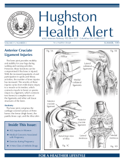


© Copyright 2026