
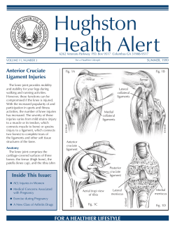
ACL Lesions in high level football players
ESPREGUEIRA-MENDES - Sports Clinic
Dragão Stadium – FC Porto
“FIFA MEDICAL CENTRE OF EXCELLENCE”
ACL Lesions in high level
football players
JOÃO ESPREGUEIRA-MENDES, MD, PhD
A. Monteiro, H. Pereira, P. Varanda, N. Sevivas, João Pedro Araújo, Isabel Lopes,
R. Pereira, F. Brandão, M. Oliveira, RA Sousa, R.L. Reis and Niek van Dijk
Chairman and Professor
Orthopaedic Department - Minho University
President of the European Society of Knee Surgery, Arthroscopy and Sports Trauma
Senior Researcher of 3B`s
PORTO – PORTUGAL
MINHO UNIVERSITY PORTO UNIVERSITY
ISAKOS
ISAKOS approved
teaching center
LITERATURE
ACL BIOMECHANICS
1/3.000 - new tears each year USA
150.000/year
(S. Woo, JOS, 2006)
Meta-analyses evaluating success rates of ACL recon:
69%-95%
(Freedman AJSM 2003)
ESSKA approved
teaching center
ACL is the primary restraint to tibial
anterior translation that is normally
accompanied by a couple tibial rotation
AM bundle is more important
resisting AP translation.
for
PL bundle is tight near extension and
plays a role in tibial internal rotation
KSSTA, April 2012
MENISCUS & ACLR
•
Cross all rehab protocols and assess time frames and criteria of each associated
injury
•
Respect timeframe of each repair/reconstruction technique/tissue healing. Flexion
may be limited to 90 degrees - 3/4 weeks - in repairs of the posterior horn
•
Criteria based progress – accept a delay on ROM, weightbearing, strenghtening
exercises, proprioception, neuromuscular reeducation if needed
•
Take advantage of time window for meniscal repair in the setting of concomitant
injuries (ACLR)
NO CONSENSUS IN LITERATURE ABOUT TECHNIQUE & RETURN TO SPORT
3D kinematic analysis to evaluate the functional levels of
the knee, it has been found that in the ACL-deficient
knee there is anterior tibial translation and excessive
tibial rotation during everyday activities.
ACLR is successful in restoring these functions when lowdemanding activities such as walking are performed.
During high-demanding activities, ACLR seems to fail to
restore excessive tibial rotation, which may be the cause
of further degeneration in the medial compartment even
after ACLR.
PREVENTION
GOALS OF ACLR
“YOU CAN MAKE A DIFFERENCE!”
• Absence of pain, swelling
• Restoration of ROM and muscle strength
• Restoration of proprioception
• Return to work
• Return to sport activity
• Return to sports at the same level
• Prevention of degenerative OA
• Restoration of normal knee kinematics
Lars Engebretsen, Editorial, KSSTA, May 2009
“The soccer players do not appear to spend much
time on preventive work
You need to bring the coaching staff & players to
your side early (with the youngsters)
The using the available science will reduce the
injuries and post-injury problems”
Engebretsen et al , AJSM, 2008
Hurd et al, AJSM, 2008
Soligard, et al, BMJ, 2008
RISK FACTORS
Does bone morphology play a role in laxity & rotation?
M. Fernandes, A. Monteiro, N. Sevivas H. Pereira and Espregueira-Mendes (in press)
“Bone morphology of
the external femoral &
tibial condyles are the
trochlea of the ACL”
Conclusion:
Methods
36 patients 29 ♂ and 7♀, with recent rupture of ACL were compared with other 36 subjects 29 ♂
and 7 ♀, without any knee pathology.
We measured in the femur: the diameter of the shaft, height and anteroposterior diameter of the
external condyle, the flattened lower limit of the epiphysis and the distance of the latter to the
anterior and posterior cortical diaphysis (yz and xw).
In the tibia, we measured the AP diameter of the tibial plateau and tibial slope.
•There are significant differences in bone morphology
Results
♂ with non traumatic rupture of the ACL (compared with healthy population) we found:
A bigger height and AP length of the external condyle (yz and xw).
A smaller AP diameter of the tibial plateau.
•Condyle size is a risk factor for ACL injury – may influence
knee Kinematics
• Significant differences in bone morphology between
ACL-injured and non-injured subjects
•Notch width measures on MRI and arthroscopically have no
correlation
The results suggest that the parameters above can be risk factors in the ACL rupture.
DIAGNOSIS
HEMARTROSIS : 82% ACL
Clinical evaluation
X-Ray
MRI
Arthrometer/PKTD
Arthroscopy
CLINICAL EVALUATION
“..THE DIAGNOSIS OF AN ACL
RUPTURE IS DONE BY HAND..”
MRI
Evaluate associated lesions
Meniscus
Cartilage
Bone bruise
Other Ligaments
Measure instability &
evalute partial ruptures
Is a diagnosis of partial or even total ACL rupture enough to
indicate surgery in 2013 (PL in high level football players?)
How to be sure about the functioning of the remaining bundle?
Can we correctly measure AP translation & rotation?
Can we control rotation without knowing the value of normal
pattern?
Do we know how much AP translation, rotation or both combined
causes instability (“cut point”)?
“A simple clinically applicable tool, similar to KT 1000 arthrometer, that
could be used to quantify laxity and rotation needs to be developed”
J. Irrgang, J. Bost & F. Fu Letter of AJSM 2009
“ Both instrumented laxity and MRI need to be used in combination
with proper clinical evaluation to possibly acquire a greater diagnosis
value.”
D. Dejour et al, Arthroscopy, March, 2013
PORTO KNEE TESTING DEVICE
PORTO-KTD
ACL evaluation with IR & ER
PORTO-KTD
ROTATION OF KNEE MRI EVALUATION
PCL PL & PM Instability
NO PRESSURE
AP TRANSLATION
AP TRANSLATION
& I ROTATION
PORTO – KTD
KSSTA,Vol. 20, Nº 4
April 2012
28 cases with symptomatic and
arthroscopy confirmed total
ACL RUPTURE
MRI EVALUATION with PKTD
(injured + healthy knee)
GRAFT CHOICE
•
B-P-B (gold standard)
•
Triple or quadrupled hamstrings
•
Quadriceps tendon
•
Fresh frozen allograft (multiple ligament recon)
Identify the patients
reconstruction
that
will
need/not
need
Correct indications for partial ruptures
Verification of the functioning of the remaining bundle
Useful in comparing SB with DB
Prevention? (increased internal rotation?)
Can be useful for objective evaluation of AP &
Rotation laxity of an ACL deficient knee
Our Preferred Technique in 2013
SINGLE BUNDLE
BPB
Graft
position
FEMUR
RESTORE ANATOMY (NO 10h or …)
AM PORTAL TO VIEW THE PLACE OF F/TUNNEL
AM ACESSORY PORTAL TO DRILL F/TUNNEL
NO TRANSTIBIAL
F/TUNNEL IN FULL FLEXION
“12h”
AM
AAM
TIBIAL
PLACEMENT
Restore anatomy (remaining ACL/footprint)
Measure footprint to size the graft
No 10 mm or 14 mm… in front of PCL…
ACL Partial Rupture
WHEN?
“Double bundle concept – F. Fu”
As soon As:
Attempt of 0 - 90º mobility
No pain and no swelling
AM torn and PL intact
IN ACUTE EXCEPTIONAL !!
Blocked knee
We repair MCL grade III in football
ACL+MCL+IM (PCL…)
LITERATURE
Postoperative range of motion following
ACL recon with autograft hamstrings – a
prospective, randomized study
- Bottoni at al AJSM, Vol. 38, 2008
Excellent clinical results can be achieved in acute with a
rehab protocol emphasizing extension and early range
of motion
DOUBLE-BUNDLE ?
Restore the anatomy
Restoration of normal
knee kinematics &
function
Improvement of the
knowledge of
anatomy & better
placement of a SB
Indication?
Difficulty in revision?
Long-term outcomes?
IMPORTANT INSTABILITY
Severe antero-lateral instability
LPS +++ and varus test +
PAST:
Lemaire
AM
PL
PRESENT:
Double Bundle (1TT&2FT)
TERM in Athletes?
MENISCAL SUTURE
“RED-RED-ZONE” AND “RED-WHITE-ZONE”.
1. Scarce tissue characterization concerning cells
Suture in Red-Red
and
simple ruptures in RR and RW
Do not suture stable
and
peripheral ruptures < 1cm
Arnoczky und Warren (1982)
Am J Sports Med
2. Limited information about native repair mechanisms and injury
response
3. Limitations of comercial scaffolds:
early failure;
reduced size with time;
neo-tissue different from fibrocartilage.
4. No clinical study testing advanced TERM strategy combining scaffolds
enhanced by cells, GFs, nanotechnology…
NO SUFFICIENT DATA IN LITERATURE REGARDING SPORTS
CARTILAGE
CARTILAGE
WB < 1,5cm2 and all NWB – III/IV
> 1,5 cm2 in WB area “GUT” MOSAICPLASTY
MICROFRACTURES in > 90% cases
A new ostheochondral graft from the upper tibio-fibular joint - GUT
Espregueira-Mendes, A. Monteiro, P. Amado
ICRS, 2004, Medimond
Anatomy of the proximal tibio peroneal joint
Espregueira-Mendes, M. Vieira da Silva
KSSTA, March 2006
Osteochondral transplantation using autografts from the upper tibiofibular joint for the treatment of knee cartilage lesions – 10y FU
Espregueira-Mendes, H. Pereira, M. Oliveira, Rui L. Reis
KSSTA, June 2012
BPB + VALGUS OSTEOTOMY
GROWTH FACTORS
(… high level soccer players?)
NEVER in High Level Football Players!
PRP (42 cases)
Stimulate revascularization?
> Ligamentization?
> Bone healing?
Less pain and haematoma
?
POST-OP
Full extension (orthosis 5 days)
CPM at 24 hrs.
Ice (Criocuff) pre & post-op
Weight bearing 24 hrs.
90º 5th day
POST-OP PAIN
MULTIMODAL ANALGESIA
Synergistic effects from different analgesics
Reduction in the drugs’ doses
Lower incidence of side effects
Less pain / Better mobility
PRP
ICE (CrioCuff pre-op and post –op)
Local ropivacaine
COX 2 inib (peripheral/central effect)
CRITERIA FOR SAFE RETURN TO SPORT
AFTER ACLR
1989 - 2005
Muscle strength and performance:
Isokinetic test & one leg hop test < 1020% deficit
Rehabilitation
No pain or effusion
Full ROM
Return
to
Sports
1364 PRIMARY ACLR
Functional Knee stability :
Clinical examination and objective measurments ex:
motion analysis
Surgery
Static knee stability:
Clinical examination and objective measures
Kt 1000 & PORTO KTD
Other Factors
Associated Injuries
I.E. menisci, cartilage, other ligaments, etc
Social
I,e. family, pregnancy,
finished college, etc.
82 (6%)
Professional Football Players
Psychological factors
I,e. motivation, scholarship,
fear of re-injury, etc.
RETURN TO SPORTS
TAKE HOME MESSAGE
82
Professional Football Players
83% (90%)
75%
97%
ProFootball
Same level
Sports
(7% related other joints/motif)
High Level Sports Players special prob.& concerns
(coach, directors etc)
Important correct pre-op evaluation
Associated instabilities
Correct surgical technique
Need to improve rotational stability
Basic science studies support anatomic
Growth factors are promising (more studies needed)
Rehabilitation and Good Team
Prevention is the best treatment!
double-bundle ACL reconstruction (indication)
THANK YOU!
FEMUR
AT 10H30 or 2H30
POSTERIOR WALL WITH 2mm.
MEDIAL APPROACH
Reconstruction of PL Bundle Only
BONE/BONE
BONE/TENDON FIXATION
BPB, HAMSTRINGS or QT GRAFTS
ACL Partial Rupture
Resofix
(PLLA R&L)
AM intact with PL rupture
MENISCAL SUTURE
“RED-RED- ZONE” AND “RED-WHITE-ZONE”.
CARTILAGE
< 1cm2 or NWB
MICROFRACTURES
Arnoczky und Warren (1982)
Am J Sports Med
Suture in Red-Red and simple ruptures in RR and RW
Do not suture stable and periferic ruptures < 1cm
CARTILAGE
> 1cm2 in WB MOSAICPLASTY with “GUT”
GROWTH FACTORS
Platelet-rich plasma stimulates osteoblastic differentiation in
the presence of BMPs
Autologous platelet gel and fibrin sealant enhance the efficacy of total knee
arthroplasty: improved range of motion, decreased length of stay and a reduced
incidence of arthrofibrosis
Akihiro Tomoyasu et al
Peter Everts et al Journal Knee Surgery, Sports Traumatology, Arthroscopy Volume 15 Number 7 July 2007
Biochemical and Biophysical Research Communications
Volume 361, Issue 1, 14 September 2007 pages 62-67
Platelet gel and fibrin sealant reduce allogenic blood transfusions in total knee
arthroplasty.
Everts PAM, Devilee RJJ, Brown-Mahoney C et al - Acta Anaesthesiol Scand 50:593–599 (2006)
Enhanced histologic repair in a central wound in tha ACL
with a collagen-platlet – rich plasma scaffold
Martha Murray et alt, JO Research, August 2007
Comparision of Surgically Repaired Achilles Tendon
Tears Using Platelet-Rich Fibrin Matrices
M Sanchez et al. 35: 245-251 , Am J Sports Med 2007 Feb
Faster reabilitation
“The use of a collagen- PRP-scaffold can ameliorate the healing of na ACL
rupture in dogs”
Less wound problems
Less tendon volume on the scar tissue
LITERATURE
Use of growth factors in ACL surgery : preliminary study
Ventura et alt, JO Traumatology, 2005
Thirty-five years of f-u of ACL deficient knees in high level athletes
(Olympic) - W. Nebelung, H. Wuschech, Arthroscopy, Vol. 21,
Issue 6, 2005
•
•
10 patients with hamstrings graft and GPS
10 patients with hamstrings graft without GPS
1963-1965 ACL ruptures without recon. and return to high level sports (19)
20y after: Meniscectomy in 18 (94%), Condral lesion IV in 13 (68%)
TKA in 10 (52%) - until 2000
Prospective trial of a treatment of algorithm for the
management of the ACL injured knee
NO Dif in Tegner score and KT 1000
“In the PRP with GPS group the density of the ACL graft was better in CT
scan and the integration in the tunnels was faster”
Eithian DC, et al. AJSM 2005
•
•
Degenerative radiographic changes in 90% of patients 7 years after
ACL reconstruction
47% of patients returned to previous activity level after ACL
reconstruction
ACL Reconstruction
Graft fixation
G
Graft passage
GRAFT FIXATION
DOUBLE-BUNDLE CONTRA
INDICATIONS
Narrow footprint
Skeletal immaturity
Early OA
LFC osseous contusion
IC notch stenosis
Combined ligament injury
1 YEAR
Xray and TELOS
G
INJURY PATTERN
The Injury Mechanism of the
ACL is Complex (Rotation /
Flexion / Hyperextension /
Varus / Valgus), and is
Reflected by the Different
Rupture Patterns of the ACL
+++ Valgus/Flexion/External
Rotation in our series
CLASSICAL GRAFT POSITION
(Our preferred technique 1989 – 2005)
1989 - 2005
1364 PRIMARY ACLR
1301
23
17
12
11
- BPB
- Hamstrings
- BPB/Lemaire
- Quadriceps tendon
- Allografts
1989 - 2005
1364 PRIMARY ACLR
236 (17,3%) F / 1128 (82,7%) M
Mean age : 24,5 years (11 - 53)
SINGLE BUNDLE
PL
FEMUR
“AT 10H (9h30) or 2H (2h30)” ….?
POSTERIOR WALL WITH 2mm…?
TUNNEL in FULL FLEXION!
Sports activities:
Sports in 956 ( 70,3 %)
Football > 75%
Reconstruction of AM Bundle Only
“Double bundle concept”
Is “nearly normal” good enough for ACL treatment in
2013 (high level football players?)
Can we correctly measure AP translation & rotation?
Can we control rotation without knowing the value of
normal pattern?
Do we know how much AP translation and rotation
causes instability (“cut point”)?
“A simple clinically applicable tool, similar to KT
1000 arthrometer, that could be used to quantify
laxity and rotation needs to be developed”
J. Irrgang, J. Bost & F. Fu Letter of AJSM 2009
Tsai AG, F. Fu et al, .
BMC Muscu Disord. (2008)
Most of the times
impingement is an
incorrect placement
of the graft!
KT1000/2000
NO ROTATION MEASUREMENT
NO MEASUREMENTS BETWEEN FEMUR & TIBIA
“GLOBAL” AP MEASUREMENT
Kubo S. et al.
Clin Orthop Relat Res.
A. Hemmerich,
B. Van der Merwe, et al,
C. J. Biomechanics, 2009
T. Branch, H. Mayr, et al
Arthroscopy, 2010
2008 - 2010
NOTCHPLASTY
Rare!
Only in chronic cases
Robert H. et al,
Rev. COT (2009)
Branch TP et al,
KSSTA (2009)
AGE
Mean 33,4 ± 9.4 y
SEX
25 MALES & 3 FEMALES
SIDE
BMI
MRI
13 LEFT & 15 RIGHT
25,3 (SD = 3,1)
1,5 T GE Healthcare Signa, USA
T1,T2, STIR, FatSat, 3D SPGR


© Copyright 2026