
Anterior Cruciate Ligament Injuries
Anterior Cruciate Ligament Injuries
http://orthoteers.com/(S(l4gom345o1tssvrsb0qg1c55))/printPage.aspx?ar...
Printout from Orthoteers.com website, member id 1969. © 2007 All rights reserved. Please refer to
the site policies for rules on diseminating site content.
Anterior Cruciate Ligament Injuries
Anatomy & Biomechanics
completely intraarticular
anteromedial fibres - tight in flexion - limits anterior translation of tibia on femur
posterolateral fibres - tight in extension - limits anterior translation PLUS external rotation
ACL strength = 50% PCL strength
Load to failure = 1700N
strain rate plays a role in the location of ligament failure
midsubstance tears occurring at higher rates
bone ligament complex tears occurring at lower rates
Blood supply - middle genicular artery (post) & synovial vessels (ant)
Mechanoceptors with a proprioceptive role
Examination:
See Knee Examination
Mechanism of Injury:
substantial anterior tibial shear forces that stress ACL are produced from quadriceps contraction,
esp in 0-30 deg of extension
Typically, the ACL is torn in a noncontact deceleration situation that produces a valgus twisting
injury - when the athlete lands on the leg and quickly pivots in the opposite direction.
Associated Injuries: ( Noyes, 1980 )
1. Meniscal tear - 62% - lateral > medial (Noyes)
2. Capsular tears - 21%
3. Chondral fracture - 10%
Natural History:
Controversial !?
Left untreated, the torn ACL can lead to:
1. Instability (10 years following injury & exploratory arthrotomy) ( McDaniel &
Dameron, 1980 )
1/3 of patients with isolated ACL injuries will show minimal instability, with no
pain or discomfort
1/3 have no pain or instability
1/3 will have significant instability & pain
2/3 returned to strenuous sports (1/3 of these had to change their specific
sport)
2. Meniscal tears
3. Degenerative joint Disease (DJD) ( Jomha et al., 1999 )
1 of 6
10/7/2007 12:43 AM
Anterior Cruciate Ligament Injuries
http://orthoteers.com/(S(l4gom345o1tssvrsb0qg1c55))/printPage.aspx?ar...
cartilage damage correlates with length of time from injury; medial
compartment > lateral compartment
2-3 giving-way episodes per year has been correlated with DJD
most often seen in ACL deficient knees with meniscal injury
4. Quadriceps avoidance gait:
between 0 to 45 deg of flexion, contraction of the quadriceps will cause
anterior translation of the tibia (which is normally resisted by the ACL);
maximum anterior translation of the tibia occurs at 15-25 deg of flexion
There is no good evidence that brace wear decreases the rate of re-injury
Low demand patients with isolated ACL injury who are willing to moderate their activity will find
non-operative treatment to be satisfactory in the majority of cases (over 80%).
Non-operative Treatment:
Good results if:
1. thigh circumference is equal or better than opp. side
2. stable
Poor results if:
1. thigh atrophy
2. meniscectomy performed
3. instability
Operative Treatment:
1. Direct Repair
For avulsion fractures
Not recommended for mid-substance tears due to poor healing potential.
2. Extra-articular reconstruction
MacIntosh Procedure: (historical)
Pass a mobilised strip of iliotibial band to the posterolateral corner of the knee through a tunnel deep
to the lateral collateral
ligament
2 of 6
10/7/2007 12:43 AM
Anterior Cruciate Ligament Injuries
http://orthoteers.com/(S(l4gom345o1tssvrsb0qg1c55))/printPage.aspx?ar...
3. Intra-articular Reconstruction
Options:
1. bone patellar bone reconstruction
2. hamstring reconstruction - semitendinosis + gracilis
3. allograft reconstruction
4. Synthetic grafts
Isometry:
Isometric placement of ACL refers to the concept that a full range of knee can be achieved without
causing long-term ligament deformation
isometry can not exist because, during ROM, there is no one point on femur that maintains a fixed
distance from a single point on tibia; elongation always will occur
placement of graft as closely as possible to centers of tibial & femoral attachments of anterior medial
band results in least amount of strain (least change in the length of ACL during complete ROM of
knee)
Technique: See Wheeless
3 of 6
10/7/2007 12:43 AM
Anterior Cruciate Ligament Injuries
http://orthoteers.com/(S(l4gom345o1tssvrsb0qg1c55))/printPage.aspx?ar...
Post-op Care:
CPM range of motion immediate post-op
Closed chain exercises (foot is maintained on the ground or a platform)
Running & any activity that involves excessive knee rotation (cutting) is discouraged for the first
several months.
Complications:
1. Loss of knee extension / Arthrofibrosis
more common with early reconstruction
incorrect tunnel placement can cause decreased motion and fibrosis (diagnose with x-rays &
MRI)
2. Tibial tunnel syndrome
incr. size of tibial tunnel over 1yr following surgery
3. Graft failures:
1. Improper placement of graft tunnels
2. Impingement of the graft in the femoral notch:
1. due to improper tunnel placement
2. due to inadequate notchplasty
3. Inadequate graft fixation
4. Associated injuries (varus knee, significant osteochondral defects)
4 of 6
10/7/2007 12:43 AM
Anterior Cruciate Ligament Injuries
http://orthoteers.com/(S(l4gom345o1tssvrsb0qg1c55))/printPage.aspx?ar...
5. CRPS (0.5%)
6. Infection (<1%)
4. Patellofemoral pain
5. Patella fractures
6. CRPS (0.5%)
7. Infection (<1%)
8.
ACL Instability with Degenerative changes
Ligamentous stabilisation alone often fails
Usually medial compartment OA
Rx: combined tibial osteotomy plus extra-articular reconstruction
Noyes FR, Bassett RW, Grood ES, Butler DL
J Bone Joint Surg Am 1980 Jul;62(5):687-95, 757
Arthroscopy in acute traumatic hemarthrosis of the knee. Incidence of anterior cruciate tears and
other injuries.
In a prospective study, all injured knees that had traumatic hemarthrosis and absent or negligible instability
on clinical examination underwent arthroscopy and examination under anesthesia. Eighty-five knees
(eighty-three patients) were examined over a 125-week period. Some degree of disruption of the anterior
cruciate ligament was found in sixty-one (72 per cent) of the knees (a partial tear in 28 per cent and a
complete tear in 44 per cent), frequently associated with an injury of varying severity to other joint structures.
These included minor ligament sprains without laxity in 41 per cent, a major associated ligament injury in 21
per cent, meniscal tears in 62 per cent (partial in 30 per cent and complete in 70 per cent), and a femoral
chondral fracture or surface defect in 20 per cent. A popping sensation at injury occurred in 33 per cent of
knees with a normal anterior cruciate ligament and in 36 per cent of those with a disruption. One-third of the
knees had no to slight pain at the time of injury. The anterior drawer test without anesthesia was positive in
only 24 per cent of the knees with a torn anterior cruciate ligament. We concluded that: (1) a traumatic
hemarthrosis indicates a significant knee injury; (2) examination under anesthesia plus arthroscopy allows a
more accurate diagnosis of injury to joint structures; and (3) such data are required for a rational treatment
program to be outlined.
Jomha NM, Borton DC, Clingeleffer AJ, Pinczewski LA
Clin Orthop 1999 Jan;(358):188-93
Long-term osteoarthritic changes in anterior cruciate ligament reconstructed knees.
Australian Institute of Musculo-Skeletal Research, Crows Nest, NSW, Australia.
To consolidate the indications for anterior cruciate ligament reconstruction and clarify the long-term
prognosis associated with current surgical and rehabilitation techniques, the incidence of osteoarthritis in
arthroscopically anterior cruciate ligament reconstructed knees requires investigation. Seventy-two patients
with anterior cruciate ligament ruptures who were active in sports requiring sidestepping and pivoting, or
who had recurrent episodes of giving way, underwent arthroscopic bone-patellar tendon-bone anterior
cruciate ligament reconstruction. These patients were evaluated for meniscal damage and osteoarthritic
changes at the time of surgery and followed up for 7 years. Fifty-three patients underwent radiographic
evaluation at 7 years, which included anteroposterior, lateral, skyline, and 30 degrees posteroanterior
weightbearing views. Radiographic evaluation was performed by three independent surgeons and graded
as per International Knee Documentation Committee criteria. Results revealed that knees with chronic
anterior cruciate ligament deficiency, even those with intact menisci before reconstruction, suffered early
osteoarthritic changes. More severe changes were seen with meniscectomy. Acute anterior cruciate
ligament reconstruction with meniscal preservation was shown to have the lowest incidence of degenerative
change. Controversy exists regarding the timing of anterior cruciate ligament reconstruction. This study
supports early reconstruction of anterior cruciate ligament deficient knees before episodes of giving way
occur in individuals intent on continuing activities that involve sidestepping and pivoting.
McDaniel WJ Jr, Dameron TB Jr
5 of 6
10/7/2007 12:43 AM
Anterior Cruciate Ligament Injuries
http://orthoteers.com/(S(l4gom345o1tssvrsb0qg1c55))/printPage.aspx?ar...
J Bone Joint Surg Am 1980 Jul;62(5):696-705
Untreated ruptures of the anterior cruciate ligament. A follow-up study.
Fifty patients (fifty-three knees) with surgically verified ruptures of the anterior cruciate ligament were
evaluated at an average of ten years after injury. There was a high incidence of anterior laxity, rotatory
instability, and meniscal tears at follow-up. The roentgenographic incidence of osteoarthritis at follow-up
was low. Patients who had regained normal thigh circumference had better results than those with thigh
atrophy. Although few of the patients felt that the knee was completely normal, 72 per cent of the patients
returned to strenuous sports and 47 per cent felt that they had no restrictions because of the knee. This
study of untreated ruptures may provide a baseline for evaluation of procedures to repair or reconstruct the
anterior cruciate ligament.
[ Close Window ]
6 of 6
10/7/2007 12:43 AM
Knee - Osteotomies
http://orthoteers.com/(S(l4gom345o1tssvrsb0qg1c55))/printPage.aspx?ar...
Printout from Orthoteers.com website, member id 1969. © 2007 All rights reserved.
Please refer to the site policies for rules on diseminating site content.
Knee - Osteotomies
60% of the load of the body weight passes through the medial compartment of the knee
Loads up to 4 times body weight are produced on climbing stairs
Osteotomy redistributes the force
Valgus osteotomy most commonly performed (varus knee) and is indicated in patients that
have uni compartmental disease, are less than 60 - 70 years old, are of optimal weight, have
an active occupation or lifestyle which they want to maintain and have a good range of motion
NB: ROM is not likely to improve with an osteotomy
INDICATIONS
1. Age: physiologic age < 60 yrs in an athlete, laborer, or anyone who needs to knee
down such as for gardening (TKR will generally not allow the patient to kneel)
2. Weight: > 80 kg are at increased risk for component failure;
3. Angular Deformity:
1. > 15 deg of fixed varus deformity (often patients will have varus laxity)
2. < 15 degrees flexion contracture
3. > 90 degrees flexion
4. Radiologically intact lateral (or medial) and patellofemoral compartments
CONTRAINDICATIONS:
1.
2.
3.
4.
Tibial Subluxation > 1 cm
RA & inflammatory arthritis
ACL tear
osteochondral injuries with involvement of more than 1/3 the condylar surface or OCD
lesion of more than 5 mm deep
CLINICAL
Observe patient walk (look for varus thrust)
Stability
Q Angle
Compensatory arc of motion - to correct a valgus knee deformity - for a 20-degree
varus osteotomy, 20 deg of abduction at the hip is required so pt does not end up with
an adduction deformity
Examine the foot and ankle to rule out fixed varus deformities which may worsen
medial compartment loading
Leg length discrepancy Coventry closing wedge osteotomy might be indicated,
where as, if the arthritic side is shorter (than the other leg), then consider opening
wedge osteotomy
INVESTIGATIONS
Radiology
Standing long-leg film with patellae facing forward (rather than the patient's feet)
Mechanical axis = centre of femoral head - medial tibial spine - centre of ankle
joint
Weight-bearing axis = centre of femoral head - centre of ankle joint
Anatomical axis = line along axis of the femur to the intercondylar notch and the
line formed by the interspinous region to the centre of the ankle
Supine films
Arthroscopy
METHODS
Medial Compartment Disease
High Tibial Osteotomy (HTO) above the tibial tubercle
Lateral closing wedge & fibular shortening [ Technique, Wheeless ]
Overcorrection of the mechanical axis by 3 degrees is ideal
1 of 2
10/7/2007 12:44 AM
Knee - Osteotomies
http://orthoteers.com/(S(l4gom345o1tssvrsb0qg1c55))/printPage.aspx?ar...
Complications:
1. Undercorrection - most common
2. Overcorrection
3. Penetration of the articular surface
4. AVN of tibial plateau
5. Patella baja
6. Peroneal nerve injury
7. Anterior compartment syndrome
8. TKR may be more difficult
Lateral Compartment Disease
< 12 degrees valgus = Varus tibial osteotomy
> 12 degrees = Medial closing wedge osteotomy of distal femur (supracondylar)
RESULTS
Satisfactory results obtained in as many as 70% at 10 years have been reported (study
of 51 knees JBJS, 1988) with 30% fair or poor
Best results are obtained if slight over correction achieved that is 2 - 3 o beyond the
normal 7 o of valgus
Results relate to the preoperative knee scores and the degree of correction /
over-correction of the mechanical axis
Odenbring et al 1990
75% of patients under the age of 50 w/ early medial DJD had at good result at
11 years post surgery
Billings et al (JBJS 1999)
64 valgus producing high tibial osteotomies were performed using a calibrated
cutting guide w/ plate fixation
43 out of 64 knees had a good to excellent clinical result w/ an average knee
score of 94 points at an average of 8.5 years follow up
using total knee arthroplasty as an end point, there was 85 % survival at 5 yrs
and 53 % at 10 years
no patient had patella baja postoperatively (the authors fell that early ROM w/
CPM prevented baja)
average initial postoperative correction (and standard deviation) for all knees
was to 9.2 - 3.69 degrees of valgus
5 knees were corrected to less than 5 deg of valgus
3 of them were treated with a subsequent arthroplasty (at twenty-four, sixty-five,
and sixty-six months)
13 knees had lost more than 2 deg of correction at the time of the latest
follow-up
average initial postoperative correction for these knees was to 9.4 - 4.12 deg
(range, 4 to 17 degrees) of valgus
of knees that lost more than 2 degrees of correction, four subsequently had a
total knee arthroplasty.
[ Close Window ]
2 of 2
10/7/2007 12:44 AM
Knee Arthrodesis
http://orthoteers.com/(S(l4gom345o1tssvrsb0qg1c55))/printPage.aspx?ar...
Printout from Orthoteers.com website, member id 1969. © 2007 All rights reserved.
Please refer to the site policies for rules on diseminating site content.
Knee Arthrodesis
Indications:
1. failed knee replacement (most common)
better fusion rate following failed condylar components (80%) than failed
hinged prostheses (55%)
2. uncontrollable septic arthritis with complete joint destruction
3. young patient with severe articular & ligamentous damage
4. neuropathic joint disease
Contra-indications:
1. bilateral knee disease
2. ipsilateral ankle or hip disease
3. severe segmental bone loss
4. contralateral leg amputation
Ideal Position:
10-15 degrees flexion
0-7 degrees valgus
Techniques of Arthrodesis:
1. External Fixation
Ilizarov technique
minimal soft tissue dissection
allows for late adjustment
allows arthrodesis in the presence of active infection
2. Intramedullary Nailing Arthrodesis
most reliable for achieving fusion
2 stage technique in the
presence of active infection
can insert nail antegrade thro
the piriform fossa or thro the
knee joint
complication rates of 20-50%
3. Plate Fixation
2 twelve hole plates
Complications:
1. Non-union- 20%
2. Malunion
3. Delayed union
1 of 2
10/7/2007 12:45 AM
Knee Arthrodesis
http://orthoteers.com/(S(l4gom345o1tssvrsb0qg1c55))/printPage.aspx?ar...
Ellingsen etal JBJS 76A: 870-877, 1994
18 knees with intramedullary arthrodesis after failed TKR
16/18 united , mean time to union 5.5 mths
high rate of complications - 2 AKA (persisting deep infection)
1 rod #, 1 rod migration
Ellingsen DE, Rand JA - Intramedullary arthrodesis of the knee after failed total knee
arthroplasty.
Department of Orthopedics, Mayo Clinic, Rochester, Minnesota 55905.
J Bone Joint Surg Am 1994 Jun;76(6):870-7
Eighteen patients (eighteen knees) were managed with an intramedullary arthrodesis after a
failed total knee arthroplasty. Twelve knees had had a revision total knee arthroplasty and six, a
primary total knee arthroplasty. Three knees had had failure of a hinged prosthesis. In eleven
knees, the arthroplasty had failed because of infection. Nine patients had had previous
attempts at arthrodesis with external fixation. The average duration of the operation was six
hours, and the average blood replacement was 2975 milliliters. A vascularized fibular pedicle
graft was used in four patients. At a mean of thirty-seven months after the arthrodesis, sixteen
of the eighteen patients had a complete radiographic union. The mean time to union was 5.5
months. Although a high rate of union was achieved in these patients, complications occurred
in ten of the eighteen knees and this must be considered. Intramedullary arthrodesis is a
successful method of salvage for a failed total knee arthroplasty or one complicated by
infection that is not amenable to revision, but it is technically demanding and has frequent
complications
[ Close Window ]
2 of 2
10/7/2007 12:45 AM
Knee Ligament Injuries
http://orthoteers.com/(S(l4gom345o1tssvrsb0qg1c55))/printPage.aspx?ar...
Printout from Orthoteers.com website, member id 1969. © 2007 All rights reserved.
Please refer to the site policies for rules on diseminating site content.
Knee Ligament Injuries
Anterior Cruciate Ligament
Posterior Cruciate Ligament
Posterolateral Instability &
Knee Dislocation
Classification of knee joint instability resulting from ligament injury:
I. One-plane instability (simple or straight)
A.One plane medial
B.One plane lateral
C.One plan posterior
D.One plane anterior
II. Rotary instability
A.Anteromedial
B.Anterolateral
1.In flexion
2.Approaching extension
C.Posterolateral
D.Posteromedial
III. Combined instability
A.Anterolateral-anteromedial rotary
B.Anterolateral-posterolateral rotary
C.Anteromedial-posteromedial rotary
Classifications of Ligament Injury / Laxity Testing:
O'Donaghue:
First Degree Sprain
ligament injury with no instability
Second Degree Sprain
Third Degree Sprain
partial tear with some laxity
complete tear with marked instability
Noyes:
Grade 1
0-5mm
Grade 2
Grade 3
Grade 4
6-10mm
11-15mm
16-20mm
[ Close Window ]
1 of 1
10/7/2007 12:46 AM
Knee Mechanics
http://orthoteers.com/(S(l4gom345o1tssvrsb0qg1c55))/printPage.aspx?ar...
Printout from Orthoteers.com website, member id 1969. © 2007 All rights reserved. Please refer to the site policies for rules on
diseminating site content.
Knee Mechanics
KNEE RESTRAINTS - Sectioning Studies
Primary Restraints
Secondary Restraints
1.
2.
3.
4.
5.
6.
iliotibial band: 24%
mid medial capsule: 22%
mid lateral capsule: 20%
MCL: 16%
LCL: 12%
Meniscii
Anterior
translation
ACL
Posterior
translation
PCL (large anterior bundle more NB progressively from 0
to 90 degrees)
LCL
Internal rotation
ACL
POL/PMC has secondary effect within 0 - 45 degrees
External
rotation
Popliteofibular ligament
LCL and the posterolateral complex - mainly at 30 degrees
POL/PMC
flexion
MCL - at all degrees of flexion
Valgus
Superficial MCL is the primary restraint to valgus stress at
all angles (least effect at full extension)
Posterior Oblique ligament (POL) - especially near or at full
Postero-Medial Capsule is tightened at full extension; past
extension
30 degrees it slackens
ACL
Deep MCL (medial capsular ligament) has little resistance
to valgus load
Varus
LCL in all positions of flexion. Greatest effect at 30
degrees, least at full extension
Posterolateral structures (Popliteofibular lig.)
ACL
Posterolateral Structures:
1. Arcuate Ligament
2. Fibular Collat ligament
3. Popliteus tendon
4. Popliteofibular ligament
fibers originate from the popliteal tendon and insert onto the fibula
deep to the arcuate ligament and its fibers orientation are opposite from the arcuate ligament
See Pictures - posterior & lateral
Details of Sectioning Studies
KINEMATICS
Instant Center of Rotation
"If one rigid body rotates about another rigid body, its motion at any instant can be described by a point or axis of rotation called the instant center of
rotation (ICOR)." (Simon, AAOS Basic Science 1994).
Method developed by Reuleaux in 1876.
1 of 3
10/7/2007 12:48 AM
Knee Mechanics
http://orthoteers.com/(S(l4gom345o1tssvrsb0qg1c55))/printPage.aspx?ar...
Surface Joint Motion
Motion between the tibia and the femur is both rotational and translational. The femoral condyles both roll and glide as they articulate with tibial
plateaus. As the knee moves from full extension into flexion the ICOR moves posteriorly relative to both the femur and the tibia
Screw Home Mechanism
During the normal gait pattern the tibia undergoes internal rotation during the swing phase and external rotation during the stance phase.
external rotation of the tibia on the femur occurs during the terminal degrees of knee extension, because of the difference in radius of
curvature of the medial and smaller lateral condyle.
This screw home mechanism in terminal extension results in tightening of both cruciate ligaments and locks the knee such that the
tibia is in the position of maximal stability with respect to the femur .
Patellofemoral Joint
primary function of the extensor mechanism of the knee is deceleration during the swing phase of gait.
Functions:
1. increases the moment arm of the quadriceps
2. allows wider distribution of compressive stress between the patellar tendon & the femur
from full extension to full flexion the patella glides caudally 7cm on the femoral condyles.
by 20 degrees of knee flexion the patella first begins to articulate with the trochlear groove. Beyond 90 degrees the patella rotates externally &
only the medial facet articulates. At extreme flexion the patella lies in the intercondylar groove.
Initially the patella contact occurs distally and with increased flexion the contact areas shift proximally on the patella
patellofemoral contact pressure is 0.5 times body weight with walking, and increases to 2.5 to 3.3 times body weight with stair climbing and
descending
2 of 3
10/7/2007 12:48 AM
Knee Mechanics
http://orthoteers.com/(S(l4gom345o1tssvrsb0qg1c55))/printPage.aspx?ar...
Sponsored Links
www.biomet.co.uk
www.biomet.com
www.oxfordknee.net
[ Close Window ]
3 of 3
10/7/2007 12:48 AM
Meniscal Injuries
1 of 6
http://orthoteers.com/(S(l4gom345o1tssvrsb0qg1c55))/printPage.aspx?ar...
Printout from Orthoteers.com website, member id 1969. © 2007 All rights reserved.
Please refer to the site policies for rules on diseminating site content.
Meniscal Injuries
Anatomy
ANATOMY
Function
Meniscal Tears
Meniscal Cysts
Discoid
Meniscus
[Back To Top]
elasofibrocartilagenous
crescent shaped; triangular in cross-section
anterior horns attached to each other by the small transverse anterior intermeniscal
ligament
lateral meniscus is more circular; medial meniscus more C-shaped
lateral meniscus has twice the excursion of the medial meniscus during knee motion.
anterior horn of lateral meniscus & post horns of both meniscii attach to the intercondylar
eminence
popliteus muscle (not tendon) is attached to lateral meniscus
semimembranosis is attached to medial meniscus
Blood supply:
from branches of the lateral, middle & medial genicular arteries
vascular synovial tissue from the capsule supplies the peripheral third of
meniscus
Frontal section of the medial compartment demonstrates the microvasculature
of the medial meniscus. The perimeniscal capillary plexus (PCP) permeates
through the peripheral border of the meniscus. F: Femur; T: Tibia. [Arnoczky
SP, Warren RF. Microvasculature of the human meniscus. Am J Sports Med.
1982;10:90-95.]
Constituents:
1. Type 1 collagen fibres arranged radially & longitudinally (circumferential)
longitudinal fibres - dissipate hoop stresses in the meniscus
10/7/2007 12:49 AM
Meniscal Injuries
2 of 6
http://orthoteers.com/(S(l4gom345o1tssvrsb0qg1c55))/printPage.aspx?ar...
radial fibres & longit fibres - allows meniscii to expand under compressive force
2. Proteoglycans
trapped within collagen fibres to absorb energy
FUNCTION
[Back To Top]
1. Load bearing:
at least 50% of the compressive load of the knee joint is transmitted through the
meniscus in extension , and approx 85% of the load is transmitted in 90 o flexion.
In the meniscectomised knee the contact area is reduced approx 50%
Partial meniscectomy also increases the contact pressures
2. Shock absorption:
menisci may attenuate the intermittent shock waves generated by impulse
loading during gait- the shock absorbing capacity of normal knees is ~ 20%
higher than in meniscectomised knees.
The ability of a system to absorb shock has been implicated in development of
OA ( Radin and Rose " The role of subchondral bone in the initiation and
progression of Osteoarthritis" CORR 213:34-40, 1986)
3. Knee joint stability:
meniscectomy alone may not seriously affect stability. However, in assoc with
ACL tears, meniscectomy increases ant laxity of the knee
4. Lubrication
5. Proprioception:
this has been inferred from the finding of type 1 and type 2 nerve endings in the
ant and post horns of the menisci
MENISCAL TEARS
[Back To Top]
Meniscal tears can be either traumatic or degenerative in nature.
Meniscal tears are uncommon in persons under 10 years of age, but become increasingly
common during and after adolescence.
Degenerative tears can be found in as much as 60% of the population over age 65 . The
majority of these tears, however, are asymptomatic and occur in association with degenerative
joint disease. The changing patterns of meniscal injury with chronological age most likely
correlate with normal alterations in collagen fiber orientation with aging, as well as increasing
intrasubstance degeneration.
10/7/2007 12:49 AM
Meniscal Injuries
3 of 6
http://orthoteers.com/(S(l4gom345o1tssvrsb0qg1c55))/printPage.aspx?ar...
The majority of meniscal tears affect the medial meniscus and tend to involve the posterior horn.
Meniscal tears are either partial or full thickness and stable or unstable.
An unstable tear is one where the entire tear or a portion thereof can be displaced into the joint
space. There it may become trapped, causing pain by traction at the meniscocapsular junction.
It may be responsible for symptoms of catching, locking, and effusion.
Meniscal injuries can be further classified based on their tear patterns:
Dandy (1990) looked at 1000 meniscal injuries ->70% were medial with a slight increase in the
average age (39 years compared to 30 years for lateral meniscus injuries)
Occur only when weight is being taken, in the young the knee is flexed and there is a twisting
strain, in older patients tears may result from minor force
The torn portion may be displaced into the joint ->locking
Clinically:
Acute history of injury usually with localised pain +/- locking (a locked knee will flex but not
extend fully, the history of unlocking is characteristic of a mechanical block)
Patients are usually fit and young and symptoms may settle but ->repeated episodes
Local signs will depend on the time the joint is examined and whether or not it is still locked
(usually locked in 10 o - 20 o flexion)
Medial or lateral joint line tenderness and clicking with knee rotation in full flexion ->pain
(McMurrays test)
Special Tests for Meniscal Tears
Investigation
(Arthrogram)
MRIMackenzie et al. Clin Radiol. 1996 - multicentre review of 2000 patients.
Sensitivity 93%
Specificity 84%
Lat. meniscus- lower sensitivity 76%
Post-meniscectomy = <25% accuracy if meniscus has been resected,
25-75% if not resected.
Myxoid Degeneration of the post. third of the med. meniscus causes high
signal intensity & is commonly reported as a tear. But if the signal of the
'tear' = fluid signal it is more likely to be a tear.
Meniscofemoral lig. can resemble a tear of the ant. or post. horns.
60% of people >60yrs have complete meniscal tears. Intrasubstance
tears common >40yrs.
NB- Tear defn= must extend to articular margin on 3 consecutive slices
Problems of MRI - high cost, high false negative rate
Elvenes et al. Arch Orthop Trauma Surg 2000
'On the basis of the high predictive value of negative MRI, we conclude
that MRI is useful to exclude patients from unnecessary arthroscopy'
10/7/2007 12:49 AM
Meniscal Injuries
4 of 6
http://orthoteers.com/(S(l4gom345o1tssvrsb0qg1c55))/printPage.aspx?ar...
Classification:
Grade I tear: small disruption of the homogenous signal
Grade II tear: - disruption is more pronounced but does not extend thru
either superior or inferior surface - arthroscopically, a grade I or II tear
cannot be visualized
Grade III tear: - disruption of homogenous signal w/ extension to either
superior or inferior surface - is a clinically significant tear
Arthroscopy
Treatment:
Options
Conservative ->restrict activity
Manipulative to reduce ->conservative treatment
Operative ->arthroscopic partial menisectomy or meniscopexy
Meniscal transplantation- experimental
Meniscal repair
Blood supply to the meniscus is age dependent- in the adult the periph 3mm as well as the ant
and post horns are well vascularised
Repair should be reserved for traumatic tears in the vascular region of the meniscus
within 3mm of periphery = vascular ( = red- red tears)
3-5 mm from periphery = grey zone ( = red- white tears)
> 5mm from periphery = avascular ( = white- white tears)
Techniques:
1. Open Repair:
advantage of better preparation of the tear site
only the most peripheral of tears in the red-red zone are amenable to this
technique because of exposure and accessibility
Long-term follow-up of open meniscal repairs has revealed success rates
ranging from 84% to 100%.
2. Arthroscopically assisted:
1. Inside-out technique:
First described by Henning
utilizes zone-specific cannulas to pass sutures through the joint and
across the tear. The sutures are swaged onto flexible needles. A small
posterior joint line incision is used to retrieve the sutures and tie directly
on the capsule. The use of a posterior retractor, such as a gynecologic
speculum, is vital in order to protect the posterior neurovascular
structures.
2. Outside-in techniques:
described by Warren and Morgan and Casscells
involve passing sutures percutaneously through spinal needles at the
joint line across the tear, and then retrieving the sutures intra-articularly.
Mulberry knots can then be tied on the intra-articular free ends of the
suture. A small incision is then made at the joint line, where the
protruding suture ends are retrieved and tied directly on the capsule. An
alternative technique is to retrieve the intra-articular portion of the suture
with another pass across the tear using a wire snare and tying the suture
back on itself on the capsule. This technique eliminates the need for
Mulberry knots.
A potential disadvantage of the outside-in technique is difficulty in
reducing the tear and opposing the edges while passing the sutures.
3. All-inside technique
suitable for repairs of the far posterior horns
implantable anchors, arrows, screws, and staples
Healing :
similar to other connective tissues- exudation, organisation, vascularisation cellular
proliferation , remodelling
10/7/2007 12:49 AM
Meniscal Injuries
5 of 6
http://orthoteers.com/(S(l4gom345o1tssvrsb0qg1c55))/printPage.aspx?ar...
following injury there is formation of a fibrin clot rich is inflammatory cells. Vessels from
the perimeniscal capillary plexus proliferate into this fibrin scaffold , followed by
mesenchymal cell proliferation forming a cellular fibrovascular scar. Modulation of this
scar tissue into normal appearing fibrocartilage requires several months
Approx 80% of repairable menisci are found in knees with an acute or chronic tear of the
ACL- thus repair of the meniscus is linked to the management of the ACL tear
NB risk of injury to peroneal n in lat meniscus repair, saphenous n in medial repair
Aftercare:
FWB post-op
Limit knee flexion to 90 degrees
Low impact activity from 3 mths
Full activity at 6 mths
Results of meniscal sutures:
62% heal, 17% heal incompletely and 21% do not heal
92% are clinically stable
80% return to active sport
NB: 30-40% failure rate in 5 yrs in meniscal repair in knees that are ACL deficienttherefore need to reconstruct ACL to protect meniscal repair
the success rate in stable knees is ~ 90% at 9 yrs
MENISCAL CYSTS
[Back To Top]
Parameniscal cysts occur relatively infrequently
They are usually associated with horizontal cleavage tears . However, isolated cysts without
meniscal pathology have also been reported.
Usually more common on the lateral side, but some studies report an equal incidence.
Incidence ranges from 1% to 22%.
Meniscal cysts typically are multilocular and are lined with synovial endothelial tissue.
Aetiology theories:
traumatic origin
purely degenerative origin
Barrie performed histopathologic studies & postulated that meniscal cyst formation
originated by influx of synovial fluid through microscopic and gross tears in the substance
of the meniscus. In 112 cysts, he demonstrated a meniscal tear with a horizontal
component, as well as a tract that provided an exchange of fluid between the joint and
the cyst.
In the absence of a meniscal tear, it has been proposed that a parameniscal cyst may
develop from a compression injury to the periphery of a meniscus that has central
degeneration. A meniscal cyst may then develop more peripherally, leaving the body of
the meniscus abnormal, but not torn.
Clnical:
A meniscal cyst may present with signs and symptoms consistent with typical meniscal
pathology. Intermittent swelling at the joint line is variable, while pain over the area is
quite common.
Pisani described that a lesion that decreases in size with knee flexion and increases
with extension is consistent with a meniscal cyst.
Investigations:
MRI is valuable for confirming the presence of a suspected meniscal cyst and identifying
any concurrent meniscal tear & excluding other pathologies
Management:
Diagnostic arthroscopy to determine the presence of a meniscal tear.
In the presence of a meniscal tear, partial meniscectomy followed by arthroscopic cyst
decompression is the treatment of choice.
If a tear is not confirmed at the time of arthroscopy, then open-cyst decompression with
10/7/2007 12:49 AM
Meniscal Injuries
6 of 6
http://orthoteers.com/(S(l4gom345o1tssvrsb0qg1c55))/printPage.aspx?ar...
peripheral meniscal repair becomes the logical treatment option, thereby leaving the
body of the meniscus unviolated.
In the presence of a small meniscal tear, an arthroscopic limited partial meniscectomy
may be performed, and if no tract is identified, then conversion to an open cystectomy
may similarly preserve the peripheral meniscal body
References:
1. Magnetic resonance imaging of the knee: assessment of effectiveness.
Mackenzie R, Dixon AK, Keene GS, Hollingworth W, Lomas DJ, Villar RN
[Back To Top]
Department of Radiology, University of Cambridge, UK.
Clin Radiol 1996 Apr;51(4):245-50
OBJECTIVES: To quantify how magnetic resonance imaging (MRI) influences clinicians'
diagnoses, diagnostic confidence and management plans in patients with knee problems. To
investigate whether these changes can bring about an improvement in health. METHODS: This
was a prospective observational study on all patients referred to a regional unit for MRI of the
knee over a 6-month-period. Data on diagnosis, diagnostic confidence and proposed
management before MRI was compared with diagnoses and actual management after MRI. In
addition, short form 36 item (SF-36) health survey data was collected at referral and again 6
months later. RESULTS: Three hundred and thirty-two patients were entered into the study. MRI
led to previously unsuspected diagnosis in 69 of 269 patients with available data. When MRI
confirmed the clinical diagnosis, significant improvements in clinicians' diagnostic confidence
were found (P < 0.01 for medical meniscus, P < 0.05 lateral meniscus, P < 0.05 anterior
cruciate). MRI led to a change in management in 180 (63%) of 288 patients (where data
available). There was a significant shift away from surgical management after MRI (P < 0.01).
SF-36 results were available in 206 patients. There was a significant improvement over time in
five of the eight SF-36 scales (four at P < 0.001, one at P < 0.01). CONCLUSIONS: Magnetic
resonance imaging significantly influences clinicians' diagnoses and management plans. These
patients, examined by MRI, also recorded an improvement in health related quality of life.
2. Magnetic resonance imaging as a screening procedure to avoid arthroscopy for
meniscal tears.
Elvenes J, Jerome CP, Reikeras O, Johansen O
[Back To Top]
Department of Orthopaedics, University Hospital of Tromso, Norway. [email protected]
Arch Orthop Trauma Surg 2000;120(1-2):14-6
The objective of this study was to evaluate the role of magnetic resonance imaging (MRI) as a
screening procedure before arthroscopy of meniscal tears. Forty-one knees in 40 patients
underwent MRI and arthroscopy. Compared with arthroscopy, the sensitivity, specificity, positive
predictive value and negative predictive value for MRI for the medial meniscus were 100%, 77%,
71% and 100%, respectively, while the values for the lateral meniscus were 40%, 89%, 33% and
91%, respectively. The overall accuracy for MRI of the medial and lateral menisci combined was
84%. On the basis of the high predictive value of negative MRI, we conclude that MRI is useful to
exclude patients from unnecessary arthroscopy.
3. DeHaven and Arnoczky " Meniscal repair- Part 1: Basic science, Indications for repair and
open repair" JBJS 76A: 140-152, 1994
4. Dehaven etal " Open meniscal repair. Technique and 2-9 yr results" Am J Sports Med
17:188-795, 1989
5. Cannon and Morgan " Meniscal repair- Part 2: Arthroscopic repair techniques" JBJS 76A:
294-311, 1994
6. Meniscal Lesions: Diagnosis and Treatment. Robert S. P. Fan, MD, Richard K. N. Ryu,
MD. [Medscape Orthopaedics & Sports Medicine 4(2), 2000. © 2000 Medscape, Inc.]
[ Close Window ]
10/7/2007 12:49 AM
Meniscal Special Tests
http://orthoteers.com/(S(l4gom345o1tssvrsb0qg1c55))/printPage.aspx?ar...
Printout from Orthoteers.com website, member id 1969. © 2007 All rights reserved.
Please refer to the site policies for rules on diseminating site content.
Meniscal Special Tests
Numerous rotation tests for meniscal pathology have been described- all have the common
purpose of trapping abnormally mobile or torn fragments of menisci between the joint surfaces,
causing pain or clicking.
McMurray Test
( McMurray, TP: "The Semilunar Cartilages" Br J Surg 29: 407, 1941 )
- intended to diagnose lesions of the posterior horn of the meniscus
Pt supine, hip flexed 90o and knee flexed more than 90o. For examining the R knee, the
examiner stands to the pts R side with L hand on the knee and R hand holding the foot. The
foot is taken from a position of abduction and ER to one of adduction and IR- this is repeated
for various angles bw full flexion and 90o- trapping of damaged meniscus is felt as a clunk by
the fingers on the jt line
Steinmann Test
( Ricklin etal, " Meniscal lesions: Problems of clinical diagnosis , arthrography and therapy",
Grune and Stratton, Orlando, FL, 1971)
Pt seated and with knee hanging loose over the edge of the table- knee flexed at least 90o, foot
grasped and tibia is sharply rotated into IR then ER- a meniscal lesion is demonstrated by pain
at the appropriate jt line
1 of 2
10/7/2007 12:51 AM
Meniscal Special Tests
http://orthoteers.com/(S(l4gom345o1tssvrsb0qg1c55))/printPage.aspx?ar...
Apley Test
( Apley AG: " The diagnosis of meniscus injuries: some new clinical methods" JBJS 29B:78,
1947)
Pt prone , knee flexed > 90o, downward pressure is applied to the foot and the jt surfaces
thereby rotated and compressed slightly. The maneuvre is repeated this time with sistraction
rather than compression. Meniscal lesions will be demonstrated by clicking or pain in the
compression part of the test, while ligamentous injuries cause pain when the jt is distracted
Images from - Insall JN. Examination of the knee. In: Insall JN, ed. Surgery of the Knee. New
York, NY: Churchill Livingstone; 1984:
[ Close Window ]
2 of 2
10/7/2007 12:51 AM
Osteochondritis dissecans
http://orthoteers.com/(S(l4gom345o1tssvrsb0qg1c55))/printPage.aspx?ar...
Printout from Orthoteers.com website, member id 1969. © 2007 All rights reserved.
Please refer to the site policies for rules on diseminating site content.
Osteochondritis dissecans
Definition
A localised condition affecting an articular surface that involves separation of a segment of
cartilage & subchondral bone.
Bilateral in 20-30%
posterior lateral aspect of the medial femoral condyle in 70%
lateral femoral condyle in 20%
patella in 10%
Guhl
Arthroscopic
Classification
Lesions are
classified based
on articular
cartilage integrity
(open or closed)
and the stability
of the underlying
subchondral
bone and its bed
(stable or
unstable).
(Clanton &
DeLee, CORR
1982);
A - Intact lesions
B - Lesions
showing signs of
early separation
C - Partially
detached lesions
D - Craters with
loose bodies
(salvageable or
unsalvageable)
Investigations
X-rays & tunnel
views
MRI - to assess the fragment's articular cartilage continuity and the size and viability of its
subchondral bone.
Natural History
The natural history is directly dependent on age at presentation (Pappas Classification):
In the juvenile type (patients with completely open distal femoral physis), the prognosis
is excellent if the lesion is a closed, stable one.
In the adolescent with partial physeal closure, the prognosis is unknown because the
lesion may act as either the juvenile or adult type.
The adult type (closed physis) has a poorer prognosis because of the limited healing
1 of 3
10/7/2007 12:51 AM
Osteochondritis dissecans
http://orthoteers.com/(S(l4gom345o1tssvrsb0qg1c55))/printPage.aspx?ar...
potential of the lesion.
Treatment (based on Pappas)
Skeletally Immature Patient (< 12yrs):
Non Operative treatment is recommended, since the lesion will frequently heal if the
fragment has not detached.
Articular cartilage overlying these lesions should be normal & should protect the OCD
defect during healing.
Protected crutch walking and gentle ROM, since ROM is thought to have beneficial
effects on cartilage healing.
Skeletally Mature Patient (> 12yrs):
Guhl recommends arthroscopic evaluation and treatment of all patients who are 12
years of age or older as determined by bone age roentgenograms, and who have
lesions larger than 1 cm in diameter located primarily in a weight-bearing area.
Treatment of the lesion is based on the arthroscopic examination.
Lesions that are massive (over 3 cm in diameter), lesions having large or multiple loose
bodies that are thought to be replaceable, or lesions that are inaccessible to
arthroscopic techniques are best treated by open arthrotomy.
Arthroscopic method:
30-degree viewing arthroscope through the anterolateral portal and a probe through
the anteromedial portal.
Removal of any loose bodies
Carefully probe the area of OCD.
Stable - If the surface is basically smooth, with only an area along the margin of
the lesion fissured and loose, the disorder is classified as an early separated
lesion. Before overlying articular cartilage has separated, antegrade or
retrograde arthroscopic drilling yields successful results
Unstable - Pushing on the lesion with the arthroscope or probe will reveal only
minor movement of the fragment where the articular surface defect is present.
Carefully debride this defect in the articular surface with basket forceps or a
small curette through the anteromedial portal. Secure the fragment in its bed
using cannulated differential pitch screws (e.g. Herbert), whose low-profile head
& compressive effect help prevent iatrogenic articular trauma while promoting
chondro-osseous repair.. Kirschner wires introduced under arthroscopic control
have been used in the past.
Other New & Experimental Treatments for irreparable lesions in the weightbearing
zone:
1.
2.
3.
4.
5.
2 of 3
Soft tissue grafts - periosteal / perichondral
Chondrocyte transplantation
Mosaicplasty (See Maitrise Orthopedique)
Artificial matrix - carbon fibre, collagen, polylactic acid
Fresh osteochondral grafts (allograft)
[Kish et al. Clin Sports Med 1999 Jan;18(1):45-66, vi]
10/7/2007 12:51 AM
Osteochondritis dissecans
http://orthoteers.com/(S(l4gom345o1tssvrsb0qg1c55))/printPage.aspx?ar...
Mosaicplasty
Patellar OCD
Uncommon and presents as mechanical knee pain during adolescence.
The lesion occurs in the distal half of the patella.
30% of the lesions are bilateral.
The differential diagnosis should include dorsal patellar defect, infection, or tumor.
The prognosis for patellar OCD is even less clear than it is for femoral OCD.
Subchondral bed sclerosis denotes a poor prognosis, similar to femoral lesions.
Osteochondritis dissecans. History, pathophysiology and current treatment concepts.
Clanton TO, DeLee JC ; Clin Orthop 1982 Jul;(167):50-64
The past and current status of osteochondritis dissecans suggests that there is still no clear cut
etiology. The etiologic mechanism is generally assumed to be multifactorial and related to minor
trauma occurring at a susceptible locations. The existence of two clinical patterns is important.
Conservative treatment should be emphasized in the young patient who has open physes and
a more aggressive approach in the older symptomatic patient. Drilling has a use in the loose
unseparated fragment. Free fragments should be replaced when possible if they involve a
portion of the weight-bearing articular surface. When replacement is impossible, treatment
must be individualized, either by trephining or spongialization followed by joint ranging
exercises with nonweight-bearing, or in cases which involve a large portion of the
weight-bearing surface of the femoral condyle, a more radical treatment, including osteotomy,
hemiarthroplasty, or allograft
Arthroscopic treatment of osteochondritis dissecans.
Guhl JF; Clin Orthop 1982 Jul;(167):65-74
Forty-nine knees with osteochondritis dissecans were evaluated and in many cases, treated by
arthroscopic means. The lesions were classified as to location and degree of separation.
Arthroscopic treatment involved drilling, pinning, reduction of fragments, removal and
replacement of fragments, and bone grafting. Of the cases, 90% had healed in an average
period of approximately five months. The mean follow-up was three years.
[ Close Window ]
3 of 3
10/7/2007 12:51 AM
Patellofemoral Disorders
http://orthoteers.com/(S(l4gom345o1tssvrsb0qg1c55))/printPage.aspx?ar...
Printout from Orthoteers.com website, member id 1969. © 2007 All rights reserved. Please
refer to the site policies for rules on diseminating site content.
Patellofemoral Disorders
Anatomy &
Mechanics
Anatomy
Clinical
Investigations
Patellofemoral
Trauma
Non-arthritic
patellofemoral
pain
[Back To Top]
Wiberg's Classification of patella shape:
(Descriptive only and has no correlation to pathological conditions)
Type I Concave facets, symmetrical and equal in size (10%)
Type II Medial facet is smaller. Lateral facet is concave (65%)
Type III Medial is distinctly smaller with marked lateral predominance (25%)
Patellofemoral kinematics
[Back To Top]
Patella increases the moment arm of the quadriceps thus increasing quad strength by
33-50%
The femur articulates only with a portion of the patella in each position of flexion, moving
from proximal to distal with increasing flexion
Patellofemoral joint reaction force
0.5 times body weight with walking
3.3 times body weight with stairs
CLINICAL
[Back To Top]
History
Determine if complaint is instability or pain
Examination (Also see Torsional Profile Assessment )
Standing examination
Varus/ valgus alignment
Examination of gait
Pelvic obliquity and leg length inequality
Q-angle
Femoral and tibial torsion
Miserable malalignment syndrome :
internal torsion of the femur, external torsion of the tibia and pronated feet
Position of subtalar joint. Pes planus.
Sitting examination
Grasshopper eyes appearance: high and lateral patellas
VMO atrophy
Lateral patellar tilt
Patellar tracking: pain and crepitation, 'J' sign
1 of 7
10/7/2007 1:01 AM
Patellofemoral Disorders
http://orthoteers.com/(S(l4gom345o1tssvrsb0qg1c55))/printPage.aspx?ar...
Position of the tibial tubercle with respect to the midline of the trochlea - Should lie
< 20mm lateral to the midline of the femur
Supine examination
Q angle (Normal M 10 o F 15)
Quadriceps mass (VMO atrophy)
Hamstring tightness (popliteal angle)
Examination for medial plica
Tibial torsion
Tenderness on quadriceps or patellar tendon insertion, patellar facets, retinaculum
tightness hamstrings, or heel cord
Crepitation and patellar compression
Apprehension test (20-30 o flexion)
Clarke's Snatch test (pain on contraction of the quadriceps with the patella fixed)
Patellar tilt (evaluates tension of the lateral restraint)
Patellar glide test (knee flexed 20 to 30 o )
Decreased: 1 quadrant or less medial glide is indicative of tight lateral
restraint
increased: subluxable, or dislocatable patella
Prone examination
Hip motion - femoral neck anteversion (abnormal if IR exceeds ER by more than
30 o )
Quadriceps tightness - Ely test (especially rectus femoris)
Leg-heel alignment (Normal 2-3 o of varus)
Hindfoot-forefoot alignment: (Normal: long axis of heel 90 o perpendicular to
transverse axis of forefoot)
INVESTIGATIONS
[Back To Top]
Radiography
AP + True lateral view
Blumensaats line
Insall / Salvati Ratio
Skyline views:
Merchant view
Laurin view (30deg. flexion)
More Detail in Patellofemoral Instability & Summary Table
CT
More accurate assessment of sulcus and congruence angle
Perform with knee in different positions of flexion and with/without quads contraction
Magnetic Resonance Imaging
2 of 7
10/7/2007 1:01 AM
Patellofemoral Disorders
http://orthoteers.com/(S(l4gom345o1tssvrsb0qg1c55))/printPage.aspx?ar...
Midpatella transverse images with knee flexed 15 o
Allows assessment of other areas of the knee: articular cartilage, muscle and supporting
retinacular structures
Dynamic MRI to assess patella tracking
Patellofemoral Trauma
[Back To Top]
Patella fracture
**************
Patellar stress fractures
Rare overuse injury reported in endurance runners, volleyball players, fencers and high
jumpers (Orava 1996, Iwaya 1985))
Two types: longitudinal and transverse (Iwaya 1985)
In cases with delayed diagnosis operative treatment
Quadriceps or Patellar tendon rupture
Non-arthritic patellofemoral pain
[Back To Top]
Also see Differential Diagnosis of Anterior Knee Pain
1. Patellar tendonitis (Jumpers knee)
Secondary to repetitive trauma: running, jumping and kicking sports
Tenderness usually on the inferior pole
Classification
I
Pain only after activity
II
Some pain with activity, does not interfere
with participation
III
Pain during and after participation.
Interferes with competition
Treatment
Activity modification, warm-up, stretching, ice,
NSAID's
Initially as above. If fails then:
Steroid injection
IV
Complete tendon disruption
2. Plica syndrome
Surgical debridement
Primary repair of the tendon
[Back To Top]
Medial patellar (most common), lateral and suprapatellar
Inflammation and impingement on medial femoral condyle
Treatment
NSAID's, quadriceps exercises
Steroid injection
Arthroscopic excision if symptoms persist.
3. Chondromalacia
[Back To Top]
"softening" of the articular cartilage
Aetiology
50% idiopathic
15% post traumatic
3 of 7
10/7/2007 1:01 AM
Patellofemoral Disorders
http://orthoteers.com/(S(l4gom345o1tssvrsb0qg1c55))/printPage.aspx?ar...
20% secondary to maltracking- lateral patellar compression syndrome
15% due to unstable (recurrent dislocaters) patellae
Treatment is dependent on cause
Classification (Outerbridge)
I
Articular cartilage softening
II Chondral fissures and fibrillation < 1.25mm
III Chondral Fibrillation > 1.25 mm (crabmeat changes)
IV Exposed subchondral bone
4. Osteochondritis dissecans (OCD)
Knee is most common site
Typically in teenage athletes
Location in the patella is rare
Medial facet (70%), lateral facet (30%)
More Detail
5. Dorsal defect of the patella
Benign lesion - Non-specific fibrous tissue
Located along the superolateral aspect of the articular surface of the patella
Radiolucency with sclerotic margins and intact overlying articular cartilage
Frequently heals spontaneously by sclerosis
6. Bipartite patella
[Back To Top]
Evident in 15% of people in childhood and 2% in adulthood
57% are unilateral with a male:female ratio of 9:1
Classification (Saupe)
Type I
Distal pole
Type II
Lateral
Type III
Supero-lateral
7. Reflex sympathetic dystrophy
see CRPS
8. Patellofemoral Malalignment
[Back To Top]
Medial patellar subluxation/dislocation
Usually iatrogenic, secondary to realignment procedures, but has also been
described without previous surgery (Richman 1998)
When associated with internal tibial torsion
If Tibio-Tubercle distance is decreased: external derotational tibial
osteotomy proximal to tibial tubercle
If Tibio-Tubercle distance is normal: external derotational tibial osteotomy
distal to tibial tubercle
Lateral patellar subluxation/dislocation
5% are associated with an osteochondral fracture
Treatment
First time instability episode with no malalignment and normal radiographs
4 of 7
10/7/2007 1:01 AM
Patellofemoral Disorders
http://orthoteers.com/(S(l4gom345o1tssvrsb0qg1c55))/printPage.aspx?ar...
immobilisation followed by early range of motion and PT
If osteochondral loose bodies are present = arthroscopy with removal or
anatomical reduction and fixation
Classification (Fulkerson)
I
II
III
IV
Subluxation alone
Subluxation and tilt
Tilt alone
No malalignment
Subdivided:
A
Absence of articular lesion
B
Presence of minimal chondromalacia
C
Presence of osteoarthritis
Relationship of patellofemoral malalignment to femoral and tibial torsion
Internal femoral torsion / Femoral Neck Anteversion
Toeing-in if it exists alone and external rotation of hip < 30 o
Feet straight : if compensatory external tibial torsion, pes planus, or external hip rotation at
o
More Detail
External tibial torsion
Primary or secondary to medial femoral torsion
Primary deformity responsible for PF malalignment
Gait with normal foot progression angle (straight ahead) but with kneeing-in medial thrust
with stance phase.
More Detail
Treatment
Conservative
Activity modification, rest, NSAID's and rehabilitation are successful in 90%
Supervised exercise program should be continued for at least 6 months
Patellar taping (Powers et al, 1997):
Average pain reduction of 78%
Taping changes the timing of VMO and VL activity in subjects with patellofemoral
pain during step-up and step-down tasks
When the patellofemoral joint was taped, the VMO was activated earlier. (Guilleard
et al 1998)
Surgical
A. Proximal realignment procedures:
1. Lateral release (open or arthroscopic)
Indication: for patellar tilt < 8 degrees or lateral retinacular tightness
Do only when it is tight
Adequate release should allow inversion of patella to 70-90 degrees
5 of 7
10/7/2007 1:01 AM
Patellofemoral Disorders
http://orthoteers.com/(S(l4gom345o1tssvrsb0qg1c55))/printPage.aspx?ar...
The superficial and deep layers of the retinaculum must be divided.
Best results in patients with pain. Worse in patients with instability and severe DJD.
85 % good results in patients with pain and radiographic evidence of tilt and
Outerbridge I chondromalacia (Oglivie-Harris 1984)
92% good & excellent results in patients with minimal articular degeneration and tilt
(Shea, 1992)
2. Lateral release and medial plication (proximal realignment)
Attempt to decrease the valgus quadriceps moment on the patella
Indication : retinacular tightness and medial laxity
for recurrent lateral subluxation /dislocation in skeletally immature patients
82% good/excellent results in patients with instability (Scuderi 1988)
Poor results in patients with significant chondromalacia
3. Galleazi procedure (semitendonosis tenodesis)
Reconstruction with semitendonosis tendon
Tensioned at 30 o of flexion
B. Combined Proximal and distal realignment procedures
1. Lateral release with medialisation of tibial tubercle (Elsmlie-Trillat)
Best candidates have recurrent instability, evidence of increased Q angle and
minimal, if any articular degeneration
2. Lateral release with antero-medialisation of the tibial tubercle (Fulkerson)
Biomechanical studies have shown that the patellofemoral joint reaction force
decreases about 50 % after a 2-cm anteriorisation of the tibial tubercle
Due to the obliquity of the osteotomy, no bone graft is necessary.
Best suited for patients with documented lateral patellar tilt and with Outerbridge
grade III-IV chondrosis
Summary of distal realignment procedures:
1. Hauser (Abandoned)
Tibial tubercle osteotomy with the tubercle moved medial, distal and posterior
Complicated by development of patellofemoral DJD in 70 % of patients because of
increased PFJ joint reaction forces (Hampson et al, 1975)
2. Goldthwaite-Roux
3. Hemi-patellar tendon transfer Ú ± lateral release/medial reefing
4. Elmslie-Trillat
Medialisation of tibial tubercle + lateral release + medial capsular reefing
Considered when there is minimal or absent articular injury in patients with lateral
instability caused by malalignment
Good/excellent results in 81% (Brown, 1984)
Best results when postoperative congruence angle < 15 o (Shelbourne et al, 1994)
5. Fulkerson
Anteromedial tibial tubercle osteotomy
6 of 7
10/7/2007 1:01 AM
Patellofemoral Disorders
http://orthoteers.com/(S(l4gom345o1tssvrsb0qg1c55))/printPage.aspx?ar...
Malalignment and lateral facet chondrosis
Slope of cut dependent on amount of subluxation and DJD
Not as successful with medial facet chondromalacia
90% good/excellent results (Fulkerson et al.1990)
6. Maquet
Elevation (anteriorisation) of tibial tubercle
Salvage operation in patellofemoral DJD without malalignment
Risk of anterior skin necrosis when anteriorisation > 2 cm
C. Patellofemoral arthroplasty
Hemiarthroplasty (patellar resurfacing) or total patellofemoral arthroplasty
Indicated for OA of the patella and trochlea
Great care must be taken to ensure that any malalignment or maltracking is
identified and corrected otherwise the procedure will fail
Lubinus
Avon
D. Femoral osteotomy (Albee)
For trochlear dysplasia
Elevation of the lateral facet of the femoral trochlea with osteotomy and bone graft
E. Patellectomy
Last resort surgery which may not eliminate pain
Complicated by loss of quadriceps strength 30 to 50%
Satisfactory results in 77% (Blatter et 1988)
Sponsored Links
www.biomet.com
[ Close Window ]
7 of 7
10/7/2007 1:01 AM
Patello-Femoral Instability
http://orthoteers.com/(S(l4gom345o1tssvrsb0qg1c55))/printPage.aspx?ar...
Printout from Orthoteers.com website, member id 1969. © 2007 All rights reserved. Please refer to the site policies for rules
on diseminating site content.
Patello-Femoral Instability
PFJ Mechanics
Presentation
Anterior Knee Pain
Radiology
Radiology Summary
Treatment
Patello-femoral Joint Mechanics
Risk Factors
[Back To Top]
Patella increases moment ( lever ) arm of extensor muscles
Slides 7 cm in trochlear groove
Patello-femoral contact minimal until 20 deg flexion
Contact area moves from proximal -> distal in trochlea & distal to proximal on patella
> 90deg flexion quads tendon is in contact with the trochlea
10deg flexion -> lever arm increased 10 %
45deg -> 30 % then decreases
Patello-femoral joint reaction force determined by quads force and amount of knee flexion
Normal walking -> joint compressive forces = half bodyweight
Up stairs -> 2.5 - 3.3 times body wt
Deep knee bends -> 7-8 x body wt
Presentation
[Back To Top]
PFJ instability may present as:
1. Anterior Knee Pain
2. Patello-femoral subluxation
3. Patello-femoral Dislocation
Anterior Knee Pain
[Back To Top]
Other causes of anterior knee pain include:
1.
2.
3.
4.
5.
6.
7.
8.
9.
10.
Patello-femoral overload (Chondromalacia)
Plica Syndrome
'Jump' knee (enthesitis of patella tendon origin)
Sindig-Johansson-Larsen's disease (traction tendinitis at lower pole of patella, w/ calcification)
Torn Meniscus
Discoid Meniscus
Osteochondritis Dissecans
Patella Bursitis
Bipartite Patella
Patella cysts or tumours
Risk Factors / Causes (Hedden (Toronto); Curr Orth. 9:249-52. 1995):
[Back To Top]
A. Bony (Static Stability)
Shallow femoral trochlea (Dejour et al.)
Hypoplastic lateral femoral condyle
Patella Shape (Shutzer et al.)
Patella Alta
B. Malalignment
External tibial torsion
Incr. femoral anteversion
Incr. genu valgum
Incr. Q angle (unreliable)
C. Soft Tissue (Dynamic stability)
Ligamentous laxity
contribution of distal oblique portion of vastus medialis muscle is critical
(tight hamstring & gastrocnemius w/ pronated feet)
Clinical
1 of 4
[Back To Top]
10/7/2007 1:02 AM
Patello-Femoral Instability
http://orthoteers.com/(S(l4gom345o1tssvrsb0qg1c55))/printPage.aspx?ar...
Mechanism of injury, acuity, previous treatment, and status of the opposite knee.
Q angle
The integrity of the ACL (because similar derotation, deceleration mechanisms that cause patellar instability can cause ACL injury)
Radiology
[Back To Top]
Lateral X-ray:
Should be with knee at 30deg. flexion (Patella is centered in trochlear groove at 30deg flexion)
The superior pole of the patella should not be higher than a line extended form the central part of the distal femoral growth plate =
Blumensaat's Line
Insall-Salvati index- length of patella tendon to length of patella, normal=1. Patella Baja = < 0.8
Blackburne-Peel index - length of patella articular surface to the distance of its inferior margin from the tibial plateau w/ knee in 30deg.
flexion. normal=0.8-1.1. More accurate.
Skyline View:
See Summary Table
Trochlear signs- Crossing sign, 'Bump' sign, Dysplastic condyles, Trochlear depth < 8mm.
MERCHANT AXIAL/ SKYLINE X-RAY ( Merchant; JBJS, 56-A:1391-6, Oct 1974 ):
= the patient on their back with the knee flexed 45deg. over the end of the table & with the XR cassette resting on the shin & at 90deg.
to the XR beam which is angled at 30deg. to the horizontal.
From This:
1) Sulcus angle of Brattström : N=126-150deg. (avg. 143deg.). ( Buard et al. / Brattstrom. Acta Orthop Scand. Vol 68. 1969. p
135 )
2) Congruence angle : betw. a line bisecting the sulcus angle & a line thro the lowest point of the patella articular ridge. = -ve on the
medial side, +ve on lateral side. N= -16 to +4deg.
2 of 4
10/7/2007 1:02 AM
Patello-Femoral Instability
http://orthoteers.com/(S(l4gom345o1tssvrsb0qg1c55))/printPage.aspx?ar...
LAURIN LAT. PATELLOFEMORAL ANGLE ( Laurin et al.; CORR, 144:16-26, 1979 ):
Taken w/ knee in 20deg. flexion. Lateral PF angle= betw. a line across the tops of the femoral condyles & a line along the lat. facet of
patella. If the lines are parallel or open medially = AbN.
AP & Tunnel views:
for osteochondral fractures.
CT Scan
CT is helpful in assessing the relationship of the patellofemoral joint in terms of tilt or translation, or both.
Arthroscopy
Use a suprapatella portal to watch patella centre in trochlear groove betw. 30-60deg.
Treatment
[Back To Top]
Treatment is individualized and is based on the patient's lower extremity alignment, joint motion, ligamentous laxity, muscle strength,
and quadriceps competence.
The goal of treatment is to prevent recurrence.
PROXIMAL REALIGNMENT
to alter the tension of tissues attached to the patella.(Lat release & med. reefing)
Arthroscopic evaluation of articular surface injury with removal or replacement of osteochondral fragments
Lateral retinacular release
Direct medial retinacular repair in acute dislocations.
Quadriceps transfers, particularly the vastus medialis obliquus are used to restore medial vector balance (Medial plication).
Medial hamstring transfer (Galeazzi technique) may be required to provide a tenodesis effect in troublesome cases.
DISTAL REALIGNMENT
to transpose the tibial tubercle
3 of 4
10/7/2007 1:02 AM
Patello-Femoral Instability
http://orthoteers.com/(S(l4gom345o1tssvrsb0qg1c55))/printPage.aspx?ar...
Elmslie-Trillat Procedure:
medial tibial tubercle transfer which has no posterior displacement
does not involve anterior / posterior displacement of the tuberosity
References:
1. Use of a modified Elmslie-Trillat procedure to improve abnormal patellar congruence angle ( Shelborne , 1994).
2. An evaluation of the Elmslie-Trillat procedure for management of patellar dislocations and subluxations. A
preliminary report. Cox JS. Americal Journal of Sports Medicine. 4: 72-77, 1976.
Goldthwaite-Roux Procedure:
The patella tendon is split & the lateral half is passed under the medial half & attached to the periosteum on the tibia in a medial position.
Hauser Procedure: (Historical)
Involves medialization of the tibial tubercle in order to decrease Q angle. due to the anatomy of the proximal tibia, translating the tibial
tubercle medially, will also translate the tubercle posteriorly. Posterior translation of the tibial tubercle will have the effect of increasing
patellofemoral contact pressures which leads to pain and OA. Also can produce a low patella (baja)
Maquet Procedure: (Historical)
Anterior translation of the tibial tubercle which has the effect of decreasing patellofemoral contact forces. Patients with pain due to early
patellofemoral arthrosis may expect pain relief following the Maquet Procedure. Disadvantages with this procedure include high
incidence of skin necrosis, compartment syndrome and no effect on the Q angle;
A combination of tibial tubercle transfer, proximal lateral release, medial capsulorrhaphy and tendon transfer may be required to
establish appropriate alignment.
Contra-indications to Re-alignment:
absence of clear physical examination and radiographic findings of subluxation
patellofemoral pain which results from "dashboard" car accident injuries
these patients often have significant patellofemoral pain and extensive patellar chondromalacia, but do not tend to improve with
surgery
Patellar instability associated with abnormal ligamentous laxity; eg, Down syndrome and Ehlers-Danlos syndrome, presents a significant
challenge. These patients must be advised of the abnormal nature of their collagen biology and should recognize that surgical
reconstruction may not overcome this genetic predisposition for instability. In general, patients with Down syndrome function quite well,
despite chronic patellar displacement due to a combination of increased laxity, genu valgum, and hypotonia.
Further Reading:
D. Dandy; JBJS: 78-B(2):328-35. Mar 1996
[ Close Window ]
4 of 4
10/7/2007 1:02 AM
Posterior cruciate ligament injury
http://orthoteers.com/(S(l4gom345o1tssvrsb0qg1c55))/printPage.aspx?ar...
Printout from Orthoteers.com website, member id 1969. © 2007 All rights reserved.
Please refer to the site policies for rules on diseminating site content.
Posterior cruciate ligament injury
Anatomy & Mechanics:
orientated vertically
blends with post horn lateral meniscus
prevents post translation of tibia on femur & prevents hyperextension
Secondary constraints are: posterolateral complex & MCL.
Twice as strong as ACL:
1. larger cross-sectional area
2. higher tensile strength
3. located closer to the central axis of the knee joint
Mechanism of Injury:
Direct blow causing posterior displacement of tibia in a flexed knee
Hyperextension injury
Site of Injury:
Femoral avulsions in 25%
Tibial Avulsions in 25%
Midsubstance tears in 50%
Associated Injuries:
MCL - When PCL injury occurs with MCL injury, expect large increase in valgus
instability when the knee is in full extension
Posteromedial Capsule - 10-90%
ACL - 50%
Meniscal tear - 30%
It is important to distinguish one plane PCL posterior instability from posterolateral
instability because isolated PCL reconstruction will not correct the rotatory instability
Examination:
See Knee Examination
Natural History:
Unknown
Most with unidirectional instabillity probably return to pre-injury functional level, but most
reports in the literature have relatively short follow-up and include a mix of acute and
chronic injuries, as well as isolated and complex ligament injuries.
Poor results of non-operative management are associated with:
1. meniscectomy
2. quadriceps insufficiency
3. patellofemoral disease (tibial drop-back shortens the patellar tendon moment
arm)
1 of 2
10/7/2007 1:02 AM
Posterior cruciate ligament injury
http://orthoteers.com/(S(l4gom345o1tssvrsb0qg1c55))/printPage.aspx?ar...
Management:
Traditionally, most authors have recommended nonoperative treatment for isolated
posterior cruciate ligament tears.
Proven methods for reconstructing this ligament are few
most surgeons have had limited experience with these procedures
results often are unpredictable
no association with OA shown with non-operative treatment
[ Close Window ]
2 of 2
10/7/2007 1:02 AM
Spontaneous Osteonecrosis of the Knee
http://orthoteers.com/(S(l4gom345o1tssvrsb0qg1c55))/printPage.aspx?ar...
Printout from Orthoteers.com website, member id 1969. © 2007 All rights reserved. Please
refer to the site policies for rules on diseminating site content.
Spontaneous Osteonecrosis of the Knee
First reported by Alback et al. in 1968. They described a radioluscent area in the femoral condyle
surrounded by a sclerotic halo and associated with a focally active bone scan.
Believed to be an important but underestimated cause of osteoarthritis of the knee
Can occur in the medial femoral condyle, lateral femoral condyle or medial tibial plateau
Aetiology
Unknown
Vascular or traumatic lesions are the two main theories
Trauma theory:
elderly women, who may be relatively osteoporotic, minor trauma causes a
microfracture in the subchondral bone; this allows fluids to be expressed through the
articular cartilage into the subchondral bone and marrow space, creating increased
interosseous pressure and pain. This increased pressure in a closed space interferes
with the blood supply and initiates the cycle of compromised circulation and resultant
osseous ischaemia.
Secondary causes of AVN of the knee:
1. Steroids
2. Alcohol
3. renal transplantation
4. Gaucher disease
5. haemoglobinopathies
6. Caisson disease
7. SLE
8. etc.
Spontaneous Osteonecrosis of the Femoral Condyles
Clinical
Typically female > 60yrs
sudden onset of severe pain on the medial side of the knee
pain worse at night
well localised tenderness over the affected condyle
Radiology
X-Rays
Stage-1
Stage 2
Stage 3
Stage 4
Stage 5
1 of 3
normal (In some patients a radiographically visible lesion never develops, and the
symptoms resolve spontaneously)
slight flattening on the convexity of the condyle
area of radiolucency surrounded by a sclerotic area in the subchondral bone
the radiolucency is surrounded by a definite sclerotic halo, of variable thickness and
density
secondary OA changes
10/7/2007 1:02 AM
Spontaneous Osteonecrosis of the Knee
http://orthoteers.com/(S(l4gom345o1tssvrsb0qg1c55))/printPage.aspx?ar...
Bone scans
Increased uptake is necessary to make the diagnosis
The osteonecrotic lesion appears as a focally intense area of uptake over the affected
femoral condyle.
MRI
extremely valuable in defining osteonecrosis about the knee
T1 - discrete low-intensity signal in the femoral condyle
T2 - corresponding low signal-intensity area in the central lesion, with a high-intensity signal
about the margin (oedema surrounding the lesion)
Prognosis
Prognosis is related to the size of the lesion at presentation
a large lesion (> 50% of the width of the femoral condyle, or > 5cm2) become disabled with
increasing pain, deformity, and eventually secondary destruction of the joint.
Treatment
Making the diagnosis is the most NB as arthroscopy or meniscectomy are initiated before the
diagnosis is established. Only later, when the condyle has collapsed, is the correct diagnosis
recognized
Initially, most patients should be treated conservatively, as the stage of the lesion and the
2 of 3
10/7/2007 1:02 AM
Spontaneous Osteonecrosis of the Knee
http://orthoteers.com/(S(l4gom345o1tssvrsb0qg1c55))/printPage.aspx?ar...
size of the osteonecrotic segment are not clearly defined at the onset of
symptoms.
If the lesion is small, it will do well and no surgical treatment is required
Surgical options for the more advanced stages:
arthroscopic debridement
proximal tibial osteotomy
drilling, with or without bone-grafting
Core decompression
prosthetic replacement
allografting
[ Close Window ]
3 of 3
10/7/2007 1:02 AM
Antibiotics
1 of 7
http://orthoteers.com/(S(l4gom345o1tssvrsb0qg1c55))/printPage.aspx?ar...
Printout from Orthoteers.com website, member id 1969. © 2007 All rights reserved. Please refer
to the site policies for rules on diseminating site content.
Antibiotics
Choosing an Antibiotic
Choosing an antibiotic
Types of Antibiotics
Forms of Antibiotic Delivery
[Back To Top]
Consider patient factors:
1. Allergy
2. Renal and hepatic function
3. Immune status
4. Ability to tolerate oral medication
5. Severity of the illness
6. Pregnancy, breastfeeding, taking oral contraceptive?
Consider the organism :
1. What is the likely organism?
2. What is its likely or proven sensitivity?
Before starting therapy
Always take samples for C+S, do not prescribe blindly before samples taken
Dose will vary according to age, weight, renal function and severity of infection
Route of administration depends on the severity of the infection,
Duration of therapy, for most conditions 5 days is enough but for TB or osteomyelitis relatively
long courses required
Remember the risk of superinfection (eg pseudomembranous colitis, candida) more likely with
broad spectrum antibiotics
Prophylaxis against infection
Cephalosporin given perioperatively when hardware is inserted (cefuroxime)
Grade 1 and 2 open fractures, cephalosporin (cefuroxime)
Grade 3A open fractures, cephalosporin and an aminoglycoside (cefuroxime and Gentamycin)
Grade 3B open fractures, add in Penicillin to Cefuroxime and Gentamycin and Metronidazole
Joint prostheses and dental treatment- The advice of the working party of the British Society for
Antimicrobial therapy is that patients with prosthetic joints do not require antibiotic prophylaxis for
dental treatment. Joint infections have rarely been shown to to follow dental procedures (BNF)
Prior to urinary catheterisation during perioperative period for joint replacement = Gentamycin IM
(Controversy about whether it is necessary)
Ritter MA, Fechtman RW. Orthopedics 1987 Mar;10(3):467-9. Urinary tract sequelae: possible
influence on joint infections following total joint replacement.
A total of 277 patients receiving 364 total joint replacements in a period of 2 years were analyzed to
determine whether urinary tract sequelae (infections, catheterization, and genitourinary
instrumentation) encountered preoperatively or perioperatively had any significant influence on the
development of postoperative joint infections. Three of these patients developed joint injections, none
as a result of urinary tract infection. None of these infected patients required any catheterizations or
instrumentation of the genitourinary system postoperatively. To enhance the previous data, all joint
infections encountered in a period of 16 years were analyzed using the same criteria. Only one
10/7/2007 1:03 AM
Antibiotics
2 of 7
http://orthoteers.com/(S(l4gom345o1tssvrsb0qg1c55))/printPage.aspx?ar...
infection spread hematogenously from a urinary tract infection, and this occurred 20 months after
surgery. The results of this study show no correlation between preoperative or perioperative urinary
tract sequelae and postoperative joint infections. Bacteriuria should not be considered a
contraindication for total joint replacement.
Treatment of infection (Common infections seen in orthopaedic patients)
Osteomyelitis and septic arthritis Clindamycin alone or Flucloxacillin and Fusidic acid.
If Haemophilus influenza (age<5 yrs) add Amoxycillin or Cefuroxime. (decreasing due to HIB
vaccine)
Treat acute disease for at least 6 weeks and chronic disease for at least 12 weeks
Cellulitis Phenoxymethylpenicillin (oral) or Benzylpenicillin (IV) and Flucloxacillin (oral or IV). Or
Coamoxiclav alone. If penicillin allergic, use erythromycin alone
Animal bites Coamoxyclav (Augmentin)
Hospital acquired Pneumonia
Broad spectrum cephalosporin- cefatoxime or ceftazidime
Or anti pseudomonal penicillin(eg piperacillin) and an aminoglycoside (Gentamycin)
Exacerbations of chronic bronchitis
Amoxycillin or trimethoprim or tetracycline
Uncomplicated pneumonia
Amoxycillin
Erythromycin if penicillin allergic
Add flucloxacillin if staph aureus suspected eg in influenza or measles
Urinary tract infection
Trimethoprim, amoxycillin or cephalosporin
TB
Initial phase for 2 months, 4 drugs
Continuation phase, 4 months, 2 drugs
Isoniazid (Throughout 6 months)
Rifampicin (Throughout 6 months)
Pyrazinomide (first 2 months only)
Ethambutol (first 2 months only)
Adult 300mg OD, Child 5-10mg/kg OD
Adult 450-600mg OD, Child 10mg/kg OD
Adult 1.5-2g OD, Child 35mg/kg OD
Adult and child 15mg/kg OD
Liver function should be monitored.
Septicaemia
Initial blind therapy until blood cultures results, Gentamycin + Benzylpenicillin + Flucloxacillin
Or cefatoxime/ Ceftazidime alone
Add metronidazole if anaerobes suspected
Add flucloxacillin or Vancomycin if gram + ve infection suspected
TYPES OF ANTIBIOTICS Bactericidal = Kill bacteria Penicillins and Cephalosporins
[Back To Top]
10/7/2007 1:03 AM
Antibiotics
3 of 7
http://orthoteers.com/(S(l4gom345o1tssvrsb0qg1c55))/printPage.aspx?ar...
Aminoglycosides
Co-trimoxazole
Isoniazid, Rifampicin & Ethambutol
Vancomycin
Eythromycin (high dose)
Ciprofloxacin
Bacteriostatic = hinder growth of bacteria Sulphonamides
Tetracyclines
Chloramphenicol
Erythromycin
PAS
Lincamycin / Clindamycin
Fucidic Acid
Beta Lactam antibiotics (Penicillins & Cephalosporins)
Similar structure and action but different spectra of anti microbial activity
Bind at various sites in the cell membrane -> growth inhibition and alter protein synthesis ->cell lysis ?
exact mechanism
Toxicity: (low incidence) Anaphylaxis and cross reactivity in 5 - 20% therefore use cephalosporins with
caution in those with penicillin hypersensitivity
Some varieties -> bleeding disorders due to inhibition of platelet aggregation (methicillin and ampicillin)
Gastrointestinal upset diarrhoea, nausea, vomiting
Pseudo membranous colitis
Methicillin associated rarely with nephrotoxicity 10% of children -> haematuria (microscopic) less than 1%
-> renal insufficiency
High dose of penicillin -> seizures
Penicillins
Penicillins: (Penicillin G, Phenoxymethyl penicillin (oral))
Bactericidal and interfere with bacterial cell wall synthesis
Probenecid blocks the renal excretion
Most important side effect is hypersensitivity
Rare but serious side effect is encephalopathy if given at high dose or in renal failure
Natural
Penicillin V (Phenoxymethylpenicillin) Oral
Gram +ve cocci (streptococci, pneumococcal,
gonococcal, meningococcal) anthrax, diptheria,
Clostridium , leptospirosis, tetanus
Penicillinase resistant penicillins
Most staphylococci are resistant to natural penicillin
because of the production of B lactamases
(penicillinases)
Used for staph aureus infections, cellulitis
Flucloxacillin
Acid stable so can be given IV and Orally
Cloxacillin
MRSA are resistant and can sometimes only be
sensitive to Vancomycin
Temocillin
New, has activity against penicillinase producing gram
â€"ve bacteria except pseudomonas aeruginosa.
Not active against gram + ve bacteria
Penicillin G (Benzylpenicillin) IV
Flucloxacillin
Cloxacillin
Temocillin
Antipseudomonal penicillins
Ticarcillin (only available in combination with
clavulinic acid)
Principally used for infections caused by
pseudomonas auruginosa although some activity
against other gram neg bacilli
10/7/2007 1:03 AM
Antibiotics
4 of 7
http://orthoteers.com/(S(l4gom345o1tssvrsb0qg1c55))/printPage.aspx?ar...
Azlocillin and piparicillin
Broad spectrum penicillins
Ampicillin
Amoxycillin
CoAmoxyclav (Augmentin)
More active than ticarcillin against pseudomonas
Effective against some gram + and gram - bacteria,
but inactivated by penicillinases therefore ineffective
against staph aureus, 50% of Ecoli, 15% H.
Influenzae
Used in UTI, Otitis media, chronic bronchitis
Amoxycillin better absorbed
Cofluampicil (Magnapen)
Pivampicillin
Amoxycillin with clavulanic acid which is a penicillinase
inhibitor therefore can be effective against staph
aureus. Used in cellulitis, animal bites
Combination of flucloxacillin and ampicillin
Cephalosporins
Beta lactam antibiotic
Pharmacology similar to penicillins
Broad spectrum, gram + and gram Principal side effect is hypersensitivity 10% of penicillin allergic patients have allergy to
cephalosporins
1st Generation: (Cephalothin, Cephalexin (oral), Cefazolin)
Active against
Staph aureus, streptococcus
Non hospital acquired E coli, klebsiella and proteus
Clostridia
Meningococcus
Not active against
MRSA, haemophillus, pseudomonas
Serratia (indole positive proteus)
Dose
Cephalothin 25 - 30mg/kg IV 6/24
Cephalexin 25 - 50mg/kg/day Oral 6/24
Adults 1 - 4gm/day (divided)
Fluclox better for staph if broad spectrum not required
Don't use when haemophillus or proteus potential pathogens
Has poor penetration of CSF
2nd Generation: (Cephamandole, Cefoxitin, Cefuroxime, Cefaclor (oral))
Active against
Enhanced activity against gram negative organisms and many E coli and klebsiella resistant to
the 1st generation cephalosporins
Less gram positive activity
Dose
Cephamandole 50 - 100mg/kg/day IV 6/24
Cefaclor 20 - 40mg/kg/day 8/24
3rd Generation: (Cefotaxime, Ceftriaxone)
Active against
Further increased gram negative activity being active against haemophilus and pseudomonas
Only 1/10 to 1/40 activity against staph aureus
Not active against pseudomonas
Dose
Cefotaxime 100 - 200mg/kg/day IV 8/24
(Usually 100 - 150mg/kg/day)
1st generation agents should always be used for sensitive organisms
10/7/2007 1:03 AM
Antibiotics
5 of 7
http://orthoteers.com/(S(l4gom345o1tssvrsb0qg1c55))/printPage.aspx?ar...
2nd and 3rd generation agents should be used only for organisms resistant to 1st generation
cephalosporins as they are preferable to the more toxic aminoglycosides
Duration of administration 5/7 to 6/52 IV and 4/52 to 6/12 oral
Other B Lactam antibiotics
Aztreonam
Activity limited to gram negative aerobes eg pseudomonas, haemophilus influenzea; No
activity against gram + organisms
Broad spectrum including aerobic and anaerobic gram + and gram - bacteria; Needs to
be given with cilastatin to prevent renal metabolism
Similar to above except less likely to be metabolised by kidney
Imipenem
(Primaxin)
Meropenem
Tetracyclines
Broad spectrum but with reducing value due to bacterial resistance
Bind to calcium in growing bones and teeth therefore not used for children, pregnant women or
breastfeeding mothers
Not to be given in renal failure
Eg. Tetracycline, Doxycycline, Lymecycline, minocycline, oxytetracycline
Aminoglycosides
All are bactericidal and active against some gram + and some gram- organisms
Inhibit bacterial protein synthesis
Not absorbed from the gut, therefore needs to be given IM or IV
Accumulation occurs in renal impairment
Important side effects- ototoxicity, nephrotoxicity (dose related -> monitor levels)
Not to be given with potential ototoxic diuretics eg frusemide
Plasma concentration should be measured 1 hour after a dose and just before the next dose
Neuromuscular blockade may follow surgery and use of muscle relaxants or may potentiate
weakness in myasthenia gravis or botulism
Gentamycin Aminoglycoside of choice in UK
Active against gram -ve bacilli and strains of pseudomonas
Broad spectrum but inactive against anaerobes and poor against haemolytc strep and
pneumococci, therefore usually given in conjuction with a penicillin and or metronidazole in
undiagnosed serious infections
Amikacin For gram -ve bacilli resistant to gentamycin
Netilmycin Similar activity to gentamycin but less ototoxicity.
Neomycin Too toxic for parenteral adinistration so only used for skin or bowel preparation
Macrolides
Erythromycin
Similar antibacterial spectrum to penicillin
Useful in penicillin allergic patients
Can cause nausea, vomiting, diarrhoea
Azithromycin
Clarithromycin
Spiramycin
Glycopeptides
Active against aerobic and anaerobic gram positive bacteria
10/7/2007 1:03 AM
Antibiotics
6 of 7
http://orthoteers.com/(S(l4gom345o1tssvrsb0qg1c55))/printPage.aspx?ar...
Like beta- lactam antibiotics inhibits cell membrane synthesis -> altered cell membrane
permeability and inhibition of RNA synthesis
Vancomycin
Useful in MRSA
Used orally in pseudomembranous colitis
Beware renal toxicity and ototoxicity
Teicoplanin
Levels should be monitored
Similar to vancomycin, but allows OD administration and can be given IM
Clindamycin
Limited use because of toxic effects
Most serious toxic effect is pseudomembranous colitis
Active against gram positive cocci including staphylococci and active against many anaerobes
Well concentrated in bone and excreted in bile and urine
Recommended for staphylococcal bone and joint infections
Chloramphenicol
Broad spectrum but associated with severe side effects if given systemically, so reserved for very
severe infections
Fusidic acid
Narrow spectrum
Only indication is in infections caused by penicillinase producing staphylococci
Used in osteomyelitis as well concentrated in bone
A second antistaphyloccoccal antibiotic usually added (fluclox) to prevent emergence of
resistance
Check LFTs
Sulphonamides and Trimethoprim
Less used now because of increased bacterial resistance
Trimethoprim used for UTI
The combination of trimethoprim and sulfametoxazole (cotrimoxazole) are synergistic but its less
used now
Inhibits formation of folic acid necessary for cell growth, a step which is not required in human
cells therefore not toxic
Metronidazole
High activity against anaerobic bacteria and protozoa
Used where anaerobes suspected
Effective in pseudomembranous colitis
Topically it can reduce the odour of fungating tumours
Ciprofloxacin
Active against gram + and gram - bacteria but mainly gram â€"ve esp pseudomonas
Interferes with DNA gyrase -> bactericidal action
Used in septicaemia if organisms sensitive
10/7/2007 1:03 AM
Antibiotics
7 of 7
http://orthoteers.com/(S(l4gom345o1tssvrsb0qg1c55))/printPage.aspx?ar...
High incidence of staphylococcal resistance so not used in MRSA
Forms of antibiotic delivery
1.
2.
3.
4.
5.
[Back To Top]
Oral
IM
IV
Home IV therapy is possible with a Hickmann or Broviac catheter
Antibiotic beads or spacers
Polymethylmethacrylate impregnated beads with gentamycin useful in osteomyelitis. Only
useful for 6-8 weeks. Tissue concentrations are much higher. They should always be
removed as the PMMA beads are foreign bodies
Nearly all antibiotics penetrate the synovium well in the presence of inflammation -> 60 -90% serum
levels
Definitive therapy depends on prolonged administration of the single most effective, least toxic and least
costly agent
Oral therapy can be used if:
1.
2.
3.
4.
5.
Clinical response to parenteral anti microbial therapy
Isolation of pathogen that is susceptible to orally administered antibiotic
Patient tolerance of the oral agent
Adequate serum activity of the oral agent
Assurance of patient compliance
[ Close Window ]
10/7/2007 1:03 AM
Microbiology
1 of 2
http://orthoteers.com/(S(l4gom345o1tssvrsb0qg1c55))/printPage.aspx?ar...
Printout from Orthoteers.com website, member id 1969. © 2007 All rights reserved.
Please refer to the site policies for rules on diseminating site content.
Microbiology
Gram Positive Cocci:
Staph aureus
Staph epidermidis
Streptococcus
Enterococci
Characteristics of Gram Positive Bacteria: - gram positive bacteria retain crystal violet
after alcohol decolorization
Gram Negative Cocci
Neisseria gonorrhea
Neisseria meningitidis
Branhamella catarrhalis
Gram Positive Bacilli:
Listeria
Bacillus anthracis
Clostridium tetani
Clostridium perfringens
Clostridium difficile
Cornybacterium diphthiae
Actinomyces israeli
Nocardia asteroides
Gram Negative Bacilli:
Enteric Gm Neg Bacilli:
Bacteroides fragilis
Citrobacter diversus
Enterobacteriaceae
Escherichia coli
Klebsiella Pneumoniae
Proteus mirabilis
Salmonella typhi
Serratia
Non Enteric Gm Neg Bacilli:
Eikenella corrodens
H. influenza
Legionella pneumophila
Pasteurella multocida
Pseudomonas aeruginosa
Acinetobacter anitratus
Vibrio
Characteristics of Gm Neg Bacteria: - cell membrane does not retain crystal-violet
indium dye after an alcohol rinse but does retain safrinin O counterstain; lipopolysaccharide is a cell membrane endotoxin which is associated w/ the clinical effects
of gm negative sepsis
HEPATITIS
Hepatitis B:
Transmitted via parental, congenital, sexual & blood routes
6 months incubation
Contains double stranded DNA coding for 3 surface proteins:
1. surface antigen (HBsAg)
2. core antigen (HBcAg) (not detectable in blood)
10/7/2007 1:04 AM
Microbiology
2 of 2
http://orthoteers.com/(S(l4gom345o1tssvrsb0qg1c55))/printPage.aspx?ar...
3. precore protein (HBeAg)
HBs Ag = current infection
HBe Ag (HBAg) = associated with HBs Ag - high infectivity
anti-HBs= Recovery & immunity, developes after vaccine or infection, persists for life
anti-HBc (IgM) = current infection
anti-HBc (IgG) = recent infection (not due to vaccination)
anti-HBe = a good sign and indicates a favourable prognosis
More Detail from Hepatitis Central - http://hepatitis-central.com/hbv/hepbfaq/viroligy.html
HIV & AIDS
[ Close Window ]
10/7/2007 1:04 AM
Orthopaedic infections in children
http://orthoteers.com/(S(l4gom345o1tssvrsb0qg1c55))/printPage.aspx?ar...
Printout from Orthoteers.com website, member id 1969. © 2007 All rights reserved. Please refer to the site
policies for rules on diseminating site content.
Orthopaedic infections in children
Acute Haematogenous
Osteomyelitis
Subacute Haematogenous
Osteomyelitis
Subacute epiphyseal
osteomyelitis
Post-Fracture Infection
ACUTE HAEMATOGENOUS OSTEOMYELITIS
Pathology
1. Inflammation
Acute inflammatory reaction with vascular congestion
Rise in intra-osseous pressure causing intense pain
2. Suppuration
At 2-3 days pus forms within the bone and forces its way down the haversian canals to the surface where it forms a
sub-periosteal abscess
The pus can spread from here back into the bone, into an adjacent joint or into the soft tissues (Where there is an
intra-articular physis)
Vertebral infection can spread through the end plate, disc and into the next vertebral body
3. Necrosis
At 7 days, rising pressure, vascular stasis, infective thrombosis and periosteal stripping compromise the blood supply
to the bone resulting in bone death resulting in a sequestrum
4. New bone formation
At 10-14 days this forms from the deep surface of the stripped periosteum forming the involucrum
5. Resolution
With release of the pressure and appropriate antibiotics healing can occur
There may be permanent deformity
Unpublished work (quoted in Dee) shows that experimentally bacteria injected intravenously will settle in the
metaphyses of bone preferentially
NB - in 10% of cases there is more than one site of infection.
1 of 5
10/7/2007 1:05 AM
Orthopaedic infections in children
http://orthoteers.com/(S(l4gom345o1tssvrsb0qg1c55))/printPage.aspx?ar...
Clinical Features
Children (invariably)
Pain, malaise, fever
Limp or not weight bearing
Infants
Failure to thrive, drowsiness, irritable
Adults
The commonest site is the thoracolumbar spine (Batson's venous complex from the pelvis)
Other bones involved especially in DM, IVDA, immunosuppressed
Examination
Local erythema, swelling and tenderness indicates that the pus has broken through the periosteum
Investigations
FBC incr. WCC Differential shows incr. neutrophils
ESR may be normal within the first 48 hours but rises rapidly and may exceed 100mm/hr; Its gradual decline
indicates effective treatment
CRP raised
Blood cultures Positive in 50% of cases
ASO titres raised in 50%
Antibodies to acid cell wall of S.aureus sensitivity 82% in acute osteomyelitis
Radiographic studies
X-rays essentially normal in the first 10 days
2-3 days deep soft tissue swelling adjacent to the metaphysis, with displaced fat planes
10-14 days demineralisation at the site of the infection and new bone formation at the surface
Bone scan
99m
Technetium
Positive before any x-ray changes (24-48hrs of infection)
False positives common in the hands and feet
Valueless in the neonate
67
Gallium
Uptake related to the local accumulation of PMN and can be used sequentially following a 99m Tc scan to increase
the specificity
An accumulation of tracer on both scans has an accuracy of 62%
Increase also seen in fractures
111
2 of 5
Indium
10/7/2007 1:05 AM
Orthopaedic infections in children
http://orthoteers.com/(S(l4gom345o1tssvrsb0qg1c55))/printPage.aspx?ar...
111
In labelled white cells can be prepared in a few hours
More accurate and easier to interpret than dual isotope scans
Reported specificity 86% and sensitivity 83% and accuracy 83%
MRI
Intra and extra osseous changes will be detected early but are not diagnostic
Aspiration and biopsy
This will yield a positive culture in 80% of cases
Pathogens
S. Aureus in 60-90% of cases
H. influenza (Hib) makes up 20% of cases under 4yrs
Empirical antibiotic therapy
Age
Most likely pathogen
Infant < 1yr
Grp B Streptococcus
Antibiotic
S.aureus
H.influenza
E.coli
Children 1-16 yr & No
underlying disease
S.aureus
Strep.pyogenes
H.influenza
Sickle cell
S.aureus
Salmonella
Adults
S.aureus
E.coli
Serratia marcescens
Pseudomonas aeruginosa
Minimum duration of treatment is 6 weeks
20% failure of treatment if antibiotics given for only 3 weeks
Blood levels should be 8 times the minimum bactericidal level
Surgery
If clinical abscess formed or not settling with 48hrs of antibiotics.
Incision and drainage of the affected area
Drilling of bone is not recommended but any soft areas of bone can be probed
Skin closed over a drain
Prognosis and complications
Recurrence
Dependent on the site involved and the time from onset to treatment
Metatarsal lesions have the highest rate of failure 50%
Distal femur/proximal tibia 20% failure with a more favourable outcome in the upper limb and spine
Risk of recurrence in successfully treated osteomyelitis = 4% 1 year following treatment
Early diagnosis 92% cured
Late diagnosis 25% cured (Cole et al.)
In neonates irreversible damage to the physis and joint may occur
Damage to the physis may cause either overgrowth or growth retardation with resulting limb length discrepancy
Pathological fracture through the abnormal area of bone especially if it has been drilled
3 of 5
10/7/2007 1:05 AM
Orthopaedic infections in children
SUBACUTE OSTEOMYELITIS
http://orthoteers.com/(S(l4gom345o1tssvrsb0qg1c55))/printPage.aspx?ar...
[Back To Top]
Patient presents with a painful limp, systemically well and may have no signs of local infection
There may be signs of a subperiosteal collection, synovitis or pus within a joint
X-rays show a well-established lesion in the bone
Femur and tibia are by far the most common sites
Blood tests
WCC and ESR may be raised but in 50% cases tests are normal
Brodie's Abscess
Commonly occur in the metaphyses of tubular bones but can also occur in flat bones, vertebral body and the
diaphysis
They are usually manifestations of subacute osteomyelitis
Gledhill classification
Type s
I
solitary metaphyseal area lesion that may communicate with the epiphysis
II
radiolucent lesion metaphyseal lesion not surrounded by sclerotic new bone but with adjacent loss of cortex
III
diaphyseal lesion associated with cortical hypertrophy and periosteal or endosteal new bone. May be confused with an
osteoid osteoma .
IV
lesion associated with layers of subperiosteal new bone formation giving an onion skin appearance which may be
confused with early Ewing's sarcoma
Cierney & Mader Classification:
Also see Post-fracture Infections
SUBACUTE EPIPHYSEAL OSTEOMYELITIS (Green at al.)
[Back To Top]
The blood supply of the epiphysis has similarities with the metaphysis in that there is sluggish blood flow and
vascular loops making it susceptible to infection
They identified lesions of the epiphysis that did not communicate with the metaphysis either radiological or at surgery
Other presentations identified were patients aggressive lesions that had clinical, radiological and haematological
features indistinguishable from primary bone tumours such as Ewing's
S.aureus is the only organism causing this pathological entity
Treatment
IV antibiotics 48hr followed by 6/52 oral therapy
Surgery is restricted to those with signs pus
Opening and curreting these lesions is successful for healing
4 of 5
10/7/2007 1:05 AM
Orthopaedic infections in children
http://orthoteers.com/(S(l4gom345o1tssvrsb0qg1c55))/printPage.aspx?ar...
Curetted tissue shows the characteristics of osteomyelitis but 50% were negative on culture
Recovery is usually complete although growth arrest lines and defects in the epiphysis and metaphysis can occur
[ Close Window ]
5 of 5
10/7/2007 1:05 AM
Orthopaedic infections in children 2
http://orthoteers.com/(S(l4gom345o1tssvrsb0qg1c55))/printPage.aspx?ar...
Printout from Orthoteers.com website, member id 1969. © 2007 All rights reserved. Please refer to the site
policies for rules on diseminating site content.
Orthopaedic infections in children 2
SEPTIC ARTHRITIS
CHRONIC OSTEOMYELITIS
Garr's
SPECIAL SITES
OTHER ORGANISMS
POST-FRACTURE INFECTION
SEPTIC ARTHRITIS
In children septic arthritis can occur at any age but 50% of cases occur in children under 5years and 30% of
cases occur in children under 2years
Hip most commonly affected in infants, and knee in older children
10% of cases will have more than 1 joint affected
Route of spread
1.
2.
3.
4.
Haematogenous
Spread from metaphyseal osteomyelitis where the metaphysis is intra-articular
Spread from contiguous soft tissue infection
Direct inoculation
Causative organism
1 of 6
10/7/2007 1:06 AM
Orthopaedic infections in children 2
http://orthoteers.com/(S(l4gom345o1tssvrsb0qg1c55))/printPage.aspx?ar...
Under 2 years
2-16 years
16-30 years
over 30 years
S.aureus
S.aureus
S.aureus
S.aureus
E.coli
Strep. Pyogenes
Strep. pyogenes
Streptococci
(A,B,C,G,pneumon)
Group B Strep
Streptococci (C,G)
N.gonorrhoea
Haemophilus
Haemophilus
Since the introduction of the HiB vaccine the incidence of haemophilus infections has dropped dramatically (Personal
correspondence from Stepping Hill Hospital microbiology department)
Investigations
FBC, ESR, CRP
USS for detection of hip effusion
XR may show subluxation or dislocation
Diagnostic aspiration
Send sample for
Gram stain and microscopy
Septic arthritis strongly suspected if the WCC is >50,000mm -3 with 90% PMN , even if the cultures are negative
Culture
Differential diagnosis
Child
Irritable hip (transient synovitis)
Acute rheumatic fever
Henoch-Schonlein purpura
Adult
Gout
Pseudogout
Acute RA/OA
Acute
monarthropy
Treatment
IV antibiotics broad spectrum aimed at best guess first then adjusted according to microbiology results
Length of treatment (minimum)
IV 2 weeks
Oral child 2-4 weeks
Adult 4-6 weeks
Anti-staphylococcal antibiotics
Anti-streptococcal
antibiotics
First line
Second line
Benzylpenicillin
Flucloxacillin
Vancomycin
Clindamycin
Fucidin
Teicoplanin
Rifampicin
Clindamycin
Rifampicin
2 of 6
10/7/2007 1:06 AM
Orthopaedic infections in children 2
http://orthoteers.com/(S(l4gom345o1tssvrsb0qg1c55))/printPage.aspx?ar...
Anti-haemophilus antibiotics
Cefotaxime
Surgical drainage
Hips should always be drained surgically
Best approach anterolateral
Arthroscopic washout acceptable in the knee but open drainage may be required
Complications
Despite alarming XR changes there is a favourable outcome in many children
AVN
Coxa vara
CHRONIC OSTEOMYELITIS
[Back To Top]
Aetiology
Inadequately treated acute osteomyelitis
Haematogenous spread
Iatrogenic
Penetrating trauma
Open fractures
Contiguous focus infection is the term used when the infection of bone is secondary to a breakdown in the overlying soft
tissue e.g. vascular/neuropathic ulcer, DM
The adjacent soft tissues are always involved except in Brodie's abscess
Causative organism
If secondary to acute osteomyelitis the organism is almost always S.aureus
Following trauma S.aureus is most common but it may be polymicrobial
Gram â€"ve organisms are now isolated from ~50% of patients with osteomyelitis
Animal bites - pasturella multocida
Human bites - eikenella corrodens
Because the sinus tracts can become colonised by many organisms, superficial swabs are unhelpful
Classification (Cierny)
Type I Medullary
Type II Superficial
Type III Localised
Type IV Diffuse
3 of 6
10/7/2007 1:06 AM
Orthopaedic infections in children 2
http://orthoteers.com/(S(l4gom345o1tssvrsb0qg1c55))/printPage.aspx?ar...
Host Type
Risk
A : Normal immune system, non-smoker
Low
B : Local or mild systemic deficiency, smoker
Moderate
C : Major nutritional or systemic disorders
High
Treatment Principles
Surgical debridement and bony stabilisation
Control of dead space
Soft tissue cover
Antibiotics
Surgical debridement
Aim is to remove all dead and infected tissue and bone
Send samples for
Microscopy
Culture
Histology (0.5% will develop SCC, Marjolin's ulcer)
Type I
Medullary
cortical de-roofing and medullary debridement
Type II
Superficial
shallow decortication back to bleeding bone
Type III
Localised
saucerisation and debridement
Type IV
Diffuse
infected area excised en-bloc and stabilised with ex-fix
Antibiotic choice
Guided by microbiology department
Clindamycin (98% serum level) and vancomycin(14% serum level) have good bone penetration
Minimum length 6 weeks with 3 months being the standard treatment course
May need to treat for 6-12 months
Closure of dead space
Local flaps
Free flap transfer
Up to 40 % failure when these are used for chronic osteomyelitis
Open cancellous grafting - Papineau technique
Useful for bone deficiencies of less than 4cm
Labour intensive
Vascularised bone graft
Heals as a segmental fracture
Indicated when defect is > 6cm
Iliac crest for defects > 8cm
Fibula 6-35cm can be bridged
Bypass graft
Involves the establishment of a cross union between the fibula and tibia proximally and distally to the defect
which has been debrided and bone grafted
Ilizarov technique
4 of 6
10/7/2007 1:06 AM
Orthopaedic infections in children 2
http://orthoteers.com/(S(l4gom345o1tssvrsb0qg1c55))/printPage.aspx?ar...
Acute shortening of the area debrided and the stabilised with ring fixator
Coticotomies then performed either above or below and then distraction performed to correct length
Amputation
Should always be considered especially early in the type "C" host
Garr's Chronic Sclerosing Osteomyelitis
[Back To Top]
Mainly affects children and young adults, average age 16 years
No necrosis or pus present but intense periosteal proliferation leading to bone formation
Aetiology unclear but may be due to anaerobic organisms such as proprionobacterium acnes
Local pain and tenderness in shaft of long bones
Clinically and radiologically can be difficult to distinguish from primary osteogenic sarcoma
No satisfactory treatment and antibiotic therapy does not affect course
Recurrent for years then gradually subsides
SPECIAL SITES
[Back To Top]
Hands
Fight bites
Animal bites
Pelvis
S.aureus in 70% of cases
Because of excellent blood supply antibiotic treatment alone is usually successful
Spine
Occurs either in the vertebral body or the disc
IV drug abusers
Common organisms are S.aureus and pseudomonas
Serratia marcescens can cause multifocal osteomyelitis
OTHER ORGANISMS
[Back To Top]
Brucella
Associated with the meat trade
Treatment doxycycline
Salmonella
Common in sickle cell disease
S.aureus still the most common organism
Other diseases affected by salmonella include SLE and the immunocompromised
Anaerobic organisms
Suspect if foul smelling
Care must be taken when taking and transporting the samples as organism may die
Fungi
Blastomycosis: treatment amphotericin B
Coccidiomycosis: treatment amphotericin B
Actinomycosis: sulphur granules, treat with penicillin G, IV for 6 weeks then oral for 1 year
Cryptococcosis: associated with AIDS, leukaemia, sarcoid and DM. treatment amphotericin B
Mycetoma (Madura foot): surgical debridement and ketoconazole
Syphilis
Treponema pallidum
5 of 6
10/7/2007 1:06 AM
Orthopaedic infections in children 2
http://orthoteers.com/(S(l4gom345o1tssvrsb0qg1c55))/printPage.aspx?ar...
Children - congenital syphilis
responds rapidly to antibiotics
clutton's joints - late stage of congenital syphilis - large painless joints
Adults - involvement of bone and joint in late tertiary stage
Painless non tender swelling of long bone or skull
Charcot joint may develop
Yaws
spirochetal infection in tropical countries
Similar to syphilis
Rx - penicillin
TB
Think about in cases of immunosuppression and AIDS
Spine most common
Others - Knee, hip, ankle, SI joints, symphysis pubis,
hands and feet ( TB dactylitis)
Rx - antituberculous therapy
Atypical mycobacteria
M. marinarum - fish tank workers
M. avium-intracellulare
[ Close Window ]
6 of 6
10/7/2007 1:06 AM
Poliomyelitis
1 of 2
http://orthoteers.com/(S(l4gom345o1tssvrsb0qg1c55))/printPage.aspx?ar...
Printout from Orthoteers.com website, member id 1969. © 2007 All rights reserved.
Please refer to the site policies for rules on diseminating site content.
Poliomyelitis
Poliovirus is primarily spread by fecal-hand-oral transmission from one host to another. The
virus is shed in oral secretions for several weeks and in the feces for several months.
It destroys the anterior horn cells in the spinal cord.
Poliovirus infections can be divided into minor and major forms:
The minor illnesses occur 1 to 3 days before the onset of paralysis, with gastrointestinal
complaints of nausea and vomiting, abdominal cramps and pain, and diarrhea and the
systemic manifestations of sore throat, fever, malaise, and headache.
The major illness includes all forms of central nervous system (CNS) disease caused by
poliovirus, including aseptic meningitis or nonparalytic polio, polioencephalitis, bulbar
polio, and paralytic poliomyelitis, alone or in combination
Clinical findings:
there is fever, stiffness of the neck (nuchal rigidity), and a plecocytosis in cerebrospinal
fluid
profound asymmetrical muscle weakness develops
initial phase is typically followed by some recovery of muscle strength, but permanent
weakness results from necrosis of anterior horn cells
foot and ankle:
1. calcaneocavus (hindfoot cavus) which occurs as a result of a weak
gastrocnemius
2. foot intrinsics are typically spared in polio
3. claw toes: results from relative overactivity of the long toe flexors and
extensors (to compensate for weakness of the triceps
Rarely, a transverse myelitis with paraparesis, urinary retention, sensory complaints and
signs and autonomic dysfunction including hyperhidrosis or hypohidrosis, and
decreased limb temperature may occur
Post-polio Syndrome:
years following a polio infection, develop slowly progressive muscle weakness in
the already involved muscles
common finding is weakness of the quadriceps and calf muscles
when occurring individually, the quadriceps can help compensate for for a weak calf
with triceps weakness, the ability to decelerate the tibia is lost and therefore, flexion of
the knee will persist throughout stance phase - in order to prevent this, the patient may
attempt to compensate with increased quadriceps activity during a larger portion of
stance phase
in the case of a weak quadriceps and triceps, the occurrence of an equinus
contracture or a hinged AFO with dorsiflexion block will both prevent excessive knee
flexion and excessive ankle dorsiflexion during stance phase
avoid the pitfall of lengthening of the Achilles tendon in these patients
these patients may require an ischial bearing, double upright locked knee orthosis,
which helps prevent the knee from buckling during gait
Diagnostic criteria for post-polio syndrome:
1. A prior episode of paralytic poliomyelitis with residual motor neuron loss (which
can be confirmed through a typical patient history, a neurologic examination,
and, if needed, an electrodiagnostic exam).
2. A period of neurologic recovery followed by an interval (usually 15 years or
more) of neurologic and functional stability.
3. A gradual or abrupt onset of new weakness or abnormal muscle fatigue
(decreased endurance), muscle atrophy, or generalized fatigue.
4. Exclusion of medical, orthopedic, and neurologic conditions that may be
causing the symptoms mentioned in 3.
Links: Lincolnshire Post-Polio Library
10/7/2007 1:06 AM
Poliomyelitis
2 of 2
http://orthoteers.com/(S(l4gom345o1tssvrsb0qg1c55))/printPage.aspx?ar...
[ Close Window ]
10/7/2007 1:06 AM
Tuberculosis
1 of 4
http://orthoteers.com/(S(l4gom345o1tssvrsb0qg1c55))/printPage.aspx?ar...
Printout from Orthoteers.com website, member id 1969. © 2007 All rights reserved. Please
refer to the site policies for rules on diseminating site content.
Tuberculosis
Tuberculosis is common throughout the world
Causes significant morbidity and mortality particularly in Africa and Asia
Over 10,000 cases per year occur in United Kingdom
Accounts for 1,000 deaths mainly in immigrant Asian population
Usually due to Mycobacterium tuberculosis or Mycobacterium bovis infection
Primary tuberculosis
Usually a respiratory infection that occurs in childhood
Infection results in sub-pleural Ghon focus and mediastinal lymphadenopathy
Regarded as the primary complex
Symptoms are often few
Resolution of infection usually occurs
Complications include:
Haematogenous spread causing miliary TB affecting lungs, bones, joints,
meninges
Direct pulmonary spread resulting in TB bronchopneumonia
Post-primary tuberculosis
Occurs in adolescence or adult life
Due to reactivation of infection or repeat exposure
Results in more significant symptoms
Reactivation may be associated with immunosuppression (e.g. drugs or HIV infection)
Pulmonary infection accounts for 70% of cases of post-primary TB
Usually affects apices of upper or lower lobes
Cavitation of infection into the bronchial tree results in 'open' TB
Clinical features include cough, haemoptysis, malaise, weight loss and night sweats
Infection of lymph glands results in discrete, firm and painless lymphadenopathy
Confluence of infected glands can result in a 'cold' abscess
Infection of the urinary tract can cause haematuria and 'sterile pyuria'
Investigations
Large volume specimens should be collected preferably in the early morning
Repeated samples may be required
Microscopy
If Mycobacteria infection suspected samples should be submitted to a Ziehl-Neelsen
stain
Mycobacteria appear as red acid-alcohol fast organisms
Organisms also fluoresce with auramine staining
Negative microscopy does not exclude tuberculosis
Need supporting histological examination and microbiological culture
Photomicrograph of granulomatous
tissue obtained from the synovium
of a knee joint in a patient with
tuberculosis. Many focal giant cells,
nodular collections of histiocytes,
and an infiltration of chronic
inflammatory cells are present
(H&E, x 4 obj.).
10/7/2007 1:07 AM
Tuberculosis
2 of 4
http://orthoteers.com/(S(l4gom345o1tssvrsb0qg1c55))/printPage.aspx?ar...
Photomicrograph showing the
typical appearance of the giant
cells in tuberculosis, with
peripherally arranged nuclei - the
so-called Langerhans giant cells
(H&E, x 10 obj.).
Photomicrograph of tissue obtained
from a periarticular abscess in a
middle-aged male who had renal
transplantation and
immunosuppressive therapy.
Microscopically there is a heavy
infiltration of acute inflammatory
cells with many admixed large
histiocytic cells. No granulomas are
recognized. (H&E, x 10 obj.).
An acid-fast bacilli (AFB) stain
shows an abundance of organisms
mostly intracellular. This is a typical
microscopic presentation for
atypical mycobacterial infection
(Ziehl Neelsen, x 50 obj.).
Culture
Mycobacteria can be difficult to culture
Need to:
Collect adequate and relevant specimens (e.g. early morning urine x3)
Concentration of specimen (e.g. centrifugation)
Decontamination to remove other organisms (e.g. Petroff method)
Culture on Lowenstein-Jensen method at 35-37o for at least 6 weeks
Confirm that any Mycobacteria cultures are pathological
Histology
Histological examination shows evidence of a delayed hypersensitivity reaction
Classical appearance is of caeseating necrosis
Tuberculous follicle consists of central caseaous necrosis
Surrounded by lymphocytes, multi-nucleate giant cells and epitheloid macrophages
Organisms may be identified within the macrophages
10/7/2007 1:07 AM
Tuberculosis
3 of 4
http://orthoteers.com/(S(l4gom345o1tssvrsb0qg1c55))/printPage.aspx?ar...
Caseating necrosis due to tuberculosis
Skin tests
Delayed hypersensitivity reaction used to diagnose tuberculosis
The two commonest tests are the Mantoux and Heaf test
In the Mantoux test 0.1 ml of purified protein derivative is injected intradermally
Positive reaction is a papule of > 5 mm diameter at 72 hours
In the Heaf test purified protein derivative is placed on the skin
A gun is used to produce multiple punctures
Positive reaction is more than 4 papules at puncture sites at 72 hours
Positive skin test are indicative of active infection or previous BCG vaccination
Treatment
First line chemotherapeutic agents are rifampicin, isoniazid and ethambutol
Given as 'triple therapy' for first 2 months until sensitivities available
Rifampicin and isoniazid are the usually continued for further 7 months
Less than 5% of organisms are resistant to first-line agents
Second line treatment includes pyrazinamide
MUSCULOSKELETAL INVOLVEMENT
pulmonary tuberculosis is evident in only half the patients with skeletal involvement
Tuberculous Spondylitis
TB Arthritis:
although the disease is generally more likely to be chronic, acute mycobacterial arthritis
has been reported
Hips and knees are affected most frequently
may present as gradually worsening arthritis but is often mistaken for some other form of
arthritis (such as "mono-articular rheumatoid arthritis" or PVNS)
mono-articular
joint space will often be maintained (unlike RA)
periarticular bone lesions may accompany the synovial involvement
peri-articular osteopenia is common
Appendicular skeleton:
look for metaphyseal lytic lesions with little or no sclerosis, and no periosteal reaction
Phalangeal tuberculous osteitis: (TB dactylitis)
10/7/2007 1:07 AM
Tuberculosis
4 of 4
http://orthoteers.com/(S(l4gom345o1tssvrsb0qg1c55))/printPage.aspx?ar...
look for soft tissue swelling, cortical thinning, medullary destruction, and periosteal reaction
involving the middle and distal phalanx
[ Close Window ]
10/7/2007 1:07 AM
Polarized Light Microscopy
http://orthoteers.com/(S(l4gom345o1tssvrsb0qg1c55))/printPage.aspx?ar...
Printout from Orthoteers.com website, member id 1969. © 2007 All rights reserved.
Please refer to the site policies for rules on diseminating site content.
Polarized Light Microscopy
from: The Internet Pathology Laboratory
Some materials have the property of "birefringence" which is the ability to pass light in a
particular plane. Such materials are called "anisotropic" because of this property. These
are typically crystals or fibers. Normally, most materials are "isotropic" because any light
that passes through them will be scattered in all directions. When viewed under
polarized light, however, anisotropic materials will be brightly visible in one plane
("birefringent"), but will be dark in a plane turned 90 degrees.
The birefringence observed with polarized light can be further subdivided into "positive"
and "negative" birefrigence. This is based upon the property of birefringence in which
rays of light travelling through the anisotropic material in perpendicular planes (at right
angles) will travel at different velocities through the material. Thus, a birefringent
material actually has two refractive indices, a higher one for the "fast" rays of light and
smaller refractive index for the "slow" rays travelling through the material. These rays of
light can also be called "ordinary" when they are reflected by the material and
"extraordinary" when the rays pass straight through the material.
A substance is positively birefringent if the "ordinary" reflected ray becomes the "fast"
ray that travels faster in parallel with the crystalline structure of the material than the
"extraordinary" ray that is "slow" when it traverses the material. Negative birefringence
occurs when the "ordinary" ray becomes the "slow" ray when it is reflected and travels
across the crystalline structure.
For polarized light microscopy, there must be two polarizing filters made of glass or
plastic material which will pass light in only one plane. The material is generally made of
a thickness that will absorb the "ordinary" or "slow" ray and pass the "fast" rays as
"plane polarized" light. One filter (the "analyzer") is placed above the specimen (on top
of the slide, or in a filter holder in the turret of the microscope above the lenses). The
other (the "polarizer") is placed over the light source below the specimen on the glass
slide. Polarizing sets made for a particular microscope can be purchased; however, a
polarizing lens for a single lens reflex (SLR) camera can act as a good "polarizer" while
1 of 3
10/7/2007 1:11 AM
Polarized Light Microscopy
http://orthoteers.com/(S(l4gom345o1tssvrsb0qg1c55))/printPage.aspx?ar...
an inexpensive sheet of plastic polarizing material can be cut to size to provide the
"analyzer".
Properties of positive and negative birefringence can be assessed only with an
additional "compensator" plate placed on top of the polarizer (or in the condenser).
This compensator is made of a material (quartz or selenite) which magically retards the
light a quarter wavelength and produces an interference pattern with a red background
on which the properties of positive and negative birefringence can be seen. Thus, the
compensator is often called a "red plate". A material is said to be positively birefringent
when it appears blue if its axis is aligned parallel to the long axis of the compensator, or
"red plate". A negatively birefringent material will appear yellow under the same
circumstances. The background will appear dark pink to red. In reality, birefringent
materials will be oriented many directions on the slide, so you need to make note of the
red plate's axis.
A red plate can be purchased (expensive) or you can make your own by applying two
layers of clear adhesive tape to a glass slide. You will have to experiment with different
tapes to get the right thickness, which you will know when your homemade "red plate"
produces the reddest background. The long axis is the same as the long axis of the
glass slide. Place the red plate on top of the polarizer over the light source and rotate it
for the desired effect.
There are a number of usages for polarized light microscopy. These include
identification of the following:
Exogenous crystalline material (most common example is talc crystals found in
subcutaneous injection site, in lungs, and in organs of the mononuclear
phagocyte system of persons engaging in injection drug use)
Endogenous crystalline material: crystals of sodium urate in gouty tophi,
calcium pyrophosphate crystals in persons with calcium pyrophosphate
deposition disease
Collagen: collagen fibrils are naturally anisotropic and polarize a dull
yellow-white
Formalin-heme pigment: an artefact of poor fixation, this pigment appears as
stippled black material under light microscopy, but has bright white
2 of 3
10/7/2007 1:11 AM
Polarized Light Microscopy
http://orthoteers.com/(S(l4gom345o1tssvrsb0qg1c55))/printPage.aspx?ar...
birefringence in a stippled pattern with polarized light
Amyloid stained with Congo red dye: the structure of amyloid gives it
anisotropic properties, and when stained with Congo red, it produces a
characteristic "apple green" birefringence
For optimal polarized light microscopy, the microscope's light source should be made
as bright as possible (remove any other filters, but be careful viewing through the
eyepiece). The analyzer filter is placed over the specimen. The polarizer filter is then
rotated until the light is extinguished as much as possible. If material on the glass slide
has anisotropic properties, it will "bend" the light from the polarizer to pass through the
analyzer and appear bright when viewed through the eyepiece. Such materials will be
"birefringent".
Examples of the use of polarized light microscopy include:
Intravenous drug use.
Silicosis.
Lymph node, silicoanthracosis.
Amyloid, cardiac.
Sodium urate crystals, negatively birefringent.
Calcium pyrophosphate crystals, positively birefringent.
Urine, oval fat bodies.
[ Close Window ]
3 of 3
10/7/2007 1:11 AM


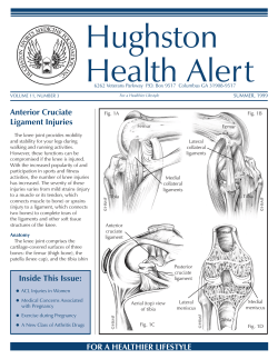


© Copyright 2026