
Small-Volume Resuscitation from Hemorrhagic Shock by
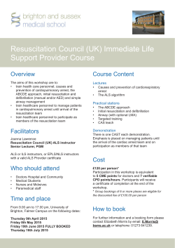
Surgical Research – World of Microcirculation
Eur Surg Res 2002;34:138–144
Small-Volume Resuscitation from Hemorrhagic
Shock by Hypertonic Saline Dextran –
Conceptional Basis and Historical Background
Uwe Kreimeier a, b Stephan Prueckner a, b
a Department of Anesthesiology and b Institute for Surgical Research, Ludwig-Maximilians-University Munich,
Grosshadern, Munich, Germany
Key Words
Hemorrhagic shock W Volume therapy W Hypertonic
saline W Dextran W Regional blood flow W Microcirculation W
Endothelium
Introduction
Early and adequate fluid resuscitation is essential for
the survival of the severely hypovolemic trauma patient.
Studies have shown that, besides age, severity of injury
and arterial hypotension, the duration of shock is the
most important factor influencing the recovery from multiple trauma [1]. While for optimal resuscitation rapid restoration of blood volume is essential, the lack of compatible blood or blood components for infusion at the accident site has directed therapy towards the use of blood
substitutes. The controversy regarding the relative merits
of crystalloids and colloids for primary volume therapy
has been exacerbated by conflicting results and, recently,
by cost considerations. The increasing involvement of
paramedics in the primary care of accident and emergency cases has highlighted the requirements for the ‘ideal’
solution for primary resuscitation. These are efficacy,
practicability and safety.
ABC
© 2002 S. Karger AG, Basel
0014–312X/02/0342–0138$18.50/0
Fax + 41 61 306 12 34
E-Mail [email protected]
www.karger.com
Accessible online at:
www.karger.com/journals/esr
Early reports showed that, on average, only 500 to
1,000 ml of any fluid is given prior to hospital arrival [2].
Due to the short transportation intervals in many sites
these data have not substantially changed. The crucial factor thus remains early and adequate resuscitation of the
patient from shock with its characteristic sequelae of
severely compromised microcirculation. This requirement has focused the interest in hypertonic saline/colloid
solutions which, when given as small-volume bolus infusions, have a pronounced effect on the cardiovascular system. The data from both experimental and clinical studies
suggest that these solutions should be particularly useful
for primary resuscitation in severe trauma.
Early Studies on Hypertonic Electrolyte
Solutions
The first report of the beneficial effects of administering isotonic saline solutions to patients suffering from
hemorrhagic shock was made by Penfield [3]. Later Silbert [4] showed that this treatment was also effective in
patients suffering from Buerger’s disease. The first investigation of the clinical efficacy of small-volume bolus infusions of hypertonic saline solutions was made by De
Felippe et al. [5], who reported on the effect of repetitive
intravenous injections of 50-ml aliquots of 7.5% sodium
Priv.-Doz. Dr. med. U. Kreimeier
Department of Anesthesiology, Ludwig-Maximilians-University Munich, Grosshadern
Marchioninistrasse 15, D–81377 München (Germany)
Tel. +49 89 7095 6412, Fax +49 89 7450 2817
E-Mail [email protected]
chloride into 12 patients suffering from hypovolemic
shock which had been refractory to vigorous volume
replacement and corticosteroid therapy as well as to catecholamines. They noted that infusions of 100–400 ml of
the hypertonic sodium chloride solution resulted in an
immediate rise of arterial pressure, resumption of urine
flow and recovery of consciousness. The beneficial effects
of these infusions tended to persist for several hours, during which it was noted that the volume of isotonic fluids
required to sustain the new state was reduced by 90%
compared to patients resuscitated with conventional isotonic fluids. After this prompt reversal of shock 9 of the 12
patients were ultimately discharged from hospital.
De Felippe’s report [5] on the clinical application of
small-volume infusion followed on a series of studies performed on animals which had been submitted to hemorrhagic shock. In one such study, Brooks et al. [6] investigated the efficacy of 2.74% sodium bicarbonate and 1.8%
NaCl solutions in preventing tissue damage in mongrel
dogs which had been subjected to 90 min hemorrhagic
hypotension (mean arterial pressure, MAP = 40 mm Hg).
When resuscitated with either of the two solutions, the
dogs made rapid recoveries; furthermore the workers
found no evidence of tissue lesions. Reinert [7] used
hypertonic solutions to resuscitate rats from ‘irreversible’
hemorrhagic shock (MAP = 30 mm Hg for 180 min). He
found that on reinfusing 30–40% of the shed blood volume together with either 6–7 ml of 1.8% or 1.25–1.5 ml of
10% NaCl (i.e. equivalent amounts of solute delivery in
each case), the survival rates were 5/6 and 2/5 animals,
respectively. The authors concluded that, besides solute,
sufficient fluid had to be provided.
Baue et al. [8] were the first to use hypertonic solutions
combined with a colloid for the initial stage of post-hemorrhagic resuscitation. In experiments on beagles they
used NaHCO3, NaCl and dextrose as crystalloid solutes
and dextran as colloid. They demonstrated that the increase in cardiac output and mesenteric flow, measured
using electromagnetic flow probes, which occurred after
primary resuscitation was related to the concentration of
the solute administered. They also noted that the hematocrit fell after infusion of the hypertonic solutions, and that
the extent of the hemodilution was related to the amount
of solute delivered. On the basis of these observations the
authors suggested that both intravascular volume and
blood flow were increased through the mobilization of
body water (‘autotransfusion’).
The use of small volume of highly concentrated sodium chloride (2,400 mosmol/kg) was investigated by Velasco et al. [9]. They used dogs subjected to hemorrhagic
hypotension (MAP = 40 mm Hg for 30 min). They found
that the infusion of 4 ml/kg body weight of 7.5% NaCl,
given without any further volume substitution, rapidly
restored both MAP and acid-base equilibrium and resulted in indefinite survival of all animals, whereas all ten
control animals receiving identical amounts of isotonic
saline died. Rocha e Silva et al. [10] studied the survival
rates as well as hemodynamic and metabolic effects of
resuscitation with hypertonic solutions containing either
sodium salts, chloride and/or non-electrolytes. They
found that plasma sodium but not plasma osmolality was
significantly correlated with survival, and concluded that
high plasma sodium was essential for survival. These
data, however, were at variance with the earlier observations of Messmer [11], who had demonstrated that the
beneficial effect of hypertonic solutions (1,200 mosmol/
kg) in rats subjected to hemorrhagic shock (MAP =
30 mm Hg for 240 min) depended upon the tonicity but
not upon the concentration of sodium ions in the solution
per se.
McNamara [12] found that intravenous injections of
50% glucose solution (1 ml/kg body weight) during hypovolemic shock in rabbits raised MAP significantly and
improved long-term survival compared to the survival
achieved by similar osmolar loads of either normal saline
or 25% mannitol solution. In the light of the widespread
use of Ringer’s lactate solution for resuscitation, Cone et
al. [13] investigated the efficacy of resuscitating with a
hypertonic lactate saline solution (sodium 250 mM, chloride 100 mM, lactate 150 mM) using conscious dogs
which had been subjected to acute, major blood loss. They
found that animals treated with hypertonic lactate/saline
solution required smaller volumes of fluid to normalize
and then maintain cardiac output and MAP, and that
these animals sustained a better urine output. It was also
noted that, despite rapid infusion, neither significant nor
protracted plasma hyperosmolality was observed.
Traverso et al. [14] compared the effects of administration of various concentrations of sodium chloride solutions (0.9, 5, 7.5, and 10%) in unanesthetized pigs which
had been subjected to severe hemorrhage. The fluid volume substituted was 14 and 25% of the shed blood, which
was equivalent to 8 and 14 ml/kg body weight, respectively. They found that 7.5% NaCl solution given at 14 ml/kg
body weight produced significantly better survial rates
compared to the other concentrations studied. By contrast, the use of 10% NaCl was associated with early
death, which might have been due to the negative inotropic effect of rapid infusion of extremely hypertonic solutions.
Small-Volume Resuscitation from
Hemorrhagic Shock
Eur Surg Res 2002;34:138–144
139
Role of Hypertonic Dextran Solutions
The duration of the cardiovascular response to the
infusion of hypertonic dextran solutions was investigated
by Smith et al. [15]. They studied the responses to six solutions (NaCl, NaCl/NaHCO3, NaCl/sodium acetate, NaCl/
mannitol, NaCl/6% dextran 70, glucose) each with a
tonicity of 2,400 mosmol/kg in conscious sheep. Their
results suggested that animals subjected to standardized
hemorrhagic shock were best resuscitated when they received a single bolus infusion of 7.5% NaCl/6% dextran
70. In these animals, the cardiac output remained at significantly higher levels during the 3-hour observation
period. These observations were confirmed by Kramer et
al. [16], who demonstrated that following small-volume
resuscitation of severely hemorrhaged conscious sheep by
infusion of 200 ml of hypertonic saline/dextran (2,400
mosmol/kg, 6% dextran 70, which is slightly hyperoncotic), both cardiovascular and metabolic functions were fully restored for at least 30 min post-resuscitation. Furthermore, the total volume requirements were significantly
lower than those needed when using normal saline for primary resuscitation.
Maningas et al. [17] also noted the efficiency of the
hypertonic saline/dextran 70 mixture. In a study on conscious pigs they observed that after the removal of 46 ml
of blood per kg body weight over a period of 15 min,
replacement of 25% of the shed volume by 7.5% NaCl/6%
dextran 70 resulted in a 100% survival rate (over a 96hour observation period).
The efficacy of administering colloids for prolongation
of the circulatory effects which had been initiated by
hypertonic saline is not restricted to the use of dextran 60
or 70. Hetastarch has been shown to be effective both in
experimental hypovolemic and endotoxin shock [18, 19].
Results from one of these studies in which the effects of
using 7.5% saline/6% dextran were compared to those
obtained resuscitating with 7.5% saline/6% hetastarch
showed that both colloids maintained cardiovascular
function for a longer period than hypertonic saline alone
[18]. It was also observed that the hypertonic saline/dextran solution gave a slightly better response than the solution containing hetastarch. The difference was attributed
to the possibility that dextran gave a higher effective
oncotic pressure and was cleared more slowly from the
intravascular space.
In studies using a canine model, developed to parallel
the normal hemorrhagic resuscitation regimen, Kreimeier
and Messmer [20] studied the effects of hypertonic (7.2%)
saline, hyperoncotic (10%) dextran 60, and the combina-
140
Eur Surg Res 2002;34:138–144
tion of the two solutes on lung function, central hemodynamics and nutritional blood flow. Even though only
small volumes of the selected fluid (4 ml/kg body weight),
which was equivalent to 10% of the shed blood, were
injected intravenously, within 5 min restoration of macro- and microhemodynamics were observed. The addition of 10% dextran 60 to the 7.2% saline solution
resulted in higher cardiac output and oxygen delivery to
the tissues as compared to hypertonic saline alone.
In 1988, in this journal Kreimeier et al. [21] reported
on most favorable results obtained from a detailed analysis of the effects of hypertonic saline as well as hypertonic
saline/hyperoncotic dextran 60 solution on regional blood
flow when given as primary resuscitation from hemorrhage. Regional blood flow was determined in 11 organs
by means of radioactively labeled microspheres 15 Ìm in
diameter. 18 beagles under anesthesia and controlled ventilation were bled to a MAP of 40 mm Hg, which was
maintained for 45 min. Then 10% of the shed blood volume were substituted intravenously within 2 min by either 7.2% saline alone, 10% dextran 60 alone, or a combined hypertonic-hyperoncotic solution containing 7.2%
saline/10% dextran 60. The data revealed that already
5 min after the infusion of 4 ml/kg body weight of both
hypertonic solutions, cardiac output had increased
beyond the initial control values, while peripheral vascular resistance was significantly decreased. Stroke volume
index was highest upon hypertonic-hyperoncotic saline/
dextran resuscitation. MAP reached 70 mm Hg, corresponding to 60% of control and did recover during further
volume substitution. At the end of the hypotension period, the regional blood flow had significantly decreased in
all organs except brain, heart and adrenal glands. 5 min
after bolus injection of either of the three test solutions,
however, blood flow to the brain, adrenal glands and
colon had increased above baseline, while control values
were regained in kidneys, small intestine, liver and thyroid gland. Moreover, myocardial blood flow was 2–3
times the baseline values and had increased in all regions
of the heart investigated. Blood flow to the pancreas and
gastric mucosa remained diminished (fig. 1). The total
peripheral arteriovenous shunt calculated for 15-Ìmdiameter microspheres decreased during hypotension and
remained low upon primary volume substitution by
means of either of the three solutions under investigation
(14–20% of cardiac output), indicating resumption of
nutritional blood flow in general. For the first time, these
results thus impressively documented the instantaneous
restoration of both the macro- and microhemodynamics
with normalization of nutritional blood flow within 5 min
Kreimeier/Prueckner
after primary resuscitation by means of hypertonic saline/
hyperoncotic dextran solution [21].
Specific Effect of Hypertonic Saline/Colloid
Solution on Post-Ischemic Leukocyte/
Endothelial Interaction
Chemotactic accumulation of circulating leukocytes
and their adhesion to the endothelial lining of postcapillary venules have long been recognized as key features of
post-ischemic reperfusion injury [22, 23]. The molecular
mechanisms of leukocyte adhesion to endothelial cells in
response to hyperosmolarity have been investigated by
Thiel et al. [24] in vitro. Incubation of isolated leukocytes
in buffer solutions with increasing osmolarity seems to
counteract the FMLP-induced upregulation of ß2-integrins in a dose-dependent manner. At the same time
FMLP-induced shedding of L-selectin is reduced, suggesting that these changes are not due to a nonspecific artifact,
such as effects of the hypertonic medium on epitope-antibody recognition [25]. When compared to physiologic
osmolarity (290 mM ), FMLP-induced CD18 upregulation and L-selectin shedding were significantly reduced at
osmolarities that are typically found in humans and animals after resuscitation with hypertonic solutions.
Special pharmacologic properties exist and speak for
the combined use of hypertonic saline together with a colloid: Nolte et al. [26] published their data on the leukocyte/endothelial interaction of hypertonic-hyperoncotic
dextran solution after ischemia/reperfusion injury in the
hamster dorsal skin fold model. The authors demonstrated that after 4 h of ischemia and reperfusion of
striated muscle the number of leukocytes adhering to the
endothelium of postcapillary venules was significantly
reduced for 24 h after bolus infusion of 7.2% NaCl/10%
dextran 60 and hypertonic saline/dextran effectively attenuated macromolecular leakage and reduced capillary
endothelial swelling as assessed by measurements of capillary luminal diameters. The hypertonic saline alone was
significantly less efficient in protecting from post-ischemic leukocyte/endothelial interaction and its sequelae.
The impact of the synthetic colloid dextran is also
known from hemodilution experiments: Already very
small amounts of dextran have been shown to be capable
of reducing reperfusion injury after a period of shock and
low/no flow in the microcirculation [27, 28]. In the same
animal model, post-ischemic leukocyte accumulation and
adherence were found to be significantly attenuated following prophylactic isovolemic hemodilution with 6%
Small-Volume Resuscitation from
Hemorrhagic Shock
Fig. 1. Changes in regional blood flow in gastric mucosa, small intes-
tine and pancreas during hemorrhagic hypotension and after smallvolume resuscitation by means of hypertonic saline/hyperoncotic
dextran 60. i = 7.2% NaCl/10% dextran 60: HHS; Z = 10% dextran
60: HDS; W = 7.2% NaCl: HSS. Median, q1-/q3-quartile, * p ! 0.05
vs. 7.2% NaCl/10% dextran 60.
dextran 60 to a hematocrit of 30% – a value clinically relevant in the trauma patient – when compared to controls
[29]. This effect was absent when using 6% hydroxyethyl
starch, and the authors postulated a compound-specific
pharmacologic effect of dextran.
Basic Mechanisms of Action
Intravenous bolus application of hypertonic sodium
chloride results in a rapid and pronounced increase of the
plasma sodium concentration and thereby initiates a
steep transmembranous osmotic gradient. The most important mechanism of action of hypertonic saline is the
instantaneous mobilization of endogenous fluid along the
osmotic gradient with increase of intravascular volume
[30, 31] (fig. 2). In addition, direct myocardial stimula-
Eur Surg Res 2002;34:138–144
141
Fig. 2. Endogenous fluid shift induced by hyperosmolarity: The
intravascular volume gain primarily stems from the endothelial cell
lining in response to the rapid and pronounced increase of the plasma
sodium concentration and thereby steep transmembranous osmotic
gradient. IVS = Intravascular space, ICS = intracellular space,
PMNL = polymorphonuclear leukocyte.
tion, CNS stimulation, neurogenic reflex mechanisms,
enhanced sympathetic discharge, hormone release, improvement of blood fluidity, re-establishment of spontaneous arteriolar vasomotion, and peripheral arterial vasodilatation have been discussed as mechanisms of action
[30, 32]. Data from experimental studies in dogs with
burn injury [33] and in pigs resuscitated from hemorrhagic shock [34] have failed, however, to confirm a direct
myocardial stimulating effect. The authors were unable to
demonstrate significant changes in the end-systolic pressure-volume relationship, and stroke work-end-diastolic
volume relationship [33], or end-systolic elastance and
segmental preload recruitable stroke work [34] upon hypertonic saline bolus infusion. Both groups of investigators attribute the circulatory effect to rapid augmentation
of ventricular preload and a reduction of afterload.
Vassar and Holcroft [35], on the basis of their broad
clinical experience in the field of small-volume resuscitation, estimate that administration of 250 ml 7.5% NaCl/
6% dextran 70 to a 70-kg patient who has suffered a 2-liter
blood loss will result in plasma volume expansion of at
least 700 ml. To achieve equivalent plasma volume expansion with lactated Ringer’s solution, these authors
estimate 2.8 liters of solution to be necessary. Mazzoni et
al. [31] have calculated that, after a 20% blood loss, 7.5%
saline solution given over 10 s in an amount equivalent to
1/7 of the actual blood loss allows to re-establish normal
blood volume within 1 min. These authors ascribe the
instantaneous circulatory effect to the rapid influx of fluid
142
Eur Surg Res 2002;34:138–144
first of all from the microvascular endothelium and red
blood cells [31, 36]. This fluid shift effect is most pronounced in those capillary districts with swollen endothelium: the more swollen the endothelium, the greater is the
effect of hypertonic solutions in reducing hydraulic resistance and improving tissue perfusion [31]. In another
study, Mazzoni et al. [37] investigated the volume
changes of endothelial cell monolayers on exposure to anisotonic media, from which can be deduced that the
increase in plasma osmolality to 460 mosmol/kg – which
is transiently obtained at the end of bolus infusion of 7.5%
saline [31] – should result in shrinkage of endothelial volume by 20%.
Further results from animal studies have suggested
that the rapid cardiovascular response to hypertonic solutions with increase of cardiac output and restoration of
peripheral blood flow might, at least in part, be mediated
by the instantaneous release of eicosanoids [38] with an
enhanced 6-keto-PGF1·/thromboxane B2 ratio [39]. This
may also explain the characteristic, acute hypotensive
response observed following rapid hypertonic saline infusion in dogs, which is not mediated by cardiac depression,
but by a decrease in total peripheral resistance [40]. Since,
from studies of endothelial cells grown on beads, the link
between capillary flow (shear-stress) and the production
of nitric oxide has been emphasized [41], the involvement
of NO in this process seems most likely.
The neuroendocrine response to resuscitation with
7.5% NaCl/6% dextran 70 following hemorrhagic hypotension has been quantified in conscious pigs [42]. Following small-volume resuscitation plasma ACTH, cortisol and aldosterone levels decreased, this effect, however,
being primarily due to hemodilution associated with the
expansion of plasma volume. In contrast, the reduction in
plasma norepinephrine, epinephrine, lysine, vasopressin
and plasma renin concentration was greater than attributed to hemodilution alone, which indicates also a role of
altered hormone release.
Resumé and Clinical Impact
The concept of small-volume resuscitation – the rapid
infusion of a small dose (4 ml/kg body weight) of 7.2–
7.5% NaCl/colloid solution – has been advocated for initial therapy of severe hypovolemia and shock almost two
decades ago. It is based on the instantaneous mobilization
of endogenous fluid along the osmotic gradient from
intracellular into the intravascular compartment. For primary volume resuscitation from severe hypovolemia and
Kreimeier/Prueckner
shock this is attractive for reasons of rapid mobilization of
endogenous water, especially from the intracellular compartment representing a huge reservoir amounting to
about 25 l of fluid. In addition, during shock and ischemia
the endothelial cell volume in particular increases due to
loss of ATP of the cells and cell membrane exchange dysfunction, leading to accumulation of water in the cells.
Thus any mobilization of water from this intracellular site
bears two important advantages: (1) plasma volume is
rapidly increased to 3- to 4-fold the volume infused; (2) by
normalization of the endothelial cell volume the luminal
diameter of the microvessels increases, consequently the
microcirculatory blood flow.
In the preclinical scenario this is attractive with regard
to the small infusion volume needed to elicit an instantaneous cardiovascular effect without the risk of fluid overload. Moreover, this concept is unique with regard to the
specific mode of action at the microvascular level providing prompt restoration of nutritional blood flow. During
the 80s various research groups were able to demonstrate
in clinically relevant animal models of hemorrhagic as well
as traumatic shock that even in the presence of a 50%
blood loss a volume as small as 4 ml/kg of 7.2–7.5% NaCl is
sufficient to restore cardiac output and regional organ
blood flow to pre-shock values almost instantaneously,
while at the same time systemic arterial pressure is significantly increased. In prehospital trials, small-volume resuscitation by means of hypertonic saline/colloid solution
have shown favorable results in hypotensive trauma patients [43], in case of severe trauma requiring immediate
surgery [44], and in patients with head trauma with a Glasgow Coma Scale of 8 or less in the presence of hypotension
(systolic blood pressure !90 mm Hg) [45, 46].
Epilogue
Decades of studies have solved many of the problems
concerning the pathogenesis of hemorrhagic shock. In
1985 Schoenberg, Smedegard, Gerdin, Messmer and Ar-
fors presented the ‘uptake-model’ of hemorrhagic shock,
i.e. terminating the duration of hypotension when an
uptake volume of 15% of the animal’s shed blood had
been reached. They arrived at a reproducible mortality
rate of about 50% in mongrel dogs. We therefore thought
to have an excellent model for our studies investigating
the effects of primary resuscitation from traumatic hypotension by means of hypertonic saline colloid solution. At
that time Konrad Messmer was Director of the Department of Experimental Surgery at the Ruprecht Karls University in Heidelberg and I was in his team. Michael
Schoenberg and I started our first experimental series.
The necessity to use dogs stemmed from the fact that we
intended to measure regional organ blood flow by means
of the radioactive microspheres method, i.e. a large animal model was needed. Since the animal rights movement
led to regulations excluding animals not specifically bread
for research purposes from investigations, we decided to
use the beagle as experimental animal. The first experiments were disappointing, and nobody in the team understood why the uptake model didn’t work although validated so nicely by Michael Schoenberg’s research work in
Sweden. It turned out that there was one fact that proved
to be ‘the’ crucial factor: in contrast to former experiments, where mongrel dogs had been used, now we had
beagles which we submitted to the identical shock procedure. After some experiments (and some time, also) we
had to face the fact that the ‘uptake model’ didn’t work
properly in the beagle, since the outcome was far away
from being reproducible. We decided to continue our
research work in the beagle, but taking a fixed hypotension period, first 45 min of hemorrhagic hypotension
(MAP = 40 mm Hg), then 75 min of traumatic-hemorrhagic hypotension – and the model worked properly, as
documented in the literature [21, 47, 48]. However, our
initial experiments appeared to be of specific impact for
the development of standardized hemorrhagic shock
models in dogs and the beagle in particular. Finally Konrad Messmer came to the simple conclusion: ‘The beagle
is no dog!’
References
1 Baker CC, Oppenheimer L, Stephens B, Lewis
FR, Trunkey DD: Epidemiology of trauma
deaths. Am J Surg 1980;140:144–150.
2 Smith JP, Bodai BI, Hill AS, Frey CF: Prehospital stabilization of critically injured patients:
a failed concept. J Trauma 1985;25:65–70.
3 Penfield WG: The treatment of severe and progressive hemorrhage by intravenous injections.
Am J Physiol 1919;48:121–132.
Small-Volume Resuscitation from
Hemorrhagic Shock
4 Silbert S: The treatment of thromboangiitis
obliterans by intravenous injection of hypertonic salt solution: Preliminary report. JAMA
1926;86:1759–1761.
5 De Felippe J Jr, Timoner J, Velasco IT, Lopes
OU: Treatment of refractory hypovolaemic
shock by 7.5% sodium chloride injections. Lancet 1980;ii:1002–1004.
6 Brooks DK, Williams WG, Manley RW,
Whiteman P: Osmolar and electrolyte changes
in hemorrhagic shock. Hypertonic solutions in
the prevention of tissue damage. Lancet 1963;
i:521–526.
7 Reinert M: Die Wirkung hypertonischer Kochsalzlösung im irreversiblen hämorrhagischen
Schock der Ratte. Zeitschr Kreislaufforsch
1966;55:134–137.
Eur Surg Res 2002;34:138–144
143
8 Baue AE, Tragus ET, Parkins WM: A comparison of isotonic and hypertonic solutions and
blood flow and oxygen consumption in the initial treatment of hemorrhagic shock. J Trauma
1967;7:743–756.
9 Velasco IT, Pontieri V, Rocha e Silva M Jr,
Lopes OU: Hyperosmotic NaCl and severe
hemorrhagic shock. Am J Physiol 1980;239:
664–673.
10 Rocha e Silva M, Velasco IT, Nogueira da Silva
RI, Oliveira MA, Negraes GA: Hyperosmotic
sodium salts reverse severe hemorrhagic shock:
other solutes do not. Am J Physiol 1987;253:
H751–H762.
11 Meßmer K: Die Wirkung hypertoner Lösungen
bei Ratten im irreversiblen Schock. Anaesthesist 1968;17:295–299.
12 McNamara JJ, Mills D, Aaby GV: Effect of
hypertonic glucose on hemorrhagic shock in
rabbits. Ann Thorac Surg 1970;9:116–121.
13 Cone JB, Wallace BH, Caldwell FT Jr, Smith
SD, Searcey R: Beneficial effects of a hypertonic solution for resuscitation in the presence
of acute hemorrhage. Am J Surg 1987;154:
585–588.
14 Traverso LW, Bellamy RF, Hollenbach SJ,
Witcher LD: Hypertonic sodium chloride solutions: Effect on hemodynamics and survival
after hemorrhage in swine. J Trauma 1987;27:
32–39.
15 Smith GJ, Kramer GC, Perron P, Nakayama S,
Gunther RA, Holcroft JW: A comparison of
several hypertonic solutions for resuscitation of
bled sheep. J Surg Res 1985;39:517–528.
16 Kramer GC, Perron PR, Lindsey DC, Ho HS,
Gunther RA, Boyle WA, Holcroft JW: Smallvolume resuscitation with hypertonic saline
dextran solution. Surgery 1986;100:239–247.
17 Maningas PA, de Guzman LR, Tillman FJ,
Hinson CS, Priegnitz KJ Volk KA, Bellamy
RF: Small-volume infusion of 7.5% NaCl in
6% Dextran 70 for the treatment of severe
hemorrhagic shock in swine. Ann Emerg Med
1986;15:1131–1137.
18 Walsh JC, Perron PR, Holcroft JW, Gunther
RA, Kramer GC: Comparison of resuscitation
of hypovolemia using hypertonic saline-dextran and hypertonic saline-hetastarch. Circ
Shock 1988:24:248.
19 Vincent JL, Armistead CW, Preiser JC, Thuc
IM, De Backer D: Hypertonic hydroxyethylstarch for fluid resuscitation in endotoxin
shock. Circ Shock 1988;24:203.
20 Kreimeier U, Messmer K: New perspectives in
resuscitation and prevention of multiple organ
system failure; in Baethmann A, Messmer K
(eds): Surgical Research: Recent Concepts and
Results. Berlin, Springer, 1987, pp 39–50.
21 Kreimeier U, Brückner UB, Messmer K: Improvement of nutritional blood flow using hypertonic-hyperoncotic solutions for primary
treatment of hemorrhagic hypotension. Eur
Surg Res 1988;20:277–279.
22 Hernandez LA, Grisham MB, Twohig B, Arfors K-E, Harlan JM, Granger DN: Role of
neutrophils in ischemia-reperfusion-induced
microvascular injury. Am J Physiol 1987;253:
H699–H703.
144
23 Granger DN, Benoit JN, Suzuki M, Grisham
MB: Leukocyte adherence to venular endothelium during ischemia- reperfusion. Am J Physiol 1989;20:G683–G688.
24 Thiel M, Buessecker F, Eberhardt K, Chouker
A, Setzer F, Kreimeier U, Arfors KE, Peter K,
Messmer K: Effects of hypertonic saline on
expression of human polymorphonuclear leukocyte adhesion molecules. J Leukoc Biol
2001;70:261–273.
25 Lehr HA, Saetzler RK, Thiel M, Arfors KE:
Microvascular salvage by small volume resuscitation with hypertonic fluids: concepts and
facts; in Messmer K (ed): Compromised Perfusion. Prog Appl Microcirc. Basel, Karger, 1996,
vol 22, pp 167–180.
26 Nolte D, Bayer M, Lehr HA, Becker M, Krombach F, Kreimeier U, Messmer K: Attenuation
of postischemic microvascular disturbances in
striated muscle by hyperosmolar saline dextran. Am J Physiol 1992;263:H1411–H1416.
27 Steinbauer M, Harris A, Hoffmann T, Messmer K: Pharmacological effects of dextrans on
the postischemic leukocyte-endothelial interaction; in Messmer K (ed): Compromised Perfusion. Prog Appl Microcirc. Basel, Karger, 1996,
vol 22, pp 114–125.
28 Steinbauer M, Harris AG, Leiderer R, Abels C,
Messmer K: Impact of dextran on microvascular disturbances and tissue injury following
ischemia/reperfusion in striated muscle. Shock
1998;9:345–351.
29 Menger MD, Thierjung C, Hammersen F,
Messmer K: Dextran vs. hydroxyethylstarch in
inhibition of postischemic leukocyte adherence
in striated muscle. Circ Shock 1993;41:248–
255.
30 Kreimeier U, Messmer K: Small-volume resuscitation; in Kox WJ, Gamble J (eds): Fluid
Resuscitation. Baillière’s Clinical Anaesthesiology, vol 2. London, Baillière Tindall, 1988, pp
545–577.
31 Mazzoni MC, Borgstroem P, Arfors KE, Intaglietta M: Dynamic fluid redistribution in hyperosmotic resuscitation of hypovolemic hemorrhage. Am J Physiol 1988;255:H629–H637.
32 Kreimeier U, Frey L, Pacheco A, Messmer K:
Hypertonic volume therapy: A feasible regimen to contribute to the prevention of multiple
organ failure? in Faist E, Ertel W (eds): Host
Defense Dysfunction in Trauma, Shock and
Sepsis. Mechanisms and Therapeutic Approaches. Berlin, Springer, 1993, pp 831–841.
33 Suzuki K, Ogino R, Nishina M, Kohama A:
Effects of hypertonic saline and Dextran 70 on
cardiac functions after burns. Am J Physiol
1995;268:H856–H864.
34 Welte M, Goresch T, Frey L, Holzer K, Zwissler B, Messmer K: Hypertonic saline dextran
does not increase cardiac contractile function
during small volume resuscitation from hemorrhagic shock in anesthetized pigs. Anesth Analg
1995;80:1099–1107.
35 Vassar MJ, Holcroft JW: Use of hypertonichyperoncotic fluids for resuscitation of trauma
patients. J Intensive Care Med 1992;7:189–
198.
Eur Surg Res 2002;34:138–144
36 Mazzoni MC, Borgström P, Intaglietta M, Arfors KE: Capillary narrowing in hemorrhagic
shock is rectified by hyperosmotic saline-dextran reinfusion. Circ Shock 1990;31:407–418.
37 Mazzoni MC, Lundgren E, Arfors KE, Intaglietta M: Volume changes of an endothelial
cell monolayer on exposure to anisotonic media. J Cell Physiol 1989;140:272–280.
38 Marti-Cabrera M, Ortiz JL, Dura´ JM, Cortijo
J, Barrachina MD, Morcillo E: Hemodynamic
effects of hyperosmotic mannitol infusion in
anesthetized open-chest dogs: Modification by
cyclooxygenase inhibition. Research in Surgery
1991;3:29–33.
39 Rabinovici R, Yue TL, Krausz MM, Sellers TS,
Lynch KM, Feuerstein G: Hemodynamic, hematologic and eicosanoid mediated mechanisms in 7.5 percent sodium chloride treatment
of uncontrolled hemorrhagic shock. Surg Gynecol Obstet 1992;175:341–354.
40 Kien ND, Kramer GC, White DA: Acute hypotension caused by rapid hypertonic saline infusion in anesthetized dogs. Anesth Analg 1991;
73:597–602.
41 Buga GM, Gold ME, Fukuto JM, Ignarro LJ:
Shear stress-induced release of nitric oxide
from endothelial cells grown on beads. Hypertension 1991;17:187–193.
42 Wade CE, Hannon JP, Bossone CA, Hunt MM,
Loveday JA, Coppes RI, Gildengorin VL: Neuroendocrine responses to hypertonic saline/
dextran resuscitation following hemorrhage.
Circ Shock 1991;35:37–43.
43 Younes RN, Aun F, Ching CT, Goldenberg
DC, Franco MH, Miura FK, Santos SS, Sequeiros IMM, Rocha e Silva M, Fujimura I, Birolini
D: Prognostic factors to predict outocome following the administration of hypertonic/hyperoncotic solution in hypovolemic patients.
Shock 1997;7:79–83.
44 Mattox KL, Maningas PA, Moore EE, Mateer
JR, Marx JA, Aprahamian C, Burch IM, Pepe
PE: Prehospital hypertonic saline/dextran infusion for post-traumatic hypotension – the
U.S.A. multicenter trial. Ann Surg 1991;213:
482–491.
45 Vassar MJ, Fischer RP, O’Brien PE, Bachulis
BL, Chamber JA, Hoyt DB, Holcroft JW: A
multicenter trial for resuscitation of injured
patients with 7.5% sodium chloride: The effect
of added dextran 70. Arch Surg 1993;128:
1003–1013.
46 Wade CE, Kramer GC, Grady JJ, Fabian TC,
Younes RN: Efficacy of hypertonic 7.5% saline
and 6% dextran-70 in treating trauma: A metaanalysis of controlled clinical studies. Surgery
1997;122:609–616.
47 Kreimeier U, Brückner UB, Schmidt J,
Messmer K: Instantaneous restoration of regional organ blood flow after severe hemorrhage: Effect of small-volume resuscitation
with hypertonic-hyperoncotic solutions. J Surg
Res 1990;49:493–503.
48 Kreimeier U, Brückner UB, Niemczyk S,
Messmer K: Hyperosmotic saline dextran for
resuscitation from traumatic-hemorrhagic hypotension: Effect on regional blood flow. Circ
Shock 1990;32:83–99.
Kreimeier/Prueckner


© Copyright 2026