
GUIDELINES FOR ANTE-NATAL CARE AND SKILLED ATTENDANCE AT BIRTH
GUIDELINES FOR ANTE-NATAL CARE AND SKILLED ATTENDANCE AT BIRTH BY ANMs AND LHVs Maternal Health Division Ministry of Health & Family Welfare (Department of Family Welfare) Government of India Nirman Bhavan, New Delhi - 110 011 Maternal Health Division Department of Family Welfare Ministry of Health & Family Welfare Government of India 2005 GUIDELINES FOR ANTE-NATAL CARE AND SKILLED ATTENDANCE AT BIRTH BY ANMs AND LHVs PREFACE The need for bringing down maternal mortality rate significantly and improving maternal health in general has been strongly stressed in the National Population Policy 2000. India is committed to reducing maternal mortality ratio to less than 100 per 100,000 live births by the year 2010 from the current 407/100,000 live births (SRS, RGI, 1998). Maternal mortality in India continues to remain unacceptably high. The majority of births in India take place at home and a large proportion are assisted by unskilled persons. In such situations, women who experience life threatening complications may never receive the required life saving emergency services because of several factors including lack of skilled birth attendant at hand. The major causes of these deaths have been identified as hemorrhage (both ante and post partum), toxemia (Hypertension during pregnancy), anemia, obstructed labor, puerperal sepsis (infections after delivery) and unsafe abortion. Historical evidence on a global level demonstrates that Skilled Birth Attendance can effectively reduce maternal mortality and that a package of essential obstetric services provided close to the woman’s house in the event of an obstetric emergency are effective in reducing maternal mortality. The Skilled Birth Attendant is a person who can handle such obstetric exigencies and is also aware when the situation reaches a point beyond his/her capability and hence needs to be referred to a higher centre. In an effort to reduce maternal mortality Government of India has recently taken the decision to permit ANMs/LHVs/Staff Nurses to use certain drugs for specific situations in emergency obstetric care. In consultation with experts from the Federation of Obstetrics and Gynaecology of India and nursing professionals it has also been decided to permit ANM/LHV to perform simple procedures like active management of third stage of labour, use of partograph for diagnosis etc. before referral. The ANM/LHV will however, need to be trained in the requisite skills and empowered to be the skilled birth attendant. Under the forthcoming Reproductive and Child health Programme, Phase II, efforts have to be made for improving deliveries by skilled birth attendants to 100% by the year 2010. The guidelines for ante natal care and skilled attendance at birth for ANMs and LHVs have been developed to assist the health personnel to effectively provide the requisite services both quantitatively and qualitatively at the community level. I would like to acknowledge the efforts put by the Maternal Health Division in preparing the guidelines with the help and assistance of Federation of Obstetric and Gynaecological Societies of India, nursing professionals and other experts. I am sure this effort will go a long way in ensuring uniform and good quality obstetric services, particularly at the community level. (P. Hota) Secretary (Health & FW) Date: April, 2005. i ACKNOWLEDGEMENTS Reducing Maternal Mortality to less than 100 per 100,000 live births is a commitment enshrined in the National Population Policy – 2000. This entails putting in place strategies and interventions which would accelerate the rate of decline of Maternal Mortality. Promoting skilled attendance at birth is an important strategy that has been adopted as part of the Reproductive and Child Health Programme, Phase – II (2005-2010). Implementation of this strategy would mean empowering the Auxiliary Nurse Midwife, Lady Health Visitors and Staff Nurses not only in handling normal deliveries but also for actively managing the third stage of labour and providing the required emergency care before referring any woman who develops complication during pregnancy or child birth. Government of India have taken the decision to allow the above category of nursing personnel to use certain drugs in specific situations during pregnancy and child birth. They have also been permitted to perform simple procedures like removal of retained products etc. This has entailed obtaining regulatory permissions and also the involvement of professional bodies like Federation of Obstetricians and Gynecological Societies of India (FOGSI), Nursing Council of India (NCI) and participation of Technical Experts and Programme Managers. It would not have been possible to achieve this without the blessings of Shri P. Hota, Secretary, Ministry of Health & Family Welfare. He has been a constant source of guidance and support in this endeavor and I express my gratitude to him for the same. I am also thankful to Shri S.S. Brar, Joint Secretary (RCH) for his support. Invaluable guidance was provided by Dr. Ashwani Kumar, Drugs Controller General of India and I thank him for his willing and active co-operation. The decision to empower the Health Workers has now to be followed up by effective training and logistic inputs to make skilled attendance at every birth in the country, a reality. These guidelines have been prepared keeping this in mind. The assistance and active involvement of White Ribbon Alliance of India for this purpose has been very helpful. I would like to thank Dr. Bulbul Sood, Co-Chairperson and Dr. Aparajita Gogoi, National Coordinator, WRAI for the same. The FOGSI, Nursing Council of India and Trained Nurses Association of India have taken keen interest in the whole process including finalisation of these guidelines. I would like to place on record my deep sense of gratitude to these professional organizations. Technical expertise and other assistance in the preparation of these guidelines has been provided by WHO, UNFPA, UNICEF and John D & Catherine T MacArthur Foundation. I am particularly thankful to Dr. Arvind Mathur, National Professional Officer, WHO-India and Dr. Dinesh Aggarwal, Team Manager Technical Support Group & Technical Advisor Reproductive Health, UNFPA for their active involvement and help. A team of Experts from various fields met several times for finalizing these guidelines. While it may not be possible to individually acknowledge the role of everyone, I would like to place on record appreciation for their contribution to the whole effort. I acknowledge and place on record my sincere thanks to Dr. Anchita Patil, Consultant, Dr. Narika Namshum, Asstt. Commissioner (MH), Dr. H.P. Anand, Sr. Specialist in Safdarjang Hospital and Dr. (Mrs.) Vijay Zutshi, Sr. Specialist in LNJPN Hospital who handled the task of writing and editing these guidelines. Dr. Kamla Ganesh, Dr. D.K. Tank and Dr. Himanshu Bhushan, Asstt. Commissioner reviewed the whole guidelines and deserve special mention for their efforts. I acknowledge the help provided by Mrs. Rita Madaan and Shri Pradeep Kumar Sohpaul for their secretarial help in putting together the document. (Dr. V.K. Manchanda) Deputy Director General Maternal and Child Health Division Ministry of Health & Family Welfare 4th April, 2005. ii CONTENTS 1. Abbreviations and units 2. Introduction 3. Module 1: Management of normal pregnancy, labour and the postpartum period • Care during pregnancy-Antenatal care • Care during labour and delivery-Intrapartum care • Care after delivery-Postpartum care • Essential newborn care 4. Module 2: Management of common obstetric complications Management of complications during pregnancy, labour and delivery, and in the postpartum period 1. Vaginal bleeding 2. Convulsions 3. Hypertension and pre-eclampsia 4. Anaemia 5. Urinary tract infection 6. Premature rupture of membranes 7. Obstructed labour 8. Preterm labour 9. Foetal distress 10. Prolapsed cord 11. Retained placenta and placental fragments 12. Vaginal/perineal tears 13. Puerperal sepsis 14. Sore and cracked nipples 5. Module 3: Ensuring the quality of care • Community involvement • Counselling and supportive environment • Prevention of infection 6. Annexures A. Methods of examination I. How to measure blood pressure II. How to look for pallor III. How to measure fundal height IV. How to determine foetal lie and presentation V. How to auscultate for foetal heart sounds B. Procedures for conducting investigations I. How to measure haemoglobin II. How to test urine for the presence of protein III. How to test urine for the presence of sugar C. Procedures for various interventions I. How to prepare "clean" gloves II. How to insert an intravenous (IV) line and give IV fluids III. How to carry out controlled cord traction (CCT) IV. How to carry out uterine massage and expel clots V. Examination of the placenta, membranes and the umbilical cord iii Page iv vi 3 23 35 39 45 45 49 49 51 51 51 52 52 53 53 54 54 55 55 59 61 63 69 70 70 72 74 75 75 75 76 76 77 78 78 ABBREVIATIONS AND UNITS @ % ANC ANM APH ASHA AWW BP CBOs CCT CHC DDK e.g. EDD etc. FHR FHS FRU GoI h/o Hb Hg HIV HLD i.e. IFA IM Inj. IU IUCD IUD IUGR IV LAM LHV LMP MMR MO MoHFW MP MPW-F MTP NAMP NFHS NGO(s) ORS at the rate of per cent Antenatal care Auxiliary nurse-midwife Antepartum haemorrhage Accredited social health activist Anganwadi worker Blood pressure Community based organisation(s) Controlled cord traction Community health centre Disposable delivery kit for example Expected date of delivery et cetera Foetal heart rate Foetal heart sound First referral unit Government of India History of Haemoglobin Mercury Human immunodeficiency virus High level disinfection that is Iron-folic acid Intramuscular Injection International units Intrauterine contraceptive device Intrauterine death Intrauterine growth retardation Intravenous Lactational amenorrhoea method Lady health visitor Last menstrual period Maternal mortality ratio Medical officer Ministry of Health and Family Welfare Malarial parasite Multi-purpose worker - female Medical termination of pregnancy National Anti-Malaria Programme National Family Health Survey Non-governmental organization(s) Oral rehydration solution iv P/V PHC PHD PIH PNC PPH PROM RCH RGI RR RTI SBA SDM STI Tab. TBA TT U UTI vs. per vaginam Primary health centre High Level Disinfection Pregnancy-induced hypertension Postnatal care Postpartum haemorrhage Premature or prelabour rupture of membranes Reproductive and Child Health Registrar General of India Respiratory rate Reproductive tract infection Skilled birth attendant Standard days' method Sexually transmitted infection Tablet Traditional birth attendant Tetanus toxoid Units Urinary tract infection versus °C cc cm dl g kcal kg mg ml mm degree Celsius cubic centimetre centimetre decilitre gram kilocalories kilogram milligram millilitre millimetre v INTRODUCTION The maternal mortality ratio (MMR) in India is very high. The data given by the Registrar General of India for 1998 estimate the MMR to be around 407 per 100,000 live-births. Like everywhere else in the world, the five major direct obstetric causes of maternal mortality in India are haemorrhage, puerperal sepsis, hypertensive disorders of pregnancy, obstructed labour and unsafe abortions. Maternal anaemia is a major contributor to the "indirect" obstetric causes. While most of these causes cannot be reliably predicted, early detection and timely management can save many lives. Provision of emergency obstetric care is the answer to these problems. Every woman should be cared for by a skilled birth attendant (SBA) during pregnancy, childbirth and the postpartum period. The SBA is a person who can handle obstetric emergencies and is also aware when the situation reaches a point beyond his/her capability, and hence needs to refer the woman to a higher centre. Therefore, the presence of an SBA at every delivery, along with the availability of an effective referral system, can help reduce the maternal morbidity and mortality to a considerable extent. The guidelines given in this manual are meant for the auxiliary nurse-midwife (ANM), the multi-purpose health worker female (MPW-F), the lady health visitor (LHV), or any other paramedical health worker who is engaged in providing maternal care at the village level. The guidelines have been prepared keeping in mind that these workers would be providing care in a domiciliary setting or at the level of the subcentre. However, ANMs/LHVs can also use these guidelines while working at the primary health centre (PHC) or any other health care facility. The guidelines incorporate evidence-based best practices for the provision of skilled attendance during pregnancy and at birth by these providers. It is hoped that these guidelines will serve as reading material during training in the Reproductive and Child Health (RCH)-II Programme as reflected in the state programme implementation plans. We hope that the corresponding interventions, infrastructure and programmatic support will be in place so as to enable health personnel to adhere to these guidelines for the delivery of services. Programme managers and supervisors should monitor the implementation of these guidelines during their routine supervisory visits. These guidelines can also be used by non-governmental organizations (NGOs) and private sector health facilities engaged in the delivery of services under the RCH Programme. Women face several constraints in seeking care during pregnancy and childbirth. Lack of finances, transportation problems, unwilling husbands and family members whose permission is often required to go to a health centre, are some of the major social barriers for accessing care. As skilled providers, the health workers will not only need technical competence to provide care during pregnancy and childbirth, but will also have to address some of these problems. Mobilising community action for these issues will help the women access the services as and when required and would ultimately help in achieving the goal of ensuring the provision of Skilled Attendance during Pregnancy and Childbirth to all the women in India. vi CARE DURING PREGNANCY-ANTENATAL CARE KEY MESSAGES Good record-keeping assists in better case management and follow-up. Ensure ironfolic acid supplementation to every pregnant woman. Do not give a pregnant woman any medication during the first trimester, unless specifically indicated. Adequate rest and diet for the pregnant woman results in better maternal and neonatal outcomes. Effective antenatal care (ANC) can improve the health of the mother and give her a chance to deliver a healthy baby. Regular monitoring during pregnancy can help detect complications at an early stage before they become life-threatening emergencies. However, one must realize that even with the most effective screening tools currently available, one cannot predict which woman will develop pregnancyrelated complications. Hence, every pregnant woman needs special care. You must: Recognize that Every pregnancy is at risk. Ensure that ANC is used as an opportunity to detect and treat existing problems. Make sure that services are available to manage obstetric emergencies when they occur. Prepare pregnant women and their families for the eventuality of an emergency. The important components of ANC are discussed below. Early registration Timing of the first visit/registration The first visit or registration of a pregnant woman for ANC should take place as soon as the pregnancy is suspected. Every married woman in the reproductive age group should be encouraged to visit her health provider or inform you if she believes herself to be pregnant. Ideally, the first visit should take place in the first trimester (first three months of pregnancy), before or at the 12th week of pregnancy. However, even if a woman comes late in her pregnancy for registration, she should be registered, and care given to her according to the gestational age. Seek help Some pregnant women will come by themselves to the antenatal clinics that are organized by you. However, many may not come. You, as the health provider, will have to find them. Take the help of various community-based functionaries such as the anganwadi worker (AWW), the accredited social health activist (ASHA), the traditional birth attendant (TBA)/dai, depot holders, members of Mahila Mandals, self-help groups, the Panchayat and Village Health Committees, schoolteachers and other important people in the village, who are likely to be aware of the pregnant women in the village, and can help update your list. 3 Importance of early registration Early registration is required to: Assess the health status of the mother and to obtain baseline information on blood pressure (BP), weight, etc. Screen for complications early and manage them appropriately by referral as and where required. Help the woman recall the date of her last menstrual period (LMP). Give the woman the first dose of tetanus toxoid injection (Inj. TT) well within time (after 12 weeks of pregnancy). Help the woman access facilities for an early and safe abortion if she does not want to continue with her pregnancy. Be alert to the possibility that the abortion might be an attempt at female foeticide. (Refer to the Government of India [GoI] Ministry of Health and Family Welfare [MoHFW], Guidelines for the Medical Termination of Pregnancy [MTP].) Build a good rapport with the pregnant woman. You, as the provider of care during pregnancy, must give plenty of time to counsel the woman and her family. Start the woman on a regular dose of folic acid during the first trimester [see later in this Chapter under Folic acid supplementation]. Estimation of the number of pregnancies to be registered annually To estimate the expected number of pregnancies that should be registered with you annually, you must know the birth rate and the population size of the area under your jurisdiction. The expected number of live-births in a year in a given area can be calculated by multiplying the birth rate (per 1000 population) with the population of the area, and then dividing it by 1000. As some of the pregnancies may not result in a live-birth (i.e. abortions and stillbirths may occur), the expected number of live-births is an underestimation of the total number of pregnancies. Hence, a correction factor of 10% is required, i.e. add 10% to the figure obtained above. This will give the total number of expected pregnancies (see Box 1). As far as possible, you should use the local birth rate. If that is not known, the district-level, statelevel or national-level figures can be used (in that order of preference). Use the latest census report to know the exact population of the area under your jurisdiction. Box 1. Example for estimation of the number of pregnancies annually Birth rate = 25/1000 population Population under the subcentre = 5000 Therefore, Expected number of live-births = (25 x 5000) / 1000 = 125 births Correction factor = 10% of 125 (i.e. [10/100] x 125) = 13 Therefore, Total number of expected pregnancies in a year in that subcentre = 125+13 = 138 4 As a rule of thumb, in any given month, approximately half the number of pregnancies estimated above should be in your records. If the number of women registered with you is less than expected, then you should approach the community leaders and key people, as mentioned earlier, to ensure that more pregnant women are registered and come for ANC. It is possible that some women may be receiving ANC from the private sector. At least ensure that their names are mentioned in your antenatal register. Attach a note giving the name of the facility from where they are getting ANC. Estimation of the number of pregnant woman will also help you and your PHC in calculating the requirement of TT vaccine, ironfolic acid (IFA) tablets and disposable delivery kits (DDKs). Record-keeping For the purpose of record-keeping, the following must be done: An antenatal card should be duly completed for every woman registered by you. The card should be handed over to the woman. She should be instructed to bring the card with her for all subsequent check-ups/visits, and should also carry it along with her at the time of delivery. This information should also be recorded in your antenatal register. Antenatal check-up Number and timing of visits You must ensure that every pregnant woman makes at least 4 visits for ANC, including the first visit/ registration. These are sufficient and, for pregnancies without complications, studies have shown that additional visits do not improve the maternal or perinatal outcome. The first visit is recommended as soon as the pregnancy is suspected. This is meant for registration of the pregnancy and the first antenatal check-up. The second visit should be scheduled between the 4th and 6th month (around 26 weeks). The third one should be planned in the 8th month (32 weeks), and the fourth one in the 9th month (36 weeks). Preparing for antenatal care Before beginning each ANC clinic, ensure that all the required instruments/equipment, such as stethoscope, sphygmomanometer, weighing scale, inch-tape, are available and in working condition. You must greet every pregnant woman in a friendly manner at each visit. Listen to the womans problems and concerns and offer advice or refer to a higher-level health centre as appropriate. Remember, all women need social support during pregnancy. Confirm that the present pregnancy is wanted. If not, and the woman wishes to go in for an abortion, refer the woman to a 24-hour PHC providing safe abortion services, or to a first referral unit (FRU) (whichever is closer) as soon as possible. This is important especially during the first visit when MTP is still feasible. The antenatal examination should be conducted at a culturally appropriate place that allows privacy for conducting an abdominal palpation. All findings must be accurately recorded on the antenatal card, and in the antenatal register. History-taking During the first visit, a detailed history of the woman needs to be taken (i) to diagnose pregnancy (first 5 visit only, if required); (ii) to identify any complications during previous pregnancies that may have a bearing on the present one; (iii) to identify any medical or obstetric condition(s) that may complicate the present pregnancy (first and subsequent visits). While taking the history, the following questions must be put to a pregnant woman: Date of the last menstrual period Remember that the LMP refers to the FIRST day of the womans last menstrual period. Ensure that the woman, while telling you her LMP, is NOT referring to the date of the first MISSED PERIOD. This mistake will lead to a miscalculation of the gestational age and expected date of delivery (EDD) by 4 weeks. If the woman is unable to remember the exact date, encourage her to remember some major event/ festival, etc. which she might link with her LMP. A calendar with the Indian system of months, dates and local festivals might come in handy. If the exact date of the LMP is not known, and it is late in the pregnancy, ask for the date when the foetal movements were first felt. This is known as quickening and is felt at around 20 weeks of gestation. Also assess the fundal height to estimate the gestational age [see Annexure A. III: How to measure fundal height]. Calculate the EDD based on these, and make a special note in the records of these cases. If the woman has undergone a test to confirm the pregnancy, ask her the approximate date when it was done, and also after how many days of amenorrhoea. This will also assist you in estimating her LMP. You should also ask the woman if her menses were regular before she conceived. If they were regular, ask for the duration of the menstrual cycle. The LMP is used to calculate the gestational age at the time of check-up and the EDD. The following formula is based on the assumption that the menstrual cycle of the woman was regular before conception and it was a 2830 days cycle. EDD = LMP + 9 months + 7 days Age of the woman This is required as women below the age of 16 years or above 40 years have greater chances of having pregnancy-related complications. Order of the pregnancy Primigravidas and those who have had 4 or more pregnancies are at higher risk of developing complications during pregnancy and labour. Birth interval Research shows that women who have spaced their children less than 36 months apart have greater chances of delivering a premature and low birth-weight baby, with consequently increased risk of infant mortality. An interval of less than 2 years from the previous pregnancy or less than 3 months from the previous abortion increases the chances of the mother developing anaemia. 6 Symptoms during the present pregnancy You must ask for symptoms that might be causing the woman some discomfort, and also for symptoms that are indications of a complication arising. Ask the woman for the following symptoms in the present pregnancy: Symptoms indicating discomfort nausea and vomiting heartburn constipation increased frequency of urination Symptoms indicating that a complication may be arising fever vaginal discharge palpitations, easy fatiguability and breathlessness at rest generalized swelling of the body; puffiness of the face passing smaller amounts of urine vaginal bleeding decreased or absent foetal movements leaking of watery fluid per vaginam (P/V) Previous pregnancies It is essential to ask a woman about her previous pregnancies or obstetric history, especially if she had suffered from any complications. This is important as some complications may recur during the present pregnancy. Ask the woman about: the total number of earlier pregnancies and deliveries abortion(s) premature birth(s) stillbirth(s) or neonatal loss hypertensive disorders of pregnancy (if not known, ask for a history of convulsions in previous pregnancies) prolonged labour obstructed labour malpresentation, such as breech delivery antepartum haemorrhage postpartum haemorrhage assisted delivery (forceps or vacuum extraction) delivery by caesarean section birth weight of the previous baby any surgery on the reproductive tract (e.g. uterine surgery, cone biopsy, uterine perforation during an MTP, etc.) iso-immunization (Rh ve) in the previous pregnancy (ask her for the history of any costly injection given to her within 72 hours of her previous delivery) 7 Ask especially for notes of the previous pregnancy, if available. Box 2. Conditions under which a pregnant woman must be referred to a medical officer Refer the woman to the medical officer if her obstetric history reveals any of the following: * previous stillbirth or neonatal loss * history of three or more spontaneous consecutive abortions * birth weight of the previous baby <2500 g * birth weight of the previous baby >4500 g * hospital admission for hypertension or pre-eclampsia/eclampsia in the previous pregnancy * previous surgery on the reproductive tract * iso-immunization (Rh ve) in the previous pregnancy History of any systemic illness(es) Rule out any personal history of systemic illnesses such as high BP (hypertension) diabetes breathlessness on exertion, palpitations (heart disease) chronic cough, blood in the sputum, prolonged fever (tuberculosis) renal disease convulsions (epilepsy) attacks of breathlessness or dama (asthma) rashes jaundice Family history of systemic illness If the woman does not have any of the above-mentioned systemic illnesses, ask for a family history of hypertension, diabetes and tuberculosis. If present, such a history predisposes the woman to developing the same herself during pregnancy (e.g. hypertensive disorders of pregnancy, gestational diabetes, etc.). As pregnancy is a physiologically stressful period, it can unmask the underlying tendency to develop these disorders. In addition, ask for a family history of thalassaemia, or whether anybody in her family has received blood transfusions.You must also ask for a family history of delivery of twins and/or the delivery of an infant with congenital malformation, as the presence of such a history in the family increases the chances of the woman giving birth to a child with the same defect. History of drug intake or allergies It is important to find out if the woman is allergic to any drug, or if she is taking any drug that might be harmful to the foetus. Find out whether the woman had taken any treatment or drugs for infertility. If yes, then these women have a higher chance of having twins and other multiple pregnancies. History of intake of habit-forming or harmful substances Ask the woman if she takes tobacco (chewing or smoking) and/or alcohol. If yes, she needs to be counselled to discontinue them during pregnancy, as they harm the developing foetus. Even after the delivery, the woman should be advised to continue to abstain from taking alcohol and tobacco because it may cause other complications such as addiction and/or cancer. 8 Physical examination This activity will be nearly the same during all the visits. Initial readings may be taken as a baseline and compared with the later readings. General examination Weight A pregnant womans weight should be taken AT EACH VISIT. The weight taken during the first visit/ registration should be treated as the baseline weight. For the ANC clinics conducted by you at the village level, it is realized that you might find it difficult to carry the weighing scales provided to you. Hence, you are advised to borrow the weighing machine from the AWW. Normally, a woman should gain 911 kg during her pregnancy. After the first trimester, a pregnant woman gains around 2 kg every month or 0.5 kg per week. To calculate the expected weight gain since her previous visit, multiply the number of weeks elapsed since the previous visit by 0.5 kg. This should be compared with the actual weight gained. If the diet is not enough, with less than the required amount of calories, the woman might gain only 56 kg during her pregnancy. An inadequate dietary intake can be suspected if the woman has gained less than 2 kg per month. She needs to be put on food supplementation. You should take the help of the AWW for food supplementation, especially for those categories of women who need it the most [see under Counselling for Diet and Rest later in this Module]. A low weight gain usually points towards intrauterine growth retardation (IUGR) and results in a low birth-weight baby. Excessive weight gain (more than 3 kg in a month) should arouse the suspicion of pre-eclampsia/twins (multiple pregnancy). Take the womans BP, and test her urine to check if she has proteinuria. Refer the woman to the Medical Officer (MO). The following points should be kept in mind while taking the weight: * The weighing machine should be checked for zero error before taking the weight. * The woman should be wearing light clothing. * She should stand erect on the weighing machine, in such a way that her weight is evenly distributed on the platform. * The weight must be measured to the nearest 100 g. Blood pressure [See Annexure A. I: How to measure blood pressure] Measure the BP of pregnant women AT EVERY VISIT. This is important to rule out hypertensive disorders of pregnancy. If the BP is high (more than 140/90 mmHg; or diastolic more than 90 mmHg), check the BP again after 1 hour. If it is still high, check the womans urine for the presence of albumin, as the combination of a high BP and proteinuria is sufficient to categorize the woman as having pre-eclampsia. Refer her to the MO. If the diastolic BP of the woman is above 110 mmHg, it is a danger sign pointing towards imminent eclampsia. Such a woman must be referred to the community health centre (CHC)/FRU IMMEDIATELY. A woman with pregnancy-induced hypertension (PIH)/pre-eclampsia requires hospitalization at a PHC/FRU. 9 Pallor [See Annexure A. II: How to look for pallor] Pull down the lower eyelid and look at the lower palpebral conjunctiva, and also the palms and nails, the oral mucosa and tongue of the woman for the presence of pallor. If present, it is an indication that the woman is anaemic. Respiratory rate (RR) It is important to check the RR, especially if the woman complains of breathlessness. If the RR is more than 30 breaths/minute and pallor is present, it indicates that the woman has severe anaemia and needs immediate referral to the MO. If the RR of the woman is more than 30 breaths/minute, and she has other associated medical problems, refer her to the MO for further investigation and management of any systemic illness, if present. Generalized oedema The presence of generalized oedema or puffiness of the face should arouse the suspicion of pre-eclampsia. Abdominal examination Examine the abdomen to monitor the progress of pregnancy and foetal growth, and to check the foetal lie and presentation. Fundal height [See Annexure A. III: How to measure fundal height] This indicates the progress of the pregnancy and foetal growth. The uterus becomes an abdominal organ after 12 weeks of gestation. The gestational age (in weeks) can be estimated from the fundal height (in cm) after 24 weeks of gestation. If there is any disparity between the fundal height and the gestational age as calculated from the LMP, the woman should be referred to the MO. If there is a difference of 3 cm or more, or if there is no growth compared to the previous check-up, these are considered significant signs, and the woman requires further investigations. If the height of the uterus is more than that indicated by the period of amenorrhoea, the possible reasons could be: * wrong date of LMP * full bladder * multiple pregnancy * polyhydramnios * hydatidiform mole * pregnancy with a pelvic tumour If the height of the uterus is less than that indicated by the period of amenorrhoea, the possible reasons could be: * wrong date of LMP * IUGR * missed abortion * intrauterine death (IUD) * transverse lie 10 Foetal lie and presentation [See Annexure A. IV: How to determine foetal lie and presentation] Palpate for the foetal lie and assess whether it is longitudinal, transverse or oblique. Remember, even if a malpresentation is diagnosed before 36 weeks, no active management or intervention is recommended at that point of time. All health workers should be able to recognize a transverse lie. Missing it can be disastrous because there is no mechanism by which a woman with a transverse lie can deliver normally/vaginally. This woman needs a caesarean section, and hence should be referred to a health centre (FRU) where emergency obstetric services are available, including the facility for a caesarean section. Failure to do a timely caesarean section in this woman can lead to obstructed labour, rupture of the uterus and death of the woman. The foetal presentation should be checked, especially in the case of a longitudinal lie, to see whether the presenting part is the vertex (normal) or any other part of the cephalic end (face, brow), or a breech. At your level, it is sufficient if you are able to diagnose whether the presenting part is the cephalic end or the breech. Foetal heart sound (FHS) and rate [See Annexure A. V: How to auscultate for foetal heart sounds] If the foetal heart rate (FHR) is between 120 and 160 beats per minute, it is normal. Both foetal bradycardia (FHR less than 120 per minute) and foetal tachycardia (FHR more than 160 per minute) indicate foetal distress. If either of these is present, refer the woman to the MO. Remember that the FHS is not heard before 24 weeks of pregnancy; hence checking for the FHS should start only from the second visit. Multiple pregnancy This must be suspected if the following are present on examination: An unexpectedly large uterus for the estimated gestational age Multiple foetal parts are felt on abdominal palpation If a multiple pregnancy is suspected, refer the woman to the MO in the PHC for confirmation, and arrange for delivery in an institution. Breast examination Observe the size and shape of the nipples for the presence of inverted or flat nipples. Try and pull out the nipples to see if they can be pulled out easily. Flat nipples that can be pulled out do not interfere with breastfeeding. Truly inverted nipples might create a problem in carrying out successful breastfeeding. If present, the woman must be advised to pull on the nipples and roll them between the thumb and the index finger. Another technique for correcting inverted nipples includes the use of a 10 or 20 cc disposable plastic syringe. Cut the barrel of the syringe from the end where the needle is attached. Take out the plunger and put it in from the opposite end, which is the cut end of the syringe. Push the piston forward fully, and place the open end of the barrel so that it encircles the nipple and areola. Pull back the plunger thus creating negative pressure. The nipple will be sucked into the barrel, and thus be pulled out in the process. Crusting and soreness of the nipples must be looked for. If present, the woman must be advised regarding breast hygiene. If the nipples do not heal, refer the woman to the MO. The breasts must be palpated for any lumps or tenderness. If present, refer the woman to the MO. 11 Table 1. Symptoms and signs that an ANM might encounter, probable diagnosis and action required to be taken at the subcentre level Symptoms Signs/investigations Vomiting during the first trimester Most probable diagnosis May be physiological (morning sickness) Action(s) to be taken Advise the woman to eat small frequent meals; avoid greasy food; eat lots of green vegetables and drink plenty of fluids. If vomiting is excessive in the morning, ask her to eat dry foods such as biscuits or toast after waking up in the morning. Excessive vomiting, especially after the first trimester The woman may be dehydrated Hyperemesis gravidarum Refer the woman to the MO. Palpitations, easy fatiguability, breathlessness at rest Conjunctival and/or palmar pallor present Hb level <7 g/dl Severe anaemia Refer her to the MO for further management. Advise her to have a hospital delivery. Puffiness of the face, generalized body oedema BP >140/90 mmHg Proteinuria absent Hypertensive disorder of pregnancy Refer her to the MO. Refer to the MO for antihypertensive medication BP >140/90 mmHg Proteinuria present Pre-eclampsia Advise her on the danger signs of imminent eclampsia and eclampsia and refer to the MO. Reflux Advise the woman to avoid spicy and rich foods. Ask her to take cold milk during attacks. If severe, antacids may be prescribed. Heartburn and nausea Increased frequency of urination up to 1012 weeks of pregnancy Increased frequency of urination after 12 weeks, or persistent symptoms, or burning on urination Constipation Tenderness may be present at the sides of the abdomen and back Body temperature may be raised May be physiological due to pressure of the gravid uterus on the urinary bladder Urinary tract infection (UTI) Physiological 12 Reassure her that it will be relieved on its own. Give the woman the first dose of ampicillin (1 g orally) and injection gentamicin 80 mg IM stat). Refer the woman to the MO. Advise the woman to take more fluids, leafy vegetables and a fibre-rich diet. If not relieved, give her Isabgol, 2 tablespoonfuls to be taken at bedtime, with water or with milk. Do NOT prescribe strong laxatives as they may start uterine contractions. Symptoms Bleeding P/V, before 20 weeks of gestation Signs/investigations Most probable diagnosis Check the pulse and BP to assess for shock Action(s) to be taken Threatened abortion/ spontaneous abortion/ hydatidiform mole/ectopic pregnancy Bleeding P/V, after 20 weeks of gestation If woman is bleeding and the retained products of conception can be seen coming out from the vagina, remove them with your finger. Refer to the MO of a 24-hour PHC Ask for history of violence Spontaneous abortion due to violence Put her in touch with local support groups. Check the pulse and BP to assess for shock Antepartum haemorrhage Do NOT carry out a vaginal examination under any circumstances. Refer to an FRU. Site of infection somewhere, including possible sepsis Refer to MO. Body temperature is raised Blood peripheral smear is positive for malarial parasite Malaria Manage according to the NAMP guidelines for malaria in pregnancy Decreased or absent foetal movements (NOTE: foetal movements are felt only after about 4 months of gestation) FHS heard, and within the normal range of 120160/ minute Baby is normal Reassure the woman Foetal distress Repeat FHS after 15 minutes. If the FHS is still out of the normal range, refer to the MO. Fever FHS heard, but the rate is <120/minute or >160/ minute FHS not heard Intrauterine foetal death Vaginal discharge, with or without abdominal pain Leaking of watery fluids P/V Wet pads/cloths Inform the woman and her family that the baby might not be well. Refer to the MO. RTI/STI Refer the woman to the MO. Advise the woman regarding vaginal hygiene, i.e. cleaning the external genitalia with soap and water. Premature rupture of membranes Refer the woman to the MO. FRU: first referral unit; NAMP: National Anti-Malaria Programme; FHS: foetal heart sound; RTI: reproductive tract infection; STI: sexually transmitted infection; P/V: per vaginum 13 Remember it is not advisable to give a pregnant woman any medication during the first trimester, unless absolutely essential. Even then it must be ensured that the drugs given are proven to be safe when taken during pregnancy, and do not have effects on the foetus which cause disability (teratogenic). Laboratory investigations The following laboratory investigations are recommended at the primary health care provider level to be carried out as a part of ANC. Haemoglobin (Hb) estimation [See Annexure B. I: How to measure haemoglobin] Estimation of the level of haemoglobin is essential for the following: To check for the presence of anaemia and, if present, to what degree; For the further management, prevention and/or treatment of anaemia, in so far as the administration of IFA tablets is concerned [see below, under Ironfolic acid supplementation]. If the anaemia is severe, the woman may need referral for taking injectable iron preparations or undergo a blood transfusion. For the diagnosis of postpartum haemorrhage (PPH) in an anaemic woman, in whom a smaller amount of blood loss is taken as PPH. Estimate the Hb levels of pregnant women at the initial antenatal visit and again at 28 weeks. The initial Hb level will serve as a baseline to compare with the later results at 2830 weeks. An Hb level below 11 g/dl at any time in pregnancy is considered to be anaemia; an Hb level of 7 to 11 g/dl as moderate anaemia, and less than 7 g/dl as severe anaemia. If the woman is found to be anaemic, start her on the therapeutic dose of IFA [see below, under Iron folic acid supplementation]. Estimate the Hb level again after 1 month. If there is no rise in the Hb level, refer the woman to a higher facility with a good laboratory infrastructure and trained personnel to find out the cause of anaemia. Blood grouping Encourage the woman to go to the PHC and get her blood group tested. Knowing the blood group can be of great help in cases of haemorrhage, when precious time could be saved and, if required, blood transfusion could be started as soon as possible. It is also an essential prerequisite in case the woman wishes to go in for an MTP. Testing the urine for the presence of albumin [See Annexure B. II: How to test urine for the presence of protein] This is a test used in the definition of pre-eclampsia, which (along with eclampsia) is a very important cause of maternal mortality. This test can be carried out at the field level too. Testing the urine for the presence of sugar [See Annexure B. III: How to test urine for the presence of sugar] This is a test used to diagnose women with gestational diabetes. If a womans urine is positive for sugar, refer her to the MO at the PHC. 14 Interventions Folic acid supplementation If the woman is registered within the first trimester of pregnancy, she should be given folic acid supplementation for improving the growth of the foetal neural tissue. It is recommended that the woman be given 5 mg of folic acid once a day, till 12 weeks of pregnancy. After that, she is to be advised a combination of iron and folic acid (IFA) [see below under Ironfolic acid supplementation] Ironfolic acid (IFA) supplementation Stress the need for increased requirements of iron during pregnancy and the dangers of anaemia to pregnant women. All pregnant women need to be given one tablet of IFA (100 mg elemental iron and 0.5 mg folic acid) every day for at least 100 days, starting after the first trimester at 1416 weeks of gestation. This is the dose of IFA given to prevent anaemia (prophylactic dose). If a woman is anaemic (Hb <11 g/dl) or she has pallor, give her two IFA tablets per day for three months. This means a woman with anaemia in pregnancy needs to take at least 200 tablets of IFA. This is the dose of IFA needed to correct anaemia (therapeutic dose). Women with severe anaemia (Hb <7 g/dl) or those who have breathlessness and tachycardia due to anaemia, should be started on the therapeutic dose of IFA and also referred to the MO in the PHC for further management. Many women do not take IFA regularly due to some common side-effects. The necessity of taking IFA and the dangers associated with anaemia should be explained to the mother. Tell her: * Though the tablets should be taken preferably early in the morning on an empty stomach, she may take the tablets with meals or at night. This will help avoid nausea. * She should not worry if she passes black stools. This is normal while taking IFA tablets. * If she has constipation, she should drink more water. * These side-effects are not serious. * She should avoid taking the tablets with tea or coffee as they reduce the absorption of iron. * Tablets containing IFA may make her feel less tired than before. However, despite feeling better, she should not stop taking the tablets. * She should return to you if she has problems taking IFA tablets. Refer such women to the MO for further management. Injection tetanus toxoid (Inj. TT) administration Administration of two doses of Inj. TT to a pregnant woman is an important step in the prevention on neonatal tetanus (tetanus of the newborn). The first dose of TT should be given just after the first trimester, or as soon as the woman registers for ANC, whichever is later. This means that Inj. TT is NOT to be given in the first trimester of pregnancy. The second dose is to be given one month after the first dose, but at least one month before the EDD. [Refer to the GoIs National Immunisation Schedule for the same.] Inj. TT is to be given as 0.5 ml per dose, deep IM in the upper arm. Inform the woman that there may be slight swelling, pain and/or redness at the injection site for a day or two. 15 Malaria prophylaxis You are advised to follow the guidelines of the National Anti-Malaria Programme (NAMP) for malaria prophylaxis. At the time of printing of this document, the NAMP recommends that in malaria-endemic areas of India, pregnant women should be given intermittent malaria prophylaxis. Counselling Birth preparedness and complication readiness Four out of ten pregnant or postpartum women will experience some complication related to their pregnancy; for about 15% of these women, the complication will be potentially life-threatening and will require immediate emergency obstetric care. Since most of these complications cannot be predicted, every pregnancy necessitates preparation for a possible emergency. Birth preparedness Identification of a skilled provider for birth: All pregnant women should be helped to reach a decision regarding the health provider they want for conducting their delivery. An SBA should be preferred over an unskilled one. (Note that TBAs, trained or untrained, do not fall into the category of SBAs). Other factors such as the condition of the pregnancy (complicated or uncomplicated), the distance to the provider, transport facilities, financial situation, etc. all need to be kept in mind before finally reaching a decision about the choice of birth attendant. ALL PREGNANT WOMEN MUST BE ENCOURAGED TO OPT FOR AN INSTITUTIONAL DELIVERY. Explain to the woman why delivery at a health facility is recommended. Tell her that * Any complication can develop during delivery; complications are not always predictable; they can cost the life of the mother and/or the baby. * A health facility has staff, equipment, supplies and drugs available to provide the best care, if needed. It even has a referral system should the need to refer arise. Delivery kit: All pregnant women, especially when they are nearing completion of their term, should be equipped with supplies required for conducting the delivery at home, especially if the woman has decided to deliver at home. The kit is also required in case of emergencies, in case the woman cannot make it to the health facility in time, and is forced to deliver at home. If the woman, despite all arguments to the contrary, decides that she wishes to be delivered by the TBA you, as the health personnel providing ANC, must contact the TBA in question, and ensure that she knows how to conduct a clean delivery. If a delivery kit is not available, the following items should be made available individually to ensure the five "cleans" (i.e. Clean surface, Clean hands, Clean cord cut, Clean cord tie and Clean umbilical stump): * A clean plastic sheet (for ensuring "clean surface") * Soap and clean water (for ensuring "clean hands") * A new razor blade (for ensuring "clean cord cut") * A clean piece of thread (for ensuring "clean cord tie") * Nothing to be applied to cord ( for ensuring a "clean cord stump ") 16 The other items that are required during and immediately after delivery include: * * * * * Home-based antenatal card (for complete information regarding the antenatal period) Clean towels/cloth for washing, drying and wrapping the baby Clean clothes for the mother and the baby Sanitary pads/clean cloth for the mother Food and water for the woman and the support person. Identify support people: These people are needed to help the woman care for her children and/or household, arrange for transportation, and/or accompany the woman to the health facility in an emergency. Seek help from either the close relatives of the woman or community-based health functionaries such as the AWW and the TBA. Finances: The woman and her family should be given an estimate of the expected expenses for the delivery and related aspects (such as transport, etc.). They should also be advised to keep some emergency fund, or have a source for emergency funding, should a complication arise and more money is required than initially anticipated. You should also be aware of the existing schemes that provide funds for maternal health, and any other schemes that may be launched from time to time. Help the women and their families access these schemes and receive the allocated funds to pay for the delivery. Signs of labour: Advise the woman to go to the health facility or contact the SBA if she has any one of the following signs which indicate the start of labour: * A bloody, sticky discharge P/V * Painful abdominal contractions every 20 minutes or less * The bag of waters has broken, and she has clear fluid coming out P/V (leaking). Complication readiness Danger signs: The woman and her family/caretakers should be informed about potential danger signs during pregnancy, delivery and the postpartum period. She must be told that if she has any of the following during pregnancy, delivery or postpartum/post-abortion, she should immediately visit a hospital or health centre, WITHOUT WAITING, be it day or night. The woman should visit an FRU if she has any of the following conditions: * Any bleeding P/V during pregnancy, and heavy (>500 ml) vaginal bleeding during and following delivery * Severe headache with blurred vision * Convulsions or loss of consciousness * Labour lasting longer than 12 hours * Failure of the placenta to come out within 30 minutes of delivery * Preterm labour (labour starting before 8 gestational months) * Premature or prelabour rupture of membranes (PROM) * Continuous severe abdominal pain The woman should visit a 24-hour PHC if she has any of the following conditions: * * * * High fever with or without abdominal pain, and feels too weak to get out of bed Fast or difficult breathing Decreased or absent foetal movements Excessive vomiting, wherein the woman is unable to take anything orally, leading to a decreased urinary output 17 Location of the nearest PHC/FRU: The woman and her family members should be aware of the nearest health facility, both the PHC where 24-hour functioning emergency obstetric care services are available and the FRU, where facilities for a blood transfusion and surgery are available. Identification of transportation facilities: Delay in reaching a health care facility is one of the major delays responsible for maternal mortality. If the woman has decided to deliver at a health facility, a vehicle should be identified which should be available whenever the woman needs it, to take her to that health facility. Even if the woman decides to deliver at home, a vehicle should be identified and ideally be kept ready to transport her to the nearest health facility or referral centre in case she develops some complications that need immediate referral and care. The help of the Panchayat, Village Health Committee, Mahila Mandals, youth groups, or any other such groups can be taken to decide on how to obtain a vehicle in case of an emergency, if a vehicle is not available in the village. The various schemes which are presently available for assisting the woman with transportation facilities should be kept in mind. Also keep yourself updated regarding any new schemes that may be launched from time to time. Preparedness for blood donation: Haemorrhage, both antepartum and postpartum, is an important cause of maternal mortality. Blood transfusion can be life-saving in such cases. As blood cannot be bought one needs a voluntary donor to replace the blood before it is issued for transfusion. Such donors (23 in number) must be ready, should the need for transfusion arise. Diet and rest The woman should be advised to eat more than her normal diet throughout her pregnancy. Remember, a pregnant woman needs about 300 extra kcal per day compared to her usual diet. She should be told that she needs these extra calories for: * * * Maintenance of her health as a mother The needs of the growing foetus Successful lactation Special categories of women have been identified who should be given priority for additional nutrition during pregnancy. They include the following: * * * * * * Women with a reduction in the dietary intake below habitual levels during pregnancy Women who have an increased level of physical activity above the usual levels during pregnancy Women with a combination of both the above-mentioned factors Pregnancy in adolescent girls Pregnancy during lactation Pregnancy within two years of the previous delivery. The womans food intake should be especially rich in proteins, iron, vitamin A and other essential micronutrients. The other members of the family, especially those who take decisions regarding the type of food brought home and/or given to the pregnant woman, such as her husband and mother-in-law, should also be taken into confidence and counselled regarding the recommended diet for the pregnant woman. Encourage them to help ensure that the woman eats enough and avoids hard physical work. 18 Some of the recommended dietary items are cereals, milk and milk products such as curd, green leafy vegetables and other vegetables, pulses, eggs and meat, including fish and poultry (if the woman is a non-vegetarian), nuts (especially groundnuts), jaggery, fruits, etc. Give examples of the types of food, suggested preparations, if possible, and how much to eat. Tell her about the locally available foods rich in iron such as groundnuts and jaggery. Tell the woman to avoid taking tobacco, tea or coffee, especially within 1 hour of a meal, as they have been shown to interfere with the absorption of iron. Also advise her to take foods rich in proteins and vitamin C (e.g. lemon, amla, guava, oranges, etc.) as both help in the absorption of iron. The diet should be rich in fibre so that she does not have constipation. The diet should be advised keeping in mind the socioeconomic conditions, food habits and taste of the individual. Food taboos must be looked into while counselling the woman regarding her dietary intake. If there are taboos about nutritionally important foods, the woman should be advised against these taboos. In certain communities, food taboos (especially omissions) exist for sex selection of the foetus. These should be strongly discouraged. If a woman has PIH, she should be encouraged to eat a normal diet with no restrictions on fluid, calorie and/or salt intake; such restrictions do not prevent PIH from converting into pre-eclampsia, and may be harmful for the foetus. The woman should be advised to refrain from taking alcohol or smoking during pregnancy. The woman should be advised NOT to take any medication unless prescribed by a qualified health practitioner. The woman should be advised to sleep for 8 hours at night and rest for another 2 hours during the day. She should be told refrain from doing heavy work, especially lifting heavy weights, as it can adversely affect the birth weight of the baby. The other members of the household should be taken into confidence and advised to help the woman in carrying out her routine household chores. All pregnant women should be told to avoid the supine position, especially in late pregnancy, as it affects both the maternal and the foetal physiology. During pregnancy, the pressure exerted by the pregnant uterus on the main pelvic veins results in a reduced quantity of circulating blood reaching the right side of the heart. This causes a reduced supply of oxygen to the brain and can therefore lead to a fainting attack, a condition referred to as the supine hypotension syndrome. It can also result in abnormal foetal heart rate patterns, and may also cause a reduction in the placental blood flow. If the supine position is necessary, a small pillow under the lower back at the level of the pelvis is recommended. Infant and young child feeding Pregnancy is the ideal time to counsel the mother regarding the benefits of breastfeeding her baby. Though breastfeeding is almost universal in India, a few points need to be emphasized to the would-be mother. Initiation of breastfeeding: Counsel the mother that breastfeeding should ideally be initiated within half-an-hour of a normal delivery (or within two hours of a caesarean section, or as soon as the mother regains consciousness, in case she undergoes a caesarean section). It is common practice in India to delay initiation. Colostrum (the first milk) is thrown away, and prelacteal feeds are given instead. This has obvious disadvantages. First, the pre-lacteal feed may not be hygienic and can cause an intestinal infection in the baby. Second, the baby is deprived of colostrum which is very rich in protective antibodies. 19 Most importantly, the sucking and rooting reflex in the child, which are essential for the baby to successfully start breastfeeding, are the strongest immediately after delivery, making the process of initiation much easier for the mother and the baby. These reflexes gradually become weaker over the span of a few hours, thus making breastfeeding difficult later on. Exclusive breastfeeding for 6 months: It should be emphasized to the mother that only breast milk and nothing but breast milk should be given to the baby for the first 6 months, not even water. The mother should be assured that breast milk has enough water to quench the babys thirst (even in the peak of summer) and satisfy its hunger for the first 6 months. Take special care in the case of a female child to ensure that she is adequately breastfed and not discriminated against because of her sex. Demand feeding: This refers to the practice of breastfeeding the child whenever he/she demands it, as can be made out by the child crying. The practice of feeding the child by the clock should be actively discouraged. After a few days of birth, most children will develop their own hunger cycle and will feed every 24 hours. Remember that each child is different as far as the feeding requirements and timings are concerned. The practice of giving night feeds should be actively encouraged. Often, there is a misconception that breastfeeding the baby at night disturbs the mothers sleep, thus depriving her of adequate rest. Inform the woman and her husband that this is not so. Night feeds help the baby to sleep more soundly. Rooming in: This refers to the practice of keeping the mother and baby in the same room and preferably on the same bed. This is usually practised in the Indian setting. This practice should be encouraged as it has certain advantages. * Makes demand feeding easier to practise, as the mother can hear the child cry. * Keeps the baby warm, thus preventing hypothermia in the newborn. * Helps build a bond between the mother and the baby. Complementary feeding at 6 months: The mother should be told that after 6 months of age, breast milk alone does not meet the babys nutritional requirements. The baby needs supplementary food, IN ADDITION TO BREAST MILK. Advise the mother to begin with semi-solid soft food devoid of spices, supplemented with a small amount of ghee/butter/oil. The frequency of feeds and the quantity of each feed should be increased gradually. Over a period of time the baby may be given solid foods. A one-year-old child should start eating from the family pot, and should have an intake that is about half the adult diet. Feeding bottles should be strictly discouraged. Sex during pregnancy It is safe to have sex throughout the pregnancy, as long as the pregnancy is normal. Sex should be avoided during pregnancy if there is a risk of abortion (history of previous recurrent spontaneous abortions), or a risk of preterm delivery (history of previous preterm labour). Some women experience a decreased desire for sex during pregnancy. The husband should be informed that this is normal, and the womans consent should be sought before engaging in sex. Some couples find engaging in sex uncomfortable during pregnancy. The comfort of the woman should be ensured by her husband during sexual relations. 20 Contraception The woman should be advised regarding birth spacing (or limiting, as the case may be). Explain to the woman and her husband that, after birth, if she has sex and is not exclusively breastfeeding, she can become pregnant as early as six weeks after delivery. Therefore, it is important to start thinking early about what family planning method they will use. The couple should be advised to abstain from having sex during the first six weeks postpartum, or longer if the perineal wounds have not healed by then. Ask about the couples plans for having more children. If they desire more, advise them that a gap of 3 5 years between pregnancies is healthier for the mother and the child. They should be given the range of family planning methods available to them, such as the ones described below. Lactational amenorrhoea method (LAM) A woman can use lactational amenorrhoea as a method of contraception, provided she keeps three points in mind. Amenorrhoea: The woman should be amenorrhoeic, i.e. she should not have re-started her menses after delivery. Whenever the woman restarts her menstruation, she cannot use this method. Lactation: The woman should be feeding her baby exclusively, i.e. no complementary foods or fluids; she should be feeding 8 times or more in a day, including at least one night feed; and with a gap of not more than 4 hours between feeds during the day, and not more than 6 hours during the night. Even a single missed feed increases her risk for pregnancy. Six months: The woman cannot use this method for more than six months postpartum, even if she has not started menstruating. Intrauterine contraceptive device (IUCD) This can be inserted either immediately postpartum, or 6 weeks after delivery. This has the advantage of offering protection for 10 years or even more, depending on the type of IUCD inserted. Condoms These can be safely used as soon as, and for as long as, the woman so desires. It should be emphasized to her to use them correctly and consistently, with each act of sexual intercourse. The brand supplied free of cost by the Government is Nirodh. Many other brands are available, which are either socially marketed, or available in the open commercial market. These may also be offered to the couple if they are interested. Injectables Injectable hormonal depot preparations for contraception are commercially available in the market. They can be added to the basket of contraceptive choices offered to the woman. 21 Natural methods Natural methods of contraception such as abstinence, periodic abstinence (the standard days method [SDM]), cervical mucus method, etc. may be advocated to the couple. This is especially important in cases where religious bindings prohibit the couple from using any other method of contraception. Permanent methods/sterilization If the woman has achieved her desired family size, the permanent methods of contraception, such as tubectomy or vasectomy, may be advised to her. Oral contraceptive pills The use of combined oral contraceptive pills (such as the government-supplied Mala-D, Mala-N, and other commercially and socially marketed brands) is NOT advisable during the postpartum period as the woman is lactating during that time. Combined oral contraceptive pills are known to decrease the milk output. However, the woman may be advised to use them after 6 months of delivery. The woman may, however, use progestin-only pills. At present, these are not available through the government sector, and have to be bought from the commercial market. These pills have the advantage of having no effect on the output of breast milk and can therefore be safely used by lactating women. Advise for an institutional deliverywhen to give it? Every pregnant woman should be advised and encouraged to go in for an institutional delivery. There are situations when complications arise and a home delivery may be risky and potentially life-threatening. Under such conditions, it should be explained to the woman why the delivery needs to be at the referral level only and she should be strongly advised to deliver in an institutional setting only. Such conditions/ complications are: Severe anaemia Pre-eclampsia/eclampsia (in either the previous pregnancy or in the present one) APH PPH in the previous pregnancy More than 5 previous births Transverse foetal lie or any other obvious malpresentation within one month of the EDD Previous caesarean section Previous assisted vaginal delivery Multiple pregnancy Age less than 16 years Previous documented third-degree tear PROM, with no labour pains even after 8 hours of rupture. 22 CARE DURING LABOUR AND DELIVERY-INTRAPARTUM CARE KEY MESSAGES Motivate the woman and her family to have a clean and safe delivery. Promote and ensure skilled attendance at every birth. Promote institutional delivery. Let the woman choose the position she desires and feels comfortable in during labour and delivery. Maintain a partograph which will help you in recognizing the need for action at the appropriate time and thus ensure timely referral. Ensure active management of the third stage of labour, which will help in the prevention of postpartum haemorrhage. Conducting a normal delivery at home/at the subcentre Stages of labour The first stage of labour starts with the onset of labour pains to the full dilatation of the cervix. This stage takes about 12 hours in primigravidas and half that time for subsequent deliveries. The second stage starts from the full dilatation of the cervix to the delivery of the baby. This stage takes about 2 hours for primigravidas and only about half an hour for subsequent deliveries. The third stage starts from after the delivery of the baby and ends with the delivery of the placenta. This stage takes about 15 minutes to half an hour, irrespective of whether it is a primigravida or multigravida. The fourth stage of labour is the first one hour after delivery of the placenta. This is a critical period as PPH, which is a fatal complication, can occur during this stage. True labour pains vs. false pains True labour pains have the following features: The woman complains of intermittent abdominal pain which can start any time after 22 weeks of gestation. The pain is often associated with a blood-stained mucus discharge known as show. The woman might have a watery vaginal discharge or a sudden gush of water. On vaginal examination, you will find: Cervical effacement: This refers to the progressive shortening and thinning of the cervix during labour. Cervical dilatation: This refers to an increase in the diameter of the cervical opening. It is measured in centimetres. A fully dilated cervix has a cervical opening that is 10 cm in diameter, which means that the cervix is no longer felt on vaginal examination. Supplies required for a home delivery If it is planned to conduct the delivery at home, certain supplies are required. Ask the woman and her family to arrange for and keep ready the following in case of a home delivery: Warm area for the birth with a clean surface or a clean cloth. The plastic sheet provided in the DDK (wherever available) is meant for providing the clean surface. Clean cloths of different sizes: for the cot/bed, for drying and wrapping the baby, for cleaning the babys eyes, for the birth attendant (ANM) to wash and dry her hands, and for the woman to use as sanitary pads. 23 Blankets Buckets of clean water and some means to heat this water Soap. This is provided in the DDK (wherever available) Bowls2 for washing and 1 for the placenta Plastic sheet/bag for wrapping the placenta Supportive care to the woman during labour Explain all procedures, seek permission for examination and carrying out procedures, and discuss the findings with the woman. Keep the woman informed about the progress of labour. Praise the woman, encourage her and reassure her that things are going well. Ensure and respect the privacy of the woman during examinations and discussions. Encourage the woman to bathe or wash herself and her genitals at the onset of labour. Always wash your hands with soap and water before examining the woman. Ensure cleanliness of the birthing area. Enema should NOT be routinely given during labour. Enema should be given only if there is an indication, e.g. when the woman complains of constipation on admission or at the onset of labour, or if the woman wishes to have an enema. Encourage the woman to empty her bladder frequently. Remind her every 2 hours or so. The presence of a second person or a birth companion of the womans choice in addition to an SBA is beneficial. Birth companions provide comfort, emotional support, reassurance, encouragement and praise. On a practical level too, the presence of a second person is valuable, in that if at any point during the labour additional assistance is required, or in an emergency, this second person can be useful, even if it is only to go and seek help. But one must ensure cleanliness and concentrate on preventing infection. The woman should be allowed to remain mobile during labour, especially the first stage, as this helps in having a shorter and less painful labour. The woman should be free to choose any position she desires and feels comfortable in during labour and delivery. She may choose from the left lateral, squatting, kneeling, or even standing (supported by the birth companion) positions. Remember, given a choice, the woman will often change positions, as no position is comfortable for a long period of time. To relieve the woman of pain and discomfort, a change in position and mobility is helpful. Encourage the birth companion to massage the womans back if she finds this helpful, to hold the womans hand and sponge the womans face between contractions Other non-pharmacological methods of relieving pain during labour include: the calm and gentle voice of the birth attendant offering the woman encouragement, reassurance and praise relaxation techniques performed by the woman such as deep breathing exercises and massage placing a cool cloth on the womans forehead assisting the woman in voiding urine and in changing her position. Women who are not at risk of requiring general anaesthesia can have light, easily digested, low-fat food during labour, if they wish. This is because labour requires large amounts of energy. In women who have not eaten for some time, or who are undernourished, the effects of labour can quickly lead to physiological exhaustion, dehydration and ketosis (maternal acidosis), which can lead to foetal distress. Therefore, encourage the woman to eat and drink as she wishes throughout labour. 24 Vaginal examination to decide the stage of labour Do NOT shave the perineal area. Prepare clean gloves, swabs and pads [see Annexure C. I: How to prepare clean gloves]. Wash your hands with soap and water before and after each examination. Carry out the vaginal examination under strict aseptic conditions. Always ask for the womans consent before doing a vaginal examination. Perform a vaginal examination very gently. Do not start a vaginal examination during a contraction. REMEMBER, do not carry out a vaginal examination if the woman is bleeding at the time of labour or at any time after 5 months (20 weeks) of pregnancy. Manage this as a case of Vaginal bleeding in late pregnancy [see Module 2, Management of common obstetric complications]. Always examine the abdomen before doing a vaginal examination. Clean the vulva and perineal area with a mild antiseptic solution. Use a cotton swab soaked in antiseptic solution to clean the vulva. Wipe the vulva from the anterior to posterior direction. Use a swab only once. Place the woman in the supine position with her legs flexed and apart. Separate the labia with the thumb and forefinger of the left hand and clean the area once again. Use two fingers of the right hand (index and middle fingers) and insert them gently into the vaginal orifice without hurting the woman. During a vaginal examination, determine the following: Cervical effacement [see in this Chapter under True labour pains vs. false pains] Cervical dilatation in centimetres [see in this Chapter under True labour pains vs. false pains] The presenting part. Try and judge if it is hard, round and smooth (the head?) If not, try and identify the presenting part. Feel for the membranes. Are they intact? If the membranes have ruptured, check whether the amniotic fluid is clear or meconium-stained. Feel for the umbilical cord. If it is felt, it is a case of prolapsed cord. In such cases, urgent referral of the woman to an FRU is required. Explain to the woman and her family that a caesarean section may be required. Manage the woman as given under the management of Prolapsed cord [see Module 2, Management of common obstetric complications]. The stage of labour can be decided as follows: If the cervix is dilated 13 cm and the contractions are weak and less than 2 in number in 10 minutes, this is the first stage of labour; but the woman is not in active labour yet. If the cervix is dilated >4 cm, but not fully, the woman still in the first stage of labour. But now she is in active labour. If there is full cervical dilatation (10 cm, i.e. the cervix is no longer felt on vaginal examination), a bulging thinned-out perineum, a gaping anus and vagina, and the head visible even in between uterine contractions, the woman is in the second stage of labour (signs of imminent delivery). Remember, vaginal examinations are rarely required more frequently than once every 4 hours. Oxytocic drugs, such as injection oxytocin should not be given before the delivery. The use of oxytocic drugs is associated with an increased incidence of rupture of the uterus and subsequent severe APH. 25 Management of the first stage of labour (not in active labour: The cervix is dilated 03 cm and contractions are weak, less than 2 in 10 minutes) Monitor the following every hour: Contractions. Frequency (once in how many minutes), intensity (how strong), and duration (for how many seconds does it last) of contractions. FHR [see Annexure A. V: How to auscultate for foetal heart sounds]. The normal FHR is between 120 and 160 beats/minute. The presence of any sign that denotes an emergency (such as difficulty in breathing, shock, vaginal bleeding, convulsions or unconsciousness). Monitor the following every 4 hours: Cervical dilatation (in cm). Unless otherwise indicated, do not perform a vaginal examination more frequently than once every 4 hours. Temperature Pulse Blood pressure Record the time of rupture of the membranes and the colour of the amniotic fluid. Never leave the woman alone. Allow the woman to remain mobile if she so wishes. Let her choose the position in which she is comfortable. If after 8 hours, the contractions are stronger and more frequent, but there is no progress in cervical dilatation with or without rupture of the membranes, this is a case of non-progress of labour. Refer the woman urgently to an FRU. On the other hand, if after 8 hours, there is no increase in the intensity/frequency/duration of contractions, and the membranes have not ruptured and there is no progress in cervical dilatation, ask the woman to relax. Advise her to send for you again when the pain/discomfort increases, and/or there is vaginal bleeding, and/or the membranes rupture. (in active labour: when the cervix is dilated 4 cm or more) Monitor the following every 30 minutes: Frequency, intensity and duration of the contractions FHR Presence of any emergency sign [see above]. Monitor the following every 4 hours: Cervical dilatation (in cm) Temperature Pulse Blood pressure Never leave the woman alone. Start maintaining a partograph when the woman reaches active labour. The partograph is a graphic recording of progress of labour & salient conditions of mother and foetus. It is a tool to assess the progress of labour and recognize need for action at the appropriate time & timely referral 26 Follow the instructions carefully while filling the Partograph: Foetal Condition Foetal Heart rate should be counted and recorded every half hourly. Count the FHS for one full minute. The rate should be preferably counted immediately following a uterine contraction. If the FHS is > 160 / minute or <120 / minute, it indicates foetal distress. Mange as given under Foetal Distress [see Module 2]. Remember, each of the small boxes in the vertical column represents half hour intervals Simultaneously, every 30 minutes, also observe the condition of the membranes and the colour of the amniotic fluid as visible at the vulva, and record it as * Membranes intact (mark I) * Clear (mark C) * Meconium stained (mark M) * No liquor (mark A), as the case may be Labour Start plotting on the labour graph, only after the woman is in active labour. Active labour is when the cervical dilatation is more than 3 cms and at least 2 good contractions (i.e. each lasting for more than 20 seconds) per 10 minutes. The cervical dilatation in cms is to be recorded, first when the woman first reports in labour and then every four hourly. The initial recording is placed to the left the Alert Line (Cervical dilatation must be 3 cms and above, i.e. active labour, before you start plotting) and normally the line should continue to remain to the left of the Alert Line. Write the time accordingly in the row for time If the alert line is crossed (the graph moves to the right of the alert line) it indicates a prolonged labour, and you should be alert that something is abnormal with the labour. Note the time when the Alert Line is crossed. Start preparing for referral to an FRU. Crossing of the Action line (the graph moves to the right of the action line) indicates the need for intervention and referral. There is a difference of four hours between the alert and the Action Line. By the time the action line is crossed the woman should ideally have reached the FRU for the appropriate intervention to take place. The number of good contractions (lasting over 20 seconds) in 10 minutes are recorded every half hourly, and the appropriate number of boxes are blackened Maternal Condition Maternal pulse and BP are recorded half hourly and plotted on the graph. Record both systolic and diastolic BP using a vertical arrow, with the upper end of the arrow signifying the systolic BP and the lower end indicating the diastolic BP. Use crosses to mark the pulse. Intervention Mention here any drug that you have administered during labour, including the dose and route of administration, and when. Also include the food items and liquids consumed by the labouring woman during that period. (A simplified version is provided for your reference) 27 THE SIMPLIFIED PARTOGRAPH 28 Management of the second stage of labour If the cervix is fully dilated or the perineum is thinned-out and bulging, the anus gaping, with the head of the baby visible, the woman is in the second stage of labour. Monitor the following every 5 minutes: Frequency, duration and intensity of contractions FHR Perineal thinning and bulging Visible descent of the foetal head during contractions Presence of any emergency signs [see above] The upright positions such as standing, sitting, squatting and being on all fours makes pushing easier. Therefore, if the woman finds it difficult to push, or there is slow descent of the presenting part, you should help the woman to change her position. During the second stage of labour, the woman should be allowed to push down when she has contractions if she has the urge to do so. Asking the woman to hold her breath and bear down in the second stage of labour should not be done. Holding the breath can be potentially harmful. It may reduce the blood flow through the uterus and placenta. It may reduce the supply of oxygen to the foetus. Bearing down efforts are not required until the head has descended into the perineum. Therefore, the woman should not be advised to push actively until the foetal head is distending the perineum. Occasionally, the woman feels the urge to push before the cervix is fully dilated. This should be discouraged as it can result in oedema of the cervix which may delay the progress of labour. To prevent pushing at the end of the first stage of labour (before the cervix is fully dilated), teach the woman to pant, i.e. to breathe with an open mouth, take in 2 short breaths followed by a long breath out. Teach the woman to be aware of her normal breathing. Encourage her to breathe out more slowly, making a sighing noise, and to relax with each breath. It is not advisable to give the woman oxytocics to shorten the second stage of labour. Ensure a controlled delivery of the head by taking the following precautions: Keep one hand gently on the head as it advances with the contractions. Support the perineum with the other hand during delivery and cover the anus with a pad held in position by the side of the hand. Leave the perineum visible (between the thumb and the index finger). Ask the mother to breathe deeply and steadily with her mouth open, and to not push during delivery of the head. Feel gently around the babys neck for the presence of the umbilical cord around the neck. If the cord is present: and it is loose around the neck, deliver the baby through the loop of the cord, or slip the cord over the babys head. and is tight, clamp it and cut the cord, and then unwind it from around the neck. 29 Delivery of the shoulders and the rest of the baby Wait for the spontaneous rotation and delivery of the shoulders. This usually happens within 12 minutes. Apply gentle pressure downwards to deliver the top (anterior) shoulder. Then lift the baby up, towards the mothers abdomen, to deliver the lower (posterior) shoulder. The rest of the babys body follows smoothly. Place the baby on the mothers abdomen/baby tray. Note the time of delivery. Give immediate newborn care [see Box 3]. Rule out the presence of another baby by palpating the abdomen and trying to feel for foetal parts. Follow these steps to cut the cord: Tie and cut the cord after 12 minutes, during which time the cord will normally stop pulsating. This will result in an increased amount of blood being transfused into the foetal circulation, and thus help in avoiding neonatal anaemia. Put ties tightly around the cord at 2 cm and 5 cm from the babys abdomen. Cut between the ties with a sterile blade. Look for oozing of blood from the stump. If there is oozing, place a second tie between the babys skin and the first tie. It is recommended that the umbilical cord stump be left dry, and only routine daily care given with clean, safe water. Do not apply any substance to the stump. Place the baby on the mothers chest for skin-to-skin contact. Cover the baby to prevent loss of body heat. If the room is cool, use additional blankets to cover the mother and the baby. Encourage the mother to initiate breastfeeding. Box 3. Elements of essential newborn care * Maintain the body temperature and prevent hypothermia. * Maintain the airway and breathing. * Breastfeed the baby. * Take care of the cord. * Take care of the eyes. Management of the third stage of labour Active management of the third stage of labour The active management of the third stage of labour consists of the following three activities. Uterotonic drug Giving a uterotonic drug (one that enhances contraction of the uterine muscles) has been shown to be effective in preventing PPH. 30 The drug that is now recommended for use and provided in your kits is Tab. misoprostol. This drug should be given in a dose of 3 tablets of 200 g each (a total dose of 600 g) immediately after the delivery. It should be given either sublingually or orally. Before giving this drug, it is important to ensure that there is no additional baby(s). This can be done by palpating the abdomen and ruling out the presence of foetal parts. Controlled cord traction (CCT) This is a technique to assist in expulsion of the placenta and helps to reduce the chances of a retained placenta and subsequent PPH [see Annexure C. III: How to carry out controlled cord traction]. Ensure that the placenta is delivered completely with all the membranes [see Annexure C. V: Examination of the placenta, membranes and the umbilical cord]. Retained placental fragments or pieces of membrane will cause PPH. This can be suspected if a portion of the maternal surface of the placenta is missing or the membranes with their vessels are form. Do NOT exert excessive traction on the cord while performing CCT. Never squeeze or push the uterus to deliver the placenta. Uterine massage This technique helps in contraction of the uterus and thus prevents PPH. Immediately after delivery of the baby, massage the uterus by placing your hand on the womans abdomen until it is well contracted. Repeat the massage every 15 minutes for the first 2 hours. Ensure that the uterus does not become relaxed (soft) after the massage is stopped. After delivery of the placenta, check that the placenta and membranes are complete. If not, manage as given under Management of retained placenta and placental fragments [see Module 2]. If the placenta is not delivered after 30 minutes of giving misoprostol, and the woman is not bleeding, try and remove the placenta again by CCT. Empty the bladder, and encourage the woman to breastfeed. If the placenta cannot be delivered after another 30 minutes, or if the woman is bleeding, manage as given under Management of retained placenta and placental fragments [see Module 2]. This woman needs urgent referral to a PHC for manual removal of the placenta. Immediate postpartum care (the first 24 hours) The first one hour after delivery of the placenta is sometimes referred to as the fourth stage of labour. After delivery of the placenta, check that the uterus is well-contracted, i.e. it is hard and round, and there is no heavy bleeding. Repeat the checking every 5 minutes. If the uterus is not well-contracted, massage the uterus and expel the clots. If bleeding continues even after 10 minutes, manage as given under the Management of postpartum haemorrhage [see Module 2]. Examine the perineum, lower vagina and vulva for tears. If present, manage as given under Management of vaginal and perineal tears [see Module 2]. Estimate and record the amount of blood loss throughout the third stage and immediately afterwards. If the loss is around 250 ml, but the bleeding has stopped, observe the woman for the next 24 hours. 31 Check the following every 10 minutes for the first 30 minutes, then every 15 minutes for the next 30 minutes, and then every 30 minutes for the next three hours: BP, pulse, temperature Vaginal bleeding Uterus, to make sure that it is well-contracted. Check for vaginal or perineal tears. Clean the woman and the area beneath her. Put a sanitary pad or a folded cloth under her buttocks to collect blood. This will also help in estimating the amount of blood lost, by counting the number of pads/cloths soaked. Help her change clothes, if necessary. Ensure that the mother has enough sanitary napkins or clean cloths to collect the vaginal blood. Dispose of the placenta in the correct, safe and culturally appropriate manner. Use gloves while handling the placenta. Put the placenta into a leak-proof bag. Incinerate the placenta or bury it at least 10 metres away from a water source, in a 2 metre deep pit. Keep the mother and the baby together; do not separate them. Encourage the woman to eat and drink, and rest. Encourage the woman to pass urine. If the woman has difficulty in passing urine, or the bladder is full (as evidenced by a swelling over the lower abdomen) and she is uncomfortable, help her pass urine by gently pouring water over her vulva. Ask the birth companion to stay with the mother. Do not leave the mother and the newborn alone. Ask the companion to watch the woman and call for help if any of the following occurs: The bleeding increases. The woman feels dizzy. The woman has severe headache. The woman has visual disturbance. The woman has epigastric distress. The woman complains of pain in the abdomen. The woman complains of increased pain in the perineum. Counselling Counsel the woman regarding the aspects discussed below. Postpartum care and hygiene Advise and explain to the woman: * To always have someone near her for the first 24 hours after delivery to respond to any change in her condition. * Not to insert anything into the vagina. * To wash the perineum daily and after passing faeces. * To change the perineal pads every 46 hours, or more frequently, if there is heavy lochia. * If cloth pads are used, wash the pads with plenty of soap and water and dry them in the sun. * To bathe daily. 32 * To have enough rest and sleep. * To avoid sexual intercourse until the perineal wound heals. * To wash her hands before handling the baby. Nutrition * Advise the woman to eat a greater amount and variety of healthy foods. Give her examples of the types of food and how much to eat [see Module 1 under Care during pregnancyAntenatal care]. * Reassure the mother that she can eat normal food; these will not harm the breastfed baby. * Spend more time on nutrition counselling with very thin women and adolescents. * Determine if there are important food taboos, especially against foods which are nutritionally healthy. Advise the woman against these taboos. * Talk to the family members such as her husband and mother-in-law, to encourage them to help ensure that the woman eats enough and avoids heavy physical work. Contraception Advise the couple regarding birth spacing or limiting as the case may be. Advise the couple to abstain from sex if the perineal wound has not healed. Care of the newborn The newborn needs to be taken care of. The elements of essential newborn care are given in Box 3. For details, please refer to the guidelines of the Government of India on Essential Newborn Care. Breastfeeding [See Module 1 under Care during pregnancy - Antenatal care] Registration of birth Emphasize to the woman that she must get the birth of the baby registered with the local Panchayat. This is a legal requirement. Also, the birth certificate issued is an important document stating the date of birth of the child, and is required for many purposes, e.g. for admission into a school. Postpartum visit The first postpartum visit should be within the first 48 hours. The second postpartum visit should be planned within 7 days of the delivery. Danger signs For the following symptoms and signs in the mother, advise the woman and her family to go to a PHC/ FRU immediately, day or night, WITHOUT WAITING. Excessive vaginal bleeding, i.e. soaking more than 2 or 3 pads in 2030 minutes after delivery, OR bleeding increases rather than decreases after the delivery Convulsions 33 Fast or difficult breathing Fever and weakness so that she cannot get out of bed Severe abdominal pain For the following symptoms or signs, the woman should be advised to visit a PHC as soon as possible. Fever Abdominal pain The woman feels ill Swollen, red or tender breasts, or sore nipple Dribbling of urine or painful micturition Pain in the perineum, or pus draining from the perineal area Foul-smelling lochia 34 CARE AFTER DELIVERY-POSTPARTUM CARE KEY MESSAGES You should make two postpartum visits, one in the first 48 hours and another in the first 710 days, to help ensure that any major complications during the postpartum period are recognized in time. Look out for the symptoms and signs of postpartum haemorrhage, and puerperal sepsis during your postpartum visits, as they are important causes of maternal mortality. Research has shown that more than 50% of maternal deaths take place during the postpartum period. Conventionally, the first 42 days (6 weeks) after delivery are taken as the postpartum period. Of this, it is the first 48 hours, followed by the first one week, which is the most crucial period for the health and survival of both the mother and her newborn, as most of the fatal and near-fatal maternal and neonatal complications occur during this period. Of all the components of maternal and child health care delivery, postnatal care (PNC) and early newborn care are the most neglected components. Only 1 in 6 women receive care during the postpartum period in India. The National Family Health Survey (NFHS) data indicate that only 17% of the women delivering at home were followed by a check-up within two months of delivery. Again, of those delivering at home, only 2% received postpartum care within two days of delivery, and a meagre 5% within the first 7 days. Even out of this minor fraction of women, most of them were not provided the entire range of information and services that should have been provided to a woman during a postpartum visit. The following guidelines are meant for you, the ANM, who provides PNC at the village or subcentre level. Postnatal check-ups Number and timing of PNC visits The first 48 hours following delivery are the most critical in the entire postpartum period. Most of the important complications of the postpartum period which can lead to maternal death occur during these 48 hours. Hence, a woman who has just delivered needs to be closely monitored during the first 48 hours. If you have been involved in the delivery, you should provide the care during the first 48 hours, which has been described under Immediate postpartum care [see Module 1 under Care during labour and delivery - intrapartum care]. However, if you have not been involved in conducting the delivery, you should go and pay a visit to the woman during the first 2448 hours. Take a history and do a quick examination, as described later. Find out who attended the delivery and ask the birth attendant about the delivery. If she is not an SBA (for example, she might be a relative of the patient, or a TBA), and she is staying with the woman during the initial postpartum period, explain to her about the possible complications that could arise, the symptoms and signs to look out for, and the necessary action to be taken, including referral. The next most critical period is the first week following the delivery. A substantial number of complications can occur during this period, both for the mother as well as for the baby. Hence, another visit has to be paid to the mother (and the baby) in the first 710 days. 35 The first postpartum visit As explained earlier, the first postpartum visit should take place within the first 48 hours after delivery. History-taking The following questions should be asked to the woman during the first visit. This is especially important if you were not present for the delivery, and this is your first postpartum visit to the woman. Where did the delivery take place? Who conducted the delivery? h/o heavy bleeding P/V: This is important to assess for immediate PPH. Though PPH is defined as vaginal bleeding in excess of 500 ml after childbirth, it is not practically useful in judging for the presence of PPH. Hence, a more practical question would be to ask the woman about the number of pads or cloth pieces getting soaked with blood. If the woman is bleeding heavily, i.e. she soaks a pad or cloth in less than 5 minutes, this is immediate PPH. It requires urgent management and referral [see Module 2, under Management of PPH]. h/o convulsions or loss of consciousness h/o abdominal pain h/o fever When did the child pass urine and/stools (meconium)? Ideally, a newborn should pass urine within 24 hours and meconium within the first 48 hours. h/o any problems with the newborn such as: * The child has fever * The child is not suckling well * The child has difficulty in breathing Examination Check the pulse, BP [see Annexure A. I: How to measure blood pressure] and temperature. Look for pallor [see Annexure A. II: How to look for pallor]. Conduct an abdominal examination to see if the uterus is well-contracted (hard and round) and to rule out the presence of any uterine tenderness. Examine the vulva and the perineum for the presence of any tear, swelling or pus discharge. Examine the pad for bleeding and assess if the bleeding is heavy. The second postpartum visit As explained earlier, the second postnatal visit should take place in the first 710 days following delivery. History-taking A similar history needs to be taken again, except for a few additional questions that should be asked. Apart from the questions asked during the first visit [see above, The first postpartum visit], also ask the woman for: Continued bleeding P/V: This is known as delayed PPH, i.e. postpartum bleeding occurring 24 hours or more after delivery. Manage accordingly [see Module 2, under Management of PPH]. h/o foul-smelling vaginal discharge: This could be indicative of puerperal sepsis. Manage accordingly [see Module 2, under Management of puerperal sepsis]. h/o swelling (engorgement) and/or tenderness of the breasts h/o pain or problem while passing urine (dribbling or leaking) h/o easy fatiguability and not feeling well 36 h/o feeling unhappy or crying easily. This indicates postpartum depression, and usually occurs after the first one week. h/o any of the following problems with the child: * The child has a cough/cold * The child has loose stools * The child has fever * The child is not feeding well * The child has pus discharge from the umbilicus. Examination This is similar to the examination conducted during the first visit. It includes the following: Check the pulse, BP [see Annexure A. I: How to measure blood pressure] and temperature Look for pallor [see Annexure A. II: How to look for pallor] Conduct an abdominal examination to see if the uterus is well-contracted (hard and round) and to rule out the presence of any uterine tenderness Examine the vulva and the perineum for the presence of any tear, swelling or pus discharge. Examine the pad for bleeding and lochia. Assess if it is profuse and whether it is foul-smelling. Examine the breasts for the presence of any lumps or tenderness. Check the condition of the nipples. If they are cracked or sore, manage as given under Management of sore and cracked nipples. Manage accordingly [see Module 2, under Management of sore and cracked nipples]. Counselling Diet and rest Inform the woman that during lactation she needs approximately 550 kcal extra in a day for the first six months, and 400 kcal extra for the next 6 months, compared to her pre-pregnancy diet. This is not only because she needs to regain her strength, but also because, during the period of exclusive breastfeeding, the baby relies solely on her for his/her nutritional requirements. Foods rich in calories, proteins, iron, vitamins and other micronutrients should be advocated [see Module 1 under Care during pregnancyAntenatal care]. Food taboos immediately postpartum and during lactation are usually stronger and more in number than during pregnancy. These should be enquired into and, if they are harming the woman and/or her baby, she should be advised against them. The woman needs sufficient rest during the postpartum period to be able to regain her strength. She and her husband and other family members should be advised that she should not be allowed to do any heavy work during the postpartum period, except looking after herself and her baby. Contraception This issue must be emphasized again. Remind the woman that whenever she restarts her menses, and/or stops exclusive breastfeeding, she can conceive even after a single act of unprotected sex [see Module 1, under Care during pregnancy - Antenatal care] The various choices of contraceptive methods available to the couple must be told to them, so that they can make an informed choice. 37 Infant and young child feeding [See Module 1 under Care during pregnancy - Antenatal care]. The issues that need to be discussed and the woman counselled about have been detailed previously. In addition, the following points about feeding the child should be discussed. Breastfeeding should be initiated early. Pre-lacteal feeds should not be given. Colostrum should be fed to the baby. Exclusive breastfeeding should be carried out for 6 months. Demand feeding should be given. Rooming in should be encouraged. Weaning should start at 6 months of age. Infant care It is important to remove the apprehensions of the woman related to caring for the baby, especially if she is a first-time mother. You must talk to the mother about Child development and milestones; what are delayed milestones, and when to seek help for the same Maintaining the hygiene of the baby Feeding the baby When and where to seek help in case of illness How to interact with the child, etc. 38 ESSENTIAL NEWBORN CARE KEY MESSAGES Care of the umbilical cord involves ensuring a clean cord cut, a clean cord tie and a clean cord stump. This will help to prevent neonatal infections. Keep the newborn warm as babies can die of hypothermia. Breast milk keeps babies well-fed and healthy. Introduction Care of the newborn at birth is primarily aimed at helping the newborn to adapt to the extra-uterine environment. Physiological adaptation includes: Initiating respiration and oxygenation of the arterial blood Temperature adaptation Initiation of feeding. Preparing for birth Make sure that the following things are available for the newborn: Two clean and warm towels/cloths for keeping the baby warm; one for drying and wrapping the baby initially, the other one for covering the newborn to prevent heat loss The room where the delivery takes place should be clean, warm, well-lighted and ventilated, but draught-free Ensure the "five cleans during" delivery have soap, water, new razor blade , a clean plastic sheet and a clean piece of thread. A clean delivery kit for cord care An oral mucus extractor A blanket A watch to note the time of delivery. Routine care at birth Over 90% of newborns do not require any active resuscitation at birth. Efforts are directed to maintain asepsis and prevent infection of the newborn, prevent hypothermia and keep the airways patent. Asepsis Wash your hands with soap and water when preparing for the birth. Use gloves. Deliver the newborn under aseptic conditions. Note the time (hour and minute) of birth. Clamping of the cord The umbilical cord should be clamped 23 minutes after the neonate is delivered completely. Wait till the cord has stopped pulsating before clamping and cutting it. This will result in an extra amount of blood being transfused in the neonate and prevent neonatal anaemia. However, early and immediate clamping of the cord is recommended in newborns with severe birth asphyxia, cord around the neck and rhesus iso-immunization. 39 Care of the cord The umbilical cord must be cut with a pair of sterile scissors/blade 3.5 cm from the abdominal skin surface. Note the following: Nothing needs to be applied to the cord .The cord is frequently infected because many mothers apply substances which may not be clean. The cord will dry and fall off on its own. Tell the mother to prevent the cord from getting soiled with the newborns urine or faeces. The mother should wash her hands with soap and water after cleaning the baby every time it passes stools. Box 4. Care of the umbilical stump Inspect the cord for bleeding 2 hours after tying. Do NOT apply anything on the stump; keep the cord clean and dry. Inspect for discharge or infection till healing occurs. Maintaining the body temperature Newborns may be hypothermic at birth. Hypothermia is a body temperature of <36 °C. How to measure body temperature in the newborn The simplest way to measure body temperature in a newborn is by placing a thermometer in the axilla of the child. The thermometer should be kept for at least 5 minutes before taking the reading off the thermometer. The normal temperature of the baby is between 36.5ºC and 37ºC. Axillary temperature is comparable to rectal temperature and is safer (less chances of injury and / or infection) Hypothermia results in increased oxygen consumption and hypoxaemia, increased glucose consumption, and hypoglycaemia and metabolic acidosis. Hypoxaemia and hypoglycaemia can result in death of the newborn. Among survivors, it can lead to permanent impairment of the brain resulting in developmental handicaps. Heat loss at birth can be prevented by the following simple interventions: Receive the baby in a dry, warm, clean towel. Dry the baby well. While drying, make sure that the head is in a neutral position, neither too flexed nor too extended. Discard the wet towel immediately and wrap/cover the baby (except for the face and upper chest), in a fresh, clean dry towel. The baby should be kept wrapped during the assessment, and suction ventilation applied (if required) to prevent heat loss. Wrap the baby in loose multiple layers of light but warm cloth. Blood, meconium and some of the vernix will have been wiped off during drying at birth. The remaining vernix does not need to be removed as it is harmless, may reduce heat loss and is reabsorbed through the skin during the first few days of life. Place the baby near a source of warmth. A normal baby, who is crying well after birth, can be placed in skin-to-skin contact with the mothers abdomen and covered with a dry cloth. The maternal body heat will provide the extra warmth required. It is also an additional assurance to the mother of the babys well-being. In a PHC setting, additional heat can be provided by placing the baby under a source of heat such as a lamp with a 200 Watt bulb or under a radiant warmer. Ensure that during and after the delivery, no fans are running in the delivery room, and no windows are open through which air currents blow into the room. 40 While the baby needs to be kept clean, discourage the mother from giving a bath to the baby on the first day after birth. The mother or the birth attendant can clean the baby by wiping with a soft moist cloth. When the baby is given a bath, it should be done quickly in a warm room, using warm water. In summer, depending upon the environmental temperature, the baby should be dressed in loose cotton clothes and kept indoors as far as possible. Low birth-weight infants should not be given a bath. Instead, clean the baby with a soft, clean cloth soaked in lukewarm water. Airways and breathing If the baby is crying and the breathing is normal, then there is no need for resuscitation. Provide normal care and clear the upper airway by wiping the nose and mouth of the baby and removing the secretions present therein. If the baby is not crying, assess the breathing; if the chest is rising symmetrically and the respiratory rate is >30/minute, no immediate action is needed. Remember, occasional gasps are not considered breathing. Care of the skin Clean the blood, mucus and meconium on the baby before presenting it to the mother. Bathing babies soon after birth is not recommended. Postpone the first bath for the next day. Ensure that the babys temperature is normal before giving a bath to the baby. Care of the eyes The eyes should be cleaned at birth and once every day using sterile cotton swabs soaked in sterile water or normal saline. Each eye should be cleaned using a separate swab. The routine use of local antiseptic drops for prophylaxis is not recommended. Feeding Initiate breastfeeding within half an hour of a normal delivery. Ensure that the baby is suckling well. If suckling is poor, ensure correct positioning and attachment of the baby to the breast [see under Care during pregnancyAntenatal care in Module 1]. Apgar score The Apgar score of the baby indicates his/her well-being. It should be calculated at 1 minute and at 5 minutes after birth. The following table gives the criteria for judging the Apgar score. An Apgar score of >7 is considered satisfactory. Table 2. Criteria for Apgar score Parameter 0 1 2 Respiratory effort Absent Gasping Good cry Heart rate Zero <100/min >100/min Colour (cyanosis) Central cyanosis Peripheral cyanosis Pink Muscle tone Flaccid Partial flexion of the extremities Complete flexion Reflex (response to nasal catheter) None Grimace Sneeze 41 Counselling Counsel when to come to a health facility immediately. It is particularly important to watch out for the signs mentioned below. Teach the mother these signs. Ask her check questions to be sure that she knows when to come to a healthy facility immediately. Advise the mother to come to a health facility immediately if the baby has any of these signs: Poor breastfeeding or drinking Looks ill Develops a fever or is cold to the touch Fast breathing Difficult breathing Blood in the stool Box 5. Essential postnatal care Nurse in thermal comfort (the baby should be warm to the touch at the abdomen and the soles of the feet should be pink. Check the umbilicus, skin and eyes. Ensure good suckling at the breast. Screen for danger signs. Advise the family, especially the mother, on immunization. 42 MANAGEMENT OF COMPLICATIONS DURING PREGNANCY, LABOUR AND DELIVERY, AND IN THE POSTPARTUM PERIOD KEY MESSAGES Educate the woman, her family and the community regarding the danger signals during pregnancy. Organize and ensure local arrangements for transporting the woman to a higher health facility should the need arise. Always refer the woman to the appropriate health facility with her detailed case record. Encourage and prepare the family members for blood donation should the need arise. Do not carry out a vaginal examination in women who have bleeding after 24 weeks of pregnancy. Injecting oxytocin can help reduce bleeding in cases of atonic postpartum haemorrhage. Unless proved otherwise, assume that all cases of convulsions during pregnancy, labour and the postpartum period are due to eclampsia. The drug of choice for controlling eclamptic fits is injection magnesium sulphate. 1. Vaginal bleeding Early pregnancy This refers to vaginal bleeding before 20 weeks of pregnancy. The probable causes could be a threatened or spontaneous abortion, an ectopic pregnancy, or a hydatidiform mole. In some cases, it may be very early pregnancy, and the woman might not even be aware that she is pregnant. On the other hand, the woman might not be pregnant, and the vaginal bleeding might instead be menorrhagia. If the woman is bleeding profusely, i.e. she is soaking a pad or cloth in less than 5 minutes, or she is in shock, establish an IV line immediately, and start giving IV fluids rapidly [see Annexure C. II: How to insert an intravenous (IV) line and give IV fluids]. Prepare to transport the woman to a 24-hour PHC. If the woman is sure of her pregnancy status, a vaginal examination may be carried out. In case of an incomplete spontaneous abortion, the cervical opening will be found to be open. If so, gently remove the retained products of conception from the uterine cavity with a finger. Ensure asepsis while carrying out a vaginal examination and evacuation. In case of light vaginal bleeding in early pregnancy (it might be a case of threatened abortion) or heavy bleeding which has decreased or stopped for the moment (it might be a case of complete abortion), observe the woman for 46 hours. Advise her complete bed rest. If the bleeding decreases or stops, reassure the woman and advise her to go home after you have checked her vital signs. After an abortion, the woman must also be advised on when to return for follow-up. She should visit you if she has Increased bleeding Continued bleeding for two days Foul-smelling vaginal discharge Abdominal pain Fever, feels unwell Weakness, dizziness or fainting. Under all these circumstances, refer the woman to the MO. 45 After an abortion, a woman must be given advice on self-care. She should rest for a few days, especially if she is feeling tired. She should change the cloth/pad every 46 hours. The cloth should be washed regularly with soap and water and dried in the sun. She should wash the perineum daily with soap and water. She should avoid having sexual intercourse until the bleeding stops. A woman who has aborted must also be given advice regarding family planning. Explain to the woman that she can conceive soon after the abortion, i.e. as soon as she resumes having sexual intercourse, unless she uses a contraceptive. Any family planning method can be used after an uncomplicated first trimester (up to 12 weeks gestation) abortion. However, if the woman has an infection, insertion of an IUCD or female sterilization should be delayed till the infection has resolved. Advise her on the correct and consistent use of condoms if she or her partner are at risk of sexually transmitted infection (STI) or human immunodeficiency virus (HIV) infection. Tell the woman that, after the abortion, if there is a delay of 6 weeks or more in resuming her menstrual periods, she should inform you. Under these circumstances, refer her to the MO in the PHC. Late pregnancy (APH) Vaginal bleeding any time after 20 weeks of pregnancy is classified as APH. The most serious causes are placenta praevia, abruptio placentae or a ruptured uterus. Any bleeding (light or heavy) at this time of pregnancy is dangerous. Remember, do NOT do a vaginal examination in such cases. Refer these women to an FRU where facilities for carrying out a blood transfusion exist. Insert an IV line and start IV fluids (Ringer lactate/Normal saline) [see Annexure C. II: How to insert an intravenous (IV) line and give IV fluids]. If the woman is bleeding heavily (soaking 1 cloth or pad in less than 5 minutes), or if she is in shock, give IV fluids rapidly. During and within 24 hours after delivery (immediate PPH) PPH is defined as the loss of 500 ml or more of blood during and after delivery of the baby. As this is difficult to measure, for the sake of convenience, if the woman is bleeding for more than 10 minutes after delivery, label her as a case of PPH and take the necessary action as described below. PPH may be immediate or delayed. In immediate PPH, there is increased vaginal bleeding within the first 24 hours following childbirth, whereas in delayed PPH increased vaginal bleeding occurs after the first 24 hours of childbirth. Immediate PPH may be due to a number of causes such as an atonic uterus, tears in the vagina, cervix or perineum, retained placenta or placental fragments, inverted or ruptured uterus, etc. It is important to be able to at least differentiate between conditions that can be partially managed at the domiciliary/subcentre level, and those for which nothing can be done at the grassroots level. For the latter set of conditions, a general management for PPH must be followed before referring the woman to an FRU. The following flowchart gives the method by which the cause of immediate PPH can be diagnosed (Figure 1). 46 Figure 1. Flowchart to diagnose the cause of immediate postpartum haemorrhage (PPH) 47 The general steps to be taken for the management of PPH, before referring the woman to an FRU are as follows: Make a rapid evaluation of the general condition of the woman, especially the pulse, BP, respiration and temperature. Try and ascertain the cause of PPH using the flowchart given above. Give the woman Inj. oxytocin 10 U IM stat. Massage the uterus to expel blood and blood clots [see Annexure C. IV: How to carry out uterine massage and expel clots]. Blood clots trapped in the uterus will inhibit effective contractions. Establish an IV line and start an IV infusion. Infuse Ringer lactate or normal saline [see Annexure C. II: How to insert an intravenous (IV) line and give IV fluids]. Add 10 U of oxytocin to every bottle (500 ml) of IV fluid that is infused. If the woman is bleeding heavily, i.e. soaking 1 pad or cloth in less than 5 minutes, or if there is constant trickling of blood, or if the amount of bleeding after the baby is born exceeds 250 ml, or if the woman is in shock, give fluids rapidly, i.e. @ 60 drops/minute. If an IV line cannot be arranged, ensure that the woman has enough fluids to drink; but if the woman is unconscious do NOT give her anything to eat or drink. Raise and support the womans legs so that her head is lower than her body. This will help increase the blood going to her heart. Keep the woman warm and covered with a blanket. If the woman is in shock, she will feel cold even if the weather is warm. Monitor the pulse and BP every 15 minutes. Encourage the woman to pass urine to empty the bladder as this facilitates uterine contraction. Rapidly arrange for transport, and refer the woman to an FRU where blood transfusion facilities are available. During transportation continue IV fluids at a slower rate (30 drops/minute). Accompany the woman to the referral centre. Ensure that another companion/attendant accompanies the woman to the FRU. Prepare donors (23) for donating blood in case blood transfusion is required. The donors should also accompany the woman during referral. On the way to the FRU, try and estimate the amount of blood lost (by counting the number of pads soiled). Remember, the interval from onset to death in a case of PPH can be as little as two hours, unless appropriate life-saving steps are immediately taken. After 24 hours of delivery (delayed/secondary PPH) Delayed PPH refers to postpartum bleeding which occurs >24 hours after delivery. It could be either bleeding lasting for >24 hours after delivery, or bleeding occurring >24 hours after delivery It could be due to an infection in the uterus or due to retained clots or placental fragments. An infection can be suspected by the presence of fever and/or foul-smelling vaginal discharge. Manage the case as given under Puerperal sepsis below. Give Inj. oxytocin 10 U IM. Start an IV infusion. Inject 10 U of oxytocin into each 500 ml (1 bottle) of IV fluids. Look for signs of pallor and other signs related to severe anaemia. Also try and estimate the Hb level of the woman. If severe anaemia is present, refer the woman to an FRU as she might need a blood transfusion. 48 For all those cases in which the bleeding does not stop after oxytocin, refer the woman to an FRU. 2. Convulsions Convulsions that occur during pregnancy delivery or in the postpartum period should be assumed to be due to eclampsia, unless proved otherwise. If the woman is convulsing, offer supportive care as the first step in the management. This includes the following: Do not leave the woman on her own. Protect the woman from fall or injury. Ensure a clear airway and breathing. If the woman is unconscious, keep her on her back with her arms at the side; tilt her head backwards and lift her chin to open the airway. Remove from her mouth any obstruction or foreign body, if visible. After the convulsion is over, help her turn to a left lateral position. Keep the woman in this position throughout transportation. Keep a mouth gag between the upper and lower jaw to prevent tongue bite. (Do not attempt this during a convulsion.) The mouth gag has been provided in your kit. Measure the BP and temperature of the woman. Maintain a record of these. Give the first dose of Inj. magnesium sulphate. Inj. magnesium sulphate has been provided in your kit as a 50% solution. Give 10 ml of Inj. magnesium sulphate deep IM in each buttock (a total of 20 ml of magnesium sulphate). It is important to ensure that this is given deep because otherwise it can lead to the formation of an abscess at the injection site. A 22 gauge needle and 10 cc syringe has been provided in your kit for the above purpose. Inform the woman that she may feel warm during the injection. After receiving the magnesium sulphate the woman may have flushing, feel thirsty, have a headache, nausea or may even vomit. Do NOT repeat the dose of magnesium sulphate except under the supervision of the MO. Start an IV infusion, and give IV fluids slowly @ 30 drops/minute [see Annexure C. II: How to insert an intravenous (IV) line and give IV fluids]. Immediately arrange to refer the woman to an FRU. Remember it is important to refer the woman to a health facility where resources and manpower for early termination of pregnancy are available as, in these cases, this intervention may be required to save the life of the woman. Ensure that the woman reaches the referral centre within 2 hours of receiving the first dose of magnesium sulphate. The management of a woman who has convulsions in the first stage of labour is similar to the management of such a case during pregnancy. Give the woman the first dose of Inj. magnesium sulphate and refer her to an FRU/CHC where the process of delivery can be hastened. In case convulsions occur during labour and delivery is imminent, you may not have the time to transport the woman to an FRU. Then try and deliver the baby in a domiciliary setting, after giving her the first dose of Inj. magnesium sulphate as detailed above. Refer the woman to an FRU after delivery. 3. Hypertension and pre-eclampsia Women who have a history of hypertension in previous pregnancies have a greater chance of having a raised BP in the present pregnancy also. 49 Measure the BP of the woman at every antenatal and postnatal visit [see Annexure A. I: How to measure blood pressure]. If the BP is high (more than 140/90 mmHg), check the BP again after 1 hour. (Hypertension is diagnosed when the systolic BP is 140 mmHg or more and/or the diastolic BP is 90 mmHg or more, on two consecutive readings taken 4 hours or more apart. A time interval of less than 4 hours is acceptable if urgent delivery must take place, or if the diastolic BP is 110 mmHg or more.) If the woman has hypertension, check her urine for the presence of proteins [see Annexure B. II: How to test urine for the presence of proteins]. The combination of a raised BP and proteinuria (during pregnancy, labour or in the postpartum period) is sufficient to categorize the woman as having pre-eclampsia. If the woman has hypertension, but the BP is less than 160/110 mmHg, and there is no proteinuria, the woman can be managed at home. Monitor the BP of the woman on a daily or alternate day basis. Also check the urine for proteinuria. The woman should be advised to reduce her workload and take adequate rest (bed rest: the woman should be allowed to get up only to go to the toilet, have a bath and sit up to have meals). She should be advised to lie down on her left side. If the BP of the woman falls or remains the same, continue home management as above. If the BP increases despite the above measures, refer this woman to the MO at the PHC for receiving anti-hypertensive medication. Explain the danger signs of imminent eclampsia and eclampsia to the woman and her family [see below]. If the woman has a BP of >160/110 mmHg without proteinuria, refer her to the MO at the PHC for receiving anti-hypertensive medication. If the woman has pre-eclampsia (hypertension with proteinuria) refer her to an FRU for admission and further management. The risks and possible complications of having a raised BP during or after pregnancy must be explained to the woman and her family. The danger signs related to imminent eclampsia [see below] and eclampsia (i.e. the occurrence of convulsions in a woman with pre-eclampsia) should be explained to the woman and her family. They should be told that these conditions are life-threatening for the mother (and the baby), if they occur during pregnancy or labour and therefore, she should be taken to an FRU immediately. The danger signs are: very high BP (above 160/110 mmHg) severe headache visual disturbances (blurring, double vision, blindness) pain in the upper part of the abdomen oliguria (passing a reduced quality of urine) sudden or severe oedema (swelling), especially of the face, sacrum/lower back. A woman with pre-eclampsia during pregnancy must be advised to deliver at an FRU. If a woman with pre-eclampsia presents to you in the early stage of labour, refer her immediately to an FRU. However, if a woman with pre-eclampsia presents to you in the late first stage or the second stage of labour and the delivery is imminent, then there is no time for transportation. Carry out the delivery as usual. Monitor the BP every hour. 50 Refer the woman to an FRU after delivery. Ensure that the womans condition is stable before transporting her to the FRU. A woman who develops eclampsia should be managed as given under Management of convulsions [see above]. 4. Anaemia An Hb level of less than 11 g/dl at any time during pregnancy or the postpartum period is considered to be moderate anaemia; and below 7 g/dl as severe anaemia. All women must be given prophylaxis against anaemia during pregnancy in the form of IFA tablets (each with 100 mg elemental iron and 0.5 mg folic acid) at a dose of one tablet every day for three months [see Module 1, under Care during pregnancy - Antenatal care]. All women with anaemia, i.e. Hb less than 11 g/dl, must be given the therapeutic dose of IFA, i.e. one tablet twice a day for a period of at least hundred days (three months). Dietary advice regarding foods rich in iron should be given to the woman. As anaemia is usually associated with proteinenergy malnutrition, an anaemic woman should be advised to increase her overall dietary intake. When the haemoglobin level of the woman is less than 7 g/dl and/or she has severe palmar and conjunctival pallor; or the woman has pallor (of the conjunctiva, nails, oral mucosa, tongue or palms) and any of the following: 30 breaths/minute Gets tired easily Is breathless at rest such a woman has severe anaemia. If the pregnancy is <34 weeks of gestation, refer the woman to the MO at the PHC for further management. If the gestation is >34 weeks, refer the woman to an FRU as a blood transfusion may be required. A woman with severe anaemia must be advised to deliver in an institutional setting only, especially an FRU or any other health facility that has provisions for a blood transfusion. 5. Urinary tract infection (UTI) UTI should be suspected when a woman complains of fever and/or burning on urination and/or pain in either of the flanks. If the woman has only burning urination with or without fever, she might be having a lower urinary tract infection. Give her the first dose of antibiotics (i.e. ampicillin 1 g orally and Inj. gentamicin 80 mg IM). Ask her to drink plenty of water and fluids. Refer her to the MO for further management. 6. Premature or pre-labour rupture of membranes (PROM) When a woman complains of watery fluid-like discharge P/V (leaking) before the onset of labour, it is known as pre-labour rupture of membranes. It is defined as the rupture of membranes (bag of waters) any time after 20 weeks of gestation but before the onset of labour. Ask the woman when her EDD is and calculate the gestational age. Examine the woman for the presence of fever. Examine the discharge/fluid on her underwear/pad for evidence of Amniotic fluid; and, if present, assess the colour of the fluid, whether greenish or colourless Foul-smelling vaginal discharge. If there is no evidence of any fluid/discharge, give her a pad to wear and assess again after 1 hour. 51 If the membranes rupture after 8 months of pregnancy and there is no fever or foul-smelling discharge, it could signify the beginning of labour. Wait for uterine contractions. If the contractions start within 812 hours of rupture of the membranes, manage the case like a normal delivery. Give the woman the first dose of antibiotics (i.e. ampicillin 1 g orally, metronidazole 400 mg orally, and gentamicin 80 mg IM stat) and refer her to an FRU for further management in the following cases: The membranes rupture after 8 months of pregnancy and labour pains do not start even after 12 hours; OR The membranes rupture before 8 months of pregnancy, there is a risk of ascending infection, resulting in uterine and foetal infection; OR The woman has fever (temperature >38 °C), or she has a foul-smelling vaginal discharge, it signifies a uterine and/or foetal infection. If the amniotic fluid that is discharged is greenish in colour, it indicates foetal distress. The woman should be transported to an FRU immediately. She may need a caesarean section or an assisted delivery to hasten the process and save the life of the baby. 7. Obstructed labour This should be suspected if the woman has any of the following while she is in labour: Continuous contractions, without intervening periods of uterine relaxation Constant pain even between contractions Severe abdominal pain which is suddenly relieved (signifying uterine rupture) Horizontal ridge across the lower abdomen that keeps rising (signifying a uterine retraction ring, known as the Bandl's ring) Labour lasting for more than 24 hours. Transverse lie is an important cause of obstructed labour. Remember, this is a major obstetric emergency and the cause of numerous maternal deaths due to its potential of resulting in a ruptured uterus. This requires a caesarean section immediately. If the woman is in great distress, insert an IV line and give fluids at a moderate rate [see Annexure C. II: How to insert an intravenous (IV) line and give IV fluids]. Give the woman the first dose of antibiotics (i.e. ampicillin 1 g orally, metronidazole 400 mg orally, and gentamicin 80 mg IM stat). Refer the woman immediately to an FRU. During transportation: The birth attendant or another health worker, who has sufficient knowledge and skills related to labour and delivery, should accompany the woman to the FRU. Establish an IV line if possible [see above]. If you cannot establish IV access, give the woman sweet fluids or ORS to drink to prevent hypoglycaemia and dehydration. If the woman has high fever (temperature >30 ºC), keep cool cloths on her forehead, neck, armpits, abdomen and thighs to bring down the temperature. Give her a dose of paracetamol 500 mg stat. Manage shock, if necessary, by keeping the woman covered and keeping her feet at a higher level than her head. 8. Preterm labour If labour pains start before 8 completed months of pregnancy (i.e. more than one month before the EDD), it is a preterm labour. 52 If the delivery is not imminent, i.e. there is enough time to transport the woman, refer her to a higher centre. This is because the newborn may need resuscitation, which might not be possible at the domiciliary level. If the delivery is imminent, reassess the foetal presentation; breech presentation is more common in preterm deliveries. If the woman is lying down, ask her to lie on her left side. Explain to the woman and the family the risk to the babys life under such circumstances. Conduct the delivery very carefully as the baby, being small, may come out suddenly. In particular, control the delivery of the head. 9. Foetal distress This should be suspected if the FHR is either <120 or >160 beats/minute. Repeat the count again after every 15 minutes. If the membranes have ruptured, separate the labia and look for the presence of a prolapsed cord. Check the colour of the amniotic fluid. If the amniotic fluid/liquor has a greenish/brownish tinge, it is meconium-stained and indicates foetal distress. If the cord has prolapsed, this is an emergency. Manage the woman as given under the Management of prolapsed cord [see below Prolapsed cord]. If the FHR remains >160 or <120 beats/minute even after 30 minutes, but there is no prolapse of cord, and the woman is in early labour then do the following: Tell the woman that the baby is not well. Refer the woman to a PHC where facilities for newborn resuscitation exist. Keep the woman lying on her left side all through the transportation. If the FHR remains >160 or <120 beats/minute even after 30 minutes, and there is no prolapse of cord, but the woman is in late labour and delivery is imminent, and there is no time for transportation then: Call for assistance during delivery. Someone with experience, such as a TBA, is preferred. Conduct the delivery while monitoring the FHR after every contraction. If it does not return to normal, explain to the woman and her family that the baby may not be well. Be prepared for newborn resuscitation. Let the TBA manage the woman after delivery while you are busy with the resuscitation process. Ensure that the TBA is managing the woman in the correct manner as described. If the FHR returns to normal after some time, reassure the woman. Continue monitoring the FHR every 15 minutes, and act accordingly. 10. Prolapsed cord In this condition, the umbilical cord can be seen coming out of the vagina, before the delivery of the baby. This is associated with foetal distress and can lead to death of the foetus because of an obstruction to the blood flow to the foetus from the placenta. In case of a prolapsed cord, feel the cord gently for the presence of pulsations. If it is not pulsating, the foetus is probably dead. Explain to the mother that the baby is not doing well, and refer the woman to a PHC. A prolapsed cord is often associated with a transverse lie. Hence re-palpate the abdomen for a transverse lie. If present, this may lead to obstructed labour. This woman needs a caesarean section immediately. Refer her to an FRU for the same. If the foetal lie is not transverse, and the cord is still pulsating, it means that the baby is alive. 53 In the above situation, if the woman is in early labour, refer her to an FRU for delivery. Wash your hands and place the umbilical cord back in the vagina. Instruct the person assisting you to position the womans buttocks higher than the shoulders. If the foetal lie is not transverse, and the cord is still pulsating, and the woman is in late labour and delivery is imminent, then there is no time to transport the woman. You will have to conduct the delivery at home/the subcentre. Call for assistance. Ask the woman to assume an upright or squatting position to help in the progress of labour. Encourage the woman to push (bear down) with each contraction. Be prepared for newborn resuscitation. 11. Retained placenta and placental fragments The placenta is said to be retained if it is not delivered within one hour of delivery of the baby. Bleeding may or may not occur in cases with a retained placenta. A partially separated placenta, or retained placental fragments are the conditions that cause continuous vaginal bleeding leading to PPH. It is an obstetric emergency if vaginal bleeding after delivery continues despite administration of Inj. oxytocin (10 U IM, followed by 10 U in 500 ml of Ringer lactate infusion) and uterine massage. In certain cases the placenta is delivered incompletely, and there are retained placental fragments in the uterine cavity and vaginal bleeding continues. Refer this woman immediately to the MO at the PHC for manual removal of the placenta (or placental fragments). Do NOT attempt to undertake this procedure at the domiciliary level. Before referral, insert an IV line. If the woman is bleeding, give fluids rapidly. If she is not bleeding, give fluids slowly; i.e. manage as detailed in the General steps for management of PPH. [see above] Occasionally, the placenta may be partially separated, and a part of it may be felt in the vagina, coming out through the cervical os (opening). In such cases, assist in removing the placenta by gently inserting a gloved hand inside the vagina, and slowly pulling out the placenta. 12. Vaginal and perineal tears You must be able to distinguish between a superficial tear (first-degree) and a deep perineal tear. Remember a superficial/first-degree tear involves only the skin and mucous membrane. A superficial tear need not be sutured. All that needs to be done is to clean the area and cover it with a clean pad. If the tear is bleeding, apply pressure on it for some time. This will help control the bleeding in case of superficial tears. For deeper perineal tears (i.e. tears involving the muscles and deeper structures), refer the patient to the MO at the PHC. If a second- or third-degree perineal tear is bleeding profusely, apply pressure on the area for some time. Before transporting the woman, cover the tear with a sterile pad or gauze. Put the legs of the woman together, but do NOT cross the ankles. If the woman is bleeding heavily because of tears and you are unable to decide the nature of the tear, put a vaginal pad into the vaginal cavity and refer the woman to the MO at the PHC. Before referral, start an IV line and infuse fluids rapidly. Give the woman plenty of oral fluids. Raise her feet and keep her warm during transportation. 54 13. Puerperal sepsis Puerperal sepsis should be suspected if the woman has the following signs and symptoms: fever (temperature >38 °C) lower abdominal pain abnormal and foul-smelling lochia burning micturition If the general condition of the woman is poor, i.e. if the body temperature of the woman is >38 °C, and any of the following conditions is present: weakness abdominal tenderness foul-smelling lochia profuse lochia severe lower abdominal pain h/o heavy vaginal bleeding burning micturition, with or without flank pain THEN, Start IV fluids Give the first dose of antibiotics (i.e. ampicillin 1 g orally, metronidazole 400 mg orally, and gentamicin 80 mg IM stat). Refer the woman urgently to the MO at the PHC. If the general condition of the woman is fair, give the first dose of the required oral antibiotic, and refer her to the MO at the PHC. 14. Sore and cracked nipples This occurs commonly during lactation, and is usually associated with engorgement of the breasts. Ask the mother to breastfeed the child in your presence. Check for the proper attachment of the baby to the breast. Proper attachment means: The babys mouth is wide open. The nipple and the maximum part of the areola is in the babys mouth. The lower lip of the baby is everted. Swallowing movements of the jaw are visible, and occasionally swallowing sounds are heard too. If the baby is properly attached, and is suckling well, advise the woman the following to help in healing of the cracked nipples. Continue breastfeeding. If she does not, there will be engorgement of the breasts, which will exacerbate the problem. If the breasts are engorged, and the baby is unable to take the areola and nipple in and suckle, advice the mother to express a little milk before feeding. This will decrease the size of the breasts, and make them softer, and thus easier for the baby to suckle. Feed the baby from each breast alternately. If despite regular feeding there is engorgement, the mother may be advised to express breast milk and empty her breasts at regular intervals. Applying hind milk (the milk which comes out during the latter part of a breastfeeding session) to sore and cracked nipples has a healing effect. Advise the mother to NOT wash the breasts and nipples very often with soap and water, nor to regularly wipe them with a napkin or cloth. This removes the natural lubrication from the areola and nipple area, causing the nipples to crack. 55 Referral for complications during pregnancy, labour and delivery, and the postpartum period Keep the following points in mind while referring the woman to a higher centre: After appropriate management of the emergency, discuss the decision to refer with the woman and her relatives, especially the people who are decision-makers in the family. Quickly organize transport and possible financial aid. Inform the referral centre by phone, if possible. Accompany the woman, if possible; otherwise send another health worker trained in maternal health care. Also send along a relative who can donate blood should the need arise. If the referral is being made after delivery, as far as possible, send the baby with the mother. Send the emergency drugs and supplies in the transporting vehicle. Write a referral note to the health personnel at the referral centre. The note should contain salient points about the history main clinical findings medication given (dose, route and time of administration) other interventions done, if any. During the journey: watch the IV infusion. if the journey is long, give appropriate treatment on the way. keep a record of all the IV fluids and medications given, including the time of administration and the condition of the woman from time to time. 56 COMMUNITY INVOLVEMENT KEY MESSAGES Raise the awareness of the community regarding the danger signs during pregnancy, labour and delivery, and the postpartum period. Seek the cooperation of other partners in the community such as self-help groups, CBOs (Community based Organisations), non-governmental organizations, and other communitylevel health functionaries. Informing and involving the community in the process of improving the health of women will go a long way in bringing down the maternal mortality. The community should be empowered to tackle the health problems affecting their women. The following is a list of a few things that you can do as a part of your responsibility to empower the community to improve their state of health. Sit and discuss these with various groups. Find out what the people know about the maternal morbidity and mortality in their locality. Share the information that you have with them and discuss how deaths and morbidity can be prevented. Discuss with them what families and communities can do to prevent these deaths and illnesses. Discuss the health messages that are provided. Have the community members talk about their knowledge in relation to these messages. Discuss some practical ways in which families and others in the community can support the woman during pregnancy and delivery, after abortion, and in the postpartum period. Recognize and rapidly respond to emergency/danger signs during pregnancy, delivery and the postpartum period. Provide food and care for children and other family members when the woman needs to be away from home during delivery, or when she needs rest. Accompany the woman for delivery. Provide financial support for payment of fees and supplies. Motivate partners to help with the workload, accompany the woman to the clinic, allow her to rest and ensure that she eats properly. Motivate communication between husbands and their wives, including discussing postpartum family planning needs. Discuss the following issues to support the community in preparing an action plan to respond to emergencies. Engage other groups, such as SHGs, CBOs (Community based Organisations), NGOs and various community-level functionaries such as ASHA, TBAs and AWWs in these discussions. Emergency/danger signs: when to seek care Importance of rapid response to emergencies to reduce maternal death, disability and illness Transport options available, giving examples of how transport can be organized Reasons for delays in seeking care and possible difficulties What services (emergency obstetric care) are available and where Costs and options for payment A response plan during emergencies, including roles and responsibilities Importance of blood transfusion for the mother in an emergency, and the need for blood donation. Violence against women during pregnancy results in poor maternal & newborn health outcomes. 59 It is also important to establish links with TBAs and traditional healers, who provide health care in the community. The people have faith in them, and thereby seek their help. You can increase their credibility and acceptability in the community. Moreover, as these practitioners are responsible for handling a number of cases, give them the correct information on safe motherhood, and seek their help to reduce maternal mortality. Contact the TBAs and the traditional healers in your area of work. Discuss with them how you can support each other. Respect their knowledge, experience and influence in the community. Share with them the information you have on maternal morbidity and mortality, and listen to their opinions on this. Provide copies of the health education material that you distribute to community members and discuss the content with them. Have them explain to you the knowledge that they share with the community. Together you can create new knowledge that is more locally appropriate. Review together how you can provide support for maternal health to women with families and groups. Involve TBAs and healers in counselling sessions in which advice is given to families and other community members. Include TBAs in meetings with community leaders and other groups. Discuss the recommendation that all deliveries should be conducted by an SBA. When this is not possible or not preferred by the woman and her family, discuss the requirements for a safe delivery at home, postpartum care, and when to seek emergency care. Invite TBAs to act as labour companions for women they have followed during pregnancy, if the women want this. Make sure that TBAs are included in the referral system. Clarify how and when to refer, and provide TBAs with feedback on women they have referred. Social review of maternal deaths Maternal deaths are rare events at the village or subcentre level, and therefore the community may not register their importance. You, as the ANM, the health worker visiting the area, should build a rapport with SHGs and Panchayati Raj institution (PRI) members to undertake a social review of the maternal deaths reported from the villages under your care. This review focuses on finding the social factors responsible for the death of the woman. Thus, you should find out about the utilization of ANC services, the place of delivery, who attended the birth, etc. Find out who made the decision to seek care in the event of the obstetric complication, and how soon this was done after the complication arose. Find out about the availability of transport, attitude of the health provider, access to money, timely availability of blood and donors when required, etc. A member of the bereaved family should also be included in this exercise. The findings of the social review should be shared in PRI/SHG meetings with view to prevent recurrence of such an event in the future. 60 COUNSELLING AND SUPPORTIVE ENVIRONMENT KEY MESSAGE Respectful communication with women and their family members ensures better cooperation. Pregnancy is a physiological event and is, typically, a time of joy and anticipation. Any complication or the risk of a complication occurring that could lead to a not normal pregnancy shatters the dreams of the pregnant woman and her family members. Often, one comes across instances when family members blame the health providers for adverse pregnancy outcomes, which lead to unpleasant situations. An increasing trend of initiating legal cases against service providers is also being noticed. Hence, to prevent all the unpleasantness you, as the grassroots-level health care provider, should keep the following points in mind while dealing with the woman and her family. Respect womens dignity and their right to privacy. Be sensitive and responsive to a womans needs. Be non-judgemental about the decisions that the woman and her family have made regarding her care. You should provide corrective counselling, if required, but only after the complication has been dealt with and not before or during the management of problems. Respect the rights of women to receive maternity care services. Rights of women You, as the health care provider, should be aware of the rights women have when they receive maternity care services: Every woman receiving care has a right to information about her health. Every woman has the right to discuss her concerns in an environment in which she feels confident. Every woman should know in advance about the type of procedure and other relevant information regarding the procedure that will to be performed on her. While working in a facility, procedures should be conducted in an environment (e.g. labour ward) in which the womans right to privacy is respected. Every woman has a right to express her views about the service she receives. When you talk to a woman about her pregnancy or a related complication, you should be aware of and use the basic communication techniques. These techniques will help you establish an honest, caring and trusting relationship with the woman. If a woman trusts you and feels that you have her best interests at heart, she will be more likely to either go to the PHC or call you at home to conduct her delivery, or approach you early in case there is a complication. In fact, she might also share her experience with other women in the community, who may also be encouraged to use the services provided by you and the PHC. Supportive care during a normal delivery Ensure that the woman has a companion of her choice and, where possible, the same caregiver throughout labour and delivery. Supportive companionship can enable a woman to face fear and pain, and reduce loneliness and distress. Where possible, encourage companions to take an active role in care. Position the companion at the head end of the woman to allow her/him to focus on talking to the woman and caring for her emotional needs. Both during and after the delivery/event provide as much privacy as possible to the woman and her family. 61 Supportive care during an emergency/complication Emotional and psychological reactions of the woman and her family The reaction of various members of the family to an emergency situation depends on the social, cultural and religious situations, the personalities of the people involved and the gravity of the problem. Common reactions of people to obstetric emergencies or maternal death include: Denial (feelings of it cant be true); Guilt regarding possible responsibility; Anger (frequently directed towards the health care staff but often masking anger that patients direct at themselves for failure); Depression and loss of self-esteem, which may be long-lasting; Disorientation. General principles of communication and support While each emergency situation is unique, the following general principles offer guidance on how to handle emergencies. Communication and genuine empathy are probably the most important keys to effective care in such situations. At the time of the event Listen to those who are distressed. The family/woman will need to discuss their hurt and sorrow. Do not change the subject or move on to easier or less painful topics of conversation. Show empathy. Tell the family/woman as much as you can and as much as they can understand about what is happening. Understanding the situation and its management can reduce their anxiety and prepare them for what happens next. Be honest. Do not hesitate to admit what you do not know. Maintaining trust matters more than appearing knowledgeable. If language/dialect is a barrier to communication identify someone to translate for you. After the event Give practical assistance, information and emotional support. Respect traditional beliefs and customs and accommodate the familys needs as far as possible. Explain the problem to help reduce anxiety and guilt. Repeat information several times and give written information, if possible. People going through an emergency will not remember much of what is said to them. Many families and women blame themselves for what has happened. Provide counselling to the family and woman and allow them to reflect on the event. Listen and express understanding and acceptance of the womans feelings. Non-verbal communication may speak louder than words: a squeeze of the hand or a look of concern can say an enormous amount. You yourself may feel anger, guilt, sorrow, pain and frustration in the face of obstetric emergencies that may lead you to avoid talking to the family/woman. Remember, expressing your emotions is not a weakness. 62 PREVENTION OF INFECTION KEY MESSAGES Hand-washing, both before and after carrying out procedures, will go a long way in preventing infections. Always wear gloves when conducting procedures during which there is a risk of touching blood, body fluids, secretions, excretions or contaminated items. The major objectives of the prevention of infection is to prevent the occurrence and minimize the risk of transmitting infections when providing services, e.g. hepatitis B, C and HIV/AIDS to clients and the health care staff. Sources of infection Sources of infection may be the health care delivery personnel, other patients/people in the community carrying microorganisms, or the environment. Why prevent infection? With appropriate infection prevention practices, you can: Prevent post-procedure infection, including surgical-site infections, i.e. stitch abscess. Prevent infections in service providers and other housekeeping staff. Lower the costs of health care, since prevention is cheaper than the treatment of infections and their related complications. Principles of prevention of infection The following are the recommended principles: All objects that come in contact with the patient should be considered as potentially contaminated. Every person (members of the community/patient/health care personnel) must be considered potentially infectious. If an object is disposable, it should be discarded as waste. If it is reusable, transmission of infective agents must be prevented by cleaning, disinfecting or sterilizing the object. Standard precautions Standard precautions should be followed with every client/patient regardless of whether or not you think the client/patient might have an infection. This is important because it is not possible to tell who is infected with viruses such as HIV and the hepatitis viruses, and often infected persons themselves do not know that they are infected. 1. Hand-washing Wash your hands after touching blood, secretions, excretions or contaminated items, whether or not you have worn gloves during the procedure. Wash your hands immediately after you remove the gloves. Use plain soap for routine hand-washing. Hand-washing and hand disinfection is a primary preventive measure. Use an antimicrobial (Savlon, spirit, etc.) agent under specific circumstances. Do not use shared towels to dry your hands. 63 The main forms of hand hygiene Technique Main purpose Agents Residual effect Routine hand-washing Cleansing Non-medicated soap Short Careful hand-washing Cleansing after patient contact Non-medicated soap Short Hygienic hand Short Disinfection after contamination Alcohol contamination Surgical hand disinfection Preoperative disinfection Antibacterial soap, alcoholic solutions disinfection Long Preparing for hand washing: Remove jewellery (rings, bracelets) and watches before washing hands, Ensure that the nails are clipped short, Roll the sleeves up to the elbow. Procedure for hand washing: * * * Wet the hands and wrists, keeping hands and wrists lower than the elbows (permit the water to flow to the fingertips, avoiding arm contamination). Apply soap and lather thoroughly. Use firm, circular motions to wash the hands and arms up to the wrists, covering all areas including palms, back of the hands, fingers, between fingers and lateral side of fifth finger, knuckles, and wrists. 64 * * * * * * * * * * Rub for minimum of 10-15 seconds. Repeat the process if the hands are very soiled. Clean under the fingernails. Rinse hands thoroughly, keeping the hands lower than the forearms. If running water is not available, use a bucket and pitcher. Do no dip your hands into a bowl to rinse, as this re contaminates them. Collect used water in a basin and discard in a sink, drain or toilet. Dry hands thoroughly with disposable paper towel or napkins, clean dry towel, or air-dry them. Discard the towel used, in an appropriate container without touching the bin lids with hand. Use a paper towel, clean towel or your elbow/foot to turn off the faucet to prevent recontamination. A general procedure for hand washing is given in figure given below. 2. Gloves Wear gloves when there is a risk of touching blood, body fluids, secretions, excretions or contaminated items during the procedure. Put on clean gloves [see Annexure C. I: How to prepare clean gloves] just before touching the mucous membranes and non-intact (broken) skin. A separate pair of gloves should be used for each woman to avoid cross-contamination. Although disposable gloves are preferred, when resources are limited, surgical gloves can be reused provided they have been: decontaminated by soaking in 0.5% chlorine solution for 30 minutes washed and rinsed sterilized by autoclaving or HLD (High Level Disinfection) by steaming or boiling Do not use gloves that are cracked, or are peeling, or have detectable holes and/or tears. Clean, but not necessary sterile, gloves should be worn during all delivery procedures [see Annexure C. I: How to prepare clean gloves]. 3. Patient care equipment Ensure that reusable supplies/equipment are not used for the care of another patient until they have been cleaned and reprocessed appropriately. 4. Linen (in a PHC setting) Handle used linen soiled with blood, body fluids, secretions and excretions in a manner that prevents exposure to the skin and mucous membranes, and avoids transfer of microorganisms to other patients and the environment. 5. Occupational health and blood-borne pathogens Take care to prevent injuries when using needles and other sharp instruments or devices. Hypodermic needles and syringes Use each disposable hypodermic needle and syringe ONLY ONCE. Do not disassemble the needle and syringe after use. Do not recap, bend or break needles before disposal. Dispose of needles and syringes in a puncture-proof container. Make hypodermic needles unusable after single use by burning them. 65 Note: Where disposable needles are either not available or cannot be disposed of safely immediately after use (such as while working in the domiciliary setting or at the village level), recapping may be practised. Use the one-handed recap method: Place the cap on a hard, flat surface. Hold the syringe with one hand and use the needle to scoop up the cap. When the cap covers the needle completely, hold the base of the needle and use the other hand to secure the cap. Waste disposal There is evidence of transmission of infections due to hepatitis B and HIV viruses via health care waste. These viruses can be transmitted through injuries from needles contaminated with human blood. The purpose of waste disposal is to: Prevent the spread of infection to hospital personnel who handle waste. Prevent the spread of infection to the local community. Protect those who handle waste from accidental injury. Proper handling of contaminated waste (blood or body fluid-contaminated items) is required to minimize the spread of infection to hospital personnel and the community. Proper handling means: Wearing utility gloves; Transporting solid contaminated waste to the disposal site in covered containers; Disposing of all sharp items in puncture-resistant containers; Carefully pouring liquid waste down a drain or flushable toilet; Burning or burying contaminated solid waste; Washing hands, gloves and containers after disposal of infectious waste. 66 A: METHODS OF EXAMINATION I. How to measure blood pressure The palpatory method This method is useful for measuring the systolic BP only. This is used in the absence of a stethoscope. Ask the woman to sit or lie down comfortably and relax. If the woman has come walking, let her rest for 510 minutes before measuring her BP. The woman should be tilted to her left side using a cushion placed behind her back. Place the sphygmomanometer on a flat surface, level with the womans heart. Ensure that the pointer on the dial is at zero. If not, adjust it by rotating the knob attached to the dial. Fix the inflatable cuff on the upper part of either arm, after removing all clothing from that arm. The lower border of the cuff should not be more than 2.5 cm from the cubital fossa (elbow). The dial/manometer is placed at the same level as your eye. Feel for the brachial artery over the cubital fossa, just medial to the biceps tendon, or alternatively feel for the pulse at the wrist of the arm, to which the cuff is tied, with your left hand. Tighten the screw of the rubber bulb and inflate the cuff by repeatedly squeezing the bulb with your right hand. The pointer of the dial will show increasing deflections above zero as the pressure increases within the cuff. Keep on inflating the cuff and increasing the pressure by squeezing the rubber bulb till you do not feel the pulse. Note the manometer reading. Increase the pressure by 10 mmHg above the level at which the pulse disappeared. Deflate the cuff gradually till you feel the pulse appear again. The level at which the pulse reappears gives the systolic BP. Deflate the cuff by loosening the screw of the rubber bulb, and remove the cuff from the womans arm. The auscultatory method This method is used if a stethoscope is available. It measures both the systolic and the diastolic BP levels. Follow the same initial steps as mentioned in the palpatory method, and note down the womans systolic BP. Now raise the pressure of the cuff to 30 mmHg above the level at which the radial pulse was no longer palpable. Place the stethoscope on the cubital fossa, ensuring that the diaphragm is in contact with the fossa. Ideally, you should not hear any sounds. Ensure that you are using the stethoscope correctly, with the ear pieces facing forwards when placed in the ears. Lower the pressure of the cuff slowly, about 2 mmHg at a time, till you start hearing repetitive thumping sounds. The reading at which the sound first starts is the systolic BP. 69 II. Continue lowering the pressure until the sound first muffles and finally disappears. The reading at which the sound finally disappears is the diastolic BP of the woman. The blood pressure is noted down on paper as systolic BP/diastolic BP How to look for pallor You must examine a pregnant woman for the presence of pallor. You must examine her conjunctiva, nails, tongue, oral mucosa and palms for the same. To look for conjunctival pallor, ask the woman to look up and pull down the lower lid with gentle but firm pressure of your index finger. Look at the colour of the inside of the lid. It should be bright pink or red. If it is a pale pink or white, the woman has pallor. Examine the tongue. If it is white and smooth, the woman has pallor. Also examine the oral mucosa and palate. Examine the nails. If they look white instead of the usual light pink, the woman has pallor. In case of severe and long-standing iron deficiency anaemia, the nails also become thin and brittle. They lose the normal convexity and become concave or spoon-shaped. This is known as koilonychia. III. How to measure fundal height Ask the woman to completely empty her bladder immediately before proceeding with the abdominal examination. This is important as even a half full bladder might result in an increase in the fundal height. Ask the woman to lie on her back with the upper part of her body supported by cushions. Never make a pregnant woman lie flat on her back as the heavy uterus may compress the main blood vessels returning to the heart and cause fainting (supine hypotension). Ask her to partially flex her hips and knees. Stand on the right side of the woman to examine her in a systematic manner. The attention of the woman may be diverted by conversation. Your hand must be warm and should be placed on the abdomen till the uterus is relaxed before the palpation is actually begun. Poking the abdomen with the fingertips should be avoided at all costs. To measure the fundal height, place the ulnar (medial/inner) border of the hand on the womans abdomen, parallel to the symphysis pubis. Start from the xiphisternum (the lower end of the sternum/breastbone), and gradually proceed downwards towards the symphysis pubis, lifting you hand between each step down, till your finally feel a bulge/resistance, which is the uterine fundus. Mark the level of the fundus. Using a measuring tape (a tailors tape measure which is made of non-stretchable material), measure the distance (in cm) from the upper border of the symphysis pubis to the top of the fundus. After 24 weeks of gestation, the fundal height (in cm) corresponds to the gestational age in weeks (within 12 cm deviation). Remember, at the time of measuring the fundal height in cm, the legs of the woman should be kept straight and not flexed. 70 The supine position in late pregnancy and labour has also been shown to be associated with higher fundal height readings; therefore, this can give rise to false readings and an inaccurate estimate of the gestational age. It is therefore recommended that the woman lies down in a half-lying position when measuring the fundal height. When the same operator is measuring the fundal height at each visit, this technique has been shown to have good predictive values, especially for identifying major intrauterine growth retardation (IUGR) and multiple pregnancies. The normal fundal height is different at different weeks of pregnancy. To estimate the gestational age through the fundal height, the abdomen is divided into parts by imaginary lines. The most important one is the one passing through the umbilicus. Then divide the lower abdomen (below the umbilicus) into 3 parts with 2 equidistant lines between the symphysis pubis and the umbilicus. Similarly, divide the upper abdomen into three parts, again with two imaginary equidistant lines, between the umbilicus and the xiphisternum. Look where the fundal height is and judge as given below: At 12th week: just palpable above the symphysis pubis. At 16th week: lower one-third of the distance between the symphysis pubis and umbilicus. At 20th week: two-thirds of the distance between the symphysis pubis and umbilicus At 24th week: at the level of the umbilicus. At 28th week: lower one-third of the distance between the umbilicus and xiphisternum. At 32nd week: two-thirds of the distance between the umbilicus and xiphisternum At 36th week: at the level of the xiphisternum. At 40th week: sinks back to the level of the 32nd week, but the flanks are full, unlike that in the 32nd week. Uterine (fundal) height during pregnancy, and the corresponding gestational age (in weeks) 71 IV. How to determine foetal lie and presentation The pelvic grips (four in number) are performed to determine the lie and the presenting part of the foetus. A. Fundal palpation/fundal grip This palpation helps determine the lie and presentation of the foetus. Palpate the uterine fundus gently by laying both hands on the sides of the fundus in an attempt to determine which pole of the foetus (the breech or the head) is occupying the uterine fundus. The head feels like a hard globular mass which is ballotable (moves between the fingertips of the two hands), whereas the breech is of a softer consistency and has an indefinite outline. In the case of a transverse lie, the fundal grip will be empty. Fundal Palpation B. Lateral palpation/lateral grip This palpation is used to locate the foetal back to determine the foetal lie. Place the hands on either side of the uterus at the level of the umbilicus and apply gentle pressure. The back of the foetus is felt like a continuous hard, flat surface on one side of the midline and the limbs are felt as irregular small knobs on the other side. In the case of a transverse lie, the back is felt transversely, i.e. stretching across both sides of the midline. 72 Lateral Palpation C. First pelvic grip/superficial pelvic grip a. The third manoeuvre must be performed gently, or it will cause pain to the woman. Spread you right hand widely over the symphysis pubis, with the ulnar border of the hand touching the symphysis pubis. Try to approximate the finger and thumb, putting gentle but deep pressure over the lower part of the uterus. The presenting part can be felt between the fingers and the thumb. Determine whether it is the head or the breech (in the case of a longitudinal lie). b. The mobility of the presenting part can also be determined by gripping the presenting part and trying to move it. If it cannot be moved, it indicates that the presenting part is engaged. The foetal head is said to be engaged if the widest diameter of the foetal head has passed through the brim of the pelvis, or only two finger-breadths are felt above the pelvic brim. c. In the case of a transverse lie, the third grip will be empty. The Superficial Pelvic Grip 73 The Deep Pelvic Grip D. Second pelvic grip/deep pelvic grip V. To perform this grip, you must face the foot end of the mother. Keep both the palms of your hand on the sides of the uterus, with the fingers held close together, pointing downwards and inwards, and palpate to recognize the presenting part. If the presenting part is the head (felt like a firm, round mass, which is ballotable, unless engaged), this manoeuvre, in experienced hands, will also be able to tell us about its flexion. If the woman cannot relax her muscles, tell her to flex her legs slightly and to breathe deeply. Palpate in between the deep breaths. How to auscultate for foetal heart sounds (FHS) Use a foetoscope or the bell of the stethoscope for this. Remember, the FHS is best heard on the side where the spine/back of the foetus is. For a normal vertex presentation, the FHS is best heard midway between the line joining the umbilicus and the anterior superior iliac spine, on the side where the back is. In a breech presentation, the foetal heart is usually heard above the umbilicus. Count the FHR rate for one full minute. 74 B: PROCEDURES FOR CONDUCTING INVESTIGATIONS I. How to measure haemoglobin The level of haemoglobin is estimated by using a World Health Organization (WHO)-approved Haemoglobin Colour Scale The Haemoglobin Colour Scale is a simple, reliable and inexpensive tool developed by WHO to screen for anaemia in the absence of laboratory-based haemoglobinometry. The Haemoglobin Colour Scale comprises a small card with six shades of red that represent the Hb levels at 4, 6, 8, 10, 12 and 14 g/dl. The device is simple to use. Follow these instructions while using the scale: II. Use only approved test strips. Add a drop of blood to one end of the test strip, just enough to cover an aperture in the colour scale. Wait for about 30 seconds; then immediately read by comparing the blood stain with the Colour Scale to find the best colour match. If the bloodstain matches one of the shades of red exactly, record the Hb value. If the colour lies between two shades, record the mid-value. If in doubt between two shades, record the lower value. Discard the test strip after use. Wipe the back surface of the scale at the end of each session, or if it becomes soiled during use. How to test urine for the presence of protein By using Uristix (Instructions to be followed from the leaflet provided by the manufacturer) III. How to test urine for the presence of sugar By using Diastix (Instructions to be followed from the leaflet provided by the manufacturer) 75 C: PROCEDURES FOR VARIOUS INTERVENTIONS I. How to prepare clean gloves Wash the gloves with soap and water. Check the gloves for damage: Blow the gloves full of air, twist the open end till it is closed, then hold under clean water and look for air leaks, as will be evident if bubbles are formed. Discard the gloves if they are damaged. Soak the gloves overnight in a solution of bleaching powder with 0.5% available chlorine. (This solution can be made by adding 90 ml of water to 10 ml of bleach containing 5% available chlorine.) Dry these gloves away from direct sunlight. Dust the inside of the gloves with talcum powder or starch. Remember, this procedure produces disinfected gloves. These gloves are NOT sterile. Good quality latex gloves can be disinfected in this manner and used 5 or more times. II. How to insert an intravenous (IV) line and give IV fluids Wash your hands with soap and water and put on a pair of clean gloves. Clean the womans skin with spirit at the site for the IV line. Insert an IV line using a 1618 gauge needle. Attach a bottle of Ringer lactate or normal saline. Ensure that the infusion is running well. If the woman is in shock (systolic BP <90 mmHg, and/or pulse >110/minute), or if the woman has heavy vaginal bleeding, infuse fluids rapidly. Infuse the first 1 litre (2 bottles) in 1520 minutes, i.e. as fast as possible. Infuse the next 1 litre in 30 minutes (@ 30 ml/minute). Repeat if necessary. Monitor the BP and pulse every 15 minutes. Check for the presence of shortness of breath and/or puffiness. If the systolic BP increases to 100 mmHg or more, and the pulse slows down to less than 100/minute, slow down the infusion rate to 3 ml/minute (i.e. 1 litre in 68 hours). Reduce to 0.5 ml/minute if the woman has difficulty in breathing or puffiness. Fluids must be given at a moderate rate in cases of obstructed labour. Infuse 1 litre (2 bottles) of fluid in 23 hours. Give fluids at a slow rate in cases of severe anaemia, severe pre-eclampsia and eclampsia. Infuse 1 litre (2 bottles) of fluid in 68 hours. The rates at which intravenous fluids should be given are listed in Box 6. 76 Box 6. Rate at which intravenous fluids should be given Amount of fluid Time period Drops per cc of fluid as specified for the tubings Drops per minute 1 litre 20 minutes 10 Too fast to count 1 litre 20 minutes 20 Too fast to count 1 litre 4 hours 10 40 1 litre 4 hours 20 80 1 litre 6 hours 10 28 1 litre 6 hours 20 56 1 litre 8 hours 10 20 1 litre 8 hours 20 40 III. How to carry out controlled cord traction (CCT) Clamp the maternal end of the umbilical cord close to the perineum with a pair of forceps. Hold this clamped end and the forceps with one hand. Place the other hand just above the womans pubic bone. This is to stabilize the uterus by applying counter-traction (pressure in the opposite/upward direction) on the uterine fundus during CCT. Keep slight tension on the cord and wait for a strong uterine contraction. When the uterus contracts, as will be evidenced by the uterus becoming hard and globular, or when the extra-vulval portion of the cord lengthens, gently pull downwards on the cord to deliver the placenta. Continue to apply counter-traction on the uterus with the other hand. If the placenta does not descend within 3040 seconds of CCT, i.e. there are no signs of placental separation, do NOT continue to pull on the cord. The signs of placental separation are: The uterus becomes hard and globular (uterine contraction). The extra-vulval portion of the cord lengthens. There is a sudden gush of blood when the placenta separates. If the fundus of the uterus is gently pushed up towards the umbilicus, the cord will not recede into the vagina. Wait for the next uterine contraction and repeat CCT with counter-traction. As the placenta delivers, hold it with both hands to prevent tearing of the membranes. If the membranes do not slip out spontaneously, gently turn the placenta so that the membranes are twisted into a rope and move them up and down to assist separation. If pulled at, the thin membranes can tear off and get retained in the uterus. If the membranes tear, gently examine the upper vagina and cervix and use your fingers or a pair of sponge forceps to remove any pieces of membrane that might be present. Remember, you should never apply cord traction (pull) without applying counter-traction (push) above the pubic bone with the other hand. 77 IV. How to carry out uterine massage and expel clots V. This should be carried out in case heavy postpartum bleeding persists after the placenta is delivered, or the uterus is not well-contracted (is soft). Place your cupped palm on the uterine fundus and feel for the state of contraction. Massage the uterine fundus in a circular motion with the cupped palm until the uterus is wellcontracted. When well-contracted, place your fingers behind the fundus and push down in one swift action to expel clots. Collect the blood in a container or over a clean plastic sheet placed close to the vulva. Estimate and record the amount of blood lost. Examination of the placenta, membranes and the umbilical cord Examine the placenta and the membranes for completeness as follows: Maternal surface of the placenta Hold the placenta in the palms of the hands, keeping the palms flat and the maternal surface facing you. Look for the following: All the lobules must be present. The lobules should fit together. There should be no irregularities in the margins. After rinsing the maternal side carefully with water, it should shine because of the decidual covering. If any of the lobes are missing or the lobules do not fit together, suspect that some placental fragments may have been left behind in the uterus. Foetal surface Hold the umbilical cord in one hand and let the placenta and membranes hang down like an inverted umbrella. The umbilical vessels will be seen passing from the cord and gradually fading into the edge of the placenta. Look for free-ending vessels and holes which may indicate that a succenturiate lobe has been left behind in the uterus. Look for the insertion of the cord, particularly the velamentous insertion (the point where the cord is inserted into the membranes and from where it travels to the placenta. Membranes The chorion is the layer in contact with the uterus. It is rough and thick. The amnion is the inner layer. It is thin and shiny. The amnion can be peeled up to the level of insertion of the cord. Both the layers can be seen at the edge of the hole where the membranes rupture and the foetus comes out. If the membranes are ragged, place them together and make sure that they are complete. Umbilical cord The umbilical cord should be inspected. It has two arteries and one vein. If only one artery is found, look for congenital malformations in the baby. 78


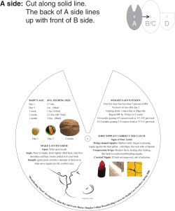


© Copyright 2026