
PERCEPTUAL AND MNEMONIC MATCHING-TO-SAMPLE IN HUMANS: CONTRIBUTIONS OF THE HIPPOCAMPUS,
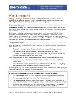
PERCEPTUAL AND MNEMONIC MATCHING-TO-SAMPLE
IN HUMANS: CONTRIBUTIONS OF THE HIPPOCAMPUS,
PERIRHINAL AND OTHER MEDIAL TEMPORAL LOBE
CORTICES
J.S. Holdstock1*, S.A. Gutnikov2**, D. Gaffan2 and A.R. Mayes1
(1Section of Clinical Neurology, University of Sheffield, Royal Hallamshire Hospital,
Sheffield, U.K.; 2Department of Experimental Psychology, University of Oxford, U.K.)
ABSTRACT
Two questions were addressed by the present study. The first was whether the
previously reported item recognition deficit which is shown by amnesic patients may be
due to a perceptual rather than a memory deficit. To address this question a group of
amnesic patients were tested on a 14-choice forced-choice visual item recognition test which
included a “simultaneous” condition in which the sample remained visible during the
matching decision and a zero second delay. Eacott, Gaffan and Murray (1994) have reported
an impairment in simultaneous matching-to-sample following perirhinal damage in
monkeys. In our amnesic patients, a deficit was found only after filled delays of 10 seconds
or longer and this was also the case for a subgroup of patients whose damage included the
perirhinal cortex. The second question, which arose from the model of Aggleton and Brown
(1999), was whether performance on the DMS task would remain intact following selective
damage to the hippocampus. We tested a patient with bilateral damage to the hippocampus
on the 14-choice DMS task and found that her performance was not significantly impaired
at delays of up to 30 seconds.
Key words: item recognition, amnesia, hippocampus, perirhinal cortex
INTRODUCTION
A deficit in recognition memory has been considered to be a central feature
of anterograde amnesia (Parkin and Leng, 1993). As a result, object recognition
tests have been used extensively to assess animal models of this memory deficit.
The favoured format of these tasks has involved the presentation of a single
sample item and then, after a delay of varying duration, the presentation of the
previously seen item along with a new (novel or less familiar) item. The animal
has either to select the previously seen item (delayed-match-to-sample – DMS)
or, more often, to select the new item (delayed-nonmatch-to-sample – DNMS) in
order to receive a reward. Early studies showed that large medial temporal lobe
(Mishkin, 1978; Zola-Morgan, Squire and Mishkin, 1982) and large medial
diencephalic lesions (Aggleton and Mishkin, 1983a, 1983b) impaired DNMS
performance. Recently, studies have investigated more specifically the
contribution to performance of structures within these regions. The use of more
* Now at Department of Clinical Neurology, University of Oxford, Radcliffe Infirmary, Oxford, OX2 6HE, U.K.
** Now at Department of Psychology, University of Liverpool, UK.
Cortex, (2000) 36, 301-322
302
J.S. Holdstock and Others
selective lesions have pointed to the perirhinal cortex as being the medial
temporal lobe region critical for object recognition memory of this sort
(Meunier, Bachevalier, Mishkin et al., 1993; Meunier, Hadfield, Bachevalier et
al., 1996). In contrast, aspiration lesions of the hippocampus have produced only
a mild DNMS impairment (Murray and Mishkin, 1986; Zola-Morgan and Squire,
1986; Zola-Morgan, Squire and Amaral, 1989; Zola-Morgan, Squire, Clower et
al., 1993). Further, more precise stereotaxic lesions of the hippocampus, which
produce less incidental damage to adjacent cortex, have left DNMS performance
intact at delays of up to 60 seconds (Alvarez, Zola-Morgan and Squire, 1995).
DNMS was impaired after longer delays during which the animal was removed
from the test apparatus (Alvarez et al., 1995), but it was not impaired at delays
of up to 40 minutes when the animals remained in the test apparatus during all
retention intervals (Murray and Mishkin, 1996).
The fornix is a major efferent pathway of the hippocampus which connects
this structure with the mammillary bodies and anterior thalamic nuclei. Together
these structures make up what has been referred to as the extended hippocampal
system (Aggleton and Brown, 1999). Like hippocampal damage, fornix
transection has been demonstrated to have little or no effect on DNMS and
DMS in monkeys (Bachevalier, Parkinson and Mishkin, 1985; Bachevalier,
Saunders and Mishkin, 1985; Gaffan, Sheilds and Harrison, 1984; Zola-Morgan
et al., 1989) and no effect on DNMS performance in rats (Aggleton, Hunt and
Shaw, 1990; Rothblat and Kromer, 1991; Shaw and Aggleton, 1993). In fact, in
monkeys, a double dissociation has been demonstrated between the effects of
perirhinal lesions and fornix lesions on memory. Perirhinal lesions had a much
greater effect on DNMS performance than fornix transection, whereas fornix
transection impaired performance on a spatial memory task on which perirhinal
lesions had little effect (Gaffan, 1994). Lesions of the parahippocampal cortex
have been found not to cause a DNMS deficit (Meunier et al., 1996; Ramus,
Zola-Morgan and Squire, 1994) and, similarly, lesions of the adjacent entorhinal
cortex have only a very mild or transient effect on DNMS performance
(Leonard, Amaral, Squire et al., 1995; Meunier et al., 1993). Within the
diencephalic region, damage to the medial dorsal nucleus of the thalamus, to
which the perirhinal cortex projects, has been shown to disrupt item recognition
(see Aggleton and Brown, 1999). In contrast, lesions to the mammillary bodies,
structures thought to form part of the extended hippocampal system, have been
found to leave DNMS performance intact (Aggleton and Mishkin, 1985;
Aggleton et al., 1990; Zola-Morgan et al., 1989).
The importance of the perirhinal cortex in object recognition has also been
suggested by electrophysiological studies. In the study of Brown, Wilson and
Riches (1987), 15 percent of cells in the inferomedial temporal cortex, which
included the perirhinal and entorhinal cortices, responded more strongly to the
first presentation of a stimulus than to subsequent presentations. These cells
therefore appeared to be discriminating between unfamiliar and familiar item
stimuli. Brown et al. (1987) found no such cells in the hippocampus or
subiculum. These findings have been supported by the results of more recent
studies (for a review, see Brown and Xiang, 1998).
The evidence from the animal literature which suggests that the perirhinal
Matching-to-sample in human amnesia
303
and hippocampal regions make different contributions to memory led Aggleton
and Brown (1999) to propose the existence of two memory systems. One
memory system, comprising the perirhinal cortex and dorsomedial nucleus of the
thalamus, was proposed to be important for familiarity-based item recognition.
In contrast, a second system which Aggleton and Brown (1999) refer to as the
extended hippocampal system was thought to be critical for successful recall of
new episodic information, but to be unnecessary for successful item recognition
when this was dependent on stimulus familiarity (Mandler, 1980). Aggleton and
Brown (1999) considered forced-choice recognition tasks such as DMS and
DNMS to be tasks which are likely to be tapping this aspect of recognition. The
extended hippocampal memory system was suggested to comprise the
hippocampus, mammillary bodies and anterior thalamic nuclei.
Aggleton and Brown’s theory predicts that, in humans, deficits in recall and
recollection will be seen following lesions restricted to structures within the
extended hippocampal system, but that item recognition on tasks in which
relative familiarity contributes substantially to the memory decision will remain
intact. In contrast, Aggleton and Brown’s theory predicts that item recognition
will be impaired following damage to the perirhinal cortex or the dorsomedial
nucleus of the thalamus.
Although the findings from the lesion and electrophysiological studies
described above have highlighted the importance of the perirhinal cortex in
visual item recognition (Aggleton and Brown, 1999), a study by Eacott et al.
(1994) has questioned whether this is a selective deficit of recognition memory
or one of perceptual processing of visual information that results in a memory
deficit. Although these authors found that monkeys with rhinal cortex lesions
were impaired at delayed-match-to-sample (DMS) when trial unique stimuli
were used, they also found that rhinal lesions impaired performance when there
was no delay between sample and test and in a “simultaneous” condition in
which both the sample and test items were visible while the matching decision
was made. As these latter two conditions made no demands on memory this
poor performance must have been due to a perceptual deficit. Subsequent studies
have shown that concurrent discrimination learning is impaired by perirhinal
cortex damage when a large set of discriminations have to be learned (Buckley
and Gaffan, 1997), a large number of foils are used (Buckley and Gaffan, 1997)
or the rewarded item is presented in different views on different trials (Buckley
and Gaffan, 1998). When small sets, few foils and the same views of an item
were used, no concurrent discrimination learning deficit was seen after perirhinal
damage. It has been argued that the manipulations which resulted in a deficit
“increased the demands that these tasks placed on object identification” (Buckley
and Gaffan, 1998). Buckley, Booth, Rolls et al. (1998) have also shown that
perirhinal cortex damage impaired monkeys’ ability to select the odd-one from
among six pictures: five showing different views of a single object and the other
showing a different object. They could, however, select the odd-one when the
five pictures of the single object were identical views. In considering the animal
literature, Murray and Bussey (1999) have suggested that the perirhinal cortex
plays a role in both perception and memory. They propose that its perceptual
role is that of representing “complex conjunctions of stimulus features” (Murray
304
J.S. Holdstock and Others
and Bussey, 1999, p. 148) and that this is the final stage in the ventral visual
processing stream.
Consistent with the animal literature, studies of item recognition in human
amnesia have found that large medial temporal lobe lesions or large diencephalic
lesions impaired performance on tests of forced-choice item recognition
modelled on the DNMS and the DMS procedures used with monkeys (Aggleton,
Nicol, Huston et al., 1988; Squire, Zola-Morgan and Chen, 1988). Furthermore,
when performance of control and amnesic subjects has been matched at an
initial delay, and performance of the controls is well below ceiling, both medial
temporal lobe and diencephalic (Korsakoff) amnesics have been demonstrated to
forget visual item information at an accelerated rate on an analogue of the DMS
task (Holdstock, Shaw and Aggleton, 1995). Such accelerated forgetting has also
been demonstrated in a number of other forced choice and yes/no recognition
memory studies (Carlesimo, Sabbadini, Fadda et al., 1995; Downes, Holdstock,
Symons et al., 1998; Mayes, Downes, Symons et al., 1994; Squire, 1981;
Huppert and Piercy, 1979). This accelerated forgetting strongly suggests that
poor performance in the amnesics is due to a deficit in memory. However, an
additional perceptual deficit cannot be ruled out. None of these studies used
either a simultaneous condition or immediate memory test (i.e. a 0s delay),
rather, initial performance was measured at delays of between three seconds and
10 seconds and in the majority of cases the amnesic patients were given extra
exposure to the stimuli in order to match their performance at this initial delay
with that of the control group. However, a recent study (Buffalo, Reber and
Squire, 1998), which investigated the effects of perirhinal damage on object
recognition, did include very short initial delays. It was found that two patients
whose damage included this region were unimpaired when recognition was
tested after delays of zero and two seconds but were impaired at later delays.
These results therefore indicated that, contrary to the results from animal studies
(Eacott et al., 1994; for a review, see Murray and Bussey, 1999), in humans, the
perirhinal cortex may have a role in memory, but not perception. This finding, if
replicable, suggests that one should be careful in generalising from monkeys to
humans. In the first part of the present study we investigated further the
possibility that the poor recognition performance of amnesic patients could be
due to a perceptual deficit by testing a group of amnesics of mixed aetiology,
including four patients with damage to the perirhinal cortex, on a DMS task
which included both a simultaneous condition and a 0s delay.
The second question which was investigated in the present study was
Aggleton and Brown’s (1999) prediction that damage to the extended
hippocampal system will not impair performance on item recognition tasks such
as DMS even though recall of information will be impaired. There is some
evidence in support of Aggleton and Brown’s prediction. In their meta-analysis
of studies which assessed recognition memory using the Recognition Memory
Test (RMT) (Warrington, 1984), Aggleton and Shaw (1996) found that patients
with damage restricted to the hippocampus, fornix or mammillary bodies were
impaired at recall as assessed by the WMS-R, but were only mildly impaired, or
even in some cases unimpaired, at item recognition tested by the RMT. The
RMT consists of two recognition tests, one of words and one of faces. Each test
Matching-to-sample in human amnesia
305
uses a 2-choice forced-choice recognition paradigm for 50 items and so is
equivalent to a DMS task with a list length of 50. A similar finding was
reported by Vargha-Khadem, Gadian, Watkins et al. (1997), who found that
three young people with relatively selective hippocampal pathology were
unimpaired at two-choice forced-choice item recognition whilst being amnesic
for the recall of episodic information and impaired at recognising object-location
and face-voice associations. Consistent with this finding, Volpe, Holtz and Hirst
(1986) reported impaired recall but apparently intact yes-no recognition in a
group of patients who had suffered a cardiac arrest, an event which results in
preferential pathology in limbic structures such as the hippocampus (Volpe et
al., 1986). Another relevant finding is that of McMackin, Cockburn, Anslow et
al. (1995) whose data suggested that bilateral fornix damage resulted in only a
mild impairment of recognition memory (assessed by the RMT). As described
above, it is within the fornix that efferents from the hippocampus travel to the
mammillary bodies and the anterior nucleus of the thalamus and when this fibre
pathway is lesioned, a similar pattern of memory deficits is obtained to that
found after hippocampal lesions (see Gaffan, 1994). The findings of these
studies are all consistent with the prediction which arises from Aggleton and
Browns’ (1999) model.
However, there are also some apparently conflicting findings. Reed and Squire
(1997) described the recognition memory performance of six patients who were
reported to have selective damage of the hippocampal formation (hippocampus
proper, dentate gyrus, subiculum and entorhinal cortex), including one patient
whose damage appeared to be restricted to the CA fields of the hippocampus.
Recognition memory was reported to be impaired in these patients. Reed and
Squire’s (1997) paper considered performance on a range of recognition tests
whereas all but one (Volpe et al., 1986) of the studies supporting Aggleton and
Browns’ model used a two-choice forced-choice recognition paradigm. The
conflicting findings highlight the need both to test further patients with selective
hippocampal damage and to use other item recognition tests in addition to the twochoice forced-choice design to determine the extent to which previously reported
findings generalise to other recognition paradigms.
In the present study, we tested a patient, YR, who has relatively selective
bilateral hippocampal damage, on the DMS task completed by the group of
amnesic patients of mixed aetiology. Patient YR has been found to be
unimpaired on the RMT (Warrington, 1984) and the recognition subtests of the
Doors and People Test (D&P) (Baddeley, Emslie and Nimmo-Smith, 1994)
despite showing a substantial recall deficit on the Wechsler Memory ScaleRevised and the recall subtests of the D&P (see Table I of the present paper).
YR therefore showed the same pattern of impaired recall but unimpaired forcedchoice item recognition as the patients described by Aggleton and Shaw (1996),
McMackin et al. (1995) and Vargha-Khadem et al. (1997). The present study
allowed us to investigate whether YR’s item recognition would also be impaired
on another forced-choice item recognition task which was designed to be
difficult and to avoid ceiling effects in the control group by using a large
number of foils (13) at test, used different stimulus materials to the RMT and
D&P, and assessed memory over delays of up to 30 seconds.
306
J.S. Holdstock and Others
In summary, there were two questions which the present study addressed. The
first was whether the previously reported deficit in DMS in amnesic patients with
perirhinal damage may be due to a perceptual deficit rather than a memory deficit
(Eacott et al., 1994; Murray and Bussey, 1999). To address this question a group
of amnesic patients were tested on a difficult 14-choice forced-choice visual item
recognition test which included a “simultaneous” condition in which the sample
remained visible during the matching decision and a zero second delay. The stimuli
were abstract complex patterns which had to be discriminated from foils containing
similar features. This discrimination required the representation of both the
stimulus features and the way in which they were combined. A deficit in the
perceptual processes necessary for this would impair performance in these two
conditions. The second question, which arose from the model of Aggleton and
Brown (1999), was whether performance on the DMS task would remain intact
following selective damage to the hippocampus.
MATERIALS
AND
METHODS
Subjects
A group of 9 patients (2 female, 7 male) of mixed aetiology were tested. Three patients
had suffered encephalitis (CF, RS, YW). These patients’ pathology was investigated using
Magnetic Resonance Imaging (MRI). CF was found to have complete damage to the amygdala,
hippocampus, parahippocampal cortex, perirhinal cortex and entorhinal cortex on the right
with additional partial damage in this hemisphere to the superior and middle temporal gyri, the
occipito-temporal gyrus and the insular cortex. CF also had some damage to the amygdala, and
the parahippocampal, perirhinal and entorhinal cortices on the left. RS’s hippocampus was
small throughout its length bilaterally with greater volume loss at the head. In addition, this
patient had some damage to the parahippocampal cortex, perirhinal cortex and entorhinal
cortex on the left and some damage to the perirhinal cortex on the right. RS also had some
general cortical atrophy. Volume estimates for medial temporal lobe structures in this patient
have been reported elsewhere (Holdstock, Mayes, Cezayirli et al., 1999a). In patient YW, the
amygdala, hippocampus, parahippocampal gyrus, perirhinal and entorhinal cortices were
almost completely destroyed on the right but were less extensively damaged on the left. There
was extensive damage to both the inferior and middle temporal gyri on the right. On the left
the inferior temporal gyrus was damaged but the middle and superior temporal gyri were
relatively spared. There was some mild cortical atrophy of the frontal, and parietal lobes but no
evidence of focal damage to these regions. A fourth patient (NM) had suffered from meningitis
in 1969 and was found by MRI to have partial bilateral damage to the amygdala, hippocampus,
parahippocampal cortex, perirhinal cortex and entorhinal cortex. Volume measures of medial
temporal lobe structures in this patient have been provided elsewhere (Holdstock et al., 1999a).
In addition, NM had some atrophy of the mammillary bodies, cerebellum, and in the superior
frontal and parietal lobes. One patient had amnesia as a result of a posterior communicating
artery aneurysm (CW) and a CT scan showed damage to the posterior temporal region, low
density change in the right temporal region medially and a zone of reduced density in the
occipital cortex. Two patients had had an anterior communicating artery aneurysm clipped
(AB, RB). MRI data was only available for RB and showed high signal changes in the gyrus
rectus and the medial orbital frontal gyri on the right which extended up to the head of the
caudate nucleus. There was also cystic change in the genu of the corpus callosum and high
signal in the anterior cingulate sulcus on the left. Atrophy of the frontal poles and high signal
changes in the superior surface of the right temporal pole were also reported. The final two
patients had Korsakoff’s syndrome (RT, JT). Unfortunately scan information for these patients
was not available. The patient group had a mean age of 48.1 years (S.D. = 7.3).
The results of the psychometric assessment of the patients is shown in Table I. The
TABLE I
PE
PE
PE
M
PAA
AAA
AAA
K
K
CF
RS
YW
NM
CW
AB
RB
RT
JT
YR
39
50
55
47
40
47
62
50
43
48
Age
118
111
96
82
102
107
110
98
85
115
101
106
100
84
86
89
108
104
90
102
105
103
103
82
92
93
106
106
91
108
VIQ
FSIQ
FSIQ
WAIS-R
N-R
98
110
96
87
80
86
109
102
92
97
PIQ
89
74
63
77
64
65
84
87
78
66
GEN
105
92
118
88
80
74
130
93
90
122
ATT/C
WMS-R
88
55
<50
<50
<50
50
50
62
59
73
DEL
50
<1
<5
<1
<1
<5
<5
<5
<1
<1
P
<1
10-25
<5
<1
<1
<1
50
10
<1
50
D
75
<1
<1
<1
<1
<1
1-5
<1
<1
1-5
S
Doors & People
25-50
25
<5
1
5
1
<1
1
25
99
N
48
33*
29*
26*
41
30*
31*
31*
40
45
W
F
31*
33*
23*
32*
27*
28*
38
32*
37
48
RMT
N-R = NART-R; FSIQ = full scale IQ; VIQ = verbal IQ; PIQ = performance IQ; VERB = verbal memory; VIS = visual memory; GEN = general memory; ATT/C =
attention/concentration; DEL = delayed memory; P = people subtest; D = doors subtest; S = shapes subtest; N = names subtest; W = words; F = faces.
Aet.
NM
Details of Age, Aetiology and Performance on Standardised Tests of Intellectual Function and Memory for the Nine Patients Participating in the Group Study
and for Patient YR [The test scores shown are predicted premorbid full scale IQ from the National Adult Reading Test-Revised (NART-R), index scores (mean
of 100, SD of 15) from the Wechsler Adult Intelligence Scale-Revised (WAIS-R) and the Wechsler Memory Scale-Revised (WMS-R), percentile equivalents for
scores on the Doors and People Test, number correct out of 50 on the Recognition Memory Test (RMT) with performance at less than the fifth percentile
marked with an asterisk]
Matching-to-sample in human amnesia
307
308
J.S. Holdstock and Others
premorbid IQ of the patients was estimated using the NART-R (Nelson and Willison, 1991)
and the group had a mean predicted full scale IQ of 101 (range 82-118). A measure of
present full scale IQ was obtained using the Wechsler Adult Intelligence Scale – Revised
(WAIS-R) on which a mean score of 96.4 (range 84-108) was obtained. Memory was
measured using the Wechsler Memory Scale – Revised (WMS-R), the Doors and People
Test (D&P) (Baddeley, Emslie and Nimmo-Smith, 1994) and the Recognition Memory Test
(RMT) (Warrington, 1984). On the WMS-R the mean general memory index score was
75.7 (range 63-89), with mean index scores of 96.7 (74-130) for attention and concentration
and 57.1 (range 50-88) for delayed memory. The mean raw score obtained by the amnesic
group on the D&P was 7.8 (range 0-30) for the people subtest, 11.8 (range 8-18) for the
doors subtest, 15.1 (range 8-36) for the shapes subtest and 13.4 (range 10-18) for the names
subtest; these mean scores fell at less than the fifth percentile, first to fifth percentile, less
than the first percentile and first to tenth percentile, respectively. The mean number of
items recognised out of 50 on the RMT was 34 (range 26-48) for words and 31 (range 2338) for faces.
The performance of the amnesic group was compared with a group of 9 control subjects
(2 female, 7 male) who were well matched for age and estimated IQ. The mean age of the
control group was 48.7 years (S.D. = 8.2). A t-test demonstrated that control and amnesic
groups did not differ significantly in age [t (16) = 0.18, p > 0.05]. The mean estimated full
scale IQ of the control group was determined from the NART-R and was 101.2 (S.D. =
10.9). A t-test comparing the NART-R predicted full scale IQ of the control subjects with
the WAIS-R full scale IQ of the amnesic patients revealed no significant difference [t (16)
= 0.76, p > 0.05].
The patient, YR, with relatively selective hippocampal damage was a clerical worker
aged 59 at the time of testing. In 1986 she received an opiate drug to relieve a severe back
pain and may then have suffered an ischaemic incident. Following this incident she suffered
a memory impairment which has persisted for 12 years. A Magnetic Resonance Imaging
(MRI) scan was obtained for patient YR using a 3D T1-weighted radio-frequency spoiled
gradient echo (SPGR) sequence [TE = 9 ms, TR = 34 ms, flip angle = 45 degrees, matrix
size = 256 × 192, 2 NEX, field of view = 20 cm, acquisition time = 27 minutes and 52
seconds] available on a 1.5T SIGNA whole-body magnetic imaging system (General
Electric, Milwaukee, WI). The volumes of the hippocampus, parahippocampal gyrus,
temporal lobe, parietal lobe and grey and white matter of the prefrontal lobe were estimated
using the Cavalieri method of modern design stereology (Gunderson and Jensen, 1987) via
stereology menus within Analyze (Mayo Foundation, Minnesota, USA) software running
on a SPARC 10 workstation (SUN Microsystems, CA, USA). The scan revealed bilateral
damage to the hippocampus throughout its entire length. Volume measures, corrected for
intracranial volume were 2.25 S.D.s and 3 S.D.s smaller than the control mean on the right
and left respectively. The amygdala was small but showed no sign of pathology and there
were no other visible abnormalities within the medial temporal lobe. The volume of the
temporal lobe was also normal. Grey and white matter volumes for the frontal lobe were
normal and the volume of the parietal lobe was within two standard deviations of the control
mean volume bilaterally (a more detailed report of the volumetric analysis of YR’s scan is
provided by Holdstock, Mayes, Cezayirli et al., 1999b).
YR’s results from standardised tests of intellectual function and memory are shown in
Table I. Psychometric testing revealed that YR has an IQ, assessed by the WAIS-R, which
is a little above average and which is higher for Verbal than Performance tests. The
difference between her premorbid IQ (measured by the NART-R) and her present IQ was
less than 1 S.D. on the WAIS-R scale, so there was no indication of a significant decline in
IQ in this patient. Memory testing showed that recall, measured by the two recall subtests
of the Doors and People Test (Baddeley, Emslie, Nimmo-Smith, 1994) and the general and
delayed memory indices of the WMS-R, was impaired. In contrast, recognition of visual
and verbal items from the D&P and the RMT was unimpaired. YR’s executive and
visuospatial abilities were also assessed. She showed no impairment on verbal fluency
(FAS) (Benton, 1968), Cognitive Estimates (Shallice and Evans, 1978) and the Wisconsin
Card Sorting Test (WCST) (Heaton, 1981), all of which tap executive functions. Her
performance on the FAS was 0.1 S.D.s below her control group’s mean, on the CET was
Matching-to-sample in human amnesia
309
0.69 S.D.S. below her control group’s mean, and on the WCST the number of correct
categories achieved was at the sixth to tenth percentile and number of perseverative errors
was at the 88th percentile. YR’s perception of objects and space, assessed by the Visual
Object and Space Perception Battery (VOSP) (Warrington and James, 1991), the Judgement
of Line Orientation test (Benton, Hamsher, Varney et al., 1983) and the “Little Men” Test
of mental rotation (Ratcliff, 1979), was unimpaired. On the VOSP performance ranged
from 18th to 76th percentile, on the Judgement of Line Orientation test performance was
above the control mean as was performance on those conditions of the “Little Men” test
requiring mental rotation (performance ranging from 0.05 to 0.5 S.D.s better than the control
mean). Spatial reasoning, assessed using the Verbal and Spatial Reasoning Test (VESPAR)
(Langdon and Warrington, 1995), was also unimpaired (50th-75th percentile).
As YR was older than most of the subjects making up the amnesic group, her
performance was compared with that of a separate control group of a more appropriate age.
The control group, which consisted of 11 females, had a mean age of 60 years (S.D. = 4.8)
which was 0.21 standard deviations older than YR. The mean full scale IQ of the control
group which was estimated from the NART-R was 110.5 (S.D. = 7) which was 1.2 standard
deviations higher than YR’s full scale IQ from the WAIS-R and 0.64 standard deviations
lower than YR’s estimated premorbid (NART-R) full scale IQ.
Apparatus and Stimuli
Subjects were tested on a 14-choice visual delayed-match-to-sample task which used
trial unique stimuli. The task was presented on a PC computer connected to a 39 cm × 29
cm touch screen. The stimuli were computer-generated monochrome complex abstract
designs (ranging from 4 cm × 4.5 cm to 5 cm × 4.5 cm) which comprised a white pattern
overlaid by a grey pattern. The stimuli were difficult to verbalise (see Figure 1). Stimulus
complexity was similar to that of the stimuli used by Eacott et al. (1994), each of which
comprised a large typographic character overlaid by a small typographic character.
Design
Performance was investigated in seven conditions: a simultaneous condition and six
delays. In the simultaneous condition the sample item remained visible while the matching
decision was made and so did not necessitate memory. The delays were chosen to be
consistent with those which have been used to assess animal models of amnesia and were
of zero seconds, two seconds, five seconds, ten seconds, twenty seconds and thirty seconds.
In the zero second condition the choice stimuli appeared immediately after the
disappearance of the sample item.
Performance on the simultaneous condition and the zero second condition was assessed
on 50 trials and for the remaining conditions subjects completed 20 trials. Each subject
completed all the trials for one condition before moving on to the next condition. For the
three longest delays a mental arithmetic task was interposed between study and test.
Each stimulus was seen only once throughout testing although other similar stimuli
were used. The stimuli were allocated randomly to the seven conditions prior to the study.
The stimuli seen within a particular condition were the same for all subjects. In the group
study, each patient completed the conditions in a different random order. These same nine
orders were used for the control group. Each patient was allocated a control subject of the
same sex and similar age who completed the conditions in the same order to that amnesic.
Patient YR and her controls completed the conditions in a different random order to the
subjects in the group study.
Procedure
In the simultaneous condition a stimulus was presented in the centre of the screen for
2.5 seconds. This sample stimulus then remained visible while the 14 choice stimuli
appeared on the screen. The foils contained similar features to the target stimuli. The choice
stimuli appeared in three rows, with one row above and one below the sample item (each
310
J.S. Holdstock and Others
A
B
C
D
Fig. 1 – An example of the monochrome abstract patterns used for the match-to-sample task. A
illustrates what subjects saw at presentation in the simultaneous condition; B illustrates what subjects
saw at test in the simultaneous condition; C illustrates what subjects saw at presentation in the
memory conditions; D illustrates what subjects saw at test in the memory conditions.
row consisting of five stimuli) and two stimuli to the left and two to the right of the sample
stimulus on the middle row (see Figure 1). The subject was instructed to touch the design
which matched the sample in the centre of the screen.
In the delay conditions a sample stimulus was presented in the centre of the screen for
2.5 seconds in exactly the same way as the simultaneous condition. The sample then
disappeared and the screen remained blank for the duration of the delay. For retention
intervals of zero seconds, two seconds and five seconds the delay was unfilled and subjects
allowed to continue looking at the computer screen while waiting for the choice items to
appear. For the three longer delays subjects were asked to complete mental arithmetic
problems as quickly and accurately as possible. The problems, which were presented on A4
sheets of paper, required subjects to add, subtract, divide or multiply four numbers as
quickly as possible and to write the answer alongside each problem. Following the delay
the choice stimuli were presented in the same screen locations as in the simultaneous
condition i.e. in the delay conditions the central position (which had been occupied by the
sample) remained blank in the test phase (see Figure 1). Subjects selected the design which
matched the previously presented sample.
In all conditions, an unlimited amount of time was allowed at test for subjects to make
their decision and their response time was recorded. Sets of 4 practice trials were provided
prior to each block of trials in order to ensure that the subject understood the requirements
for that condition.
Matching-to-sample in human amnesia
311
RESULTS
Exploration of the results considered both percent correct scores and response
time. All post hoc testing employed Tukey’s HSD test.
The first analysis compared the percentage of correct responses made by the
amnesic group and their healthy controls for each of the seven delays. A 2 × 7
ANOVA with factors of group and condition showed a significant effect of group
[F (16, 1) = 34.95, p < 0.001] which was due to the amnesic group making fewer
correct choices overall than the control group. There was also a significant effect
of condition [F (96, 6) = 69.91, p < 0.001]. Post hoc tests indicated that
performance was significantly better in the simultaneous condition than in any of
the delay conditions and was significantly better after any of the three shortest
delays than after any of the three longest delays (ps < 0.05). Condition was found
to interact significantly with group [F (96, 6) = 22.39, p < 0.001]. Post hoc tests
compared the performance of the two groups in each condition. This revealed that
the two groups differed significantly in performance only at the three longest
delays. The analysis was repeated following arcsine transformation of the data
which was performed because of the debate as to whether transforming the results
of such tests produces a qualitatively different pattern of results (Alvarez-Royo,
Zola-Morgan and Squire, 1992; Ringo, 1993). Consistent with a similar study of
amnesic patients (Holdstock et al., 1995), the pattern of our results which was
obtained after transformation of the scores was identical to that obtained with the
untransformed data.
The analysis was repeated after selecting just those patients with MRI evidence
of perirhinal damage (n = 4) to compare with the control group (n = 9). A very
similar pattern of results was found as for the entire amnesic group; the patients
with damage to the perirhinal cortex were only significantly impaired after the
filled delays of 10 seconds or longer. The only difference that was obtained
between this analysis and that of the entire amnesic group was that, for the
perirhinal patients, post hoc tests showed that performance in the simultaneous
condition did not differ from that at the three shortest delays. In addition, a 2 × 7
ANOVA with factors of group and delay compared the perirhinal subgroup
with the rest of the amnesic group. There was no significant effect of group
[F (7, 1) = 1.71, p > 0.05] and no significant interaction between group and delay
[F (42, 6) = 1.58, p > 0.05]. Figure 2 shows graphically the percent correct scores
for the amnesic group split into patients with or without MRI evidence of perirhinal
cortex damage and the control group.
Figure 2 also plots percent correct scores for patient YR and her control group
for each of the seven conditions. YR’s performance expressed as standard
deviations from the control mean are shown in Table II. We have considered YR’s
performance to be impaired if her scores were more than 1.96 S.D.s worse than the
control mean giving a type 1 error probability of 0.05, 2 tailed. YR’s performance
was not significantly impaired in any condition. At delays of 5 s, 10 s and 30 s her
performance was over 1 S.D. below the mean of the control group but was within
the control range for all conditions. A comparison of YR’s scores with the mean of
the amnesic group (see Table II) showed that YR’s score was over 2 S.D.s better
than the mean of the amnesic group at the 20 s delay and over 1 S.D. better for
312
J.S. Holdstock and Others
Fig. 2 – Graph displaying the percent correct scores from the simultaneous condition and each
of the six delays for patient YR and the mean percent correct scores for her age and IQ matched
control group (YR CONTROLS), the amnesic subgroup with perirhinal damage (GRP PERI), the
amnesic subgroup with no confirmed perirhinal damage (GRP NONPERI) and a control group which
was matched for age and IQ to the overall amnesic group (GRP CONTROLS).
delays of 10 and 30 seconds. These were the delays which were filled by the
distractor task and were the conditions in which performance would be most likely
to be tapping long-term memory. When YR’s performance was compared with
only those patients with MRI evidence of perirhinal damage, a similar pattern of
superior performance was found for YR, which was over 2 S.D.s better than the
perirhinal subgroup at the 20 second delay and 0.83 S.D.s better than the perirhinal
group mean at the 10 second and 30 second delays.
An analysis was also performed on subjects’ response times pooled over
correct and incorrect responses. A 2 × 7 ANOVA with factors of group (amnesic
TABLE II
Number of Standard Deviations that YR’s Mean Percent Correct Score was from the Control Mean
(YR v Con), the Mean of the entire amnesic group (N = 9) (YR v Amn), and the Mean of Those
Amnesics with MRI Evidence of Perirhinal Damage (N = 4) (YR v Peri) for each of the Seven
Conditions of the Match-to-Sample Task [Positive scores indicate that YR was performing above the
mean of that comparison group]
Comparison
Sim.
0s
2s
5s
10 s
20 s
YR v Con
– .47
– .26
YR v Amn
+ .75
+ .7
YR v Peri
+ .5
+ .87
– .39
– 1.5
– 1.6
+ .32
– 1.1
+ .55
– 1.42
+ 1.12
+ 2.74
+ 1.14
– 2.74
+ .83
+ 2.16
+ .83
– 1.2
30 s
Matching-to-sample in human amnesia
313
group and control group) and condition was carried out. The analysis showed that
the response time of the two groups did not differ significantly [F (16, 1) = 4.05,
p > 0.05]. There was a significant effect of delay [F (96, 6) = 31.85, p < 0.01]
which post hoc tests revealed was due to response time being significantly longer
after filled delays of 10, 20 and 30 seconds than after the shorter delays and the
simultaneous condition. The effect of delay did not interact significantly with
group [F (96, 6) = 1.05, p > 0.05]. The response time, pooled over correct and
incorrect responses, was also investigated specifically in the perirhinal group
because, although percent correct scores gave no indication of a perceptual deficit
in this group, an increase in response time would be consistent with such a
deficit. Patient mean response times were very similar to those of the control
subjects and an ANOVA with factors of group (control and perirhinal groups)
and delay indicated no significant difference between the response time of the
control and perirhinal group [F (11, 1) = .22, p > 0.05] and no significant
interaction with delay [F (66, 6) = .58, p > 0.05].
In contrast to the amnesic group, YR’s response time was significantly
slower than her controls’ for all conditions other than the simultaneous
condition. As shown in Table III, which shows YR’s response time expressed as
the number of standard deviations above the control mean, this increased
response time was particularly striking at the longer delays. Her mean response
time (collapsed over correct and incorrect response) was 10.45, 12.96, 30.31,
39.3, 38.55 and 45.5 seconds at delays of 0 seconds, 2 seconds, 5 seconds, 10
seconds, 20 seconds, and 30 seconds, respectively, whereas the mean control
response time for these delays were 5.01, 6.48, 6.41, 10.73, 12.23, 11.29,
respectively. This increase in response time was found for both correct and
incorrect responses (see Table III). As shown in Table III YR’s response time
was also significantly slower than the amnesic group at delays of zero seconds
onwards. The results suggest that the intervals over which YR was remembering
the stimuli were considerably longer than both her control group and the
amnesic group. Due to this it is possible that YR’s percent correct scores may be
providing an underestimate of her performance.
TABLE III
Number of Standard Deviations that YR’s Mean Response Time, Collapsed over Correct and Incorrect
Responses (Overall RT), was from the Control Mean (YR v Con) and the Amnesic Group Mean (YR v
Amn) for each of the Seven Conditions. Number of Standard Deviations that YR’s Mean Response
Time, for Correctly Recognised Items (Correct RT) and for Incorrectly Recognised Items (Incorrect
RT), was from the Control Mean. [Positive scores indicate longer response time].
Comparison
Overall RT
YR v Con
YR v Amn
Correct RT
YR v Con
Incorrect RT
YR v Con
Sim.
0s
2s
5s
10 s
20 s
30 s
1.18
– .14
2.4
2.2
2.1
2.1
8.69
10.1
7.6
6.0
6.5
6.6
10.5
6.9
1.18
2.3
3.2
6.9
5.3
6.6
7.2
1.18
2.7
5.9
4.5
4.2
6.7
.94
314
J.S. Holdstock and Others
DISCUSSION
A group of amnesic patients of mixed aetiology, including four patients with
MRI evidence of perirhinal cortex damage, were found to be unimpaired on a
14-choice visual matching-to-sample task when the sample item remained visible
during the choice phase (simultaneous condition) and when the study-test
interval was unfilled and of five seconds or less. In contrast, the performance of
this group was significantly impaired after a delay of 10 seconds or longer
during which rehearsal was prevented by a distractor task.
The unimpaired performance of the amnesic group in the simultaneous and
zero second conditions confirms that the impairment in DMS found in amnesic
patients is due to a deficit in memory rather than perception. The fact that the
amnesic group were impaired only when study and test were separated by a
filled delay suggests, consistent with other studies, that both encoding (e.g.
Mayes, Downes, Shoqeirat et al., 1993; Smith and Milner, 1989; Shoqeirat and
Mayes, 1991; Downes et al., 1998, Warrington and Baddeley, 1974) and shortterm/working memory (e.g. Mayes and Meudell, 1983) were normal. The
amnesic patients’ impaired performance at the three longer filled delays indicates
that long-term memory for this information was impaired because rehearsal was
not possible and the delays were such that short-term memory was unlikely to
be contributing to performance (see Downes et al., 1998, Isaac and Mayes,
1999). This deficit was seen as soon as long-term memory was being tapped, as
marked by the introduction of the filler task, and in agreement with the bulk of
the literature (for a review see Isaac and Mayes, 1999) this deficit did not appear
to increase with time. The apparent accelerated loss of information by the
perirhinal group between 10 s and 30 s delays was not statistically significant
(t = 1.963, p > 0.05). However, as the patients’ performance was impaired even
at the first of the longer filled delays, scaling effects make it difficult to reliably
compare the relative rate of forgetting of the amnesic and control groups.
As the findings of Eacott et al. (1994) specifically implicated the perirhinal
cortex in perceptual processing we also examined the performance of four of the
amnesic patients for whom we had MRI confirmation of perirhinal cortex
damage. The mean percent correct scores of these patients was 98.5 (S.D. = 3)
in the simultaneous condition and 87 (S.D. = 3.5) in the zero second condition.
An analysis of variance which compared the performance of these four patients
with the control performance on the simultaneous condition and each of the six
delays indicated that the perirhinal damaged patients were only significantly
impaired after the filled delays of 10 seconds or longer. No significant difference
was found between perirhinal damaged patients and controls on the simultaneous
condition or the zero second delay. In addition, the response time of the
perirhinal group did not differ significantly from that of the control group at
these or the longer delays. Due to ceiling effects, it may be argued that the
simultaneous condition was not sensitive enough to reveal a deficit in our patient
group and that a deficit would be visible if a more difficult discrimination task
had been used. However, the subjects are clearly away from ceiling in the zero
second condition and yet the amnesic group, and the subgroup with perirhinal
damage, did not differ significantly in performance from the controls.
Matching-to-sample in human amnesia
315
It is possible that a deficit in the simultaneous and zero second delay
conditions is related to the extent of perirhinal damage, particularly that on the
right given the visual nature of the task. The animal studies which have reported
perceptual deficits following perirhinal cortex lesions have involved complete
bilateral removal of this region so the effect of incomplete removal of the
perirhinal cortex in monkeys is unknown. The extent of the patients’ perirhinal
damage was rated separately for the right and left hemispheres by a
neuroanatomist on a scale ranging from 0 (no damage) to 5 (complete damage).
Spearman rank order correlations between the extent of right perirhinal damage
and percent correct scores did not approach significance for either the
simultaneous (rs = –.58, p > 0.05) or zero second (rs = –0.71, p > 0.05)
conditions, but they did suggest a trend for a relationship between extent of
damage and performance.
However, because of the small group size, the correlational results should be
interpreted with caution and more detailed inspection of the data indicates that
the obtained trend may be misleading. For each of the simultaneous and zero
second delay conditions three of the patients performed at around the control
mean whereas the fourth patient performed at a slightly lower level. In each case
the patient who performed more poorly was one of the two patients with
complete right perirhinal damage. However, it was a different patient who
performed poorly in each of the two conditions. So a stable deficit following
right perirhinal cortex damage was not found. Furthermore, at the 2 s delay
greater perirhinal damage was actually related, but not significantly, to better
task performance (rs = 0.71, p > 0.05) and at the 5 s delay there was zero
correlation between extent of perirhinal damage and performance. Had the
correlations from the simultaneous and zero second delays been indicating that
more complete perirhinal damage resulted in a perceptual deficit, similar
correlations would also have been expected for the two and five second delays
which tapped short-term/working-memory.
The lack of a consistent deficit in the two patients with total right perirhinal
cortex damage is illustrated clearly by the performance of the patient who was
impaired in the simultaneous condition (YW). Although YW’s deficit in the
simultaneous condition was in agreement with Eacott et al.’s (1994) and Murray
and Bussey’s (1999) view, her performance was unimpaired in the zero, two and
five second delay conditions and was actually better than the control mean in the
two second delay condition. Also, her response time was faster than the control
mean at these short delays suggesting that the few errors that she made may
have been due to a speed-accuracy trade off. Therefore, a consideration of her
performance from all four conditions provides no strong evidence for a
perceptual deficit. So, our results suggest that total right perirhinal cortex
damage is not sufficient to produce a perceptual deficit in humans for the kind
of stimuli used in our task but we cannot exclude the possibility that such a
deficit may only become apparent when complete damage to the perirhinal
cortex is bilateral.
The results of Buffalo et al. (1998), however, indicate that our patients’
unimpaired performance may not be due to incomplete damage to the perirhinal
cortex. Buffalo et al. (1998) showed that complete bilateral damage to the
316
J.S. Holdstock and Others
perirhinal cortex in two patients left item recognition unimpaired at delays of
zero and two seconds. Our results are, therefore, consistent with another study of
perirhinal cortex damage in humans (Buffalo et al., 1998), which seems not to
agree with the animal literature (e.g., Eacott et al., 1994; Buckley et al., 1998).
An appropriate test of the hypothesised perceptual function of the perirhinal
cortex should both be difficult for normal subjects and tap the right processes.
The control subjects found our simultaneous discrimination task easier than
Eacott et al.’s monkeys. This may have mattered. More important, however, is
whether the tasks tap relevant perceptual processes. Perirhinal cortex lesions in
monkeys do not cause a crude perceptual deficit. Concurrent discrimination
learning for colours (Buckley, Gaffan and Murray, 1997; Buckley and Gaffan,
1997) and small sets of objects (Buckley and Gaffan, 1997; Thornton, Rothblat
and Murray, 1997) are spared by such damage. Determining whether a task is
tapping the appropriate perceptual processes will require fuller specification of
Murray and Bussey’s hypothesis. For the kind of stimuli we used, which were
comparable to those used in Eacott et al.’s study, no perceptual deficit was
found following perirhinal lesions. What we did find was that these patients
were unable to maintain a representation of the stimuli in memory over a period
of time during which rehearsal was prevented and performance was reliant on
long-term memory. This finding is consistent with the view that the perirhinal
cortex is involved in the recognition of visual stimuli which have been normally
represented (Aggleton and Brown, 1999).
Unlike Buffalo et al. (1998), we found no significant difference between the
item recognition performance of our amnesic patients with and without damage
to the perirhinal cortex. Buffalo et al. (1998) report that item recognition was
more impaired in their patients with perirhinal damage than in a group of
amnesics comprising patients with Korsakoff’s syndrome and MRI confirmed or
suspected hippocampal damage. There are a number of differences between our
study and the Buffalo et al. (1998) study which may account for this
discrepancy. First, unlike Buffalo et al.’s patients, our patients did not have total
bilateral perirhinal damage. It is possible that our patients’ residual perirhinal
cortex may have been able to process information sufficiently to keep the
performance of these patients comparable to the other amnesic patients. We
investigated this possibility with a Spearman’s rank order correlation between
the performance of our four patients with perirhinal damage at the 30 second
delay and the rated extent of their perirhinal damage. Combining the rated
perirhinal damage for the left and right hemispheres, ratings ranged from 8.5 to
2 out of a possible total of 10. Similarly there was a considerable range of
percent correct scores from the memory test (5 to 65 percent correct). The
correlation coefficient was very low (rs = 0.11) giving no indication of a
relationship between extent of perirhinal damage and performance. A correlation
of a similar magnitude was obtained when extent of right perirhinal damage only
was considered (rs = 0.24). However, the availability of only a small number of
data points did limit the power of the analysis.
Second, in our task rehearsal was blocked by the inclusion of a distractor
task during the three longest retention intervals. This would have minimised the
contribution to performance of short-term or working memory so that primarily
Matching-to-sample in human amnesia
317
long-term memory would have been tapped. Such a rehearsal blocking task was
not included in the design of Buffalo et al.’s study so one cannot exclude the
possibility that their results are reflecting differences between patients in
working memory or short-term memory.
Finally, and perhaps most importantly, Buffalo et al.’s mixed amnesic group
had a different composition to the mixed group included in our study. Both
studies included Korsakoff patients (four in the Buffalo et al. group and two in
our group). However, whereas the other patients in our group had lesions which
included cortical regions other than the perirhinal cortex, Buffalo et al.’s group
included two patients who are reported to have selective hippocampal damage.
The performance of our patient with selective hippocampal damage (YR) was
not significantly impaired on the DMS task and was better than both the
amnesic group and those patients with perirhinal damage. Our results therefore
indicate that item recognition performance will differ depending on whether
damage is restricted to the hippocampus or is more extensive. Given that YR’s
item recognition was unimpaired, the inclusion of two patients with reported
selective hippocampal lesions in Buffalo et al.’s group may have inflated the
amnesic group’s mean level of performance. This cannot be determined from the
data provided by Buffalo et al. If this was the case, however, then the difference
between the performance of the amnesic and perirhinal groups in their study
may have been reflecting a difference between the behavioural effects of
selective hippocampal damage and more widespread damage, as found in the
present study.
Patient YR was tested in order to address the issue of whether selective
damage to the human hippocampus will leave item recognition unimpaired on
tasks such as DMS. She was not significantly impaired on the DMS task at any
delay. Her scores were within the control range at all delays and her
performance was actually better than the control mean at the 20 second delay.
This clearly indicates that she was not impaired on this forced-choice item
recognition task. This intact item recognition was found in the context of a recall
deficit which, at comparable delays, was as impaired as the amnesic group
(WMS-R general memory index, D&P people and shapes subtests) and so
cannot be explained by the severity of her memory disorder. This unimpaired
performance in YR is particularly striking given that her response time is
between 6.5 and 10.5 standard deviations longer than the control mean for the
memory conditions. This would have increased the retention interval for YR
relative to both her controls and the amnesic group and as a result YR’s scores
in the present study may actually underestimate her performance at delays
comparable to those experienced by the controls. This may explain why her
performance is below the control mean (although still within the control range)
after two of the three filled delays. It is of interest to note that of the three filled
delays, the one on which YR recognised more items than the control mean was
that on which her response time was least slowed relative to the controls
(although it was still significantly slower than the control mean).
YR’s long response times were not due to a general slowness in processing
and responding to stimuli, as her reponses were not significantly slower than the
control mean in the simultaneous condition and were slightly faster than the
318
J.S. Holdstock and Others
amnesic group mean in that condition. Her response time was significantly
slower than the control and amnesic groups only in the memory conditions. This
slowness therefore appears to be related to her ability to make a decision based
on the remembered information she has available. She is very aware that her
memory is often impaired and this may have affected her confidence or
willingness to make memory related decisions. Alternatively, YR’s slow
response times may indicate that her recognition performance depends on at
least partially different processes to those used by healthy control subjects which
take longer to complete. Therefore, although YR’s results suggest that
nonhippocampal systems have the capacity to support recognition memory,
further research will be needed to address the related question of whether
nonhippocampal systems alone normally support recognition memory in healthy
subjects with intact hippocampal systems.
YR’s item recognition was better than that of the amnesic group mean at all
delays apart from five seconds and was more than one standard deviation better
at the three longest delays. At the 20 second delay YR’s performance was
significantly better (i.e. greater than two standard deviations better) than the
amnesic group. A similar pattern of results was found when YR’s scores were
compared with the subgroup of amnesics with perirhinal cortex damage. These
considerable differences in performance between YR and the other amnesic
patients were obtained even though YR and the amnesic group differed in a
number of ways which would reduce any memory difference between them.
These differences included: response time, which, as already discussed, would
increase the retention interval for YR relative to the amnesic group; age – YR
was older than the mean of the amnesic group; sex – the amnesic group and
perirhinal subgroup were predominantly male and it has been shown that
visuospatial abilities tend to be better in males than females (see Lezak, 1995).
Memory for the difficult to verbalise abstract patterns used in the present study
is likely to have relied considerably on visuospatial abilities. As this was the
case an additional comparison was made. YR’s performance was compared with
that of one of the patients from the amnesic group (YW) who was female and
of a similar age to YR. YW also had the most extensive damage to the
perirhinal cortex of the patients included in our study (perirhinal damage was
total on the right and was rated as 3-4 on the left where 0 = no damage and
5 = total damage) and so was of particular interest given Aggleton and Browns’
model. As YW was a similar age to YR, both patients’ scores were expressed as
the number of standard deviations above or below the mean of YR’s control
group. YW’s performance was found to be 2.09 S.D.s lower than YR’s at the 10
second delay, 5.98 S.D.s lower at the 20 second delay and 2.67 S.D.s lower at
the 30 second delay. YW was therefore considerably more impaired than YR at
all three long delays.
In summary, our data showed that YR performed consistently better than the
amnesics at the three longest delays and was over two standard deviations better
than them at one of these delays, even though factors such as sex, age and
response time meant that any comparison provided a conservative estimate of
the memory differences due to their differing pathology. YR also performed
considerably better than another female patient of a similar age (and with
Matching-to-sample in human amnesia
319
comparable scores on the general memory index of the WMS-R and the recall
subtests of the D&P) who has more extensive medial temporal lobe damage
which includes the entire perirhinal cortex on the right and approximately 70%
of the perirhinal cortex on the left. These findings, coupled with the fact that she
performed better than the control mean at one of the longest delays (and within
1.96 S.D.s of the control mean at all other delays) and was within the control
range at all delays, provides strong evidence that YR’s object recognition
memory is intact. Consistent with several other human studies (Aggleton and
Shaw, 1996; McMackin et al., 1995; Vargha-Khadem et al., 1997), YR’s scores
indicate that selective damage to the hippocampus has little effect on forcedchoice item recognition.
This conclusion is inconsistent with Reed and Squire’s (1997) report that six
patients with selective hippocampal damage were impaired on a variety of
recognition memory tests. These patients had a recall deficit of similar severity
to YR as measured by the general memory index of the WMS-R. It is difficult
to directly compare the recognition memory of YR and Reed and Squire’s
patients because, apart from the RMT, they have not been tested on identical
tests. However, YR has been tested on over 40 item recognition tests which
have differed in paradigm (forced-choice or yes/no), number of foils, delay, and
difficulty for healthy subjects (Mayes, van Eijk, Gooding et al., 1999). None of
these variables were found to affect the normality of her performance. As these
tests correspond to the kinds of item recognition tests used by Reed and Squire
(1997), it is very likely that YR would perform normally on the item recognition
tests on which Reed and Squire’s patients are impaired. On the one test which
all patients had completed, the RMT, YR’s performance was better than Reed
and Squire’s patients.
The discrepancy may reflect a difference in the extent of the patients’
pathology. Structural MRI has revealed pathology restricted to the hippocampus
in YR. At post-mortem two of Reed and Squire’s patients were found to have
pathology extending into the entorhinal cortex and the location and extent of
pathology is not known in one other patient. The possibility that there is
significant pathology outside the hippocampus, not identified by MRI, and
present only in patients with clear item recognition deficits, such as those of
Reed and Squire, remains to be systematically explored.
Two conclusions arose from the results of the study. First, performance of a
group of amnesic patients of mixed aetiology was impaired on a 14-choice DMS
task only when filled delays were interposed between study and test indicating
that amnesic patients’ impairments on this task were due to memory rather than
perceptual deficits. Exactly the same pattern of performance was found for that
subset of patients with MRI evidence of perirhinal damage which suggests that
in humans, if the perirhinal cortex has a perceptual role, this is not necessary for
the representation of the complex visual stimuli that we used. Second, on this
task which was highly sensitive to human amnesia, a patient with relatively
selective damage to the hippocampus, and who has a recall deficit comparable to
that of the amnesic group at short delays, was not significantly impaired and, in
fact, performed better than the control mean at one of the longest delays. This
therefore supports the view put forward by Aggleton and Brown (1999) that the
320
J.S. Holdstock and Others
hippocampus is not necessary for successful item recognition and extends the
range of item recognition tasks over which such nonimpaired performance has
been found.
Acknowledgments. This research was supported by Grant No. G9300193 from the
Medical Research Council of the United Kingdom awarded to A.R. Mayes.
REFERENCES
AGGLETON, J.P., and BROWN, M.W. Episodic memory, amnesia and the hippocampal-anterior thalamic
axis. Behavioral and Brain Sciences, 22: 425-443, 1999.
AGGLETON, J.P., HUNT, P.R., and SHAW, C. The effects of mammillary body and combined amygdalarfornix lesions on tests of delayed non-matching-to-sample in the rat. Behavioural Brain Research,
40: 145-157, 1990.
AGGLETON, J.P., and MISHKIN, M. Visual recognition impairment following medial thalamic lesions in
monkeys. Neuropsychologia, 21: 189-197, 1983a.
AGGLETON, J.P., and MISHKIN, M. Memory impairments following restricted medial thalamic lesions in
monkeys. Experimental Brain Research, 52: 199-209, 1983b
AGGLETON, J.P., and MISHKIN, M. Mammillary-body lesions and visual recognition in monkeys.
Experimental Brain Research, 58: 190-197, 1985.
AGGLETON, J.P., NICOL, R.M., HUSTON, A.E., and FAIRBAIRN, A.F. The performance of amnesic subjects
on tests of experimental amnesia in animals: Delayed matching-to-sample and concurrent learning.
Neuropsychologia, 26: 265-272, 1988.
AGGLETON, J.P., and SHAW, C. Amnesia and recognition memory: A re-analysis of psychometric data.
Neuropsychologia, 34: 51-62, 1996.
ALVAREZ, P., ZOLA-MORGAN, S., and SQUIRE, L.R. Damage limited to the hippocampal region produces
long-lasting memory impairment in monkeys. Journal of Neuroscience, 15: 3796-3807, 1995.
ALVAREZ-ROYO, P., ZOLA-MORGAN, S., and SQUIRE, L.R. Impairment of long-term memory and sparing
of short-term memory in monkeys with medial temporal lobe lesions: A response to Ringo.
Behavioural Brain Research, 52: 1-5, 1992.
BACHEVALIER, J., PARKINSON, J.K., and MISHKIN, M. Visual recognition in monkeys: Effects of separate
vs. combined transection of fornix and amygdalofugal pathways. Experimental Brain Research, 57:
554-561, 1985a.
BACHEVALIER , J., SAUNDERS, R.C., and MISHKIN, M. Visual recognition in monkeys: Effects of
transection of fornix. Experimental Brain Research, 57: 547-553, 1985b.
BADDELEY, A., EMSLIE, H., and NIMMO-SMITH, I. Doors and People. Burry St. Edmunds: Thames Valley
Test Company, 1994.
BENTON, A.L. Differential behavioural effects in frontal lobe disease. Neuropsychologia, 6: 53-60, 1968.
BENTON, A.L., HAMSHER, K., VARNEY, N.R., and SPREEN, O. Judgement of Line Orientation,
Contributions to Neuropsychological Assessment. Oxford: Oxford University Press, 1983.
BROWN, M.W., WILSON, F.A.W., and RICHES, P. Neuronal evidence that inferomedial temporal cortex is
more important than hippocampus in certain processes underlying recognition memory. Brain
Research, 409: 158-162, 1987.
BROWN, M.W., and XIANG, J.-Z. Recognition memory: Neuronal substrates of the judgement of prior
occurrence. Progress in Neurobiology, 55: 149-189, 1998.
BUCKLEY, M.J., and GAFFAN, D. Impairment of visual object-discrimination learning after perirhinal
cortex ablation. Behavioral Neuroscience, 111: 467-475, 1997.
BUCKLEY, M.J., GAFFAN, D., and MURRAY, E.A. Functional double dissociation between two inferior
temporal cortical areas: Perirhinal cortex versus middle temporal gyrus. Journal of
Neurophysiology, 77: 587-98, 1997.
BUCKLEY, M.J., and GAFFAN, D. Learning and transfer of object-reward associations and the role of the
perirhinal cortex. Behavioral Neuroscience, 112: 15-23, 1998.
BUCKLEY, M.J., BOOTH, M.C.A., ROLLS, E.T., and GAFFAN, D. Selective visual-perceptual deficits
following perirhinal cortex ablation in the macaque. Society for Neuroscience Abstract, 1998.
BUFFALO, E.A., REBER, P.J., and SQUIRE, L.R. The human perirhinal cortex and recognition memory.
Hippocampus, 8: 330-339, 1998.
CARLESIMO, G.A., SABBADINI, M., FADDA, L., and CALTAGIRONE, C. Forgetting from long-term memory
in dementia and pure amnesia: Role of task, delay of assessment and aetiology of cerebral damage.
Cortex 31: 285-300, 1995.
DOWNES, J.J., HOLDSTOCK , J.S., SYMONS, V., and MAYES, A.R. Do amnesics forget colours
pathologically fast? Cortex, 34: 337-355, 1998.
EACOTT, M.J., GAFFAN, D., and MURRAY, E.A. Preserved recognition memory for small sets, and
Matching-to-sample in human amnesia
321
impaired stimulus identification for large sets, following rhinal cortex ablations in monkeys.
European Journal of Neuroscience, 6: 1466-1478, 1994.
GAFFAN, D. Dissociated effects of perirhinal cortex ablation, fornix transection and amygdalectomy:
evidence for multiple memory systems in the primate temporal lobe. Experimental Brain Research
99: 411-422, 1994.
GAFFAN, D., SHEILDS, C., and HARRISON, S. Delayed matching by fornix-transected monkeys: The
sample, the push and the bait. Quarterly Journal of Experimental Psychology, 36B: 305-317, 1984.
GUNDERSON, H.J.G., and JENSEN, E.B. The efficiency of systematic sampling in stereology and its
prediction. Journal of Microscience, 147: 229-263, 1987.
HEATON, R.K. Wisconsin Card Sorting Test Manual. Odessa, Florida: Psychological Assessment
Resources Inc., 1981.
HOLDSTOCK, J.S., Shaw, C., and AGGLETON, J.P. The performance of amnesic patients on tests of delayed
matching-to-sample and delayed matching-to-position. Neuropsychologia, 33: 1583-1596, 1995.
HOLDSTOCK, J.S., MAYES, A.R., CEZAYIRLI, E., AGGLETON, J.P., and ROBERTS, N. A comparison of
egocentric and allocentric spatial memory in medial temporal lobe and Korsakoff amnesics. Cortex,
35: 479-501, 1999a.
HOLDSTOCK, J.S., MAYES, A.R., CEZAYIRLI, E., ISAAC, C.L., AGGLETON, J.P., ROBERTS, N. A comparison
of egocentric and allocentric spatial memory in a patient with selective hippocampal damage.
Neuropsychologia, 1999b (in press).
HUPPERT, F.A., and PIERCY, M. Normal and abnormal forgetting in amnesia: Effects of locus of amnesia.
Cortex, 15: 385-390, 1979.
ISAAC, C.L., and Mayes, A.R. Rate of forgetting in amnesia I: Recall and recognition of prose. Journal
of Experimental Psychology: Learning, Memory and Cognition, 25: 942-962, 1999.
LANGDON, D.W., and WARRINGTON, E.K. Verbal and Spatial Reasoning Test. Hove: Lawrence Erlbaum
Associates, 1995.
LEZAK, M.D. Neuropsychological Assessment. Oxford: Oxford University Press, 1995.
LEONARD, B.W., AMARAL, D.G., SQUIRE, L.R., and ZOLA-MORGAN, S. Transient memory impairment in
monkeys with bilateral lesions of the entorhinal cortex. Journal of Neuroscience, 15: 5637-5659,
1995.
MANDLER, G. Recognizing: The judgement of previous occurrence. Psychological Review, 87: 252-271,
1980.
MAYES, A.R., and MEUDELL, P. Amnesia in humans and other animals. In A. Mayes (Ed.), Memory in
Animals and Humans. Van Nostrand Reinhold Co. Ltd, 1983.
MAYES, A.R., DOWNES, J.J., SHOQEIRAT, M., HALL C., and SAGAR, H. Encoding ability is preserved in
amnesia: Evidence from a direct test of encoding. Neuropsychologia, 31: 745-759,1993.
MAYES, A.R., DOWNES, J.J., SYMONS, V., and SHOQEIRAT, M. Do amnesics forget faces pathologically
fast? Cortex, 30: 543-563, 1994.
MAYES, A.R., VAN EIJK, R., GOODING, P.A., ISAAC, C.L., and HOLDSTOCK, J.S. What are the functional
deficits produced by hippocampal and perirhinal cortex lesions? Behavioral and Brain Sciences, 22:
460-461, 1999.
MCMACKIN, D., COCKBURN, J., ANSLOW, P., and GAFFAN, D. Correlation of fornix damage with memory
impairment in six cases of colloid cyst removal. Acta Neurochirurgica, 135: 12-18, 1995.
MEUNIER, M., BACHEVALIER , J., MISHKIN, M., and MURRAY, E.A. Effects on visual recognition of
combined and separate ablations of the entorhinal and perirhinal cortex in rhesus monkeys. Journal
of Neuroscience, 12: 5418-5432, 1993.
MEUNIER, M., HADFIELD, J., BACHEVALIER , J., and MURRAY, E.A. Effects of rhinal cortex lesions
combined with hippocampectomy on visual recognition memory in rhesus monkeys. Journal of
Neurophysiology, 75: 1190-1205, 1996.
MISHKIN, M. Memory in monkeys severely impaired by combined, but not by separate removal of
amygdala and hippocampus. Nature, 273: 297-298, 1978.
MURRAY, E.A., and BUSSEY, T.J. Perceptual-mnemonic functions of the perirhinal cortex. Trends in
Cognitive Sciences, 3: 142-151, 1999.
MURRAY, E.A., and MISHKIN, M. Visual recognition in monkeys following rhinal cortical ablations
combined with either amygdalectomy or hippocampectomy. Journal of Neuroscience, 7: 1991-2003,
1986.
MURRAY, E.A., and MISHKIN, M. 40-Minute visual recognition memory in rhesus monkeys with
hippocampal lesions. Society for Neuroscience Abstracts, 116.9, 1996.
NELSON, N.E., and WILLISON, J.R. The National Adult Reading Test (second edition). Windsor: NFERNelson, 1991.
PARKIN, A.J., and LENG, N.R.C. Neuropsychology of the Amnesia Syndrome, Hove: Lawrence Erlbaum
Associates, 1993.
RAMUS, S.J., ZOLA-MORGAN, S., and SQUIRE, L.R. Effects of lesions of perirhinal cortex or
parahippocampal cortex on memory in monkeys. Society for Neuroscience Abstracts, 20: 1074,
1994.
322
J.S. Holdstock and Others
RATCLIFF, G. Spatial thought, mental rotation and the right cerebral hemisphere. Neuropsychologia, 17:
49-54, 1979.
REED, J.M., and SQUIRE, L.R. Impaired recognition memory in patients with lesions limited to the
hippocampal formation. Behavioural Neuroscience, 111: 667-675, 1997.
RINGO, J.L. Spared short-term memory in monkeys following medial temporal lobe lesions is not yet
established: A reply to Alvarez-Royo, Zola-Morgan and Squire. Behavioural Brain Research, 59:
65-72, 1993.
ROTHBLAT, L.A., and KROMER, L.F. Object recognition memory in the rat: The role of the hippocampus.
Behavioural Brain Research, 42: 25-32, 1991.
SHALLICE, T., and EVANS, M.E. The involvement of the frontal lobes in cognitive estimations. Cortex,
14: 294-303, 1978.
SHAW, C., and AGGLETON, J.P. The effects of fornix and medial prefrontal lesions on delayed nonmatching-to-sample by rats. Behavioural Brain Research, 54: 91-102, 1993.
SHOQEIRAT, M.A., and MAYES, A.R. Disproportionate incidental spatial-memory and recall deficits in
amnesia. Neuropsychologia, 29: 749-769, 1991.
SMITH, M.L., and MILNER, B. Right hippocampal impairment in the recall of spatial location: Encoding
deficit or rapid forgetting? Neuropsychologia, 27: 71-81, 1989.
SQUIRE, L. Two forms of amnesia: An analysis of forgetting. Journal of Neuroscience, 1: 633-640, 1981.
SQUIRE, L.R., ZOLA-MORGAN, S., and CHEN, K.S. Human amnesia and animal models of amnesia:
Performance of amnesic patients on tests designed for the monkey. Behavioural Neuroscience, 102:
210-221, 1988.
THORNTON, J.A., ROTHBLAT, L.A., and MURRAY, E.A. Rhinal cortex removal produces amnesia for
preoperatively learned discrimination problems but fails to disrupt postoperative acquisition and
retention in rhesus monkeys. The Journal of Neuroscience, 17: 8536-8549, 1997.
VARGHA-KHADEM, F., GADIAN, D.G., WATKINS, K.E., CONNELLY, A., VAN PAESSCHEN, W., and MISHKIN,
M. Differential effects of early hippocampal pathology on episodic and semantic memory. Science,
277: 376-380, 1997.
VOLPE, B.T., HOLTZMAN, J.D., and HIRST, W. Further characterisation of patients with amnesia after
cardiac arrest: Preserved recognition. Neurology, 36: 408-411, 1986.
WARRINGTON, E.K. Recognition Memory Test. Windsor, UK: NFER-Nelson, 1984.
WARRINGTON, E.K., and BADDELEY, A.D. Amnesia and memory for visual location. Neuropsychologia,
12: 257-263, 1974.
WARRINGTON, E.K., and JAMES, M. Visual Object and Space Perception Battery. Bury St. Edmunds:
Thames Valley Test Company, 1991.
ZOLA-MORGAN, S., SQUIRE, L.R., and MISHKIN, M. The neuroanatomy of amnesia: Amygdalahippocampal versus temporal stem. Science 218: 1337-1339, 1982.
ZOLA-MORGAN S., and SQUIRE, L.R. Memory impairment in monkeys following lesions limited to the
hippocampus. Behavioral Neuroscience, 100: 155-160, 1986.
ZOLA-MORGAN, S., SQUIRE, L.R., and AMARAL, D.G. Lesions of the hippocampal formation but not
lesions of the fornix or the mammillary nuclei produce long-lasting memory impairment in
monkeys. Journal of Neuroscience, 9: 898-913, 1989.
ZOLA-MORGAN, S., SQUIRE, L.R., CLOWER, R.P., and REMPEL, N.L. Damage to the perirhinal cortex
exacerbates memory impairment following lesions to the hippocampal formation. Journal of
Neuroscience, 13: 251-265, 1993.
J.S. Holdstock, Department of Psychology, University of Liverpool, Eleanor Rathbone Building, Bedford Street South, Liverpool, L69
7ZA, U.K. E-mail: [email protected]
(Received 6 May 1999; accepted 26 October 1999)


© Copyright 2026