
Evaluation of Sample Preparation Methods for LC/MS
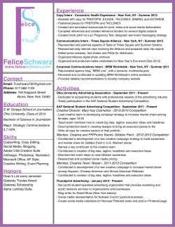
Evaluation of Sample Preparation Methods for LC/MS
that do not require Acetonitrile
1
Lee Williams1, Elena Gairloch1, Rhys Jones1, Helen Lodder1, Steve Jordan1, Richard Jones2, Rick Edmondson2,
Jason Taylor2 & Nathan Twaddle2
Biotage GB Limited, Dyffryn Business Park, Ystrad Mynach, Mid Glamorgan, CF82 7RJ, UK. 2National Center for Toxicological Research,
3900 NCTR Rd, Jefferson, AR, 72079. USA.
Introduction
Sample preparation is essential prior to LC-MS/MS analysis of drugs in biological fluid samples to
remove unwanted matrix components, specifically proteins and phospholipids. These matrix
components can interfere with compounds of interest leading to regions of ion suppression or
enhancement possibly resulting in inaccurate quantitation and sensitivity issues. With the recent
world-wide shortage of acetonitrile and subsequent price increases, many labs have been forced to
re-evaluate acetonitrile-dependent methods. This poster investigates the extract cleanliness of
various alternative approaches to protein precipitation (‘protein crash’) in terms of overall ion
suppression, protein and phospholipid removal. The alternative techniques compared to protein
precipitation (using ISOLUTE PPT+) in this poster were: generic resin-based SPE (EVOLUTE ABN);
resin-based mixed-mode strong cation exchange SPE (EVOLUTE CX); and supported liquid
extraction (ISOLUTE SLE+).
Experimental Procedure
Reagents
Ammonium acetate, ammonium hydroxide, formic and acetic acids were purchased from Sigma
Chemical Co. (Poole, UK). Blank human plasma was purchased from the Welsh Blood Service
(Pontyclun, UK). Control rat serum was obtained from the NCTR animal colony. All solvents were
HPLC grade from Fisher Scientific (Loughborough, UK) and Milli-Q water used throughout. All
materials for the gel electrophoresis work were purchased from Invitrogen (Carlsbad, CA. USA).
Sample Preparation
ISOLUTE PPT+ Plate: Plasma/serum (100 µL) precipitated with 300 µL of MeCN (solvent first
methodology).
Supported Liquid Extraction 200 Plate: Plasma/serum (100 µL) pre-treated 1:1 (v/v) with 0.5M
NH4OH. The pre-treated plasma was loaded onto the plate, a pulse of vacuum applied to initiate
flow and the samples left to absorb for 5 minutes. Extraction was facilitated by application of MTBE
(1 mL).
EVOLUTE ABN: Plasma/serum (100 µL) pre-treated 1:3 (v/v) with 1% formic acid aq. Column
conditioning with MeOH followed by 0.1% formic acid aq (both 1 mL). 400 µL loaded onto the
column and washed with 95/5 (v/v) H2O/MeOH (1 mL). Elution was afforded by application of
MeOH (500 µL).
EVOLUTE CX: Plasma/serum (100 µL) pre-treated 1:3 (v/v) with 50 mM NH4OAc buffer at pH 6.
Column conditioning with MeOH followed by 50 mM NH4OAc buffer at pH 6 (both 1 mL). 400 µL
loaded onto the column and sequentially washed with 50 mM NH4OAc buffer at pH 6 and MeOH
(both 1 mL). Elution was afforded by application of 5% NH4OH/MeOH (1 mL).
-1-
Experiment 1: Overall Ion Suppression FIA-MS/MS
Blank plasma extracts were reconstituted in 1 mL of 50:45:5:0.1% H2O/ACN/MeOH/Formic acid
(v/v) mobile phase spiked with 1 µg/mL caffeine. The 2795 liquid handling unit delivered an
isocratic mobile phase of 50:45:5:0.1% H2O/ACN/MeOH/Formic acid (v/v) at 0.25 mL/min.
Injection volumes were set to 5 µL and the MS set to monitor the MRM transition for caffeine
(195>138, cone voltage 45 V and collision energy 16 eV).
Experiment 2: Gel Electrophoresis
Blank serum extracts were reconstituted in water and 5 µL serum equivalents removed. LDS
sample buffer (4 µL) and reducing agent (2 µL) were added to each aliquot and boiled at ~100°C
for 5 minutes, spun to pull down volume, and allowed to cool to room temperature. Electrophoresis
was performed using NuPAGE Novex 12% Bis-Tris mini gels with MOPS SDS running buffer at 200
V, 120 mA and 12.5 W. The total method time was set to 65 minutes to allow complete migration
of the protein in each gel. 5 µL equivalent of each serum extract was compared to 0.5 µL
equivalent of raw serum and 4 µL of the Benchmark Protein Ladder molecular weight marker in
each gel.
Experiment 3: Phospholipid Removal
Blank plasma extracts were reconstituted in 1 mL of
70:30 (v/v) H2O/MeOH prior to analysis. Initial MS work was performed in the full scan positive ion
mode (250-900 Da) to identify the most abundant phospholipid ions. MRM transitions were then
employed for quantitative purposes using the 184 Da product ion.
HPLC Conditons
Instrument: Waters 2795 Liquid Handling System (Waters Assoc., Milford, MA, USA).
Column: Luna Phenyl-Hexyl 5 µm analytical column (50 x 2.0 mm id) (Phenomenex, Cheshire UK).
Guard Column: Luna Phenyl-Hexyl security guard column (Phenomenex, Cheshire, UK).
Mobile Phase: 0.1% formic acid aq and 0.1% formic acid/MeCN at a flow rate of 0.3 mL/min.
Gradient: The gradient conditions were set to 60%, 0.1% (v/v) formic acid aq and 40% MeCN
increasing to 100% MeCN over 6 minutes. The high organic mobile phase was held for 7 minutes
and initial starting conditions resumed at 13.1 minutes.
Injection Volume: 5 µL
Temperature: Ambient
Mass Spectrometry
Instrument: Ultima Pt triple quadrupole mass spectrometer (Waters Assoc., Manchester, UK)
equipped with an electrospray interface for mass analysis.
Desolvation Temperature: 350 °C
Ion Source Temperature: 100 °C
Collision Gas Pressure: 2.6 x 10⁻³ mbar
Results
Figure 1. illustrates FIA-MS/MS suppression results obtained comparing the various sample
preparation techniques. The extracts using PPT+ exhibited increased overall suppression and
returned a matrix factor of 0.11. Far better matrix factors were returned using the other
techniques; 0.73 using EVOLUTE ABN, 0.8 using EVOLUTE CX and 0.91 using ISOLUTE SLE+.
-2-
EVOLUTE ABN
PPT+
STD
100
STD
STD
MF: 0.73
Time
20.00
0
Time
130.00
30.00
140.00
MRM of 1 Channel ES+
TIC
9.86e5
100
MF: 0.8
%
%
0
10.00
100
MRM of 1 Channel ES+
TIC
1.86e6
STD
MF: 0.91
%
MF: 0.11
ISOLUTE SLE+
EVOLUTE CX
MRM of 1 Channel ES+
TIC
STD
1.84e6
%
100
MRM of 1 Channel ES+
TIC
STD 2.01e6
150.00
0
Time
100.00
160.00
110.00
0
Time
40.00
120.00
50.00
60.00
Figure 1.Overall ion suppression comparison using FIA-MS/MS
Mol. Wt. Marker
ISOLUTE SLE+
Raw Serum
Mol. Wt. Marker
EVOLUTE CX
Raw Serum
Mol. Wt. Marker
EVOLUTE ABN
Raw Serum
PPT+
Mol. Wt. Marker
Raw Serum
Figure 2. illustrates the amount of protein removal afforded by each technique. All four gel profiles
exhibited very little protein in the final extracts. A rough protein quant (n=1) showed
approximately 99% or greater removal of total serum protein for all four techniques.
220
220
220
220
120
120
120
120
70
70
70
50
50
50
40
40
40
30
30
30
20
20
20
10
10
10
70
50
40
30
20
10
Figure 2. Gel Electrophoresis profiles for the various sample prep approaches
Figure 3. illustrates the MRM comparisons for the amount of phospholipid removal from the
extracts. The chromatogram was split into two scan events; the first corresponding to lyso-PL
elution from the HPLC column, and the second corresponding to the less polar larger molecular
weight PL’s. The PPT+ extracts exhibited very high phospholipid content for both the lyso and
larger PL’s. EVOLUTE ABN showed > 95% removal of the larger molecular weight PL’s but
demonstrated reduced lyso PL removal, only removing about 65%. Both the EVOLUTE CX and
ISOLUTE SLE+ showed > 97% both lyso and larger PL removal.
-3-
6.74
0
6.00
8.00
2.00
1: MRM of 5 Channels ES+
TIC
3.39e7
6.00
2.00
8.00
1: MRM of 5 Channels ES+
TIC
3.39e7
4.00
6.00
2.00
8.00
1: MRM of 5 Channels ES+
TIC
3.39e7
100
%
%
Lyso Phospholipids
4.00
100
%
4.00
3.77
0
0
0
2.00
100
2: MRM of 11 Channels ES+
TIC
1.36e7
100
4.00
6.00
8.00
1: MRM of 5 Channels ES+
TIC
3.39e7
100
%
6.25
ISOLUTE SLE+
2: MRM of 11 Channels ES+
TIC
1.36e7
100
%
Phospholipids
EVOLUTE CX
2: MRM of 11 Channels ES+
TIC
1.36e7
100
%
%
EVOLUTE ABN
2: MRM of 11 Channels ES+
7.79
TIC
1.36e7
%
PPT+
100
3.64
3.75
3.61
4.38
0
Time
2.00
4.00
6.00
8.00
0
Time
2.00
4.00
6.00
0
8.00
Time
2.00
4.00
6.00
8.00
0
Time
2.00
4.00
6.00
8.00
Figure 3. Phospholipid MRM comparison of the various sample prep approaches
Overall Conclusions
• Sample clean up was achieved with various sample preparation techniques, using simple generic
methods, without the use of acetonitrile.
• All techniques evaluated removed >99% protein (as shown by gel electrophoresis results)
• EVOLUTE ABN removed 95% of higher MWt PLs, but showed significant suppression due to lysoPLs. This could be reduced further using alternative wash/elution procedures.
• EVOLUTE CX and ISOLUTE SLE+ both removed >97% lyso-PLs and higher MWt PLs
• Compared to acetonitrile based protein precipitation (protein crash), the alternative sample
preparation techniques evaluated in this poster provided superior sample clean up, with
significantly reduced ion suppression due to phospholipids.
Copyright © 2009 Biotage. All rights reserved. All brand and product names are trademarks or registered trademarks of
their respective companies. The information contained in this document is subject to change without notice.
www.biotage.com
United States and Canada
Tel: +1 704 654 4900
Toll-Free: +1 800 446 4752
[email protected]
Sweden
Biotage
Tel: +46 18 56 59 00
[email protected]
United Kingdom, EIRE
Biotage
Tel: +44 1992 501535
[email protected]
Japan
Biotage
Tel: +81 422 281233
[email protected]
-4-


© Copyright 2026