
Mass Spec Sample Handbook
Mass Spec Sample Handbook
Featuring Thermo Scientific Pierce® Products to extract, separate,
purify, detect, and quantify proteins
Table of Contents
Thermo Scientific Products for Mass Spectrometry
Introduction
1
An Introduction to Mass Spectrometry
2
Cells and Tissue
3–5
SILAC Reagents
Cell Surface Protein Isolation Kit
3–4
5
Cell Lysis and Protein Extraction
6–11
Cell Lysis Reagents
Halt™ Protease Inhibitors
Halt Phosphatase Inhibitor
Protein Stabilizing Cocktail
High Abundant Proteins
SwellGel™ Blue Albumin Removal Kit
Pierce IgG/Albumin Removal Kits
6–7
8–9
10
11
12–13
12
13
Pre-Fractionation and Enrichment
14–22
Phosphoprotein Enrichment Kit
Glycoprotein Isolation Kits
Ubiquitin Enrichment Kits
Organelle Enrichment Kits:
Lysosome, Perixisome, Nuclei
Mitochondria Isolation Kits
14–15
16
17
18–19
Sample Clean-Up
23–28
Slide-A-Lyzer ® Dialysis Unit and Cassettes
Zeba™ Desalt Spin Columns
Pierce SDS-PAGE Sample Prep Kit
23–24
25
26
Gel Separation and Protein Detection
27–32
20–22
In-Gel Detection
34–35
In-Gel Tryptic Digestion Kit and
Pierce C-18 Spin Columns
34–35
In-Solution Digestion
36–37
In-Solutions Tryptic Digestion and
Guanidation Kit
36–37
Peptide Enrichment
38
Phosphopeptide Isolation Kit
38
Peptide Clean-Up
39
Pierce Strong Cation and Anion Ion
Exchange Spin Columns
39
Mass Spectrometry
40
Ion Pairing Reagents
Crosslinking Technical Handbook
40
40
Mass Spectrometry Applications
41–43
Crosslinking Reagents for the Analysis of Protein
Interactions by Mass Spectrometry Analysis
Gain new insights into protein interaction
and folding
27
28–29
30
31
32
Protein Digestion
33
GelCode® Blue Stain Reagent
33
42
Protein Quantification by Mass Spectrometry
44–46
TMT Isobaric Mass Tagging Kits
Custom HeavyPeptides™ BASIC and AQUA Kits
Modified HeavyPeptides for Quantification of
Phosphorylated Proteins
44
45
Instruments for Mass Spectrometry
™
SuperSignal® Chemiluminescent Substrates
2-D Sample Prep Kits
2-D Protein Markers
Imperial™ Protein Stain
Pierce Silver Stain for Mass Spectrometry
41
LTQ Orbitrap XL ETD
MALDI LTQ XL
TSQ Quantum Ultra
46
47-48
47
48
48
Introduction
Introduction to Sample Preparation
for 2-D and Mass Spectrometry
Proper sample preparation means better results.
Mass spectrometry (MS) is a powerful and versatile analytical
tool that is playing an increasingly vital role in proteomics,
metabolomics, small-molecule synthesis, and the drug discovery
process. MS enables identification and quantification of known
and unknown compounds by revealing their structural and
chemical properties. Proper sample preparation for MS-based
analysis is a critical step in the proteomics workflow because the
quality and reproducibility of sample extraction and preparation
for downstream analysis significantly impacts the separation and
identification capabilities of MS instruments.
With all of its forms of ionization and detection (e.g., ESI, MALDI,
APcI, FT-MS, ion trap, TOF, Quad), MS allows for the analysis of
samples ranging in mass from 50-300,000 daltons in low femtomole
to high nanomole quantities. Because the proteome is so complex,
there is no one standard method for preparing protein samples
for MS analysis. Protocols differ depending on sample type,
experiment and method of analysis. For example, preparing
samples from a biological fluid involves a different set of procedures than those used for cell culture or tissue. Many factors are
considered, including source, type, physical properties abundance,
complexity, matrix effects and cellular location of the proteins.
Proteins of interest to biological researchers are usually part
of a very complex mixture of other proteins. This presents two
significant problems. First, the two ionization techniques used for
large molecules only work well when the mixture contains roughly
equal amounts of constituents, while in biological samples, different
proteins tend to be present in widely differing amounts. If such a
mixture is ionized using electrospray or MALDI, the more abundant
species have a tendency to “drown” or suppress signals from less
abundant ones. The second problem is that the mass spectrum
from a complex mixture is very difficult to interpret because of the
overwhelming number of mixture components. This is exacerbated
by the fact that enzymatic digestion of a protein gives rise to a
large number of peptide products.
To contend with these problems, scientists for the Pierce
Product Line, now part of the Thermo Scientific brand portfolio
of products have developed a complete workflow of sample
preparation solutions design for better MS analysis. Our researchers
understand the need for integrated proteomics solutions and
work to develop products and technologies that are compatible
and supportive of MS analysis. The following workflow highlights
options for preparing samples from a variety of starting materials
for successful MS analysis.
Sample ionization
Preparation of protein for MS analysis can be accomplished by
many methods, however the initial principle of ion addition to the
protein or peptide is essential for MS analysis. First, intact proteins
are ionized by one of several techniques, and then introduced to a
mass analyser. In the second, proteins are enzymatically digested
into smaller peptides using a protease such as trypsin. Sample
preparation is easier once whole proteins have been digested
into smaller peptide fragments. Subsequently these peptides are
introduced into the mass spectrometer and identified by peptide
mass fingerprinting or tandem mass spectrometry. This latter
approach uses identification at the peptide level from known
peptide sequences in databases to infer the existence of proteins.
Outside the United States, contact your local branch office or distributor.
1
Introduction
Thermo Scientific SILAC
Cell Surface Biotinylation
Cell Lysis Reagents
Protease Inhibitors
Phosphatase Inhibitors
Detergents
Cells and Tissue
Bodily Fluids
Cell Lysis and Protein Extraction
High Abundant Protein Removal
Thermo Scientific Pierce Protein Concentrators
Phosphoprotein Enrichment Kit
Glycoprotein Isolation Kits
Ubiquitin Isolation Kits
Organelle Enrichment
Pre-fractionation & Enrichment
Thermo Scientific Slide-A-Lyzer Dialysis Casettes
Thermo Scientific Zeba Desalt Spin Columns
Thermo Scientific Pierce SDS-PAGE
Sample Prep Kit
Sample Cleanup
2D Sample Prep Kits
Thermo Scientific Imperial Stain
Silver Stain for MS
Gel Separation
IgG/Albumin Removal Kits
Protein A & G
In-Solution Digestion
In-Solution Tryptic Digestion Kit
In-Gel Digestion
Peptide Enrichment
Phosphopeptide Isolation Kit
Thermo Scientific IMAC Resin
Peptide Cleanup
Mass Spectrometry
Thermo Scientific Pierce C-18
Spin Columns
Thermo Scientific SCX and WCX Ion
Exchange Columns
Reagents: Formic Acid, TFA
Instruments: Thermo Scientific
LTQ Orbitrap, TSQ Quantum and
LTQ FT Mass Spectrometers
Thermo Scientific
LTQ Orbitrap XL
Mass Spectrometer
Thermo Scientific Mass Spec Sample Prep Products. Proper sample prep means better results.
2
For more information, or to download product instructions, visit www.thermo.com/pierce
Cells and Tissue
Thermo Scientific SILAC Reagents
SILAC Applications:
• Quantitative analysis of relative changes in protein abundance
from different cell treatments
• Quantitative analysis of proteins for which there are no
antibodies available
• Protein expression profiling of normal vs. disease cells
• Identification and quantification of hundreds to thousands
of proteins in a single experiment
Highlights:
• Efficient – 100% label incorporation into proteins of living cells
• Reproducible – eliminates intra-experimental variability caused
by differential sample preparation
• Flexible – media deficient in both L-lysine and L-arginine,
allowing for more complete proteome coverage through dual
amino acid isotope labeling
• Compatible – label proteins expressed in a wide variety of
mammalian cell lines adapted to grow in DMEM or RPMI 1640
medium, including HeLa, 293T, COS7, U2OS, A549, A431, HepG2,
NIH 3T3, Jurkat and others
SILAC requires growing mammalian cells in specialized media
supplemented with light or heavy forms of essential amino acids;
i.e., 12C6 and 13C6 L-lysine, respectively. A typical experiment
involves growing one cell population in medium containing light
amino acids (control), while the other population is grown in the
presence of heavy amino acids (experimental). The heavy and
light amino acids are incorporated into proteins through natural
cellular protein synthesis. After alteration of the proteome in one
sample through chemical treatment or genetic manipulation, equal
amounts of protein from both cell populations are then combined,
separated by SDS-polyacrylamide gel electrophoresis and
digested with trypsin before MS analysis. Because peptides
labeled with heavy and light amino acids are chemically identical,
they co-elute during reverse-phase column prefractionation and,
therefore, are detected simultaneously during MS analysis. The
relative peak intensities of multiple isotopically distinct peptides
from each protein are then used to determine the average change
in protein abundance in the treated sample (Figure 1).
Cells Grown in Light
Isotope-containing Media
Cells Grown in Heavy
Isotope-containing Media + Treatment
Harvest & Lyse Cells
Quantitate Extracted Protein
Mix Lysates
Excise Bands
Trypsin Digestion
LC-MS/MS
SDS-PAGE
Relative Intensity
Stable isotope labeling using amino acids in cell culture (SILAC)
is a powerful method to identify and quantify relative differential
changes in complex protein samples. The SILAC method uses
in vivo metabolic incorporation of “heavy” 13C- or 15N-labeled
amino acids into proteins followed by mass spectrometry (MS)
analysis for accelerated comprehensive identification,
characterization and quantitation of proteins.
Ratio
Determination
m/z
Light
Heavy
Figure 1. Schematic of SILAC workflow. A549 cells adapted to DMEM
were grown for six passages (10 days) using Thermo Scientific SILAC DMEM
(Product # 89983) containing 0.1 mg/ml heavy 13C6 L-lysine-2HCl or light L-lysineHCl supplemented with 10% dialyzed FBS. After 100% label incorporation, 13C6 Llysine-labeled cells were treated with 5 µM camptothecin (Sigma, St. Louis,
Product # C9911) for 24 hours. Cells from each sample (light and heavy) were
lysed using Thermo Scientific M-PER Mammalian Protein Extraction Reagent
(Product # 78501). Samples were normalized for protein concentration using
the Thermo Scientific Pierce BCA Protein Assay (Product # 23225), and 50 mg of
each sample were equally mixed before 4-20% sodium dodecyl sulfate-polyacrylamide gel electrophoresis (SDS-PAGE) analysis. Gels were stained with
Thermo Scientific GelCode Blue Stain Reagent (Product # 24592) and proteins
were digested and alkylated using the Thermo Scientific In-Gel Tryptic
Digestion Kit (Product # 89871) before analysis using an LTQ Orbitrap®
Hybrid Mass Spectrometer (Thermo Fisher Scientific, San Jose, CA).
Using a Thermo Scientific SILAC Quantitation Kit, A549 cells adapted to grow in Dulbecco’s Modified Eagle Medium (DMEM) were
labeled with 13C6 L-lysine to > 98% isotope incorporation. Heavylabeled cells treated with camptothecin were lysed, mixed with
control lysates, separated by SDS-PAGE and digested with trypsin
before MS analysis. More than 350 proteins were successfully
identified by MS/MS sequencing using a Thermo Scientific LTQ
Orbitrap Mass Spectrometer. Identified peptides were then
quantitated using the Thermo Scientific Bioworks Software Suite
to generate SILAC ratios corresponding to relative changes in
protein abundance (Figure 1).
To order, call 800-874-3723 or 815-968-0747. Outside the United States, contact your local branch office or distributor.
3
Cells and Tissue
Thermo Scientific SILAC Reagents (cont.)
Most of the proteins identified had no change in abundance level
after camptothecin treatment; however, 20% of proteins quantified
in heavy-labeled cells had protein levels (SILAC ratios) 1.5-fold
higher than control cells. One protein that was identified as being
up-regulated in response to camptothecin treatment was proliferating cell nuclear antigen (PCNA), a protein with involvement in
DNA repair (Figure 2). To validate SILAC data, protein levels were
separately quantitated by Western blot (Figure 3). PCNA protein
levels increased 1.9-fold; however, glyceraldehyde-3-phosphate
dehydrogenase (GAPDH) protein did not significantly change. The
abundance ratios determined by Western blot were comparable
to those determined by SILAC.
Thermo Scientific Pierce SILAC Protein Quantitation Kits allow for
complete isotopic labeling of the proteome for analysis of relative
protein abundance by mass spectrometry.
Relative Abundance
Ordering Information
Product # Description
89982
89983
1041.017
1041.518
100
90
80
SILAC Ratio
H:L = 2.1
70
1038.508
1038.007
60
40
1039.010
30
20
1043.023
10
0
1037
1038
1039
1040
Light (L)
1041 1042
m/z Heavy (H)
1043
1044
1045
Figure 2. Representative MS spectra generated using SILAC. Light and heavy
(13C6) L-lysine-containing peptides (AEDNADTLALVFEAPNQEK) from PCNA were
analyzed by MS. Mass spectra of heavy peptides containing 13C6 L-lysine have
an increased mass of 6 Da and are shifted to the right of light peptide spectra
by a mass to charge ratio (m/z) of 3 caused by a +2 ionization of peptides.
α - p53
5.9
α - β-actin
α - GAPDH
Light (L) Heavy (H)
+
1.2
Figure 3. Comparison of A549 protein levels detected by Western blotting
after camptothecin treatment. Ten micrograms of each light (L) and heavy (H)
sample (Figure 1) were analyzed by 4-20% SDS-PAGE and Western blotting
using specific antibodies.
SILAC Protein Quantitation Kit –
DMEM
Kit
Includes: SILAC DMEM Media
Dialyzed FBS
13
C6 L-Lysine-2HCl
L-Lysine-2HCl
L-Arginine-HCl
2 x 500 ml
2 x 50 ml
50 mg
50 mg
2 x 50 mg
500 ml
89986
Dialyzed FBS
50 ml
89987
L-Lysine-2HCl
50 mg
89988
13
50 mg
89989
L-Arginine-HCl
50 mg
89990
13
50 mg
C6 L-Lysine-2HCl
15
C6 N4 L-Arginine-HCl
Complementary Products
78430
Halt Protease Inhibitor
Single-Use Cocktail (100X)
1 ea.
Each 100 µl microtube contains sufficient cocktail to
treat 10 ml of lysate. Protease inhibitors prepared in
DMSO solution.
24 x 100 µl microtubes
0.5 M EDTA Solution (100X), 2.5 ml
Halt Protease Inhibitor Single-Use
Cocktail, EDTA-free (100X)
1 ea.
Each 100 µl microtube contains sufficient cocktail to
treat 10 ml of lysate. Protease inhibitors prepared in
DMSO solution.
24 x 100 µl microtubes
78420
Halt Phosphatase Inhibitor Cocktail
78501
M-PER® Mammalian Protein Extraction Reagent 250 ml
89826
Mem-PER® Membrane Protein
Extraction Reagent
Kit
78833
NE-PER® Nuclear and Cytoplasmic
Extraction Reagents
Kit
89874
Mitochondria Isolation Kit
Kit
89839
Lysosome Enrichment Kit or Tissue
and Cultured Cells
Kit
89840
Peroxisome Enrichment Kit for Tissue
Kit
89841
Nuclei Enrichment Kit for Tissue
Kit
1.1
5 µM camptothecin
2 x 500 ml
2 x 50 ml
50 mg
50 mg
2 x 50 mg
SILAC DMEM Media
(DMEM Medium minus L-Lysine and L-Arginine)
78425
1.9
Includes: SILAC RPMI Media
Dialyzed FBS
13
C6 L-Lysine-2HCl
L-Lysine-2HCl
L-Arginine-HCl
89985
H:L
α - PCNA
Kit
SILAC RPMI Media
500 ml
(RPMI-1640 Medium minus L-Lysine and L-Arginine)
1042.521
1039.512
Pkg. Size
SILAC Protein Quantitation Kit –
RPMI 1640
89984
1042.020
50
1036
References:
1. Everly, P.A., et al. (2004). Quantitative cancer proteomics: Stable isotope labeling
with amino acids (SILAC) as a tool for prostate cancer research. Mol & Cell Proteomics.
3.7: 729-735.
2. Mann, M. (2006). Functional and quantitative proteomics using SILAC. Nature Reviews.
7: 952-959.
3. Levine, A.J. (1997). p53, the cellular gatekeeper for growth and division. Cell. 88: 323–331.
1 ml
The purchase of this product conveys a non-transferable license to the Purchaser to
use this product in methods protected under U.S Patent 6,653,076 (owned by University
of Washington) for research purposes only.
4
For more information, or to download product instructions, visit www.thermo.com/pierce
Thermo Scientific Cell Surface Protein Isolation Kit
Adherent or suspended cells are
first incubated with Sulfo-NHS-SSBiotin, a cleavable reagent. The cells
are lysed with a mild detergent and
labeled proteins are isolated with immobilized NeutrAvidin Resin.
The bound proteins are recovered by incubating the resin with
SDS-PAGE sample buffer containing 50 mM DTT. The reducing
agent cleaves the disulfide bond within the spacer arm of
the biotinylation reagent (Figure 5). Nearly 100% of the bound
proteins are released (Figure 4). The protocol is optimized for
diverse cell lines including NIH 3T3, HeLa, C6 and A431. Isolated
proteins can be analyzed by Western blot, allowing for differential
expression analysis between treated and untreated cells
(Figure 6) or between two or more cell lines.
Convenient biotinylation and isolation of cell surface proteins for
Western blot analysis.
Highlights:
• Isolates cell surface proteins – reduces complexity of lysates
• Efficiently recovers labeled proteins – cleavable biotin allows
for nearly 100% recovery of isolated cell surface proteins
• Convenience – includes all reagents and complete instructions
for labeling, lysis and purification
• Western blotting applications – proteins recovered in SDS-PAGE
buffer are loaded directly onto polyacrylamide gels
• Robust system – protocol designed for diverse cell lines
EGF
The Thermo Scientific Cell Surface Protein Isolation Kit specifically
targets mammalian cell surface proteins to the exclusion of intracellular proteins. The kit efficiently labels proteins with accessible
lysine residues and sufficient extracellular exposure (Figure 4).
-
+
-
+
EGF
Integrin α5
-
-
+
+
EGFR
-
+
-
+
Integrin β1
A. A431
B. HeLa
Figure 6. Differential expression of cell surface proteins in response to EGF.
A431 and HeLa cells were treated with or without 20 ng/ml and 10 ng/ml EGF
for 16 hours, respectively. Both cell types were processed with the Thermo
Scientific Cell Surface Protein Isolation Kit protocol. Elution fractions were
analyzed by Western blot for the quantities of A. integrin β1 and integrin α5
subunits or B. EGFR.
Thermo Scientific
GelCode Blue Safe Stain
Supplier I
Ordering Information
Product # Description
89881
Supplier B
Pkg. Size
Cell Surface Protein Isolation Kit
8 applications
Includes: EZ-Link™ Sulfo-NHS-SS-Biotin
Quenching Solution
Lysis Buffer
Immobilized NeutrAvidin Gel
8 x 12 mg vials
16 ml
4.5 ml
2.25 ml settled gel
supplied as 50% slurry
(4.5 ml total volume)
34 ml
8 spin columns with caps
and collection tubes
Supplier S
Wash Buffer
Column Accessory Pack
Figure 4. Specificity of isolation. HeLa cells were treated with or without
Sulfo-NHS-SS-Biotin and processed with the Thermo Scientific Cell Surface
Protein Isolation Kit protocol. Elution fractions, post-elution resin and flow-through
were analyzed by Western blot for A. cell surface proteins EGFR, IGF-1Rβ, integrin
β1 and integrin α5 and B. intracellular proteins, including heat shock protein 90™
(hsp90) and calnexin. Legend: (+) label, (-) no label, (F) flow-through, (R) Thermo
Scientific NeutrAvidin™ Gel and (E) elution. Only labeled cell surface proteins are
present in the elution fractions.
Quench Reaction
Biotinylate cells
No-Weigh™ Dithiothreitol (DTT)
BupH Phosphate Buffered
Saline
BupH Tris Buffered Saline
Transfer cell pellet
to 1.5 ml tube
45
40
8 x 7.7 mg microtubes
2 packs
1 pack
Isolate biotinylated proteins
on Thermo Scientific
NeutrAvidin Gel
35
30
25
Harvest Cells
30 minutes at 4˚C
20
15
10
75
50
N
gel
N
N
B
Wash gel then elute with SDS-PAGE
sample buffer + 50 mM DTT
gel
Lyse cells
30 minutes on ice
1-D gel
N
+
protein – SH
Perform
electrophoresis
or other application
B
SH
S-S-protein
N
Thermo Scientific NeutrAvidin
Biotin-Binding Protein
B
Biotin
Figure 5. Procedure for the Thermo Scientific Cell Surface Protein Isolation Kit.
To order, call 800-874-3723 or 815-968-0747. Outside the United States, contact your local branch office or distributor.
5
Thermo Scientific Products for Cell Lysis and Protein Extraction
Table 1. Thermo Scientific Cell Lysis Reagents selection guide.
Dialyze1 Compatibility
Description
Organisms/Samples
B-PER® Reagent†
78243, 165 ml
78248, 500 ml
Gram(-) bacteria, S. aureus, H. pylori, E. coli strains
BL21(D3)> JM109> DH5a >M15, Archaebacteria, nematodes
and Acinetobacter sp.
Yes
Reporter assays, IPs2, Western blot, GST- and
histidine-tag purification
B-PER II Reagent
78260, 250 ml
(A 2X version of B-PER Reagent)
Gram(-) bacteria, S. aureus, H. pylori, E. coli strains BL21(D3)>
JM109> DH5α>M15, Archaebacteria, nematodes and
Acinetobacter sp.
Yes
Reporter assays, IPs2, Western blot, GST- and
histidine-tag purification
B-PER PBS Reagent
78266, 500 ml
Gram(-) bacteria, S. aureus, H. pylori, E. coli strains BL21(D3)>
JM109> DH5α>M15, Archaebacteria, nematodes and
Acinetobacter sp.
Yes
Reporter assays, IPs2, Western blot, GST- and
histidine-tag purification
Y-PER® Reagent
78991, 200 ml
78990, 500 ml
S. cerevisiae, Schizo-saccharomyces pombe, C. albicans,
B. subtilis, E. coli, P. pastoris, Strep. avidinii and
Acinetobacter sp.
No
IPs2, Western blot, β-Gal enzyme assays, IEF after
dialysis, GST- and histidine-tag purification
Y-PER Plus Reagent
78998, 25 ml
78999, 500 ml
Yeast (S. cerevisiae) and Acinetobacter sp.
Yes
GST- and histidine-tag purification, Western blot
M-PER Reagent
78503, 25 ml
78501, 250 ml
78505, 1 L
Cultured mammalian cells, COS-7, NIH 3T3, Hepa 1-6, 293,
CHO, MDA, MB231 and FM2
Yes
Luciferase, β-Gal (low signal), CAT, kinase assays,
ELISAs, immobilized glutathione, Western blot
P-PER® Plant Protein
Extraction Reagent
89803, Kit
Multiple plant organs (leaf, stem, root, seed and flowers);
multiple plant species (Arabidopsis, tobacco, maize,
soybeans, peas, spinach, rice and other plant tissues);
and fresh, frozen and dehydrated plant tissues
No
1-D and 2-D gel electrophoresis, Western blotting,
activity assays and protein affinity purifications*
T-PER® Reagent
78510, 500 ml
Heart, liver, kidney and brain
Yes
Luciferase, β-Gal, CAT, kinase assays, Western blot,
ELISAs, immobilized glutathione
I-PER® Reagent
89802, 250 ml
Baculovirus-infected insect cells grown in suspension or
monolayer culture
No
Western blot, 6xHis-tagged protein purification,
protein assays and ion-exchange chromatography
NE-PER Reagent
78833
Tissue: calf liver. Cultured cells: epithelial (HeLa), fibroid
(COS-7), kidney (NIH 3T3), liver (Hepa 1) and brain (C6)
Mem-PER Reagent
89826
Cultured cells: brain (C6), epithelial (HeLa), fibroblasts
(NIH 3T3) and yeast (S. cerevisiae)
Yes4
Western blot and 2-D6
Mitochondria Isolation Kit
for Cultured Cells† 89874
Mitochondria Isolation Kit
for Tissue† 89801
Mammalian cells
Yes7
Western blot, 2-D Western blots, electrophoresis.
Applications include apoptosis, signal transduction
and metabolic studies.
Pierce RIPA Buffer
89900, 100 ml
89901, 250 ml
Cultured mammalian cells and cytoplasmic, membrane
and nuclear proteins
Yes
Reporter assays, protein assays, immunoassays and
protein purification
Lysosome Enrichment Kit for
Tissues and Cultured Cells
89839
Tissues and Cultured Cells
N/A
2D/MS, electron microscopy, disease profiling, gene
expression, signal transduction, and interaction or
localization studies
Peroxisome Enrichment Kit
for Tissue
89840
Heart, liver, kidney and brain
N/A
2D/MS, electron microscopy, disease profiling, gene
expression, signal transduction, and interaction or
localization studies
Nuclei Enrichment Kit for Tissue
89841
Heart, liver, kidney and brain
N/A
2D/MS, electron microscopy, disease profiling, gene
expression, signal transduction, and interaction or
localization studies
No (CER) EMSA (if using < 3 µl or 10%, otherwise dialyze first in
Yes (NER) SAL MINIs5), Western blot, reporter assays, IEF (after
dialysis to reduce salt concentration) and 2-D6
Heart, liver, kidney and brain
1. The detergent can be removed by dialysis
2. Immunoprecipitation
3. Halt Protease Inhibitor Cocktail, Product # 78410 and 78415 (EDTA-free)
4. Samples prepared in Mem-PER Reagent can be dialyzed if the buffer contains detergent (e.g., CHAPS), otherwise use Thermo Scientific Advanced Sample Clean-Up Kit (Product # 89888)
5. Slide-A-Lyzer MINI Dialysis Units
6. 2-D Sample Prep for Nuclear Proteins (Product # 89863) and 2-D Sample Prep for Membrane Proteins (Product # 89864) are designed using our popular NE-PER and Mem-PER Reagents.
7. Need to lyse mitochondria first.
*Although kit works without liquid nitrogen/freeze-grinding, Dounce homogenization, blade-shearing or glass-bead agitation for cell disruption, it is compatible with these alternative mechanical aids.
† See patent information.
6
For more information, or to download product instructions, visit www.thermo.com/pierce
Protein Assay Compatibility
Notes
Pierce BCA Assay and Coomassie Plus Assay
Protease inhibitors3 may be added to prevent protein degradation. Salts, chelating agents and reducing
agents can be added for more efficient lysis. Do not exceed 0.5 M NaCl. Better lysis if cells are frozen in
B-PER Reagent.
Pierce BCA Assay and Coomassie Plus Assay after
Compat-Able™ Protein Assay Reagent Set
(Product # 23215) or dilute two to four times
Protease inhibitors3 may be added to prevent protein degradation. Salts, chelating agents and reducing
agents can be added for more efficient lysis. Better lysis if cells are frozen in B-PER Reagent.
Pierce BCA Assay and Coomassie Plus Assay after
Compat-Able Protein Assay Reagent Set
(Product # 23215) or dilute two to four times
Protease inhibitors3 may be added to prevent protein degradation. Salts, chelating agents and reducing
agents can be added for more efficient lysis. Better lysis if cells are frozen in B-PER Reagent.
Pierce BCA Assay
Protease inhibitors3 may be added to prevent protein degradation. Use at room temperature. Double
incubation time for use at 4°C. Use log-phase cells. For stationary phase cells, add 0.1 M DTT or 20-50
mM TCEP. Will work with 1 mM EDTA. Does not lyse spores. Cannot use with ion exchange columns.
Pierce BCA Assay and Coomassie Plus Assay
Protease inhibitors3 may be added to prevent protein degradation. The addition of up to 2 M NaCl may
result in increased efficiency of lysis and protein yield.
Pierce BCA Assay and Coomassie Plus Assay
Protease inhibitors3 may be added to prevent protein degradation. Adding 150 mM NaCl results in
increased efficiency of lysis and higher protein yield in some cells lines. A PBS rinse of cells prior to lysis
removes contaminants such as phenol red and increases protein yield.
Pierce BCA Assay, Reducing Agent-Compatible
Not compatible with Bradford, Coomassie
or Pierce BCA Assay
Kit lyses most plant cells without harsh mechanical lysis aids; extremely fibrous tissues such as woody
stems may require mechanical grinding by devices not included in this kit.
P-PER Extracts can be quantified using the Pierce BCA Protein Assay Kit, Reducing Agent Compatible
(Product # 23250).
Pierce BCA Assay (dilute 1:1) and
Coomassie Plus Assay
Protease inhibitors3 may be added to prevent protein degradation. Mechanical disruption of the tissue is
still required. Can also be used for cultured cells.
Pierce BCA Assay
Protease inhibitors3 may be added to prevent protein degradation.
Pierce BCA Assay and Coomassie Plus Assay
(dilute CER Reagent mixture four times)
Protease inhibitors3 may be added to prevent protein degradation. Packed cell vol.: 2 x 10⁶ HeLa cells =
10 µl = 20 mg. Tissue yield (calf liver): 3-4 mg cytoplasmic protein/100 mg tissue; 1-1.5 mg nuclear protein/100
mg tissue. Cell yield (HeLa): 300-400 µg cytoplasmic protein/106 cells; 40-60 µg nuclear protein/106 cells.
Positive controls tested: cytoplasmic (β-Gal, PKC, Hsp90); nuclear (Oct-1, p53, DNA polymerase).
Pierce BCA Assay and Coomassie Plus Assay;
hydrophobic phase needs to be dialyzed first;
see instruction book
Protease inhibitors3 may be added to prevent protein degradation. Can dialyze against another detergent
(e.g., CHAPS). Extraction efficiency is generally > 50% with the cell lines tested (having proteins with up
to two transmembrane segments).
Pierce BCA Assay (after lysis)
Protease inhibitors may be added to prevent protein degradation. Douncing will increase isolation
efficiency vs. detergent alone; however, multiple samples can be processed simultaneously using the
reagent-based methods.
Pierce BCA Assay
Protease inhibitors3 may be added to prevent proteolysis and maintain phosphorylation of proteins
Coomassie Plus – The Better Bradford Assay Kit
Protease inhibitors3 may be added to prevent proteolysis and maintain phosphorylation of proteins
Coomassie Plus – The Better Bradford Assay Kit
Protease inhibitors3 may be added to prevent proteolysis and maintain phosphorylation of proteins
Coomassie Plus – The Better Bradford Assay Kit
Protease inhibitors3 may be added to prevent proteolysis and maintain phosphorylation of proteins
To order, call 800-874-3723 or 815-968-0747. Outside the United States, contact your local branch office or distributor.
7
Thermo Scientific Products for Cell Lysis and Protein Extraction
Halt Protease Inhibitor Cocktails
0%
New single-use, ready-to-use protease inhibitor cocktails.
Proteases make up a large category of enzymes, including
endopeptidases and exopeptidases, which catalyze the hydrolytic
breakdown of proteins into peptides or amino acids in a wide
variety of cell and tissue types. This breakdown can lead to the
loss of a large number of valuable proteins, adversely affecting
downstream applications. Thermo Scientific Halt Single-Use
Protease Inhibitor Cocktails help preserve the integrity of target
proteins intended for downstream analysis. These cocktails target
all major classes of proteases including aspartic acid, cysteine,
serine and metalloproteases. AEBSF, aprotinin, bestatin, E64,
leupeptin and pepstatin A, all known potent inhibitors of proteases,
have been expertly formulated in a ready-to-use cocktail and
specially packaged to maintain product integrity and offer a new
level of convenience.
Ready-to-use Halt Protease Inhibitor Cocktails are conveniently
packaged in single-use microtubes, each of which contain
sufficient reagent to treat up to 10 ml of sample. Our Protease
Inhibitor Cocktails are the convenient alternative to protease
inhibitor tablets, saving time, eliminating the mess of splitting tablets
and providing a measure of consistency because you do not have
to use a razor blade to treat samples that are < 10 ml (Figure 1).
Each Halt Single-Use Cocktail contains 24 x 100 µl sealed microtubes
sufficient to treat up to 240 ml of cell extract. As a 100X concentrate
in DMSO, the single-use format provides access to only the
cocktail needed to knock-down protease activity in up to 10 ml
of cell lysate per microtube. The Halt Single-Use Cocktail contains
reversible and irreversible inhibitors for serine-, cysteine-, aspartic
acid-proteases and aminopeptidases present in virtually all
cellular lysate samples. A 0.5 M EDTA solution is provided with
one formulation to promote the inhibition of metalloproteases.
59%
61%
97%
98%
Control;
Extract Only
Protease Inhibitor Thermo Scientific Halt
Cocktail Tablet
Single-Use Cocktail
Tablet;
EDTA-Free
Thermo Scientific Halt
Single-Use EDTA-Free
Cocktail
Percent Inhibition
Figure 1. Thermo Scientific Halt Protease Inhibitor Single-use Cocktails are
more effective than tablet-format cocktails. Using a validated protease assay
and 1.0 mg/ml of rat pancreas extract, the Halt Protease Inhibitor Single-Use
Cocktails, with and without EDTA added, were tested against commercially
available tablet-format protease inhibitor cocktails under the same conditions.
A 1X final concentration of each inhibitor was added. The single-use formulation
resulted in ≥ 97% inhibition compared to ≥ 59% inhibition for the tablet.
Each microtube in the strip is heat-sealed with a foil that can be
easily punctured by a pipette tip. Only that amount of protease
inhibitor cocktail needed for the application is removed from the
microtube. The remaining tubes in the strip are preserved and
protected from exposure to air, moisture and other potential
contamination until needed for subsequent applications. Although
many manufacturers won’t disclose the composition or concentration of their protease inhibitors, we publish the exact identity and
concentration of each cocktail component (Table 2).
Table 2. Thermo Scientific Halt Protease Inhibitor Single-Use Cocktail formulation and concentrations.
8
Protease
Inhibitor
MW
Protease
Family Targeted
Inhibitor
Type
Solubility
(Solvent system)
Concentration of Each Inhibitor
in the Protease Inhibitor Cocktails
(100X in DMSO)
(Product # 78430 and 78425)
AEBSF•HCl
239.5
Serine proteases
Irreversible
200 mg/ml (H2O)
100 mM
Aprotinin
6511.5
Serine proteases
Reversible
10 mg/ml (H2O)
80 µM
Bestatin
308.38
Amino-peptidases
Reversible
5 mg/ml (methanol)
5 mM
E-64
357.4
Cysteine proteases
Irreversible
20 mg/ml (1:1 EtOH/H2O)
1.5 mM
EDTA
(not included in
Product # 78425)
372.24
Metalloproteases
(Chelates divalent cations)
Reversible
10 g/100 ml (H2O)
0.5 M
Leupeptin
475.6
Serine and cysteine
proteases
Reversible
1 mg/ml (H2O)
2 mM
Pepstatin A
685.9
Aspartic acid proteases
Reversible
1 mg/ml (MeOH)
1 mM
For more information, or to download product instructions, visit www.thermo.com/pierce
Highlights:
• Single-use, ready-to-use packaging keeps protease inhibitors
fresh, reduces the risk of reagent contamination and minimizes
waste
• Innovative microtube design allows immediate inhibition of
lysates, no thawing or waiting for tablets to dissolve
• EDTA-containing and EDTA-free formulations published – know
exactly what is in the cocktail
• Compatible with Thermo Scientific Pierce Cell Lysis Buffers
(Figure 2)
• Effective for suppressing proteolytic activity in detergent-based
cell lysis reagents
• Outstanding performance demonstrated with Thermo Scientific
M-PER Mammalian, B-PER Bacterial, T-PER Tissue and Y-PER
Yeast Protein Extraction Reagents
• Stable for one year when refrigerated
• Same high-quality protease inhibitor components available
separately; prepare a custom cocktail or increase the
concentration of a class of protease inhibitor in the formulation
to improve the stability or recovery of a specific target protein
1
2
3
4
1
2
3
Protease Inhibitor References
1. Kulakowska-Bodzon, A., et al. (2006). Methods for samples preparation in proteomic
research. J. Chromatogra. B. (doi:10.1016/j.jchromb.2006.10.040).
2. www.mnhn.fr/mnhn/bpy/2DMS/2DE.pdf: PART I Sample Preparation, p.9 (2.2 Protection
against proteolysis)
3. North, M.J. (1989). Prevention of unwanted proteolysis in Proteolytic Enzymes:
A Practical Approach (Beynon, R.J., Bond, J.S. eds), pp. 105-124, IRL Press, Oxford.
4. Salvensen, G. and Nagase, H. (1989). Inhibition of proteolytic enzymes in Proteolytic
Enzymes: A Practical Approach (Beynon, R.J., Bond, J.S. eds). pp. 83-104, IRL Press,
Oxford.
Ordering Information
Product # Description
78430
Halt Protease Inhibitor
Single-Use Cocktail (100X)
Pkg. Size
1 ea.
Each 100 µl microtube contains sufficient cocktail to
treat 10 ml of lysate. Protease inhibitors prepared in
DMSO solution.
24 x 100 µl microtubes
0.5 M EDTA Solution (100X), 2.5 ml
78425
Halt Protease Inhibitor Single-Use
Cocktail, EDTA-free (100X)
1 ea.
Each 100 µl microtube contains sufficient cocktail to
treat 10 ml of lysate. Protease inhibitors prepared in
DMSO solution.
24 x 100 µl microtubes
4
Also available in new sizes:
78429
Halt Protease Inhibitor Cocktail
5 ml
78437
Halt Protease Inhibitor Cocktail, EDTA-free
5 ml
78438
Halt Protease Inhibitor Cocktail
10 ml
78439
Halt Protease Inhibitor Cocktail, EDTA-free
10 ml
Individually Packaged Protease Inhibitors
78431
AEBSF
100 mg
[4-(2-aminoethyl)benzenesulfonyl fluoride•HCl]
78432
Aprotinin
25 mg
78433
Bestatin
10 mg
N-[(2S,3R)-3-amino-2-hydroxy-4-phenylbutyryl]-L-leucine
78434
A. Thermo Scientific B-PER
Bacterial Protein Extraction Reagent
B. Thermo Scientific M-PER
Mammalian Protein Extraction Reagent
E-64
10 mg
[N-trans-epoxysuccinyl-L-leucine-4 guanidinobutylamide]
Also known as N-[N-(L-3-trans-carboxirane-2-carbonyl)]
-L-leucyl agmatine
78435
Leupeptin
50 mg
(acetyl-leucyl-leucyl-argininal)•Hemisulfate
Figure 2. Thermo Scientific Halt Protease Inhibitor Single-Use Cocktails are
compatible with Thermo Scientific M-PER Mammalian Protein Extraction and
B-PER Bacterial Protein Extraction Reagents. Bovine serum albumin (BSA)
was incubated overnight at 37°C in the presence of 0.1 mg/ml trypsin in M-PER
Extraction Reagent and B-PER Extraction Reagent, respectively. Lane 1. Control
1, BSA only; Lane 2. Control 2, Trypsin (0.1 mg/ml) (+) BSA and no Halt Protease
Inhibitor Single-Use Cocktail present; Lane 3. BSA (+) Trypsin with Halt
Protease Inhibitor Single-Use Cocktail with EDTA; and Lane 4. BSA (+) Trypsin
with Halt Protease Inhibitor Single-Use Cocktail. Under extreme time and temperature conditions, substantial protection from degradation was observed as
marginal to barely detectable cleavage products of BSA.
78436
Pepstatin A
25 mg
Compatible Products
78420
Halt Phosphatase Inhibitor Cocktail
1 ml
1 ml of cocktail protects up to 100 ml of sample.
A mixture of four phosphate inhibitors including
sodium fluoride, sodium orthovanadate, sodium
pyrophosphate and β-glycerophosphate.
89806
Protein Stabilizing Cocktail
10 ml
4X Concentrated Solution
To order, call 800-874-3723 or 815-968-0747. Outside the United States, contact your local branch office or distributor.
9
Thermo Scientific Products for Cell Lysis and Protein Extraction
Halt Phosphatase Inhibitor Cocktails
MEK
–
PMEK
+
–
+
–
+
–
+
Offering protection from both serine/threonine and protein tyrosine
phosphatases.
Highlights:
• All-in-one format – inhibits both serine/threonine and protein
tyrosine phosphatases
• Compatible – works with standard protein assays, including
Pierce BCA Protein Assay Kit and Coomassie Plus (Bradford) Assay
• User-friendly storage at 2-8˚C – saves valuable space in the freezer
• Convenient size – 1 ml of cocktail protects up to 100 ml of sample
Phosphorylation and dephosphorylation is a molecular on/off
switch that regulates many key biological pathways within the
cell, including signal transduction, cell division and apoptosis. The
Thermo Scientific Halt Phosphatase Inhibitor Cocktail (Product #
78420) preserves phosphorylation of proteins in cells and tissues.
The cocktail contains a mixture of four phosphatase inhibitors of
broad specificity, including sodium fluoride, sodium orthovanadate,
sodium pyrophosphate and β-glycerophosphate. Unlike other
commercially available phosphatase inhibitor cocktails that protect
against either serine/threonine phosphatases or protein tyrosine
phosphatases, Halt Phosphatase Inhibitor Cocktail protects
phosphoproteins from both families of phosphatases (Figure 3).
Conveniently packaged in 100X format, the cocktail is stable
at 4˚C and is compatible with common protein assays, including
the BCA Protein Assay Kit (Product # 23225) and Bradford-based
assays such as the Coomassie Plus (Bradford) Assay (Product #
23236).
A.
MAPK
–
PMAPK
+
B.
STAT3
–
PSTAT3
+
C.
Figure 3. Thermo Scientific Halt Phosphatase Inhibitor Cocktail preserves
phosphorylation of MEK, MAP Kinase 42/44 and STAT3 in HeLa cell lysate.
Cells were lysed in the absence (-) and presence (+) of Halt Phosphatase
Inhibitor Cocktail. Lysates were analyzed by Western blot for total and
phosphorylated protein as indicated. A. MEK and phosphorylated MEK (PMEK),
B. MAP kinase and PMAP kinase and C. STAT3 and PSTAT3. The proteins are
phosphorylated on serine, threonine/tyrosine and tyrosine, respectively.
Ordering Information
Product # Description
78420
Halt Phosphatase Inhibitor Cocktail
Pkg. Size
1 ml
Sufficient reagent to protect up to 100 ml of sample.
78426
Halt Phosphatase Inhibitor Cocktail
5 x 1 ml
78427
Halt Phosphatase Inhibitor Cocktail
10 ml
78428
Halt Phosphatase Inhibitor Single-Use
Cocktail
24x 100 µl
Each 100 µl microtube contains sufficient cocktail to
treat 10 ml of lysate.
10
For more information, or to download product instructions, visit www.thermo.com/pierce
Protein Stabilizing Cocktail
Extends shelf-life of precious proteins.
Highlights:
• Stabilizes proteins more than buffer alone
• Does not destabilize biomolecules in downstream assays
• All components are dialyzable
• Easy to pipette (vs. 50% glycerol)
Protein Classes Tested:
• Kinase
• Peroxidase
• Cytokine
• Restriction Enzyme
• Phosphatase
• Luciferase
• Antibody
Although the degree of stabilization is protein-specific, the cocktail
significantly stabilizes proteins better than conventional buffer
alone. The Protein Stabilizing Cocktail is nontoxic. If needed, all
cocktail components can be removed by dialysis or desalting
before use in downstream assays.
Visit our website to view data, including luciferase activity
stabilized by the Protein Stabilizing Cocktail.
Ordering Information
Product # Description
89806
Protein Stabilizing Cocktail,
4X Concentrated Solution
Pkg. Size
10 ml
Sufficient reagent to make 40 ml of storage solution.
The Thermo Scientific Protein Stabilizing Cocktail is a versatile
solution that increases the shelf-life of purified or partially purified
proteins during routine storage. The proprietary formulation of
low-molecular weight, naturally occurring molecules helps protect
proteins from environmental stresses that can otherwise lead to
enzyme inactivation, aggregation and freeze-thaw damage.
To order, call 800-874-3723 or 815-968-0747. Outside the United States, contact your local branch office or distributor.
11
Thermo Scientific Products for High Abundant Proteins
SwellGel Blue Albumin Removal Kit
Offers ease-of-use, speed and performance not found in other kits.
Thermo Scientific SwellGel Blue Albumin Removal Discs are for
high-capacity albumin removal from small (10-100 µl) serum
samples. Each disc can bind approximately 2 mg of human serum
albumin.* Just add water and the discs hydrate in less than
20 seconds to form an equilibrated chromatographic resin bed
of approximately 200 µl. Processed cells are ready for immediate
downstream analysis by 2-D or MS applications. The simplicity
of sample preparation and the versatility offered by the SwellGel
Technology make it ideal for standard chromatography, 2-D and
micro-spin column applications.
Albumin
Contamination
Albumin
Depleted
Highlights:
• Excellent for 2-D applications – allows you to study less
abundant proteins without interference from albumin (Figure 1)
or processing buffers
• Convenient and versatile – kit includes everything needed to
remove albumin, also works with 96-well filter plates
• Room temperature storage – discs do not leak or spill, and the
kit stores conveniently on bench top
• Pre-measured, easy-to-handle discs with an easy-to-follow
protocol (Figure 2) – no waste – all resin can be used; no need
to pipette messy slurries
SwellGel Blue Albumin Removal Kits contain ready-to-hydrate
Cibacron™ Blue Activated Discs, binding/wash buffer and
Mini-Spin Columns. Separations are performed in microcentrifuge
columns and can be processed by low-speed centrifugation or
syringe-based methods.
Reference
1. Steel, L.F., et al. (2003). Efficient and specific removal of albumin from human serum
samples. Mol Cell Proteomics 2, 262-270.
Ordering Information
A.
B.
Product # Description
Figure 1. Albumin removal for 2-D analysis. Serum sample obtained by diluting
10 µl human serum with 40 µl TBS and loading 5 µl onto Gel A. Albumin-free
sample obtained by diluting 50 µl human serum 1:1 with buffer, adding it to a
Thermo Scientific SwellGel Blue Disc, washing the resin three times with 50 µl
of buffer, and combining the fractions; loading 5 µl onto Gel B. Both samples
were focused using pH 4-7 IEF strips and run on 8-16% Tris-glycine gels.
Step 1.
Hydrate Thermo Scientific
Disc in water. Spin.
SERUM
FILTRATE
Step 2.
Add serum sample
to the resin. Incubate
2 minutes.
Step 3.
Recover filtrate
and add to the resin.
Incubate 2 minutes
and spin.
89845
SwellGel Blue Albumin Removal Kit*
Includes: SwellGel Blue Albumin
Removal Discs
Binding/Wash Buffer
Mini-Spin Columns
Step 5.
Combine desired fractions.
Figure 2. Thermo Scientific SwellGel Blue Albumin Removal Kit Protocol.
12
12-25
Reactions
25 discs
6.25 ml
25 columns
*SwellGel Blue Discs have differing levels of affinity for species-specific albumins.
SwellGel Blue Discs bind human, swine and sheep albumin. They can also be used
with a minor protocol change for bovine, calf and goat albumin. They do not bind
mouse albumin.
BUFFER
Step 4.
Add Binding/Elution Buffer.
Pkg. Size
For more information, or to download product instructions, visit www.thermo.com/pierce
Pierce Albumin/IgG Removal Kits
Process multiple 2-D or 2D/LC samples in less than 40 minutes.
Low-abundant proteins in human serum and plasma can provide
information about human diseases. However, human fluid analysis
is often complicated by high concentrations of albumin and IgG,
which accounts for up to 70% of total serum protein (Figure 4). We
offer several kits for removing albumin and/or IgG from samples,
allowing analysis of low-abundant proteins.
For Detailed Proteomic Analysis
The Thermo Scientific Pierce Albumin/IgG Removal Kit
(Product # 89875) contains a mixed bed matrix of immobilized
Cibacron Blue Dye and high-capacity immobilized Protein A
and provides an efficient method for depleting excess abundant
proteins while enriching the lower molecular weight proteins,
peptides and other small components. The kit is formulated for
depleting human serum albumin (HSA) and the major subclasses
of immunoglobulin (IgG) from serum, plasma or spinal fluids.
An Economical – Yet Effective – Method
Thermo Scientific Pierce Antibody-Based Albumin/IgG Removal Kit
(Product # 89876) contains an Immobilized Anti-IgG/Anti-Albumin
Gel (immobilized antibodies are polyclonal from goat) for removing
HSA and all major subclasses of gamma globulin (IgG) from human
serum plasma or spinal fluids. This kit uses highly specific antibodies, providing minimal nonspecific interactions. This product
can deplete greater than 95% of HSA and 90% of IgG.
Each kit is optimized for downstream analysis by 2-D
electrophoresis or 2D/LC mass spectrometric methods and allows
for multiple samples to be processed simultaneously.
Figure 4. 2-D gel of human serum processed with the Thermo Scientific Pierce
Antibody-Based Albumin/IgG Removal Kit. Approximately 30 µg of processed
human serum was isoelectric-focused on a pH 5-8 strip and then separated in
the second dimension on an 8-16% SDS-polyacrylamide gel. Thermo Scientific
Silver Stain II (Product # 24612) was used to visualize bands. With albumin and
IgG removed, low-abundant proteins are readily identified.
Ordering Information
Product # Description
89875
89876
Kit
Sufficient material for up to 25 samples
of 600 µg of serum.
Includes: Dye/Protein A Resin
(sold as 50% slurry [4.25 total volume])
Spin Columns and Accessories
2.13 ml
resin bed
27
Pierce Antibody-Based
Albumin/IgG Removal Kit
Sufficient material for up to 12 samples
of 600 µg of serum.
Includes: Antibody-Based Resin
(sold as 62% slurry
[7.25 total volume])
Spin Columns and Caps
Albumin
Pkg. Size
Pierce Albumin/IgG Removal Kit
Kit
4.5 ml of
immobilized
anti-HSA/
anti-IgG
resin bed
12
lgG Heavy Chain
lgG Light Chain
Figure 3. 2-D gel unprocessed human serum. Approximately 30 µg of
unprocessed human serum was isoelectric-focused on a pH 5-8 strip and
then separated in the second dimension on an 8-16% SDS-polyacrylamide gel.
Thermo Scientific Silver Stain II (Product # 24612) was used to visualize bands.
Large poorly resolved bands corresponding to human serum and IgG obscure
much of the gel.
To order, call 800-874-3723 or 815-968-0747. Outside the United States, contact your local branch office or distributor.
13
Thermo Scientific Products for Pre-fractionation and Enrichment
Pierce Phosphoprotein Enrichment Kits
Process cell and tissue samples in less time and with greater purity.
Phosphorylation is one of the most frequently occurring posttranslational modifications in proteins. It is estimated that as
many as 30% of all cellular proteins are transiently phosphorylated
on serine, threonine and tyrosine residues.
Reversible protein phosphorylation regulates nearly all
intracellular biological events, including signal transduction,
protein-protein interactions, protein stability, protein localization,
apoptosis and cell-cycle control. Deregulation of protein
phosphorylation is a hallmark of numerous human diseases,
including cancer and metabolic and immune disorders.
Detecting changes in protein phosphorylation can be a difficult
task because of the transient labile state of the phosphate
group. Furthermore, low phosphoprotein abundance and poorly
developed phospho-specific antibodies contribute to difficulties in
phosphoprotein detection. Recent advances in mass spectrometry
technology in combination with phosphoprotein enrichment using
immobilized metal affinity chromatography (IMAC) have resulted in
greater resolution of the phosphoproteome.
The new Thermo Scientific Pierce Phosphoprotein Enrichment Kit
efficiently enriches phosphorylated proteins derived from
mammalian cells and tissues. The proprietary metal and buffer
composition produces superior yields with negligible nonspecific
binding.
Phospho-specific antibodies recognizing key regulatory proteins
involved in growth factor signaling were used to monitor binding specificity of our Phosphoprotein Enrichment Kit (Figure 1).
Specificity of the kit is further demonstrated by the absence of
Cytochrome C (pI 9.6) and p15Ink4b (pI 5.5), two proteins not
predicted to be phosphorylated, in the elution fraction and their
emergence in the flow-through and wash fractions (Figure 1).
Furthermore, dephosphorylation of HeLa cell extract in vitro
resulted in diminished binding of PTEN, MAPK and GSK3β to the
Pierce Phosphoprotein Enrichment Column as evidenced by their
absence in the elution fraction. Conversely, all three proteins
were present in the elution fraction from non-treated HeLa extract
(Figure 2). Our Phosphoprotein Enrichment Kit provided superior
and efficient phosphoprotein enrichment yields when compared to
competitors’ products (Table 1). It also effectively enriched phosphoproteins from homogenized mouse liver tissue (Figure 3).
Table 1. The Thermo Scientific Pierce Phosphoprotein Enrichment Kit
provides higher phosphoprotein yields in less time than competitors’ kits.
Kit
Thermo Scientific
Pierce Phosphoprotein
Enrichment Kit
Yield (%)
Enrichment Time (Hours)
15
1.5
Supplier Q Kit
4.4
4.5
Supplier I Kit
2.6*
3.5
Supplier C Kit
8
3
Supplier E Kit
Too dilute to determine
5
* Based on maximum 1 mg load per manufacturer’s protocol.
Highlights:
• Specific – low contamination from nonspecific proteins
• Fast – easy-to-use spin format enriches of phosphorylated
proteins in less than 2 hours
• Superior yield – high yield from complex biological samples, cell
culture lysate and mouse tissue extract
• Convenient format – complete kit includes pre-dispensed spin
columns, buffers, reagents and Thermo Scientific Pierce Protein
Concentrators
• Compatible – works with downstream applications, including
mass spectrometry, Western blotting and 2D-PAGE
14
For more information, or to download product instructions, visit www.thermo.com/pierce
FT
W
E
L10
L
FT
W
E
L25
Phospho-PTEN S380
HeLa + EGF
Phospho-MAPK T202/Y204
Phospho-Rb S795
Phospho-PTEN S380
Cytochrome C
Phospho-GSK3β S9
Phospho-MAPK T202/Y204
NIH 3T3 + PDGF
Phospho-Akt S473
Phospho-Src Y527
Cytochrome C
p15Ink4b
Figure 1. Highly pure phosphoprotein enrichment from complex biological
samples. Serum-starved HeLa and NIH 3T3 cells were stimulated with EGF and
PDGF, respectively. Cell lysate (2 mg) was used for enrichment. Concentrated
flow-through, wash and elution fractions were resolved by SDS-PAGE. Gel
lanes were normalized by protein concentration (10 µg/lane). Western blot
analysis was performed using antibodies that detect site-specific
phosphorylation events. Cytochrome C (pI 9.6) and p15Ink4b (pI 5.5) served
as negative controls for nonspecific binding of non-phosphorylated proteins.
FT = flow-through fraction, W = pooled wash fractions, E = pooled elution
fractions and L = non-enriched total cell lysate.
Total Cell Lysate
25 µg
λ Pase +
10 µg
+
Figure 3. Efficient enrichment of phosphoproteins from mouse liver extract.
Homogenized mouse liver extract (~2 mg) was loaded onto a Thermo Scientific
Pierce Phosphoprotein Enrichment Column. Concentrated flow-through, wash
and elution fractions were resolved by SDS-PAGE. Gel lanes were normalized by
protein concentration (10 µg/lane). Western blot analysis was performed using
antibodies that detect site-specific phosphorylation events. Cytochrome C
(pI 9.6) served as a negative control for nonspecific binding. L10 = non-enriched
total cell extract (10 µg), FT = flow-through fraction, W = wash fraction,
E = elution fraction and L25 = non-enriched total cell extract (25 µg).
Ordering Information
Product # Description
90003
Pkg. Size
Pierce Phosphoprotein Enrichment Kit
Kit
Includes: Phosphoprotein Enrichment Column Resin
Bed (1 ml)
Lysis/Binding/Wash Buffer
Elution Buffer
CHAPS
White Column Caps
Pierce Protein Concentrator
7 ml/9K MWCO
10 ea.
325 ml
60 ml
1g
10 Devices
Elution
10 µg
+
Phospho-PTEN S380
Phospho-MAPK T202/Y204
Phospho-GSK3β S9
Figure 2. Highly specific phosphoprotein purification from lambda
phosphatase-treated cells. Non-treated and lambda dephosphorylated HeLa
cell extract (2 mg) was loaded onto separate Thermo Scientific Pierce
Phosphoprotein Enrichment Columns. Concentrated elution fractions were
resolved by SDS-PAGE. Gel lanes were normalized by protein concentration
(10 µg/lane). To determine enrichment, 10 µg and 25 µg of non-treated or
lambda phosphatase-treated total cell extract (non-enriched) was loaded onto
each gel. Western blot analysis was performed using phospho-specific
antibodies recognizing key proteins in the Ras-MAPK and PI3K-Akt signaling
cascades.
To order, call 800-874-3723 or 815-968-0747. Outside the United States, contact your local branch office or distributor.
15
Thermo Scientific Products for Pre-fractionation and Enrichment
Highlights:
• High recovery – equivalent or greater glycoprotein recovery vs.
competitor kits and lectin resins
• Fast – glycoprotein purification in less than one hour
• Versatile – isolate glycoproteins from various sample types;
e.g., human serum and cell lysate
• Robust – lectin does not leach from resin when processing sample
• Convenient – complete kit contains lectin resins and spin
columns with all necessary reagents
• Compatible with Bradford-based protein assays – dialysis or
protein precipitation of recovered glycoproteins is not required
before protein assay
A.
Glycoprotein Isolation Kit, ConA
89805
Glycoprotein Isolation Kit, WGA
Sufficient reagents to isolate glycoproteins with strong
affinity for WGA from 10 samples of up to 640 µl (1-1.5
mg total protein) each
Includes: WGA Lectin Resin, 1.1 ml resin supplied as a
50% slurry
Binding/Wash Buffer, 5X Stock Solution,
6.5 ml
Elution Buffer, 5 ml
Column Accessory Pack, 10 Spin Columns
with Caps and 20 Collection Tubes
A.
B.
Figure 4. Glycoprotein isolation from human serum and cell lysate:
performance comparison of kits using ConA resin. Human serum and CHO
lysate samples were processed with the Thermo Scientific Glycoprotein
Isolation Kit, ConA and with other commercially available ConA resins. An
equivalent amount of total protein was applied to each resin. Eluted glycoprotein
fractions were compared with ConA Resin boiled in SDS-PAGE Buffer to
release lectins. All fractions were normalized by volume and resolved on 8-16%
polyacrylamide gels. Gels were silver-stained. A. Eluted glycoprotein fractions
from applied human serum and B. eluted glycoprotein fractions from applied
CHO lysate. Arrows identify protein bands that result from ConA leaching from
the resin during the elution process.
16
Pkg. Size
Kit
Sufficient reagents to isolate glycoproteins with
strong affinity for ConA from 10 samples of up to 640 µl
(1-1.5 mg total protein) each.
Includes: ConA Lectin Resin, 1.1 ml resin supplied as
a 50% slurry
Binding/Wash Buffer, 5X Stock Solution,
6.5 ml
Elution Buffer, 5 ml
Column Accessory Pack, 10 Spin Columns
with Caps and 20 Collection Tubes
ConA
Supplier G
Supplier A
Thermo Scientific
B.
Product # Description
CHO Lysate
ConA
Supplier G
Supplier A
Thermo Scientific
Figure 5. Glycoprotein isolation from
human serum and cell lysate: performance
comparison of kits using WGA resin.
Human serum and CHO lysate samples
were processed with the Thermo Scientific
Glycoprotein Isolation Kit, WGA and with
other commercially available WGA resins.
An equivalent amount of total protein was
applied to each resin. Eluted glycoprotein
fractions were normalized by volume and
resolved on 8-16% polyacrylamide gels. A.
Eluted glycoprotein fraction from applied
human serum and B. eluted glycoprotein
fraction from applied CHO lysate.
Ordering Information
89804
Human Serum
Supplier A
Two lectin-based glycoprotein isolation kits, concanavalin A
(ConA) and wheat germ agglutinin (WGA), allow isolation of
glycoproteins from complex protein mixtures, including serum,
tissue and cultured cell lysates, thus enabling downstream
analysis. ConA lectin recognizes α-linked mannose and terminal
glucose residues, while WGA lectin selectively binds to N-acetyl
glucosamine (GlcNAc) groups and to sialic acid.
CHO Lysate
Thermo Scientific
Isolate glycoproteins from complex protein mixtures.
Human Serum
Thermo Scientific
Glycoprotein Isolation Kits
For more information, or to download product instructions, visit www.thermo.com/pierce
Kit
The Thermo Scientific Ubiquitin Enrichment Kit isolates
polyubiquitin protein conjugates from cultured cells and tissue
samples. The enriched fraction is analyzed to determine the
amount of general ubiquitin conjugates present or to identify a
specific protein by Western blotting. The assay protocol is
fast, straightforward and allows isolation of polyubiquitinated
proteins and the fractionation of monoubiquitinated species in
the resin flow-through. The Ubiquitin Enrichment Kit outperforms
other suppliers’ kits and provides a clean and specific preparation
of proteins when compared to a control resin.
Highlights:
• Fast – less than 45 minutes hands-on time
• Complete – includes all reagents needed for ubiquitin-modified protein enrichment from cultured cells and tissue samples,
including spin columns and ubiquitin antibody
• Flexible – sample incubation from 2 hours to overnight allows
assay to be completed in several hours or in less than 30 minutes
after overnight incubation
• Robust – compatible with all Thermo Scientific Cell Lysis
Solutions and standard RIPA formulations
• Multiple-sample format – easily processes 1-15 samples
concurrently
Elution
Flow-through
GSH Resin
Elution
Ab-based
Method
Flow-through
Elution
Supplier C
Elution
EHela load
The ubiquitin proteasome pathway is the principal non-lysosomal
pathway that controls the proteolysis of proteins. This pathway
is significantly involved in a variety of cellular processes,
including DNA repair, transcriptional regulation, signal transduction, cell metabolism and morphogenesis. Differences in total
ubiquitination or the ubiquitination of specific proteins affect
numerous pathological conditions, including malignancies, certain
genetic diseases and neurodegenerative diseases.1
Flow-through
Thermo
Scientific
Recover ubiquitin modified protein in < 45 minutes.
Flow-through
Ubiquitin Enrichment Kit
kDa
220
120
100
80
60
50
40
20
Figure 6. The Thermo Scientific Ubiquitin Enrichment Kit recovers more
ubiquitin-modified proteins than any other method. Epoxomicin-treated HeLa
cell lysates (EHeLa, 150 µg) were enriched. After elution, all samples were
normalized to the initial load (EHeLa load). The flow-through and elution
obtained using the Thermo Scientific Ubiquitin Enrichment Kit are shown first.
The results using another supplier’s enrichment kit are shown for comparison
(Supplier C, manufacturer’s instructions for this kit were followed). Additionally,
the results obtained using an anti-ubiquitin monoclonal antibody-based enrichment scheme (antibody-based) and a negative control resin (GSH resin) are
shown. The elution from each resin shows the amount of ubiquitin-modified
protein that was captured using that method.
Reference
1. Ciechanover, A. (1998). The ubiquitin-proteasome pathway: on protein death and cell life.
EMBO J. 17(24), 7151-1760.
Ordering Information
Product # Description
89899
Ubiquitin Enrichment Kit
Pkg. Size
Kit
Contains sufficient materials for enriching up to
15 lysate samples containing ~0.15 mg total protein
per sample.
Pack 1
Polyubiquitin Positive Control (1,000X), 50 µl, 2 mg/ml
Anti-ubiquitin Antibody, 50 µl rabbit antiserum
Pack 2
Polyubiquitin Affinity Resin, 300 µl, supplied as a
25% slurry
Binding Capacity: ~1 µg per 20 µl of slurry
BupH™ Tris Buffered Saline Pack, 1 ea., makes
500 ml of 0.025 M Tris, 0.15 M NaCl; pH 7.2
Spin Columns and Accessories, 18 columns with top
and bottom caps
To order, call 800-874-3723 or 815-968-0747. Outside the United States, contact your local branch office or distributor.
17
Thermo Scientific Products for Pre-fractionation and Enrichment
Organelle Enrichment Kits
Lamp-1
Enrichment kits for lysosomes, peroxisomes and nuclei.
Liver
Subcellular fractionation simplifies complex protein mixtures,
thereby facilitating proteomic analysis. We developed three
organelle enrichment kits for lysosomes, peroxisomes and nuclei
that enable enrichment of intact organelles from cells and
tissue. Each kit uses density gradient centrifugation to separate
organelles from contaminating cellular structures (Figures 7-11).
The isolated organelles are suitable for applications such as
2D/MS, electron microscopy, disease profiling, gene expression,
signal transduction and interaction or localization studies.
Highlights:
• Efficient and easy to use – kit reagents and gradient
centrifugation separate organelles from contaminating
structures (Table 2)
• Compatible – prepare samples for downstream applications,
including 2D/MS, electron microscopy, disease profiling, gene
expression, signal transduction and interaction or localization
studies
• Complete – kits contain sufficient material for 25 applications
Kidney
Lysosomes
Lysate
Figure 8. Lysosome enrichment from tissue. Liver and kidney tissues (200 mg
each) were processed using the Thermo Scientific Lysosome Enrichment Kit
for Tissue and Cultured Cells. Total lysate and isolated lysosomes were
analyzed by Western blotting for Lamp-1, a lysosomal membrane protein marker.
PMP70
Liver
Table 2. Thermo Scientific Organelle Enrichment Kits are a convenient and
fast means for sample preparation.
Target
Organelle
Sample
Source
Lysosome
Cells
OptiPrep® Density
Gradient
Centrifugation
Speed (x g)
Centrifugation
Time (Mins)
15%, 17%, 20%, 23%,
27% and 30%
145,000
120
Kidney
Tissue
(soft & hard)
Peroxisome
Soft Tissue
27.5%, 30% and 35%
180,000
90
Hard Tissue
18%, 20% and 27.5%
180,000
90
Nuclei
Tissue
(soft & hard)
7%, 23% and 27.5%
40,000
90
Lamp-1
Lysate
Peroxisomes
Figure 9. Peroxisome enrichment from tissue. Liver and kidney tissues (300 mg
each) were processed using the Thermo Scientific Peroxisome Enrichment
Kit for Tissue. Total lysate and isolated peroxisomes were analyzed by
Western blotting for PMP70, a peroxisomal membrane protein marker.
Cathepsin D
Figure 10. Nuclei enrichment from tissue. Liver and kidney tissue (400 mg each)
Ab: HDAC
A431 cells
Liver
HeLa cells
Lysate
Lysosomes
Lysate
Lysosomes
Figure 7. Lysosome enrichment from cultured cells. A431 and HeLa wet
cell paste (~200 mg) was processed using the Thermo Scientific Lysosome
Enrichment Kit for Tissue and Cultured Cells. Total cell lysate and isolated
lysosomes were analyzed by Western blotting for Lamp-1 and Cathepsin D,
membrane-bound and soluble lysosome markers, respectively.
Kidney
Lysate
Nuclei
were processed using the Thermo Scientific Nuclei Enrichment Kit for Tissue.
Total cell lysate and isolated nuclei were analyzed by Western blotting for
histone deacetylase (HDAC), a soluble nuclear marker.
18
For more information, or to download product instructions, visit www.thermo.com/pierce
Thermo Scientific
COX4
PMP70
Supplier S
COX4
PMP70
Liver
Ordering Information
Product # Description
Lysosomes
89839
Kidney
Lysosome Enrichment Kit for Tissues and
Cultured Cells
Sufficient material for 25 applications.
Includes: Lysosome Enrichment Reagent A
Lysosome Enrichment Reagent B
OptiPrep Cell Separation Media
BupH Phosphate Buffered Saline
Liver
Nuclei
89840
Kidney
Peroxisome Enrichment Kit for Tissue
Sufficient material for 25 applications.
Includes: Peroxisome Enrichment Reagent A
Peroxisome Enrichment Reagent B
OptiPrep Cell Separation Media
BupH Phosphate Buffered Saline
Liver
Peroxisomes
89841
Kidney
Figure 11. Cross-contamination of enriched organelles. Liver and kidney
tissues (200 mg each) were processed using each organelle isolation kit and
analyzed via Western blotting for contamination by probing for COX4 and
PMP70, mitochondria and peroxisome markers, respectively. Samples were
normalized by protein amount (~ 10 µg of total proteins), except for the liver
and kidney fractions from the peroxisome experiment, which were normalized
by volume. Tissues from rats of identical weight and ages were processed
with similar kits from Supplier S according to the manufacturer’s instructions.
Significant contamination in the lysosome and nuclei fractions is observed with
the procedure from Supplier S.
References
Gjoen, T., et al. (1997). Lysosomes and endocytosis. Subcellular fractionation: A practical
approach. Eds. Graham, J.M. and Rickwood, D., IRL Press Limited, Oxford, England,
169-200.
Graham, J.M. (1997). Homogenization of tissues and cells. Subcellular fractionation:
A practical approach. Eds. Graham, J.M. and Rickwood, D., IRL Press Limited, Oxford,
England, 1-28.
Hajra, A.K., et al. (1985). Anal Biochem, 148(2), 233-244.
Hinton, R.H. and Mullock, B.M. (1997). Isolation of subcellular fractions. Subcellular fractionation: A practical approach. Eds. Graham, J.M. and Rickwood, D., IRL Press Limited,
Oxford, England, 31-69.
Luers, G., et al. (1993). J. Cell. Bio., 121(6), 1271-1280.
Lukong, K.E., et al. (2001). J. Biol Chem., 276, 46172-46181.
Van Veldhoven, P.P., et al. (1996). Anal Biochem, 237(1), 17-23.
Nuclei Enrichment Kit for Tissue
Sufficient material for 25 applications.
Includes: Nuclei Enrichment Reagent A
Nuclei Enrichment Reagent B
OptiPrep Cell Separation Media
BupH Phosphate Buffered Saline
89874
Mitochondria Isolation Kit for Cultured Cells†
Sufficient reagents for 50 applications.
Includes: Mitochondria Isolation Reagent A
Mitochondria Isolation Reagent B
Mitochondria Isolation Reagent C
89801
Mitochondria Isolation Kit for Tissue†
Sufficient reagents for 50 applications.
Includes: Mitochondria Isolation Reagent A
Mitochondria Isolation Reagent B
Mitochondria Isolation Reagent C
Bovine Serum Albumin
BupH Phosphate Buffered Saline
Pkg. Size
Kit
90 ml
90 ml
50 ml
1 pack
Kit
90 ml
90 ml
50 ml
1 pack
Kit
90 ml
90 ml
50 ml
1 pack
Kit
50 ml
500 ml
70 ml
Kit
50 ml
500 ml
65 ml
230 mg
1 pack
Complementary Products
89910
HDAC Polyclonal Antibody
50 µg
89911
PMP 70 Polyclonal Antibody
50 µg
89912
Beta’-COP Polyclonal Antibody
50 µg
89913
VDAC Polyclonal Antibody
50 µg
89915
Cathepsin Monoclonal Antibody
25 µg
89916
Nucleoporin p62 Monoclonal Antibody
25 µg
89917
Lamp-1 Monoclonal Antibody
50 µg
89918
Cytochrome C Monoclonal Antibody
100 µg
† See patent information.
To order, call 800-874-3723 or 815-968-0747. Outside the United States, contact your local branch office or distributor.
19
Thermo Scientific Products for
Pre-fractionation and Enrichment
Mammalian cells (2 x 10⁷)
Mitochondria Isolation Kit for Cultured Cells
Add 800 µl Reagent A
Option A.
Add 10 µl
Incubate
Reagent B
2 minutes on ice
Isolate intact mitochondria with maximum yield in only 40 minutes.
Option B.
Dounce
homogenize
Highlights:
• Fast – isolate intact mitochondria in approximately 40 minutes
• Multi-sample format – reagent-based method allows for
concurrent preparation of multiple samples
• Optional alternate method – reagents and protocol included
for traditional Dounce homogenization
• Benchtop-compatibility – isolation performed in a
microcentrifuge tube
The isolation of mitochondria is typically a laborious process
requiring single-sample processing with Dounce homogenization.
The Thermo Scientific Mitochondria Isolation Kit for Cultured Cells
uses a non-mechanical, reagent-based method (Figure 12, Option
A) that allows multiple samples (≤ 6) to be processed concurrently.
Cultured mammalian cell pellets are gently lysed using a
proprietary formulation that results in maximum yield of mitochondria with minimal damage to integrity. The kit also offers a second
isolation method based on traditional Dounce homogenization
(Figure 12, Option B), which results in two-fold more mitochondria
recovery, as determined by protein assay. Both methods use
differential centrifugation to separate the mitochondrial and
cytosolic fractions with a bench-top microcentrifuge and are
completed in approximately 40 minutes (post-cell harvest).
Once isolated, the mitochondria can be used in downstream
applications such as apoptosis, signal transduction and metabolic
studies, as well as to facilitate mitochondrial proteomics efforts.
Incubate 5 minutes on ice
Vortex every minute
Add 800 µl Reagent C
Centrifuge 700 x g
10 minutes at 4°C
Pellet
(Nuclei and cell debris)
Supernatant
*Centrifuge 12,000 x g
15 minutes at 4°C
Cytosol
Mitochondria
Wash with
500 µl Reagent C
Centrifuge 12,000 x g
5 minutes at 4°C
Figure 12. Procedure for the isolation of mitochondria from cultured
mammalian cells using Option A. the reagent-based method and Option B.
the Dounce-based method.
* A more purified preparation of mitochondria can be obtained by centrifuging at
3,000 x g instead of 12,000 x g.
The reagent- and Dounce-based isolation procedures are outlined in
Figure 12. Approximately 2 x 10⁷ mammalian cells (NIH 3T3 or C6) were
pelleted per sample in a 2 ml microcentrifuge tube. The cells were
resuspended in Mitochondria Isolation Reagent A and incubated on
ice for 2 minutes. Using the reagent-based method, the cells were
lysed by adding Mitochondria Isolation Reagent B in conjunction with
frequent vortexing. The lysate was mixed with Mitochondria Isolation
Reagent C and centrifuged to remove nuclei, unbroken cells and cellular debris. The supernatant was subsequently centrifuged to collect
the mitochondria, and the pellet was surface-washed to remove cytosolic contaminants. Mitochondria prepared using Dounce homogenization followed a similar protocol. Briefly, 2 x 10⁷ NIH 3T3 cells were
resuspended in Mitochondria Isolation Reagent A, incubated for
2 minutes and then transferred to a tissue grinder and lysed with
80 strokes. The cell homogenate was mixed with Mitochondria
Isolation Reagent C and the remainder of the protocol as detailed in
Figure 12, Option B was performed.
Damage to the outer membrane was assessed by Western blot
analysis of Cytochrome C and voltage-dependent anion channel
(VDAC) (Figure 13). Cytochrome C resides in the intermembrane
space of undamaged mitochondria and VDAC is an integral
membrane protein in the outer mitochondrial membrane.
Negligible amounts of both proteins were present in the cytosol,
indicating that the mitochondria remained intact during isolation.
Contamination of the mitochondria with cytosolic components
was negligible. Following a single wash of the collected pellet of
mitochondria, minimal heat shock protein 90 (Hsp90) contamination
was detected in a Western blot (Figure 14).
20
For more information, or to download product instructions, visit www.thermo.com/pierce
Cytochrome C
M
Table 3. Collection of mitochondria (reagent-based method).
VDAC
C
M
C
Reagentbased
A.
B.
C.
D.
RCF (x g)
Protein (µg)
% Total Protein
3,000
159.1
62
12,000
98.2
38
Once isolated, mitochondria are ready for many downstream
applications, including Western blotting. 2-D Western analysis of
isolated mitochondria was used to identify Mn-superoxide
dismutase, a detoxifying enzyme residing in the mitochondrial
matrix (Figure 16).
Douncebased
Figure 13. Analysis of mitochondrial integrity. Mitochondria and cytosol fractions
were prepared from C6 cells using the reagent-based method (A. and B.) or
Dounce homogenization (C. and D.) Fractions were analyzed via Western blot
for cytochrome C (A. and C.) or voltage-dependent anion channel (VDAC) (B.
and D.). Thermo Scientific SuperSignal West Pico Chemiluminescent Substrate
(Product # 34080) was used for detection. M = mitochondria and C = cytosol.
3
pl
MW (K)
222 —
114 —
88 —
50 —
10
33 —
27 —
Protein
Location
hsp90
Cytosol
M
C
17 —
Figure 14. Analysis of cytosolic contamination in mitochondria fraction.
Mitochondria and cytosol fractions were prepared from NIH 3T3 cells.
Each fraction was analyzed by Western blot for the cytosolic protein, Hsp90.
M = mitochondria and C = cytosol.
A more purified preparation of mitochondria was obtained by
decreasing the centrifugation speed used to collect the organelle.
Mitochondria collection, normally performed at 12,000 x g, was
split into a low-speed collection at 3,000 x g and a subsequent
high-speed collection of remaining mitochondria in the supernatant at 12,000 x g. Western blot analysis of PMP70, an abundant
integral membrane protein of the peroxisome, and Cathepsin S, a
cysteine protease in the lysosome, resulted in > 50% reduction in
contamination from these organelles (Figure 15) while recovering
approximately 60% of the mitochondria routinely collected with a
higher centrifugation speed (Table 3).
A.
Protein
Location
PMP70
Peroxisome
B. Cathepsin S
M1
M2
C
M1
M2
C
Figure 16. 2-D Western blot of superoxide dismutase (Mn-SOD) in isolated
mitochondria. Mitochondria were isolated from NIH 3T3 cells using the Dounce
method, resolved by 2-DE and analyzed by Western blot for manganese-containing SOD. Approximately 15 µg of mitochondrial protein was focused on an
11 cm, pH 3-10 IPG strip. The second dimension was performed using 8-16%
SDS-PAGE.
Ordering Information
Product # Description
89874
89918
Pkg. Size
Mitochondria Isolation Kit for Cultured Cells
Kit
Sufficient reagents for 50 applications.
Includes: Mitochondria Isolation Kit Reagent A
Mitochondria Isolation Kit Reagent B
Mitochondria Isolation Kit Reagent C
50 ml
500 µl
70 ml
Cytochrome C Monoclonal Antibody
100 µg
Lysosome
Figure 15. Reduction of lysosomal and peroxisomal contaminants in
mitochondrial fraction. Mitochondria and cytosol fractions were prepared
using a modified reagent-based isolation method. Heavy, more purified
mitochondria were collected at 3,000 x g and the supernatant was centrifuged
at 12,000 x g to collect remaining mitochondria. Each fraction was analyzed by
Western blot for A. Peroxisomal membrane protein 70 (PMP70, C6 cells) and
B. Lysosomal Cathepsin S (NIH 3T3 cells). M1 = 3,000 x g mitochondria fraction,
M2 = 12,000 x g mitochondria fraction and C = cytosol
**See Table 3 for protein quantification.
To order, call 800-874-3723 or 815-968-0747. Outside the United States, contact your local branch office or distributor.
21
Thermo Scientific Products for Pre-fractionation and Enrichment
Mitochondria Isolation Kit for Tissue
MW (K)
Isolate intact mitochondria with maximum yield in only 60 minutes.
Highlights:
• Quick and convenient – isolates intact mitochondria in less than
60 minutes
• Versatile – isolate soft- and hard-tissue samples
• Multi-sample format – reagent-based approach enables
simultaneous processing of multiple samples
• Optional alternate method – reagents and protocol included
for the traditional Dounce homogenization procedure with fewer
required strokes
• Bench-top-compatible – both procedures are performed using
a microcentrifuge tube
The Thermo Scientific Mitochondria Isolation Kit for Tissue
(Product # 89801) isolates intact mitochondria from soft- and
hard-tissue samples. The kit offers two methods for mitochondria
isolation. The first method uses a unique reagent-based procedure
that enables simultaneous multi-sample processing. The second
method relies on traditional Dounce homogenization for tissue disruption and subsequent isolation. Both procedures use differential
centrifugation to separate the intact mitochondria using a benchtop microcentrifuge and are completed in less than 60 minutes. In
addition, both procedures have been optimized for maximum yield
of mitochondria with minimal damage to its integrity (Figure 17).
The isolated mitochondria can be used for many applications,
including 1-D and 2-D Western blotting (Figure 18) and protein
profiling by mass spectroscopy.
COX4
M
Reagentbased
1.
VDAC
C
M
2.
M
5.
32.1 —
26.1 —
16.1 —
Figure 18. 2-D Western blot of superoxide dismutase (Mn-SOD) in isolated
mitochondria. Intact mitochondria from the liver of a female Sprague-Dawley
rat was processed using the Dounce homogenization method. The isolated
mitochondria was lysed using Thermo Scientific M-PER Mammalian Protein
Extraction Reagent (Product # 78501) and approximately 35 µg of total mitochondrial protein was added to 2-D sample buffer (8 M urea, 4% CHAPS,
pH 5-8 carrier ampholytes, 50 mM DTT). Proteins were resolved on a pH 5-8 IPG
strip followed by 8-16% SDS-PAGE and analyzed by Western blot for Mn-SOD.
References for Mitochondrial Proteome
1. Lescuyer, P., et al. (2003). Progress in the definition of a reference human mitochondrial
proteome. Proteomics 3, 157-167.
2. Taylor, S.W., et al. (2003). Characterization of the human heart mitochondrial proteome.
Nat. Biotechnol. 21, 281-285.
Ordering Information
Product # Description
89801
C
3.
Pkg. Size
Mitochondria Isolation Kit for Tissue
Kit
Sufficient reagents for 50 isolations of intact
mitochondria from soft and hard tissue.
Includes: Mitochondria Isolation Kit Reagent A
Mitochondria Isolation Kit Reagent B
Mitochondria Isolation Kit Reagent C
BSA
BupH Phosphate Buffered Saline
50 ml
500 µl
65 ml
235 mg
1 pack
Cytochrome C Monoclonal Antibody
6.
Figure 17. Analysis of mitochondrial integrity. Mitochondria (M) and cytosolic
(C) fractions were prepared from fresh rat liver (Panels 1, 2, 3, 5 and 6) and
heart (Panel 4) tissue samples using the reagent-based and Dounce
homogenization methods. Fractions were analyzed via Western blot for
COX4, voltage-dependent anion channel (VDAC) and Cytochrome C. Thermo
Scientific SuperSignal West Pico Chemiluminescent Substrate (Product # 34080)
was used for detection. COX4 is an inner-mitochondria membrane protein, VDAC
is an outer-mitochondria membrane protein and cytochrome C is located in the
intermembrane space.
22
10
49.3 —
89918
Douncebased
4.
pl
220 —
114 —
80.2 —
Cytochrome C
C
3
For more information, or to download product instructions, visit www.thermo.com/pierce
100 µg
Thermo Scientific Products for Sample Clean-up
High-Performance Dialysis
Product Selection Guide
10-100 µl
0.1-30 ml
15-100 ml
MWCO Membrane
Thermo Scientific Slide-A-Lyzer®
MINI Dialysis Unit†
Thermo Scientific Slide-A-Lyzer
Dialysis Cassette†
Thermo Scientific SnakeSkin®
Dialysis Tubing
2K
N/A
•
N/A
3.5K
•
•
•
7K
•
•
•
10K
•
•
•
20K
•
•
N/A
† See patent information.
High-Performance Dialysis
Slide-A-Lyzer MINI Dialysis Units
Our dialysis products take the hassle and the mess out of sample
dialysis.
For sample volumes as small as 10 µl.
The Thermo Scientific High-performance Dialysis Products are
ideally suited for the variety of sample sizes common in research
laboratories. The different MINI Units, Cassettes and Tubing
products pictured here are available in 2K, 3.5K, 7K, 10K and 20K
molecular-weight cutoff membrane options (MWCO, denoted by
blue, pink, green, orange and purple labels and cassette colors,
respectively).
12-30 ml
0.5-3 ml
Highlights:
• 100% leak-tested – patented design does not permit “wicking”
that can occur in homemade devices
• Very affordable
• Excellent sample recoveries – the Thermo Scientific
Slide-A-Lyzer MINI Dialysis Unit generally recovers 9-10 µl
after dialysis of a 10 µl sample
• Time of dialysis drastically reduced – converts 100 µl of
pH 2.8 buffer to pH 9.4 dialyzing against 1 L bicarbonate
buffer, pH 9.4 in less than 10 minutes
3-12 ml
0.1-0.5 ml
10-100 µl
Thermo Scientific
Slide-A-Lyzer
MINI Dialysis Unit
1. Apply sample with a pipette.
2. Place the Thermo Scientific
Slide-A-Lyzer MINI Dialysis Unit
into the float.
3. Insert the float into the beaker
containing the dialysate.
4. Recover sample.
15-100 ml
Thermo Scientific
SnakeSkin
Dialysis Tubing
Figure 1. Four-step procedure for buffer exchange using Thermo Scientific
Slide-A-Lyzer MINI Dialysis Units.
To order, call 800-874-3723 or 815-968-0747. Outside the United States, contact your local branch office or distributor.
23
Thermo Scientific Products for Sample Clean-up
Slide-A-Lyzer Dialysis Cassettes
Require just half the time of dialysis tubing!
The Thermo Scientific Slide-A-Lyzer Dialysis Cassette is the
product of choice for rapidly dialyzing sample volumes from
100 µl to 30 ml.
Highlights:
• > 95% sample recovery – sample volume remains visible
throughout dialysis
• No knots or clamps to loosen and leak – secure design prevents
sample loss due to leaks
• Rigid frame permits smooth sample withdrawal of submilliliter
volumes – removing every last drop is easy – even for scientists
who have never before performed dialysis
• High surface area/sample volume ratio will dialyze twice as fast
as dialysis via conventional tubing – patented Cassette design
spreads the sample over a large surface area and the double
membrane promotes fast dialysis
• Clear color-coded plastic allows you to see your injection
1. Remove a Cassette from the
protective pouch. Fill the Cassette
cavity with your sample through one
of the guide inlets in the corner of
the cassette. With the syringe still
inserted into the cavity, draw up on
the syringe to remove air.
2. Attach a flotation buoy and dialyze.
Each buoy serves as an effective
flotation device and also as a
convenient bench-top stand for the
Cassette. There is no need to worry
about suspension of the dialysis bag.
Updated dialysis and desalt brochure!
Request a copy of the updated Dialysis
and Desalt Handbook that features the
following products:
• Slide-A-Lyzer MINI Dialysis Units with
3.5K, 10K, and 20K membranes for highrecovery dialysis of 100-100 µl samples
• Slide-A-Lyzer Dialysis Cassettes with
2K, 3.5K, 7K, 10K and 20K membranes
for high-recovery dialysis of 0.1-30 ml
samples
• SnakeSkin Dialysis Tubing, made with regenerated cellulose
with 3.5K, 7K and 10K MWCO for quick, clean and easy dialysis
of 15-100 ml samples
• Zeba Protein Desalt Products
3. Inject the Cassette chamber with
air and withdraw your dialyzed sample
from the Cassette.
Figure 2. Use of a Thermo Scientific Slide-A-Lyzer Dialysis Cassette.
Visit our website www.thermo.com/dialysis for information and
ordering information for the complete Slide-A-Lyzer Product Line.
Log on to our website or contact your distributor to request a copy.
24
For more information, or to download product instructions, visit www.thermo.com/pierce
Zeba Desalt Spin Columns
Desalt sample volumes ranging from 2 µl to 4 ml and experience
exceptional protein recovery quickly.
Although numerous techniques and resins for desalting are
available, most have many drawbacks, including significant
sample loss, long processing times and the need to collect multiple
fractions. Thermo Scientific Zeba Desalt Spin Columns provide
excellent protein recovery without the limitations associated
with other desalting methods. Zeba Desalt Spin Columns are
available in micro†, 0.5, 2, 5 and 10 ml formats and allow processing
of samples ranging from 2 µl to 4 ml (Table 1).
Table 1. Recommended sample volumes for Thermo Scientific Zeba
Spin Columns.
Resin Bed
Sample Volume
75 µl (micro) column
2-12 µl
0.5 ml column
30-130 µl
2 ml column
200-700 µl
5 ml column
600-2,000 µl
10 ml column
1,500-4,000 µl
96-well
20-100 µl
Zeba Desalt Spin Columns contain a proprietary and exclusive
high-performance desalting resin that offers exceptional desalting
and protein-recovery characteristics compared to other commercially available resins (Figure 3). Samples containing as low as
25 µg/ml of protein can be processed, providing exceptional
protein recovery and ≥ 95% retention of salts and other small
molecules (< 1,000 MW).
Supplier G
Supplier B
Control
Thermo Scientific
Sample Size
200 µl + 50 µl stacker
Supplier G
Supplier B
Thermo Scientific
Sample Size
700 µl
Control
Highlights:
• Exceptional protein recovery
• Wide product offering accommodates your sample needs
• Easy to use with no cumbersome column preparation
or equilibration
• No screening fractions for protein or waiting for protein to
emerge by gravity flow
• Minimal sample dilution
Sample
250 µg/ml BSA
(66 kDa)
A.
5.2
97%
78%
79%
0.19
0.45
0.73
5.2
90%
56%
29%
Recovery
0.14
0.07
0.14
Conductivity
(mho)
Sample
25 µg/ml BSA
(66 kDa)
B.
Figure 3. Increased protein recovery with Thermo Scientific Zeba Desalt
Spin Columns. Samples of bovine serum albumin (BSA) at A. 250 µg/ml and
B. 25 µg/ml in 1 M NaCl were desalted with the 2 ml Zeba Desalt Spin Columns
and other commercial desalting resins using similar formats. A portion of the
recovered sample (10 µl) was analyzed by SDS-PAGE. The remaining sample
was used for conductivity measurements and the Thermo Scientific Pierce
BCA Protein Assay (Product # 23225) was performed to determine protein
concentration. Zeba Desalt Resin provides significantly greater protein recovery
under all conditions tested. Conductivity and protein recovery values after
desalting are indicated for 250 µg/ml samples.
Ordering Information
Product # Description
Pkg. Size
89877
Zeba Micro Desalt Spin Columns†
25/pkg.
89878
Zeba Micro Desalt Spin Columns
50/pkg.
89882
Zeba Desalt Spin Columns, 0.5 ml
25/pkg.
89883
Zeba Desalt Spin Columns, 0.5 ml
50/pkg.
89889
Zeba Desalt Spin Columns, 2 ml
5/pkg.
89890
Zeba Desalt Spin Columns, 2 ml
25/pkg.
89891
Zeba Desalt Spin Columns, 5 ml
5/pkg.
89892
Zeba Desalt Spin Columns, 5 ml
25/pkg.
89893
Zeba Desalt Spin Columns, 10 ml
5/pkg.
89894
Zeba Desalt Spin Columns, 10 ml
25/pkg.
89807
Zeba 96-well Desalting Spin Plates
2 Plates.
89808
Zeba 96-well Desalting Spin Plates
4 Plates.
Spin Columns (No Resin)
89879
Pierce Centrifuge Micro Spin Columns
50/pkg.
89868
Pierce Centrifuge Columns, 0.8 ml
50/pkg.
89896
Pierce Centrifuge Columns, 2 ml
25/pkg.
89897
Pierce Centrifuge Columns, 5 ml
25/pkg.
89898
Pierce Centrifuge Columns, 10 ml
25/pkg.
† See patent information.
To order, call 800-874-3723 or 815-968-0747. Outside the United States, contact your local branch office or distributor.
25
Thermo Scientific Products for Sample Clean-up
Eliminate band distortions caused by contaminants.
Highlights:
• Eliminates artifacts caused by incompatible contaminants
– removes dyes, reducing agents, detergents, sugars, glycerol,
guanidine, urea and ammonium sulfate to provide reproducible
results on SDS-PAGE analysis
• Compatible with the Thermo Scientific Pierce BCA Protein Assay
– allows quantification of the processed sample
• Enriches dilute protein solutions – concentrates protein sample
by eight-fold in less than 20 minutes
• Fast and easy-to-use for up to 70 µg of protein per sample – uses
new spin-cup format that allows processing of high amounts of
protein
The Thermo Scientific Pierce SDS-PAGE Sample Prep Kit
improves SDS-PAGE analysis of difficult-to-analyze samples.
While numerous compounds are incompatible with typical
electrophoretic running buffers and SDS-PAGE separation, our kit
cleans up protein samples in minutes, removing a wide range of
interfering chemicals, such as chaotropic agents, detergents,
lipids, pH extremes and salts.
Untreated
M
S
Thermo Scientific - Treated
M
S
M
S
M
Prepare the following sample types for SDS-PAGE analysis:
• Inclusion bodies solubilized in guanidine•HCl
• Samples containing low-pH buffers, thiocyanate or urea
• Proteins precipitated in ammonium sulfate
• Dilute protein solutions
100
Percent Protein Recovered
Pierce SDS-PAGE Sample Prep Kit
88%
80
85%
77%
75%
77%
40
20
0
Carbonic
Anhydrase
Ovalbumin
Transferrin
Ubiquitin Cytochrome C Bacterial
Lysate
Figure 5. Consistent protein recovery is achieved. Pure proteins (60 µg) of
assorted molecular weights from left to right: 30K, 44K, 80K, 86K and 12K and
bacterial lysate at 27K were processed using this kit. Protein concentrations
were determined with the Thermo Scientific Pierce BCA Protein Assay and
reported as percent protein recovered.
Table 2. Compatibility of common SDS-PAGE-interfering reagents.
S
Interfering Reagents
Figure 4. Eliminate distortion caused by detergents. Rat C6 cells were lysed
and a membrane protein fraction isolated using Thermo Scientific Mem-PER
Eukaryotic Membrane Protein Extraction Reagent (Product # 89826). Membrane
and hydrophilic cell fractions were separated by SDS-PAGE using 4-20% gradient
gels with or without prior treatment using our kit. Western blot analysis was
performed using an antibody against cytochrome oxidase subunit 4 (COX 4)
and Thermo Scientific SuperSignal West Femto Chemiluminescent Substrate
(Product # 34095). The kit-treated samples exhibit better band straightness
and resolution with low molecular weight proteins than samples that were
untreated. S = Soluble fraction (hydrophilic) and M = Membrane fraction.
Percent Protein Recovered
(Starting amount = 20 µg BSA)
Control (Water)
75%
0.5 M Sodium Chloride
80%
2 M Ammonium Sulfate
76%
20% SDS
75%
10% Triton® Detergent
75%
6 M Urea: DMSO (1:3 ratio)
75%
1M Sodium Chloride
75%
6M Urea
74%
10% CHAPS
80%
25% Glycerol
71%
10% OTG
71%
2 M Guanidinium•HCl
70%
40% Sucrose
70%
Ordering Information
Product # Description
89888
26
74%
60
Pkg. Size
Pierce SDS-PAGE Sample Prep Kit
Kit
Sufficient reagents for 50 2-300 µl samples.
This product replaces Product # 26800.
Includes: Protein Binding Resin
Elution Buffer
Purified DMSO
Spin Cups
Collection Tubes
1 ml
5.0 ml
27 ml
50
72
For more information, or to download product instructions, visit www.thermo.com/pierce
Thermo Scientific Products for
Protein Detection and Gel Separation
SuperSignal Chemiluminescent Substrates†
A sensitive, user-friendly detection system for 2-D Western blotting.
MW (K)
116 —
84 —
5
pl
8
pl
10
48 —
Methods
Membrane protein extracts were prepared from cultured NIH
3T3 or S. cerevisiae EGY194 cells using the 2-D-Sample Prep
for Membrane Proteins Kit (Product # 89864). Nuclear protein
extracts were prepared from TNFα-induced HeLa cells using
the 2-D Sample Prep for Nuclear Proteins Kit (Product # 89863).
Approximately 15-30 µg of purified protein was processed and
focused on 11 cm pH 5-8 or pH 3-10 IPG strips. The second
dimension was performed using 8-16% SDS-PAGE, and the
resulting 2-D gels were transferred by electroblotting to
nitrocellulose membranes for Western blot analysis. The nonspecific sites were blocked overnight using Thermo Scientific
SuperBlock (TBS) Blocking Buffer (Product # 37535) containing
0.05% Tween-20 at 4°C with shaking. The blots were incubated
with primary antibodies against the integral membrane
proteins flotillin and mitochondrial porin, and the nuclear proteins
NF-κB:p50 and p65 for one hour at room temperature (RT). The
blots were then washed three times with TBS (Product # 28376)
containing 0.05% Tween -20. Following the wash, the blots were
incubated in HRP-conjugated secondary antibodies (Product #s
31402, 31430 and 31460) for one hour at RT. After washing with
TBS (Product # 28376) containing 0.1% Tween -20, the blots were
incubated for five minutes in SuperSignal West Femto Maximum
Sensitivity Chemiluminescent Substrate (Product # 34095). Excess
substrate was drained and the blots were exposed to Thermo
Scientific CL-XPosure™ Film (Product # 34090) for 1 second.
Results and discussion
Thermo Scientific SuperSignal West Femto Chemiluminescent
Substrate was highly sensitive and effective for detecting proteins
in both low and high abundance in complex protein mixtures
resolved on 2-D gels. The integral membrane proteins flotillin
and mitochondrial porin were identified in mammalian and yeast
membrane protein extracts, respectively (Figure 1). Similar
sensitivity also occurred with transcription factors stimulated
to translocate from the cytosol to the nucleus in response to
TNFα. NF-κB p50 and its precursor, p105, were readily detected in
nuclear extracts prepared from TNFα-stimulated cells (Figure 2A).
Degradation of the p50 factor was also seen that is likely from
inhibitor IkB loss. Another NF-κB transcription factor, p65, was
also identified (Figure 2B).
Conclusion
SuperSignal West Femto Chemiluminescent Substrate’s enhanced
sensitivity and signal duration allows for an improved signal-tonoise ratio that requires less sample, faster signal generation and
greater light emission longevity.
31 —
25 —
A. Flotillin
3
MW (K)
116 —
84 —
48 —
31 —
25 —
B. Mitochondrial Porin
Figure 1. 2-D Western blots of membrane proteins prepared with the Thermo
Scientific 2-D Sample Prep for Membrane Proteins Kit. Membrane protein
extract was prepared from mammalian or yeast cells and then concentrated
and cleaned up. Processed membrane protein samples (approximately 30 µg
each as determined by parallel Thermo Scientific Pierce BCA Assay of 2-D
SDS-PAGE Sample Prep Resin eluate) were resolved on 2-D gels and
analyzed by Western blot for A. flotillin (48 kDa, pI 6.46-6.96, NIH 3T3 cells)
and B. mitochondrial porin (30 kDa, pI 6.58-7.08, S. cerevisiae EGY194 cells).
MW (K)
116 —
84 —
3
pl
10
48 —
31 —
A. NF-κB p105 & p50
MW (K)
84 —
48 —
3
pl
10
31 —
25 —
B. NF-κB p65
Figure 2. 2-D Western blots of nuclear proteins prepared with the Thermo
Scientific 2-D Sample Prep for Nuclear Proteins Kit. Nuclear protein extract
was prepared from TNFα-induced HeLa cells and then desalted/bufferexchanged. Two-dimensional gels were prepared with 15 µg of protein each
and analyzed by Western blot for A. NF-κB p105 and p50 and B. NF-κB p65.
Ordering Information
Product # Description
Pkg. Size
34080
SuperSignal West Pico
Chemiluminescent Substrate
500 ml
34075
SuperSignal West Dura Extended
Duration Substrate
100 ml
34095
SuperSignal West Femto Maximum
Sensitivity Substrate
100 ml
89863
2-D Sample Prep for Nuclear Proteins
Kit
89864
2-D Sample Prep for Membrane Proteins
Kit
† See patent information.
To order, call 800-874-3723 or 815-968-0747. Outside the United States, contact your local branch office or distributor.
27
Thermo Scientific Products for
Protein Detection and Gel Separation
Novel 2-D Sample Preparation Kits
and Other Tools
The mechanics of preparing gels have improved greatly over
the years; however, sample preparation remains a bottleneck.
Problems typically include charged contaminants, dilute solutions,
insoluble proteins and low-abundant proteins. Furthermore,
common methods to remove contaminants and concentrate
proteins, such as dialysis and precipitation, are time-consuming
and can be unreliable. We have developed four 2-D sample
preparation kits to address these challenges. The kits use protein
desalting spin columns to remove small charged contaminants
and concurrently buffer-exchange proteins into 2-D Sample Buffer,
thereby increasing the amount of protein that can be loaded onto
an immobilized pH gradient (IPG) strip. The Thermo Scientific
2-D Sample Prep for Membrane Proteins and the 2-D Sample
Prep for Nuclear Proteins combine the isolation of membrane
and nuclear proteins, respectively, with sample preparation.
Alternatively, the 2-D Sample Preps for Soluble and Insoluble
Proteins are for researchers who require clean up of samples
isolated by their own methods.
MW (K)
5
pl
8
pl
8
97 —
66 —
43 —
29 —
20 —
14 —
A. Processed
MW (K)
5
97 —
66 —
43 —
29 —
20 —
14 —
2-D Sample Prep for Membrane Proteins Kit
B. Unprocessed
Cell fractionation and sample preparation are key for optimal 2-D
gel analysis, but are challenging when working with membrane
proteins. The Thermo Scientific 2-D Sample Prep for Membrane
Proteins Kit combines membrane protein extraction with 2-D
sample preparation in a fast, convenient and reliable protocol.
Membrane proteins are first extracted from cultured cells using
the Thermo Scientific Mem-PER Eukaryotic Membrane Protein
Extraction Kit which works on hard or soft tissue samples.3 The
prepared extract is subsequently cleaned up in a two-step
process. Isolated membrane proteins are first treated with Thermo
Scientific Pierce 2-D SDS-PAGE Resin, a modified form of
diatomaceous earth that selectively binds to proteins. The resulting eluate is desalted/buffer-exchanged and electrophoresed.
Multiple chaotropes present in the 2-D Sample Buffer improve
protein solubility1,2 and result in high-resolution 2-D gels. The kit
effectively isolates integral membrane proteins from both cultured
mammalian and yeast cells (Figure 3) and concentrates proteins
up to six-fold, improving detection of low-abundant proteins
(Figure 3).
Approximately 30-100 µg of membrane proteins are ready for
analysis in less than 90 minutes.
Highlights:
• Requires no precipitation – eliminates difficult resolubilization steps
• Isolates efficiently – isolates 30-100 µg of membrane proteins
• Concentrates and cleans up – membrane proteins are
concentrated up to six-fold and excess detergent is removed
• Buffer-exchanges membrane proteins into 2-D Sample Buffer
– maintaining proteins in solution
• Works quickly – membrane proteins are prepared in less than
one hour and are cleaned up in 30-40 minutes
• Contains thiourea in the 2-D Sample Buffer – increases protein
solubility and improves protein resolution
28
Figure 3. Concentration of membrane protein extract prepared with the
Thermo Scientific 2-D Sample Prep for Membrane Proteins Kit. Membrane
protein extract was prepared from C6 cells. A. Approximately 75 µl of extract
(representing approximately four times the volume shown in B) was processed
and buffer-exchanged. The entire sample obtained (35 µg as determined by
parallel Thermo Scientific Pierce BCA Assay of 2-D SDS-PAGE Resin eluate)
was analyzed on a 2-D gel. B. Approximately 18.5 µl of unprocessed membrane
protein extract, corresponding to the maximum volume recommended for
IPG strip rehydration, was analyzed directly on a 2-D gel. Both samples were
focused on 11 cm, pH 5-8 IPG strips followed by 8-16% SDS-PAGE and then
silver stained.
References
1. Rabilloud, T., et al. (1997). Electrophoresis 18, 307-316.
2. Lanne, B., et al. (2001). Proteomics 1, 819-828.
3. Benton, B., et al. (2004). A better alternative to Dounce homongenization for the isolation
of mitochondria. Previews 8(1), 1-3.
Ordering Information
Product # Description
89864
2-D Sample Prep for Membrane Proteins Kit
Sufficient material for 25 applications.
Includes: Mem-PER Eukaryotic Membrane Protein
Extraction Kit:
Mem-PER Cell Lysis Reagent
Mem-PER Buffer
Mem-PER Membrane Protein
Solubilization Reagent
2-D Sample Buffer for Membrane Proteins:
2-D Sample Buffer for Membrane
Proteins Component A
2-D Sample Buffer for Membrane
Proteins Component B
2-D Pierce SDS-PAGE Protein Clean-up
and Enrichment Kit:
2-D Pierce SDS-PAGE Protein Binding Resin
Binding/Wash Buffer Stock Solution
Protein Desalting Spin Columns
Elution Buffer
Spin Cups
Collection Tubes
For more information, or to download product instructions, visit www.thermo.com/pierce
Pkg. Size
Kit
5 ml
12.5 ml
20 ml
18 ml
16.5 g
0.5 ml
50 ml DMSO
25 columns
1.25 ml
25 cups
50 tubes
2-D Sample Preparation for Nuclear Proteins
Preparation of nuclear proteins for 2-D gel electrophoresis (2-DE)
can be time-consuming and frustrating as nuclear proteins are
prone to aggregation both in solution and during isoelectric
focusing.1 We developed the Thermo Scientific 2-D Sample Prep
for Nuclear Proteins Kit to circumvent these issues. This kit
combines nuclear protein extraction, using the popular Thermo
Scientific NE-PER Nuclear and Cytoplasmic Reagents, with a fast
and reliable protocol for 2-D sample preparation.
Once isolated, nuclear proteins are buffer exchanged directly
into a specially formulated 2-D Sample Buffer using equilibrated
protein desalting columns. This procedure eliminates salts that
interfere with isoelectric focusing (Figure 4A), while multiple
chaotropes1,2 maintain the solubility of nuclear proteins, resulting
in minimal sample loss and high-resolution 2-D gels (Figure 4B).
Nuclear proteins from a variety of mammalian cell lines and
tissues are recovered with ≥ 90% efficiency, while ≥ 98% of salt
contaminants are removed. Moreover, as processed samples are
recovered in 2-D Sample Buffer, they can be immediately applied
to an IPG strip in their entirety, effectively increasing the allowable
sample load volume.
The 2-D Sample Prep for Nuclear Proteins Kit is a significant
improvement over standard precipitation methods that require
lengthy incubation and resolubilization steps, often introducing
contaminants that can partially obscure 2-D gels (Figure 4C).
Highlights:
• Removes small charged contaminants that interfere with 2-D
electrophoresis – reduces the time for isoelectric focusing and
prevents loss of data on 2-D gels due to salt fronts
• Buffer-exchanges proteins into 2-D Sample Buffer, increasing
amount of protein that can be applied to an IPG strip while
maintaining solubility throughout the desalting process
• Uses NE-PER Nuclear and Cytoplasmic Reagents – prepares a
highly purified nuclear protein extract
• Protein extraction and 2-D sample preparation are combined for
fast and efficient protocol
• Thiourea in sample buffer increases protein solubility and
improves protein resolution on 2-D gels1,2
• Multiple samples can be processed in less than 15 minutes
References
1. Rabilloud, T., et al. (1997). Electrophoresis 18, 307-316.
2. Lanne, B., et al. (2001). Proteomics 1, 819-828.
4
MW (K)
pl
7
97 —
66 —
43 —
Missing
Data
29 —
20 —
14 —
A. Unprocessed
97 —
66 —
43 —
Retained
Data
29 —
20 —
14 —
B. Desalted
97 —
66 —
43 —
Missing
Data
29 —
20 —
14 —
C. Precipitated
Figure 4. Comparison of unprocessed, desalted and precipitated nuclear
extract. Nuclear extract (2 mg/ml) was prepared using the Thermo Scientific
2-D Sample Prep for Nuclear Proteins. Approximately 30 µg of protein was
focused on a pH 4-7 IPG strip followed by 8-16% SDS-PAGE and the 2-D gels
were silver-stained. A. unprocessed, B. desalted with the 2-D Sample Prep for
Nuclear Proteins and C. precipitated with a commercial precipitation kit.
Ordering Information
Product # Description
89863
2-D Sample Prep for Nuclear Proteins Kit
Sufficient reagents for 25 applications.
Includes: NE-PER Nuclear and Cytoplasmic
Extraction Reagents:
Cytoplasmic Extraction Reagent I (CER I)
Cytoplasmic Extraction Reagent II (CER II)
Nuclear Extraction Reagent (NER)
2-D Sample Buffer for Nuclear Proteins:
2-D Sample Buffer for Nuclear
Proteins, Component A
2-D Sample Buffer for Nuclear
Proteins, Component B
Protein Desalting Spin Columns
89865
2-D Sample Prep for Soluble Proteins Kit
Sufficient reagents for 25 applications.
Includes: 2-D Sample Buffer for Soluble Proteins:
2-D Sample Buffer for Soluble
Proteins, Component A
2-D Sample Buffer for Soluble
Proteins, Component B
Protein Desalting Columns
Pkg. Size
Kit
5 ml
0.275 ml
2.5 ml
18 ml
16.5 g
25 columns
Kit
19.5 ml
15 g
25 columns
To order, call 800-874-3723 or 815-968-0747. Outside the United States, contact your local branch office or distributor.
29
Thermo Scientific Products for
Protein Detection and Gel Separation
Table 2. 500 µl of the Thermo Scientific 2-D Marker Mix is sufficient for the
following number of applications, depending on gel size and staining method.
2-D Protein Molecular Weight Marker Mix
A 2-D gel marker mix with all the proteins you want – with no
document to sign.
Ready-to-use Thermo Scientific 2-D Protein Molecular Weight Gel
Marker Mix was designed specifically to aid the proteome analyst.
This 2-D gel marker mix contains a complement of seven reduced
and denatured proteins. When performing protein 2-D separation
and analysis, each protein in the mix provides important features
for assessing system performance or estimating pI and molecular
weight values. It’s also convenient – this marker mix doesn’t
require you to sign a notarized document just to use it.
A unique complement of proteins
Each protein in this marker mix was carefully selected to give a
useful range of molecular weight and pI coverage. Proteins were
selected that give a variety of features from tight single spots to
characteristic charge trains to aid the analyst in gel orientation.
Molecular weights range from 17 K to 80 K, with pI values ranging
from 4.5 to 8.7.
Gel Size
Stain Method
Marker Volume
# of Gels/ Vial
Mini-Gels
Coomassie Blue Dye
2.5 µl
200
Mini-Gels
Silver
0.5-1.0 µl
500-1,000
Large Format Gels
Coomassie Blue Dye
5-7.5 µl
66-100
Large Format Gels
Silver
1.0-2.5 µl
200-500
The 2-D Protein Molecular Weight Marker Mix is supplied frozen. For optimal
long-term stability, aliquot into sample vials upon receipt and refreeze.
Ordering Information
Product # Description
26659
Coomassie Blue Dye Stains
24590
GelCode Blue Stain Reagent
500 ml
Stains up to 25 mini-gels.
GelCode Blue Stain Reagent
3.5 L
Stains up to 175 mini-gels.
Silver Stain
24600
Pierce Silver Stain for Mass Spectrometry
Kit
Spot
2-D Marker Protein
MW
pI Value
A.
Apo-Transferrin (human plasma)
80K
6.2
Other Protein Molecular Weight Markers
B.
Glutamic Dehydrogenase (bovine liver)
56K
6.5, 6.7, 6.9
26691
Pierce 3-Color Prestained Protein
Molecular Weight Marker Mix
C.
Actin (bovine muscle)
43K
5.2
1 x 48
microtube
plate
D.
Carbonic Anhydrase (bovine erythrocytes)
29K
6.3
26681
E.
Myokinase (chicken muscle)
22.5K
8.7
Pierce Blue Prestained Protein
Molecular Weight Marker Mix
1 x 48
microtube
plate
F.
Trypsin Inhibitor (soybean)
20K
4.5
G.
Myoglobin (equine skeletal muscle)
17K
7.0, 7.4
A
A
B
B
C
C
F
A. Stained with Silver
E
D
E
D
F
G
G
B. Coomassie Blue Dye
Figure 5. Thermo Scientific 2-D Marker shown stained with A. Silver and
B. Coomassie Blue Dye.
30
500 µl
Compatible Products for use in Proteome Analysis
24592
Table 1. Component proteins in the Thermo Scientific 2-D Protein Molecular
Weight Marker Mix.
2-D Protein Molecular Weight Marker Mix
Pkg. Size
For more information, or to download product instructions, visit www.thermo.com/pierce
Imperial Protein Stain
MS can be effectively combined with staining for protein
identification. After sodium dodecyl sulfate-polyacrylamide
gel electrophoresis (SDS-PAGE) protein separation, the individual
protein bands are stained to detect low quantities of proteins
adequate for downsteam MS analysis. We offer an exceptional line
of stains for detecting proteins in gels. These stains not only offer
superior performance for standard detection, but they are directly
compatible with MS applications. Each stain produces little to no
protein fixation or modification for clean and accurate mass spectral analysis.
Thermo Scientific Imperial Protein Stain, a ready-to-use coomassie
stain for detecting protein bands in SDS-polyacrylamide and
2-D gels, was designed for researchers who require maximum
sensitivity and speed. The stain is a unique formulation of
coomassie R-250 that delivers substantial improvements in
protein-staining performance compared with homemade or other
commercial stains (Figure 6). Multiple staining protocols are
provided to meet demanding time and sensitivity requirements.
Bands containing as low as 6 ng protein can be detected in as
little as 20 minutes (Figure 8). Imperial Stain does not require
a fixation step and uses a simple water wash to yield a clear
background.
1. Wash the gel three
times with deionized
water (15 minutes).
Convenience
• Destain with water
• No fixation step required
• Ready-to-use reagent
• Stable – store on your bench top for up to one year
• Flexible – multiple protocols to meet demanding time/sensitivity
requirements
DI H2O
2. Add Imperial Protein
Stain (5 minutes-1 hour).
3. Water destain
(15 minutes-overnight).
Figure 7. Thermo Scientific Imperial Protein Stain protocol.
1
2
3
4
5
6
7
8
9
1
2
3
4
5
6
7
8
9
Rabbit IgG
BSA
Protein A
Protein G
Lysozyme
5-minute stain;
15-minute water destain
1
Highlights:
• Outstanding performance
• Mass spectrometry-compatible
• Sensitive – 3 ng protein/band and less can be detected with
the enhanced protocol (3 hours)
• Fast – detect down to 6 ng protein/band in just 20 minutes
• Robust – highly consistent, reproducible protein staining
• Excellent photo-documentation – photographs/scans better
than other coomassie stains
S
DI H2O
2
3
4
5
6
7
8
1-hour stain;
2-hour water destain
9
Rabbit IgG
BSA
Protein A
Protein G
Lysozyme
1-hour stain;
overnight water destain
Figure 8. Enhanced sensitivity and crystal-clear background using Thermo
Scientific Imperial Protein Stain. For even greater sensitivity and reduced
background, gels are stained with Imperial Protein Stain for 1 hour and
destained in water from 1 hour to overnight. Lane 1. BSA only (6 µg), Lanes
2-9 contained the indicated proteins at the following concentrations: Lane 2.
1,000 ng, Lane 3. 200 ng, Lane 4. 100 ng, Lane 5. 50 ng, Lane 6. 25 ng, Lane 7. 12 ng,
Lane 8. 6 ng and Lane 9. 3 ng.
Ordering Information
Product # Description
Pkg. Size
24615
Imperial Protein Stain
1L
24617
Imperial Protein Stain
3x1L
Figure 6. Thermo Scientific Imperial Protein Stain reveals spots that are
faint or not detected with other Coomassie stains. Mitochondrial protein
extract was prepared from heart tissue of six-week-old Sprague-Dawley rat.
Processed protein extract (72 µg) was focused on a pH 5-8 IPG strip followed
by 8-16% SDS-PAGE. The gels were stained for 1 hour and destained overnight
following manufacturer-recommended protocols.
To order, call 800-874-3723 or 815-968-0747. Outside the United States, contact your local branch office or distributor.
31
Thermo Scientific Products for Protein Detection
GelCode Blue Stain Reagent
A sensitive 2-D electrophoresis (2-DE) stain that is mass
spec-compatible.
Highlights:
• No destaining steps required for 2-DE – never needs pungentsmelling methanol/acetic acid solvents1
• Fast – works with a one-step, one-hour staining
• More sensitive than standard coomassie gel stain formulations
• Optional Water Wash Enhancement™ Step increases
staining sensitivity
• Easily washed away prior to MS analysis1,2
• Compatible with MALDI-TOF analysis3,4,5
• Compatible with sequence analysis6
References
1. Aulak, K.S., et al. (2001). Proteomics method identified proteins nitrated in vivo during
inflammatory challenge. Proc. Natl. Acad. Sci. USA 98(21), 12056-12061.
2. Hilton, J.M., et al. (2001). Phosphorylation of a synaptic vesicle-associated protein by an
inositol hexakisphosphate-regulated protein kinase. J. Biol. Chem. 279(19), 16341-16347.
3. Aulak, K.S., et al. (2001). Proc. Natl. Acad. Sci. USA 98, 12056-12061.
4. Lim, J., et al. (2002). J. Biol. Chem. 277, 20774-20782.
5. Hilton, J.M., et al. (2001). J. Biol. Chem. 276, 16341-16347.
6. Tani, M., et al. (2002). J. Biol. Chem. 275, 3462-3468.
Ordering Information
Product # Description
24590
GelCode Blue Stain Reagent
Pkg. Size
500 ml
Sufficient for staining up to 25 SDS-PAGE
mini gels (8 cm x 10 cm).
24592
GelCode Blue Stain Reagent*
3.5 L
Sufficient for staining up to 175 SDS-PAGE
mini gels (8 cm x 10 cm).
72300
Pump (for 3.5 L package only)
1 pump
* A complimentary reagent dispensing pump attachment is available free upon request
for Product # 24592. Specify Product # 72300 when you place your order.
Figure 1. A Thermo Scientific GelCode Blue Stain Reagent-stained 2-D gel.
The gel is an isolated hydrophilic fraction obtained using the Thermo Scientific
Mem-PER Kit (Product # 89826) from NIH 3T3 cells run on a pH 5-8 IPG strip.
Approximately 80 µg of protein was applied to the gel. The second dimension
was performed on an 8-16% SDS-PAGE.
32
For more information, or to download product instructions, visit www.thermo.com/pierce
Pierce Silver Stain for Mass Spectrometry
250
1440.5
200
1923.0
1601.8
Intensity
Silver stains offer exceptional detection characteristics, enabling
detection of bands containing < 0.25 ng of protein. However, silver
stain formulations are often incompatible with MS applications
because of fixation and crosslinking that can occur during
staining. We recognized the need for a silver stain that is not only
compatible with MS applications, but is truly optimized to provide
the best results. Thermo Scientific Pierce Silver Stain for Mass
Spectrometry (Product # 24600) provides superior reliability,
sensitivity and robustness for MS-target applications.
150
1989.0
100
1815.9
1385.8
569.4
50
677.2
1038.7
842.5
882.6
975.7
793.5
1090.0 1226.0
0
600
800
1,000
1,200
1,400
1,600
1,800
m/z
A.
250
1439.9
1923.0
Intensity
200
Figure 2. 2-D gel analysis of rat mitochondrial preparation (stained with
Thermo Scientific Pierce Silver Stain for MS.) A preparation of rat mitochondria was extracted, loaded and separated using identical 2-D gel conditions.
All gels were electrophoresed in a pH 5-8 gradient. Each gel was stained with
Pierce Silver Stain for Mass Spectrometry (shown), Supplier I MS compatible
stain and Thermo Scientific GelCode Blue Stain Reagent, respectively. Ten spots
were identified that stained well for all staining conditions. These spots were
picked for in-gel digestion and MS analysis.
Proteins Identified
1
ATP synthase, H+ transporting, mitochondrial
F1 complex, β-subunit
Methods
All
2
Thermo Scientific Pierce
Silver Stain for MS and
Thermo Scientific GelCode
Blue Stain Reagent
Supplier I
3
4
5
6
7
8
9
10
AJ18 protein
ATP synthase, H+ transporting, mitochondrial
F1 complex, subunit Δ
Electron transfer flavo protein (ETF protein)
H+ transporting two-sector ATPase (EC 3.6.3.14),
α-chain precursor
Unknown protein for MGC:93808
Mitochondrial aldehyde
dehydrogenase precursor
Glutamate dehydrogenase 1
Glucose-regulated protein ER-60 protease
Enoyl coenzyme A hydratase,
short-chain mitochondrial
Translocase of inner mitochondrial membrane
homolog 44
Enoyl coenzyme A hydratase,
short-chain mitochondrial
ATP synthase, H+ transporting, mitochondrial
F1 complex, β-subunit
All
Thermo Scientific Pierce
Silver Stain for MS and
Thermo Scientific GelCode
Blue Stain Reagent
Supplier I
All
All
All
Thermo Scientific Pierce
Silver Stain for MS and
Thermo Scientific GelCode
Blue Stain Reagent
Supplier I
150
1601.0
1989.0
100
1815.9
50
1038.7
569.4
842.6
679.6
0
600
800
1,000
1,200
1,400
1,600
1,800
m/z
B.
Figure 3. Comparative peptide fingerprint analysis of 2-D spot #1 by MS.
MALDI ion trap MS was performed on an Agilent Technologies LC/MSD Trap
XCT. Indicated spots (Figure 4) were picked from 2-D gels run identically
with mitochondrial extract. Spots from both the A. Thermo Scientific Pierce
Silver Stain for MS- and B. Thermo Scientific GelCode Blue Reagent-stained
gels were reduced, alkylated and trypsinized using the Thermo Scientific
In-Gel Tryptic Digestion Kit (Product # 89871). Peptide mass fingerprinting was
performed with ProteinProspector. Both analyses identified the same mitochondrial protein: ATP Synthase, H+ Transporting, Mitochondrial F1 Complex.
Ordering Information
Product # Description
24600
Pkg. Size
Pierce Silver Stain for Mass Spectrometry
Kit
Sufficient reagents to stain up to 20 SDS-PAGE
mini-gels (8 cm x 8 cm) and to destain more than
500 gel plugs for subsequent analysis by MS.
Includes: Pierce Silver Stain Sensitizer
Pierce Silver Stain
Pierce Silver Stain Developer
Pierce Silver Stain Enhancer
Silver Destain Reagent A
Silver Destain Reagent B
2 ml
500 ml
500 ml
25 ml
4 ml
14 ml
All
All
33
Thermo Scientific Products for In-Gel Digestion
In-Gel Tryptic Digestion Kit and Pierce C-18
Spin Columns
Highlights of the In-Gel Tryptic Digestion Kit:
• Convenience – includes reagents for destaining of coomassie
or fluorescent dye-stained proteins, reduction and alkylation of
cystines, and tryptic digestion
• Robustness – procedure and reagents produce reliable digestions
and data generation across a wide range of conditions without
requiring optimization
• Accuracy – contains highly purified and modified MS-grade trypsin with no chymotryptic activity and minimal autolytic activity
Highlights of the C-18 Spin Columns:
• Efficient contaminant removal – significantly reduces signal
suppression, improves signal-to-noise ratios and sequence
coverage and minimizes the need to repeat experiments due
to failed or poor spectra
• Robustness – works with a wide variety of load volumes and
concentrations; no need to reduce sample volume before application
• Convenience – easier handling and no special equipment
required for processing multiple samples compared to tip-driven
systems that require single-sample processing
• Sensitivity – special C-18 resin allows excellent recovery
percentages, even at low sample loads (≤ 200 fmol)
Sensitivity and accuracy for MS analysis is obtained only if proper
sample preparation is performed. Two key sample preparation
steps are the fragmentation of proteins into peptides and
subsequent concentration and clean up of peptides. The In-Gel
Tryptic Digestion Kit and C-18 Spin Columns are tailored
to the needs of MS analysis.
The In-Gel Tryptic Digestion Kit is a complete set of reagents to
perform approximately 150 digestions on colloidal coomassie or
fluorescent dye-stained protein bands. The kit includes modified
porcine trypsin, destaining buffers, reduction reagents, alkylation
reagents and digestion buffers along with detailed and simple
instructions. Each component and step was optimized and balanced to produce complete, accurate and clean digests using
a variety of conditions for dependable MS analysis (Figure 1).
Our C-18 Spin Columns concentrate peptide samples and remove
contaminants common to biological systems, increasing the sensitivity, reliability and quality of MS analysis. Each spin column
contains a porous C-18 reverse-phase resin with excellent binding
and recovery characteristics for a wide range of peptide concentrations. The spin column format allows simultaneous
processing of multiple samples (10-150 µl) in approximately
30 minutes without laborious, repetitive pipetting or specialized
equipment (Figure 2). The columns effectively process peptides
derived from ≤ 10 ng or up to 30 µg of protein. Sensitivity and
detection limits are dependent on the downstream application.
Methods
Cytosolic and mitochondrial protein extracts were prepared from
NIH 3T3 cells and separated by 2-D electrophoresis. Gels were
stained with GelCode Blue Stain Reagent (Product # 24592) and
excised using a manual spot picker. Protein bands were treated
with the In-Gel Tryptic Digestion Kit and prepared for MALDI
analysis with the C-18 Spin Columns. After processing the sample
was dried in a vacuum evaporator for approximately 20 minutes,
then resuspended in 1.1 µl of matrix (10 mg/ml α-cyano-4-hydroxycinnamic acid in 65% acetonitrile, 0.1% trifluroacetic acid). This
mixture was spotted and analyzed by mass spectrometry.
2-D Gel
Excised protein
bands.
Add 200 µl
Destaining Solution
1. Destain gel slices.
(2 x 30 minutes at 37°C)
30 µl
Reduce Buffer
30 µl
Alkylation Buffer
2. Reduce.
(10 minutes at 60°C)
200 µl
Destaining Solution
3. Alkylate.
4. Wash.
Perform in the dark
(2 x 15 minutes at 37°C)
(1 hour at room temperature)
Steps 2-4 are optional, but recommended.
Figure 1. Schematic protocol for the Thermo Scientific In-Gel Tryptic Digestion Kit.
34
For more information, or to download product instructions, visit www.thermo.com/pierce
Add 50 µl 100% ACN
5. Shrink and dry gel
slice.
(25 minutes)
35 µl Digestion Buffer and
Activated Trypsin
6. Digest.
(4 hours at 37°C or
overnight at 30°C)
The In-Gel Tryptic Digestion Kit produced clean, high-sequence
coverage digests containing minimal autolysis products. Our C-18
Spin Columns efficiently purified and concentrated digests for
MALDI-TOF analysis. MS analysis of digests that were not
concentrated with C-18 yielded little to no signal (data not shown).
Ordering Information
100
2242.1354
80
1425.6243
% Intensity
70
60
963 5254
1059 5423
50
40
1711.7308
842.5 00
30
20
1196.6809
1219.6092
10
0
700
1,060
1,420
1,780
2,140
2,500
Mass (m/z)
A.
2.6E+4
100
1553.7458
90
80
70
60
892.4963
50
1026.5919
40
723.4489
30
Product # Description
2.1E+4
1723.8519
90
% Intensity
Results and discussion
The In-Gel Tryptic Digestion Kit and Pierce C-18 Spin Columns
were used in the preparation of 2-D gel electrophoresis-separated
proteins from mitochondrial isolates for MALDI-TOF analysis
(see Figures 3, A-C for the mass spectra). Database searches
identified unknown proteins as A) glutamate dehydrogenase,
B) ATP synthase alpha chain and C) voltage-dependent anion
selective channel protein 1, with 55.8, 53.3, and 46.0% sequence
coverage, respectively.
1120.7355
1575.7776
1287 6963
20
Pkg. Size
2338.1694
10
89871
In-Gel Tryptic Digestion Kit
Kit
Sufficient for approximately 150 in-gel digestions.
Includes: Modified Trypsin
Trypsin Storage Solution
Acetonitrile
Ammonium Bicarbonate
Tris[2-carboxyethyl]phosphine (TCEP)
Iodoacetamide
20 µg
40 µl
70 µl
300 mg
500 µl
500 mg
0
700
1,060
1,420
1,780
2,140
2,500
Mass (m/z)
B.
100
842.5100
854.4695
917.2736
90
80
2.4E+4
1400.6435
Pierce C-18 Spin Columns
Each column contains 8 mg of C-18 resin.
89873
Pierce C-18 Spin Columns
Each column contains 8 mg of C-18 resin.
25 spin
columns
50 spin
columns
% Intensity
70
89870
60
2128.0591
50
2176.0994
40
1528.7888
30
20
1575.7776
10
0
700
1,060
1,420
1,780
2,140
2,500
Mass (m/z)
C.
Figure 3. MALDI-TOF MS of unknown proteins. Samples were processed with
the Thermo Scientific In-Gel Tryptic Digestion Kit and Pierce C-18 Spin Columns.
1. Activate Resin
2. Equilibrate Resin
3. Bind Sample
4. Wash
5. Elute
6. Dry and
resuspend for
MS Analysis
Wet resin with 200 µl
50% methanol or 50% ACN.
Centrifuge and repeat.
Equilibrate resin with
200 µl 5% ACN, 0.5% TFA.
Centrifuge and repeat.
Load 10-150 µl of
sample to wetted resin.
Centrifuge and repeat.
3-5 minutes
3-5 minutes
3-5 minutes
Wash resin with 200 µl
0.5% TFA in 5% ACN.
Centrifuge and repeat
1-2 additional times.
Elute sample using
20 µl 70% ACN.
Centrifuge and repeat.
3 minutes
5 minutes
Figure 2. Schematic protocol for Thermo Scientific Pierce C-18 Spin Columns.
To order, call 800-874-3723 or 815-968-0747. Outside the United States, contact your local branch office or distributor.
35
Thermo Scientific Products for In-Solution Digestion
In-Solution Tryptic Digestion and Guanidination Kit
Analyze proteins by mass spec with confidence.
Highlights:
• Optimized – complete digestion is achieved for 0.025-10 µg
protein samples with minimal to no side reactions
• Quick – protein can be reduced, alkylated, digested and
guanidinated all in one day
• Convenient – kit includes reagents for reduction, alkylation,
digestion and guanidination
Trypsin specifically cleaves peptide bonds at the carboxyl side of
arginine and lysine residues, generating a peptide map unique for
each protein. Analysis of tryptic peptides by mass spectrometry
(MS) provides a powerful tool for identifying proteins or analyzing
post-translational modifications. Reliable mass spectral analysis
requires accurate and complete digestion of the target proteins
as well as modification of peptides to optimize ionization and
detection. The Thermo Scientific In-Solution Tryptic Digestion and
Guanidination Kit contains optimized procedures and reagents for
reduction, alkylation, digestion and guanidination to provide reliable MS analysis of approximately 90 protein samples containing
0.025-10 µg of protein (Figure 1).
The In-Solution Tryptic Digestion and Guanidination Kit contains a
proteomics-grade modified trypsin that produces clean, complete
digests with minimal autolysis products present. A reduction and
alkylation protocol eliminates disulfide bonds, improving peptide
identification and simplifying data analysis. Guanidination
eliminates ionization bias between peptides with C-terminal
arginine residues over C-terminal lysine residues, improving
detection and overall sequence coverage.
36
In the example below, a variety of proteins (200 ng per sample)
were processed using the In-Solution Tryptic Digestion and
Guanidination Kit. Following reduction, alkylation and digestion,
each sample was divided in half. One-half was guanidinated, and
the other half was saved as a no-guanidination control. Each
sample was processed by Pierce C-18 Spin Columns and then
analyzed on an Agilent LC/MSD Trap XCT equipped with an APMALDI ionization source using α-CHCA as the matrix.
Proteins processed with the In-Solution Tryptic Digestion and
Guanidination Kit produced clean and reliable mass spectra
with high sequence coverage (Table 1, Figure 2). Using the
guanidination procedure to convert lysines to homoarginines
enhanced the overall signal intensity of lysine-containing peptides
by an average of 1.5- to 4-times, eliminating the ionization bias for
peptides with a terminal arginine and improving sequence
coverage and the reliability of data analysis.
Table 1. Sequence coverage data for tryptic digestions with and without guanidination for three proteins.
Sequence Coverage
Protein
No Guanidination
With Guanidination**
Lysozyme
(14,000 MW)
6/8 peptides
66/86aa
77%
8/8 peptides
86/86aa
100%
Myoglobin
(17,000 MW)
6/12 peptides
78/134aa
58%
8/12 peptides
90/134aa
67%
BSA
(66,000 MW)
25/44 peptides
318/489aa
65%
28/44 peptides
344/489aa
70%
** High levels of sequence coverage occur for all test proteins processed with the
Thermo Scientific In-Solution Tryptic Digestion and Guanidination Kit with significant
increase in sequence coverage for samples undergoing the guanidination procedure.
Sequence coverage based only on those peptides expected to be identified based on
scanning from 600-2,000 m/z.
For more information, or to download product instructions, visit www.thermo.com/pierce
5
Guanidinate
1
2
Denature/
Reduce
5 minutes at 95°C
3
Alkylate
4
12 minutes at 65°C
6
Digest
5
Process for
MS Analysis
2 hours at 37°C
or overnight at 30°C
Split for
Non-Guanidinated
Control (Optional)
Digest
20 minutes
at room temp
3 hours at 37°C
Figure 1. Thermo Scientific In-Solution Tryptic Digestion and Guanidination Kit protocol.
Intens.
x104
Intens.
x104
1.5
1.5
1.0
1.0
0.5
0.5
#
0.0
600
700
800
900
*
1000
1100
0.0
A.
(m/z)
0.0
A.
1200
1300
1400
1500
1600
1700
1800
1900
0.0
(m/z)
0.5
0.5
#
1.0
1.0
1.5
1.5
B.
Figure 2. Comparison of a guanidinated digest with a non-guanidinated control
demonstrates improved ionization and sequence coverage after guanidination.
A. MS spectra of digested BSA (100 ng). B. MS spectra of digested BSA (100 ng)
with guanidination. Guanidination results in a mass shift of +42 m/z for each lysine
in a peptide. Symbols (#) and (*) indicated two lysine-containing peptides with
and without guanidination for demonstration purposes.
*
B.
Ordering Information
Product # Description
89895
In-Solution Tryptic Digestion and
Guanidination Kit
Sufficient reagents for approximately 90 digests of
samples containing 0.025-10 µg of protein.
Includes: Modified Trypsin
Trypsin Storage Solution
Ammonium Bicarbonate
No-Weigh DTT
Iodoacetamide (IAA)
O-Methylisourea Hemisulfate Salt (OMI)
Ammonium Hydroxide
Pkg. Size
Kit
20 µg
50 µg
50 mg
7.7 mg
500 mg
400 mg
1 ml
Complementary Products
28904
TFA, Sequanal Grade (10X)
10 x 1 ml
89870
Pierce C-18 Spin Columns
25/pkg.
To order, call 800-874-3723 or 815-968-0747. Outside the United States, contact your local branch office or distributor.
37
Thermo Scientific Products for Peptide Enrichment
A novel tool for isolating phosphorylated peptides from complex
protein digests.
The central role of reversible protein phosphorylation/dephosphorylation in regulating cell function makes protein phosphorylation
a major topic of study and one of the cornerstones of proteomic
research. Identifying phosphoproteins and their phosphorylation
sites is a difficult endeavor, requiring a variety of techniques. The
Thermo Scientific Phosphopeptide Isolation Kit (Product # 89853)
simplifies the isolation and identification of phosphopeptides
from protein digests through the specific interaction of phosphate
groups with immobilized gallium. This kit has a high binding capacity
for phosphopeptides (~150 µg), making it compatible with classical
methods for phosphopeptide analysis (e.g., 32P radiolabeling, Edman
degradation and TLC/HPLC), yet it is more sensitive and more powerful for LC-MS and MALDI-TOF mass spectrometric (MS) methods
because samples are eluted directly in MS-compatible buffers.
• Ideal for MS analysis – simplifies MS analysis by isolating
phosphorylated peptides from complex mixtures; peptides can
be eluted with MS-compatible buffers
• Reliable – isolates single and multiple phosphorylated peptides;
low nonspecific binding
• Room temperature stable – one year shelf life from the date
of purchase
Relative Intensity
Phosphopeptide Isolation Kit
2.3E+4
100
830
90
80
70
60
780
50
646
40
742
30
748
20
10
0
500
2,910
2,186
1,200
1,900
2,600
3,300
4,000
Mass (m/z)
The Phosphopeptide Isolation Kit uses a gallium-chelated IDAbased resin that provides less nonspecific peptide binding
and greater specificity for both single and multi-phosphorylated
peptides than iron-chelated and NTA-based resins.2,3,4 Additionally
phosphopeptide isolation can be performed using MS-compatible elution buffers such as 0.1 N ammonium hydroxide or 100 mM
ammonium bicarbonate, allowing direct analysis of sample with
no desalting or additional sample handling required.1,2,3
The MALDI-TOF mass spectrum of peptides isolated from the
tryptic-digested casein sample is shown in Figure 2. Peaks at
masses 2,062 and 3,123 are the main signals detected in the
eluted sample and represent peptides of 1,981 Da containing one
phosphate group and of 2,802 Da containing four phosphate
groups (+80 Da for each phosphate).
Highlights:
• Fast – total procedure time is just 15 minutes
• Easy to use – no metal chelating or resin equilibration required; each
mini spin tube contains one ready-to-use SwellGel Gallium Disc
• High binding capacity – each SwellGel Gallium Disc is made of
25 µl gallium (III) resin with a binding capacity for phosphopeptides of approximately 150 µg
38
Figure 1. MALDI-TOF mass spectrum of tryptic digest of β-Casein. Peaks at
masses 646, 742, 748, 780, 830 and 2,186 represent the expected peptides from
a tryptic digest of β-casein. Peaks for the known phosphopeptides at 2,062 Da
and 3,123 Da are not resolved in positive ion mode due to the additional
negative charge. The peak at mass 2,910 represents a partial digest fragment.
2.4E+4
2,062
1,200
1,900
3,025
2,795
2,557
1,963
1,971
1,529
1,360
830
971
1,013
1,071
568
3,123
1,516
1,660
100
90
80
70
60
50
40
30
20
10
0
500
Relative Intensity
Beta-Casein was digested with immobilized trypsin for 4 hours at
37°C and 3 µl (approximately 5 µg) of this digest was adjusted to
pH 3.0 by adding 50 µl 0.1% acetic acid. The sample was applied
to the column and washed with 75 µl of 0.1% acetic acid, followed
by 75 µl of 0.1% acetic acid, 10% acetonitrile and a final wash of
75 µl water. Sample was then eluted in three separate fractions
using 20 µl of 100 mM ammonium bicarbonate. Elution # 1 (0.5
µl) was mixed with 0.5 µl of MALDI matrix (saturated α-cyano-4hydroxycinnamic acid in 50% acetonitrile, 0.1% trifluoroacetic acid)
and 0.5 µl ammonium citrate to improve detection of phosphopeptides. This mixture was spotted and analyzed in positive ion, linear,
delayed-extraction mode on an Applied Biosystems Voyager DE™
PRO MALDI-TOF mass spectrometer (Figures 1 and 2).
2,600
3,300
4,000
Mass (m/z)
Figure 2. MALDI-TOF mass spectrum of tryptic digest of β-Casein after
sample was processed with the Thermo Scientific Phosphopeptide Isolation
Kit. The 2,062 peak is the single phosphorylated peptide representing the amino
acid sequence from residue 48 to 63. The 3,123 peak is the peptide from residue
16 to 40 containing four phosphate groups. None of the non-phosphorylated
peptides of β-casein are detected in significant quantities.
References
1. Giorgianni, F., et al. (2004). Identification and characterization of phosphorylated proteins
in the human pituitary. Proteomics 4, 587-598.
2. Poseqitz, M.C. and Tempst, P. (1999). Immobilized gallium (III) affinity chromatography of
phosphopeptides. Anal. Chem. 71, 2883-2892.
3. Zhou, W., et al. (2000). Detection and sequencing of phosphopeptides affinity-bound
to immobilized metal ion beads by matrix-assisted laser desorption/ionization mass
spectrometry. J. Am. Soc. Mass. Spectrom. 11, 273-282.
4. Ficarro, S.B., et al. (2002). Phosphoproteome analysis by mass spectrometry and its
application to Saccharomyces cerevisiae. Nature Biotechnology 20, 301-305.
Ordering Information
Product # Description
89853
Phosphopeptide Isolation Kit
Sufficient discs to analyze 30 protein digestions.
Kit contains: 30 mini-spin columns containing a
SwellGel Gallium (III) Disc.
For more information, or to download product instructions, visit www.thermo.com/pierce
Pkg. Size
Kit
Thermo Scientific Products for Peptide Clean-up
Pierce Strong Cation and Anion Ion Exchange
Spin Columns
Charge through your purifications with ease.
Ion exchange chromatography separates molecules based on
differences in their accessible surface charges. This technique
is widely used in the pre-fractionation or purification of a target
protein from crude biological samples. Novel membrane-based ion
exchange chromatography is attracting attention because of its
advantages over resin-based column chromatography. Membranebased ion exchange chromatography has shorter diffusion
times than resin-based chromatography. Interactions between
molecules and active sites on the membrane occur in a convective
manner through pores, which shortens the diffusion time
compared with fluid inside the pores of a resin particle. Also, the
relatively mild binding and eluting conditions of this separation
method produce high protein recovery with intact biological activity.
Thermo Scientific Pierce Strong Cation and Anion Ion Exchange
Spin Columns use the membrane-adsorber technology as a
chromatographic matrix to fractionate proteins based on their
charge differences. The membrane adsorbers have a highly
porous structure with pores larger than 3,000 nm, providing
proteins easy access to the membrane’s charged ligands.
Therefore, adsorptive membranes maintain high efficiencies at
high-flow rates and when fractionating large biomolecules with
small diffusivities.
Highlights:
• Fast and simple – membrane-based spin format eliminates
column packing
• Convenient and expandable – centrifugal format enables
convenient processing of multiple samples in parallel
• Robust – membrane adsorber spin columns do not crack or
run dry
• Low bed volume – small membrane adsorber bed volumes
make working with low buffer volumes possible, leading to
concentrated elution fractions
Our Strong Cation and Anion Ion Exchange Spin Columns are
available in two sizes:
• Mini, offering binding capacities of 4 mg (500 µl)
• Maxi, offering binding capacities of 80 mg (20 ml)
Applications:
• Purification of phosphopeptides before MS analysis
• Pre-fractionation of proteins in lysate
• Scouting purification conditions for new protein preparation
protocols
• Removal of endotoxins from monoclonal antibodies
• Polishing His-tagged proteins after metal chelate
chromatography
• Purification and concentration of proteins and viral particles
• Purification of antibodies from serum, ascites or tissue culture
supernatant
• Removal of detergent from protein solutions
• Sample preparation before 1D or 2D PAGE
Figure 1. Procedure summary.
Ordering Information
Product # Description
Pkg. Size
SPIN
Original
sample
SPIN
Protein is bound
to membrane
Wash
Elute
Pure protein
90008
Strong Cation Exchange
Spin Column, Mini
24 Spin
Columns
90009
Strong Cation Exchange
Spin Column, Maxi
8 Spin
Columns
90010
Strong Anion Exchange
Spin Column, Mini
24 Spin
Columns
90011
Strong Anion Exchange
Spin Column, Maxi
8 Spin
Columns
Complementary Products
89853
Phosphopeptide Isolation Kit
Kit
Sufficient for analyzing 30 protein digestions.
Kit contains: Mini-spin columns each contain one
SwellGel Gallium-Chelated Disc
30 ea.
Acknowledgments: We would like to thank Dr. Bassam Wakim, Michael Pereckas and
Dr. Joyce Thompson at the Protein and Nucleic Acid Core Facility at the Medical College of
Wisconsin, Milwaukee, for their helpful contribution with mass spectrographic analysis of
phosphopeptides.
To order, call 800-874-3723 or 815-968-0747. Outside the United States, contact your local branch office or distributor.
39
Thermo Scientific Products for Mass Spectrometry
Ion Pairing Reagents
Ion pairing agents are essential for studying proteins and
peptides by LC-MS and MALDI-MS. For complex peptide
separations and analysis, the ion pairing reagent is often the key
to success. Varying mobile phase composition on the same
separation column or sample handling device can often change
selectivity enough to resolve overlapping peaks or isolate
previously unidentified peptides. We offer the commonly used ion
pairing reagents, trifluoroacetic acid (TFA) and heptafluorobutyric
acid (HFBA), in exceptional purity (> 99.5%) and clarity and
high-purity Formic Acid in 1 ml ampules to simplify preparation
of HPLC mobile phases.
TFA is the most commonly used ion pairing agent in reverse-phase
peptide separations because it:
Ordering Information
Product # Description
28901
Trifluoroacetic Acid, Sequanal Grade
500 ml
28902
Trifluoroacetic Acid (TFA), Sequanal Grades
for making 0.1% w/v TFA solutions
10 x 1 g
28903
Trifluoroacetic Acid, Sequanal Grade
100 g
28904
Trifluoroacetic Acid (TFA), Sequanal Grade
for making 0.1% v/v TFA solutions
10 x 1 ml
25003
Heptafluorobutyric Acid
100 ml
53104
Heptafluorobutyric Acid
10 x 1 ml
28905
Formic Acid, 99+%
10 x 1 ml
Crosslinking Technical Handbook
• Sharpens peaks and improves resolution
• Is volatile and easily removed
• Has a low absorbance within the wavelengths of detection
• Is a proven reagent for peptide and protein analysis
Request your free copy of the Crosslinking
Technical Handbook today!
®
Thermo Sc entific Pierce
Crossl nking Technical Handbook
Highlights:
• Excellent purity – ≥ 99.5% pure with exceptional clarity,
allowing sensitive, nondestructive peptide detection at low
UV wavelengths in reverse-phase HPLC protein and peptide
separation systems
• Integrity – packaged under nitrogen in amber glass ampules
or bottles with protective TFE-lined fluorocarbon caps for
high performance
40
Pkg. Size
For more information, or to download product instructions, visit www.thermo.com/pierce
Our Crosslinking Technical Handbook
(Product # 160167 ) provides an
introduction to crosslinking, an overview
of applications and chemistries, a
selection guide, and other technical and
product information to help maximize
results for crosslinking procedures. To
request a free copy or to view an online
selection guide, visit our website.
Mass Spectrometry Applications
Crosslinking Reagents for the Analysis of Protein
Interactions by Mass Spectrometry Analysis
Homobifunctional Crosslinking Reagents for MS Analysis
As the proteome becomes more defined, protein interactions
become increasingly important to understand. Protein
modification, labeling and Thermo Scientific Pierce Crosslinking
Reagents have had a central role in the study of protein interaction
for many years. The combination of MS and protein crosslinking
further facilitates the studies of interactions.1-2
References
1. Sinz, A. (2003). J. Mass Spectrom. 38, 1225-1237.
2. Dihazi, G.H. and Sinz, A. (2003). Rapid Commun. Mass Spectrom. 17, 2005-2014.
3. Muller, D.R., et al. (2001). Anal. Chem. 73, 1927-1934.
4. Chen, X., et al. (1999). Anal. Biochem. 273, 192-203.
5. Bennett, K.L., et al. (2000). Protein Sci. 9, 1503-1518.
6. Back, J. W., et al. (2002). Protein Sci. 11, 2471-2478.
7. Wine, R.N., et al. (2002). Anal. Chem. 74, 1939-1945.
8. Itoh, Y., et al. (2001). Proc. Natl. Acad. Sci. USA 98, 4883-4887.
9. Pearson, K.M., et al. (2002). Rapid Commun. Mass Spectrom. 16, 149-159.
10. Schutz, D.M., et al. (2004). Biochemistry 43, 4703-4715.
MS analysis of crosslinking reactions can yield low-resolution
three-dimensional protein structure information, providing insight
into how a protein folds or helping identify sites and sequences
responsible for protein interactions.
DSG, BS3, Sulfo-EGS, DTSSP, Sulfo-SBED and BS2G are popular
crosslinking reagents for use in MS analysis. To simplify the interpretation of resulting mass spectra from inter- or intramolecular
crosslinking experiments, BS3 and BS2G are also available with
defined isotope labeling (4 deuterium atoms). See our website
for more information on our Crosslinking Reagents.
Ordering Information
Product # Description
21580
Key Features
BS3
1,2,3,10
50 mg
MW 204.09
Spacer Arm 3.0 Å
• Amine reactive
• Short distance cross-links
4
1g
MW 245.15
Spacer Arm 8.6 Å
• Amine reactive
• Retains charge character
2.9
50 mg
MW 273.20
Spacer Arm 11.0 Å
• Amine reactive
• Retains charge character
2
1g
MW 326.26
Spacer Arm 7.7 Å
• Amine reactive
• Non-cleavable
1,3
50 mg
MW 608.51
Spacer Arm 12 Å
• Amine reactive
• Reducing agents cleavable
• Water soluble
5,6
50 mg
MW 548.32
Spacer Arm 6.4 Å
• Water soluble
• Periodate cleavable
• Protein S-S bonds remain intact
1,2
50 mg
MW 660.45
Spacer Arm 16.1 Å
• Water soluble
• Hydroxyl-amine cleavable
1,2
50 mg
MW 621.60
Spacer Arm 23.6 Å
• Amine reactive
• Photo-reactive
• Fluorescent
7
5 mg
MW 312.37
Spacer Arm 6.8 Å
• Amine reactive
• Thiol reactive
• Cleavable by reducing agents
8
50 mg
N-Succinimidyl 3-(2-pyridyldithio)propionate
EDC
MW 191.70
• Cross-links –COOH with –NH2
• Amide linkage
• Zero-length linkage
1
5 mg
DFDNB
1,5 Difluoro-2,4-dinitrobenzene)
20663
DMA
Dimethyl adipimidate•2HCl
20700
DMS
Dimethyl suberimidate•2HCl
20593
DSG
Disuccinimidyl glutarate
21578
DTSSP
3,3’Dithiobis(sulfosuccinimidyl propionate)
20589
DST
Disuccinimidyl tartarate
21566
Sulfo-EGS
Ethylene glycol bis(sulfosuccinimidyl succinate)
33030
SAED
Sulfosuccinimimidyl 2-(7-azido-4-methyl-coumarin-3acetamido)ethyl-1,3’dithio-propionate
21857
22980
Pkg. Size
• Water soluble
• Amine reactive
• Non-cleavable
Bis(Sulfosuccinimidyl) suberate
21525
Ref.
MW 572.43
Spacer Arm 11.4 Å
SPDP
1-Ethyl-3-(3-dimethyl-aminopropyl)carbodiimide HCl
To order, call 800-874-3723 or 815-968-0747. Outside the United States, contact your local branch office or distributor.
41
Mass Spectrometry Applications
Gain new insights into protein interaction
and folding
O
Deuterated crosslinkers and mass spec: a powerful combination in
protein structure-function analysis
A rapidly emerging strategy to analyze protein structure and
function integrates the proven usefulness of protein crosslinking reagents with the power of mass spectrometry (MS) to yield
insights into protein tertiary1,6-8,10 structure and protein complex
formation.1-6,11 We continue being a leader in protein crosslinking
by synthesizing and characterizing the deuterated analogs of BS3
and BS2G (Figure 1) for the mass spectral study of intramolecular
distances and intermolecular interactions.
O
Na+O–
D
O
S
N
O
Strategy for crosslinker reagent pairs and MS analysis
Heavy (–dn) and light (–d0) crosslinker analogs are reacted
simultaneously with the target protein or protein complex. Protein
crosslinking and labeling occur in a single step. Using a 1:1 ratio
of two identical crosslinking agents differing only in the number
of deuterium atoms (e.g., d4 vs. d0) is a powerful identifier of
low-abundant crosslinked peptides. MS patterns of the resultant
crosslinked peptides differ by four mass units after enzymatic
digestion (Figure 2). Further analysis of the reaction products can
yield low-resolution, three-dimensional structure information.
Intermolecular crosslinking of an interacting protein complex and
MS analysis have been used to determine the molecular contact
surfaces of binding partners in a protein complex.2,5
42
O
O
O
O
D
S
O
–
O Na+
O
BS3-d4
MW 576.45
Spacer Arm 11.4 Å
O
Na+O–
O
O
N
O
D
O
O
S
N
O
O
O
O
Highlights:
• Well-characterized, high-purity, deuterium-labeled crosslinkers
and their hydrogen-containing analogs
• Instructions edited by an expert in the crosslinking/MS
strategy field
• Suberate- and glutarate-based reagent pairs – provide a
“molecular ruler” option to study inter- and intra-molecular
distances to gain structural information not possible with just
one reagent
• Efficient and convenient – requires only microgram amounts of
protein; an excellent alternative to NMR or X-ray crystallographybased methods that require large amounts of protein, special
solvents or crystal formation
D
S
O
–
O Na+
O
BS3-d0
MW 572.43
Spacer Arm 11.4 Å
O
O–Na+
O
O
N
Na+O–
S
O
O
O
O
O
O
N
O
O
O
O
S
D
D
D
N
O
D
BS2G-d4
MW 534.38
Spacer Arm 7.7 Å
O–Na+
O
Na+O–
S
O
O
O
O
N
O
O
O
O
O
BS2G-d0
MW 530.35
Spacer Arm 7.7 Å
O
S
N
O
Figure 1. Structures of the heavy and light analogs of BS3 and BS2G developed
for MS applications. Homobifunctional, amine-reactive, non-cleavable, watersoluble crosslinking agents with defined spacer arm lengths act as molecular
rulers for estimation of spatial relationships in protein structure-function studies. Both the light analogs (BS2G-d0 and BS3-d0) and the heavy analogs (BS2G-d4
and BS3-d4) react efficiently with primary amino groups (–NH2) at pH 7-9 to form
stable amide bonds. Proteins contain primary amines in the side chain of lysine
(K) residues and the N-terminus of each polypeptide. All reagents are supplied
as a sodium salt and are water-soluble up to 10 mM.
For more information, or to download product instructions, visit www.thermo.com/pierce
What is a heavy crosslinker?
A heavy crosslinker is the deuterated analog of a specific
crosslinking reagent. In a heavy crosslinker, deuterium (D) is
substituted for hydrogen (H) on specific methylene carbons within
the crosslinker hydrocarbon spacer arm. For reagents introduced
here, –CH2- is converted to a –CD2- on two methylene carbons in
their respective hydrocarbon spacer arms.
Why are heavy crosslinkers key to structure-function
analysis by MS?
Peptide masses differing by four units unambiguously identify a
crosslink at a specific locus within a protein structure or between
protein-binding partners. Crosslinker spacer arm length can be
used to assign distance constraints between crosslinked peptides.
100
2730.329
2726.307
R elative Intensity ( % )
75
2727
2731
2735 m/z
50
25
n.a
n.a.
n.a.
n.a.
0
1000
References
Applications of Heavy/Light Crosslinking Reagents and Mass Spectrometry
1. Sinz, A. (2006). Chemical crosslinking and mass spectrometry to map three-dimensional
protein structures and protein-protein interactions. Mass Spectrom. Rev.
DOI:10.1002/mas.20082.
2. Schmidt, A., et al. (2005). Mapping protein interfaces by chemical cross-linking and
FTICR mass spectrometry: Application to a calmodulin/adenylyl cyclase 8 peptide
complex. Eur. J. Mass Spectrom. 11, 525-534.
3. Schmidt, A., et al. (2005). Studying calmodulin/adenylyl cyclase 8 interaction using
isotope-labeled cross-linkers and FTICR mass spectrometry. Amer. Soc. Mass Spec.
53rd Conference (San Antonio, TX). Poster #361. See www.piercenet.com. Search on
deuterated crosslinkers and click on Application 2.
4. Kalkhof, S., et al. (2005). Probing laminin self-interaction using isotope-labeled crosslinkers and ESI-FTICR Mass Spectrometry. Amer. Soc. Mass Spec. 53rd Conference
(San Antonio, TX). Poster #352. See www.piercenet.com. Search on deuterated
crosslinkers and click on Application 2.
5. Kalkhof, S., et al. (2005). Chemical cross-linking and high-performance fourier transform
ion cyclotron resonance mass spectrometry for protein interaction analysis: Application
to a calmodulin/target peptide complex. Anal. Chem. 77, 495-503.
6. Sinz, A. (2003). Chemical cross-linking and mass spectrometry for mapping threedimensional structures of proteins and protein complexes. J. Mass Spectrom. 38,
1225-1237.
7. Dihazi, G.H. and Sinz, A. (2003). Mapping low-resolution three-dimensional protein
structure using chemical cross-linking and Fourier transform ion-cyclotron resonance
mass spectrometry. Rapid Commun. Mass Spectrom. 17, 2005-2014.
8. Back, J.W., et al. (2003). Chemical cross-linking and mass spectrometry for protein
structural modeling. J. Mol. Biol. 331, 303-313.
9. Schilling, B., et al. (2003). MS2 assign, automated assignment and nomenclature of
tandem mass spectra of chemically cross-linked peptides. J. Am. Soc. Mass
Spectrom. 14, 834-850.
10. Pearson, K.M., et al. (2002). Intramolecular cross-linking experiments on cytochrome c
and ribonuclease A using an isotope multiplet method. Rapid Commun. Mass
Spectrom. 16, 149-159.
11. Muller, D.R., et al. (2001). Isotope-tagged cross-linking reagents. A new tool in mass
spectrometric protein interaction analysis. Anal. Chem. 73, 1927-1934
12. Peri, S., et al. (2001). GPMAW – a software tool for analyzing proteins and peptides.
Trends Biochem. Sci. 26, 687-689.
2000
3000
n.a.
4000
Ordering Information
m/z
Figure 2. Deconvoluted ESI-FTICR mass spectrum of the tryptic peptide mixture
from intramolecularly crosslinked calmodulin (CaM). One hundred-fold excess
of BS2G-d0/d4 over protein/peptide concentration was used and incubated for
60 minutes. Legend: circle: CaM peptides, diamond: autolytic peptides from
trypsin, star: crosslinker containing species. The insert is a crosslinking product
within CaM sequence 14-37 in which Lys-21 has reacted with Lys-31. The 1:1
pattern with a mass difference of 4 u caused by the isotope-labeled crosslinker
enhances confidence in the assignment of crosslinking products.2,9,12
For a schematic of analytical strategies for 3-D structure and
protein interaction mapping, visit www.thermo.com/pierce. Once
there, search on the product # for a deuterated crosslinker and
then select General Protocol for Tertiary Structure and Protein
Interaction Applications.
Product # Description
21590
Pkg. Size
BS3-d0
10 mg
BS3-d4
10 mg
BS2G-d0
10 mg
BS2G-d4
10 mg
Bis(sulfosuccinimidyl) suberate-d0
21595
Bis(sulfosuccinimidyl) 2,2,7,7 suberate-d4
21610
Bis(sulfosuccinimidyl) glutarate-d0
21615
Bis(sulfosuccinimidyl) 2,2,4,4 glutarate-d4
To order, call 800-874-3723 or 815-968-0747. Outside the United States, contact your local branch office or distributor.
43
Thermo Scientific Products for
Protein Quantification by Mass Spectrometry
TMT Isobaric Mass Tagging Kits
Sample 1
Label TMT6-126
Sample 2
Label TMT6-127
Sample 3
Sample 4
Sample 5
ne
Combine
Label TMT6-130
Fractionate,
Clean-up,
LC-MS/MS
analysis
Label TMT6-131
LC Separation
p
m/z
Relative Abundance
MS Selection
m/z
O N
O
Cleavable
Linker
Label TMT6-129
Protein
Reactive
Group
Figure 1. Structural design of a tandem mass tag. Mass reporter: Each member
has a unique mass and reports sample-specific abundance of a labeled peptide
during MS/MS analysis. Cleavable linker: Preferentially fragments under typical
MS/MS conditions to release the mass reporter. Mass normalizer: Each member has a unique mass that balances the mass reporter, ensuring the same
overall mass for all tags in a set. Reactive group: Reactive NHS ester provides
high-efficiency amine-specific labeling of proteins/peptides.
MS/MS Identification
MS/MS Quantitation
Intensity (Counts)
O
O
Mass Reporter Mass Normalizer
Label TMT6-128
O
N
H
Denature,
Reduce,
Alkylate &
Tryptic
Digest
Sample 6
Thermo Scientific Tandem Mass Tags (TMT) are isobaric tags
uniquely designed to enable a rapid and cost-effective transition
from method development to high-throughput protein quantitation.
The tags consist of TMT0 (zero), the TMT2 two-plex set and the
TMT6 six-plex set. The TMT0 tag allows testing and optimization
of sample preparation, labeling, fractionation and MS fragmentation for peptide identification and reporter detection without using
the more costly isotope-labeled compounds. The TMT2 reagent
set allows two-plex protein profiling for small studies. The TMT6
reagent set allows six-plex protein profiling for multiple conditions,
including time courses, dose responses, replicates or multiple
sample comparisons. Each TMT tag is based on the same chemical structure, eliminating the need to modify labeling conditions
or HPLC separation conditions between experiments.
N
Treat
Samples
& Isolate
Proteins
Intensity [counts] (10 9)
Isobaric chemical tags are powerful tools that enable concurrent
identification and quantitation of proteins in different samples
using tandem mass spectrometry. Isobaric tags are small
chemical molecules with identical structure that covalently bind
to the free amino termini of lysine residues of peptides, thereby
labeling various peptides in a given sample (Figure 2). During the
MS/MS analysis, each isobaric tag produces a unique reporter ion
signature that makes quantitation possible. In the first MS analysis,
the labeled peptides are indistinguishable from each other; however, in the tandem MS mode during which peptides are isolated
and fragmented, each tag generates a unique reporter ion. Protein
quantitation is then accomplished by comparing the intensities of
the six reporter ions in the MS/MS spectra.
m/z
m/z
Descript on
Isopentenyl diphosphate Delta isomerase 1
Isoform 1 of Growth factor receptor bound prote n 2
Hypothetical prote n
U2 small nuclear ribonucleop otein A
14 3 3 protein sigma
S gnal recogn tion particle receptor subunit beta
Calcyclin b nding protein
Vesicle assoc ated membrane p otein associated protein A
Protein tyrosine k nase (Fragmen )
Isoform 1 of Estradiol 17 beta dehydrogenase 12
Hypoxanthine guanine phosphoribosyltransferase
Proteasome subunit alpha type 1
Translin
Enoyl CoA hydratase m tochondrial precursor
Isoform Long of Spl c ng factor arginine/ser ne rich 3
Lactoylglutathione lyase
Succinate dehydrogenase (ubiqu none) iron sulfur subunit mitochondrial precursor
Figure 2. Protein profiling with tandem mass tags. Proteins from up to six treated
samples are: 1. denatured; 2. digested with trypsin; 3. labeled with TMT6 tags;
4. combined; 5. cleaned or fractionated by strong cation exchange; 6. chromatographically separated, isolated and fragmented as peptides by in-line reversed
phase LC-MS/MS; and 7. identified and quantified with Thermo Scientific
BioWorks, Discoverer or Matrix Science Mascot™ Search Engine.
44
For more information, or to download product instructions, visit www.thermo.com/pierce
Highlights:
• Enabling protein ID and quantitation from multiple samples of
cells, tissues or biological fluids
• Consistent chemistry allowing efficient transition from method
development to multiplex quantitation, enabling biomarker
discovery research
• Efficient labeling of membrane and post-transitionally modified
proteins
• Expandable system allows concurrent multiplexing of up to six
different samples in a single experiment (Figure 2)
• Optimized fragmentation and fully supported quantitation with
Protein Discoverer™ 1.0 for all Thermo Scientific LC MS/MS
platforms, such as LTQ™ XL and LTQ Orbitrap™ XL Systems
• TMT0 allows testing and method optimization without more
costly isotope-labeled compounds
The TMT tags are provided as standalone sets or in optimized kit
formats containing all necessary reagents and controls for maximum flexibility, convenience and reliability. The TMT Reagents
combined with Thermo Scientific instruments and software provide a complete and integrated solution to perform absolute
quantitation of target proteins (Figure 2).
Ordering Information
Product # Description
Custom HeavyPeptides™ BASIC and AQUA Kits
Custom kits for relative and absolute quantification of protein
by LC-MS.
Highlights:
• Quantification of low abundance proteins
• Higher accuracy, sensitivity and specificity
• Easy to order, custom-made HeavyPeptides
• Relative or absolute quantification
• Economically viable for high throughput screening
One of the key challenges in Proteomics is the quantification of
proteins at very low concentrations in complex protein mixtures.
The Thermo Scientific HeavyPeptide solution is based on the well
established isotopic dilution technique. HeavyPeptide kits are
the ultimate choice for specific, accurate and sensitive protein
quantification.
As one of the leading manufacturers of mass spectrometers
we have a large base of customers confronted daily to these
challenges. We were also the first company to install a pilot
research center dedicated to Biomarker discovery [(BRIMS1), in
Boston, MA. Carefully listening to these end-users led us to the
development of a fast, economical, and easy-to-use solution:
Custom HeavyPeptide Basic and AQUA2 kits.
Pkg. Size
90063
TMTduplex™ Isobaric Mass Tagging Kit
Labeling Reagents for Multiplexed and
Absolute Protein Quantification
Kit
90064
TMTsixplex™ Isobaric Mass Tagging Kit
Labeling Reagents for Multiplexed and
Absolute Protein Quantification
Kit
90065
TMTsixplex Label Reagent Set
Labeling Reagents for Multiplexed
and Absolute Protein Quantification
Kit
90066
TMTsixplex Label Reagent Set
Labeling Reagents for Multiplexed
and Absolute Protein Quantification
Kit
90067
TMTzero™ Label Reagent Labeling Reagent
for Multiplexed and Absolute Protein
Quantification
Kit
HeavyPeptides Kits are isotopically-labelled, nonradioactive
custom peptides for mass spectrometry (MS) and nuclear
magnetic resonance (NMR) spectroscopy applications. They
can be used as sequence-identical internal weight standards for
quantitation of proteins and protein mixtures.
Researchers can choose from a variety of custom-made
HeavyPeptide kits to preform relative or absolute quantitation,
including the AQUA technique3, developed by Harvard Medical
School. Combined with our high performance range of MS
instruments, such as the LTQ FT™, customers have access to the
most powerful tools in Proteomics.
For more detailed kit information, please visit www.thermo.com/pierce
To order, call 800-874-3723 or 815-968-0747. Outside the United States, contact your local branch office or distributor.
45
Thermo Scientific Products for
Protein Quantification by Mass Spectrometry
Modified HeavyPeptides for Quantification
of Phosphorylated Proteins
Tryptic digestion
HeavyPeptide Spiking
LC separation
HeavyPeptides can be prepared with covalent modifications
(e.g. phosphorylation, methylation, acetylation, etc). These are
chemically identical to the naturally occurring post-translational
modifications. HeavyPeptides represent one of the most powerful
tools for relative and absolute quantification of post-translational
modified proteins by LC-MS.
Applications:
• Biomarker discovery and validation
• Pharmacokinetics
• Metabolomics
• Clinical biochemistry (drug and metabolite monitoring)
• Anti-doping testing
• Protein expression
• Validation of siRNA experiments
• Cell signalling profiling
For a more detailed explanation, please refer to www.thermo.com/
heavypeptide. To apply for your free demo kit please send an email
to [email protected] using the code BROHP07.
1. BRIMS: Biomarker Research Initiatives in Mass Spectrometry
2. IP and Trademark: AQUA: This method was developed by Dr.Steve Gygi and colleagues
at Harvard Medical School [Stemmann O, Zou H, Gerber SA, Gygi SP, Kirschner MW; Dual
inhibition of sister chromatid separation at metaphase, Cell 2001, Dec 14, 107:715-726] and
is the subject of both US and PCT Patent Applications. Limited noncommercial use of this
method is permitted under a licensing arrangement with Harvard Medical School. AQUA is
a trade mark of Harvard Medical School.
MS/MS analyses
Absolute quantification
References
1. Wienkoop, W. Weckwerth,: J. Exp. Botany , 2006;57(7): 1529-3
2. Aebersold, M. Mann, Nature 422, 198-207 (2003)
3. Arnott et al., Mol. Cell. Proteomics 1, 148-156 (2002)
4. Barnidge et al., Anal. Chem. 75, 445-51 (2003)
5. A. Gerber, J. Rush, 0. Stemman, M. W. Kirschner, S. P. Gygi, PNAS USA 100, 6940 ff. (2003)
6. Kuster et al, Nature Reviews, Molecular Cell Biology, Vol 6, 577-581 (2005)
7. Peng, S. P. Gygi, J. Mass Spectrom. 36, 1083 ff. (2001)
8. Stemmann, H. Zou, S.A. Gerber, S.P. Gygi, M.W. Kirschner, Cell 107, 715-726 (2001)
9. Washburn, D. Wolters, J. R. Yates, 3rd, Nat. Biotechnol. 19, 242 ff. (2001)
Native peptide
HeavyPeptide AQUA
Figure 3. Thermo Scientific HeavyPeptide and LC-MS quantification. When a
protein is enzymatically digested and analyzed by LC-MS some of the resulting peptides are unique identifiers of the protein. These are called signature
peptides. When labeled with heavy isotopes (13C / 15N) and quantified by
amino acid analysis (AAA) they are the ideal internal standard for protein
quantification by mass spectrometry: HeavyPeptides AQUA. After spiking the
sample containing the protein of interest with the HeavyPeptide it is analyzed
by LCMS. The peaks of the light and the HeavyPeptide are detected and
measured. From the comparison of the two peak areas it is possible to derive
the quantity of the light peptide, and therefore the concentration of the protein
in the sample.
46
For more information, or to download product instructions, visit www.thermo.com/pierce
Thermo Scientific Instruments for Mass Spectrometry
LTQ Orbitrap XL ETD Hybrid Mass Spectrometer
The Thermo Scientific
LTQ Orbitrap XL ETD™ mass
spectrometer (LC-MS) is the
most powerful instrument
available for proteomics
analyses, combining three
different and complementary
fragmentation techniques, CID,
HCD and ETD, with the benefit
of Orbitrap™ performance. This
unique combination provides
the most comprehensive
solution available for complex
PTM analysis, intelligent sequencing of peptides, top-down and
middle-down analysis, and protein quantitation using label-free,
metabolic, or stable isotope labelling techniques.
The LTQ Orbitrap XL ETD hybrid mass spectrometer provides
protein researchers:
• Identification of unexpected PTMs using high resolution and
accurate mass sample analysis
• Absolute confidence for sequence assignments using multiple,
complementary activation types
• High-throughput sequencing applications with parallel
MS acquisition capabilities
• Quantitation of low abundance peptides with a wide dynamic
MS range
Analysis of Post-translational Modifications (PTMs)
Post-translational modifications to protein are the most important
regulatory event in the operation of cells, and understanding the
mechanism of these modifications requires the ability to identify
the type, as well as the site of the PTM. The study of labile
PTMs, such as glycosylation and phosphorylation, is limited
by inadequate sequence information when using traditional
fragmentation techniques, such as collisional induced dissociation
(CID), because the modification is preferentially lost during peptide
fragmentation. Electron transfer dissociation (ETD), conversely,
is a soft fragmentation mechanism enabling highly-sensitive
analyses of labile PTMs.
Unparalleled Protein Sequence Coverage
The automated Thermo Scientific Data Dependent™ Decision Tree
selects the optimal fragmentation technique (based on the
peptide’s charge state, molecular weight, m/z, etc.) to achieve the
optimal fragmentation efficiency. Complementary ETD, CID and
HCD fragmentation produces a higher sequence coverage for
unambiguous protein characterization and de novo sequencing.
Parallel acquisition (in both the linear ion trap and Orbitrap mass
analyzers) permits the identificaion of the largest number of
peptides and proteins possible within a single analytical run.
Top-down, Middle-down Protein Analysis
The Thermo Scientific top-down solution enables the definitive
identification and characterization of intact proteins and their
post-translational modifications. The resolution and accuracy
of the LTQ Orbitrap XL ETD Mass Spectometer enables the
unambiguous charge state determination of precursor and fragmentation ions. This data is used by ProSightPC™ and ProMass
Deconvolution™ software suites for the analysis of intact proteins
and large peptides and oligonucleotides.
For more information on the LTQ Orbitrap XL ETD mass
spectrometer, visit www.thermo.com/orbitrap or email
[email protected]
To order, call 800-874-3723 or 815-968-0747. Outside the United States, contact your local branch office or distributor.
47
Thermo Scientific Instruments for Mass Spectrometry
MALDI LTQ XL Mass Spectrometer
The Thermo Scientific MALDI
LTQ XL™ Mass Spectrometer
provides scientists with an
MSn solution for analyzing
whole tissue, biological and
polymer samples without
extensive sample preparation.
A sensitive linear ion trap with
MALDI (matrix-assisted laser
desorption/ionization) capabilities provides faster sample preparation
and analysis, providing data complementary to electrospray ionization
(ESI) for more reliable and definitive answers for protein identification
and structural elucidation.
The addition of MALDI to the LTQ XL linear ion trap provides
information-rich spectra that are essential for the analysis of
digested proteins, peptides and post-translational modifications.
The MALDI LTQ XL mass spectrometer solution provides:
• High throughput analysis of in-gel digests for protein ID
• Direct tissue analysis by MALDI MS imaging
• Thermo Scientific ImageQuest™ Software for visualization of
tissue imaging data
Tissue Imaging
The Thermo Scientific tissue-imaging solution provides researchers
with the unparalleled sensitivity and unmatched MSn spectral
quality of linear ion trap technology, as well as faster sample
analysis compared to the standard practice of homogenizing
and extracting the biomolecules from tissue.
ImageQuest software is used for visualization of imaging data,
and for creating two-dimensional and three-dimensional maps of
analyzed tissue, offering a complete package for the acquisition
and presentation of MS images directly from tissue samples.
TSQ Quantum Ultra Triple Quadrupole
Mass Spectrometer
The Thermo Scientific TSQ
Quantum Ultra™ triple quadrupole mass spectrometer provides simultaneous qualitative
and quantitative targeted protein
analysis. Using the highly selective reaction monitoring (H-SRM)
assay for peptide detection and
confirmation enables much
lower detection limits than the
traditional approach for peptide detection and identification using
SRM-triggered MS/MS data acquisition. This provides a wider
quantitative dynamic range, essential for trace-level biomarkers.
The TSQ Quantum Ultra mass spectrometer delivers:
• Excellent sensitivity, analytical assay precision and quantitative
accuracy for targeted protein quantitation
• Wide dynamic range and excellent quantitative accuracy for
targeted peptide quantitation
• Higher resolution precursor ion isolation and faster cycle time
for the identification of the myriad targeted peptides
• Elimination of non-specific interference from sample matrix
Targeted Protein Quantitation
The intuitive user interface of the SRM Workflow allows a rapid
progression from biomarker discovery to targeted protein quantitation
experiments. Pre-defined and user-defined parameters dramatically
increase the ability to create a proteotypic peptide list, building
a clinically useful, high throughput, and highly selective assays.
For more information on the TSQ Quantum Ultra mass
spectrometer, visit www.thermo.com/tsq or email
[email protected]
For more information on the MALDI LTQ XL mass spectrometer,
visit www.thermo.com/maldi or email [email protected]
48
For more information, or to download product instructions, visit www.thermo.com/pierce
Thermo Scientific Technical Handbooks & Guides
Cell Lysis Technical Handbook
RED Device Brochure
This 45-page handbook provides
protocols, technical tips and product
information to help maximize results
for Protein/Gene Expression studies.
The handbook provides background,
Thermo Scientific Pierce
helpful hints and troubleshooting
Cell Lysis Technical Handbook
advice for cell lysis, protein
purification, cell fractionation,
protease inhibitors and protein
refolding. The handbook is an
essential resource for any laboratory
studying Protein/Gene Expression.
To request a free copy, log on to
www.thermo.com/pierce or call 800-874-3723 or 815-968-0747.
Outside the United States, contact your local branch office
or distributor.
Rapid Equilibrium Dialysis (RED)
Device is a transforming technology
for plasma protein binding assays.
Learn more about this technology
and how it can accelerate lead
optimization and reduce attrition
rate in this brochure. Also learn
about applications in drug partition
studies and protein binding in liver
micorsomes. To request a free copy,
log on to www.thermo.com/pierce
or call 800-874-3723 or 815-968-0747.
Outside the United States, contact
your local branch office or distributor.
Dialysis and Desalting Technical Handbook
Protein Purification Technical Handbook
This updated 23-page handbook
features the popular Thermo Scientific
Slide-A-Lyzer Dialysis Cassettes,
SnakeSkin Dialysis Tubing and Zeba
Protein Desalt Products. The handbook
presents numerous tips to improve
usage of these products, as well as
helpful selection criteria to choose
the most appropriate tool for your
application. To request a free copy, log
on to www.thermo.com/pierce or call
800-874-3723 or 815-968-0747. Outside
the United States, contact your local
branch office or distributor.
This updated 79-page handbook
provides technical and product
information to help maximize results
for affinity purification procedures.
The handbook provides background,
helpful hints and troubleshooting
advice for covalent coupling of
affinity ligands to chromatography
supports, avidin:biotin-binding,
affinity purification of antibodies,
immunoprecipitation and coimmunoprecipitation assays, affinity
procedures for contaminant removal,
and related procedures. To request a free copy, log on to
www.thermo.com/pierce or call 800-874-3723 or 815-968-0747.
Outside the United States, contact your local branch office
or distributor.
Tandem Mass Tags and methods of their use are protected by pending patent applications
and granted patents worldwide including European Patent EP # 1,275,004, United States
Patent # 7,294,456 and United States Patent Application 10/489,341.
Thermo Scientific SuperSignal Technology is protected by U.S. Patent # 6,432,662.
Thermo Scientific SwellGel Technology is protected by U.S. Patent # 6,709,743.
Thermo Scientific B-PER Technology is protected by U.S. Patent # 6,174,704.
U.S. patent pending on Mitochondria Isolation Kit Technology.
U.S. patent pending on Zeba Micro Column Technology and
Imperial Protein Stain Technology.
Tandem Mass Tag, TMT, TMTzero™, TMTsixplex™ and TMTduplex™ are trademarks of
Proteome Sciences plc.
Triton® is a registered trademark of Rohm & Haas Company.
Cibacron™ is a trademark of Ciba Specialty Chemicals.
SimplyBlue™ is a trademark of Invitrogen Life Sciences.
EZBlue™ is a trademark of Sigma-Aldrich. Bio-Safe™ is a trademark of
Bio-Rad Laboratories.
Tween® is a trademark of ICI Americas.
Voyager-DE™ is a trademark of Applied Biosystems.
LabChip® is a trademark of Caliper Technologies.
Optiprep® is a trademark of Axis-Shield P C AS.
AQUA® is a trademark of Harvard Medical School.
Agilent® is a trademark of Agilent Technologies, Inc.
ProSightPC™ is a trademark of the University of Illinois at Urbana-Champaign.
ProMass™ is a trademark of Novatia, LLC.
®
Fea ur ng Ce l Lys s Reagen s and Deterg n s
To order, call 800-874-3723 or 815-968-0747. Outside the United States, contact your local branch office or distributor.
49
Contact Information
Belgium and Europe,
the Middle East
and Africa Distributors
Tel: +32 53 85 71 84
France
Tel: 0 800 50 82 15
The Netherlands
Tel: 076 50 31 880
Germany
Tel: 0228 9125650
United Kingdom
Tel: 0800 252 185
Switzerland
Tel: 0800 56 31 40
Email: [email protected]
www.thermo.com/perbio
United States
1601518 07/08
Tel: 815-968-0747 or 800-874-3723
Customer Assistance E-mail:
[email protected]
www.thermo.com/pierce
© 2008 Thermo Fisher Scientific Inc. All rights reserved.
These products are supplied for laboratory or manufacturing
applications only. Unless indicated otherwise on the inside
back cover, all trademarks are property of Thermo Fisher
Scientific Inc. and its subsidiaries.


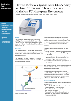


© Copyright 2026