
Nuclear Complex Co-IP Kit
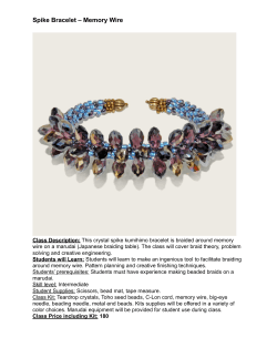
Nuclear Complex Co-IP Kit
(version B1)
Catalog No. 54001
Active Motif North America
1914 Palomar Oaks Way, Suite 150
Carlsbad, California 92008, USA
Toll free:
877 222 9543
Telephone:
760 431 1263
Fax:
760 431 1351
Active Motif Europe
Avenue Reine Astrid, 92
B-1310 La Hulpe, Belgium
UK Free Phone:
France Free Phone:
Germany Free Phone:
Telephone:
Fax:
0800 169 31 47
0800 90 99 79
0800 181 99 10
+32 (0)2 653 0001
+32 (0)2 653 0050
Active Motif Japan
Azuma Bldg, 7th Floor
2-21 Ageba-Cho, Shinjuku-Ku
Tokyo, 162-0824, Japan
Telephone:
+81 3 5225 3638
Fax:
+81 3 5261 8733
Copyright 2014 Active Motif, Inc.
www.activemotif.com
Information in this manual is subject to change without notice and does not constitute a commitment on the part of Active Motif, Inc. It is supplied on an “as is” basis without any warranty of any
kind, either explicit or implied. Information may be changed or updated in this manual at any time.
This documentation may not be copied, transferred, reproduced, disclosed, or duplicated, in
whole or in part, without the prior written consent of Active Motif, Inc. This documentation is
proprietary information and protected by the copyright laws of the United States and international treaties.
The manufacturer of this documentation is Active Motif, Inc.
© 2014 Active Motif, Inc., 1914 Palomar Oaks Way, Suite 150; Carlsbad, CA 92008. All rights reserved.
All trademarks, trade names, service marks or logos referenced herein belong to their respective
companies.
www.activemotif.com
TABLE OF CONTENTS
Page
Introduction . . . . . . . . . . . . . . . . . . . . . . . . . . . . . . . . . . . . . . . . . . . . . . . . . . . . . . . . . . . . . . . . . . . . . . . . . 1
Flow Chart of Process . . . . . . . . . . . . . . . . . . . . . . . . . . . . . . . . . . . . . . . . . . . . . . . . . . . . . . . . . . . . . . . . 2
Kit Performance . . . . . . . . . . . . . . . . . . . . . . . . . . . . . . . . . . . . . . . . . . . . . . . . . . . . . . . . . . . . . . . . . . . . . 3
Kit Components and Storage . . . . . . . . . . . . . . . . . . . . . . . . . . . . . . . . . . . . . . . . . . . . . . . . . . . . . . . . . 3
Additional Materials Required . . . . . . . . . . . . . . . . . . . . . . . . . . . . . . . . . . . . . . . . . . . . . . . . . . . . . . . . 4
Protocols
Nuclear Extraction – Buffer Preparation and Recommendations . . . . . . . . . . . . . . . . . . . . . 4
Preparation of Nuclear Extract . . . . . . . . . . . . . . . . . . . . . . . . . . . . . . . . . . . . . . . . . . . . . . . . . . . . 5
Co-IP – Buffer Preparation and Recommendations . . . . . . . . . . . . . . . . . . . . . . . . . . . . . . . . . 7
Co-Immunoprecipitation Protocol . . . . . . . . . . . . . . . . . . . . . . . . . . . . . . . . . . . . . . . . . . . . . . . . 8
Western Blotting Protocol . . . . . . . . . . . . . . . . . . . . . . . . . . . . . . . . . . . . . . . . . . . . . . . . . . . . . 10
Appendix
Section A. Protein G Agarose Columns Wash Stand . . . . . . . . . . . . . . . . . . . . . . . . . . . . . . . . . 11
Section B. Troubleshooting Guide . . . . . . . . . . . . . . . . . . . . . . . . . . . . . . . . . . . . . . . . . . . . . . . . 12
Section C. Related Products . . . . . . . . . . . . . . . . . . . . . . . . . . . . . . . . . . . . . . . . . . . . . . . . . . . . . . 13
Technical Services . . . . . . . . . . . . . . . . . . . . . . . . . . . . . . . . . . . . . . . . . . . . . . . . . . . . . . . . . . . . . . . . . . 14
www.activemotif.com
Introduction
Co-Immunoprecipitation (Co-IP) is a powerful method used to study protein/protein interactions. In Co-IP, one antibody is used to immunoprecipitate a target antigen and also co-precipitate
any bound interacting proteins within a sample. These protein complexes are then captured by
antibody binding beads (such as Protein A or G). This is followed by elution of the antibody-bound
proteins, SDS-PAGE and Western blot using a second antibody targeted against one of the bound,
interacting proteins. Traditional methods for performing Co-IP are not optimal for studying DNAbinding protein complexes as the complexes are often disrupted during the extraction process. In
addition, many unstable protein complexes can be affected by the salt and detergent composition
of the buffers used in the immunoprecipitation process.
Because of this, Active Motif’s Nuclear Complex Co-IP Kit has been developed specifically for the
preparation and immunoprecipitation of nuclear protein complexes from cell or tissue samples.
The kit is composed of two optimized modules: nuclear extract preparation and Co-IP. The kit’s
extraction module provides a simple, fast and effective method to obtain and maintain protein
complexes contained in nuclear compartments of the cell, specifically those previously bound to
DNA. The kit’s versatile Co-IP module enables the user to study many different protein/protein
interactions of varying strength.
In the kit’s extraction process the cells are collected in ice-cold PBS in the presence of Phosphatase
Inhibitors, which limits further protein modifications (expression, proteolysis, dephosphorylation,
etc.). The cells are then resuspended in Hypotonic Buffer to swell the cell membrane and make
it fragile. Addition of Detergent causes leakage of the cytoplasmic proteins into the supernatant. After removal of the cytoplasmic fraction, the nuclei are lysed and the nuclear proteins are
recovered in a low-salt buffer in the presence of the Protease Inhibitor Cocktail and PMSF. This is
followed by the addition of a proprietary Enzymatic Shearing Cocktail. The use of low-salt buffers
protects protein complexes in the nucleus; DNA digestion allows a gentle release of undissociated
protein complexes from the DNA. After the protein complexes are collected, an immunoprecipitation reaction is carried out to detect the bound proteins. In the immunoprecipitation module, two
different immunoprecipitation buffers with either a low or high stringency starting composition
are provided. In addition, detergent and salt are provided separately to enable the user to vary the
salt and detergent concentrations. The addition of salt and detergent is ideal for use with robust
protein/protein interactions because such conditions reduce background. However, as unstable
protein complexes may not withstand high stringencies, the kit format enables stringency to be
modified as required by each complex.
The Nuclear Complex Co-IP Kit provides sufficient reagents to perform 50 Co-IP experiments. It includes optimized nuclear protein complex extraction reagents, immunoprecipitation buffers and a
comprehensive protocol to provide a simplified method to capture and detect protein complexes.
product
format
catalog no.
Nuclear Complex Co-IP Kit 50 rxns
54001
Protein G Agarose Columns
30 rxns
53039
www.activemotif.com
1
www.activemotif.com
2
�
�
�
���������������������������
���������������
����������������������
�������������
������������������
���������������������������������������������������������
�����������������������������������������������������������������
�
�������������������������������
���������������������������������
�������������������
���������������������������
�����������������������������
�������������������������������
���������������������
��������������������
�������������
�������������
Flow Chart of Process
Kit Performance
250
1
2
3
Figure 1: Co-IP of the p33 subunit of the RNA pol II complex.
HeLa cells were grown to confluence on 100 mm plates and nuclear extracts were
prepared using the kit’s extraction reagents. For IP experiments, the stringency
of the IP High Buffer was increased by supplementing with NaCl (150 mM Final)
and Detergent (1% Final). 100 µg of nuclear extract was used per IP reaction and
incubated with either 2 µg p33 antibody or no antibody (negative control). Protein
G beads were added to each IP reaction. Following the IP, 2X sample buffer was
added to each IP reaction, samples were boiled and run on an SDS-PAGE gel.
Western blot analysis was performed using RNA pol II mouse mAb at 0.1 µg/ml
followed by anti-mouse HRP at 1:1000. Detection of the p33/RNA pol II complex
by the RNA pol II antibody (lane 3) demonstrates that the Co-IP was successful
in maintaining the protein complex. The input HeLa extract (lane 1) was run as a
control for the western blot using 0.1 µg/ml RNA pol II.
Lane 1: Western blot control – 10 µg HeLa extract
Lane 2: Negative Control – no antibody added in IP
Lane 3: Co-IP: IP p33/WB RNA pol II
Kit Components and Storage
Kit components can be stored at -20°C prior to first use. Then, we recommend storing each
component at the temperatures recommended in the table below:
Reagents
Quantity
Storage / Stability
10X Hypotonic Buffer
50 ml 4°C for 6 months
Phosphatase Inhibitors
40 ml 4°C for 6 months
Protease Inhibitor Cocktail
626 µl -20°C for 6 months
10X PBS
80 ml 4°C for 6 months
Detergent
22.8 ml RT for 6 months
100 mM PMSF
50 µl
-20°C for 6 months
0.5 M EDTA
200 µl
4°C for 6 months
Digestion Buffer
10 ml
4°C for 6 months
Enzymatic Shearing Cocktail
50 µl
-20°C for 6 months
5X IP Low Buffer
22.5 ml
4°C for 6 months
5X IP High Buffer
22.5 ml
4°C for 6 months
Dithiothreitol (DTT)
225 µl
-20°C for 6 months
5 M NaCl
6.8 ml
RT for 6 months
BSA
300 mg
4°C for 6 months
www.activemotif.com
3
Additional materials required
• 5 and 10 ml pipettes
• Pipettors
• Cell scraper
• 15 ml conical tubes
• Microcentrifuge tubes
• Centrifuge (with swinging buckets adapted to 15 ml conical tubes) and microcentrifuge
pre-cooled to 4°C
• Rocking platform
• Distilled water
• Apparatus to rotate tubes end-to-end at 4°C (for co-immunoprecipitation)
• Protein G Agarose Columns (Active Motif Catalog No. 53039) or Antibody Binding Beads
(such as Protein A or G beads)
• Antibodies for IP and Western blot
• 37°C Water Bath
• Reagents and equipment for performing SDS-PAGE and Western blot
Protocols
Nuclear Extraction – Buffer Preparation and Recommendations
Preparation of PBS/Phosphatase Inhibitors
The Phosphatase Inhibitors solution should be a clear yellow color. It may precipitate during
storage and become turbid in appearance. If this occurs, heat at 50°C for 10 minutes. To make
nuclear extract from a 100 mm plate of cells, prepare 8 ml of PBS/Phosphatase Inhibitors solution
as follows: mix 0.8 ml 10X PBS in 6.8 ml distilled water, then add 0.4 ml Phosphatase Inhibitors.
The Phosphatase Inhibitors lose their activity 24 hours after dilution. Therefore, use the PBS/Phosphatase Inhibitors solution the same day it is prepared and discard any remaining solution.
Preparation of 1X Hypotonic Buffer
To make nuclear extract from a 100 mm plate of cells, prepare 500 µl of 1X Hypotonic Buffer as
follows: mix 50 µl 10X Hypotonic Buffer and 450 µl distilled water. Any remaining 1X Hypotonic
Buffer can be stored at 4°C for 1 week.
Preparation of Complete Digestion Buffer
To make nuclear extract from a 100 mm plate of cells, prepare 100 µl of Complete Digestion Buffer
as follows: mix 98.5 µl Digestion Buffer with 1 µl Protease Inhibitor Cocktail (PIC) and 0.5 µl PMSF.
The PMSF and PIC lose their activity 24 hours after dilution. Therefore, use the Complete Digestion Buffer solution the same day that it is prepared and discard any remaining solution.
www.activemotif.com
4
Quick Chart for Preparing Buffers
Reagents to Prepare
Components
60 mm plate or
3.2 x 10⁶ cells
100 mm plate or
8.8 x 10⁶ cells
150 mm plate or
or 2 x 10⁷ cells
PBS/Phosphatase Inhibitors
10X PBS
0.4 ml
0.8 ml
1.6 ml
Distilled water
3.4 ml
6.8 ml
13.6 ml
Phosphatase Inhibitors
0.2 ml
0.4 ml
0.8 ml
TOTAL REQUIRED
4.0 ml
8.0 ml
16.0 ml
1X Hypotonic Buffer
10X Hypotonic Buffer
25.0 µl
50.0 µl
100.0 µl
Distilled water
225.0 µl
450.0 µl
0.9 ml
TOTAL REQUIRED
250.0 µl
500.0 µl
1.0 ml
Complete Digestion Buffer
100 mM PMSF
0.25 µl
0.5 µl
1.0 µl
Digestion Buffer
49.25 µl
98.5 µl
197.0 µl
Protease Inhibitor Cocktail
0.50 µl
1.0 µl
2.0 µl
TOTAL REQUIRED
50.0 µl
100.0 µl
200.0 µl
Preparation of Nuclear Extract
Starting from Cells:
The following protocol is based on samples of approximately 8.8 x 10⁶ cells, which correspond
to HeLa cells grown to confluence in a 100 mm tissue culture plate, and will yield approximately
0.20-0.35 mg of protein. Each sample is one extraction reaction. Prepare PBS/Phosphatase Inhibitors, Hypotonic Buffer and Complete Digestion Buffer as described above in the section Buffer
Preparation. Adjust the volumes accordingly using the chart above if using plates of different sizes.
Place buffers and any tubes needed on ice before beginning assay.
Section A: Cell Collection
1. Aspirate media out of dish. Wash with 5 ml ice-cold PBS/Phosphatase Inhibitors. Aspirate
solution out then add 3 ml ice-cold PBS/Phosphatase Inhibitors.
2.
Remove cells from dish by gently scraping with cell lifter. Transfer cells to a pre-chilled 15 ml
conical tube.
3.
Centrifuge cell suspension for 5 minutes at 430 x g in a centrifuge pre-cooled at 4ºC.
4.
Discard supernatant. Keep cell pellet on ice. At this point the protocol can be continued or
the pellet can be frozen at -80°C.
Section B: Isolation of the Nuclei
1. Gently resuspend cell pellet in 500 µl 1X Hypotonic Buffer by pipetting up and down several
times. Transfer to a pre-chilled microcentrifuge tube. Incubate for 15 minutes on ice.
2.
Add 25 µl Detergent and gently pipet up and down 3-5 times to mix.
www.activemotif.com
5
3.
Centrifuge suspension for 30 seconds at 14,000 x g in a microcentrifuge pre-cooled at 4ºC.
4.
Discard supernatant (cytoplasmic fraction). Keep pellet (nuclear fraction) on ice.
Section C: Nuclear Fraction Digestion and Collection
1. Resuspend nuclear pellet in 100 µl Complete Digestion Buffer by pipetting up and down
3-5 times.
2.
Add 0.5 µl of the Enzymatic Shearing Cocktail. Vortex gently for 2 seconds.
3.
Incubate suspension for 10 minutes at 37°C.
Note:
If you are studying protein/protein interactions that may be fragile or unstable,
perform this Enzymatic Shearing Cocktail incubation at 4°C for 90 minutes.
4.
Add 2 µl 0.5 M EDTA to stop the reaction. Vortex gently on a low setting for 2 seconds and
place on ice for 5 minutes.
5.
Centrifuge for 10 minutes at 14,000 x g in a microcentrifuge pre-cooled at 4ºC. Transfer supernatant into a pre-chilled microcentrifuge tube.
6.
Save a small aliquot for protein quantification by a Bradford-based assay. Aliquot and store at
–80ºC. Avoid freeze/thaw cycles.
Note:
The presence of certain detergents may interfere with protein determination
assays, such as Bradford or BCA. We therefore recommend using the Complete
Digestion Buffer as the blank and performing a 1:50 or 1:250 dilution of your
samples. Alternatively, try Active Motif’s ProStain™ Protein Quantification Kit,
which is ideal for quantifying extracts because it offers greater sensitivity and
resistance to many contaminating reagents used in extract preparation.
Starting from Tissue:
Section D: Tissue Homogenization
1. Weigh tissue and dice into very small pieces using a clean razor blade. Collect pieces in a
pre-chilled, clean Dounce homogenizer.
2.
On ice, add 3 ml ice-cold 1X Hypotonic Buffer supplemented with DTT and Detergent
(3 µl of the provided 1 M DTT and 3 µl of the provided Detergent) per gram of tissue and
homogenize. Incubate on ice for 15 minutes.
3.
Centrifuge for 10 minutes at 850 x g at 4°C.
4.
At this point, the tissue is homogenized. However, most of the cells are not yet lysed. Therefore, continue the procedure with the cell pellet at Section B, No. 1 of the Nuclear Extract
protocol for cells (page 5), based on a 150 mm plate (2 x 10⁷ cells).
www.activemotif.com
6
Co-IP – Buffer Preparation and Recommendations
The Nuclear Complex Co-IP Kit has been designed to enable the study of a variety of protein/
protein interactions. The Kit includes two IP Buffers with different stringencies: IP High has a
higher stringency than IP Low. Each IP Buffer can be further supplemented with the provided
salt and detergent depending on the complex being studied. Increased stringency is suitable for
stable protein complexes as it reduces background levels without disrupting protein interactions.
IP Buffers containing low salt and detergent enable the detection of more weakly bound protein
complexes. If the strength of the protein complex is not known, we recommend performing a
series of experiments to determine the optimal buffer stringency as outlined in the table below.
IP Buffer
5 M NaCl
Detergent
5 M NaCl for
Wash Buffer
1X High
N/A
N/A
N/A
1X Low
N/A
N/A
N/A
1X High
150 mM Final
N/A
N/A
1X Low
150 mM Final
N/A
N/A
3. Addition of salt and detergent
1X High
150 mM Final
1% Final
N/A
1X Low
150 mM Final
1% Final
N/A
4. Addition of salt and detergent,
and extra salt for final Wash Step
1X High
150 mM Final
1% Final
300 mM Final
1X Low
150 mM Final
1% Final
300 mM Final
Approach
1. No supplements
2. Addition of salt
Preparation of IP Buffer
The IP Buffers should be supplemented with fresh Protease Inhibitor Cocktail (PIC) and DTT before
use. However, if DTT is not compatible with the protein complex being studied, do not use.
Note:
IP Incubation Buffer should be prepared fresh on Day 1. IP Wash Buffer should be
prepared fresh on Day 2. If you wish to increase stringency of the IP Buffers add
NaCl and Detergent as listed below. If you do not wish to increase stringency
simply add dH2O to recommended Total Volume.
Volume Required for 1 Co-IP
Volume Required for 5 Co-IP’s
Day 1
Day 2
Day 1
Day 2
IP Incubation
IP Wash
IP Incubation
IP Wash
5X IP Buffer
100 µl
800 µl
500 µl
4000 µl
PIC
2.5 µl
8 µl
12.5 µl
40 µl
1 M DTT
0.5 µl
4 µl
2.5 µl
20 µl
5 M NaCl*
7.5 µl
60 µl
37.5 µl
300 µl
Detergent*
45 µl
360 µl
225 µl
1800 µl
dH2O
X µl
X µl
X µl
Total Volume
500 µl
4000 µl
2500 µl
* Add these components to the IP Incubation and Wash Buffers only if you wish to increase stringency.
www.activemotif.com
7
X µl
20 ml
Co-Immunoprecipitation Protocol
IMPORTANT: Please read the previous section, Co-IP – Buffer Preparation and Recommendations
before starting the assay. When possible, all IP steps should be performed on ice. The protocol
below provides volumes sufficient for one IP experiment and can be scaled up as needed. We also
recommend including one IP negative control (no antibody added in the IP reaction).
DAY 1: Preparation of Antibody/Extract mixture
1. Prepare IP Incubation Buffer as described in the above section. Each IP reaction will require 500 µl.
2.
For preparation of samples, we recommend using 100-500 µg of extract and 1-5 µg of antibody
diluted to 500 µl in IP Incubation Buffer in a 1.5 ml microcentrifuge tube. Keep tubes on ice.
3.
Incubate each Antibody/Extract mixture overnight at 4°C on a rotator.
DAY 2: Addition of Antibody binding beads and IP of the Proteins of Interest
Active Motif recommends the use of its Protein G Agarose Columns (Catalog No. 53039) for the
capture of the IP reaction. The Protein G agarose beads supplied in the column are pre-washed
and ready to use. If using an alternative source of Protein A or G beads, the beads should be thoroughly washed before use. Follow the protocol for the antibody capture method being used.
1.
Prepare IP Wash Buffer as described in the above section. Each IP reaction will require 4 ml of
the IP Wash Buffer. Keep the buffer on ice until ready to use.
2.
Prepare a 100 mg/ml BSA solution by dissolving the provided BSA in 2.8 ml dH2O and vortex
to mix. Store unused BSA at -20°C.
3.
For each IP reaction, transfer 1.5 ml of IP Wash Buffer to a new tube. Supplement with
1 mg/ml BSA by adding 15 µl BSA to 1.5 ml IP Wash Buffer. Keep the buffer on ice until ready
to use.
If using Protein G Column-based Capture
1. Add the IP reaction directly to the column. Cap the column tightly to ensure it is sealed. If
desired, use parafilm to seal the cap. (Do not remove the tab from the column.)
2.
Incubate antibody/bead complex for 1 hour at 4°C on a rotator.
3.
During the incubation, prepare a 2X Reducing Buffer (130 mM Tris pH 6.8, 4% SDS, 0.02%
Bromophenol blue, 100 mM DTT).
4.
Following the incubation, spin the column at 1250 x g for 1 minute to collect liquid from
inside of the cap.
5.
The columns are designed to fit within an empty 1 ml pipet tip box which will serve as a
stand for wash steps (see Appendix A for set up). Remove the cap and the tab from the bottom of the column and place the columns in the empty tip box. Add 500 µl ice-cold IP Wash
Buffer supplemented with BSA to the column. Allow flow-through to occur by gravity.
6.
Repeat the wash step twice with 500 µl ice-cold IP Wash Buffer supplemented with BSA.
www.activemotif.com
8
7.
Add 500 µl ice-cold IP Wash Buffer (no BSA) to each column.
Note:
To increase the stringency of this wash step, add 22.5 µl 5 M NaCl to each 500 µl
of IP Wash buffer to be used.
8.
Repeat the wash step two more times with 500 µl ice-cold IP Wash Buffer (no BSA).
9.
Transfer the column to a microcentrifuge tube and centrifuge at 1250 x g at 4°C for 3 minutes
to remove residual IP Wash Buffer.
10. Transfer the column to a new microcentrifuge tube.
11.
Add 25 µl of 2X Reducing Buffer to each column and let stand for 1 minute at room temperature. Centrifuge at 1250 x g at room temperature for 3 minutes to elute the IP reaction.
12. Add 5 µl of pure glycerol to each 25 µl elution. Pipet up and down to mix.
13. Boil samples at 95-100°C for 5 minutes. Allow sample to cool to room temperature and quick
spin to collect condensation. Load gel for electrophoresis and subsequent Western blotting.
If using Protein A or G Bead Capture in Solution
1. Antibody binding beads (Protein A or G) must be washed thoroughly before addition to
the Antibody/Extract mixture from Day 1. For each IP reaction, aliquot 50 µl of resuspended
beads to fresh 1.5 ml microcentrifuge tubes.
2.
Add 250 µl IP Wash Buffer to each tube and flick to mix. Centrifuge at 1430 x g for 30 seconds
at 4°C. Remove supernatant. Repeat wash step twice and place beads on ice.
3.
Add 40 µl ice-cold IP Wash Buffer to the pelleted beads (total volume will exceed 60 µl).
4.
Add 60 µl of the washed beads to each Antibody/Extract tube from Day 1.
5.
Incubate antibody/bead complex for 1 hour at 4°C on a rotator.
6.
During the incubation prepare a 2X Reducing Loading Buffer (130 mM Tris pH 6.8, 4% SDS,
0.02% Bromophenol blue, 100 mM DTT, 20% glycerol)
7.
Following the incubation, centrifuge tubes at 1430 x g for 30 seconds at 4°C. Remove and
discard the supernatant.
8.
Add 500 µl ice-cold IP Wash Buffer supplemented with BSA to each tube. Centrifuge at
1430 x g for 30 seconds at 4°C. Remove and discard the supernatant. Repeat wash step twice
with 500 µl ice-cold IP Wash Buffer supplemented with BSA. Do not vortex or flick tubes to
mix. Keep tubes on ice when possible.
9.
Add 500 µl ice-cold IP Wash Buffer (no BSA) to the pelleted beads. Centrifuge at 1430 x g for
30 seconds at 4°C. Remove supernatant. Repeat wash step twice with 500 µl ice-cold IP Wash
Buffer (no BSA). Do not vortex or flick tubes to mix. Keep tubes on ice when possible.
Note:
9.
To increase the stringency of this wash step, add 22.5 µl 5M NaCl to each 500 µl
of IP Wash Buffer to be used.
Resuspend each bead pellet in 8 µl of 2X Reducing Loading Buffer and boil samples at
95-100°C for 5 minutes.
www.activemotif.com
9
10. Allow sample to cool to room temperature, spin briefly to collect condensation and apply to
gel for electrophoresis and subsequent Western blotting.
Western Blotting Protocol
For your convenience, a Western blotting protocol is provided below. When performing Western
blot, individual optimization of antigen and antibody quantities may be required in order to
detect the protein of interest.
Electrophoresis
1. Load sample onto an appropriate SDS-PAGE gel and run until migration front reaches bottom
of gel.
Transfer
2. Transfer protein to polyvinylidene difluoride (PVDF) membranes at 400 mA for 90 minutes
using Transfer Buffer (25 mM Tris-HCl, pH 8.3, 192 mM glycine, 15% methanol).
Blocking
3. Block membranes by incubating overnight at 4°C with shaking in 5% Blotto (5% dry, non-fat
skim milk powder in PBS (pH 7.4)). The addition of 0.1% Tween 20 to all Blotto and PBS
solutions may increase the antibody specificity and decrease the background.
Incubation with Primary Antibody
4. Rinse membranes with PBS (pH 7.4). Dilute the antibody (at the appropriate dilution) in
5% Blotto. Incubate for 1 hour at 37°C, 2 hours at RT (room temperature) or overnight at 4°C
with agitation.
5.
Decant antibody solution. Wash the membrane 5 times for 5-10 minutes at RT in PBS. Washing should be performed with vigorous agitation over a minimum 30-minute period.
Incubation with Secondary Antibody
6. Incubate membranes with secondary HRP conjugate diluted in 5% Blotto for 1 hour at RT
with gentle shaking. The dilution of the secondary antibody conjugate will vary according to
manufacturer’s specifications.
7.
Repeat step 5.
8.
Wash membrane with PBS for 5 minutes with agitation before proceeding to the chemiluminescence reaction.
Chemiluminescence Reaction
9. Prepare and use the chemiluminescent substrate according to the manufacturer’s instructions.
www.activemotif.com
10
Appendix
Section A. Protein G Agarose Columns Wash Stand
The Protein G Agarose columns are designed to fit within an empty 1 ml pipet tip box which will
serve as a stand for wash steps. Remove the cap and the tab from the bottom of the column and
place the columns in the empty tip box. All wash steps can be performed using gravity flow.
www.activemotif.com
11
Section B. Section A. Troubleshooting Guide
Problem/question
Possible Cause
Recommendation
No or low signal
of IP’d proteins by
Western blot
Proteins of interest are not located in the
nucleus or their level of expression is very
low.
Confirm presence of proteins of interest in
the nucleus and nuclear extract. Alternatively,
use more extract in the IP reaction (up to 500
µg per IP).
Target proteins lost or destroyed during
sample preparation.
Confirm presence of proteins of interest in
the nuclear extract.
The protein/protein interaction of interest is
dependent on specific induction conditions.
Optimize conditions of culture and induction.
The protein/protein interaction of interest is
not stable and dissociates during the extraction process.
Perform the Enzymatic Shearing Cocktail
incubation for 90 minutes at 4°C instead of
10 minutes at 37°C.
Amount of antibody used in the assay is
too low.
Use more antibody in IP and/or WB; perform
a titration to determine how much.
Antibodies used in IP and/or WB are not suitable for these applications.
It is important to perform IP and WB controls,
as antibodies raised against denatured antigen
and native epitopes may not be recognized.
IP Positive Control - IP antigen of interest
and WB same antigen. WB Positive Control direct WB analysis of the extract.
The IP buffer is too stringent.
Use the IP High and IP Low buffers without
the addition of salt and detergent. Favor use
of IP Low buffer, which is less stringent.
Large number of competing proteins
present in the sample.
Centrifuge the extract at 20,000 x g for 20
minutes before adding antibody. This should
remove insoluble aggregates.
Interfering substances present in the nuclear
extract.
Use the appropriate amount of inhibitors
when performing the assay.
Antibody breakdown.
Reduce concentration of DTT, or avoid use.
Interference with the establishment or maintenance of the antigen-antibody complex.
Reduce concentration of detergent.
Incorrect bead type used to precipitate the
antigen-antibody complex.
Antibody binding beads are not included in
the kit. Confirm Protein A or G use.
Too much antibody used in IP and/or WB.
Titrate antibody concentrations and decrease
primary antibody dilution or incubation time.
Non-specific binding of other proteins to the
Protein A/G beads or antibodies.
Preclear extracts with Protein A/G beads prior
to addition of the primary antibody.
Wash steps are not sufficiently stringent.
0.5% sodium deoxycholate can be added to
the last 3 IP washes and may reduce signal
from the negative (no antibody) control.
Aggregated proteins in extracts.
Spin extracts at 20,000 x g for 20 minutes and
discard pellet prior to addition of antibody.
IgG heavy chains appear at 55 kDa, light
chains appear at 28 kDa.
Use different antibody origins for IP and WB.
High background
signal
www.activemotif.com
12
Problem/question
Possible Cause
Recommendation
Can whole-cell
extracts be used?
If you wish to work with whole-cell extracts,
we recommend use of Active Motif’s
Universal Magnetic Co-IP Kit, which contains
reagents for making both nuclear or wholecell extracts. It also utilizes Protein G Magnetic Beads and a magnet for the IP and Wash
steps, which greatly reduces background.
Section C. Related Products
Universal Co-IP Kit
Format
Catalog No.
Universal Magnetic Co-IP Kit
25 rxns
54002
ChIP-IT™ Kits
Format
Catalog No.
ChIP-IT® Express
ChIP-IT® Express Enzymatic
ChIP-IT® Express Shearing Kit
ChIP-IT® Express Enzymatic Shearing Kit
ChIP-IT® High Sensitivity
ChIP-IT® qPCR Analysis Kit
ChIP-IT® ChIP-Seq
ChIP-IT® FFPE
ChIP-IT® FFPE Chromatin Preparation Kit
ChIP-IT® Express HT
Re-ChIP-IT®
RNA ChIP-IT®
Chromatin IP DNA Purification Kit
EpiShear™ Probe Sonicator
EpiShear™ Cooled Sonication Platform, 1.5 ml
ChIP-IT® Protein G Magnetic Beads
Protein G Agarose Columns
Siliconized Tubes, 1.7 ml
ChIP-IT® Control qPCR Kit – Human
ChIP-IT® Control qPCR Kit – Mouse
ChIP-IT® Control qPCR Kit – Rat
ChIP-IT® Control Kit – Human
ChIP-IT® Control Kit – Mouse
ChIP-IT® Control Kit – Rat
Ready-to-ChIP HeLa Chromatin
Ready-to-ChIP Hep G2 Chromatin
Ready-to-ChIP K-562 Chromatin
Ready-to-ChIP NIH/3T3 Chromatin
Bridging Antibody for Mouse IgG
Dounce Homogenizer
Dounce Homogenizer
25 rxns
25 rxns
10 rxns
10 rxns
16 rxns
10 rxns
10 libraries
16 rxns
5 rxns
96 rxns
25 rxns
25 rxns
50 rxns
110 V
1 platform
25 rxns
30 rxns
25 tubes
5 rxns
5 rxns
5 rxns
5 rxns
5 rxns
5 rxns
10 rxns
10 rxns
10 rxns
10 rxns
500 µg
1 ml
15 ml
53008
53009
53032
53035
53040
53029
53041
53045
53030
53018
53016
53024
58002
53051
53080
53014
53039
53036
53026
53027
53028
53010
53011
53012
53015
53019
53020
53021
53017
40401
40415
www.activemotif.com
13
Technical Services
If you need assistance at any time, please call Active Motif Technical Service at one of the
numbers listed below.
Active Motif North America
1914 Palomar Oaks Way, Suite 150
Carlsbad, CA 92008
USA
Toll Free:
877 222 9543
Telephone: 760 431 1263
Fax:
760 431 1351
E-mail:
[email protected]
Active Motif Europe
Avenue Reine Astrid, 92
B-1310 La Hulpe, Belgium
UK Free Phone:
0800 169 31 47
France Free Phone: 0800 90 99 79
Germany Free Phone: 0800 181 99 10
Telephone: +32 (0)2 653 0001
Fax: +32 (0)2 653 0050
E-mail: [email protected]
Active Motif Japan
Azuma Bldg, 7th Floor
2-21 Ageba-Cho, Shinjuku-Ku
Tokyo, 162-0824, Japan
Telephone: +81 3 5225 3638
Fax:
+81 3 5261 8733
E-mail:
[email protected]
Visit Active Motif on the worldwide web at http://www.activemotif.com
At this site:
• Read about who we are, where we are, and what we do
• Review data supporting our products and the latest updates
• Enter your name into our mailing list to receive our catalog, MotifVations newsletter and
notification of our upcoming products
• Share your ideas and results with us
• View our job opportunities
Don’t forget to bookmark our site for easy reference!
www.activemotif.com
14


© Copyright 2026