
Nyt vindue kan sætte danmarksrekord i isolering
Danish Association of Sports Medicine and Danish Association of Sports Physiotherapy
-
Sports Medicine
Congress 2013
”From Research to Clinical Practice”
Thursday 31st of January to Saturday 2nd of February 2013,
Hotel Comwell, Kolding
fagforum
for
idrætsfysioterapi
S P ORT S M E D I CINE CONGRESS 2013
Velkommen
Table of Contents
2
3
12
14
16
17
18
31
32
Welcome..............................................
Scientific program...............................
Poster Abstracts...........................
Thursday - Program overview.......
Friday - Program overview............
Saturday - Program overview.......
Abstracts......................................
General information......................
Exhibition Plan.............................
Det er mig en stor ære at byde velkommen til DIMS/FFI
årsmøde 2013 som for anden gang afholdes i Kolding.
Som du er allerede har erfaret – ellers havde du nok ikke
tilmeldt dig, så har arrangørudvalget sammensat et godt
program. Jeg vil faktisk strække mig til at sige, at jeg vil
garantere at du får lært noget, som du måske burde have
vidst i forvejen. Som i tidligere år har vi fire sideløbende
sessioner og prioriteringen bliver som altid svær. Tabet
ved at mise noget fra den store sal bliver forhåbentligt
knapt så stort som tidligere, da vi forventer at du efter
kongressen kan finde optagelser af disse sessioner på
hjemmesiden. At et kryds kan være svært at sætte, er ikke
nyt, men forhåbentligt finder du det let at placere det ved
de sociale arrangementer og ikke mindst gallafesten, hvor
en idrætsmedicinsk nyskabelse vil blive afsløret.
I skrivende stund er næste års arrangørudvalg ved at blive
sammensat og måske har du noget du gerne vil byde ind
med eller også har du bare lyst til at give en hånd med og
lære hvordan der arbejdes. Kongresmanualen har vist sig
at fungere som en fantastisk skabelon og det er givetvis
et redskab som gør kongresarbejdet lettere og sjovere.
Brænder du for at bidrage til kongressen og gennem dette
arbejde at skabe et idrætsmedicinsk netværk må du give
dig til kende.
Tak fordi du deltager og med ønsket om en god kongres,
vil jeg sige velkommen til Kolding.
Lars Blønd, Formand DIMS
Welcome
Welcome to the annual sports congress hosted by DIMS
and FFI. Once again we find ourselves in the in the beautiful surroundings of Kolding. Once again the organising
committee succeeded in putting together an outstanding
program, where best practice and evidensbased sports
medicine sometimes unite, sometimes contradict, but
always bring forth interesting discussions. We are here
to learn from the discussions and it is my request that we
keep the discussions sober as always. Like John Wooden,
former coach of the NCAA team UCLA cleverly stated:
“Be a good listener, your ears will never get you in
trouble”
Have a great time
Karen Kotila
2
SPORTS MEDICINE CON G R ES S 2 0 1 3
SCIENTIFIC PROGRAM
ANNUAL CONGRESS DANISH SPORTS MEDICINE (DIMS-FFI)
THURSDAY JANUARY 31st 2013
MAIN HALL (Thursday)
Thursday, Main hall, 10.45-11.40 (lecture)
10.45 – 11.40 ”Sports injuries – an unavoidable event?”
Prof Malachy McHugh, FYI Lenox Hill Hospital,
New York, USA
Chair:
Michael Kjaer
Thursday, Main hall, 11.45-12.40 (lecture)
11.45 – 12.30 “Elbow problems in sports: Surgical or nonsurgical treatment?”
Prof Denise Eygendaal, Academic Medical Centre, Amphia Hospital, Amsterdam, the Netherlands
12.30 – 12.40 Abstract no 3. Oral communications:
“Regeneration of articular cartilage in sheep by
osteochondral distraction”
Becker JA et al. Section of Sports Traumatology,
Dept Orthop Surgery, Bispebjerg Hospital
Chair:
Bo Sanderhoff Olsen
Thursday, Main hall, 13.30-14.55 (symposium)
Title: “Hip pain”
13.30 – 14.20 “FAI and other closely-related hip joint problems: Etiology, Consequences, Prevention and
Treatment (When Needed)”
Prof Michael B. Millis, Orthopedic Surgery,
Harvard Medical School, Adolescent and Young
Adult Hip Unit, Boston
14.20 – 14.40 “Extraarticular differential diagnosis – examination techniques and possible relations to the
hip problem”
Ass. Prof Per Hölmich, Arthroscopic Centre,
Amager Hospital
14.40 – 14.55 Discussion
Chair:
Per Hölmich
Thursday, Main hall, 15.00-15.55 (lecture)
15.00 – 15.45 “Sarcopenia – why do we all loose skeletal
muscle with ageing and can we preserve
strength and function with training”
Prof Marco Narici, University of Nottingham,
United Kingdom,
15.45 – 15.55 Abstract no 7. Oral communications:
“The effect of hypercholesterolemia and obesity
on the mechanical properties of mice tail collagen
fascicles”
Eriksen CE et al. Inst of Sports Medicine and
Centre of Healthy Aging, Bispebjerg Hospital.
Chair:
Michael Kjaer
Thursday, Main hall, 16.30-18.00 (symposium)
Title: “Diagnosis and treatment of pelvic girdle pain - an
update”
16.30 – 17.00 “A integrated mechanism based approach for
the evaluation and rehabilitation of Low Back
Pain”
Prof Lieven Danneels, Ghent, Belgium
17.00 – 17.30 “How to diagnose sacroiliac joint pain by
means of clinical examination”
Senior Researcher Tom Petersen, Copenhagen
Back Centre, Denmark 17.30 – 18.00 ”The lumbar multifidus: state of the art anno
2012”
Prof Lieven Danneels, Ghent, Belgium
Chair:
Christian Couppé
ROOM A (Thursday)
Thursday, Room A, 11.45-12.40 (lecture)
11.45 – 12.30 “Diagnosis, treatment and prevention of ankle
sprains: An evidence-based clinical guideline”
Prof Rob A. de Bie, Maastricht University, The
Netherlands
12.30 – 12.40 Abstract no 1. Oral communications:
“Foot orthotics reduce the navicular drop – a
novel method allowing for in-shoe measurement”
Pedersen KS et al. Dept Health Sci and Techn,
Aalborg University and Dept Engineering and
Orthop Surg, Aarhus University
Chair:
Christian Couppé
Thursday, Room A, 13.30-14.55 (symposium)
Title: “Stretching”
13.30 – 14.00 “To stretch or not to stretch; Flexibility and its
effects on sports injury and performance”
Prof Malachy MacHugh, FYI Lenox Hill Hosp,
New York, USA
14.00 – 14.30 “Stretching as a supplement to the return to
sport algorithm for hamstring injuries”
PT, PhD Carl Askling, The Swedish School of
Sport and Health Sciences, Stockholm, Sweden
14.30 – 14.55 “Implementation of stretching in ballet”
PT Charlotte Anker-Petersen and Chief Surgeon
Henrik Aagaard, Royal Danish Ballet,
Chair:
Karen Kotila
Thursday, Room A, 15.00-15.55 (pro-et-contra)
Title: “Achilles tendon rupture: To operate or not to operate?”
15.00 – 15.15 ”In favor of operative treatment” (Pro)
Prof Michael Krogsgaard, Department of Sports
Surgery and Arthroscopy , Bispebjerg Hospital
15.15 – 15.30 “In favor of conservative treatment” (Con)
Dr Kristoffer Barfod, Hvidovre Hospital, Denmark
15.30 – 15.55 Discussion pro-et-contra
Chair:
Per Hölmich
3
S P ORT S M E D I CINE CONGRESS 2013
Thursday, Room A, 16.30-18.00 (SAKS symposium)
Title: “Achilles tendinopathy – diagnosis, imaging, surgical
interventions and high volume injection (HVI)
in Achilles tendinopathy”
16.30 – 16.55 “Diagnosis and imaging of Achilles tendinopathy”
Chief phys Michel Court-Payen, Gildhøj Private
Hospital, Denmark
16.55 – 17.20 “High Volume Injection in Achilles tendon”
Dr Anders Ploug Boesen, Institute of Sports
Medicine, Bispebjerg Hospital
17.20 – 18.00 “Surgical interventions in Achilles tendinopathy”
Ass Prof Gino M. Kerkhoffs, Dept. of Orthopedic
Surgery, Academic Medical Center, University of
Amsterdam, the Netherlands
Chair: Johnny Frøkjaer
ROOM B (Thursday)
Thursday, Room B, 11.45-12.40 (lecture)
11.45 – 12.30 “Rehabilitation after ACL injury”
Prof Richard Frobell, Lund University, Sweden
12.30 – 12.40 Abstract no 28. Oral communications:
“Adolescents with patellofemoral pain syndrome
do not have decreased isometric muscle strength
of the hip and knee compared to pain free adolescents”.
Rathleff CR et al. Orthopaedic Surg Research
Unit, Aalborg University, Dept Rheum, Aarhus
Univ and Univ Southern Denmark
Chair:
Tommy Øhlenschlæger
Thursday, Room B, 13.30-14.55 (symposium)
Title: “Musculo-skeletal ultrasound - form basic science to
clinical work”
13.30 – 14.00 “Musculo-skeletal ultrasound: Do we need
certification, who should use it and how, and
can we document the usefulness of ultrasound
in basic science and clinical work”
Chief phys Søren Torp-Pedersen, Frederiksberg
Hospital
14.00 – 14.30 “The use of ultrasound in a sports medicine
setting, in clinical work and in the field”
Chief phys Ulrich Fredberg, Silkeborg Hospital
14.30 – 14.55 “Ultrasound in physiotherapy, perspectives on
the use”
Prof Henning Langberg, Institute of Public
Health, University of Copenhagen
Chair: Phillip Hansen
Thursday, Room B, 15.00-15.55 (lecture)
Title: “Chiropractors in Sports Medicine”
15.00 – 15.30 “How does Danish chiropractors contribute to
research in physical activity and sports medicine?”
Prof Jan Hartvigsen, University of Southern Denmark, Odense
4
15.30 – 15.55
Chair:
“Neck and back pain in children and adolescent”
Chiropract, PhD stud, Ellen Årtun, University of
Southern Denmark, Odense
Jonas Thorlund
Thursday, Room B, 16.30-18.00 (seminar)
Title: “Patient related outcome scores: Why use them when
they don’t work?”
16.30 – 16.50 “Why use a PRO? Basic requirements to the
construction and validation of a PRO, reflecting what it can be used for”
Sen Lec John Brodersen, Research Unit for General Practice, University of Copenhagen
16.50 – 17.10 “Practical steps in the construction of a PRO”
PhD stud Jonathan Comins, Sports Surgery Unit,
Orthopedic Surgery, Bispebjerg Hospital and
SAHVA
17.10 – 17.35 “Practical validation of a PRO”
Sen Lec John Brodersen, Department of Public
Health, University of Copenhagen
17.35 – 18.00 “Overview of PROs for sports’ knee and
shoulder and their validation”
PhD stud Jonathan Comins, Sports Surgery Unit,
Orthopedic Surgery, Bispebjerg Hospital and
SAHVA
Chair:
Michael Krogsgaard
ROOM C (Thursday)
Thursday, Room C, 11.45-12.45 (workshop)
11.45 – 12.45 “Practical sports medicine – How to work with
elite athletes?”
Chief phys Morten Storgaard, Team Denmark
Chair:
Anders Boesen
Thursday, Room C, 13.30-14.30 (workshop)
13.30 – 14.30 “Injury prevention in handball”
Ass prof, PhD Mette Zebis, University of Southern Denmark
Chair:
Rie Harboe Nielsen
Thursday, Room C, 15.00-15.55 (workshop)
15.00 – 15.55 ”Elbow examination”
Prof. Denise Eygendaal, Academic Medical Centre, Amphia Hospital, Amsterdam, The Netherlands
Chair:
Bo Sanderhoff Olsen
Thursday, Room C, 16.30-17.30 (workshop)
16.30 – 17.30 “Screening tests in athletes to prevent injuries”
Prof Malachy McHugh, FYI Lenox Hill Hospital,
New York, USA
Chair:
Thomas Bandholm
SPORTS MEDICINE CON G R ES S 2 0 1 3
circuscom.se
Minimotion
Minimotion er et multifunktionelt og alsidigt
trænings træningsredskab til træning af hele
kroppen, der kan bruges gennem et helt
genoptræningsforløb – kan deles ind i 4 faser
Fase 1
Akut fase/Efter operation – mål er
at formindske smerte og øge
mobilitet og fleksibilitet
Fase 2
Genoptræning – stabilisering og
øge styrke af led
Fase 3
Funktionel træning med vægte
Fase 4
Afsluttende træning / Sport hvor
man er øvet bruger.
Man træner ved at sætte den fleksible stang i bevægelse, hvor man skal
holde stangen i gang ved at bevæge delen man træner frem og tilbage.
Sværhedsgraden bestemmes via hastighed man bevæger stangen, samt ved
placering af holderne. Dette gør, at man både træner styrke, stabilitet,
udholdenhed og koordinering.
Testudstyr – Hvordan tester du dine patienter?
Sportskadeprodukter
i verdensklasse
Vores tape, bandager og kulde/varme-produkter fungerer
godt for såvel profesionelle som amatører. Uanset hvilken
sport og hvilket niveau det handler om, er målet altid det
samme: At forebygge skader og fremskynde rehabilitering.
Chiroform sætter fokus på testudstyr på kongressen – kom og få en
demonstration af udstyret.
Industrivej 23, 8800 Viborg - Tel. 86613611 www.chiroform.dk - Email: [email protected] Vil du vide mere?
Kontakt BSN Medical på tel. +45 3336 3534.
Begrænser artrose eller
overbelastningsgener
dine patienter?
Musculoskeletal Ultralyd
Re5 er et dansk1, klinisk dokumenteret behandlingskoncept2,
som benytter elektriske impulser til at genopbygge biologisk
væv.
Det patenterede3 Re5-koncept består af en bærbar pulsgenerator
og Re5-applikatorer. Spolernes bikubestruktur danner et kraftigt
pulserende elektrisk felt, hvis pulser og form efterligner kroppens
naturlige impulser.
500
Detaljerede billeder af
strukturer i overfladen
Re5-behandling genopbygger grundlæggende fysiologiske og
anatomiske strukturer – både i overfladen og 8-10 cm inde i det
behandlede område. Dette medfører dannelse af nye blodkar,
øget blodflow samt forbedret lymfedræn4.
Ergonomisk og nem
skannerbetjening
Nem rengøring og
desinficering
De foreløbige 2.285 Re5-patientforløb gennemført på landets
85 Re5-certificerede klinikker viser overbevisende signifikans
P<0.001 for smerter og stivhed. Derfor er Re5 også en oplagt
behandling til patienter med overbelastningsgener eller artrose.
Både som enkeltstående og som supplerende behandling.
Re5 kan sikre dine patienter
• hurtigererestitution
• øgetbevægelighed
• færresmerter
Kongrestilbud
Få Re5 på prøve i 30 dage uden omkostninger
og uden binding. Besøg vores stand og prøv
selv en Re5-behandling.
2
3
4
Professor, dr.scient. Steen Dissing, Institut for Cellulær og Molekylær Medicin,
Det Sundhedsvidenskabelige Institut, Københavns Universitet m.fl.
Vavken P et al. In J Rehabil Med. 2009 May;41(6):406-11
A method and an apparatus for stimulating/modulating biochemical processes
using pulsed electromagnetic fields.
Rahbek UL et al. In Oral. Biosciences & Medicine 2005;2(1)
Re5 ApS
·
Niels Ebbesens Vej 31
·
1911 Frederiksberg C
Over 30 Years of Pioneering Innovation in Ultrasound
·
T: 52 600 500
·
M: [email protected]
Europe and Rest of World: Mileparken 34 • 2730 • Herlev • Denmark • T +45 4452 8100 • F +45 4452 8199
Headquarters USA: 8 Centennial Drive • Peabody MA 01960 • T +1 800 876 7226 / +1 978 326-1300
www.bkmed.com
AD0114-B
1
5
S P ORT S M E D I CINE CONGRESS 2013
FRIDAY FEBRUARY 1st 2013
MAIN HALL (Friday)
Friday, Main hall, 8.00-9.25 (symposium)
Title: “Patello-femoral pain syndrome”
08.00 – 08.15 “Practical and differential diagnostics”
Chief phys Christoffer Brushøj, Institute of Sports
Medicine, Bispebjerg Hospital
08.15 – 08.30 “Prevalence of PFPS across age-groups and
gender”
Physiother Michael Skovdal Rathleff, Ålborg
University
08.30 – 08.55 “Frequent deficits in patients with PFPS”
Dr Christian Barton, Queen Mary University,
London, UK and Pure Sports Medicine, London,
UK
08.55 – 09.25 “Evidence based treatment of PFPS”
Prof Jenny McConnel, Mosman University, Australia
Chair:
Michael Rathleff
Friday, Main hall, 9.30-10.25 (lecture)
09.30 – 10.15 “New strategies for injured runners”
Prof Reed Ferber, University of Calgary, Canada
10.15 – 10.25 Abstract no 26. Oral communication
“Running-related injuries among novice runners:
A 1-year prospective follow-up study”
Nielsen RO et al. Sect of Sports Science, Aarhus
University, Univ Groningen, NL, and Dept Orthopaedics, Aalborg Univ.
Chair:
Michael Rathleff
Friday, Main hall, 11.00-12.30 (symposium)
Title: ”Professor lectures within sports medicine”
11.00 – 11.30 “Sports traumatology”
Prof Martin Lind, Department of Orthopaedic
Surgery, Aarhus University Hospital
11.30 – 12.00 “Sports traumatology and arthroscopic surgery”
Prof Michael Krogsgaard, Department of Sports
Surgery and Arthroscopy , Bispebjerg Hospital,
12.00 – 12.30 “Rehabilitation”
Prof Henning Langberg, Institute of Public
Health, Copenhagen University
Chair: Michael Kjaer
Friday, Main hall, 13.30-14.55 (DSSAK symposium)
Title: “Shoulder impingement syndrome”
13.30 – 13.40 ”Impingement treatment in Denmark” where
are we – setting the scene”
Chief Surg Hans Viggo Johanssen, Department of
Orthopaedic Surgery, Aarhus University Hospital
13.40 – 14.00 ” Psychological and social determinants: Interventions that might prevent the development
of chronic shoulder pain. Patients
perceived recovery”
Prof Rob A. de Bie, Maastricht University, The
Netherlands
6
“Impingement syndrome and degenerative
rotator cuff disease; Can surgeons prevent
degenerative cuff disease?”
Prof Andrew Carr, University of Oxford, United
Kingdom,
Pro-et-con battle: ”To operate or not to operate”
14.20 – 14.30 ”When not to operate shoulder impingement
syndrome”
Prof Rob A. de Bie, Maastricht University, The
Netherlands
14.30 – 14.40 “When to operate shoulder impingement syndrome”
Prof Andrew Carr
14.40 – 14.55 Panel discussion; cases and questions
Chairs:
Anne Kathrine Belling Sørensen and Hans Viggo
Johanssen
14.00 – 14.20
Friday, Main hall, 15.00-15.55 (lecture)
15.00 – 15.45 “Scapula instability in sports – problems and
treatment”
Prof Ann Cools, University of Ghent, Belgium
15.45 – 15.55 Abstract no 29. Oral communications
“Home-exercises as the drug-of-choice in physical medicine and rehabilitation: Exact quantification of exercise adherence and quality using new
technology”
Rathleff MS et al. Orthop Surgt Unit, Aalborg
Univ, Dept Orthop.Surg, Hvidovre Hospital, and
Århus University.
Chair:
Henning Langberg
Friday, Main hall, 16.30-18.10 (symposium)
Title: “Young investigator award – Oral presentations for
competition” and “The Lindhard Award prize
lecture”
16.30 – 17.40 Oral communications from abstract, 6 selected
abstracts (8 min), Questions by panel (2-3 min)
16.30 - 16.41 Abstract 5: High injury – incidence in adolescent
female soccer: The influence of weekly soccerexposure and playing level
Clausen MB et al. Arthroscopic Centre Amager,
Hvidovre, Copenhagen Univ Hospital, Aarhus
Univ and Univ Southern Denmark
16.41 – 16.52 Abstract 7: The effect of life-long endurance
exercise on collagen cross-linking of the human
patellar tendon
Couppe C et al. Institute of Sports Medicine and
Dept Physiotherapy, Centre for Healthy Aging,
Bispebjerg Hospital
16.52 – 17.03 Abstract 16: Patella-tendon graft-fixation in ACL
reconstruction: Metal or bioabsorbable screws – a
10-year follow up with RSA and MRI
Irdal-Jeppesen L et al. Section for Sports Traumatology and Dept Radiology, Bispebjerg Hospital
17.03 – 17.14 Abstract 32: Aging impairs muscle recovery,
myogenic precursor cell expansion and affects
transcriptional responses after immobility-induced atrophy in human skeletal muscle.
Suetta C et al. Institute of Sports Medicine,
Bispebjerg Hospital, Dept Clin Physiol, Glostrup
Hospital, and Univ Southern Denmark.
SPORTS MEDICINE CON G R ES S 2 0 1 3
17.14 – 17.25
17.25 – 17.36
Chair:
Panel:
17.40 – 18.10
Chair:
Abstract 33: The Copenhagen groin-pain test:
Giving the green light for soccer play!
Thorborg K et al. Arthroscopic Centre Amager,
Copenhagen Univ Hospital and Metropol Univ
College, Copenhagen
Abstract 36: The effects of extracorporeal shockwave therapy on inflammatory mediators in the
healthy Achilles tendon
Waugh CM et al. Centre for Sports and Exercise
Med and School for Engineering and Material
Sci, Queen Mary University
Henning Langberg
Thomas Bandholm, Jens L Olesen, Ulrich Fredberg, Martin Lind
Presentation of “The Lindhard Award Prize” winner followed by a prize winner lecture (20 min)
Michael Kjær
Friday, Exhibition area, 19.00-20.00 (poster)
Title: “Walk, talk and wine – guided poster-walk and appetizer”
Chair:
Henning Langberg
ROOM A (Friday)
Friday, Room A, 8.00-9.25 (symposium)
Title: ”Skeletal muscle – how to improve muscle strength and
function in performance, recreation and rehabilitation”
08.00 – 08.30 “What regulates muscle tissue growth?”
Prof Truls Raastad, Norwegian School of Sports
Science, Oslo, Norway
08.30 – 08.55 “Importance of neuro-muscular interaction
for strength development”
Prof Per Aagaard, University of Southern Denmark
08.55 – 09.25 “How to practice strength training dependent
upon who you are”
Ass prof Lars Andersen, National Research Centre for the Working Environment
Chair: Jesper Løvind Andersen
Friday, Room A, 9.30-10.25 (lecture)
09.30 – 10.15 ”Shoulder Impingement: treatment modalities
from a physiotherapist’s perspective - Methods
and evidence”
Prof Rob A. de Bie, University of Maastricht, The
Netherlands
10.15 – 10.25 Abstract no 22. Oral communications
“Status of physical activity is not related to level
of exercise-induced hypoalgesia and conditioned
pain modulation”
Madsen HB et al. Denter for Sensory-Motor
Interact, Aalborg University, and Inst Sports Science, Univ of Southern Denmark
Chair:
Anne Kathrine Belling Sørensen
Friday, Room A, 11.00-12.30 (ADD/DIMS/FFI symposium)
Title: “Doping substances in nutritional supplementation and
food - is it a problem?”
11.00 – 11.20 “Doping substances in food and dietary
supplements – how big is the problem?”
Director Lone Hansen, Anti-doping Denmark
11.20 – 11.45 “Positive doping test caused by ingestion of
food and dietary supplements – cases”
Director Lone Hansen, Anti-doping Denmark
11.45 – 12.30 “Doping substances in food and supplements How to handle the problem”
Dr Catherine Judkins, United Kingdom
Chair:
Mette Hansen
Friday, Room A, 13.30-14.55 (symposium)
Title: “Female athletes and training adaptations”
13.30 – 13.55 “Gender difference in fuel combustion during
exercise”
Prof Bente Kiens, Institute of Sports Science,
University of Copenhagen
13.55 – 14.15 “Gender difference in strength training adaptation”
Ass. Prof Mette Hansen, Institute of Sports Science, Aarhus University
14.15 – 14.35 “Gender differences in biomechanical properties”
Post doc Christian Couppé, Institute of Sports
Medicine, Bispebjerg Hospital
14.35 – 14.55 “Stress fracture – a gender specific problem?”
Post doc Anders Vinther, Dept of Physiotherapy,
Herlev Hospital and Lund University, Sweden
Chair:
Anders Vinther
Friday, Room A, 15.00-15.55 (pro-et-contra)
Title: “ACL injury in physically active individuals: To reconstruct or not to reconstruct?”
15.00 – 15.15 “Rehabilitation as the primary treatment option for ACL injury”
Prof Richard Frobell, University of Lund, Sweden
15.15 – 15.30 “Surgical reconstruction as first choice for
ACL injury”
Prof Michael Krogsgaard, Department of Sports
Surgery and Arthroscopy, Bispebjerg Hospital,
15.30 – 15.55 Pro-et-contra discussion
Chair: Martin Englund, Sweden
ROOM B (Friday)
Friday, Room B, 8.00-9.25 (symposium)
Title: “Bone and physical training”
08.00 – 08.30 “Training and bone – from health to injury”
Prof Magnus Karlsson, Department of Orthopedic
Surgery, University of Lund, Sweden
08.30 – 09.00 “Training in individuals with osteoporosis”
Chief Phys Niklas Rye Jørgensen, Department of
Orthopedic Surgery, Glostrup Hospital
7
S P ORT S M E D I CINE CONGRESS 2013
09.00 – 09.25
Chair: “Too much training on bone – or too low energy intake – in elite female athletes”
Ass prof Ewa Wulff Helge, Department of Nutrition, Exercise and Sports, University of Copenhagen.
Inge-Lis Kanstrup
Friday, Room B, 9.30-10.25 (lecture)
09.30 – 10.15 “Exercise and cancer”
Prof Lis Adamsen, UCSF, University of Copenhagen
10.15 – 10.25 Abstract no 21. Oral communications
“Progr resistance training and dietary suppl in radiotherapy treated head and neck cancer patients
– the Danhanca 25A trial”
Lønbro S et al. Sect Sports Science, and Dept of
Oncology, Aarhus University.
Chair:
Henning Langberg
Friday, Room B, 11.00-12.30 (symposium)
Title: “Core training: What is the evidence and a functional
application using a skiing ergometer”
11.00 – 11.45 “Skiing ergometer a functional approach to
core training in elite and couch potatoes” Dr Ulrich Ghisler
11.45 – 12.30 “Functional training in a ski-ergometer- does
it work?”
Ass Prof Niels Ørtenblad, University of Southern
Denmark
Chair:
Thor Munch Andersen
Friday, Room B, 13.30-14.50 (symposium)
Title: “PhD-lectures from candidates who defended sports
medicine relevant thesis in 2012”
13.30 – 13.45 Stig Mølsted, Dept Cardiology, Nephrology &
Endocrinology, Hillerød Hospital.
Title: “Strength training in patients undergoing
dialysis”
13.45 – 14.00 Jessica Pingel, Institute of Sports Medicine,
Bispebjerg Hospital
Title: “Tendon morphology, biochemistry, and
microvasculature characteristics in humans with
Achilles tendinopathy: Influence of exercise and
anti-inflammatory treatment”
14.00 – 14.15 Jonathan Comins, Sports Surgery Unit, Orthopedic Surgery, Bispebjerg Hospital and SAHVA
Title: “Measuring Symptoms, Function and Psychosocial Consequences in Patients with Anterior
Cruciate Ligament Rupture”
14.15 – 14.30 Monika Bayer, Institute of Sports Medicine,
Bispebjerg Hospital
Title: “Human tendon fibroblasts ex vivo: Role
of tensile load and circulating humoral factors on
collagen fibrillogenesis and expression of tendon
specific genes”
14.30 – 14.40 Abstract no 34. Oral communication
“Copenhagen hip and groin outcome score (hagos) in male soccer: Reference values for hip and
groin injury-free players”
Thorborg K et al. Arthroscopic Centre Amager,
Copenhagen University Hospital
8
14.40 – 14.50
Chair:
Abstract no 24. Oral communication
“Adverse metabolic risk profiles in Greenlandic
Inuit children compared to Danish children”
Munch-Andersen T et al. Centre of Healthy
Aging, Dept Biomedical Sciences, University of
Copenhagen
Michael Kjaer
Friday, Room B, 15.00-15.55 (pro-et-con debate)
Title: “Exercise as a pill – True or false?”
Speakers:
15.00 – 15.15 “Exercise as a pill – pro”
Sen res Romain Barres, University of Copenhagen
15.15 – 15.30 “Exercise as a pill – contra”
Prof Flemming Dela, University of Copenhagen (Con)
15.30 – 15.55 Debate pro-et-contra
Chair:
Michael Kjaer
ROOM C (Friday)
Friday, Room C, 8.00-9.00 (workshop)
08.00 – 09.00 “Advanced shoulder ultra sound examination”
Dr Phillip Hansen, Department of Radiology,
Rigshospitalet
Chair:
Thor Munch Andersen
Friday, Room C, 9.30-10.25 (workshop)
09.30 – 10.25 “Hip and groin ultra sound examination”
Chief phys Søren Torp-Pedersen, Frederiksberg
Hospital
Chair:
Thor Munch Andersen
Friday, Room C, 11.00-12.00 (workshop)
11.00 – 12.00 “Shoulder impingement” – “Evaluation and
examination of the patient with chronic shoulder pain. Clinical tests and methods”
Prof Ann Cools, University of Ghent, Belgium
Prof Rob A. de Bie, University of Maastricht, The
Netherlands
Chair:
Mogens Dam
Friday, Room C, 13.30-14.30 (workshop)
13.30 – 14.30 “Optimal foot kinetics during walk and run”
Prof Reed Ferber, University of Calgary, Canada,
Chair:
Karen Kotila
Friday, Room C, 15.00-15.55 (workshop)
15.00 – 15.55 “Core training” What is the evidence?
Phys Prep Coach Mats Mejdevi, Sportsbasics.
com
Chair:
Karen Kotila
SPORTS MEDICINE CON G R ES S 2 0 1 3
®
Storz Medical DUOLITH SD1 »ultra«
Innovativ Chokbølgeterapi
Kombineret Chokbølge med alt i ét apparat
Stor Touchskærm
Radierende enhed – RSW
Fokuserende enhed – FSW
Ultralydsscanner m. color/power doppler
www.fitpartner.dk
Shockwave_A5_jan13.indd 1
www.storzmedical.com
www.chokbølgeklinik.dk
17/12/12 13.36
Alt du behøver til
Knærehabilitering
Uanset om det handler om at forebygge eller at lindre skader så har DJO
alt det du behøver for at gennemføre en succesfuld rehabilitering. Med
vores elektrostimulatorer (NMES) fra CefarCompex kan du træne specifikke
muskelgrupper og få effektiv træning. Med vores brede sortiment af funktionelle knæskinner, post-operative bandager, patella-femorale bandager og
osteoarthritis knæskinner fra DonJoy kan du håndtere alle typer af instabilitet
i knæleddet, både under genoptræningen og som forebyggelse.
Shockwave
Med Chattanooga Shockwave
får du markedets ypperste
løsning til behandling af svære
kroniske skader. Kontakt os for
et leasingstilbud.
Knæskinner
DonJoy knæskinner kombinerer
den bedste teknologi, design
og materiale. Det giver en rigid
knæskinne til ACL, PCL, MCL og
LCL instabilitet.
Elektrostimulering
Med Elektrostimulatorer fra
CefarCompex, har du mulighed
for at behandle dine patienter
effektivt og sikkert med mange
nye muligheder.
Behandlingsbænke
Montane fra Chattanooga er
super kvalitetsbænke som
findes i mange modeller og
farver, og har mange funktioner
og muligheder.
TIS e
GRcAkwavr!
o
Sh emine haos dliinst.
s mer pecia
Hør dukts
pro
Ankelbandager
Markedets største udvalg
af ankelbandager, både
til akutte skade, under
rehabilitering og som
forebyggende.
KONTAKT DIN PRODUKTSPECIALIST
Sjælland/Bornholm:
Pernille Schrøder: +45 40 87 44 14
[email protected]
Jylland/Fyn:
Marianne Rømer: +45 29 40 05 69
[email protected]
DJO Nordic AB | Murmansgatan 126 | 212 25 Malmö | Tel +46 40 39 40 00 | E-mail [email protected] | www.DJOglobal.dk
9
S P ORT S M E D I CINE CONGRESS 2013
SATURDAY FEBRUARY 2nd 2013
MAIN HALL (Saturday)
Saturday, Main hall, 9.00-10.20 (symposium)
Title: “Knee injuries – the direct road to knee osteoarthritis?”
09.00 – 09.40 “ACL injuries and risk of knee osteoarthritis”
Post Doc Britt-Elin Øiestad, Norwegian Research
Centre for Active Rehabilitation, Oslo, Norway,
09.40 – 10.20 ”Meniscal injuries and risk of knee osteoarthritis”
Ass prof Martin Englund, University of Lund,
Sweden
Chair:
Jonas Thorlund
Saturday, Main hall, 11.00-12.30 (symposium)
Title: “Fasciitis plantaris – from research to clinic”
11.00 – 11.30 “What do we know about fasciitis plantaris
treatment”
Physiother Michael Skovdal Rathleff, University
of Ålborg
11.30 – 12.00 “How to diagnose and conservatively treat the
disease?”
Chief phys Finn Johannsen, Institute of Sports
Medicine, Bispebjerg Hospital
12.00 – 12.30 “Surgical options in the treatment of fasciitis
plantaris“
Chief surg Christian Dippmann, Department
of Sports Surgery and Arthroscopy, Bispebjerg
Hospital
Chair:
Rie Harboe Nielsen
ROOM A (Saturday)
Saturday, Room A, 9.00-10.30 (symposium)
Title: “Athletes and their career – when is it too much and
when do they quit?”
09.00 – 09.30 “How long do top-athletes live, and what role
do injuries play for their career termination”
Prof Urho Kujala, University of Jyväskylä, Finland
09.30 – 10.00 “The psychological aspects of athletes burnout”
Prof Peter Hassmen, University of Umeå, Sweden
10.00 – 10.30 “The thin line between optimal performance,
overreaching and overtraining”
Ass prof Ola Rønsen, Olympiatoppen, Oslo,
Norway
Chair:
Michael Kjaer
Saturday, Room A, 11.00-12.30 (symposium)
Title: “Role of nutrition and ergogenic supplements during
training adaptation”
11.00 – 11.25 “How to manipulate carbohydrate intake to
improve endurance training adaptation”
Prof Bente Kiens, Institute of Sports Science,
University of Copenhagen
11.25 – 11.55 “Protein intake – before, during or after to
enhance endurance and strength training
adaptation”
Prof Luc van Loon, University of Maastricht, The
Netherlands
10
11.55 – 12.05
12.05 – 12.15
12.15 – 12.30
Chair:
“Ergogenous effect of beet-root juice in trained
individuals”
PhD stud Peter Møller Christensen, Institute of
Sports Science, University of Copenhagen
“Ergogenous effects of bicarbonate – update”
PhD stud Peter Møller Christensen “Ergogenous effect of beta-alanine –
update”This e-mail address is being protected
from spambots. You need JavaScript enabled to
view it
PhD stud Signe Refsgaard Bech, Institute of
Sports Science, University of Copenhagen
Mette Hansen
ROOM B (Saturday)
Saturday, Room B, 9.00-10.30 (symposium)
Title: “Sports, training and medicine”
09.00 – 09.30 “Cardio-vascular medicine and training with
an introduction to sports cardiology”
Chief Phys Hanne Rasmusen, Department of
Cardiology, Bispebjerg Hospital
09.30 – 10.00 “Sports related respiratory conditions, introduction to management and treatment”
Prof Vibeke Backer, Respiratory Research Unit,
Bispebjerg Hospital
10.00 – 10.30 “NSAID and the effect on muscles, implications from basic science to use in treatment”
Ass Prof Abigail Mackey-Sennels, Institute of
Sports Medicine, Bispebjerg Hospital,
Chair:
Thor Munch Andersen
Saturday, Room B, 11.00-12.30 (symposium)
Title: “Soccer Science”
11.00 – 11.30 “How to train to be physically fit for soccer?”
Sen res Jesper Løvind Andersen, Institute of
Sports Medicine, Bispebjerg Hospital
11.30 – 12.00 “Screening of soccer players prior to the season”
Chief phys Ulrich Fredberg, Silkeborg Hospital
12.00 – 12.30 “Prevention of soccer injuries”
Dr Kristian Thorborg, Arthroscopic Centre,
Amager Hospital
Chair: Per Hölmich
ROOM C (Saturday)
Saturday. Room C, 9.00-10.00 (workshop)
09.00 – 10.00 ”How to feel a Pivot-shift”
PT Peter Rheinlænder, Clinic on Bülowsvej,
Copenhagen
Chair:
Tommy Øhlenschlæger
Saturday, Room C, 11.00-12.00 (workshop)
Title:
11.00 – 12.00 “Treatment of patello-femoral pain syndrome
(PFPS) – a practical approach”
Prof Jenny McConnel, Mosman University, Australia ”
Chair:
Jens Lykkegaard Olesen
SPORTS MEDICINE CON G R ES S 2 0 1 3
Dit kompetencecenter indenfor avanceret test- og
genoptræningsudstyr
Intramedic A/S
Thera Trainer - aktiv/passivt træningsudstyr
og unikt dynamisk ståbord til balancetræning
er et kompetent
distributørfirma
med mere end 20
års erfaring i
branchen. Vi er
leverandør af
avanceret test- og
træningsudstyr til
bl.a.: Team
Danmark, Institut
for Idræt ved
Københavns, Århus,
Ålborg og Odense
Universitet, Active
Institute og diverse
hospitaler,
klinikker og
private testcentre.
BTS EMG – Datalogger og trådløst EMGudstyr med trådløse prober på < 9 gram
og intern hukommelse (2 min)
DEEP OSCILLATION – Behandlingsudstyr
til bl.a. sportsskader, lymfødem, arvæv og
sårheling.
RSscan – Gang og løbeplatform med
indbyggede tryksensorer til plantartrykmåling.
CON-TREX – Dynamometre af højeste
kvalitet med bl.a. ”ballistic mode” for måling
med funktionel bevægelseshastighed
Ring venligst for
yderligere
information om
produkterne og
deres funktioner.
Jaeger Iltoptagelsesudstyr - Bærbart og
stationært iltoptagelsesudstyr til forskning
og test
Få styr på fysikken...
Med vores aktigrafer
og livsstilsmonitorer kan du:
• Monitorere døgnrytme, søvn, aktivitetsniveau, forbrænding mv.
• Få viden og information om fysiologisk formåen
• Validere videnskabelige studier
• Få input til forbedring af præstation
Mød os på IMÅ 2013!
shs.maribomedico.dk
HOCOMA – Robotstyrede orthoser med
performance feedback til genoptræning
af over- og underekstremiteterne.
Gentoftegade 118, 2.sal
2820 Gentofte
Tel: 70 23 61 62
www.intramedic.dk
Email:[email protected]
WOODWAY – Løbebånd med patenteret
lamelsystem, der er særdeles velegnet til
biomekanisk gang- og løbestilsanalyse
Kidnakken 11, DK-4930 Maribo · Tel. +45 5475 7549 · www.shs.maribomedico.dk
Unavngivet 1 1
They’re small. They’re strong.1,2
And they’re all suture.
JuggerKnot™ Soft Anchor
– 1.0 mm Mini (New)
• 20lbs.*ofpull-out
strengthfor3-0suture
and26lbs.*ofpull-out
strengthfor2-01
JuggerKnot™ Soft Anchor
– 1.4 mm
JuggerKnot™ Soft Anchor
– 1.4 mm Short
JuggerKnot™ Soft Anchor
– 1.5 mm (New)
20-12-2012 21:21:27
Idrætsudøvere over hele verden stoler på
Bauerfeinds produkter og kundskaber
I fortæller gerne hvorfor
JuggerKnot™ Soft Anchor
– 2.9 mm (New)
• 52lbs.**ofpull-out
strength2
• 52lbs.**ofpull-out
strength2
• 66lbs.*ofpull-out
strength1
• 140lbs.*ofpull-out
strength1
• #1MaxBraid™ Suture
• #1MaxBraid™ Suture
with needles
• #2MaxBraid™ Suture
• Doubleloaded#2
MaxBraid™ Suture
• Both2-0and3-0
MaxBraid™ Suture options
with needles
Introducing the Family
The JuggerKnot™ Soft Anchor Sutures are small, decreasing the removal of healthy bone and
providing additional points of fixation, are strong, with up to 140 lbs pull-out strength1 and are allsuture to eliminate the possibility of rigid material loose bodies in the joint.
The award winning JuggerKnot™ Soft Anchor began with a 1.4 mm diameter, 100% suture
based anchor system and has quickly grown into a family of anchors that offer sizing options for 25
different procedures.
With more than 50,000 anchors sold and counting, the family of JuggerKnot™ Soft Anchors remain
the first of their kind.
1.) Data on file at Biomet Sports Medicine. Testing was performed in bone block.
Bench test results not indicative of clinical performance.
2.) Barber FA, Herbert Ma, Hapa O. Rapley JH. Barber CA, Bynum JA, Hrnack SA.
“Biomechanical Analysis of Pullout Strength of Rotator Cuff and Glenoid Anchors.
2011 Update.” Arthroscopy 2011.
*Testing was performed in bone block.
Besøg vores stand for at høre mere om Bauerfeinds produkter og samarbejdet med de Olympiske Lege i London.
**Testing was performed in porcine bone.
This material is intended for the sole use and benefit of the Biomet sales force and
physicians. It is not to be redistributed, duplicated or disclosed without the express
written consent of Biomet.
For indications, risks and warnings, visit:
biometsportsmedicine.com
0086
All trademarks herein are the property of Biomet, Inc. or its subsidiaries unless
otherwise indicated.
JuggerKnot Family-148.5 x 210mm-Denmark-Jan2013.indd 1
SPORTS MEDICINE
Her finder du også vores nyeste studie, der viser at vores
bløde SofTec Genu er en hard frame ortose overlegen på
flere parametre.
Motion is Life: www.bauerfeinddanmark.dk
12/21/12 11:39 AM
11
S P ORT S M E D I CINE CONGRESS 2013
Friday, Exhibition area, 19.00-20.00 (poster)
Abstract no 2 Selected shoulder directions measured with electromyography and a handheld dynamometer are
reliable
Andersen KS et al. Orthopaedic Surgery Research
Unit – Aalborg University Hospital
Abstract no 4 Frontal plane knee excursion dose not predict
external knee valgus moment
Bencke J et al. Gait analysis laboratory, Hvidovre
University Hospital, Hvidovre, Denmark
Abstract no 8 Hip arthroscopy with labral repair, a prospective
evaluation of the clinical outcome within the first
year after surgery
Dippmann C et al. Arthroscopic Center Amager,
Copenhagen University Hospital, Hvidovre,
Denmark
Abstract no 9 Platelet Rich Plasma injection for distal bicipital
tendinopathy
Gosens T et al. Department of Orthopaedics and
Traumatology, St. Elisabeth Hospital Tilburg, The
Netherlands
Abstract no 10 Pain and activity levels before and after L-PRP
treatment of patellar tendinopathy: a prospective
cohort study and the influence of previous treatments.
Gosens T et al. Department of Orthopaedics and
Traumatology, St Elisabeth Hospital Tilburg, the
Netherlands
Abstract no 11 The cost effectiveness of platelet rich plasma
versus corticosteroids in the treatment of lateral
epicondylitis
Gosens T et al. Department of Orthopaedics and
Traumatology, St Elisabeth Hospital Tilburg, the
Netherlands
Abstract no 12 Long term chronic complications in first-time
ankle sprain: an 11-year follow-up study
Gundtoft P et al. Orthopaedic Department, Sygehus Lillebælt Kolding
Abstract no 13 Determination of human muscle protein fractional
breakdown rate
Holm L et al. Institute of Sports Medicine, Dept.
of Orthopedic Surgery M, Bispebjerg Hospital
and Center of Healthy Aging, Faculty of Health
Sciences, University of Copenhagen, Denmark.
Abstract no 14 Does bony hip pathology affect the outcome of
treatment for groin pain? – Long term results of a
randomized clinical trial
Hölmich P et al. Arthroscopic Center Amager,
Copenhagen University Hospital, Hvidovre
12
Abstract no 15 Goalkeepers are prone to acute adductor and
overuse hip and groin injuries: A 3 years epidemiological study in professional football
Hölmich P et al. Arthroscopic Center Amager,
Copenhagen University Hospital, Hvidovre
Abstract no 17 Intra- and interrater-reliability of ultrasound elastography of the quadriceps tendon
Kristensen J et al. Aalborg University, Department of Health Science and Technology
Abstract no 18 Snapping scapula in Denmark. Diagnostic strategy and treatment during one year
Rathcke MV et al. Section for Sports Traumatology M51, Bispebjerg University Hospital,
Copenhagen
Abstract no 19 Arthroscopy of the sternoclavicular joint – establishing portals, anatomy, structures at risk and
arthroscopic procedures
Rathcke MV et al. Section for Sports Traumatology M51, Bispebjerg University Hospital,
Copenhagen
Abstract no 20 Pain relief among young soccer players using
insoles AFTER transition from natural grass to
artificial turf
Kaalund, S et al. Kaalunds Klinik, Aalborg, Denmark and Aalborg University Aalborg, Denmark
Abstract no 23 Inter-tester reliability of lower extremity functional tests for total hip replacement patients
Mikkelsen LR et al. Department of Orthopaedics,
Silkeborg Regional Hospital
Abstract no 25 Biomechanical properties of the patellar tendon
in patients with classic type of Ehlers-Danlos
syndrome – a new diagnostic test?
Nielsen RH et al. Institute of Sports Medicine,
Department of Orthopedics Surgery M, Bispebjerg Hospital and Center for Healthy Aging,
Faculty of Health Sciences, University of Copenhagen, Denmark
Abstract no 27 Handball and functional training - A comparison
of recruitment pattern
Petersen SR et al. University College Zealand
Abstract no 30 Electromyographic evaluation of hip adduction
exercises for groin injuries in soccer
Serner A et al. Arthroscopic Centre Amager,
Copenhagen University Hospital, Copenhagen,
Denmark
Abstract no 31 Intra- and interobserver reliability of ultrasonographic measurement of patellar tendon thickness
Skou ST et al. Orthopaedic Surgery Research
Unit, Aalborg Hospital, Denmark and Department
of Health Science and Technology, Centre for
Sensory-Motor Interaction, Aalborg University,
Denmark
SPORTS MEDICINE CON G R ES S 2 0 1 3
Abstract no 35 Properties of musculo-skeletal tissue in children
with genetic collagen disorders
Jensen JK et al. Institute of Sports Medicine
Copenhagen, Department of Orthopedic Surgery
M, Bispebjerg Hospital and Center for Healthy
Aging, Faculty of Health and Medical Sciences,
University of Copenhagen, Denmark
OrtoNordic · Bogensevej 13 A · 8940 Randers · tlf. 8640 7500 · www.ortonordic.dk
VI SES PÅ
IDRÆTSMEDICINSK
ÅRSKONGRES
Secma S-mSK
NÅR KUN DET BEDSTE ER GODT NOK
S-MSK udmærker sig ved markedets bedste billedkvalitet og et enkelt brugerinterface. Det hele
pakket ind i flot design. S-MSK anvender ”Zero
Footprint” konceptet, som betyder at der ingen
forvirrende eller overflødige knapper findes.
I
RAN
T
G
Secma m-Turbo
NÅR mOBiliTETEN ER viGTiG
I
G
RAN
T
5 ÅR
A
S G R
NTI
A
A
M-Turbo er baseret på samme hardware platform som S-Serien. Forskellen ligger
udelukkende i design og brugerinterface.
M-Turbo er det idelle apparat hvis man har et
behov for at bringe udstyret ud af klinikken.
Secma edge
BillEDKvaliTET SOm alDRiG føR
A
S G R
RAN
T
G
I
NTI
A
A
5 ÅR
ÖSSUR
AND OSCAR
REDEFINING
ABILITY
A
S G R
NTI
A
A
5 ÅR
2013
Den store skærm, og ikke mindst det store 2D billedfelt,
kombineret med den forbedrede billedkvalitet, gør det
nemmere at se detaljer og styrker den diagnostiske
sikkerhed. Dette gør Edge til markedets bedste ”point of
care” ultralydsscanner.
Secma leverer service og medicotekniske løsninger til den danske, svenske og hollandske sundhedssektor. Vi er blandt de førende leverandører indenfor
levering af point of care ultralydsscannere, udstyr til luftvejshåndtering, samt medicoteknisk testudstyr. Vores vision er at være den danske, svenske og
hollandske sundhedssektors foretrukne leverandør af medicotekniske koncepter indenfor udvalgte områder. Vores kunder vil finde det naturligt at benytte
Secma som deres ligeværdige og fortrolige samarbejdspartner i alle medicotekniske spørgsmål, fordi de ved, at vi er med helt fremme rent fagligt, at vi er
innovative og at vi altid leverer den mest værdiskabende rådgivning inden for vores felt.
13
S P ORT S M E D I CINE CONGRESS 2013
THURSDAY 31. JANUARY 2013
Main hall
10.45-11.45 Sports injuries – an
unavoidable event?
Malachy McHugh (USA)
Room A
Room B
Room C - Workshops
11.45-12.45 Elbow problems in sports:
Surgical vs. non-surgical
treatment
Denise Eygendaal
(Netherlands)
Diagnosis, treatment
and prevention of ankle
sprain:
Clinical guidelines
Rob A. de Bie
(Netherlands)
Rehabilitation after ACL
injury
Richard Frobell (Sweden)
Practical sports medicine
Morten Storgaard
12.45-13.30
Lunch
13.30-15.00 Hip pain – arthroscopy
when and why?
Michael Millis (USA)
Per Hölmich
Lunch
Stretching
Malachy McHugh (USA)
Carl Askling (Sweden)
Charlotte A Petersen
Lunch
Musculo-skeletal
ultrasound
Søren Torp-Pedersen,
Ulrich Fredberg,
Henning Langberg
Lunch
Injury prevention in
handball
Mette Zebis
15.00-16.00 Sarcopenia – Why do we
loose our muscle with
age – and can we preserve
strength and function?
Marco Narici (UK)
Achilles tendon rupture:
To operate or not to
operate?
Michael Krogsgaard
Christoffer Barfod
Chiropractors and Sports Elbow examination
Medicine
Denise Eygendaal
Jan Hartvigsen
(Netherlands)
Ellen Årtun
16.00-16.30
Coffee break
16.30-18.00 Diagnosis and treatment
of pelvic girdle pain
Lieven Danneels
(Netherlands)
Tom Petersen
Coffee break
SAKS symposium
Tendinopathy – diagnosis,
imaging, surgical
interventions and HVI in
Achilles tendinopathy
Gino M. Kerkhoffs
(Netherlands)
Michel Court-Payen
Anders Boesen
Coffee break
PRO – Patient related
outcome scores: Why use
them when they don’t
work?
John Brodersen
Jonathan Comins
Michael Krogsgaard
General assembly DIMS
General assembly FFI
18.0019.30
19.30
14
Get together
Coffee break
Screening tests in athletes
to prevent injuries
Malachy McHugh, (USA)
SPORTS MEDICINE CON G R ES S 2 0 1 3
FAKTUM.
ARTHROCARE®COBLATION® WANDS
HAR VÆRET ANVENDT I OVER 1.000.000
ARTROSKOPISKE KNÆOPERATIONER.
’AAAHHH...’
Som seriøs løber kommer du på et eller andet tidspunkt til at løbe
ind i skader og opleve smerte. Og det kan blive en irriterende trussel
for dit løbeprogram.
Men der er hjælp at hente!
+PLUSSOCK er en ny sportskompressionsstrømpe, der er udviklet af
læger specielt til seriøse løbere. Skåret ind til benet kan +PLUSOCK
hjælpe dig med tre ting:
- +PLUSSOCK skubber din præstationsgrænse længere ved at
reducere smerte, muskelkramper og stivhed i underbenene
- +PLUSSOCK giver dig et nyk mere energi og udholdenhed, så
dine løb og efterløb bliver mere komfortable
- +PLUSSOCK hjælper dig med at restituere hurtigere, så du
kan komme hurtigere ud og lave det du holder så meget af.
Brusk før behandling
med Coblation. *
Så I sidste ende kan du godt løbe fra meget smerte ...
Nysgerrig?
Brusk ved
efterfølgende artroskopisk
undersøgelse. *
Så dyk ned i den information og de film vi har lavet om +PLUSSOCK
og sportskompression i al almindelighed på www.plussock.com
Coblation-teknologien giver en kontrolleret vævsfjernelse
og muliggør præcis afretning af det ønskede væv.
Mød os
på
Idræts
MedIcIn
s
årskon
gres 20
13
• Coblation-teknologien har været anvendt i mere
end 16 år og er beskrevet i over 90 publikationer
• ArthroCare har det største udvalg af Coblation
wands til behandlinger indenfor knæartroskopi
For mere information kontakt din lokale ArthroCare-repræsentant
eller ring +45 7023 2850
Ambient™ Super
MultiVac™ 50 Wand
Paragon T2™
Wand
arthrocare.com/international
*Voloshin, et al., Arthroscopic Evaluation of Radiofrequency Chondroplasty of the Knee,
Am J Sports Med. 2007 Oct; 35(10):1702-7
©2012 ArthroCare Corporation. All rights reserved. All trademarks, service marks and logos listed herein are
registered and unregistered trademarks of ArthroCare Corporation, its affiliates or subsidiaries or a third party
P/N 44468.dk Rev. A
who has licenced its trademarks to ArthroCare.
Se meget mere om udviklingen af
+PLUSSOCK scan med din tlf. eller
kig ind på www.plussock.com
44468.dk.indd 1
A5_flyer.indd 1
26/11/2012 14:56:54
20/12/12 09.56
Knee Preservation System
™
This comprehensive instrumentation system from ConMed Linvatec
delivers innovative fixation solutions to safely facilitate a more
accurate and reproducible reconstruction that stimulates the
patient’s biological healing and restores the natural anatomy.
Sequent™ Meniscal Repair Device
Multiple continuous stitches for repair
of any size meniscal tear.
6 STITCH . 3 STITCH . 2 STITCH
Shoulder Restoration System
™
ConMed Linvatec’s suite of instability repair solutions includes the
recently released 1.3mm Y-Knot™ All-Suture Anchor and 2.1mm
PressFT™ Suture Anchor, whose minimal sizes preserve native
bone for optimal restoration of a patient’s natural anatomy. These
anchors are the smallest of their kind and complement our established line of PopLok® knotless anchors and Spectrum® suture
passing instruments.
Y-Knot™ All-Suture Anchor
Small size, soft construct and secure
360° FormFit™ fixation
SOFT . SMALL . SECURE
G-Lok
VersiTomic
Suspension Fixation System
Flexible Reaming System
Linvatec Denmark | Roskildevej 163, DK-2620 Albertslund
call +45 43 25 20 23 or visit www.linvatec.com
©2012 ConMed Linvatec. All rights reserved. M2012095
15
S P ORT S M E D I CINE CONGRESS 2013
FRIDAY 1. FEBRUARY 2013
8.00-9.30
Main hall
Patello-femoral pain
syndrome
Christian Barton (Australia)
Jenny McConnell
(Australia)
Christoffer Brushøj
Michael Rathleff
9.30-10.30 How to run? New
strategies for injured
runners
Reed Ferber (Canada)
10.30-11.00
Coffee break
11.00-12.30 Professor lectures in
Sports Med.
Martin Lind – Sports
traumatology
Michael Krogsgaard –
Sports traumatology and
arthroscopy
Henning Langberg rehabilitation
12.30-13.30
Lunch
13.30-15.00 DSSAK symposium
Shoulder Impingement
syndrome
Andy Carr (UK)
Rob A. de Bie
(Netherlands)
Hans Viggo Johanssen
Anne Katrine B Sørensen
15.00-16.00 Scapula instability in
sports – problems and
treatment
Ann Cools (Belgium)
16.00-16.30 Coffee break
16.30-18.00 Oral presentations –
Competition
Room A
Skeletal muscle – how
to improve muscle
strength and function in
performance, recreation
and rehabilitation
Truls Raastad (Norway)
Per Aagaard,
Lars Andersen
Room B
Room C - Workshops
Bone and physical
Advanced US shoulder
training
examination
Magnus Karlsson (Sweden) Phillip Hansen
Niklas Rye Jørgensen
Eva Wulff Helge
Shoulder impingement
syndrome:
Treatment modalities
from a physiotherapists
perspective
Rob A. de Bie
(Netherlands)
Coffee break
ADD symposium:
Doping substances
in nutritional
supplementation and food
– is it a problem?
Catherine Judkins (UK)
Lone Hansen
Exercise and cancer
Lis Adamsen
Ultrasound examination
of the hip
Søren Torp-Pedersen
Coffee break
Core training and ski
ergometer training
Ulrich Ghisler
Niels Ørtenblad
Coffee break
Shoulder impingement
Ann Cools (Belgium)
Rob A. de Bie
(Netherlands)
Lunch
Female athletes and
training adaptations
Bente Kiens
Mette Hansen
Christian Couppé
Anders Vinther
Lunch
PhD lectures
Young scientists who
defended their phd-thesis
in 2012
Stig Mølsted, Jessica
Pingel, Jonathan Comins,
Monika Bayer
Lunch
Optimal foot kinetics
during walk and run
Reed Ferber (Canada)
ACL injury in physically
active individuals: to
reconstruct or not to
reconstruct?
Richard Frobell (Sweden)
Michael Krogsgaard
Coffee break
Exercise as a pill - True or Core training – What is
false?
the evidence?
Romain Barres
Mats Mejdevi
Flemming Dela
Lindhard prize lecture
19.30-20.00 Posters – walk, talk and wine
20.00
Galla dinner and party
16
Coffee break
Coffee break
SPORTS MEDICINE CON G R ES S 2 0 1 3
SATURDAY 2. FEBRUARY 2013
Main hall
9.00-10.30 Knee injuries – the
direct road to knee
osteoarthritis?
Britt Elin Øiestad (Norway)
Martin Englund (Sweden)
Room A
Athletes: When is it too
much and when do they
quit
Ola Rønsen (Norway)
Peter Hassmen (Sweden)
Urho Kujala (Finland)
Room B
Sports training and
medicine –
how does medicine
influence cardio-vascular
function and muscletendon adaptation
Hanne Rasmusen
Vibeke Backer
Abigail Mackey
Room C - Workshops
How to feel a Pivot-shift
Peter Rheinlænder
10.30-11.00
Coffee break
11.00-12.30 Fasciitis plantaris – from
research to clinic
Michael Rathleff
Finn Johannsen
Christian Dippmann
Coffee break
Role of protein and
carbohydrates in training
adaptation
Luc van Loon (Netherlands)
Bente Kiens
Peter Møller Christensen
Signe Refsgaard Bech
Coffee break
Soccer science
Jesper Løvind Andersen
Kristian Thorborg
Ulrich Fredberg
Coffee break
Treatment of PFPS
Jenny McConnell
(Australia)
Online formidling af træningsprogrammer og øvelser
Prøv gratis i 14 dage
www.exorlive.dk
ExorLive:
• ExorLive er en professionel Internet løsning, der giver dig over
3500 træningsøvelser.
• Med video, praktisk kalender, dagbogfunktioner, illustrationer
og anatomifigur.
• Et nyttigt værktøj til at lave træningsprogrammer og planlægning.
Eller lad dig inspirere af, hvor let det er, at finde alternative øvelser
for en muskel gruppe.
Anvendes i dag af 13.000 fagfolk
og er markedets bedste løsning.
ExorLive fungerer også sammen
med journal program.
E-post: [email protected]
ExorLive Personal er her!
Nu kan du lave træningsprogrammet til udøverens mobil og tablet.
www.exorlive.dk
17
S P ORT S M E D I CINE CONGRESS 2013
ABSTRACTS
ANNUAL CONGRESS DANISH SPORTS MEDICINE (DIMS-FFI)
1
Foot orthotics reduce the navicular drop – A novel
method allowing for in-shoe measurement
Pedersen KS¹, Bengtsen BS¹, Andersen KS¹, Christensen BH¹,
Kappel SL2, Rathleff MS3.
¹Aalborg University, Department of Health Science and Technology,
² Signal Processing and Control Group, Department of Engineering,
Aarhus University. 3 Orthopaedic Surgery Research Unit, Aalborg
Hospital – Aarhus University Hospital
Introduction: Studies show that increased Navicular Drop
(ND) may increase the risk of developing over-use injuries in
the lower extremity. It has been hypothesized that orthotics can
reduce ND. However no methods have proven successful in
measuring in-shoe ND during walking in standard shoes. The
purpose of this study was to 1) investigate the reliability of
measuring in-shoe ND using a stretch-sensor, and 2) investigate
if orthotics reduce in-shoe ND.
Material and Method: Interday intra- and interraterreliability
were tested on 27 healthy subjects walking on a treadmill
on two separate days. The strain-sensor was placed between
two points on the medial side of the foot; 20mm posterior
to malleolus medialis and 20mm posterior to tuberositas
naviculare. The subjects walked six minutes on the treadmill
before ND was measured. To measure if orthotics reduce ND,
24 subjects walked six minutes on a treadmill with and without
foot orthotics (Formthotics) in a random order. The reliability
was quantified by Intraclass Correlation Coefficient, absolute
agreement (ICC 3.1). The effect of orthotics on ND was tested
using paired t-test.
Results: The strain-sensor was reliable (interrater: ICC 0.80
and intrarater ICC 0.77 and 0.78). On average, orthotics
reduced the ND by 0.4mm (95%CI: 0.1-0.7 mm, p=0,02)
corresponding to 19.0% of the average ND without orthotics.
Conclusion: Measurement of ND using a strain-sensor attached
to the foot is reliable. Adding foot orthotics to a standard shoe
reduced the ND by 19%. This may explain why some studies
find that orthotics decrease the risk of over-use injuries.
Introduction: Patients with irreparable rotatorcuff rupture have
decreased muscle strength and altered recruitment patterns in
the surrounding muscles. Reliable measurements are needed, to
asses if strengthening exercises are effective. In the literature
many tests with specific muscles are used. But often movement
direction will be more relevant to test with patients in clinical
settings. Therefore the aim was to asses the between-day
interrater reliability of movement directions in the shoulder
measured with 1) a dynamometer and 2) electromyography for
m. trapezius superior (TS) and m. deltoideus anterior (DA).
Material and Method: 10 shoulders from five individuals
with no pain or pathology were tested in flexion (45º and 90º),
abduction (45º), internal- and externalrotation in a randomized
sequence. The electrode placement for TS and DA was in line
with recommendations. Subjects made an isometric contraction
for each direction lasting 4x5 seconds with 60 seconds between.
The reliability of electromyography for TS and DA was
calculated for flexion and abduction. The strength measurement
was calculated for all directions. The statistical analysis was
Intraclass Correlation Coefficient, absolute agreement, two-way
mixed model.
Results: All movement directions measured with the
dynamometer showed ICC 0,81-0,97. Movement directions
measured with electromyography for TS and DA showed ICC
for TS 0,70-0,84 and for DA:0,97-0,98.
Conclusion: The methods used to test shoulder flexion,
abduction, internal- and externalrotation with a handheld
dynamometer was reliable and therefore useful to asses if
an intervention targeting muscle strength has had an effect.
Furthermore flexion and abduction for TS and DA were also
reliable.
3
Regeneration of articular cartilage in sheep by
osteochondral distraction
Becker JA1, Christensen LH2, Blyme P3, Strange-Vognsen HH3,
Krogsgaard MR1
Department of Sports Traumatology, Bispebjerg University Hospital,
Department of Pathology, Bispebjerg Hospital, 3Department of
Orthopedic Surgery, Rigshospitalet, Copenhagen, Denmark.
1
2
Selected shoulder directions measured with
electromyography and a handheld dynamometer
is reliable
Christensen BH¹, Andersen KS¹, Rasmussen S¹´³, Andreasen
EL², Nielsen LM², Jensen SL³
Orthopaedic Surgery Research Unit – Aalborg University Hospital,
Occupational therapy and physiotherapy department, section B,
Aalborg Hospital – Aarhus University Hospital, 3Shoulder and Elbow
Clinic, Orthopaedic Department, Aalborg Hospital – Aarhus University
Hospital
1
2
18
2
Objective: Articular cartilage defects show poor prognosis
for regeneration. The procedure of osteodistraction is used to
stimulate formation of new genuine bone tissue by dividing
the bone into two segments and then gradually distracting
the segments by use of an external fixation. The purpose
of our study was to establish whether a similar technique,
osteochondral distraction, could be applied to regenerate
hyaline articular cartilage as a potential treatment of cartilage
defects.
Methods: 8 sheep were subjected to a cutting of the olecranon
perpendicular to the articulation surface leaving the cartilage
SPORTS MEDICINE CON G R ES S 2 0 1 3
intact. An external distraction device was then mounted, that
allowed the two segments to be moved slowly apart from
one another. The sheep were randomised into two groups and
distraction rate set at 0.5 mm/day and 1 mm/day respectively,
until 10 mm was reached. Consolidation period was 1 and 6
months respectively. The joints were excised and examined
histologically using H&E, Masson’s trichrome, Van Gieson/
Alcian, Azan-Mallory and Safranin-O. The stained samples
were scored using the ICRS II grading system for cartilage
repair.
Results: Macroscopically it was possible to form a new
joint surface that resembled normal cartilage. Histological
assessments indicated formation of fibrocartilage with minor
amounts of hyaline cartilage. ICRS II scores (max 1400): 355
(group 1), 1310 (group 1 control = contralateral elbow), 470
(group 2) and 1320 (group 2 control).
Conclusion: The osteochondral distraction procedure did
produce a new joint surface, but the generated tissue consisted
largely of fibrocartilage with only minor amounts of hyaline
cartilage present.
4
Frontal plane knee excursion does not predict
external knee valgus moment
Bencke J1, Petersen MB1, Lauridsen HB1, Hölmich P2,
Andersen LL3, Aagaard P4, Zebis MK1
Gait analysis laboratory, Hvidovre University Hospital, Hvidovre,
Denmark, 2Dept of Orthopaedic Surgery, Amager Hospital,
Copenhagen, Denmark, 3National Research Centre for the Working
Environment, Copenhagen, Denmark,
4
Inst. of Sports Science and Clinical Biomechanics, University of
Southern Denmark, Odense, Denmark
1
Introduction: External knee valgus moments obtained during
3D biomechanical tests have previously been shown to predict
risk of anterior cruciate ligament (ACL) injury. In clinical
tests, 2D observations of frontal plane knee excursions are
often used as a screening measure of knee instability and
increased risk of ACL-injury, however only few studies have
examined the relation between this clinical measure and knee
valgus moments. The purpose was to examine the relation of
the frontal plane knee excursion and the external knee valgus
moment.
Material and Methods: Forty young female athletes
volunteered to participate with their parents’ written consent.
Reflexive markers were placed on the lower limbs for 3D
biomechanical analyses. They performed 3 drop jumps from a
40 cm box, and the minimal distance between knees in relation
to ankles and peak external valgus moments were subsequently
calculated along with other kinematic and kinetic parameters.
Results: The frontal plane knee excursion was not correlated
to knee valgus moment (r= 0.037-0.18, p>0.3, for left and right
legs, respectively). Moderate correlation between frontal plane
knee excursion and hip internal rotation was found during
drop jumping (r=0.40-0.55, p<0.01, for right and left legs,
respectively).
Conclusion: Although moderate correlation between frontal
plane knee excursion and hip internal rotation was found, no
correlation to knee valgus moments was observed. The use of
frontal plane knee excursion observations as predictor of ACLinjury may therefore be questioned.
5
High injury-incidence in adolescent female
soccer: The influence of weekly soccer-exposure
and playing-level
Clausen MB1, Zebis MK2, Møller M3, Krustrup P4,5, Hölmich
P1, Wedderkopp N6, Andersen LL7, Christensen KB8, Thorborg
K1
Artroskopisk Center Amager, Copenhagen University Hospital, 2Gait
Analysis Laboratory, Hvidovre University Hospital, 3Department of
Public Health (Section of Sport Science), Aarhus University, 4Institut
for Idræt og Ernæring, Københavns Universitet, 5Sport and Health
Sciences, University of Exeter, UK, 6Ortopædkirurgisk afd. SLB,
inst. For Regional Sundhedsforskning, SDU, 7National Research
Centre for the Working Environment, Copenhagen, 8Institut for
Folkesundhedsvidenskab, Københavns Universitet
1
Introduction: In a health-perspective, soccer has important
benefits, such as reduced risk of obesity and diabetes, but
also includes an inherent risk of injury. Soccer is increasingly
popular among adolescent females. Previous studies report
varying injury-rates (2.4-5.3 injuries per 1.000 hours), using
traditional medical-staff or coach reports, methods that
significantly underestimate injury-rates when compared to selfreport via text-messaging (SMS). The aim of this study was to
investigate the injury-incidence and the association between
soccer-exposure, playing-level and injury-risk, using self-report
via SMS.
Material and Method: 499 girls aged 15-18 years reported
soccer-injuries and exposure weekly, by answering standardised
SMS questions, followed by individual injury-interviews,
during a full soccer-season (February-June, 2012). Injury-rates
were calculated as the number of injuries divided by the total
exposure. Generalized Estimating Equation with Poisson-link
was used to estimate relative risks, as players were clustered
within teams. A priori, soccer-exposure and playing-level were
chosen as independent variables.
Results: A total of 424 soccer-injuries were recorded. Total
injury-incidence was 15.3(13.9-16.8) and time-loss injuryincidence was 9.7(8.6-11.0) per 1.000 hours of soccer-exposure.
Higher average weekly exposure in injury-free weeks was
associated with lower injury-risk (p-value for trend<0.001), and
players with low exposure (≤1 hours/week) were up to 10 times
more likely to sustain a time-loss injury compared to other
players (p<0.01). Playing-level was not associated with the risk
of time-loss injury (p>0.05).
Conclusion: The injury-incidence in adolescent female
soccer is high, and players with low soccer-attendance have
a significantly increased injury-risk. Future studies should
investigate the causal mechanism for this association.
19
S P ORT S M E D I CINE CONGRESS 2013
6
The effect of hypercholesterolemia and obesity
on the mechanical properties of mice tail collagen
fascicles
Eriksen CE, Svensson RB, Fisker Hag AM, Kjær M,
Magnusson SP, Couppé C.
Institute of Sports Medicine Copenhagen, Bispebjerg Hospital &
Centre for Healthy Aging, University of Copenhagen
Introduction: Hypercholesterolemia and obesity are metabolic
conditions associated with significant health problems that have
also been linked to tendon pathology. However, it is unknown
if these metabolic conditions affect the mechanical properties
of tendon collagen tissue or if there is a synergistic effect of
obesity and hypercholesterolemia on collagen mechanical
properties.
Materials and Methods: 21 apolipoprotein E deficient (apoE-/) male mice were used as a model for hypercholesterolemia and
26 wild-type mice functioned as controls. Half of the mice from
each group were fed ad libitum Western Diet to induce obesity.
All were sacrificed at 32 weeks and collagen tail fascicles were
isolated and mechanically tested to failure. A 2-way ANOVA
was used for statistical analysis (mean±SE).
Results: ApoE-/- mice displayed increased modulus of the
third linear phase (plateau modulus) compared to control
mice (275±12 vs. 242±10 MPa, p=0.045), and a trend towards
increased total modulus (609±27 vs. 553±19 MPa, p=0.085).
Western diet induced obesity (weight: 47.7 ±1.2 g vs. 34.4
±0.7 g, p=0.0001), and decreased plateau modulus (238±12 vs.
272±10 MPa, p = 0.033) and total modulus compared to lean
mice (537±19 vs. 610±23 MPa, p = 0.023). There were no
interactions between ApoE-/- and diet.
Conclusion: These findings demonstrate that both
hypercholesterolemia and obesity have a systemic effect
on the mechanical properties of collagen fascicles.
Hypercholesterolemia and obesity showed opposing effects on
tendon modulus. Further research is needed to understand the
pathogenic mechanisms and the relevance of these new findings
to human tendon pathology.
7
The effect of life-long endurance exercise on
collagen cross-linking of the human patellar
tendon
Couppé C, Svensson RB, Grosset JF, Kovanen V, Karlsen A,
Nielsen RH, Skovgaard D, Hansen M, Kjær M, Magnusson SP
Institute of Sports Medicine and Center for Healthy Aging, Dept.
of Physical Therapy, Bispebjerg Hospital, Center for Health and
Rehabilitation, Danish Association of Rheumatism, Denmark.
Introduction: It remains unknown if life-long habitual
endurance exercise influences the accumulation of Advanced
Glycation Endproducts (AGE) cross-links that are closely
associated with aging and disease in connective tissue.
Purpose: To examine the effect of aging and life-long habitual
20
endurance exercise on the collagen cross-linking of the human
patellar tendon.
Material and Methods: We investigated 13 healthy injury free
master athletes (old trained men, OT; age 59-75 years, running
distance of 49±3 km/wk over 29±3 yrs (mean±SEM)), 12 old
untrained controls (OC; matched to OT for BMI and age) and
10 young men matched for current running distance (young
trained,YT; age 21-34, 48±4 km/week) and 12 young untrained
controls (YC; matched to YT for BMI and age). Percutaneous
tendon biopsies were obtained and analyzed for hydroxylysyl
pyridinoline (HP), lysyl pyridinoline (LP), pentosidine, and
collagen concentrations from the patellar tendon. A 2-way
ANOVA was used for statistical analysis. Results: Pentosidine
increased with age (P<0.001)(Pentosidine: OT, 39±4; OC,
48±3; YT, 11±2 and YC, 9±1 mmol/mol collagen). There was
an interaction between age and training (P=0.019), such that
master athletes (OT) had a lower tendon pentosidine than OC
(P = 0.006). Conclusion: These are the first data to demonstrate
AGE cross-links in tendon in male master athletes are lower
compared to old untrained controls. The results suggest that
life-long habitual endurance exercise can counteract the aging
process in tendon and thereby possibly reducing the risk of
injury.
8
Hip Arthroscopy with labral repair, a prospective
evaluation of the clinical outcome within the first
year after surgery
Dippmann C¹ ², Thorborg K¹, Kraemer O¹, Winge S³, Hölmich
P¹
¹ Arthroscopic Center Amager, Copenhagen University Hospital,
Hvidovre, Denmark; ² Department of Orthopedic Surgery, Copenhagen
University Hospital, Hvidovre, Denmark; ³Copenhagen Private
Hospital, Denmark
Introduction: The clinical outcome after hip arthroscopy
for femoro-acetabular impingement (FAI) has been shown to
improve substantially after 1 to 2 year. The purpose of this
study was to evaluate when these clinical improvements occur
during the first postoperative year.
Methods: From May 2009 to December 2010, 58 consecutive
patients, 37F (mean age 37 (16-59)) and 21M (mean age
43 (range 22-61)), underwent a hip arthroscopy and labral
repair. Standardised, but unstructured, post-op. mobilisation
instructions were provided. Preop. 3, 6 and 12 months post-op.,
the patients were assessed with the modified Harris Hip Score
(mHHS) and pain VAS score. Data was prospectively collected
and analysed using non-parametric statistics.
Results: Patient-reported improvements were seen for
both outcome measures over time, p< 0.001. The primary
outcome, mHHS (Median (25-75 percentiles)) improved
from preoperatively to three months (60 (48-67) to 71 (6492), p<0.001), and continued to improve from 3 to 6 months
71(63.5-92) to 83.5 (64.5-99), p<0.01, with no further
improvement. The pain VAS score also improved from
preoperatively to 3 months (61(47-73) to 24(9-44), p<0.001),
and from 3 to 6 months from 24(9-44) to (14(5-37), p<0.05),
with no further improvement.
SPORTS MEDICINE CON G R ES S 2 0 1 3
Conclusions: Large clinically relevant improvements in
function and pain were seen in patients with FAI after hip
arthroscopy, including labral repair, at 3 and 6 months. As no
further clinical improvement occurs after 6 months, patients
might benefit from a more structured post-op. exercise program.
9
Platelet Rich Plasma (PRP) has shown to be a
general stimulation for repair
T. Gosens1, MD, PhD, Lennard Funk2, Prof. BSc MSc
FRCS(Tr&Orth) FFSEM(UK)
Orthopaedic surgeon, St. Elisabeth Hospital Tilburg, NL, 2Shoulder
& Upper Limb Surgeon,Professor of Orthopaedics & Sports Science,
Salford University, UK
1
Objectives: To determine the effectiveness of PRP injections in
patients with distal bicipital tendinopathy.
Patients and Methods: a prospective cohort of 12 patients
was composed in two centers with chronic distal bicipital
tendinopathy. They were sonographically guided injected with
autologous PRP with a peppering needling technique.
Results: After a minimum of 6 months, the results showed that
10 of the 12 patients had significant improval of scores for pain
and function (p<0.001). Also MRI images showed relevant
diminishing of local tendon hyperemia and edema.
Conclusions: Treatment of patients with chronic distal bicipital
tendinopathy with PRP reduces pain and increases function
significantly. Future decisions for application of the PRP for
distal bicipital tendinopathy should be confirmed by further
follow-up from this series and should take into account possible
costs and harms as well as benefits.
10
Pain and activity levels before and after L-PRP
treatment of patellar tendinopathy: a prospective
cohort study and the influence of previous
treatments.
Gosens T (MD, PhD)1, Den Oudsten BL(PhD)2,3, Fievez E4
(MD), van ‘t Spijker P5, Fievez A (MD)6
Department of Orthopaedics and Traumatology, St Elisabeth Hospital
Tilburg, the Netherlands,
2
Center of Research on Psychology in Somatic Diseases, Department
of Medical Psychology, Tilburg University, 3 Department of Education
and Research, St. Elisabeth Hospital, Tilburg, The Netherlands, 4
Department of Orthopaedics, ORBIS Medical Center, Sittard, the
Netherlands,
5
Department of Orthopaedics, Medinova Hospital, Rotterdam, 6
Department of Orthopaedics, Medinova Hospital, Rotterdam.
1
Purpose: The aim of this study was to evaluate the outcome of
patients with patellar tendinopathy treated with Platelet-Rich
Plasma injections (PRP). Furthermore, this study examined
whether effectiveness is associated with certain characteristics,
such as activity level or whether patients were treated before.
Methods: Patients (n = 36) were asked to fill in the Victorian
Institute of Sports Assessment – Patellar questionnaire
(VISA-P) questionnaire and Visual Analogue Scales (VAS),
assessing pain in daily life (ADL), during work and sports,
before and after treatment with PRP. Of these patients, 14 were
treated before with cortisone, ethoxysclerol, and/or surgical
treatment (Group 1), while the remaining patients were not
treated before (Group 2).
Results: Overall, Group 1 and Group 2 improved significantly
on the VAS scales (p<.05). However, Group 2 also improved on
VISA-P (p=.003), while Group 1 showed less healing potential
(p=.060). Although the difference between Group 1 and Group
2 at follow-up was not considered clinically meaningful, over
time both groups showed a clinically meaningful improvement.
Conclusion: After PRP treatment, patients with patellar
tendinopathy showed a statistically significant improvement. In
addition, these improvements can also be considered clinically
meaningful. However, patients who were not treated before
with ethoxysclerol, cortisone, and/or surgical treatment showed
the largest improvement.
Keywords: Patellar tendon, Jumpers’ knee, Platelet-rich
plasma, Tendinopathy, Pain, Disability
11
The cost effectiveness of platelet rich plasma
versus corticosteroids in the treatment of lateral
epicondylitis
Peerbooms J, Gosens T, Poole C, Jorgensen E.
Objective: To analyze the cost effectiveness of patelet rich
plasma versus corticosteroids in the treatment of lateral
epicondylitis in a Norwegian setting.
Method: A probabilistic Markov model was developed in
Microsoft Excel, based on clinical data from two papers
reporting results from a randomized double blind clinical trial
comparing the effect of platelet rich plasma (L-PRP, n=49) to
corticosteroids (CCS, n=51) as treatment of lateral epicondylitis
(Peerbooms et al 2010, Gosens et al 2011). The primary
outcome of these to papers, were Disability of Arm, Shoulder
and Hand (DASH) and the Numerical Pain Visual Analogue
Scale (NPRSVAS). The study which was conducted in Holland,
showed statistically significant differences on the visual
analogue scale in favor of L-PRP after 6, 12 and 24 months.
In order to make a cost utility analysis, the VAS-scores were
mapped to EQ-5D. This was made possible using the method
suggested in Dixon et al 2011. In this study the derived utility
values for a series of EQ-5D health states replaced the pain
value with the NPRSVAS, thereby allowing a greater range
of pain intensities to be captured and included in economic
analyses. This raises an issue regarding transferability of
QALY-values. Nord E. 1991 concludes that QALY-values
elicited in Norway, Holland, England or Sweden can be used
for medical decision making purposes in any of the other three
countries.
Results: The results show an incremental cost effectiveness
ratio of € 5 000 per QALY. This is well within what is
considered cost effective in Norway. The probabilistic
analysis demonstrates that the probability of L-PRP being
the cost effective alternative is as high as 99% even when the
willingness to pay for additional QALY is as low as € 13 000.
21
S P ORT S M E D I CINE CONGRESS 2013
Conclusions: Compared to corticosteroids, treating lateral
epicondylitis with L-PRP represents the most cost effective
treatment strategy in Norway.
References: Peerbooms, J. et al. Positive effect of an
autologous platelet concentrate in lateral epicondylitis in a
doubleblind randomized controlled trial: platelet-rich plasma
versus corticosteroid injection with a 1-year follow-up. Am J
Sports Med. 2010;38(2):255-262.
Gosens T. et al. Ongoing Positive Effect of Platelet-Rich
Plasma Versus Corticosteroid Injection
in Lateral Epicondylitis - A Double-Blind Randomized
Controlled Trial With 2-Year Follow-Up. Am J Sports Med. 2011;39(6): 1200-1208.
Dixon S. et al. Deriving health state utilities for the numerical
pain rating scale. Health and Quality of Life Outcomes 2011,
9:96.
Nord, E. EuroQol: health-related quality of life measurement.
Valuations of health states by the general public in Norway.
Health Policy 18 (1991) 25-36.
12
Long term chronic complications in first-time
ankle sprain: an 11-year follow-up study.
Hviid Gundtoft P, Rasmussen S
Sygehus Lillebælt Kolding, Ortopaedic Department; Aarhus University
Aalborg Hospital, Ortopaedic Department.
Introduction: Ankle sprains are one of the most common
injuries treated in the casualty department. Several studies
have shown that the injury can result in persistent symptoms
for months or even years. No previous long term studies have
investigated solutary on first-time sprains. The aim of this
study was to determine what the incidens of long term chronic
complications was in patient with a first time ankle sprain.
Materials and method: Patients who were diagnosed with
ankle joint distorsion (DS934) at Aalborg Hospital between
1-4-1994 to 31-12-1994 was contacted in 2009. Inclusion
criteria were age between 18-30 and x-ray imaging at initial
assessment. Exclusion criteria were ankle sprain prior to the
injury in 1994 and known osteoarthritis. They were questioned
by phone about their symptoms and number of recurrent
sprains, followed by an interview based on the AOFAS ankle
score and SF36.
Result: 100 patients were interviewed. When asked 36 % had
any symptoms. Additionally 25 % reported symptoms when
interviewed about specific symptoms based on the AOFAS
ankle score. Recurrent sprains were reported by 40%.
Conclusion: Patients with a first-time ankle sprains have a high
risk of long term chronic complications and recurrent sprains.
This highlights the need for further research on treating and
preventing ankle
22
13
Determination of human muscle protein
fractional breakdown rate
Holm L, Reitelseder S, Dideriksen K, Nielsen RH, Doessing S,
Kjaer M.
Institute of Sports Medicine, Dept. of Orthopedic Surgery M,
Bispebjerg Hospital and Center of Healthy Aging, Faculty of Health
Sciences, University of Copenhagen, Denmark.
Introduction: The capability of tissues to remodel is a basic
prerequisite to improve function or heal from injury. Therefore,
the study of protein turnover is an essential tool. While a
method for measuring the fractional synthesis rate (FSR)
of specific proteins have existed for decades no comparable
method has been developed for determining protein fractional
breakdown rate (FBR). We here validate a newly developed
FBR-method in a human setting.
Material and Method: The principle of the approach is: 1)
deuterated water (2H2O) is ingested, 2) in vivo metabolism
transferes deuterium (2H) to alanine, 3) 2H-alanine is build into
proteins, 4) and the rate of loss of 2H-alanine from the protein
is directly dependent on the protein FBR. Nineteen males were
recruited and ingested 2H2O at one occasion. Hereof, N=8
had the FSR determined, and N=5 (preliminary) performed
unilateral resistance training (RT) to assess the effect of
exercise on muscle protein turnover rates.
Results: The intake of 2H2O labelled free alanine acutely and
for the subsequent 70 days. After 80 days the plasma 2H-alanine
had dropped to non-detectable levels and the loss of muscle
protein-bound 2H-alanine could be detected. The myofibrillar
protein FBR determined over 10-14 days weeks was
0.063±0.005%·hr-1 and no effect of RT was detected (p>0.05).
In comparison, RT increased myofibrillar protein FSR (p>0.05).
Discussion: The 2H2O-approach for FBR-measurement confers
with the underlying assumptions and provides reproducible
results in separate settings in human experiments. Preliminary
data reveal that RE did not affect myofibrillar protein FBR.
14
Does bony hip pathology affect the outcome of
treatment for groin pain? – Long term results of a
randomized clinical trial
Hölmich P1, Nyvold P1, Thorborg K1 Klit J2 & Troelsen A2
1) Arthroscopic Center Amager, Copenhagen University Hospital,
Hvidovre, 2) Department of Orthopedic Surgery, Copenhagen
University Hospital, Hvidovre
Introduction: FAI is an important cause of arthritis in
the young hip. A frequent complain is groin pain and the
combination with FAI might lead to a wish for hip arthroscopic
treatment.
The purpose of this study was to evaluate if radiologic signs
of FAI or dysplasia would have effect on the clinical outcome,
initially and at follow-up at 8-12 years and if it would lead to
arthritis.
SPORTS MEDICINE CON G R ES S 2 0 1 3
Material and methods: In 1999 a RCT was published
including active and passive treatment groups (AT&PT) for
patients with longstanding groin pain. A follow-up study was
conducted after 8–12 years. 47 of 59 patients (80%) from the
original study agreed to participate.
A blinded observer examined all patients standardized (PN) and
blinded observers did a standardized evaluation of radiographic
parameters (AT&JK) including reliability tests.
Results: At time of inclusion no significant difference was
found between the 2 groups regarding the CE angle, Alpha
angle, Cross-over Index (CI) or Tönnis grade. No difference
in Tönnis grade between the 2 groups was found at follow-up.
Totally 7 patients (n=47) developed an increase of 1 in Tönnis
grade.
No development of Tönnis grade was seen in the 14 patients
with pathologic CE.
There was a significant decrease in clinical outcome among the
patients in the AT group with Alpha>55 compared to those with
Alpha<55.
A pathologic CI did not influence the outcome at any time
(n=10).
Conclusion: It is possible to recover form longstanding groin
pain in spite of FAI and/or hip dysplasia without long-term (10
years) development of arthritis. However, there are indications
that an Alpha angle above 55 could influence the clinical
outcome in the long term.
15
Goalkeepers are prone to acute adductor
and overuse hip and groin injuries: A 3 years
epidemiological study in professional football
Eirale C1, Toll JL1, Whiteley R1 & Hölmich P1&2
1) Department of Sports Medicine, Aspetar-Qatar Orthopedic and
Sports Medicine Hospital, Doha, Qatar & 2) Arthroscopic Center
Amager, University Hospital Copenhagen, Hvidovre, Denmark
Introduction: Goalkeepers have a specific physiological and
biomechanical profile including hip loading with increased
frontal plane kinetics and explosive side jumps, that could
result in an injury profile different from field players. The aim
of this study is to analyze the injury incidence and patterns in
professional goalkeepers.
Material and Methods: Prospective registration of injuries and
playing exposure of first division professional footballers of
Qatar for three seasons 2008-2011. A doctor or physiotherapist
for each club recorded in accordance with the UEFA & FIFA
consensus.
Results: Of the 527 players, 49 were goalkeepers. Sixty-seven
injuries occurred during 17.858 hours of exposure. Most
common locations were knee, groin, and thigh (thigh, knee,
then ankle were the most frequent in field players). Adductor
muscle injuries were the most common strain (38.9%). For
goalkeepers, the incidence of hamstring strains was lower
compared with field players while the incidence of adductor
strains was higher. In goalkeepers, mean lay off time for
adductor strain injuries were 2.5 times more than hamstring
strains.
Discussion: This is the first paper focusing on injury
epidemiology of football goalkeepers. Adductor strains were
the most common subtype of injury, more prevalent than
hamstring strains and often associated with longer layoff times.
The overall and lower body injury incidence in goalkeepers was
lesser than in field players, upper body incidence was higher.
Conclusion: Football goalkeepers have a peculiar injury
epidemiology, possibly due to their specific physiological and
biomechanical performance requirements. Goalkeepers are
prone to acute adductor and overuse hip and groin injuries.
16
Patella-tendon graft-fixation in ACL
reconstruction: metal or bioabsorbable screws - a
10-year follow up with RSA and MRI.
Irdal-Jeppesen L, Christensen H, Kourakis AH, Krogsgaard MR
Section for Sportstraumatology M51 and Department of radiology,
Bispebjerg University Hospital.
Introduction: Knee stability decreases after ACLreconstruction postoperatively in > 15 %. The graft is often
fixed to tibia and femur by interference screws. Metal screws
have to be removed later in some cases and the defect from the
screw can complicate revision surgery. Bio-absorbable screws
are supposedly replaced by bone.
We aimed to measure the movement of bone-plugs relative to
femur/tibia in patella tendon ACL reconstruction, to measure if
this is different in metal- compared to bio-screwfixation, and to
visualize whether bio-screws are replaced by bone.
Material and method: Prospective clinical trial. 40 patients
(29,5±4,2 years) with unilateral ACL rupture which required
reconstruction, had the patellatendon graft fixed by blind
randomization with metal interference screws or bioabsorbable
screws (Bilok).
Tantalum markers were placed in tibia/femur and the
bone-plugs. Follow-up: 1 week, and 1, 2, 5 and 10 years.
By Roentgen Stereometric Analysis (RSA) the bone-plug
movements relative to tibia/femur was measured. By MRI the
bio-screws were visualized.
Results: In all patients there was only minor motion of the
boneplugs relative to femur/tibia at any time. Bio-screws were
visable in all patients at 1 and 2 years. At 5 years the signal
had changed, but in most patients it did not resemble bone. At
10 years the screws were replaced by bone-like tissue in most
cases. At 10 years the tendon of the graft in femur/tibia was still
visible.
Conclusion: The postoperative laxity in some patients was not
due to failure of fixation. The bio-screws were slowly replaced
by bone-like tissue.
23
S P ORT S M E D I CINE CONGRESS 2013
17
Intra- and interrater-reliability of ultrasound
elastography of the quadriceps tendon
Kristensen J1, Nielsen J1, Damgaard S1, Vangsø C1; Rathleff
MS2, Olesen JL3
Aalborg University, Department of Health Science and Technology,
Orthopaedic Surgery Research Unit, Aalborg Hospital – Aarhus
University Hospital, 3Department of Rheumatology, Aalborg Hospital
– Aarhus University Hospital
1
2
Introduction:
Ultrasound elastography is a non-invasive measurement
technique for quantifying soft tissue elasticity. Changes in
elasticity may be indicative of soft tissue pathology. The
purpose of this study was to investigate the intra- and interrater
reliability of ultrasound elastography measurements of the
healthy human quadriceps tendon.
Material and Methods: 27 healthy subjects were included
(13 women; mean age 23.5±3.9). Two raters each made six
elastography measurements of the quadriceps tendon just
proximal to the patella. Three measurements were done in
longitudinal direction and three in transversal direction. All
subjects were scanned two times by each rater with 30 minutes
between each session. The ultrasound elastography was
performed with Hitachi Preirus with a 5.0-18.0MHz transducer.
Main outcome was the strainindex described as the ratio
between the quadriceps tendon and the suprapatellar fat pad
during compression. Reliability was calculated using single
measure absolute Intraclass Correlation Coefficient 2.1 (ICC).
Agreement was calculated as Limits of Agreement (LoA).
Results: The strainindex was 0.14±0.01 (mean±95%CI) for
the transversal direction and 0.14±0.01 for the longitudinal
direction. The intra and interrater reliability of the strain values
of the transversal and longitudinal direction ranged from ICC
-0.07 to 0.40. Intra and interrater agreement showed LoA of
the transversal and longitudinal direction between 0.28-0.35%
corresponding to between 200 and 250% of the mean strain.
Conclusion: The results show that ultrasound elastography
for measuring quadriceps tendon elasticity have a poor
reliability and agreement. These results suggest that ultrasound
elastography have limited relevance in measuring strain in the
quadriceps tendon.
18
Snapping scapula in Denmark – Diagnostic
strategy and treatment during one year
Rathcke MW, Krogsgaard MR
Section for Sports Traumatology M51, Bispebjerg University Hospital,
Copenhagen
Introduction: Snapping scapula is a symptom with several
pathologies. The treatment in Denmark is centered at
Bispebjerg Hospital. We report the experience during the first
year.
24
Material and method: All patients with snapping
scapula (scapula crepitans), defined as a painful and noisy
dyscoordination of scapula during movement of the arm, were
prospectively recorded. The diagnostic strategy included:
3-D-CT scan of scapula, MRI of the thoracoscapular region,
injection of carbocaine/depomedrol in the bursa, UL-scan of
the thoracoscapular space and neurophysiological investigation
when it was found relevant. The last seven patients tried NMS
treatment prior to decision about surgery.
Results: 21 patients were admitted.
MRI did not result in additional information compared to 3-DCT. In only one patient an inflamed bursa was visible on UL.
Two had true exostoses, which were removed arthroscopically.
One had lasting effect of the corticosteroid injection Thirteen
had a short effect of the injection and were regarded as true
snapping scapulae with dynamic impingement between the
superomedial corner of scapula and the thoracic wall. They had
the superomedial corner of scapula removed arthroscopically.
Two patients had no effect of NMS, two reported that NMS
reduced symptoms, and three have not terminated this
treatment.
We have not had complications to surgical treatment. Two
patients had re-resection of bone.
Conclusion: 3-D-CT gave valuable information in several
patients. MRI is probably not necessary as a diagnostic tool.
Positive effect of carbocaine injection in the superior part of the
scapulothoracic bursa was useful in the decision about surgical
treatment.
19
Arthroscopy of the sternoclavicular joint –
Establishing portals, anatomy, structures, at risk
and arthroscopic procedures
Rathcke M, Tranum-Jensen J, Radi D, Knudsen A, Okholm M,
Krogsgaard MR.
Section of Sportstraumatology M5, Bispebjerg University Hospital and
Medical Anatomical Institute, Panum, University of Copenhagen.
Introduction: Description of the sternoclavicular joint (SCJ)
is scanty, and the possibility of arthroscopic procedures have
not been established. The aim was in cadavers to characterize
the arthroscopic and macroscopic anatomy of the SCJ, establish
safe arthroscopic portals and try out various arthroscopic
procedures, viewing the results by dissection. Following this,
application of the procedures to patients.
Material and method: In twenty freshly frozen cadaveric
SCJs optimal portal placement and arthroscopic accessibility
to the joint was established. Four arthroscopic procedures were
performed. Finally, the joints were dissected and the results of
the procedures were determined.
In twelve patients synovectomy, disc resection, removal
of loose bodies and resection of the medial clavicle was
performed.
Results: The STJ is an inverse saddle joint with features
of a ball and socket joint, separated completely by the SCdisc (SCD) into a medial and a lateral compartment. The
SCD inserts proximally on the upper end of the clavicular
head, distally on the junction between manubrium and 1st rib
SPORTS MEDICINE CON G R ES S 2 0 1 3
cartilage. The disc functions as a ligament, stabilizing the
clavicular head. Two arthroscopy portals were established.
Resection of SCD and synovectomy was possible. Resection of
the medial clavicular end has a learning curve.
The vital structures at risk in the posterior vicinity of the
SCJ are the pleura and the internal thoracic vein. We had no
complications in the twelve patients.
Conclusions: Anatomy of the STJ is visualized clearly at
arthroscopy, and arthroscopic procedures can be performed
with caution in relation to the nearby vital structures.
20
Pain relief among young soccer players using
insoles AFTER transition from natural grass to
artificial turf
Søren Kaalund, Pascal Madeleine
Kaalunds Klinik, Aalborg, Denmark and Aalborg University Aalborg,
Denmark
Playing soccer on artificial turf can provoke pain in young
players. Shock-absorbing insoles can result in enhanced
comfort and decreased pain perception.
Purpose: To investigate the comfort, pain intensity during
switch from natural grass to third-generation artificial turf
as well as with the usage of insoles on artificial turf during
training among young soccer players.
Methods: A prospective randomized controlled study was
conducted. 58 players completed the entire study protocol,
26 with insoles and 32 without insoles. The comfort and pain
intensity were assessed using a 0-10 numeric rating scale where
“0” was “no pain/best comfort” and “10” “worst thinkable
pain/ worst thinkable comfort” after training on only grass turf
for three months, after three weeks on artificial turf (baseline),
and three more weeks on artificial turf with/without insoles.
Randomization was made in each team so half of the players
were equipped with insoles.
Results: The comfort decreased, and the pain intensity
increased significantly after three weeks training on artificial
grass compared with natural grass (p < 0.05). The addition
of insoles resulted in a significantly reduced pain intensity
compared with no insoles (p < 0.05).
Conclusion: The switch of playground surface is associated
with less comfort and more pain during training among young
soccer players.
21
Progressive resistance training and dietary
supplements in radiotherapy treated head and
neck cancer patients – The Dahanca 25A trial.
Lønbro S1,2, Dalgas U1, Primdahl H3, Overgaard J2 & Overgaard
K1.
Section for Sports Science, Dept. of Public Health, Aarhus University;
Dept. of Experimental Clinical Oncology, Aarhus University Hospital;
3
Dept. of Oncology, Aarhus University Hospital
1
2
Introduction: Head and neck cancer (HNC) patients lose a
considerable amount of muscle mass following radiotherapy
treatment. This is an independent mortality predictor, lowering
muscle strength and functional performance (FP). Progressive
Resistance Training (PRT) with or without protein/creatine
supplementation increases muscle mass, but it has not been
investigated in HNC patients. Objectives: 1) Is 12 weeks
of PRT ± protein/creatine supplementation feasible among
radiotherapy treated HNC patients. 2) Investigate changes over
time and group differences regarding lean body mass (LBM),
muscle strength and FP.
Material and Method: Thirty patients was randomized into
two groups: the PROCR group underwent a 7-day pre-trial
creatine loading protocol followed by 12 weeks of PRT with
creatine/protein supplementation and PLA group underwent
a 7-day pre-trial placebo ingestion protocol followed by an
identical PRT protocol with placebo supplementation. Before
the pre-trial and pre and post PRT evaluation of LBM, maximal
muscle strength and FP were performed.
Results: Seventy percent completed the intervention with a
PRT 97% adherence rate. No significant group differences were
found in any endpoints. From pre to post PRT, LBM increased
significantly in the PROCR group by 2.6±2.2kg (p<0.0001)
and increased in the PLA group (1.3±1.1kg, p=0.07). Maximal
muscle strength increased by 28±15% (p<0.0001) in PROCR
and 19±13% (p=0.011) in PLA. FP improved significantly in
both groups.
Conclusion: PRT is feasible in radiotherapy treated HNC
patients. Following PRT LBM, muscle strength and FP
increased significantly (LBM only borderline significant in PLA
group). We are currently finalizing a randomized controlled trial
to confirm these findings.
22
Status of physical activity is not related to level
of exercise-induced hypoalgesia and conditioned
pain modulation
Madsen HB ab, Handberg G a, Jørgensen M c, Kinly A c, GravenNielsen T b
Pain Center South, University Hospital Odense, Odense, Denmark,
Center for Sensory-Motor Interaction (SMI), Department of Health
Science and Technology, Aalborg University, Aalborg, Denmark,c
The Institute of Sports Science and Clinical Biomechanics, Faculty
of Health Sciences at the University of Southern Denmark, Odense,
Denmark
a
b
Introduction: Exercise and experimental pain cause an acute
decrease of the pain sensitivity. The aim of the present study
was to investigate whether exercise-induced hypoalgesia (EIH)
and conditioned pain modulation (CPM) differed between
active and non-active subjects.
Material and Methods: On two separate days 56 healthy
subjects (28 females) with a mean age of 22.8±2.1 years (range:
18-30) participated in 3 different conditions in randomized
and counterbalanced order. Pressure pain thresholds (PPT)
were assessed with handheld algometry on the non-dominant
quadriceps and deltoid muscles before, immediately after,
and 10 minutes after each condition. The 3 conditions were
25
S P ORT S M E D I CINE CONGRESS 2013
bicycling (15 minutes at 75% VO2 max), a cold pressor test
for the dominant hand (stirred ice water at 1-2 ºC; 120 seconds
duration), and a 15 minutes rest. Participants were classified
into two groups; active (according to the recommendations by
the Danish Health and Medicines Authority, N=33) and inactive
(N=23). PPTs were analysed with mixed model ANOVA and
multiple comparisons.
Results: In both groups of subjects, PPT on the quadriceps
muscle was significantly increased immediately after exercise
compared with the cold pressor test and rest condition
(P<0.001). PPT at the deltoid muscle was significantly
increased by exercise compared with cold pressor test and rest
immediately after and 10 minutes after (P<0.05). Moreover,
PPT at both sites was significantly increased immediately after
cold pressor test compared with the resting condition (P<0.05).
Conclusion: Aerobic exercise and cold pressor test produced
significant EIH/CPM but it was not significantly related to
gender or physical activity behaviour.
23
Inter-tester reliability of lower extremity
functional tests for total hip replacements patients
Mikkelsen LR1, Petersen MK2, Mikkelsen S1, Søballe K3,
Mechlenburg I3
Department of Orthopaedics, Silkeborg Regional Hospital,
Department of Physiotherapy, Aarhus University Hospital,
3
Department of Orthopaedics, Aarhus University Hospital
1
2
Introduction: There is need for reliable, objective performance
tests when outcome after Total Hip Replacement (THR) and
effects of rehabilitation is assessed. The aim of this study was
to determine the inter-tester reliability of four lower extremity
performance tests in THR patients three months after surgery.
Material and Method: Four tests were investigated in a
sample of 20 THR patients. The tests: five repetitions sit-tostand (STS-5rep), 30 second sit-to-stand (STS-30sec), 20
m maximum walking speed (20MWT) and stair climbing
test (SCT) were performed twice on the same day with 1,5
hour rest between sessions. The tests were assessed by two
physiotherapist. The relative reliability was estimated with
Intraclass Correlation Coefficient (ICC), the absolute reliability
with Limits of Agreement (LOA) and standard error of
measurement (SEM).
Results: STS-5rep: Mean tester A/B: 10.8/10.3 sec (p=0.05).
ICC: 0.82 [0.66;0.90], LOA: ±2.5 sec, SEM: 0.8 sec. STS30sec: Mean tester A/B: 14.4/14.7 rep (p=0.26). ICC: 0.92
[0.85;0.96], LOA: ±2.5 rep, SEM: 0.8 rep. 20MWT: Mean
tester A/B: 12.4/12.1 sec (p=0.03). ICC: 0.96 [0.91;0.98], LOA:
±1.2 sec, SEM: 0.4 sec. SCT: Mean tester A/B: 3.84/3.93 sec
(p=0.15). ICC: 0.88 [0.74;0.93], LOA: ±0.7 sec, SEM: 0.3 sec.
Conclusion: Statistically significant differences between the
two testers were seen in the STS-5rep and 20MWT (p<0.05)
indicating that these tests might be tester-influenced. However,
in the 20MWT we consider the difference (0.3 sec) to be
clinically irrelevant. The relative inter-tester reliability of
all tests was high (ICC>0.8). When measuring sit-to-stand
performance we suggest the use of STS-30sec rather than STS5rep.
26
24
Adverse metabolic risk profiles in Greenlandic
Inuit children compared to Danish children
Munch-Andersen T, Sorensen K, Andersen LB, AachmannAndersen NJ, Aksglaede L, Juul A, Helge JW
Center for Healthy Aging, Department of Biomedical Sciences,
University of Copenhagen, Denmark Department of growth and
reproduction, Rigshospitalet, Copenhagen University Hospital.
Norwegian School of Sport Sciences, Department of Sports Medicine.
Institute of Sports Science and Clinical biomechanics, Department of
Exercise Epidemiology, University of Southern Denmark.
Introduction: A low incidence of cardiovascular disease
(CVD) has traditionally been reported in the adult Greenlandic
Inuit population. However, during the recent decades the
prevalence of CVD and type 2 diabetes has increased
rapidly. To what extent this is also reflected in the juvenile
Inuit population is unknown. The objective was to evaluate
metabolic risk profiles in Greenlandic Inuit children from the
capital Nuuk in the south and from the northern most villages,
and compare these profiles with a population of Danish
children.
Material and Method: In a cross sectional design, 187 Inuit
and 132 Danish children and adolescents were examined with
anthropometrics, pubertal staging, fasting blood samples and a
progressive maximal aerobic fitness test.
Results: Both Inuit children living in Nuuk and the northern
villages had significantly higher glucose, total cholesterol,
apolipoprotein A1 levels and diastolic blood pressure compared
with Danish children even after adjustment for differences
in adiposity and aerobic fitness levels (p < 0.05). The Inuit
children living in Nuuk had significantly higher BMI, body fat
%, HbA1c and significantly lower aerobic fitness and ApoA1
levels than northern living Inuit children (all p < 0.05).
Conclusion: The Greenlandic Inuit children had a more
adverse metabolic health profile compared with the Danish
children. The differences were most pronounced in the Inuit
children living in Nuuk, the capital. We conclude that the
tendencies toward higher prevalence of diabetes and metabolic
morbidity in the adult Greenlandic Inuit population may also be
present in the Inuit childhood population.
25
Biomechanical properties of the patellar tendon
in patients with classic type of Ehlers-Danlos
syndrome – A new diagnostic test?
Nielsen RH, Couppé C, Olsen MR, Jensen J, Svensson R,
Heinemeier KM, Magnusson P, Remvig L, Kjaer M
Institute of Sports Medicine, Department of Orthopedics Surgery M,
Bispebjerg Hospital and Center for Healthy Aging, Faculty of Health
Sciences, University of Copenhagen, Denmark
Department of Rheumatology, Rigshospitalet, University of
Copenhagen, Denmark
Introduction: Classic type of Ehlers-Danlos Syndrome (EDS)
SPORTS MEDICINE CON G R ES S 2 0 1 3
is a genetic disorder mainly caused by mutations in collagen
V. The phenotype is associated with joint hypermobility, pain
and dislocations. The diagnosis Benign Joint Hypermobility
syndrome (BJHS) can sometimes be very difficult to distinguish
from EDS.
Material and Method: Eight patients with classic type of EDS
fulfilling all major Villefranche criteria (age 39 ± 11 years, BMI
24 ± 4 kg/m2, mean ± SD) were matched on gender and selfreported physical activity level with eight BJHS patients (age
40 ± 7 years, BMI 27 ± 7 kg/m2) and eight healthy controls (age
40 ± 11 years, BMI 24 ± 5 kg/m2). Patellar tendon dimensions
were measured from magnetic resonance imaging (MRI) scans,
and assessment of tendon elongation (ultrasonography-based)
during isometric knee contractions provided biomechanical
properties. Results: We found no differences in tendon
dimensions between the 3 groups. The biomechanical
properties of the patellar tendon were severely altered in EDS
patients; showing more than a 60% reduction in stiffness (1486
± 698 vs. 4029 ± 1645 N/mm, p<0.01) and Young’s modulus
(0.56 ± 0.29 vs. 1.68 ± 0.75 GPa, p<0.05) compared to healthy
controls at maximum force.
Conclusion: We were able to clearly distinguish the patients
with EDS from healthy controls on the markedly lower
patellar tendon stiffness. If the BJHS patients show normal
tendon stiffness the biomechanical test could be an interesting
diagnostic tool to distinguish EDS from other phenotypic
related disorders.
26
Running-related injuries among novice runners:
A 1-year prospective follow-up study.
Nielsen RO, Buist I, Sørensen H, Lind M, Rasmussen S
Section of Sports Science, Department of Public Health, Aarhus
University, Denmark. University Medical Center Groningen,
University of Groningen, The Netherlands. Department of
Orthopaedics, Aarhus University Hospital, Denmark. Orthopaedic
Surgery Research Unit, Science and Innovation Center, Aalborg
Hospital, Aarhus University, Denmark.
Introduction: In a recent review, the most frequent runningrelated injuries (RRI) among recreational runners were Medial
tibial stress syndrome, Achilles tendinopathy and Plantar
fasciitis. To our knowledge, no studies have investigated the
incidence of the most common RRIs among novice runners.
The purpose of this study was to describe the most incident
injuries among inactive persons taking up a 1-year running
regime.
Material and Methods: Healthy participants between the age
of 18 to 65, who have not been running on a regular basis in
the previous twelve months, were eligible for inclusion. During
the follow-up period, participants had to take up running
in a neutral running shoe and use a GPS watch to quantify
training patterns. In case of RRI, the participant attended a
clinical examination and diagnose was registered. An injury
was defined as: A musculo-skeletal complaint in the lower
extremities or back causing a restriction of running for at least
one week.
Results: A total of 933 inactive persons were included. During
the follow-up the participants ran 163.357 kilometers in 33.924
training sessions. The 1-year injury-incidence was 27.4%.
Two-hundred and fifty-six participants sustained a RRI. The
cumulative injury survival after 800 kilometers was 66.8%
[95% CI: 61.8 to 71.3]. The most incident RRIs were Medial
tibial stress syndrome (n=39, 15.2%), Patello-femoral pain
syndrome (n=27, 10.5%), Meniscus injuries (n=20, 7.8%) and
Achilles tendinopathy (n=15, 5.9%).
Conclusion: Medial tibial stress syndrome was the most
common injury followed by patello-femoral pain syndrome and
meniscus injuries.
27
Handball and functional training – A comparison
of recruitment pattern
Petersen SR, Høegh-Andersen A, Normann A
University College Zealand
Introduction: In this project we investigate the transferability
of training in myofascial meridians based on the fact that
functional training is used in sports to enhance the performance.
To elucidate whether elastic pull can be used in functional
training in handball we have used theories of Stretch
Shortening-Cycle, Rate of Force Development, mechanical
properties of the cell and the hypothesis of Anatomy Trains.
Method: The recruitment pattern between elastic pull and
overhand throw was measured by three sEMG measurement on
m. deltoid anterior, m. abdominis obliquus externus, m. rectus
abdominis and m. pectoralis major, during arm acceleration.
A test-protocol was formulated for reproducibility of the
sEMG measurement in the project. All data were rectified and
processed in KinePro. sEMG measurements were performed on
a 28-year-old male, who was a former handball player.
Results: Measurements were divided into onset, peak and
onset to peak. Results show that muscles in the elastic
pull and overhand throw were recruited in the same order.
Furthermore the elastic pull generally had a greater maximal
force development than the overhand throw, and it takes longer
time for maximal force development in the elastic pull. M.
deltoid anterior deviates from this by having the largest power
development in the overhand throw.
Conclusion: there is a correlation between recruitment pattern
in the overhand throw and the elastic pull. However it remains
uncertain whether it is relevant to practice in myofascial
meridians, or whether it is training in muscles synergies that
can provide the increased throwing velocity.
28
Adolescents with patellofemoral pain syndrome
do not have decreased isometric muscle strength
of the hip and knee compared to pain free
adolescents
Rathleff CR1, Baird WN2, Olesen JL2, Roos EM3, Rasmussen
S1, Rathleff MS1
1
Orthopaedic Surgery Research Unit, Research and Innovation
27
S P ORT S M E D I CINE CONGRESS 2013
Center, Aalborg Hospital, Aarhus University Hospital, Denmark,
2
Department of Rheumatology, Aalborg Hospital, Aarhus University
Hospital, Denmark, 3Research Unit for Musculoskeletal Function and
Physiotherapy, Institute of Sports Science and Clinical Biomechanics,
University of Southern Denmark, Odense, Denmark
Introduction: Decreased strength of the quadriceps is a risk
factor in the development of Patellofemoral Pain Syndrome
(PFPS) and a common deficit among adults with PFPS. Nobody
has yet investigated knee and hip strength among adolescents
with PFPS. The purpose of this study was to compare isometric
muscle strength of the lower extremity among adolescents with
PFPS compared to pain-free adolescents.
Material and Method: A cross-sectional study was conducted
in a population-based cohort consisting of 768 adolescents aged
12-16 years from 8 local schools. All adolescents in the cohort
diagnosed with PFPS (n=20, 16 females) were recruited. Painfree adolescents from the same cohort (n=20) formed a gender
and age-matched control group. Isometric strength in knee
flexion/extension, hip abduction/adduction and internal/external
rotation was tested using a dynamometer. KOOS subscales Pain
and ADL were used to describe self-reported pain and function.
Results: Average pain duration among adolescents with PFPS
was 28.5 months (IQR: 24-36 months). On average adolescents
with PFPS had 26.9 points (95%CI 21.0-32.9) worse KOOSPain and 39.0 points (95%CI 31.2-48.8) worse KOOS-ADL.
Mean difference between groups in isometric muscle strength
of the six movement directions was -0.8 to 0.3 N. 95%CI (-7.05.4 N), indicating no difference in hip or knee strength.
Conclusion: Despite self-report of functional limitations and
long-lasting severe pain, adolescents with PFPS do not have
decreased isometric muscle strength of hip and knee compared
to gender and age-matched pain-free adolescents. Our results
question the relevance of targeting strength deficits in the
treatment of 12-16 year olds with PFPS.
29
Home-exercises as the drug-of choice in physical
medicine and rehabilitation: Exact quantification
of exercise adherence and quality using new
technology
Rathleff MS1, Bandholm T2, Ahrendt P3, Olesen JL4, Thorborg
K5
Orthopaedic Surgery Research Unit, Aalborg Hospital – Aarhus
University Hospital, 2Clinical Research Centre, and Departments of
Orthopaedic Surgery and Physical Therapy, Copenhagen University
Hospital, Hvidovre, 3Signal processing and control group, Department
of engineering, Aarhus University, 4Department of Rheumatology,
Aalborg Hospital – Aarhus University Hospital, 5Arthroscopic Centre
Amager, Copenhagen University Hospital, Copenhagen
1
Introduction: Adherence to home exercise programs is
essential. Currently, there are no simple tools capable of
objectively monitoring adherence to home-exercises. The
purpose of this study was to investigate if data-recordings from
a new stretch-sensor, attached to a standard elastic exercise
band can assist health-professionals in evaluating adherence
to home-exercises, by differentiating exercises performed as
28
prescribed, from exercises NOT performed as prescribed.
Methods: Ten healthy participants were recruited. All
participants randomly performed four different shoulderabduction exercises in two rounds (80 exercise-scenarios in
total). The scenarios were: (1) Low contraction speed, full
range of motion (ROM) (0-90 degrees), (2) High contraction
speed, full ROM (0-90 degrees), (3) Low contraction speed,
diminished ROM (0-45 degrees), and (4) un-systematic pull
of the elastic exercise band. Stretch-sensor data together
with date and time-of-day were automatically recorded from
each participant and presented randomly to different healthprofessionals.
Results: The first two raters (a physician and a physiotherapist)
were both able to differentiate between un-systematic pull of
the elastic exercise band (scenario 4) and shoulder abduction
strength-exercises (scenario 1-3) with a 100% success rate. The
second two raters (also a physician and a physiotherapist) could
specifically identify each of the 80 scenarios (scenario 1-4) with
a 100% success rate.
Conclusion: Data from a stretch-sensor attached to a standard
elastic exercise band allow health-professionals to objectively
quantify exercise adherence. Furthermore, specific exercisescenarios (exercise quality) can be differentiated from each
other. These findings have great implications for future clinical
practice and research, where home-exercises are the drug of
choice.
30
Electromyographic evaluation of hip adduction
exercises for groin injuries in soccer
Serner A1, Jakobsen MD2, Andersen LL2, Hölmich P1,
Sundstrup2, Thorborg1.
Arthroscopic Centre Amager, Copenhagen University Hospital,
Copenhagen, Denmark, 2National Research Centre for the Working
Environment, Copenhagen, Denmark.
1
Introduction: Exercise programs are used in the prevention
and treatment of adductor-related groin injuries in soccer;
however, there is a lack of knowledge concerning the intensity
of frequently used exercises. The objective of the study was
to investigate muscle activity of m. Adductor Longus during 6
traditional and 2 new hip adduction exercises.
Materials and Methods: Forty healthy male elite soccer
players, training >5 hours a week, participated in the study.
Muscle activity using surface electromyography (sEMG)
was measured bilaterally for the Adductor Longus during 8
hip adduction strengthening exercises and peak EMG was
normalized (nEMG) using an isometric maximal voluntary
contraction (MVC) as reference. Furthermore, muscle
activation of Gluteus Medius, Rectus Abdominis and the
External Abdominal Obliques was analyzed during the
exercises.
Results: There were large differences in peak nEMG of the
Adductor Longus between the exercises, with values ranging
from 14%-108% nEMG (p<0.0001).There was a significant
difference between legs in three of the eight exercises (35%48%, p<0.0001). The peak nEMG results for the gluteals and
the abdominals showed relatively low values (5%-48% nEMG,
p<0.001).
SPORTS MEDICINE CON G R ES S 2 0 1 3
Conclusion: Specific hip adduction exercises can be graded
by exercise intensity, providing athletes and therapists with
the knowledge to select appropriate exercises during different
phases of prevention and treatment of groin injuries. The
Copenhagen Adduction and the hip adduction with an elastic
band are dynamic high intensity exercises, which can easily be
performed at any training facility and therefore seem relevant to
include in future prevention and treatment programs.
31
Intra- and interobserver reliability of
ultrasonographic measurement of patellar tendon
thickness
Skou ST 1,2, Aalkjaer JM3
Orthopaedic Surgery Research Unit, Aalborg Hospital, Denmark and
Department of Health Science and Technology, Centre for SensoryMotor Interaction, Aalborg University, Denmark
3
University College of Northern Denmark, Aalborg, Denmark
1,2
Introduction: Ultrasonographic (US) measurement of patellar
tendon (PT) thickness is important when evaluating the tendon
in clinical practice. Therefore it is important to know precision
and intra- and intertester reliability of the method. The purpose
of this study was to determine intra- and intertester reliability
and measurement precision of US assessment of PT thickness
using one, the mean of two or the mean of three measurements.
Material and Method: Eighteen healthy subjects without
current or prior pain or injury in PT were scanned three times
in two sessions with a 45min. interval by two experienced
examiners. The measurement of PT thickness was made
using the built-in software of the scanner 1cm from apex
patella. Reliability was evaluated using Intraclass Correlation
Coefficient (ICC) and Limits Of Agreement (LOA).
Results: Using one measurement, intratester reliability was
ICC 0.84 for examiner 1 and 0.70 for examiner 2 and intertester
reliability was ICC 0.70. Intratester reliability increased to
ICC 0.94 and 0.89, and intertester reliability to ICC 0.78,
when using the mean of two measurements, while the addition
of a third measurement did not improve the reliability. LOA
was 0.07cm for intratester reliability for both examiners and
0.10cm for intertester reliability when using the mean of two
measurements.
Conclusion: When measuring PT thickness a mean of two
measurements is recommended and changes larger than
0.07cm can be considered actual changes and not a result of
measurement uncertainty.
32
Aging impairs muscle recovery, myogenic
precursor cell expansion and affects
transcriptional responses after immobilityinduced atrophy in human skeletal muscle
Suetta C, Frandsen U, Mackey A, Jensen L, Hvid LG, Beyer M,
Petersson SJ, Schroder HD, Andersen JL, Aagaard P, Schjerling
P, Kjaer M
Institute of Sports Medicine and Centre of Healthy Ageing, Faculty
of Health, University of Copenhagen, Bispebjerg Hospital, Denmark.
2
Department of Clinical Physiology and Nuclear Medicine, Glostrup
Hospital, University of Copenhagen, Denmark.3Institute of Exercise
Physiology and Clinical Biomechanics, University of Southern
Denmark, Denmark, 4Department of Pathology, Odense University
Hospital, Odense, Denmark
Introduction: Only scarce knowledge exists about the
molecular mechanisms responsible for the impaired ability of
aged human skeletal muscle to recover from immobilisationinduced muscle atrophy.
Material & Methods: In the present study we examined the
cellular and molecular regulation of muscle recovery in young
and old human subjects subsequent to 2 wks of immobilityinduced muscle atrophy. Re-loading consisted of 4 weeks of
supervised resistive exercise in 9 old (~70 yrs) and 11 young
(~25 yrs) healthy males. Measures of myofibre area, analysis
of Pax7-positive myogenic satellite cells associated with type
I and type II muscle fibres as well as targeted gene expression
analysis associated with the local skeletal muscle milieu were
performed after immobility and following 3 days and 4 weeks
of re-training.
Results & Conclusion: In contrast to young, old individuals
demonstrated no detectable gains in muscle fibre area (VL
muscle) or increases in number of Pax7-positive cells in
response to 4 weeks of re-training, despite no age-related
differences was observed in signalling factors promoting
skeletal muscle hypertrophy (IGF-Ea, MGF, MyoD, myogenin,
HGF). Notably, an age-specific regulation of myostatin mRNA
was observed characterized by greater up-regulation subsequent
to 2 wks of lower limb immobilisation and attenuated downregulation in response to 3 days and 4 weeks of re-training in
aging vs. young skeletal muscle, which may have contributed
to the attenuated muscle recovery response observed in aged
individuals.
33
The Copenhagen groin-pain test: Giving the
green light for soccer play!
Thorborg K1, Andersen B2, Langelund MT2, Madsen MM2,
Lundquist LR2, Hölmich P1
1
2
Arthroscopic Centre Amager, Copenhagen University Hospital,
Metropol University College, Copenhagen
Introduction: Complex symptoms related to athletic groin-pain
makes it difficult for sports-practitioners to establish severity,
and provide guidelines for cessation or continuance of sporting
activity. The purpose of this study was to investigate whether
a simple groin-pain test is related to hip- and groin-related
sporting function in soccer-players. A priori it was hypothesized
that a strong correlation of ≥ -0.5, would exist.
Material and Methods: 668 male soccer-players, from 40
clubs (Division 1-5), in Eastern Denmark, mean age (SD)
23.4(4), training soccer 3.4(1) per week, were included. All
players answered HAGOS, and underwent the Copenhagen
groin-pain test, a bilateral maximal 5-second isometric hip29
S P ORT S M E D I CINE CONGRESS 2013
adduction contraction with extended legs in the supine position.
The players rated groin-pain intensity associated with this
procedure on a numerical rating scale (NRS) ranging from
0-10.
Results: Increased groin-pain intensity during maximal
hip-adduction contraction correlated significantly with
lesser HAGOS score (Sports-scale) (Spearman rho= -0.61,
p<0.0001). Age and playing level was not related to sporting
function. Furthermore, large clinically-relevant between-group
differences (≥20 points) existed for HAGOS (Sport-scale)
scores, for players reporting groin-pain intensity at the three
different pain-levels proposed by Thomee (1997): NRS(02), HAGOS (Sports-scale) 97(86-100), compared to players
reporting NRS(3-5), HAGOS (Sports-scale) 69(56-84), again
compared to players reporting NRS(6-10), HAGOS (Sportscale) 47(31-61), (p<0.0001).
Conclusion: The Copenhagen groin-pain test is strongly related
to hip- and groin-related sporting function. When minimal
groin-pain intensity is experienced by soccer-players during
this test (NRS= 0-2), optimal hip- and groin-related sporting
function exists, and the green light for soccer-play can be given.
34
Copenhagen hip and groin outcome score (Hagos)
in male soccer: Reference values for hip and groin
injury-free players
Thorborg K1, Stensbirk F1, Jensen J1 Hölmich P1
Arthroscopic Centre Amager, Copenhagen University Hospital
1
Introduction: Reference data are needed to be able to interpret
The Copenhagen Hip And Groin Outcome Score (HAGOS)
in male soccer-players with hip- and groin-related injury.
The purpose of this study was to establish reference data for
HAGOS in hip and groin injury-free male soccer-players.
Material and Methods: We included 444 male soccer-players
out of 700 from 40 clubs (Division 1-5), in Eastern Denmark,
mean age (SD) 23.4(4), training soccer 3.4(1) times per
week. All players were hip and groin injury-free at the time
of inclusion (beginning of new season). All players answered
HAGOS within the first 6 weeks after resuming training for the
new season (July/August 2011).
Results: Of the 444 hip and groin injury-free players at the
beginning of the season, 143 had experienced hip and/or groin
pain in the previous season, and displayed lower scores than the
other players for all HAGOS subscales (p<0.0001). Age and
playing level was not related to HAGOS subscale scores. In
soccer-players with no hip and/or groin pain in either present or
previous season (n=301), median (25-75 percentiles) scores for
HAGOS were, Pain: 100, (97.5-100), Symptoms: 89.3 (82.196.4), ADL: 100 (100-100), Sport/rec: 100 (90.6-100), PA: 100
(100-100), QOL: 100 (95-100).
Conclusion: Differences in HAGOS scores are seen between
hip and groin injury-free players who had hip and/or groin pain
in the previous season, and those who did not. The HAGOS
profile in hip and groin injury-free soccer-players is near
the maximum scores of a 100 for all subscales, except for
Symptoms.
30
35
Properties of musculo-skeletal tissue in children
with genetic collagen disorders
Jensen JK1, Nielsen RH1, Couppé C1, Svensson RB1, Olsen
MR1, Hove H2, Kjær M1
Institute of Sports Medicine Copenhagen, Department of Orthopedic
Surgery M, Bispebjerg Hospital and Center for Healthy Aging,
Faculty of Health and Medical Sciences, University of Copenhagen,
Denmark, 2Department of Clinical Genetics, Rigshospitalet,
Copenhagen, Denmark
1
Introduction: Genetic connective tissue disorders constitute
a variety of diseases, but common for them is that patients
experience pain and dislocations in response to minor trauma.
These patients are from childhood often advised to avoid
physical activity, which for many leads to a sedentary lifestyle.
This may further affect the development of muscle mass, bone
mineral density (BMD) and tendon strength. The aim of this
study is therefore to characterize bone, muscle mass and tendon
tissue in children with Ehlers-Danlos syndrome (EDS), Marfan
syndrome (MS) and Osteogenesis imperfect (OI), which all
affect the connective tissue.
Materials and methods: We want to include 5-17 years old
children with EDS, MS and OI, and match them on gender, age
and activity level with healthy controls.
All the subjects will undergo the same test protocol. Magnetic
resonance imaging (MRI) scans of the dominant leg is used
to measure cross-sectional area of the quadriceps muscle and
the patella tendon. Biomechanical testing of the patella tendon
is performed during isometric knee extension with ultrasound
measures of tendon elongation. A whole body dual emission
x-ray (DXA) scan is performed and allows for measurement of
bone mineral density and lean body mass.
Results and conclusion: At this time, 2 children with genetic
collagen disorders have successfully completed the test
protocol. From this study we will be able to conclude whether
the musculo-skeletal system in children with genetic connective
tissue disorders is altered compared to healthy controls with
respect to anatomy and function.
36
The effects of extracorporeal shockwave therapy
on inflammatory mediators in the healthy Achilles
tendon
Waugh CM1,2, Morrissey D1, Mani-Babu S1, Riley GP3,
Langberg H4, Maffulli N1, Screen HRC 2
Centre for Sports and Exercise Medicine, Queen Mary University, 2
School of Engineering and Materials Science, Queen Mary University,
3
School of Biological Sciences, University of East Anglia
4
Department of Public Health, University of Copenhagen,
Copenhagen, Denmark.
1
Introduction: Extracorporeal shockwave therapy (ESWT),
a non-invasive treatment in which acoustic shockwaves are
concentrated on an injury area, is being increasing used to treat
SPORTS MEDICINE CON G R ES S 2 0 1 3
tendinopathies. However, little is known about the biological
effect of ESWT on human tendons, thus its mechanisms of
action are currently unclear.
Purpose: To examine the inflammatory response of healthy
Achilles tendons to ESWT treatment.
Methods: A single session of ESWT was administered
perpendicularly to the longitudinal axis of the free Achilles
tendon of ten healthy individuals (aged 24.6±1.6 years).
Total energy delivered was 200 mJ/mm. Dialysate samples
from the Achilles peritendinous space were collected using a
microdialysis technique before and for four hours after ESWT
administration. Quantification of inflammatory cytokines and
chemokines IL-1β, IL-2, IL-4, IL-6, IL-8, IL-10, IL-12p70,
IL-17A, VEGF, IFN-γ and TNF-α was accomplished using a
cytometric bead array.
Results: IL-1β, IL-2, IL-6 and IL-8 were detected; all other
cytokines investigated were below detection levels in the
microdialysate. Of the measurable cytokines, IL-1β and IL-2
did not show any significant change in concentration at any
time point examined. IL-6 and IL-8 both demonstrated elevated
concentration levels (p=0.001) when compared with pretreatment concentration; concentrations remained significantly
elevated four hours post-ESWT.
Conclusion: To our knowledge, these findings are the first to
provide evidence of the biological mechanisms underpinning
ESWT in humans in vivo. Our findings are similar to the
inflammatory profile of tendon repair and suggests that the
mechanical stimulus provided by ESWT might aid the initiation
of tendon regeneration in tendinopathy.
VI SES
PÅ DA
NSK
IDR Æ
TSME
D
IC
IN
Å RSK
SK
O NG R
ES 20
13
General information
Official language in all sessions: ENGLISH. There are
many foreign speakers and participants this year, so it
has been decided that all sessions will be in English.
Oral presentations: All oral presentations are 8 min
presentation plus 2 min for questions. All slides should
be with English text and presented in power-point.
Posters: Guided poster-walk on Friday from 19.3020.00, where every poster presenter will have 2-3 min
to briefly explain the scientific findings, guided by a
chairperson and allowing for questions afterwards.
Workshops: There will be no signing up for these prior
to the meeting and thus open admittance to all sessions
on a “first-come” basis.
On Friday evening the galla dinner is starting with a
glass of wine at the poster-walk, followed directly by
Dinner and party to live music.
Leverandør af idrætsmedicinsk udstyr
GE ultralydsscanner
Monark LC7 Test
Cosmed udstyr
til måling af
iltoptagelse
Kine bevægelsesanalyse
HJÆL
P
KROP TIL
PEN
W
W W.S
AHVA
.DK
SAHVA ER SPECIALISTER I KROPPENS BEVÆGELIGHED
Vi fremstiller proteser, bandager, indlæg,
håndsyede sko og semi-ortopæisk fodtøj.
Personalet er specialiseret i afhjælpning af
sportsskader, ryg- og ledproblemer samt
følsomme fødder. Vi forhandler endvidere
kompressionsstrømper og brystproteser.
Vi glæder os til at se dig på Sahvas stand.
Kom og få en snak om hvad Sahva kan gøre
for at få kroppen i balance og bevægelse.
Hør også om hvilke kurser og foredrag vi
tilbyder behandlere.
HÅNDSYET FODTØJ · FODINDLÆG · PROTESER · ORTOSER · SKINNER
BANDAGER · KOMPRESSIONSSTRØMPER · BRYSTPROTESER · KORSETTER
Tlf. 7011 0711 · www.sahva.dk
ProTerapi A/S • Sdr. Ringvej 37 • 2605 Brøndby
Telefon: 4344 4200 • Telefax: 4344 4202 • e-mail: [email protected] • www.proterapi.dk
31
S P ORT S M E D I CINE CONGRESS 2013
EXHIBITION PLAN
ANNUAL CONGRESS DANISH SPORTS MEDICINE (DIMS-FFI)
MAIN HALL
COFFEE
COFFEE
ROOM B
COFFEE
ROOM A
ROOM C
32
1
Kebomed
2
BK Medical
3
BSN Medical
4
Ortonordic
5
Exorlive
6
Fitpartner
7
Re5
8
Diagnosica
9
Secma
10
DJO Nordic AB
11
Linvatec
12
Proterapi
13
Protesekompagniet
14
Biomet
15
Intramedic
16
Arthrocare Danmark
17
Otto Bock
18
Maribo Medico
19
Arthrex
20
Stryker
21
Bauerfein Nordic
22
MediDanmark
23
Smith & Nephew
24
Chiroform
25
Sahva
26
Clinical Innovation
27
Ossür Nordic
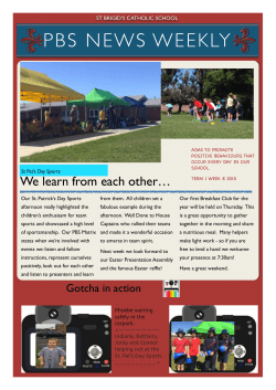

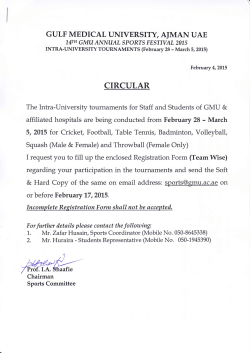
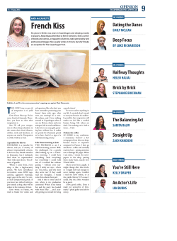
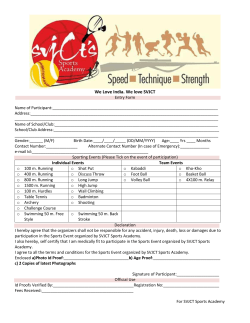
© Copyright 2026