
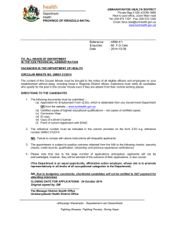
Human immunodeficiency virus-1 infection of the human
From www.bloodjournal.org by guest on October 28, 2014. For personal use only.
1993 81: 437-445
Human immunodeficiency virus-1 infection of the human
promyelocytic cell line HL-60: high frequency of low-level infection
and effect of subsequent cell differentiation
PM Cannon, DG Tenen, MB Feinberg, HS Shin and S Kim
Updated information and services can be found at:
http://www.bloodjournal.org/content/81/2/437.full.html
Articles on similar topics can be found in the following Blood collections
Information about reproducing this article in parts or in its entirety may be found online at:
http://www.bloodjournal.org/site/misc/rights.xhtml#repub_requests
Information about ordering reprints may be found online at:
http://www.bloodjournal.org/site/misc/rights.xhtml#reprints
Information about subscriptions and ASH membership may be found online at:
http://www.bloodjournal.org/site/subscriptions/index.xhtml
Blood (print ISSN 0006-4971, online ISSN 1528-0020), is published weekly by the American
Society of Hematology, 2021 L St, NW, Suite 900, Washington DC 20036.
Copyright 2011 by The American Society of Hematology; all rights reserved.
From www.bloodjournal.org by guest on October 28, 2014. For personal use only.
Human Immunodeficiency Virus-1 Infection of the Human Promyelocytic Cell
Line HL-60: High Frequency of Low-Level Infection
and Effect of Subsequent Cell Differentiation
By Paula M. Cannon, Daniel G. Tenen, Mark B. Feinberg, Hyung Sik Shin, and Sunyoung Kim
As a model system to study the infection of early myeloid
cells by human immunodeficiency virus-I (HIV-1), w e have
infected the human promyelocyticcell line, HL-60, with a
recombinant selectable HIV-1 clone. A fully infected population showed a relatively high frequency of low-level infection, with 40% of subcloned cells being negative by reverse transcriptase and p24 indirect immunofluorescence
analysis and displaying only low levels of supernatant p24.
The same treatment of a T-lymphoid cell line produced
100%productive infections. HIV-1 infection of HL-60 did
not appear to alter the state of differentiation of the cells,
as assessed by surface antigen expression, regardless of
the level of viral expression. Furthermore, infected cells
were able to respond normally to chemical inducers of dif-
ferentiation. Induction of differentiationtowards monocyte/
macrophages by phorbol myristate acetate activated the
HIV-1 long terminal repeat in a transient transfection system, and there was a corresponding increase in viral production from the infected subclones. Granulocytic differentiation, as stimulated by dimethyl sulfoxide or retinoic
acid, had no effect on long terminal repeat activity and did
not stimulate viral replication. These data suggest that lowlevel HIV-1 infections may be established at a relatively
high frequency in myeloid precursor cells, and that different
pathways of promyelocyticdifferentiation vary in their ability to stimulate HIV-1 replication.
0 1993 by The American Society of Hematology.
T
eloid cell differentiation may influence the susceptibility of
cells to i n f e ~ t i o n l ~or
* *affect
~
the subsequent replication of
HIV- 1 in already infected cells.'Ox2'
As a possible route to account for HIV- 1 infection of mature monocytes/macrophages, we hypothesize that early myeloid cells can be infected by HIV-1 and later differentiate
into mature infected cells. Such a pathway seems feasible
because some myeloid precursor cells express CD4,28 and
virus has been detected in the hematopoetic stem cells of
infected individ~als.'~
As a model system to study HIV-1
infection of such cells, we have used the human promyelocytic
cell line HL-60,29,30which we infected with a recombinant,
selectable viral clone. This approach enabled us to overcome
the low efficiency of infection of these cells by viral clones
and to select for a fully infected population. We report here
the finding of a high frequency of low-level infection of HL60 cells by this virus (defined as supernatant p24 antigen
levels 4 0 0 pg/mL in subcloned cells). This was not seen in
a T-lymphoid cell line similarly infected. In addition, because
this cell line has the potential to differentiate along either a
monocytic or granulocytic pathway in response to specific
HE PRINCIPAL TARGET cells of human immunodeficiency virus (HIV) infection in the body are the
CD4-expressing T lymphocytes and cells of the monocyte/
macrophage lineage.'" Although most of the circulating virus
has been shown to be present in T4
infection of
monocytes and macrophages is of particular interest because
of the major role that these cells play in the immune system.
In addition, the reported lower cytopathicity of HIV for these
cells,7 coupled with their lower surface expression of viral
antigens,' has led to the idea that these cells may serve as
cellular reservoirs for HIV in vivo.'.lo Infected macrophages,
with their wide tissue distribution, may allow HIV access to
other cells of the body not primarily infected by HIV; there
is also evidence to suggest that infection of T lymphocytes
can occur by this route."
Therefore, determining the origin and nature of the infection in monocytes and macrophages is of importance
to our understanding of the overall mechanism of the
pathogenesis of acquired immunodeficiency syndrome
(AIDS). HIV- 1 has been found in the mature tissue macrophages of the lungs, central nervous system, lymph
nodes, and skin of AIDS patients." These macrophages
could be infected in situ or could result from the migration
of infected blood monocytes into the tissues that then differentiated into macrophages. Mature macrophages have
low surface expression of CD4I3 and the apparently nonproliferating nature of these cells is at odds with the expected retroviral requirement for host DNA synthesis to
establish i n f e ~ t i 0 n . lHowever,
~
it has clearly been shown
that certain HIV-1 strains, such as HIV-1Ba.L3and HIVlJR.FL,15 can grow efficiently in macrophages prepared from
peripheral blood or bronchial l a ~ a g e . ~ , ~ ? ' ~
Myeloid cells from different stages of differentiation have
been shown to be susceptible to HIV-1 infection in vitro,
including hematopoietic stem cells,"~" blood monocyte^,^
and mature macrophages.' Virus has also been isolated from
these cell types in patient t i s s ~ e . In
~ ~addition,
~ ~ . ~ several
~
human myeloid cell lines have proved infectible by HIV-I,
including the immature HL-60 and U937 linesl~2~21-25
and
the more mature THP-I and Mono-Mac 6 line^.*^-^^ Furthermore, studies have shown that the normal process of myBlood, V o l 8 1, No 2 (January 15). 1993:pp 437-445
From the New England Deaconess Hospital, and Hematology/
Oncology Division, Beth Israel Hospital, Harvard Medical School,
Boston, MA; the Whitehead Institute for Biomedical Research, Cambridge, MA; the Department of Pathology, College of Medicine. Hallym University, Chun Cheon, Kang Won Do, Korea; and the Institute
for Molecular Biology and Genetics, Seoul National University, Seoul,
Korea.
Submitted December 17, 1991; accepted September 16, 1992.
Supported by the Korea Science and Engineering Foundation (S.K.),
and Public Health Service Grants No. AI 30897 and HL 43510 (S.K.),
and AI 29847 (D.G.T.). D.G.T. is a Scholar ofthe Leukemia Society
of America.
Address reprint requests to Sunyoung Kim, DPhil, Institute for
Molecular Biology and Genetics, Bldg 105, Seoul National University,
Kwan-Ak-Gu, Seoul 151-742, Korea.
The publication COSIS of this article were defrayed in part by page
charge payment. This article must therefore be hereby marked
"advertisement" in accordance with 18 U.S.C. section I734 solely to
indicate this fact.
0 I993 by The American Society of Hematology.
0006-4971/93/81 02-0001$3.00/0
437
From www.bloodjournal.org by guest on October 28, 2014. For personal use only.
CANNON ET AL
438
chemical inducers;' we investigated the effect of differentiation along either pathway on HIV-I long terminal repeat
(LTR) activity and on viral replication.
MATERIALS AND METHODS
Cell culture and induction of differentiation. The human promyeb
cytic cell line, HL60, and the T-lymphocytic cell line, H9, were grown
in RPMI medium, supplemented with glutamine, penicillin and strep
tomycin, and 10%and 20% heat-inactivatedfetal bovine m m (GIBCO
Labratories, Grand Island, NY), respxtively. Cells were maintained at
densitiesbetween 1 X 10s and 1 X 10⁶cek/mL. HL60cells were induced
to differentiate along the monocytic pathway by the addition of 8 X
mol/L phorbol myxistate acetate (PMA) to the growth medium. Granulocytic differentiation was induced by either 1.1% dimethyl sulfoxide
(DMSO) or
mol/L refinoic acid.
V i m infction and production of chronically infeted cell lines. The
viral clone, HIV-",l
is a fully infectiousvirus containinga selectable
marker. This strain was derived from the plasmid R7/Neo," which is
identical to HXB2 and RIF'73334
except that the bacterialneo gene replaces
part of the nefsequences. (This construct confers resistanceto G418 on
the host cell, so the addition of the antibiotic to the culture medium
after infection by this virus allows a population to be selected for that
harborsthe viral genome. Because the HIV strain HXB2 does not express
the @and vpu proteins, the recombinant HIV stran R7/Ne0 is identical
to HXB2 in terms of the pattem of protein expression.)A stock of R7/
Ne0 virus was prepared by transfectingthe human T-lymphoid cell tine
CEM and filtering the supematant through a 0.45-pm pore size filter
when syncytia were apparent (approximately 7 days after transfection).
In our preparations, the virus titer generally contained 1 to 5 X lo6cpm/
mL of revex transcriptaseactivity or 10s to 106 TCIDSO units (measured
by syncitia formation using the human T-cell line (3166). HMO and
H9 cells (lo') were infected with 1 mL of this stock at 37°C for 1.2
hours, followed by the addition of media. The antibiotic G418 (GIBCO
BRL, Gaithersburg, MD) was added to the media after 14 days at a
concentration of 1 mg/mL and selection continued until fully resistant
populationswere obtained.Cells were subclonedfrom these populations
by limiting dilution and the properties of IO such clones from each cell
tine were analyzed. Viral growth was assessed by measuring reverse transcriptax (RT) and p24 Concentration (Abbott Laboratories, Chicago,
IL) in the cell culture supematant and the percentage of infected cells
was assayed by indirect immunofluorescence(IF)with p24 monoclonal
antibody (MoAb). Supematant RT activity was assayed by the method
of Poiesz et d:5 except that the enzyme reaction was performed at 37°C
for 1 hour in a buffer (total volume of 100 mL) containing 40 mmol/L
TrisHCl (ph 7.Q 8 mmol/L dithiothreitol (DIT), 10 mmol/L M a z ,
50 pg of template primer p~Iy(rA)-(dT)~~.~~
(Pharmacia 27-7878; Pharmacia, Uppsala, Sweden) or poly(dA)-(dT)l2.18 (Pharmacia 27-7868) per
milliliter and 15 mmol/L ['HITTP (10 to 20 Ci/mol; New England
Nuclear NET-221H; New England Nuclear, Boston,
Indirect
immunofluorescence analysis was performed on cells ked in an 1:1
mixture of methanol and acetone at 4"C.%The first-stage antibody was
a p24 MoAb (Dupont, Boston, MA, diluted 1/40) and the second-stage
antibody was fluorescein isothiocyanate (FITC)-conjugated goat antimouse IgG (Cappel, Durham, NC; diluted 1/40),
DNA transfection. HIV-1 LTR activity in HL-60 cells under a
variety of conditions was assessed by transiently transfecting cells
with LTR-CAT constructs and assaying for chloramphenicol acetyl
transferase (CAT) activity. Essentially, 2 X IO7 cells per sample-point
were transfected by the diethylaminoethyl (DEAENextran procedure
of Grosschedl and Ba1timo1-e~~
(but without the addition of chloroquine) with 2 pg of the LTR-CAT plasmid and 4 pg of a Tat expression
vector. Eighteen hours later, cells were split and plated at 1 X IO6
cells/mL in media containing appropriate inducers and harvested 24
and 48 hours postinduction. Equivalent amounts of protein were
assayed for CAT activity@and the percent conversion of I4C-chloramphenicol to its acetylated forms was determined by cutting out
regions containing unreacted and acetylated forms and quantitating
the amount of radioactivity in each by liquid scintillation counting.
The LTR-CAT plasmid used, p938, contains the HIV-1 LTR from
pU3R-III.24.4'The Tat expression vector, pCMV-Tat, was provided
by D. Trono (Salk Institute, San Diego, CA) and consists of the first
exon of tat on a Sal I-Kpn I fragment from HXB233cloned into the
multilinker site of the expression vector P C P L K . ~ ~
Electrophoretic mobility shij assays (EMSA). Cellular extracts
were prepared from HL-60 cells grown in medium alone or after
incubation with PMA or DMSO for 2 days, and EMSA was performed
as described by Baeuerle and Baltimore.43Approximately 1.5 X IO'
cells were harvested, washed with ice-cold phosphate-buffered saline
(PBS), and transferred to eppendorf tubes. Cells were resuspended
in buffer containing 20 mmol/L HEPES (pH 7.9), 0.35 mol/L NaC1,
20% glycerol, 1% NP-40, 1 mmol/L MgCI2, 1 mmol/L DTT, 0.5
mmol/L EDTA, 0.1 mmol/L EGTA, 1% aprotinin (Sigma, St Louis,
MO), and I mmol/L phenylmethylsulfonyl fluoride (PMSF), and
lysed on ice for 10 minutes. Particulate material was removed by
centrifugation at 4°C for 15 minutes and the amount of protein in
the resulting supernatant was quantitated with bicinchoninic acid
(Micro BSA Protein Assay Reagent No. 23235; Pierce, Rockford,
IL). For the EMSA, I O pg of protein was incubated with a "P-labeled
KBoligonucleotide, prepared as described by Sen and Baltimore."
Specific DNA-protein complexes were identified after electrophoresis
through a 5% acrylamide gel by competition with unlabeled wildtype and mutant oligonucleotides. The nucleotide sequences of the
oligonucleotides used were:
Wild-type KBoligonucleotide:
GATCCTCCGCTGGGGACTTTCCAGGGAGGA
GAGGCGACCCCTGAAAGGTCCCTCCTCTAG
Mutant KBoligonucleotide:
***
GATCCTCCGCTCTCGACTTTCCAGGGAGGA
GAGGCGAGAGCTGAAAGGTCCCTCCTCTAG
(***, mutations introduced).
Analysis of cell surfaceantigens. Fluorescein-activated cell sorter
(FACS) analysis was performed as previously de~cribed.4~
MoAbs to
CDI l b (903) and CD14 (MY4) were provided by J.D. Griffin (DanaFarber Cancer Institute, Boston, MA); antibody to CD18 (10F12)
was provided by J. Ritz (Dana-Farber Cancer Institute); and antibody
to CD4 (Leu-3A) was purchased from Becton Dickinson (Mountain
View, CA). Fluorescein-conjugated goat antimouse IgG (Cappel) was
used as the second-stage antibody.
RESULTS
HL-60, but not H9 cells, display a high frequency of lowlevel infection by HZV-I. To obtain a population of HL-60
cells fully infected with HIV- 1, we have used a recombinant
virus, R7/Neo, that can be selected for i n the presence of
G4 18.32 R7/Neo is a fully infectious viral clone that infects
the T-lymphocytic H 9 cell line as efficiently as its parental
virus, HXB2,33although it replicates slightly more slowly
(data not shown). I n the presence of G418, a fully resistant
population was established and subclones were derived from
this population by limiting dilution. These resistant cell lines
grew with the same doubling times as the parental HL-60.
Also, the pattem of the expression of many cell surface
From www.bloodjournal.org by guest on October 28, 2014. For personal use only.
439
HIV-1 INFECTION OF HL-60 CELLS
Table 1. Properties of Infected HL-60 Subclones
Subclone No.
IF
~ 2 (w/mU
4
RT (X 1O6 cpm/mL)
7
3
1
11
6
8
4
2
9
10
-
<50
<50
-
300
400
4 x 10⁶
3 x 10⁶
8 x lo6
3 x 10⁶
4 x 10⁶
1 x 10⁸
-
-
+
+
++
++
+++
++++
1
2
9
15
10
48
Subclones were derived by limiting dilution from a fully G418-resistant
population of HL-60 cells and were analyzedfor markers of viral infection.
The propenies of 10 HL-60 subclones are shown.
Abbreviations: IF, arbitrary assessment of the IF intensityto p24 MoAb;
p24, supernatant p24 antigen levels.
markers used in this study was essentially identical between
the parental and selected lines (see below). These results suggest that the G4 I8 selection procedure itself had no significant
effect on the experiments we describe below.
When fully resistant to G4 18, the H L 6 0 and H9 cell p o p
ulations were markedly different. The T-lymphocytic H9 cells
were 100%positive for HIV-I infection, as assessed by p24
immunofluorescence analysis, whereas the promyelocytic
HL-60 population was only 60% positive. These ratios were
stable over a period of 8 weeks, and the populations remained
of similar composition when G418 selection was discontinued. Subclones were derived from both populations by limiting dilution techniques, and 10 randomly chosen clones
from each population were analyzed in further detail. The
H9-derived subclones were all highly positive for p24 expression by immunofluorescence analysis, and had RT levels in
the range of lo5 to IO6 cpm/mL. However, the IO HL-60
subclones displayed a range of levels of infection (Table 1).
Four of the subclones analyzed had low levels of HIV-1 infection, being negative for supernatant RT activity and for
expression of p24 by IF analysis. The levels of supernatant
p24 antigen in these cultures was also very low (400 pg/
mL), comparable to the levels seen in the cell lines ACHz
and U 1:6*47 which are well-characterized models of low-level
infection. The levels of viral expression in all the subclones
were more or less constant at a given cell concentration
throughout the culture. These results are consistent with the
population p24 IF ratios.
The doubling times of both the high- and low-level-infected
HL-60 subclones were comparable, and all IO subclones
contained HIV-I-specific RNA (see below), which argues
against spontaneous G4 I8 resistance having developed in
uninfected HL-60 cells. By comparison with the H9 population, which was 100%productively infected, it is apparent
that this high frequency (40%) of low-level HIV-I infection
is a property of the HL-60 cell line and not the R7/Neo virus.
Expression of viral RNA in HIV-I-infected HL-60
clones. To gain a better understanding of the differences in
viral gene expression in HL-60 and H9 cells, we have analyzed
the pattern of HIV-I-specific RNA transcripts present in the
subclones from each population (Fig I). As controls, we also
examined RNA from H9 cells infected by HIV-IIIIBand by
HXB2 (Fig 1, lanes 1 1 and 12). In each case, HIV-I produced
three specific size classes of RNA, corresponding to the genomic size RNA (band l); the singly spliced messages, including the env message (band 2); and the small multiplyspliced messages coding for the regulatory proteins (band 3
for HIV-IR7/Neoand band 4 for HIV-IlllB and HXB2). The
presence of the additional 1 kb of ne0 DNA at the 3’ end of
the genome in R7/Neo produced multiply-spliced RNA
transcripts that were markedly larger than the corresponding
HIV-IIIIB or HXBZ species (compare bands 3 and 4). An
effect on the size of the other two RNA bands is not apparent,
which could be a result of the gel conditions used or could
be possibly caused by an additional splice acceptor site being
present in the HIV-lR7/Neogenome, which could alter the
final size of the transcripts.
Previous analyses of RNA from cells infected productively
and at low level by HIV-I have suggested a difference in the
ratios of the three size classes of RNA in the two types of
HL60
H9-
I
I
7
-2
-3
-4
B
Lane
1
2 3 4
5 6 7 8 9 10
11 12 13 14 15
UL60 Clones 10 9 2 4 8 6 11 3 1 7
IF t t t
4- - - - Rr 481015 9 2 i - - - fx fo5cpm/m~j
++
Fig 1. HIV-1 RNA expressionin H9 and HL-60 subclones. Total
RNA was prepared and hybridized to an LTR probe as previously
described.” Short (A) and long (B) exposures of the same autoradiogram are shown. Lanes 1 through 10 are the infected HL-60
subclones; lanes 1 3 through 15 are three representative infected
H9 subclones. As controls, we have also included RNA from H9
cells infected with HIV-1 (Cl) and HXB2 (C2) (lanes 11 and 12).
Band 1 representsthe genomic size RNA transcripts, band 2 includes
the env message, band 4 is the multiply-spliced messages from
HXBZ and HIV-11118, band 3 is the multiply-spliced messages from
HIV-lR,
(which contain additional ne0 RNA). T, an a-tubulin RNA
loading control; IF, reaction to a p24 MoAb by immunofluorescence
analysis; and RT, reverse transcriptase activity.
From www.bloodjournal.org by guest on October 28, 2014. For personal use only.
CANNON ET AL
440
CD f f&
CD f4
CD i8
Fig 2. Analysis of HL-60 surface markers. FACS analysis was used to determine the levels of expression of the surface markers CD4,
and those incubated for 48 hours in the presence of DMSO (-)
or PMA (- - -). (Top)
CDI 1b, CDI 4, and CDI 8 on uninduced cells (-),
Uninfected HL-60 cells; (bottom) R7/Neo-infected subclone 2. The pattern of expression of these markers and the changes induced upon
differentiation along either pathway were similar in both populations, with the exception of CD4 expression, which is downmodulated
anyway in productively infected cells. The vertical axis represents cell number and the horizontal axis represents fluorescence intensity
(arbitrary units).
in the expression of all three antigen^:^,^' with the increase
i n f e ~ t i o n . In
~ ~the
, ~ ~latently infected T-lymphoid ACH-246
and monocytic U 147 cell lines, a pattern of RNA expression
in CD I4 expression being especially characteristic of monocytic differentiation. CD4 expression is unchanged by granhas been observed in which the smaller, multiply-spliced
ulocytic differentiation, but is markedly reduced upon
transcripts predominate over the genomic size s p e ~ i e s . ~ ' , ~ ~
monocytic differentiation by the addition of PMA.
Of the four HL-60 clones that were negative for surface p24
FACS analysis was performed on uninfected, uninduced
expression, clones I , 7, and 1 1 demonstrated a similar pattern
of RNA transcription to these latently infected cell lines, ie,
cells to establish the pattern of expression of these surface
antigens in HL-60 cells. CDI l b and CD14 antigens were
the smaller transcripts were more prevalent than the genomic
transcripts (Fig 1, lanes 7, 9, and IO). In contrast, most of
present at low levels (less than 15% of the cells), CD18 was
expressed by 100% of the cells, and CD4 was present on apthe productively infected HL-60 clones, and all of the H9
proximately 90% of the population (Fig 2). The levels of these
clones, displayed patterns more typical of a productive infection, with larger amounts of genomic RNA. Furthermore,
markers in the HIV- 1-infected subclones were also studied
HL-60 clones 9 and 10, which had the highest overall levels
and compared with the levels in the uninfected population.
Figure 2 shows a comparison between the uninfected popof supernatant RT activity, contained the greatest amount
ulation and one representative clone (subclone 2; Table 1).
of HIV-I RNA (Fig 1, lanes 1 and 2). (However, it has to be
noted that this correlation between viral production and the
CD4 expression was downregulated in this infected clone, as
ratio of genomic to subgenomic transcripts is not always the
is commonly observed in HIV-infected cells, but the levels
of expression of CD 1 I b, CD 14, and CD 1 8 were comparable
case, because clone 3, despite being a low producer, expressed
significant levels of genomic RNA, whereas the high producer
between the two populations, indicating that HIV- 1 infection
clone 4 had very little of the genomic transcript.) These data
did not alter the state of differentiation of the HL-60 cells.
suggest a variation in the nature of the infections that establish
Similar results were also obtained for a number of other inin HL-60 cells. The relatively high frequency of establishment
fected subclones for CDI 1b, CD14, and CDI 8. CD4 expression was downregulated in all of the productively infected
of such low-level infections (40% of the clones) is a property
subclones, but 3 of 4 of the low level infections (clones I , 7,
of the HL-60 cell line and did not occur in T cells infected
and 11) still expressed surface CD4. These data suggest that
in the same manner.
in our experimental system, HIV- 1 infection of HL-60 cells
HIV-I infection of HL-60 does not alter the expression of
did not induce cell differentiation.
a number of differentiation-associated cell surface markers. Differentiation of myeloid cells is associated with charHIV-I-infected HL-60 cells respond normally to inducers
acteristic changes in cell surface markers. In HL-60 cells, the
of differentiation. Although our data suggested that HIV- 1
expression of surface antigens CD4, CD 11b, CD 14, and CD I8
infection did not alter the state of differentiation of HL-60
cells, it is possible that chronic viral infection could prevent
are useful indicators of the state of cellular differentiation of
the ~ e l l s . ~The
~ , ~ CDI
'
l b and CD14 antigens are markers
these cells from responding normally to differentiation signals.
FACS analysis was used to determine the antigen changes
associated with mature myeloid cells, and their expression is
correspondingly low on undifferentiated HL-60 cells, whereas
associated with the differentiation of uninfected HL-60 cells
in response to PMA or DMSO. Differentiation along both
CD18 is expressed on HL-60 cells at all stages of differentiation. Induction of differentiation in HL-60 cells along either
pathways resulted in expected increases in the expression of
the monocytic or granulocytic pathway results in an increase
the CDI lb, CD14, and CD18 antigens (Fig 2). The same
From www.bloodjournal.org by guest on October 28, 2014. For personal use only.
HIV-1 INFECTION OF HL-60 CELLS
441
Viral replication is stimulated by monocytic but not granulocytic differentiation. The establishment of chronically
infected HL-60 subclones allowed us to study the effects of
cellular differentiation on viral replication at various timepoints postinduction. We therefore treated both high- and
low-level-infected HL-60 subclones with PMA or DMSO,
and analyzed the changes in viral replication by measuring
p24 concentration in the cell culture supernatant. PMA
stimulation increased supernatant p24 antigen levels by
threefold to fivefold from both high- and low-level producers
over a control population (Fig 5), whereas treatment with
DMSO had no significant effect. In the case of high producer
lines, this induction of threefold to fivefold was also observed
when RT activity in the cell culture supernatant was measured
(data not shown). (It should be noted that PMA treatment
arrests cell division and kills up to 50% of the HL-60 cells
within 48 hours. This makes it difficult to draw a firm conclusion from the data for the low producer clones, which
produce only small levels of p24 antigen and have undetectable levels of RT in the culture supernatant.) The increases
in viral production from several HL-60 subclones were of a
similar range to the fourfold induction previously reported
for a chronically infected Molt-4 cell line treated with PMA:3
but were considerably less than the induction obtained for
U 1 or ACH-2 cells treated with PMA!8*49 Our results indicate
that monocytic, but not granulocytic, differentiation enhances
viral replication, at least in the case of high-level-infected
HL-60 subclones.
pattern was observed when several HIV-I-infected subclones
were treated in the same way, and the results for one representative subclone are shown (Fig 2). In addition, cell differentiation was assessed by cytochemical assays for monocyteand granulocyte-specific esterase enzymes, with the infected
subclones demonstrating the same pattern of enzyme activity
as the uninfected cells, both before and after induction of
differentiation (data not shown). This demonstrates that
chronic HIV-I infection did not impair the ability of HL-60
cells to respond normally to chemical inducers of differentiation.
Eflect of cell dixerentiation on HIV-1 LTR activity. The
effect of HL-60 differentiation on HIV-I LTRdriven gene
expression was studied by transiently cotransfecting LTRCAT and Tat expression plasmids into undifferentiated HL60 cells. Eighteen hours later, the cells were induced to differentiate along either the monocytic pathway, by addition
of 8 X IO-' mol/L PMA, or the granulocytic pathway, by
mol/L retinoic acid. Cells
addition of I. 1 % DMSO or
were harvested at I , 2, and 3 days postinduction and assayed
for CAT activity (Fig 3). Treatment with retinoic acid or
DMSO had no significant effect on LTR activity, giving levels
of CAT activity of only onefold to twofold above that of the
uninduced control. Induction of monocytic differentiation
by PMA, however, led to a marked increase in CAT activity,
with increases of IO- to 30-fold over the uninduced level.
These results were reproducible in several independent experiments. Transcription from the HIV- I LTR was therefore
highly stimulated by PMA treatment, but was unaffected by
DMSO or retinoic acid induction.
The transient transfection assays demonstrated that PMA
stimulation of HL-60 cells increased LTRdriven gene
expression. PMA treatment of HL-60 cells has previously
been shown to stimulate NF-KBbindingacti~ity,~'
and as the
LTR contains two KBsites, induction of NF-KBbinding leads
to enhanced LTR t r a n s c r i p t i ~ n We
. ~ ~ here confirmed the
induction of NF-KBin HL-60 cells in response to PMA by
electrophoretic mobility shift analysis of a 32P-labeled oligonucleotide probe corresponding to the LTR KBsites. This
was incubated with cell extracts from uninduced and PMAtreated cells, and the induction of NF-KBbinding at 2 days'
poststimulation was shown by the appearance of a specific
protein-DNA complex on an acrylamide gel (Fig 4). Treatment of HL-60 cells with DMSO did not induce NF-KB.
Fig 3. Effect of cell differentiation on HIV-1 LTR
expression. Two microgramsof an HIV-1 LTR-CAT
plasmid, p938, and 4 pg of a Tat expression plasmid,
pCMV-Tat, were cotransfected into 2 x 10' HL-60
cells per timepoint and cells were plated in media
at 2 x 10' cells/mL. Eighteen hours later, the cells
were split and plated at 1 x 10' in media alone or
with 1.1 % DMSO, 1O-B mol/L retinoic acid (RA), or
8 x 10⁻⁸mol/L PMA, and harvested at 1 , 2 , and 3
days postinduction. CAT activity was assessed for
20 pg of protein extract incubated at 37°C for 1
hour. The data from one representative experiment
are shown. Lane 1 is the basal level CAT activity in
uninduced cells. C, unconverted ''C-chloramphenicol substrate; Ac, the acetylated forms.
DISCUSSION
Using the human promyelocytic cell line, HL-60, we have
studied various aspects of HIV- 1 infection of early myeloid
cells. To reduce variability in our experiments, our approach
has been to infect HL-60 cells with a selectable viral clone,
to establish a fully infected population, and to derive infected
subclones from this population. This system has enabled us
to compare the frequency of establishment of productive infections in early myeloid and T-lymphoid cell lines, and also
to investigate the effect of myeloid cell differentiation on the
replication of HIV- 1.
This system, which allows the selection of both low level
and productive infections, has demonstrated that HIV-I infection of the early myeloid cell line, HL-60, appears to be
remarkably different from that of the T-lymphoid H9 cell
C-
5
1
2
3
4
6
7
Doysl
%6bnversim 3
Inducer 0
1
2
36
3
1
2
3
x) 6
-DMSO-
3
1
46
--MA2
8
9
1
2
35 25
LPMA-
10111213
3
1 2 3
22 6 3 1
-RA-
From www.bloodjournal.org by guest on October 28, 2014. For personal use only.
CANNON ET AL
442
1 2 3 4 5
Fig 4. NF-xB induction by PMA. Ten micrograms of protein from
unstimulated HL-60 cells (lane 1) or those treated with DMSO (lane
2) or PMA (lane 3)for 2 days were incubated at m m temperature
for 20 minutes with a 3zP-labeledoligonucleotide correspondingto
the enhancer sequence of the HIV-1 LTR. Gel electrophoresisdemonstrated the presence of a specific protein-DNA complex induced
by PMA treatment (arrow) whose specificity was confirmed by incubation of the day 2 sample with 30 ng of unlabeledwild-type (W)
or mutant (M) oligonucleotide (lanes 4 and 5). FP, the unreacted
probe.
line. In particular, low-level infectionsappear to be established
at a relatively high frequency. Of the IO G418-resistant HL60 subclones analyzed, 40%were negative for supernatant
RT activity and for p24 expression by IF analysis, while still
expressing viral RNA. In contrast, 100% of the subclones
obtained by the same treatment of the H9 cell line were productively infected. While it could be argued that cells infected
at a low level have a selective growth advantage over productively infected cells in the presence of G4 18, the HL-60
Clone 2
population composition remained similar in the absence of
G4 18 selection. In addition, the doubling times ofall the HL60 subclones were comparable.
The nature of these low-level infections remains unclear.
The predominance of the multiply-spliced transcripts in three
of the four low-level subclones is reminiscent of the situation
in the U1 and ACH-2 cell line models of latent infection:”
49 Alternatively, these subclones may harbor defective proviruses incapable of making wild-type virions, as has been
reported to occur with other retroviral infections. Attempts
to determine whether this low-level phenotype was caused
by defective virions or was the result of a specific interaction
between the virus and the host cell were hampered by the
loss of infectivity of all of the HL-60 clones (both high- and
low-level producers) over time (data not shown). We are currently exploring the reasons for this observation. Whether
these low-level infections are latent or defective, they are established at a relatively high frequency, and are only seen
with the HL-60 cell line. We believe that that greater tendency
for these low-level infections to establish in early myeloid
cells than in mature T lymphocytes could have relevance to
HIV-I infection in vivo.
HIV-I infection of early myeloid cell lines has previously
been proposed to induce d i f f e r e n t i a t i ~ n ? ~although
* ~ ~ * ~ ~this
remains c~ntroversial?~*~~
Analysis of the cell surface markers
in a number of infected H L 6 0 subclones demonstrated the
retention of an almost identical pattern to the parental pop
ulation, arguing against HIV-1 infection stimulating differentiation in HL-60 cells and further demonstrating that HIVI did not selectively infect cells of greater or lesser maturity
from within the population. Treatment of both infected and
uninfected HL-60 cells with PMA or DMSO induced differentiation and led to characteristic changes in cell surface antigens. In addition, PMA treatment caused the cells to rapidly
become adherent and develop a macrophage-like morphology. Therefore, it appears that infection by HIV-1 did not
prevent HL-60 from differentiating normally in response to
these chemical inducers.
It is well documented that treatment of infected cell lines
~-~’*~’
with phorbol ester stimulates HIV-I r e p l i c a t i ~ n ~ ’ . ~ and
that the differentiation of infected monocytes to macrophages
Clone 3
I20
300
.
z
h
100
00
3
200
-5
80
Y)
U
6
0
4
0
N
d
100
a
20
0
0
CONTROL PMA
DMSO
CONTROL PMA
DMSO
Fig 5. PMA shmulates viral
production from infected HL-60
subclones. Several HL-60 subclones were splii, plated in fresh
media at a density of 2 X 10’
cells/mL, and treated with PMA
(10-* mol/L) or DMSO (1.1%) or
left untreated as a control population. After 24 or 48 hours,
viral activity in bothgroups was
assessed by supematant p24
antigen levels. Notethat the p24
concentration of the high producer (clone 2) is shown as
nanograms per milliliter,
whereas the low producer (clone
3)is shown as picograms per
milliliter.
From www.bloodjournal.org by guest on October 28, 2014. For personal use only.
443
HIV-1 INFECTION OF HL-60 CELLS
in vitro is accompanied by an increase in viral
PMA treatment of both high- and low-level-infected clones
resulted in a threefold increase in viral production, whereas
DMSO-treated cells retained the same level of viral replication
as the uninduced control population. Furthermore, transient
transfection analyses showed a 10- to 30-fold enhancement
of LTR transcription in cells treated with PMA above the
control or DMSO-treated populations. Enhancement of HIV1 replication and LTR transcription in PMA-treated cells has
previously been attributed to the induction of NF-KB.PMA
increases KB-binding activity in HL-60 cell extracts (as first
reported by Griffin et al,5' and confirmed here by our work),
which could act to increase LTR transcription. In contrast,
NF-KB binding is not increased by DMSO treatment. At
present, we cannot account for the discrepancy in the level
of PMA enhancement of LTR-directed transcription and viral
replication, but it is probable that additional, posttranscriptional processes, such as viral assembly and release, limit the
final rate of virus production.
If cells of the monocyte/macrophage lineage can be infected
at the myeloid stem cell stage, then it is possible that mature
neutrophils could also be infected in this manner and harbor
virus. Neutrophils lack the CD4 receptor and infection of
these cells has not been demonstrated. Furthermore, a study
by Spear et a16to determine whether proviral DNA sequences
were present in neutrophils was inconclusive. However, impaired neutrophil function has been reported in AIDS pat i e n t ~ , ~ so
' . ~it~is possible that neutrophils are infected with
HIV in vivo, but at a very low level. In addition, HIV infection
of neutrophils may be more difficult to detect because of
their short half-life relative to the macrophage. Induction of
granulocytic differentiation in HL-60 cells by the addition of
DMSO or retinoic acid neither increased levels of LTR-driven
CAT activity in our transient transfection assays nor stimulated viral production above control levels from our infected
subclones. It is therefore possible that any in vivo infection
of neutrophils or their precursor cells would be nonproductive.
Our observations with the HL-60 cell line suggest that infection of early myeloid cells by HIV-1 may frequently be
relatively nonproductive, and that myeloid precursors could
therefore serve as reservoirs of viral infection in the body.
Noncytopathic infection of myeloid stem cells has precedents
in other retroviral infections, including Visna virus infection
of sheepm and FeLV infection of cats6' Furthermore, the
completion of the Visna life-cycle and the production of mature virions is dependent on the maturation of these infected
stem cells to fully differentiated macrophages.62It is therefore
possible that a similar situation occurs in the case of HIV-1,
with early myeloid cells serving as a route of entry into the
more quiescent, mature macrophage, and that viral replication would be stimulated by monocytic, but not by granulocytic differentiation of these precursor cells.
ACKNOWLEDGMENT
We thank Heike Pahl for technical assistance and Catherine Ulich
for help in manuscript preparation.
REFERENCES
I. Dalgliesh AG, Beverly PCL, Clapham PR, Crawford DH,
Greaves MF, Weiss RA: The CD4 (T4) antigen is an essential component ofthe receptor for the AIDS retrovirus. Nature 3 12:763, I984
2. Levy JA, Shimabukuro J, McHugh T, Casavant C, Stites D,
Oshiro L AIDS-associated retroviruses(ARV) can productively infect
other cells besides human T helper cells. Virology 147:441, 1985
3. Gartner S, Markovits P, Markovitz DM, Kaplan MH, Gallo
RC, Popovic M: The role of mononuclear phagocytes in HTLV-III/
LAV infection. Science 233:215, 1986
4. McElrath MJ, Pruett JE, Cohn ZA: Mononuclear phagocytes
of blood and bone marrow: Comparative roles as viral reservoirs in
human immunodeficiency virus type 1 infections. Proc Natl Acad
Sci USA 86:675, 1989
5. Schnittman SM, Psallidopoulos MC, Lane HC, Thompson L,
Baseler M, Massari F, Fox CH, Salzman NP, Fauci AS: The reservoir
for HIV-1 in human peripheral blood is a T cell that maintains
expression of CD4. Science 245:305, 1989
6. Spear GT, Ou C, Kessler HA, Moore JL, Schochetman G, Landay A L Analysis of lymphocytes, monocytes, and neutrophils from
human immunodeficiency virus (HIV-1)-infected persons for HIV
DNA. J Infect Dis 162:1239, 1990
7. Nicholson JKA, Cross GD, Calaway CS, McDougal JS: In vitro
infection of human monocytes with human T lymphotropic virus
type III/lymphadenopathy-associatedvirus (HTLV-III/LAV). J Immunol 137:323, 1986
8. Potts BJ, Maury W, Martin MA: Replication of HIV-I in primary monocyte cultures. Virology 175:465, 1990
9. Salahuddin SZ, Rose RM, Groopman JE, Markham PD, Gallo
R C Human T lymphotropic virus type I11 infection ofhuman alveolar
macrophages. Blood 65:281, 1986
10. Gendelman HE, Orenstein JM, Martin MA, Fenua C, Mitra
R, Phipps T, Wahl LA, Lane HC, Fauci AS, Burke DS, Skillman D,
Meltzer MS: Efficient isolation and propogation of human immunodeficiency virus on recombinant colony-stimulatingfactor 1-treated
monocytes. J Exp Med 167:1428, 1988
11. Mann DL, Gartner S, Le Sane F, Buchow H, Popovic M:
HIV- 1 transmission and function of virus-infected monocytes/macrophages. J Immunol 144:2152, 1990
12. Gendelman HE, Orenstein JM, Baca LM, Weiser B, Burger
H, Kalter DC,Meltzer MS: The macrophage in the persistence and
pathogenesis of HIV infection. AIDS 3:475, 1989
13. Valentin A, Matsuda S, Asjo B: Characterization of the in
vitro maturation of monocytes and the susceptibilityto HIV infection.
AIDS Res Hum Retroviruses 6:977, 1990
14. Varmus HE, Swanstrom R: Replication of retroviruses, in
Weiss R, Teich N, Varmus H, Coffin J (eds): RNA Tumor Viruses,
vol 2. Cold Spring Harbor, NY, Cold Spring Harbor Laboratory,
1985, p 75
15. Koyanagi Y, Miles S, Mitsuyasu RT, Memll JE, Vinters HV,
Chen ISY Dual infection of the central nervous system by AIDS
viruses with distinct cellular tropisms. Science 236319, 1987
16. Collman R, Hassan NF, Walker R, Godfrey B, Cutilli J, Hastings JC, Friedman H, Douglas SD, Nathanson N: Infection of
monocyte-derivedmacrophages with human immunodeficiency virus
type 1 (HIV-I). J Exp Med 1701 149, 1989
17. Folks TM, Kessler SW, Orenstein JM, Justement JS, Jaffe ES,
Fauci AS: Infection and replication of HIV- 1 in purified progenitor
cells of normal human bone marrow. Science 242:919, 1988
18. Steinberg HN, Crumpacker CS, Chatis P A In vitro suppression
of normal human bone marrow progenitor cells by human immunodeficiency virus. J Virol 65: 1765, 199 1
19. Donahue RE, Johnson MM, Zon LI, Clark SC, Groopman
JE: Suppression of in vitro haematopoiesis following human immunodeficiency virus infection. Nature 326:200, 1987
From www.bloodjournal.org by guest on October 28, 2014. For personal use only.
444
20. Popovic M, Gartner S: Isolation of HIV-1 from monocytes
but not T lymphocytes. Lancet 2:916, 1987
21. Clapham PR, Weiss RA, Dalgliesh AG, Exley M, Whitby D,
Hogg N: Human immunodeficiency virus infection of monocytic
and T-lymphocytic cells: Receptor modulation and differentiation
induced by phorbol ester. Virology 158:44, 1987
22. Folks TM, Justement J, Kinter A, Schnittman S, Orenstein
J, Poli G, Fauci AS: Characterization of a promonocyte clone chronically infected with HIV and inducible by 13-phorbol-12-myristate
acetate. J Immunol 140:11 17, 1988
23. Pauza CD, Galindo J, Richman DD: Human immunodeficiency virus infection of monoblastoid cells: Cellular differentiation
determines the pattern of virus replication. J Virol 62:3558, 1988
24. Kim S, Ikeuchi K, Groopman J, Baltimore D Factors affecting
cellular tropism of human immunodeficiency virus. J Virol64:5600,
1990
25. Kitano K, Cocita Baldwin G, Raines MA, Golde DW: Differentiating agents facilitate infection of myeloid leukemia cell lines
by monocytropic HIV-1 strains. Blood 76: 1980, 1990
26. L'age-stehr J, Niedrig M, Gelderblom HR, Sim-Brandenburg
JW, Urban-Schriefer M, Rieber EP, Haas JG, Riethmuller G, ZieglerHeitbrock HWL: Infection of human monocytic cell line Mono Mac6
with human immunodeficiency virus types 1 and 2 results in longterm production of virus variants with increased cytopathogenicity
for CD4+ T cells. J Virol 64:3982, 1990
27. Schrier RD, McCutchan JA, Venable JC, Nelson JA, Wiley
CA: T-cell induced expression of human immunodeficiency virus in
macrophages. J Virol 64:3280, 1990
28. Fredrickson GG, Basch RS: L3T4 antigen expression by hematopoietic precursor cells. J Exp Med 169:1473, 1989
29. Collins SJ, Gallo RC, Gallagher RE: Continuous growth and
differentiation of human myeloid leukemic cells in suspension culture.
Nature 270:347, 1977
30. Gallagher R, Collins S, Trujillo J, McCredie K, Ahearn M,
Tsai S, Metzgar R, Aulakh G, Ting R, Ruscetti F, Gallo R: Characterization of the continuous, differentiating myeloid cell line (HL60) from a patient with acute promyelocytic leukemia. Blood 54:
713, 1979
3 1. Collins SJ: The HL-60 promyelocytic leukemia cell line: Proliferation, differentiation, and cellular oncogene expression. Blood
70:1233, 1987
32. Feinberg MB, Baltimore D, Frankel AD
the human immunodeficiency virus life cycle
effect on transcriptional elongation. Proc Natl Acad Sci USA 88:
4045, 1991
33. Fisher AG, Feinberg MB, Josephs SF, Harper ME, Marselle
LM, Reyes G, Gonda MA, Aldovini A, Debouk C, Gallo RC, WongStaal F The trans-activator gene of HTLV-111 is essential for virus
replication. Nature 320:367, 1986
34. McCune JM, Rabin LB, Feinberg MB, Lieberman M, Kosek
JC, Reyes GR, Weissman IL: Endoproteolytic cleavage of gp160 is
required for the activation of human immunodeficiency virus. Cell
5355, 1988
35. Poiesz BJ, Ruscetti F W , Gazder AF, Bunn PA, Minna JD,
Gallo RC: Detection and isolation of type C retrovirus particles from
fresh and cultured lymphocytes of a patient with cutaneous T-cell
lymphoma. Proc Natl Acad Sci USA 77:7415, 1980
36. Kacian D L Methods for assaying reverse transcriptase. Methods Virol 6:143, 1977
37. Hoffman AD, Banapour B, Levy J: Characterization of the
AIDS-associated reverse transcriptase and optimal conditions for its
detection in virions. Virology 147:326, 1985
38. Ho DD, Schooley RT, Rota TR, Kaplan JC, Flynn T, Salahuddin SZ, Gonda MA, Hirsch MS: HTLV-I11 in the semen and
blood of a healthy homosexual man. Science 226:45 I, 1984
CANNON ET AL
39. Grosschedl R, Baltimore D: Cell-type specificity of immunoglobulin gene expression is regulated by at least three DNA sequence
elements. Cell 41:885, 1985
40. Gorman CM, Moffat LF, Howard BH: Recombinant genomes
which express chloramphenicol acetyl-transferasein mammalian cells.
Mol Cell Biol 2:1044, 1982
41. Rosen CA, Sodroski JG, Haseltine WA: The location of cisacting regulatory sequences in the human T cell lymphotropic virus
type 111 (HTLV-III/LAV) long terminal repeat. Cell 412313, 1985
42. Trono D, Baltimore D: A human cell factor is essential for
HIV-I Rev action. EMBO J 9:4155, 1990
43. Baeuerle PA, Baltimore D IKB:A specific inhibitor of the NFKBtranscription factor. Cell 53:21 I, 1988
44. Sen R, Baltimore D: Inducibility of K immunoglobulin enhancer-bindingprotein NF-KBby a posttranslational mechanism. Cell
47:921, 1986
45. Rosmarin AG, Weil SC, Rosner GL, Griffin JD, Amout MA,
Tenen DG: Differential expression of CDI 1b/CD18 (Mol) and myeloperoxidase genes during myeloid differentiation. Blood 73: 13I ,
1989
46. Clouse KA, Powell D, Washington I, Poli G, Stebel K, Farrar
W, Barstad B, Kovacs J, Fauci AS, Folks TM: Monokine regulation
of HIV-1 expression in a chronically-infected human T cell clone. J
Immunol 142:43I , I989
47. Folks T, Justement J, Kinter A, Dinarello CA, Fauci A S Cytokine induced expression of HIV- I in a chronically-infected promonocyte cell line. Science 238:800, 1987
48. Pomerantz RJ, Trono D, Feinberg MB, Baltimore D: Cells
nonproductively infected with HIV-I exhibit an aberrant pattern of
viral RNA expression: A molecular model for latency. Cell 6 1: 127I ,
1990
49. Michael NL, Morrow P, Mosca J, Vahey M, Burke DS, Redfield RR: Induction of human immunodeficiency virus type 1
expression in chronically infected cells is associated primarily with a
shift in RNA splicing patterns. J Virol 65: 129I , 1991
50. Graziano RF, Ball ED, Fanger MW: The expression and
modulation of human myeloid-specific antigens during differentiation
ofthe HL-60 cell line. Blood 61:1215, 1983
5 1. Griffin GE, Leung K, Folks TM, Kunkel S, Nabel GJ: Activation of HIV gene expression during monocyte differentiation by
induction of NF-KB.Nature 339:70, 1989
52. Nabel G, Baltimore D: An inducible transcription factor activates expression of human immunodeficiencyvirus in T cells. Nature
326:711, 1987
53. Harada S, Koyanagi Y, Nakashima H, Kobayashi N, Yamamoto N: Tumor promoter, TPA, enhances replication of HTLVIII/LAV. Virology 154:249, 1986
54. Petit AJC, Terpstra FG, Miedema F: Human immunodeficiency virus infection down-regulates HLA class I1 expression and
induces differentiation in promonocytic U937 cells. J Clin Invest 79:
1883, 1987
55. Rossen RD, Smith CW, Laughter AH, Noonan CA, Anderson
DC, McShane WM, Hurvitz MY, Orson FM: HIV-1 stimulated
expression of CD I I/CD 18 integrins and ICAM- 1: A possible mechanism for extravascular dissemination of HIV- I-infected cells. Trans
Assoc Am Phys 102:117, 1989
56. Locardi C, Petrini C, Boccoli G, Testa U, Dieffenbach C, Botto
S, Belardelli F Increased human immunodeficiency virus (HIV)
expression in chronically infected U937 cells upon in vitro differentiation by hydroxyvitamin D3: Roles of interferon and tumor
necrosis factor in regulation of HIV production. J Virol 64:5874,
1990
57. Harada S, Koyanagi Y, Nakashima H, Kobayashi N, Yamamoto N Tumor promoter, TPA, enhances replication of HTLVIII/LAV. Virology 154:249, 1986
From www.bloodjournal.org by guest on October 28, 2014. For personal use only.
HIV-1 INFECTION OF HL-60 CELLS
58. Ellis M, Gupta S, Galant S, Hakim S, VandeVen C, Toy C,
Cairo MS: Impaired neutrophil function in patients with AIDS or
AIDS-related complex: A comprehensive evaluation. J Infect Dis
158:1268, 1988
59. Murphy PM, Lane HC, Fauci AS, Gallin JI: Impairment of
neutrophil bactericidal capacity in patients with AIDS. J Infect Dis
158:627, 1988
60. Gendelman HE, Narayan 0,Molineaux S, Clemens JE, Ghotbi
Z Slow, persistent replication of lentiviruses: Role of tissue macrophages and macrophage precursors in bone marrow. Proc Natl Acad
Sci USA 82:7086, 1985
445
6 1. Mullins JI, Chen CS, Hoover EA: Disease-specific and tissuespecific production of unintegrated feline leukemia virus variant in
feline AIDS. Nature 319:333, 1986
62. Gendelman HE, Narayan 0, Kennedy-Stoskopf S, Kennedy
PGE, Ghotbi Z, Clemens JE, Stanley J, Pezeshkpour G: Tropism of
sheep lentiviruses for monocytes: Susceptibility to infection and virus
gene expression increase during maturation of monocytes to macrophages. J Virol 58:67, 1986
63. Kim S, Byrn R, Groopman J, Baltimore D Temporal aspects of
DNA and RNA synthesis during human immunodeficiency virus infection: Evidence for differentialgene expression.J Viol 63:3708, 1989


© Copyright 2026