
ORIGINAL ARTICLE HISTOCHEMICAL STUDY OF MUCOSUBSTANCES OF NORMAL HUMAN PAROTID SALIVARY GLAND
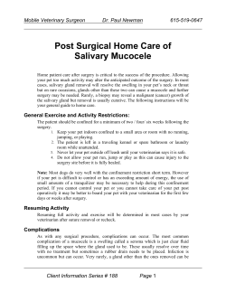
DOI: 10.14260/jemds/2014/3739
ORIGINAL ARTICLE
HISTOCHEMICAL STUDY OF MUCOSUBSTANCES OF NORMAL HUMAN
PAROTID SALIVARY GLAND
Rohini R. Karambelkar1, Avinash D. Shewale2, Rajesh A. Karambelkar3, B. N. Umarji4
HOW TO CITE THIS ARTICLE:
Rohini R. Karambelkar, Avinash D. Shewale, Rajesh A. Karambelkar, B. N. Umarji. “Histochemical Study of
Mucosubstances of Normal Human Parotid Salivary Gland”. Journal of Evolution of Medical and Dental Sciences
2014; Vol. 3, Issue 58, November 03; Page: 13079-13085, DOI:10.14260/jemds/2014/3739
ABSTRACT: INTRODUCTION: The oral cavity is one of the important routes for infections and
infestations. Majority of the infections occur through food and air. The oral cavity is said to be even
worst than anal canal due to its direct exposure to atmosphere and the moist condition, which
facilitate the bacterial growth. Plenty of lymphoid tissue and glandular secretions protects the
mucosa of oral cavity. Glandular secretions are serous and mucous in nature and are rich in
Mucosubstances which protect the oral mucosa from bacteria and viruses. They help to lubricate the
oral mucosa. In unstimulated state major bulk of secretion is from parotid salivary gland. METHODS:
In the present study Mucosubstances present in parotid salivary gland are studied. 30 normal human
parotid salivary glands were collected and fixed in 10% formalin with 2% Ca acetate and pinch of
phosphotungstic acid. 4 to 5 micron thick sections were cut and blocks were prepared. The sections
were stained with periodic acid Schiff (PAS), alcian blue (AB) at different pH and Aldyhyde fuschin
(AF) individually and in combinations. Confirmatory tests were carried out. RESULTS: the normal
parotid salivary gland shows presence of neutral Mucosubstances with very trace amount of
sialomucins and sulphomucins. Presence of neutral mucins indicates that the secretions of parotid
salivary gland are rich in enzymes.
KEYWORDS: Parotid, Mucosubstances, PAS, AB, salivary mucins.
INTRODUCTION: The parotid salivary gland is the largest salivary gland which communicates the
oral cavity through vestibule of mouth. It is situated upon the side of the face, immediately below and
in front of the external ear.
It is pyramidal in shape with apex directed downwards. It is drained by a duct which opens in
the vestibule of mouth opposite the crown of upper second molar tooth. Histologically it is a
compound tubule-alveolar gland with lobes and lobules separated by connective tissue septa. The
parotid gland contains 95% serous alveoli and 5% mucous alveoli.
These alveoli secrete Mucosubstances which performs a wide variety of functions like
lubrication, protection against acids, maintenance of hydration etc. They also contain
immunoglobulin’s mainly IgA type, lactoferrins which chelate the iron necessary for growth of some
bacteria and lysozymes which destroy some of bacteria. The mucins play an important role defense
against bacteria. Sulphomucins are acting as antiulcerogenic as they coat and protect mucosal surface
while neutral mucins help for secretion of enzymes.1-4
Many workers like Dr. Ganga (2004),5 Gad,6,7 Filpe and Fenger8 have studied the
Mucosubstances of GIT but very few have studied the salivary gland mucins. So the present study has
been undertaken to study the mucins in the parotid gland in human and correlate it with previous
studies.
J of Evolution of Med and Dent Sci/ eISSN- 2278-4802, pISSN- 2278-4748/ Vol. 3/ Issue 58/Nov 03, 2014
Page 13079
DOI: 10.14260/jemds/2014/3739
ORIGINAL ARTICLE
MATERIAL AND METHODS: Thirty normal human parotid salivary glands were obtained during
autopsies and routine dissection. The tissue was fixed in 10% formalin with 2% Ca acetate. By
routine procedure paraffin blocks were prepared and 4-5 micron thick sections were cut. They were
stained by.9-11
1. Haematoxylene and Eosin (H&E).
2. Periodic Acid- Schiff (PAS).
3. Periodic Acid- Schiff with diastase digestion (PAS-D).
4. Periodic Acid- Schiff with phenyl hydrazine (PAS-PH).
5. Alcian blue 2.5 pH (AB 2.5 pH).
6. Alcian blue 1 pH (AB 1 pH).
7. Combined AB-PAS.
8. Aldehyde fuschin (AF).
9. Combined AF-AB.
The slides were photographed using digital camera. The histochemical data staining methods
employed in the present work are recorded according to visually estimated intensity of staining and
shades with four plus representing strongest activity. Nomenclature applied to the mucosubstances is
taken from the discussion of a proposed general terminology of histochemically recognized
material.12,13 histochemical results requiring further description and consideration are presented
here along with their interpretations.
OBSERVATION AND RESULTS:
i.
Numerous lobes and lobules separated by connective tissue septa are seen. Each lobule
contains the secretory acini of parotid salivary gland. They are mainly seromucus. Majority are
serous acini with very few mucus acini are seen. The cells in the serous acini are roughly
pyramidal. Nuclei are more spherical and less basally placed. The cytoplasm is darkly stained
showing basal basophilia suggesting the presence of the rER. Secretory granules are seen near
apical part of cells, showing supranuclear secretional zone. Plenty of intercalated ducts, striated
and excretory ducts are seen scattered. The cells lining the intercalated ducts are flat or low
cuboidal. The cells lining the striated duct are simple cuboidal. All these acini and ducts are
packed with a cushion of adipose tissue. (Figure No. 1).
ii.
When the sections of parotid gland are stained with PAS stain, it was found that the acinar cells
are deeply stained in SN region with magenta showing the presence of PAS positive substances
like carbohydrate and neutral mucins. (Figure No. 2).
iii.
With diastase digestion the magenta colour intensity was reduced indicating the presence of
non-mucinous carbohydrates like glycogen. (Figure No. 3)
iv.
Reduction of colour intensity with PAS-PH indicates the presence of neutral mucins in the
acinar cells. Few cells show presence of acidic mucins also. (Figure No. 4).
v.
With AB 2.5 pH the acini are stained dark blue which confirms the presences of both types of
acidic mucins. (Figure No. 5).
vi.
When stained with AB 1 pH very few acini are stained showing presence of very few
sulfomucins. (Figure No. 6).
J of Evolution of Med and Dent Sci/ eISSN- 2278-4802, pISSN- 2278-4748/ Vol. 3/ Issue 58/Nov 03, 2014
Page 13080
DOI: 10.14260/jemds/2014/3739
ORIGINAL ARTICLE
vii.
viii.
ix.
When the sections are stained with AF, it shows very low colour intensity, so the presence of
trace amounts of sulfomucins is confirmed. (Figure No. 7).
With combined AB-PAS staining the parotid acini show varied colour intensity. Many acini are
intensely stained with magenta colour with few blue acini. It indicates the presence of
combination of acidic and neutral mucins. (Figure No. 8).
With AF-AB staining few acini are stained blue showing presence of sialomucins while very few
are stained purple confirming the presence of very few sulfomucins. (Figure No. 9)
The results are tabulated in table no. 1. From the table no. 1 we can say that the parotid gland
shows presence of both neutral and acidic mucins in SN region. In that also neutral are more and in
acidic sialomucins are more than sulfomucins.
DISCUSSION: The parotid gland is the largest salivary gland. It is composed of multiple lobules
separated by interlobular fascial septa and is nearly a pure serous secreting gland. In resting state the
gland contributes about 20% of saliva but in stimulated state it contributes about 50% of salivary
secretion.
The mucins are complex molecules in the sense that they contain both proteins and
polysaccharide components linked together by strong covalent chemical bonds. The mucosubstances
are conveniently divided into glycoprotein and proteoglycans on the basis of structural
characteristics.
The glycoproteins contain one or many carbohydrate side chains of relatively small size and
gross analysis shows that polypeptide chain to be the major component of the molecule. The side
chains are often branched and contain sugar units with neutral charge. There is an important group
of glycoprotein which have residues of strongly acidic sugars, called sialic acid, at free ends of their
carbohydrate side chains so that molecules as a whole acquire a strong negative charge.15
The mucins may be present as a mixture of different types. Epithelial mucins have been
classified into neutral and acidic types, the later being subdivided into sulphomucin and
sialomucins.16 It is therefore not sufficient to apply a single stain for their identification. So the
mucins are studied by many methods specific for different chemical components.
Mucin barrier is formed of the mucin blanket covering the luminal surface of the
epithelium of the mucosa. Mucins create a medium in which macromolecules and large particles are
insoluble. PH gradient exists across mucin barrier and protects underlying mucosa. Mucins also play
an antibacterial activity17,18 and lubrication.7 Sulphomucins are antiulcerogenic.19 Sialomucins and
other components of glycocalyx are responsible for cell recognition, contact inhibition and various
immunological phenomena.
The mucins are the main components of the pre epithelial protective layer of the mucosa
not only represent a mechanical barrier but also a dynamic structure modeling the oral cavity
environment. As a result of reduction in mucin concentration in saliva of patients, the mucosa may be
more susceptible to the effect of damaging factors which, with long term exposure may lead to
initiation, promotion & progression of the carcinogenic process.19
Mucins are altered in normal and pathological states so it is of ever increasing
importance in the investigation of normal and disease process. Using special stains like PAS, AB, AF,
they are categorized into acidic, both sulphomucin and sialomucins and neutral mucins.
J of Evolution of Med and Dent Sci/ eISSN- 2278-4802, pISSN- 2278-4748/ Vol. 3/ Issue 58/Nov 03, 2014
Page 13081
DOI: 10.14260/jemds/2014/3739
ORIGINAL ARTICLE
In normal parotid salivary gland, the acini are positively stained with PAS indicating the
presence of PAS positive substances like carbohydrates and neutral mucins. Few of the acini are
negative for PAS staining which may contain enzymes or some of the sulfomucins which are PAS
negative. While little reduction in the magenta color intensity after diastase digestion shows the
presence of non-mucinous carbohydrates also. In PAS-Ph many acini are non-reactive indicating the
presence of large amount of neutral mucins.
The serous acini show strong positivity for AB 2.5 but negativity to AB 1 pH and AF.
Occasional AF and AB1 pH positive acini are also seen. It shows presence of both types of acidic
mucins but in that sulfomucins are present in very much trace amounts. These results are in
accordance with the study of S. Naag14 on salivary gland mucins.
The combined AB-PAS and AF-AB also showed the acini positive for AB and PAS individually
indicating the presence of both neutral and acidic mucins in equal amounts.
The sialomucins are important for their antibacterial and antiviral property. Also they have
secretory IgA while the neutral mucins are important in the secretion of various digestive enzymes.11
Priya 198520 conducted a similar study of histochemical analysis of normal salivary glands in
mammals by using H&E, PAS, mucicarmine, AB & AF. The parotid gland of guinea pigs and sheep
showed neutral mucins and that of the cat and dogs showed sulfomucins.
CONCLUSION: The parotid gland is a predominantly serous gland having 5% of mucus acinii. The
serous cells in parotid show presence of neutral mucins with very trace amounts of sialomucins and
sulfated mucins. Acidic mucins may be due to heterogeneity i.e. having mucous cells and acinii spread
amongst serous acinii. This shows parotid has watery secretion which is rich in enzymes. Any change
in its nature may induce disease process.
BIBLIOGRAPHY:
1. Bailey F, Randolph S, Philip E. Text-book of histology, 16th Ed. Baltimore, Williams & Wilkins
Co. 1971. P 410-411.
2. Standring S editor. Gray’s Anatomy, 40th ed. Churchill Livingstone Elsevier 2008.p -507.
3. Young B, James SL, Alan S, John WH editors. Wheater's Functional Histology, 5th ed. Churchill
Livingstone Elsevier 2006. p-246.
4. http://www.utmb.edu/otoref/Grnds/Salivary-Gland-2001-01/Salivary-gland-2001-01.pdf
5. Ganga GM. Study of mucin histochemistry in stomach and large intestine of human fetuses and
comparison with mucins of adult normal and malignant epithelial tumors. (PhD Thesis).
Maharashtra; Shivaji University; 2003.
6. Gad A. A histochemical study of human alimentary tract mucosubstances in health and disease:
normal and tumors. British J Cancer 1969, 23:64-8.
7. Gad A. Pathophysiology of GIT mucins. Adv Physio Sic 1982, 29: 161-63.
8. Fenger C and Filip MI. Mucin histochemistry of anal canal epithelium studies of normal anal
mucosa and mucosa adjacent to carcinoma. Histochemical Journal, 1981; 13 p: 921-30.
9. Bancroft JD. and Gamble M. Theory and practice of histological technique 5th ed. Bancroft and
Gamble, Churchill Livingstone, Toronto 2002.p- 179.
10. Carleton. Carletons Histologic Technique; 4th Ed. New York: McGraw Hill Book Company; 1965.
p-205.
J of Evolution of Med and Dent Sci/ eISSN- 2278-4802, pISSN- 2278-4748/ Vol. 3/ Issue 58/Nov 03, 2014
Page 13082
DOI: 10.14260/jemds/2014/3739
ORIGINAL ARTICLE
11. Masson PL and Hermans JF. Sputum proteins in Dulfano M.J. in Sputum. Charles CT; Springfield,
1973.p-404.
12. Cook HC. A comparative evaluation of the histological demonstration of mucin. Journal of
medical laboratory technology 1959; 16:1-6.
13. Cook HC. Neutral mucin content of gastric carcinomas as a diagnostic aid in the identification of
secondary deposits. Histopathology, 1972; 6:591-9.
14. Naag S and Adi RP. Histochemical study of salivary mucins in normal and neoplastic salivary
glands. Journal of clinical and diagnostic research 2010 December; 4:3450-3458.
15. Barrett AJ. The biochemistry and function of mucosubstances. Histochemical Journal 1971; p
213-221.
16. Filipe, M. I., and Branfoot, A. C. Mucin histochemistry of the colon. Current Topics in Pathology;
1976, p 143-178.
17. Forstner G, Wesly A, Forstner J. Clinical aspect of gastrointestinal mucous in health and disease
II’ Elastin M and Parke, D. V. eds, Plenus Press N.Y., P-199-225, 1981.
18. Allen HJ, Johnson EA, Mattak AL. A comparison of binding specificities of lectins from Ulex
Europius and Lotus Tetragonolobus. Immunology communications 1977; 6:585-602.
19. Forstner G, Wesly A, Forstner J. Clinical aspect of gastrointestinal mucous in health and disease
II’ Elastin M and Parke, D. V. eds, Plenus Press N.Y., P-199-225, 1981.
20. Priya S. Comparative study of Histochemistry of salivary glands in certain mammals (PhD
Thesis). Maharashtra; University of Poona; 1986.
No.
Stain
Intensity
Inference
1
H&E
-
2
PAS
++++
3
PAS-D
++
4
PAS-PH
+/-
Presence of large amount
of neutral mucins.
5
AB 2.5
+++
Presence of acidic mucins.
6
AB 1
+
Presence of sulfomucins in
trace amount.
7
AF
+
Confirms presence of sulfomucins.
8
AB–PAS
9
AF-AB
Serous acini present with ducts
and adipose tissue
Presence of PAS positive
substances i.e. carbohydrates
and neutral mucins.
Presence of glycogen.
Magenta +++ Presence of neutral and
acidic mucins.
Blue ++
Blue ++
Purple +
Confirms presence of sialomucins
with trace amounts of sulfomucins.
Table No. 1 showing results of parotid staining
J of Evolution of Med and Dent Sci/ eISSN- 2278-4802, pISSN- 2278-4748/ Vol. 3/ Issue 58/Nov 03, 2014
Page 13083
DOI: 10.14260/jemds/2014/3739
ORIGINAL ARTICLE
Fig. 1: Parotid section stained
with H & E stain (400 x)
Fig. 3: Parotid section stained
with PAS-D stain (100 x)
Fig. 5: Parotid section stained
with AB 2.5 pH stain (100 x)
Fig. 2: Parotid section stained
with PAS stain (100 x)
Fig. 4: Parotid section stained
with PAS-PH stain (100 x)
Fig. 6: Parotid section stained
with AB 1 pH stain (100 x)
J of Evolution of Med and Dent Sci/ eISSN- 2278-4802, pISSN- 2278-4748/ Vol. 3/ Issue 58/Nov 03, 2014
Page 13084
DOI: 10.14260/jemds/2014/3739
ORIGINAL ARTICLE
Fig. 7: Parotid section stained
with AF stain (100 x)
Fig. 8: Parotid section stained
with AB-PAS stain (100 x)
Fig. 9: Parotid section stained with AF-AB stain (400 x)
4.
AUTHORS:
1. Rohini R. Karambelkar
2. Avinash D. Shewale
3. Rajesh A. Karambelkar
4. B. N. Umarji
PARTICULARS OF CONTRIBUTORS:
1. Associate Professor, Department of
Anatomy, Institute of Medical Sciences and
Research, Vidyagiri Mayani.
2. Assistant Professor, Department of
Anatomy, Institute of Medical Sciences and
Research, Vidyagiri Mayani.
3. Professor, Department of ENT, Institute of
Medical Sciences and Research, Vidyagiri
Mayani.
Professor, Department of Anatomy, Institute
of Medical Sciences and Research, Vidyagiri
Mayani.
NAME ADDRESS EMAIL ID OF THE
CORRESPONDING AUTHOR:
Dr. Rohini R. Karambelkar,
CG-1/G-2, Bhagirthi Garden,
Behind Shivaji Stadium,
Karad-415110, Maharashtra.
Email: [email protected]/gmail.com
Date of Submission: 15/10/2014.
Date of Peer Review: 16/10/2014.
Date of Acceptance: 30/10/2014.
Date of Publishing: 31/10/2014.
J of Evolution of Med and Dent Sci/ eISSN- 2278-4802, pISSN- 2278-4748/ Vol. 3/ Issue 58/Nov 03, 2014
Page 13085


© Copyright 2026