
Document 438910
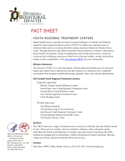
European Journal
(it"
Orthodontics 18 ( 1996) 435-444
© 1996 European Orthodontic Society
Healing of the root surface-associated periodontium: an
immunohistochemical study of orthodontic root resorption
In man
Chrissavgi Sismanidout-? ". Marita Hilliges* and Sven Lindskog*
Divisions of *Oral Histology and Cell Biology and **Orthodontics, School of Dentistry, Karolinska
Institutet, Huddinge, Sweden
Introduction
The periodontium consists of a complex mixture
of mineralized and non-mineralized tissues
derived from the ectoderm and mesoderm. It
develops as a single phylogenetic unit (Ten Cate
et al., 1971; Yoshikawa and Kollar, 1981;
Palmer and Lumsden, 1987; Lumsden, 1988;
MacNeil and Thomas, 1993), a fact which is
reflected in periodontal healing mechanisms.
Initial healing of wounds in the periodontal
ligament (POL) does not differ from the healing
of other types of connective tissue wounds. It
begins with the formation of granulation tissue
subsequent to necrosis and blood clot formation
regardless of type of periodontal challenge.
Organization of the granulation tissue follows,
during which vascular and nervous components
enter the area (Parlange and Sims, 1993) as
well as new periodontal connective tissue including fibroblasts, collagenous fibres (Melcher,
1970, 1976;Line et al., 1974; Caton and Nyman,
1980; Caton et al., 1980; Nyman et al., 1982;
Harrison and Juronsky, 1991; Wikesjo et al.,
1992) and the junctional epithelium (Taylor and
Cambell, 1972; Wirth1in et al., 1980). Although
evidence also indicates the possibility of the
epithelial rests of Malassez regenerating (Brice
et al., 1991), their role in periodontal healing
remains obscure (Spouge, 1980).
POL healing and the expression of different
periodontal mesenchymal phenotypes is intimately associated with formation of mineralize
Downloaded from by guest on November 20, 2014
SUMMARY The purpose of the present investigation was to study resorption and regeneration of periodontal tissues incident to orthodontic tooth movement, in particular cells
resorbing the root surface and the subsequent regeneration of the periodontal epithelial
network and forming reparative cementum. The study was carried out using a select number
of immunohistochemical markers on extracted human teeth which had been treated
orthodontically.
The most striking finding in the resorbing areas was the presence of what appeared to
be two populations of KP 1+ mononuclear cells located at a distance of 50-100 Jim from
the root surface and multinucleated cells in resorption lacunae in close contact with the
root surface. KP 1 + has previously not been reported for odontoclasts. The mononuclear
KP 1 + cells in the periodontal ligament may represent either precursors to odontoclasts or
phagocytic scavenger cells of the macrophage lineage.
The subsequent healing of the resorption lacunae was characterized by re-establishment
of nervous, vascular and epithelial tissues as evidenced by 5-100+ filamentous delicate
structures, factor VIII + vessels and cytokeratin + clusters of cells, respectively. However,
cytokeratin + single cells in close contact with the unresorbed cementum did not re-appear
within the healing period. Although the present results are not quantitative in nature,
cementoblasts located in the vicinity of resorption lacunae, especially healing ones, appeared
to show an up-regulation of epidermal growth factor (EGF) receptors. It may be suggested
the intense positive staining for EGF receptors may be an expression of an auto- or paracrine
stimulatory pathway increasing the rate of reparative cementum formation.
436
number of immunohistochemical markers, for
the demonstration of resorbing cells and cells
participating in the subsequent healing of the
periodontal epithelial network and forming reparative cementum. Antibodies directed against
the following human antigens were used: lysosome-associated glycoprotein CD68 (KP 1),
cytokeratins nos. 10, 17 and 18, von Willebrand
(coagulation) factor VIn and epidermal growth
factor (EGF) receptor. In addition, antibodies
directed against protein S-100 from cow brain,
which also reacts with the human sequence of
the protein (Stefansson et al., 1982) was used.
Materials and methods
Patients
A total of 10 premolars in five patients were
selected for the study. The patients were scheduled for maxillary expansion with fixed appliances and bilateral extraction of premolars. The
patients were selected on a voluntary basis and
appropriate ethical approval was granted prior
to instigation of the study. The age of the
patients ranged from 11-14 years (mean 13.3
years) at the start of the treatment.
Orthodontic appliance
Quad-helix fixed appliances were used. The
appliances were made with 0.9 mm wire (FederHart, Remanium, Dentaurum, Germany),
ensuring contact with the lingual aspects of the
premolars selected for the study. Immediately
prior to cementing the appliances they were
activated corresponding to half the buccolingual width of the premolars. This corresponded to a force of 0.8 N.
Experimental outline
Both premolars in each patient which were
scheduled for extraction were subjected to an
orthodontic force in a buccal direction for a
mean of 52 days (SD 37 days, Table 1). On the
day the force was released, one premolar in
each patient was randomly chosen for extraction
(resorbing teeth). The other premolar was kept
in place without retention and not exposed to
any orthodontic force for periods of 13-114
days (Table 1) in order to allow repair of any
root resorption to take place (healing teeth),
before extraction. Both premolars in each
patient were extracted as gently as possible in
order to preserve as much root surfaceassociated PDL as possible.
Downloaded from by guest on November 20, 2014
tissues, either root surface-associated or associated with the alveolar bone (Lindskog and
Blomlof, 1992). In this respect, the PDL shares
not only morphological similarities with the
periosteum, with which it interlaces at the alveolar crest, but also functional similarities such
as a mineralized tissue-forming capacity evidenced by its intense activity of non-specific
alkaline phosphatases (Lilja et al., 1984; Arceo
et al., 1991; Lindskog and Blomlof, 1994)
It has been shown that two different types of
reparative cementum may
result after
re-population of a damaged root surface areas
by mesenchymal cells expressing different
phenotypes (Lindskog et al., 1983; Melcher
et al., 1987; McCulloch and Bordin, 1991;
Lindskog and Blomlof, 1992, 1994; Tenorio
et al., 1993; Blomlof and Lindskog, 1994), either
a cementoblast phenotype (root resorption) or
an osteoblast phenotype (instrumented root surfaces). Expression of the cementoblast phenotype may only be possible after a superficial
resorption of the dentine surface. However, this
can not be the only factor which promotes
formation of attached reparative cementum
since resorption has also been shown to precede
non-attached bone-like reparative cementum
formation (Lindskog and Blomlof, 1992;
Blomlof and Lindskog, 1994). It is likely that
additional factors, such as the potency of the
source of undifferentiated mesenchymal cells
(most pronounced in young teeth with incomplete root closure) also determine which type of
root surface healing will occur (Blomlof et al.,
1992; Lindskog and Blomlof, 1992).
Orthodontic root resorption is preceded by
an aseptic necrosis in the pressure zones of the
PDL (Lilja et al., 1983, 1984). Upon release of
the force and removal of the necrotic area, the
resorbed dentine surface heals with adhering
reparative cementum (Langford and Sims, 1982;
Hammarstrom and Lindskog, 1985; Vardimon
et al., 1993). However, during the past decade
other components, such as the nervous and
vascular supply of the regenerating periodontium have been devoted only little attention,
and thus the relationship between regenerating
components of the PDL remains largely
unexplored.
The purpose of the present investigation was
to study resorption and regeneration of the root
surface-associated periodontal tissues incident
to orthodontic tooth movement, using a select
C. SISMANIDOU ET AL.
437
IMMUNOHISTOCHEMISTRY OF THE PDL
Table 1 Details of the experimental material. On
the day the force was released, one premolar in each
patient was randomly chosen for extraction to study
root resorption (resorbing teeth). The other premolar
was kept in place not exposed to any orthodontic
force for varying periods in order to allow repair to
take place (healing teeth).
Patient
2
3
4
5
Tooth
Force application
time (days)
Healing time
(days)
Resorbing
Healing
Resorbing
Healing
Resorbing
Healing
Resorbing
Healing
Resorbing
Healing
27
27
31
31
31
31
62
62
114
114
0
114
0
29
0
79
0
44
0
13
The teeth were immersed in 4% formaldehyde
in a phosphate buffer (pH 7.4) for at least 48
hours. The buccal root was separated from the
rest of the tooth and decalcified in 24% EDTA
(pH 7.4) at room temperature for about 4
weeks. After demineralization the roots were
embedded in paraffin. Longitudinal sections of
the buccolingual aspect of the roots were cut at
4 urn, taken up on silane-coated slides and
processed for indirect immunohistochemistry
(see below).
Between 20-30 longitudinal central sections
from each buccal root were evaluated making
a total of in excess of 250 sections. Incubation
for demonstration of the different antigens was
performed on alternating sections throughout
the roots. The description below is based on
Antibodies
All primary antisera, anti-KP 1, anticytokeratin, anti-Factor VIII, anti-EGF-receptor and anti-S-lOO (Table 2) were diluted in 4%
normal serum in a 0.5 M Tris-saline buffer,
pH 7.6 (TRIS). The antibody solution immunoreactive towards EGF receptor also contained
0.3% Triton X-lOO. The secondary antibodies
were biotinylated rabbit anti-mouse or goat
anti-rabbit (Dako) diluted 1: 200 or 1: 300 in
TRIS. Staining specificity was assessed by omission of the primary or secondary antisera or by
incubation of the primary antiserum previously
absorbed with the antigen.
Immunohistochemistry
Sections were, after deparaffination, processed
with the avidin-biotin-peroxidase complex
(ABC) method (Hsu et al., 1981). In brief, the
sections were incubated with the primary antisera followed by the secondary biotinylated
antibody and the ABC complex (Dako). The
incubations were performed at room temperature for 30 minutes or at 4°C overnight (EGF
receptor antibody only). The immunoreactions
were visualized by a cromogen substrate solution consisting of 0.6 mg/ml diaminebenzidine
and 0.01% hydrogen peroxidase in TRIS for 5
minutes. Non-specific endogenous peroxidase
staining was reduced by section immersion in
hydrogen peroxidase before the antibody
incubations. The sections were thoroughly
rinsed in TRIS before and after incubations,
counterstained with Harris haematoxylin and
were mounted in Eukitt after dehydration.
For observation and photography a Leitz
Table 2 Specifications for the primary antibodies.
Antibody against
Specificity
Supplier*
Dilution
Species
Reference
KPI
Cytokeratin
Human CD 68
Human cytokeratins
nos. 10, 17 and 18
Human von Willebrand factor
Human EGF receptor peptide
corresponding to residues 1005-1016
Cow proteins S-100 A and B
Dako
Dako
1:100
1:50
Mouse
Mouse
Pulford et al., 1989
Moll et aI., 1982
Dako
SDS
1:200
1:50
Rabbit
Rabbit
Bukh et al., 1986
Hunter et al., 1984
Dako
1:400
Rabbit
Kindblom et al., 1984
Factor VIII
EG F receptor
Protein S-100
* Antibodies from Dako and SDS were purchased from Dako AjS, Glostrup, Denmark and Santa Cruz Biotechnology Inc.,
Santa Cruz, CA, USA respectively.
EGF=epidermal growth factor.
Downloaded from by guest on November 20, 2014
Preparation of tissue
consistent representative staining patterns
observed throughout the experimental material.
438
C. SISMANIDOU ET AL.
Aristoplan microscope equipped with Image
digitizing facilities was used.
Results
KP J
Figure 2 Intense positive staining for cytokeratin in tightly
adherent clusters of cells at a distance of approximately
50 urn from the intact cementum surface (broken arrow)
and in single cells in contact with the non-resorbed
cementum surface (solid arrows). D, dentine; C, cementum;
patient 2 (Table I ); bar = 50 urn.
Downloaded from by guest on November 20, 2014
Intensely KP I -i- cells were present in what
appeared to be two populations in the resorbing
teeth. The majority of cells were mononuclear
cells located at a distance of 50-100 urn from
the root surface (Figure I). They appeared in
clusters of several cells with a uniform staining
for KP I throughout their cytoplasm. In the
vicinity of these clusters, intensely KP I + multinucleated cells were seen in close contact with
the dentine surface in resorption lacunae
(Figure I). KP I staining in these cells was
preferentially located to the part of the cell
closest to the dentine surface. The remaining
cells in the adhering POL were void of K P I
staining.
Cytokeratin
Intense staining for cytokeratin was seen in two
locations in both resorbing and healing teeth:
tightly adherent clusters of cells at a distance of
approximately 50 urn from the intact cementum
surface and in single cells in contact with the
non-resorbed cementum surface (Figure 2). The
cytokeratin + clusters of cells appeared to be
more frequent in the apical part of the roots,
Figure 3 Small cluster of cytokeratin + cells in a healing
resorption lacunae (solid arrow). D = dentine; C =
cementum; patient I (Table I ). Bar = 50 urn.
where they were also seen as elongated strands
of cells. Such formations, although smaller were
also observed in the healing but not in the
active resorption lacunae (Figure 3). However,
no single cytokeratin + cells were seen in contact
with the healing cementum surfaces.
Factor VIII
Figure I Intensely KP I t mononuclear cells located at a
distance of 50- 100/lm from the root surface (solid arrows)
as well as a resorbing multinucleated KP I + cell in close
contact with the dentine surface in a resorption lacunae
(broken arrow) with staining preferentially located to the
part of the cell closest to the dentine surface. D = dentine;
C=cementum; patient 4 (Table I). Bar=50 urn.
Intense staining for factor VIII was seen in the
walls of vessels throughout the adhering POL
and in the pulps in all teeth. In the resorbing
teeth, no preferential localization to active
resorption areas could be observed, although
these areas were not void of factor VI II + vessels.
In the healing teeth factor VIII + vessels were
439
IMMUNOHISTOCHEMISTRY OF THE PDL
present in the healing resorption bays but
always at a distance from the reparative
cementum surface (Figure 4).
8-100
S-l 00 + delicate filamentous structures as well
as thicker nerve-like bundles were consistently
found in the vicinity of the non-resorbed
cementum surfaces in both resorbing and healing teeth. In the active resorption lacunae, no
S-IOO+ structures were observed, while the healing lacunae consistently displayed S-lOO+ delicate filamentous structures (Figure 5).
EGF receptor
A uniform weak to moderate EGF receptor
positive staining was observed in mesenchymal
cells throughout the PDL in both resorbing and
healing teeth. Some epithelial cell rests of
Malassez, especially those close to healing
resorption lacunae were intensely EGF
receptor" (Figure 6). Cementoblasts preferentially located to healing resorption lacuna also
displayed an intense staining for EGF receptors
(Figure 7), similar to that seen in odontoblasts
and endothelial cells (Figure 8).
Figure 6 Epidermal growth factor (EGF) receptor + staining in an epithelial cell rest of Malassez (solid arrow) close
to an intact cementum surface in a resorbing tooth with
active resorption lacunae. D = dentine; C = cementum;
patient 1 (Table 1). Bar = 50 11m.
Figure 5 S-lOO+ delicate filamentous structures in a healing resorption lacunae (solid arrows). D = dentine; C =
cementum; patient 3 (Table l). Bar = 50 11m.
Figure 7 Epidermal growth factor (EGF) receptor" staining in cementoblasts and cementocytes located in a healing
resorption lacunae. R = reparative cementum; patient 2
(Table 1). Bar=50 11m.
Downloaded from by guest on November 20, 2014
Figure 4 Intense staining for factor VIII in endothelial
cells in a healing resorption lacunae at a distance from the
reparative cementum surface (solid arrows). D=dentine;
C=cementum; patient 3 (Table 1). Bar=50 11m.
440
Figure 8 Epidermal growth factor (EGF ) receptor + staining in odontoblasts (solid arrows) and in endothelial cells.
D=dentine; B=blood vessel; patient 5 (Table l ), Bar=
100 11m.
Discussion
1982). Human odontoblasts from deciduous
tooth germs taken from 12-18 week old foetuses
may be S-lOO+ (Lombardi et al., 1992), a
finding supported by Carbone et al. (1987) in
normal and teratomatous tooth germs. The
S-I 00 + delicate filamentous structures seen in
the regenerating PDL, thus indirectly indicates
regenerating peripheral myelinated nerves.
The antibody directed against the EGF receptor reacts specifically with the 170 kDa EGF
receptor protein in human cells by Western
blotting analysis. It can also be used for immune
complex kinase assays and it reacts with EGFreceptor in formaline-fixed, paraffin-embedded
tissue sections. EGF mediates its effects on cell
growth through its interaction with a 170 kDa
cell surface glycoprotein designated the EGF
receptor (Hunter, 1984). EGF receptors have
been identified on basal cells of stratified squamous epithelia and skin adnexa (Gustavson et al.,
1984). Immunoreactivity was detected in palatal
gingiva, buccal gingiva, soft palate, lateral
tongue, dorsal tongue and floor of the mouth.
The connective tissues of the PDL and dental
pulp were non-reactive (Whitcomb et al., 1993).
EGF receptors are present on a wide range of
normal epithelial tissues and tumours arising
from those sites. The distribution of the receptor
suggests that EGF may be involved in the
control of proliferation and possibly differentiation of surface epithelia (Gustavson et al.,
1984 ).
Antibodies to KP I stain monocytes/macrophages in a wide variety of human tissues
including Kupffer cells and macrophages in the
red pulp of the spleen, in the lamina propria of
the gut, in lung alveoli and in bone marrow
(Pulford et al., 1989). Peripheral blood monocytes are also positive with a granular staining
pattern. All mast cell subtypes, that are normal
and reactive and also malignant or neoplastic,
exhibit strong consistent cytoplasmic immunoreactivity (Horny et al., 1990). KP I recognizes
a fixation-resistant epitope in a wide variety of
tissue macrophages and in gran ulocyte precursors, thus reacting with cells of the mononuclear phagocytic lineage (Pulford et al.,
1989).
Antibodies to factor VIII react with the von
Willebrand factor on endothelial cells, where it
shows a granular pattern of reactivity and can
also be detected within the megakaryocytes in
sections of human bone marrow. Detection of
Downloaded from by guest on November 20, 2014
The purpose of the present investigation was to
study the relationship between cells resorbing
the root surface and the subsequent regeneration of the periodontal epithelial network and
forming reparative cementum incident to orthodontic tooth movement. For this purpose, a
select number of immunohistochemical markers
were employed. Thus, before discussing the
particular findings some methodological issues
need to be addressed.
Immunohistochemistry uses antibodies to
detect specific substances in tissue sections. The
antibodies anti-KP I, anti-cytokeratin, antifactor VIII and anti-EGF receptor are directed
against human antigens. Although the antiserum directed against protein S-IOO has been
manufactured against cow protein S-IOO, it also
reacts with the human sequence of the protein.
Antibodies to S-IOO in the brain stains glial and
ependymal cells. Moreover, Schwarm cells of the
peripheral nervous system are positive. In
healthy skin and mucosa, melanocytes and
Langerhans cells (Cocchia et al., 1981;
Nakajima et al., 1982; Charbit et al., 1986;
Cruchley et al., 1989; Barrett et al., 1994) are
positive, as well as interdigitating reticulum cells
in lymph nodes. It also stains all the above
tumour cells. In peripheral nerves, Schwann
cells and the outermost part of the myelin
sheath are positive; but not axons. In all other
organs, Schwann cells, of both myelinated and
unmyelinated peripheral nerves, and satellite
cells of ganglia are positive (Stefansson et al.,
C. SISMANIDOU ET AL.
IMMUNOHISTOCHEMISTRY OF THE POL
resorption lacunae as evidenced by S-I 00 +
filamentous delicate structures, factor VIII +
vessels and cytokeratin + clusters of cells,
respectively, in accordance with previous morphological (Parlange and Sims, 1993) and histochemical (Kvinnsland et al., 1991; Saito et al.,
1993) studies. However, the cytokeratin + single
cells in close contact with the unresorbed
cementum did not re-appear within the healing
period. The function of these cells as well as
the cytokeratin + clusters of cells in the POL
remains obscure, although the literature is not
lacking in suggestions. These range from an
involvement in maintaining the width of the
periodontal space (Lindskog et al., 1988;
Wallace and Vergona, 1990) to an active role
in cementogenesis (Brice et al., 1991). The lack
of cytokeratin + single cells in close contact with
reparative cementum may, however, cast some
doubt on the latter suggestion.
Although the present results are not quantitative in nature, cementoblasts located in the
vicinity of resorption lacunae, especially the
healing type appeared to show an up-regulation
of EGF receptors. This appears to contradict a
previously reported decrease in EGF binding
sites during differentiation of 'POL fibroblastic
cells into mineralized tissue-forming cells'
(Matsuda et al., 1993) or of cementoblasts (Cho
et al., 1991). However, these studies were performed with radioactively-labelled EGF without directly assessing the presence of EGF
receptors. Furthermore, it is reasonable to
assume that cementoblasts which produce reparative cementum at a higher rate compared
with the continuous slow deposition of
cementum
on
surrounding
undamaged
cementum surfaces, may up-regulate their EGF
receptors as an expression of an auto- or paraerine stimulatory pathway. The intermittent
expression of EGF receptors on epithelial cell
rests of Malassez is in accordance with previous
data on an intense binding of EGF to these
cells (Thesleff, 1987). However, the functional
implications remain unclear.
In conclusion, the data presented in this study
indicates that other tissue components of the
POL as well as connective tissue such as nerves,
epithelial cell rests of Malassez and blood vessels
are capable of regeneration following orthodontic root resorption. Furthermore, an
up-regulation of EGF receptors on cementoblasts may be involved in the increased formation of cementum repairing the resorbed areas.
Downloaded from by guest on November 20, 2014
factor VIII is of value for delineating endothelial
cells in human tissue (Mukai et al., 1980).
Tumours arising from endothelial cells or megakaryocytes may be identified by staining for this
antibody (Hruban et al., 1987). Blast cells in
acute megacaryocytic leukaemia are strongly
positive for factor VIII (Hruban et al., 1987).
Antibodies to cytokeratins show a broad pattern of reactivity with human epithelial tissues
from simple glandular to stratified squamous
epithelium, which include epidermis, eccrine
sweat gland, mammary gland ducts, tracheal
epithelium and amniotic epithelium (Moll et al.,
1982). Epithelial cells are labelled whether they
are ectodermal or endodermal in origin (Moll
et al., 1982).
Antibody specificity has been ascertained
through
immunoelectrophoresis,
immunoabsorbant columns, enzyme-linked immunosorbent assay (ELISA), control tissue staining
or Western blotting analysis by the supplying
companies. However, cross-reactivity with peptides and proteins, present in the tissue section,
with a chemical composition similar to the
antigen cannot be excluded. Furthermore, negative staining results do not necessarily prove the
absence of the antigen. The detectable epitope
could be hidden from the antibody in the tissue
situation.
The most striking finding in the resorbing
areas was the presence of what appeared to be
two populations of KP I + mononuclear cells
located at a distance of 50 to 100 urn from the
root surface and multinucleated cells in resorption lacunae in close contact with the root
surface. The distribution of KP 1 staining
differed between these two types of cells. In the
multinucleated cells, it was preferentially located to the part of the cell closest to the dentine
surface, while the mononuclear cell displayed a
uniform cytoplasmic staining. KP 1 + has previously not been reported for odontoclasts only
for osteoclasts (Athanasou et al., 1991),
although there is strong evidence to indicate a
common origin for both of these cells. The
mononuclear KP 1 + cells in the POL may thus
represent either precursors to odontoclasts or
phagocytic scavenger cells of the macrophage
lineage (Roodman, 1991; Pierce et al., 1991;
Brudvik and Rygh, 1993; Jager et al., 1993).
The subsequent healing of the resorption
lacunae was characterized by re-establishment
of nervous, vascular and epithelial tissues in the
441
442
Address for correspondence
Sven Lindskog
Department of Oral Histology and Cell Biology
School of Dentistry
Karolinska Institutet
Box 4064
S-141 04 Huddinge
Sweden
Acknowledgements
This study was supported by grants from the
Swedish Medical Research Council (Grant no.
6651) and the Faculty of Dentistry at
Karolinska Institutet.
References
Charbit Y, Monteil R A, Hitzig C, Sauget P, Benaiche N,
Jasmin J R 1986 S-IOO immunolabelling of Langerhans
cells in oral epithelium. Journal of Oral Pathology 15:
419-22
Cho M-N, Lin W-L, Garant P R 1991 Occurrence of
epidermal growth factor-binding sites during differentiation of cementoblasts and PDL fibroblasts of the young
rat: a light and electron microscopic study. Anatomical
Record 231: 14-24
Cocchia D, Michetti F, Donato R 1981 Immunochemical
and immunocytochemical localisation of S-l 00 in normal
skin. Nature 294: 85-87
Cruchley A T, Williams D M, Farthing P M, Lesch C A,
Squier C A 1989 Regional variation in Langerhans cell
distribution and density in normal human oral mucosa
determined using monoclonal antibodies against CDl,
HLADR, HLADQ and HLADP. Journal of Oral
Pathology and Medicine 18: 510-516
Gustavson B, Cowley G, Smith J A, Ozanne B 1984 Cellular
localization of human EGF receptor. Cell Biology
International 8: 649-658
Hammarstrom L, Lindskog S 1985 General morphologic
aspects of resorption of teeth and bone. International
Endodontic Journal 18: 93-108
Harrison J W, Juronsky K A 1991 Wound healing in the
tissues of the periodontium following periradicular surgery. II. The dissectional wound. Journal of Endodontics
17: 544-552
Horny H P, Schaumburg-Lever G, Bolz S, Geerts M L,
Kaiserling E 1990 Use of monoclonal antibody KPI for
identifying normal and neoplasmatic human mast cells.
Journal of Clinical Pathology 43: 719-722
Hruban R H, Kuhajda F P, Mann R B 1987
Immunohistochemical study of four cases and comparison with acute megakaryocytic leukemia. American
Journal of Clinical Pathology 88: 578-588
Hsu S M, Raine L, Fanger H 1981 Use of avidin-biotinperoxidase complex (ABC) in immunoperoxidase techniques. A comparison between. ABC and unlabeled antibody (PAP) procedures. Journal of Histochemistry and
Cytochemistry 29: 577-580
Hunter T 1984 The epidermal growth factor receptor gene
and its product. Nature 311: 414-416
Jager A, Radlanski R J, Gotz W 1993 Demonstration of
cells of the mononuclear phagocyte lineage in the
periodontium following experimental tooth movement in
the rat. Histochemistry 100: 161-166
Kindblom L-G, Lodding P, Rosengren L, Baudier J, Haglid
K 1984 S-IOO protein in melanocytic tumors. Acta
Pathologica
et
Microbiologica
Immunologica.
Scandinavica Section A 92: 219-230
Kvinnsland I, Heyeraas K J, Byers M R 1991 Regeneration
of calcitonin gene-related peptide immunoreactive nerves
in replanted rat molars and their supporting tissues.
Archives of Oral Biology 36: 815-826
Langford S R, Sims M R 1982 Root surface resorption,
repair, and periodontal attachment following rapid maxillary expansion in man. American Journal of Orthodontics
81: 108-115
Lilja E, Lindskog S, Hammarstrom L 1983 Histochemistry
of enzymes associated with tissue degradation during
Downloaded from by guest on November 20, 2014
Arceo N, Sauk J J, Moehring J, Foster R A, Sommerman
M J 1991 Human periodontal cells initiate mineral-like
nodules in vitro. Journal of Periodontology 62: 499-503
Athanasou N A, Puddle B, Quinn J, Woods C G 1991 Use
of monoclonal antibodies to recognise osteoclasts in
routinely processed bone biopsy specimens. Journal of
Clinical Pathology 44: 664-666
Barrett A W, Beynon A D, Reid D J 1994 A comparative
study on tissue processing methods for the immunohistochemical demonstration of human oral mucosa
Langerhans cells. Journal of Histochemistry 26: 134-141
Blomlof L, Lindskog S 1994 Quality of periodontal healing
II: Dynamics of reparative cementum formation. Swedish
Dental Journal 18: 131-138
Blomlof L, Lengheden A, Lindskog S 1992 Endodontic
infection and calcium hydroxide-treatment-Effects on
periodontal healing in mature and immature replanted
monkey teeth. Journal of Clinical Periodontology 19:
652-658
Brice G L, Sampson W J, Sims M R 1991 An ultrastructural
evaluation of the relationship between epithelial rests of
Malassez and orthodontic root resorption and repair in
man. Australian Orthodontic Journal 12: 90-94
Brudvik P, Rygh P 1993 Non-clast cells start orthodontic
root resorption in the periphery of hyalinized zones.
European Journal of Orthodontics 15: 467-480
Bukh A, Ingerslev J, Stenbjerg S, Moller N P H 1986 The
multimetric structure of plasma FVIII: RAg studied by
electroelution
and
immunoperoxidase
detection.
Thrombosis Research 43: 579-584
Carbone A, Poletti A, Manconi R, Sulfaro S, Volpe R 1987
S-IOO protein immunostaining in teratomatous welldifferentiated tissues. Histopathology 11: 980-983
Caton J, Nyman S 1980 Histometric evaluation of periodontal surgery. I. The modified Widman flap procedure.
Journal of Clinical Periodontology 7: 212-223
Caton J, Nyman S, Zander H 1980 Histometric evaluation
of periodontal surgery. II. Connective tissue attachment
levels after four regenerative procedures. Journal of
Clinical Periodontology 7: 224-231
C. SISMANIDOU ET AL.
IMMUNOHISTOCHEMISTRY OF THE POL
stration of S-100 protein in epidermal Langerhans cells.
Biomedical Research 3: 226-231
Nyman S, Gottlow J, Karring T, Lindhe J 1982 The
regenerative potential of the PDL. An experimental study
in the monkey. Journal of Clinical Periodontology 9:
257-265
Palmer R M, Lumsden A G S 1987 Development of PDL
and alveolar bone in hemografted recombinations soft
tissue enamel organs and papillary, pulpal and follicular
mesenchyme in the mouse. Archives of Oral Biology
32: 281-289
Parlange L M, Sims M R 1993 A TEM stereological analysis
of blood vessels and nerves in marmoset periodontal
ligament following orthododontic and magnetic extrusion. European Journal of Orthodontics 15: 33-44
Pierce A M, Lindskog S, Hammarstrom L 1991 Osteoclasts:
structure and function. Electron Microscopy Reviews
4: 1-45
Pulford K A F, Rigney E M, Micklem K J, Jones M, Stross
W P 1989 KPl: a new monoclonal antibody that detects
a monocyte/macrophage associated antigen in routinely
processed tissue sections. Journal of Clinical Pathology
42: 414-421
Roodman G D, 1991 Osteoclast differentiation. Oral
Biology and Medicine 2: 389--409
Saito I, Hanada K, Maeda T 1993 Alteration of nerve
growth factor-receptor expression in the periodontalligament of the rat during experimental tooth movement.
Archives of Oral Biology 38: 923-929
Spouge J D 1980 A new look at the rests of Malassez. A
review of their embryological origin, anatomy, and possible role in periodontal health and disease. Journal of
Periodontology 51: 437-444
Stefansson K, Wollmann R L, Moore B W 1982
Distribution of S-l 00 protein outside the central nervous
system. Brain Research 234: 309-317
Taylor A C, Cambell M M 1972 Reattachment of gingival
epithelium to the tooth. Journal of Periodontology 43:
281-293
Ten Cate A R, Mills C, Solomon G 1971 The development
of the periodontium: a transplantation and auto radiographic study. Anatomical Record 170: 365-380
Tenorio
D, Cruchley A, Hughes
F J 1993
Immunocytochemical investigation of the rat cementoblast phenotype. Journal of Periodontal Research 28:
411-419
Thesleff I 1987 Epithelial cells rests of Malassez bind
epidermal growth factor intensely. Journal of Periodontal
Research 22: 419-421
Vardimon A D, Graber T M, Pitaru S 1993 Repair process
of external root resorption subsequent to palatal expansion treatment. American Journal of Orthodontics and
Dentofacial Orthopedics 103: 120-130
Wallace J A, Vergona K 1990 Epithelial rests' function in
replantation: Is splinting necessary in replantation? Oral
Surgery, Oral Medicine, Oral Pathology 70: 644-649
Whitcomb S S, Eversole L R, Lindemann R A 1993
Immunohistochemical mapping of epidermal growthfactor receptors in normal human oral soft tissue.
Archives of Oral Biology 38: 823-826
Downloaded from by guest on November 20, 2014
orthodontic tooth movement. American Journal of
Orthodontics 83: 62-75
Lilja E, Lindskog S, Hammarstrorn L 1984 Alkaline phosphatase activity and tetracycline incorporation during
initial tooth movement in rats. Acta Odontologica
Scandinavica 42: 1-11
Lindskog S, BlomlOf L 1992 Mineralized tissue-formation
in periodontal wound healing. Journal of Clinical
Periodontology 19: 741-748
Lindskog S, Blomlof L 1994 Quality of periodontal healing
IV: Enzyme histochemical evidence for an osteoblast
origin of reparative cementum. Swedish Dental Journal
18: 181-189
Lindskog S, Blomlof L, Hammarstrom L 1983 Repair of
periodontal tissues in vivo and in vitro. Journal of Clinical
Periodontology 10: 188-205
Lindskog S, Blornlof L, Hammarstrom L 1988 Evidence
for a role of odontogenic epithelium in maintaining the
periodontal space. Journal of Clinical Periodontology
15: 371-373
Line S E, Polson A M, Zander M A 1974 Relationship
between periodontal injury and selective cell repopulation
and ankylosis. Journal of Periodontology 45: 725-730
Lombardi T, Di-Felice R, Samson J 1992 Human odontoblasts contain S-IOO protein-like immunoreactivity.
Anatomical Record 232: 190-193
Lumsden A G S 1988 Spatial organization of the epithelium
and the role of neural crest cells in the initial development
of the mamalian tooth grem. Development 103 (supp!.):
155-169
MacNeil R L, Thomas H F 1993 Development of the
murine periodontium. II. Role of the epithelial root
sheath in formation of the periodontal attachment.
Journal of Periodontology 64: 285-291
Matsuda N, Kulmar N M, Ramakrishnan P R, Lin W-L,
Genco R J, Cho M I 1993 Evidence for an up-regulation
of epidermal growth factor receptors on rat PDL fibroblastic cells associated with stabilization -of phenotype in
vitro. Archives of Oral Biology 38: 559-569
McCulloch C A G, Bordin S 1991 Role of fibroblast
subpopulations in periodontal physiology and pathology.
Journal of Periodontal Research 26: 144-154
Melcher A H 1970 Repair of wounds in the periodontium
of the rat. Influence of PDL on osteogenesis. Archives of
Oral Biology 15: 1183-1204
Melcher A H 1976 On the repair potential of periodontal
tissues. Journal of Periodontology 47: 256-260
Melcher A H, McCulloch C A G, Cheong T, Nemeth E,
Shiga A 1987 Cells from bone synthesize cementum-like
and bone-like tissue in vitro and may migrate into PDL
in vivo. Journal of Periodontal Research 22: 246-247
Moll R, Franke W W, Schiller D L, Geiger B, Krepler R
1982 The catalog of human cytokeratins: patterns of
expression in normal epithelia, tumors and cultured cells.
Cell 31: 11-24
Mukai K, Rosai J, Burgdorf W H C 1980 Localization of
factor VIII-related antigen in vascular endothelial cells
using an immunoperoxidase method. American Journal
of Surgical Pathology 4: 273-276
Nakajima T, Sato Y, Watanabe S, Shimosato Y, Ishihara
K, Isobe T 1982 Immunoelectron microscopical demon-
443
444
Wikesjo U M, Nilveus R E, Selvig K A 1992 Significance
of early healing events on periodontal repair: a review.
journal of Periodontology 63: 158-165
Wirthlin M R, Yeager j E, Hancock E-B, Gaugler R W
1980 The healing of gingival wounds in miniature swine.
journal of Periodontology 51: 318-327
C. SISMANIDOU ET AL.
Yoshikawa D K, Kollar E J 1981 Recombination experiments on the odontogenic roles of mouse dental papilla
and dental sac tissues in ocular grafts. Archives of Oral
Biology 26: 303-307
Downloaded from by guest on November 20, 2014


© Copyright 2026