
SESSION C POSTERS XXI Congress of the International Academy of Legal Medicine
XXI Congress of the International Academy of Legal Medicine
(IALM)
SESSION C
POSTERS
MAY, 29th – FRIDAY – Room 3
XXI Congress of the International Academy of Legal Medicine
(IALM)
POSTERS
P.C1 - ABOUT A CASE.
V.C.R.M.Abreu, R.M.C. Silva
P.C2 - AN UNUSUAL ACCIDENTAL ELECTRICAL INJURY.
K. Acar, A. Kurtulus, B. Boz, N.S. Turk
P.C3 - THE SMELL OF DEATH: FATAL INHALATION OF VOLATILE SUBSTANCES IN 48 YEARS OF NECROSCOPICAL
ACTIVITY.
A. Battistini, S. Andreola, G. Gentile, E. Palazzo, A. Taborelli
P.C4 - GUN SHOT FATALITIES: SUICIDE, HOMICIDE OR ACCIDENT? A SERIES OF 37 CASES.
M. Delavari, A.R. Owhadi, M. Movagharipour
P.C5 - IMMIGRATION AND HOMICIDE IN MILAN: ANALYSIS OF THE PHENOMENON AND COMPARISON
BETWEEN NATIONAL AND NON-EU VICTIMS.
A. Taborelli, S. Andreola, G. Gentile, E. Palazzo
P.C6 - SPECIFICITY AND SENSITIVITY OF BLUESTAR AND SYNERGIC USE WITH 3D LASER SCANNING.
B. Mancini, A. Galassi, D. Gaudio, O. Burkler, L. Saravo, S. Turrina, D. De Leo
P.C7 - ANALYSIS OF WOUND TRACKS IN GELATINE.
C. Schyma, B. Madea
P.C8 - CRIMINAL EVENTS AMONG HOMOSEXUAL PEOPLE IN ITALY FROM 1990 TO 2008.
A. Taborelli, S. Andreola, A.G. de Micheli, G. Gentile, E. Palazzo
P.C9 - THE IMPORTANCE OF PREVENTION IN ORDER TO AVOID INFECTION FROM POSTMORTEM MATERIAL.
A. Taborelli, S. Andreola, G. Gentile, E. Palazzo
P.C10 - DIFFICULTIES CONCERNING THE ESTIMATION OF THE POST MORTEM INTERVAL (PMI) OF CORPSES
FOUND IN WATER.
E. Doberentz, B. Madea
P.C11 - HIGH RESOLUTION DIGITAL IMAGE MAPPING ON 3D CRIME SCENES SCANNING.
G.P.M. Vassena, M. Sgrenzaroli, A. Galassi, D. Gaudio, M. L. Saravo, D. De Leo, C. Bonacina
P.C12 - WORKPLACE SUICIDE IN THE NORTH OF TUNISIA.
M. Ben Khelil, O. Bekir, M. Allouche, F. Gloulou, M. Shimi, A. Banasr, M. Zhioua, M. Hamdoun
P.C13 - COMPARISON OF POSTMORTEM METABOLIC CHANGES IN SHEEP BRAIN TISSUE IN ISOLATED HEADS
AND WHOLE ANIMALS USING 1H-MR SPECTROSCOPY – PRELIMINARY RESULTS.
F. Musshoff, W. Block, F. Traeber, B. Madea
INDEX
XXI Congress of the International Academy of Legal Medicine
(IALM)
P.C14 - IMA LEVELS FOLLOWING MILD CLOSED HEAD TRAUMA IN THE RAT.
T. Çolak, C. Duman, B. Bamaç, A. Aydin, A. Özbek, K. Yildiz, Ü. Bicer
P.C15 - SEROTONERGIC SYSTEM POLYMORPHISMS AND SUICIDE.
D. Azenha, C. Pereira, C. Mendonça, L. Cortesão, G. Castanheira, C. Santos, F. Bessa, J. F. Santa, D.N. Vieira,
A.M. Ambrósio
P.C16 - FAHR´S DISEASE - CONSIDERATIONS ABOUT THE CAUSE OF DEATH.
S. Unkrig, E. Doberentz, B. Madea
P.C17 - IMMUNOHISTOCHEMESTRY OF PULMONARY SURFACTANT APOPROTEIN-A IN FATALITIES BY
HYDROGEN SULFIDE POISONING.
B. Solarino, G. Di Vella, F. Zotti, M. Colonna
P.C18 - SAFETY OF TRADITIONAL MALE CIRCUMCISION IN MTHATHA AREA OF SOUTH AFRICA.
R.P. Kaswa, B.L. Meel
P.C19 - LAB-EXPERIMENTAL RESULTS OF THE INVESTIGATION OF DIFFERENTLY CONSTRUCTED BOLT SHOT
DEVICES.
L. Hagemeier, C. Schyma, B. Madea
P.C20 - DYSEMBRYOPLASTIC NEUROEPITHELIAL TUMOUR - A RARE CAUSE OF SUDDEN DEATH.
R. Yallapur
P.C21 - ETUDE BIOMECANIQUE ET LESIONNELLE DES TRAUMATISMES MORTELS DU RACHIS CERVICAL CHEZ
L’ENFANT.
A.Aissaoui, N.Haj Salem, T.Mghirbi, A.Chadly
P.C22 - SUICIDE IN HOSPITAL SETTINGS.
E. Sousa, C. Cordeiro, M. C. de Mendonça, F. Corte-Real, D. N. Vieira
P.C23 - COMPARATIVE ANALYSIS OF THE HOMICIDES COMMITED BY KNIFE AND FIRE WEAPON AFTER THE LAW
CHANGINGS.
D. Cukic, N. Radojevic
P.C24 - IDENTIFICATION BY DIGITAL IMAGE SUPERIMPOSITION OF BODY PARTS: A CASE REPORT.
D. De Angelis, D. R. Schillaci, C. Cattaneo
P.C25 - COMPARTMENT SYNDROME FOLLOWING AN ACUTE CARBON MONOXIDE INTOXICATION-CASE
REPORT.
D. G. Sirbu, A. Sirbu, N. Sirbu
P.C26 - ENDOTHELIAL DYSFUNCTION (NPY AND ENOS GENES) AND INFLAMATION RELATED GENES (lp-PLA2,
COX-2 AND iNOS GENES) AND THE PREDISPOSITION TO CARDIOVASCULAR DISEASE.
D. Azenha, B. Silva, C. Cordeiro, R. Matos, J. F. Santa, S. Tavares, D. N. Vieira, A. M. Ambrósio
XXI Congress of the International Academy of Legal Medicine
(IALM)
P.C27 - CHILD ABUSE FOLLOWED BY FATAL SYSTEMIC PSEUDOMONAS INFECTION.
M. Senati, F. De Giorgio, F. Ausania, C. Colecchi, G. Vetrugno, A. Carbone, V. L. Pascali
P.C28 - CLINICAL AND MEDICOLEGAL ASPECTS OF SUDDEN PREGNANT WOMAN DEATH WITH MARFAN
SYNDROME CAUSED BY HEART TAMPONADE – A CASE REPORT.
S. Pavlekic, D. Jecmenica, D.J. Alempijevic, M. Terzic, I. Baralic, B. Aleksandric
P.C29 - GIANT HYDATID CYST – AN UNSUSPECTED POSSIBLE CAUSE OF SUDDEN DEATH.
D. Nikolic, R. Gouveia, F. Pedrosa, I. P. Ribeiro, J. C. Santos
P.C30 - NON-TRAUMATIC EPIDURAL AND SUBDURAL HAEMATOMA IN EARLY CHILDHOOD.
D. Radoinova, Y. Kolev
P.C31 - FIREARMS RELATED DEATHS IN THE CITY OF SÃO PAULO, 2006. A RETROSPECTIVE STUDY.
D.V. Fuzinato, G. Cecchetto, P. Fais, G.Viel, M. Montisci, D. N. Vieira
P.C32 - DEATH IN THE “MICROWAVE OVEN”: A FORM OF EXECUTION BY CARBONIZATION.
C. Durão, L. Miranda, D. N. Vieira
P.C33 - SUDDEN DEATH FROM HYPOPLASTIC LEFT CORONARY ARTERY.
M. Kalyva, A. G. Papadomanolakis, A. Papavdi, J. Douzis, D. Nathena, M. N. Michalodimitrakis
P.C34 - FORENSIC PATHOLOGISTS AND RADIATION HAZARDS.
E. Sousa, M. C. Mendonça, F. Corte Real, D. N. Vieira
P.C35 - CEREBRAL VENOUS AIR EMBOLISM AS A FATAL COMPLICATION OF SPINE SURGERY.
F. De Giorgio, E. Arena, F. Purchiaroni, L. Baldassari, M. Miscusi, G. Vetrugno, A. Capelli, V. L. Pascali
P.C36 - FATAL NONCARDIOGENIC ACUTE PULMONARY EDEMA DUE TO PHEOCHROMOCYTOMA - A CASE
REPORT AND REVIEW OF THE LITERATURE.
P. Mylonakis, F. Chatzinikolaou, D. Papadogianni, S. Papamichail, G. Charalampakis, S.G. Popeskou
P.C37 - FATAL HEAD INJURIES DUE TO ROAD TRAFFIC ACCIDENTS.
S. Fehér, K. Töro
P.C38 - A CASE-CONTROL STUDY OF CEREBELLAR TONSILLAR ECTOPIA AND CERVICAL SPINE TRAUMA.
M.D. Freeman, S. Rosa, D. Harshfield, F. Smith, R.M. Bennett, C.J. Centeno, E. Kornel, A. Nystrom, D. Heffez,
S.S. Kohles
P.C39 - DEATHS INTO PRISONS: EXPERIENCE OF THE INSTITUTE OF LEGAL MEDICINE OF MILAN FROM 1970 TO
2008.
U. Genovese, A. Migliorini, F. Mobilia
P.C40 - MORT SUBITE CARDIAQUE PAR HYDATIDOSE A LOCALISATION SEPTALE. A PROPOS D'UNE
OBSERVATION.
B. Boulassel, D. Azzouz, S. Ouabdesselam
XXI Congress of the International Academy of Legal Medicine
(IALM)
P.C41 - SUDDEN DEATH DUE TO RUPTURE OF DISSECTION OF THE AORTA - A CASE REPORT.
F. Yagmur, H. Din, M. Kaya
P.C42 - LIVER AND RENAL STRUCTURE AND FUNCTION EVALUATED IN MALE BODYBUILDERS USING
ANDROGENIC ANABOLIC STEROIDS.
G. Cecchetto, G. Viel, A. Nalesso, G. Bombonato, M. Montisci, S.D. Ferrara
P.C43 - A CASE OF BULL’S HORNS INJURIES - THE MECHANISMS OF INJURIES BY PERFORATING INSTRUMENTS.
I. P. Ribeiro, F.C. Santos, L. Eiras
P.C44 - DEATH CAUSES OF CHILDREN BORN WITH PREMATURITY IN FORENSIC MEDICINE EVALUATION.
R. Yilmaz, I. Pakis, E. Kara, N. Turan, S. Ekemen
P.C45 - POST MORTEM CHANGES AND ARTEFACTS: “THE GREAT PRETENDERS”.
F. Cardoso, A. Afonso, J.F. Santa, S. Tavares, D.N. Vieira
P.C46 - RECONSTITUTION OF SOFT TISSUES AS A NEW TOOL IN DETERMINING THE MANNER OF DEATH IN
SHOTGUN CASES.
J. Pinheiro, A.S. Coelho, C. Pereira, R. L. Silva, A. Baltasar
P.C47 - Air Rifle – Air rifle – a harmless toy?
J. Rainio, A. Oksala, P. Keipilä, H. Kahilainen, P. J. Karhunen
P.C48 - DEATH BY PRESCRIPTION DRUG INTOXICATION – A RETROSPECTIVE ANALYSIS OF THE CASES
AUTOPSIED AT THE CENTRE BRANCH OF THE PORTUGUESE NATIONAL INSTITUTE OF LEGAL MEDICINE,
BETWEEN 1996 AND 2007.
C. Pereira, M.C. Mendonça, D.N. Vieira
P.C49 - UNUSUAL FINDINGS IN SUICIDAL HANGING.
L. Coelho, L. Santos, D. Almeida, J. B. de Oliveira, A. Santos
P.C50 - BALÍSTICA FORENSE: "DISTANCIA DISPARO CON ESCOPETAS RECORTADAS".
F. S. Moyano
P.C51 - HF-HR MRI IN FORENSIC PATHOLOGY AND LEGAL MEDICINE: 10 YEARS OF EXPERIENCE.
A. Osculati, L. Andrello, L. Guzzetti, L. Tajana
P.C52 - “CAFE CORONARY”.
L.A.S. de Paiva, W.R. G. Teixeira, Z. Morrone Jr., R.P. Teixeira, B. M. G. de Paiva
P.C53 - FORENSIC MEDICAL INVESTIGATION OF AN AIR CRASH IN ESFAHAN-IRAN.
G. Masoud
P.C54 - HISTOPATHOLOGICAL LESIONS IN ORGANIC SOLVENT INTOXICATION.
M. Petcu, A. Enache, D. Lungu, V. Sturz, G. Pasare
XXI Congress of the International Academy of Legal Medicine
(IALM)
P.C55
- CASE REPORT: DETECTION OF CRACK COCAINE IN THE PULMONARY TISSUES BY
IMMUNOHISTOCHEMISTRY METHOD TO INVESTIGATE CAUSE OF SUDDEN DEATH IN SOUTHEASTERN
BRAZIL.
M.F.S. Mello, M.J.A Alves-Junior, P.J. Thyssen, S. Kitamura, K. Metze, M.A. Ferreira
P.C56 - CSI IN A COMPLEX MURDER CASE.
A. Barbaro, P. Cormaci, A. La Marca, A. Barbaro
P.C57 - DEVELOPMENT OF BITE MARKS METHODOLOGIES ON SKIN IN CRIMINALS INVESTIGATIONS.
J.A. M. Marques, J.O. Musse, L. C. C. Galvão, C. A. S. Garbin
P.C58 - COMPRESSION TRACHEALE PAR LE COUTEAU LORS D’UNE PLAIE CERVICALE.
M. Ben Dhiab, W. Majdoub, M. Jedidi, S. Mlayeh, T. Masmoudi, M. Zemni
P.C59 - BLOOD ALCOHOL CONCENTRATION AS A RISK FACTOR IN CAR OCCUPANTS - A RETROSPECTIVE STUDY.
V. Zivkovic, M. Soc, S. Nikolic, F. Jukovic
P.C60 - ASPECTS OF SUICIDAL PHENOMENON IN THE DISTRICT OF BIHOR – ROMANIA.
G. Mihalache, C. Buhas
P.C61 - GUNSHOT WOUND.
N. Uka
P.C62 - FORENSIC EVALUATION OF OCCUPATIONAL MARKS IN ESTABLISHING IDENTITY.
B.S.K. Shetty, P. P. Jagadish Rao
P.C63 - PHYSICAL PROPERTIES OF BLOODSTAINS: LET THE STORY COME FROM DRY DROPLETS.
F. Ramsthaler, S. Potente, C. Kaiser, R. Bux, M. Kettner
P.C64 - SUDDEN CARDIAC DEATH - A CASE WITH TWO DISTINCT AND UNRELATED CAUSES.
R.H. Gouveia, M. Paulo, M.J. Dias, I.P. Ribeiro, J. C. Santos
P.C65 - OCULAR SIGNS IN FORENSIC MEDICINE – A BRIEF REVIEW.
V.R. Romeiro, C.A.N. Romeiro
P.C66 - FATAL ELECTROCUTION: A 10-YEAR RETROSPECTIVE STUDY IN LISBON AREA.
A.R. Pereira, D. Nikolic, F. Gallo, L. Eiras, I.P. Ribeiro, F. C. Santos, J. C. Santos
P.C67 - HOMICIDES IN THE REGION OF CRETE DURING THE PERIOD 2001-2009.
A. Papavdi, D. Nathena, E. Spanoudaki, J. Douzis, A. Papadomanolakis, M. Michalodimitrakis
P.C68 - GSR ANALYSIS IN A MURDER CASE: RELEVANCE OF THE COMPARATIVE EXAMINATION.
A. Barbaro
P.C69 - ESOPHAGEAL-RELATED FISTULAE – CASES REPORT.
T. Ribeiro, N. Pinto, A. Santos
XXI Congress of the International Academy of Legal Medicine
(IALM)
P.C70 - RETROGRADE CEREBRAL AIR EMBOLISM FROM DISCONNECTED CENTRAL VENOUS CATHETER: AN
EXPERIMENTAL MODEL.
T. Fracasso, B. Karger, P. F. Schmidt, H. Pfeiffer
P.C71 - BLOOD STAINS ANALYSIS ON CLOTHES: A CASE REPORT.
A. Verzeletti, N. Cerri, V. Cortellini, F. de Ferrari
P.C72 - RETROSPECTIVE STUDY OF ASPHYXIA DUE TO NECK COMPRESSION IN SÃO PAULO – BRAZIL.
V.A.P. Gianvecchio, T. Zerbini, P.S.T. Alves, C.C.S. Martin, D.R. Muñoz
XXI Congress of the International Academy of Legal Medicine
(IALM)
P.C1
ABOUT A CASE
Authors:
V.C.R.M.Abreu, R.M.C. Silva
Institution: Gabinete Médico Legal de Torres Vedras, Portugal
Abstract:
In the 1st of May 2008, we received from the authorities a body for a medico-legal autopsy. The
information received was that he was found on the beach after the ingestion of a large quantity of alcohol.
This information was obtained from a witness who said to the authorities that he does not have any
relation with the victim but had seen him the night before during a beach party, and in his testimony he
told that the victim have ingested a huge quantity of alcohol, namely a half bottle of whisky, and a bottle
of vodka. After that the victim was seen walking on the beach, and later lying down in the sand.
The rest of the group had placed the victim in a safe lateral position, to prevent suffocation. One hour later
the witness arrived near the victim and he as been informed that they have called for the emergency
services, because the victim was not making any noise during his sleep.
When the paramedics arrived they declared him dead.
When, in the autopsy room, we first saw the body, we noticed that he had a large quantity of white foam
in his mouth, what would have apparently indicated a death from drowning.
During the autopsy we found no evidence for drowning because there was no water in the lungs, instead
we found a strange yellow liquid in his aiways and a strong smell of alcohol; this was also present on the
stomach. The results of blood analysis showed a value over to 5,01g/l of ethanol.
The cause of death was mechanical asphyxia by aspiration of gastric content in an alcoholic coma context.
XXI Congress of the International Academy of Legal Medicine
(IALM)
P.C2
AN UNUSUAL ACCIDENTAL ELECTRICAL INJURY
Authors:
K. Acar, A. Kurtulus, B. Boz, N.S. Turk
Institution: Pamukkale University - Turkey
Abstract:
The passage of electric current through the body causes variable harm, changing from unconsciousness to
death caused by paralysis of the bulbar nerve centers. This is a case report of a fatal industrial accident
due to electrical injury. Our case was a male who was driving a concrete mixer which also had a long
tortous drainage system. After emptying the content of the mixer, the metal part of the drainage hose
contacted to a high voltage line passing rather close to the construction area, while he was retracting this
long drainage hose system. Electric current achieved first to the concrete mixer vehicle, then to his body
via a manual control panel which contacted a long cable to the mixer. There were electrical contact lesions
on the inguinal, urogenital parts and on the palms of the body. This unusual occurrence was presented
with external, internal and histopathological findings and evaluated by preventive conclusions.
XXI Congress of the International Academy of Legal Medicine
(IALM)
P.C3
THE SMELL OF DEATH: FATAL INHALATION OF VOLATILE SUBSTANCES IN 48
YEARS OF NECROSCOPICAL ACTIVITY
Authors:
A. Battistini, S. Andreola, G. Gentile, E. Palazzo, A. Taborelli
Institution: Institute of Legal Medicine, University of Milan - Italy
Abstract:
Introduction: The inhalation of volatile substances is a form of abuse often underestimated. However, in
the adolescent and young people’s age range, for individuals who live in isolated communities and for
professions in which they may be exposure to these substances, this abuse is a known recreational
activity. The sniffing phenomenon (abuse of volatile substances by inhlation to achieve intoxication) has
been reported in several parts of the world and it is an problem that is becoming more prevalent.
Materials and Methods: The authors reviewed a wide record of cases of death caused by this sniffing
phenomenon (suicides, accidental events at work and at home) from 1960 to 2008 in the Institute of Legal
Medicine of Milan.
Results and conclusions: In these 48 years a number of 44 cases were observed and were characterized for
sex, age and cause of death. Collected data show a change in use of these substances: in the past these
deaths were ooccupational accidents and occasional poisoning, today they are the result of a recreational
poisoning.
XXI Congress of the International Academy of Legal Medicine
(IALM)
P.C4
GUN SHOT FATALITIES: SUICIDE, HOMICIDE OR ACCIDENT? A SERIES OF 37 CASES
Authors:
M. Delavari, A.R. Owhadi, M. Movagharipour
Institution: Iran legal medicine organisation - Iran
Abstract:
INTRODUCTION: Fire arm deaths are the second leading caus of injury – related death.
This study is based on an investigation of gun shot fatalities that were autopsied by the Legal Medicine
Organization in Kerman province from March 2004 to March 2005.
METHODS: Data in this report are based on information from all fire-arm death certificated files which
presented by (age – sex – level of education – past history of psychological disease, crime or drug abuse –
site of entrance wound, and cause of death (homicide, suicide or accident).
RESULTS: Cases in this study comprise 37 firearm deaths; 28 (75.7%) homicides – 23 males and 5 females –
6 (16.2%) suicides all were male, and 3 (8.1%) accidental shootings all of which were male .
The age range in the study period was 9 to 70 years. The majority were in the group aged 15-25 years
(62.1%).
Most victims were of a low education background (81.8%) – 3 cases had a history of psychological disease
(8%) – 8 cases had criminal history (22%), and 7 cases were drug abusers (19%).
In suicides a single entrance wound was found; homicides showed both single and multiple entrance
wounds. The most common sites of entrance wound were in the head (54%) and chest (22%).
CONCLUSION: These results suggest a possible asscoiation between youth aged individual and fire arm
fatality. Though we belive that due to Iran’s strict gun control regulations and cultural background the
pattern of fire arm fatalities found in this series was not similar to that reported in earlier studies, this
study showed the need for a broader investigation and prevention efforts about violence related firearm
deaths in young victims in Iran.
XXI Congress of the International Academy of Legal Medicine
(IALM)
P.C5
IMMIGRATION AND HOMICIDE IN MILAN: ANALYSIS OF THE PHENOMENON AND
COMPARISON BETWEEN NATIONAL AND NON-EU VICTIMS
Authors:
A. Taborelli, S. Andreola, G. Gentile, E. Palazzo
Institution: Institute of Legal Medicine, University of Milan - Italy
Abstract:
Introduction: The multiculturalism of Milan, with more than 90 different nationalities, involves inevitably
legal integration problems with increasingly involvement of irregular people in crimes.
Materials and methods: the authors reviewed the homicidal cases occured in Milan from 1993 to 2008.
Results and conclusions: From 1993 to 2008 571 cases of homicides were recorded in our historical files.
The authors report the incidence of sex and age of the victims, type and site of injuries and type of
conventional and not conventional weapons, and the incidence in the ethnic groups of the victims.
XXI Congress of the International Academy of Legal Medicine
(IALM)
P.C6
SPECIFICITY AND SENSITIVITY OF BLUESTAR AND SYNERGIC USE WITH 3D LASER
SCANNING
Authors:
B. Mancini, A. Galassi, D. Gaudio, O. Burkler, L. Saravo, S. Turrina, D. De Leo
Institution: Forensic Pathology – ULSS6 Vicenza; FARO Technologies, Inc. RIS Roma; Department of Medicine
and Public Health, Institute of Legal Medicine, University of Verona - Italy
Abstract:
For several years luminol was the most popular test for the detection of latent blood stain evidence into
the crime scene; however this powerful reagent has some limits (short-lasting luminescence, total
darkness photography, etc.).
The purpose of this study is to demonstrate the great specificity and sensitivity of Bluestar, a new luminolderived molecule, testing it on bloodstains diluted or denaturated with different substancies and testing
false positivity on several substrates.
Further, the use of Bluestar has been tested in synergy with the use of a 3D FARO Laser Scanner that
permits the scanning of a complete area of interest. The results obtained by our experiments confirm that
Bluestar detects blood diluted 1:100,000 with sterile water and traces of blood treated with different
substances of common use such as bleach, ethanol, soaps, etc. Bluestar’s reactivity was tested with the
denaturating agent alone without detecting any false positive result.
The synergy of Bluestar and Laser Scanner has been tested in laboratory and in a real forensic case where
we were able to mount manually digital pictures over the 3D scansion obtaining the x, y, z spatial
measures of the latent traces.
XXI Congress of the International Academy of Legal Medicine
(IALM)
P.C7
ANALYSIS OF WOUND TRACKS IN GELATINE
Authors:
C. Schyma, B. Madea
Institution: Institute of Forensic Medicine, Bonn - Germany
Abstract:
In wound ballistics experimental shots in gelatine have been carried out for decades to compare the effect
of different bullets. The bullet forms, depending on the dissipated energy, a temporary cavity which
leaves, after collapsing, expansion cracks in the gelatine. However, up to now the evaluations of the cracks
are carried out very differently. Particularly well known are the TCL (total crack length) procedure and the
wound profile according to Fackler (addition of the two longest radii) which are supposed to describe the
energy transfer.
Method: blocks of 10 percent gelatine were shot at 4°C through a thin pad of paint using pistol
ammunition Action-5 and Quick Defence 1 (cal. 9 mm x 19) and a gun bullet in the calibre .223 (soft point
bullets). The bullet speed was metered in front of the target and the shot was taped with high speed
video. The blocks were cut in 1 cm discs which were scanned. The evaluation was carried out by means of
image analysis according to the TCL and Fackler method. From high speed video the photo of maximum
temporary cavity was selected and analyzed.
Result: the simple diameter D of the temporary cavity in the high speed video corresponded neither to the
wound profile nor to the TCL. The, from D calculated, circular area D²?/4 showed a high resemblance with
Facklers wound profile and with the perimeter of the polygon built by the cracks. However, the evaluation
of the TCL profiles was heterogeneous. In single cases the beginning of the energy transfer was shown
better.
XXI Congress of the International Academy of Legal Medicine
(IALM)
P.C8
CRIMINAL EVENTS AMONG HOMOSEXUAL PEOPLE IN ITALY FROM 1990 TO 2008
Authors:
A. Taborelli, S. Andreola, A.G. de Micheli, G. Gentile, E. Palazzo
Institution: Institute of Legal Medicine, University of Milan - Italy
Abstract:
Introduction: The attitude of society towards homosexuality has been very different during times and
countries. At the moment Italian general position is towards acceptance of a condition which is no longer
considered as pathological. The incedence of crimes among homosexual people seems quite low, at least
according to our experience.
Material and Methods: We reviewed the series of homicidal crimes recorded in our Institute from 1990 to
2208 and examined their main characteristics.
Results and Conclusion: One hundred and eighty four cases of crimes in which homosexual people had
been involved were found in our historical files. The mean age of the victims was 48.4 years. Most of the
crimes happened during chance meetings and 3.5% was in cases of family type relationships.
XXI Congress of the International Academy of Legal Medicine
(IALM)
P.C9
THE IMPORTANCE OF PREVENTION IN ORDER TO AVOID INFECTION FROM
POSTMORTEM MATERIAL
Authors:
A. Taborelli, S. Andreola, G. Gentile, E. Palazzo
Institution: Institute of Legal Medicine, University of Milan - Italy
Abstract:
Introduction: the present of unknown infectious diseases in corpses requires a constant surveillance and a
properly trained staff to carry out during postmortem investigations. Despite the enormous progress in the
diagnosis and treatment, infectious diseases still cause many deaths and their identification during the
postmortem activity involves high risks of infection.
Materials and Methods: the authors report on safety measures to avoid possibilities of infection from
postmortem material.
Results and Conclusions: the high risk of infection from bacterial and viral agents during necropsy requires the
use of appropriate safety measures.
XXI Congress of the International Academy of Legal Medicine
(IALM)
P.C10
DIFFICULTIES CONCERNING THE ESTIMATION OF THE POST MORTEM INTERVAL
(PMI) OF CORPSES FOUND IN WATER
Authors:
E. Doberentz, B. Madea
Institution: Institute of Forensic Medicine, Bonn - Germany
Abstract:
The estimation of the time of immersion is a common but difficult problem in forensic medicine. Beside
several rules of thumb which give only a rough estimation of duration of immersion about 40 years ago a chart
was developed by the German forensic pathologist Reh for estimating the minimum time interval of
immersion. This chart was designed taking into account signs of progressive putrefaction and maceration and
the actual water temperature and was based on observations on originally 277 bodies recovered from water
and of the water temperatures of the river Rhine.
In retrospect, the post mortem intervals of 73 waterlogged bodies with known missing time from our
autopsies have been investigated. The PMI according to Reh had been compared to the highest possible PMI
in water. By using the average water temperature of the month in 12 cases the PMI was overestimated. In
further 33 cases for the estimation of the time of immersion the actual water temperature instead of the
average monthly water temperature was taken into account; again in 16 cases the time of immersion
according to the table of Reh exceeded the time the persons had been missing. Compared to the monthly
water temperatures used in the chart the actual water temperatures have raised in the last 40 years,
especially in summer. Reliable results can only be expected when the actual water temperature is near the
temperatures in the chart. Especially for higher water temperatures or when there are considerable
fluctuations of the water temperatures the time of immersion may be under- or overestimated when using
the chart since systematic observations on the progression of putrefaction in correlation with the water
temperature are missing. Therefore the chart should be adapted to the meanwhile raised water temperature.
XXI Congress of the International Academy of Legal Medicine
(IALM)
P.C11
HIGH RESOLUTION DIGITAL IMAGE MAPPING ON 3D CRIME SCENES SCANNING
Authors:
G.P.M. Vassena, M. Sgrenzaroli, A. Galassi, D. Gaudio, M.L. Saravo, D. de Leo, C. Bonacina
Institution: University of Brescia, Dept. DICATA, Gexcel srl, Forensic Pathology ULSS 6 Vicenza, RIS Carabinieri
Roma, Institute of Legal Medicine University of Verona - Italy
Abstract:
The use of three dimensional surveys in medical and forensic application is always more used. In particular the
use of not contact instruments (as scan arms, laser scanners or sensors based on structured light projectors)
for the reconstruction of 3D shapes of crime scenes and human bodies seems to open interesting and still not
completely investigated applications. Some experiences have shown how the enrichment of the three survey
with the real colors of the scanned surface can deeply improve the volume of data and information obtainable
from the survey.
The commonly applied approach in this topic, see the use of colored cloud of points that means that the
digital images of the scanned surface, even if acquired in high resolution, are used at the same (low) resolution
of the 3d measurements. A new approach, carried on and tested by the authors, show how, if the output of
the survey is in not a simple 3dimensional cloud of points but a meshed surface, it is possible, with particular
algorithms, to project on the body surveyed the digital image in full resolution. The paper documents , how, in
real tested cases, this approach cause an important improvement on the data extraction from the crime scene
reconstruction and analysis.
XXI Congress of the International Academy of Legal Medicine
(IALM)
P.C12
WORKPLACE SUICIDE IN THE NORTH OF TUNISIA
Authors:
M. Ben Khelil, O. Bekir, M. Allouche, F. Gloulou, M. Shimi, A. Banasr, M. Zhioua, M. Hamdoun
Institution: Service de Médecine Légale. Hôpital Charles Nicolle, Tunis - Tunisie
Abstract:
Although there is considerable information on suicide in the general population, little is known about those
who kill themselves at work especially in the Arabic countries and the north of Africa.
The purpose of this study was to examine the epidemiology, analyze the causes and the circumstances of the
deaths and to identify the risk factors for suicide among professions at risk for workplace suicide.
Our study concerns 14 cases of workplace suicide autopsied between 2005 and 2008, in the Department of
Forensic Medicine of the Charles Nicolle Hospital of Tunis.
The workplace suicide cases represents 3,2 % of all the suicide cases autopsied during the study period. 93% of
cases were males; the victim was a woman in only one case.
The mean age was 33,7 years old [16 years – 54 years], the victims age was between 20 and 39 years old in
56% of the cases.
In most of the cases the victims were regular duty military personnel (28, 6%) and agricultures (21,4%).
In 64,3% of the cases the suicide occured in a rural place.
5 cases were known and treated for depression, 2 cases were known being schizophrenics and 7 cases were
not known having a psychiatric illness.
In most of the cases the reason for the suicide was a financial problems (28,6%) and/or a stress related to the
work conditions (28,6%).
The suicide method was firearm in 36% of the cases (all the cases of military), hanging (21%), pesticide
ingestion and burns (14% of the cases each).
We also report a case of a schizophrenic person working as a carpenter who killed himself with a band saw.
XXI Congress of the International Academy of Legal Medicine
(IALM)
P.C13
COMPARISON OF POSTMORTEM METABOLIC CHANGES IN SHEEP BRAIN TISSUE
IN ISOLATED HEADS AND WHOLE ANIMALS USING 1H-MR SPECTROSCOPY –
PRELIMINARY RESULTS
Authors:
F. Musshoff, W. Block, F. Traeber, B. Madea
Institution: Institute of Forensic Medicine, Department of Radiology, University of Bonn - Germany
Abstract:
In the literature post-mortem decomposition of brain tissues by 1H-magnetic resonance spectroscopy (MRS) is
described in an animal model using isolated whole heads. Within a time interval of 2 to 3 weeks spectra
revealed different compositions of metabolites. Therefore this method was proposed as a procedure for the
eventual estimation of post-mortem intervals in forensic medicine. The aim of our study was a comparison of
the metabolic changes in an isolated whole head and a head of a whole animal.
An isolated whole head and a whole young sheep from an abattoir with known time of death were storaged at
app. 18°C. 1H-MRS was performed with a clinical 1.5 T whole body scanner using a quadratur head coil at
different times (TR/TE 2500/25 ms).
In the isolated head new metabolites including free trimethylammonium, propionate, and butyrate, and
especially acetate could be detected as described in previous studies. In the head of the whole animal
propionate und especially butyrate and less acetate were found in the late post-mortem interval.
These preliminary results support our hypothesis that in a whole body the distribution of bacteria from the
gastrointestinal tract up to the brain in the late post-mortem interval can influence the metabolic
decomposition of brain tissues. In further studies it should be kept in mind that an isolated head do not
represent authentic circumstances.
XXI Congress of the International Academy of Legal Medicine
(IALM)
P.C14
IMA LEVELS FOLLOWING MILD CLOSED HEAD TRAUMA IN THE RAT
Authors:
T. Çolak, C. Duman, B. Bamaç, A. Aydin, A. Özbek, K. Yildiz, Ü. Bicer
Institution: Society of Forensic Medicine Specialists - Turkey
Abstract:
Objective:
Mild traumatic brain injury (MTBI) is one of the most frequent neurological damage and complex physiological
and biochemical events occur intraumatized area. Regional ischemia can occur within the acute phase after
trauma. Recently, both clinical and experimental studies have shown that Ischemia Modified Albumin (IMA) is
one of the most promising blood markers of ischemia. In this study, we investigated whether rat serum IMA
values demonstrate an intracranial ischemia.
Materials and Methods:
Wistar-albino adults rats were utilized in this study. Rats underwent MTBI using a trauma device. Frontal and
parietal brain tissues were taken for histopathological investigation. IMA was measured from blood obtained
from traumatized and control rats.
Results:
The absolute IMA values were slightly increased in trauma group compared with control group (0.576 ± 0.048
and 0.568 ± 0.055 respectively). However, the difference between these groups did not reach statistical
significance.
In trauma group, light microscopic examinations revealed neuron damage in inner layer of cortex and
neuronal necrosis were observed in outher layers of cortex of the rat brains following cerebral hypoxia. Red
neurons were seen in early stage of ischemia by haematoxylin and eosin staining.
Conclusion:
We concluded that IMA values shortly after closed MTBI cannot be used for detect intracranial ischemia.
XXI Congress of the International Academy of Legal Medicine
(IALM)
P.C15
SEROTONERGIC SYSTEM POLYMORPHISMS AND SUICIDE
Authors:
D. Azenha, C. Pereira, C. Mendonça, L. Cortesão, G. Castanheira, C. Santos, F. Bessa, J. F.
Santa, D.N. Vieira, A.M. Ambrósio
Institution: Clinical and Molecular Genetics Unit of NILM, National Institute of Legal Medicine, Faculty of
Medicine of University of Coimbra - Portugal
Abstract:
Suicide constitutes a major health problem worldwide and figures amongst the leading causes of death in
people aged between 15 and 44 years.
Since alterations in the normal functioning of the serotonergic system are implied in suicide, genes of this
system have been studied regarding predisposition to suicide. The serotonin transporter (5-HTT), 5-HT2A and
5-HTD1? receptors genes have been previously studied with mixed results, but never in the Portuguese
population. Thus, we intended to further investigate the role of the 5-HTT gene 3’UTR SNP and promoter
insertion/deletion polymorphism, 5-HTD1? gene 1350 T/C SNP and 5-HT2A gene Hist452Tyr polymorphism in
the susceptibility to suicide.
Blood samples were collected in routine autopsies after RENNDA’s (“National Registry of Non Donors”)
consultation. Genomic DNA was extracted by an enzymatic salting out method and amplified by the
Polymerase Chain Reaction. The 5-HTT gene 3’UTR SNP, 5-HTD1α gene 1350 T/C SNP and 5-HT2A gene
Hist452Tyr polymorphism were genotyped by submitting the amplified fragment to digestion with the
restriction enzyme Mse I, Taq I and Bbv I. The digestion fragments were visualized on agarose gels stained with
Ethidium Bromide under UV light. The insertion/deletion polymorphism was genotyped by directly analyzing
the fragments, either corresponding to the insertion or/and deletion, on an agarose gel.
The comparison of the genotypic and allelic distributions between the suicide victims and controls revealed no
statistically significant association between the polymorphisms studied and suicide.
The results suggest that the 5-HTT, 5-HT2A and the 5-HTD1 α genes might not be involved, at least directly, in
the genetic predisposition to suicide in the Portuguese population.
However, these genes remain potential candidate genes and a larger sample could be necessary to unravel
their involvement in the etiology of suicide.
XXI Congress of the International Academy of Legal Medicine
(IALM)
P.C16
FAHR´S DISEASE - CONSIDERATIONS ABOUT THE CAUSE OF DEATH
Authors:
S. Unkrig, E. Doberentz, B. Madea
Institution: Institute of Forensic Medicine, Bonn - Germany
Abstract:
.
Sudden deaths from natural causes are frequently dealt with in forensic pathology. Among all illness- related
fatalities in forensic medicine, deaths due to vessel or heart disease are clearly the most common.
We report the case of a 42-year-old woman whose unexpected death could not be established by macroscopic
autopsy findings. Moreover, additional analysis (toxicology, immunohistochemistry, post-mortem chemical
analysis) did not obtain satisfying results. The woman had collapsed at home while preparing supper, her
breakdown being followed by severe dyspnoea and rapid loss of consciousness. There was no history of
potentially lethal disease or severe illness in the case history, but several episodes of depression and an
attempt to commit suicide a few years before were reported by relatives. Autopsy findings were adiposity,
mild atheromatosis of the coronary system and the systemic arteries, eczema of the inguinal skin and
moderate atheromatoid changes of the mitral valve. When histopathology was carried out, slides from various
parts of the central nervous system showed vast mineralization of arterioles and capillary vessels. The findings
allowed the diagnosis of Fahr´s disease. By means of illustration of pathophysiological conditions and
prognosis of the illness histological findings and postmortem biochemical results are going to be discussed
with regard to their possible significance for the patient´s death.
XXI Congress of the International Academy of Legal Medicine
(IALM)
P.C17
IMMUNOHISTOCHEMESTRY OF PULMONARY SURFACTANT APOPROTEIN-A IN
FATALITIES BY HYDROGEN SULFIDE POISONING
Authors:
B. Solarino, G. Di Vella, F. Zotti, M. Colonna
Institution: Section of Legal Medicine (DiMIMP), University of Bari, Bari - Italy
Abstract:
Introduction: Acute occupational poisoning by Hydrogen Sulfide (H2S) are unusual events that have been reported in
literature since they involved workers in industrial settings, sewage disposal facilities, and septic tanks. Moreover it must
be emphasized that currently the patophysiological mechanism of death has not been completely understood although
all the reports are agree to hypothesize a toxic direct action of H2S on Central Nervous System (CNS).
Lethal human exposure to H2S is often due to worker’s activities involving heavily contaminated spaces where high level
of gas (e.g. = 500 ppm) cause rapid onset of respiratory paralysis and imminent death.
The lung is the portal entry of this gas and transitory pulmonary edema and fibrinocellular alveolitis were described in
rats exposed to sub-lethal concentrations of H2S (1). Nevertheless histologic and ultrastructural alterations of the lung
has been rarely studied in H2S fatalities as well as no studies were conducted about the possible role of pulmonary
surfactant apoprotein A (SP-A) that has been indeed well analyzed in peripheral origin and alveolar damage in people
died from asphyxiation, respiratory distress and alveolar injury (2).
Aim of this study was to observe if fatal poisoning by Hydrogen Sulfide may cause a direct alveolar lung damage.
Materials and Methods: The lungs of 5 workers (males, mean age: 37.6 years) died from H2S poisoning were examined.
The workers were found motionless in an empty truck tank which had previously contained liquid sulphur. They were
soon removed from the tanker; 4 of the 5 men had already died. The fifth man, who was also the youngest, died at ICU of
the Hospital 16 hours after the tragic event. Postmortem toxicological examination showed that peripheral blood
thiosulfate levels ranged from 2.6 to 183 mg/l. These results, according to other international reports, was enough to
determine that the cause of death was due to fatal hydrogen sulfide poisoning.
Upon internal examination, it was noted that the lungs of all the workers were heavy with edema and congestion; two
samples from peripheral and central region of the lungs were obtained by each workers and fixed with 4 %
paraformaldehyde prior to embedding in paraffin.
Serial tissue sections 4 µm thick were made by rotative microtome and stained with a routine coloration, hematoxylin
and eosin (HE). SP-A immunostaining was analysed with the aim to observe the surfactant distribution as well as staining
with CD-68 antibody was performed to evaluate the intra-alveolar cellular response.
Results: Microscopic examination in samples stained with HE exhibited passive congestion and edema in the lungs. Intraalveolar SP-A granular deposits distribution was observed in a few concentration in all cases and was higher in the worker
who “survived” for 16 hours. Immunoistochemical distribution of CD-68 revealed consistent intra-alveolar macrophages
also in 3 of 5 cases.
Conclusion: Our data showed the scattered presence of granular deposits of SP-A in alveolar spaces except in the lung of
the worker who survived for 16 hours and which the aggregated SP-A aspects tended to be higher. Increase of SP-A was
not associaitedwith the intra-alveolar effusion. Some authors (2-3) reported the amount of intra-alveolar granular SP-A
staining may be a possible indicator of severity and duration of respiratory distress from peripheral (non-CNS) origin and
alveolar damage. However the correlation of SP-A expression and alveolar injury in H2S poisoning fatalities could be
appropriately evaluate only in a large number of cases. The presence of a great number of macrophages in the alveolar
spaces in 3 of 5 cases in the workers who suddenly collapsed don’t allow to make hypothesis regarding survival time
because the correlation between of numerous alveolar macrophages and pulmonary giant cells with agonal activation
and proliferation is still arguing (4).
XXI Congress of the International Academy of Legal Medicine
(IALM)
P.C18
SAFETY OF TRADITIONAL MALE CIRCUMCISION IN MTHATHA AREA OF SOUTH
AFRICA
Authors:
R.P. Kaswa, B.L. Meel
Institution: Walter Sisulu University, Mthatha - South Africa
Abstract:
Objective
To study the safety of traditionally circumcised Xhosa boys in Mthatha area of South Africa.
Methods
This is a retrospective record review of the male circumcision-related fatalities at Mthatha General Hospital
during 2005 and 2006. The name, address, age, and causes of deaths have been recorded.
Result
Twenty-five deaths related to traditional circumcisions were recorded over the period. The common causes of
deaths were septicemia 9(25%), pneumonia 5(20%), dehydration 3(12%), assault 3 (12%), thromboembolism 2
(8%), gangrene 2 (8%) and congestive heart failure 1(4%). There were 7 (28%) deaths from Libode District. All
fatalities were among Black Africans with a median age of 17.56 (SD=2.56) years. The youngest victim was 12
years old. Most of deaths 13(52%) were in July 2006.
Conclusion
The traditional circumcisions in Mthatha area of South Africa are not safe.
Key words: Safety, Initiates, Circumcision, Septicemia and dehydration
XXI Congress of the International Academy of Legal Medicine
(IALM)
P.C19
LAB-EXPERIMENTAL RESULTS OF THE INVESTIGATION OF DIFFERENTLY CONSTRUCTED BOLT
SHOT DEVICES
Authors:
L. Hagemeier, C. Schyma, B. Madea
Institution: Institute of Forensic Medicine, Bonn - Germany
Abstract:
We report on a case of an extended suicide with a bolt shot device.. The stud gun did not have any powder
exhausts drains, as this is usual for stud guns resulting in a lack of powder deposit zones beside the central
punched defect on the wounds in the corpses.
In which way the explosion gases escape with the bolt from the barrel of the stud gun and if there are possible
resemblances to a firearm could not be cleared by a literature research, because only very little data were
available regarding the used model (Schermer type ME).
Therefore, especially preserved ballistic head models (with gelatine filling and silicone coat) were fired with
different bolt shot devices and different load strengths. The experimental shots were filmed with high-speed
cameras, afterwards the head models were examined by means of computer tomography. Then the head
models were further worked off ballistically.
The first results of the several complementary investigations will be presented.
XXI Congress of the International Academy of Legal Medicine
(IALM)
P.C20
DYSEMBRYOPLASTIC NEUROEPITHELIAL TUMOUR - A RARE CAUSE OF SUDDEN
DEATH
Authors:
R. Yallapur
Institution: Kasturba Medical College - India
Abstract:
Sudden death in adults from presumably natural causes occurs more frequently than is commonly thought.
Numerically they constitute a significant fraction of the total mortality of which central nervous system causes
account for 15 percent of the cases. The neoplasms of neuroepithelial origin account for 3 per cent of these
cases. We present a case of 40 year old women suffering from hypotension since 4 months suddenly
developed vomiting, abdominal pain and she died on the way to the hospital. A cystic swelling of the right
frontal lobe was present at autopsy, which on histopathological examination was revealed to be
Dysembryoplastic Neuroepithelial Tumour (DNT).
DNT is a rare neurological tumour characterized by presence of neurons, astrocytes and oligodendrocytes
presenting with complex partial seizures. It frequently affects the frontal and temporal lobes of adoloscents
and young adults. The cause of death was reported as death due to Dysembryoplastic Neuroepithelial Tumour
of the brain.
Keywords: DNT, Sudden death, Autopsy
XXI Congress of the International Academy of Legal Medicine
(IALM)
P.C21
ETUDE BIOMECANIQUE ET LESIONNELLE DES TRAUMATISMES MORTELS DU
RACHIS CERVICAL CHEZ L’ENFANT
Authors:
A.Aissaoui, N.Haj Salem, T.Mghirbi, A.Chadly
Institution: Hôpital Universitaire Fattouma Bourguiba, Faculté de Médecine, Monastir - Tunisie
Abstract:
Introduction :
Le traumatisme du rachis cervical peut engendrer des lésions variables et graves des structures osseuses et
nerveuses, surtout chez l’enfant du fait d’une fragilité de cette région anatomique à cet âge. Le médecin peut
passer à côté de ces lésions, surtout du fait qu’elles peuvent rentrer dans un contexte de polytraumatisme.
Objectif :
Nous nous proposons d’étudier les lésions anatomiques et les mécanismes biomécaniques de décès par des
lésions cervicales chez l’enfant.
Matériel et méthode :
Nous avons colligé, sur une période de 17 ans, 111 cas d’autopsies médico-légales réalisées au Service de
Médecine Légale de l’Hôpital Universitaire Fattouma Bourguiba de Monastir sur des enfants (âge varie de 1 à
15 ans), décédés dans les suites d’un accident de la circulation.
Résultats :
Dans sept cas (6.3%), nous avons constaté à l’autopsie des lésions cervicales non diagnostiquées lors de la
prise en charge initiale aux Urgences. Il s’agissait de 5 garçons et 2 filles. Les enfants étaient des piétons dans 5
cas et des passagers dans 2 cas. Le décès est survenu immédiatement après l’accident dans 4 cas et durant les
neuf premières heures dans trois cas.
Les lésions du rachis cervical étaient isolées dans un seul cas et associées à un traumatisme crânien (5 cas), un
traumatisme thoracique (1 cas) et un traumatisme abdominal (1 cas).
Dans tous les cas, les lésions correspondaient à des fractures déplacées et instables du rachis cervical
associées à des lésions ou à une section de la moelle épinière.
La fréquence et la gravité de ces lésions chez l’enfant sont liées essentiellement à la taille et au poids
important de la tête par rapport aux autres parties du corps mais aussi à la fragilité de la région cervicale. Ces
deux principales caractéristiques engendrent souvent des mouvements de déplacement transversaux, voir des
torsions du rachis expliquant les lésions observées (whiplash injury).
Conclusion :
Les lésions cervicales de l’enfant secondaires à des accidents sont de mauvais pronostic et de mécanisme
lésionnel complexe. Ces lésions passent souvent inaperçues dans un contexte de polytraumatisme et
d’urgence. Le médecin doit être vigilant et doit rechercher systématiquement ces lésions afin de prévenir des
complications graves et des problèmes de responsabilité médicale.
XXI Congress of the International Academy of Legal Medicine
(IALM)
P.C22
SUICIDE IN HOSPITAL SETTINGS
Authors:
E. Sousa, C. Cordeiro, M. C. de Mendonça, F. Corte-Real, D. N. Vieira
Institution: National Institute of Legal Medicine - Centre Branch, Faculty of Medicine - University of Coimbra Portugal
Abstract:
Suicide in the hospital environment is a phenomenon that may have legal implications, which has been
increasingly studied because, sometimes, law suits are triggered in which are questioned the custody of
patients from the hospital units.
Consequently, the hospital administrations find themselves dealing with criminal and civil cases more and
more, for which they tend to invest in means for the prevention of suicide in health institutions and the study
of triggering factors.
These suicidal patients are found predominantly hospitalized in psychiatric services, being more easily
understood the suicide in these individuals, attending to the underlying pathology, but also in patients
hospitalized or attending the consultation of other services, such as when they suffer of a terminal or highly
incapacitating illness.
The authors proceeded to review the cases of victims of suicide in hospital environments that were autopsied
in the Centre Branch, National Institute of Legal Medicine in the period between 2000 and 2008.
It was registered, in addition to age and sex of the victim, the hospital service that followed it, the main
pathology, other associaitedpathologies and the method used to achieve the suicide.
It was also sought to determine whether, as a result of these deaths, if any measure of prevention was taken
by the health unit involved.
XXI Congress of the International Academy of Legal Medicine
(IALM)
P.C23
COMPARATIVE ANALYSIS OF THE HOMICIDES COMMITED BY KNIFE AND FIRE
WEAPON AFTER THE LAW CHANGINGS
Authors:
D. Cukic, N. Radojevic
Institution: Forensic medicine Department, Clinical Centre of Montenegro, Podgorica - Montenegro
Abstract:
INTRODUCTION
Historically and habitually, Montenegro is recognized as country of adoration of fire weapon, as a
consequence of centuries-lasting war against Turkish-Ottoman empire. Old Montenegrin national costume,
nowdays as touristic attraction, has also two pistols combined in it. This cult has been preserved since
nowdays, and abused often as high percentage of fire weapon homicides, but also suicides. The aim of the
study is to find out the results and consequences of the new law restriction of fire weapon carring, realised at
the beginning of 2007, and incidence of cold (knife) weapon homicides for the observed period. The law
restriction of knife carring was deffined as restriction of carring the knives with blade longer than 8 cm.
MATERIAL
Statistical analysis was performed on autopsic data on Forensic medicine Department, for the period 20052008. We separated two groups of homicides, those before law restriction (2005-2006), and those after (20072008). Also, we separated the subgroups of fire and cold weapon commited homicides, and statistical analysis
of the number of wounds, the lenght of knife blade, the blood-alcohol concentration of the victims.
RESULTS
In the pre-law restriction period, there were 63 homicides, mostly fire weapon commited ones (58,7%) and
20,6% knife related ones. Average length of the knife blade was 9,4 cm (SD=3,5). The 35,6% of the all
homicides victims had blood-alcohol concentration over the drunken limit (0,50‰), and average blood-alcohol
concentration was 0,45‰.
In the post-law restriction period, there were 36 homicides, singificantly lower than before (p<0,01); 50% of
them was commited by fire weapon, which is not significantly lower number (p=0,05), but the decrease of
absolute number was obvious and directly produced the significance of total number of the homicides in this
group. The number of knife homicides was 12 or 33,3% of total homicides in this group, and it represent the
fear of rising this type of homicides. Also, average length of knife blade in this group was 7,6 cm (SD=2,9).
Observing all data, the average numbers of wounds were 1,43 (SD=1,8) for the fire weapon, and 2,45
(SD=4,72) for the knife homicides. The were not significant levels in these findings among the groups.
CONCLUSION
The authorities recognized the problem and fixed it by law restriction of fire weapon carring, which reached
the point very soon, but it should generate new problem – high percentage of deaths commited by knife. We
concluded that small knives (blade shorter than 8cm) could be as fatal as longer ones. The law restriction of
weapon carring seems to be appropriate method to control homicides, and sould be used to prevent
increasing of homicides commited by knife.
XXI Congress of the International Academy of Legal Medicine
(IALM)
P.C24
IDENTIFICATION BY DIGITAL IMAGE SUPERIMPOSITION OF BODY PARTS: A CASE
REPORT
Authors:
D. De Angelis, D. R. Schillaci, C. Cattaneo
Institution: Dept. of Human Morphology and Biomedical Sciences – Section of Legal Medicine, LabAnOF (Forensic
Anthropology and Odontology Laboratory) - University of Milan (Italy), Dept. of Surgical Sciences–
Section of Pathology and Molecular Medicine - Italy
Abstract:
Introduction:
Image identification is nowadays a common request in forensic practice and direct digital image superimposition can be
considered a valid technique for establishing identity. This procedure applied in forensic sciences is not only limited to
facial identification but also to other body parts.
Case report:
The authors present an unusual case where the judge requested personal identification on digital images of body parts of
a living woman which did not include the face.
A young wife, after two violent domestic quarrels with her husband, took some digital snapshots of her upper and lower
limbs of the neck with fresh, nonpatterned contusions and abrasions caused by blunt force trauma years before.
These images were preserved and used a few years later, during a troubled legal separation, just to show that the selfclaimed kind husband “was not really such a lovely spouse during the last period of their marriage”, whereas the husband
affirmed that the body parts in the digital snapshots belonged to his mother-in-law and not to his wife.
Method of Investigation and Results:
The digital images proposed for evaluation to the forensic expert were taken in the conjugal house, well recognizable by
furniture and others personal domestic objects, and were all self-made photographs which included only the body parts
of a woman with blunt trauma injuries, with only a very limited part of the face visible.
The identification process begun with a forensic examination of the young woman (2008), wearing the same black wool
dress, with an analysis of peculiar, legible, time-stable individual traits, present in the photographed body parts:
melanocytic nevi, scars, telangiectases, nasolabial sulcus.
The technical procedure chosen for identity verification was digital image superimposition, therefore new digital photos
were taken, approximately in the same anatomical areas and geometric conditions.
Of the original series only good quality snapshots, with well visible distinctive features, were considered for comparison.
In order to express an identification judgement via digital superimposition it is not sufficient to project the actual photos
onto the original series, resizing, reorienting and repositioning them via specific softwares, but a complete forensic
examination with recognition of unique morphological factors must be guaranteed for a correct identification procedure.
Melanocytic nevi in the adult period can be considered substantially time-stable in pattern and structure.
The possible solar induced nevogenesis, in such a time period (four years) could be not excluded but the various and
different body parts (upper and lower limbs, part of the chin) present in the images allowed us to identify the same
pattern of distribution and nevi size; nevi regression may be excluded by the woman’s relatively young age. Additionally
the presence of a surgical scar on the left knee and telangectasia on the left lower limb, led to a positive identification.
Conclusions:
The case presented reaffirms the need for caution in managing new identification tools in biological matters that always
need a complete and exhaustive forensic evaluation, even if the solution may seem to simply proceed with a softwareaided superimposition.
Keywords: melanocytic nevi; superimposition; personal identification .
XXI Congress of the International Academy of Legal Medicine
(IALM)
P.C25
COMPARTMENT SYNDROME FOLLOWING AN ACUTE CARBON MONOXIDE
INTOXICATION-CASE REPORT
Authors:
D. G. Sirbu, A. Sirbu, N. Sirbu
Institution: National Institute of Legal Medicine Bucharest - Hungary
Abstract:
Introduction: Acute limb compartment syndrome is a surgical emergency characterized by elevated pressure
in an unyielding osteofascial compartment. Sustained increase of tissue pressure reduces capillary perfusion
below the level necessary for tissue viability, and irreversible muscle and nerve damage may occur within
hours. Any condition that leads to an increase in compartment contents or reduction in a compartment’s
volume could lead to the development of an acute compartment syndrome. When pressure is elevated
capillary blood flow is compromised.
Objectives: the aim of this study is to present a case of compartment syndrome due to lying on lower limbs as
a consequence of carbon monoxide intoxication.
Nature of study: Case report and literature review.
Materials and methods: A 68 years old woman was involved in a household injury after setting the fire in the
fireplace, being unable to move for many hours, during this time she remained in a kneeling position
(suspected carbon monoxide intoxication). She was taken to a hospital, where based on the clinical and
laboratory findings she was diagnosed with 24 hours old bilateral shank compartment syndrome,
rhabdomyolysis and acute renal failure. At first, a dialysis session was performed and 8 hours after admission
she was treated surgically by decompressing incisions on the internal end external part of the thighs and
shanks and fasciotomia. The evolution was unfavorable with anemia, severe bleeding from the surgical
wounds, coagulation anomalies, hemodynamic instability, ischemia and muscular necrosis followed by severe
bleeding. Exitus was due to asystola, hemorrhagic shock , acute renal failure, bilateral compartment
syndrome, ischemic cardiac disease, high blood pressure II degree. A complete autopsy was performed; the
histopatological exam consisted of microscopic examination of samples from internal organs, which were
formaldehyde fixed and paraffin embedded and stained with hematoxylin/eosin.
Results and conclusions: From the clinical and necropsy analysis of the case it was concluded that death
occured due to multiorgan failure consequence of a bilateral thigh compartment syndrome, without
therapeutic options even from admission to the hospital. The cause of this syndrome was the decrease in the
capacity of the thigh’s muscular compartment because of long lasting compression followed by the
compartmental increased pressure.
Keywords: carbon monoxide, compartment syndrome, death.
XXI Congress of the International Academy of Legal Medicine
(IALM)
P.C26
ENDOTHELIAL DYSFUNCTION (NPY AND ENOS GENES) AND INFLAMATION
RELATED GENES (LP-PLA2, COX-2 AND INOS GENES) AND THE PREDISPOSITION TO
CARDIOVASCULAR DISEASE
Authors:
D. Azenha, B. Silva, C. Cordeiro, R. Matos, J. F. Santa, S. Tavares, D. N. Vieira, A. M. Ambrósio
Institution: Clinical and Molecular Genetics Unit of NILM, National Institute of Legal Medicine; Faculty of
Medicine of University of Coimbra - Portugal
Abstract:
Endothelial dysfunction and inflammation are two pathological processesunderlying cardiovascular disease
(CVD). Hence, the genes of the proteins that participate in those mechanisms are potential candidate genes
underlying the susceptibility to the disease.
Therefore, this study aimed to investigate the role of SNPs in the genes NPY (1128T/C), eNOS (894G/T), lpPLA2 (I317N), COX-2 (8473T/C and -765G/C) and iNOS (150T/C) and their relation in the pathophysiology of
CVD. The blood samples were drawn after the consultation of the “National Registry of Non Donors”
(RENNDA) and DNA was isolated using an enzymatic method. After PCR amplification the NPY gene 1128T/C,
eNOS 894G/T, lp-PLA2 I317N, COX-2 gene 8473T/C and -765G/C and iNOS gene 150T/C SNPs were genotyped
by incubating the amplicon with the restriction enzymes BsiE I, Mbo I, Mse I, Bcl I, Aci I and Tsp509 I,
respectively. The genotypes were visualized under UV light on an agarose gel stained with Ethidium Bromide.
No statistically significant results were obtained when comparing genotypic and allelic frequencies between
cases and controls regarding CVD. However, the comparison of the genotypic frequencies revealed a trend
towards Associationof the iNOS gene 150C/T SNP with CVD.
With the exception of the iNOS gene 150C/T SNP, the results seem to contradict the role of those genes in the
pathophysiology of CVD. However, more studies are warranted since others polymorphisms in those genes
should be studied. These results should also be confirmed in a larger sample, to clarify the role of the iNOS
gene 150C/T SNP in the susceptibility to CVD.
XXI Congress of the International Academy of Legal Medicine
(IALM)
P.C27
CHILD ABUSE FOLLOWED BY FATAL SYSTEMIC PSEUDOMONAS INFECTION
Authors:
M. Senati, F. de Giorgio, F. Ausania, C. Colecchi, G. Vetrugno, A. Carbone, V. L. Pascali
Institution: Institute of Legal Medicine andPathologic Anatomy, Catholic University, School of Medicine, Rome Italy
Abstract:
Child abuse most frequently becomes manifest in various traumatic lesions, but sometimes in infection. In children with
infection resulting from abuse or neglect, psychological and physical stress have been reported and also thymic
involution, i.e., decreased cellular immunity, resulting from nutritional disturbance. This marked thymic atrophy has been
termed "nutritional thymectomy", and it has been suggested that this induces Gram-negative bacillary sepsis,
disseminated Herpes simplex infection, anergic response to infection, and gangrene rather than suppuration.
We herein report the case of a 7-year-old child who died from injuries sustained from child abuse and suffered a
prolonged state of malnutrition associaitedto a systemic infection disease as a result of neglect.
The little child who was found dead in his home, in poor hygienic conditions. A forensic autopsy was ordered by the
Prosecutor.
At external examination multiple and multicolored contusions, with different shades and colors on the face, limbs, chest
and abdomen were found. A tumefaction with exfoliation of the skin in the right inguinal region also was present. In
particular, about 10 contusions were imprint, arched contusions, the width of the mark varied from min 2 cm to 4 cm,
maximum size was about 10 cm, and they were close and bilaterally overlapping in the gluteus and lumbar regions.
Similar lesions were observed on the chest, abdomen and in the left scapula region. These lesions could be attributed to
blows delivered with an instrument found at the site during the police investigation. The instrument was a thread-like
instrument, flexible, that could be held at both ends and form a loop whose width would vary according to motion type.
A total body post-mortem CT scan showed a hypodense, fluid-density material in the head in both maxillary and
spheroidal sinuses. The scan also revealed fluid completely filling the trachea and main bronchi. The lungs presented with
multiple parenchymal consolidations. The CT revealed a hypodense 43 mm x 25 mm formation in the right iliac muscle
that extended from the iliac wing to the proximal third of the thigh. The same finding was observed in the left inguinal
region, it was smaller in size (34 mm x 38 mm) and extended to the proximal third of the thigh.
The most significant findings at autopsy were: respiratory distress syndrome, massive pulmonary edema, pelvic abscesses
with significant amounts of necrotic purulent material, signs of aspiration of food material, bilateral cryptorchidism,
thymic involution. Microscopical examination confirmed the presence of inguinal infection with chronic reactive
lymphadenitis, disseminated intravascular coagulation, pneumonia and thymolipoma.
Microbiological examinations documented the presence of two bacteria, Staphylococcus aureus and Pseudomonas
aeruginosa, in the sample taken from the site of pelvic-inguinal infection.
Based on these observations and on medical documents ation, we think that an initial event involving the surgical
manipulation of external genitalia (circumcision) could have caused a chronicized and ascending infection that reached
the pelvic lymph nodes causing necrotizing lymphadenitis. This conclusion is supported also by the fibrous/keloid lesion
found on the skin of the penis, which was probably reparative. Necrotizing lymphadenitis (caused by Pseudomonas
aeruginosa and/or Staphylococcus aureus) would have evolved into abscesses/phlegmons of the pelvic tissue causing
bilateral pelvic infection/inflammatory disease. Infection would have spread to the inner and outer thighs, especially to
the right thigh, toward the gluteus. The infection would have triggered sepsis that induced acute respiratory distress
syndrome, intravascular coagulation and then multi-organ failure and exitus.
XXI Congress of the International Academy of Legal Medicine
(IALM)
P.C28
CLINICAL AND MEDICOLEGAL ASPECTS OF SUDDEN PREGNANT WOMAN DEATH
WITH MARFAN SYNDROME CAUSED BY HEART TAMPONADE – A CASE REPORT
Authors:
S. Pavlekic, D. Jecmenica, D.J. Alempijevic, M. Terzic, I. Baralic, B. Aleksandric
Institution: Faculty of Medicine, University of Belgrade - Institute of Forensic Medicine; Clinical Center of Serbia Institute for Gynecology and Obstetrics - Serbia
Abstract:
28 year-old primigravida was admitted in 34th gestational weeks suffering from epigastric pain and vomiting.
All laboratory results were within normal values. Three days after admission she complained of abrupt
epigastric pain, accompanied by hypotension and prostration. She died instantly, and postmortem evaluation
confirmed the presence of clinical and histological signs of Marfan’s syndrome. Cause of death was found to
be a cardiac tamponade caused by the rupture of the aorta. In conclusion, fatal pregnancy outcome reported
in this article points out necessity of detailed obtaining of anamnestic data followed by careful physician
examination. This should be an obligation in cases when some specific body characteristics present the
suspicion of pre-existing Marfan’s syndrome. In cases with confirmed diagnosis of Marfan’s syndrome, careful
evaluation and follow-up is obligatory, throughout the whole pregnancy performed by the team consisting of
gynecologist, cardiologist and cardio surgeon.
XXI Congress of the International Academy of Legal Medicine
(IALM)
P.C29
GIANT HYDATID CYST – AN UNSUSPECTED POSSIBLE CAUSE OF SUDDEN DEATH
Authors:
D. Nikolic, R. Gouveia, F. Pedrosa, I. P. Ribeiro, J. C. Santos
Institution: South Branch of the INML, IP – Forensic Pathology Department - Portugal
Abstract:
Introduction: Hydatid cyst (Echinococcus multilobularis) is an uncommon parasitic infestation in the so-called
developed countries. Usually it is located in the liver (55-75%) and the diagnosis requires a high level of
suspicion.
Material and methods: The present case refers to a Caucasian 93-year old female with a non-natural cause of
death, due to severe craneo-encephalic lesions in the setting of a fall. A postmortem examination was
performed.
Results: The autopsy showed blunt force trauma to the right side of the head with cranial fractures and brain
contusion. Unexpectedly, a hepatic mass weighing 9500 g and measuring 33 cm was found. It occupied most
of the abdominal cavity and extended to the pelvic area. The cut surface was cystic and contained a yellowish
amorphous material with multiple vesicles resembling “daughter cysts”.
Microscopic examination disclosed the presence of a surrounding fibrous capsule, an outer laminated and an
inner germinative membranes, as well as scolices amidst necrotic material and neutrophilic infiltrates; thus
confirming the macroscopic hypothesis of hydatid cyst of the liver.
Discussion and conclusions: The relevance of this case lays on the gigantic dimension (the 2nd largest in the
literature, to our knowledge) and especially on the possibility of spontaneous or traumatic (with legal
implications) rupture and consequently – due to anaphylactic shock – sudden death.
Keywords: pathology, hydatid cyst, sudden death
XXI Congress of the International Academy of Legal Medicine
(IALM)
P.C30
NON-TRAUMATIC EPIDURAL AND SUBDURAL HAEMATOMA IN EARLY
CHILDHOOD
Authors:
D. Radoinova, Y. Kolev
Institution: Department of Forensic Medicine and Deontology, Medical University of Varna, Bulgaria, e-mail:
[email protected]; Department of Forensic Medicine, District Hospital MBAL, Gabrovo Bulgaria
Abstract:
In early childhood epidural and subdural haematoma are relatively rare, since the elasticity of bone and
vessels is greater. The pathogenesis is usually traumatic, but could be non-traumatic. Two cases are presented
together with an analysis of the data. General rules for the differentiation of the two types of haematoma are
given.
1st case: a boy of 7 months, who was grown with love and attention in good conditions. Pregnancy (first), birth
and prenatal period were normal. One day the child suddenly became restless, started winking his eyes and
crying. He was admitted to the hospital where a CT was made, which showed a large epidural haematoma. An
operation was performed but two hours later the child died of severe brain dislocation and cerebral edema.
On CT a large epidural haematoma was seen on the left with heavy (III gr.) displacement of the ventricles and
brain substance to the opposite side. The bleeding was of mixed density, mainly increased which indicated a
several days disorder duration. Epidural haematoma in children are very rare, because the tissues are elastic
and direct vessel injury of the branches reaching dura is not likely (middle meningeal artery). Therefore in case
of epidural haematoma it is accompanied by a skull fracture. Epidural haematoma is possible to take place as a
result of a lesser trauma or congenital vascular malformations of the dura as assumed in this case.
Malformations can be identified during the operation through careful examination of the structures.
2nd case: a boy of 1.5 months was admitted to the intensive care unit with bilateral subdural haematoma and
comma. There were no visible injuries to head and body. He was born following a second normal pregnancy
and normal birth period. He was artificially fed and had serious anemia. On CT: many, probably traumatic
lesions in the brain substance; subdural bilateral haematoma. Changes in cell media may be due to
posttraumatic edema or cerebral infarction following birth trauma. From examinations and consultations with
a hematologist: haemorrhagic diathesis, a possible congenital deficiency of coagulation factors. An urgent
operation was performed with the diagnosis: sub acute subdural haematoma in the left temporoparietal lobe.
The examination of the blood clot from the removed intracerebral haematoma showed a duration of bleeding
of not less than 7 days and not more than 20-25 days. Comparing the data on coagulation disorder of the child
and the period of bleeding with the fact that in the opposite brain hemisphere there had been another small
haemorrhage in the frontal lobe of less duration it was concluded that the cause of bleeding was a coagulation
disorder. The initiating event was a micro trauma due to movements of the head, daily domestic services,
bathing etc. The following general principles for determination the origin of the haematoma are worked out: a
careful examination of the witness and medical data. Witnesses give information about traumas of a different
nature and medical, laboratory and image studies clarify the existence of vascular malformations or
coagulation disorders.
Keywords: epidural haematoma, subdural haematoma, forensic medical aspects
XXI Congress of the International Academy of Legal Medicine
(IALM)
P.C31
FIREARMS RELATED DEATHS IN THE CITY OF SÃO PAULO, 2006. A RETROSPECTIVE
STUDY
Authors:
D.V. Fuzinato, G. Cecchetto, P. Fais, G. Viel, M. Montisci, D. N. Vieira
Institution: Institute of Legal Medicine, Scientific Police, São Paulo - Brazil
Department of Environmental Medicine and Public Health – Section of Legal Medicine, University of
Padova - Italy
National Institute of Legal Medicine, University of Coimbra – Portugal
Abstract:
BACKGROUND. The state of São Paulo is located in the south-east of Brazil. His capital is the city of São Paulo,
with an estimated population of 11 million people. The State of São Paulo has a rate of homicides of 5000
cases/year (Secretaria de Segurança Pública). The principl reason for this seems to be due to the high-density
of poor population that lives in the peripheral area of the Brazilian metropolis and the high social disparity.
Despite that a considerable number of publication deal with the characteristics of deaths by firearms, in the
international scientific literature there is a lack of published works about the epidemiology and the profile of
the victims of firearms in Brazil.
OBJECTIVES. In this study all the autopsies performed at the Equip-Center of the Institute of Legal Medicine of
the Scientific Police of São Paulo in the year 2006 were investigated retrospectively. The preliminary results
about the month of August are reported.
MATERIAL AND METHODS. The files of the computerized register were checked for firearm related deaths. The
records included a detailed autopsy report with photographs and sketches of the single wounds and the
auxiliary analyses such as toxicology and histology.
The following parameters were analyzed: day of the week, gender, age and race, identification, number and
sites of entrance and exit wound, shooting distance, direction of the internal bullet, internal injury,
hospitalization before death, toxicological analyses, cause and manner of death.
RESULTS. The results will be present in the poster.
CONCLUSION. In the future we will analyze the autopsy reports regarding long periods. The results of this work
could be useful to compare the firearm fatalities of cities of different countries in order to study their
epidemiology in the South America.
XXI Congress of the International Academy of Legal Medicine
(IALM)
P.C32
DEATH IN THE “MICROWAVE OVEN”: A FORM OF EXECUTION BY CARBONIZATION
Authors:
C. Durão, L. Miranda, D. N. Vieira
Institution: Institute of Legal Medicine Afrânio Peixoto (IML-AP) – Rio de Janeiro- Brazil
Abstract:
Introduction: “Death in the microwave oven” has nothing to do with microwaves energy. It is a criminal form
of execution by carbonization that has been adopted by drug dealers in Rio de Janeiro (Brazil). The goal is to
torture and intimidate victims, to attempt occultation of the corps and to make identification more difficult.
The method consists in placing the victim, still alive, inside a pile of tires and setting fire to it. The resemblance
to an oven inspired the popular term “death in the microwave oven”. This paper intends to make public this
very violent form of death and discusses the forensic aspects of carbonization.
Discussion: In a forensic examination of a carbonized corps, identification is essential. Even in cases of total
carbonization, it is possible to establish the victim’s gender by finding the uterus or prostate which in many
cases may be found well preserved. Additional methods of identification such as radiology, forensic
odontology and forensic biology (DNA) are of major utility. However, in many of these Brazilian cases of
“Death in the microwave oven”, the victim’s identification becomes quite difficult or even impossible, because
relatives, intimidated trough fear of retaliation, refuse to collaborate with the authorities. Another important
step in these “execution” cases is the investigation to determine if the victim was still alive or already dead
during the carbonization. In the former, Montalti’s signal is of great valour. Just as valuable is the knowledge
that injuries with hiperemia may also be inflicted in post mortem. The toxicological analysis of carbon
monoxide is mandatory; however cyanides may be associaitedto the death mechanism.
Conclusion: This paper brings the knowledge of the forensic international community an unusual and very
cruel form of execution and shows the importance of the correct acknowledgement of the vital reaction
phenomenon, as a way to document these situations.
XXI Congress of the International Academy of Legal Medicine
(IALM)
P.C33
SUDDEN DEATH FROM HYPOPLASTIC LEFT CORONARY ARTERY
Authors:
M. Kalyva, A. G. Papadomanolakis, A. Papavdi, J. Douzis, D. Nathena, M. N. Michalodimitrakis.
Institution: Department of Forensic Sciences, Medical School, University of Crete - Greece
Abstract:
The first case of a single coronary artery was reported by Thebesius in 1716. In 1827, Mayer described, as an
incidental anomaly in a fetus of 7 months gestation with cor biloculare, a single coronary artery that arose
from the right carotid artery and extended to the heart to divide into two branches to supply the myocardium.
Congenital anomalies of the coronary arteries have been early noticed in autopsy material and has been
estimated to occur in less than 1% of the general population. The following case presents a typical although
unusual cause of death in a young man due to such an isolated coronary artery anomaly.
A 28-years-old male although he was active in the everyday military operations with no other previous health
problem unexpectedly died inside shallow sea waters. Shortly before his collapse he was complaining to his
friends for sudden difficulties in breathing. Postmortem examination of the body revealed an abnormal
coronary arteries tree with an ectopic in origin and an anomalous in origin and very hypoplastic (almost
aplastic) left coronary artery. Right coronary artery was of normal size and course. Actually it was a single
coronary artery with the right coronary artery only to supply the myocardium with blood. The heart apart for
the findings in the coronary arteries was of normal size, shape and weigh with no lesions in the valves and
myocardium grossly. No free water in lungs and stomach were found but rather a common and diffuse pinkish
in color pulmonary edema in the lungs and the upper respiratory track. The findings from the detailed
macroscopic and histopathologic examination are presented and a review of the literature is made. Some
other typical and historical cases from the literature are figured.
XXI Congress of the International Academy of Legal Medicine
(IALM)
P.C34
FORENSIC PATHOLOGISTS AND RADIATION HAZARDS
Authors:
E. Sousa, M. C. Mendonça, F. Corte Real, D. N. Vieira
Institution: National Institute of Legal Medicine - Centre Branch; Faculty of Medicine - University of Coimbra Portugal
Abstract:
Forensic pathologists may eventually be involved with bodies radiologically contaminated, either by way of
medical radiation for diagnosis purposes (low doses) or therapeutic purposes (medium doses), concerning
accidental circumstances or terrorist attack (both situations with varying doses, but able to attain disaster
magnitudes).
There must be taken into account the difference between victims irradiated by external sources, carrying
related injuries, with no danger regarding the forensic team’s safety (being considered as any other victim)
and victims in which irradiation is caused by dispersion of particles deposited over the surface or absorbed (in
such situation conferring serious risk to the team).
Under such circumstances, first of all there must be considered whether complete forensic autopsy is
compulsive or not (pursuant to Article 18 of Law 45/2004) due to the risk involving the forensic staff. In such
cases, summarized external examination must be taken in account.
The handling of victims, whether at the scene or at the autopsy room, must be based on the fundamental
pillars of radiation protection: distance, time and physical barriers.
Therefore, forensic staff should carry dosimeters which allow, in real time, to check the absorbed dose; they
should also carry proper equipment and establish right proceduresto allow general care of personal
protection.
Walking around on contaminated sites should be reduced to a minimum; bodies should be placed in remote or
radiologically isolated areas; as products are often absorbed by clothes and washing drags out mostly from the
dust, bodies should be undressed and washed, the clothes placed in shielded areas to decay; as many
products are volatile and can be absorbed through the skin, it is important that staff should keep their airways
and skin protected by effective artefacts (masks with air filters and good quality protective suits);
administration of iodine to block thyroid uptake should be considered whenever iodine131 is in cause; lead
aprons and gloves should be used when handling for long periods or higher doses.
Finally, the delivery of bodies to relatives should only be through if and when radiation is diminished; if not,
bodies must not be delivered, but stored or buried, assuring correct identification for eventual later delivery,
in special places especially adapted for the effect.
XXI Congress of the International Academy of Legal Medicine
(IALM)
P.C35
CEREBRAL VENOUS AIR EMBOLISM AS A FATAL COMPLICATION OF SPINE
SURGERY
Authors:
F. De Giorgio, E. Arena, F. Purchiaroni, L. Baldassari, M. Miscusi, G. Vetrugno, A. Capelli, V. L. Pascali
Institution: Institute of Legal Medicine or Internal Medicine or Neurosurgery and Medical Directorate, Catholic
University, School of Medicine, Rome - Italy
Abstract:
We report the case of a young woman, 11 years old, who had a severe left thoracolumbar scoliosis (Cobb angle 65°)
associaitedwith type II spinal muscular atrophy and was admitted to the Pediatric Surgery ward of a Rome hospital for
vertebral instrumental arthrodesis.
Cardiological examination showed no pathological alterations and the cardiology service cleared the patient for surgery.
In the days before surgery the patient underwent pediatric, cardiological, transfusional examinations, and radiographic
examinations including CT scan of the spine and no contraindications for surgery were found. The surgical procedure was
technically successful and the postoperative course was characterized by intense pain which was gradually managed and
reduced with appropriate analgesic therapy. The surgical wound was regularly dressed and on postoperative day 15
doctors decided to remove alternate stitches. After the stitches were removed, doctors observed the presence of a 1 cm
wound gape in the distal one third of the wound with modest serous corpuscular fluid leakage, in the following days a
progressive diastasis occured in the distal one third of the surgical scar with subcutaneous tissue planes separation.
Therefore, the doctors decided that the patient required wound revision, she underwent surgery and during the
operation the wound was cleaned with hydrogen peroxide pressure irrigation and debrided of necrotic tissue. However,
during surgery the patient had two episodes of extreme bradycardia with cardiac arrest, ventricular fibrillation,
electromechanical dissociation and pulmonary edema and died despite immediate resuscitation efforts, which lasted for
2 hours.
At forensic autopsy, the Harrington rods were correctly positioned and a diffuse cerebral venous air embolism was
noticed.
Therefore, in our case, the patient, who during the first surgery had already been exposed to risk factors for air embolism
such as the prolonged prone position and the large cranial-caudal incision, underwent another surgical procedure in
which the wound was cleaned with hydrogen peroxide pressure irrigation, which was used on macerated and necrotic
tissue where open blood vessels are more likely to be found. Therefore we can speculate that during the second surgery,
during routine wound cleansing air bubbles entered the venous system from the venules of the intrinsic spinal muscles or
of the overlying teguments, which were mobilized during surgery and were dehiscent due to probable infection.
Therefore it is probable that the lumbar portion was involved in the pathogenesis of venous air embolism. Then the air
would have initially reached the posterior extra-spinal venous plexuses and from here it would have passed directly into
one of the lumbar ascending veins. Then, through direct continuity, the air would have entered into the azygosemiazygos vein system and from here pass directly into the superior vena cava bypassing the pulmonary circulation. The
air bubbles would have passed, in a retrograde way facilitated by the patient’s position and gravity, through the jugular
veins, then through the dural venous sinuses into the cerebral cortical veins.
This etiopathogenic mechanism is the most adequate hypothesis for the explanation of the absence of air bubbles in
pulmonary circulation, which is the elective site for air embolism, and of foamy blood in the right ventricular cavity of the
heart.
XXI Congress of the International Academy of Legal Medicine
(IALM)
P.C36
FATAL NONCARDIOGENIC ACUTE PULMONARY EDEMA DUE TO
PHEOCHROMOCYTOMA - A CASE REPORT AND REVIEW OF THE LITERATURE
Authors:
P. Mylonakis, F. Chatzinikolaou, D. Papadogianni, S. Papamichail, G. Charalampakis, S.G. Popeskou
Institution: Medical Examiner’s Office of Thessaloniki; Department of Forensic Medicine, General Hospital of
Katerini, Pieria County: Laboratory of Clinical Virology, Faculty of Medicine, University of Crete,
Heraklion - Greece
Internal medicine/Nephrology; Internal medicine/Cardiology Mora Lasarett, Dalarna - Sweden
CHUV Lausanne - Switzerland
Abstract:
.
Pheochromocytoma is a catecholamine-producing neuroendocrinetumor deriving from chromaffin cells of the
adrenal medulla or extra-adrenal paraganglia.
Typical clinical findings include headaches, palpitations, excessive sweating and paroxysmal hypertension.
Other clinical findings may include hypotension, arrhythmias, cardiomyopathy, myocarditis and myocardial or
peripheral ischemia. Pulmonary edema, acute renal failure, diabetic ketoacidosis and abdominal bleeding may
occur as well.
Pulmonary edema as the first presentation of pheochromocytoma is uncommon and usually fatal. In most
cases it is cardiogenic in origin. Nonetheless, non-cardiogenic pulmonary edema may also be observed rarely
(only 7 cases have been reported in the literature since 1966).
Non-cardiogenic pulmonary edema is believed to occur as a result of catecholamine-induced transient
increase in pulmonary capillary pressure due to pulmonary vasoconstriction and altered pulmonary capillary
permeability.
The authors present the case of a 39-year-old female, former drug abuser who died unexpectedly at her
residence, in her 6-year-old son’s presence. During autopsy, a spherical tumor 6×7cm was detected on the left
adrenal gland, along with massive edema of the lungs each weighing more than 900g. No further pathological
lesions were identified during autopsy. Microscopic and immunohistochemical examination of the tumor
specimen revealed a pheochromocytoma. The toxicological analysis of whole blood, urine, bile and gastric
content was negative. The individual’s death was attributed to non-cardiogenic acute pulmonary edema due
to pheochromocytoma-derived catecholamine hypersecretion.
XXI Congress of the International Academy of Legal Medicine
(IALM)
P.C37
FATAL HEAD INJURIES DUE TO ROAD TRAFFIC ACCIDENTS
Authors:
S. Fehér, K. Töro
Institution: Semmelweis University Department of Forensic and Insurance Medicine - Hungary
Abstract:
Introduction: Injuries in traffic accidents are important causes of mortality in industrialized countries. Head
trauma represents one of the most severe damages in road traffic accidents. Pedestrians, bicyclists and motor
vehicle occupants have often suffered fatal intracranial injuries. The incidence of fatal head trauma depends
on the role of victim, the speed, the protecting facilities of motor vehicles, and the available medical care. The
purpose of our examination was to calculate the rate of lethal intracranial injuries in different
pathomechanisms of traffic accidents.
Material and methods: Cases of 1416 (1002 males, 414 females) fatal traffic accident were collected from
2000 to 2007 using the database of the Department of Forensic and Insurance Medicine at Semmelweis
University in Budapest. Autopsy reports were analyzed to determine the proportion of fatal head injuries.
Types and identification of injuries, blood alcohol concentrations were evaluated. Program Virtual crash
version 2.2. was used for the simulation of vehicle accidents.
Results: There were 329 (23.2%) cases with fatal head trauma suffered by 171 pedestrians, 34 bicyclists and
124 motor vehicle occupants. Among bicyclists the rate of head injuries (skull fractures, epidural hemorrhage,
subdural hemorrhage, brain contusion) was significantly higher (p=0.012) then amid other victims, as
pedestrians or drivers.
Conclusion: Head injuries play an important role in fatal traffic accidents. Our results underline the
importance of preventive strategies in transportation, the effective traffic management and administration.
XXI Congress of the International Academy of Legal Medicine
(IALM)
P.C38
A CASE-CONTROL STUDY OF CEREBELLAR TONSILLAR ECTOPIA AND CERVICAL
SPINE TRAUMA
Authors:
M.D. Freeman, S. Rosa, D. Harshfield, F. Smith, R.M. Bennett, C.J. Centeno, E. Kornel, A.
Nystrom, D. Heffez, S.S. Kohles
Institution: Institute of Forensic Medicine, Faculty of Health Sciences - Aarhus University- Denmark
Department of Public Health and Preventive Medicine - Oregon Health and Science University School
of Medicine
Abstract:
Introduction: Cerebellar tonsillar ectopia (CTE) is a condition of unknown origin in which the most caudal portion of the
cerebellum extends below the plane of the foramen magnum. Most authors consider tonsillar ectopia of 5 mm or more
to fit the definition of a Chiari Type I malformation. CTE may or may not be associaitedwith symptoms, regardless of
whether the 5 mm threshold for Chiari has been met. Activation of symptoms associaitedwith CTE meeting the definition
of Chiari Type I has been described in the literature following crash-related trauma. The purpose of the present study was
to assess the prevalence of CTE in a traumatic and atraumatic neck pain populations using magnetic resonance imaging
(MRI). Additionally, a comparison of the sensitivity to CTE of a gravity dependent vertical (upright) MR position versus
horizontal (recumbent) positioning was conducted.
Methods: Cervical MRI scans for 1200 consecutive neck pain patients 18 years and older were obtained and reviewed;
600 were from patients with neck pain resulting from a recent traffic crash (cases) and 600 were from patients with
atraumatic neck pain (controls). Half of the cases and controls each were scanned in a recumbent position and half were
scanned in an upright position. The level of the cerebellar tonsils relative to the foramen magnum as visualized in the
sagittal MR scans were interpreted by two radiologists who were blinded to the trauma and scan position status of each
case. The average tonsil position or “station” relative to the foramen magnum was reported and compared for each
group, as was the frequency of cases fitting the definition of tonsillar ectopia.
Results: A total of 1195 of 1200 scans were found to be interpretable for cerebellar tonsil station. The most cephalad
mean tonsil station was found in the two non-trauma groups, with the most superior average station calculated for the
recumbent nontrauma group, followed closely by the nontrauma upright position group (mean station of 1.1 and 0.9,
respectively). The recumbent trauma group was next lowest at 0.7, and the lowest station by far the upright trauma
group at a mean station of 0.1, nearly even with the plane of the foramen magnum. All intergroup differences were
significant, and female subjects were found to have a slightly lower mean station than the males in all groups. The most
dramatic intergroup difference was seen in the proportion of scans with CTE; 5.7% and 5.3% in the recumbent and
upright non-trauma groups, respectively, versus 9.5% and 23.7% in the recumbent and upright trauma groups.
Discussion: After accounting for age, gender, and other potential alternative explanatory patient dissimilarities between
the study groups, it is reasonable to conclude that the difference in average tonsil station and prevalence of CTE between
groups is a result of the history of crash-related injury to the head and neck (whiplash) in the trauma groups. Although it
is possible that the CTE pre-existed the trauma, this explanation would not explain the difference in the CTE prevalence
ratio (2.5:1) seen in the upright vs. recumbent trauma group that was not observed in the non-trauma groups. The
difference in prevalence of CTE associaitedwith gravity dependence observed only in the trauma group suggests the
presence of structural instability associaitedwith and possibly resulting from the trauma.
Conclusion: The results described in the present investigation are first to suggest a neuropathologic difference between
neck pain patients with and without a recent history of whiplash trauma. Further investigation including biomechanical
study of the effects of whiplash trauma on the ligamentous support of the upper cervical spinal cord and brain as well as
clinical studies correlating CTE with symptom severity, distribution, and persistence are needed.
XXI Congress of the International Academy of Legal Medicine
(IALM)
.
P.C39
DEATHS INTO PRISONS: EXPERIENCE OF THE INSTITUTE OF LEGAL MEDICINE OF
MILAN FROM 1970 TO 2008
Authors:
U. Genovese, A. Migliorini, F. Mobilia
Institution: Institute of Legal Medicine, University of Milan - Italy
Abstract:
The health of imprisoned population is not only a ‘political issue’ and neither a technical or medico-legal
problem, but the ground on which we measure and compare the degree of evolution of that complex or rules
known under the name of penal system.
We therefore found interest to carry out a scientific work on the deaths which occured in prisons, when
judges always order autopsy and therefore of cases who are examined at the Institute of Legal Medicine of the
University of Milan.
Our analysis focusses on imprisoned subjects who died in four prison structures of Milan ad surrounding area.
Imprisoned subjects also at home jail or under police supervision during hospitalizazion into a civil hospital
were also considered.
Among 55.699 necroscopy performed at the Institute of Legal Medicine of the University of Milan during the
38 years of observation from January 1970 to December 2008, imprisoned subject died for any reason were
368.
Specifically: 197 were due to violent death (133 suicides, 13 homicides, 12 death for accidental reasons, and
39 acute heart failure suspect to be due to drug abuse). 171 were due to natural reasons.
In this study the incidence and the reasons of death into prison will be discussed and carefully analyzed. One
of the more relevant finding is the higher incidence of death in jail due to violence when compared to natural
reasons.
Of particular importance is also the problem of deaths related to drug abuse.
Finally, our results seem to offer clues that deserve reflection and useful elements in order to implement a
correct and an effective protection of imprisoned subjects’ health.
XXI Congress of the International Academy of Legal Medicine
(IALM)
P.C40
MORT SUBITE CARDIAQUE PAR HYDATIDOSE A LOCALISATION SEPTALE. A
PROPOS D'UNE OBSERVATION
Authors:
B. Boulassel, D. Azzouz, S. Ouabdesselam
Institution: E.P.H Boumerdes- Algeria
Abstract:
Le kyste hydatique intra cardiaque peut rester asymptomatique longtemps, la rupture du kyste peut entrainer
le décès. Toute fois par la situation et le volume du kyste hydatique au sein même du myocarde, peut définir
de graves troubles hémodynamiques et des arythmies, qui peuvent causés l’arrêt soudain de la pompe
cardiaque.
Mots clés: mort subite; kyste hydatique intra cardiaque; autopsie.
XXI Congress of the International Academy of Legal Medicine
(IALM)
P.C41
SUDDEN DEATH DUE TO RUPTURE OF DISSECTION OF THE AORTA - A CASE
REPORT
Authors:
F. Yagmur, H. Din, M. Kaya
Institution: Department of Forensic Medicine, Erciyes University Medical Faculty Kayseri, Branch Office Manager
of Forensic Medicine Kayseri, Department of Cardiology, Erciyes University Medical Faculty Kayseri Turkey
Abstract:
Acute aortic dissection is an emergency that may not only cause significant morbidity but often results in
death. A timely diagnosis can prove difficult in the event of an atypical presentation. Classically, aortic
dissection presents as sudden, severe chest, back, or abdominal pain that is characterized as ripping or tearing
in nature. This article reports on a case of back pain chronic dissection and acute rupture in a 49-year-old man
who presented with syncope and dead. Death occured owing to cardiac tamponade and the diagnosis was
made by autopsy.
Keywords: aortic dissection, autopsy, sudden death.
XXI Congress of the International Academy of Legal Medicine
(IALM)
P.C42
LIVER AND RENAL STRUCTURE AND FUNCTION EVALUATED IN MALE
BODYBUILDERS USING ANDROGENIC ANABOLIC STEROIDS
Authors:
G. Cecchetto, G. Viel, A. Nalesso, G. Bombonato, M. Montisci, S.D. Ferrara
Institution: Department of Environmental Medicine and Public Health – Section of Legal Medicine, University of
Padova Department of Clinical and Experimental Medicine, University of Padova - Italy
Abstract:
BACKGROUND: Androgenic anabolic steroids (AAS) are synthetic derivates of the male hormone testosterone.
They can exert strong effects on the human body that may beneficial for athletic performance. Long-term use
of AAS has been shown to cause functional and toxic multiorgan alterations such as cardiovascular diseases,
hepatic neoplasm and carcinoma, tendon damage, psychiatric and behavioural disorders, abnormalities in lipid
profile and endocrinedysfunction with testicular atrophy, oligospermia and decrease testosterone levels.
OBJECTIVES: The study examined liver and renal structure and function in bodybuilders using AAS, compared
to non-AAS-using bodybuilder controls.
MATERIALS AND METHODS: We studied 27 adult male bodybuilders (age 23 to 38 years), 11 who had used
AAS since at least 1 year (AAS users) and actively eating a high protein diet, 8 who had never used AAS nor
other performance-enhancing drugs and actively eating a high protein diet (HPD users), 9 who had never used
AAS nor other performance-enhancing drugs and actively eating a normal protein diet (NHPD users). As
control group we analyzed 10 adult male sedentary subjects (controls). Training, dietary and toxicological data
were collected for all subjects. Anthropometric parameters, heart rate, blood pressure, serum hormone levels,
haematology, blood lipids and biochemistry were measured. Microbiology tests were performed. Dimension,
morphology and hemodynamics of liver, spleen and kidneys were evaluated with echo-Doppler. Urine and hair
samples were screened for AAS using high performance liquid chromatography mass spectrometry (HPLC/MS).
RESULTS: The use of AAS was associaitedwith a significant decrease in high density lipoprotein cholesterol and
serum gonadotrophins, and with a significant increase of liver enzyme levels and liver dimensions. The high
protein diet was associaitedwith a significant increase of renal function indexes and kidneys dimensions. The
other examined parameters were similar in both bodybuilder and control groups.
DISCUSSION: In AAS users, the increase of hepatic enzymes and liver enlargement were associaitedto normal
hemodynamic parameters. This allowed us to exclude a hypervascularization as the cause of the
hepatomegaly, and led us to hypothesize a direct and prolonged hepatotoxicity of the AAS. On the other hand,
the increase of renal function indexes and the kidney enlargement were correlated to the high protein diet,
thus excluding a direct renal toxicity of AAS.
CONCLUSIONS: This study confirms the multi-organ toxicity related to long-term use of AAS. Moreover, it
underlines the importance of investigating clinical, laboratory and instrumental parameters in Association to
blood, urine and hair anti-doping analyses when an AAS abuse is suspected.
XXI Congress of the International Academy of Legal Medicine
(IALM)
P.C43
A CASE OF BULL’S HORNS INJURIES - THE MECHANISMS OF INJURIES BY
PERFORATING INSTRUMENTS
Authors:
I.P. Ribeiro, F.C. Santos, L. Eiras
Institution: Department of Forensic Pathology – South Branch - INML, IP - Portugal
Abstract:
Summary:
Introduction: a main issue in Legal Medicine/Forensic Pathology, is the evaluation of skin wounds in order to
determine the instrument that produce them as well as contributing to the establishment of the
circumstances of death. Most perforating wounds result from firearm missiles however, other instruments,
cylindrical shaped, can produce similar injuries.
Material and methods: the authors present a case of a victim of bull’s horns injuries at a bullfight event and
compare some of the injuries produced by other perforating instruments.
Results:
The morphological characteristics of wounds produced by perforantig instruments are mainly those resulting
from blunt force. In contrast to sharp action, the wedges are irregular, because of the tearing of skin and
subdermal tissues. Furthermore, the blunt force applied to the chest can cause bone fractures with the end
top of a fractured rib protruding to the skin resulting in a wound and surrounding abrasion.
Despite round penetrating wounds with or without abrasion collars are usually gunshot injuries the evaluation
of the mechanisms of penetrating injuries can thus comprehend the perforanting action of bone fractured end
inside out or any cylindrical object that penetrates the body.
The case we present is illustrative of the wounds produced by the bull’ horns – one superficial blunt wound of
the scalp, another, perforating wound of the chest.
Conclusions:
Understanding the mechanisms of perforanting injuries is essential for the identification of the instrument
and so to contribute to the reconstitution of the circumstances of the action.
Perforantig injuries result mainly from blunt force and so, instruments other than firearm missiles, like a bull’s
horn, can produce wounds that mimic the injury pattern of a gunshot entry orexit wound.
Key words: Mechanisms of injuries – perforating wounds – bull’s horns.
XXI Congress of the International Academy of Legal Medicine
(IALM)
P.C44
DEATH CAUSES OF CHILDREN BORN WITH PREMATURITY IN FORENSIC MEDICINE
EVALUATION
Authors:
R. Yilmaz, I. Pakis, E. Kara, N. Turan, S. Ekemen
Institution: Council of Forensic Medicine, Istanbul; Department of Forensic Medicine Karadeniz Teknik University
Medical Faculty, Trabzon; JFK Hospital, Istanbul - Turkey
Abstract:
WHO defines prematurity as babies born before 37 weeks from the first day of the last menstrual period. In
England and Wales, in 2005, there were 11,657 infants born at less than 33 weeks of gestation, more than 90%
of whom survived the immediate postpartum period. In Turkey low birth weight delivery ratio is 10% and
every year nearly 100.000 premature babies are born. Risk factors of early delivery are pre-eclampsia or
abruptio placentae, multiple pregnancy, polyhydramnios, premature rupture of membranes, absent antenatal
care, smoking and excessive alcohol consumption, drug abuse, maternal age under 17 or over 35, and
bacterial vaginosis. The most important causes of death are perinatal asphyxia, respiratory distress syndrome
(RDS), and infections.
The deaths of premature cases investigated by the 1st Specialization Board of the Council of Forensic Medicine
between the years 2003 and 2008 were reviewed and evaluated regarding the cause of death. Cases were
categorized according to clinical findings and autopsy findings. 51 cases born between 28 and 37 gestational
weeks were included in the study.
27 of the cases were male, with a Male/Female ratio of 1.42. The survival time of the cases was between 6
hours and 6 months. 45% of the cases survived between 6 hours and 7 days. Weights of the cases at the
delivery were between 880 and 4950 grams. The type of delivery was Cesarean section in 20 cases and normal
vaginal birth in 19 cases. Regarding the cause of death, neonatal pneumonia was the leading cause with 13
cases, followed by hyaline membrane disease, intrauterine growth retardation, anoxia, neglect, and congenital
heart disease. The cause of death could not be determined in 11 cases.
Perinatal and neonatal death ratios are significant health level indicators of countries. Thus, accurate
determination of the cause of death will contribute to the development of health policies for preventing
deaths and implementing abuse and neglect prevention measures in the community.
Keywords: Prematurity, autopsy, forensic medicine
XXI Congress of the International Academy of Legal Medicine
(IALM)
P.C45
POST MORTEM CHANGES AND ARTEFACTS: “THE GREAT PRETENDERS”
Authors:
F. Cardoso, A. Afonso, J.F. Santa, S. Tavares, D.N. Vieira
Institution: National Institute of Legal Medicine, Medico-Legal Office of Aveiro - Portugal
Abstract:
As stated by Sharma et al. “ A good forensic expertise is one who has not merely a vast experience in
conducting autopsies, but one who has trained himself to make precise and correct interpretation of the
findings.” In forensic practice it is common to find post mortem changes, either by natural decomposition of
the body or by environmental influence (animals, weather, flora), which mimic trauma or alter real injuries.
Equally medical and surgical procedures , as some pathological conditions, can have the same effect. Forensic
pathologist should be familiarized with and recognize these to avoid misinterpretations in the medicolegal
investigation.
The authors report several examples of these so called “great pretenders”, observed in their daily work,
emphasizing aspects that were fundamental in the establishment of a differential diagnosis.
XXI Congress of the International Academy of Legal Medicine
(IALM)
P.C46
RECONSTITUTION OF SOFT TISSUES AS A NEW TOOL IN DETERMINING THE
MANNER OF DEATH IN SHOTGUN CASES
Authors:
J. Pinheiro, A.S. Coelho, C. Pereira, R. L. Silva, A. Baltasar
Institution: National Institute of Legal Medicine, Medico-Legal Cabinet of Figueira da Foz, Orthopedic
Department of Figueira da Foz Hospital, Surgery Department of Figueira da Foz Hospital - Portugal
Abstract:
The goal of this presentation is to encourage attendees to use all the resources they may have to solve
difficulties in diagnosing the manner of death in complex cases of firearm injuries, particularly shotguns.
There are many features that can assist forensic pathologists in establishing the manner of death in firearm
cases: the size and characteristic of the entry and exit holes, the distance of the shot, the site of the injuries,
etc. Nevertheless this may be, sometimes, extremely difficult, especially when a close range shot destroys the
head in many soft tissues rags and multiple bone fragments. Forensic anthropology gave in the recent past, a
strong help trough the reconstruction of the skull. The authors present a case of a suspect suicide with a
shotgun not totally clarified, where the use of this new methodology, provided the key to solve the question.
A girl of 26 yrs. old appeared laying on the ground, near the back of her car in an unused road, shot into the
head. The shotgun was laying on her legs and the head was destroyed, with brain tissue fragments projected
some meters away. A suicide was suspected as she also had suicidal backgrounds and subsequent psychiatric
assistance. However the injuries observed pointed out to an entry hole on the back of the head which,
considering the position of the firearm on the victim’s body, suggested an eventual homicide.
In order to elucidate the precise identification of the entrance hole and the direction of the shot we brought
into play a simple surgical tool used in Orthopedics and Emergency - a skin stapler. After careful shaving of the
hair, we patiently made a post mortem suture of the soft tissue rags. We could identify the entry hole in the
classic right temporal region, and the exiting one on the parietal opposite region, therefore describing a
pathway from right to the left, upwards and from the front to the back compatible with the original suicide
hypothesis. The position of the shotgun is explained trough the identification of this hole.
The authors strongly recommend apart from a careful autopsy of the gunshot wounds, the reconstruction of
both the bone and of the soft tissues as an useful tool in assisting the forensic pathologist to clarify complex
cranial firearm injuries cases.
XXI Congress of the International Academy of Legal Medicine
(IALM)
P.C47
AIR RIFLE - A HARMLESS TOY?
Authors:
J. Rainio, A. Oksala, P. Keipilä, H. Kahilainen, P. J. Karhunen
Institution: Institute of Forensic Medicine, University of Tampere, Finland, Crime Scene Investigation, Tampere
Police Department - Finland
Abstract:
We report a case of experimental shooting, carried out during the investigation of an attempted homicide
with an air rifle FX T12. According to the ballistic investigation, the muzzle velocityof the rifle was 219 m/sec.
The pellets weighted 1.5 g and the rifle was equipped with a 12 shot magazine. Crime scene investigators
together with forensic pathologists performed an experimental shooting using an animal model.
The heads of two swine, one scalped and one natural, obtained from a slaughterhouse, were shot with the air
rifle approximately 20 times from a distance of 30 m. After that the heads were brought to the autopsy room
for an x-ray examination followed by a macroscopical examination.
The X-ray examination showed several pellets inside the swine heads. During the dissection, some of the
pellets were found in the soft tissues but the skulls also had several entry wounds and when the skulls were
sawed, pellets were found in the sinuses and even in the cerebral cavity thus having perforated the skin and
then the 6 to 7 mm thick skull.
As the pellets, shot with a FX T12 air rifle, were capable of penetrating the cerebral cavity of a swine skull, we
concluded that the weapon can cause fatal injury. However, this type of weapon can be acquired in a
department store or sports shop without any license.
XXI Congress of the International Academy of Legal Medicine
(IALM)
P.C48
DEATH BY PRESCRIPTION DRUG INTOXICATION – A RETROSPECTIVE ANALYSIS OF
THE CASES AUTOPSIED AT THE CENTRE BRANCH OF THE PORTUGUESE NATIONAL
INSTITUTE OF LEGAL MEDICINE, BETWEEN 1996 AND 2007
Authors:
C. Pereira, M.C. Mendonça, D.N. Vieira
Institution: Portuguese National Institute of Legal Medicine - Portugal
Abstract:
Introduction: Prescription drug intoxications represent, currently, a public health issue in many developed
countries. The acquaintance of prescription drugs involved in fatal cases on this kind of intoxications is very
important, not only as far as forensic medicine is concerned, but also under an epidemiologic and legal
approach. Information achieved from the assessment of such cases will allow not only checking the use of the
drugs involved, but also evaluating its development and creating ways to prevent the occurenceof new cases.
Objective: Retrospective evaluation of the autopsy reports, carried out at the Centre Branch of the
Portuguese National Institute of Legal Medicine, concerning fatal cases occured between 1996 and 2007 was
performed, in order to determine how often the intoxication by prescription drugs was the main cause of
death.
Methods: Retrospective analysis of autopsy reports, selection of violent deaths (homicide, suicide and
accident) excluding all cases in which the cause of death was natural or inconclusive. Statistical analysis
performed was descriptive, in order to characterize the prevalence of deaths due to prescription drugs
intoxication, to identify the population involved and establish the main prescription drugs found.
Results: From 5929 conferred autopsy reports, 4972 were selected, 3848 of which concerning violent deaths.
Among the latter, in 36 cases death was clearly due to prescription drug intoxication (0.94% of 3848 cases); 23
of which had been classified as suicide, 10 not differentiated between accident or suicide and 3 as accidental.
The target population of this kind of intoxication concerned 16 men and 20 women, whose average age was
41.5 year-old for males and 48.5 for females; 33.3% where married, 19.4% divorced, 44.4% single and 2.8%
widowers. The main prescription drugs found, as fatal cause of intoxication, where psychotropic agents:
tryciclic antidepressants (16.7%), benzodiazepines (33.3%), antipsychotics (30.6%) and anticonvulsants (8.3%).
Conclusions: Currently, violent death by prescription drug intoxication is not a relevant problem of public
health. However, the high prevalence of psychotropic agents in this kind of death, suggests the need of a close
follow-up of patients under this sort of treatment. The diagnosis of fatal prescription drugs intoxication is still
extremely difficult, not only due to the absence of objective characteristics in the external and internal
examination of the body, but also because it deeply depends on clinical information given to the forensic
pathologist before the autopsy.
Keywords: prescription drug intoxication, autopsy, violent death
XXI Congress of the International Academy of Legal Medicine
(IALM)
P.C49
UNUSUAL FINDINGS IN SUICIDAL HANGING
Authors:
L. Coelho, L. Santos, D. Almeida, J. B. de Oliveira, A. Santos
Institution: National Institute of Legal Medicine – North Branch, Faculty of Medicine and Biomedical Sciences
Institute Abel Salazar, Porto University - Portugal
Abstract:
Introduction
Hanging is a type of mechanical asphyxia by means of ligature, in which the force applied to the neck is
derived from the weight of the body. This mechanism is a frequently used suicidal method. Autopsy findings
are variable, ranging from subtitle or even absent injuries, to complete decapitation, reported by some
authors. These findings are believed to be related to the type of suspension, victim’s bodyweight and
suspension time. Classically described findings include some rarely seen injuries, such as muscular disruptions.
Objective
Contributing to the comprehension of different lesions in suicidal hanging, the significance of findings and the
importance of autopsy technique.
Case Reports
We describe unusual findings in a series of 5 suicidal hanging cases, recently autopsied in the North Branch of
the National Institute of Legal Medicine. We summarize the pertinent autopsy findings.
Autopsy findings included several non-specific signs of asphyxia along with the ligature furrows. Internal
findings included rare, although classically described findings, namely sternocleidomastoid disruptions. In one
case, with documentedcomplete suspension, there was also bilateral disruption of omohyoid, sternohyoid and
tyrohyoid muscles. These muscular injuries showed macroscopic signs of haemorrhage in one case only. These
findings emphasize the capital importance of a careful dissection of all muscular neck planes in cases with
suspicion of compression of the neck.
Keywords: hanging; suicide; muscular injuries
XXI Congress of the International Academy of Legal Medicine
(IALM)
P.C50
BALÍSTICA FORENSE: "DISTANCIA DISPARO CON ESCOPETAS RECORTADAS"
Authors:
F. S. Moyano
Institution: Direccion Latinoamericana de Ciencias Forenses - Argentina
Abstracts:
Objetivos Del Trabajo De Investigacion:
1- La Distancia de disparo conforme las lesiones o marcas que dejan el paso de los perdigones es la misma, tanto para
escopetas standart u originales, como para armas recortadas ?
2- Respecto de los efectos de dispersion de perdigones, rosa de fuego, es posible establecer la distancia a la cual se habría
producido el disparo?
Vision general de balistica forense: Balistica Forense, Definición: es la parte de conocimiento de la Criminalística que
tiene por estudio las armas de fuego portátil, su munición, efectos y fenómenos propios del disparo, en tanto y en cuanto
fueran de utilidad como aporte material al esclarecimiento de un hecho.
Balistica Interior: se ocupa de todos los fenómenos que acontecen a partir de la deflagración de la pólvora, que hacen
que el proyectil se ponga en movimiento hasta abandonar la boca de fuego.
Balistica Exterior: se ocupa de las acciones que produce el proyectil como consecuencia de su inserción en el espacio,
afectado por la gravedad, desde que abandona la boca de fuego hasta su detención.
Balistica De Efectos: se ocupa del estudio de los efectos que produce el proyectil al penetrar el blanco elegido o todo
aquel que se haya interpuesto en su trayectoria.
Arma De Fuego: Es la que utiliza la energía proporcionada por los gases a consecuencia de la deflagración de la pólvora,
para lanzar un proyectil al espacio.
Arma De Hombro O Larga: Es el arma de fuego portátil, que para su empleo normal, requiere estar apoyada en el hombro
o cintura del tirador, con el uso de ambas manos.
Escopeta: Es el arma de hombro de uno o dos cañones de anima lisa, que se carga normalmente con cartuchos
conteniendo perdigones, postas o monopostas. (Ej.: Brenneke de Alemania, Guanal di de Italia, Foster de USA.)
Cuando son de dos caños pueden ser superpuestos o yuxtapuestos, a veces pueden tener dos tipos de calibre, en el caso
de la de tres cañones, llamadas Drilling, puede presentar uno de ellos estriados. Las de cuatro cañones , llamadas Vierling,
suelen tener dos de los cañones estriados y dos lisos.
Dentro de los tipos de escopetas se encuentran las de apertura basculante, las de trombón, las semiautomáticas y las
automáticas. Tema Central: Escopetas Recortadas.
Introducción: Definición. Escopetas Recortadas: Es toda arma de fuego de uno o dos caños, de ánima lisa, que utiliza para
los propósitos balísticos cartuchos generalmente con perdigones, postas o monopostas, siendo su característica
sobresaliente el seccionamiento de él o los caños en su largo original, como así también el de la culata, con el objeto de
hacer de la misma un arma portátil, práctica para su accionamiento y fácil de ocultar, teniendo como finalidad este acto
hechos delictuosos. Como Surgen Los Calibres: Las Escopetas poseen calibres medidos en unidades absolutas, por
ejemplo, si tenemos un determinado cañón, el calibre 12 surge del hecho de que con una sola libra de plomo, se pueden
obtener doce esferas macizas de ese material del diámetro del mismo cañón. Si con una libra de plomo, se obtienen 16
esferas, ése será su calibre y así sucesivamente. Una libra Inglesa de plomo equivale 453,59 gramos.
Distancias De Disparo De Las Escopetas Recortadas: -Componentes De Un Cartucho; -Distancia De Disparo; Sabemos que
la bibliografía actual existente sobre las distancias de disparo de las Escopetas, nos enseña que la apertura de la
perdigonada o Rosa de Fuego, alcanza el diámetro de tiro único hasta casi los dos metros de distancia después del
disparo, desde la boca del cañón, es decir que ese diámetro al que se hace referencia debería ser el mismo que le
corresponde al calibre.
XXI Congress of the International Academy of Legal Medicine
(IALM)
.
P.C51
HF-HR MRI IN FORENSIC PATHOLOGY AND LEGAL MEDICINE: 10 YEARS OF
EXPERIENCE
Authors:
A. Osculati, L. Andrello, L. Guzzetti, L. Tajana
Institution: Insubria University - Italy
Abstract:
The present job wants to represent a synthetic but complete overview of the experience of application of HRHF MRI in forensic pathology and in experimentation on living animal. In particular it is wanted to be testified
of the improvement of post-mortem examination technique in isolated organs (mostly brain and heart), from
the extraction to the reconstruction of the three-dimensional images of the structure.
A gallery of images acquired in various real cases of forensic pathology is introduced and are shown the new
possibilities that the technology offers in its application in forensic pathology.
The acquaintance and the use of these techniques can be really efficient in placing side by side usual
techniques of macro and microscopical morphologic surveying.
XXI Congress of the International Academy of Legal Medicine
(IALM)
P.C52
“CAFE CORONARY”
Authors:
L.A.S. de Paiva, W.R.G. Teixeira, Z. Morrone Jr., R.P. Teixeira, B. M. G. de Paiva
Institution: Law School, Unicastelo; Institute of Legal Medicine (IML), São Paulo; Law Schools, Braz Cubas
University (UBC), Modulo Universitary Centre, United Faculty of Suzano (UniSuz) - Brazil
Abstract:
“Café Coronary” (CC) is an obstructive kind of asphyxia caused by the impaction of a solid meal in the glottis
while eating. It is sadly surprising that in Brazil most people, public health authorities and even a great
majority of physicians we know are not aware of this respiratory life threatening or even fatal emergency if
not promptly recognized and properly treated. The authors never saw ads warning on the “Café Coronary” in
any Brazilian restaurant. CC occurs during meals, frequently at restaurants or equivalent eating places, in such
a manner that for the observers its abrupt onset with fast choking to death seems to be a heart attack, hence
its name linked to coronary cardiac disease. Despite being commonly missed, its diagnosis is not so difficult:
sudden installation of aphonia, inspiratory dyspnea and cough with cyanosis while eating demand quick
thinking on CC from both parts of the onlookers and the patient who may even point with the finger to his or
her neck: “choking’s sign”. Since myocardial infarction (MI) is the most common cause of sudden natural death
in adults, such cases may be labeled under this erroneous diagnosis and, consequently, the manner of death
be falsely considered as natural, instead of accidental. Unless a complete autopsy is performed with
demonstration of the bolus of meal found stuck in the upper airway, the cause and manner of death may be
misdiagnosed and, consequently, the insurance compensation corresponding to an accidental death, which is
the actual case, will be denied to the beneficiaries of the deceased. The authors present three cases of CC and
comment on its diagnosis, treatment and medicolegal importance.
XXI Congress of the International Academy of Legal Medicine
(IALM)
P.C53
FORENSIC MEDICAL INVESTIGATION OF AN AIR CRASH IN ESFAHAN-IRAN
Authors:
G. Masoud
Institution: Kerman Legal Medicine - Iran
Abstract:
Overview: When the number of killed people is over 12 the disaster is called a mass disaster. A disaster
happens suddenly and without warning that makes sorrow. It increases by traveling, flying and public
transporation. Unfortunately, there are a great number of such disasters in the world. Corpse identification is
one of the most important parts of forensic investigation. Such disaster is divided in to two groups: 1-natural
2- man made (transportation catastrophe).
Materials and methods: At 15:15 pm at 3/02/2003. Within a call, it announced that a plane has crashed to a
mountain around esfahan. The rescue team was at the scene right a way and they transferred the corpses to
the forensic medical center of esfahan. 43 person were killed in this air crash who were from Ukraine.
Finding: Because of the severity of crash and post mortem changes of corpses, there was some difficulty to
identify the 43 persons who died in this air crash, which 18 of themwere identified by identification cards and
19 of them were identified by their other ways: 3 of them from their teeth formula and one from his beard
and the beads with him , a child from his length and her appearance, 2 women from their sexuality and also
from thir clothes and 2 from their rings and one from his comb in his over coat and the pilot from his uniform
and 9 from their body characteristics. 6 of them didn’t identify with hard effort that was done by our team.
Toxicological study (Specially Alcohol and co sample) were done from pilot and his assistant.
Conclusions : 86.4 percents of copses were identified in this air crash, photography of passengers and holding
i:d . card by some of them helped identification very much. The following researches were done step by step
1-Disaster victim ldentification
2-Research post mortem
3- Research anti mortem
4-Reconciliation phase
5-DVI phase and the notes on the notice board .
6-Classification of the done jobs DVI phase
Key word : air crash , identification , forensic , mass disaster
XXI Congress of the International Academy of Legal Medicine
(IALM)
P.C54
HISTOPATHOLOGICAL LESIONS IN ORGANIC SOLVENT INTOXICATION
Authors:
M. Petcu, A. Enache, D. Lungu, V. Sturz, G. Pasare
Institution: Institute of Legal Medicine, University of Medicine and Pharmacy Victor Babes - Romania
Abstract:
Introduction: Organic solvents are some of the toxic compounds which lead to dependence. The
administration route may be oral, parenteral and respiratory (sniffing). The manner of death may be suicidal,
accidental and homicidal. The goal of this article (poster) is to present the gross and microscopic lesions in
acute and chronic intoxications with organic solvents and to show a few cases from the IML Timisoara.
Material and methods
There were 890 autopsies with toxicological examination performed at IML Timisoara from 2003 to 2008. 14
cases(15,73%) had intoxication with organic solvents such as, organic hydrocarbons (3 cases), methanol (6
casea), acetone(2 cases), and toluene(3 cases).
Pieces of organs (liver, kidney, lung), gastric contents, blood, and urine were taken and sent for toxicology. The
toxicology examination was done using the chromatographic method. The toxicological examination revealed
the presence of the toxic compound in blood (methanol, organic hydrocarbons, acetone, toluene), urine
(methanol, toluene), lung (organic hydrocarbons, acetone, toluene), kidney and liver (organic hydrocarbons,
acetone, toluene). In cases of toluene and methanol intoxication, the ethanol was also found.
The histopathological examination found multiorgan vascular congestion in all cases, followed by cerebral
edema, acute tubular necrosis, pneumonia and bronchopneumonia, ischemic myocardial lesions, hepatic
necrosis, multiple foci of hemorrhage in the gastric mucosa (non-specific lesions).
Our results confirm the data, which were already published.
Case presentation
1. Female, 41 year-old.
The death was violent and was due to acute intoxication with acetone.
2. Male, 35 year-old
The death was violent and was due to acute intoxication with ethanol and methanol.
3. Unidentified male person, 35-40 year-old
The death was violent and was due to acute intoxication with toluene by sniffing.
4. Male, 19 year-old
The death was violent and was due to acute intoxication with 1,2-dichloroethane by sniffing.
Conclusions.
1. The deaths by methanol intoxication represent a the largest part of the cases, in oposition with the death by
acetone which yielded the lowest number of cases.
2. The histopathological lesions in death by organic slovents intoxication are non-specific, and consist of
multiorgan vascular congestion, cerebral edema, pulmonary edema, pneumonia, bronchopneumonia, hypoxic
myocardial injury, liver necrosis and acute tubular necrosis.
Key Words: organic solvent
XXI Congress of the International Academy of Legal Medicine
(IALM)
P.C55
CASE REPORT: DETECTION OF CRACK COCAINE IN THE PULMONARY TISSUES BY
IMMUNOHISTOCHEMISTRY METHOD TO INVESTIGATE CAUSE OF SUDDEN DEATH
IN SOUTHEASTERN BRAZIL
Authors:
M.F.S. Mello, M.J.A Alves-Junior, P.J. Thyssen, S. Kitamura, K. Metze, M.A. Ferreira
Institution: UNICAMP and Institute of Legal Medicine and USP and UNESP, São Paulo State - Brazil
Abstract:
Report cases of sudden death are common among users of crack cocaine. However, when the toxicology is
negative or when it has not carried out, doubts persist about the real cause of death. In this report, the
authors suggest a immunochemistry method to elucidate these cases, establishing the Associationbetween
sudden death with drug abuse. The chronic use of crack causes various types of injuries. In the heart, there are
signs of hypertension, myocardial infarction and myocarditis; there might be groups of lung siderophages
indicating bleeding episodes. The presence of lung macrophages containing dark pigment, negative for
hemosiderin, probably carbonaceous material from the incomplete burning of the cocainepaste may also be
described. In the Legal Medicine and in the Forensic Sciences, studies have been conducted to detect the
presence of several types of drugs in tissues, and to provide useful information for estimating the real cause of
death. .The use of this method to study these two cases was motivated by the suspicion that the victims could
be users of crack cocaine. The first case was a 17-year-old white man, with no history of serious diseases or
drug abuse. He was in his bedroom, when he lost his consciousness and was taken to the hospital already
dead. The second case was a 27-year-old homeless white man, found dead at a park, with no signs of violence.
After conducting laboratorial and necroscopic examinations, nonspecific characteristics were observed: Case
1: the heart was weighing 445g, and presented dilated cardiac chambers. Also, congested and swollen lungs
have been observed. Upon microscopic examination, the heart presented lymphocyte myocarditis, interstitial
aedema and hypertrophy of myocardiac fibers. The lungs presented large amount of macrophages with dark
pigment in the alveoli. The toxicological examination was not carried out for this case. Case 2: the heart was
weighing 500g, and presented hypertrophy of the left ventricle. Also, congested and swollen lungs were
observed. Upon microscopy, the heart was presenting interstitial aedema with lymphocyte myocarditis. The
lungs were presenting large amount of macrophages with dark pigment in the alveoli. Toxicological
examination was negative. In both cases, the necroscopic exams have revealed pulmonary and cardiac
alterations compatible with crack cocaineabuse, although relatives of the victim in the first case have denied
the drug use by the teenager. In the second case, the victim was a homeless and any historical could not be
accessed. The immunohistochemical study has confirmed the presence of crack cocainein the pulmonary
macrophages, and though, it has allowed to conclude and to link the cause of death and the drug abuse in
both cases.
XXI Congress of the International Academy of Legal Medicine
(IALM)
P.C56
CSI IN A COMPLEX MURDER CASE
Authors:
A. Barbaro, P. Cormaci, A. La Marca, A. Barbaro
Institution: SIMEF - Reggio Calabria - Italy
Abstract:
Crime scene investigation is the most important step of the forensic investigation and it involves purposeful
documents ation of the conditions at the scene and the collection of any physical evidence that could possibly
illuminate what happened and point to who did it.
In the present paper we described a complex murder case that required a full collaborative work between
different forensic specialistic areas since there was no typical crime scene, no typical body of evidence and in
particular because the investigations was by us performed after a long time from the recovery of the body and
after many other persons came to the crime scene altering the original scenario.
XXI Congress of the International Academy of Legal Medicine
(IALM)
P.C57
DEVELOPMENT OF BITE MARKS METHODOLOGIES ON SKIN IN CRIMINALS
INVESTIGATIONS
Authors:
J.A. M. Marques, J.O. Musse, L. C. C. Galvão, C. A. S. Garbin
Institution: UESB- Bahia southwest State University - Brazil
Abstract:
One of the most intriguing, complex and controversial challenges of the Legal Odontology is the recognition,
registers and analysis of bite mark. This study objective was: evaluate comparatively four impressions
materials for bite marks analysis on skin and test the proposed technique applicability, with a simulated case.
It was used five pigs, with eight weeks ages. They were cute, soon afterwards, among ten volunteers it was
made a draw and four raffled they bit each one of the parts, without the researchers knowledge. The analysis
of the bites was made in agreement with the norms of ABFO. The molding materials were used: Alginate,
Poliéter, Condensation Silicon and Addition Silicon. During the case study, starting from the models of plaster
and of the bitten foods, it was made the agents of the bites identification, using the technique of the Metric
Analysis, and, as control, the technique to put upon of images with use of the Software Adobe Photoshop 7.0.
For each area of the body, materials that stand out exist. In a general evaluation, the addition silicon
presented better results, however, other materials of less high cost can substitute him in several situations.
This discovery becomes of extreme importance due to the reality of the Forensic Institutes of Brazil, because
many of them lack financial resources for acquisition of equipments and materials for the accomplishment of
expertise. Starting from the eight marks, it was possible to identify with success the authorship of five bitten
(62,5%). In a (12,5%) of the cases it was possible to exclude 13 suspects and the authorship possibility was
among two suspects. In a case (12,5%) the found marks didnt allow the comparison with the suspects dental
arches and, in other, (12,5%) the suspects identification was made sound failure. It could be concluded that
the technique used in this study can be used with safety in cases of bites in the human skin.
XXI Congress of the International Academy of Legal Medicine
(IALM)
P.C58
COMPRESSION TRACHEALE PAR LE COUTEAU LORS D’UNE PLAIE CERVICALE
Authors:
M.Ben Dhiab, W. Majdoub, M. Jedidi, S. Mlayeh, T. Masmoudi, M. Zemni
Institution: Service de Medecine Legale - Tunisia
Abstract:
Les plaies pénétrantes cervicales, dues aux armes blanches peuvent être hémorragiques ou « sèches » mais
responsables de lésions des zones adjacentes.
Les risques vitaux sont l’obstruction des voies aériennes supérieures (VAS) (par le sang, les débris ostéocartilagineux ou les vomissements), l’hémorragie brutale, l’ischémie cérébrale ou médullaire. La compression
des VAS résulte généralement d’un hématome expansif.
En cas d’arme laissée dans la plaie, il est admis de ne pas l’extraire hors du bloc opératoire au risque de
déclencher une hémorragie incontrôlable. Cependant, dans une observation inédite, l’arme était la cause de la
compression et de l’asphyxie mécanique.
Il s’agit d’un homicide par un couteau où l’arme a été laissé enfoncée dans le cou, refoulant et comprimant la
trachée et l’œsophage.
L’autopsie a confirmé l’intégralité des structures vasculaires du cou et la compression trachéale avec le
syndrome asphyxique manifeste.
Nous développons les particularités de cette observation sur le plan thanatologique et physiopathologique.
XXI Congress of the International Academy of Legal Medicine
(IALM)
P.C59
BLOOD ALCOHOL CONCENTRATION AS A RISK FACTOR IN CAR OCCUPANTS - A
RETROSPECTIVE STUDY
Authors:
V. Zivkovic, M. Soc, S. Nikolic, F. Jukovic
Institution: Institute of Forensic Medicine, School of Medicine, University of Belgrade, Serbia; University School
of medicine, Podgorica, Montenegro; County Hospital, Novi Pazar -Serbia
Abstracts:
Introduction: The causes of road-traffic crashes usually involve a combination of factors related to either the
vehicle (brakes, speed, tires, steering), the environment (weather, traffic intensity, road conditions) and,
above all, the human element (age, infirmity, fatigue, use of cellular phone and impairment of the driver by
use of alcohol or drugs). Statistics from different countries show that the driver’s blood-alcohol concentration
(BAC) was above the prescribed statutory limit in 20–50% of all crashes. This strongly implicates overconsumption of alcohol and alcohol-related impairment of sensory, cognitive and motor functioning as a
major risk factor in road traffic accidents. The aim of this study was primarily to determine the significance of
acute alcohol intoxication of car occupants in cases of fatal crashes.
Materials and methods: The retrospective autopsy study for 3-year-period was performed. We singled out all
the cases of accidental deaths of car occupants, who died instantaneously, in which blood alcohol
concentration was determined postmortally. We considered person to be under influence of alcohol, if the
blood alcohol concentration (BAC) was 0.50 g/L or more. The obtained data were statistically analyzed. A pvalue less than 0.05 was considered significant, and less than 0.01 highly significant.
Results: The sample included 146 deceased: 121 men and 25 women, average age 40.0±17.9 years (range
from 15–85 years), with significantly higher number of men (?2=63,123, p=0.000). A total of 51 deceased were
under influence of alcohol, 48 men and 3 women, average age 31.6±13.6 years (range from 18–71 years).
Mean BAC value in this group was 1.66±0.77 g/L (range from 0.6–3.40), with median at 1.60 g/L and mode at
0.80 g/L. We divided this group into three subgroups. The first subgroup included 34 drivers, with average age
35.8±14.0 years, and mean BAC value in this subgroup was 1.90±0.76 g/L (range from 0.6–3.40), with median
at 1.60 g/L and mode at 0.80 g/L. The second subgroup included 11 front seat passengers, with average age
24.6±5.4 years, and mean BAC value 1.22±0.58 g/L (range from 0.6–2.40), with median at 1.08 g/L and mode
at 1.6 g/L. The third subgroup included 6 rear seat passengers, with average age 20.7±3.0 years, and mean
BAC value 1.09±0.38 g/L (range from 0.70–1.70), with median at 1.10 g/L and mode at 0.7 g/L. Although the
drivers were not more often drunk than front and rear seat passengers (?2=1.247, p=0.536), comparison
between subgroups showed that drivers’ BAC were significantly higher than BAC of front and rear seat
passengers (t=2.740, p=0.009; t=2.562, p=0.015, respectively), while there was no difference between front
and rear seat passengers (t=0.504, p=0.622). Observing the group of alcoholised, the older the person, the
higher the BAC (Spearman’s correlation coeff. 0.415, p=0.002). Conclusion: Significant number of car
occupants after fatal car crash, was under influence of alcohol in our observed group (35%). Statutory blood–
alcohol limits for driving range from 0.20 g/L to 0.80 g/L in different countries, and the legal limit of BAC for
driving in Serbia is 0.50 g/L. Reducing the number of motor vehicle crashes caused by alcohol impaired drivers
deserves serious attention from government agencies, and one of the possible ways is introducing zero–
tolerance laws for driving after use of alcohol.
XXI Congress of the International Academy of Legal Medicine
(IALM)
P.C60
ASPECTS OF SUICIDAL PHENOMENON IN THE DISTRICT OF BIHOR – ROMANIA
Authors:
G. Mihalache, C. Buhas
Institution: Faculty of Medicine and Pharmacy of Oradea - Romania
Abstract:
The authors present various aspects of the suicidal phenomenon in the district of Romania in which they work
in. The population of this area is 600 thousand people and not homogeneous in terms of ethnicity, culture and
religion. The population is divided in towns and villages and the economic and socialdifferences between
these two are very high. The authors present all the suicidal modalities and also the age, gender, religion,
economical and socialstatus of the self-murderers. The study refers to a period of time of 5 years (from 2004
to 2008).
The conclusions have prophylactic value regarding the suicidal phenomenon in general.
Keywords: suicidal, gender, nationality, religion, alcohol consumption.
XXI Congress of the International Academy of Legal Medicine
(IALM)
P.C61
GUNSHOT WOUND
Authors:
N. Uka
Institution: Office on Missing Persons and Forensics - Kosovo
Abstract:
Body measurements:
Sex: Female
Age: 22 years
Height: 166 cm
Summary of findings:
The deceased has received gun shot injuries.
1. In right side of the spine, 111,5cm from right heel till down lips of the wounds and 5 cm from medial line, its évidence
of entrance wounds with diamétral of 2x3cm, renamed as wound nr.1 This was further confirmed by the presence of an
abrasion collar around the wound. (see pic 11,12 and 13)
2. In the right front side of the thorax, 122cm from right heel till down lips of the wounds, and 3 cm from medial line, its
évidence of exit wounds with diamétral of 1x0.6cm, which one is renamed as wound nr 1.1 (see pic 23 and 24)
3.In lateral part of right side its evidence of drainage by diameter 2x0.8cm, 115cm from right heel, which is renamed as 2
wound (see pic. 12).
4. In the right side of spine, 100cm from right heel till down lips of the wounds, 9cm from medial line, its evidence of
entrance wounds by diameter of 1x2cm, which one is renamed as wound nr.3 (see pic 11,12 and 15). Direction of this
wound is back front, right left (slightly), which wound corresponds with seat position of subject.
5. In the right side of the abdomen, 88cm form right heel till down part of the wound lips, 7cm from medial line, its
evidence of exit wounds with diameter of 2,5x2cm, which is renamed as wound nr.3.1 (see pic 25, 26 and 27). In uper
part 0.2cm from exit wound its evidence of scratch with diameter of 2x1cm
6.In the right part of abdomen, in the linear line with exit wound with number 3, 82cm from right heel till down part of
lips scratch, its évidence of scratch with diamétral 3x2cm, which is supposé to be as result of description way of the bullet
from exit wound by nr 3.1. to the re entrance wounds by nr. 3.3 (see pic 25,26 and 28)
7. In the right leg, in femur region, 82 cm from right heel till down part of the lips of wounds, 3.2 cm from medial line, its
evidence of re entrance wounds with diameter 1x0,6cm, which is renamed as wound 3,3 ( see pic 25, 29 and 30). 0.5cm
from this wound in upper part its evidence of scratch with diameter 2.5x1cm
8. In femur region, in ½ femoral part its evidence of larg hematoma with large inclusion of hypodermic part and thats
renamed as nr 3.4
9. In right part of spine, 90cm from right heel, till upper lips of scratch with diameter 2x0.3cm and his renamed as nr.4
(see pic 16 and 17)
10.In left part of the spine, 87.5cm from left heel, 6cm from medial line, its évidence of entrance wounds with diameter
of 1x1cm, which is renamed wound nr.5 (see pic 16, 18 and 20). Its corresponding with seat position of subject.
11. In lineare line with entrance wound nr.5, 70 cm from heel, in back femoral region, its evidence of exit wound with
diameter 1x0.5cm, which one is renamed as wound 5.1 (see pic 19 and 20)
The deceased was shot from the back right side in several directions.
CAUSE OF DEATH:
1A. Penetrating Injury to Chest and Abdome
1B. Gunshot Injury.
XXI Congress of the International Academy of Legal Medicine
(IALM)
P.C62
FORENSIC EVALUATION OF OCCUPATIONAL MARKS IN ESTABLISHING IDENTITY
Authors:
B.S.K. Shetty, P. P. Jagadish Rao
Institution: Kasturba Medical College, Mangalore, Karnataka - India
Abstract:
The livings have responsibilities for the dead; and in particular, civilized societies recognize the need for
identity both during life and at death, particularly in circumstances when an unknown body is summoned for
medico-legal autopsy. There are numerous tools for identification of an unknown body which includes visual
identification, fingerprints, medical prostheses, odontological techniques, DNA fingerprints and to include in
the list is the occupational marks over the body sustained during the course of his or her profession. The
medico-legal investigators should possess comprehensive knowledge of such occupational marks, which aid in
identification. We are highlighting a case of rare entity, where a forensic evaluation of occupational marks was
done to establish the identity of an unknown elderly male, who committed suicide by ligature hanging.
Keywords: Identification, Unknown body, Forensic tool, Occupational marks
XXI Congress of the International Academy of Legal Medicine
(IALM)
P.C63
PHYSICAL PROPERTIES OF BLOODSTAINS: LET THE STORY COME FROM DRY
DROPLETS
Authors:
F. Ramsthaler, S. Potente, C. Kaiser, R. Bux, M. Kettner
Institution: Center of Forensic Medicine Goethe University Frankfurt - Germany
Abstract:
Distribution, form, shape and size of single bloodstains and the whole pattern itself provide substantial
information in bloodstain pattern analysis allowing for the reconstruction of a crime. The question whether
bloodstains at crime scenes were still wet or had already dried when a suspect or victim had contact with
them can obviously be answered by the resulting blood wipe pattern. But in those cases, where the period of
time, in which pre-existing blood stains might be wiped, is crucial, e.g. to establish or eliminate an alibi,
detailed knowledge of blood stain drying properties may be of utmost importance. In the present study,
untreated blood samples were obtained and standardized blood droplets were placed on different indoor
surfaces under standardized environmental conditions within one minute. We examine whether wipe
characteristics such as sceletonization (ring thickness) and smearing features could be correlated to time.
These experiments were carried out at different ambient temperatures on five typical indoor surfaces
including linoleum, wood, glas, tile, and mirror. Furthermore, we tested the influence of different
anticoagulation treatments on drying behaviour of extracorporal blood. Droplet diameters ranged from 9.45
to 15.62 mm. F-Test (F= 64.8, p < 0.000) demonstrated that the blood droplet size was independent from the
examined surface. Interestingly, more than 95% of all untreated droplets dried within 60 minute after free
falling. Further results of our experiments based on metric and morphological changes of the bloodstains on
differing surface in changing environmental and clotting conditions are presented.
XXI Congress of the International Academy of Legal Medicine
(IALM)
P.C64
SUDDEN CARDIAC DEATH - A CASE WITH TWO DISTINCT AND UNRELATED CAUSES
Authors:
R.H. Gouveia, M. Paulo, M.J. Dias, I.P. Ribeiro, J. C. Santos
Institution: Pathology and Toxicology Departments, National Institute of Legal Medicine, South Branch, Lisbon Portugal
Abstract:
Introduction: Sudden Death is a worldwide important problem, not only due to its non-suspected nature, but
also in virtue of the multiplicity of possible causes. Cardiovascular disorders account for the highest rate (90%).
Material and Methods: The present case refers to a 44-year-old male with family history of diabetes mellitus
and personal antecedents of hypertension, who died suddenly when driving from Lisbon to Coimbra. There
was no evidence of trauma / accident, for he was found parked at the side of the motorway. Lately, he was
living under a great professional stress for what he used to take benzodiazepines; and complained of a pain on
the shoulder for which he self-medicated with a non-steroid anti-inflammatory analgesic. Four month earlier,
he had an episode of syncope, which he neglected. The death circumstances required a forensic postmortem
examination.
Results: The autopsy presented the heart as the target-organ. It weighed 600g; had concentric hypertrophy of
the left ventricle, whose myocardium showed grey and bright foci; and the three coronary arteries contained
major occlusive atherosclerotic plaques, some complicated with thrombosis. Microscopic examination
confirmed the aforementioned lesions, characterized them (myocardial hypertrophy, severe and complicated
coronary atherosclerosis – type VI, “American Heart Association” classification –, acute myocardial infarction
with an evolution of 7-10 days) and disclosed other unexpected pathology – myocarditis (drug-induced).
Toxicology was negative for alcohol and illicit drugs, but positive (within therapeutic concentrations) for
benzodiazepines and metabolites, as well as for the analgesic ibuprofen.
Discussion and Conclusions: This case (1) highlights the importance of systematic microscopic examination in
the investigation of Sudden Death; (2) emphasizes the fact that multiple noxa may converge and contribute to
the final event; (3) shows the complexity of the interactions among potential causes of death and the
challange of knowing the real influence of each contribution.
Keywords: Sudden Death, Infarction, Myocarditis
XXI Congress of the International Academy of Legal Medicine
(IALM)
P.C65
OCULAR SIGNS IN FORENSIC MEDICINE – A BRIEF REVIEW
Authors:
V.R. Romeiro, C.A.N. Romeiro
Institution: Faculdade de Direito do Sul de Minas - Brazil
Abstract:
The eye balls changes in the early post-mortem period has been known since ancient times. At least thirteen
signs are reported in the medical forensic classic textbooks; nevertheless the medical examiners haven’t the
accurate understanding of their mechanisms.
In the early medicine, changings in the gaze had been part of the condition known as “facies hipocratica”.
In contemporary forensic medicine, since Legrand du Saulle and Lacassagne (1865) several of this signs has
been described.
Although these phenomena are well known by the medical examiners and forensic pathologists, even making
part of their descriptions and reports, their mechanisms and meanings aren’t understood and interpreted by
them.
The objective of this poster is to revive the memory of theForensic examiners and ophthalmologists regarding
those signs.
It also proposes to stand out the necessity of to improve knowledge about physiological mechanisms that
produce the phenomena mentioned above.
As example of the signs ares the following ones:
Sign of Bouchut: Fundus alterations and Descemet membrane folds
Sign of D’Hallouin and Tenon: Conjuntival reaction
Sign of Jodl and Ripault: papillary deformation
Sign of Legrand: Loss of transparency
Sign Of Louis: Loss of the tonicity of the globe
Sign of Reiman and Prokop: Fundus alterations
Sign of Sommer-Larcher: dehydration of the sclera
Sign of Stenon-Louis: albuminous film of the cornea
Referencias Bibliográficas /References:
1. BONNET, E. F. P. - Medicina Legal. 2.ed. Buenos Aires Lopez Libreros Editores, 1993. 2v.
2. CANGER RODRIGUES, A. - Estimativa do tempo decorrido após a Morte pela restauração da Pressão Intra
Ocular. Arquivos da Polícia Civil,31:85-116,1978.
3. DU SAULLE, L. - Traité de Medecine Légale. Paris, Adrien Delahaye et Émile Lecrosnier Éditeurs, 1886.1680p.
4. DUKE-ELDER,W.S. – Text-Book of Ophthalmology , St. Louis, The C. V. Mosby Company, 1946.1v.
5. SPITZ, W.U. – Medicolegal Investigation of Death. 3rd ed. Springfield Charles C. Thomas 1993 829p.
6. GISBERT CALABUIG, J. A., Medicina Legal y Toxicologia , 5ed. Barcelona Masson 2001 1214p.
XXI Congress of the International Academy of Legal Medicine
(IALM)
P.C66
FATAL ELECTROCUTION: A 10-YEAR RETROSPECTIVE STUDY IN LISBON AREA
Authors:
A.R. Pereira, D. Nikolic, F. Gallo, L. Eiras, I.P. Ribeiro, F. C. Santos, J. C. Santos
Institution: South Branch of the National Institute of Legal Medicine – Department of Forensic PathologyPortugal
Abstract:
Objective: Characterization of the victims of fatal Electrocutions.
Materials and methods: A retrospective 10-year study of autopsy cases of the South Branch of the National
Institute of Legal Medicine in Lisbon, Portugal from 01/01/1999 to 31/12/2008, whose deaths were due to or
related to Electrocution. Several variables based on social-demographic aspects, the circumstances of death,
the available information and the results of the medico-legal autopsy were analyzed.
Results: Of the 25 victims of Electrocution only one was female, 60% (15 cases) were between 18 and 34 years
of age and 64% had professions related to construction and electricity industry. In 76% (19 cases) death
occured on site. 68% were the result of a work accident. The remainder 32% refers to household accidents,
accidents not otherwise specified and one case of undetermined manner of death.
The passage of current was direct in 76% of cases; high voltage in 40% and low voltage in 52 %; the remaining
8 % had no information. External examination revealed electrical burns in 84% (21 cases), the region most
affected being the hands, and thermal burns, mainly due to Joule effect or to the ignition of garments, in
about half the cases (52%). In the internal examination, beyond non specific findings, signs of passage of
electric current, mainly focal diaphragmatic hemorrhages, were identified in about half the cases. Toxicology
exams were positive in two cases - ethanol – 1,97 g/l in a work accident and cocaine– 2650 ng/ml in an
otherwise not specified accident. In 84% of cases death was directly due to the passage of electric current, of
which 24% were associaitedto other causes of death: blunt force trauma, thermal burns or acute
cocaineintoxication. In 16% of cases the death resulted from infectious complications consecutive to
Electrocution. These results were compared with other international studies.
Conclusions: In this study, 68% (17 cases) death was due to work accidents, all males, 60% aged 18 to 34, with
construction and electricity industry related professions, which is similar to other authors. The hands were the
areas most affected by electrical burns. In about half the cases, there were thermal burns. Internal
examination revealed in 48% of signs of the passage of electric current, the majority of which were focal
diaphragmatic hemorrhages. Toxicology analysis revealed the presence of ethanol and cocainein 2 separate
cases. Death was caused directly by the passage of electric current in 84% (21 cases), of which 24% were
associaitedwith blunt force trauma and with thermal burns.
In the other 16% (4 cases), deaths was due to post Electrocution infectious complications.
Keywords: Electrocution, electric burns, work accidents
XXI Congress of the International Academy of Legal Medicine
(IALM)
P.C67
HOMICIDES IN THE REGION OF CRETE DURING THE PERIOD 2001-2009
Authors:
A. Papavdi, D. Nathena, E. Spanoudaki, J. Douzis, A. Papadomanolakis, M. Michalodimitrakis
Institution: Department of Forensic Sciences, Medical School, University of Crete, Heraklion - Greece
Abstract:
Introduction: Homicide is a crime motivated by intense emotions, like love passion or revenge, financial issues,
personal or professional differences. The authors analyze the homicides in the region of Crete (excluding the
area of Chania), which counts 350000 inhabitants, during the last 7,5 years –period September 2001-February
2009.
Material and method: A number of 137 homicides was examined and recorded at the Department of Forensic
Sciences in Crete. Data were collected and analyzed as to age, sex, socialand family status (were available), the
place where the crime took place, the weapon-method used and the identity of the guilty person, where it
was possible to be provided this information.
Results: The mean and the range age of the victims were 42 and 0 to 90 years old respectively, while 103 of
them were males and 34 females. The method which was used in most cases was gunshots (44%- of all cases),
while the place most of the incidents happened was victim s residence (63,5 %). The majority of cases were
motivated by financial issues or land differences, while love passion was ranked in the second place.
XXI Congress of the International Academy of Legal Medicine
(IALM)
P.C68
GSR ANALYSIS IN A MURDER CASE: RELEVANCE OF THE COMPARATIVE
EXAMINATION
Authors:
A. Barbaro
Institution: SIMEF - Reggio Calabria - Italy
Abstract:
Gunshot residues (GSR) are expelled as tiny particles from the barrel of a firearm when it is fired and they
show special morphology and metal composition that makes them typical of a determined type of weapon.
In fact commonly investigators test a suspects hands, arms and face for particles of gunshot residue as
evidence of having recently handled or fired a gun.
Unfortunately often testing methods are inconclusive as a suspects clothing or body can easily become
contaminated after the firing from sources such as being handled by police officers or being transported in
contaminated containers.
In the present paper we described a casework where the suspect has been charged as the author of an
homicide only because some typical particles where found on him, without any comparison with the weapon
used for the murder. For the investigators the findings was sufficient to conclude the chemicals were the
result of a gunshot.
XXI Congress of the International Academy of Legal Medicine
(IALM)
P.C69
ESOPHAGEAL-RELATED FISTULAE – CASES REPORT
Authors:
T. Ribeiro, N. Pinto, A. Santos
Institution: National Institute of Legal Medicine – North Branch, I.P.; Faculty of Medicine of Porto University Portugal
Abstract:
INTRODUCTION: Lethal oesophageal-related fistulae are uncommon causes of sudden death. These can either be related
with a oesophageal condition (ex. tumour), with disease manifestation that does not primarily involve the oesophagus
(ex. tracheal injuries) pr with produced injuries (ex. impact of foreign bodies). Usually, fistulae are associaitedwith sepsis
and haemorrhage that can determine death by suffocation or hypovolemic shock . In this study we reviewed 3 autopsy
cases with different kinds of oesophageal-related fistulae: tracheo-oesophageal, tracheo-aorto-oesophageal and aortoaesophageal. The autopsies were performed in the North Branch of the National Institute of Legal Medicine. In all of the
cases, light microscopic analysis was also requested.
CASE REPORT:
Case 1: male, 45 years old, with medical history of oesophageal tumour, submitted to surgery, radiotherapy and
chemotherapy went to the emergency department with hematemesis and a history of chronic fatigue. Pertinent autopsy
findings were: externally – cachectic, chirurgic scars in the neck and thorax, and blood driving from the mouth and nose.
Internally - all organs were pale; absence of the initial and distal third of the oesophagus and the pharynx was in
continuity with an intestinal ansa and the stomach was absent. The remaining portion of the oesophagus had its walls
invaded by a greyish rubbery mass, extending to the walls of the trachea and aorta. Presence of linear fistulous
communication, with 1 cm long, intersecting oesophagus, aorta and trachea. Blood clots obstructing completely the
trachea and bronchi.
Case 2: male, 42 years old, with medical history of chronic alcoholism, admitted and treated in a burned unit with thirddegree burns in over 40% of his body, after falling into a bonfire. He was hospitalized for 1 and a half months. Pertinent
autopsy findings were: externally – few third-degree burns in the anterior thoracic wall and major third-degree burns in
both inferior limbs. Internally – we saw a tracheo-oesophageal fistula, with 3x4 cm, with irregular and necrotic edges,
intercalated with small suppuration areas and evidence of recent bleeding; there was oedema and hyperaemia in the
involving tissue; the lumen of the trachea, pharynx and oesophagus were completely obstructed with blood and clots; the
stomach contained a 750 grams clot of blood.
Case 3: female, 62 years old, found unconscious at home and bleeding from the mouth. She went to the hospital, were
she died a few hours later. The daughter said her mother was very fragile and was frequently given anti-inflammatory
medication. Pertinent autopsy findings were: externally – there was blood in the oral and nose cavities. Internally – there
was an aorto-oesophageal fistula with 2 cm and 1 cm in diameter, respectively, in the oesophagus and in the aorta, with
darkened edges; the lumen of the oesophagus was completely obstructed with blood and clots; the stomach contained
600 ml of blood and clots.
DISCUSSION AND CONCLUSIONS: In these cases, as in every autopsy, prior the knowledge of the medical history is
fundamental. A detailed autopsy investigation is required for evidence of systemic or local disease or traumatic injuries of
the affected organs. When finding a fistula, we should not only completely describe it and the involving tissues, but also
require a light microscopic analysis of the fistula. The cause of death can only be determined after taking into
consideration all these findings.
Keywords: oesophageal-related fistulae, Legal Medicine
XXI Congress of the International Academy of Legal Medicine
(IALM)
P.C70
RETROGRADE CEREBRAL AIR EMBOLISM FROM DISCONNECTED CENTRAL
VENOUS CATHETER: AN EXPERIMENTAL MODEL
Authors:
T. Fracasso, B. Karger, P. F. Schmidt, H. Pfeiffer
Institution: Insitute of Legal Medicine, University Hospital Münster - Germany
Abstract:
There are few reported cases of death attributed to retrograde cerebral air embolism from central venous
catheter. The pathophysiological mechanism and the necessary conditions are not fully understood, also
because of missing experimental data. We performed experimental simulation while working on a possible
case of retrograde cerebral air embolism.
A hermetic system consisting of two containers connected to each other and to an electric pump by means of
rubber hoses was built. In this system, a fluid (water and blood) could continuously flow under conditions
similar to those of the common jugular vein. The part of the system representing the jugular vein could be
freely positioned at angles between 0° and 90°. A central venous catheter was inserted into this part. After
disconnection, the behaviour of the air bubbles entering the hose through the tip of the catheter was
evaluated at different positions. At angles between 0° and 45°, the air bubbles followed the fluid flow. At
angles > 45°, the air bubbles showed the tendency to flow upstream; this phenomenon was more evident the
more vertically the hose was located.
We were able to demonstrate that a retrograde air embolism can be caused by a disconnected catheter and is
even more likely if the neck is in a vertical position.
XXI Congress of the International Academy of Legal Medicine
(IALM)
P.C71
BLOOD STAINS ANALYSIS ON CLOTHES: A CASE REPORT
Authors:
A. Verzeletti, N. Cerri, V. Cortellini, F. De Ferrari
Institution: Department of Forensic Medicine, University of Brescia, Brescia - Italy
Abstract:
Some years ago a woman was found dead in her husband’s factory. The cause of death was multiple head
injury with skull fractures and neck vessels injury. The body was lying on the floor, near a washing machine,
and the crime scene showed a lot of blood stains, different in form and dimension, on the floor, on the walls
and on the washing-machine.
The woman’s husband was the first and only suspect of the murder. He was the first to reach the crime scene
and a lot of blood was found on his clothes. In particular, his shirt showed a big blood stain on its front and
some little stains (round and oval drops) on its front, on its sleeves and on its collar. On the internal surface of
the shirt, a big blood stain was also found. On the proximal side of the trousers some little drops of blood,
almost all round shaped, were detected. The man’s shoes were clean, without macroscopic evidence of blood.
The suspect said he was dirty because of having trampled on the bloody floor and also because of having
touched his wife’s bloody body. An analysis of the blood stains on the man’s clothes and their compatibility
with the dynamic of the murder were requested by the judge during the trial. Some remarks on the
production of the blood stains and their compatibility with the reconstruction of the crime were evaluated. In
particular, it was demonstrated the bloody nature of the stains by TLC (Thin Layer Chromatography) and a
genetic female profile was obtained by typing some blood stains (victim’s profile). As well the possibility for
the suspect of making himself dirty by embracing the dead corpse and/or by trampling the blood on the floor
was investigated. The possibility of soiling the internal surface of the shirt was also evaluated, according to the
suspect’s statements. The collected data were very useful for providing valuable information for the accurate
reconstruction of the crime history.
XXI Congress of the International Academy of Legal Medicine
(IALM)
P.C72
RETROSPECTIVE STUDY OF ASPHYXIA DUE TO NECK COMPRESSION IN SÃO PAULO
– BRAZIL
Authors:
V.A.P. Gianvecchio, T. Zerbini, P.S.T. Alves, C.C.S. Martin, D.R. Muñoz
Institution: University of São Paulo Medical School - Brazil
Abstract:
Asphyxial deaths are an important cause of violent deaths in the entire world, particularly the asphyxia due to
neck compression because of their close relation with crimes. The diagnosis of the type (hanging, ligature or
manual strangulation) achieved by the autopsy has a major role in the police investigation in order to
elucidate the case, as though as ligature or manual strangulation are almost always a homicide, while hanging
is a most common method of suicide. In the city of São Paulo, with near 11 million people, our Institute of
Legal Medicine performs about 7500 autopsies per year. Although homicides are an usual manner of violent
death in our country, national researches about the asphyxia victim’s profile are rare. It was analyzed,
retrospectively, reports of autopsies performed in the Institute of Legal Medicine of City of São Paulo, from
2006, July to 2007, July. During this period, it was performed 7638 autopsies, out of what 189 were asphyxia
due to neck compression (2.5%). According to police information, in the most of the cases the preliminary
manner of death was suicide (66.1%). In the analyzed data, the majority of the victims was male (male:female
ratio 3.5:1), in the predominant age group of 21 – 30 years (31.2%). The method of death was hanging in 109
cases (57.6%), ligature strangulation in 19 cases (10%) and manual strangulation in 3 cases (1.5%); although, in
53 cases (28%), the type of neck compression was not specified. Among the autopsies findings collected in this
survey, laryngeal fractures occured in 3.7% and hyoid bone fractures occured in 27.5%, these ones were more
frequent in manual ligature strangulation cases (33.4%). Toxicological analysis was positive in 31.7% of the
cases; ethanol in 30.1%, cocainein 2.6% and barbiturate in 1.05%. These data about the victim’s profile are
similar to those in the international literature. By the other side, the percentage of not specified types of neck
compression is much higher in our survey. This diagnosis process might be more efficient if we had the
presence of a medico-legal doctorin the crime scene, what we do not have here in Brazil, and with the
adoption of specific exams protocols to these cases.


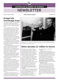


© Copyright 2026