
3.1. physiology
CLINICAL STUDIES REFERENCE GUIDE November 2014 Sales offices: North America: All other Countries: NeuMeDx Inc. 2014 Ford Road, Unit G Bristol, PA 19007 USA [email protected] tel: (215) 826 9998 fax:(215) 826 8102 Manatec Biomedical 44, rue de Laborde 75008 PARIS FRANCE [email protected] tel: +33 (3) 72 82 50 00 fax:+33 (1) 43 87 70 83 Manatec and PhysioFlow are registered trademarks TABLE OF CONTENTS INTRODUCTION ......................................................................................................... 8 1. VALIDATIONS ...................................................................................................... 9 1.1. FICK PRINCIPLE/EXERCISE ...................................................................................................... 10 A New Impedance Cardiograph Device for the Non-invasive Evaluation of Cardiac Output at Rest and During Exercise: Comparison with the "Direct" Fick Method ..................... 10 Measurements of Cardiac Output during Constant Exercises: Comparison of Two NonInvasive Techniques ............................................................................................................................................. 12 Does Thoracic Bioimpedance Accurately Determine Cardiac Output in COPD patients during Maximal or Intermittent Exercise? .................................................................................................. 13 Does Advanced Cardiac Impedance Technology Accurately Measure Cardiac Output During Submaximal Steady State Exercise? ............................................................................................... 15 Poor accuracy of noninvasive cardiac output monitoring using bioimpedance cardiography [PhysioFlow(R)] compared to magnetic resonance imaging in pediatric patients ...................................................................................................................................................................... 17 Evaluation of two methods for continuous cardiac output assessment during exercise in chronic heart failure patients ...................................................................................................................... 18 Cardiac output during exercise: A comparison of four methods ............................................. 19 Comparison of Thoracic Bioimpedance with Acetylene Uptake for Measuring Cardiac Output. ....................................................................................................................................................................... 20 1.2. THERMODILUTION/ECHO-DOPPLER/REST ....................................................................... 21 Cardiac Output Measurements: Comparison between a New Transthoracic Electrical Bioimpedance Method (Physioflow™) and the Swan-Ganz Method (Continuous Cardiac Output or Bolus Technique).............................................................................................................................. 21 Conclusion of the report on PhysioFlow Studies performed at MUSC Charleston, SC and UPMC Pittsburgh, PA, 2007 ............................................................................................................................... 22 Value of Impedance Cardiography in Pulmonary Hypertension ............................................. 23 Value of Impedance Cardiography in Patients Studied for Pulmonary Hypertension.... 24 The PhysioFlow Thoracic Impedancemeter Is Not Valid for the Measurements of Cardiac Hemodynamic Parameters in Chronic Anemic Patients ....................................................... 25 1.3 REPRODUCIBILITY/ELECTRODE PLACEMENT .................................................................. 26 Measurement of Cardiac Output Using PhysioFlow® with Different Positions of Electrode Placement ............................................................................................................................................ 26 2. APPLICATION STUDIES ................................................................................. 27 2.1. CARDIOLOGY ................................................................................................................................. 28 Best Detection of Coronary Artery Disease using a New Generation Impedance Cardiography: Comparison to Exercise Thallium 201 Scintigraphy ................................................ 28 Manatec and PhysioFlow are registered trademarks Detection of Coronary Artery Disease (CAD) during Bicycle Exercise, using New Generation Impedance Cardiography ........................................................................................................... 29 Changes in Transthoracic Impedance Signal Predict Outcome of 70º Head-up Tilt Test. 30 Head-upright Tilt Test with Sublingual Nitroglycerin Predicts Hemodynamic Abnormalities in 70° Head upright Tilt ...................................................................................................................................... 31 Impedance Cardiography and Quantitative Tissue Doppler Echocardiography for Evaluating the Effect of Cardiac Resynchronization Therapy ............................................................ 32 Impedance Cardiography - a Rapid and Cost-effective Screening Tool for Cardiac Disease ....................................................................................................................................................................... 33 Isolated Left Ventricular Diastolic Dysfunction: Implications for Exercise Left ventricular Performance in Patients without Congestive Heart Failure ........................................ 34 Thoracic Bioimpedance for Optimizing Atrioventricular and Intraventricular Delays after Cardiac Resynchronization Therapy .................................................................................................. 35 Evaluation of a New Noninvasive, Thoracic Bioimpedance Monitor for Hemodynamic Monitoring in Pediatric Patients ..................................................................................................................... 36 New Non Invasive Marker of Heart Failure with Pulmonary Congestion: Thoracic Electrical Bioimpedance ..................................................................................................................................... 37 Patients with Systolic Heart Failure Show Improvement with Long-Acting, Cardioselective Beta-Blocker Nebivolol when Others Fail .................................................................. 38 Central hemodynamic responses during high-intensity interval exercise and moderate continuous exercise in patients with chronic heart failure ................................................................. 39 Cardiopulmonary and Noninvasive Hemodynamic Responses to Exercise Predict Outcomes in Heart Failure ................................................................................................................................. 40 Peak cardiac output is the principal determinant of exercise capacity in hypertrophic cardiomyopathy: the key role of a complete non-invasive hemodynamic assessment. .......... 41 2.2. LUNG DISEASE AND INTERNAL MEDICINE .......................................................................... 42 Haemodynamics during Exercise are a Better Measure of Vasodilator Response in Human Subjects with Pulmonary Hypertension ..................................................................................... 42 Dynamic Monitoring during Exercise in Familial Amyloid Polyneuropathy (FAP) Type I 43 Unique cardiac response during apneas in obstructive sleep apnea (OSA) patients ...... 44 CHRONIC OBSTRUCTIVE PULMONARY DISEASE Respiratory muscle unloading improves leg muscle oxygenation during exercise in patients with COPD ................................... 45 Skeletal muscle reoxygenation after high-intensity exercise in mitochondrial myopathy. 46 Haemodynamic effects of proportional assist ventilation during high-intensity exercise in patients with chronic obstructive pulmonary disease. .................................................................... 47 Effects of hyperoxia on the dynamics of skeletal muscle oxygenation at the onset of heavy-intensity exercise in patients with COPD....................................................................................... 48 3 Influence of respiratory pressure support on hemodynamics and exercise tolerance in patients with COPD. .............................................................................................................................................. 49 Microvascular oxygen delivery-to-utilization mismatch at the onset of heavy-intensity exercise in optimally treated patients with CHF. ..................................................................................... 50 Inspiratory resistive loading after all-out exercise improves subsequent performance. 51 Effects of respiratory muscle unloading on leg muscle oxygenation and blood volume during high-intensity exercise in chronic heart failure. ........................................................................ 52 Kinetics of muscle deoxygenation are accelerated at the onset of heavy-intensity exercise in patients with COPD: relationship to central cardiovascular dynamics. .................. 53 Evaluation of intradialytic hypotension using impedance cardiography ............................ 54 Use Of Bioimpedance To Assess Changes In Hemodynamics During Acute Administration Of Continuous Positive Airway Pressure .................................................................... 55 Resistant Hypertension Comparing Hemodynamic Management to Specialist Care ...... 56 Effects of sauna alone and post-exercise sauna baths on blood pressure and hemodynamic variables in patients with untreated hypertension. ................................................. 57 On- and off-exercise kinetics of cardiac output in response to cycling and walking in COPD patients with GOLD Stages I-IV. .......................................................................................................... 58 Hemodynamic parameters in preeclampsia mesured by transthoracic cardiac impedancemetry in the third trimester of pregnancy: An observationnal pilot study ............ 59 Determination of Hemodynamic Parameters During 6-Minute Walk Test In Pulmonary Hypertension ........................................................................................................................................................... 60 2.3. INTENSIVE CARE .......................................................................................................................... 61 Advances in Non-invasive Cardiac Output Monitoring ................................................................ 61 Hemodynamic Responses of a Spontaneous Breathing Trial Monitoring by an Impedance Cardiograph ..................................................................................................................................... 62 Non-invasive Haemodynamic Monitoring to Predict Outcome and Guide Therapy in Acute Critical Illness ............................................................................................................................................. 63 Non-invasive Haemodynamic Monitoring to Predict Outcome and Guide Therapy in Acute Critical Illness ............................................................................................................................................. 64 Thoracic electrical bioimpedance: a tool to determine cardiac versus non-cardiac causes of acute dyspnoea in the emergency department ..................................................................... 65 2.4. ANESTHESIA .................................................................................................................................. 66 Decreased Cardiovascular Hemodynamics as Possible Mechanisms of Hypotension during Cesarean Delivery under Spinal Anesthesia: Role of Thoracic Impedance Cardiography........................................................................................................................................................... 66 Maternal haemodynamics at elective caesarean section: a randomised comparison of oxytocin 5-unit bolus and placebo infusion with oxytocin 5-unit bolus and 30-unit infusion 68 IMPEDANCE CONSULTATION PREOPERATOIRE - SFAR 2006 ................................................ 69 4 Non-Invasive Measurement of Cardiac Contractility, Stroke Volume and Cardiac Output 70 Continuous Cardiac Output Monitoring During Major Intra-Abdominal Surgery: Physioflow Signal-Morphology Impedance Cardiography vs. Flotrac/Vigileo vs. Central Venous Oxygen Saturation ................................................................................................................................ 72 3. RESEARCH.......................................................................................................... 73 3.1. PHYSIOLOGY .................................................................................................................................. 74 Exercise Capacity and Idebenone Intervention in Children and Adolescents with Friedreich Ataxia ................................................................................................................................................... 74 Post Immersion Delayed Vasomotor Adjustments to Dehydration?...................................... 75 Cardiovascular and Oxygen Uptake Kinetics during Sequential Heavy Cycling Exercises 76 Effect of Exercise Intensity on Relationship between VO2max and Cardiac Output ....... 77 Cardiac Output and Oxygen Release during very High-intensity Exercise Performed until Exhaustion ..................................................................................................................................................... 78 Faut-il Mesurer le Débit Cardiaque à L’exercice? ........................................................................... 79 Eccentric Cycle Exercise: Training Application of Specific Circulatory Adjustments ..... 80 Heart Rate Deflection Point as a Strategy to Defend Stroke Volume during Incremental Exercise ..................................................................................................................................................................... 81 Evolution of Cardiac Output during Resistive Exercise in the Healthy Subject ................. 82 Vasoconstrictive Response in the Vascular Beds of the Non-Exercising Forearm during Leg Exercise in Patients with Mild Chronic Heart Failure .................................................................... 83 Non-invasive Evaluation of Maximal Arteriovenous Oxygen Difference and Adolescent Boys' Fitness Levels .............................................................................................................................................. 84 Children Cardiorespiratory Performance Index by Simultaneous and Independent Measure of Oxygen Uptake and Cardiac Output ....................................................................................... 85 Effect of Interval versus Continuous Training on Cardiorespiratory and Mitochondrial Functions: Relationship to Aerobic Performance Improvements in Sedentary.......................... 86 Shock and Awe: Hemodynamic Changes during ECT Measured with a Non-Invasive Cardiac Output Monitor ...................................................................................................................................... 87 Relationships between hemodynamic, hemorheological and metabolic responses during exercise ....................................................................................................................................................... 88 Oxygen uptake efficiency slope’ in trained and untrained subjects exposed to hypoxia 89 Determinant factors of the decrease in aerobic performance in moderate acute hypoxia in women endurance athletes .......................................................................................................................... 90 Determinants of maximal oxygen uptake in moderate acute hypoxia in endurance athletes ...................................................................................................................................................................... 91 5 Improvement of VO2 max by cardiac output and oxygen extraction adaptation during intermittent versus continuous endurance training .............................................................................. 92 Vasoconstrictive Response in the Vascular Beds of the Non-Exercising Forearm During Leg Exercise in Patients With Mild Chronic Heart Failure ................................................................... 93 Expiratory muscle loading increases intercostal muscle blood flow during leg exercise in healthy humans ................................................................................................................................................. 94 Cardiac function and arteriovenous oxygen difference during exercise in obese adults 95 Effects of Acute Hypoxia at Moderate Altitude on Stroke Volume and Cardiac Output During Exercise ...................................................................................................................................................... 96 Reliability of exercise cardiac output measurement in COPD using impedancemetry: comparison with CO2 and inert gas rebreathing. .................................................................................... 97 Maximal exercise limitation in functionally overreached triathletes: role of cardiac adrenergic stimulation. ....................................................................................................................................... 98 Cardiovascular and hemodynamic responses on dryland vs. immersedcycling ............... 99 Effect of interval training on cognitive functioning and cerebral oxygenation in obese patients : a pilot study ...................................................................................................................................... 100 3.2. PHARMACOLOGY ....................................................................................................................... 101 Ephedrine Fails to Accelerate the Onset of Neuromuscular Block by Vecuronium ...... 101 Short-term Vasomotor Adjustments to Post Immersion Dehydration are Hindered by Natriuretic Peptides .......................................................................................................................................... 102 Sildenafil Inhibits Altitude-induced Hypoxemia and Pulmonary Hypertension ............ 103 Sildenafil Improves Cardiac Output and Exercise Performance during Acute Hypoxia, but not Normoxia ............................................................................................................................................... 104 Side-effects of L-dopa on Venous Tone in Parkinson's Disease: a Leg-weighing Assessment ........................................................................................................................................................... 105 Influence of beta-blocker on cardiac output in a maximum exercise bicycle ramp test. 106 4. EORTA/VASCULAR STIFFNESS .................................................................107 4.1. EORTA/VASCULAR STIFFNESS .............................................................................................. 108 Assessment of aortic stiffness by local and regional methods ............................................... 108 Measurement of the local aortic stiffness by a non-invasive bioelectrical impedance technique ............................................................................................................................................................... 109 Modeling and interpretation of the bioelectrical impedance signal for the determination of the local arterial stiffness ............................................................................................ 110 Time and Spatial Invariance of Impedance Signals in Limbs of Healthy Subjects by Time–Frequency Analysis............................................................................................................................... 111 6 7 INTRODUCTION This document is a list of abstracts of the studies that have been published involving the PhysioFlow technology. Some of them are abstracts of full papers published in peer reviewed journals and some are abstracts of presentations at conventions. They are categorized following the intent of the study (validation, application, or pure research) and also following applications (critical care, cardiology, physiology, etc.). They represent the start of the art about PhysioFlow, to the best of our knowledge. Some of these studies are quite old and reflect the performance of the technology at an early stage and some are recent and involve our latest innovations (HD-Z motion cancellation filter for instance). Most of the papers are about our PF05-Lab1 design but some recent ones are about the PF07-Enduro, which is newer but offers the same degree of performance. Also, it is important to note that some teams follow our recommendations strictly and obtain impressive results and some other ones intentionally or unintentionally drift away from our instructions (choice of electrodes, software version, protocol, etc.) and thus generally underperform. Enjoy your reading and please feel free to contact us should you have questions. 8 1. VALIDATIONS 9 1.1. FICK PRINCIPLE/EXERCISE 1.1. FICK PRINCIPLE/EXERCISE A New Impedance Cardiograph Device for the Non-invasive Evaluation of Cardiac Output at Rest and During Exercise: Comparison with the "Direct" Fick Method Abstract, European Journal of Applied Physiology, Volume 82 Issue 4 (2000) pp 313-320 Authors: Anne Charloux, Evelyne Lonsdorfer-Wolf, Ruddy Richard, Eliane Lampert, Monique Oswald-Mammosser, Bertrand Mettauer, Bernard Geny, Jean Lonsdorfer Service des Explorations Fonctionnelles Respiratoires, Cardio-Circulatoires et de l'Exercice, Hôpitaux Universitaires de Strasbourg, Hôpital Civil, BP 426, 67091 Strasbourg Cedex, France e-mail: [email protected] Tel: +33-388-116122; Fax: +33-388-116467 Accepted: 3 April 2000 Abstract: The objectives of this study were to evaluate the reliability and accuracy of a new impedance cardiograph device, the PhysioFlow, at rest and during a steady-state dynamic leg exercise (work intensity ranging from 10 to 50 W) performed in the supine position. We compared cardiac output determined simultaneously by two methods, the PhysioFlow (Q˙cPF) and the direct Fick (Q˙cFick) methods. Forty patients referred for right cardiac catheterisation, 14 with sleep apnoea syndrome and 26 with chronic obstructive pulmonary disease, took part in this study. The subjects' oxygen consumption values ranged from 0.14 to 1.19 l · min-1. The mean difference between the two methods (Q˙cFick-Q˙cPF) was 0.04 l · min-1 at rest and 0.29 l · min-1 during exercise. The limits of agreement, defined as mean difference ± 2SD, were -1.34, +1.41 l · min-1 at rest and -2.34, +2.92 l · min-1 during exercise. The difference between the two methods exceeded 20% in only 2.5% of the cases at rest, and 9.3% of the cases during exercise. Thoracic hyperinflation did not alter Q˙cPF. We conclude that the Physio Flow provides a clinically acceptable and non-invasive evaluation of cardiac output under these conditions. This new impedance cardiograph device deserves further study using other populations and situations. Keywords: .Impedance .Cardiography, .Cardiac output, .Fick principle, .Exercise 10 1.1. FICK PRINCIPLE/EXERCISE Non-invasive Cardiac Output Evaluation during a Maximal Progressive Exercise Test, Using a New Impedance Cardiograph Device European Journal of Applied Physiology, Volume 85 Issue 3/4 (July 2001) pp 202-207 DOI 10.1007/s004210100458 Authors: Ruddy Richard, Evelyne Lonsdorfer-Wolf, Anne Charloux, Stéphane Doutreleau, Martin Buchheit, Monique OswaldMammosser, Eliane Lampert, Bertrand Mettauer, Bernard Geny, Jean Lonsdorfer Service des Explorations Fonctionnelles Respiratoires, CardioCirculatoires et de l'Exercice, Hôpitaux Universitaires de Strasbourg, Hôpital Civil, BP 426, 67091 Strasbourg Cedex, France E-mail: [email protected] Tel: +33-388116122 Fax: +33-388116467 Accepted: 10 April 2001 / Published Online: 4 July 2001 Abstract: One of the greatest challenges in exercise physiology is to develop a valid, reliable, non-invasive and affordable measurement of cardiac output (CO). The purpose of this study was to evaluate the reproducibility and accuracy of a new impedance cardiograph device, the Physio Flow, during a 1-min step incremental exercise test from rest to maximal peak effort. A group of 12 subjects was evaluated to determine the reproducibility of the method as follows: (1) each subject performed two comparable tests while their CO was measured by impedance cardiography using the new device (COImp1, COImp2), and (2) in a subgroup of 7 subjects CO was also determined by the direct Fick method (COFick) during the second test. The mean difference between the values obtained by impedance (i.e. COImp1-COImp2) was -0.009 l·min-1 (95% confidence interval: -4.2 l·min-1, 4.2 l·min-1), and CO ranged from 3.55 l·min-1 to 26.75 l·min-1 (n=146). When expressed as a percentage, the difference (COImp1-COImp2) did not vary with increasing CO. The correlation coefficient between the values of COImp and COFick obtained during the second exercise test was r=0.94 (P<0.01, n=50). The mean difference expressed as percentage was -2.78% (95% confidence interval: -27.44%, 21.78%). We conclude that COImp provides a clinically acceptable evaluation of CO in healthy subjects during an incremental exercise. Keywords: .Impedance .Cardiography, .Cardiac output, .Maximal exercise test 11 1.1. FICK PRINCIPLE/EXERCISE Measurements of Cardiac Output during Constant Exercises: Comparison of Two Non-Invasive Techniques Int J Sports Med 2004; 25: 145-149 DOI: 10.1055/s-2004-819949 Authors: N. Tordi 2, 3, L. Mourot 1, 2 B. Matusheski 3, R. L. Hughson3 Laboratoire de Physiologie Médecine, Place St Jacques, Besançon, France Laboratoire des Sciences du Sport, Place St Jacques, Besançon, France 3 University of Waterloo, Department of Kinesiology, Waterloo, On, Canada 1 2 Abstract: We compared cardiac output (CO) determined simultaneously by electrical impedance cardiography method (COICG) and by the CO2 rebreathing technique (CO2REB) during three different steady-state exercises (target heart rate of 120, 140, and 160 min-1) in 8 healthy fit young men. The mean difference correlation coefficient obtained between the values of COICG and CO2REB was 0.85 and the mean difference (COICG-CO2REB) was 0.06 l/min (0.12 %). At 120 min-1, COICG was lower than CO2REB but the tendency was reversed at 140 and 160 min-1 where COICG was higher than CO2REB. This evolution may be explained by the difficultly of using CO2 rebreathing technique at the highest steady-state exercises and by the progressive acidemia due to exercise. The present results suggest that electrical impedance cardiography method provides acceptable evaluation of CO and may favourably replace the CO2 rebreathing technique during mild (or moderate) to high steady-state exercises. Keywords: .Impedance .Cardiography, .CO2 rebreathing technique, .Indirect Fick principle, .Steady state exercise 12 1.1. FICK PRINCIPLE/EXERCISE Does Thoracic Bioimpedance Accurately Determine Cardiac Output in COPD patients during Maximal or Intermittent Exercise? Chest. 2005 Apr;127(4):1122-31. Authors: Bougault V, Lonsdorfer-Wolf E, Charloux A, Richard R, Geny B, Oswald-Mammosser M. Service des Explorations Fonctionnelles Respiratoires et de l'Exercice, Hopital Civil, BP 426, 67091 Strasbourg Cedex, France Study objectives: The monitoring of cardiac output (CO) during exercise rehabilitation in patients with COPD, often including strenuous exercise, is advisable. Invasive methods (thermodilution, Fick method) are accurate, but for clinical routine use noninvasive CO estimation is required. We have shown that impedance cardiography (Physio Flow; Manatec Biomedical; Macheren, France) is reliable in COPD patients at rest and during a recumbent, light-intensity exercise. The aim of our study was to evaluate the validity of this noninvasive device in COPD patients during a maximal incremental exercise test (IET) and also during a strenuous intermittent work exercise test (IWET). Measurements and main results: Forty-nine simultaneous measurements of CO by means of the direct Fick method (COfick) and CO measured by the impedance cardiograph (COpf) were obtained during the IET, and 108 measurements were made during the IWET. The correlation coefficients between the two measurements were r = 0.85 and r = 0.71 for the IET and the IWET, respectively. COpf was higher than COfick. The difference between the two methods was 3.2 +/- 2.9 L/min during the IET and 2.5 +/- 2.1 L/min during the IWET. Expressed as a percentage of the mean of the two measurements, this corresponded to 31 +/- 21% and 25 +/- 20%, respectively. Design Prospective comparative study of the impedance cardiograph vs the direct Fick method applied to oxygen. Patients Eight patients with moderate-to-severe COPD (59 +/- 6 years old; FEV(1), 38 +/- 15% predicted; residual volume, 194 +/- 64% predicted) [mean +/- SD]. Conclusions: The relatively high number of values differing by > 20% precludes the use of impedance cardiography in clinical routine in such a difficult setting (hyperinflated patients and intense exercise). IMPORTANT NOTE FROM THE MANUFACTURER: In an effort to present all publications involving PhysioFlow we have decided to include this particular one in our list. Its results may not look as favourable as all the other abstracts. However, it is to be noted that the protocol was performed in a way that is not compliant with the manufacturer’s instructions: The investigators have used a particular prototype version of the PhysioFlow software in spite of our insistence that they should not do so. They have used electrodes that were not those recommended for optimal results. Moreover, data of this study have disappeared from the hard disk of the computer, making it impossible to reanalyse them with the appropriate version of the software. In addition, it is questionable that the Fick would perform well under incremental or strenuous intermittent work exercise test. 13 1.1. FICK PRINCIPLE/EXERCISE Reliability of Peak VO(2) and Maximal Cardiac Output Assessed using Thoracic Bioimpedance in Children Eur J Appl Physio,. 2005 Jun; 94(3):228-34. Epub 2005 May 26. Authors: Welsman J, Bywater K, Farr C, Welford D, Armstrong N. Children's Health and Exercise Research Centre, University of Exeter, Heavitree Road, Exeter, EX1 2LU, UK. [email protected] Abstract: The purpose of this study was to evaluate the reliability of a thoracic electrical bioimpedance based device (PhysioFlow) for the determination of cardiac output and stroke volume during exercise at peak oxygen uptake (peak VO(2) in children. The reliability of peak VO(2) is also reported. Eleven boys and nine girls aged 10-11 years completed a cycle ergometer test to voluntary exhaustion on three occasions each 1 week apart. Peak VO(2) was determined and cardiac output and stroke volume at peak VO(2) were measured using a thoracic bioelectrical impedance device (PhysioFlow). The reliability of peak VO(2) cardiac output and stroke volume were determined initially from pairwise comparisons and subsequently across all three trials analysed together through calculation of typical error and intraclass correlation. The pairwise comparisons revealed no consistent bias across tests for all three measures and there was no evidence of non-uniform errors (heteroscedasticity). When three trials were analysed together typical error expressed as a coefficient of variation was 4.1% for peak VO(2) 9.3% for cardiac output and 9.3% for stroke volume. Results analysed by sex revealed no consistent differences. The PhysioFlow method allows non-invasive, beat-to-beat determination of cardiac output and stroke volume which is feasible for measurements during maximal exercise in children. The reliability of the PhysioFlow falls between that demonstrated for Doppler echocardiography (5%) and CO(2) rebreathing (12%) at maximal exercise but combines the significant advantages of portability, lower expense and requires less technical expertise to obtain reliable results. 14 1.1. FICK PRINCIPLE/EXERCISE Does Advanced Cardiac Impedance Technology Accurately Measure Cardiac Output During Submaximal Steady State Exercise? Control/Tracking Number: 09-SA-3991-ACSM Activity: Scientific Abstract Current Date/Time: 11/3/2008 8:13:33 PM Authors: Craig E. Broeder, FACSM, Jarod Hickock, Amanda Burditt. Benedictine University, Lisle, IL. Email: [email protected] Abstract: This study determined if advanced cardiac impedance technology (ACI) could accurately measure cardiac output during steady-state cycling exercise compared to values calculated using the direct Fick equation developed by Stringer et al (1997). METHOD: VO2 max was determined on both a treadmill (Mean = 3.96 ± 1.2 liters/min) and cycle ergometer (Mean = 3.42 ± 1.2 liters/min) in 15 subjects (Age; 34.3. ± 9.4 yrs). Steady-state exercise wattage was set at 25%, 50%, and 75% of peak watts achieved. Subjects exercised 8 mins at each stage. The last 4 mins were used to determine the ACI values for cardiac output (Q), heart rate, stroke volume, EDV, SBP, DBP, and systemic vascular resistance. Both the submaximal and maximal exercise trials were performed in duplicate to assure accurate data collection. No significant differences were observed in test-retest trials. Thus, the mean of duplicate trials was used for all data analyses. Keywords: .Cardiac impedance .Steady state exercise .Cycle ergometry Category: 204 acute exercise Author Disclosure Information: C.E. Broeder, PhysioFlow Corporation, Contracted Research. Results: There was no significant difference between the cardiac output determined by the Stringer equation and the ACI measured Q value. The percent differences across exercise intensity’s for Q were 4.2%, 1.5%, and -6.7% for the 25%, 50%, and 75% of max watts, respectively. Linear regression analyses indicated rsquared = 0.99, SEE = 0.20 liters, p = .001. For all trials combined, the mean percent difference between the stringer cardiac output and the ACI cardiac output was 0.5%. Conclusion: In conclusion, these data indicate that the ACI system used in this study was highly accurate in determining a person’s Q during steady state exercise ranging between 40.1% and 88.3% of VO2 max. Future studies need to determine if similar accuracy can be achieved using other forms of exercise, i.e., treadmill. 15 1.1. FICK PRINCIPLE/EXERCISE The Ergogenic Effect of Recombinant Human Erythropoietin on VO2max Depends on the Severity of Arterial Hypoxemia Citation: Editor: Robach P, Calbet JAL, Thomsen JJ, Boushel R, Mollard P, et al. (2008) The Ergogenic Effect Timothy Secomb, of Recombinant Human Erythropoietin on V ˙ O2max Depends on the Severity of Arterial University of Arizona, Hypoxemia. PLoS ONE 3(8): e2996. doi:10.1371/journal.pone.0002996 United States of America Funding: This study was supported by research fundings from Anti Doping Danmark (Denmark), Kulturministeriet (Denmark) and Bundesamt fu¨ r Sport (Switzerland) to C.L. This study was also supported in part by research funding from Gobierno de Canarias (Ayudas para estancias postdoctorales)(Spain) to J.A.L.C. The funders had no role in study design, data collection and analysis, decision to publish, or preparation of the manuscript. Competing Interests: Email: paul.robach@jeunesseThe authors have declared that no competing interests exist. sports.gouv.fr Copyright: 2008 Robach et al. This is an open-access article distributed under the terms of the Creative Commons Attribution License, which permits unrestricted use, distribution, and reproduction in any medium, provided the original author and source are credited. Received May 6, 2008; Accepted July 28, 2008; Published August 20, 2008 Abstract: Treatment with recombinant human erythropoietin (rhEpo) induces a rise in blood oxygen-carrying capacity (CaO2) that unequivocally enhances maximal oxygen uptake (V˙O2max) during exercise in normoxia, but not when exercise is carried out in severe acute hypoxia. This implies that there should be a threshold altitude at which V ˙ O2max is less dependent on CaO2. To ascertain which are the mechanisms explaining the interactions between hypoxia, CaO2 and V ˙ O2max we measured systemic and leg O2 transport and utilization during incremental exercise to exhaustion in normoxia and with different degrees of acute hypoxia in eight rhEpotreated subjects. Following prolonged rhEpo treatment, the gain in systemic VO2max observed in normoxia (6–7%) persisted during mild hypoxia (8% at inspired O2 fraction (FIO2) of 0.173) and was even larger during moderate hypoxia (14–17% at FIO2 = 0.153–0.134). When hypoxia was further augmented to FIO2 = 0.115, there was no rhEpoinduced enhancement of systemic V ˙ O2max or peak leg V ˙ O2. The mechanism highlighted by our data is that besides its strong influence on CaO2, rhEpo was found to enhance leg V ˙ O2max in normoxia through a preferential redistribution of cardiac output toward the exercising legs, whereas this advantageous effect disappeared during severe hypoxia, leaving augmented CaO2 alone insufficient for improving peak leg O2 delivery and V ˙ O2. Finally, that V ˙ O2max was largely dependent on CaO2 during moderate hypoxia but became abruptly CaO2-independent by slightly increasing the severity of hypoxia could be an indirect evidence of the appearance of central fatigue. Agreement between impedance and dye dilution for cardiac output measurement: This graph shows the agreement (Bland-Altman plot) between the cardiac impedance technique and the indocyanine green dye dilution method for measuring cardiac output during exercise obtained from 55 measurements in seven subjects. For each measurement, the difference between the two methods is plotted against the average of both techniques. The solid line indicates the mean bias, while the dotted lines indicate the 95% confidence intervals (2×standard deviation). 16 1.1. FICK PRINCIPLE/EXERCISE Poor accuracy of noninvasive cardiac output monitoring using bioimpedance cardiography [PhysioFlow(R)] compared to magnetic resonance imaging in pediatric patients Authors: Taylor K, Manlhiot C, McCrindle B, Grosse-Wortmann L, Holtby H Department of Anesthesia, Hospital for Sick Children, 555 University Ave., Toronto, ON M5G 1X8, Canada. Email: [email protected] Abstract: Identification of low cardiac output (CO) states in anesthesia is important because preoperative hemodynamic optimization may improve outcome in surgery. Accurate real-time CO measurement would be useful in optimizing "goal-directed" therapy. We sought to evaluate the reliability and accuracy of CO measurement using bioimpedance cardiography (PhysioFlow®, NeuMeDx, Bristol, PA) in pediatric patients with and without cardiac disease undergoing anesthesia for magnetic resonance imaging (MRI). Methods: All consenting patients undergoing anesthesia for cardiac MRI were enrolled. After equilibration of anesthesia for ≥10 minutes, 6 PhysioFlow electrodes were applied to the patient's chest for continuous real-time monitoring for 10 minutes. Data were stored in 15-second epochs and later averaged offline to obtain CO. Phase contrast MRI measurements of flow volumes in the superior vena cava and ascending and descending aorta were made from a single imaging plane through all 3 vessels at the level of the right pulmonary artery. Both CO measurements were indexed to body surface area. The anesthetic technique was the same for both measurements. Agreement was assessed using BlandAltman analysis. Results: Conclusion: Thirty-one patients were enrolled and 23 were analysed. The PhysioFlow performance was median age at study was 2.8 years (range, 0.02-8.02 years) and not sufficiently accurate in this median body surface area was 0.54 m(2) (range, 0.21-1.00 m(2)). population. Modifications of Eleven of the 23 patients (48%) were males. Patients were the algorithm and further grouped into those with univentricular physiology, 6 of 23 (26%); testing are required before biventricular physiology with shunt, 3 of 23 (13%); biventricular this device can be without shunt, 10 of 23 (43%); and no structural heart disease, 4 recommended for routine of 23 (17%). The mean bias was -0.34 ± 1.50 L/min/m(2) (P = clinical use in pediatric 0.29). The 95% limits of agreement were -3.21 to +2.69 patient. L/min/m(2). Only 8 of 23 measurements (35%) were within 20% and 14 of 23 measurements (61%) were within 30% of each other. IMPORTANT NOTE FROM THE MANUFACTURER: We do respect the Toronto Sick Children hospital team for their friendliness and professionalism. However there was a technical mistake in their data manipulation and all PhysioFlow recordings were lost by the research team before we could take a look at it and see what happened. Please bear in mind that it was the very first time our system was applied to such a patient population and it was never calibrated for it before. It would have been extremely helpful to access the PhysioFlow recordings to 1) Assess their quality and potential operator's mistakes 2) Reprocess them with newer algorithms we developed later (these ones date back to 2004) 3) Calibrate the system to adapt it to this patient population. This was the initial plan but it could never be implemented because of the loss of data. 17 1.1. FICK PRINCIPLE/EXERCISE Evaluation of two methods for continuous cardiac output assessment during exercise in chronic heart failure patients Submitted 20 March 2008. Accepted16 October 2008. Authors: 1. Hareld M. C. Kemps1,2, Eric J. M. Thijssen2, 2. Goof Schep1, 3. Boudewijn T. H. M. Sleutjes3, 4. Wouter R. De Vries4, 5. Adwin R. Hoogeveen1, 6. Pieter F. F. Wijn5,6, and 7. Pieter A. F. M. Doevendans7 Departments of 1Sports Medicine and of 2Cardiology, Máxima Medical Centre, Veldhoven; 3Department of Biomedical Engineering, Eindhoven University of Technology, Eindhoven; 4Rudolf Magnus Institute of Neuroscience, Section of Rehabilitation and Sports Medicine, University Medical Centre Utrecht, Utrecht; 5Department of Medical Physics, Máxima Medical Centre, Veldhoven; 6Department of Applied Physics, Eindhoven University of Technology, Eindhoven; 7Department of Cardiology, University Medical Centre Utrecht, Utrecht, The Netherlands Address for reprint requests and other correspondence: H. M. C. Kemps, Dept. of Sports Medicine, Máxima Medical Centre, P.O. Box 7777, 5500 MB Veldhoven, The Netherlands (email:[email protected]) Abstract: The purpose of this study was to evaluate the accuracy of two techniques for the continuous assessment of cardiac output in patients with chronic heart failure (CHF): a radial artery pulse contour analysis method that uses an indicator dilution method for calibration (LiDCO) and an impedance cardiography technique (Physioflow), using the Fick method as a reference. Ten male CHF patients (New York Heart Association class II–III) were included. At rest, cardiac output values obtained by LiDCO and Physioflow were compared with those of the direct Fick method. During exercise, the continuous Fick method was used as a reference. Exercise, performed on a cycle ergometer in upright position, consisted of two constant-load tests at 30% and 80% of the ventilatory threshold and a symptom-limited maximal test. Both at rest and during exercise LiDCO showed good agreement with reference values [bias ± limits of agreement (LOA), −1% ± 28% and 2% ± 28%, respectively]. In contrast, Physioflow overestimated reference values both at rest and during exercise (bias ± LOA, 48% ± 60% and 48% ± 52%, respectively). Exercise-related within-patient changes of cardiac output, expressed as a percent change, showed for both techniques clinically acceptable agreement with reference values (bias ± LOA: 2% ± 26% for LiDCO, and −2% ± 36% for Physioflow, respectively). In conclusion, although the limits of agreement with the Fick method are pretty broad, LiDCO provides accurate measurements of cardiac output during rest and exercise in CHF patients. Although Physioflow overestimates cardiac output, this method may still be useful to estimate relative changes during exercise. Published online before print October 23, 2008, doi: 10.1152/japplphysiol.90430.2008 Journal of Applied Physiology December 2008 vol. 105 no. 6 1822-1829 18 1.1. FICK PRINCIPLE/EXERCISE Cardiac output during exercise: A comparison of four methods Accepted for publication 27 January 2014 Authors: C.Siebenmann1, P. Rasmussen1, H. Sørensen2, M. Zaar2, M. Hvidtfeldt2, A. Pichon3, N. H. Secher2, C. Lundby1,4 1Center for Integrative Human Physiology, Institute of Physiology, University of Zürich, Zürich, Switzerland, 2Department of Anesthesia, The Copenhagen Muscle Research Center, Rigshospitalet, University of Copenhagen, Copenhagen, Denmark, 3Laboratoire Réponses Cellulaires et Fonctionnelles à l’Hypoxie, Université Paris 13, Bobigny, France, 4Food & Nutrition & Sport Science, Gothenburg University, Gothenburg, Sweden Corresponding author: Christoph Siebenmann, Center for Integrative Human Physiology (ZIHP), University of Zürich, Institute of Physiology, Winterthurerstrasse 190, 8057 Zürich, Switzerland. Tel: +41 44 635 64 62, Fax: +41 44 635 68 14, E-mail: [email protected] or [email protected] Abstract: Several techniques assessing cardiac output (Q) during exercise are available. The extent to which the measurements obtained from each respective technique compares to one another, however, is unclear. We quantified Q simultaneously using four methods: the Fick method with blood obtained from the right atrium (QFick-M), Innocor (inert gas rebreathing; QInn), Physioflow (impedance cardiography; QPhys), and Nexfin (pulse contour analysis; QPulse) in 12 male subjects during incremental cycling exercise to exhaustion in normoxia and hypoxia (FiO2 = 12%). While all four methods reported a progressive increase in Q with exercise intensity, the slopes of the Q/oxygen uptake (VO2) relationship differed by up to 50% between methods in both normoxia [4.9 ± 0.3, 3.9 ± 0.2, 6.0 ± 0.4, 4.8 ± 0.2 L/min per L/min (mean ± SE) for QFick-M, QInn, QPhys and QPulse, respectively; P = 0.001] and hypoxia (7.2 ± 0.7, 4.9 ± 0.5, 6.4 ± 0.8 and 5.1 ± 0.4 L/min per L/min; P = 0.04). In hypoxia, the increase in the Q/VO2 slope was not detected by Nexfin. In normoxia, Q increases by 5–6 L/min per L/min increase in VO2, which is within the 95% confidence interval of the Q/VO2 slopes determined by the modified Fick method, Physioflow, and Nexfin apparatus while Innocor provided a lower value, potentially reflecting recirculation of the test gas into the pulmonary circulation. Thus, determination of Q during exercise depends significantly on the applied method. Keywords: .Inert gas rebreathing .Impedance .Cardiography .Pulse contour .Analysis .Hypoxia .Maximal oxygen uptake IMPORTANT NOTE FROM THE MANUFACTURER: The research team, contacted for the first time after publication, did not wish to share details about the software version that was used (which is important regarding the filtering of signals), nor the skin preparation methodology/electrode type. However they found that PhysioFlow generates fewer impossible /implausible readings than other methods. © 2014 John Wiley & Sons A/S. Published by John Wiley & Sons Ltd Scandinavian Journal of Medicine & Science in Sports 19 1.1. FICK PRINCIPLE/EXERCISE Comparison of Thoracic Bioimpedance with Acetylene Uptake for Measuring Cardiac Output. Int J Sports Med. 2014 Jun 2. [Epub ahead of print] Authors: Wang E, Cruz C, Pettersen MR, Hoff J, Helgerud J. Department of Circulation and Medical Imaging, Faculty of Medicine, the Norwegian University of Science and -Technology, Trondheim, Norway. Abstract: Cardiac output is shown to be a key determinant for oxygen transport, performance and health. Reliable and accurate non-invasive measurements of cardiac output, especially during exercise, are therefore of importance. The present study compared a new thoracic bioimpedance method with the established single-breath acetylene uptake method. We assessed cardiac output in 20 (24±4 years.) moderately trained males, at rest and during cycling. Both methods showed good test-retest reliabilities with ±2 SD limits of agreement of 3.67 and -4.50 L ∙ min-1 (thoracic bioimpedance) and 4.46 and -5.69 L ∙ min-1 (single breath), respectively. When thoracic bioimpedance was compared with single breath, the ±2 SD limits of agreement were poor (-6.05 and 9.57 L ∙ min-1). Thoracic bioimpedance displayed significantly lower (p<0.05) absolute cardiac output values than single breath, and the cardiac output-oxygen consumption slopes (y=5.7x+5.5 (single breath) and y=5.0x+5.0 (thoracic bioimpedance) tended (p=0.08) to show less increase for thoracic bioimpedance. Conclusions: Results from the single-breath method are in line with previous findings, showing a good reliability. Although thoracic bioimpedance showed a similar reliability as the single-breath method, and is easier to use, the agreement with single breath was poor, and thoracic bioimpedance seems not to be able to replace it. IMPORTANT NOTE FROM THE MANUFACTURER: The Enduro unit on loan in Trondheim was an early stage device and had a defective ECG detection in the firmware, which has been corrected in the meantime. It resulted in a poor calculation of HR and also a bad trigger for the signal stabilization filter. We were offered the opportunity to reprocess the data. Unfortunately the team did not maintain the patients at real rest after calibration and therefore re-calculating the calibration and the test was not productive. This study should not have been published without mention of these issues as they affect its entire validity. © Georg Thieme Verlag KG Stuttgart · New York. PMID: 24886928 [PubMed - as supplied by publisher] 20 1.2. THERMODILUTION/ECHO-DOPPLER/REST 1.2. THERMODILUTION/ECHO-DOPPLER/REST Cardiac Output Measurements: Comparison between a New Transthoracic Electrical Bioimpedance Method (Physioflow™) and the Swan-Ganz Method (Continuous Cardiac Output or Bolus Technique) Published at the French Anaesthesiology Society, Sept 2001 Authors: Moreau X, Rousseau JM*, Thiranos JC**, Dubé L, Corbeau JJ, Granry JC, Beydon L. ICU – University Hospital Angers * ICU, Val de Grâce Military Hospital, Paris ** ICU, Cardiovascular Surgery, Strasbourg Abstract: Results: 107 ICU patients underwent simultaneous CO measurements using the two methods (CO Swan Ganz : 20 B and 87 CCO). One measurement was performed on every patient (84 Male/23 Female, age 69±11 years, weight 75±15 Kg, height 167±8 cm). Pathologies : aortic and bypass surgery 65 %, septic shock 18%, heart failure 7%, pulmonary patients 7%, misc. 3%. Linear regression factor was 0.88 (p<0.001). CO PhysioFlow = 0.75 CO Swan + 1.33. Bland and Altman diagram is represented below (bias = -0.014 L/min). CO : Difference between the two methods (L/min) Cardiac Output (CO) measurement using Transthoracic Electrical Bioimpedance (TEB) has been recently improved. We have tested a system using exclusively relative values of the impedance signal, and not absolute values (Z0). Indeed, the Z0 value has been described as being at origin of the practical limitations of TEB. We have chosen the Swan-Ganz method to provide reference values, using boluses (B), or continuous cardiac output (CCO) (BAXTER™ Vigilance®). 3 + 2SD 2 1 0 2.5 5.0 7.5 10.0 12.5 -1 -2 - 2SD -3 CO : average of the two methods (L/min) Conclusion: This study has been done under the most difficult conditions for TEB : one single measurement per patient, and patients presenting a very large variety of pathologies. This new TEB method deserves further investigations, using the Fick method, on the same range of unselected ICU patients. Indeed, literature displays that Fick as a reference method features a reduced dispersion of results compared to thermodilution1. References: Charloux et al., Eur. J. Appl. Physiol. (2000) 82: 313-320 1: 21 1.2. THERMODILUTION/ECHO-DOPPLER/REST Conclusion of the report on PhysioFlow Studies performed at MUSC Charleston, SC and UPMC Pittsburgh, PA, 2007 This multicenter study has been performed on a population of representative American patients referred to major medical institutions for severe cardiovascular diseases. In that frame PhysioFlow has proven that it is substantially equivalent to the predicate device (Philips) and FDA should grant clearance because: It performs much better that the predicate device in terms of accuracy and ability to provide clinically relevant numbers, even in difficult patients. Its comparative measurements to a clinically accepted reference method are as good as expected with reference to the best scientific literature. It performs as well as the best established invasive reference technique (Fick) with comparison to thermodilution In conclusion, this study achieved its objective of demonstrating 1) The agreement in CO between the PhysioFlow ICG PF-05 and thermodilution is similar to or better than the agreement between the Philips ICG and thermodilution; and 2) Absolute agreement between the PhysioFlow ICG PF-5 itself and thermodilution is adequate when accounting for the known variability in the thermodilution reference method. 22 1.2. THERMODILUTION/ECHO-DOPPLER/REST Value of Impedance Cardiography in Pulmonary Hypertension Pulmonary Hypertension Posters Tuesday, November 2, 2010 Authors: Adriano R. Tonelli, MD*; Hassan Alnuaimat, MD; Robin Carrie, APRN-BC and Kamal Mubarak, MD University of Florida, Gainesville, FL Purpose: To assess impedance cardiography as a method for obtaining a non-invasive hemodynamic evaluation in patients with pulmonary hypertension (PH). Methods: A total of 39 patients (age 57± 14 years, 87% women) with presumed (23%) or confirmed PH (77%) of different etiologies who underwent right heart catheterization (RHC) at University of Florida from August 2009 to March 2010 agreed to be studied by impedance cardiography (PhysioFlow(r) PF-05, Manatec Biomedical, Macheren, France) immediately after RHC. PhysioFlow(r) measures cardiac output (CO) and end-diastolic volume (EDV), among other parameters. Results: The median pulmonary artery pressure was 36 (IQR 2656)mm Hg. The CO (mean ± SD in l/m) by thermodilution (CO-T), Fick methodology (CO-F) and impedance cardiography (CO-IC) was 5.9 ± 2.2, 5.5 ± 1.6 and 5.6 ± 1.5, respectively. Bland-Altman analysis of CO-T versus CO-F showed mean of 0.4 L/min (95% limit of agreement (LoA) 3.4 and -2.6), CO-T versus CO-IC a mean of 0.3 L/min (95% LoA 2.8 and -2.2) and CO-F versus CO-IC a mean of -0.1 L/min (95% LoA 2.5 and -2.7). Correlation between CO-T and CO-IC was R2 = 0.7, p < 0.001. In patients with PH the correlation of CO-T and CO-IC had a mean of 0.4 L/min(95% LoA 2.9 and -2.2), R2 = 0.75, p < 0.001.Pulmonary artery occlusion pressure (PAOP) correlated with EDV (R2 = 0.2, p = 0.005). By ROC analysis EDV of 200 ml had a sensitivity of 53% and a specificity of 86% for PAOP > 15 mm Hg (AUC: 0.78). Conclusion: Clinical Implications: Impedance cardiography reliably measures cardiac output in patients with pulmonary hypertension. This methodology may serve as a valid tool for the hemodynamic evaluation of this group of patients. Impedance cardiography allows a reliable and non-invasive measurement of cardiac output in patients with PH. End-diastolic volume correlated with pulmonary artery occlusion pressure. Disclosure: Adriano Tonelli, No Financial Disclosure Information; No Product/Research Disclosure Information doi:10.1378/chest.10266 (Chest. 2010; 138:359A) © 2010 American College of Chest Physicians 23 1.2. THERMODILUTION/ECHO-DOPPLER/REST Value of Impedance Cardiography in Patients Studied for Pulmonary Hypertension Pulmonary Hypertension Posters 24 May, 2011 Authors: Adriano R. Tonelli, Hassan Alnuaimat, Ning Li, Robin Carrie, Kamal K. Mubarak Abstract: The aim of this study was to evaluate the accuracy and precision of impedance cardiography as a method for noninvasive hemodynamic evaluation of patients with pulmonary hypertension (PH). We performed a prospective and blinded study of patients who underwent right heart catheterization (RHC) for evaluation of known or presumed PH at the University of Florida from August 2009 to March 2010. The cohort consisted of a total of 39 patients (age = 57 ± 14 years, 87% women) with presumed (23%) or confirmed PH (77%) of different etiologies. Patients underwent RHC and impedance cardiography using the PhysioFlow PF-05. The PhysioFlow PF-05 measures cardiac output (CO) and LV end-diastolic volume (LVEDV), among other parameters. The median pulmonary artery pressure was 36 (IQR 26-56) mmHg. The CO (mean ± SD) by thermodilution (CO-T) and by impedance cardiography (CO-IC) was 5.9 ± 2.2 and 5.6 ± 1.5 L/min, respectively. Bland-Altman analysis of CO-T versus CO-IC revealed a mean of 0.3 L/min (95% LoA: -2.2 to ?2.8). In patients with PH, the correlation of CO-T and CO-IC had a mean of 0.4 L/min (95% LoA: 2.9 and -2.2). Pulmonary artery occlusion pressure (PAOP) correlated with LVEDV (R2 = 0.2, p = 0.005). By ROC analysis, EDV C 200 ml had a sensitivity of 53% and a specificity of 86% for PAOP[15 mmHg (AUC = 0.78). In patients with PH, impedance cardiography had good accuracy and fair precision for CO determination when compared with thermodilution. Impedance cardiography may provide information about the preload status and has the potential to become a cost-effective and noninvasive method for the follow-up of patients with PH. 24 1.2. THERMODILUTION/ECHO-DOPPLER/REST The PhysioFlow Thoracic Impedancemeter Is Not Valid for the Measurements of Cardiac Hemodynamic Parameters in Chronic Anemic Patients Received: May 12, 2013; Accepted: September 26, 2013; Published: October 22, 2013 Authors: Pascal Bogui, Edwige Balayssac-Siransy, Philippe Connes, Nalourgo Tuo, Soualiho Ouattara, Aurélien Pichon, Cyrille Serges Dah Abstract: The aim of the present study was to test the validity of the transthoracic electrical bioimpedance method PhysioFlow® to measure stroke volume in patients with chronic anemia. Stroke volume index (SVI), as well as cardiac index (CI) obtained by transthoracic electrical bioimpedance method and doppler echocardiography were compared in healthy subjects (n = 25) and patients with chronic anemia (i.e. mainly with sickle cell anemia; n = 32), at rest. While doppler echocardiography was able to detect difference in SVI between the two populations, the Physioflow® failed to detect any difference. Bland & Altman analyses have demonstrated no interchangeability between the two methods to assess CI and SVI in anemic patients and healthy subjects. While doppler echocardiography displayed a good concordance for SVI results with those obtained in the literature for anemic patients, the Physioflow® did not. Finally, in contrast to doppler echocardiography: 1) the CI obtained with the Physioflow® was not correlated with the hemoglobin level and 2) the stroke volume determined by the Physioflow® was highly influenced by body surface area. In conclusion, our findings indicate that the Physioflow® device is inaccurate for the measurement of SVI and CI in patients with chronic anemia and has a poor accuracy for the measurement of these parameters in African healthy subjects. IMPORTANT NOTE FROM THE MANUFACTURER: This is an absurd outcome generated by a very early development software that should never have been used by any customer and was unfortunately released by mistake to one center in France and passed on to this center in Abidjean. In truth, PhysioFlow SVi is NOT correlated to body height or weight. The research team, contacted for the first time after publication, did not wish to share details about the software version that was used nor accepted our offer to reprocess recordings with the appropriate version of the software, which is highly regrettable. There is absolutely no evidence precluding the use of PhysioFlow in the African population. 25 1.3. REPRODUCIBILITY/ELECTRODE PLACEMENT 1.3 REPRODUCIBILITY/ELECTRODE PLACEMENT Measurement of Cardiac Output Using PhysioFlow® with Different Positions of Electrode Placement Singapore Med J 2006; 47(11): 967 Authors: Tan K H, Lai F O, Hwang N C Abstract: PhysioFlow is a non-invasive impedance cardiograph device that measures cardiac output. Recommended electrode placements involve six electrodes, including two near the xiphisternum (Z3 and Z4/EcG3/neutral). This study aims to evaluate if changing the positions of these two leads to the left fourth and fifth intercostal spaces along the mid- axillary line results in a change in the cardiac output measurement. Keywords: .Electrode placements .Impedance Non-invasive cardiac output monitoring , .PhysioFlow® Methods: This was a prospective, controlled, crossover, paired study of 30 patients where electrodes were placed in the recommended positions and cardiac output (CO1) obtained after two minutes. The second cardiac output (CO2) was then obtained with the electrodes Z3 and Z4/EcG3/neutral repositioned at the left mid- axillary line at the fourth and fifth intercostal spaces. The final step involved switching the Z3 and Z4/EcG3/neutral leads back to the recommended position and the cardiac output (CO3) was measured. Results: the average of the initial and third readings (COave) was compared with the measured CO2 and analyzed. The regression equation was: CO at the proposed site (CO2) = COave at the recommended site + 0.058. The paired samples correlation was 0.995. Within the 95 percent limits of agreement, the bias with CO measured at the proposed site of electrode placement was 0.046 L/min with the limits at -0.24 L/min and 0.34 L/min. the mean difference was 0.86% of the average CO. 26 2. APPLICATION STUDIES 27 2.1. CARDIOLOGY 2.1. CARDIOLOGY Best Detection of Coronary Artery Disease using a New Generation Impedance Cardiography: Comparison to Exercise Thallium 201 Scintigraphy Authors: JM Dupuis, F Prunier, W Abi-Khalil, Ph Pezard, F Bour, Ph Geslin, Department of cardiology, C.H.U. Angers France. Abstract: During exercise in patients with ischemia, contractility is impeded before electrical signs or angina appear. Therefore, measurement of contractility impairment could provide a highly sensitive approach to the detection of an ischemia. The protocol was designed to determine if a new, non invasive cardiac output measuring device (Physio Flow® : PF03, Manatec France) whose measurements are based on analysis of instant thoracic impedance (ICG) variations could be helpful to detect ischemia during exercise thallium scintigraphy. Methods: The efficiency of ICG in detecting myocardial ischemia was compared to treadmill exercise/redistribution thallium-201 scintigraphy. During exercise, patients had simultaneous measurement of stroke volume with ICG. ICG was considered abnormal if stroke volume at the peak of exercise was lower than another stroke volume measured before, and separated by at least 1 min from peak. Clinical was considered as positive if typical angina occurred and an abnormal ECG if ST-segment horizontal or descending depression > 1 mV lasting for at least 30 seconds. Ischemia was affirmed by a mismatch between exercise/redistribution thallium-201 scintigraphy. Results: 36 patients (30 men, age 62+/-11 years), 30 with proved CAD were submitted to treadmill exercise/redistribution thallium-201 scintigraphy. Stroke volume profile alteration always occurred earlier in exercise than ECG ST segment depression and angina. The sensitivity (Se), specificity (Sp), positive (PPV) and negative (NPV) predictive values are summarized in the following table. Angina ECG(+) ECG(+) and/or Angina ECG(+) and Angina ICG(-) Se 46% 31% 41% 33% 100% Sp 74% 65% 66% 76% 74% PPV 50% 33% 41% 43% 67% NPV 71% 63% 66% 68% 100% Conclusion: Use of ICG during exercise allows for the estimation of stroke volume changes over time. These preliminary results show that it is a promising technique compared to ECG or other criteria for the detection of ischemia during exercise test with an excellent NPV. 28 2.1. CARDIOLOGY Detection of Coronary Artery Disease (CAD) during Bicycle Exercise, using New Generation Impedance Cardiography Heart Journal, June 2000 Volume 83 supplement II Authors: J.M. Dupuis, J. Bour, P Abraham, K. Kalife, C.H.U. Angers France. Abstract: During exercise in patients with CAD, contractility is impeded before electrical signs or angina appear. Therefore, measurement of contractility impairment could provide a highly sensitive approach to the detection of CAD. The protocol was designed to determine if a new, non-invasive cardiac output measuring device (Physio Flow® : PF03, Manatec France) whose measurements are based on analysis of instant thoracic impedance (ICG) variations that does not use average impedance baseline values could be helpful to detect CAD. Methods: On a 12 months period, subjects suspected of CAD had been submitted to an incremental bicycle exercise test (30 W / 2 min). Those who presented either an interpretable and abnormal ECG : [ECG(+)] ST-segment horizontal or descending depression > 1 mV lasting for at least 30 seconds, or typical angina during the exercise test were submitted to coronarography (n=29. 18 men/11 women, age = 57+/-10 years. Weight = 80+/-12 Kg. Height = 170+/-9 cm). Coronarography, considered as the gold standard for comparison, were performed and analyzed by independent observers and quoted abnormal for at least 50% stenosis of the coronary arteries : coro(+). During exercise, patients had simultaneous measurement of stroke volume with ICG. ICG was considered abnormal if stroke volume at the peak of exercise was lower than another stroke volume measured before, and separated by at least 1 min from peak. Results: Number of patients in each group are summarized in the table. Stroke volume profile alteration always occurred earlier in exercise than ECG ST segment depression. The sensitivity, specificity, positive and negative predictive values were respectively: 63, 20, 60, 22% for ECG alone; 68, 20, 62, 25 for angina alone: 84, 10, 64, 25% for ECG or angina; 100, 50, 79, 100% for ICG. ECG(+) Angina ECG(+) and Angina ECG(+) and/or Angina ICG SV depression coro(+) n=19 12 13 9 16 19 coro(-) n=10 8 8 7 9 5 Conclusion: Use of ICG during exercise allows for the estimation of stroke volume changes over time. As such, these preliminary results show that it is a promising technique for the non-invasive diagnosis of CAD. 29 2.1. CARDIOLOGY Changes in Transthoracic Impedance Signal Predict Outcome of 70º Head-up Tilt Test. Accepted by Clinical Science October 21st 2002 Authors: Elisabeth Bellard1, Jacques-Olivier Fortrat1, Daniel Schang2, Jean-Marc Dupuis3, Jacques Victor3 and Georges Lefthériotis1 Georges Lefthériotis Tel: 0-33-2-41-35-46-17, Fax: 0-33-2-41-35-50-42 E-mail: [email protected] From the 1Laboratoire de Physiologie - UPRES EA 2170, Faculté de Médecine, rue haute de Reculée, 49035 Angers, France, 2Ecole Supérieure d’Electronique de l’Ouest, 4 rue Merlet de la Boulaye, PO Box 926, 49009 Angers Cedex 01, France, 3Département de Cardiologie, Centre Hospitalier d'Angers, rue Larrey, 49033 Angers cedex, France. Abstract: We determined if the early changes in central hemodynamics determined by transthoracic impedance induced by head-upright tilt test (70°HUT) could predict syncope. Heart rate, arterial blood pressure and central hemodynamics (pre-ejection period and rapid left ventricular ejection time (T1), slow ejection time (T2) and dZ/dtmax, assessed by the transthoracic impedance technique), were recorded during supine rest and 45 min 70°HUT in 68 patients (402 years) with history of unexplained recurrent syncope. Thirty-eight patients (42 3 years) had a symptomatic outcome to 70°HUT (fainters) and 30 (392 years) had a negative outcome (non-fainters). Between the 5th and 10th minutes of 70°HUT, T2 increased significantly only in the fainters and a change of T2 >40 ms from baseline predicted a positive outcome with a sensitivity of 68% and a specificity of 70%. During supine rest, fainters exhibited a shorter T2 than non-fainters (183±10 ms vs. 233±14 ms, p<0.01). A T2 <199ms predicted a positive 70°HUT outcome with a sensitivity of 68% and a specificity of 63%. The combination to the changes from rest to 70°HUT of the other hemodynamic variables (heart rate >11 bpm, systolic <2 mmHg, diastolic <7 mmHg and pulse <-3 mmHg pressures) increased the specificity to 97% as well as the positive predictive value (93%). Transthoracic impedance could detect differences between fainters and non-fainters in central hemodynamics during supine rest and the initial period of 70°HUT with a consistent sensitivity and specificity when combined with peripheral hemodynamic variables. 30 2.1. CARDIOLOGY Head-upright Tilt Test with Sublingual Nitroglycerin Predicts Hemodynamic Abnormalities in 70° Head upright Tilt European Journal of Physiology (2002, 444, R49, S3 26) (Physiomed 2002 Société de Physiology Abstracts of the 70th Annual Meeting 18-20 Septembre 2002, Québec, Canada) Authors: Bellard Elisabeth, Fortrat Jacques-Olivier, Dupuis Jean-Marc, Victor Jacques, Lefthériotis Georges Pflugers Arch - Eur J Physiol (2002) 444: R49,S3 26. Abstract: We aimed to determine if the outcome to a head upright tilt test (70°HUT) with sublingual nitroglycerin (NTG) could retrospectively help to determine abnormal changes in central and peripheral hemodynamics to a standard (STD) 45 min 70°HUT without NTG in patients with unexplained syncope. 32 patients with negative outcome to a 70°HUT-STD were submitted to consecutive 70°HUT+NTG. Heart rate, arterial blood pressure (BP) and central hemodynamic assessed by transthoracic impedance variables (pre ejection +rapid left ventricular ejection time (T1, ms) and peak of first derivative of signal (dZ/dtmax) were recorded during supine rest, initial 5 min and 40-45 min of a 70°HUT-STD. Changes from rest value of these variables (mean+/-SEM) were retrospectively compared (unpaired T test) between patients with a negative (n=15,NTG-) and positive (n=17,NTG+) outcome to a 70°HUT+NTG. Differences were only observed during 40-45 min of 70°HUT-STD: systolic (NTG+:-18+/-4 vs NTG-:-2+/-4 mmHg;p<0.01), mean BP (-1+/-2 vs 6+/-2 mmHg;p<0.05) and dZ/dtmax (-51+/-30 vs 35+/-21 Ohm.s-1;p<0.05). A drop of systolic BP >10mmHg and/or dZ/dtmax >13 Ohm.s-1 predicted positive outcome to 70°HUT+NTG with a sensitivity of 82% and a specificity of 60% suggesting that abnormal response to a 70°HUT+NTG was linked to 70°HUT-STD outcome. 31 2.1. CARDIOLOGY Impedance Cardiography and Quantitative Tissue Doppler Echocardiography for Evaluating the Effect of Cardiac Resynchronization Therapy Japanese Journal of Cardiology, reprinted from 2003 July, Vol. 42 n°1 : 37-42 (Japanese College of Cardiology) Authors: Hitoshi Adachi, MD; Tomoya Hiratsuji, MD ; Shigeki Sakurai, MD; Hiroshi Tada, MD; Takuji Tomoya, Koichi MD FJCC; Shigeto Naito, MD; Hiroshi Hoshizaki, MD; Shigeru Oshima, MD FJCC ; Taniguchi, MD FJCC. From Division of Cardiology, Gunma Prefectural Cardiovascular Center Gunma Abstract: An 83-year-old woman presented with dilated cardiomyopathy. Cardiac resynchronization therapy was performed. Two weeks later, cardiac output and ventricular wall motion were estimated using impedance cardiography and tissue Doppler echocardiography with and without pacing. Cardiac output increased from 3.5 to 4.5 l/m during biventricular pacing with a 120 msec atrioventricular interval. Intraventricular phase difference for contraction decreased from 190 to 150 msec. When the atrioventricular interval was 180 msec, cardiac output and phase difference became 4.6 l/m and 170 msec. These assessments were performed rapidly and non-invasively. New impedance cardiography and tissue Doppler echocardiography are useful to evaluate the effect of cardiac resynchronization therapy Conclusion: The present case demonstrates usefulness of impedance cardiography and tissue Doppler echocardiography in the evaluation of the effect of cardiac resynchronization therapy on cardiac function. Using impedance cardiography, the cardiac output could be assessed easily and non-invasively. Impedance cardiography and tissue Doppler are useful for evaluating the beneficial effect of cardiac resynchronization therapy and determined the optimal AV delay interval. References: 1/ Hochleitner M, Hortnagl H, Ng CK, Gschnitzer F, Zechmann W : Usefulness of physiologic dual-chamber pacing in drug-resistant idiopathic dilated cardiomyopathy. Am J Cardiol 1990 ; 66 : 198 – 202 2/ Brecker SJ, Xiao HB, Sparrow J, Gibson DG : Effects of dual-chamber pacing with short atrioventricular delay in dilated cardiomyopathy. Lancet 1992 ; 340 : 1308 – 1312 3/ Abraham WT, Fischer WG, Smith AL, Delurgio DB, Leon AR, Loh E, Kocovic DZ, Packer M, Clavell AL, Hayes DL, Ellestad M, Trupp RJ, Underwood J, Pickering F, Truex C, McAtee P, Messenger J, MIRACLE Study Group : Multicenter InSync Randomized Clinical Evaluation : Cardiac resynchronization in chronic heart failure. N Engl J Med 2002 ; 346 : 1845 – 1853 4/ Gras D, Leclercq C, Tang AS, Bucknall C, Luttikhuis HO, Kirstein-Pedersen A : Cardiac resynchronization therapy in advanced heart failure the multicenter InSync clinical study. Eur J Heart Fail 2002 ; 4 : 311 – 320 5/ Charloux A, Lonsdorfer-Wolf E, Richard R, Lampert E, Oswald-Mammosser M, Mettauer B, Geny B, Lonsdorfer J : A new impedance cardiograph device for the non-inscrit evaluation of cardiac output at rest and during exercise : Comparison with the “direct” Fick method. Eur J Appl Physiol 2000 ; 82 : 313 – 320 6/ Pianosi PT : Impedance cardiography accurately measures cardiac output during exercise in children with cystic fribrosis. Chest 1997 : 111 : 333 – 337 7/ Miyatake K, Yamagishi M, Tanaka N, Uematsu M, Yamasaki N, Mine Y, Sano A, Hirama M : New method for evaluating left ventricular wall motion by color-coded tissue Doppler imaging : In vitro and in vivo studies. J Am Coll Cardiol 1995 : 25 : 717 – 724 8/ Yu CM, Chau E, Sanderson JE, Fan K, tang MO, Fung WH, Lin H, Kong SL, Lam YM, Hill MR, Lau CP : Tissue Doppler echocardiographic evidence of reverse remodeling and improved synchronicity by simultaneously delaying regional contraction after biventricular pacing therapy in heart failure. Circulation 2002 ; 105 : 438 – 445 32 2.1. CARDIOLOGY Impedance Cardiography - a Rapid and Cost-effective Screening Tool for Cardiac Disease European Journal of Heart Failure, October 2005 (Vol. 7, Issue 6, Pages 974-983) Authors: Jean Bour, MD; John Gale Kellett, MD Abstract: Impedance cardiography (ICG) charts the rises and falls of thoracic impedance as the fluid content of the chest changes with each heartbeat. Breathing, arrhythmia, movements and posture interfere with the ICG. Modern pattern recognition software can now produce a composite Signal Averaged ICG, which considerably simplifies interpretation. Keywords: .Impedance .Cardiography, .Heart failure .Diastolic dysfunction .Dyssynchrony The first derivative velocity waveform shows a smooth S wave that corresponds with systole, while the second derivative acceleration waveform (dZ/dt) contains several reference points that outline the Awave, S and O-wave. Normally the A-wave follows atrial contraction and occurs in late diastole. It can, therefore, be abnormal in both atrial and ventricular arrhythmias, and is abnormally increased when there is diastolic dysfunction. The S-wave reflects ventricular contractility, and is deformed by ventricular dyssynchrony. The O-wave is associated with mitral valve opening and is abnormally enlarged in heart failure. These different patterns of ICG waveform are relatively easy to recognise and can be cost-effectively and quickly obtained to reliably distinguish between normal and abnormal cardiac function. 33 2.1. CARDIOLOGY Isolated Left Ventricular Diastolic Dysfunction: Implications for Exercise Left ventricular Performance in Patients without Congestive Heart Failure Journal of the American Society of Echocardiography, Volume 19, Issue 5, Pages 491 - 498 Authors: V. Palmieri , C . Russo, E. Palmieri , E. Arezzi , S . Pezzullo , S. Minichiello , S. Martino, P. Migliaresi , A. Celentano Abstract - Objective: Clinical relevance of left ventricular (LV) diastolic dysfunction in the absence of congestive heart failure (CHF) and LV systolic dysfunction is not fully established. Methods: Asymptomatic outpatients, sedentary, with cardiovascular risk factors but no history of cardiovascular events, underwent echocardiographic evaluation of LV structure and function by standard Doppler, color M-mode, and Doppler tissue methods, and exercise testing with simultaneous noninvasive assessment of LV stroke index and cardiac index. LV ejection fraction less than 50% and significant valvular disease or stress test suggestive of coronary disease were additional exclusion criteria. Results: In 70 patients selected (40 ± 10 years old, 63% men, 34% hypertensive, 34% diabetic, 4% diabetic and hypertensive, 11% with LV hypertrophy), LV diastolic dysfunction was detected in 26%, which was associated with hypertension, higher LV mass index, lower systolic function, lower peak exercise heart rate, and chronotropic reserve (all P < .05), and with lower peak exercise stroke index and cardiac index (both covariates adjusted P < .05), but not with lower peak exercise metabolic equivalents (P > .5). Abnormal LV relaxation was independently correlated with lower peak exercise cardiac index and stroke index (both P < .05). Peak exercise systolic and cardiac indices were comparable between patients with CHF risk factors (74%) versus those without. Conclusion: Isolated LV diastolic dysfunction was independently associated with lower peak exercise LV systolic performance in patients without CHF. Its diagnosis may provide a target for aggressive CHF risk management. 34 2.1. CARDIOLOGY Thoracic Bioimpedance for Optimizing Atrioventricular and Intraventricular Delays after Cardiac Resynchronization Therapy Authors: Fabrice Bauer, MD, Mathieu Lemercier, MD, Sidney Tapiero, MD, Arnaud Savouré, MD, Bénédicte Godin, MD, Frédéric Anselme, MD, PhD, Alain Cribier, MD, PhD. From the Section of Echocardiography, Department of Cardiology, Rouen University Hospital, Rouen, FR. and from unité INSERM U644, Rouen University Medical School, Rouen FR. Abstract - Background: The lack of easy and fast method for optimizing AV and VV delay after cardiac resynchronization therapy (CRT), is a major deficiency of two-dimensional echocardiography combined with tissue Doppler imaging (TDI). The aim of our human study was to test a new non-invasive system for optimizing cardiac resynchronization. Keywords: .Bioimpedance .Echocardiography .Systolic dysfunction .Heart failure .Resynchronization therapy Methods - Results: Six to 12 months before bioimpedance and 2D echocardiographic study, 10 patients with symptomatic systolic heart failure were resynchronized with CRT device. At the subsequent session, a total of 78 different steady-state hemodynamic conditions were studied by serially changing the pacemaker delays. 2D echocardiography with TDI capability was used as gold standard for optimal cardiac resynchronization. A validated system was employed for measuring the thoracic electrical bioimpedance (TEB). Physioflow TEB signals were recorded on a computer and left ventricular stroke volume was determined online by morphological analysis of the impedance waveforms. Optimal AV and VV delays by Physioflow TEB were defined when stoke volume was maximal. Optimal VV and AV delays obtained by echocardiography averaged 0±10 ms and 129±18 ms. Those obtained by bioimpedance averaged 0±21 ms and 133±28 ms, respectively. Bland-Altman analysis showed a good agreement between the echocardiography-obtained optimal delays and bioimpedance (mean difference, 2±22ms). Conclusion: The Physioflow thoracic electrical bioimpedance system provided an accurate approach for adjusting AV and VV delays, suggesting an important application in cardiac resynchronization therapy. 35 2.1. CARDIOLOGY Evaluation of a New Noninvasive, Thoracic Bioimpedance Monitor for Hemodynamic Monitoring in Pediatric Patients 36 2.1. CARDIOLOGY New Non Invasive Marker of Heart Failure with Pulmonary Congestion: Thoracic Electrical Bioimpedance Heart Failure Congress, Milan 2008 Authors: F. Bauer1, S. Cordier Pouchain2, E. Benmokhtar2, B. Kurtz2, G. Grezis Soulie2, A. Cribier2 1 2 : C.H.U. Charles Nicolle, Rouen Cedex, France : C.H.U. Charles Nicolle, Rouen, France Abstract - Objective: Brain Natriuretic Peptide (BNP) is usually utilized to diagnose heart failure. However, BNP is expensive, measured invasively and is not specific to the left ventricle. The aim of this study was to calculate the thoracic fluid index (TFI, ohm), a new marker of pulmonary congestion measured using the electrical bioimpedance, and to correlate it to BNP level in normals and patients with heart failure. Methods - Results: 6 normals and 15 patients with EF < 35% were investigated. The BNP level was measured with the Triage assay (Biosite). A commercially available system for measuring the thoracic bioimpedance (Physioflow, FR) was used for calculating TFI from 4 electrodes (2 at the upper chest and 2 in front of the xyphoid process) connected to a computer interfacing with a dedicated platform. Both BNP level and TFI were measured simultaneously. Mean BNP concentration and TFI were 777 ± 700 pg/ml (range from 5 to 2038 pg/ml) and 114 ± 37 ohm (range from 45 to 170 ohm), respectively. Multiple regression analyses showed a good correlation and agreement between the BNP and TFI (r=0.93, figure. Cut-offs are in represented by dashed lines). Measurement of TFI was obtained in less than 2 minutes. Conclusion: The Thoracic Fluid Index provided good estimation of heart failure, suggesting an important application of this new parameter and bioimpedance method to detect heart failure. 37 2.1. CARDIOLOGY Patients with Systolic Heart Failure Show Improvement with Long-Acting, Cardioselective Beta-Blocker Nebivolol when Others Fail ESC Congress 2007 Authors: F. Bauer1, J. Bour2, B. Kurtz1, M. Lemercier1, B. Godin1, F. Anselme1, A. Cribier 1 2 : C.H.U. Charles Nicolle, Rouen Cedex, France :Centre Hospitalier Lemire , Saint Avold, France Abstract - Purpose: β-blockers are highly effective by reducing morbimortality in patients with systolic heart failure (SHF). However, failure to (re-)start or to titrate β-blockers is frequent, occurring in 10% of these patients. We hypothesized that β-blockers intolerance is attributable to persistent elevated afterload and that nebivolol would efficiently normalize it. Patients - Methods : We selected 20 SHF patients intolerant to β-blockers. Reasons for intolerance were worsening heart failure and symptomatic peripheral hypoperfusion during up-titration. Using commercially available bioimpedancemeter (Physioflow, FR), we measured cardiac index (CI), total intrathoracic fluid (TIF), blood pressure, heart rate and NYHA functional class with the first β-blocker and after substituting it to equidose nebivolol. Systemic vascular resistance index (SVRi) was calculated along with left cardiac work index (LCWi) using validated equations. Results - Discussion: Patients aged 63±15y. Both mean arterial pressure and heart rate significantly decreased with nebivolol from 106±12 to 99±7 mm Hg (p=0.002) and 74±14 to 71±12 (p=0.05), respectively. Effects of replacing the β-blocker to nebivolol are shown in the table. There was a significant improvement in NYHA functional class from 2.9±0.5 to 2.1±0.9 (p=0.001) CONCLUSION: Patients with systolic heart failure, intolerant to β-blockers, exhibit persistent elevated systemic vascular resistance. By reducing the systemic vascular resistances, nebivolol enhances cardiac output, decreases intrathoracic fluid, and therefore improves NYHA functional class. Conclusion: Patients with systolic heart failure, intolerant to β-blockers, exhibit persistent elevated systemic vascular resistance. By reducing the systemic vascular resistances, nebivolol enhances cardiac output, decreases intrathoracic fluid, and therefore improves NYHA functional class. Hemodynamic Data by Bioimpedance With the first β-blocker Under nebivolol CI (l/min/m²) p 2,7±0,6 2,9±05 0.02 SVRi (Dynesxs/cm5/m² 3028±540 2564±470 0.00002 LCWi (J ules) 3,96±1,16 3,93±0,82 NS TIF (1/ohm) 129±27 113±22 0.0003 CI: Cardiac Index TIF: Total Intrathoracic Fluid SVRi: Systemic Vascular Resistance index LCWi: Left Cardiac Work index 38 2.1. CARDIOLOGY Central hemodynamic responses during high-intensity interval exercise and moderate continuous exercise in patients with chronic heart failure Citation: The European Journal of Cardiovascular Prevention and Rehabilitation ( April 2011 ) 18 ( Supplement 1 ), S90 Authors: P Meyer1, E Normandin2, A Nigam2, M Juneau2, L Bosquet3, T Guiraud1, M Gayda2 : University Hospital of Geneva - Geneva - Switzerland : Montreal Heart Institute - Montreal - Canada 3 : Department of sports science, University of Poitiers Poitiers - France 1 2 Topic(s) : Heart Failure (Rehabilitation & Implementation) Purpose: We have previously proposed an optimized high-intensity interval exercise (HIIE) protocol in patients with chronic heart failure (CHF). However, central hemodynamic response during HIIE has not been studied in patients with CHF. The aim of this study was to compare central hemodynamic responses during our optimized HIIE protocol compared to that of an isocaloric moderate-intensity interval exercise (MICE) session. Methods : Thirteen CHF patients (59±6 years, NYHA I-III, LVEF 27 %) performed in random order a single session of HIIE (2×8min) consisting in 30s at 100% of maximal aerobic power (MAP) alternating with 30s of passive recovery or an isocaloric MICE (22min) at 60% of MAP. Gas exchange, central hemodynamic measured by cardiac bioimpedance, ECG and blood pressure were monitored continuously. Mean pulmonary VO2 uptake, cardiac output and arterio-venous differences as well as kinetics of those variables were compared during MICE and HIIE. Results : See Table 1. A mode effect was noted for pulmonary VO2 and C(a-v)O2 kinetics (p<0.0001) with lower values measured during HIIE compared to MICE. A mode effect was noted for cardiac output kinetics (p<0.01) with higher values measured during HIIE vs. MICE. Conclusion: Compared to MICE, optimized HIIE elicited a greater central hemodynamic response in patients with CHF associated with a lower pulmonary VO2 uptake and arterio-venous difference. HIIE may be an interesting complementary exercise training modality that could favorably improve central hemodynamic responses during exercise training intervention in patients with CHF. Central hemodynamic responses during MICE and HIIE: MICE HIIE ANOVA 22min 1052±300 9.25±2.33 11.57±3.42 58±17 22 16 min 977±276 9.89±4.29 10.55±3.42 48±14 8 P value <0.0001 0.0007 <0.0001 <0.0001 --- Parameters VO2 (ml/min) Cardiac output (l/min) C(a-v)O2 (ml/100 ml) Mean power (Watts) Total exercise time 39 2.1. CARDIOLOGY Cardiopulmonary and Noninvasive Hemodynamic Responses to Exercise Predict Outcomes in Heart Failure Received 13 September 2012; received in revised form 19 November 2012; Accepted 27 November 2012 Authors: Jonathan Myers, PhD, Myo Wong, BA, Chandana Adhikarla, MD, Madhavi Boga, MD, Sridevi Challa, MD, Joshua Abella, MD, Euan A. Ashley, MRCP, DPhil Veterans Administration Palo Alto Health Care System, Division of Cardiovascular Medicine, Stanford University, Palo Alto, California Abstract - Background: An impaired cardiac output response to exercise is a hallmark of chronic heart failure (HF). We determined the extent to which noninvasive estimates of cardiac hemodynamics during exercise in combination with cardiopulmonary exercise test (CPX) responses improved the estimation of risk for adverse events in patients with HF. Keywords: .Exercise testing .Oxygen uptake .Cardiac Output .Mortality Methods - Results: CPX and impedance cardiography were performed in 639 consecutive patients (mean age 48 ± 14 years), evaluated for HF. Clinical, hemodynamic, and CPX variables were acquired at baseline and subjects were followed for a mean of 460 ± 332 days. Patients were followed for the composite outcome of cardiac-related death, hospitalization for worsening HF, cardiac transplantation, and left ventricular assist device implantation. Cox proportional hazards analyses including clinical, noninvasive hemodynamic, and CPX variables were performed to determine their association with the composite endpoint. There were 113 events. Among CPX variables, peak oxygen uptake (VO2) and the minute ventilation (VE)/carbon dioxide production (VCO2) slope were significant predictors of risk for adverse events (age-adjusted hazard ratio [HR] 1.08, 95% confidence interval [CI] 1.05–1.11 for both; P < .001). Among hemodynamic variables, peak cardiac index was the strongest predictor of risk (HR 1.08, 95% CI 1.0–1.16; P = .01). In a multivariate analysis including CPX and noninvasively determined hemodynamic variables, the most powerful predictive model included the combination of peak VO2, peak cardiac index, and the VE/VCO2 slope, with each contributing significantly and independently to predicting risk; an abnormal response for all 3 yielded an HR of 5.1 (P < .001). Conclusion: These findings suggest that noninvasive indices of cardiac hemodynamics complement established CPX measures in quantifying risk in patients with HF. 40 2.1. CARDIOLOGY Peak cardiac output is the principal determinant of exercise capacity in hypertrophic cardiomyopathy: the key role of a complete non-invasive hemodynamic assessment. Short heading: Cardiopulmonary test in Hypertrophic Cardiomyopathy Source of Funding: Stanford Cardiovascular Institute and Biotronik Italia. : Stanford University School of Medicine, Department of Medicine, Division of Cardiovascular Medicine 2 : Cardiovascular Department, Ospedali Riuniti and University of Trieste, Italy 3 : Veterans Affairs Palo Alto Health Care System, Palo Alto 4: Stanford Cardiovascular Institute Corresponding author: Gherardo Finocchiaro, MD, Division of Cardiovascular Medicine, Stanford University. Email: [email protected] Euan Ashley MD, PhD, Division of Cardiovascular Medicine, Stanford University. Email: [email protected] FAX: (650) 725-1599. Authors: Gherardo Finocchiaro1 MD, Francois Haddada1,4 MD, Joshua W. Knowles1,4 MD PhD, Aleksandra Pavlovic1 BS, Gianfranco Sinagra2 MD, Emma Magavern1BA, Myo Wong MD3, Marco Perez1 MD, Jonathan Myers PhDc, Euan Ashley1,4 MD PhD 1 Abstract – Background & aims: Keywords: The intrinsic mechanisms leading to reduced functional . Hypertrophic tolerance in hypertrophic cardiomyopathy (HCM) are unclear. cardiomyopathy . Heart failure We hypothesized that peak exercise cardiac index (CI) and . Cardiopulmonary test diastolic parameters of the left ventricle would be key . Echocardiography determinants of maximal oxygen consumption (VO2) and ventilatory efficiency (VE/VCO2 slope). Methods : The study sample included 156 patients consecutively enrolled from 2008 to 2012 with a complete clinical assessment, including rest and stress echocardiography and CPET with impedance cardiography. Patients were also followed for the composite outcome of cardiacrelated death, heart transplant and functional deterioration leading to septal reduction therapy (myectomy or septal alcohol ablation). Results : Abnormalities in CPET responses were frequent, with 39% (n=61) of the sample showing a reduced exercise tolerance (VO2 max < 80% of predicted) and 19% (n=30) characterized by impaired ventilatory efficiency (VE/VCO2 slope >34). The variables most strongly associated with metabolic equivalents (METs), were peak CI (r=0.51, p<0.001), age (r=-0.25, p<0.01), male gender (r=0.24, p=0.02) and indexed RVEDA (r=0.31, p=0.002), resulting in an R2 of 0.51, p<0.001. Peak CI was also the main predictor of peak V02 (r=0.61, p<0.001). The variables most strongly related to VE/VCO2 slope were E/E’ (r=0.23, p=0.021) and indexed left atrial volume (LAVI) (r=0.34, p=0.005) (model R2=0.15). The composite end-point occurred in 21(13%) patients. In an exploratory analysis, three variables were independently associated with the composite outcome (mean follow-up 27±11 months): peak VO2 less than 80% of predicted (HR: 4.11 CI: 1.46 to 11.59, p=0.008), VE/VCO2 slope > 34 (HR: 3.14 CI: 1.26 to 7.87, p=0.014) and LAVI > 40 ml/m2 (HR: 3.32 CI: 1.08 to 10.16, p=0.036). Conclusion: In HCM, peak CI is the main determinant of exercise capacity, but it is not significantly related to ventilatory efficiency. Diastolic parameters, age and right ventricular function are strongly related to CI and exercise capacity. Peak VO2, ventilatory inefficiency and LAVI may be independent predictors of outcomes in HCM. 41 2.2. LUNG DISEASE AND INTERNAL MEDICINE 2.2. LUNG DISEASE AND INTERNAL MEDICINE Haemodynamics during Exercise are a Better Measure of Vasodilator Response in Human Subjects with Pulmonary Hypertension Accepted for the British Thoracic Society winter meeting Authors: R. Syyed, A.J. Peacock Scottish Pulmonary Vascular Unit, Western Infirmary, Glasgow, UK Abstract : Patients with pulmonary hypertension (PHT) are deemed ‘non-responders’ (NR) if they show no response to vasodilators at rest. We therefore decided to investigate the effects of vasodilators on pulmonary haemodynamics during exercise. Methods: We investigated 4 patients, (2 female, 2 male) with PHT to determine pressure and flow changes over a range of flows. Flow was changed by straight leg raising. A micromanometer tipped continuous pulmonary artery pressure (PAP) catheter was inserted. All 4 were non-responders to a vasodilator challenge (defined as a reduction of >20% in pulmonary vascular resistance). Resting pressure was measured and then 3 mins of supine alternate straight leg raising was performed, whilst the subjects inhaled air or nitric oxide (NO, 40-80 ppm) and oxygen (O2, 15L min). Cardiac Output (CO) was measured by non-invasive impedance cardiography. Subject data was pooled using the method described by Poon (J. Appl Physiol. 1998; 64:854-9). The best-fit line for Pressure Flow (P-Q) plots was determined by linear regression. An adjusted two paired student t-test was used to compare the line gradients. Results: We found that although total pulmonary vascular resistance (as defined as mean PAP/ CO) showed no change at rest, the slope of the P-Q plots decreased with vasodilators during exercise (p<0.0005). Conclusion: In each of these 4 subjects, whilst there was no vasodilator response at rest, there was an improving relationship between pressure and flow during exercise whilst receiving the vasodilators NO & O2. In patients with PHT, the assessment of vasodilator response may be better performed during exercise than at rest. 42 2.2. LUNG DISEASE AND INTERNAL MEDICINE Dynamic Monitoring during Exercise in Familial Amyloid Polyneuropathy (FAP) Type I Respiration and Circulation (Japan), vol. 51 N°12, December 2003 Authors: Tetsuro Kusaba1, Yoshifumi Nakahara, Akiyoshi Matsumuro, Takashi Nakamura, Shouhei Sawada, Masamitsu Nakazato2. Abstract: : Department of Cardiology, Saiseikai Shiga Hospital : Third Department of Internal Medicine, Miyazaki Medical College 1 2 A 64-years-old man was admitted to our hospital with complaints of orthostatic faintness and occasional diarrhea. An echocardiogram of the left ventricule demonstrated a severe restrictive disorder and granular sparkling appearance in the thickened walls. Microscopic findings of the myocardial biopsy revealed massive intramuscular accumulation of eosinophilic exudates and severe atrophy of myocytes. Cardiac 99m Tc-pyrophosphate (PYP) findings showed diffuse marked uptake in both left and right ventricules. Cardiac 123Imeta-iodobenzyl-guanidine (MIBG) findings showed complete defect on both early and delayed images. Genetic analysis revealed a single amino acid substitution at codon 30 of transthyretine (TTR), named FAP type I. Dynamic monitoring of the cardiac index peripheral vascular resistance in postural positions and exercise was measured by non-invasive methods (PhysioFlowTM Lab-1). The results of analysis indicated that the fall in blood pressure during exercise in our case was markedly affected by the lowering of peripheral vascular resistance. The cardiac index showed almost the same value during monitoring. These findings suggest that orthostatic hypotension without increase in heart rate and output in a denervated myocardium is markedly accelerated by peripheral vascular sympathetic denervation in FAP type I. The present case is considered to be the first one encountered in Shiga. Keywords: .FAP type I, .Cardiac amyloidosis, .PhysioFlowTM .Lab-1 43 2.2. LUNG DISEASE AND INTERNAL MEDICINE Unique cardiac response during apneas in obstructive sleep apnea (OSA) patients Authors: Adrian Aron1, Wesley DuBose1, Dwayne Ellis1, John M.Gregg3, Donald Zedalis2, William G. Herbert3 : Radford University, Radford, Va : Sleep Disorders Network of Southwest Virginia, Christiansburg, Va 3 : Virginia Tech, Blacksburg, Va. 1 2 Introduction : Negative intrathoracic pressure (NIP) is a defining features of OSA that acutely augments demand on cardiac function during apneas. Previous studies have shown distinct hemodynamic changes in healthy subjects undergoing simulated apneas. We sought to investigate the cardiovascular effects of NIP during and after a simulated apnea in awake patients with OSA versus healthy subjects. Methods: Subjects included 15 healthy males (Mean ± SD: age = 38.6 ± 6.3 yr; BMI = 22.7 ± 5.4; all low risk by Berlin questionnaire; neck circumference = 38.71 ± 2.4) and 10 recently diagnosed OSA patients (age = 44.3 ± 10.7 yr; BMI = 33.3 ± 8.0; AHI = 45.4 ± 37.1). Cardiac function was monitored by non-invasive bioimpedance at baseline and during and 3 minutes after two 30-second Mueller maneuvers (MM). Results: During simulated apneas, stroke volume (SV) decreased in both groups with no response difference between control and OSA groups (-5.4 ± 6.5 % and -2.7 ± 11.0 %, p=0.5, respectively). When compared on myocardial contractility index (MCI), the OSA group showed an increase (11.8 ± 14.3 %) and controls a decrease (-7.5 ± 4.9%; p<0.0001) during apnea. In the post-apnea period, SV in controls increased in a compensatory fashion and returned to baseline by the end of the 3 minutes. In contrast, SV declined in OSA patients to pre-apnea values 30 seconds after breathing was restored, suggesting a blunted response. Post-apnea, MCI was different only immediately after termination of the MM, when the OSA response was higher than for controls (18.9 ± 27.5 % versus -8.5 ± 11.9 %, p<0.004). Conclusion: NIP appears to provoke unique hemodynamic changes in patients with untreated OSA. This indicates possible chronic adaptations of left ventricle arising from repetitive nocturnal apneas in untreated OSA. Support: Study supported by NeuMeDx Inc. 44 2.2. LUNG DISEASE AND INTERNAL MEDICINE CHRONIC OBSTRUCTIVE PULMONARY DISEASE Respiratory muscle unloading improves leg muscle oxygenation during exercise in patients with COPD Published Online First: 20 May 2008. doi:10.1136/thx.2007.090167 Thorax 2008;63:910-915 Copyright © 2008 BMJ Publishing Group Ltd & British Thoracic Society. Authors: A Borghi-Silva1, C C Oliveira1, C Carrascosa1, J Maia1, D C Berton1, F Queiroga, Jr1, E M Ferreira1, D R Almeida2, L E Nery1, J A Neder1 Dr J A Neder, Pulmonary Function and Clinical Exercise Physiology Unit (SEFICE), Respiratory Division, Department of Medicine, Federal University of Sao Paulo – Paulista School of Medicine (UNIFESP-EPM), Rua Professor Francisco de Castro 54, Vila Clementino, CEP 04020-050, São Paulo, Brazil; [email protected] : Pulmonary Function and Clinical Exercise Physiology Unit, Division of Respiratory Diseases, Department of Medicine, Federal University of São Paulo (UNIFESP), São Paulo, Brazil 2 : Division of Cardiology, Department of Medicine, Federal University of São Paulo (UNIFESP), São Paulo, Brazil 1 Background : Respiratory muscle unloading during exercise could improve locomotor muscle oxygenation by increasing oxygen delivery (higher cardiac output and/or arterial oxygen content) in patients with chronic obstructive pulmonary disease (COPD). Methods: Sixteen non-hypoxaemic men (forced expiratory volume in 1 s 42.2 (13.9)% predicted) undertook, on different days, two constant work rate (70–80% peak) exercise tests receiving proportional assisted ventilation (PAV) or sham ventilation. Relative changes ( %) in deoxyhaemoglobin (HHb), oxyhaemoglobin (O2Hb), tissue oxygenation index (TOI) and total haemoglobin (Hbtot) in the vastus lateralis muscle were measured by near-infrared spectroscopy. In order to estimate oxygen delivery (DO2est, l/min), cardiac output and oxygen saturation (SpO2) were continuously monitored by impedance cardiography and pulse oximetry, respectively. Results: Exercise tolerance (Tlim) and oxygen uptake were increased with PAV compared with sham ventilation. In contrast, end-exercise blood lactate/Tlim and leg effort/Tlim ratios were lower with PAV (p<0.05). There were no between-treatment differences in cardiac output and SpO2 either at submaximal exercise or at Tlim (ie, DO2est remained unchanged with PAV; p>0.05). Leg muscle oxygenation, however, was significantly enhanced with PAV as the exercise-related decrease in (O2Hb)% was lessened and TOI was improved; moreover, (Hbtot)%, an index of local blood volume, was increased compared with sham ventilation (p<0.01). Conclusion: Respiratory muscle unloading during high-intensity exercise can improve peripheral muscle oxygenation despite unaltered systemic DO2 in patients with advanced COPD. These findings might indicate that a fraction of the available cardiac output had been redirected from ventilatory to appendicular muscles as a consequence of respiratory muscle unloading. 45 2.2. LUNG DISEASE AND INTERNAL MEDICINE Skeletal muscle reoxygenation after high-intensity exercise in mitochondrial myopathy. Eur J Appl Physiol. 2011 Sep 4. Authors: Bravo DM, Gimenes AC, Nascimento RB, Ferreira EV, Siqueira AC, Meda ED, Neder JA, Nery LE Division of Respiratory Medicine, Department of Medicine, Pulmonary Function and Clinical Exercise Physiology Unit, Federal University of Sao Paulo, Paulista School of Medicine (UNIFESP-EPM), Rua Botucatu, 740 3rd floor, Vila Clementino, Sao Paulo, CEP 04023-062, Brazil, [email protected]. Abstract : This study addressed whether O(2) delivery during recovery from high-intensity, supra-gas exchange threshold exercise would be matched to O(2) utilization at the microvascular level in patients with mitochondrial myopathy (MM). Off-exercise kinetics of (1) pulmonary O(2) uptake [Formula: see text] (2) an index of fractional O(2) extraction by near-infrared spectroscopy (Δ[deoxy-Hb + Mb]) in the vastus lateralis and (3) cardiac output (Q (T) (') ) by impedance cardiography were assessed in 12 patients with biopsy-proven MM (chronic progressive external ophthalmoplegia) and 12 age- and gender-matched controls. Kinetics of [Formula: see text] were significantly slower in patients than controls (τ = 53.8 ± 16.5 vs. 38.8 ± 7.6 s, respectively; p < 0.05). Q (T) (') , however, declined at similar rates (τ = 64.7 ± 18.8 vs. 73.0 ± 21.6 s; p > 0.05) being typically slower than [Formula: see text] in both groups. Importantly, Δ[deoxy-Hb + Mb] dynamics (MRT) were equal to, or faster than, [Formula: see text] in patients and controls, respectively. In fact, there were no between-group differences in [Formula: see text]/MRTΔ[deoxy-Hb + Mb] (1.1 ± 0.4 vs. 1.0 ± 0.2, p > 0.05) thereby indicating similar rates of microvascular O(2) delivery. These data indicate that the slower rate of recovery of muscle metabolism after high-intensity exercise is not related to impaired microvascular O(2) delivery in patients with MM. This phenomenon, therefore, seems to reflect the intra-myocyte abnormalities that characterize this patient population. 46 2.2. LUNG DISEASE AND INTERNAL MEDICINE Haemodynamic effects of proportional assist ventilation during high-intensity exercise in patients with chronic obstructive pulmonary disease. Respirology. 2010 Nov;15(8):1185-91. doi: 10.1111/j.1440-1843.2010.01846.x. Authors: Pulmonary Function and Clinical Exercise Physiology Carrascossa CR, Oliveira CC, Borghi-Silva Unit, Division of Respiratory Diseases, Department of A, Ferreira EM, Maya J, Queiroga F Jr, Berton Medicine, Federal University of Sao Paulo (UNIFESP), DC, Nery LE, Neder JA. São Paulo, Brazil. Abstract : Background & Objective: Proportional assist ventilation (PAV) has been proposed as a more physiological modality of noninvasive ventilation, thereby reducing the potential for deleterious cardio-circulatory effects during exercise, in patients with COPD. We therefore evaluated whether PAV modulates the kinetic and 'steady-state' haemodynamic responses to exercise in patients with moderate-to-severe COPD. Methods : Twenty patients underwent constant-load (75-80% peak work rate) cycle ergometer exercise testing to the limit of tolerance (T(lim)), while receiving PAV or breathing spontaneously. Stroke volume (SV), heart rate (HR) and cardiac output (CO) were monitored by impedance cardiography. Results : Compared with unassisted breathing, PAV increased T(lim) in 8/20 patients (median improvement 113% (range 8 to 212) vs -20% (range -40 to -9)). PAV had no significant effects on 'steady-state' haemodynamic responses either in patients with or those without increased T(lim) (P > 0.05). However, at the onset of exercise, SV kinetics were slowed with PAV, in 13/15 patients with analysable data. HR dynamics remained unaltered or failed to accelerate sufficiently in nine of these patients, thereby slowing CO kinetics (T(1/2) 61 s (range 81-30) vs 89 s (range 100-47)). These deleterious effects were not, however, associated with PAV-induced changes in T(lim) (P > 0.05). Conclusion: PAV slowed the SV and CO kinetics at the onset of high-intensity exercise in selected patients with moderate-to-severe COPD. However, these adverse effects of PAV disappeared during the stable phase of exercise, and were not related to the ergogenic potential of PAV in this patient population. © 2010 The Authors; Respirology © 2010 Asian Pacific Society of Respirology. 47 2.2. LUNG DISEASE AND INTERNAL MEDICINE Effects of hyperoxia on the dynamics of skeletal muscle oxygenation at the onset of heavy-intensity exercise in patients with COPD. Respir Physiol Neurobiol. 2010 Jun 30;172(1-2):8-14. Epub 2010 Apr 24. Authors: Siqueira AC, Borghi-Silva A, Bravo DM, Ferreira EM, Chiappa GR, Neder JA. Pulmonary Function and Clinical Exercise Physiology Unit (SEFICE), Division of Respiratory Diseases, Department of Medicine, Federal University of Sao Paulo (UNIFESP), Rua Professor Francisco de Castro 54, Vila Clementino, CEP: 04020050 São Paulo, SP, Brazil. Abstract : This study addressed whether hyperoxia (HiOX=50% O2), compared to normoxia, would improve peripheral muscle oxygenation at the onset of supra-gas exchange threshold exercise in patients with chronic obstructive pulmonary disease (COPD) who were not overtly hypoxemic (resting Pa O₂> 60 mmHg ). Despite faster cardiac output and improved blood oxygenation, HiOX did not significantly change pulmonary O2 uptake kinetics ( VO₂p ). Surprisingly, however, HiOX was associated with faster fractional O2 extraction ( approximately Delta[deoxy-Hb+Mb] by near-infrared spectroscopy) (p<0.05). In addition, an "overshoot" in Delta[deoxy-Hb+Mb] was found after the initial fast response only in HiOX (7/11 patients) thereby suggesting impaired intra-muscular O2 delivery ( Q'O ₂mv)-toutilization. These data indicate that, despite improved "central" O2 delivery, Q'O₂mv adapted at a slower rate than muscle VO₂ under HiOX in non-hypoxaemic patients with COPD. Our results question the rationale of using supplemental O2 to improve muscle oxygenation during the transition to highintensity exercise in this patient sub-population. 48 2.2. LUNG DISEASE AND INTERNAL MEDICINE Influence of respiratory pressure support on hemodynamics and exercise tolerance in patients with COPD. Eur J Appl Physiol. 2010 Jul;109(4):681-9. Epub 2010 Mar 6. Authors: Oliveira CC, Carrascosa CR, Borghi-Silva A, Berton DC, Queiroga F Jr, Ferreira EM, Nery LE, Neder JA. Pulmonary Function and Clinical Exercise Physiology Unit (SEFICE), Division of Respiratory Diseases, Department of Medicine, Paulista School of Medicine (UNIFESP-EPM), Federal University of Sao Paulo, Rua Professor Francisco de Castro 54 Vila Clementino, São Paulo, SP, CEP 04020-050, Brazil. Erratum in Eur J Appl Physiol. 2010 Aug;109(6):1219. Alberto Neder, J [corrected to Neder, J Alberto]. Abstract : Inspiratory pressure support (IPS) plus positive end-expiratory pressure (PEEP) ventilation might potentially interfere with the "central" hemodynamic adjustments to exercise in patients with chronic obstructive pulmonary disease (COPD). Twenty-one non- or mildly-hypoxemic males (FEV(1) = 40.1 +/- 10.7% predicted) were randomly assigned to IPS (16 cmH(2)O) + PEEP (5 cmH(2)O) or spontaneous ventilation during constant-work rate (70-80% peak) exercise tests to the limit of tolerance (T (lim)). Heart rate (HR), stroke volume (SV), and cardiac output (CO) were monitored by transthoracic cardioimpedance (Physioflow, Manatec, France). Oxyhemoglobin saturation was assessed by pulse oximetry (SpO(2)). At similar SpO(2), IPS(16) + PEEP(5) was associated with heterogeneous cardiovascular effects compared with the control trial. Therefore, 11 patients (Group A) showed stable or increased Delta "isotime" - rest SV [5 (0-29) mL], lower DeltaHR but similar DeltaCO. On the other hand, DeltaSV [-10 (-15 to -3) mL] and DeltaHR were both lower with IPS(16) + PEEP(5) in Group B (N = 10), thereby reducing DeltaCO (p < 0.05). Group B showed higher resting lung volumes, and T (lim) improved with IPS(16) + PEEP(5) only in Group A [51 (-60 to 486) vs. 115 (-210 to 909) s, respectively; p < 0.05]. We conclude that IPS(16) + PEEP(5) may improve SV and exercise tolerance in selected patients with advanced COPD. Impaired SV and CO responses, associated with a lack of enhancement in exercise capacity, were found in a sub-group of patients who were particularly hyperinflated at rest. 49 2.2. LUNG DISEASE AND INTERNAL MEDICINE Microvascular oxygen delivery-to-utilization mismatch at the onset of heavy-intensity exercise in optimally treated patients with CHF. Am J Physiol Heart Circ Physiol. 2009 Nov;297(5):H1720-8. Epub 2009 Sep 4. Authors: Sperandio PA, Borghi-Silva A, Barroco A, Nery LE, Almeida DR, Neder JA. Pulmonary Function and Clinical Exercise Physiology Unit (SEFICE), Division of Respiratory Diseases, Department of Medicine, Federal University of Sao Paulo (UNIFESP), São Paulo. Abstract : Impaired muscle blood flow at the onset of heavy-intensity exercise may transiently reduce microvascular O(2) pressure and decrease the rate of O(2) transfer from capillary to mitochondria in chronic heart failure (CHF). However, advances in the pharmacological treatment of CHF (e.g., angiotensin-converting enzyme inhibitors and third-generation beta-blockers) may have improved microvascular O(2) delivery to an extent that intramyocyte metabolic inertia might become the main locus of limitation of O(2) uptake (Vo(2)) kinetics. We assessed the rate of change of pulmonary Vo(2) (Vo(2)(p)), (estimated) fractional O(2) extraction in the vastus lateralis (approximately Delta[deoxyHb+Mb] by near-infrared spectroscopy), and cardiac output (Qt) during high-intensity exercise performed to the limit of tolerance (Tlim) in 10 optimally treated sedentary patients (ejection fraction = 29 + or - 8%) and 11 controls. Sluggish Vo(2)(p) and Qt kinetics in patients were significantly related to lower Tlim values (P < 0.05). The dynamics of Delta[deoxy-Hb+Mb], however, were faster in patients than controls [mean response time (MRT) = 15.9 + or - 2.0 s vs. 19.0 + or - 2.9 s; P < 0.05] with a subsequent response "overshoot" being found only in patients (7/10). Moreover, tauVo(2)/MRT[deoxy-Hb+Mb] ratio was greater in patients (4.69 + or - 1.42 s vs. 2.25 + or - 0.77 s; P < 0.05) and related to Qt kinetics and Tlim (R = 0.89 and -0.78, respectively; P < 0.01). We conclude that despite the advances in the pharmacological treatment of CHF, disturbances in "central" and "peripheral" circulatory adjustments still play a prominent role in limiting Vo(2)(p) kinetics and tolerance to heavyintensity exercise in nontrained patients. 50 2.2. LUNG DISEASE AND INTERNAL MEDICINE Inspiratory resistive loading after all-out exercise improves subsequent performance. Eur J Appl Physiol. 2009 May;106(2):297-303. Epub 2009 Mar 6. Authors: Chiappa GR, Ribeiro JP, Alves CN, Vieira PJ, Dubas J, Queiroga F Jr, Batista LD, Silva AC, Neder JA. Exercise Pathophysiology Research Laboratory, Cardiology Division, Hospital de Clinicas de Porto Alegre, Rua Ramiro Barcelos 2350, Porto Alegre, RS, 90035-007, Brazil. Abstract : We have previously shown that post-exercise inspiratory resistive loading (IRL) reduces blood lactate ([Lac(b)(-)]). In this study, we tested the hypothesis that IRL during recovery could improve subsequent exercise performance. Eight healthy men underwent, on different days, two sequential 30s, cycle ergometer Wingate tests. During the 10-min recovery period from test 1, subjects breathed freely or through an inspiratory resistance (15 cm H(2)O) with passive leg recovery. Arterialized [Lac(b)(-)] values, perceptual scores (Borg), cardiac output by impedance cardiography (QT), and changes in the deoxygenation status of the M. vastus lateralis by near-infrared spectroscopy (DeltaHHb), were recorded. [Lac(b)(-)] was significantly reduced after 4 min of recovery with IRL (peak [Lac(b)(-)] 12.5 +/- 2.3 mmol l(-1) with free-breathing vs. 9.8 +/- 1.5 mmol l(-1) with IRL). Effort perception was reduced during late recovery with IRL compared with free-breathing. Cardiac work was increased with IRL, since heart rate and QT were elevated during late recovery. Peripheral muscle reoxygenation, however, was significantly impaired with IRL, suggesting that post-exercise convective O(2) delivery to the lower limbs was reduced. Importantly, IRL had a dual effect on subsequent performance, i.e., improvement in peak and mean power, but increased fatigue index (P < 0.05). Our data demonstrate that IRL after a Wingate test reduces post-exercise effort perception and improves peak power on subsequent all-out maximal-intensity exercise. 51 2.2. LUNG DISEASE AND INTERNAL MEDICINE Effects of respiratory muscle unloading on leg muscle oxygenation and blood volume during high-intensity exercise in chronic heart failure. Am J Physiol Heart Circ Physiol. 2008 Jun;294(6):H2465-72. Epub 2008 Mar 28. Authors: Borghi-Silva A, Carrascosa C, Oliveira CC, Barroco AC, Berton DC, Vilaça D, Lira-Filho EB, Ribeiro D, Nery LE, Neder JA. Pulmonary Function and Clinical Exercise Physiology Unit, Division of Respiratory Diseases, Federal University of Sao Paulo, São Paulo, Brazil. Abstract : Blood flow requirements of the respiratory muscles (RM) increase markedly during exercise in chronic heart failure (CHF). We reasoned that if the RM could subtract a fraction of the limited cardiac output (QT) from the peripheral muscles, RM unloading would improve locomotor muscle perfusion. Nine patients with CHF (left ventricle ejection fraction = 26 +/- 7%) undertook constant-work rate tests (70-80% peak) receiving proportional assisted ventilation (PAV) or sham ventilation. Relative changes (Delta%) in deoxy-hemoglobyn, oxi-Hb ([O2Hb]), tissue oxygenation index, and total Hb ([HbTOT], an index of local blood volume) in the vastus lateralis were measured by near infrared spectroscopy. In addition, QT was monitored by impedance cardiography and arterial O2 saturation by pulse oximetry (SpO2). There were significant improvements in exercise tolerance (Tlim) with PAV. Blood lactate, leg effort/Tlim and dyspnea/Tlim were lower with PAV compared with sham ventilation (P < 0.05). There were no significant effects of RM unloading on systemic O2 delivery as QT and SpO2 at submaximal exercise and at Tlim did not differ between PAV and sham ventilation (P > 0.05). Unloaded breathing, however, was related to enhanced leg muscle oxygenation and local blood volume compared with sham, i.e., higher Delta[O2Hb]% and Delta[HbTOT]%, respectively (P < 0.05). We conclude that RM unloading had beneficial effects on the oxygenation status and blood volume of the exercising muscles at similar systemic O2 delivery in patients with advanced CHF. These data suggest that blood flow was redistributed from respiratory to locomotor muscles during unloaded breathing. 52 2.2. LUNG DISEASE AND INTERNAL MEDICINE Kinetics of muscle deoxygenation are accelerated at the onset of heavy-intensity exercise in patients with COPD: relationship to central cardiovascular dynamics. J Appl Physiol. 2008 May;104(5):1341-50. Epub 2008 Mar 20. Authors: Chiappa GR, Borghi-Silva A, Ferreira LF, Carrascosa C, Oliveira CC, Maia J, Gimenes AC, Queiroga F Jr, Berton D, Ferreira EM, Nery LE, Neder JA. Pulmonary Function and Clinical Exercise Physiology Unit, Division of Respiratory Diseases, Department of Medicine, Federal University of São Paulo-Paulista School of Medicine, Rua Professor Francisco de Castro 54, Vila Clementino, São Paulo, Brazil. Abstract : Patients with chronic obstructive pulmonary disease (COPD) have slowed pulmonary O(2) uptake (Vo(2)(p)) kinetics during exercise, which may stem from inadequate muscle O(2) delivery. However, it is currently unknown how COPD impacts the dynamic relationship between systemic and microvascular O(2) delivery to uptake during exercise. We tested the hypothesis that, along with slowed Vo(2)(p) kinetics, COPD patients have faster dynamics of muscle deoxygenation, but slower kinetics of cardiac output (Qt) following the onset of heavy-intensity exercise. We measured Vo(2)(p), Qt (impedance cardiography), and muscle deoxygenation (near-infrared spectroscopy) during heavyintensity exercise performed to the limit of tolerance by 10 patients with moderate-to-severe COPD and 11 age-matched sedentary controls. Variables were analyzed by standard nonlinear regression equations. Time to exercise intolerance was significantly (P < 0.05) lower in patients and related to the kinetics of Vo(2)(p) (r = -0.70; P < 0.05). Compared with controls, COPD patients displayed slower kinetics of Vo(2)(p) (42 +/- 13 vs. 73 +/- 24 s) and Qt (67 +/- 11 vs. 96 +/- 32 s), and faster overall kinetics of muscle deoxy-Hb (19.9 +/- 2.4 vs. 16.5 +/- 3.4 s). Consequently, the time constant ratio of O(2) uptake to mean response time of deoxy-Hb concentration was significantly greater in patients, suggesting a slower kinetics of microvascular O(2) delivery. In conclusion, our data show that patients with moderate-to-severe COPD have impaired central and peripheral cardiovascular adjustments following the onset of heavy-intensity exercise. These cardiocirculatory disturbances negatively impact the dynamic matching of O(2) delivery and utilization and may contribute to the slower Vo(2)(p) kinetics compared with age-matched controls. 53 2.2. LUNG DISEASE AND INTERNAL MEDICINE Evaluation of intradialytic hypotension using impedance cardiography Int Urol Nephrol. 2010 May 7. [Epub ahead of print] Authors: Bayya A, Rubinger D, Linton DM, Sviri S. Medical Intensive Care Unit, Division of Medicine, Hadassah-University Medical Centre, Ein Kerem, Jerusalem, 91120, Israel. Background: Hypotension during hemodialysis is frequent in patients with cardiovascular disease who have a limited physiological compensatory response. Recent advances in technology allow non-invasive monitoring of cardiac output and derived hemodynamic parameters. This prospective study evaluated episodes of intradialytic hypotension using clinical data and continuous non-invasive hemodynamic monitoring by impedance cardiography. Methods : Forty-eight chronic hemodialysis patients, with prevalence for intradialytic hypotensive episodes, underwent evaluation with non-invasive impedance cardiography (Physioflow((R))) before, during and after a regular dialysis session. Results : During continuous non-invasive cardiac monitoring, a fall of systolic arterial blood pressure of 20% or more at least once during hemodialysis was detected in 18 patients (37.5%)-thereafter identified as the "Unstable" group. In 30 patients-thereafter called the "Stable" group, the blood pressure did not change significantly. During hypotension, a decrease in cardiac output was found in 11 of the 18 unstable patients, and a significant fall in peripheral resistance in the remaining 7. End-diastolic filling ratio was significantly lower in the unstable group. The most significant predictors associated with intradialytic hypotension were the presence of ischemic heart disease (P = 0.05), and medication with beta blockers (P = 0.037) and calcium channel blockers (P = 0.018). Conclusion: Hemodynamic changes in dialysis patients with hypotensive episodes included decreased cardiac output or decreased peripheral resistance. A lower end-diastolic filling ratio may be regarded as a marker for reduced preload in these patients. Non-invasive impedance cardiography may be used to evaluate risk factors for hypotension in dialysis patients. PMID: 20449654 [PubMed - as supplied by publisher] 54 2.2. LUNG DISEASE AND INTERNAL MEDICINE Use Of Bioimpedance To Assess Changes In Hemodynamics During Acute Administration Of Continuous Positive Airway Pressure C44 CARDIOPULMONARY INTERACTIONS AND NON-PULMONARY CRITICAL CARE / Thematic Poster Session / Tuesday, May 18/8:15 AM-4:00 PM / Area H, Hall G (First Level), Morial Convention Center Authors: G. C. Digby1, H. S. Driver1, M. Fitzpatrick1, G. Ropchan1, C. Parker1 : Kingston General Hospital, Kingston, Canada 1 Rationale: Attempts to investigate the mechanisms by which continuous positive airway pressure (CPAP) therapy improves heart function in patients with obstructive sleep apnea (OSA) have been limited by the lack of non-invasive methods to assess cardiac performance. Measurements using transthoracic electrical bioimpedance (TEB) correlate closely with cardiac output measurements obtained by pulmonary artery catheterization (PAC). We used TEB to assess acute hemodynamic changes including heart rate (HR), stroke volume (SV), cardiac output (CO) and cardiac index (CI) during PAP titration in (1) in post-operative cardiac surgery patients, (2) patients with severe OSA, and (3) normal healthy volunteers. Methods : Post-operative cardiac surgery patients were studied via TEB and PAC during acute titration of positive end-expiratory pressure (PEEP) while mechanically ventilated. Patients with severe OSA were studied non-invasively by TEB during acute CPAP titration in supine stage 2 sleep, and normal subjects while awake and recumbent. Results : In three post-operative cardiac surgery patients, increasing PEEP to 18 cmH2O reduced SV by 13.8 +/2.0% (P = 0.0003) and CI by 12.0 +/- 1.9% (P = 0.0004) Relative to baseline. There was no statistical difference between TEB and PAC in terms of ability to assess variations in hemodynamic parameters. In patients with severe OSA (n=3), CPAP titration to optimal pressure to alleviate obstructive apneas (median 8 cmH2O, range 5-9 cmH2O) reduced HR significantly from 71.9 +/- 3.9 min -1 to 61.8 +/- 6.3 min -1 (P < 0.0001); SV from 93.5 +/- 7.8 mL to 82.9 +/- 11.2 mL (P < 0.0001), CO from 6.7 +/- 0.7 L/min to 5.1 +/- 0.8 L/min (P < 0.0001) and CI from 2.9 +/- 0.3 L/min/m2 to 2.3 +/- 0.4 L/min/m2 (P <0.0001) compared to without CPAP but in the absence of apneas. In three healthy subjects, maximal tolerated CPAP (median 16 cmH2O, range 14-18 cmH2O) reduced SV and CO by 10.3% +/- 0.4% and 13.0% +/- 9.9% respectively when compared to baseline. Conclusion: Acute administration of CPAP causes a decrease in CO and CI, apparently a consequence of a reduction in SV. TEB appears to be an accurate and reproducible non-invasive method of detecting changes in hemodynamics, rendering it a suitable alternative to PAC in measuring hemodynamic parameters in patients on PAP therapy. This abstract is funded by: No funding for project. Submitting author received the Heart & Stroke Foundation of Ontario (HSFO) Summer Medical Student Award for undertaking the project Am J Respir Crit Care Med 181;2010:A4541 55 2.2. LUNG DISEASE AND INTERNAL MEDICINE Resistant Hypertension Comparing Hemodynamic Management to Specialist Care (Hypertension. 2002;39:982.) © 2002 American Heart Association, Inc. Authors: Sandra J. Taler; Stephen C. Textor; Jo Ellen Augustine From the Department of Medicine, Division of Hypertension and Internal Medicine, Mayo Clinic, Rochester, Minn Correspondence to Sandra J. Taler, MD, Division of Hypertension and Internal Medicine, Mayo Clinic, 200 First St SW, Rochester, MN 55905. E-mail: [email protected] Abstract: Although resistant hypertension affects a minority of all hypertensives, this group continues to experience disproportionately high cardiovascular event rates despite newer antihypertensive agents. Hypertension represents an imbalance of hemodynamic forces within the circulation, usually characterized by elevated systemic vascular resistance. We studied the utility of serial hemodynamic parameters in the selection and titration of antihypertensive medication in resistant hypertensive patients using highly reproducible noninvasive measurements by thoracic bioimpedance. Resistant hypertension patients (n=104) were randomized to drug selection based either on serial hemodynamic (HD) measurements and a predefined algorithm or on drug selection directed by a hypertension specialist (SC) in a 3-month intensive treatment program. Blood pressure was lowered by intensified drug therapy in both treatment groups (169±3/87±2 to 139±2/72±1 mm Hg HD versus 173±3/91±2 to 147±2/79±1 mm Hg SC, P<0.01 for systolic and diastolic BP), using similar numbers and intensity of antihypertensive medications. Blood pressures were reduced further for those treated according to hemodynamic measurements, resulting in improved control rates (56% HD versus 33% SC controlled to 140/90 mm Hg, P<0.05) and incremental reduction in systemic vascular resistance measurements. Although the number of patients taking diuretics did not differ between groups, final diuretic dosage was higher in the hemodynamic cohort. Our results demonstrate superior blood pressure control using a treatment algorithm and serial hemodynamic measurements compared with clinical judgment alone in a randomized prospective study. Our measurements of thoracic fluid volume support occult volume expansion as a mediator of antihypertensive drug resistance and use of impedance measurements to guide advancing diuretic dose and adjustment of multidrug antihypertensive treatment. Keywords: .Hypertension .Resistant .Hemodynamics .Drug therapy .Cardiography IMPORTANT NOTE FROM THE MANUFACTURER: The device used for this study was a standard impedance cardiograph device 56 2.2. LUNG DISEASE AND INTERNAL MEDICINE Effects of sauna alone and post-exercise sauna baths on blood pressure and hemodynamic variables in patients with untreated hypertension. AQSAP, 19th March 2011 Authors: Mathieu Gayda, Philippe Sosner, François Paillard, Martin Juneau, Mauricio Garzon, Marielle Gonzales et Anil Nigam. Institut de Cardiologie de Montréal, département de kinésiologie U de M, Faculté des Sciences du Sport, U de Poitiers et CHU Rennes, France Purpose: The aim of our study was to measure the effects of sauna alone (S) vs. exercise and sauna (ES) on blood pressure (BP) and central hemodynamic variables during and after sauna exposure in patients with untreated hypertension. Methods: 16 untreated hypertensive subjects (61±9 years) were randomly assigned to 3 conditions: a resting control period (C), ES (30-min ergocycle exercise at 75% HRmax followed by two successive saunas) and S (two successive 8 min-sauna). Manual BP and hemodynamic measurements (cardiac bioimpedance, PhysioFlow®) were performed at baseline, during the 2 saunas (for ES and S), 15 and 120 min after saunas. 24h-ambulatory BP (ABP, Spacelabs®) was installed 120 min after sauna. Results: ES decreased ABPM daytime SBP, 24h-SBP (-5 mmHg, p<0.05) and 24-h mean BP. During sauna, SBP decreased at 2nd min of the 1st sauna (p<0.05) for S and ES. During saunas, HR and cardiac output increased (p<0.0001) for S and ES conditions, whereas ventricular ejection time and total vascular resistance (TVR) decreased (p<0.0001). End-diastolic volume increased in S and ES after 120 min (p<0.0001). TVR was reduced after 15 and 120 min for S and ES compared to C (p<0.0001). Conclusion: Exercise and sauna have positive effects on daytime and 24h SBP in patients with untreated hypertension on contrary to sauna alone. Exercise and sauna or sauna alone reduces TVR, with positive effects lasting 120 min after heat exposure. Exercise and sauna could be recommended as a nonpharmacological intervention for hypertension. 57 2.2. LUNG DISEASE AND INTERNAL MEDICINE On- and off-exercise kinetics of cardiac output in response to cycling and walking in COPD patients with GOLD Stages I-IV. Authors: Vasilopoulou MK, Vogiatzis I, Nasis I, Spetsioti S, Cherouveim E, Koskolou M, Kortianou EA, Louvaris Z, Kaltsakas G, Koutsoukou A, Koulouris NG, Alchanatis M Department of Physical Education and Sports Sciences, National and Kapodistrian University of Athens, Greece. Abstract : Exercise-induced dynamic hyperinflation and large intrathoracic pressure swings may compromise the normal increase in cardiac output (Q) in Chronic Obstructive Pulmonary Disease (COPD). Therefore, it is anticipated that the greater the disease severity, the greater would be the impairment in cardiac output during exercise. Eighty COPD patients (20 at each GOLD Stage) and 10 healthy age-matched individuals undertook a constant-load test on a cycle-ergometer (75% WR(peak)) and a 6min walking test (6MWT). Cardiac output was measured by bioimpedance (PhysioFlow, Enduro) to determine the mean response time at the onset of exercise (MRTon) and during recovery (MRToff). Whilst cardiac output mean response time was not different between the two exercise protocols, MRT responses during cycling were slower in GOLD Stages III and IV compared to Stages I and II (MRTon: Stage I: 45±2, Stage II: 65±3, Stage III: 90±3, Stage IV: 106±3s; MRToff: Stage I: 42±2, Stage II: 68±3, Stage III: 87±3, Stage IV: 104±3s, respectively). In conclusion, the more advanced the disease severity the more impaired is the hemodynamic response to constant-load exercise and the 6MWT, possibly reflecting greater cardiovascular impairment and/or greater physical deconditioning. 58 2.2. LUNG DISEASE AND INTERNAL MEDICINE Hemodynamic parameters in preeclampsia mesured by transthoracic cardiac impedancemetry in the third trimester of pregnancy: An observationnal pilot study Authors: Leroy1, Addes2, Didi1, S A N B Lacroix1, B Dureuil2 1: Anesthesia and intensive care department,” Le Belvedere” general Hospital, 72 rue Pasteur, 76130, MONT SAINT AIGNAN, France. 2 : University Hospital « Charles Nicolle », 1 rue de Germont, 76000 ROUEN, France Introduction: During preeclampsia, symptoms and signs are often late in this disease’s history, and the challenge is its early diagnostic, resulting early therapy, and consequently a lower morbidity and mortality. Nowadays, no per partum test is enough sensitive or specific to diagnose preeclampsia before the clinical signs appearance. Transthoracic Cardiac impedancemetry (TCI) is a totally non invasive technique measuring systolic ejection volume (SEV), and calculating cardiac output (CO, cardiac index CI) and indexed systemic vascular resistances (ISVR), which could be interesting in the early detection of abnormal hemodynamic state in preeclampsia. The purpose of our study was to describe the variations of hemodynamic variables in preeclampsia during the third trimester of pregnancy using TCI in supine (S), left lateral (LL) and right lateral (RL) position, compared with non preeclamptic pregnant women, and non pregnant women. Methods: We conducted a prospective observational case control study. We included pregnant volunteer or with isolated pregnancy related hypertension women in the third trimester between the 32th and the 36th week of gestationnal age and divided them depending of their prognosis on term (preeclampsia, PE group, or eutocic birth, EUT group). We compared theses patients, with 10 non pregnant women SUPINE, 15 minutes LEFT, 15 minutes RIGHT, 15min (TEM group). We measured by plethysmography and TCI (Physioflow ®, Manatec) for every women, systolic blood pressure, diastolic and average blood pressure , CO, cardiac index (CI), ISVR. The measures were during 15 minutes in strict S, then the measures were repeated. We calculated the difference between each position sequence for every parameter. After the measures, the patients were followed until the 15 days postpartum and distributed according to their prognosis. Results: We included 10 patients per group. The TCI was perfomed at 35 SA for the EUT and PE group. The median term was 40 amenorrhea weeks (AW) in the EUT group and 36,6 AW in the PE group. 7 patients on 10 of the PE group had a negative cardiac index variation when changing from supine to lateral position (left or right) vs 2 in the EUT group. The principal hemodynamic variables between group is described in the following table (*= p<0,05). median [extremes]) Parameter/position EUT (n=10) Indexed Cardiac output/ Supine Cardiac output/ Left Lateral Indexed Systemic vacular resistances/Supine Indexed Systemic vacular resistances/Left Lateral Cardiac index variations between Supine and LL %[+- SEM] . p=0,3 3,5 [2,7-4,5] 3,4 [2,6-4,4] 1894[1050-2790] 1687[1197-3617] 0,98 [+-4,31] PREECLAMPSIA (n=10) 3 [1,6-3,6] * 2,4 [1,8-3,3] *3376[2523-7018] *3382 [2463-3971] -7,39 [+-7,3] NON PREGNANT(n=10) 3,3 [2,8-4] 2,9 [2,5-3,3] 2142 [1730-2409] 2345 [2128-2668] -10,9 [+- 4,92] Conclusion: In preeclampsia, high RVSI have already been described in several studies. Theses RVSI don’t significantly change when the patient changes his position from SUPINE to LATERAL POSITION, but CO and CI do. Moreover, the CI and RVSI achieve the levels found in the litterature in preeclamptic patients. CO measurement with Physioflow© TCI device seems well correlated with CO measurement thermodilution technics (1) in the litterature. In our study, variation of cardiac index (measured with TIC) during changes of position between SUPINE to RIGHT OR LEFT position measured at 35 SA, could have a high prognosive power for an evolution toward preeclampsia. Our study is limited by our cohort‘s smallness. Further studies are needed to conclude discriminative power of the delta IC in the diagnostic of PE. We have already started a new study comparing cardiac output correlation between transthoracic echocardiography and TCI in third trimester pregnant and non preeclamptic,which are nowadays, missing data, to validate TCI in these patients. Furthtermore we are still including patients in a larger cohort study, allowing more focused search on relations between known prognosis factors, prognosis and hemodynamic parameters measured by TCI. 59 2.2. LUNG DISEASE AND INTERNAL MEDICINE Determination of Hemodynamic Parameters During 6-Minute Walk Test In Pulmonary Hypertension Authors: Vineesha Arelli MD, Jose Ramos RRT, Kevin McCarthy RPFT, Adriano Tonelli MD, Raed A. Dweik MD, Adriano R. Tonelli MD Cleveland Clinic, Cleveland, Ohio, USA Chapter DOI: 10.1164/ajrccm-conference.2012.185.1_MeetingAbstracts.A409310.1164/ajrccmconference.2012.185.1_MeetingAbstracts.A4093 Publication Date: 2012 • • • • • • • • • • • • • • • • • Background: Pulmonary hypertension (PH) is a disease, characterized by an increase in pulmonary pressures, ultimately leading to right heart failure and death. The diagnosis and management of PH requires right heart catheterization (RHC). Impedance cardiography is a noninvasive methodology that determines resting cardiac output with good accuracy and fair precision in patients with PH. Objective functional capacity in this disease is commonly assessed by the six-minute walk test (6MWT), at study that provides information on disease severity, prognosis and response to therapy. No previous studies have assessed the cardiac output response during 6MWT in PH using impedance cardiography. Methods: Patients with PH, confirmed with RHC, were recruited at the time of their 6MWT. We used a new generation portable impedance cardiography device (Physioflow enduro, Manatec Biomedical, Paris, France) with real time wireless monitoring via a bluetooth USB adapter. Physioflow measures changes in transthoracic impedance in response to the administration of a high frequency and low-amperage alternating electrical current via skin electrodes. We determined heart rate (HR), stroke volume (SV), cardiac output (CO) and cardiac index (CI) before, during and for three minute after the 6MWT. Results: We included 18 patients (61% female), mean (SD) age 51.4 (16) years. Pulmonary arterial hypertension was present in 78% of patients. The rest had chronic thromboembolic PH or PH associated with parenchymal lung disease. All but 3 patients were on PH-specific therapies. Baseline HR is inversely associated with SV at baseline (r = -0.7, p < 0.001) and at its peak (r = -0.48, p = 0.04). Similarly, maximum HR is inversely associated with SV at baseline (r = -0.75, p <0.001) and at its peak (r = -0.56, p = 0.017). The CO acceleration slope is associated with peak CO (r = 0.72, p < 0.001) and distance walked in meters (r = 0.49, p = 0.039) Peak CO is significantly associated with maximal SV (r = 0.66, p = 0.003) but not maximal HR ( r = 0.2, p = 0.38) on univariable linear regression analysis. Conclusion: Real time wireless impedance cardiography allows the non-invasive determination of hemodynamic parameters in PH patients during 6MWT. SV, CO and CI increase with activity. CO acceleration slope is directly associated with the distance walked and peak CO. The addition of impedance cardiography to the 6MWT can potentially increase the value of this test and provide insight into the hemodynamic changes during exercise in PH. 60 2.3. INTENSIVE CARE 2.3. INTENSIVE CARE Advances in Non-invasive Cardiac Output Monitoring Annals of Cardiac Anaesthesia 2002; 5:141-148 Authors: David M. Linton and Dan Gilon Medical Intensive Care Unit and Departement of Cardiology Hadassah Medical Organization, P.O. Box 12000, Jerusalem II 911201. Abstract : In EIC (Electrical Impedance Cardiographs) devices using Zo baseline impedance, large amounts of thoracic fluid may interfere with the impedance signal, making haemodynamic data unattainable or unreliable.52-54 Severe pulmonary oedema may decrease the signal-to-noise ratio, damp the dZ/dt waveform, and inhibit haemodynamic data acquisition. The latest methods of EIC (PhysioFlow®) are baseline impedance independent and use more advanced forms of impedance waveform morphology analysis.43-46 New noninvasive irnpedance monitors are able to provide continuous trend monitoring of HR and SV giving derived CO and index parameters without the need for baseline impedance measurement. They use stroke waveform morphology analysis to determine SV and then calculate all the derived parameters. Preload assessment is essential in any patient who may be at a risk of hypoperfusion. Assessment and management of preload catheter can be a challenge for clinicians. The insertion of a CVP catheter may help decision making but isolated measurements of CVP are not very informative. The trend of the CVP and in particular its response to a fluid challenge is far more valuable in planning the therapy. In PA catheterisation, the PAOP indirectly measures LV end-diastolic pressure and is related to LV enddiastolic volume, or preload. However, many factors affect the extrapolation of the PAOP to LV preload, such as reduced L V compliance, pulmonary hypertension or mechanical ventilation. A simple manoeuvre using EIC to assess intravascular volume is to give a fluid challenge using the patient's own circulating volume as the fluid bolus. By elevating the legs or placing the patient in the "head clown" position, fluid moves from the lower extremities, increasing venous return. In a normal heart, the SV will increase. Patients who are hypovolaemic may show a significant increase in SV and would benefit from volume administration. Patients with LV dysfunction or fluid overload may have minimal or no change in SV in response to a physiologic fluid bolus. These individuals do not have adequate cardiac reserve and cannot tolerate additional fluid. These patients may require treatment with inotropes or agents that reduce preload and afterload. Use of EIC to assess preload with a fluid affords valuable information regarding the patient's ability to tolerate additional fluids. The latest methods of El use advanced waveform morphology analysis to determine a filling index (FI) for the heart. Where CVP measurements are available the information can be used to supplement the FI data. The trend of the FI is likely to be o more value than isolated measurements particularly for monitoring response to interventions or planning therapy. Conclusion: Recent technological advances have allowed the development of noninvasive methods of measuring CO with continuous on-line measurement and trending of SV and HR. Derived parameters such as SVI, CI, SVR, LCWI and EF can also be shown and recorded continuously. The new noninvasive technology is safe, reliable and relatively inexpensive and is increasingly being used in clinical practice and research. Completely noninvasive CO monitoring by modern EIC technology is suitable for continuous on-line and trend monitoring of SV, HR and derived parameters; and echocardiography, mainly TEE in the ICU, should be used for structural and functional evaluation of the heart as well as confirmation of SV and EF. 61 2.3. INTENSIVE CARE Hemodynamic Responses of a Spontaneous Breathing Trial Monitoring by an Impedance Cardiograph Accepted by the American Thoracic Society, May 17-22, 2002 Authors: Service de réanimation Le Tacon S, Kara F, Harlay ML, Bosquet Strasbourg, France. C, Hasselmann M and Sauder P. médicale. Hôpital Civil, Introduction: Left ventricular insufficiency is a common cause of ventilator weaning failure. We evaluated the hemodynamic parameters during a T-piece trial. Methods: We conducted a prospective, open study in an intensive care unit. Heart rate (Hr), stroke volume (SV) and cardiac index (Ci) measurements were performed by PhysioFlowTM in ventilated adults ready for weaning. Patients with a SaO2>90% while breathing with a FiO2 of 40% or less, a PEEP<5cmH2O, a hemoglobin level above 8g/dl and without electrolyte disorders were included. Patients with inotropic drugs, altered mental status or pregnant women were excluded. We assessed the cardiac function during mechanical ventilation (MV) and during a T -piece trial of 30 minutes (SV). If a patient had any signs of poor tolerance during the trial the mechanical ventilation was reinstituted. Comparison of continuous variables was done with Student’s T-test. A p value under 0.05 was considered as significant. Results are expressed as mean +/- standard error. Results: 7 patients were included and 10 trials were executed. The rate of success was 70%. Three patients failed the trial (two met the criteria of a respiratory distress (respiratory frequency greater than 30/min) and one altered his Glasgow score. For the patients with a respiratory distress the Hr (108 +/11 bpm vs 108+/- 15 bpm), Sv (73 +/- 10 ml vs 89 +/- 16 ml ) and Ci (4.5 +/- 1.5 l/min.m2 vs 5.6 +/2.1 l/min.m2 ) were unchanged during SV compared with MV. For the other patients Hr increased from 85 +/- 21 bpm to 90 +/- 19 bpm (p<0.05), Sv dropped from 80 +/- 10 ml to 72 +/- 14 ml (p<0.05) and Ci was unchanged with 3.5 +/- 0.7 vs 3.3 +/- 0.6 l/min.m2. Conclusion: We showed that the hemodynamic consequences of a spontaneous breathing trial was a decrease of Sv for the patients without signs of respiratory distress. Thoracic bioimpedance could be a good tool for the hemodynamic assessment during ventilator weaning. 62 2.3. INTENSIVE CARE Non-invasive Haemodynamic Monitoring to Predict Outcome and Guide Therapy in Acute Critical Illness International Journal of Intensive Care, Spring 2007 Authors: William Shoemaker, MD. Abstract - Aim: To compare invasive pulmonary artery catheter (PAC) data with continuous noninvasive haemodynamic monitoring using a program to predict outcome and guide therapy beginning shortly after emergency department (ED) admission in a university-run inner city public hospital. Methods: We compared PAC data with noninvasive monitored: cardiac function by cardiac output (CI), mean arterial blood pressure (MAP), and heart rate (HR); respiratory function by arterial oxygen saturation (SapO2); tissue perfusion/oxygenation by transcutaneous tensions of CO2 and O2 indexed to FIO2. A search and display program calculated survival probabilities (SP) and a decision support program predicted effects of various therapies Results: Survivors’ MAP, CI, SapO2, and PtcO2,/FIO2, and SP were significantly higher (p <0.05) than nonsurvivors’ values in each diagnostic category. Conclusion: Compared with the PAC, noninvasive monitoring is safer, simpler, easier, quicker, cheaper, reasonably accurate, and available anywhere in the hospital or prehospital areas. Increased CI and tissue oxygenation determined by the distribution of metarteriolar flow are underlying haemodynamic patterns associated with survival. 63 2.3. INTENSIVE CARE Non-invasive Haemodynamic Monitoring to Predict Outcome and Guide Therapy in Acute Critical Illness Authors: Shih-Yi Lee, Chien-Liang Wu, Pei-Jan Chen, Chiao-Hsien Lee, Fung-J Lin Abstract - Background: Several weaning parameters have been broadly used for prediction the probability of weaning. Spontaneous breath trial does, too. However, there still some patients (pts) well tolerate T-piece for 2hrs, but fail to wean from mechanical ventilation (MV). Hemodynamic conditions have been though one of the reasons of weaning failure. Do they really affect the result of weaning? Methods: A prospective study in medical ICU in patients (pts) meeting the criteria for weaning was analyzed the hemodynamic parameters, including ejection fraction (EF), stroke volume (SV), heart rate (HR), cardiac out (CO) and cardiac index (CI) during 2hr-spontaneous breath trial. Nineteen pts were included between Oct 2004 and Jan 2006, in Mackay Memorial Hospital, Taiwan. All the hemodynamic data were collected by Physio flow. Results: Ten pts succeeded to wean, and nine pts failed. In success group, the CI (l/min/m2), CO (l/min), SV (ml), and HR (bpm) presented fine fluctuation. In failure group, CI (l/min/m2), CO (l/min), SV (ml), and HR (bpm) increased persistently during spontaneous breath trial. All pts did not show oxygen desaturation. (The initial and last CI (l/min/m2) during 2 hr-spontaneous breath were 3.75 and 3.69; CO (l/min) 4.97 and 4.91; SV (ml) 54.53 and 54.01; HR (bpm) 91.99 and 93.05 in success group. In failure group, the initial and last CI (l/min/m2) during 2 hr-spontaneous breath were 3.82 and 4.66; CO (l/min) 6.36 and 7.79; SV (ml) 63.50 and 69.34; HR (bpm)100.55 and 109.93.) The trends of hemodynamic parameters have been shown in figures. Conclusion: 1. Increasing oxygen demand during weaning from MV will be compensated by cardiac performance. However, persistent increasing cardiac work may result in failure of weaning. 2. Monitoring the hemodynamic parameters during spontaneous breath trial may be a tool to avoid unnecessary muscle fatigue 64 2.3. INTENSIVE CARE Thoracic electrical bioimpedance: a tool to determine cardiac versus non-cardiac causes of acute dyspnoea in the emergency department Emerg Med J 2010;27:359-363 doi:10.1136/emj.2009.073437 Authors: Christiane Vorwerk, Hanusha Jeyanithi, Timothy J Coats Correspondence to Dr Christiane Vorwerk, Emergency Department, Leicester Royal Infirmary, Leicester LE1 5WW, UK; [email protected] Objectives: To determine whether cardiohaemodynamic parameters, using non-invasive thoracic electrical bioimpedance (TEB), can differentiate between cardiac and non-cardiac causes of acute breathlessness in adult emergency department (ED) patients. Methods: A prospective cohort study of adult patients who presented with acute breathlessness to the ED of a large urban teaching hospital. Study patients had their cardiohaemodynamic parameters measured, using a TEB device. The patient’s hospital discharge diagnosis was used as the definitive diagnosis to determine whether the underlying cause of acute dyspnoea was cardiac or non-cardiac related. The definitive diagnosis was compared with the TEB data and the ED physician’s diagnosis Results: 52 patients were recruited into the study, of whom 51 had complete TEB data and were included in the analysis. There were statistically significant differences in cardiac output (6.2 vs 7.9, p<0.001), cardiac index (CI; 3.1 vs 4.4, p<0.001), systemic vascular resistance (1227 vs 933, p=0.002) and systemic vascular resistance index (2403 vs 1681, p<0.001) between the cardiac and non-cardiac cohort. CI was found to be an excellent discriminator (receiver operating characteristics area under the curve 0.906). The optimal diagnostic criterion for CI to distinguish between cardiac and non-cardiac dyspnoea was 3.2 l/min per square metre or less (positive likelihood ratio 7.9; negative likelihood ratio 0.14). Conclusion: This study demonstrated that non-invasive TEB cardiohaemodynamic parameters can differentiate between cardiac and non-cardiac-related causes of dyspnoea in ED patients presenting with acute breathlessness. A large-scale trial is required to determine if TEB-derived cardiohaemodynamic information can aid ED clinicians in their early clinical decision-making and improve the care and outcome of patients with dyspnoea. 65 2.4. ANESTHESIA 2.4. ANESTHESIA Decreased Cardiovascular Hemodynamics as Possible Mechanisms of Hypotension during Cesarean Delivery under Spinal Anesthesia: Role of Thoracic Impedance Cardiography Society of Obstetrics Anesthesiology and Perinatology (SOAP), March 2008 Authors: Nico Terblanche, MD, FCA (SA); Bruno Department of Anesthesia and Pain Management, Mount Borges, MD; Jose C.A. Carvalho, MD, PhD, Sinai Hospital, University of Toronto, 600 University FANZCA, FRCPC; Kong E. You-Ten , MD, Avenue, Room 781, Toronto, Ontario, Canada, M5G 1X5 PhD, FRCPC Abstract - Background: Maintaining normal blood pressure in pregnant patients is important for normal placental blood flow and to avoid undesirable symptomatic adverse effects such as nausea and vomiting. Despite our practice to maintain normal baseline blood pressure with phenylephrine, an α1-agonist, hypotension remains the most common complication during Cesarean delivery under spinal anesthesia. A decrease in cardiac output as a result of decreased preload, and a decrease in systemic vascular resistance from spinal-induced sympathetic blockade are two mechanisms postulated to be responsible for the hypotension in this patient population. Other drugs used intra-operatively, such as oxytocin, may further compromise patient hemodynamics. Normal cardiovascular functions, including cardiac output, stroke volume, heart rate, and systemic vascular resistance, are known to maintain blood pressure. We propose that a continuous monitoring of these cardiovascular functions (ie. hemodynamics) may provide insights into the mechanism(s) of hypotension during a Cesarean delivery under spinal anesthesia. Objectives: In the present pilot study, we aim to (1) measure several cardiovascular hemodynamics continuously during elective Cesarean delivery under spinal anesthesia using noninvasive thoracic impedance cardiography, and (2) to determine whether these cardiovascular hemodynamics may be responsible for changes in bloodpressure. Methods: With Institutional Research Ethics Board approval, and patients’ informed consent, 10 ASA I and II patients undergoing elective Cesarean delivery under spinal anesthesia were observed in a prospective, nonrandomized, non-blinded observational cross-sectional pilot study. The Physioflow Impedance Device (VasoCom Inc, Philadephia) was used to measure the cardiac index (CI), systemic vascular resistance (SVRI), stroke volume (SV), systolic blood pressure (SBP) and heart rate (HR). Baseline hemodynamics were measured before the spinal anesthesia. All patients were in a left lateral position with a wedge and standard anesthesia monitors were applied. Spinal anesthesia was then performed with 12.5 mg of 0.75% hyperbaric bupivacaine, 10 mcg fentanyl and 100 mcg morphine via a 27G Whittacre needle. Thereafter, hemodynamics were monitored every minute until completion of the surgical procedure. All patients were preloaded with 1000 ml of Lactated Ringers’ solution prior to the insertion of the spinal anesthesia. Phenylephrine at doses of 100-200 mcg bolus was the preferred treatment of hypotension with the aim to preserve systolic blood pressure at 100% of baseline. Repeated measures analysis of variance (ANOVA) was used to compare any differences from baseline controls, with p value < 0.05 to be statistically significant. 66 2.4. ANESTHESIA Results: We were successful in measuring continuously several cardiovascular functions in all 10 patients until the completion of the Cesarean delivery. We observed the greatest change of the cardiovascular hemodynamics before and after delivery. Table 1 showed that the SBP before (98.3 •} 7.0) and after (98.3 •} 5.7) were significantly decreased by 25% when compared to baseline control (133 •} 5.6). In addition, all measured cardiovascular functions (CI, HR, SV, SVRI) were also significantly decreased before and after delivery. Conclusion: Despite a preventive approach using phenylephrine to maintain baseline blood pressure, significant hypotension still occurs during Cesarean delivery under spinal anesthesia.. Our results suggest that significant decrease in several cardiovascular functions (CI, HR, SV, and SVRI) may be responsible for the occurrence of hypotension. In addition, thoracic impedance cardiography with the Physioflow Impedance device provides a reliable noninvasive monitoring of the cardiovascular hemodynamics. Thus, continuous monitoring of cardiovascular functions with noninvasive impedance may provide insights into the mechanisms of hypotension. This may lead to preemptive treatments before significant hypotension occurs and to avoid undesirable symptomatic effects. A larger study is further required to confirm these preliminary findings. References: Tihtonen, K et al. BJOG 2006; 113, 657 67 2.4. ANESTHESIA Maternal haemodynamics at elective caesarean section: a randomised comparison of oxytocin 5-unit bolus and placebo infusion with oxytocin 5-unit bolus and 30-unit infusion International journal of Obstetric Anesthesia, Volume 19, Issue 2, Pages 155-160 (April 2010) Authors: G. McLeod a, B. Munisha nkar a, H. MacGregor b, D.J. Murphy c a:Department of Anaesthetics, of Maternal and Child Health, Ninewells Hospital & Medical School, University of Dundee, UK c:Academic Department of Obstetrics & Gynaecology, Coombe Womens & Infants University Hospital & Trinity College, University of Dublin, Ireland b:Division Background: Rapid intravenous injection of oxytocin is associated with marked hypotension secondary to decreased venous return. Reductions in dose and rate of bolus administration have reduced the incidence of cardiovascular side effects, but no study has yet investigated cardiovascular stability when oxytocin is infused for several hours after delivery. This study compared maternal haemodynamics during a 4-h 30-unit oxytocin infusion and during a placebo infusion following caesarean section. Methods : Keywords: .Maternal haemodynamics .Caesarean section .Oxytocin .Bioimpedance Women booked for elective caesarean section were randomised to receive either oxytocin 5-unit bolus and placebo infusion or oxytocin 5-unit bolus and oxytocin 30-unit infusion. Before, during and for 4 h after surgery electrocardiogram, oxygen saturation, systolic and diastolic pressure and heart rate were monitored non-invasively and cardiac index (CI), left ventricular work index (LVWi) and systemic vascular resistance index (SVRi) by thoracic bioimpedance. Results : A total of 74 women agreed to haemodynamic measurements. Heart rate, systolic and diastolic pressure, CI, LCWi and SVRi all fell following the onset of spinal anaesthesia, and, with the exception of SVRi, continued to decrease throughout surgery. After delivery of the baby, slow injection of oxytocin 5 units was associated with a temporary rise in CI, LCWi and heart rate, a decrease in SVRi and no change in systolic or diastolic pressure. Thereafter, haemodynamic measures returned to normal over 60 min with no adverse effects apparent from the additional oxytocin infusion. Conclusion: An additional oxytocin infusion at elective caesarean section did not adversely affect maternal haemodynamics either during or after surgery. 68 2.4. ANESTHESIA IMPEDANCE CONSULTATION PREOPERATOIRE - SFAR 2006 Introduction: Les évènements cardiovasculaires indésirables péri opératoires (ECVI) sont associés à une morbidité accrue à long terme (Anesthesiology 1996, 84 : 772-81). Le but de cette étude était d’identifier parmi les paramètres de cardio-impédancemétrie mesurés en préopératoire les facteurs prédictifs de survenue d’ECVI. Matériel & Méthodes : Après avis d’un comité d’éthique et recueil d’un consentement éclairé, une mesure des paramètres de cardio-impédancemétrie (Physioflow®) a été réalisée en préopératoire chez 67 patients devant subir une anesthésie générale pour chirurgie abdominale ou vasculaire entre octobre et décembre 2005. Pendant l’intervention et pour chaque patient inclus, ont été rapportées par un médecin ne connaissant pas les paramètres d’impédance mesurés en préopératoire, la survenue ou non d’un ECVI (hypotension, hypertension, bradycardie, tachycardie, modification ST). La fréquence des ECVI a été notée et des comparaisons ont ensuite été effectuées entre l’ECVI le plus fréquent et les différents paramètres mesurés en préopératoire. Une ANOVA ou un test de Kruskal-Wallis ont été utilisés selon l’égalité de variance ou non. Résultats : Les caractéristiques de l’échantillon étaient : âge moyen 65,3 ans, sex ratio M/F 2,5, score de Goldman 10 points, coronariens : 28 %, ASA ≥ 3 : 13 %. Une hypotension a été notée chez 48 % des patients, une HTA (10 %), une bradycardie (12 %). Une association significative a été notée entre la survenue d’une hypotension et les paramètres suivants : Fc (p=0,02), VES (p<0,05), RPD (indice de remplissage proto diastolique (p=0,014). La survenue d’une hypotension augmentait avec l’âge (p=0,05), et l’ICT (indice de contractilité) (NS : p=0,056). Il n’existait pas d’association significative entre l’hypotension et les paramètres suivants : index cardiaque, PAS, résistances indexées, TEV, IFT (eau intra thoracique). Discussion: Bien qu’une conférence internationale propose un algorithme décisionnel sur le risque cardiovasculaire péri opératoire (J Am Coll Cardiol 2002, 39 : 542-53), l’appréciation de ce dernier reste difficile à apprécier, consommateur de temps et parfois de moyens d’investigation agressifs. La cardio-impédancemétrie est une technique de mesure non invasive du débit cardiaque facile à effectuer en préopératoire. Les renseignements fournis et en particulier les indices de remplissage pourraient être une aide à la décision d’investigations complémentaires pour les cas de risque intermédiaire. 69 2.4. ANESTHESIA Non-Invasive Measurement of Cardiac Contractility, Stroke Volume and Cardiac Output ASA, October 17-21, 2009, New Orleans, LA Authors: Anesthesiology, Cooper University Hospital, Robert A. Hirsh, M.D., Marc C. Torjman, RWJMS, UMDNJ, Camden, NJ Ph.D. Introduction: The Electrical-Mechanical Interval is the time between a specific event on ECG, and its corresponding mechanical event in a peripheral artery. Let E be the instant when the ECG voltage, ECG(t) accelerates maximally upward. E, then, is the time of ECG''(t)max. Let M be the instant when the arterial pressure, ABP(t) accelerates maximally upward. M, then, is the time of ABP''(t)max. ECG''(t) and ABP''(t) are second time derivatives. The time from E to M, (E-M), is the 'Electrical Mechanical Interval.' This interval is related to the ratio of the Stroke Volume SV and Left Ventricular Systolic Ejection Interval EI by - ln(SV/EI)= A+B* (1/(E-M)) eq. 1 where A and B are empirical constants. We have previously shown that ln(SV/EI) is linearly proportional to ln(dP/dtmax) where P is Left Ventricular Pressure. Hence, - ln(dP/dtmax)= C +D*(1/(E-M)) eq. 2 where C and D are empirical constants. So 1/(E-M) is also an index of dP/dtmax, or myocardial contractility. The purpose of this study was to demonstrate feasibility of a new method for the noninvasive measurement of myocardial contractility, stroke volume, and cardiac output. Methods: After IRB approval a Physioflow® trans-thoracic impedance monitor (Manatec Inc.) was used to measure SV and EI, in 6 human volunteers. Physioflow® provides an ECG lead II output. A T-line® (Tensys Medical Inc.) was applied over the radial artery. This provided non-invasive ABP(t). (E-M) intervals for each heartbeat were calculated using second derivative maxima of ECG and ABP. Subjects were exercised on a stepping machine (Nautilus Inc.) to 80% of maximal heart rate. Data were collected as the heart rate declined from maximum to baseline. Results: Corresponding 30-second epochs of ln(SV/EI) and 1/(E-M) data were averaged. We plotted corresponding ln(SV/EI) against 1/(E-M). A calibration curve is shown in fig. 1. The scaling parameters A and B in eq. 1 derived from linear regression of the plotted data were shown to be reproducible with repeated exercise sessions.[figure1] Discussion: Because the T-line and ECG are non-invasive, it is possible, using a device such as Physioflow, to calibrate eq. 1 and determine SV/EI from 1/(E-M). Since EI can be measured non-invasively using Doppler, or impedance measurements, we can determine beat-by-beat SV, and Stroke Volume Variability (SVV). SVV can be used to assess preload in patients having positive-pressure ventilation. Cardiac Output is easily obtained from SV/EI*EI/T where T is the period of the cardiac cycle. These results demonstrate feasibility of a novel non-invasive cardiac monitoring technology Anesthesiology 2008; 109 A1493 70 2.4. ANESTHESIA 71 2.4. ANESTHESIA Continuous Cardiac Output Monitoring During Major IntraAbdominal Surgery: Physioflow Signal-Morphology Impedance Cardiography vs. Flotrac/Vigileo vs. Central Venous Oxygen Saturation Authors: Irwin Gratz, D.O., Edward Deal, D.O., Cooper University Hospital, Cooper Medical School at Francis Spitz, M.D., Elaine Allen, Ph.D., Rowan University, Camden, New Jersey, United States Erin Pukenas, M.D. Introduction: Hemodynamic monitoring and optimizing cardiac output during surgery improves patient outcome [1]. The clinical standard for cardiac output (CO) determination has been thermodilution which requires a pulmonary artery catheter, an invasive procedure with the possibility of numerous complications. Central venous oxygen saturation (ScvO2) has been used as a surrogate for CO monitoring but requires the insertion of a central venous catheter. The risks associated with such catheters have led to advent of the less invasive FloTrac/Vigileo system which estimates CO based on pulse contour analysis from an arterial line. Several new totally non-invasive systems claim ability to accurately display continuous cardiac output values. One such system is the PhysioFlow which determines CO and other parameters based on signal morphology impedance cardiography. The instant changes in thoracic blood volume (a reflection of the stroke volume) cause changes in impedance between the 4 electrodes placed on the neck and thorax. On the basis of these changes in impedance, the computer calculates cardiac output. The aim of our study was to compare the CO values obtained using Vigileo/FloTrac system with those obtained with the PhysioFlow and correlate both to ScvO2 measurements during major intraabdominal surgery. Methods: With IRB approval patients scheduled for major intra-abdominal surgery had simultaneous CO measured using pulse contour analysis by the Vigileo system and PhysioFlow. In addition to central BP, ScvO2 was also monitored during the procedure. Data were combined from the PhysioFlow, Vigileo, ScvO2 and arterial line on a minute by minute basis. Pearson correlation coefficients were calculated between systolic BP and CO by PhysioFlow and Vigileo as well as ScvO2. During periods of electrocautery, data from the PhysioFlow was not analyzed secondary to interference. Patients were managed aggressively with vasopressors and fluid administration. Results: CO data from the two devices were plotted over time (see Figure 1). Plots of systolic BP and ScvO2 were overlaid with the same times in the X-axis (see Figure 1).There was virtually no correlation between the Vigileo and PhysioFlow systems (N=239, r2 = -0.002, p=0.981). The Vigileo CO values trended well with systolic BP (N=191, r2 = 0.730, p<0.001) compared to the PhysioFlow (N=178, r2 = 0.081, p=0.280). The PhysioFlow cardiac output values trended better with ScvO2 (N=224, r2 = 0.726, p<0.001) compared to the Vigileo (N=252, r2 = 0.314, p<0.001). Conclusion: Cardiac output values obtained with the Vigileo with the 3rd generation software is more variable and appears to mirror changes seen in systolic BP. In contrast, CO values obtained by PhysioFlow are less variable, and trends better with ScvO2. The 3rd-generation FloTrac/Vigileo device appears unreliable for tracking changes in CO during major abdominal surgery. 72 3. RESEARCH 73 3.1. PHYSIOLOGY 3.1. PHYSIOLOGY Exercise Capacity and Idebenone Intervention in Children and Adolescents with Friedreich Ataxia Presented in part to the American College of Sports Medicine, Seattle, WA, May 28, 2009. Authors: Bart E. Drinkard, MSPT, Randall E. Keyser, PhD, Scott M. Paul, MD, Ross Arena, PhD, PT, Jonathan F. Plehn, MD, Jack A. Yanovski, MD, PhD, Nicholas A. Di Prospero, MD, PhD Abstract: Drinkard BE, Keyser RE, Paul SM, Arena R, Plehn JF, Yanovski JA, Di Prospero NA. Exercise capacity and idebenone intervention in children and adolescents with Friedreich ataxia. Objective: To determine the exercise capacity of children and adolescents with Friedreich's Ataxia (FA) and to evaluate the effects of 6 months of idebenone treatment on exercise capacity. Design: Exploratory endpoint in a randomized double-blind, placebo-controlled, phase II clinical trial designed to investigate the effects of idebenone on a biomarker of oxidative stress. Participants: Ambulatory subjects (N=48; age range, 9–17y) with genetically confirmed FA. Setting: Exercise physiology laboratory in a single clinical research center. Intervention: Idebenone administered orally 3 times a day for a total daily dose of approximately 5, 15, and 45mg/kg or matching placebo for 6 months. Main Outcome Measures: Peak oxygen consumption per unit time (peak VO2) and peak work rate (WR) were measured during incremental exercise testing at baseline and after treatment. Echocardiography and neurologic assessments were also completed before and after treatment. Results: Baseline mean peak VO2 ± SD was 746±246mL/min (16.2±5.8mL/kg/min), and WR was 40±23W for all subjects. Peak VO2and WR were correlated with short guanineadenine-adenine allele length and neurologic function. Relative left ventricular wall thickness was increased but left ventricular ejection fraction was normal in most subjects; there was no relationship between any exercise and echocardiographic measures. There were no significant changes in mean peak VO2 or WR after idebenone treatment at any dose level relative to placebo. Keywords: Conclusion: .Exercise, Exercise capacity in children and adolescents with FA was .Friedreich Ataxia, significantly impaired. The basis for the impairment appears to .Idebenone [substance name], be multifactorial and correlated to the degree of neurologic .Rehabilitation impairment. Although idebenone has previously been shown potentially to improve features of FA, idebenone treatment did not increase exercise capacity relative to placebo. 74 3.1. PHYSIOLOGY Post Immersion Delayed Vasomotor Adjustments to Dehydration? European Underwater and Baromedical Society News letter, 2000 Authors: 1: Physiologie, C.H.U. 25030 Besançon, Wolf JP1, Galland F2, Robinet C2, Bouhaddi M1, 2: IMNSSA, 83800 Toulon Naval, Boussuges A3, Courtière A2, Meliet JL2, Regnard J1. 3: Réanimation, Hôpital Salvador, 13274 Marseille. France. Abstract : While hemodynamic and fluid balance changes have been fairly well studied during immersion, the corresponding changes post-immersion are almost totally ignored. Ten trained divers (33 +/- 5 years) underwent two similar 6 hours hyperbaric hyperoxic exposures with intermittent cycling exercise, one day in dry ambience (DY) and three weeks later immersed up to the neck (IM). They had no food or beverage intake during either session. Whole body weight was assessed and venous blood samples were taken before and 15 min after each exposure session. Venous occlusion of the thighs was performed at 30, 40, 50, 60 mmHg during segmental weighing before and after each session. Segmental weighing performed with our original device allows measuring rapid changes in weight of lower limbs, abdomen-pelvis, and thorax related fluid shifts in the body. The data collected during these manoeuvres provided information about venous tone in the legs through distensibility and compliance assessments, and about arterial flow in the leg and splanchnic vessels. During segmental weighing, stroke volume (SV), cardiac output (CO) and heart rate (HR) were recorded using a Physioflow® impedance cardiography device. As described in a companion paper, on average, the final weight losses were similar in the two ambiences (2.2 kg in DY vs 2.3 kg in IM), whereas plasma contraction was greater in the IM session (-14.7% vs –9.7% in DY; p<0.001) as evidenced by changes. In hematocrite, blood haemoglobin and plasma proteins. Plasma levels of noradrenaline (NA), argininevasopressin (APV) were increased 20 min after each session versus pre-exposure, a change 3 times higher after IM than DY (p>0.01). Inversely, atrial and brain natriuretic peptides (ANP and BNP) as well as cyclic guanosine monophosphate (cGMP) remained increased 20 min post IM (p<0.05). Heart rate was decreased after DY (-7 min-1; p<0.05) but slightly increased after IM (+3 min-1 ; p<0.05). Conversely, stroke volume was more reduced after IM than DY (-9 mL vs –4 mL respectively; p<0.05). Venous compliance and distensibility of the legs were reduced after DY (p<0.05) but preserved after IM. Indexes of arterial flow in both the leg and splanchnic vascular bed were reduced following DY. Thus on the other hand, after DY the increase in plasma vasoconstrictive mediators (NA, APV) likely supported the increased venous and arterial vasomotor tone required to preserve cardiac output and blood pressure, in turn slightly lowering heart rate through baroreflex activation. On the other hand, the paradoxical coexistence of markedly increased levels of NA and APV 30 min post IM together with unchanged vasomotor tone 1) was consistent with the decreased SV and CO but slightly increased HR ; 2) was likely explained by the persistence of high level of natriuretic peptides within the first hour post-immersion. 75 3.1. PHYSIOLOGY Cardiovascular and Oxygen Uptake Kinetics during Sequential Heavy Cycling Exercises Accepted by Canadian Journal of Applied Physiology, May 27, 2002 Authors: Stéphane Perrey l, Jodie Scott l,2, Laurent Mourot l, 3, Jean-Denis Rouillon l 1: Laboratoire des sciences du sport, place St Jacques, 25030 Besançon, France 2: University of Waterloo, Department of Kinesiology, Waterloo, On, N2L 3Gl, Canada 3: Laboratoire de Physiologie, place St Jacques, 25030 Besançon, France Current address: Stéphane Perrey Ph.D. Faculté des sciences du sport, UPRES EA 2991 Sport Performance Santé 700 Av du pic saint loup 34090 Montpellier France Phone: + 33-4 67 41 57 49 Fax: +33-4 67 41 57 50 E-mail: s. [email protected] Abstract : The purpose of the present study was to assess the relationship between the rapidity of increased oxygen uptake (VO2) and increased cardiac output (CO) during heavy exercise. Six subjects performed repeated bouts on a cycle ergometer above the ventilatory threshold (~80% of peak VO2) separated by 10-min recovery cycling at 35% peak VO2. VO2 was determined breath-by-breath and CO was determined continuously by impedance cardiography (PhysioFlow, Paris, France). CO and VO2 values were significantly higher during the 2-min period preceding the second bout. The overall responses for VO2 and CO were significantly related, and were faster during the second bout. Prior heavy exercise resulted in a significant increase in the amplitude of the fast component of VO2, with no change in the time constant, and a decrease in the slow component. Under these circumstances, the amplitude of the fast component was more sensitive to prior heavy exercise than was the associated time constant. 76 3.1. PHYSIOLOGY Effect of Exercise Intensity on Relationship between VO2max and Cardiac Output Med. Sci. Sports Exerc., Vol. 36, No. 8, pp. 000–000, 2004. Authors: Jean-Marie Lepretre 1, JeanPierre Koralsztein 2 and Veronique L. Billat 1.2 LIGE Départment of Sciences and Technology in Sports and Physical Activities (STAPS), University of Evry Val d’Essonne, Evry, France 2: Sport Medical Center of the CCAS, Paris, France 1: Abstract: Effect of Exercise Intensity on Relationship between VO2max and Cardiac Output. Purpose : The purpose of this study was to determine whether the maximal oxygen uptake (VO2max) is attained with the same central and peripheral factors according to the exercise intensity. Keywords: .Stroke volume .Arterial-venous difference .Cycling .Hypoxemia Methods : Nine well-trained males performed an incremental exercise test on a cycle ergometer to determine the maximal power associated with VO2max (pVO2max) and maximal cardiac output (Qmax). Two days later, they performed two continuous cycling exercises at 100% (tlimΔ100 = 5 min 12 s ± 2 min 25 s) and at an intermediate work rate between the lactate threshold and pVO2max (tlimΔ50 = 12 min 6 s ± 3 min 5 s). Heart rate and stroke volume (SV) were measured (by impedance) continuously during all tests. Cardiac output (Q) and arterial-venous O2 difference (a-vO2 diff) were calculated using standard equations. Results : Repeated measures ANOVA indicated that: 1) maximal heart rate, VE, blood lactate, and VO2 (VO2max) were not different between the three exercises but Q was lower in tlimΔ50 than in the incremental test (24.4 ± 3.6 L·min-1 vs 28.4 ± 4.1 L·min-1; P < 0.05) due to a lower SV (143 ± 27 mL·beat-1 vs 179 ± 34 mL·beat-1; P < 0.05), and 2) maximal values of a-vO2 diff were not significantly different between all the exercise protocols but reduced later in tlimΔ50 compared with tlim100 (6 min 58 s ± 4 min 29 s vs 3 min 6 s ± 1 min 3 s, P = 0.05). This reduction in a-vO2 diff was correlated with the arterial oxygen desaturation (SaO2 = -15.3 ± 3.9%) in tlimΔ50 (r = -0.74, P = 0.05). Conclusion: VO2max was not attained with the same central and peripheral factors in exhaustive exercises, and tlimΔ50 did not elicit the maximal Q. This might be taken into account if the training aim is to enhance the central factors of VO2max using exercise intensities eliciting VO2max but not necessarily Qmax. 77 3.1. PHYSIOLOGY Cardiac Output and Oxygen Release during very High-intensity Exercise Performed until Exhaustion European Journal of Applied Physiology- Springer – Verlag 2004, DOI10.1007/s00421-004-1149-7 Authors: R. Richard, E. Lonssdorf-Wolf, S. Dufour, S. Doutreleau, M. Oswald-Mammoser, J. Lonsdorfer R. Richard Phone: +33-3-88 11 61 22 Fax: +33-3-88 11 64 67 E-mail: [email protected] Unité de Physiologie et des Explorations Fonctionnelles de l’Exercice, Service de Physiologie Clinique, Hôpitaux Universitaires de Strasbourg, Hôpital Civil, BP 426, 67091 Strasbourg Cedex, France. V.L Billat Département STAPS, UFR de Sciences, Université d’Evry Val-d’Essone, 91025 Evry Cedex, France. Abstract: Our objectives were firstly, to study the patterns of the cardiac output (Q) and the arteriovenous oxygen difference [(a-v)O2] responses to oxygen uptake (VO2) during constant workload exercise (CWE ) performed above the respiratory compensation point (RCP), and secondly, to establish the relationships between their kinetics and the time to exhaustion. Nine subjects performed two tests: a maximal incremental exercise test (IET) to determine the maximal VO2 (VO2peak), and a CWE test to exhaustion, performed at pΔ50 (intermediate power between RCP and VO2 peak). During CWE, VO2 was measured breath-by-breath? Q was measured beat-by-beat with and impedance device, and blood lactate (LA) was sampled each minute. To calculate (a-v-)O2, the values of VO2 and Q were synchronised over 10 intervals. A fitting method was used to describe the VO2, Q and (a-v)O2 kinetics. The (a-v) O2 difference followed a rapid monoexponential function, whereas both VO2 and Q were best fitted by a single exponential plus linear increase: the time constant (τ) VO2 [57 (20s] was similar to τ(a-v) O2, whereas τ for Q was significantly higher [89(34)s, P<0.05] (values expressed as the mean and standard error). LA started to increase after 2 min CWE then increased rapidly, reaching a similar maximal value as that seen during the IET. During CWE, the rapid component of VO2 uptake was determined by a rapid and maximal (av)O2 extraction coupled with a two-fold longer Q increase. It is likely that lactic acidosis markedly increased oxygen availability, which when associated with the slow linear increase of Q, may account for the VO2 slow component. Time to exhaustion was larger in individuals with shorter time delay for (a-v)O2 and greater τ for Q. Keywords: .Oxygen slow component, .Cardiac Output, .Arteriovenous oxygen difference, .Time to exhaustion 78 3.1. PHYSIOLOGY Faut-il Mesurer le Débit Cardiaque à L’exercice? JournéesFrancophones ALVEOLE, Montpellier, Mars 2004. Dyspnées Cardiologiques Difficiles Auteurs: J. Lonsdorfer, E. Lonsdorfe-Wolf, S. Doutreleau et R. Richard Service de Physiologie appliquée et des Explorations Fonctionnelles, Hôpitaux Universitaires de Strasbourg. Méthodes: La mesure du Q à l’effort par impédancemétrie revalorisée par les acquisitions de la technologie moderne (PhysioFlow – Manatec) offre de solides perspectives par rapport aux autres méthodes non invasives (CO2 rebreathing, échocardiographie, doppler…). Nous l’avons validée par comparaison avec la méthode invasive de Fick au cours de mesures simultanées répétées lors d’une épreuve à puissance constante [3] et lors d’un test d’effort maximal [4] et l’utilisons à présent systématiquement dans nos ECRM. La variation d’impédance produite par la systole permet d’obtenir Q cycle par cycle et de là le VES. En parallèle cette mesure associée à la mesure simultanée de la VO2 aboutit à une détermination quasi continue de la da v O2 grâce au calcul du rapport VO2/Q. Résultats: 1/ A puissance constante ou lors d’un test maximal à charges croissantes on obtient une détermination directe et quasi continue des ajustements centraux (VES) et périphériques (da v O 2). Nous avons observé ainsi chez les patients des VES d’emblée maximaux ou au contraire s’ajustant jusqu’aux paliers sous maximaux de l’effort. Aucune cinétique évolutive “standard” du VES de l’effort ne peut plus être actuellement affirmée. De même s’agissant de la da v O2 les valeurs mesurées sont souvent très différentes chez les malades désadaptés que celles communément admises chez les sujets sains, sédentaires ou sportifs. 2/ L’épreuve temps limite : au cours de ce test qui consiste à soutenir à 90% de la VO2 max du sujet nous pouvons suivre grâce au PhysioFlow la cinétique des grandeurs, Q, FC, VES et, VO2, da v O2 et déterminer notamment la constante de temps (, tôt) de chaque monoexponensielle qui les décrit. Chez ces sujets sains la contribution de chaque élément de l’équation de Fick pour assurer à chaque moment l’ajustement de la VO2 peut être représentée graphiquement – en % de leur valeur maximale. Une telle approche dynamique des ajustements s’avère précieuse chez les cardiaques, tant au plan explicatif que prédictif : ainsi KOIKE et al ont montré la relation entre les constantes de temps de VO2 et de Q mesurées lors d’un effort constant et les valeurs des fractions d’éjection ventriculaire gauche mesurées au repos. Conclusion: L’origine de toute dyspnée d’effort est certes multifactorielle ; mais chez le cardiaque prédominent le dysfonctionnement myocardique et ses conséquences : la sédentarité, et donc le déconditionnement physique. La mesure continue du débit cardiaque par impédancemétrie (PhysioFlow), associée aux données de l’épreuve cardiorespiratoire, permet de cerner tous les facteurs d’ajustement de la VO2 tels qu’exprimés dans l’équation de Fick. La généralisation de cette évaluation « intégrative » grâce à une mesure continue et « non invasive » du débit cardiaque devrait ouvrir vers une meilleure compréhension des « dyspnées ». 79 3.1. PHYSIOLOGY Eccentric Cycle Exercise: Training Application of Specific Circulatory Adjustments Medicine & Science in Sports & Exercise. 36(11):1900-1906, November 2004.MSSE Authors: Stephane P. Dufour, Eliane Lampert, Stephane Doutreleau, Evelyne Lonsdorfer-Wolf, Veronique L. Billat, Fancois Piquard, Ruddy Richard Abstract - Purpose : Despite identical oxygen uptake (VO2), enhanced heart rate (HR) and cardiac output ([Latin capital letter Q with dot above]) responses have been reported in eccentric (ECC) versus concentric (CON) cycle exercise. The aim of this study was to describe the specific circulatory adjustments (HR and stroke volume (SV)) to incremental ECC cycle exercise in order to: 1) determine the HR values leading to identical VO2 in ECC and CON cycling; and 2) estimate the interindividual variability of this HR correspondence between the two exercise modes, with emphasis upon rehabilitation and training purposes. Methods: Eight healthy male subjects (age, 28 +/- 2 yr) participated in this study. They performed CON and ECC cycle incremental exercises (power output increases of 50 W every 3 min). Breath-by-breath gas exchange analysis and beat-by-beat thoracic impedancemetry were used to determine VO2 and Qc, respectively. Results: At the same metabolic power VO2 of 1.08 +/- 0.05 L[middle dot]min-1 in CON vs 1.04 +/- 0.06 Lmin-1 in ECC), SV was not different, but HR was 17% higher in ECC (P < 0.01), leading to a 27% enhanced Qc (P < 0.01). Qc and HR net adjustments (exercise minus resting values) in ECC versus CON muscle involvement demonstrated important interindividual variability with coefficients of variation amounting to 32% and 30%, respectively Conclusion: In practice, if a given level of VO2 is to be reached, ECC HR has to be set above the CON one. Taking into account the interindividual variability of the circulatory adjustments in ECC versus CON muscle involvement, a precise HR correspondence can be established individually from the VO2/HR relationship obtained using ECC incremental testing, allowing prescription of accurate target HR for rehabilitation or training purposes. (C)2004 The American College of Sports Medicine 80 3.1. PHYSIOLOGY Heart Rate Deflection Point as a Strategy to Defend Stroke Volume during Incremental Exercise J Appl Physiol 98: 1660-1665, 2005. First published December 23, 2004; doi:10.1152/japplphysiol.00837.2004 Authors: Pierre-Marie Lepretre,1 Carl Foster,2 Jean-Pierre Koralsztein,3 and Veronique L. Billat1,3 Laboratoire d'Etude de la Physiologie de l'Exercice, Department of Sciences and Technology in Sports and Physical Activities, University of Evry Val d'Essonne, Evry, France 2: University of Wisconsin-La Crosse, La Crosse, Wisconsin 3 : Sport Medicine Center of the Caisse Centrale d'Activités Sociales, Paris, France 1: Submitted 4 August 2004 ; accepted in final form 29 November 2004 Abstract: The purpose of this study was to examine whether the heart rate (HR) deflection point (HRDP) in the HR-power relationship is concomitant with the maximal stroke volume (SVmax) value achievement in endurance-trained subjects. Twenty-two international male cyclists (30.3 ± 7.3 yr, 179.7 ± 7.2 cm, 71.3 ± 5.5 kg) undertook a graded cycling exercise (50 W every 3 min) in the upright position. Thoracic impedance was used to measure continuously the HR and stroke volume (SV) values. The HRDP was estimated by the third-order curvilinear regression method. As a result, 72.7% of the subjects (HRDP group, n = 16) presented a break point in their HR-work rate curve at 89.9 ± 2.8% of their maximal HR value. The SV value increased until 78.0 ± 9.3% of the power associated with maximal O2 uptake ( O2 max) in the HRDP group, whereas it increased until 94.4 ± 8.6% of the power associated with O2 max in six other subjects (no-HRDP group, P = 0.004). Neither SVmax (ml/beat or ml·beat–1·m–2) nor O2 max (ml/min or ml·kg–1·min–1) were different between both groups. However, SV significantly decreased before exhaustion in the HRDP group (153 ± 44 vs. 144 ± 40 ml/beat, P = 0.005). In the HRDP group, 62% of the variance in the power associated with the SVmax could also be predicted by the power output at which HRDP appeared. In conclusion, in well-trained subjects, the power associated with the SVmax-HRDP relationship supposed that the HR deflection coincided with the optimal cardiac work for which SVmax was attained. Keywords: .Physical work curve break point, .Left ventricular ejection fraction, .Cycling graded test 81 3.1. PHYSIOLOGY Evolution of Cardiac Output during Resistive Exercise in the Healthy Subject Presented at the CSEP conference, Quebec, 2005 Authors: M. Lamotte, F. Feys, A. Bourgeois, G.Niset, D. Decanniere, Ph van de Borne, N. Preumont CUB-ULB Erasme Hospital -BrusselsBelgium. Abstract : Muscular reinforcement is a type of training that is often used in physical rehabilitation. The acute impact of such training on cardiac training is not well known. It seems important to understand this hemodynamic response better in order to apply this technique to numerous different patient populations, including cardiac patients. The aim of this study is to follow the evolution of cardiac parameters (heart rate (HR), blood pressure (SBP and DBP), cardiac output (Qc), stroke volume (SV), and derivative parameters (rate pressure product RPP, and peripheral systemic resistance PSR) in a continuous and non invasive manner during a classical resistive training. Methods: 23 healthy subjects (average age: 24 years) realised 3 series of 10 knee flexion-extensions on a quadriceps chair (Technogym). The imposed load was 75 of maximal voluntary contraction (MVC). The work rhythm was of 1 second for a complete extension and 1 second to return to the flexed position. The recovery period was fixed at 1 minute between series. The blood pressure was continuously measured with the “Finapres”. Qc and SV were continuously measured with a “Physioflow”. Results: We observe an 8 to 36 increase of Qc during the exercise. The Qc decreases during the periods of rest but never reaches the starting values within the imposed 1 minute of rest time. This increase of Qc is essentially due to an increase of the HR (+ 45, 19 and 11 during the first, second and third series). The SV practically doesn’t vary (+/- 5). The observed raise of Qc is relatively low compared to what is described in the literature for an equivalent dynamic effort (75 of VO2 max / 75 CMV). The DP follows an evolution previously described in the literature. Conclusion: The Qc increases in a moderate manner during a short but intense resistive effort. The Qc doesn’t return to the rest values within 1 minute of recovery. The SV contributes little to the raise of Qc. The raise of Qc is essentially due to the HR increase. 82 3.1. PHYSIOLOGY Vasoconstrictive Response in the Vascular Beds of the NonExercising Forearm during Leg Exercise in Patients with Mild Chronic Heart Failure Received July 25, 2006; revised manuscript received February 15, 2007; accepted March 2, 2007 Authors: Yoshifumi Chiba, MD; Kazuhira Maehara, MD; Hiroyuki Yaoita, MD; Akiomi Yoshihisa, MD; Jiro Izumida, MD; Yukio Maruyama, MD Abstract : Background reduced exercise capacity may be related to decreased redistribution of blood flow from the non-exercising tissues to the exercising skeletal muscle in patients with mild chronic heart failure (CHF). Methods and Results In the present study 14 patients with mild CHF and 10 healthy subjects (N) underwent symptom-limited multistage-ergometer exercise, during which forearm vascular resistance (FVR), cardiac index (CI), systemic vascular resistance index (SVRI), and oxygen uptake (VO_2) were measured non-invasively using the plethysmograph, impedance, and respiratory gas analysis methods, respectively. The VO_2 and CI at peak exercise were lower (p<0.01 each), and SVRI and FVR at both rest and peak exercise were higher in the CHF group than in N. However, both the percent increase in FVR and percent decrease in SVRI from the resting state to peak exercise were lower in CHF than N, and both of them correlated with not only peak VO_2, but also the corresponding resting value of FVR and SVRI (p<0.01 each). Conclusions Redistribution of blood flow from the non-exercising tissues to the working skeletal muscles, which may participate in exercise capacity, can be blunted in CHF. The decreased vasoconstrictive response in the non-exercising tissues is intimately related to the increased resting vascular tone in CHF. Conclusion: Not only an impaired reduction in SVRI, mainly because of attenuated reduction of working vascular resistance, but also blunted redistribution of blood flow from non-working to exercising muscles, which is expected from the attenuation of the % increase in FVR, may play a role in the exercise intolerance of CHF patients. The development of a new strategy for alleviating these abnormalities in the nonexercising vascular bed, as well as the impaired vascular relaxation in exercising skeletal muscle, is recommended to ameliorate the decreased exercise capacity of CHF patients. 83 3.1. PHYSIOLOGY Non-invasive Evaluation of Maximal Arteriovenous Oxygen Difference and Adolescent Boys' Fitness Levels Science & Sports Volume 22, Issue 2, April 2007, Pages 104-109 Authors: N. Ben Brahim Boudhinaa, J. Lonsdorferb, ,T. Vogelb, P.-M. Leprêtrec and M. Hadj Yahmed Objective : To evaluate non-invasively the patterns of the Fick equation components during an incremental ergocycle test. Methods: Simultaneous measurements of gas exchanges and cardiac output ( Physioflow Manatec — supply the Fick equation's variables: ) — thoracic impedance device . Their dynamics are studied at 1st and 2nd ventilatory threshold (SV1, SV2) and at PMT (Max Tolerated Power) in 41 active adolescent boys; 25 of them are highly trained (TP) and 16 occasionally (P). There is no anthropometric difference between the 2 groups. Results: 1) Individual slopes "a" of regression against are negative: the higher the "a" value the lower , PMT, maximal tissular O2 extraction performance; , and… the adolescents' 2) as early as at SV1, in TP is always higher than in P; whereas , FC and stroke volume (VES) have similar values in both groups at SV1, SV2 and PMT; 3) in all subjects, TP and P, VES max and were reached at the level of SV2. Conclusion: Non-invasive and simultaneous and measurements during incremental test lead to Fick equation adjustments, "a" slope and interesting indications of subjects' fitness. difference contributing thus to 84 3.1. PHYSIOLOGY Children Cardiorespiratory Performance Index by Simultaneous and Independent Measure of Oxygen Uptake and Cardiac Output Science & Sports Volume 22, Issue 2, April 2007, Pages 120-122 Authors: N. Ben Brahim Boudhina a, J. Lonsdorfer b, P.-M. Leprêtre c, T. Vogel b and M. Hadj Yahmeda Objective: Keywords: To demonstrate that the linear coefficient of the relationship is an indice of the tissular oxygen extraction capacity .Non-invasive cardiac output .O2 arteriovenous difference .PhysioFlow slope analysis in children. Synthesis of facts : Twelve soccers (11.7 ± 0.7 years) performed a maximal progressive test. Our results show that strongly correlated with the p < 0.05). (r = 0.96, p < 0.01), and the individual values of the linear coefficient of relationship are conversely correlated with max values (r = –0.91, Conclusion: It thus seems that is max is the main predicting factor for max. 85 3.1. PHYSIOLOGY Effect of Interval versus Continuous Training on Cardiorespiratory and Mitochondrial Functions: Relationship to Aerobic Performance Improvements in Sedentary Am J Physiol Regul Integr Comp Physiol, 2008 Apr 16. Authors: Daussin FN, Zoll J, Dufour SP, Ponsot E, Lonsdorfer-Wolf E, Doutreleau S, Mettauer B, Piquard F, Geny B, Richard R. Hopital Civil et Universite Louis Pasteur, Service de Physiologie et d'Explorations Fonctionnelles and Institut de Physiologie, Strasbourg, France. Abstract : The goal of the study was to determine the effects of continuous (CT) vs. intermittent (IT) training yielding identical mechanical work and training duration on skeletal muscle and cardiorespiratory adaptations in sedentary subjects. Eleven subjects (6 men and 5 women, 45+/-3 years) were randomly assigned to two periods of 24 trainings sessions over 8 weeks in a cross-over design, separated by 12 weeks of detraining. Maximal oxygen uptake (VO2max) measured during maximal exercise testing increased after both trainings (9% with CT vs. 15% with IT), whereas only IT was associated with faster VO2 kinetics (tau: 68.0+/-1.6 vs. 54.9+/-0.7 sec, p<0.05) measured during a test to exhaustion (TTE) and with improvements in maximal cardiac output (Qmax, from 18.1+/-1.1 to 20.1+/-1.2 L.min(1), p<0.01). Skeletal muscle mitochondrial oxidative capacities (Vmax) were only increased after IT (3.3+/-0.4 before and 4.5+/-0.6 micromol O2.min(-1).gdw(-1) after training, p<0.05) whereas capillary density increased after both trainings, with a 2-fold higher enhancement after CT (+21+/-1% for IT and +40+/-3% after CT, p<0.05). The gain of Vmax was correlated with the gain of TTE and the gain of VO2max with IT. The Gain of Qmax was also correlated with the gain of VO2max. These results suggest that fluctuations of workload and oxygen uptake during training sessions, rather than exercise duration or global energy expenditure, are key factor in improving muscle oxidative capacities. In an integrative view, IT seems optimal in maximizing peripheral muscle and central cardiorespiratory adaptations, permitting significant functional improvement. These data support the symmorphosis concept in sedentary subjects. Key words: mitochondria, endurance training, performance. 86 3.1. PHYSIOLOGY Shock and Awe: Hemodynamic Changes during ECT Measured with a Non-Invasive Cardiac Output Monitor Anesthesiology 2006; 105: A613 October 15, 2006 2:00 PM - 4:00 PM Room Hall E, Area G Authors: Gary R. Haynes, Ph.D., M.D., Milton J. Foust, M.D. Anesthesia and Perioperative Medicine, Medical University of South Carolina, Charleston, South Carolina Introduction : Typical cardiovascular effects of ECT include hypertension and tachycardia. Blood pressure tends to transiently increase 30-40% and there is about a 10% increase in heart rate (1). These changes result from an increase in sympathetic nervous system activity following an initial parasympathetic response to a seizure. There is little information about the hemodynamic changes occurring in patients with normal cardiac function during this procedure. We investigated the changes occurring in normal subjects using a thoracic bioimpedance monitor to assess cardiac function. Methods: Patients were enrolled in the study after providing informed consent. Patients with cardiovascular disease were excluded from this study. General anesthesia was induced with glycopyrrolate, 0.1-0.2 mg; propofol, 1-1.5 mg/kg; and succinylcholine, 1 mg/kg. Patients were ventilated by mask with 100% oxygen throughout the treatment. Continuous hemodynamic measurements were made using a PhysioFlow™ thoracic bioimpedance cardiac output monitor. Patients were followed throughout their treatment with measurements recorded at the following intervals: baseline, immediately pre-seizure stimulus, 1 min after end of seizure stimulus and 5 min after end of seizure stimulus. The data was analyzed using the student's t-test for paired samples. Results: Nine patients (M:F=2/5), were studied. The average age was 39.7 ± 9.5 yrs (range: 23-52 yr). Eleven treatments in were studied in total. The BSA was 1.91 ± 0.31 (range:1.31-2.02). The CI gradually decrease through to 1 min after the end of seizure stimulus and then increases. The HR and SV were constant through the treatment period. There is a trend of increasing EDV with decreasing EF and systemic vascular resistance during the treatment and into the post-ictal phase. Conclusion: The CI is maintained during ECT and increases in the post-ictal period. The EDV gradually increases while the EF decreases, resulting in a rather constant SV throughout the treatment. Because there is a trend toward decreasing SVR and since no intravenous fluids were administered during these treatments, the increase in EDV probably reflects transient cardiovascular depression from propofol. These observations reflect how younger patients without cardiovascular disease respond to propofol anesthesia and the stress of ECT. The changes are consistent with other reports in the literature suggesting that non-invasive hemodynamic monitoring is reliable in clinical situations. Evaluation of the hemodynamic response in older patients (>65 yr) without cardiovascular disease is in progress. 1.Ding Z, White PF. Anesthesia for electroconvulsive therapy. Anesth Analg 2002; 94:1351-1364. 87 3.1. PHYSIOLOGY Relationships between hemodynamic, hemorheological and metabolic responses during exercise Biorheology 00 (2009) 1–11 1 DOI 10.3233/BIR-2009-0529 IOS Press 1 Authors: 1 Philippe Connes , 1,2 Julien Tripette , Martin Mukisi-Mukaza 2,3 4 , Oguz K. Baskurt , 5 Kalman Toth , 6 Herbert J. Meiselman , 1 Olivier Hue and 1 Sophie Antoine-Jonville Département de Physiologie, Université des Antilles et de la Guyane, Campus de Fouillole, Pointe-à-Pitre, Guadeloupe 2: UMR S 763 Inserm/Université des Antilles et de la Guyane, CHU Pointe-à-Pitre, Pointe-à-Pitre, Guadeloupe 3 : Service d’Orthopédie et de Traumatologie, CHU Pointe-à-Pitre/Abymes, Pointe-àPitre, Guadeloupe 4 : Department of Physiology, Faculty of Medicine, Akdeniz University, Antalya, Turkey 5: 1st Department of Medicine, School of Medicine, University of Pécs, Pécs, Hungary 6: Department of Physiology and Biophysics, Keck School of Medicine, University of Southern California, Los Angeles, CA, USA 1: Abstract: Aerobic performance is dependent on both cardio-respiratory and peripheral factors with hemodynamic parameters playing a major role. However, whether blood rheology might affect aerobic performance through an effect on hemodynamic factors is not known. The aim of the present study was to assess the relationships between hemodynamic, hemorheological and metabolic parameters in response to a sub-maximal cycling exercise protocol consisting of three successive levels of nine min duration (50, 100 and 150 W). Ten young sportsmen participated in the present study. Mean arterial pressure (MAP) was measured manually, with thoracic impedance used to monitor cardiac output (Qc): systemic vascular resistance (SVR) was then calculated. Whole blood viscosity (ηb) was measured and used to calculate systemic vascular hindrance. Hematocrit (Hct) was determined by micro-centrifugation and red blood cell (RBC) deformability (EI) was determined by ecktacytometry. A breath-by-breath gas analyzer was used to measure oxygen uptake (VO2 ); the Fick equation was used to calculate arteriovenous oxygen difference [(a-v)O2] from VO2 and Qc. All measurements were performed at rest, during exercise and during recovery. Compared to baseline, Qc, MAP, Hct, EI, VO2 , and (a-v)O2 increased during exercise. ηb increased above baseline only at 150 W and remained elevated during recovery; the increase in ηb during the last level of exercise was associated with a decrease of SVR and systemic vascular hindrance. There was a significant negative correlation between EI and SVR (r = −0.35, p < 0.01) and a significant positive relationship between EI and (a-v)O2 (r = 0.35, p < 0.01) and between EI and VO2 (r = 0.37, p < 0.01) across all exercise workloads, thus suggesting a potential role for RBC deformability as a factor affecting aerobic performance via oxygen delivery to tissues. These data lend support to the concept that hemorheological parameters may contribute to hemodynamic and cardio-respiratory adaptations in response to exercise in moderately trained sportsmen. Keywords: .Blood rheology, .Exercise physiology, .Hemodynamics, .Oxygen uptake 88 3.1. PHYSIOLOGY Oxygen uptake efficiency slope’ in trained and untrained subjects exposed to hypoxia Respiratory Physiology & Neurobiology 161 (2008) 167–173 Authors: Pascal Mollard 1, Xavier Woorons 1, Sophie AntoineJonville 2, Luce Jutand 1, Jean Paul Richalet 1, Fabrice Favret 1 , Aurélien Pichon 1, Université Paris 13, UFR SMBH, STAPS, Laboratoire ‘Réponses Cellulaires et Fonctionnelles à l’hypoxie’, UPRES EA 2363, 74 rue Marcel Cachin, 93017 Bobigny Cedex, France 2: Université Antilles-Guyane, UFR STAPS, Laboratoire ‘Adaptation au Climat Tropical, Exercice et Société’, UPRES EA 3596, Fouillole, 97100 Pointe A Pitre, France 1: Accepted 26 January 2008 Abstract: We assessed the ability of the oxygen uptake efficiency slope, whether calculated on 100 and 80% of maximal exercise test duration (OUES100 and OUES80), to identify the change in cardiorespiratory capacities in response to hypoxia in subjects with a broad range of VO2 peak. Four maximal exercise tests were performed in trained (T) and untrained subjects (UT) in normoxia and at 1000, 2500 and 4500 m. The mean reductions in maximal exercise capacities at 4500m were the same in T subjects for VO2 peak (−30%), OUES80 (−26%) and OUES100 (−26%) whereas in UT subjects only OUES100 (−14%), but not OUES80 (−20%), was lower compared with ˙VO2 peak (−21%, p < 0.05). OUES100 and OUES80 were correlated with VO2 peak and the ventilatory anaerobic threshold in both groups. Multiple regression analyses showed that VO2 peak, OUES100 and OUES80 were significantly linked to O2 arterial-venous difference. The OUES80 could be considered as an interesting sub-maximal index of cardiorespiratory fitness in normal or hypoxemic subjects unable to reach VO2 peak. Keywords: .Hypoxia; .Exercise; .Ventilatory response; .Testing; .OUES; .O2 utilisation; .Fitness; .Training © 2008 Elsevier B.V. All rights reserved. 89 3.1. PHYSIOLOGY Determinant factors of the decrease in aerobic performance in moderate acute hypoxia in women endurance athletes Respiratory Physiology & Neurobiology 159 (2007) 178–186 Authors: Pascal Mollard1, Xavier Woorons1, Muriel Letournel 2, Christine Lamberto 1,2, Fabrice Favret 1, Aurélien Pichon1, Mich`ele Beaudry 1, Jean-Paul Richalet 1,2 Université Paris 13, Laboratoire “Réponses cellulaires et fonctionnelles à l’hypoxie”, EA2363, ARPE, 74 rue Marcel Cachin, 93017 Bobigny Cedex, France 2: AP-HP, Hôpital Avicenne, Service de physiologie, explorations fonctionnelles et médecine du sport, Bobigny, France 1: Accepted 23 June 2007 Abstract: The purpose of this study was to evaluate the limiting factors of maximal aerobic performance in endurance trained (TW) and sedentary (UW) women. Subjects performed four incremental tests on a cycle ergometer at sea level and in normobaric hypoxia corresponding to 1000, 2500 and 4500 m. Maximal oxygen uptake decrement ( VO2 max) was larger in TW at each altitude. Maximal heart rate and ventilation decreased at 4500m in TW. Maximal cardiac output remained unchanged. In both groups, arterialized oxygen saturation (SaO2 max) decreased at and above 2500m and maximal O2 transport (QaO2 max) decreased from 1000m. At 4500m, there was no more difference in QaO2 max between TWand UW.Mixed venous O2 pressure (PvO2 max) was lower and O2 extraction (O2ERmax) greater in TW at each altitude. The primary determinant factor of VO2 max decrement in moderate acute hypoxia in trained and untrained women is a reduced maximal O2 transport that cannot be compensate by tissue O2 extraction. Keywords: .Cardiac output; .Arterial O2 saturation; .Venous O2 saturation; .Tissue O2 extraction © 2007 Elsevier B.V. All rights reserved. 90 3.1. PHYSIOLOGY Determinants of maximal oxygen uptake in moderate acute hypoxia in endurance athletes Eur J Appl Physiol DOI 10.1007/s00421-007-0457-0 Accepted: 21 March 2007 Springer-Verlag 2007 Authors: Pascal Mollard, Xavier Woorons, Muriel Letournel, Christine Lamberto, Fabrice Favret, Aurélien Pichon, Michèle Beaudry, Jean-Paul Richalet. Abstract: The factors determining maximal oxygen consumption were explored in eight endurance trained subjects (TS) and eight untrained subjects (US) exposed to moderate acute normobaric hypoxia. Subjects performed maximal incremental tests at sea level and simulated altitudes (1,000, 2,500, 4,500 m). Heart rate (HR), stroke volume (SV), cardiac output (Q) arterialized oxygen saturation (Sa’O2), oxygen uptake (V O2max); ventilation (VE; expressed in normobaric conditions) were measured. At maximal exercise, ventilatory equivalent (VE/VO2max); O2 transport (QaO2max) and O2 extraction (O2ERmax) were calculated. In TS, Qmax remained unchanged despite a significant reduction in HRmax at 4,500 m. SVmax remained unchanged. VEmax decreased in TS at 4,500 m, VE/VO2max was lower in TS and greater at 4,500 m vs. sea level in both groups. Sa’O2max decreased at and above 1,000 m in TS and 2,500 m in US, O 2ERmax increased at 4,500 m in both groups. QaO2max decreased with altitude and was greater in TS than US up to 2,500 m but not at 4,500 m. VO2max decreased with altitude but the decrement ( 4,500 m. In both groups with Keywords: .Aerobic performance; .Cardiac output; .Arterial O2 saturation; .Venous O2 saturation; .Tissue O2 extraction VO2max) was larger in TS at VO2max in moderate hypoxia was correlated QaO2max: Several differences between the two groups are probably responsible for the greater VO2max in TS at 4,500 m : (1) the relative hypoventilation in TS as shown by the decrement in V_ Emax at 4,500 m (2) the greater QaO2max decrement in TS due to a lower Sa’O2max and unchanged Qmax 3) the smaller increase in O2ERmax in TS, insufficient to compensate the decrease in Q aO2max. 91 3.1. PHYSIOLOGY Improvement of VO2 max by cardiac output and oxygen extraction adaptation during intermittent versus continuous endurance training Eur J Appl Physiol DOI 10.1007/s00421-007-0499-3 Authors: Frédéric N. Daussin, Elodie Ponsot, Stéphane P. Dufour, Evelyne Lonsdorfer-Wolf, Stéphane Doutreleau, Bernard Geny, François Piquard, Ruddy Richard Abstract: Improvement of exercise capacity by continuous (CT) versus interval training (IT) remains debated. We tested the hypothesis that CT and IT might improve peripheral and/or central adaptations, respectively, by randomly assigning 10 healthy subjects to two periods of 24 trainings sessions over 8 weeks in a cross-over design,separated by 12 weeks of detraining. Maximal oxygen uptake (VO 2max); cardiac output (Qmax) and maximal arteriovenous oxygen difference (Da__vO2 max) were obtained during an exhaustive incremental test before and after each training period. _VO 2max and Q max increased only after IT (from 26.3 ± 1.6 to 35.2 ± 3.8 ml min–1 kg–1 and from 17.5 ± 1.3 to 19.5 ± 1.8 l min–1, respectively; P < 0.01). Da__vO2 max increased after both protocols (from 11.0 ± 0.8 to 12.7 ± 1.0; P < 0.01 and from 11.0 ± 0.8 to 12.1 ± 1.0 ml 100 ml–1, P < 0.05 in CT and IT, respectively). At submaximal intensity a significant rightward shift of the _Q/Da__vO2 relationship appeared only after CT. These results suggest that in isoenergetic training, central and peripheral adaptations in oxygen transport and utilization are trainingmodality dependant. IT improves both central and peripheral components of VO 2max whereas CT is mainly associated with greater oxygen extraction. Keywords: .Training modality .Cardiac output, .Arteriovenous difference, .Maximal oxygen consumption, .Sedentary subjects 92 3.1. PHYSIOLOGY Vasoconstrictive Response in the Vascular Beds of the NonExercising Forearm During Leg Exercise in Patients With Mild Chronic Heart Failure Circ J 2007; 71: 922 –928 (Received July 25, 2006; revised manuscript received February 15, 2007; accepted March 2, 2007) Authors: Yoshifumi Chiba, MD; Kazuhira Maehara, MD; Hiroyuki Yaoita, MD;Akiomi Yoshihisa, MD; Jiro Izumida, MD; Yukio Maruyama, MD First Department of Internal Medicine, Fukushima Medical University, Fukushima, Japan Mailing address: Yukio Maruyama, MD, First Department of Internal Medicine, Fukushima Medical University, 1 Hikarigaoka, Fukushima 960-1295, Japan. E-mail: [email protected] Background : Reduced exercise capacity may be related to decreased redistribution of blood flow from the nonexercising tissues to the exercising skeletal muscle in patients with mild chronic heart failure (CHF). Keywords: .Blood flow; .Exercise; .Forearm; .Heart failure; .Oxygen consumption Methods & Results: In the present study 14 patients with mild CHF and 10 healthy subjects (N) underwent symptomlimited multistage-ergometer exercise, during which forearm vascular resistance (FVR), cardiac index(CI), systemic vascular resistance index (SVRI), and oxygen uptake (VO2) were measured noninvasively using the plethysmograph, impedance, and respiratory gas analysis methods, respectively. The VO2 and CI at peak exercise were lower (p<0.01 each), and SVRI and FVR at both rest and peak exercise were higher in the CHF group than in N. However, both the percent increase in FVR and percent decrease in SVRI from the resting state to peak exercise were lower in CHF than N, and both of them correlated with not only peak VO2, but also the corresponding resting value of FVR and SVRI (p<0.01 each). Conclusion: Redistribution of blood flow from the non-exercising tissues to the working skeletal muscles, which may participate in exercise capacity, can be blunted in CHF. The decreased vasoconstrictive response in the non-exercising tissues is intimately related to the increased resting vascular tone in CHF. (Circ J 2007; 71: 922 – 928) 93 3.1. PHYSIOLOGY Expiratory muscle loading increases intercostal muscle blood flow during leg exercise in healthy humans J Appl Physiol (May 27, 2010). doi:10.1152/japplphysiol.01290.2009 Submitted 17 November 2009 ; revised 23 April 2010 ; accepted in final form 25 May 2010 Authors: Dimitris Athanasopoulos,1 Zafeiris Louvaris,2 Evgenia Cherouveim,2 Vasilis Andrianopoulos,2 Charis Roussos,1Spyros G. Zakynthinos,1 and Ioannis Vogiatzis2 : Evangelismos Hospital : University of Athens University of Athens: [email protected] 1 2 Abstract: We investigated whether expiratory muscle loading induced by the application of expiratory flow limitation (EFL) during exercise in healthy subjects causes a reduction in quadriceps muscle blood flow in favour of the blood flow to the intercostal muscles. We hypothesized that during exercise with EFL quadriceps muscle blood flow would be reduced, whereas intercostal muscle blood flow would be increased compared to exercise without EFL. We initially performed an incremental exercise test on eight healthy male subjects with a Starling resistor in the expiratory line limiting expiratory flow to ~ 1 L/sec-1 to determine peak EFL exercise workload (WRpeakEFL). On a different day, two constant-loadexercise trials were performed in a balanced ordering sequence during which subjects exercised with or without EFL at WRpeakEFL for 6 minutes. Intercostal (probe over the 7th intercostal space) and vastus lateralis muscle blood flow index (BFI) was calculated by near-infrared spectroscopy using indocyanine green, whereas cardiac output (CO) was measured by an impedance cardiography technique. At exercise termination CO and stroke volume (SV) were not significantly different during exercise with or without EFL (CO: 16.5 vs 15.2 l/min-1, SV: 104 vs 107 ml/beat-1, respectively). Quadriceps muscle BFI during exercise with EFL (5.4 nM/s) was significantly (p = 0.043) lower compared to exercise without EFL (7.6 nM/s), whereas intercostal muscle BFI during exercise with EFL (3.5 nM/s) was significantly (p = 0.021) greater compared to that recorded during control exercise (0.4 nM/s). In conclusion, increased respiratory muscle loading during exercise in healthy humans causes an increase in blood flow to the intercostal muscles and a concomitant decrease in quadriceps muscle blood flow. Keywords: .Exercise; .Expiratory flow limitation; .Intercostal muscle blood flow; .Quadriceps muscle blood flow. 94 3.1. PHYSIOLOGY Cardiac function and arteriovenous oxygen difference during exercise in obese adults EUROPEAN JOURNAL OF APPLIED PHYSIOLOGY DOI: 10.1007/s00421-010-1554- Authors: Chantal A. Vella, Diana Ontiveros and Raul Y. Zubia Abstract: The purpose of this study was to assess cardiac function and arteriovenous oxygen difference (a-vO2difference) at rest and during exercise in young, normal-weight (n = 20), and obese (n = 12) men and women who were matched for age and fitness level. Participants were assessed for body composition, peak oxygen consumption (VO2peak), and cardiac variables (thoracic bioimpedance)—cardiac index (CI), cardiac output (Q), stroke volume (SV), heart rate (HR), and ejection fraction (EF)—at rest and during cycling exercise at 65% of VO2peak. Differences between groups were assessed with multivariate ANOVA and mixed-model ANOVA with repeated measures controlling for sex. Absolute VO2peak and VO2peakrelative to fat-free mass (FFM) were similar between normal-weight and obese groups (Mean ± SEE 2.7 ± 0.2 vs. 3.3 ± 0.3 l min−1, p = 0.084 and 52.4 ± 1.5 vs. 50.9 ± 2.3 ml kg FFM−1 min−1, p = 0.583, respectively). In the obese group, resting Q and SV were higher (6.7 ± 0.4 vs. 4.9 ± 0.1 l min−1, p < 0.001 and 86.8 ± 4.3 vs. 65.8 ± 1.9 ml min−1, p < 0.001, respectively) and EF lower (56.4 ± 2.2 vs. 65.5 ± 2.2%, p = 0.003, respectively) when compared with the normal-weight group. During submaximal exercise, the obese group demonstrated higher mean CI (8.8 ± 0.3 vs. 7.7 ± 0.2 l min−1 m−2, p = 0.007, respectively), Q (19.2 ± 0.9 vs. 13.1 ± 0.3 l min−1, p < 0.001, respectively), and SV (123.0 ± 5.6 vs. 88.9 ± 4.1 ml min−1, p < 0.001, respectively) and a lower avO2 difference (10.4 ± 1.0 vs. 14.0 ± 0.7 ml l00 ml−1, p = 0.002, respectively) compared with controls. Our study suggests that the ability to extract oxygen during exercise may be impaired in obese individuals. Keywords: .Cardiac function .Exercise .Obese .Stroke volume 95 3.1. PHYSIOLOGY Effects of Acute Hypoxia at Moderate Altitude on Stroke Volume and Cardiac Output During Exercise International Heart Journal Vol. 51 (2010) , No. 3 pp.170-175 Authors: Taira Fukuda1, Taketeru Maegawa2, Akihiro Matsumoto34, Yutaka Komatsu2,Toshiaki Nakajima1, Ryozo Nagai1 and Takashi Kawahara2 1:Department of Cardiovascular Medicine, Graduate School of Medicine, The University of Tokyo 2:Japan Institute of Sports Sciences 3:Japan Women’s College of Physical Education 4:Tokyo University of Foreign Studies Abstract: It has been unclear how acute hypoxia at moderate altitude affects stroke volume (SV), an index of cardiac function, during exercise. The present study was conducted to reveal whether acute normobaric hypoxia might alter SV during exercise. Nine healthy male subjects performed maximal exercise testing under normobaric normoxic, and normobaric hypoxic conditions (O2: 14.4%) in a randomized order. A novel thoracic impedance method was used to continuously measure SV and cardiac output (CO) during exercise. Acute hypoxia decreased maximal work rate (hypoxia; 247 ± 6 [SE] versus normoxia; 267 ± 8 W, (P < 0.005) and VO2 max (hypoxia; 2761 ± 99 versus normoxia; 3039 ± 133 mL/min, P < 0.005). Under hypoxic conditions, SV and CO at maximal exercise decreased (SV: hypoxia; 145 ± 11 versus normoxia; 163 ± 11 mL, P < 0.05, CO: hypoxia; 26.7 ± 2.1 versus normoxia; 30.2 ± 1.8 L/min, P < 0.05). In acute hypoxia, SV during submaximal exercise at identical work rate decreased. Furthermore, in hypoxia, 4 of 9 subjects attained their highest SV at maximal exercise, while in normoxia, 8 of 9 subjects did. Acute normobaric hypoxia attenuated the increment of SV and CO during exercise, and SV reached a plateau earlier under hypoxia than in normoxia. Cardiac function during exercise at this level of acute normobaric hypoxia might be attenuated. (Int heart J 2010 : 170-175) Keywords: . Normobaric hypoxia, . Cardiac output, .Oxygen uptake, .Exercise testing 96 3.1. PHYSIOLOGY Reliability of exercise cardiac output measurement in COPD using impedancemetry: comparison with CO2 and inert gas rebreathing. Authors: S. Kapchinsky, BSc1, J. Baril, MSc1, J. Laurin1, H. Perrault PhD 1, J. Bourbeau, MD1 and T.Taivasallo, PhD1 . RECRU - Montreal Chest Institute of the McGill University Health Center, Canada Rationale : Interest in the measurement of exercise cardiac output (Qc) in patients with Chronic Obstructive Pulmonary Disease (COPD) has resurged due to the recent proposition that dynamic hyperinflation may result in blood trapping within the pulmonary circulation, thereby limiting oxygen delivery and exercise capacity. To date, the ability to measure Qc non-invasively in these patients is constrained by techniques dependent upon appropriate ventilation: perfusion for adequate pulmonary blood-gas diffusion. The technique of thoracic bioimpedance presents an interesting alternative as it does not depend on gas lung transfer factors, provides continuous measurement from rest to peak exercise and is free of patient interaction, unlike rebreathing methods. This study reports on the reliability of thoracic bioimpedance (Physioflow ®) compared to standard CO2- rebreathing and inert-gas rebreathing (Innocor ®) techniques for use in test-retest submaximal steady state exercise. Methods: Stable COPD patients (N=8; 66±4 yrs; FEV1= 56±6% pred.) were assessed on 2 occasions separated by at least 2 days. Qc was measured using the 3 techniques at the end of 5-minute steady-state cycling at 20, 35, 50 and 65%peak power bouts. The reported Qc was the average of 2 consecutive measures at each workload. Results: All techniques provided measurements in a physiologically acceptable range for the power output and showed good reproducibility with no difference in test-retest mean values. CO2 rebreathing resulted in systematically higher values both at rest and during exercise as compared to Physioflow® (mean ∆ L/min: 0.5 rest; 0.7, 0.3, 0.8 at 20, 35, 50% peak power). In contrast, Innocor® resulted in systematically lower rest and exercise values as compared to Physioflow® (mean ∆ L/min: 1.6 rest; 1.6, 1.2, 1.8 at 20, 35, 50% peak power). Results also showed the coefficient of reproducibility calculated on test-retest to be highest in Physioflow® (R2 = 0.80) compared with Innocor® and CO2 rebreathing (R2 = 0.72 and 0.53) Conclusion: These preliminary results suggest that thoracic bioimpedance presents a valuable tool to monitor Qc in COPD under resting and moderate exercise conditions resulting in significant hyperventilation and moderate dynamic hyperinflation. Funded by: Respiratory and Epidemiology Clinical Research Unit of the McGill University Health Center 97 3.1. PHYSIOLOGY Maximal exercise limitation in functionally overreached triathletes: role of cardiac adrenergic stimulation. Journal of Applied Physiology (1985). 2014 Jun 12. pii: jap.00191.2014. Authors: LE Meur Y1, Louis J2, Aubry A2, Gueneron J3, Pichon AP4, Schaal K5, Corcuff JB6, Hatem SN7, Isnard R8,Hausswirth C9. 1National Institute of Sport, Expertise and Performance [email protected]. National Institute of Sport, Expertise and Performance, Laboratory of Sport, Expertise and Performance. 3National Institute of Sport, Expertise and Performance (INSEP). 4Universite Paris 13. 5University of California. 6CHU Bordeaux. 7Université Pierre et Marie Curie, Paris 6. 8Institute of Cardiology. 9National Institute of Sport, Expertise and Performance. 2French Background : Functional overreaching (F-OR) induced by heavy load endurance training programs has been associated with reduced heart rate values both at rest and during exercise. Because this phenomenon may reflect an impairment of cardiac response, this research was conducted to test this hypothesis. Keywords: .Cardiac response .Endurance training .Fatigue .Overreaching .Overtraining Methods & Results: Thirty-five experienced male triathletes were tested (11 control and 24 overload subjects) before overloading (Pre), immediately after overloading (Mid) and after a 2-week taper period (Post). Physiological responses were assessed during an incremental cycling protocol to volitional exhaustion, including catecholamine release, oxygen uptake (VO2), arteriovenous O2 difference, cardiac output (Q), systolic (SBP) and diastolic blood pressure (DBP). Twelve subjects of the overload group developed signs of F-OR at Mid (decreased performance with concomitant high perceived fatigue), while 12 others did not (acute fatigue group, AF). VO2max was reduced only in F-OR subjects at Mid. Lower Q and SBP values with greater arteriovenous O2difference were reported in F-OR subjects at all exercising intensities, while no significant change was observed in the control and AF groups. A concomitant decrease in epinephrine excretion was reported only in the F-OR group. All values returned to baseline at Post. Conclusion: Following an overload endurance training program leading to F-OR, the cardiac response to exhaustive exercise is transiently impaired, possibly due to reduced epinephrine excretion. This finding is likely to explain the complex process of underperformance syndrome experienced by F-OR endurance athletes during heavy load programs. 98 3.1. PHYSIOLOGY Cardiovascular and hemodynamic responses on dryland vs. immersedcycling Authors: Mauricio Garzona,b,d, Martin Juneaua,b,c, Olivier Dupuya,f, Anil Nigama,b,c, Laurent Bosquetf, Alain Comtoise, Mathieu Gaydaa,b,c Cardiovascular Prevention and Rehabilitation Centre (EPIC), Montreal Heart Institute, Canada b Research Center, Montreal Heart Institute and University of Montreal, Canada c Department of Medicine, University of Montreal, Canada d Department of Kinesiology, University of Montreal, Canada e Department of Kinanthropology, University of Quebec in Montreal, Canada f Faculty of Sport Sciences, Laboratory MOVE (EA 6314), University of Poitiers, France a Article history: Received 9 June 2014 Received in revised form 29 July 2014 Accepted 13 August 2014 Objectives To investigate the effect of water immersion on oxygen uptake (˙VO2) and central hemodynamic responses during incremental maximal exercise at the same external power output (Pext) and recovery on an immersible ergocycle vs. a dryland ergocycle. Keywords: Oxygen uptake Central hemodynamic External power output Pedalling rate Immersed ergocycle Methods: Twenty healthy participants (32 ± 7 years; 173 ± 6 cm; 71.7 ± 9.7 kg) performed maximal incremental exercise tests while pedalling either immersed on immersible ergocycle (Hydrorider®) or on dryland ergocycle (Ergoline 800S; Bitz, Germany). Initial Pext of dryland ergocycle protocol was set at25 W and increased by 25 W every minute until exhaustion. Pext on immersible ergocycle was controlled by pedalling rate (rpm). Initial rpm was set at 40 rpm and was increased by 10 rpm until 70 rpm and thereafter by 5 rpm until exhaustion. Gas exchange and central hemodynamic parameters were measured continuously during exercise and a 5-min recovery period. Reported˙VO2, stroke volume, cardiac output (˙Q) and arteriovenous difference (C(a-v)O2) were compared. Conclusion: During exercise and recovery in immersion, ˙VO2 and C(a-v)O2 were reduced in healthy young participants. We may believe that the reduced muscle O2 extraction during immersion could occur due to increased blood flow and hyper perfusion of lower limb skeletal muscle, reducing red cell transit time and thereby decreasing muscle oxygen diffusion. In parallel, during immersed cycling exercise, stroke volume and cardiac output were improved for the same Pext. This may be due to a combination of decreased afterload and/or increased contractility. During recovery, immersion increased stroke volume, ejection fraction and contractility presumably via an increased sensitivity of cardiac contractile proteins to calcium. Further studies are needed to understand by which mechanisms˙VO2 is decreased during water exercise on IE at the same external power output (Pext) relative to exercise on DE. © 2014 Sports Medicine Australia. Published by Elsevier Ltd. All rights reserved 99 3.1. PHYSIOLOGY Effect of interval training on cognitive functioning and cerebral oxygenation in obese patients : a pilot study Authors: Joffrey Drigny, MSc1, Vincent Gremeaux, MD, PhD1–3,4, Olivier Dupuy, PhD1,5–7, Mathieu Gayda, PhD1–3, Louis Bherer, PhD5,6, Martin Juneau, MD1–3 Anil Nigam, MD1–3 1 Cardiovascular Prevention and Rehabilitation Centre (ÉPIC), Center, Montreal Heart Institute and “Université de Montréal”, 3 Department of Medicine, Faculty of Medicine, “Université de Montréal”, Montreal, Canada, 4 Plateforme d’investigation technologique, INSERM CIC 1432, CHU Dijon, Dijon, France, 5 PERFORM Centre, Department of Psychology, Concordia University, Montréal, Québec 6 Laboratoire LESCA, Centre de Recherche de l’Institut Universitaire de Gériatrie de Montreal, Canada 7 Faculté des Sciences du Sport, Laboratoire MOVE, Université de Poitiers, France 2 Research Objectives Keywords: To assess the effect of a 4-month high-intensity interval training programme on cognitive functioning, cerebral oxygenation, central haemodynamic and cardiometabolic parameters and aerobic capacity in obese patients. Methods: Cognitive functioning, cerebral oxygenation, central haemodynamic, cardiometabolic and exercise parameters were measured before and after a 4-month highintensity interval training programme in 6 obese patients (mean age 49 years (standard deviation 8), fat mass percentage 31 ± 7%). high-intensity interval training; obesity; cognition; cerebral oxygenation. Results: Body composition (body mass, total and trunk fat mass, waist circumference) and fasting insulin were improved after the programme (p < 0.05). VO2 and power output at ventilatory threshold and peak power output were improved after the programme (p < 0.05). Cognitive functioning, including shortterm and verbal memory, attention and processing speed, was significantly improved after training (p < 0.05). Cerebral oxygen extraction was also improved after training (p < 0.05). Conclusion: These preliminary results indicate that a 4-month high-intensity interval training programme in obese patients improved both cognitive functioning and cerebral oxygen extraction, in association with improved exercise capacity and body composition. Important note from the manufacturer : It is likely that the use of stroke volume index and cardiac index in lieu of stroke volume and cardiac output would have shown bigger changes thanks to the training-related weight loss. Normalizing SV et CO with the lean body mass could have been an interesting avenue as well. 100 3.2. PHARMACOLOGY 3.2. PHARMACOLOGY Ephedrine Fails to Accelerate the Onset of Neuromuscular Block by Vecuronium Anesth Analg 2003; 97:480-483 Authors: Ryu Komatsu, MD1, Osamu Nagata, MD1, Makoto Ozaki, MD1, and Daniel I. Sessler, MD2 1: Department of Anesthesiology, Tokyo Women’s Medical University, Tokyo, Japan 2: Outcomes ResearchTM Institute and Departments of Anesthesiology and Pharmacology & Toxicology, University of Louisville, Louisville, Kentucky Abstract : The onset time of neuromuscular blocking drugs is partially determined by circulatory factors, including muscle blood flow and cardiac output. We thus tested the hypothesis that a bolus of ephedrine accelerates the onset of vecuronium neuromuscular block by increasing cardiac output. A prospective, randomized study was conducted in 53 patients scheduled for elective surgery. After the induction of anesthesia, the ulnar nerve was stimulated supramaximally every 10 s, and the evoked twitch response of the adductor pollicis was recorded with accelerometry. Patients were maintained under anesthesia with continuous infusion of propofol for 10 min and then randomly assigned to ephedrine 210 µg/kg (n = 27) or an equivalent volume of saline (n = 26). The test solution was given 1 min before the administration of 0.1 mg/kg of vecuronium. Cardiac output was monitored with impedance cardiography. Ephedrine, but not saline, increased cardiac index (17%; P = 0.003). Nonetheless, the onset of 90% neuromuscular block was virtually identical in the patients given ephedrine (183 ± 41 s) and saline (181 ± 47 s). There was no correlation between cardiac index and onset of the blockade. We conclude that the onset of the vecuronium-induced neuromuscular block is primarily determined by factors other than cardiac output. The combination of ephedrine and vecuronium thus cannot be substituted for rapid-acting nondepolarizing muscle relaxants. Implication: Ephedrine increased cardiac index but failed to speed onset of neuromuscular block with vecuronium. We conclude that ephedrine administration does not shorten the onset time of vecuronium. 101 3.2. PHARMACOLOGY Short-term Vasomotor Adjustments to Post Immersion Dehydration are Hindered by Natriuretic Peptides UHM 2004, Vol. 31, No. 2 - Vasomotor regulation in post-immersion dehydration Authors: L. Mourot 1 , JP Wolf 1 , F.Galland 2 , C. Robinet 2 , A. Courtiere 2 , M. Bouhaddi 1 , J.L. Meliet 2 , J. Regnard 1 1: Physiologie Médecine and EA 479, Faculté de Médecine et de Pharmacie, Place St Jacques 25030 Besançon Cedex France 2: Institut de Médecine Navale du Service de Santé des Armées, BP-610, 83800 Toulon Naval Abstract : Short-term vasomotor adjustments to post immersion dehydration are hindered by natriuretic peptides. Undersea Hyperb Med 2004, 31(2) :000-000. Many studies have described the physiology of water immersion (WI), whereas few have focused on post WI physiology, which faces the global water loss of the large WI diuresis. Therefore, we compared hemodynamics and vasomotor tone in 10 trained supine divers before and after two 6h sessions in dry (DY) and head out WI environments. During each exposure (DY and WI) two exercise periods (each one hour 75W ergometer cycling) started after the 3rd and 5th hours. Weight losses were significant (-2.24 0.13 kg and -2.38 0.19 kg, after DY and WI, respectively), but not different between the two conditions. Plasma volume was reduced at the end of the two conditions (-9.7 1.6 % and -14.7 1.6 %, respectively; p<0.05). This post-WI decrease was deeper than post DY (p<0.05). Cardiac output (CO) and mean arterial blood pressure were maintained after the two exposures. Plasma levels of noradrenaline, antidiuretic hormone and ANP were twofold higher after WI than after DY (p<0.05). After DY total peripheral resistances (TPR) were increased (p<0.05) and heart rate (HR) was reduced (p<0.05). After WI there was a trend for a decrease in stroke volume (p=0.07) with unchanged TPR and HR, despite more sizeable increases in plasma noradrenaline and vasopressin than after DY. We hypothesized that the higher levels of plasma natriuretic peptides after WI were likely counteracting the dehydration-required vasomotor adjustments 102 3.2. PHARMACOLOGY Sildenafil Inhibits Altitude-induced Hypoxemia and Pulmonary Hypertension Published ahead of print on October 29, 2004, doi:10.1164/rccm.200406-804OC American Journal of Respiratory and Critical Care Medicine Vol 171. pp. 275-281, (2005) © 2005 American Thoracic Society Authors: Jean-Paul Richalet, Pierre Gratadour, Paul Robach, Isabelle Pham, Michèle Déchaux, Aude Joncquiert-Latarjet, Pascal Mollard, Julien Brugniaux and Jérémy Cornolo Laboratoire Réponses cellulaires et fonctionnelles à l'hypoxie, Université Paris 13, Bobigny; Service de Physiologie et Explorations Fonctionnelles, hôpital Avicenne, Bobigny; Service d'Anesthésie-réanimation, hôpital Hôtel Dieu, Lyon; Service de Physiologie, hôpital Jean-Verdier, Bondy; Ecole Nationale de Ski et d'Alpinisme, Chamonix; Laboratoire de Physiologie, hôpital Necker, Paris, France Correspondence and requests for reprints should be addressed to Jean-Paul Richalet, M.D., Ph.D., UFR. SMBH, 74 rue Marcel Cachin, 93017 Bobigny Cedex, France. E-mail: [email protected] Abstract: Exposure to high altitude induces pulmonary hypertension that may lead to life-threatening conditions. In a randomized, double-blind, placebo-controlled study, the effects of oral sildenafil on altitudeinduced pulmonary hypertension and gas exchange in normal subjects were examined. Twelve subjects (sildenafil [SIL] n = 6; placebo [PLA] n = 6) were exposed for 6 days at 4,350 m. Treatment (3 x 40 mg/day) was started 6 to 8 hours after arrival from sea level to high altitude and maintained for 6 days. Systolic pulmonary artery pressure (echocardiography) increased at high altitude before treatment (+29% versus sea level, p < 0.01), then normalized in SIL (–6% versus sea level, NS) and remained elevated in PLA (+21% versus sea level, p < 0.05). Pulmonary acceleration time decreased by 27% in PLA versus 6% in SIL (p < 0.01). Cardiac output and systemic blood pressures increased at high altitude then decreased similarly in both groups. PaO2 was higher and alveolararterial difference in O2 lower in SIL than in PLA at rest and exercise (p < 0.05). The altitude-induced decrease in maximal O2 consumption was smaller in SIL than in PLA (p < 0.05). Sildenafil protects against the development of altitude-induced pulmonary hypertension and improves gas exchange, limiting the altitudeinduced hypoxemia and decrease in exercise performance. Keywords: .Cardiac output, .Exercise, .Gas exchange, .Hypoxia 103 3.2. PHARMACOLOGY Sildenafil Improves Cardiac Output and Exercise Performance during Acute Hypoxia, but not Normoxia J Appl Physiol 100: 2031-2040, 2006. First published February 2, 2006; 2005 Authors: Andrew R. Hsu1, Kimberly E. Barnholt1, Nicolas K. Grundmann1, Joseph H. Lin2, Stewart W. McCallum3, and Anne L. Friedlander1, 4 1: Exercise Physiology Laboratory, Clinical Studies Unit, Veterans Affairs Palo Alto Health Care System, Palo Alto, CA 94304 2: Department of Medicine, Pulmonary and Critical Care, Stanford University School of Medicine, Palo Alto, CA 94305 3: Department of Urology, Stanford University Medical Center, Palo Alto, CA 94305; 4Geriatric Research, Education, and Clinical Center (GRECC), Veterans Affairs Palo Alto Health Care System, Palo Alto, CA 94304 Abstract: Sildenafil causes pulmonary vasodilation, thus potentially reducing impairments of hypoxia-induced pulmonary hypertension on exercise performance at altitude. The purpose of this study was to determine the effects of Sildenafil during normoxic and hypoxic exercise. We hypothesized that 1) sildenafil would have no significant effects on normoxic exercise, and 2) Sildenafil would improve cardiac output, arterial oxygen saturation, and performance during hypoxic exercise. Ten trained males performed 1 practice and 3 experimental trials at sea level (SL) and simulated high altitude (HA) of 3,874 m. Each cycling test consisted of a set work rate portion (55% Wattspeak: 1 h SL, 30 min HA) followed immediately by a time-trial (10 km SL, 6 km HA). Double-blinded capsules (placebo, 50, or 100 mg) were taken 1 h prior to exercise in a randomly, counterbalanced order. For HA testing, subjects also began breathing hypoxic gas (12.8% O2) 1 h prior to exercise. At SL, Sildenafil had no effects on any cardiovascular or performance measures. At HA, Sildenafil increased stroke volume (measured by electrical impedance cardiography), cardiac output (Q), and arterial oxygen saturation (Sao2) during set work rate exercise. Sildenafil lowered 6 km time-trial time by 15% (P < 0.05). Sao2 was also higher during the time trial (P < 0.05) in response to Sildenafil, despite higher work rates. Post-hoc analyses revealed two subject groups, Sildenafil responders and non-responders, who improved time-trial performance by 39% (P < 0.05) and 1.0%, respectively. No dose-response effects were observed. During cycling exercise in acute hypoxia, Sildenafil can greatly improve cardiovascular function, arterial oxygen saturation, and performance for certain individuals. Keywords: .Phosphodiesterase-5 inhibitor, .Simulated altitude, .Viagra, .Physioflow, .Pulmonary hypertension 104 3.2. PHARMACOLOGY Side-effects of L-dopa on Venous Tone in Parkinson's Disease: a Leg-weighing Assessment Clinical Science (2006) 110, (369–377) (Printed in Great Britain) Authors: Jean-Pierre Wolf 1, 2, Malika Bouhaddi 1, 2, Francis Louisy 3, Andrei Mikehiev 4, Laurent Mourot 1, 2, Sylvie Cappelle 2 , Fabrice Vuillier 5, Pierre Andre 4, Lucien Rumbach 5 and Jacques Regnard 1, 2 1: Physiologie, Faculté de Médecine and UPRES EA 3920 - IFR 133, Université de Franche Comté, F-25030 Besançon, France, 2: Explorations Fonctionnelles–Physiologie, Centre Hospitalier Universitaire, F-25030 Besançon, France 3: Institut de Médecine Aérospatiale du Service de Santé des Armées, 91223 Brétigny sur Orge, France 4 :Ecole Nationale Supérieure de Mécanique et des Microtechniques (ENSMM), Université de Franche-Comté, F-25030 Besançon, France, 5 : Neurologie, Centre Hospitalier Universitaire and UPRES EA 3920 IFR 133, Besançon, F-25030, France Dr. Jacques Regnard, Explorations Fonctionnelles–Physiologie, Centre Hospitalier Universitaire, F-25030 Besançon, France E-mail [email protected] Abstract: In the present study, the effects of L-dopa treatment on cardiovascular variables and peripheral venous tone were assessed in 13 patients with Parkinson's disease (PD) with Hoehn and Yahr stages 1–4. Patients were investigated once with their regular treatment and once after 12 h of interruption of L-dopa treatment. L-Dopa intake significantly reduced systolic and diastolic blood pressure, heart rate and plasma noradrenaline and adrenaline in both the supine and upright (60°) positions. A significant reduction in stroke volume and cardiac output was also seen with L-dopa. The vascular status of the legs was assessed through thigh compression during leg weighing, a new technique developed in our laboratory. Healthy subjects were used to demonstrate that this technique provided reproducible results, consistent with those provided by strain gauge plethysmography of the calf. When using this technique in patients with PD, L-dopa caused a significant lowering of vascular tone in the lower limbs as shown, in particular, by an increase in venous distensibility. Combined with the results of the orthostatic tilting, these findings support that the treatment-linked lowering of plasma noradrenaline in patients with PD was concomitant with a significant reduction in blood pressure, heart rate and vascular tone in the lower limbs. These pharmacological side-effects contributed to reduce venous return and arterial blood pressure which, together with a lowered heart rate, worsened the haemodynamic status. Abbreviations: AFI: arterial flow index; BP: blood pressure; CO: cardiac output; Cslope: slope of compliance; CV: coefficient of variation; DBP: diastolic BP; HR: heart rate; NG: nitroglycerine; PD: Parkinson's disease; SBP: systolic BP; SGP: strain gauge plethysmography; SV: stroke volume; TC: thigh compression; VDI: venous distensibility index Keywords: .Nitroglycerine, .Noradrenaline, .Orthosympathetic control, .Parkinson's disease, .Vascular tone, .Vascular plethysmography 105 3.2. PHARMACOLOGY Influence of beta-blocker on cardiac output in a maximum exercise bicycle ramp test. Authors: Fikenzer, S1., Drechsker, K4., Falz, R1., Tegtbur, U.3, Thomas, M.2, Schulze, A1., Bressau, K1., Hoppe, St.1, Busse, M.1 1:Institut für Sportmedizin/Sportmedizinische Ambulanz der Universität Leipzig (Direktor: Prof. Dr. med. M. W.Busse) 2: Orthopädische Klinik und Poliklinik der Universität Leipzig (Direktor: Prof. Dr. med. G. von Salis-Soglio) 3: Sportmedizinsches Zentrum der Med. Hochschule Hannover (Leiter: PD Dr. med. U. Tegtbur) 4 : Klinik für Kardiologie, Herzzentrum, Universität Leipzig (Direktor: Prof. Dr. med. G. Schuler) Abstract: Fikenzer S, Drechsler K, Falz R, Tegtbur U, Thomas M, Schultze A, Bressau K, Hoppe St, Busse M. Influence of beta-blocker on cardiac output in a maximum exercise bicycle ramp test. Klinische Sportmedizin/Clinical Sports Medicine – Germany KCS2007 8 (1) 17. Keywords: .Arterial hypertension, .Students, exercise test, .Cardiac output, .Bisoprolol, .Beta blocker. Objective : One of the most important risk factors for illness of the heart-circulation-system is arterial hypertension (8). In this study, the influence of Beta-Blocker on exercise and cardiac output (CO) in the special collective of the students was investigated. Material & Methods : 5 students (age mean=25,4 yrs, BMI mean=23.98 kg/m2, RRrest mean=141,2/80,8 mmHg) with hypertension and pre-hypertension and no history of respiratory disease of the University of Leipzig participated in the study. The participants performed 2 maximum exercise tests. The first test was without medical intervention the second test was performed after 3 days of received 5mg/d bisoprolol. For analyzing the CO we used the system “PhysioFlow” which is based on an impedance technology. The results were analyzed from 0% to 100% of maximum work load using 10% intervals and during recovery after the 1st, 3rd and 5th minute. Results : beta blocker does not influence cardiac output beta blocker does not influence maximum workload beta blocker does not influence maximum oxygen uptake 106 4. EORTA/VASCULAR STIFFNESS 107 4.1. EORTA/VASCULAR STIFFNESS 4.1. EORTA/VASCULAR STIFFNESS Assessment of aortic stiffness by local and regional methods Hypertension Research advance online publication, 27 January 2011; doi:10.1038/hr.2010.280 Authors: Mathieu Collette1, Anne Humeau1, Cédric Chevalier2,3, Jean-François Hamel2,3 Georges Leftheriotis2,3 1: Laboratoire d’Ingénierie des Systèmes Automatiseés (LISA), Université d’Angers, Angers, France 2: Université d’Angers, IFR 132, UMR CNRS 6214-INSERM 771, faculté de médecine, rue haute de reculé, Angers, France 3: Centre Hospitalier Universitaire d’Angers, laboratoire d’explorations fonctionnelles vasculaires, Angers, France Correspondence: Dr M Collette, Laboratoire d’Ingénierie des Systèmes Automatisés (LISA), Université d’Angers, 62, avenue Notre Dame du Lac—49000 Angers, France. E-mail: [email protected] Abstract: The stiffness of large arteries has an important role in cardiovascular hemodynamics. Aortic stiffness (AoStiff) can be assessed non-invasively with regional and local methods. In this paper, we compared these two techniques for evaluating AoStiff. Our subjects comprised of 118 consecutive patients (85 men, mean age: 49±14 years). We evaluated regional AoStiff with carotidfemoral pulse wave velocity (PWV) measured with a tonometric technique and by bioelectrical impedance (BI) wave velocity (IWV). The local AoStiff was calculated from BI signals recorded at the chest. We used glyceryl trinitrate (GTN) to test the effect of peripheral vasodilatation on both methods in a subgroup of 52 patients (37 men, mean age: 52±11 years). We found a significant correlation between IWV and PWV measurements (r¼0.88, Po0.0001) as well as between AoStiff and PWV measurements (r¼0.75, Po0.0001). GTN administration decreased mean arterial blood pressure by 4% (95% confidence interval: 2–8%, P¼0.002) without significant changes in AoStiff and regional IWV. Local AoStiff is correlated with regional measurements and is not influenced by changes in arterial pressure because of systemic peripheral vasodilatation. Keywords: . Aorta . Cardiovascular diseases . Elasticity . Peripheral resistance Conclusion: Although PWV and the AoStiff index are not strictly identical because of the inhomogeneous elastic properties of the arterial tree, both local and regional methods are well correlated, even in the presence of changes in arterial pressure due to systemic peripheral vasodilatation. In contrast to the regional methods, the local method assessed with the BI technique allows a differential analysis of the components of AoStiff (that is, resistance and distensibility). This technique could represent a useful tool for a more discriminating analysis of the effect of vasoactive drugs on the vasculature. Received 25 August 2010; revised 19 November 2010; accepted 21 November 2010 108 4.1. EORTA/VASCULAR STIFFNESS Measurement of the local aortic stiffness by a non-invasive bioelectrical impedance technique Med Biol Eng Comput - DOI 10.1007/s11517-011-0741-3 Authors: Mathieu Collette, Alain Lalande Serge Willoteaux Georges Leftheriotis Anne Humeau Correspondence: Dr M Collette, Laboratoire d’Ingénierie des Systèmes Automatisés (LISA), Université d’Angers, 62, avenue Notre Dame du Lac—49000 Angers, France. E-mail: [email protected] Abstract: Aortic stiffness measurement is well recognized as an independent predictor of cardiovascular mortality and morbidity. Recently, a simple method has been proposed for the evaluation of the local aortic stiffness (AoStiff) using a non-invasive bioelectrical impedance (BI) technique. This approach relies on a novel interpretation of the arterial stiffness where AoStiff is computed from the measurement of two new BI variables: (1) the local aortic flow resistance (AoRes) exerted by the drag forces onto the flow; (2) the local aortic wall distensibility (AoDist). Herein, we propose to detail and compare these three indices with the reference pulse wave velocity (PWV) measurement and the direct assessment of the aortic drag forces (DF) and distensibility (DS) obtained by the magnetic resonance imaging technique. Our results show a significant correlation between AoStiff and PWV (r = 0.79; P\0.0001; 120 patients at rest; mean age 44 ± 16 years), and also between AoRes and DF (r = 0.95; P = 0.0011) and between AoDist and DS (r = 0.93; P = 0.0022) on eight patients at rest (mean age 52 ± 19 years). These first results suggest that local aortic stiffness can be explored reliably by the BI technique. Keywords: . Aorta . Cardiovascular diseases . Drag .Elasticity . Flow . Magnetic resonance Received: 15 September 2010 / Accepted: 13 January 2011 © International Federation for Medical and Biological Engineering 2011 109 4.1. EORTA/VASCULAR STIFFNESS Modeling and interpretation of the bioelectrical impedance signal for the determination of the local arterial stiffness © 2009 American Association of Physicists in Medicine. DOI: 10.1118/1.3213084 Authors: Mathieu Collette1 Georges Leftheriotis 2 Anne Humeau 1 1: Groupe Esaip, 18 rue du 8 Mai 1945, BP 80022, 49180 Saint Barthélemy d’Anjou Cedex, France and Laboratoire d’Ingénierie des Systèmes Automatisés (LISA), Université d’Angers, 62 Avenue Notre Dame du Lac, 49000 Angers, France 2: Faculté de Médecine, IFR 132, UMR CNRS 6214-INSERM 771, Université d’Angers, rue Haute de Reculée, 49035 Angers, France and Laboratoire d’Explorations Fonctionnelles Vasculaires, CHU d’Angers, 4 rue Larrey, 49933 Angers Cedex 01, France Purpose: Stiffness of the large arteries (e.g., aorta) plays an important role in the pathogenesis of cardiovascular diseases. To date, the reference method for the determination of regional arterial stiffness is the measurement of the carotid-femoral pulse wave velocity (PWV) by tonometric techniques. However, this method suffers from several drawbacks and it remains limited in clinical routine. Keywords: . Artery . Cardiovascular diseases, . Elasticity, .Bioimpedance, . Model Methods : In the present study, the authors propose a new method based on the analysis of bioelectrical impedance (BI) signals for the determination of the local arterial stiffness. They show, from a theoretical model, a novel interpretation of the BI signals and they establish the relationship between the variations in the BI signal and the kinetic energy of the blood flow in large arteries. From this model, BI signals are simulated in the thigh and compared to experimental BI data. Finally, from the model, they propose a new index (Ira)related to the properties of the large artery for the determination of the local arterial stiffness. Results : The results show a good correlation between the simulated and the experimental BI signals. The same variations for both of them with different characteristics for rigid and elastic arteries can be observed. The measurement of the Ira index on 20 subjects at rest (mean age of 4416 yr) for the determination of the local aortic stiffness presents a significant correlation with the PWV reference method (R2=0.77; P<0.0001 with the Spearman correlation coefficient and Ira=4.25*PWV+23.54). Conclusion: All the results suggest that the theoretical model and the new index could give a reliable estimate of local arterial stiffness. Received 23 January 2009; revised 20 July 2009; accepted for publication 21 July 2009; published 4 September 2009 110 4.1. EORTA/VASCULAR STIFFNESS Time and Spatial Invariance of Impedance Signals in Limbs of Healthy Subjects by Time–Frequency Analysis Annals of Biomedical Engineering, Vol. 36, No. 3, March 2008 (2008) pp. 444–451 DOI: 10.1007/s10439-007-9432-5 Authors: Mathieu Collette1,2 Anne Humeau 1,2 Pierre ABRAHAM 3 1: Groupe Esaip, 18 rue du 8 Mai 1945, BP 80022, 49180 Saint Barthélemy d’Anjou Cedex, France 2: Laboratoire d’Ingénierie des Systèmes Automatisés (LISA), Université d’Angers, 62 Avenue Notre Dame du Lac, 49000 Angers, France 3: Laboratoire de Physiologie et d’Explorations Vasculaires, Centre Hospitalier Universitaire d’Angers, UMR CNRS 6214-INSERM 771, Angers Cedex 01 49033, France Abstract: The bioelectric impedance technique is a noninvasive method that provides the analysis of blood volume changes in the arteries. This is made possible by an interpretation of the impedance signal variations. In this paper, time and spatial variations of such impedance signals are studied on recordings made on limbs of 15 healthy subjects at rest. For that purpose, the scalogram of each signal has been computed and quantitative measures based on energies were determined. The results show that the signals are statistically time invariant on three anatomical segments of the limbs: pelvis, thigh and calf. p Value varies between 0.20 and 0.52 for the absolute energies computed on scalograms of signals recorded at 5 min intervals. Moreover, the analysis made on the two legs of each subject shows that the signals are spatial invariant on the three anatomical segments. p Value varies between 0.0785 and 1.000 for the absolute energies computed on the scalograms of signals recorded simultaneously on the two legs. These conclusions will therefore help the clinicians in studying the temporal variations of physiological parameters on limbs with the impedance technique. Moreover, the results on the spatial invariance make possible the comparisons of these parameters with those given by other acquisition techniques. Keywords: . Artery, . Bioimpedance, . Impedance, . Limbs, . Noninvasive measurement, . Scalogram, . Spatial invariance, . Time invariance, . Time–frequency representation. Received 8 February 2007; accepted 26 December 2007; published online 19 January 2008 111


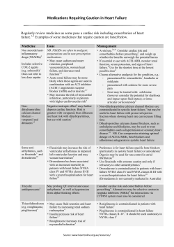

© Copyright 2026